Two-dimensional gel electrophoresis data for proteomic profiling of Sporothrix yeast cells
7
Data Article Two-dimensional gel electrophoresis data for proteomic profiling of Sporothrix yeast cells Anderson Messias Rodrigues a , Paula H. Kubitschek-Barreira b , Geisa Ferreira Fernandes a , Sandro Rogério de Almeida c , Leila M. Lopes-Bezerra b , Zoilo Pires de Camargo a,n a Department of Microbiology, Immunology and Parasitology, Cell Biology Division, Federal University of São Paulo (UNIFESP), São Paulo, Brazil b Department of Cellular Biology, The Roberto Alcantara Gomes Institute of Biology, University of Estado Rio de Janeiro (UERJ), Brazil c Department of Clinical and Toxicological Analysis, Faculty of Pharmaceutical Sciences, University of São Paulo (USP), São Paulo, Brazil article info Article history: Received 21 November 2014 Accepted 21 November 2014 Available online 2 December 2014 abstract Sporotrichosis is a chronic infection of the skin and subcutaneous tissues of human and other mammals caused by a complex of cryptic dimorphic fungi in the plant-associated order Ophiostomatales. With major differences between routes of transmission, Sporothrix infections are emerging as new threat in tropical and subtropical areas, particularly in form of outbreaks. The mechanisms underlying the pathogenesis and invasion of Sporothrix spp. are still poorly understood and many virulence factors remain unidenti fied. In this scenario, a global analysis of proteins expressed by clinical Sporothrix species combined with the identification of seroreactive proteins is overdue. Optimization of sample preparation and electrophoresis conditions are key steps toward reproducibility of gel-based proteomics assays. We provide the data generated using an efficient protocol of protein extraction for rapid and large-scale proteome analysis using two- dimensional gel electrophoresis. The protocol was established and optimized for pathogenic and non-pathogenic Sporothrix spp. includ- ing Sporothrix brasiliensis (CBS 132990), Sporothrix schenckii sensu Contents lists available at ScienceDirect journal homepage: www.elsevier.com/locate/dib Data in Brief http://dx.doi.org/10.1016/j.dib.2014.11.004 2352-3409/& 2014 The Authors. Published by Elsevier Inc. This is an open access article under the CC BY license (http://creativecommons.org/licenses/by/3.0/). DOI of original article: http://dx.doi.org/10.1016/j.jprot.2014.11.013 n Corresponding author. E-mail address: [email protected] (Z.P. de Camargo). Data in Brief 2 (2015) 32–38
Transcript of Two-dimensional gel electrophoresis data for proteomic profiling of Sporothrix yeast cells
Contents lists available at ScienceDirect
Data in Brief
Data in Brief 2 (2015) 32–38
http://d2352-34(http://c
DOIn CorrE-m
journal homepage: www.elsevier.com/locate/dib
Data Article
Two-dimensional gel electrophoresis data forproteomic profiling of Sporothrix yeast cells
Anderson Messias Rodrigues a, Paula H. Kubitschek-Barreira b,Geisa Ferreira Fernandes a, Sandro Rogério de Almeida c,Leila M. Lopes-Bezerra b, Zoilo Pires de Camargo a,n
a Department of Microbiology, Immunology and Parasitology, Cell Biology Division, Federal University of SãoPaulo (UNIFESP),São Paulo, Brazilb Department of Cellular Biology, The Roberto Alcantara Gomes Institute of Biology, University of Estado Riode Janeiro (UERJ), Brazilc Department of Clinical and Toxicological Analysis, Faculty of Pharmaceutical Sciences, University of SãoPaulo (USP), São Paulo, Brazil
a r t i c l e i n f o
Article history:Received 21 November 2014Accepted 21 November 2014Available online 2 December 2014
x.doi.org/10.1016/j.dib.2014.11.00409/& 2014 The Authors. Published by Elsevreativecommons.org/licenses/by/3.0/).
of original article: http://dx.doi.org/10.1016esponding author.ail address: [email protected] (Z.P. d
a b s t r a c t
Sporotrichosis is a chronic infection of the skin and subcutaneoustissues of human and other mammals caused by a complex of crypticdimorphic fungi in the plant-associated order Ophiostomatales. Withmajor differences between routes of transmission, Sporothrix infectionsare emerging as new threat in tropical and subtropical areas,particularly in form of outbreaks. The mechanisms underlying thepathogenesis and invasion of Sporothrix spp. are still poorly understoodand many virulence factors remain unidentified. In this scenario, aglobal analysis of proteins expressed by clinical Sporothrix speciescombined with the identification of seroreactive proteins is overdue.Optimization of sample preparation and electrophoresis conditions arekey steps toward reproducibility of gel-based proteomics assays. Weprovide the data generated using an efficient protocol of proteinextraction for rapid and large-scale proteome analysis using two-dimensional gel electrophoresis. The protocol was established andoptimized for pathogenic and non-pathogenic Sporothrix spp. includ-ing Sporothrix brasiliensis (CBS 132990), Sporothrix schenckii sensu
ier Inc. This is an open access article under the CC BY license
/j.jprot.2014.11.013
e Camargo).
A.M. Rodrigues et al. / Data in Brief 2 (2015) 32–38 33
stricto (CBS 132974), Sporothrix globosa (CBS 132922), and Sporothrixmexicana (CBS 120341). The data, supplied in this article, are related tothe research article entitled “Immunoproteomic analysis reveals aconvergent humoral response signature in the Sporothrix schenckiicomplex” (Rodrigues et al., 2014 [1]).& 2014 The Authors. Published by Elsevier Inc. This is an open access
article under the CC BY license(http://creativecommons.org/licenses/by/3.0/).
Specifications table
Subject area
BiologyMore specificsubject area
Medical Mycology, Immunology
Type of data
Image How data wasacquiredScanning electron microscopy FEI Quanta™ FEG 250, Ettan IPGphor 3 Isoelectric Focusing Unit, ImageScanner III
Data format
Analyzed Experimental factors Sporothrix yeast cells from clinical and environmental species were grown in Brain Heart Infusionmedium for 7 days at 36 1C and subjected to a whole cell protein extraction
ExperimentalfeaturesAll the samples were separated using two-dimensional gel electrophoresis
Data source location
São Paulo, Brazil Data accessibility Within this articleValue of the data
� Morphological and morphometric analyses using scanning electron microscopy revealed distinct cellular architecture ofSporothrix species.
� A gel-based proteomic profiling of pathogenic and non-pathogenic Sporothrix spp. is proposed.� Data from an efficient method of protein extraction with application to 2D gel electrophoresis and mass spectrometryanalyses.
� The optimized protocol is useful for comparative proteome analysis of Sporothrix spp., including immunoblotting and2D-DIGE.
1. Data, experimental design, materials and methods
1.1. Sporothrix culture conditions and morphological characterization
Sporothrix brasiliensis (CBS 132990), Sporothrix schenckii sensu stricto (CBS 132974), Sporothrixglobosa (CBS 132922), and Sporothrix mexicana (CBS 120341) were maintained in Sabouraud dextroseagar slants (Difco, Detroit, USA) and cultivated for 10–14 days at 25 1C prior to use. These isolates werechosen based on geographical origin and genetic diversity [2–5], virulence profile [6,7], andphysiological response to antifungal agents [8,9]. Approximately 2�106 conidia (viable cells 90%)were used to inoculate 500 ml flasks containing 100 ml of Brain Heart Infusion Broth (Difco, Detroit,USA). The cultures were incubated at 36 1C in triplicate in a rotary shaker (Multitron II – Infors HT,Switzerland) with constant orbital agitation (110 rpm) for 7 days.
The morphological characterization of Sporothrix spp. was performed by scanning electronmicroscopy (SEM), especially because we were comparing species with different ecological andpathogenic behavior. Sporothrix yeast cells, obtained as described above, were harvested bycentrifugation and washed twice in phosphate buffered saline (PBS). The cells were fixed overnight

Fig. 1. SEM-image of the parasitic phase of pathogenic and non-pathogenic Sporothrix species. The species embedded in theclinical clade are represented by reference clinical strains collected during the largest epidemic of sporotrichosis in SouthAmerica. (A) Multi-budding yeasts of S. brasiliensis (CBS 132990¼Ss54) clade I; (B) classical cigar-shaped yeasts of S. schenckii(CBS 132974¼Ss118) clade II; (C) S. globosa (CBS 132922¼Ss06) clade III. The ancestral non-virulent S. mexicana (CBS 120341)clade IV presented irregular budding yeasts. Bar¼10 mm.
A.M. Rodrigues et al. / Data in Brief 2 (2015) 32–3834
in 2.5% glutaraldehyde containing 0.1 M sodium cacodylate buffer (pH 7.2) washed in the same bufferand adhered onto poly-l-lysine-coated coverslips. Cells were post-fixed in 1% OsO4 containing 0.1 Msodium cacodylate, followed by 1% tannic acid for 30 min, each with appropriate washes. After osmium/sodium cacodylate, samples were immersed in 1% OsO4, washed in ultra-pure water, dehydrated in anethanol series dilution (50%, 70%, 90%, and 100%), and critical-point dried with CO2 (Balzers CPD 030).Specimens were ion-sputtered with a 25-nm gold layer using a Leica EM SCD500 to avoid a chargeeffect while searching for a suitable site. SEM images were obtained using a FEI Quanta™ FEG 250 (FeiCompany, USA) at a 5 kV accelerating tension (Federal University of São Paulo, Electron MicroscopyFacility [CEME]) [1]. A representative image of Sporothrix yeast cells is shown in Fig. 1.
1.2. Morphological characteristics of Sporothrix yeast cells
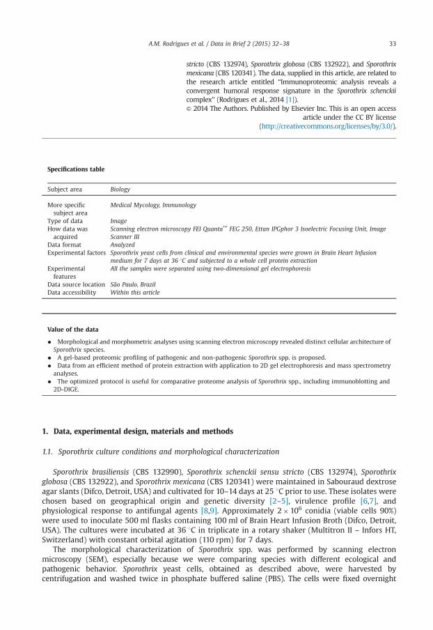
Sporothrix yeast cells were processed and analyzed using Image J 1.44 C morphometric software(NIH, Bethesda, Maryland, USA; http://rsb.info.nih.gov/ij/). All measurements were estimated on thebasis of the results obtained with at least 100 yeast cells/isolate from a 7-day-old culture on BrainHeart Infusion Broth at 36 1C as described above. Data were analyzed using one-way analysis ofvariance (ANOVA) with Tukey's multiple comparison post-hoc test. A p-value o0.05 was consideredto indicate significant differences, and analyses were performed using GraphPad (GraphPad Prism v.
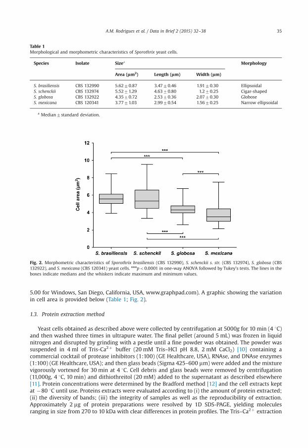
Table 1Morphological and morphometric characteristics of Sporothrix yeast cells.
Species Isolate Sizea Morphology
Area (lm2) Length (lm) Width (lm)
S. brasiliensis CBS 132990 5.6270.87 3.4770.46 1.9170.30 EllipsoidalS. schenckii CBS 132974 5.5271.29 4.6370.80 1.270.25 Cigar-shapedS. globosa CBS 132922 4.3570.72 2.5370.36 2.0770.30 GloboseS. mexicana CBS 120341 3.7771.03 2.9970.54 1.5670.25 Narrow ellipsoidal
a Median7standard deviation.
Fig. 2. Morphometric characteristics of Sporothrix brasiliensis (CBS 132990), S. schenckii s. str. (CBS 132974), S. globosa (CBS132922), and S. mexicana (CBS 120341) yeast cells. nnnpo0.0001 in one-way ANOVA followed by Tukey's tests. The lines in theboxes indicate medians and the whiskers indicate maximum and minimum values.
A.M. Rodrigues et al. / Data in Brief 2 (2015) 32–38 35
5.00 for Windows, San Diego, California, USA, www.graphpad.com). A graphic showing the variationin cell area is provided below (Table 1; Fig. 2).
1.3. Protein extraction method
Yeast cells obtained as described above were collected by centrifugation at 5000g for 10 min (4 1C)and then washed three times in ultrapure water. The final pellet (around 5 mL) was frozen in liquidnitrogen and disrupted by grinding with a pestle until a fine powder was obtained. The powder wassuspended in 4 ml of Tris-Ca2þ buffer (20 mM Tris–HCl pH 8.8, 2 mM CaCl2) [10] containing acommercial cocktail of protease inhibitors (1:100) (GE Healthcare, USA), RNAse, and DNAse enzymes(1:100) (GE Healthcare, USA); and then glass beads (Sigma 425–600 mm) were added and the mixturevigorously vortexed for 30 min at 4 1C. Cell debris and glass beads were removed by centrifugation(11,000g, 4 1C, 10 min) and dithiothreitol (20 mM) added to the supernatant as described elsewhere[11]. Protein concentrations were determined by the Bradford method [12] and the cell extracts keptat �80 1C until use. Proteins extracts were evaluated according to (i) the amount of protein extracted;(ii) the diversity of bands; (iii) the integrity of samples as well as the reproducibility of extraction.Approximately 2 mg of protein preparations were resolved by 1D SDS-PAGE, yielding moleculesranging in size from 270 to 10 kDa with clear differences in protein profiles. The Tris–Ca2þ extraction

Fig. 3. Protein profile from Sporothrix spp. after 7 days of incubation at 36 1C, 110 rpm in Brain Heart Infusion (BHI) media. Thecell extracts were subjected to 1D SDS-PAGE and the proteins developed by silver staining. The molecular masses (in kDa) ofstandard proteins are given to the left of the gel (BenchMarkTM Protein Ladder, Invitrogen). CBS 132990¼S. brasiliensis; CBS132974¼S. schenckii; CBS 132922¼S. globosa; CBS 120341¼S. mexicana.
A.M. Rodrigues et al. / Data in Brief 2 (2015) 32–3836
protocol was suitable for the study of Sporothrix antigenic molecules, generating samples with a highamount of protein with no degradation. A representative image of Sporothrix proteins resolved by 1DSDS-PAGE is shown in Fig. 3.
1.4. Two-dimensional gel electrophoresis.
To assess the complexity of the samples, 300 mg of protein was fractionated by 2D electrophoresis.Proteins were successfully precipitated using the 2D clean-up kit (GE Healthcare, Piscataway, NJ, USA)following the manufacturer's recommendations. Initially, for reproducibility, immobilized pH gradient(IPG) strips ranging from 3 to 10 were employed. The resulting gels had low resolution with aconcentration of spots in the pH range of 4–7. Thus, the experiments were conducted using animmobilized pH gradient of 4–7, which provided better sample separation and improved resolution.Proteins were diluted with rehydration solution (7 M urea, 2 M thiourea, 2% CHAPS, 1.2% DeStreak, 2%vol/vol isoelectric focusing [IEF] buffer pH 4–7, and trace bromophenol blue) to a final volume of250 ml. IEF was performed using a Ettan IPGphor III system (GE Healthcare, USA). IPG strips (pH 4–7,13 cm) were rehydrated at 30 V for 12 h. Proteins were subsequently focused at 200 V for 2 h, 500 Vfor 2 h, 1000 V for 5 h, and then a gradient applied from 1000 to 5000 V for 2 h. Finally, the voltagewas set to 5000 V until 60,000 Vh. All IEF experiments were performed at 20 1C. After one-dimensional IEF, the IPG strips were reduced for 15 min with 1.5% dithioerythritol and alkylated for15 minwith 2.5% iodocetamide in equilibration buffer (6 M urea, 50 mM Tris–HCl pH 6.8, 30% glycerol,
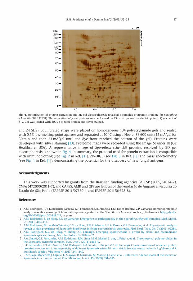
Fig. 4. Optimization of protein extraction and 2D gel electrophoresis revealed a complex proteomic profiling for Sporothrixschenckii (CBS 132974). The separation of yeast proteins was performed on 13 cm strips over isoelectric point (pI) gradient of4–7. Gel was loaded with 300 μg of total protein and silver stained.
A.M. Rodrigues et al. / Data in Brief 2 (2015) 32–38 37
and 2% SDS). Equilibrated strips were placed on homogeneous 10% polyacrylamide gels and sealedwith 0.5% low-melting-point agarose and separated at 10 1C using a Hoefer SE 600 unit (15 mA/gel for30 min and then 23 mA/gel until the dye front reached the bottom of the gel). Proteins weredeveloped with silver staining [13]. Proteome maps were recorded using the Image Scanner III (GEHealthcare, USA). A representative image of Sporothrix schenckii proteins resolved by 2D gelelectrophoresis is shown in Fig. 4. In summary, the protocol used for protein extraction is compatiblewith immunoblotting (see Fig. 2 in Ref. [1]), 2D-DIGE (see Fig. 3 in Ref. [1]) and mass spectrometry(see Fig. 4 in Ref. [1]), demonstrating the potential for the discovery of new fungal antigens.
Acknowledgments
This work was supported by grants from the Brazilian funding agencies FAPESP (2009/54024-2),CNPq (472600/2011-7), and CAPES. AMR and GFF are fellows of the Fundação de Amparo à Pesquisa doEstado de São Paulo (FAPESP 2011/07350-1 and FAPESP 2011/01628-8).
References
[1] A.M. Rodrigues, P.H. Kubitschek-Barreira, G.F. Fernandes, S.R. Almeida, L.M. Lopes-Bezerra, Z.P. Camargo, Immunoproteomicanalysis reveals a convergent humoral response signature in the Sporothrix schenckii complex, J. Proteomics, http://dx.doi.org/10.1016/j.jprot.2014.11.013, in press.
[2] A.M. Rodrigues, S. de Hoog, Z.P. de Camargo, Emergence of pathogenicity in the Sporothrix schenckii complex, Med. Mycol.51 (2013) 405–412.
[3] A.M. Rodrigues, M. de Melo Teixeira, G.S. de Hoog, T.M.P. Schubach, S.A. Pereira, G.F. Fernandes, et al., Phylogenetic analysisreveals a high prevalence of Sporothrix brasiliensis in feline sporotrichosis outbreaks, PLoS Negl. Trop. Dis. 7 (2013) e2281.
[4] A.M. Rodrigues, G.S. de Hoog, Y. Zhang, Z.P. Camargo, Emerging sporotrichosis is driven by clonal and recombinantSporothrix species, Emerg. Microbes Infect. 3 (2014) e32.
[5] A.A. Sasaki, G.F. Fernandes, A.M. Rodrigues, F.M. Lima, M.M. Marini, S. dos, L. Feitosa, et al., Chromosomal polymorphism inthe Sporothrix schenckii complex, PLoS One 9 (2014) e86819.
[6] G.F. Fernandes, P.O. dos Santos, A.M. Rodrigues, A.A. Sasaki, E. Burger, Z.P. de Camargo, Characterization of virulence profile,protein secretion and immunogenicity of different Sporothrix schenckii sensu stricto isolates compared with S. globosa and S.brasiliensis species, Virulence 4 (2013) 241–249.
[7] I. Arrillaga-Moncrieff, J. Capilla, E. Mayayo, R. Marimon, M. Mariné, J. Gené, et al., Different virulence levels of the species ofSporothrix in a murine model, Clin. Microbiol. Infect. 15 (2009) 651–655.

A.M. Rodrigues et al. / Data in Brief 2 (2015) 32–3838
[8] R.S. Brilhante, A.D. Malaquias, E.P. Caetano, S. Castelo-Branco Dde, R.A. Lima, F.J. Marques, et al., In vitro inhibitory effect ofmiltefosine against strains of Histoplasma capsulatum var. capsulatum and Sporothrix spp., Med. Mycol. 52 (2014) 320–325.
[9] A.M. Rodrigues, G.S. de Hoog, D. de Cassia Pires, R.S.N. Brihante, J.J. da Costa Sidrim, M.F. Gadelha, et al., Genetic diversityand antifungal susceptibility profiles in causative agents of sporotrichosis, BMC Infect. Dis. 14 (2014) 219.
[10] C.A. da Fonseca, R.S.A. Jesuino, M.S.S. Felipe, D.A. Cunha, W.A. Brito, C.M.A. Soares, Two-dimensional electrophoresis andcharacterization of antigens from Paracoccidioides brasiliensis, Microbes Infect. 3 (2001) 535–542.
[11] T.A. Missall, M.E. Pusateri, M.J. Donlin, K.T. Chambers, J.A. Corbett, J.K. Lodge, Posttranslational, translational, andtranscriptional responses to nitric oxide stress in Cryptococcus neoformans: implications for virulence, Eukaryot. Cell 5(2006) 518–529.
[12] M.M. Bradford, A rapid and sensitive method for the quantitation of microgram quantities of protein utilizing the principleof protein-dye binding, Anal. Biochem. 72 (1976) 248–254.
[13] H. Blum, H. Beier, H.J. Gross, Improved silver staining of plant proteins, RNA and DNA in polyacrylamide gels,Electrophoresis 8 (1987) 93–99.