Sodium dodecyl sulphate-polyacrylamide gel electrophoresis of proteins in dry-cured hams: Data...
7
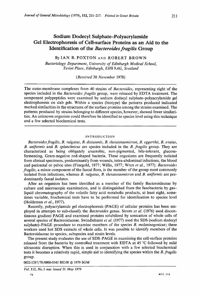
Journal of General Microbiology (1979), 112, 211-217. Printed in Great Britain Sodium Dodecyl Sulphate-Polyacrylamide Gel Electrophoresis of Cell-surface Proteins as an Aid to the Identification of the Bacteroides fragilis Group By IAN R. POXTON AND ROBERT BROWN Bacteriology Department, University of Edinburgh Medical School, Teviot Place, Edinburgh, EHS 9AG, Scotlund (Received 30 November 1978) 21 1 The outer-membrane complexes from 40 strains of Bucteroides, representing eight of the species included in the Bacteroides frdgilis group, were released by EDTA treatment. The component polypeptides were examined by sodium dodecyl sulphate-polyacrylamide gel electrophoresis on slab gels. Within a species (biotype) the patterns produced indicated marked similarities in the structures of the surface proteins among the strains examined. The patterns produced by strains belonging to different species, however, showed fewer similari- ties. An unknown organism could therefore be identified to species level using this technique and a few selected biochemical tests. INTRODUCTION Bacteroides fragilis, B. vulgatus, B. distasonis, B. thetaiotaomicron, B. eggerthii, B. OVdtUS, B. uniformis and B. splanchnicus are species included in the B. frugilis group. They are characterized as being obligately anaerobic, non-pigmented, bile-tolerant, glucose- fermenting, Gram-negative rod-shaped bacteria. These organisms are frequently isolated from clinical specimens, predominantly from wounds, intra-abdominal infections, the blood and perirectal or pelvic sites (Finegold, 1977; Willis, 1977; Wren et al., 1977). Bucteroides fragilis, a minor component of the faecal flora, is the member of the group most commonly isolated from infections, whereas B. vulgatus, B. thetaiotaomicron and B. uniformib are pre- dominantly faecal isolates. After an organism has been identified as a member of the family Bacteroidaceae by culture and microscopic examination, and is distinguished from the fusobacteria by gas- liquid chromatography of the volatile fatty acid metabolic products, at least eight, some- times variable, biochemical tests have to be performed for identification to species level (Holdeman et al., 1977). Recently, polyacrylamide gel electrophoresis (PAGE) of cellular proteins has been em- ployed in attempts to sub-classify the Bucteroides genus. Strom et al. (1976) used discon- tinuous gradient PAGE and examined proteins solubilized by sonication of whole cells of several species of Bacteroidaceae. Swindlehurst et al. (1977) used the SDS (sodium dodecyl su1phate)-PAGE procedure to examine members of the species B. meluninogenicus; these workers used hot SDS extracts of whole cells. It was possible to identify members of the Bacteroidaceae to species, subspecies and strain levels. The present study evaluates the use of SDS-PAGE in examining the cell-surface proteins released from the bacteria by controlled treatment with EDTA at 45 "C followed by mild ultrasonic disruption. When this is used in conjunction with a few selected biochemical tests it becomes a relatively rapid, simple aid to identifying the species within the B. fragilis group. 0022-1287/79/oooO-8343 $02.00 @ 1979 SGM VoZ. 112, No. 1 was issued 31 May 1979 14 &SIC I12
-
Upload
independent -
Category
Documents
-
view
4 -
download
0
Transcript of Sodium dodecyl sulphate-polyacrylamide gel electrophoresis of proteins in dry-cured hams: Data...
Downloaded from www.microbiologyresearch.org by
IP: 107.22.17.133
On: Sun, 14 Aug 2016 18:56:13
Journal of General Microbiology (1979), 112, 211-217. Printed in Great Britain
Sodium Dodecyl Sulphate-Polyacrylamide Gel Electrophoresis of Cell-surface Proteins as an Aid to the
Identification of the Bacteroides fragilis Group
By I A N R. POXTON A N D ROBERT BROWN Bacteriology Department, University of Edinburgh Medical School,
Teviot Place, Edinburgh, EHS 9AG, Scotlund
(Received 30 November 1978)
21 1
The outer-membrane complexes from 40 strains of Bucteroides, representing eight of the species included in the Bacteroides frdgilis group, were released by EDTA treatment. The component polypeptides were examined by sodium dodecyl sulphate-polyacrylamide gel electrophoresis on slab gels. Within a species (biotype) the patterns produced indicated marked similarities in the structures of the surface proteins among the strains examined. The patterns produced by strains belonging to different species, however, showed fewer similari- ties. An unknown organism could therefore be identified to species level using this technique and a few selected biochemical tests.
I N T R O D U C T I O N
Bacteroides fragilis, B. vulgatus, B. distasonis, B. thetaiotaomicron, B. eggerthii, B. OVdtUS, B. uniformis and B. splanchnicus are species included in the B. frugilis group. They are characterized as being obligately anaerobic, non-pigmented, bile-tolerant, glucose- fermenting, Gram-negative rod-shaped bacteria. These organisms are frequently isolated from clinical specimens, predominantly from wounds, intra-abdominal infections, the blood and perirectal or pelvic sites (Finegold, 1977; Willis, 1977; Wren et al., 1977). Bucteroides fragilis, a minor component of the faecal flora, is the member of the group most commonly isolated from infections, whereas B. vulgatus, B. thetaiotaomicron and B. uniformib are pre- dominantly faecal isolates.
After an organism has been identified as a member of the family Bacteroidaceae by culture and microscopic examination, and is distinguished from the fusobacteria by gas- liquid chromatography of the volatile fatty acid metabolic products, at least eight, some- times variable, biochemical tests have to be performed for identification to species level (Holdeman et al., 1977).
Recently, polyacrylamide gel electrophoresis (PAGE) of cellular proteins has been em- ployed in attempts to sub-classify the Bucteroides genus. Strom et al. (1976) used discon- tinuous gradient PAGE and examined proteins solubilized by sonication of whole cells of several species of Bacteroidaceae. Swindlehurst et al. (1977) used the SDS (sodium dodecyl su1phate)-PAGE procedure to examine members of the species B. meluninogenicus; these workers used hot SDS extracts of whole cells. It was possible to identify members of the Bacteroidaceae to species, subspecies and strain levels.
The present study evaluates the use of SDS-PAGE in examining the cell-surface proteins released from the bacteria by controlled treatment with EDTA at 45 "C followed by mild ultrasonic disruption. When this is used in conjunction with a few selected biochemical tests it becomes a relatively rapid, simple aid to identifying the species within the B. fragilis group. 0022-1287/79/oooO-8343 $02.00 @ 1979 SGM
VoZ. 112, No. 1 was issued 31 May 1979
14 &SIC I12
Downloaded from www.microbiologyresearch.org by
IP: 107.22.17.133
On: Sun, 14 Aug 2016 18:56:13
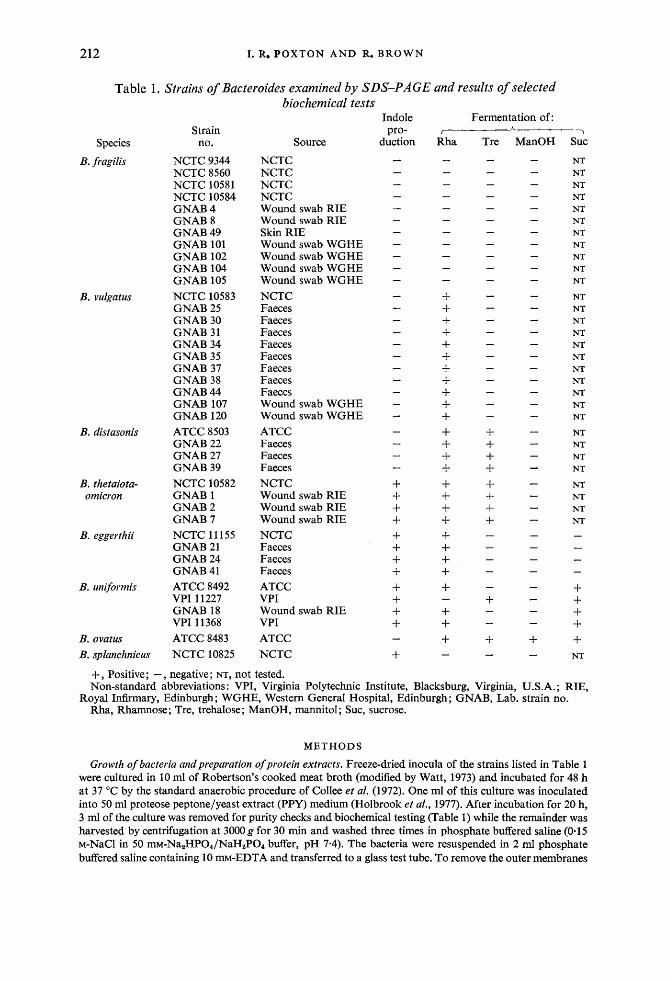
212 I. R. POXTON AND R. BROWN
Table 1. Strains of Bacteroides examined by SDS-PAGE and results of selected biochemical tests
Species
B. fragilis
B. vulgatus
B. distasonis
B. thetaiota- omicron
B. eggerthii
B. uniformis
B. ovatus B. splanchnicus
Strain no.
NCTC 9344 NCTC 8560 WCTC 10581 NCTC 10584 GNAB 4 GNAB 8 GNAB 49 GNAB 101 GNAB 102 GNAB 104 GNAB 105 NCTC 10583 GNAB 25 GNAB 30 GNAB 31 GNAB 34 GNAB 35 GNAB 37 GNAB 38 GNAB 44 GNAB 107 GNAB 120 ATCC 8503 GNAB 22 GNAB 27 GNAB 39 NCTC 10582 GNAB 1 GNAB 2 GNAB 7 NCTC 11 155 GNAB 21 GNAB 24 GNAB 41 ATCC 8492 VPI 11227 GNAB 18 VPI 11368 ATCC 8483 NCTC 10825
Source
NCTC NCTC NCTC NCTC Wound swab RIE Wound swab RIE Skin RIE Wound swab WGHE Wound swab WGHE Wound swab WGHE Wound swab WGHE NCTC Faeces Faeces Faeces Faeces Faeces Faeces Faeces Faeces Wound swab WGHE Wound swab WGHE ATCC Faeces Faeces Faeces NCTC Wound swab RIE Wound swab RIE Wound swab RIE NCTC Faeces Faeces Faeces ATCC VPI Wound swab RIE VPI ATCC NCTC
Fermentation of: 7
h >
Rha - - - - - - - - - - - t- + + + + + + + + + + + + + + + + + + + + + + + + + +
-
-
sue NT NT NT NT NT NT NT NT NT NT NT
NT NT NT NT NT NT NT NT NT NT NT
NT NT NT NT
NT NT NT NT
- - - -
+ + + + + NT
R
+, Positive; -, negative; NT, not tested. Non-standard abbreviations : VPI, Virginia Polytechnic Institute, Blacksburg, Virginia, U.S.A. ; RIE,
Rha, Rhamnose; Tre, trehalose; ManOH, mannitol; SUC, sucrose. .oyal Infirmary, Edinburgh; WGHE, Western General Hospital, Edinburgh; GNAB, Lab. strain no.
METHODS
Growth of bacteria andpreparation of protein extracts. Freeze-dried inocula of the strains listed in Table 1 were cultured in 10 ml of Robertson's cooked meat broth (modified by Watt, 1973) and incubated for 48 h at 37 "C by the standard anaerobic procedure of Collee et al. (1972). One ml of this culture was inoculated into 50 ml proteose peptone/yeast extract (PPY) medium (Holbrook et al., 1977). After incubation for 20 h, 3 ml of the culture was removed for purity checks and biochemical testing (Table 1) while the remainder was harvested by centrifugation at 3000 g for 30 min and washed three times in phosphate buffered saline (0.1 5 M-NaCl in 50 ~M-N~,HPO, /N~H,PO~ buffer, pH 7.4). The bacteria were resuspended in 2 ml phosphate buffered saline containing 10 m-EDTA and transferred to a glass test tube. To remove the outer membranes
Downloaded from www.microbiologyresearch.org by
IP: 107.22.17.133
On: Sun, 14 Aug 2016 18:56:13
SDS-PAGE of B. fragilis 213 and associated molecules, the suspension was incubated for 30 min at 45 "C, then vortex-mixed, treated in an ultrasonic bath (model 6441A; Dawe Instruments, Western Ave, London) for 1 min and again vortex- mixed. After two centrifugations (6000 g for 30 min) the supernatant fluid contained outer-membrane vesicles together with released proteins and other molecules. The above conditions were standardized to give optimum release of protein without lysis of the bacterial cytoplasmic membrane. EDTA was removed by dialysis for 4 to 5 h in two successive 1 litre volumes of 0.01 M-Tris/HCl pH 7.4 containing 0.01 % (v/v) 2-mercaptoethanol and the samples were concentrated by dusting the dialysis bags with a fine layer of Sephadex G-100 and leaving at 4 "C for 16 h. The concentrated extracts were freeze-dried and dissolved in SDS-PAGE sample buffer to give a protein concentration of 2 mg d-l. Sample buffer was 0.0625 M-TT~s/ HCl pH 6.8 containing 2 % (w/v) SDS, 10 % (v/v) glycerol, 1 % (v/v) 2-mercaptoethanol and 0.001 % (w/v) bromophenol blue. Samples were heated in a boiling water bath for 3 min just prior to application to the gel.
PoZyacrylamide gel electrophoresis. This was adapted from the method of Laemmli (1970). Slab gels (170 x 140 mm) of 10 % (w/v) acrylarnide with a 10 mm 4 % (w/v) stacking gel were used to run up to 12 samples in a Raven (Haverhill, Suffolk, CB9 7UU) slab gel apparatus. Buffers were as described by Laemmli (1970). Samples (100 pl) of extract containing 200 pg protein were electrophoresed at constant voltage, first at 50 V until the sample had entered the separating gel (approx. 1 h) then at 150 V until the bromophenol blue was near the bottom of the gel (approx. 4 h). Staining was carried out overnight and de-staining was done over 4 h with the solutions described by Poxton & Sutherland (1976).
Protein estimation. Samples of EDTA extracts were assayed for protein using the method of Lowry et al. (1951).
Characterization of strains used. All test strains had previously been characterized by the procedures of Duerden et al. (1976). Selected biochemical tests, however, were performed on each strain after growth in PPY medium (Table 1). These served as a check on the previous results. They included tests on indole pro- duction and the fermentation of rhamnose, trehalose, sucrose and mannitol by the methods described by Duerden et al. (1976).
R E S U L T S A N D D I S C U S S I O N
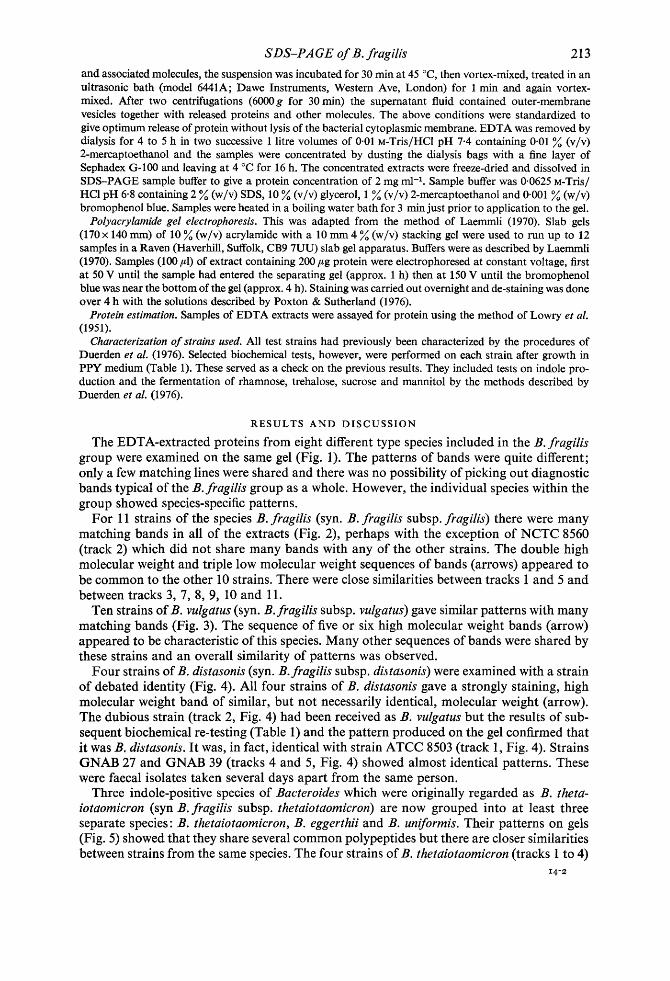
The EDTA-extracted proteins from eight different type species included in the B. fragilis group were examined on the same gel (Fig. 1). The patterns of bands were quite different; only a few matching lines were shared and there was no possibility of picking out diagnostic bands typical of the B. fragilis group as a whole. However, the individual species within the group showed species-specific patterns.
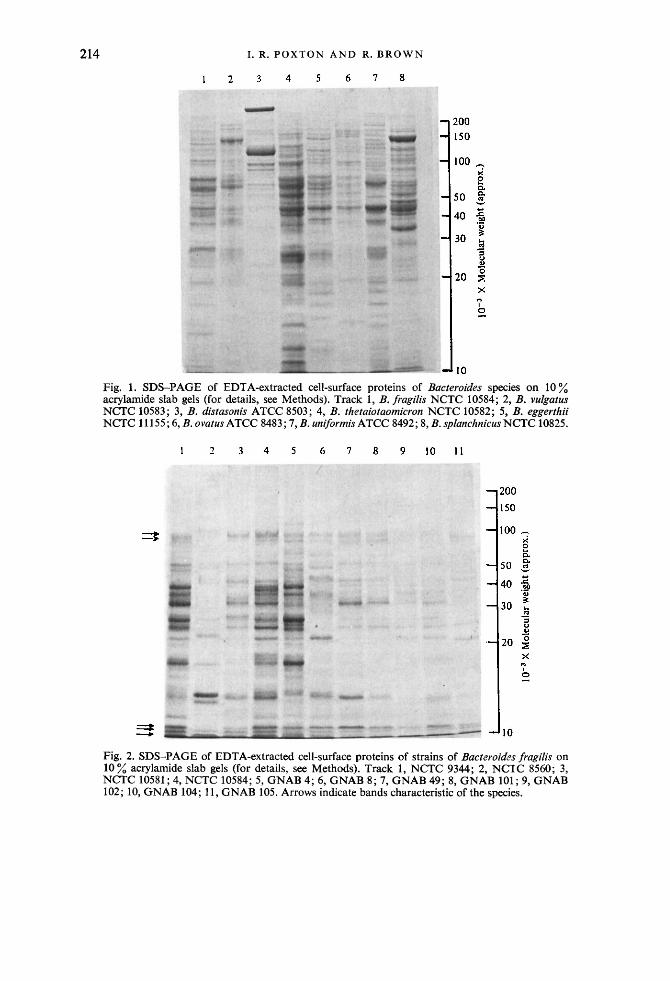
For 11 strains of the species B. fragilis (syn. B. fragilis subsp. fragilis) there were many matching bands in all of the extracts (Fig. 2), perhaps with the exception of NCTC 8560 (track 2) which did not share many bands with any of the other strains. The double high molecular weight and triple low molecular weight sequences of bands (arrows) appeared to be common to the other 10 strains. There were close similarities between tracks 1 and 5 and between tracks 3, 7, 8, 9, 10 and 11.
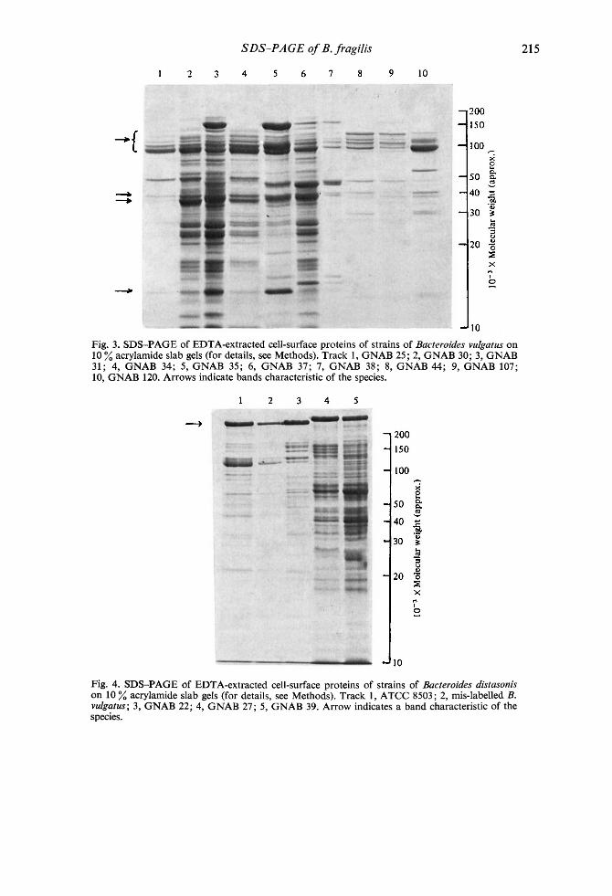
Ten strains of B. VUlgdtUS (syn. B. fragilis subsp. vulgatus) gave similar patterns with many matching bands (Fig. 3). The sequence of five or six high molecular weight bands (arrow) appeared to be characteristic of this species. Many other sequences of bands were shared by these strains and an overall similarity of patterns was observed.
Four strains of B. distasonis (syn. B. fragilis subsp. di,sta,sonis) were examined with a strain of debated identity (Fig. 4). All four strains of B. distasonis gave a strongly staining, high molecular weight band of similar, but not necessarily identical, molecular weight (arrow). The dubious strain (track 2, Fig. 4) had been received as B. vulgatus but the results of sub- sequent biochemical re-testing (Table 1) and the pattern produced on the gel confirmed that it was B. distasonis. It was, in fact, identical with strain ATCC 8503 (track 1, Fig. 4). Strains GNAB 27 and GNAB 39 (tracks 4 and 5, Fig. 4) showed almost identical patterns. These were faecal isolates taken several days apart from the same person.
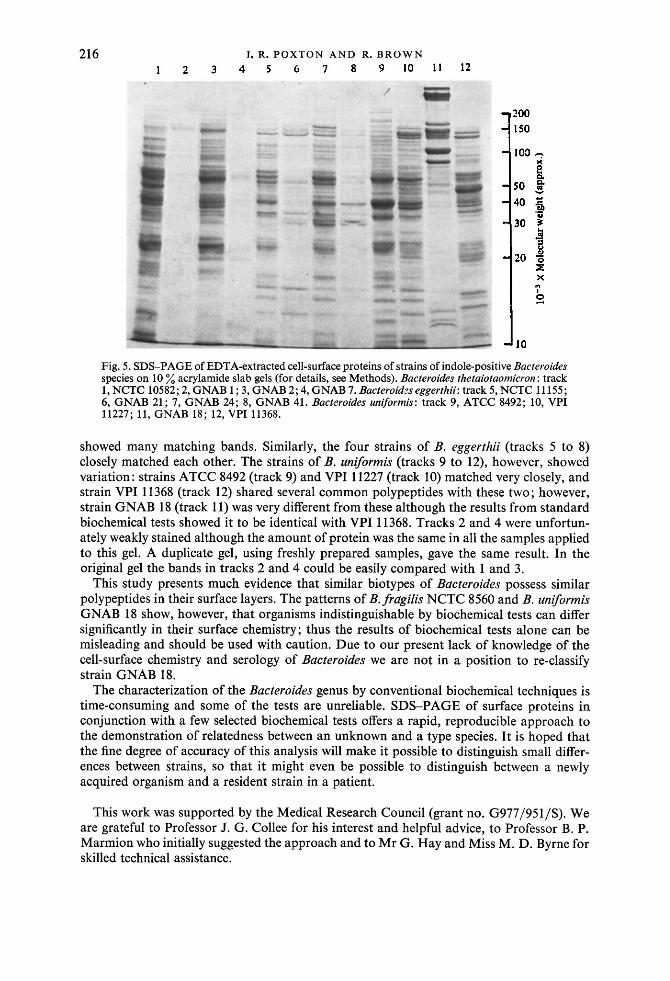
Three indole-positive species of Bacteroides which were originally regarded as B. theta- iOtdOmiCrOn (syn B. fragilis subsp. thetdiotdomicron) are now grouped into at least three separate species : B. thetaiotaomicron, B. eggerthii and B. uniformis. Their patterns on gels (Fig. 5) showed that they share several common polypeptides but there are closer similarities between strains from the same species. The four strains of B. thetaiotaomicron (tracks 1 to 4)
14-2
Downloaded from www.microbiologyresearch.org by
IP: 107.22.17.133
On: Sun, 14 Aug 2016 18:56:13
214 I. R. P O X T O N A N D R. B R O W N
Fig. 1. SDS-PAGE of EDTA-extracted cell-surface proteins of Bacteroides species on 10 % acrylamide slab gels (for details, see Methods). Track 1, B. frugilis NCTC 10584; 2, B. vuZgatus NCTC 10583; 3, B. distasonis ATCC 8503; 4, B. th.etuiotuomicron NCTC 10582; 5, B. eggerthii NCTC 11 155; 6, B. ovatus ATCC 8483 ; 7, B. uniformis ATCC 8492; 8, B. splanchnicus NCTC 10825.
Fig. 2. SDS-PAGE of EDTA-extracted cell-surface proteins of strains of Bacteroides fragilis on 10% acrylamide slab gels (for details, see Methods). Track 1, NCTC 9344; 2, NCTC 8560; 3, NCTC 10581; 4, NCTC 10584; 5, GNAB 4; 6 , GNAB 8 ; 7, GNAB 49; 8, GNAB 101; 9, GNAB 102; 10, GNAB 104; 11 , GNAB 105. Arrows indicate bands characteristic of the species.
Downloaded from www.microbiologyresearch.org by
IP: 107.22.17.133
On: Sun, 14 Aug 2016 18:56:13
SDS-PAGE of B. fragilis 21 5
Fig. 3. SDS-PAGE of EDTA-extracted cell-surface proteins of strains of Bacteroides vulgatus on 10 % acrylamide slab gels (for details, see Methods). Track 1 , GNAB 25; 2, GNAB 30; 3, GNAB 31; 4, GNAB 34; 5, GNAB 35; 6, GNAB 37; 7, GNAB 38; 8, GNAB 44; 9, GNAB 107; 10, GNAB 120. Arrows indicate bands characteristic of the species.
Fig. 4. SDS-PAGE of EDTA-extracted cell-surface proteins of strains of Bacteroides distasonis on 10 % acrylamide slab gels (for details, see Methods). Track 1, ATCC 8503; 2, mis-labelled B. vulgatus; 3, GNAB 22; 4, GNAB 27; 5, GNAB 39. Arrow indicates a band characteristic of the species.
Downloaded from www.microbiologyresearch.org by
IP: 107.22.17.133
On: Sun, 14 Aug 2016 18:56:13
21 6 I. R. P O X T O N AND R. BROWN
Fig. 5. SDS-PAGE of EDTA-extracted cell-surface proteins of strains of indole-positive Bucteroides species on 10 % acrylamide slab gels (for details, see Methods). Bucteroides thetuiotuomicron : track 1, NCTC 10582; 2, GNAB 1 ; 3, GNAB 2; 4, GNAB 7. Bucteroidzs eggerthii: track 5 , NCTC 11 155 ; 6, GNAB 21; 7, GNAB 24; 8, GNAB 41. Bucteroides uniformis: track 9, ATCC 8492; 10, VPI 11227; 1 1 , GNAB 18; 12, VPI 11368.
showed many matching bands. Similarly, the four strains of B. eggerthii (tracks 5 to 8) closely matched each other. The strains of B. uniformis (tracks 9 to 12), however, showed variation: strains ATCC 8492 (track 9) and VPI 11227 (track 10) matched very closely, and strain VPI 11368 (track 12) shared several common polypeptides with these two; however, strain GNAB 18 (track 11) was very different from these although the results from standard biochemical tests showed it to be identical with VPI 11368. Tracks 2 and 4 were unfortun- ately weakly stained although the amount of protein was the same in all the samples applied to this gel. A duplicate gel, using freshly prepared samples, gave the same result. In the original gel the bands in tracks 2 and 4 could be easily compared with 1 and 3.
This study presents much evidence that similar biotypes of Bacteroides possess similar polypeptides in their surface layers. The patterns of B.fragiZis NCTC 8560 and B. uniformis GNAB 18 show, however, that organisms indistinguishable by biochemical tests can differ significantly in their surface chemistry; thus the results of biochemical tests alone can be misleading and should be used with caution. Due to our present lack of knowledge of the cell-surface chemistry and serology of Bacteroides we are not in a position to re-classify strain GNAB 18.
The characterization of the Bacteroides genus by conventional biochemical techniques is time-consuming and some of the tests are unreliable. SDS-PAGE of surface proteins in conjunction with a few selected biochemical tests offers a rapid, reproducible approach to the demonstration of relatedness between an unknown and a type species. It is hoped that the fine degree of accuracy of this analysis will make it possible to distinguish small differ- ences between strains, so that it might even be possible to distinguish between a newly acquired organism and a resident strain in a patient.
This work was supported by the Medical Research Council (grant no. G977/951/S). We are grateful to Professor 5. G. Collee for his interest and helpful advice, to Professor B. P. Marmion who initially suggested the approach and to Mr G. Hay and Miss M. D. Byrne for skilled technical assistance.
Downloaded from www.microbiologyresearch.org by
IP: 107.22.17.133
On: Sun, 14 Aug 2016 18:56:13
SDS-PAGE of B. fragilis
R E F E R E N C E S
217
COLLEE, J. G., WATT, B., FOWLER, E. B. & BROWN, R. (1972). An evaluation of the Gas-Pak system in the culture of anaerobic bacteria. Journal of Applied Bacteriology 35, 71-82.
DUERDEN, B. I., HOLBROOK, W. P., COLLEE, J. G. & WAIT, B. (1976). The characterization of clinically important Gram-negative anaerobic bacilli by conventional bacteriological tests. Journal of Applied Bacteriology 40, 163-188.
FINEGOLD, S. M. (1977). Anaerobic Bacteria in Human Disease. New York: Academic Press.
HOLBROOK, W. P., DUERDEN, B. I. &DEACON, A. G. (1977). The classification of Bacteroides melan- inogenicus and related species. Journal of Applied Bacteriology 42, 259-273.
HOLDEMAN, L. V., CATO, E. P. & MOORE, W. E. C. (editors) (1977). Anaerobe Laboratory Manual, 4th edn. Blacksburg, Va, U.S.A. : Anaerobe Laboratory, Virginia Polytechnic Institute and State University.
LAEMMLI, U. K. (1970). Cleavage of structural pro- teins during the assembly of the head of bacterio- phage T4. Nature, London 227, 680-685.
LOWRY, 0. H., ROSEBROUGH, N. J., FARR, A. L. & RANDALL, R. J. (1951). Protein measurement with the Folin phenol reagent. Journal of Biological Chemistry 193, 265-275.
POXTON, I. R. & SUTHERLAND, I. W. (1976). The butanol-soluble proteins of Klebsiella aerogenes. Microbios 15, 93-103.
STROM, A., DYER, J. K., MARSH, C. & TRIBBLE, J. L. (1976). Identification and characterization of species of the family Bacteroidaceae by poly- acrylamide gel electrophoresis. Journal o j Dental Research 35, 252-256.
SWINDLEHURST, C. A., SHAH, H. N., PARR, C. W. & WILLIAMS, R. A. D. (1977). Sodium dodecyl sulphate-polyacrylamide gel electrophoresis of polypeptides from Bacteroides melaninogenicus. Journal of Applied Bacteriology 43, 3 19-324.
WATT, B. (1973). The influence of carbon dioxide on the growth of obligate and facultative anaerobes on solid media. Journal of Medical Microbiology
WILLIS, A. T. (1977). Anaerobic Bacteriology: Clinical andLaboratory Practice, 3rd edn. London : Butterworths.
WREN, M. W. D., BALDWIN, A. W. F., ELDON, C. P. & SANDERSON, P. J. (1977). The anaerobic culture of clinical specimens: a 14-month study. Journal of Medical Microbiology 10, 49- 61.
6, 307-314.