Monthly variations in ovine seminal plasma proteins analyzed by two-dimensional polyacrylamide gel...
12
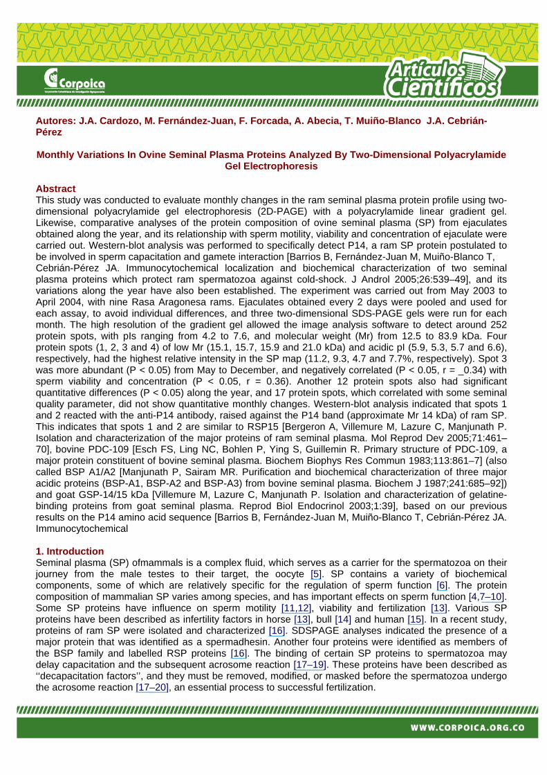
Autores: J.A. Cardozo, M. Fernández-Juan, F. Forcada, A. Abecia, T. Muiño-Blanco J.A. Cebrián- Pérez Monthly Variations In Ovine Seminal Plasma Proteins Analyzed By Two-Dimensional Polyacrylamide Gel Electrophoresis Abstract This study was conducted to evaluate monthly changes in the ram seminal plasma protein profile using two- dimensional polyacrylamide gel electrophoresis (2D-PAGE) with a polyacrylamide linear gradient gel. Likewise, comparative analyses of the protein composition of ovine seminal plasma (SP) from ejaculates obtained along the year, and its relationship with sperm motility, viability and concentration of ejaculate were carried out. Western-blot analysis was performed to specifically detect P14, a ram SP protein postulated to be involved in sperm capacitation and gamete interaction [Barrios B, Fernández-Juan M, Muiño-Blanco T, Cebrián-Pérez JA. Immunocytochemical localization and biochemical characterization of two seminal plasma proteins which protect ram spermatozoa against cold-shock. J Androl 2005;26:539–49], and its variations along the year have also been established. The experiment was carried out from May 2003 to April 2004, with nine Rasa Aragonesa rams. Ejaculates obtained every 2 days were pooled and used for each assay, to avoid individual differences, and three two-dimensional SDS-PAGE gels were run for each month. The high resolution of the gradient gel allowed the image analysis software to detect around 252 protein spots, with pIs ranging from 4.2 to 7.6, and molecular weight (Mr) from 12.5 to 83.9 kDa. Four protein spots (1, 2, 3 and 4) of low Mr (15.1, 15.7, 15.9 and 21.0 kDa) and acidic pI (5.9, 5.3, 5.7 and 6.6), respectively, had the highest relative intensity in the SP map (11.2, 9.3, 4.7 and 7.7%, respectively). Spot 3 was more abundant (P < 0.05) from May to December, and negatively correlated (P < 0.05, r = _0.34) with sperm viability and concentration (P < 0.05, r = 0.36). Another 12 protein spots also had significant quantitative differences (P < 0.05) along the year, and 17 protein spots, which correlated with some seminal quality parameter, did not show quantitative monthly changes. Western-blot analysis indicated that spots 1 and 2 reacted with the anti-P14 antibody, raised against the P14 band (approximate Mr 14 kDa) of ram SP. This indicates that spots 1 and 2 are similar to RSP15 [Bergeron A, Villemure M, Lazure C, Manjunath P. Isolation and characterization of the major proteins of ram seminal plasma. Mol Reprod Dev 2005;71:461– 70], bovine PDC-109 [Esch FS, Ling NC, Bohlen P, Ying S, Guillemin R. Primary structure of PDC-109, a major protein constituent of bovine seminal plasma. Biochem Biophys Res Commun 1983;113:861–7] (also called BSP A1/A2 [Manjunath P, Sairam MR. Purification and biochemical characterization of three major acidic proteins (BSP-A1, BSP-A2 and BSP-A3) from bovine seminal plasma. Biochem J 1987;241:685–92]) and goat GSP-14/15 kDa [Villemure M, Lazure C, Manjunath P. Isolation and characterization of gelatine- binding proteins from goat seminal plasma. Reprod Biol Endocrinol 2003;1:39], based on our previous results on the P14 amino acid sequence [Barrios B, Fernández-Juan M, Muiño-Blanco T, Cebrián-Pérez JA. Immunocytochemical 1. Introduction Seminal plasma (SP) ofmammals is a complex fluid, which serves as a carrier for the spermatozoa on their journey from the male testes to their target, the oocyte [5]. SP contains a variety of biochemical components, some of which are relatively specific for the regulation of sperm function [6]. The protein composition of mammalian SP varies among species, and has important effects on sperm function [4,7–10]. Some SP proteins have influence on sperm motility [11,12], viability and fertilization [13]. Various SP proteins have been described as infertility factors in horse [13], bull [14] and human [15]. In a recent study, proteins of ram SP were isolated and characterized [16]. SDSPAGE analyses indicated the presence of a major protein that was identified as a spermadhesin. Another four proteins were identified as members of the BSP family and labelled RSP proteins [16]. The binding of certain SP proteins to spermatozoa may delay capacitation and the subsequent acrosome reaction [17–19]. These proteins have been described as ‘‘decapacitation factors’’, and they must be removed, modified, or masked before the spermatozoa undergo the acrosome reaction [17–20], an essential process to successful fertilization.
-
Upload
independent -
Category
Documents
-
view
0 -
download
0
Transcript of Monthly variations in ovine seminal plasma proteins analyzed by two-dimensional polyacrylamide gel...
Autores: J.A. Cardozo, M. Fernández-Juan, F. Forcada, A. Abecia, T. Muiño-Blanco J.A. Cebrián-Pérez Monthly Variations In Ovine Seminal Plasma Proteins Analyzed By Two-Dimensional Polyacrylamide
Gel Electrophoresis Abstract This study was conducted to evaluate monthly changes in the ram seminal plasma protein profile using two-dimensional polyacrylamide gel electrophoresis (2D-PAGE) with a polyacrylamide linear gradient gel. Likewise, comparative analyses of the protein composition of ovine seminal plasma (SP) from ejaculates obtained along the year, and its relationship with sperm motility, viability and concentration of ejaculate were carried out. Western-blot analysis was performed to specifically detect P14, a ram SP protein postulated to be involved in sperm capacitation and gamete interaction [Barrios B, Fernández-Juan M, Muiño-Blanco T, Cebrián-Pérez JA. Immunocytochemical localization and biochemical characterization of two seminal plasma proteins which protect ram spermatozoa against cold-shock. J Androl 2005;26:539–49], and its variations along the year have also been established. The experiment was carried out from May 2003 to April 2004, with nine Rasa Aragonesa rams. Ejaculates obtained every 2 days were pooled and used for each assay, to avoid individual differences, and three two-dimensional SDS-PAGE gels were run for each month. The high resolution of the gradient gel allowed the image analysis software to detect around 252 protein spots, with pIs ranging from 4.2 to 7.6, and molecular weight (Mr) from 12.5 to 83.9 kDa. Four protein spots (1, 2, 3 and 4) of low Mr (15.1, 15.7, 15.9 and 21.0 kDa) and acidic pI (5.9, 5.3, 5.7 and 6.6), respectively, had the highest relative intensity in the SP map (11.2, 9.3, 4.7 and 7.7%, respectively). Spot 3 was more abundant (P < 0.05) from May to December, and negatively correlated (P < 0.05, r = _0.34) with sperm viability and concentration (P < 0.05, r = 0.36). Another 12 protein spots also had significant quantitative differences (P < 0.05) along the year, and 17 protein spots, which correlated with some seminal quality parameter, did not show quantitative monthly changes. Western-blot analysis indicated that spots 1 and 2 reacted with the anti-P14 antibody, raised against the P14 band (approximate Mr 14 kDa) of ram SP. This indicates that spots 1 and 2 are similar to RSP15 [Bergeron A, Villemure M, Lazure C, Manjunath P. Isolation and characterization of the major proteins of ram seminal plasma. Mol Reprod Dev 2005;71:461– 70], bovine PDC-109 [Esch FS, Ling NC, Bohlen P, Ying S, Guillemin R. Primary structure of PDC-109, a major protein constituent of bovine seminal plasma. Biochem Biophys Res Commun 1983;113:861–7] (also called BSP A1/A2 [Manjunath P, Sairam MR. Purification and biochemical characterization of three major acidic proteins (BSP-A1, BSP-A2 and BSP-A3) from bovine seminal plasma. Biochem J 1987;241:685–92]) and goat GSP-14/15 kDa [Villemure M, Lazure C, Manjunath P. Isolation and characterization of gelatine-binding proteins from goat seminal plasma. Reprod Biol Endocrinol 2003;1:39], based on our previous results on the P14 amino acid sequence [Barrios B, Fernández-Juan M, Muiño-Blanco T, Cebrián-Pérez JA. Immunocytochemical 1. Introduction Seminal plasma (SP) ofmammals is a complex fluid, which serves as a carrier for the spermatozoa on their journey from the male testes to their target, the oocyte [5]. SP contains a variety of biochemical components, some of which are relatively specific for the regulation of sperm function [6]. The protein composition of mammalian SP varies among species, and has important effects on sperm function [4,7–10]. Some SP proteins have influence on sperm motility [11,12], viability and fertilization [13]. Various SP proteins have been described as infertility factors in horse [13], bull [14] and human [15]. In a recent study, proteins of ram SP were isolated and characterized [16]. SDSPAGE analyses indicated the presence of a major protein that was identified as a spermadhesin. Another four proteins were identified as members of the BSP family and labelled RSP proteins [16]. The binding of certain SP proteins to spermatozoa may delay capacitation and the subsequent acrosome reaction [17–19]. These proteins have been described as ‘‘decapacitation factors’’, and they must be removed, modified, or masked before the spermatozoa undergo the acrosome reaction [17–20], an essential process to successful fertilization.

It is well known that the reproductive efficiency in rams is influenced by season in temperate zones of the Northern Hemisphere, affecting, among other features, quantitative and qualitative sperm production [21,22] and the fertilizing ability of cryopreserved spermatozoa [23,24]. Seasonal changes in ram semen volume and sperm concentration have also been described [25], as well as the seasonal influence on the total protein concentration in ram SP, with significant differences between the breeding and non-breeding seasons, in both the Southern [26] or Northern Hemispheres [27]. The analysis of ram SP by SDS-PAGE revealed that several protein bands (molecular weight 20–70 kDa) found in SP from all studied rams during the breeding season, were absent in the plasma of certain rams during the non-breeding season. Moreover, some proteins were present in both breeding and non-breeding season plasmas, although at higher concentration in the breeding season samples [26]. In a previous work, we showed that ram SP proteins could repair [28,29] and prevent [30] cold-shock sperm membrane damage. Very recently [1], we have proved that two SP proteins of approximately 14 kDa (P14) and 20 kDa (P20) are responsible for this protective effect. Moreover, we have also shown that seasonal differences in SP proteins could affect their ability to recover membrane integrity of cold-shocked sperm [30]. Two-dimensional polyacrylamide gel electrophoresis (2D-PAGE) has been used for the separation and characterization of several proteins from SP of bull [7,31,32], horse [13] and ram [33,34], boar [35], human [36–38]. In a recent study with 2D-PAGE using 12% acrylamide gels [33], 21 protein spots were identified in ram SP of non-breeding season. However, to our knowledge, neither the ovine SP protein profile using 2D-PAGE with a polyacrylamide linear gradient gel, nor qualitative and quantitative monthly variations and their relationship to sperm quality parameters have been described yet. The present study was conducted to establish a reference map for SP proteins of Rasa Aragonesa rams using 2D-PAGE with a polyacrylamide linear gradient gel, and to analyze monthly changes in the SP protein profile. Likewise, comparative analyses of the protein composition of ovine SP from ejaculates obtained along the year, and their relationship with sperm motility, viability and concentration of ejaculate were carried out. Western-blot analysis was performed to specifically detect P14, postulated to be involved in sperm capacitation and gamete interaction [1], and its variations along the year have also been established. 2. Materials And Methods 2.1. Sperm Collection All the experiments were performed with fresh semen taken from nine mature Rasa aragonesa rams using an artificial vagina. This breed corresponds to a local Spanish genotype with a short seasonal anoestrus between May and August. All the rams belonged to the National Association of Rasa Aragonesa Breeding (ANGRA) and were 2–4 years old. They were housed at the animal experimentation service of the University of Zaragoza under uniform nutritional conditions. Based on the positive results from a previous study, sires underwent an abstinence period of 2 days, and second ejaculates were pooled and used for each assay, to avoid individual differences [39]. The experiments were performed from May 2003 to April 2004. 2.2. Assessment Of Standard Semen Parameters Sperm concentration was calculated in duplicate using Neubauer’s chamber (Marienfeld, Lauda-Ko¨nigshofen). Sperm motility was subjectively assessed by visual estimation with a television microscopy system (100_) maintained at 37 8C. The percentage of progressively motile spermatozoa was estimated at intervals of 5%. Semen motility was assessed by the same person throughout the study. Cell viability (membrane integrity) was assessed by fluorescent staining with carboxyfluorescein diacetate and propidium iodide [40]. The cells were examined under a Nikon fluorescence microscope, and the number of propidium iodide-negative (membrane-intact) spermatozoa and propidium iodide-positive
(membranedamaged) spermatozoa per 100 cells were estimated and recorded. At least 200 cells were counted in duplicates for each sample. 2.3. Collection Of SP Seminal plasma was obtained by spinning 1 ml of semen at 7500 _ g for 5 min in a microfuge at 4 8C. The supernatant was centrifuged again, and SP was recovered and, after filtering through a 0.22 mm Millipore membrane (Millipore Ibérica, Madrid, Spain) and adding 10% of a protease and phosphatase inhibitor cocktail (Sigma Chemical Co., St. Louis, MO) was kept at _20 8C. Protein content was determined using Bradford’s method [41]. 2.4. 2d-Electrophoresis Seminal plasma samples were prepared for electrophoresis as follows: 75 mg of SP proteins were diluted in 125 ml of sample buffer containing 8 M urea, 2% [3- (3-(cholamydopropyl) dimethyl-ammonio)-1 propane sulphonate] (CHAPS), 40 mM dithiothreitol (DTT), 0.2% Bio-LyteTM 3/10 ampholyte (Bio-Rad, Hercules, CA, USA), 0.0002% Bromophenol Blue, 2 ml of tributyl phosphine (TBP) stock and 5 ml 2D-PAGE standards (Sigma Chemical Co.). Samples were subjected to the 2D-PAGE as described by O’Farrel et al. [42]. Proteins were separated by isoelectric focusing (IEF) using 7 cm immobilized pH gradients strips (IPGs; pH 3–10; Bio- Rad). The IPGs were placed overnight in the channel of a rehydration tray that contained the solution described above. Isoelectric focusing was performed using a Protean1 IEF Cell (Bio-Rad) at 8250 V and 20 8C. After isoelectrofocusing, the IPG strips were equilibrated for 10 min in 2500 ml of equilibration buffer I containing 6 M urea, 375 mM Tris–HCl, pH 8.8, 2% SDS, 20% glycerol, and 2% (w/v) DTT and 10 min longer in 2500 ml of equilibration buffer II containing 6 M urea, 375 mM Tris–HCl, pH 8.8, 2% SDS, 20% glycerol and 2.5% (w/v) iodoacetamide. Finally, the second dimension run was performed on a 9–20% polyacrylamide linear gradient gel (SDS-PAGE), using a Miniprotean II (Bio-Rad). After electrophoresing in the second dimension, the gels were stained with Sypro-Ruby Protein Gel Stain (Molecular Probes, The Netherlands) and scanned with a gel doc system with Molecular Analyst software (Bio-Rad). The 2D gel images were processed for analysis with PD-QuestTM 2D analysis software (Bio-Rad) to determine the relative protein content of the spots. We used a correlative numbering system to assign each spot. Data were used to estimate the amount of each protein and to create a map of the proteins present in each sample. 2.5. Western-Blotting The 2D gels were transferred for 2 h onto a PVDFmembrane with a Hoefer TE70 Semiphor Semidry- Transfer Unit (Pharmacia-Biotech, Uppsala, Sweden). Non-specific sites on the membranes were blocked for 1 h with 5% BSA in blocking buffer (Tris–HCl 10 mM pH 8; NaCl 120 mM, 0.05% Tween 20). The proteins were immunodetected by incubating for 3 h with the anti-P14 polyclonal antibody [1] diluted at 1:4000 in blocking buffer that contained 0.17% BSA. After exhaustive washing, the blots were incubated with a secondary goat anti-rabbit alkaline-phosphatase-conjugated IgG (Sigma Chemical Co.) at a dilution 1:30,000 for 2 h. After four washings of 5 min each, the membranes were incubated with 66 mg/ml 5-bromo- 4-chloro-3-indolyl phosphate (BCIP) and 111 mg/ml nitro blue tetrazolium (NBT) in Tris 0.19 M, MgCl2 1 mM until colour appeared. The image was scanned using the gel doc system with Molecular Analyst software (Bio-Rad). Replacing the antiserum with preimmune serum was used as a negative control to rule out non-specific binding to the transferred proteins. 2.6. Statistical Analysis Results are shown as the means (_S.E.M.) of three replicated samples. The obtained data were transformed (Logarithm) and analyzed using a one-way analysis of variance (ANOVA). The statistical analysis was carried out using the general linear model procedure of the SAS system (The SAS system for

windows, version 7, SAS Institute, 1998; Cary, NC). Correlations between sperm parameters and SP protein concentration were calculated using Pearson’s coefficient. 3. Results Three two-dimensional SDS-PAGE gels were run for each month. A representative reference map of ovine SP proteins, obtained by a 2D-PAGE gel stained with Sypro-Ruby, is shown (Fig. 1). Similar 2D-PAGE protein maps were found from seminal plasmas of different months, with excellent reproducibility as well as very low variability in the presence of protein spots. The image analysis software detected around 252 protein spots, with isoelectric points (pIs) ranging from 4.2 to 7.6, and molecular weight (Mr) from 12.5 to 83.9 kDa. However, only 35 protein spots (13.9%) had pIs above 7. Four protein spots ([1–4]; Table 1) of low Mr (15.1, 15.7, 15.9 and 21.0 kDa) and acidic pI (5.9, 5.3, 5.7 and 6.6), respectively, had the highest relative intensity in the SP map (11.2, 9.3, 4.7 and 7.7%, respectively). It is worth noting that protein spot 3 was more abundant (P < 0.05) from May to December, and negatively correlated (P < 0.05, r = _0.34) with sperm viability and concentration (P < 0.05, r = 0.36).
Fig 1. Two-dimensional polyacrylamide electrophoretic gel of ram seminal plasma proteins. Two-dimensional 9–20% linear gradient SDSPAGE gel stained with Sypro-Ruby. Molecular weight markers (Mr- 10-3) indicated at the left and pH markers indicated on top of the gel are approximate. Underlined spots have the highest relative intensity.

Table 1 Standard spot number (SSP), theoretical molecular weight (Mr), isoelectric point (pI) and relative protein content (medium% value _ S.E.M. of three replicate samples) of the major protein spots in ram seminal plasma. SSP Mr pI Jan Febrary March April May June July August September October November December 1 15.1 5.9 11.0 ±
0.91 12.2± 2.91
14.1 ± 2.1
13.0± 1.56
10.2± 1.89
12.0± 3.11
14.2± 0.76
11.4± 3.36
9.2 ±1.14 13.8± 3.65
11.6± 1.1 8.5± 1.52
2 15.7 5.3 8.7± 1.16
8.83± 2..98
8.5± 1.8
9.3± 0.79
4.53± 0.96
5.63± 1.4
7.33± 1.13
6.83± 1.33
9.3± 2.1
8.93± 0.9
7.2± 0.11 9.03± 0.45
3 15.9 5.7 4.13± 0.58
2.57± 0.78
1.93± 0.23
1.80± 0.67
7.70± 2.54
5.60 ± 0.17
5.47± 0.26
6.13± 0.50
3.80± 1.85
5.20± 1.04
6.20± 1.07
5.83± 0.54
4 21.0 6.6 8.8± 0.75
9.52± 0.59
7.53± 1.0
7.30± 1.42
6.99± 0.62
6.72 ± 0.69
8.66± 0.56
8.1± 1.51
7.55 ± 0.55
6.82± 0.43
7.6 ± 1.07 6.79 ± 1.72
Table 2 Standard spot number (SSP), theoretical molecular weight (Mr), isoelectric point (pI) and relative protein content (medium% value _ S.E.M. of three replicate samples) of protein spots with quantitative monthly changes.
Table 3 Variations of sperm quality parameters in fresh ram ejaculates of Rasa Aragonesa breed along the year (medium value _ S.E.M. of three replicate samples)
* P < 0.05 related to the highest value. Another 12 protein spots also had significant quantitative differences (P < 0.05) along the year (Table 2). One of these proteins (spot 8) decreased (P < 0.05) from June, and on other seven (spots 5, 6, 9, 11, 12, 14 and 16) diminished (P < 0.05) from July or August to the end of the year. The amount of protein spot 9 positively correlated (P < 0.05, r = 0.4) with the percentage of viable sperm in the ejaculate. However, the content of protein spots 5, 8 and 11 resulted negatively correlated (P < 0.05, r = _0.49, _0.46 and _0.54, respectively) with sperm motility. Conversely, protein spot 15 was more abundant (P < 0.05) from May to December, and negatively correlated (P < 0.05, r = _0.37) with sperm viability. The protein content of spot 13 increased significantly from September to December. Finally, the concentration of protein spots 7and 10 varied (P < 0.05) along the year, although they showed no specific tendency in their profiles. In addition, the

protein content of spot 7 was positively correlated (P < 0.05, r = 0.49) with sperm motility. Medium values of sperm quality parameters in ejaculates obtained along the year are shown (Table 3). Other interesting data of this investigation was that the image analysis software identified another 17 protein spots, which being correlated with some seminal quality parameter, did not show quantitative monthly changes (Table 4). Nine of these proteins correlated with sperm motility, two of which (spots 17 and 21) did so negatively (P < 0.05, r = _0.38). On the contrary, the other seven protein spots [22,23,26,28,31–33] were positively (P < 0.05) correlated with motility. Four protein spots, 20, 21, 27 and 29, were positively correlated (P < 0.05, r = 0.43, 0.37, 0.43 and 0.34, respectively) with sperm viability. Finally, another five proteins were correlated with sperm concentration: two of these (spots 19 and 30) correlated positively (P < 0.05, r = 0.38 and 0.34, respectively), whereas the other three proteins (spots 18, 24 and 25) did so negatively (P < 0.05, r = _0.39, _0.50 and _0.36, respectively). In an attempt to further characterize these protein spots, proteins of the 2D-PAGE gels were transferred onto PVDF-membranes and immunodetected using the polyclonal anti-P14 antibody (Fig. 2). Obtained results indicated that spots 1 and 2 reacted with the antibody.
Fig. 2. Immunoblotting of ram seminal plasma proteins analyzed by 2D-PAGE. The gel of Fig. 1 was electrotransferred to nitrocellulose, and subjected to immunodetection using a polyclonal antibody directed against P14. Molecular weight markers (Mr _ 10_3) indicated at the left and pH markers indicated on top of the gel are approximate.

Table 4 Protein spots without quantitative monthly changes (P > 0.05): standard spot number (SSP), theoretical molecular weight (Mr) and isoelectric point (pI), and correlation with sperm motility, viability and concentration in ram ejaculates.
* P < 0.05. ** P < 0.01. 4. Discussion Seasonal patterns of semen production and sperm quality from rams of different breeds have already been reported [43,44]. Likewise, the influence of season on the total protein concentration of ram SP has been described [26,27], with significant differences between breeding and non-breeding seasons. Moreover, there are several reports on seasonal variations of ram semen freezability [45]. Therefore, it could be postulated that certain SP proteins may influence semen quality. In this study, the image analysis software detected around 252 protein spots in ovine SP using 2D-PAGE with a polyacrylamide linear gradient gel. The higher resolution of the gradient gel is undoubtedly inferred by the high number of detected spots, compared to a recent work on ram SP using 12% acrylamide gels [33]. It is worth noting the presence of a major group of four proteins with low molecular weight (15.1–21 kDa) and pI ranging from 5.3 to 6.6, which accounted for 32.9% of the relative intensity of the gels, based on the intensity of the Sypro-Ruby staining. Moreover, of the 252 protein spots detected on the 2D map, only 35 have pIs above 7. Therefore, we could conclude that most proteins in SP of Rasa Aragonesa rams are acidic. Similar results have already been reported in Santa Ines hairy rams, a breed adapted in tropical regions, in which most SP proteins have a molecular weight lower than 75 kDa and acidic pIs, few having pIs above 8 [34] and more recently in ram SP of other breeds [33]. Our results showed that 13 protein spots had significant seasonal variations decreasing from summer (non-breeding season) to the end of the year (breeding season). Conversely, the protein content of spots 3 (Mr 15.9 kDa, pI 5.6) and 15 (Mr 16.1 kDa, pI 6.5) were more abundant from May to December, and negatively correlated with sperm viability. Their high content in summer could explain the reduction in sperm viability found during these months (Table 3). Protein spot 3 is negatively correlated to sperm concentration, which suggests that it could be related to the production of spermatozoa by testis. These seasonal variations are consistent with our previous results, by comparative SDS-PAGE analysis, that showed seasonal differences in ram SP protein composition, [27]. Likewise, Smith et al. [26] described seasonal changes in ram semen
composition paralleled to changes in fertility of frozen semen [46] and Strzezek et al. [9] observed seasonal effects in boar SP protein concentration independent of the male age. All these changes could be due to seasonal influence in gonadotrophin levels [47] and their receptors in the testis [48] that would affect endocrine gonadal function and secretions of seminal vesicles and epididymis [26]. Three protein spots [5,8,11] that decreased in nonbreeding season were negatively correlated with sperm motility. Their lower content in warm months could explain the maintenance of sperm motility at normal values during summer and autumn months, as already reported in rams [46]. The relative protein content of spot 9 (Mr 73.2 kDa, pI 5.0) reduced significantly from August, and correlated with viability. This protein could exert a protecting effect on sperm membrane integrity, which is essential for the maintenance of sperm functionality, and is required for capacitation, acrosome reaction and sperm binding to the oocyte. To fertilize an oocyte, spermatozoa must possess an intact acrosome. Shortly before fertilization, a sperm cell undergoes acrosome reaction, which is required for penetration through the zona pellucida and fusion with the oocyte plasma membrane [20]. Studies on several mammalian species indicate that SP contains specific factors that inhibit capacitation and thus prevent inappropriate acrosome reaction [5]. The reduction in the concentration of protein spot 9 in warm months could explain the decreases in sperm viability value of the ejaculate in these months, and suggest the possibility that this protein could play an important role in the membrane stability and subsequent viability. It is noteworthy that although 17 protein spots showed no monthly changes in their protein content, they were correlated with some seminal quality parameter. The obtained correlation coefficients were not very high, either for proteins with or without seasonal changes. These findings suggest that proteins in the seminal plasma would act in a complementary manner, and that these proteins play an important role in sperm membrane stability, and subsequent viability, motility and concentration. Results of Western-blot confirmed that spots 1 and 2 reacted with the anti-P14 antibody, raised against the P14 band (approximate Mr 14 kDa) recovered from a non-denaturing gradient polyacrylamide gel of the fraction 6 of ram SP [1]. Automated Edman degradation of P14 was performed twice until the amino acid 34 was reached and at least four times for the first 10 amino acids, and we found only one protein sequence [1]. Therefore, we might suggest that spots 1 and 2 correspond to the same protein with certain modification, as it could be due to a different degree of phosphorylation, because we also proved that P14 was phosphorylated at serine and threonine residues, but not glycosylated [1]. The sequence of the P14 fragment that we reported [1] almost equals that found by Bergeron et al. for RSP15 [16] with only two different amino acids in position 26 and 28. Likewise, this P14 fragment showed a high identity with several SP proteins of other species, particularly bovine PDC-109 [2] (also called BSP A1/A2 [3]) and GSP-14/15 kDa (goat SP protein, related to the BSP family [4]). Therefore, we could deduce that spots 1 and 2 do so. If these two spots represent two differentially glycosylated and/or phosphorylated forms of P14, this will also agree with results by Bergeron et al. [16] who found that RSP15 was eluted in two peaks during HPLC. It has been reported that BSP proteins stabilize the sperm membrane in a first step [19,49] and subsequently, participates in the female tract in capacitation by releasing cholesterol and binding HDL and heparin [19,50–53]. The homologous fragment is a conserved domain in several proteins called FN2 (Fibronectin Domain Type II) [49]. This domain binds choline phospholipids and heparin, and promotes the binding of SP proteins to the sperm membrane upon ejaculation [19,54–56]. Therefore, we could hypothesize that protein spots 1 and 2 take part in the protein structure surrounding the spermatozoa in a similar way to fibronectin, stabilizing membrane phospholipids and cytoskeleton. They could be involved in sperm capacitation and gamete interaction, stabilizing the sperm membrane in a first step (decapacitating factors), and in a later step, participating in the membrane modification during capacitation, as previously suggested for BSP proteins [19,55] and recently for P14 [1].
Our results indicate that proteomics may be useful to study sperm function, and that ovine seminal plasma contains important proteins for the adequate function of spermatozoa. The physiological importance of these findings claims for more studies to identify, characterize and elucidate their roles in sperm function. Additional research may contribute to the development of strategies to improve the fertilizing ability of semen. Acknowledgments This work was supported by grants CICYT-FEDER AGL 2004-02882, INIA RZ03-035, CICYT-FEDER AGL 2005-02614 and DGA A-26/2005. J. Cardozo was financed by CORPOICA (Colombia). The authors thank ANGRA for supplying the sires and S. Morales for the collection of semen samples. References [1] Barrios B, Fernández-Juan M, Muiño-Blanco T, Cebrián-Pérez JA. Immunocytochemical localization and biochemical characterization of two seminal plasma proteins which protect ram spermatozoa against cold-shock. J Androl 2005;26:539–49. [2] Esch FS, Ling NC, Bohlen P, Ying S, Guillemin R. Primary structure of PDC-109, a major protein constituent of bovine seminal plasma. Biochem Biophys Res Commun 1983;113:861– 7. [3] Manjunath P, Sairam MR. Purification and biochemical characterization of three major acidic proteins (BSP-A1, BSP-A2 and BSP-A3) from bovine seminal plasma. Biochem J 1987; 241:685–92. [4] Villemure M, Lazure C, Manjunath P. Isolation and characterization of gelatine-binding proteins from goat seminal plasma. Reprod Biol Endocrinol 2003;1:39. [5] Thomas CJ, Anbazhagan V, Ramakrishnan M, Sultan N, Surolia I, Swamy MJ. Mechanism of membrane binding by the bovine seminal plasma protein, PDC-109: a surface plasmon resonance study. Biophys J 2003;84:3037–44. [6] Strzezek J, KordanW, Kostyra H, Zaborniak A. Purification and partial characterization of a 5700 Da sperm motility inhibiting factor from seminal plasma of boar. Anim Reprod Sci 1992; 29:35–52. [7] Mortarino M, Tedeschi G, Negri A, Ceciliani F, Gottardi L, Maffeo G, et al. Two-dimensional polyacrylamide gel electrophoresis map of bull seminal plasma proteins. Electrophoresis 1998;19:797–801. [8] Cross NL. Multiple effects of seminal plasma on the acrosome reaction of human sperm. Mol Reprod Dev 1993;35:316–23. [9] Strzezek J, Saiz-Cidoncha F, Wysocki P, Tyszkiewicz A, Jastrzebski M. Seminal plasma proteins as markers of biological value of boar semen. Anim Sci 2002;20:255–66. [10] Miller DJ, Winer MA, Ax RL. Heparin-binding proteins from seminal plasma bind to bovine spermatozoa and modulate capacitation by heparin. Biol Reprod 1990;42:899–915. [11] Sánchez-Luengo S, Aumuller G, Albrecht M, Sen PC, Rohm KH, Wilhelm B. Interaction of PDC-109, the major secretory protein from bull seminal vesicles, with bovine sperm membrane Ca2+-ATPase. J Androl 2004;25:234–44. [12] Henricks DM, Kouba AJ, Lackey BR, Boone WR, Gray SL. Identification of insulin-like growth factor I in bovine seminal plasma and its receptor on spermatozoa: influence on sperm motility. Biol Reprod 1998;59:330–7.

[13] Brandon CI, Heusner GL, Caudle AB, Fayrer-Hosken RA. Twodimensional polyacrylamide gel electrophoresis of equine seminal plasma proteins and their correlation with fertility. Theriogenology 1999;52:863–73. [14] Bhargava PM. Seminalplasmin, an antimicrobial and transcription inhibitory protein from bovine seminal plasma, might be nature’s own antifertility agent. Ann Natl Acad Med Sci India 1986;21:53–86. [15] Audhya T, Reddy J, Zaneveld LJ. Purification and partial chemical characterization of a glycoprotein with antifertility activity from human seminal plasma. Biol Reprod 1987;36: 511–21. [16] Bergeron A, Villemure M, Lazure C, Manjunath P. Isolation and characterization of the major proteins of ram seminal plasma. Mol Reprod Dev 2005;71:461–70. [17] Fraser GS, Bucci DM, Brooks CL. Two-dimensional polyacrylamide gel electrophoresis of bovine semen after cryopreservation in half-millilitre straws. Theriogenology 1996;46:1103–15. [18] Manjunath P, Chandonnet L, Leblond E, Desnoyers L. Major proteins of bovine seminal vesicles bind to spermatozoa. Biol Reprod 1993;49:27–37. [19] Manjunath P, Thérien I. Role of seminal plasma phospholipidbinding proteins in sperm membrane lipid modification that occurs during capacitation. J Reprod Immunol 2002;53:109–19. [20] Yanagimachi R. Mammalian fertilization, 2nd ed., New York: Raven Press; 1994. [21] Pelletier J, Chemineau P, Delgadillo JA. Seasonality of sexual activity and its photoperiodic control in the adult ram and goat. In: Proceedings of the 11th international congress on animal reproduction; 1988. p. 211–9. [22] Pérez R, López A, Castrillejo A, Bielli A, Laborde D, Gastel T, et al. Reproductive seasonality of correidale rams under extensive rearing condition. Acta Vet Scand 1997;38:109–17. [23] Colas G, Brice G. Seasonal variations of the fertilizing capacity of the deep-frozen ram semen. In: Proceedings of the eighth international congress on animal reproduction; 1976. p. 980–97. [24] Guérin Y, Cognie Y, Poulin N. Freezability of freshly ejaculated and frozen ram semen in vitro. In: Proceedings of the 12th international congress on animal reproduction; 1992. p. 1418– 20. [25] Smith JF, Parr J, Smith JK, Briggs RM, Duganzich DM. Seasonal patterns in production and quality of semen of rams from flocks selected for or against an early breeding season. Proc N Z Soc Anim Reprod 1997;57:259. [26] Smith JF, Parr J, Murray GRM, Mcdonald RM, Lee RSF. Seasonal changes in the protein content and composition of ram seminal plasma. Proc N Z Soc Anim Reprod 1999;59:223–5. [27] Pérez-PéR, Barrios B, Muiño-Blanco T, Cebrián-Pérez JA. Seasonal differences in ram seminal plasma revealed by partition in an aqueous two-phase system. J Chromatogr B 2001;760: 113–21. [28] Barrios B, Pérez-Pe R, Gallego M, Tato A, Osada J, Muiño- Blanco T, et al. Seminal plasma proteins revert the cold-shock damage on ram sperm membrane. Biol Reprod 2000;63: 1531–7.
[29] Garcia-Lopez N, Ollero M, Cebrian-Perez JA, Muiño-Blanco T. Reversion of thermic-shock effect on ram spermatozoa by adsorption of seminal plasma proteins revealed by partition in aqueous two-phase systems. J Chromatogr B 1996;680:137–43. [30] Pérez-PéR, Cebrián-Pérez JA, Muiño-Blanco T. Semen plasma proteins prevent cold-shock membrane damage to ram spermatozoa. Theriogenology 2001;56:425–34. [31] Desnoyers L, Therien I, Manjunath P. Characterization of the major proteins of bovine seminal fluid by two-dimensional polyacrylamide gel electrophoresis. Mol Reprod Dev 1994;37:425–35. [32] Jobim MI, Oberst ER, Salbego CG, Souza DO, Wald VB, Tramontina F, et al. Two-dimensional polyacrylamide gel electrophoresis of bovine seminal plasma proteins and their relation with semen freezability. Theriogenology 2004;61:255–66. [33] Jobim MI, Oberst ER, Salbego CG, Wald VB, Horn AP, Mattos RC. BSP A1/A2-like proteins in ram seminal plasma. Theriogenology 2005;63:2053–62. [34] Souza CE, Moura A, Oliveira JT, Radis-Baptista G, Araujo A, Lima A. Seminal plasma proteins, testis development and semen criteria in the ram. In: 29th Annual Meeting ASA. 2004. p. 91. [35] Sanz L, Calvete JJ, Mann K, Gabius HJ, Topfer-Petersen E. Isolation and biochemical characterization of heparin-binding proteins from boar seminal plasma: a dual role for spermadhesins in fertilization. Mol Reprod Dev 1993;35:37–43. [36] Bohring C, Krause E, Habermann B, Krause W. Isolation and identification of sperm membrane antigens recognized by antisperm antibodies, and their possible role in immunological infertility disease. Mol Hum Reprod 2001;7:113–8. [37] Pixton KL, Deeks ED, Flesch FM, Moseley FLC, Bjorndahll L, Ashton PR, et al. Sperm proteome mapping of a patient who experienced failed fertilization at IVF reveals altered expression of at least 20 proteins compared with fertile donors: case report. Hum Reprod 2004;19:1438–47. [38] Starita-Geribaldi M, Poggioli S, Zucchini M, Garin J, Chevallier D, Fenichel P, et al. Mapping of seminal plasma proteins by twodimensional gel electrophoresis in men with normal and impaired spermatogenesis. Mol Hum Reprod 2001;7:715–22. [39] Ollero M, Muiño-Blanco T, López-PérezM, Cebrián-Pérez JA. Viability of ram spermatozoa in relation to the abstinence period and successive ejaculations. Int J Androl 1996;19: 287–92. [40] Harrison RAP, Vickers SE. Use of fluorescent probes to assess membrane integrity in mammalian spermatozoa. J Reprod Fertil 1990;88:343–52. [41] Bradford MM. A rapid and sensitive method for the quantitation of microgram quantities of protein utilizing the principle of dye binding. Anal Biochem 1976;72:248–54. [42] O’Farrel PZ, Goodman HM, O’Farrel PH. High resolution of two-dimensional electrophoresis of basic as well as acidic proteins. Cell 1977;12:113–42. [43] Colas G. Fertility in the ewe after artificial insemination with fresh and frozen semen at the induced oestrus, and influence of the photoperiod on the semen quality of the ram. Livest Prod Sci 1979;6:153–66.

[44] Amir DV, Volcani R. Seasonal fluctuations in the sexual activity of Awassi, German Mutton Merino, Corriedale, Border-Leicester and Dorset Horn rams. II. Seasonal changes in semen characteristics. J Agric Sci 1965;64:121–5. [45] Maxwell WMC. Fertility of ram semen frozen in autumn and spring. Proc Aust Soc Reprod Biol 1980;12:8. [46] D’Alessandro AG, Martemucci G. Evaluation of seasonal variations of semen freezability in Leccese ram. Anim Reprod Sci 2003;79:93–102. [47] Xu ZZM, McDonald MF, McCutcheon SN, Blair HT. Seasonal variation in testis size, gonadotrophin secretion and pituitary responsiveness to GnRH in rams of two breeds differing in time of onset of the breeding season. Anim Reprod Sci 1991;26: 281–92. [48] Barenton BP, Pelletier J. Seasonal changes in testicular gonadotrophin receptors and steroid content in the ram. J Endocrinol 1983;112:1441–6. [49] Greube A, Muller K, Topfer-Petersen E, Herrmann A, Muller P. Influence of the bovine seminal plasma protein PDC-109 on the physical state of membranes. Biochemistry 2001;40:8326–34. [50] The´rien I, Bousquet D, Manjunath P. Effect of seminal phospholipid- binding proteins and follicular fluid on bovine sperm capacitation. Biol Reprod 2001;65:41–51. [51] The´rien I, Moreau R, Manjunath P. Bovine seminal plasma phospholipids-binding proteins stimulate phospholipid efflux from epididymal sperm. Biol Reprod 1999;61:590–8. [52] The´rien I, Moreau R, Manjunath P. Major proteins of bovine seminal plasma and high-density lipoprotein induce cholesterol efflux from epididymal sperm. Biol Reprod 1998;59:768–76. [53] Gwathmey TM, Ignotz GG, Suarez SS. PDC-109 (BSP-A1/A2) promotes bull sperm binding to oviductal epithelium in vitro and may be involved in forming the oviductal sperm reservoir. Biol Reprod 2003;69:809–15. [54] Desnoyers L, Manjunath P. Major proteins of bovine seminal plasma exhibit novel interactions with phospholipid. J Biol Chem 1992;267:10149–55. [55] Moreau R, The´rien I, Manjunath P. Type II domains of BSP-A1/ A2 proteins: binding properties, lipid efflux, and sperm capacitation potential. Biochem Biophys Res Commun 1998;246: 148–54. [56] Muller P, Erlemann KR, Muller K, Calvete JJ, Topfer-Petersen E, Marienfeld K, et al. Biochemical characterization of the bovine seminal plasma protein PDC-109 with phospholipid vesicles. Eur Biophys J 1998;27:33–41.