TrkB overexpression in mice buffers against memory deficits and depression-like behavior but not all...
12
BEHAVIORAL NEUROSCIENCE ORIGINAL RESEARCH ARTICLE published: 12 September 2014 doi: 10.3389/fnbeh.2014.00315 TrkB overexpression in mice buffers against memory deficits and depression-like behavior but not all anxiety- and stress-related symptoms induced by developmental exposure to methylmercury Nina N. Karpova 1 *, Jesse Saku Olavi Lindholm 1† , Natalia Kulesskaya 1† , Natalia Onishchenko 2 , Marie Vahter 3 , Dina Popova 1 , Sandra Ceccatelli 2 and Eero Castrén 1 1 Neuroscience Center, University of Helsinki, Helsinki, Finland 2 Department of Neuroscience, Karolinska Institutet, Stockholm, Sweden 3 Institute of Environmental Medicine, Karolinska Institutet, Stockholm, Sweden Edited by: Francesca Cirulli, Istituto Superiore di Sanità, Italy Reviewed by: Alessandra Berry, Istituto Superiore di Sanità, Italy Jozsef Haller, Institute of Experimental Medicine, Hungary *Correspondence: Nina N. Karpova, Neuroscience Center, University of Helsinki, Viikinkaari 4, 00790 Helsinki, Finland e-mail: [email protected]; nina.karpova@helsinki.fi † These authors have contributed equally to this work. Developmental exposure to low dose of methylmercury (MeHg) has a long-lasting effect on memory and attention deficits in humans, as well as cognitive performance, depression-like behavior and the hippocampal levels of the brain-derived neurotrophic factor (Bdnf )in mice. The Bdnf receptor TrkB is a key player of Bdnf signaling. Using transgenic animals, here we analyzed the effect of the full-length TrkB overexpression (TK+) on behavior impairments induced by perinatal MeHg. TK overexpression in the MeHg-exposed mice enhanced generalized anxiety and cue memory in the fear conditioning (FC) test. Early exposure to MeHg induced deficits in reversal spatial memory in the Morris water maze (MWM) test and depression-like behavior in the forced swim test (FST) in only wild-type (WT) mice but did not affect these parameters in TK+ mice. These changes were associated with TK+ effect on the increase in Bdnf 2, 3, 4 and 6 transcription in the hippocampus as well as with interaction of TK+ and MeHg factors for Bdnf 1, 9a and truncated TrkB.T1 transcripts in the prefrontal cortex. However, the MeHg- induced anxiety-like behavior in the elevated plus maze (EPM) and open field (OF) tests was ameliorated by TK+ background only in the OF test. Moreover, TK overexpression in the MeHg mice did not prevent significant stress-induced weight loss during the period of adaptation to individual housing in metabolic cages. These TK genotype-independent changes were primarily accompanied by the MeHg-induced hippocampal deficits in the activity-dependent Bdnf 1, 4 and 9a variants, TrkB.T1, and transcripts for important antioxidant enzymes glyoxalases Glo1 and Glo2 and glutathione reductase Gsr. Our data suggest a role of full-length TrkB in buffering against memory deficits and depression-like behavior in the MeHg mice but propose the involvement of additional pathways, such as the antioxidant system or TrkB.T1 signaling, in stress- or anxiety-related responses induced by developmental MeHg exposure. Keywords: perinatal methylmercury, anxiety, hippocampus, brain-derived neurotrophic factor, Bdnf transcripts, truncated TrkB, glyoxalase, glutathione reductase INTRODUCTION Environmental risk factors (childhood abuse/neglect, exposure to environmental pollutants) drive pathological epigenetic repro- gramming early in life which may trigger the development of mood disorders in adult humans and rodents (Weaver et al., 2004; Onishchenko et al., 2007, 2008; McGowan et al., 2009). Methylmercury (MeHg) is an environmental chemical contam- inant of a major concern mainly for its detrimental effects on developing organisms that are more vulnerable to its toxicity (Ceccatelli et al., 2013). Therefore, advices for women and young children have been formulated in many countries. For instance, the U.S. Environmental Protection Agency reports that 2.3% of U.S. women 16–49 years of age have blood mercury concentra- tions higher than 5.8 micrograms per liter, which is the current reference dose (Birch et al., 2013). This means that approximately 1.4 million U.S. women at reproductive age have blood mercury concentrations posing a risk to their unborn children, which may result in 75,000 newborns per year with learning disabilities induced by in utero exposure to MeHg. Perinatal exposure to low levels of MeHg impairs memory and attention in children of high fish-consuming populations and induces long-term impairments in cognitive performance Frontiers in Behavioral Neuroscience www.frontiersin.org September 2014 | Volume 8 | Article 315 | 1
-
Upload
independent -
Category
Documents
-
view
1 -
download
0
Transcript of TrkB overexpression in mice buffers against memory deficits and depression-like behavior but not all...

BEHAVIORAL NEUROSCIENCEORIGINAL RESEARCH ARTICLE
published: 12 September 2014doi: 10.3389/fnbeh.2014.00315
TrkB overexpression in mice buffers against memorydeficits and depression-like behavior but not all anxiety-and stress-related symptoms induced by developmentalexposure to methylmercuryNina N. Karpova1*, Jesse Saku Olavi Lindholm1†, Natalia Kulesskaya1†, Natalia Onishchenko2,Marie Vahter3, Dina Popova1, Sandra Ceccatelli2 and Eero Castrén1
1 Neuroscience Center, University of Helsinki, Helsinki, Finland2 Department of Neuroscience, Karolinska Institutet, Stockholm, Sweden3 Institute of Environmental Medicine, Karolinska Institutet, Stockholm, Sweden
Edited by:Francesca Cirulli, Istituto Superioredi Sanità, Italy
Reviewed by:Alessandra Berry, Istituto Superioredi Sanità, ItalyJozsef Haller, Institute ofExperimental Medicine, Hungary
*Correspondence:Nina N. Karpova, NeuroscienceCenter, University of Helsinki,Viikinkaari 4, 00790 Helsinki, Finlande-mail: [email protected];[email protected]†These authors have contributedequally to this work.
Developmental exposure to low dose of methylmercury (MeHg) has a long-lastingeffect on memory and attention deficits in humans, as well as cognitive performance,depression-like behavior and the hippocampal levels of the brain-derived neurotrophicfactor (Bdnf )in mice. The Bdnf receptor TrkB is a key player of Bdnf signaling. Usingtransgenic animals, here we analyzed the effect of the full-length TrkB overexpression(TK+) on behavior impairments induced by perinatal MeHg. TK overexpression inthe MeHg-exposed mice enhanced generalized anxiety and cue memory in the fearconditioning (FC) test. Early exposure to MeHg induced deficits in reversal spatial memoryin the Morris water maze (MWM) test and depression-like behavior in the forced swimtest (FST) in only wild-type (WT) mice but did not affect these parameters in TK+ mice.These changes were associated with TK+ effect on the increase in Bdnf 2, 3, 4 and 6transcription in the hippocampus as well as with interaction of TK+ and MeHg factors forBdnf 1, 9a and truncated TrkB.T1 transcripts in the prefrontal cortex. However, the MeHg-induced anxiety-like behavior in the elevated plus maze (EPM) and open field (OF) testswas ameliorated by TK+ background only in the OF test. Moreover, TK overexpression inthe MeHg mice did not prevent significant stress-induced weight loss during the periodof adaptation to individual housing in metabolic cages. These TK genotype-independentchanges were primarily accompanied by the MeHg-induced hippocampal deficits in theactivity-dependent Bdnf 1, 4 and 9a variants, TrkB.T1, and transcripts for importantantioxidant enzymes glyoxalases Glo1 and Glo2 and glutathione reductase Gsr. Our datasuggest a role of full-length TrkB in buffering against memory deficits and depression-likebehavior in the MeHg mice but propose the involvement of additional pathways, such asthe antioxidant system or TrkB.T1 signaling, in stress- or anxiety-related responses inducedby developmental MeHg exposure.
Keywords: perinatal methylmercury, anxiety, hippocampus, brain-derived neurotrophic factor, Bdnf transcripts,truncated TrkB, glyoxalase, glutathione reductase
INTRODUCTIONEnvironmental risk factors (childhood abuse/neglect, exposureto environmental pollutants) drive pathological epigenetic repro-gramming early in life which may trigger the development ofmood disorders in adult humans and rodents (Weaver et al.,2004; Onishchenko et al., 2007, 2008; McGowan et al., 2009).Methylmercury (MeHg) is an environmental chemical contam-inant of a major concern mainly for its detrimental effects ondeveloping organisms that are more vulnerable to its toxicity(Ceccatelli et al., 2013). Therefore, advices for women and youngchildren have been formulated in many countries. For instance,
the U.S. Environmental Protection Agency reports that 2.3% ofU.S. women 16–49 years of age have blood mercury concentra-tions higher than 5.8 micrograms per liter, which is the currentreference dose (Birch et al., 2013). This means that approximately1.4 million U.S. women at reproductive age have blood mercuryconcentrations posing a risk to their unborn children, whichmay result in 75,000 newborns per year with learning disabilitiesinduced by in utero exposure to MeHg.
Perinatal exposure to low levels of MeHg impairs memoryand attention in children of high fish-consuming populationsand induces long-term impairments in cognitive performance
Frontiers in Behavioral Neuroscience www.frontiersin.org September 2014 | Volume 8 | Article 315 | 1
Karpova et al. TrkB and developmental neurotoxicity
in mice (Grandjean and Landrigan, 2006; Johansson et al.,2007; Onishchenko et al., 2007; Ceccatelli et al., 2013). We havepreviously shown that developmental MeHg-exposure inducedlong-lasting depression-like behavior and decreased hippocam-pal expression of the critical for brain functioning gene brain-derived neurotrophic factor (Bdnf), in adult mice (Onishchenkoet al., 2008). Impaired Bdnf expression or signaling through itsreceptor TrkB was linked to learning and memory problemsand to development of anxiety in several transgenic and stressmodels of brain disorders (Minichiello et al., 1999; Soliman et al.,2010; Karpova et al., 2011; Kemppainen et al., 2012; Lai et al.,2012). Developmental MeHg-exposure, however, did not alterthe full-length TrkB transcript levels at least in the hippocampus(Onishchenko et al., 2008), and no other study linking the long-term effect of perinatal MeHg treatment to TrkB expression hasbeen performed.
Several symptoms of neurodevelopmental disorders, includ-ing those in Rett syndrome and attention deficit hyperactivitydisorder, may be improved with the antidepressant treatment(Maneeton et al., 2011; Gökben et al., 2012; Ghanizadeh et al.,2013). Chronic fluoxetine treatment in adulthood increased hip-pocampal Bdnf expression and reversed depression-like behaviorin the MeHg-exposed mice (Onishchenko et al., 2008). Differentantidepressant drugs activate TrkB receptor independently ofBdnf (Rantamäki et al., 2011) suggesting an antidepressant effectof increased TrkB activity. Moreover, genetic overexpression ofthe full-length TrkB receptor, TK, in postnatal neurons of theTK+ transgenic mice improved learning and reduce anxiety-likebehavior in rodents (Koponen et al., 2004).
In the present study, using TK+ transgenic mice, we comparedthe influence of postnatal TrkB overexpression on behavioralalterations in adult mice with and without perinatal exposure toMeHg. Because developmental MeHg-induced changes in brainfunctioning may affect multiple behavioral domains, we useddifferent behavioral tasks to investigate the long-term effect ofperinatal MeHg-exposure on memory, anxiety- and depression-like parameters. In addition, since mood spectrum disorders inhumans are linked to impaired circadian mechanisms (Etainet al., 2011), where Bdnf signaling plays an important role(Liang et al., 2000), we aimed at analyzing diurnal physiologicalparameters and behavior of the MeHg-exposed and TK+ miceusing metabolic cages. We were interested to correlate behav-ioral data with the levels of Bdnf and TrkB transcripts in theprefrontal cortex and the hippocampus, the brain regions crit-ical for learning/memory processes and development of mooddisorders. Because MeHg-exposure might negatively impact onbrain functioning through chronic induction of oxidative stress(Stringari et al., 2008; Gawryluk et al., 2011; Huang et al., 2011;Salim et al., 2011; Moretti et al., 2012), we also analyzed theexpression of several antioxidant enzymes involved in scavengingof reactive oxygen species: glyoxalases 1 and 2 (Glo1 and Glo2) andglutathione reductase Gsr. Our data show that TK overexpressionprimarily increased the levels of several Bdnf transcripts in thehippocampus and, in the MeHg mice, buffered against memorydeficits and depression-like behavior. However, the stress-inducedweight loss in the metabolic cages and some anxiety-relatedparameters were not ameliorated; these effects were associated
with the MeHg-induced reduction in the hippocampal levelsof the Bdnf transcripts 1 and 9a, truncated TrkB.T1 and theantioxidant enzymes.
MATERIAL AND METHODSANIMALS AND MeHg TREATMENTAll animal experiments followed the Council of Europe guidelinesand were approved by the State Provincial Office of Southernand Eastern Finland. Overexpression of the full-length TrkBreceptor (TK+) is targeted to postnatal neurons by the Thy-1 promoter (Koponen et al., 2004). TK+ mice on originalBALB/c × DBA/2 background were backcrossed to more widelyused C57BL/6J background for more than 10 generations andmaintained as heterozygous in the Animal House, Universityof Helsinki. Heterozygous TK+ males were bred with C57BL/6Jfemales (purchased from Harlan, Netherlands). Pregnant damswere housed individually starting from gestational day 6 andexposed to MeHg (CH3HgCl, Sigma-Aldrich, Finland) at thedose of 0.59 mg/kg/day (equivalent to 0.47 mg/kg/day of Hgdescribed previously (Onishchenko et al., 2007) via drinkingwater ad libitum from gestational day 7 till day 7 after delivery.Concentration of MeHg in drinking water was adjusted based onbody weight and water consumption which were checked daily.Pups were weaned at the age P21, and the male littermates werehoused together (N = 3–5 per cage) in the Eurostandard cages(Tecniplast, Italy) Type II with floor area 370 cm2 for the 3-malelitters or Type III with floor area 820 cm2 for the 4–5-male litters.Mice were kept under standard laboratory conditions (21◦C, 12 hlight-dark cycle, light at 6 AM) with free access to food and water.The wild-type (WT) and heterozygous TK+ littermates were usedin the experiments.
MEASUREMENT OF TOTAL MERCURY CONTENT IN BRAIN TISSUETotal mercury (Hg) content in the offspring forebrain tissue wasmeasured in one pup from eight different litters (4 controls Ctrland 4 MeHg) at the age P8. Hg measurements in the brainsamples were carried out using the cold vapor atomic-absorptiontechnique following alkaline digestion according to Magos (1971).
BEHAVIORAL TESTSThe standard behavioral tests were performed by an experimenterunaware to the animal treatment group. Adult males (N = 9Ctrl WT, 12 MeHg WT, 12 Ctrl TK+ and 7 MeHg TK+ mice)after 9 weeks of age were tested from 9 AM to 1 PM (with theexception for the comprehensive laboratory animal monitoringsystem (CLAMS) test) with the 3–4 day intervals in the orderrecommended for a multiple testing, starting with the tests forexploratory and emotional behavior followed by assessing learn-ing/memory and finalized by the most stressful tests:
Elevated plus maze (EPM)The maze consists of two open arms (30 × 5 cm), two closedarms (30 × 5 cm with 15 cm high non-transparent side- andend-walls) and a connecting central arena (5× 5 cm). Test startedfrom placing a mouse in the central arena facing to a close arm,and then the animal was allowed to explore the maze freely for5 min in a dimly lit room. The animal was recorded with a
Frontiers in Behavioral Neuroscience www.frontiersin.org September 2014 | Volume 8 | Article 315 | 2
Karpova et al. TrkB and developmental neurotoxicity
video-tracking system (Noldus EthoVision XT 8.0, Noldus Infor-mation Technology, Wageningen, Netherlands) and the distancetraveled, number of entries and the time spent in either area werecalculated.
Open field (OF)Testing was performed for 30 min in a well illuminated (300lx) transparent acrylic cage (28.5 × 28.5 × 20 cm) (TSE, BadHomburg, Germany). The cage was divided into two compart-ments: compartment near the walls (7 cm from walls) and centralarea compartment. Interruptions of infrared photo beams wereused to calculate the distance travelled (cm) and time spent incompartments in 5-min intervals.
Fear conditioning (FC)The fear memory is present as percentage of time spent freez-ing. Freezing behavior was measured with an automatic infraredbeam detection system in the fear conditioning (FC) apparatus(TSE, Bad Homburg, Germany). Training was performed in atransparent Plexiglas chamber with metal grids on floor using twopairings of the conditioned stimulus, CS (total CS duration 30 s,5 Hz, white noise, 80 dB), co-terminated with the unconditionedstimulus, US (1 s foot-shock 0.6 mA, inter-trial interval 30 s). TheBaseline freezing to the context was measured for 2 min beforethe CS-US pairings. Contextual memory was tested 24 h later inthe same chamber for 180 s. Unconditioned freezing to the Novelcontext (a black non-transparent Plexiglas chamber with planarfloor) was measured 2 h later for 120 s, immediately followed byassessing a Cue memory with one CS presentation.
Morris water maze (MWM)The system consisted of a black circular water tank conceptuallydivided into four quadrants of equal size, the distinctive 2-D distalcues around the tank and a computer-interfaced video-trackingsystem (Noldus EthoVision XT 8.0, Noldus Information Technol-ogy, Wageningen, Netherlands). The experiment consisted of thesteps A–E. (A) The training with a hidden platform located at theannulus in one of the four quadrants was performed in twice dailysessions each consisting of three trials (60 s inter-trial interval)for 3 days (learning sessions 1–6). (B) The spatial memory oflocation of the trained platform was assessed 24 h later during thefirst probe trial without a platform (PT1, 60 s free swimming).(C) Next, the training continued with a platform located at theannulus in the opposite quadrant for 2 days (reversal learningsessions 7–10). (D) The spatial memory of the opposite platform’slocation was assessed 24 h later during the second probe trialwithout a platform (PT2, 60 s free swimming). (E) The PT2 wasfollowed by two control sessions of three trials with a platformand the position of the platform made visible by attaching yellowflag on top of it was changed for every trial. In every trial, theanimal was released to swim in random positions facing thewall. The time to find a platform, swimming distance, velocityand thigmotaxis (swimming within 10 cm from the wall) weremeasured. In the probe trials PT1 and PT2, the spatial memorywas estimated as the percentage of crossings of the target annuluscompared to the other annuli.
Forced swim test (FST)Test was conducted with a video-tracking system (Noldus Etho-Vision XT 8.0, Noldus Information Technology, Wageningen,Netherlands). Mouse was placed into a cylinder (diameter 18 cm,height 25 cm) filled with tap water at room temperature for 6 min.Frequency of immobility (passive floating) episodes, immobilitytime and latency to immobility were calculated.
Comprehensive laboratory animal monitoring system (CLAMS)The CLAMS (Columbus Instruments, Columbus, OH)individually-housed metabolic cages (floor area 258 cm2) wereused for automated, non-invasive and simultaneous monitoringof the following parameters: V O2 (volume of oxygen consumed,ml/h), V CO2 (volume of carbon dioxide produced, ml/h), energyexpenditure (Kcal/h), accumulated food (grams) and activitycounts over a 72-h period. The energy expenditure values werenormalized to weight because of significant effects of the MeHgexposure and TK genotype on the animal weight. Animals werekept under standard laboratory conditions as described above.
MOLECULAR ANALYSISAnimals were killed by carbon dioxide between 10 AM and 12AM (when the behavioral testing normally took place) 3–4 daysafter the CLAMS test to ensure that there was no acute effect ofthe CLAMS on gene expression. The hippocampus and prefrontalcortex were dissected, immediately frozen on dry ice and kept at−80◦C.
Preparation of the internal standard for real-time PCR quantificationDue to potential regulation of the housekeeping genes by theMeHg-exposure or TK+ genotype, the blasticidin resistance gene(Blast) was used for producing the internal standard, becauseno similar nucleotide sequence exists in the mouse genome. TheBlast coding region was amplified from the plasmid pCoBlastDNA (Invitrogen, Carlsbad CA, USA) using the primers Blast-T7 and Blast-polyT listed in Table 1. The resulting PCR product,containing the T7 RNA polymerase promoter, Blast region andpoly(A) tail, was purified from the agarose gel by QIAquickGel Extraction kit (Qiagen, Valencia, CA, USA) and proceededfor in vitro transcription using T7 Transcription kit (ThermoScientific, Finland). Poly(A) Blast RNA was purified with phe-nol/chloroform, extracted with ethanol, dissolved in nuclease-freewater and stored at−80◦C in small aliquots until use.
Messenger RNA analysisTotal RNA was extracted using QIAzol Lysis reagent (QiagenNordic, Sweden). 1 µg of total RNA was supplemented with 100pg of poly(A) Blast RNA, treated with DNAse I (Thermo Scien-tific, Finland) and reverse transcribed using oligo(dT) primer andSuperScript III Reverse Transcriptase mix (Invitrogen, CarlsbadCA, USA). Each cDNA sample was amplified in triplicate withprimers specified in Table 1 using Maxima SYBR Green real-time PCR mix (Thermo Scientific, Finland) and the Ct valueswere obtained using the Roche LightCycler 480 software. Relativequantification of template was performed using ∆∆Ct method,with cDNA data being normalized to the Blast level. Controlreactions with reverse-transcribed RNA without Blast, with only
Frontiers in Behavioral Neuroscience www.frontiersin.org September 2014 | Volume 8 | Article 315 | 3
Karpova et al. TrkB and developmental neurotoxicity
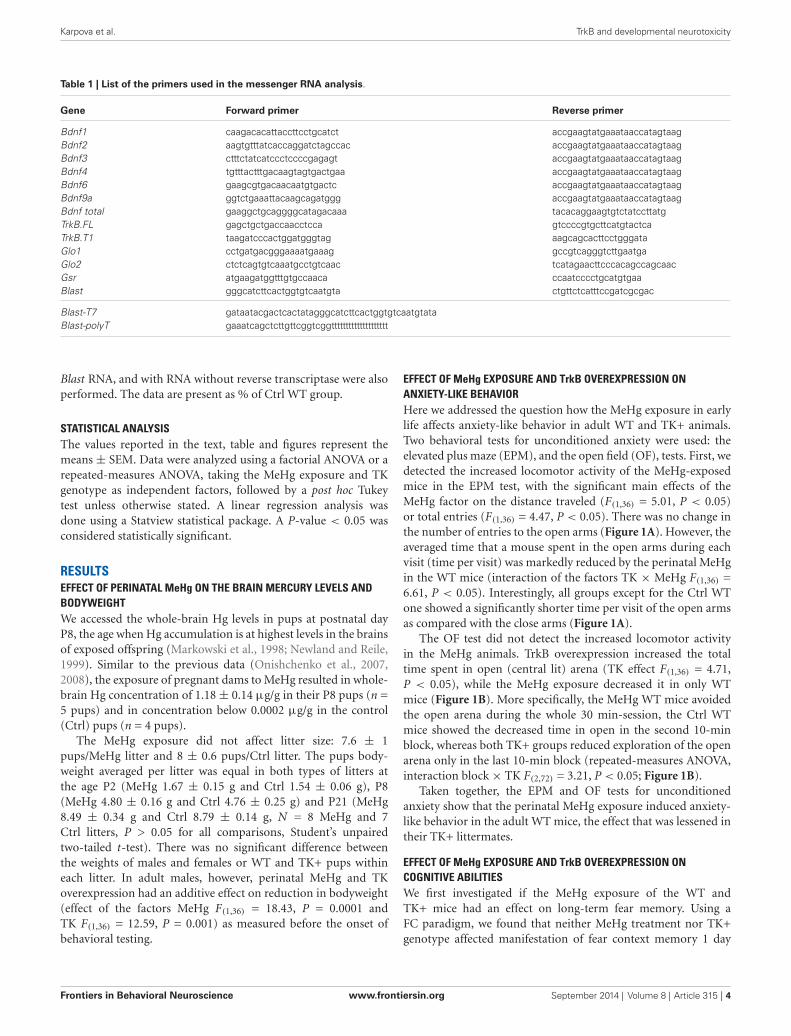
Table 1 | List of the primers used in the messenger RNA analysis.
Gene Forward primer Reverse primer
Bdnf1 caagacacattaccttcctgcatct accgaagtatgaaataaccatagtaagBdnf2 aagtgtttatcaccaggatctagccac accgaagtatgaaataaccatagtaagBdnf3 ctttctatcatccctccccgagagt accgaagtatgaaataaccatagtaagBdnf4 tgtttactttgacaagtagtgactgaa accgaagtatgaaataaccatagtaagBdnf6 gaagcgtgacaacaatgtgactc accgaagtatgaaataaccatagtaagBdnf9a ggtctgaaattacaagcagatggg accgaagtatgaaataaccatagtaagBdnf total gaaggctgcaggggcatagacaaa tacacaggaagtgtctatccttatgTrkB.FL gagctgctgaccaacctcca gtccccgtgcttcatgtactcaTrkB.T1 taagatcccactggatgggtag aagcagcacttcctgggataGlo1 cctgatgacgggaaaatgaaag gccgtcagggtcttgaatgaGlo2 ctctcagtgtcaaatgcctgtcaac tcatagaacttcccacagccagcaacGsr atgaagatggtttgtgccaaca ccaatcccctgcatgtgaaBlast gggcatcttcactggtgtcaatgta ctgttctcatttccgatcgcgac
Blast-T7 gataatacgactcactatagggcatcttcactggtgtcaatgtataBlast-polyT gaaatcagctcttgttcggtcggtttttttttttttttttttt
Blast RNA, and with RNA without reverse transcriptase were alsoperformed. The data are present as % of Ctrl WT group.
STATISTICAL ANALYSISThe values reported in the text, table and figures represent themeans ± SEM. Data were analyzed using a factorial ANOVA or arepeated-measures ANOVA, taking the MeHg exposure and TKgenotype as independent factors, followed by a post hoc Tukeytest unless otherwise stated. A linear regression analysis wasdone using a Statview statistical package. A P-value < 0.05 wasconsidered statistically significant.
RESULTSEFFECT OF PERINATAL MeHg ON THE BRAIN MERCURY LEVELS ANDBODYWEIGHTWe accessed the whole-brain Hg levels in pups at postnatal dayP8, the age when Hg accumulation is at highest levels in the brainsof exposed offspring (Markowski et al., 1998; Newland and Reile,1999). Similar to the previous data (Onishchenko et al., 2007,2008), the exposure of pregnant dams to MeHg resulted in whole-brain Hg concentration of 1.18± 0.14 µg/g in their P8 pups (n =5 pups) and in concentration below 0.0002 µg/g in the control(Ctrl) pups (n = 4 pups).
The MeHg exposure did not affect litter size: 7.6 ± 1pups/MeHg litter and 8 ± 0.6 pups/Ctrl litter. The pups body-weight averaged per litter was equal in both types of litters atthe age P2 (MeHg 1.67 ± 0.15 g and Ctrl 1.54 ± 0.06 g), P8(MeHg 4.80 ± 0.16 g and Ctrl 4.76 ± 0.25 g) and P21 (MeHg8.49 ± 0.34 g and Ctrl 8.79 ± 0.14 g, N = 8 MeHg and 7Ctrl litters, P > 0.05 for all comparisons, Student’s unpairedtwo-tailed t-test). There was no significant difference betweenthe weights of males and females or WT and TK+ pups withineach litter. In adult males, however, perinatal MeHg and TKoverexpression had an additive effect on reduction in bodyweight(effect of the factors MeHg F(1,36) = 18.43, P = 0.0001 andTK F(1,36) = 12.59, P = 0.001) as measured before the onset ofbehavioral testing.
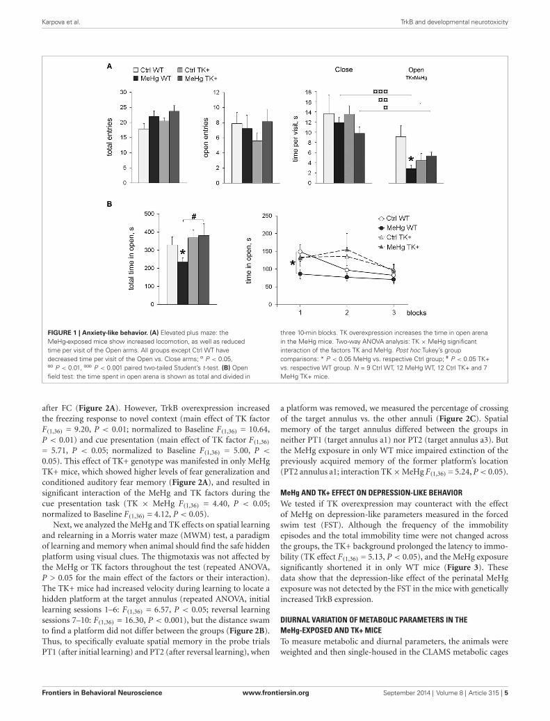
EFFECT OF MeHg EXPOSURE AND TrkB OVEREXPRESSION ONANXIETY-LIKE BEHAVIORHere we addressed the question how the MeHg exposure in earlylife affects anxiety-like behavior in adult WT and TK+ animals.Two behavioral tests for unconditioned anxiety were used: theelevated plus maze (EPM), and the open field (OF), tests. First, wedetected the increased locomotor activity of the MeHg-exposedmice in the EPM test, with the significant main effects of theMeHg factor on the distance traveled (F(1,36) = 5.01, P < 0.05)or total entries (F(1,36) = 4.47, P < 0.05). There was no change inthe number of entries to the open arms (Figure 1A). However, theaveraged time that a mouse spent in the open arms during eachvisit (time per visit) was markedly reduced by the perinatal MeHgin the WT mice (interaction of the factors TK × MeHg F(1,36) =6.61, P < 0.05). Interestingly, all groups except for the Ctrl WTone showed a significantly shorter time per visit of the open armsas compared with the close arms (Figure 1A).
The OF test did not detect the increased locomotor activityin the MeHg animals. TrkB overexpression increased the totaltime spent in open (central lit) arena (TK effect F(1,36) = 4.71,P < 0.05), while the MeHg exposure decreased it in only WTmice (Figure 1B). More specifically, the MeHg WT mice avoidedthe open arena during the whole 30 min-session, the Ctrl WTmice showed the decreased time in open in the second 10-minblock, whereas both TK+ groups reduced exploration of the openarena only in the last 10-min block (repeated-measures ANOVA,interaction block× TK F(2,72) = 3.21, P < 0.05; Figure 1B).
Taken together, the EPM and OF tests for unconditionedanxiety show that the perinatal MeHg exposure induced anxiety-like behavior in the adult WT mice, the effect that was lessened intheir TK+ littermates.
EFFECT OF MeHg EXPOSURE AND TrkB OVEREXPRESSION ONCOGNITIVE ABILITIESWe first investigated if the MeHg exposure of the WT andTK+ mice had an effect on long-term fear memory. Using aFC paradigm, we found that neither MeHg treatment nor TK+genotype affected manifestation of fear context memory 1 day
Frontiers in Behavioral Neuroscience www.frontiersin.org September 2014 | Volume 8 | Article 315 | 4
Karpova et al. TrkB and developmental neurotoxicity
FIGURE 1 | Anxiety-like behavior. (A) Elevated plus maze: theMeHg-exposed mice show increased locomotion, as well as reducedtime per visit of the Open arms. All groups except Ctrl WT havedecreased time per visit of the Open vs. Close arms; ¤ P < 0.05,¤¤ P < 0.01, ¤¤¤ P < 0.001 paired two-tailed Student’s t-test. (B) Openfield test: the time spent in open arena is shown as total and divided in
three 10-min blocks. TK overexpression increases the time in open arenain the MeHg mice. Two-way ANOVA analysis: TK ×MeHg significantinteraction of the factors TK and MeHg. Post hoc Tukey’s groupcomparisons: * P < 0.05 MeHg vs. respective Ctrl group; # P < 0.05 TK+vs. respective WT group. N = 9 Ctrl WT, 12 MeHg WT, 12 Ctrl TK+ and 7MeHg TK+ mice.
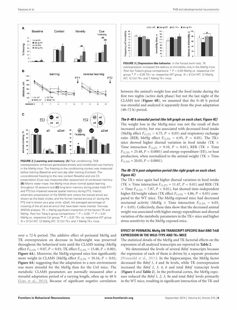
after FC (Figure 2A). However, TrkB overexpression increasedthe freezing response to novel context (main effect of TK factorF(1,36) = 9.20, P < 0.01; normalized to Baseline F(1,36) = 10.64,P < 0.01) and cue presentation (main effect of TK factor F(1,36)
= 5.71, P < 0.05; normalized to Baseline F(1,36) = 5.00, P <
0.05). This effect of TK+ genotype was manifested in only MeHgTK+ mice, which showed higher levels of fear generalization andconditioned auditory fear memory (Figure 2A), and resulted insignificant interaction of the MeHg and TK factors during thecue presentation task (TK × MeHg F(1,36) = 4.40, P < 0.05;normalized to Baseline F(1,36) = 4.12, P < 0.05).
Next, we analyzed the MeHg and TK effects on spatial learningand relearning in a Morris water maze (MWM) test, a paradigmof learning and memory when animal should find the safe hiddenplatform using visual clues. The thigmotaxis was not affected bythe MeHg or TK factors throughout the test (repeated ANOVA,P > 0.05 for the main effect of the factors or their interaction).The TK+ mice had increased velocity during learning to locate ahidden platform at the target annulus (repeated ANOVA, initiallearning sessions 1–6: F(1,36) = 6.57, P < 0.05; reversal learningsessions 7–10: F(1,36) = 16.30, P < 0.001), but the distance swamto find a platform did not differ between the groups (Figure 2B).Thus, to specifically evaluate spatial memory in the probe trialsPT1 (after initial learning) and PT2 (after reversal learning), when
a platform was removed, we measured the percentage of crossingof the target annulus vs. the other annuli (Figure 2C). Spatialmemory of the target annulus differed between the groups inneither PT1 (target annulus a1) nor PT2 (target annulus a3). Butthe MeHg exposure in only WT mice impaired extinction of thepreviously acquired memory of the former platform’s location(PT2 annulus a1; interaction TK×MeHg F(1,36) = 5.24, P< 0.05).
MeHg AND TK+ EFFECT ON DEPRESSION-LIKE BEHAVIORWe tested if TK overexpression may counteract with the effectof MeHg on depression-like parameters measured in the forcedswim test (FST). Although the frequency of the immobilityepisodes and the total immobility time were not changed acrossthe groups, the TK+ background prolonged the latency to immo-bility (TK effect F(1,36) = 5.13, P < 0.05), and the MeHg exposuresignificantly shortened it in only WT mice (Figure 3). Thesedata show that the depression-like effect of the perinatal MeHgexposure was not detected by the FST in the mice with geneticallyincreased TrkB expression.
DIURNAL VARIATION OF METABOLIC PARAMETERS IN THEMeHg-EXPOSED AND TK+ MICETo measure metabolic and diurnal parameters, the animals wereweighted and then single-housed in the CLAMS metabolic cages
Frontiers in Behavioral Neuroscience www.frontiersin.org September 2014 | Volume 8 | Article 315 | 5
Karpova et al. TrkB and developmental neurotoxicity
FIGURE 2 | Learning and memory. (A) Fear conditioning: TrkBoverexpression enhances generalized anxiety and conditioned cue memoryin the MeHg mice. The freezing to the conditioning context was measuredbefore training (Baseline) and next day after training (Context). Theunconditioned freezing to the new context (Novelty) and one CSpresentation (Cue) was measured after assessment of contextual memory.(B) Morris water maze: the MeHg mice show normal spatial learningthroughout 10 sessions and (C) long-term memory during probe trials PT1and PT2 but impaired reversal spatial memory during PT2. Inserts:schematic presentation of the MWM tank where the trained annuli areshown as the black circles, and the former trained annulus a1 during thePT2 trial is shown as a gray circle. a2/a4, the averaged percentage ofcrossing of the a2 and a4 annuli that have been never trained. Two-wayANOVA analysis: TK ×MeHg significant interaction of the factors TK andMeHg. Post hoc Tukey’s group comparisons: * P < 0.05, ** P < 0.01MeHg vs. respective Ctrl group; ## P < 0.01 TK+ vs. respective WT group.N = 9 Ctrl WT, 12 MeHg WT, 12 Ctrl TK+ and 7 MeHg TK+ mice.
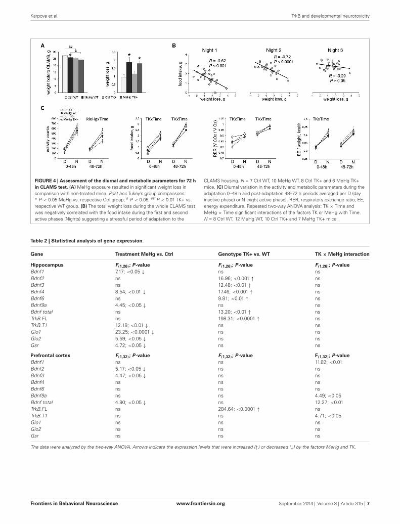
over a 72-h period. The additive effect of perinatal MeHg andTK overexpression on decrease in bodyweight was preservedthroughout the behavioral tests until the CLAMS testing (MeHgeffect F(1,33) = 9.07, P < 0.01; TK effect F(1,33) = 15.48, P < 0.001;Figure 4A). Moreover, the MeHg-exposed mice lost significantlymore weight in CLAMS (MeHg effect F(1,33) = 10.16, P < 0.01;Figure 4A) suggesting that the adaptation to a new environmentwas more stressful for the MeHg than for the Ctrl mice. Themetabolic CLAMS parameters are normally measured after astressful adaptation period of a varying length, often up to 48 h(Liao et al., 2013). Because of significant negative correlation
FIGURE 3 | Depression-like behavior. In the forced swim test, TKoverexpression increased the latency to immobility only in the MeHg mice.Post hoc Tukey’s group comparisons: * P < 0.05 MeHg vs. respective Ctrlgroup; # P < 0.05 TK+ vs. respective WT group. N = 9 Ctrl WT, 12 MeHgWT, 12 Ctrl TK+ and 7 MeHg TK+ mice.
between the animal’s weight loss and the food intake during thefirst two nights (active dark phase) but not the last night of theCLAMS test (Figure 4B), we assumed that the 0–48 h periodwas stressful and analyzed it separately from the post-adaptation(48–72 h) period.
The 0–48 h stressful period (the left graph on each chart, Figure 4C)The weight loss in the MeHg-mice was not the result of theirincreased activity, but was associated with decreased food intake(MeHg effect F(1,27) = 4.75, P < 0.05) and respiratory exchangeratio (RER; MeHg effect F(1,33) = 6.95, P < 0.05). The TK+mice showed higher diurnal variation in food intake (TK ×Time interaction F(1,27) = 9.10, P < 0.01), RER (TK × TimeF(1,33) = 25.48, P < 0.0001) and energy expenditure (EE), or heatproduction, when normalized to the animal weight (TK × TimeF(1,33) = 20.05, P < 0.0001).
The 48–72 h post-adaptation period (the right graph on each chart,Figure 4C)The TK+ mice again had higher diurnal variation in food intake(TK × Time interaction F(1,27) = 11.47, P < 0.01) and RER (TK× Time F(1,33) = 7.87, P < 0.01), but showed time-independenthigher EE/weight values (TK effect F(1,33) = 4.86, P < 0.05) com-pared to the WT mice. The MeHg-exposed mice had decreasednocturnal activity (MeHg × Time interaction F(1,33) = 6.03,P < 0.05). Collectively, these data show that the decreased animalweight was associated with higher energy expenditure and diurnalvariation of the metabolic parameters in the TK+ mice and higherstress sensitivity in the MeHg-exposed mice.
EFFECT OF PERINATAL MeHg ON TRANSCRIPT-SPECIFIC Bdnf AND TrkBEXPRESSION IN THE WILD-TYPE AND TK+ MICEThe statistical details of the MeHg and TK factorial effects on theexpression of all analyzed transcripts are reported in Table 2.
We determined the levels of several Bdnf transcripts becausethe expression of each of them is driven by a separate promoter(Pruunsild et al., 2011). In the hippocampus, the MeHg factordecreased the Bdnf 1, 4 and 9a levels, while TK overexpressionincreased the Bdnf 2, 3, 4, 6 and total Bdnf transcript levels(Figure 5 and Table 2). In the prefrontal cortex, the MeHg expo-sure reduced the Bdnf 1, 2, 3, 9a and total Bdnf levels primarilyin the WT mice, resulting in significant interaction of the TK and
Frontiers in Behavioral Neuroscience www.frontiersin.org September 2014 | Volume 8 | Article 315 | 6
Karpova et al. TrkB and developmental neurotoxicity
FIGURE 4 | Assessment of the diurnal and metabolic parameters for 72 hin CLAMS test. (A) MeHg exposure resulted in significant weight loss incomparison with non-treated mice. Post hoc Tukey’s group comparisons:* P < 0.05 MeHg vs. respective Ctrl group; # P < 0.05, ## P < 0.01 TK+ vs.respective WT group. (B) The total weight loss during the whole CLAMS testwas negatively correlated with the food intake during the first and secondactive phases (Nights) suggesting a stressful period of adaptation to the
CLAMS housing. N = 7 Ctrl WT, 10 MeHg WT, 8 Ctrl TK+ and 6 MeHg TK+mice. (C) Diurnal variation in the activity and metabolic parameters during theadaptation 0–48 h and post-adaptation 48–72 h periods averaged per D (dayinactive phase) or N (night active phase). RER, respiratory exchange ratio; EE,energy expenditure. Repeated two-way ANOVA analysis: TK × Time andMeHg × Time significant interactions of the factors TK or MeHg with Time.N = 8 Ctrl WT, 12 MeHg WT, 10 Ctrl TK+ and 7 MeHg TK+ mice.
Table 2 | Statistical analysis of gene expression.
Gene Treatment MeHg vs. Ctrl Genotype TK+ vs. WT TK × MeHg interaction
Hippocampus F(1,26); P-value F(1,26); P-value F(1,26); P-valueBdnf1 7.17; <0.05 ↓ ns nsBdnf2 ns 16.96; <0.001 ↑ nsBdnf3 ns 12.48; <0.01 ↑ nsBdnf4 8.54; <0.01 ↓ 17.46; <0.001 ↑ nsBdnf6 ns 9.81; <0.01 ↑ nsBdnf9a 4.45; <0.05 ↓ ns nsBdnf total ns 13.20; <0.01 ↑ nsTrkB.FL ns 198.31; <0.0001 ↑ nsTrkB.T1 12.18; <0.01 ↓ ns nsGlo1 23.25; <0.0001 ↓ ns nsGlo2 5.59; <0.05 ↓ ns nsGsr 4.72; <0.05 ↓ ns ns
Prefrontal cortex F(1,32); P-value F(1,32); P-value F(1,32); P-valueBdnf1 ns ns 11.82; <0.01Bdnf2 5.17; <0.05 ↓ ns nsBdnf3 4.47; <0.05 ↓ ns nsBdnf4 ns ns nsBdnf6 ns ns nsBdnf9a ns ns 4.49; <0.05Bdnf total 4.90; <0.05 ↓ ns 12.27; <0.01TrkB.FL ns 284.64; <0.0001 ↑ nsTrkB.T1 ns ns 4.71; <0.05Glo1 ns ns nsGlo2 ns ns nsGsr ns ns ns
The data were analyzed by the two-way ANOVA. Arrows indicate the expression levels that were increased (↑) or decreased (↓) by the factors MeHg and TK.
Frontiers in Behavioral Neuroscience www.frontiersin.org September 2014 | Volume 8 | Article 315 | 7

Karpova et al. TrkB and developmental neurotoxicity
FIGURE 5 | Expression of the Bdnf transcripts. The Bdnf expression wasmainly decreased by MeHg and increased by TK+ background in thehippocampus, and dependent on factors’ interaction in the prefrontal cortex.Two-way ANOVA analysis: TK ×MeHg significant interaction of the factors TK
and MeHg. Post hoc Tukey’s group comparisons: * P < 0.05, ** P < 0.01MeHg vs. respective Ctrl group; # P < 0.05, ## P < 0.01 TK+ vs. respectiveWT group. N = 7–9 Ctrl WT, 8–11 MeHg WT, 9 Ctrl TK+ and 6–7 MeHg TK+mice.
FIGURE 6 | Expression of the full-length TrkB.FL and truncated TrkB.T1transcripts. MeHg exposure did not affect the TrkB.FL expression butreduced the TrkB.T1 levels in the hippocampus and interacted with TKgenotype for TrkB.T1 in the prefrontal cortex. Two-way ANOVA analysis: TK×MeHg significant interaction of the factors TK and MeHg. Post hocTukey’s group comparisons: * P < 0.05 MeHg vs. respective Ctrl group; ##
P < 0.01 TK+ vs. respective WT group. N = 7–9 Ctrl WT, 8–11 MeHg WT, 9Ctrl TK+ and 6–7 MeHg TK+ mice.
MeHg factors found for the Bdnf 1, 9a and total Bdnf expression(Figure 5 and Table 2).
The developmental MeHg exposure did not affect the hip-pocampal and cortical levels of the TrkB full-length transcriptTrkB.FL but down-regulated the major truncated TrkB isoformTrkB.T1 in the hippocampus and, similar to the effect on Bdnfexpression, significantly interacted with TK genotype for TrkB.T1expression in the prefrontal cortex (Figure 6 and Table 2).
MeHg EFFECT ON THE EXPRESSION OF THE ANTIOXIDANT ENZYMESWe found a significant MeHg-induced reduction in the hip-pocampal levels of Glo1, Glo2 and Gsr transcripts coding for theimportant antioxidant enzymes (Figure 7 and Table 2). The TKoverexpression did not affect these transcripts in either brain area.
DISCUSSIONHere we report for the first time that developmental MeHgexposure induces anxiety-related behavior in adult mice. Thesechanges were detected in the classical tests for unconditioned
FIGURE 7 | Expression of the antioxidant enzymes. MeHg exposuredecreased the levels of Glo1, Glo2 and Gsr in the hippocampus. Post hocTukey’s group comparisons: * P < 0.05, ** P < 0.01 MeHg vs. respectiveCtrl group. N = 7–9 Ctrl WT, 8–11 MeHg WT, 9 Ctrl TK+ and 6–7 MeHg TK+mice.
anxiety and during the adaptation period in the CLAMS test,which can provide important data about stress-related physio-logical parameters. We found that the perinatal MeHg-exposuresignificantly reduced food intake and RER during adaptation tothe novel CLAMS environment, resulting in substantial weightloss in both MeHg groups. Chronic MeHg exposure in adulthoodwas also reported to decrease weight gain in diabetic obese KK-Aymice due to MeHg-induced toxicity in adipose tissue (Yamamotoet al., 2013). It is possible that MeHg toxicity in the brain inducedsensitivity to stressful stimuli, provided either by behavioralmanipulations or every-day home-cage activities, which could beamong the mechanisms that mediate a long-term effect of perina-tal MeHg on decreased bodyweight. Early-life stress is a main riskfactor for development of psychopathologies in adulthood andis often associated with a hyperactive hypothalamic–pituitary–adrenal (HPA) axis (Heim and Nemeroff, 2001) and increasedvulnerability to stress (Cirulli et al., 2009). Thus, it would be
Frontiers in Behavioral Neuroscience www.frontiersin.org September 2014 | Volume 8 | Article 315 | 8
Karpova et al. TrkB and developmental neurotoxicity
interesting to investigate the HPA axis reactivity and neuroen-docrine responses to stress in early-life-MeHg exposed subjects.
The MeHg-induced behavioral changes were accompanied bythe decreased levels of multiple Bdnf transcripts in the prefrontalcortex and hippocampus, and TrkB.T1, Glo1, Glo2 and Gsr in thehippocampus. Bdnf gene structure with multiple promoters anddifferent regulatory elements allows flexibility in Bdnf expressionin response to various environmental stimuli. Among the Bdnftranscripts, Bdnf1, Bdnf4 and Bdnf9a are the most responsiveto neuronal activity in vitro (Pruunsild et al., 2011) and onlythese transcripts were down-regulated by the perinatal MeHg inthe hippocampus. A key common mechanism for transcriptiondriven by the corresponding Bdnf promoters p1, p4 and p9is that they contain E-box-like elements which can be boundby the basic helix-loop-helix-PAS transcription factors Arnt2and Npas4 in an activity-dependent manner (Pruunsild et al.,2011; Maya-Vetencourt, 2013). Since Npas4 plays an importantrole in the maturation of inhibitory synapses (Lin et al., 2008),perinatal MeHg could preferentially target the development ofinhibitory system during hippocampal growth. In the prefrontalcortex, however, the MeHg effect was not restricted to the E-box-containing transcripts suggesting contribution of additionalmechanisms in cortical Bdnf regulation. A possible mechanisticpathway for a brain area-specific MeHg effect could involve estab-lishing of distinct DNA methylation marks at the Bdnf promotersduring development (see also Onishchenko et al., 2008). A similarphenomenon has been recently identified for a HSD11B2 genemethylation that differs between the hypothalamus and the cortexof prenatally stressed rats (Jensen Peña et al., 2012).
The effect of TrkB overexpression on the MeHg-induced Bdnfdecrease was also brain area-specific: MeHg-independent in thehippocampus and MeHg-dependent in the prefrontal cortex. Thiseffect may be due to activation of different tissue-specific ordevelopmental pathways as discussed above, or to the higherTrkB.FL levels in the cortex (≈4.4 fold over WT levels) whencompared to the hippocampus (≈2.9 fold over WT levels). Alter-natively, a different neuronal/glia ratio between the brain regions(Herculano-Houzel, 2014) could account for the tissue-specificgene levels. Glia, including microglia and astrocytes, play a criticalrole in proper brain functioning, and was shown to be sensitiveto oxidative stress and MeHg toxicity (Ni et al., 2012). TruncatedTrkB.T1 is a major TrkB isoform in astrocytes, and its expressionwas shown to be reduced in the cortex of suicide completersvia binding its 3′UTR by a microRNA Hsa-miR-185∗ (Maussionet al., 2012). The same group linked a deletion in TrkB promoterregion, which could affect the levels of all TrkB transcripts includ-ing TrkB.T1, to childhood and adulthood anxiety in humans(Ernst et al., 2011). It could be suggested that the MeHg-induceddecrease in the endogenous TrkB.T1 expression that we observedin the hippocampus may impair Bdnf signaling in astrocytes, aswell as reduce dendritic complexity in neurons and contribute todevelopment of anxiety-like phenotype (Carim-Todd et al., 2009).Further studies are needed to investigate if the tissue-dependentgene expression observed in our study is due to neuronal or glial-specific transcription.
We detected a MeHg-induced decrease in the hippocampal lev-els of important antioxidant enzymes involved in methylglyoxal
(Glo1 and Glo2) and glutathione (Gsr) processing, suggesting amechanism for a long-term MeHg effect on behavioral impair-ments. One study similarly reported the prenatal MeHg-inducedreduction in cerebral glutathione peroxidase and reductase activ-ities in mouse pups at the age P21, although adult mice werenot investigated (Stringari et al., 2008). Chronic oxidative stressand accumulation of reactive-oxygen species has been consistentlyassociated with anxiety- and depression-like behaviors, as well ascognitive dysfunctions in humans and rodents (Gawryluk et al.,2011; Salim et al., 2011; Moretti et al., 2012).
The TK overexpression did not show any beneficial effect onanxiety-like behavior in the EPM test in the adult MeHg animals,which can be explained by manifestation of anxiety phenotype inTK+ animals themselves. This result was unexpected consideringa previous finding about the decreased EPM anxiety-like behav-ior in TK+ mice (Koponen et al., 2004). Differences in geneticbackground of TK+ mice in the present and previous studies mayaccount for this discrepancy. Several behavioral studies reportabout more anxious phenotype of BALB/c and DBA/2 mice incomparison with C57BL/6J (Crawley et al., 1997; Hackler et al.,2006), which could explain the beneficial effect of TK overexpres-sion on the EPM anxiety-like parameters found by Koponen et al.(2004). Our current EPM data are in line with reduced anxiety inNtrk2-deficient heterozygote mice (Olsen et al., 2013) and with acritical role of TrkB activation in enhanced anxiety-like behaviorin mice after status epilepticus (Liu et al., 2013).
In addition to the TK effect in the EPM test, we found thatthe TK+ background increased non-associative generalized fearresponses (freezing to novel context) following FC, especially inthe MeHg-exposed mice. Generalized hyper-responsiveness afteran exposure to a traumatic event was suggested to model severalfeatures of post-traumatic stress disorder (PTSD; Balogh andWehner, 2003), but could also model an adolescent-like increasedemotionality found in late juvenile rodents with heightened feargeneralization (Ito et al., 2009). Since PTSD-like phenotype isassociated with learning and memory impairments, a secondsuggestion is more likely because the MeHg TK+ mice had betterconditioned cue memory and did not show the defects in MWMreversal spatial memory. Further studies, which are focused on theanalysis of fear renewal/reinstatement and spontaneous recoveryusing a FC-extinction paradigm, are required before drawingconclusion about potential PTSD-like phenotype of the MeHgTK+ mice.
The anxiety-related traits that we detected in the TK+ micewere not pronounced in less stressful OF test where TK+ back-ground buffered the anxiety-like impairments of the MeHg mice.Moreover, although TK overexpression did not affect depression-like behavior in the Ctrl group, it prolonged the latency toimmobility in the MeHg-exposed group. It is possible that TrkBoverexpression leads to increased neuron number (Lähteinenet al., 2003) which, in our experiment, resulted in significantbehavioral changes only in vulnerable to stress MeHg-treatedanimals. Consistently with the neurogenic hypothesis of depres-sion (Eisch and Petrik, 2012), perinatal MeHg exposure reducesdifferentiation of neural stem cells and neurogenesis in adultdentate gyrus (Bose et al., 2012). At the same time, reduced TrkBexpression is associated with increased apoptosis in depressed
Frontiers in Behavioral Neuroscience www.frontiersin.org September 2014 | Volume 8 | Article 315 | 9

Karpova et al. TrkB and developmental neurotoxicity
and suicide subjects (Dwivedi et al., 2009), and TrkB.FL defi-ciency in neuronal progenitors in adulthood impairs integrationof newborn neurons into hippocampal neural circuits and con-sequently induces anxiety-like behavior in mice (Bergami et al.,2008). Thus, it could be hypothesized that TK overexpressionspecifically in the hippocampus, possibly via increased Bdnfexpression observed in that brain area, supports more efficientintegration of newly born neurons of MeHg mice into adulthippocampal network and improves some behavioral impair-ments. Additional studies using brain site-specific alteration ofBdnf signaling could help to verify this hypothesis. In earlydevelopment, however, formation of functional neuronal net-works by newborn neurons is active throughout the growingbrain. During this sensitive period, emerging TK overexpressionin transgenic animals could have stronger interference with Bdnfsignaling throughout the brain of vulnerable to stress MeHg-exposed mice in comparison with the control ones, which couldpotentially lead to either positive or negative behavioral alter-ations in adulthood.
Depression- and anxiety-phenotypes are linked to impairedcircadian mechanisms (Etain et al., 2011). Given the diurnaloscillation pattern of both Bdnf and TrkB (Bova et al., 1998) andtheir important role in sleep homeostasis (Faraguna et al., 2008)and circadian regulation (Liang et al., 2000), impaired Bdnf-TrkB signaling may promote development of mood disordersby disturbing the normal circadian cycle. In mice, chronic TrkBdeficiency results in diminished circadian response to light (Allenet al., 2005). We found that the effect of TrkB overexpressionon diurnal physiological parameters was opposite to that pro-duced by TrkB-deficiency, but the TK+ background failed toameliorate the diminished diurnal activity pattern in the MeHg-exposed mice that was also observed previously (Onishchenkoet al., 2007). Strikingly, the increased diurnal pattern in foodintake and a fuel selection parameter RER observed in theTK+ mice resemble those seen in calorie-restricted mice wherethese effects at least partially underlie their health and longevity(Bruss et al., 2010). The decreased weight of the TK+ micewithout detectable changes in total food intake may as wellbe a result of increased EE contributing to the hypothesis ofbodyweight regulation by Bdnf-TrkB signaling (Vanevski and Xu,2013).
In summary, we can conclude that postnatal TrkB overex-pression itself does not produce significant behavioral effects inC57Bl/6J mice, but it buffers against several behavioral impair-ments induced by perinatal MeHg. However, some parametersof anxiety-like behavior and associated decrease in the levels ofactivity-dependent Bdnf transcripts, TrkB.T1 and the antioxi-dant enzymes in the hippocampus were permanently affectedin adulthood by perinatal MeHg in a TK genotype-independentmanner.
AUTHOR CONTRIBUTIONSNina N. Karpova, Sandra Ceccatelli and Eero Castrén wereresponsible for the conception and design of the work. NinaN. Karpova, Jesse Saku Olavi Lindholm, Natalia Kulesskaya,Natalia Onishchenko, Marie Vahter and Dina Popova acquiredand analyzed data. Nina N. Karpova, Natalia Kulesskaya, Sandra
Ceccatelli and Eero Castrén interpreted data. Nina N. Karpovadrafted the manuscript. All authors participated in critical revis-ing and final approval of the manuscript, and agree to be account-able for all aspects of the work.
ACKNOWLEDGMENTSWe thank Sissi Pastell for animal care. This work was supported bythe Sigrid Jusélius Foundation and the Academy of Finland Centerof Excellence program (to Eero Castrén), Centre for InternationalMobility Fellowships programme (to Dina Popova), grants fromthe Swedish Research Councils (VR, FORMAS) and KarolinskaInstitutet (to Sandra Ceccatelli).
REFERENCESAllen, G. C., Qu, X., and Earnest, D. J. (2005). TrkB-deficient mice show diminished
phase shifts of the circadian activity rhythm in response to light. Neurosci. Lett.378, 150–155. doi: 10.1016/j.neulet.2004.12.023
Balogh, S. A., and Wehner, J. M. (2003). Inbred mouse strain differences in theestablishment of long-term fear memory. Behav. Brain Res. 140, 97–106. doi: 10.1016/s0166-4328(02)00279-6
Bergami, M., Rimondini, R., Santi, S., Blum, R., Götz, M., and Canossa, M. (2008).Deletion of TrkB in adult progenitors alters newborn neuron integration intohippocampal circuits and increases anxiety-like behavior. Proc. Natl. Acad. Sci.U S A 105, 15570–15575. doi: 10.1073/pnas.0803702105
Birch, R. J., Bigler, J., Rogers, J. W., Zhuang, Y., and Clickner, R. P. (2013). Trendsin blood mercury concentrations and fish consumption among U.S. women ofreproductive age: NHANES, 1999–2010. Environ. Res. 133, 431–438. doi: 10.1016/j.envres.2014.02.001
Bose, R., Onishchenko, N., Edoff, K., Janson Lang, A. M., and Ceccatelli, S. (2012).Inherited effects of low-dose exposure to methylmercury in neural stem cells.Toxicol. Sci. 130, 383–390. doi: 10.1093/toxsci/kfs257
Bova, R., Micheli, M. R., Qualadrucci, P., and Zucconi, G. G. (1998). BDNF andtrkB mRNAs oscillate in rat brain during the light-dark cycle. Brain Res. Mol.Brain Res. 57, 321–324. doi: 10.1016/s0169-328x(98)00092-8
Bruss, M. D., Khambatta, C. F., Ruby, M. A., Aggarwal, I., and Hellerstein, M. K.(2010). Calorie restriction increases fatty acid synthesis and whole body fatoxidation rates. Am. J. Physiol. Endocrinol. Metab. 298, E108–E116. doi: 10.1152/ajpendo.00524.2009
Carim-Todd, L., Bath, K. G., Fulgenzi, G., Yanpallewar, S., Jing, D., Barrick,C. A., et al. (2009). Endogenous truncated TrkB.T1 receptor regulates neuronalcomplexity and TrkB kinase receptor function in vivo. J. Neurosci. 29, 678–685.doi: 10.1523/jneurosci.5060-08.2009
Ceccatelli, S., Bose, R., Edoff, K., Onishchenko, N., and Spulber, S. (2013). Long-lasting neurotoxic effects of exposure to methylmercury during development. J.Intern. Med. 273, 490–497. doi: 10.1111/joim.12045
Cirulli, F., Francia, N., Berry, A., Aloe, L., Alleva, E., and Suomi, S. J. (2009). Earlylife stress as a risk factor for mental health: role of neurotrophins from rodentsto non-human primates. Neurosci. Biobehav. Rev. 33, 573–585. doi: 10.1016/j.neubiorev.2008.09.001
Crawley, J. N., Belknap, J. K., Collins, A., Crabbe, J. C., Frankel, W., Henderson, N.,et al. (1997). Behavioral phenotypes of inbred mouse strains: implications andrecommendations for molecular studies. Psychopharmacology (Berl) 132, 107–124. doi: 10.1007/s002130050327
Dwivedi, Y., Rizavi, H. S., Zhang, H., Mondal, A. C., Roberts, R. C., Conley,R. R., et al. (2009). Neurotrophin receptor activation and expression in humanpostmortem brain: effect of suicide. Biol. Psychiatry 65, 319–328. doi: 10.1016/j.biopsych.2008.08.035
Eisch, A. J., and Petrik, D. (2012). Depression and hippocampal neurogenesis: aroad to remission? Science 338, 72–75. doi: 10.1126/science.1222941
Ernst, C., Wanner, B., Brezo, J., Vitaro, F., Tremblay, R., and Turecki, G. (2011).A deletion in tropomyosin-related kinase B and the development of humananxiety. Biol. Psychiatry 69, 604–607. doi: 10.1016/j.biopsych.2010.10.008
Etain, B., Milhiet, V., Bellivier, F., and Leboyer, M. (2011). Genetics of circadianrhythms and mood spectrum disorders. Eur. Neuropsychopharmacol. 21, S676–S682. doi: 10.1016/j.euroneuro.2011.07.007
Frontiers in Behavioral Neuroscience www.frontiersin.org September 2014 | Volume 8 | Article 315 | 10

Karpova et al. TrkB and developmental neurotoxicity
Faraguna, U., Vyazovskiy, V. V., Nelson, A. B., Tononi, G., and Cirelli, C. (2008). Acausal role for brain-derived neurotrophic factor in the homeostatic regulationof sleep. J. Neurosci. 28, 4088–4095. doi: 10.1523/jneurosci.5510-07.2008
Gawryluk, J. W., Wang, J. F., Andreazza, A. C., Shao, L., and Young, L. T. (2011).Decreased levels of glutathione, the major brain antioxidant, in post-mortemprefrontal cortex from patients with psychiatric disorders. Int. J. Neuropsy-chopharmacol. 14, 123–130. doi: 10.1017/s1461145710000805
Ghanizadeh, A., Freeman, R. D., and Berk, M. (2013). Efficacy and adverse effectsof venlafaxine in children and adolescents with ADHD: a systematic review ofnon-controlled and controlled trials. Rev. Recent Clin. Trials 8, 2–8. doi: 10.2174/1574887111308010002
Gökben, S., Ardiç, U. A., and Serdaroglu, G. (2012). Use of buspirone and fluoxetinefor breathing problems in Rett syndrome. Pediatr. Neurol. 46, 192–194. doi: 10.1016/j.pediatrneurol.2011.12.003
Grandjean, P., and Landrigan, P. J. (2006). Developmental neurotoxicity of indus-trial chemicals. Lancet 368, 2167–2178. doi: 10.1016/s0140-6736(06)69665-7
Hackler, E. A., Airey, D. C., Shannon, C. C., Sodhi, M. S., and Sanders-Bush, E.(2006). 5-HT(2C) receptor RNA editing in the amygdala of C57BL/6J, DBA/2Jand BALB/cJ mice. Neurosci. Res. 55, 96–104. doi: 10.1016/j.neures.2006.02.005
Heim, C., and Nemeroff, C. B. (2001). The role of childhood trauma in theneurobiology of mood and anxiety disorders: preclinical and clinical studies.Biol. Psychiatry 49, 1023–1039. doi: 10.1016/s0006-3223(01)01157-x
Herculano-Houzel, S. (2014). The glia/neuron ratio: how it varies uniformly acrossbrain structures and species and what that means for brain physiology andevolution. Glia 62, 1377–1391. doi: 10.1002/glia.22683
Huang, C. F., Liu, S. H., Hsu, C. J., and Lin-Shiau, S. Y. (2011). Neurotoxicolog-ical effects of low-dose methylmercury and mercuric chloride in developingoffspring mice. Toxicol. Lett. 201, 196–204. doi: 10.1016/j.toxlet.2010.12.016
Ito, W., Pan, B. X., Yang, C., Thakur, S., and Morozov, A. (2009). Enhancedgeneralization of auditory conditioned fear in juvenile mice. Learn. Mem. 16,187–192. doi: 10.1101/lm.1190809
Jensen Peña, C., Monk, C., and Champagne, F. A. (2012). Epigenetic effects ofprenatal stress on 11β-hydroxysteroid dehydrogenase-2 in the placenta and fetalbrain. PLoS One 7:e39791. doi: 10.1371/journal.pone.0039791
Johansson, C., Castoldi, A. F., Onishchenko, N., Manzo, L., Vahter, M., andCeccatelli, S. (2007). Neurobehavioural and molecular changes induced bymethylmercury exposure during development. Neurotox. Res. 11, 241–260.doi: 10.1007/bf03033570
Karpova, N. N., Pickenhagen, A., Lindholm, J., Tiraboschi, E., Kulesskaya, N.,Agústsdóttir, A., et al. (2011). Fear erasure in mice requires synergy betweenantidepressant drugs and extinction training. Science 334, 1731–1734. doi: 10.1126/science.1214592
Kemppainen, S., Rantamäki, T., Jerónimo-Santos, A., Lavasseur, G., Autio, H.,Karpova, N., et al. (2012). Impaired TrkB receptor signaling contributes tomemory impairment in APP/PS1 mice. Neurobiol. Aging 33, 1122.e23–1122.e39.doi: 10.1016/j.neurobiolaging.2011.11.006
Koponen, E., Võikar, V., Riekki, R., Saarelainen, T., Rauramaa, T., Rauvala, H., et al.(2004). Transgenic mice overexpressing the full-length neurotrophin receptortrkB exhibit increased activation of the trkB-PLCgamma pathway, reducedanxiety and facilitated learning. Mol. Cell. Neurosci. 26, 166–181. doi: 10.1016/j.mcn.2004.01.006
Lähteinen, S., Pitkänen, A., Koponen, E., Saarelainen, T., and Castrén, E. (2003).Exacerbated status epilepticus and acute cell loss, but no changes in epilep-togenesis, in mice with increased brain-derived neurotrophic factor signaling.Neuroscience 122, 1081–1092. doi: 10.1016/j.neuroscience.2003.08.037
Lai, K. O., Wong, A. S., Cheung, M. C., Xu, P., Liang, Z., Lok, K. C., et al. (2012).TrkB phosphorylation by Cdk5 is required for activity-dependent structuralplasticity and spatial memory. Nat. Neurosci. 15, 1506–1515. doi: 10.1038/nn.3237
Liang, F. Q., Allen, G., and Earnest, D. (2000). Role of brain-derived neurotrophicfactor in the circadian regulation of the suprachiasmatic pacemaker by light. J.Neurosci. 20, 2978–2987.
Liao, G. Y., Li, Y., and Xu, B. (2013). Ablation of TrkB expression in RGS9–2 cellsleads to hyperphagic obesity. Mol. Metab. 2, 491–497. doi: 10.1016/j.molmet.2013.08.002
Lin, Y., Bloodgood, B. L., Hauser, J. L., Lapan, A. D., Koon, A. C., Kim, T. K.,et al. (2008). Activity-dependent regulation of inhibitory synapse developmentby Npas4. Nature 455, 1198–1204. doi: 10.1038/nature07319
Liu, G., Gu, B., He, X. P., Joshi, R. B., Wackerle, H. D., Rodriguiz, R. M., et al. (2013).Transient inhibition of TrkB kinase after status epilepticus prevents developmentof temporal lobe epilepsy. Neuron 79, 31–38. doi: 10.1016/j.neuron.2013.04.027
Magos, L. (1971). Selective atomic-absorption determination of inorganic mercuryand methylmercury in undigested biological samples. Analyst 96, 847–853.doi: 10.1039/an9719600847
Maneeton, N., Maneeton, B., Srisurapanont, M., and Martin, S. D. (2011). Bupro-pion for adults with attention-deficit hyperactivity disorder: meta-analysis ofrandomized, placebo-controlled trials. Psychiatry Clin. Neurosci. 65, 611–617.doi: 10.1111/j.1440-1819.2011.02264.x
Markowski, V. P., Flaugher, C. B., Baggs, R. B., Rawleigh, R. C., Cox, C., and Weiss,B. (1998). Prenatal and lactational exposure to methylmercury affects selectparameters of mouse cerebellar development. Neurotoxicology 19, 879–892.
Maussion, G., Yang, J., Yerko, V., Barker, P., Mechawar, N., Ernst, C., et al. (2012).Regulation of a truncated form of tropomyosin-related kinase B (TrkB) by Hsa-miR-185∗ in frontal cortex of suicide completers. PLoS One 7:e39301. doi: 10.1371/journal.pone.0039301
Maya-Vetencourt, J. F. (2013). Activity-dependent NPAS4 expression and theregulation of gene programs underlying plasticity in the central nervous system.Neural Plast. 2013:683909. doi: 10.1155/2013/683909
McGowan, P. O., Sasaki, A., D’Alessio, A. C., Dymov, S., Labonté, B., Szyf, M., et al.(2009). Epigenetic regulation of the glucocorticoid receptor in human brainassociates with childhood abuse. Nat. Neurosci. 12, 342–348. doi: 10.1038/nn.2270
Minichiello, L., Korte, M., Wolfer, D., Kühn, R., Unsicker, K., Cestari, V., et al.(1999). Essential role for TrkB receptors in hippocampus-mediated learning.Neuron 24, 401–414. doi: 10.1016/s0896-6273(00)80853-3
Moretti, M., Colla, A., de Oliveira Balen, G., dos Santos, D. B., Budni, J., deFreitas, A. E., et al. (2012). Ascorbic acid treatment, similarly to fluoxetine,reverses depressive-like behavior and brain oxidative damage induced by chronicunpredictable stress. J. Psychiatr. Res. 46, 331–340. doi: 10.1016/j.jpsychires.2011.11.009
Newland, M. C., and Reile, P. A. (1999). Blood and brain mercury levels afterchronic gestational exposure to methylmercury in rats. Toxicol. Sci. 50, 106–116.doi: 10.1093/toxsci/50.1.106
Ni, M., Li, X., Rocha, J. B., Farina, M., and Aschner, M. (2012). Glia andmethylmercury neurotoxicity. J. Toxicol. Environ. Health A 75, 1091–1101.doi: 10.1080/15287394.2012.697840
Olsen, D., Kaas, M., Schwartz, O., Nykjaer, A., and Glerup, S. (2013). Loss ofBDNF or its receptors in three mouse models has unpredictable consequencesfor anxiety and fear acquisition. Learn. Mem. 20, 499–504. doi: 10.1101/lm.032045.113
Onishchenko, N., Karpova, N., Sabri, F., Castrén, E., and Ceccatelli, S. (2008).Long-lasting depression-like behavior and epigenetic changes of BDNF geneexpression induced by perinatal exposure to methylmercury. J. Neurochem. 106,1378–1387. doi: 10.1111/j.1471-4159.2008.05484.x
Onishchenko, N., Tamm, C., Vahter, M., Hökfelt, T., Johnson, J. A., Johnson, D. A.,et al. (2007). Developmental exposure to methylmercury alters learning andinduces depression-like behavior in male mice. Toxicol. Sci. 97, 428–437. doi: 10.1093/toxsci/kfl199
Pruunsild, P., Sepp, M., Orav, E., Koppel, I., and Timmusk, T. (2011). Identifi-cation of cis-elements and transcription factors regulating neuronal activity-dependent transcription of human BDNF gene. J. Neurosci. 31, 3295–3308.doi: 10.1523/jneurosci.4540-10.2011
Rantamäki, T., Vesa, L., Antila, H., Di Lieto, A., Tammela, P., Schmitt, A., et al.(2011). Antidepressant drugs transactivate TrkB neurotrophin receptors inthe adult rodent brain independently of BDNF and monoamine transporterblockade. PLoS One 6:e20567. doi: 10.1371/journal.pone.0020567
Salim, S., Asghar, M., Taneja, M., Hovatta, I., Chugh, G., Vollert, C., et al.(2011). Potential contribution of oxidative stress and inflammation to anx-iety and hypertension. Brain Res. 1404, 63–71. doi: 10.1016/j.brainres.2011.06.024
Soliman, F., Glatt, C. E., Bath, K. G., Levita, L., Jones, R. M., Pattwell, S. S.,et al. (2010). A genetic variant BDNF polymorphism alters extinction learn-ing in both mouse and human. Science 327, 863–866. doi: 10.1126/science.1181886
Stringari, J., Nunes, A. K., Franco, J. L., Bohrer, D., Garcia, S. C., Dafre, A. L., et al.(2008). Prenatal methylmercury exposure hampers glutathione antioxidant
Frontiers in Behavioral Neuroscience www.frontiersin.org September 2014 | Volume 8 | Article 315 | 11
Karpova et al. TrkB and developmental neurotoxicity
system ontogenesis and causes long-lasting oxidative stress in the mouse brain.Toxicol. Appl. Pharmacol. 227, 147–154. doi: 10.1016/j.taap.2007.10.010
Vanevski, F., and Xu, B. (2013). Molecular and neural bases underlying roles ofBDNF in the control of body weight. Front. Neurosci. 7:37. doi: 10.3389/fnins.2013.00037
Weaver, I. C., Cervoni, N., Champagne, F. A., D’Alessio, A. C., Sharma, S., Seckl,J. R., et al. (2004). Epigenetic programming by maternal behavior. Nat. Neurosci.7, 847–854. doi: 10.1038/nn1276
Yamamoto, M., Yanagisawa, R., Motomura, E., Nakamura, M., Sakamoto, M.,Takeya, M., et al. (2013). Increased methylmercury toxicity related to obesityin diabetic KK-Ay mice. J. Appl. Toxicol. 34, 914–923. doi: 10.1002/jat.2954
Conflict of Interest Statement: The authors declare that the research was conductedin the absence of any commercial or financial relationships that could be construedas a potential conflict of interest.
Received: 26 June 2014; accepted: 26 August 2014; published online: 12 September2014.Citation: Karpova NN, Lindholm JSO, Kulesskaya N, Onishchenko N, Vahter M,Popova D, Ceccatelli S and Castrén E (2014) TrkB overexpression in mice buffersagainst memory deficits and depression-like behavior but not all anxiety- and stress-related symptoms induced by developmental exposure to methylmercury. Front. Behav.Neurosci. 8:315. doi: 10.3389/fnbeh.2014.00315This article was submitted to the journal Frontiers in Behavioral Neuroscience.Copyright © 2014 Karpova, Lindholm, Kulesskaya, Onishchenko, Vahter, Popova,Ceccatelli and Castrén. This is an open-access article distributed under the terms of theCreative Commons Attribution License (CC BY). The use, distribution or reproductionin other forums is permitted, provided the original author(s) or licensor are creditedand that the original publication in this journal is cited, in accordance with acceptedacademic practice. No use, distribution or reproduction is permitted which does notcomply with these terms.
Frontiers in Behavioral Neuroscience www.frontiersin.org September 2014 | Volume 8 | Article 315 | 12