Transplantation of in vitro-expanded fetal neural progenitor cells results in neurogenesis and...
9
Transplantation of In Vitro-Expanded Fetal Neural Progenitor Cells Results in Neurogenesis and Functional Recovery After Spinal Cord Contusion Injury in Adult Rats Y. Ogawa, 1–4 * K. Sawamoto, 3–5 T. Miyata, 3 S. Miyao, 3,4 M. Watanabe, 2 M. Nakamura, 2 B.S. Bregman, 6 M. Koike, 7 Y. Uchiyama, 7 Y. Toyama, 2 and H. Okano 1–4 1 Department of Physiology, Keio University School of Medicine, Tokyo, Japan 2 Department of Orthopedic Surgery, Keio University School of Medicine, Tokyo, Japan 3 Division of Neuroanatomy (D12), Department of Neuroscience, Biomedical Research Center, Osaka University Graduate School of Medicine, Osaka, Japan 4 Core Research for Evolutional Science and Technology (CREST), Japan Science and Technology Corporation (JST), Osaka, Japan 5 Strategic Promotion System for Brain Science (SPSBS), The Ministry of Education, Culture, Sports, Science and Technology, Tokyo, Japan 6 Department of Neuroscience, Georgetown University Medical Center, Washington, DC 7 Department of Cell Biology and Neurosciences, Osaka University Graduate School of Medicine, Osaka, Japan Neural progenitor cells, including neural stem cells, are a potential expandable source of graft material for trans- plantation aimed at repairing the damaged CNS. Here we present the first evidence that in vitro-expanded fetus- derived neurosphere cells were able to generate neurons in vivo and improve motor function upon transplantation into an adult rat spinal-cord-contusion injury model. As the source of graft material, we used a neural stem cell-enriched population that was derived from rat em- bryonic spinal cord (E14.5) and expanded in vitro by neurosphere formation. Nine days after contusion injury, these neurosphere cells were transplanted into adult rat spinal cord at the injury site. Histological analysis 5 weeks after the transplantation showed that mitotic neu- rogenesis occurred from the transplanted donor progen- itor cells within the adult rat spinal cord, a nonneurogenic region; that these donor-derived neurons extended their processes into the host tissues; and that the neurites formed synaptic structures. Furthermore, analysis of mo- tor behavior using a skilled reaching task indicated that the treated rats showed functional recovery. These re- sults indicate that in vitro-expanded neurosphere cells derived from the fetal spinal cord are a potential source for transplantable material for treatment of spinal cord injury. © 2002 Wiley-Liss, Inc. Key words: therapeutic use; neural stem cells; regener- ation Traumatic spinal cord injury affects many people, including young people, and can result in severe damage, leading to paraplegia, tetraplegia, or worse. Many strate- gies, including surgical, pharmacological, neurophysiolog- ical, and technological approaches, have been used in attempts to develop new therapies that will allow patients to regain use of their paralyzed limbs. One such strategy is the transplantation of fetal spinal cord tissue into damaged spinal cords, which has been performed in rats and cats over the past several decades (Reier et al., 1983; Anderson et al., 1995; Diener and Bregman, 1998). Such transplants survive and integrate with the host tissue and may be associated with functional improvement. In fact, the trans- plantation of fetal CNS tissue has already been performed in human patients with Parkinson’s disease, resulting in some clinical improvement (Lindvall et al., 1990; Freed et al., 1992; Freeman et al., 1995). For spinal cord injury, however, such treatment has not yet been established. One underlying reason for this lack of research is that large Contract grant sponsor: Japanese Ministry of Education, Sports and Cul- ture; Contract grant sponsor: Human Frontier Science Program Organiza- tion; Contract grant sponsor: Core Research for Evolutional Science and Technology (CREST); Contract grant sponsor: Japan Science and Tech- nology Corporation; Contract grant sponsor: Science and Technology Agency of Japan; Contract grant sponsor: Mathers Charitable Foundation; Contract grant sponsor: National Multiple Sclerosis Society; Contract grant sponsor: National Institutes of Health; Contract grant number: R01NS29813; Contract grant number: R01NS33106; Contract grant number: NS27054. *Correspondence to: Yuto Ogawa, Department of Physiology, Keio Uni- versity School of Medicine, 35 Shinanomachi, Shinjuku-ku, Tokyo, 160- 8582, Japan. E-mail; [email protected] Received 14 February 2002; Revised 29 April 2002; Accepted 1 May 2002 Published online in Wiley InterScience (www.interscience.wiley. com). DOI: 10.1002/jnr.10341 Journal of Neuroscience Research 69:925–933 (2002) © 2002 Wiley-Liss, Inc.
Transcript of Transplantation of in vitro-expanded fetal neural progenitor cells results in neurogenesis and...
Transplantation of In Vitro-Expanded FetalNeural Progenitor Cells Results inNeurogenesis and Functional Recovery AfterSpinal Cord Contusion Injury in Adult Rats
Y. Ogawa,1–4* K. Sawamoto,3–5 T. Miyata,3 S. Miyao,3,4 M. Watanabe,2
M. Nakamura,2 B.S. Bregman,6 M. Koike,7 Y. Uchiyama,7 Y. Toyama,2
and H. Okano1–4
1Department of Physiology, Keio University School of Medicine, Tokyo, Japan2Department of Orthopedic Surgery, Keio University School of Medicine, Tokyo, Japan3Division of Neuroanatomy (D12), Department of Neuroscience, Biomedical Research Center, OsakaUniversity Graduate School of Medicine, Osaka, Japan4Core Research for Evolutional Science and Technology (CREST), Japan Science and Technology Corporation(JST), Osaka, Japan5Strategic Promotion System for Brain Science (SPSBS), The Ministry of Education, Culture, Sports, Scienceand Technology, Tokyo, Japan6Department of Neuroscience, Georgetown University Medical Center, Washington, DC7Department of Cell Biology and Neurosciences, Osaka University Graduate School of Medicine, Osaka, Japan
Neural progenitor cells, including neural stem cells, are apotential expandable source of graft material for trans-plantation aimed at repairing the damaged CNS. Here wepresent the first evidence that in vitro-expanded fetus-derived neurosphere cells were able to generate neuronsin vivo and improve motor function upon transplantationinto an adult rat spinal-cord-contusion injury model. Asthe source of graft material, we used a neural stemcell-enriched population that was derived from rat em-bryonic spinal cord (E14.5) and expanded in vitro byneurosphere formation. Nine days after contusion injury,these neurosphere cells were transplanted into adult ratspinal cord at the injury site. Histological analysis 5weeks after the transplantation showed that mitotic neu-rogenesis occurred from the transplanted donor progen-itor cells within the adult rat spinal cord, a nonneurogenicregion; that these donor-derived neurons extended theirprocesses into the host tissues; and that the neuritesformed synaptic structures. Furthermore, analysis of mo-tor behavior using a skilled reaching task indicated thatthe treated rats showed functional recovery. These re-sults indicate that in vitro-expanded neurosphere cellsderived from the fetal spinal cord are a potential sourcefor transplantable material for treatment of spinal cordinjury. © 2002 Wiley-Liss, Inc.
Key words: therapeutic use; neural stem cells; regener-ation
Traumatic spinal cord injury affects many people,including young people, and can result in severe damage,leading to paraplegia, tetraplegia, or worse. Many strate-
gies, including surgical, pharmacological, neurophysiolog-ical, and technological approaches, have been used inattempts to develop new therapies that will allow patientsto regain use of their paralyzed limbs. One such strategy isthe transplantation of fetal spinal cord tissue into damagedspinal cords, which has been performed in rats and catsover the past several decades (Reier et al., 1983; Andersonet al., 1995; Diener and Bregman, 1998). Such transplantssurvive and integrate with the host tissue and may beassociated with functional improvement. In fact, the trans-plantation of fetal CNS tissue has already been performedin human patients with Parkinson’s disease, resulting insome clinical improvement (Lindvall et al., 1990; Freed etal., 1992; Freeman et al., 1995). For spinal cord injury,however, such treatment has not yet been established. Oneunderlying reason for this lack of research is that large
Contract grant sponsor: Japanese Ministry of Education, Sports and Cul-ture; Contract grant sponsor: Human Frontier Science Program Organiza-tion; Contract grant sponsor: Core Research for Evolutional Science andTechnology (CREST); Contract grant sponsor: Japan Science and Tech-nology Corporation; Contract grant sponsor: Science and TechnologyAgency of Japan; Contract grant sponsor: Mathers Charitable Foundation;Contract grant sponsor: National Multiple Sclerosis Society; Contract grantsponsor: National Institutes of Health; Contract grant number:R01NS29813; Contract grant number: R01NS33106; Contract grantnumber: NS27054.
*Correspondence to: Yuto Ogawa, Department of Physiology, Keio Uni-versity School of Medicine, 35 Shinanomachi, Shinjuku-ku, Tokyo, 160-8582, Japan. E-mail; [email protected]
Received 14 February 2002; Revised 29 April 2002; Accepted 1 May 2002
Published online in Wiley InterScience (www.interscience.wiley.com). DOI: 10.1002/jnr.10341
Journal of Neuroscience Research 69:925–933 (2002)
© 2002 Wiley-Liss, Inc.
number of fetuses is required to obtain enough tissue totreat even one patient (for Parkinson’s disease, four toeight fetuses are required), a requirement that generatesboth practical and ethical problems. On the other hand,recent progress in the biology of neural stem cells has madeit possible to routinely expand neural progenitor cellsobtained from a small amount of fetal CNS tissue in vitroas floating cell aggregates called neurospheres (Reynolds andWeiss, 1992). Therefore, expansion of neural progenitorcells in vitro may overcome the practical and ethicalproblems associated with fetal tissue transplantation andprovide a potential source for the graft material needed toattempt the regeneration of injured spinal cord. However,these in vitro-expanded neural progenitor cells have somepotential problems as a source for graft material. Onepossibility is that neuronal differentiation from trans-planted cells might not occur when these are transplantedinto a nonneurogenic region for endogenous progenitor/stem cells, including adult spinal cord. In this study, weaddressed this issue and demonstrated that transplantedneurosphere cells prepared from the rat embryonic day14.5 (E14.5) spinal cord, expanded and prelabeled withbromodeoxyuridine (BrdU) in culture, can introduceneurons, astrocytes, and oligodendrocytes into the injuredadult rat spinal cord. Furthermore, we observed that thetransplantation resulted in mitotic production of new neu-rons in vivo, and these neurons extended their processesinto the host tissue, where they formed synaptic structuresupon transplantation at an appropriate time. In addition,we observed behavioral improvement in rats with trans-planted neurosphere cells compared with control rats.
MATERIALS AND METHODS
Animals
Adult (�200–230 g) female Sprague-Dawley rats (JapanSLC Inc.) were used in all experimental groups. All the exper-iments were performed in compliance with relevant laws andinstitutional guidelines.
Spinal Cord Contusion Lesions
Spinal contusion lesions were made in accordance withthe weight compression method (Holtz et al., 1989). Briefly,under a dissecting microscope, a laminectomy was performed atthe C4 and C5 level, and the spinal cord was compressed byplacing a 35 g weight on the dura for 15 min. The tip of theweight had an area of 6.6 mm2 (3.0 � 2.2 mm) and a concaveshape that ensured equal distribution of the pressure on the cord.The head and T2 spinous process were immobilized to minimizemovement of the cervical spine during the compression. Thebody temperature was kept at 37°C � 0.3°C during the oper-ation.
Primary Cultures and Passaging Procedures
With sterile technique, the spinal cords from E14.5Sprague-Dawley rats or T�1-enhanced yellow fluorescent pro-tein (EYFP) transgenic rats (described below) were dissected andprepared as described elsewhere (Reynolds and Weiss, 1992).The culture medium was DMEM/F12 supplemented with thehormone mixture used by Reynolds and Weiss (1992) and
human recombinant fibroblast growth factor-2 (FGF-2;20 ng/ml; Prepro Tech, Inc., Rocky Hill, NJ). Fresh FGF-2 wasadded every 2 days. Passages were performed once per week.
Transplantation
Transplantation was performed 9 days after the spinal cordinjury (SCI � TP, n � 15). For transplantation, spheres passagedtwo to five times were resuspended in the medium withoutFGF-2 at 5–10 � 106 cells/ml. Spheres from wild-type rats wereprelabeled with BrdU (1 �M; Sigma, St. Louis, MO), whichwas added to the medium 48 hr before the transplantation. Theinjury site was reexposed, a glass micropipette attached to aHamilton syringe was inserted into the spinal cord, and 20–40 �lof the cell suspension were injected manually into the injury-induced intraspinal cavity. Rats with induced spinal cord injuryalone (SCI, n � 13) and rats that had been received the injection ofthe culture medium without FGF-2 into the cavity (SCI � med,n � 17) were used as controls.
Analysis of Donor-Derived Cell Differentiation IntoNeurons In Vivo
To examine whether the transplanted progenitor cellsdifferentiated into neurons in vivo, we prepared neural progen-itor cells from transgenic rats carrying the T�1-EYFP transgene(Sawamoto et al., 2001). Earlier reports (Gloster et al., 1994;Wang et al., 1998; Roy et al., 2000a,b; Sawamoto et al., 2001)suggested that, in this transgene, EYFP cDNA is placed underthe control of the 1.1-kb promoter element of the T�1 tubulingene, which is expressed in the neuronal lineage. We thentransplanted neurospheres derived from the spinal cord of theT�1-EYFP transgenic rat embryos (E14.5) into the injuredspinal cord of rats. To examine whether mitotic neurogenesisoccurred within the spinal cord of the host animals, we alsoinjected BrdU (50 �g/g body weight) into the transplanted ratsintraperitoneally, once every day, from days 3 to 14 after thetransplantation.
Immunohistochemistry
After behavioral assessment was performed, anesthetizedanimals underwent intracardiac perfusion with 4% paraformal-dehyde for 15 min. Spinal cords were removed and postfixed inthe same fixative for an additional 1 hr at 4°C. Tissues weretransferred to 25% sucrose in phosphate-buffered saline (PBS)and immersed overnight at 4°C. They were then frozen inliquid nitrogen, and transverse (12 �m thick) or sagittal (14 or20 �m thick) sections were cut with a cryostat; then, were thesections immunostained. To detect BrdU, sections were incu-bated in 2 N HCl for 30 min at 60°C before the stainingprocedure. To detect EYFP expression, a TSA-Direct kit (NENLife Science Products, Boston, MA) was used to amplify thesignal. For immunostaining, sections were visualized with dia-minobenzidine solution. For double staining with BrdU andother antigens, the other antigens were stained and visualizedwith diaminobenzidine or Vector SG (Vector, Burlingame, CA)first, and then BrdU was stained and visualized with Vector SGor diaminobenzidine.
Primary antibodies were as follows: for BrdU, a mousemonoclonal, used at 1:100 (Sigma), or a sheep polyclonal, usedat 1:1,000 (Fitzgerald, Concord, MA); for EYFP, an antigreenfluorescent protein (GFP) rabbit polyclonal that also reacts with
926 Ogawa et al.
EYFP, 1:3,000 (MBL Japan); for Hu, a mouse monoclonal,1:500 (16A11 Hybridoma Bank, University of Oregon, Eugene,OR); for 2�3�-cyclic nucleotide 3�-phosphohydrolase (CNP), amouse monoclonal, 1:1,000 (Sigma); and for GFAP, a rabbitpolyclonal, 1:20 (Dako, Glostrup, Denmark). Primary antibod-ies were diluted in PBS containing 10% normal goat serum and0.01% Triton X-100. The secondary antibodies were as follows:anti-mouse IgG Cy-3, 1:2,000 (Chemicon, Temecula, CA);anti-mouse IgG biotin, 1:500 (Vector); anti-rabbit IgG biotin,1:500 (Vector); anti-sheep IgG biotin, 1:300 (Chemicon). Sec-ondary antibodies were diluted in PBS containing 10% normalgoat serum, 10% normal rat serum, and 0.01% Triton X-100.
Quantitative Analyses
Quantitative analyses of the differentiation of donor-derived progenitor cells and of donor-derived neurons gener-ated after the transplantation were performed by counting everyfourth transverse section (12 �m thick) from the injury epicen-ter in both the cranial and the caudal directions. Four rats wereanalyzed for each experiment. For the quantitative analysis of thedifferentiation of donor-derived progenitor cells, 20 sections (10cranial and 10 caudal to the injury epicenter) were taken fromspinal cords that had received neurospheres derived from wild-type rat embryos and prelabeled with BrdU. The sections wereimmunostained with antibodies to markers for each cell lineage(neurons, Hu; oligodendrocytes, CNP; astrocytes, GFAP) andBrdU. All the double-positive cells (i.e., cells that were positivefor both BrdU and a lineage-specific marker) and BrdU-positivecells were counted in each section with a light microscope(Axiophoto 2; Carl Zeiss, Jena, Germany) at a magnification of�400. The numbers of double-positive cells and BrdU-positivecells in 20 sections were summed, and the proportion of BrdU-positive cells that were doubly positive was calculated.
For the quantitative analysis of donor-derived neuronsthat were generated after the transplantation, 20 sections (10cranial and 10 caudal to the injury epicenter) were taken fromspinal cords that had received neurospheres derived from T�1-EYFP transgenic rat embryos. The sections were immuno-stained with antibodies to GFP (to detect EYFP) and BrdU.Cells were counted as EYFP positive only when the maximumcell body diameter was longer than 4 �m. All double-positivecells and EYFP-positive cells were counted in each section. Thenumbers of double-positive cells and EYFP-positive cells weresummed, and the proportion of EYFP-positive cells that weredoubly positive was calculated.
Electron Microscopic Analysis
For electron microscopy, transverse free-floating sections(30 �m thick) were prepared. After reacting with an anti-GFPantibody, sections were incubated in 1% glutaraldehyde for10 min. They were then treated as described above, and theperoxidase reaction was developed in diaminobenzidine. Immu-nostained sections were further treated with OsO4, dehydrated,and embedded in Epon 812. Ultrathin sections were cut with anultramicrotome (Reichert Ultracut, Germany) and observedusing a Hitachi electron microscope (H7100, Japan).
Behavioral Analysis
To assess whether motor functions were improved by thetransplantation of neural progenitor cells, we examined the
ability of treated and control rats to retrieve food pellets. Thepellet retrieval test was a modification of Diener and Bregman’smethod (1998). Before testing, rats were trained on the task for1 week before the spinal cord injury and were retrained for anadditional 1 week before the test was administered. Tests wereperformed on 2 days in succession, 5 weeks after transplantation,and the final score was calculated as the sum of the number ofpellets that a rat could eat on both days. The examiner perform-ing the pellet retrieval tests did not know the experimentalgroup to which the rats had been assigned.
Pellet Retrieval Test
The design of the reaching apparatus for the pellet re-trieval test was the same as described elsewhere (Diener andBregman, 1998; see Fig. 5A). Five pellets were placed in eachcubbyhole. Using this apparatus, rats could eat pellets only ifthey accurately reached their forelimb to a target (pellet) andmaintained their grasp around the pellet while they moved it totheir mouth. For training, on the first day, after a 48 hr fast, therat was placed in the apparatus with five pellets in each cubby-hole (total 60 pellets) for 3 hr or more, until it ate some pellets.Rats that could not eat any pellets on the first day of trainingwere placed back in the apparatus on the night of the first dayand water was provided, to give them additional time to learnthe task. This procedure allowed almost all of the rats to learnhow to eat pellets from the cubbyholes. From the second dayonward, each rat was placed in the apparatus for 1 hr of addi-tional training. Rats were given 15 g of food after the trainingevery day. Water was provided freely. Rats that never learned toeat pellets in the week of training before surgery were excludedfrom the experiment. During the training and the test, theapparatuses were placed in a large box, and a loud noise wasgenerated in it so that the rats would not fear the change in theirsurrounding environment. The testing sessions were performedafter a 48 hr fast. Each rat was placed in the apparatus with fivepellets in each cubby for 15 min, and the number of pellets thatthe rat ate was counted. The effects of transplantation on thisbehavior were analyzed using a Mann-Whitney U-test.
RESULTS
Transplantation of Neurosphere Cells Into theInjured Spinal Cord of Rats
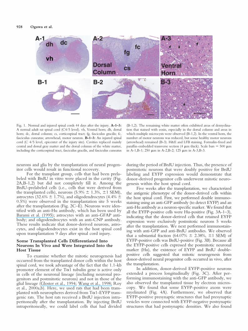
We used the contusion model as our rat model ofspinal cord injury, because it has many similarities to actualspinal cord injury, which have been reported previously(Balentine, 1978; Hughes, 1998). In this model, cavitiesreplaced mainly central and dorsal gray matter and thedorsal column of white matter, including the corticospinaltract, fasciculus gracilis, and fasciculus cuneatus, a type oflesion that is frequently seen in human spinal cord injury(Balentine, 1978; Hughes, 1998). In addition, the remain-ing white matter often exhibited areas of demyelination,especially in the dorsal column and areas in which multiplemicrocysts were observed (Fig. 1A-1,2,B-1,2). In the ven-tral horn, the number of motor neurons was reduced, butsome healthy motor neurons remained (Fig. 1A-3,B-3).Collectively, the losses of both neurons and glia are obvi-ous in this contusion model and cause a functional deficit.Thus, it is reasonable to expect that the replacement of lost
Transplantation in a Rat Spinal Cord Injury Model 927
neurons and glia by the transplantation of neural progen-itor cells would result in functional recovery.
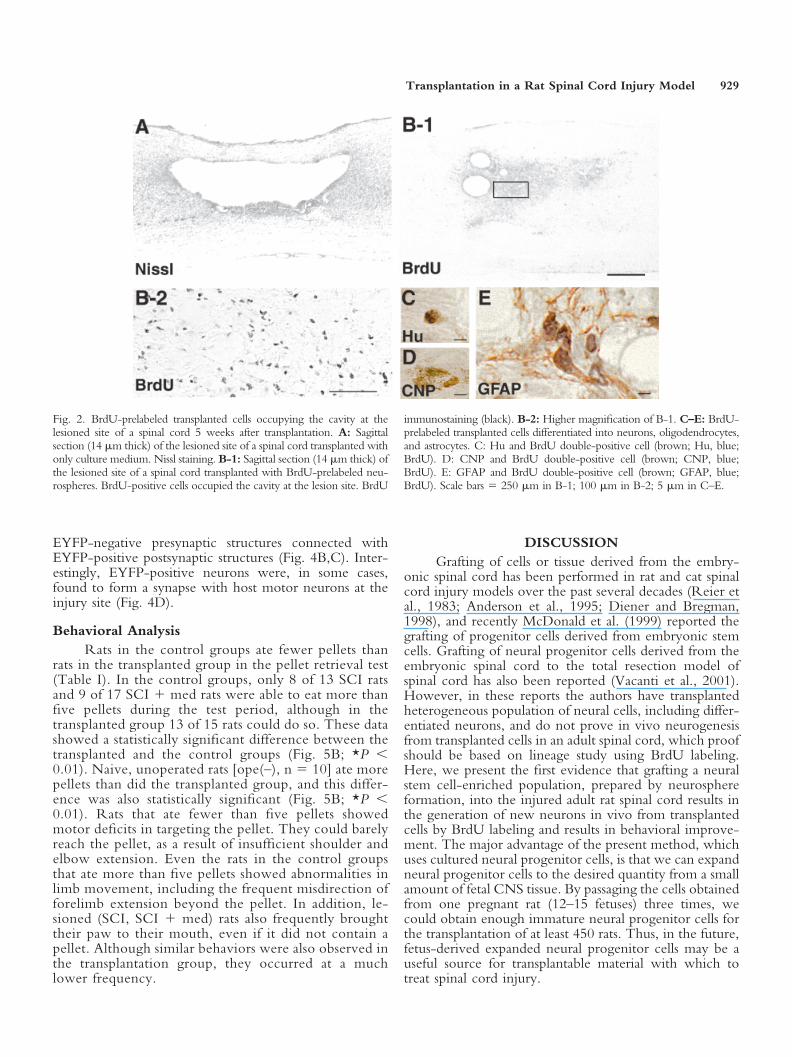
For the transplant group, cells that had been prela-beled with BrdU in vitro were placed in the cavity (Fig.2A,B-1,2) but did not completely fill it. Among theBrdU-prelabeled cells (i.e., cells that were derived fromthe transplanted cells), neurons (5.9% � 1.3%, �1 SEM),astrocytes (32.6% � 1.7%), and oligodendrocytes (4.4% �0.5%) were observed in the transplantation site 5 weeksafter the transplantation (Fig. 2C–E). Neurons were iden-tified with an anti-Hu antibody, which has been used byBarami et al. (1995); astrocytes with an anti-GFAP anti-body; and oligodendrocytes with an anti-CNP antibody.These results indicate that donor-derived neurons, astro-cytes, and oligodendrocytes exist in the host spinal cordupon transplantation 9 days after spinal cord injury.
Some Transplanted Cells Differentiated IntoNeurons In Vivo and Were Integrated Into theHost Tissue
To examine whether the mitotic neurogenesis hadoccurred from the transplanted donor cells within the hostspinal cord, we took advantage of the fact that the 1.1-kbpromoter element of the T�1 tubulin gene is active onlyin cells of the neuronal lineage (including neuronal pro-genitors and postmitotic neurons) and not in those of theglial lineage (Gloster et al., 1994; Wang et al., 1998; Royet al., 2000a,b). Here, we used rats that had been trans-planted with neurospheres derived from T�1-EYFP trans-genic rats. The host rats received a BrdU injection intra-peritoneally after the transplantation. By injecting BrdUintraperitoneally, we could label cells that had divided
during the period of BrdU injection. Thus, the presence ofpostmitotic neurons that were doubly positive for BrdUlabeling and EYFP expression would demonstrate thatdonor-derived progenitor cells underwent mitotic neuro-genesis within the host spinal cord.
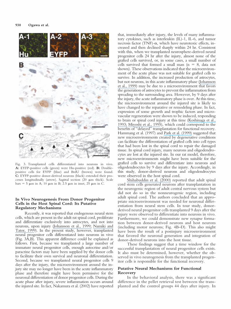
Five weeks after the transplantation, we characterizedthe antigenic phenotype of the donor-derived cells withinthe host spinal cord. First, we performed double immuno-staining using an anti-GFP antibody (to detect EYFP) and ananti-Hu antibody as a neuron-specific marker. We found thatall the EYFP-positive cells were Hu-positive (Fig. 3A-1–3),indicating that the donor-derived cells that retained EYFPexpression were exclusively postmitotic neurons 5 weeksafter the transplantation. We next performed immunostain-ing with anti-GFP and anti-BrdU antibodies. We observedthat a substantial fraction (64.07% � 2.38%, �1 SEM) ofEYFP-positive cells was BrdU-positive (Fig. 3B). Because allthe EYFP-positive cells expressed the postmitotic neuronalmarker (Hu), the existence of EYFP and BrdU double-positive cells suggested that mitotic neurogenesis fromdonor-derived neural progenitor cells occurred in vivo, afterthe transplantation.
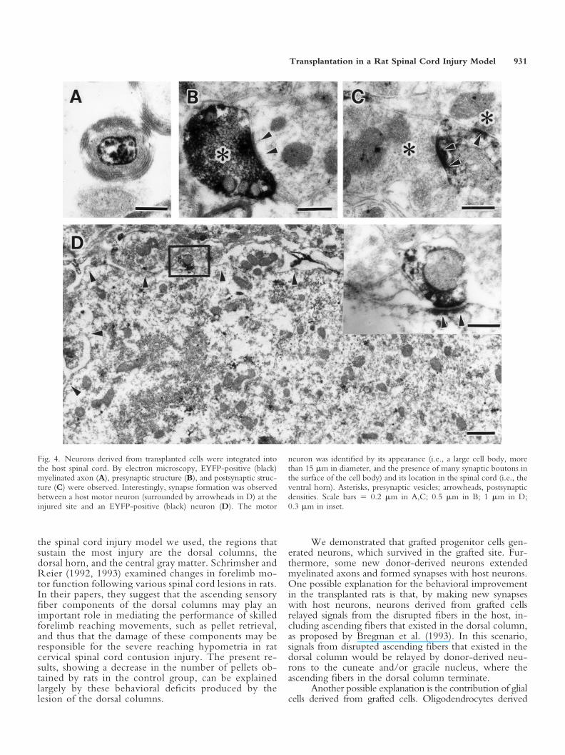
In addition, donor-derived EYFP-positive neuronsextended a process longitudinally (Fig. 3C). After per-forming immunostaining with the anti-GFP antibody, wealso observed the transplanted tissue by electron micros-copy. We found that some EYFP-positive axons weremyelinated (Fig. 4A). Furthermore, we observed thatEYFP-positive presynaptic structures that had presynapticvesicles were connected with EYFP-negative postsynapticstructures that had postsynaptic densities. We also found
Fig. 1. Normal and injured spinal cords 44 days after the injury. A-1–3:A normal adult rat spinal cord (C4/5 level). vh, Ventral horn; dh, dorsalhorn; dc, dorsal column; cs, corticospinal tract; fg, fasciculus gracilis; fc,fasciculus cuneatus; arrowhead, motor neuron. B-1–3: An injured spinalcord (C 4/5 level, epicenter of the injury site). Cavities replaced mainlycentral and dorsal gray matter and the dorsal column of the white matter,including the corticospinal tract, fasciculus gracilis, and fasciculus cuneatus
(B-1,2). The remaining white matter often exhibited areas of demyelina-tion that stained with eosin, especially in the dorsal column and areas inwhich multiple microcysts were observed (B-1,2). In the ventral horn, thenumber of motor neurons was reduced, but some healthy motor neurons(arrowhead) remained (B-3). H&E and LFB staining. Formalin-fixed andparaffin-embedded transverse section (4 �m thick). Scale bars � 500 �min A-1,B-1; 250 �m in A-2,B-2; 125 �m in A-3,B-3.
928 Ogawa et al.
EYFP-negative presynaptic structures connected withEYFP-positive postsynaptic structures (Fig. 4B,C). Inter-estingly, EYFP-positive neurons were, in some cases,found to form a synapse with host motor neurons at theinjury site (Fig. 4D).
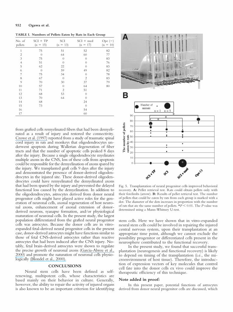
Behavioral AnalysisRats in the control groups ate fewer pellets than
rats in the transplanted group in the pellet retrieval test(Table I). In the control groups, only 8 of 13 SCI ratsand 9 of 17 SCI � med rats were able to eat more thanfive pellets during the test period, although in thetransplanted group 13 of 15 rats could do so. These datashowed a statistically significant difference between thetransplanted and the control groups (Fig. 5B; *P 0.01). Naive, unoperated rats [ope(–), n � 10] ate morepellets than did the transplanted group, and this differ-ence was also statistically significant (Fig. 5B; *P 0.01). Rats that ate fewer than five pellets showedmotor deficits in targeting the pellet. They could barelyreach the pellet, as a result of insufficient shoulder andelbow extension. Even the rats in the control groupsthat ate more than five pellets showed abnormalities inlimb movement, including the frequent misdirection offorelimb extension beyond the pellet. In addition, le-sioned (SCI, SCI � med) rats also frequently broughttheir paw to their mouth, even if it did not contain apellet. Although similar behaviors were also observed inthe transplantation group, they occurred at a muchlower frequency.
DISCUSSIONGrafting of cells or tissue derived from the embry-
onic spinal cord has been performed in rat and cat spinalcord injury models over the past several decades (Reier etal., 1983; Anderson et al., 1995; Diener and Bregman,1998), and recently McDonald et al. (1999) reported thegrafting of progenitor cells derived from embryonic stemcells. Grafting of neural progenitor cells derived from theembryonic spinal cord to the total resection model ofspinal cord has also been reported (Vacanti et al., 2001).However, in these reports the authors have transplantedheterogeneous population of neural cells, including differ-entiated neurons, and do not prove in vivo neurogenesisfrom transplanted cells in an adult spinal cord, which proofshould be based on lineage study using BrdU labeling.Here, we present the first evidence that grafting a neuralstem cell-enriched population, prepared by neurosphereformation, into the injured adult rat spinal cord results inthe generation of new neurons in vivo from transplantedcells by BrdU labeling and results in behavioral improve-ment. The major advantage of the present method, whichuses cultured neural progenitor cells, is that we can expandneural progenitor cells to the desired quantity from a smallamount of fetal CNS tissue. By passaging the cells obtainedfrom one pregnant rat (12–15 fetuses) three times, wecould obtain enough immature neural progenitor cells forthe transplantation of at least 450 rats. Thus, in the future,fetus-derived expanded neural progenitor cells may be auseful source for transplantable material with which totreat spinal cord injury.
Fig. 2. BrdU-prelabeled transplanted cells occupying the cavity at thelesioned site of a spinal cord 5 weeks after transplantation. A: Sagittalsection (14 �m thick) of the lesioned site of a spinal cord transplanted withonly culture medium. Nissl staining. B-1: Sagittal section (14 �m thick) ofthe lesioned site of a spinal cord transplanted with BrdU-prelabeled neu-rospheres. BrdU-positive cells occupied the cavity at the lesion site. BrdU
immunostaining (black). B-2: Higher magnification of B-1. C–E: BrdU-prelabeled transplanted cells differentiated into neurons, oligodendrocytes,and astrocytes. C: Hu and BrdU double-positive cell (brown; Hu, blue;BrdU). D: CNP and BrdU double-positive cell (brown; CNP, blue;BrdU). E: GFAP and BrdU double-positive cell (brown; GFAP, blue;BrdU). Scale bars � 250 �m in B-1; 100 �m in B-2; 5 �m in C–E.
Transplantation in a Rat Spinal Cord Injury Model 929
In Vivo Neurogenesis From Donor ProgenitorCells in the Host Spinal Cord: Its PutativeRegulatory Mechanisms
Recently, it was reported that endogenous neural stemcells, which are present in the adult rat spinal cord, proliferateand differentiate exclusively into astrocytes, and not intoneurons, upon injury (Johansson et al., 1999; Namiki andTator, 1999). In the present study, however, transplantedneural progenitor cells differentiated into neurons in vivo(Fig. 3A,B). This apparent difference could be explained asfollows. First, because we transplanted a large number ofimmature neural progenitor cells, enough autocrine and/orparacrine factors may have been supplied by the donor cellsto facilitate their own survival and neuronal differentiation.Second, because we transplanted neural progenitor cells 9days after the injury, the microenvironment around the in-jury site may no longer have been in the acute inflammatoryphase and therefore might have been permissive for theneuronal differentiation of donor progenitor cells. During theacute phase after injury, severe inflammation occurs aroundthe injured site. In fact, Nakamura et al. (2002) have reported
that, immediately after injury, the levels of many inflamma-tory cytokines, such as interleukin (IL)-1, IL-6, and tumornecrosis factor (TNF)-�, which have neurotoxic effects, in-creased and then declined sharply within 24 hr. Consistentwith this, when we transplanted neurosphere-derived neuralprogenitor cells 24 hr after the injury, almost none of thegrafted cells survived, or, in some cases, a small number ofcells survived that formed a small mass (n � 8, data notshown). These observations indicated that the microenviron-ment of the acute phase was not suitable for grafted cells tosurvive. In addition, the increased production of astrocytes,but not neurons, in this acute inflammatory phase (Johanssonet al., 1999) may be due to a microenvironment that favorsthe generation of astrocytes to prevent the inflammation fromspreading to the surrounding area. However, by 9 days afterthe injury, the acute inflammatory phase is over. At this time,the microenvironment around the injured site is likely tohave changed to the reparative or remodeling phase. In fact,expression of some growth and trophic factors and micro-vascular regeneration were shown to be induced, respondingto brain or spinal cord injury at this time (Koshinaga et al.,1993; Miyoshi et al., 1995), which could correspond to thebenefits of “delayed” transplantation for functional recovery.Hammang et al. (1997) and Park et al. (1999) suggested thatnew microenvironments created by degenerative conditionscan facilitate the differentiation of grafted cells into cell typesthat had been lost in the spinal cord to repair the damagedtissue. In spinal cord injury, many neurons and oligodendro-cytes are lost at the injured site. In our rat model, therefore,new microenvironments might have been suitable for thegrafted cells to survive and differentiate into neurons andoligodendrocytes by 9 days after the injury. Accordingly, inthis study, donor-derived neurons and oligodendrocyteswere observed in the host spinal cord.
Shihabuddin et al. (2000) reported that adult spinalcord stem cells generated neurons after transplantation inthe neurogenic region of adult central nervous system butdid not do so in the nonneurogenic region, includingadult spinal cord. The authors concluded that an appro-priate microenvironment was needed for neuronal differ-entiation from neural stem cells. In tour study, donor-derived neural progenitor cells transplanted 9 days after theinjury were observed to differentiate into neurons in vivo.Furthermore, we could demonstrate new synapse forma-tion between donor-derived neurons and host neurons(including motor neurons; Fig. 4B–D). This also mighthave been the result of a postinjury microenvironmentthat favored the neuronal generation and integration ofdonor-derived neurons into the host tissue.
These findings suggest that a time window for thesuccessful transplantation of neural progenitor cells exists.It also must be determined, however, whether the ob-served in vivo neurogenesis from the transplanted progen-itor cells is responsible for the functional recovery.
Putative Neural Mechanisms for FunctionalRecovery
In the behavioral analysis, there was a significantdifference in the pellet retrieval test between the trans-planted and the control groups 44 days after injury. In
Fig. 3. Transplanted cells differentiated into neurons in vivo.A: EYFP-positive cells (green) were Hu-positive (red). B: Double-positive cells for EYFP (blue) and BrdU (brown) were found.C: EYFP-positive donor-derived neurons (black) extended their pro-cesses longitudinally (arrow). Sagittal section (20 �m thick). Scalebars � 5 �m in A; 10 �m in B; 2.5 �m in inset, 25 �m in C.
930 Ogawa et al.
the spinal cord injury model we used, the regions thatsustain the most injury are the dorsal columns, thedorsal horn, and the central gray matter. Schrimsher andReier (1992, 1993) examined changes in forelimb mo-tor function following various spinal cord lesions in rats.In their papers, they suggest that the ascending sensoryfiber components of the dorsal columns may play animportant role in mediating the performance of skilledforelimb reaching movements, such as pellet retrieval,and thus that the damage of these components may beresponsible for the severe reaching hypometria in ratcervical spinal cord contusion injury. The present re-sults, showing a decrease in the number of pellets ob-tained by rats in the control group, can be explainedlargely by these behavioral deficits produced by thelesion of the dorsal columns.
We demonstrated that grafted progenitor cells gen-erated neurons, which survived in the grafted site. Fur-thermore, some new donor-derived neurons extendedmyelinated axons and formed synapses with host neurons.One possible explanation for the behavioral improvementin the transplanted rats is that, by making new synapseswith host neurons, neurons derived from grafted cellsrelayed signals from the disrupted fibers in the host, in-cluding ascending fibers that existed in the dorsal column,as proposed by Bregman et al. (1993). In this scenario,signals from disrupted ascending fibers that existed in thedorsal column would be relayed by donor-derived neu-rons to the cuneate and/or gracile nucleus, where theascending fibers in the dorsal column terminate.
Another possible explanation is the contribution of glialcells derived from grafted cells. Oligodendrocytes derived
Fig. 4. Neurons derived from transplanted cells were integrated intothe host spinal cord. By electron microscopy, EYFP-positive (black)myelinated axon (A), presynaptic structure (B), and postsynaptic struc-ture (C) were observed. Interestingly, synapse formation was observedbetween a host motor neuron (surrounded by arrowheads in D) at theinjured site and an EYFP-positive (black) neuron (D). The motor
neuron was identified by its appearance (i.e., a large cell body, morethan 15 �m in diameter, and the presence of many synaptic boutons inthe surface of the cell body) and its location in the spinal cord (i.e., theventral horn). Asterisks, presynaptic vesicles; arrowheads, postsynapticdensities. Scale bars � 0.2 �m in A,C; 0.5 �m in B; 1 �m in D;0.3 �m in inset.
Transplantation in a Rat Spinal Cord Injury Model 931
from grafted cells remyelinated fibers that had been demyeli-nated as a result of injury and restored the connectivity.Crowe et al. (1997) reported from a study of traumatic spinalcord injury in rats and monkeys that oligodendrocytes un-derwent apoptosis during Wallerian degeneration of fibertracts and that the number of apoptotic cells peaked 8 daysafter the injury. Because a single oligodendrocyte myelinatesmultiple axons in the CNS, loss of these cells from apoptosiscould be responsible for the demyelination of axons spared bythe injury. We transplanted graft cells 9 days after the injuryand demonstrated the presence of donor-derived oligoden-drocytes in the injured site. These donor-derived oligoden-drocytes could have remyelinated the demyelinated axonsthat had been spared by the injury and prevented the delayedfunctional loss caused by the demyelination. In addition tothe oligodendrocytes, astrocytes derived from donor neuralprogenitor cells might have played active roles for the gen-eration of neuronal cells, axonal regeneration of host neuro-nal axons, enhancement of axonal extension of donor-derived neurons, syanapse formation, and/or physiologicalmaturation of neuronal cells. In the present study, the largestpopulation differentiated from the grafted neural progenitorcells was astrocytes. Because the donor cells are in vitro-expanded fetal-derived neural progenitor cells in the presentcase, donor-derived astrocytes might have functions similar tothose of fetal CNS-derived astrocytes rather than reactiveastrocytes that had been induced after the CNS injury. No-tably, fetal brain-derived astrocytes were shown to regulatethe precise growth of neuronal axons (Garcia-Abreu et al.,2000) and promote the naturation of neuronal cells physio-logically (Blondel et al., 2000).
CONCLUSIONSNeural stem cells have been defined as self-
renewing, multipotent cells, whose characteristics arebased mainly on their behavior in culture. Generally,however, the ability to repair the activity of injured organsis also known to be an important criterion for identifying
stem cells. Here we have shown that in vitro-expandedneural stem cells could be involved in repairing the injuredcentral nervous system, upon their transplantation at anappropriate time point, although we cannot exclude thepossibility progenitor or differentiated cells present in theneurosphere contributed to the functional recovery.
In the present study, we found that successful trans-plantation (neurogenesis and functional recovery) is likelyto depend on timing of the transplantation (i.e., the mi-croenvironment of host tissue). Therefore, the introduc-tion of expression vectors of key molecules that controlcell fate into the donor cells ex vivo could improve thetherapeutic efficiency of this technique.
Note added in proof:In this present paper, potential functions of astrocytes
derived from donor neural progenitor cells are discussed, which
TABLE I. Numbers of Pellets Eaten by Rats in Each Group
No. ofpellets
SCI � TP(n � 15)
SCI(n � 13)
SCI � med(n � 17)
Ope ()(n � 10)
1 75 51 52 822 0 64 60 773 75 0 0 834 51 0 0 765 62 22 0 836 0 59 49 827 75 34 0 788 67 0 2 839 70 30 37 79
10 57 0 64 8011 71 2 5112 68 53 013 70 64 014 68 2415 73 016 5417 60
Fig. 5. Transplantation of neural progenitor cells improved behavioralrecovery. A: Pellet retrieval test. Rats could obtain pellets only withtheir forelimbs (arrow). B: Results of pellet retrieval test. The numberof pellets that could be eaten by rats from each group is marked with adot. The diameter of the dots increases in proportion with the numberof rats that ate the same number of pellets. *P 0.01. The P value wasdetermined using a Mann-Whitney U-test.
932 Ogawa et al.

might have played active roles for functional recovery of theinjured spinal cord. Notably, after this paper had been acceptedfor publication, the active roles of astrocytes to induce neuro-genesis were published from Dr. Fred Gage’s group (Song et al.,2002).
ACKNOWLEDGMENTWe thank Dr. Steven A. Goldman (Cornell Univer-
sity Medical College) for continuous encouragement andvaluable discussions.
REFERENCESAnderson DK, Howland DR, Reier PJ. 1995. Fetal neural grafts and repair
of the injured spinal cord. Brain Pathol 5:451–457.Balentine JD. 1978. Pathology of experimental spinal cord trauma. I. The
necrotic lesion as a function of vascular injury. Lab Invest 39:236–253.Barami K, Iversen K, Furneaux H, Goldman SA. 1995. Hu protein as an
early marker of neuronal phenotypic differentiation by subependymalzone cells of the adult song bird forebrain. J Neurobiol 28:82–101.
Blondel O, Collin C, McCarran WJ, Zhu S, Zamostiano R, Gozes I,Brenneman DE, McKay RDG. 2000. A glial-derived signal regulatingneuronal differentiation. J Neurosci 20:8012–8020.
Bregman BS, Kunkel-Bagden E, Reier PJ, Dai HN, McAtee M, Gao D.1993. Recovery of function after spinal cord injury: mechanisms under-lying transplant-mediated recovery of function differ after spinal cordinjury in newborn and adult rats. Exp Neurol 123:3–16.
Crowe MJ, Bresnahan JC, Shuman SL, Masters JN, Beattie MS. 1997.Apoptosis and delayed degeneration after spinal cord injury in rats andmonkeys. Nat Med 3:73–76.
Diener PS, Bregman BS. 1998. Fetal spinal cord transplants support thedevelopment of target reaching and coordinated postural adjustments afterneonatal cervical spinal cord injury. J Neurosci 18:763–778.
Freed CR, Breeze RE, Rosenberg NL, Schneck SA, Kriek E, Qi JX, LoneT, Zhang YB, Snyder JA, Wells TH, Olson RL, Thompson L, MazziottaJC, Huang SC, Grafton ST, Brooks D, Sawle G, Schroter G, Ansari AA.1992. Survival of implanted fetal dopamine cells and neurologic improve-ment 12 to 46 months after transplantation for Parkinson’s disease. N EnglJ Med 327:1549–1555.
Freeman TB, Olanow CW, Hauser RA, Nauert GM, Smith DA, BorlonganCV, Sanberg PR, Holt DA, Kordower JH, Vingerhoets FJ, Snow BJ, CalneD, Gauger LL. 1995. Bilateral fetal nigral transplantation into the postcom-missural putamen in Parkinson’s disease. Ann Neurol 38:379–388.
Garcia-Abreu J, Mendes FA, onofre GR, De Freitas MS, Silva LC, MouraNeto V, Cavalcante LA. 2000. Contribution of heparan sulfate to thenonpermissive role of the midline glia to the growth of midbrain neurites.Glia 29:260– 272.
Gloster A, Wu W, Speelman A, Weiss S, Causing C, Pozniak C, ReynoldsB, Chang E, Toma JG, Miller FD. 1994. The T�-1 �-tubulin promoterspecifies gene expression as a function of neuronal growth and regener-ation in transgenic mice. J Neurosci 14:7319–7330.
Hammang JP, Archer DR, Duncan ID. 1997. Transplantation of epidermalgrowth factor-responsive neural stem cell progeny into the murine centralnervous system. Exp Neurol 147:84–95.
Holtz A, Nystrom B, Gerdin B. 1989. Spinal blood flow measured by14C-iodoantipyrine autoradiography during and after graded spinal com-pression in rats. Surg Neurol 31:350–360.
Hughes JT. 1988. Pathological changes after spinal cord injury. In: Illis LS,editor. Spinal cord dysfunction: assessment. New York: Oxford Univer-sity Press. p 34–40.
Johansson CB, Momma S, Clarke DL, Risling M, Lendahl U, Frisen J.
1999. Identification of a neural stem cell in the adult mammalian centralnervous system. Cell 96:25–34.
Koshinaga M, Sanon HR, Whittemore SR. 1993. Altered acidic and basicfibroblast growth factor expression following spinal cord injury. ExpNeurol 120:32–48.
Lindvall O, Brundin P, Widner H, Rehncrona S, Gustavii B, FrackowiakR, Leenders KL, Sawle G, Rothwell JC, Marsden CD, Bjorklund A.1990. Grafts of fetal dopamine neurons survive and improve motorfunction in Parkinson’s disease. Science 247:574–577.
McDonald JW, Liu XZ, Qu Y, Liu S, Mickey SK, Turetsky D, Gottlieb DI,Choi DW. 1999. Transplanted embryonic stem cells survive, differentiate andpromote recovery in injured rat spinal cord. Nat Med 5:1410–1412.
Miyoshi Y, Date I, Ohmoto T. 1995. Three-dimensional morphologicalstudy of microvascular regeneration in cavity wall of the rat cerebralcortex using the scanning electron microscope: implication for delayedneural grafting into brain cavities. Exp Neurol 131:69–82.
Nakamura M, Houghtling RA, MacArthur L, Bayer BM, Bregman BS.2002. Difference in cytokine gene expression profile between acutely andchronically injured adult rat spinal cord. Exp Neurol (in press).
Namiki J, Tator CH. 1999. Cell proliferation and nestin expression in theependyma of the adult rat spinal cord after injury. J Neuropathol ExpNeurol 58:489–498.
Park KI, Liu S, Flax JD, Nissim S, Stieg PE, Snyder EY. 1999. Transplan-tation of neural progenitor and stem cells: developmental insights maysuggest new therapies for spinal cord and other CNS dysfunction. J Neu-rotrauma 16:675–687.
Reier PJ, Perlow MJ, Guth L. 1983. Development of embryonic spinalcord transplants in the rat. Brain Res 10:201–219.
Reynolds BA, Weiss S. 1992. Generation of neurons and astrocytes fromisolated cells of adult mammalian central nervous system. Science 255:1707–1710.
Roy NS, Benraiss A, Wang S, Fraser RAR, Goodman R, Couldwell WT,Nedergaard M, Kawaguchi A, Okano H, Goldman SA. 2000a. Promoter-targeted selection and isolation of neural progenitor cells from the adulthuman ventricular zone. J Neurosci Res 59:321–331.
Roy NS, Wang S, Jiang L, Kang J, Restelli C, Fraser RAR, CouldwellWT, Kawaguchi A, Okano H, Nedergaard M, Goldman SA. 2000b. Invitro neurogenesis by neural progenitor cells isolated from the adulthuman hippocampus. Nat Med 6:271–278.
Sawamoto K, Nakao N, Kakishita K, Ogawa Y, Toyama Y, Yamamoto A,Yamaguchi M, Mori K, Goldman SA, Itakura T, Okano H. 2001.Generation of dopaminergic neurons in the adult brain from mesence-phalic precursor cells labeled with a Nestin-GFP transgene. J Neurosci21:3895–3903.
Schrimsher GW, Reier PJ. 1992. Forelimb motor performance followingcervical spinal cord contusion injury in the rat. Exp Neurol 117:287–298.
Schrimsher GW, Reier PJ. 1993. Forelimb motor performance followingdorsal column, dorsolateral funiculi, or ventrolateral funiculi lesions of thecervical spinal cord in the rat. Exp Neurol 120:264–276.
Shihabuddin LS, Horner PJ, Ray J, Gage FH. 2000. Adult spinal cord stemcells generate neurons after transplantation in the adult dentate gyrus.J Neurosci 20:8727–8735.
Song H, Stevens CF, Gage FH. 2002. Astroglia induce neurogenesis fromadult neural stem cells. Nature 417:39–44.
Vacanti MP, Leonard JL, Dore B, Bonassar LJ, Cao Y, Stachelek SJ, VacantiJP, O’Connell F, Yu CS, Farwell AP, Vacanti CA. 2001. Tissue-engineered spinal cord. Transplant Proc 33:592–598.
Wang S, Wu H, Jiang J, Delohery TM, Isdell F, Goldman SA. 1998.Isolation of neuronal precursors by sorting embryonic forebrain trans-fected with GFP regulated by the T�-1 tubulin promoter. Nat Biotechnol16:196–201.
Transplantation in a Rat Spinal Cord Injury Model 933