Myc Prevents Apoptosis and Enhances Endoreduplication Induced by Paclitaxel
Upload
tu-braunschweigCategory
view
9download
0
Transient adenoviral gene transfer ofSmad7 prevents injury-inducedepithelial–mesenchymal transitionof lens epithelium in mice
Shizuya Saika1, Kazuo Ikeda2, Osamu Yamanaka1, Misako Sato3, Yasuteru Muragaki3,Yoshitaka Ohnishi1, Akira Ooshima3, Yuji Nakajima2, Kazuhiko Namikawa4,Hiroshi Kiyama4, Kathleen C Flanders5 and Anita B Roberts5
1Department Ophthalmology, Wakayama Medical University, Kimiidera, Wakayama, Japan; 2DepartmentAnatomy, Graduate School of Medicine, Osaka City University, Japan; 3Department of Pathology, WakayamaMedical University, Kimiidera, Wakayama, Japan; 4Department of Anatomy/Neurobiology, Graduate Schoolof Medicine, Osaka City University, Japan and 5Laboratory of Cell Regulation and Carcinogenesis, NationalCancer Institute, National Institutes of Health, Bethesda, MD, USA
We examined the effect of adenovirus-mediated transient expression of Smad7, an inhibitory Smad in TGFb/activin signaling, on injury-induced epithelial–mesenchymal transition (EMT) of lens epithelium in mice. Avolume of 3 ll of adenoviral solution was injected into the right lens of adult male C57BL/6 mice (n¼ 56) at thetime of capsular injury made using a hypodermic needle under general anesthesia. A mixture of recombinantadenovirus carrying CAG promoter-driven Cre (Cre adv) and mouse Smad7 complementary DNA (Smad7 adv)was administered to induce Smad7 expression, while control lenses were treated with Cre adv alone. Afterhealing intervals of 2, 3, 5, and 10 days, animals were killed 2 h after labeling with bromodeoxyuridine (BrdU)and eyes were processed for histology. During healing, marked expression of Smad7 was observed in lensepithelial cells in the Smad7 adv group with loss of nuclear translocation of Smads2/3, while little Smad7 andabundant nuclear Smads2/3 were seen in cells in the Cre adv group. Lens epithelial cells in the Cre adv controlgroup exhibited a fibroblastic appearance at days 5 and 10 and the capsular break was sealed with fibroustissue, while Smad7 adv-treated cells around the capsular break retained their epithelial morphology and thebreak was not sealed. Expression of snail mRNA, and a-smooth muscle actin, lumican, and collagen VI proteins,markers of EMT, was observed in control-treated eyes, but not in cells of the Smad7 adv group at day 5 withminimal expression at day 10. Additionally, cell proliferation increased in epithelium infected with Smad7 advconsistent with suppression of injury-induced upregulation of TGFb1 in epithelium. We conclude that genetransfer of Smad7 in mice prevents injury-induced EMT of lens epithelial cells and sealing of the capsular breakwith fibrous tissue.Laboratory Investigation (2004) 84, 1259–1270, advance online publication, 19 July 2004; doi:10.1038/labinvest.3700151
Keywords: lens epithelial cell; epithelial–mesenchymal transition; transforming growth factor b; Smad; mouse
Following injury, lens epithelial cells undergoepithelial–mesenchymal transition (EMT),1–4 whichcontributes to the formation of fibrotic tissue in theinjured lens.5–8 A similar phenomenon is observedin the human lens capsule following cataractextraction and implantation of an artificial intra-
ocular lens. Such an EMT-related fibrotic reaction isclinically unfavorable since it may cause opacifica-tion and contraction of the remaining anterior lenscapsule, as well as opacification in the posteriorcapsule, all of which potentially impair vision. Eyeaqueous humor contains abundant transforminggrowth factor b2 (TGFb2)9,10 and evidence suggeststhat TGFb is involved in injury-related EMT in lensepithelial cells.11–14 We have shown that endogen-ous TGFb2 induces a transient nuclear translocationof the TGFb signaling intermediates, Smads3/4 inlens epithelial cells postinjury in mice, which wasinhibited by local administration of a neutralizing
Received 22 April 2004; revised and accepted 20 May 2004;published online 19 July 2004
Correspondence: Dr S Saika, MD, PhD, Department of Oph-thalmology, Wakayama Medical University, 811-1 Kimiidera,Wakayama 641-0012, Japan.E-mail: [email protected]
Laboratory Investigation (2004) 84, 1259–1270& 2004 USCAP, Inc All rights reserved 0023-6837/04 $30.00
www.laboratoryinvestigation.org
antibody against TGFb2, indicating that this growthfactor isoform is responsible for the activation ofSmads3/4 signaling in lens epithelium.7 Humanlens epithelial cells are also regulated by TGFb/Smad signaling in vivo during wound healing.15 Werecently reported that TGFb/Smad3 signaling iscritical in the induction of EMT in lens or renalepithelium in vivo by showing that EMT is blockedin Smad3-null mice,16,17 although in vitro studiessuggest that a variety of signaling pathways may berequired for EMT.18–23
Smad7 is an inhibitory Smad that interferes withSmad signaling by preventing the phosphorylationof Smads2/3, which normally occurs followingbinding of TGFb/activin to their cell surface recep-tors.24–27 Recently, transient introduction of theSmad7 gene using an adenovirus vector has beenshown to be effective in treating tissue inflamma-tory/fibrotic disorders in lung,28 kidney,29 and liver.30
Therefore, we hypothesized that transfer of Smad7complementary DNA (cDNA) using a viral vectormight also block injury-induced EMT in lensepithelial cells. In the present study, we created anadenoviral gene-transfer system to introduce Smad7cDNA into an injured mouse lens and show thattransient adenoviral gene transfer effectively blocksinjury-induced EMT in lens epithelium while en-hancing cell proliferation. These results suggest apotential therapeutic efficacy of Smad7 gene transferin the treatment of postoperative capsular fibrosis.
Materials and methods
All the experimental procedures were approved byboth the DNA Recombinant Experiment Committeeand Animal Care and Use Committee of WakayamaMedical University, Wakayama, Japan, and con-ducted in accordance with the Association forResearch in Vision and Ophthalmology Statementfor the Use of Animals in Ophthalmic and VisionResearch.
Adenovirus Vector Construction and VirusPurification
We used the Adenovirus Cre/LoxP-Regulated Ex-pression Vector Set (#6151, Takara, Tokyo, Japan) togenerate recombinant adenovirus. Cosmid pAx-CALNLmSmad7 was constructed by the insertionof mouse Smad7 cDNA (a kind gift by Dr KMiyazono, The University of Tokyo School ofMedicine, Tokyo, Japan) into the Swa I cloning siteof pAxCALNLw.31 Using the COS-TPC method,32
recombinant adenovirus of AxCALNLmSmad7 wasgenerated by transfecting 293 cells with pAx-CALNLSmad7. AxCANCre was generated by trans-fecting 293 cells with Ax-CALNLCreDNA-TPC asdescribed in the manufacturer’s protocol. The titerof recombinant adenoviruses was measured by the50% tissue culture infectious dose method.33 Each
adenovector was used at the concentration of2� 107 PFU/ml. When these two viral vectors coin-fect cells, Cre recombinase expressed under the CAGpromoter (cytomegalovirus enhancer, chicken b-actin promoter plus a part of 30 untranslated regionof rabbit b-globin) activates the stuffer PolyAthrough the Cre/LoxP system.
Experimental Protocol
Evaluation of the efficacy of adenoviral gene transferwas performed by coinfection of viruses carrying Crerecombinase under the CAG promoter and LNL-green fluorescent protein (GFP) or by infection ofvirus carrying GFP alone in an injured lens. Theanterior lens capsule of the right eye of an adultC57BL/6 mouse was punctured as previously re-ported under general anesthesia by intraperitoneal(i.p.) pentobarbital.7,17 A volume of Three 3 ml ofviral vector solution of either group was injectedinto the injured lens at the time of capsular injury.At days 1, 2, and 5 (n¼ 2/group on each day)following injury, the right eye was enucleated andembedded in OCT compound. Cryosections (7 mm)were examined without fixation under fluorescencemicroscopy.
Upon confirmation of the expression of GFP ininjured lens epithelial cells, we introduced Smad7cDNA. One vector expresses Cre under the CAGpromoter (CAG-Cre) while the other contains Smad7that is activated by Cre recombinase (LNL-Smad7).The right eyes of 4-week-old male C57BL/6 mice(n¼ 56) were used. To induce transcription ofSmad7, both LNL-Smad7 and CAG-Cre vectors(3 ml total) were coinfected directly into the affectedlens at the time of formation of a break in theanterior capsule as described above. Control lenseswere treated with CAG-Cre alone (3 ml). At days 2(n¼ 3/group), 3 (n¼ 3 /group), 5 (n¼ 11/group), and10 (n¼ 11/group), post-treatment, the eyes wereenucleated, fixed, and embedded in paraffin aspreviously reported.7,17
In Situ Hybridization for Snail
Snail is a member of a family of zinc-finger-containing transcriptional repressors increasinglyassociated with suppression of the epithelial phe-notype associated with EMT.34–36 Digoxigenin-la-beled riboprobes for mouse snail and were preparedusing a digoxigenin-labeling kit (Roche DiagnosticsCorp-Boehringer Mannheim, Indianapolis, IN,USA). and in situ hybridization of paraffin sections(5 mm) was carried out as previously reported.16,17
Immunohistochemistry
Deparaffinized sections (5 mm) were reacted withprimary antibodies appropriately diluted with phos-
Smad7 blocks EMT in lens epitheliumS Saika et al
1260
Laboratory Investigation (2004) 84, 1259–1270

phate-buffered saline (PBS) following blocking with5% dried milk and 5% serum as previouslyreported. After washing in PBS, the specimens weretreated with fluorescein isothiocyanate (FITC)-con-jugated secondary antibodies. Primary antibodiesagainst phosphorylated Smad2 (1:50 dilution, Che-micon, Temecula, CA, USA), Smad3 (1:100 dilution,Zymed, South San Francisco, CA, USA), Smad7(1:400 dilution, Santa Cruz Biotechnology, SantaCruz, CA, USA, #sc-7004), a-smooth muscle actin(aSMA) (1:200 dilution, Neomarker, Fremont, CA,U-SA), TGFb1 (intracellular and extracellular forms),TGFb2, lumican, and collagen type VI (1:200dilution, Southern Biotechnology, Birmingham,AL, USA) were used as previously reported.16,37
BrdU immunostaining was performed as previouslydescribed.38 In brief, deparaffinized sections weretreated with 2 N HCl for 1 h at 371C, treated withanti-BrdU antibody (1:11 dilution in PBS, RocheDiagnostics, Mannheim, Germany) and then withperoxidase-conjugated secondary antibody afterwashing in PBS. Following the color reaction withdiaminobenzidine and methyl green counterstain-ing, the number of labeled cells in healing epitheliawas determined. Six specimens were counted ineach group at days 5 and 10. Histology was observedby staining tissues with hematoxylin and eosin (HE).
Results
Efficacy of Adenovirus-Mediated Expression of GFP inInjured Lens Epithelial Cells
We confirmed that the CAG-Cre/LoxP system canefficiently introduce a gene of interest by observingthe expression of GFP. When the injured lensepithelium was coinfected with adenoviral vectorsencoding Cre under control of the CAG promoterand GFP under control of the LNL promoter, greenfluorescence was observed at day 1 and later in lensepithelium, as well as in corneal endothelium andiris tissue (Figure 1a, b). No fluorescence wasdetected in eyes infected with CAG-Cre adv only.
Smad7 Expression and Inhibition of Smad3 Signaling
Our previous study showed that nuclear transloca-tion of Smad proteins in lens epithelial cellsfollowing injury generally occurs before the cellsform multilayers, but is also occasionally observedin the multilayers. At day 2 after infection, phos-phorylated Smad2 (not shown) and Smad3 weredetected in the nuclei of lens epithelial cells in theanterior lens adjacent to the capsular break (Figure1c, d) in control eyes, whereas in Smad7-infectedeyes phospho-Smad2 (not shown) was not detectedand Smad3 was seen only faintly in the cytoplasm ofthe anterior lens (Figure 1e, f). A similar phenom-enon was also observed even in the equatorial regionapart from the injury site at the same time point.
Smad3 was detected in the nuclei of lens epithelialcells in the equatorial lens region (Figure 1g, h) in acontrol specimen, but not in the equatorial epitheliaof the Smad7-infected group (Figure 1i, j). At andafter day 5, the majority of nuclei were negative forboth Smads 2/3 in the control, and in Smad7 adv-treated specimens (not shown). Smad7 protein,presumably the translated product derived fromexogenously introduced Smad7 cDNA, was stronglydetected in the healing lens epithelium from day 2onward, while faint immunoreactivity for endogen-ous Smad7 was seen in the healing lens epitheliumin the control group for up to 10 days (Figure 1k, l).
Histology of Injured Lens Epithelial Cells andExpression of EMT Markers
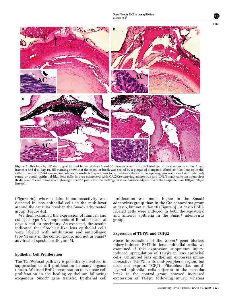
HE staining showed that the capsular break wassealed by a plaque of elongated, fibroblast-like, lensepithelial cells as early as day 5 (Figure 2a) in CAG/Cre-infected eyes, whereas the capsular opening wasnot closed at day 5 (Figure 2b) in the Smad7-infectedgroup and remained open at day 10 (Figure 2c, d).Rather, the Smad7-infected group showed round orovoid epithelial-like cells surrounding the break.This finding strongly suggests that introduction ofexogenous Smad7 cDNA inhibited EMT followinginjury.
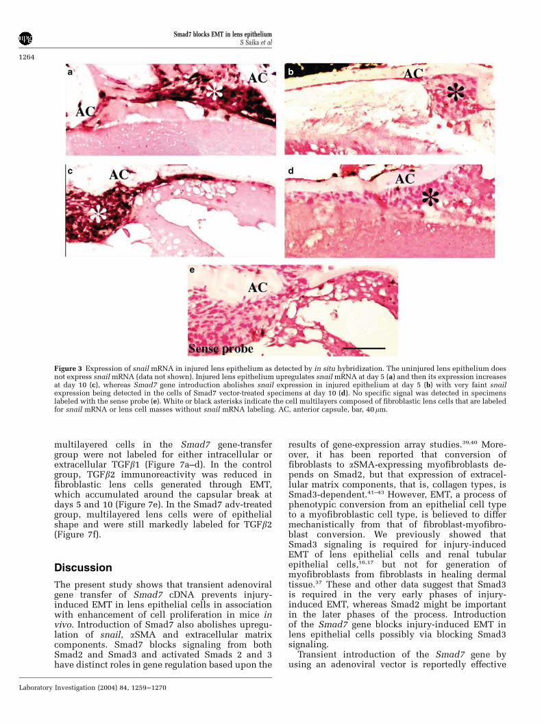
To test this hypothesis, we first carried out in situhybridization for snail mRNA in paraffin sections.Snail is a member of a family of zinc-finger-containing transcriptional repressors increasinglyassociated with suppression of the epithelial phe-notype associated with EMT. The results showedthat, as previously reported, uninjured lens epithe-lium does not express snail mRNA (data not shown).Snail mRNA is upregulated in the injured lensepithelium of the control Cre adv-treated group(Figure 3a), whereas eyes treated with Smad7 advshowed no expression of snail in injured epitheliumat day 5 (Figure 3b). At day 10, its expression in lenscells seemed to increase further (Figure 3c) in thecontrol Cre adv-treated group, while only faintexpression of snail was detected in the cells ofSmad7 adv-treated specimens (Figure 3d). Nospecific signal was detected in specimens labeledwith the sense probe (Figure 3e).
To examine if snail expression is followed byEMT, we immunostained tissues with antibodiesagainst specific markers for EMT, for example,aSMA, lumican, and collagen type VI. The resultsshowed that there was no expression of aSMA inlens epithelial cells at 2 days postinjury in eyes ofeither the Smad7 adv-treated group or the controlgroup. However, at day 5, marked immunofluores-cence for aSMA was seen in multilayered cells in thecontrol CAG/Cre adv-treated lens (Figure 4a), butalmost never in cells of Smad7 adv-treated lenses(Figure 4b). At day 10, a strong immunofluorescencefor aSMA was seen in control CAG/Cre group cells
Smad7 blocks EMT in lens epitheliumS Saika et al
1261
Laboratory Investigation (2004) 84, 1259–1270

Figure 1 Detection of GFP, Smad3, or Smad7 by fluorescence in injured lens epithelium at day 1, 2, or 10. Expression of GFP wasexamined in an injured mouse lens coinfected with viruses carrying Cre under the CAG promoter and LNL-GFP or infected only withvirus containing CAG/Cre. GFP expression is observed in coinfected lens epithelium (b, red arrows), but not in epithelium without GFP-carrying virus (a, white arrows). At day 2, Smad3 is detected in the nuclei of lens epithelial cells located adjacent to the capsular break(d) and lens equator (h) in a control, CAG/Cre- adenovirus-infected specimen, while faint cytoplasmic expression of Smad3 is observedin the injured lens epithelium adjacent to the break (f) and in the equator (j) in eyes coinfected with CAG/Cre-carrying and LNL/Smad7-carrying adenoviruses. Very faint endogenous Smad7 expression is observed in CAG/Cre-carrying adenovirus-infected specimen at day10 (k), while prominent immunoreactivity for presumably exogenous Smad7 is seen in the cell multilayer formed in an injured lenscoinfected with CAG/Cre-carrying adenovirus and LNL/Smad7-carrying adenovirus (l). Frames c, e, g, and i, indicate localization ofnuclei as stained with DAPI in the frames d, f, h, and j, respectively. Dotted lines indicate anterior capsule. AC, anterior lens capsule; bar,50mm.
Smad7 blocks EMT in lens epitheliumS Saika et al
1262
Laboratory Investigation (2004) 84, 1259–1270
(Figure 4c), whereas faint immunoreactivity wasdetected in lens epithelial cells in the multilayeraround the capsular break in the Smad7 adv-treatedgroup (Figure 4d).
We then examined the expression of lumican andcollagen type VI, components of fibrotic tissue, atdays 5 and 10 postinjury. As expected, the resultsindicated that fibroblast-like lens epithelial cellswere labeled with antilumican and anticollagentype VI only in the control group, and not in Smad7adv-treated specimens (Figure 5).
Epithelial Cell Proliferation
The TGFb/Smad pathway is potentially involved insuppression of cell proliferation in many organs/tissues. We used BrdU incorporation to evaluate cellproliferation in the healing epithelium followingexogenous Smad7 gene transfer. Epithelial cell
proliferation was much higher in the Smad7adenovirus group than in the Cre adenovirus groupat day 5, but not at day 10 (Figure 6). At day 5 BrdU-labeled cells were induced in both the equatorialand anterior epithelia in the Smad7 adenovirusgroup.
Expression of TGFb1 and TGFb2
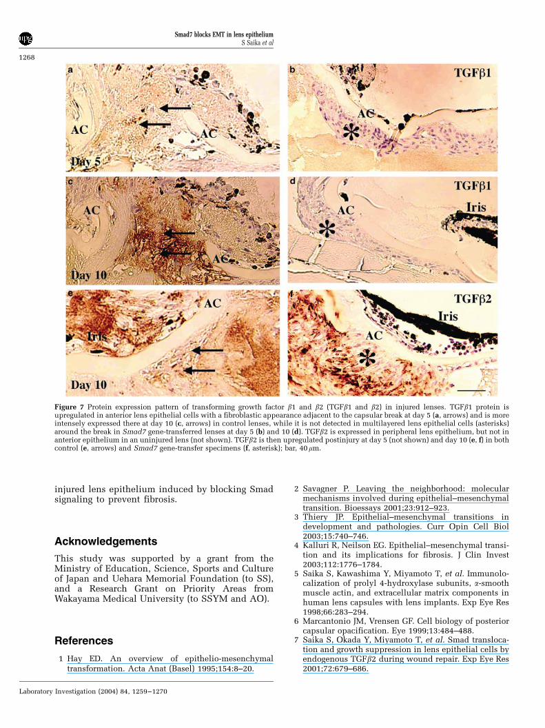
Since introduction of the Smad7 gene blockedinjury-induced EMT in lens epithelial cells, weexamined if this expression suppresses injury-induced upregulation of TGFb1 in lens epithelialcells. Uninjured lens epithelium expresses immu-noreactive TGFb2 in its mid-peripheral region, butdoes not express TGFb1. Fibroblast-like, multi-layered epithelial cells adjacent to the capsularbreak in the control group showed increasedexpression of TGFb1 following injury, whereas
Figure 2 Histology by HE staining of injured lenses at days 5 and 10. Frames a and b show histology of the specimens at day 5, andframes c and d at day 10. HE staining show that the capsular break was sealed by a plaque of elongated, fibroblast-like, lens epithelialcells in control, CAG/Cre-carrying adenovirus-infected specimens (a, c), whereas the capsular opening was not closed with relativelyround or ovoid, epithelial-like, lens cells in eyes coinfected with CAG/Cre-carrying adenovirus and LNL/Smad7-carrying adenovirus(b, d). Inset in each frame is a high-magnification picture of the rectangular area. Arrows, edge of the broken capsule. Bar, 100mm; 16mm(insets).
Smad7 blocks EMT in lens epitheliumS Saika et al
1263
Laboratory Investigation (2004) 84, 1259–1270
multilayered cells in the Smad7 gene-transfergroup were not labeled for either intracellular orextracellular TGFb1 (Figure 7a–d). In the controlgroup, TGFb2 immunoreactivity was reduced infibroblastic lens cells generated through EMT,which accumulated around the capsular break atdays 5 and 10 (Figure 7e). In the Smad7 adv-treatedgroup, multilayered lens cells were of epithelialshape and were still markedly labeled for TGFb2(Figure 7f).
Discussion
The present study shows that transient adenoviralgene transfer of Smad7 cDNA prevents injury-induced EMT in lens epithelial cells in associationwith enhancement of cell proliferation in mice invivo. Introduction of Smad7 also abolishes upregu-lation of snail, aSMA and extracellular matrixcomponents. Smad7 blocks signaling from bothSmad2 and Smad3 and activated Smads 2 and 3have distinct roles in gene regulation based upon the
results of gene-expression array studies.39,40 More-over, it has been reported that conversion offibroblasts to aSMA-expressing myofibroblasts de-pends on Smad2, but that expression of extracel-lular matrix components, that is, collagen types, isSmad3-dependent.41–43 However, EMT, a process ofphenotypic conversion from an epithelial cell typeto a myofibroblastic cell type, is believed to differmechanistically from that of fibroblast-myofibro-blast conversion. We previously showed thatSmad3 signaling is required for injury-inducedEMT of lens epithelial cells and renal tubularepithelial cells,16,17 but not for generation ofmyofibroblasts from fibroblasts in healing dermaltissue.37 These and other data suggest that Smad3is required in the very early phases of injury-induced EMT, whereas Smad2 might be importantin the later phases of the process. Introductionof the Smad7 gene blocks injury-induced EMT inlens epithelial cells possibly via blocking Smad3signaling.
Transient introduction of the Smad7 gene byusing an adenoviral vector is reportedly effective
Figure 3 Expression of snail mRNA in injured lens epithelium as detected by in situ hybridization. The uninjured lens epithelium doesnot express snail mRNA (data not shown). Injured lens epithelium upregulates snail mRNA at day 5 (a) and then its expression increasesat day 10 (c), whereas Smad7 gene introduction abolishes snail expression in injured epithelium at day 5 (b) with very faint snailexpression being detected in the cells of Smad7 vector-treated specimens at day 10 (d). No specific signal was detected in specimenslabeled with the sense probe (e). White or black asterisks indicate the cell multilayers composed of fibroblastic lens cells that are labeledfor snail mRNA or lens cell masses without snail mRNA labeling. AC, anterior capsule, bar, 40mm.
Smad7 blocks EMT in lens epitheliumS Saika et al
1264
Laboratory Investigation (2004) 84, 1259–1270

in treating inflammatory/fibrotic disorders in lung,kidney, and liver.28–30 In these tissues, Smad7 genetransfer not only suppresses the conversion ofinterstitial fibroblasts (including liver stellate cells)to aSMA-expressing myofibroblasts, but also inhi-bits extracellular matrix expression. While, themechanism of generation of aSMA-positive myofi-broblasts might differ among organs,44,45 it would beblocked by Smad7 gene transfer even if it was aSmad2-dependent process. Blocking TGFb action atthe receptor level may block various signalingcascades potentially activated upon ligand bindingto the receptor, such as MAP kinase pathways inaddition to Smad signaling. Blocking injury-in-duced EMT of lens epithelium by Smad7 genetransfer, or by knocking out Smad3, indicates it isnot necessary to block non-Smad TGFb signaling tosuppress EMT.
Our previous study in lens epithelial cells showedthat loss of Smad3 results in the abrogation ofupregulation of TGFb1 and extracellular matrixcomponents, which was mimicked by gene transferof Smad7 into the lens in the present study. Incontrast, expression of TGFb2 in the peripheral
epithelium of an injured lens and in the centralepithelium around the capsular break was similar inthe Cre- and Smad7-treated groups, consistent withthe observation that autoinduction of TGFb1, butnot expression of TGFb2, is dependent upon Smad3.
The TGFb/Smad pathway is also potentiallyinvolved in regulation (mainly suppression) of cellproliferation in many organs/tissues.24 We showhere that Smad7 overexpression enhances theinjury-induced proliferation of lens epithelial cells.Interestingly, this was not observed following lensinjury in Smad3-null mice.17 Since loss of Smad3alone does not affect cell proliferation in an injuredlens,17 it suggests that inhibition of cell proliferationin the injured lens epithelium might require eitherSmad2 or both Smad2 and Smad3, possibly incooperation with Smad-independent pathways. Wehave also observed that loss of Smad3 does not affectcell proliferation in the injured corneal epithelium(S Saika, WW-Y Kao, AB Roberts, unpublished data,2003), whereas it stimulates keratinocyte prolifera-tion following epidermal injury,46 indicating thatregulation of cell proliferation by TGFb/Smadsignaling is tissue-specific.
Figure 4 Expression of aSMA in epithelial cells of the injured lens. Frames a and b show staining of the specimens at day 5, and frames cand d at day 10. At days 5 and 10, multilayered lens epithelial cells in control, CAG/Cre-carrying adenovirus-infected specimens, aremarkedly labeled with anti-aSMA antibody (a, c), while at day 5 no aSMA immunoreactivity is detected in epithelial cells of the lenscoinfected with CAG/Cre-carrying adenovirus and LNL/Smad7-carrying adenovirus (b) and faint aSMA immunoreactivity is observed inthese cells at day 10 (d). Dotted lines indicate anterior capsule; bar, 40mm.
Smad7 blocks EMT in lens epitheliumS Saika et al
1265
Laboratory Investigation (2004) 84, 1259–1270

Clinically, after-cataract tissue is characterized byfibrous opacification and regeneration of lenticularfibers of Sommerring’s ring and Elschnig’s pearlformation, and is accompanied by lens epithelialcell proliferation.8 Using dominant-negative TGFbreceptor expression driven by the a-crystallinpromoter, de Iongh et al47 reported that lenticularfiber cells require TGFb isoforms for lens fibermaturation during development. However, it is notknown as to which signaling cascade(s) is (are)used because the TGFb receptor potentially acti-vates both Smad and non-Smad signaling cascades,that is, MAPK–Erk pathway, p38MAPK, or c-Jun-N-terminal kinase cascade.27,48 This is further sup-ported by detection of both TGFb receptors types Iand II in postoperative lens epithelial cells in theequator region of the residual lens capsule. Wehave reported that elongated, fiber-differentiating,lens cells lack nuclear Smad3/4, suggesting that
postoperative fiber regeneration does not requireTGFb signaling or is controlled by non-Smad-dependent TGFb signaling.15 Thus, it is of interestto examine if postoperative lens fiber regenerationis effectively blocked by Smad7 cDNA , althoughanother animal model will be needed to answerthis question.
In conclusion, lens capsular contraction byfibrosis is clinically unfavorable and blockingSmad signaling, in association with a therapy tosuppress cell proliferation, may be a strategy toprevent this clinical problem. In injury-inducedcell proliferation in lens epithelium, we haveshown that loss of fibroblast growth factor 2(FGF2) reduces cell proliferation, whereas it doesnot affect the physiological proliferation in anintact mouse lens. This result suggests that block-ing FGF2 activity might be potentially used tosuppress an abnormal cell proliferation in an
Figure 5 Expression of extracellular matrix components, lumican and collagen type VI, in epithelial cells of the injured lens. Frames aand b show staining of the specimens at day 5, and frames c–f at day 10. At days 5 and 10, multilayered lens epithelial cells in control,CAG/Cre-carrying adenovirus-infected specimens, are labeled with antilumican antibody (a, c), while at day 5 no lumicanimmunoreactivity is detected in epithelial cells of the lens coinfected with CAG/Cre-carrying adenovirus and LNL/Smad7-carryingadenovirus (b) and at day 10 very faint lumican immunoreactivity is observed in these cells of the lens (d). Collagen type VI is detected inthe cell multilayer in control specimens (e), but not in that of the lens coinfected with CAG/Cre-carrying adenovirus and LNL/Smad7-carrying adenovirus (f). Dotted lines indicate anterior capsule; bar, 40mm.
Smad7 blocks EMT in lens epitheliumS Saika et al
1266
Laboratory Investigation (2004) 84, 1259–1270

Figure 6 Cell proliferation in injured lens. (a), (A) Immunohistochemical detection of BrdU-labeled nuclei of cells in an injured lens thathad received Cre- (A, C) or Smad7 (B, D)-adenovirus at day 5 postinjury. A BrdU-labeled cell nucleus (arrow) is observed in the cellmultilayer formed beneath the broken anterior capsule (A), whereas no cells with a BrdU-labeled nucleus are seen in the equatorialregion (C). In contrast, BrdU-positive nuclei (arrows) are observed beneath the anterior capsule (B) and in the equatorial epithelium (D) inlenses expressing Smad7. Bar, 50mm. (b) Bar chart of the cells with BrdU-positive nuclei in injured lenses of either Cre- or Smad7-adenovirus group at days 5 and 10. At day 5, the mean number of cells with BrdU-positive nuclei is significantly (Po0.005) higher in theSmad7-adenovirus group than in the Cre-adenovirus group at day 5, but there is no significant difference in the number of labeled cells atday 10.
Smad7 blocks EMT in lens epitheliumS Saika et al
1267
Laboratory Investigation (2004) 84, 1259–1270
injured lens epithelium induced by blocking Smadsignaling to prevent fibrosis.
Acknowledgements
This study was supported by a grant from theMinistry of Education, Science, Sports and Cultureof Japan and Uehara Memorial Foundation (to SS),and a Research Grant on Priority Areas fromWakayama Medical University (to SSYM and AO).
References
1 Hay ED. An overview of epithelio-mesenchymaltransformation. Acta Anat (Basel) 1995;154:8–20.
2 Savagner P. Leaving the neighborhood: molecularmechanisms involved during epithelial–mesenchymaltransition. Bioessays 2001;23:912–923.
3 Thiery JP. Epithelial–mesenchymal transitions indevelopment and pathologies. Curr Opin Cell Biol2003;15:740–746.
4 Kalluri R, Neilson EG. Epithelial–mesenchymal transi-tion and its implications for fibrosis. J Clin Invest2003;112:1776–1784.
5 Saika S, Kawashima Y, Miyamoto T, et al. Immunolo-calization of prolyl 4-hydroxylase subunits, a-smoothmuscle actin, and extracellular matrix components inhuman lens capsules with lens implants. Exp Eye Res1998;66:283–294.
6 Marcantonio JM, Vrensen GF. Cell biology of posteriorcapsular opacification. Eye 1999;13:484–488.
7 Saika S, Okada Y, Miyamoto T, et al. Smad transloca-tion and growth suppression in lens epithelial cells byendogenous TGFb2 during wound repair. Exp Eye Res2001;72:679–686.
Figure 7 Protein expression pattern of transforming growth factor b1 and b2 (TGFb1 and b2) in injured lenses. TGFb1 protein isupregulated in anterior lens epithelial cells with a fibroblastic appearance adjacent to the capsular break at day 5 (a, arrows) and is moreintensely expressed there at day 10 (c, arrows) in control lenses, while it is not detected in multilayered lens epithelial cells (asterisks)around the break in Smad7 gene-transferred lenses at day 5 (b) and 10 (d). TGFb2 is expressed in peripheral lens epithelium, but not inanterior epithelium in an uninjured lens (not shown). TGFb2 is then upregulated postinjury at day 5 (not shown) and day 10 (e, f) in bothcontrol (e, arrows) and Smad7 gene-transfer specimens (f, asterisk); bar, 40mm.
Smad7 blocks EMT in lens epitheliumS Saika et al
1268
Laboratory Investigation (2004) 84, 1259–1270

8 Saika S. Relationship between posterior capsularopacification and intraocular lens biocompatibility.Prog Ret Eye Res 2004;23:283–305.
9 Jampel HD, Roche N, Stark WJ, et al. Transforminggrowth factor-b in human aqueous humor. Curr Eye Res1990;9:963–969.
10 Connor Jr TB, Roberts AB, Sporn MB, et al. Correlationof fibrosis and transforming growth factor-b type 2levels in the eye. J Clin Invest 1989;83:1661–1666.
11 Saika S, Miyamoto T, Tanaka S, et al. Response of lensepithelial cells to injury: role of lumican in epithelial–mesenchymal transition. Invest Ophthalmol Vis Sci2003;44:2094–2102.
12 Srinivasan Y, Lovicu FJ, Overbeek PA. Lens-specificexpression of transforming growth factor b1 in trans-genic mice causes anterior subcapsular cataracts. J ClinInvest 1998;101:625–634.
13 Gordon-Thomson C, de Iongh RU, Hales AM, et al.Differential cataractogenic potency of TGF-b1, -b2, and-b3 and their expression in the postnatal rat eye. InvestOphthalmol Vis Sci 1998;39:1399–1409.
14 Wormstone IM, Tamiya S, Anderson I, et al. TGF-b2-induced matrix modification and cell transdifferentia-tion in the human lens capsular bag. Invest Ophthal-mol Vis Sci 2002;43:2301–2308.
15 Saika S, Miyamoto T, Ishida I, et al. TGFb/Smadsignalling in postoperative human lens epithelial cells.Br J Ophthalmol 2002;86:1428–1433.
16 Sato M, Muragaki Y, Saika S, et al. Targeted disruptionof TGF-b1/Smad3 signaling protects against renaltubulointerstitial fibrosis induced by unilateral uret-eral obstruction. J Clin Invest 2003;112:1486–1494.
17 Saika S, Kono-Saika S, Ohnishi Y, et al. Smad3signaling is required for epithelial–mesenchymal tran-sition of lens epithelium after injury. Am J Pathol2004;164:651–663.
18 Piek E, Moustakas A, Kurisaki A, et al. TGF-b type Ireceptor/ALK-5 and Smad proteins mediate epithelialto mesenchymal transdifferentiation in NMuMG breastepithelial cells. J Cell Sci 1999;112:4557–4568.
19 Oft M, Akhurst RJ, Balmain A. Metastasis is driven bysequential elevation of H-ras and Smad2 levels. NatCell Biol 2002;4:487–494.
20 Bakin AV, Tomlinson AK, Bhowmick NA, et al.Phosphatidylinositol 3-kinase function is required fortransforming growth factor b-mediated epithelial tomesenchymal transition and cell migration. J BiolChem 2000;275:36803–36810.
21 Bhowmick NA, Ghiassi M, Bakin A, et al. Transform-ing growth factor-b1 mediates epithelial to mesench-ymal transdifferentiation through a RhoA-dependentmechanism. Mol Biol Cell 2001;12:27–36.
22 Bhowmick NA, Zent R, Ghiassi M, et al. Integrin b1signaling is necessary for transforming growth factor-beta activation of p38MAPK and epithelial plasticity.J Biol Chem 2001;276:46707–46713.
23 Itoh S, Thorikay M, Kowanetz M, et al. Elucidation ofSmad requirement in transforming growth factor-betatype I receptor-induced responses. J Biol Chem2003;278:3751–3761.
24 Moustakas A, Pardali K, Gaal A, et al. Mechanisms ofTGF-b signaling in regulation of cell growth anddifferentiation. Immunol Lett 2002;82:85–91.
25 ten Dijke P, Goumans MJ, Itoh F, et al. Regulation ofcell proliferation by Smad proteins. J Cell Physiol2002;191:1–16.
26 Massague J, Wotton D. Transcriptional control by theTGF-b/Smad signaling system. EMBO J 2000;19:1745–1754.
27 Derynck R, Zhang YE. Smad-dependent and Smad-independent pathways in TGF-b family signaling.Nature 2003;425:577–584.
28 Nakao A, Fujii M, Matsumura R, et al. Transient genetransfer and expression of Smad7 prevents bleomycin-induced lung fibrosis in mice. J Clin Invest1999;104:5–11.
29 Lan HY, Mu W, Tomita N, et al. Inhibition of renalfibrosis by gene transfer of inducible Smad7 usingultrasound-microbubble system in rat UUO model. JAm Soc Nephrol 2003;14:1535–1548.
30 Dooley S, Hamzavi J, Breitkopf K, et al. Smad7prevents activation of hepatic stellate cells and liverfibrosis in rats. Gastroenterology 2003;125:178–191.
31 Sato Y, Tanaka K, Lee G, et al. Enhanced and specificgene expression via tissue-specific production of Crerecombinase using adenovirus vector. Biochem Bio-phys Res Commun 1998;244:455–462.
32 Miyake K, Tohyama T, Shimada T. Two-step genetransfer using an adenoviral vector carrying the CD4gene and human immunodeficiency viral vectors.Hum Gene Ther 1996;7:2281–2286.
33 Kanegae Y, Miyake S, Sato Y, et al. Adenovirus vectortechnology: an efficient method for constructingrecombinant adenovirus and on/off switching of geneexpression. Acta Paediatr Jpn 1996;38:182–188.
34 Carver EA, Jiang R, Lan Y, et al. The mouse snail geneencodes a key regulator of the epithelial–mesenchymaltransition. Mol Cell Biol 2001;21:8184–8188.
35 Nieto MA. The snail superfamily of zinc-fingertranscription factors. Nat Rev Mol Cell Biol 2002;3:155–166.
36 Cano A, Perez-Moreno MA, Rodrigo I, et al. Thetranscription factor snail controls epithelial–mesench-ymal transitions by repressing E-cadherin expression.Nat Cell Biol 2000;2:76–83.
37 Flanders KC, Ludecke G, Engels S, et al. Localizationand actions of transforming growth factors-beta in theembryonic nervous system. Development 1991;113:183–191.
38 Saika S, Saika S, Liu CY, et al. TGFb2 in cornealmorphogenesis during mouse embryonic develop-ment. Dev Biol 2001;240:419–432.
39 Yang YC, Piek E, Zavadil J, et al. Hierarchical model ofgene regulation by transforming growth factor-b. ProcNatl Acad Sci USA 2003;100:10269–10274.
40 Piek E, Ju WJ, Heyer J, et al. Functional characteriza-tion of transforming growth factor b signaling inSmad2- and Smad3-deficient fibroblasts. J Biol Chem2001;276:19945–19953.
41 Flanders KC, Sullivan CD, Fujii M, et al. Mice lackingSmad3 are protected against cutaneous injury inducedby ionizing radiation. Am J Pathol 2002;160:1057–1068.
42 Evans RA, Tian YC, Steadman R, et al. TGF-b1-mediated fibroblast-myofibroblast terminal differentia-tion—the role of Smad proteins. Exp Cell Res2003;282:90–100.
43 Schnabl B, Kweon YO, Frederick JP, et al. The role ofSmad3 in mediating mouse hepatic stellate cellactivation. Hepatology 2001;34:89–100.
44 Serini G, Gabbiani G. Mechanisms of myofibroblastactivity and phenotypic modulation. Exp Cell Res1999;250:273–283.
Smad7 blocks EMT in lens epitheliumS Saika et al
1269
Laboratory Investigation (2004) 84, 1259–1270
45 Serini G, Bochaton-Piallat ML, Ropraz P, et al. Thefibronectin domain ED-A is crucial for myofibroblasticphenotype induction by transforming growth factor-beta1. J Cell Biol 1998;142:873–881.
46 Ashcroft GS, Yang X, Glick AB, et al. Mice lacking Smad3show accelerated wound healing and an impaired localinflammatory response. Nat Cell Biol 1999;1:260–266.
47 de Iongh RU, Lovicu FJ, Overbeek PA, et al. Require-ment for TGFb receptor signaling during terminal lensfiber differentiation. Development 2001;128:3995–4010.
48 Yu L, Hebert MC, Zhang YE. TGF-b receptor-activatedp38 MAP kinase mediates Smad-independent TGF-bresponses. EMBO J 2002;21:3749–3759.
Smad7 blocks EMT in lens epitheliumS Saika et al
1270
Laboratory Investigation (2004) 84, 1259–1270
Copyright © 2022 FDOKUMEN