Increased brain NAD prevents neuronal apoptosis in vivo
8
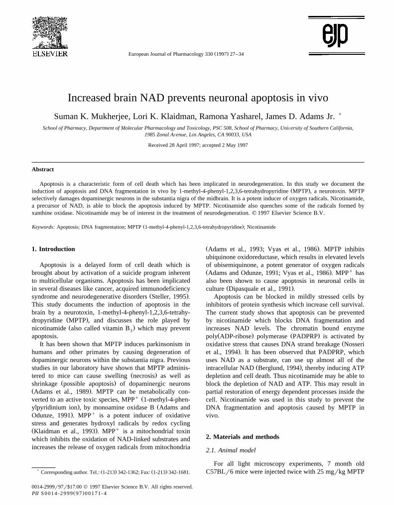
Ž . European Journal of Pharmacology 330 1997 27–34 Increased brain NAD prevents neuronal apoptosis in vivo Suman K. Mukherjee, Lori K. Klaidman, Ramona Yasharel, James D. Adams Jr. ) School of Pharmacy, Department of Molecular Pharmacology and Toxicology, PSC 508, School of Pharmacy, UniÕersity of Southern California, 1985 Zonal AÕenue, Los Angeles, CA 90033, USA Received 28 April 1997; accepted 2 May 1997 Abstract Apoptosis is a characteristic form of cell death which has been implicated in neurodegeneration. In this study we document the Ž . induction of apoptosis and DNA fragmentation in vivo by 1-methyl-4-phenyl-1,2,3,6-tetrahydropyridine MPTP , a neurotoxin. MPTP selectively damages dopaminergic neurons in the substantia nigra of the midbrain. It is a potent inducer of oxygen radicals. Nicotinamide, a precursor of NAD, is able to block the apoptosis induced by MPTP. Nicotinamide also quenches some of the radicals formed by xanthine oxidase. Nicotinamide may be of interest in the treatment of neurodegeneration. q 1997 Elsevier Science B.V. Ž . Keywords: Apoptosis; DNA fragmentation; MPTP 1-methyl-4-phenyl-1,2,3,6-tetrahydropyridine ; Nicotinamide 1. Introduction Apoptosis is a delayed form of cell death which is brought about by activation of a suicide program inherent to multicellular organisms. Apoptosis has been implicated in several diseases like cancer, acquired immunodeficiency Ž . syndrome and neurodegenerative disorders Steller, 1995 . This study documents the induction of apoptosis in the brain by a neurotoxin, 1-methyl-4-phenyl-1,2,3,6-tetrahy- Ž . dropyridine MPTP , and discusses the role played by Ž . nicotinamide also called vitamin B which may prevent 3 apoptosis. It has been shown that MPTP induces parkinsonism in humans and other primates by causing degeneration of dopaminergic neurons within the substantia nigra. Previous studies in our laboratory have shown that MPTP adminis- Ž . tered to mice can cause swelling necrosis as well as Ž . shrinkage possible apoptosis of dopaminergic neurons Ž . Adams et al., 1989 . MPTP can be metabolically con- q Ž verted to an active toxic species, MPP 1-methyl-4-phen- . Ž ylpyridinium ion , by monoamine oxidase B Adams and . q Odunze, 1991 . MPP is a potent inducer of oxidative stress and generates hydroxyl radicals by redox cycling Ž . q Klaidman et al., 1993 . MPP is a mitochondrial toxin which inhibits the oxidation of NAD-linked substrates and increases the release of oxygen radicals from mitochondria ) Ž . Ž . Corresponding author. Tel.: 1-213 342-1362; Fax: 1-213 342-1681. Ž . Adams et al., 1993; Vyas et al., 1986 . MPTP inhibits ubiquinone oxidoreductase, which results in elevated levels of ubisemiquinone, a potent generator of oxygen radicals Ž . q Adams and Odunze, 1991; Vyas et al., 1986 . MPP has also been shown to cause apoptosis in neuronal cells in Ž . culture Dipasquale et al., 1991 . Apoptosis can be blocked in mildly stressed cells by inhibitors of protein synthesis which increase cell survival. The current study shows that apoptosis can be prevented by nicotinamide which blocks DNA fragmentation and increases NAD levels. The chromatin bound enzyme Ž . Ž . poly ADP-ribose polymerase PADPRP is activated by Ž oxidative stress that causes DNA strand breakage Nosseri . et al., 1994 . It has been observed that PADPRP, which uses NAD as a substrate, can use up almost all of the Ž . intracellular NAD Berglund, 1994 , thereby inducing ATP depletion and cell death. Thus nicotinamide may be able to block the depletion of NAD and ATP. This may result in partial restoration of energy dependent processes inside the cell. Nicotinamide was used in this study to prevent the DNA fragmentation and apoptosis caused by MPTP in vivo. 2. Materials and methods 2.1. Animal model For all light microscopy experiments, 7 month old C57BLr6 mice were injected twice with 25 mgrkg MPTP 0014-2999r97r$17.00 q 1997 Elsevier Science B.V. All rights reserved. Ž . PII S0014-2999 97 00171-4
-
Upload
independent -
Category
Documents
-
view
0 -
download
0
Transcript of Increased brain NAD prevents neuronal apoptosis in vivo
Ž .European Journal of Pharmacology 330 1997 27–34
Increased brain NAD prevents neuronal apoptosis in vivo
Suman K. Mukherjee, Lori K. Klaidman, Ramona Yasharel, James D. Adams Jr. )
School of Pharmacy, Department of Molecular Pharmacology and Toxicology, PSC 508, School of Pharmacy, UniÕersity of Southern California,1985 Zonal AÕenue, Los Angeles, CA 90033, USA
Received 28 April 1997; accepted 2 May 1997
Abstract
Apoptosis is a characteristic form of cell death which has been implicated in neurodegeneration. In this study we document theŽ .induction of apoptosis and DNA fragmentation in vivo by 1-methyl-4-phenyl-1,2,3,6-tetrahydropyridine MPTP , a neurotoxin. MPTP
selectively damages dopaminergic neurons in the substantia nigra of the midbrain. It is a potent inducer of oxygen radicals. Nicotinamide,a precursor of NAD, is able to block the apoptosis induced by MPTP. Nicotinamide also quenches some of the radicals formed byxanthine oxidase. Nicotinamide may be of interest in the treatment of neurodegeneration. q 1997 Elsevier Science B.V.
Ž .Keywords: Apoptosis; DNA fragmentation; MPTP 1-methyl-4-phenyl-1,2,3,6-tetrahydropyridine ; Nicotinamide
1. Introduction
Apoptosis is a delayed form of cell death which isbrought about by activation of a suicide program inherentto multicellular organisms. Apoptosis has been implicatedin several diseases like cancer, acquired immunodeficiency
Ž .syndrome and neurodegenerative disorders Steller, 1995 .This study documents the induction of apoptosis in thebrain by a neurotoxin, 1-methyl-4-phenyl-1,2,3,6-tetrahy-
Ž .dropyridine MPTP , and discusses the role played byŽ .nicotinamide also called vitamin B which may prevent3
apoptosis.It has been shown that MPTP induces parkinsonism in
humans and other primates by causing degeneration ofdopaminergic neurons within the substantia nigra. Previousstudies in our laboratory have shown that MPTP adminis-
Ž .tered to mice can cause swelling necrosis as well asŽ .shrinkage possible apoptosis of dopaminergic neurons
Ž .Adams et al., 1989 . MPTP can be metabolically con-q Žverted to an active toxic species, MPP 1-methyl-4-phen-
. Žylpyridinium ion , by monoamine oxidase B Adams and. qOdunze, 1991 . MPP is a potent inducer of oxidative
stress and generates hydroxyl radicals by redox cyclingŽ . qKlaidman et al., 1993 . MPP is a mitochondrial toxinwhich inhibits the oxidation of NAD-linked substrates andincreases the release of oxygen radicals from mitochondria
) Ž . Ž .Corresponding author. Tel.: 1-213 342-1362; Fax: 1-213 342-1681.
Ž .Adams et al., 1993; Vyas et al., 1986 . MPTP inhibitsubiquinone oxidoreductase, which results in elevated levelsof ubisemiquinone, a potent generator of oxygen radicalsŽ . qAdams and Odunze, 1991; Vyas et al., 1986 . MPP hasalso been shown to cause apoptosis in neuronal cells in
Ž .culture Dipasquale et al., 1991 .Apoptosis can be blocked in mildly stressed cells by
inhibitors of protein synthesis which increase cell survival.The current study shows that apoptosis can be preventedby nicotinamide which blocks DNA fragmentation andincreases NAD levels. The chromatin bound enzyme
Ž . Ž .poly ADP-ribose polymerase PADPRP is activated byŽoxidative stress that causes DNA strand breakage Nosseri
.et al., 1994 . It has been observed that PADPRP, whichuses NAD as a substrate, can use up almost all of the
Ž .intracellular NAD Berglund, 1994 , thereby inducing ATPdepletion and cell death. Thus nicotinamide may be able toblock the depletion of NAD and ATP. This may result inpartial restoration of energy dependent processes inside thecell. Nicotinamide was used in this study to prevent theDNA fragmentation and apoptosis caused by MPTP invivo.
2. Materials and methods
2.1. Animal model
For all light microscopy experiments, 7 month oldC57BLr6 mice were injected twice with 25 mgrkg MPTP
0014-2999r97r$17.00 q 1997 Elsevier Science B.V. All rights reserved.Ž .PII S0014-2999 97 00171-4

( )S.K. Mukherjee et al.rEuropean Journal of Pharmacology 330 1997 27–3428
Ž .Fig. 1. Midbrain, substantia nigra: an apoptotic neuron arrow can be seen 48 h after treatment of the mouse with 25 mgrkg MPTP twice at an interval ofŽ . Ž .2 h =600 . Dark spots are apoptotic bodies from disintegrated apoptotic cells. Staining was done with the In Situ Apoptosis Detection Kit Oncor . Bar
lengths represent 10 mm. 7 month old C57BLr6 male mice were used.
Ž .intraperitoneally i.p. at an interval of 2 h. After 2–48 h,the mice were perfused through the left ventricle with
Ž .phosphate buffered saline PBS and then with 4% formal-dehyde and 0.2% glutaraldehyde in PBS. The brains werecoronally sliced into 50 mm sections in a vibrating micro-tome. All immunohistochemical studies for the identifica-tion of apoptotic cells were done 48 h after injection ofMPTP as significantly higher DNA fragmentation valuesŽ .which may reflect the induction of apoptosis were ob-served only after 48 h following administration of MPTP.Necrotic cells were identified within 2–10 h of MPTPadministration.
2.2. Histology
To detect individual cells undergoing apoptosis, the InŽ .Situ Apoptosis Detection Kit Oncor was used. In brief,
Ž .the tissues were quenched in 2% hydrogen peroxide H O2 2
in PBS to deactivate endogenous peroxides. Then twodrops of equilibration buffer containing potassium cacody-late were applied. After incubation for 10–15 s, 54 ml ofworking strength terminal deoxynucleotidyl transferase en-zyme was pipetted onto each section and incubated for 1 hin a humidified chamber. The tissues, after incubation,were washed in a prewarmed working strength stoprwashbuffer and incubated further for 30 min. Then two drops ofanti-digoxigenin-peroxidase were added to the sections.The tissues were again incubated for 30 min at roomtemperature. H O was added to a filtered solution of2 2
freshly prepared 3,3X-diaminobenzidine tetrahydrochloride-Ž .2-hydrate DAB and used to stain sections at room tem-
perature. The tissues were put on slides, air dried, washedin three changes of xylene and mounted with balsam. In
this method, the fragmented DNA ends were extendedwith digoxigenin-dUTP. An anti-digoxigenin antibody thenbound to the extension and was detected by a peroxidasereaction.
Tyrosine hydroxylase staining was done to identifydopaminergic neurons in the substantia nigra. The
Ž .immunoglobulin G IgG polyclonal antibodies used were:a rabbit antibody to tyrosine hydroxylase; a sheep antibodyto rabbit IgG and a rabbit antibody to horseradish peroxi-dases with the horseradish peroxidases coupled to the
Ž .antibody as described previously Adams et al., 1989 . The
Table 1MPTP induced DNA damage and its prevention by nicotinamide
Midbrain Striatum
Control 1.09"0.22 1.09"0.35Nicotinamide 1.25"0.33 1.21"0.12
Ž .MPTP 2 h 0.91"0.22 0.93"0.06aŽ .MPTP 48 h 1.64"0.46 1.24"0.24
MPTPqnicotinamide 1.13"0.30 1.26"0.13
C57BLr6 mice were divided into 5 groups with 4 mice in each group.Ž .MPTP 25 mgrkg, i.p. was administered twice at an interval of 2 h to
Ž . Žmice: MPTP 2 h group brains were removed 2 h after the second. Ž . Žinjection of MPTP , MPTP 48 h group brains were removed 48 h after
. Žthe second injection of MPTP and MPTPqnicotinamide group 500mgrkg nicotinamide was given i.p. 13 h before injection of MPTP and
.brains were removed 48 h after the second injection of MPTP . MPTPŽwas not administered in the nicotinamide group brains were removed 48
.h after administration of 500 mgrkg nicotinamide i.p. . DNA fragmenta-tion was measured in the midbrain and striatum. Results have been givenas means"S.D. and can be expressed as the amount of DNA damagedper gram of tissue.a Statistically significantly different from corresponding control values by
Ž .ANOVA and Newman-Keuls test P -0.05 .

( )S.K. Mukherjee et al.rEuropean Journal of Pharmacology 330 1997 27–34 29
tyrosine hydroxylase stained brain slices were mounted onglass slides, dehydrated and coverslipped with balsam.
For electron microscopy, the method employed byŽ .Adams et al. 1989 was followed. In this experiment,
mice were treated with MPTP for 2–10 h. During thistime, both apoptotic and necrotic cells were seen. Follow-ing staining for tyrosine hydroxylase, brain slices werepostfixed with 2% osmium tetroxide in PBS, dehydrated,embedded in epon-araldite, thin sectioned, mounted ongrids and finally stained with uranyl acetate and leadcitrate.
2.3. DNA fragmentation
Several reports have documented DNA damage in thebrain in Parkinson’s disease. A fluorimetric assay wasdone to detect DNA fragmentation in the midbrain and in
Ž .the striatum Mukherjee et al., 1995 . C57BLr6 mice weredivided into five groups with four animals in each group.Two groups of mice were treated twice with 25 mgrkgMPTP i.p., and 2 h between injections. The brains wereremoved and sectioned after 2 h for one group and after 48h for the other group. For a third group of mice, 500
Ž . Ž .Fig. 2. Midbrain, substantia nigra: in A , a damaged dopaminergic neuron arrow with large vacuoles and a fragmented nucleus forming apoptotic bodiesŽ . Ž .can be seen in a section removed 48 h after treatment with 25 mgrkg MPTP twice at an interval of 2 h =600 . In B , a section from a mouse injected
Ž .with 500 mgrkg nicotinamide i.p. alone can be seen =600 . Staining employed tyrosine hydroxylase immunohistochemistry. Bar lengths represent 10mm. 7 month old C57BLr6 male mice were used.
( )S.K. Mukherjee et al.rEuropean Journal of Pharmacology 330 1997 27–3430
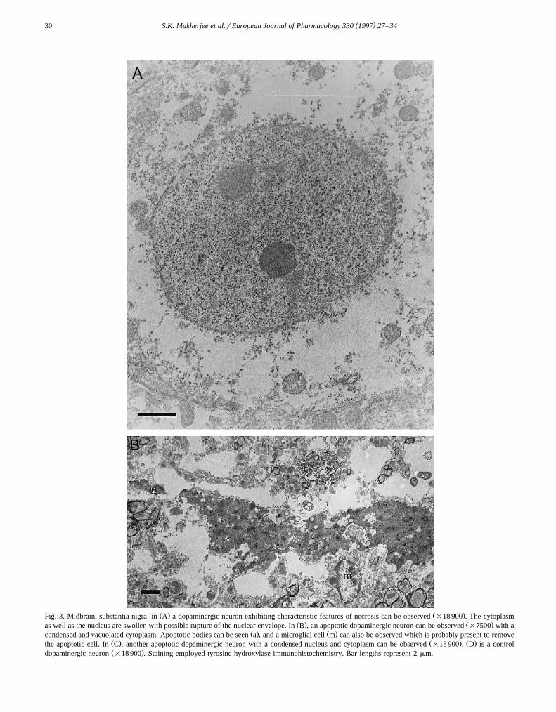
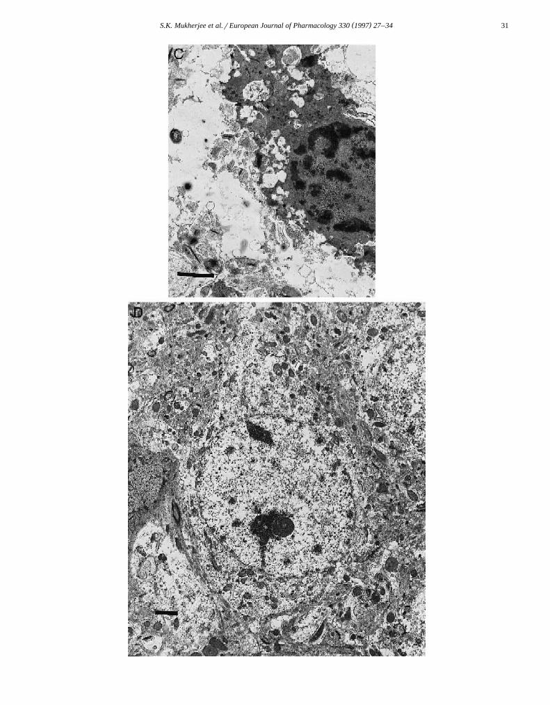
Ž . Ž .Fig. 3. Midbrain, substantia nigra: in A a dopaminergic neuron exhibiting characteristic features of necrosis can be observed =18 900 . The cytoplasmŽ . Ž .as well as the nucleus are swollen with possible rupture of the nuclear envelope. In B , an apoptotic dopaminergic neuron can be observed =7500 with a
Ž . Ž .condensed and vacuolated cytoplasm. Apoptotic bodies can be seen a , and a microglial cell m can also be observed which is probably present to removeŽ . Ž . Ž .the apoptotic cell. In C , another apoptotic dopaminergic neuron with a condensed nucleus and cytoplasm can be observed =18 900 . D is a controlŽ .dopaminergic neuron =18 900 . Staining employed tyrosine hydroxylase immunohistochemistry. Bar lengths represent 2 mm.
( )S.K. Mukherjee et al.rEuropean Journal of Pharmacology 330 1997 27–34 31
( )S.K. Mukherjee et al.rEuropean Journal of Pharmacology 330 1997 27–3432
Ž .mgrkg of nicotinamide i.p. was administered 13 h priorto MPTP injection. A fourth group of mice was treatedonly with 500 mgrkg nicotinamide. A fifth group was thecontrol. Brains were removed and dissected as described in
ŽTable 1. A volume of 0.2 ml of ice-cold lysing buffer 10mM Tris-HCl, pH 7.5, 1 mM EDTA, and 0.2% Triton
.X-100 was added to each section which was centrifugedat 12 000=g for 25 min to separate the intact from the
Žfragmented DNA. The supernatant containing the frag-.mented DNA was transferred to a separate tube. Lysing
Ž .buffer 0.2 ml was added to the pellet with sonication for10 s. The amount of DNA in the supernatant and pelletfractions was determined by a fluorimetric method, usingthe dye Hoechst 33258. DNA damage is normalized bydividing the amount of DNA in the supernatant by the totalDNA in the pellet and supernatant and is expressed as the
Žamount of damage per gram of tissue Mukherjee et al.,.1995 . The levels of NAD were measured by high pressure
Ž .liquid chromatography Klaidman et al., 1995 .
2.4. Free radical studies
Ž .Electron paramagnetic resonance EPR studies weredone with 0.1 unitsrml xanthine oxidase, 0.5 mM xan-
Ž .thine and 0.1 M 5,5-dimethyl-1-pyrroline N-oxide DMPOŽ .in 0.1 M glycine buffer pH 8.3 containing 1 mM dieth-
Ž .ylenetriaminepentaacetic acid DETAPAC ; 0.1 M nicotin-amide and 0.4 M hydrogen peroxide. EPR spectra weremeasured at 238C and 100 kHz in a Bruker model ECS
Ž .106 spectrometer Bruker, Karlsruhe, Germany . The EPRconditions were: modulation amplitude 0.483 G, mi-crowave power 20 mW and time constant 1.3 s.
3. Results
3.1. Necrotic and apoptotic morphology
By light microscopy, apoptotic dopaminergic neurons inthe midbrain with shrunken and fragmented nuclei weremost abundant after 48 h following MPTP administrationŽ .Fig. 1 . These apoptotic cells were all shrunken with largecytoplasmic vacuoles. However, no apoptotic cells wereobserved when the mice were treated with 500 mgrkg
Žnicotinamide 13 h before administration of MPTP Figure.not shown . Administration of 500 mgrkg nicotinamide
Žalone for 48 h did not generate any apoptotic cells Figure.not shown .
In the MPTP-treated brains that were stained with atyrosine hydroxylase antibody, dopaminergic neurons withapoptotic morphology, which had condensed and vacuo-lated cytoplasms and fragmented nuclei can be clearly
Ž . Ž .observed Fig. 2A . In the nicotinamide-treated Fig. 2BŽas well as the MPTP- and nicotinamide-treated 500 mgrkg
. Ž .nicotinamide prior to MPTP mice Figure not shown , thedopaminergic neurons were normal. No condensed cellswith fragmented nuclei were observed.
By electron microscopy, cells having necrotic morphol-ogy were found following 2 h of treatment with MPTPŽ .Fig. 3A . In this figure a swollen dopaminergic neuroncan be observed that has an inflated nucleus and possibly aruptured nuclear envelope. A nucleolus has also changedshape. These are characteristic features of necrosis, thecytoplasm and nuclei swell and eventually rupture. Aspreviously reported, the mitochondria appear normal innecrotic dopaminergic neurons following MPTP treatmentŽ .Adams et al., 1989 . The cytoplasm contains tyrosinehydroxylase, which is identified by the reaction with tyro-sine hydroxylase antibody.
On the other hand, following 10–48 h of MPTP treat-Ž .ment apoptotic dopaminergic neurons Fig. 3B can be
seen. In this figure a condensed dopaminergic neuron isobserved which is on the verge of disintegrating intovarious apoptotic bodies. The cytoplasm is highly con-densed and vacuolated, but contains tyrosine hydroxylasethat is identified by the antibody reaction. The nucleus ishighly condensed. The electron dense chromatin materialin the cytoplasm may be nuclear fragments about to bepackaged as apoptotic bodies. An apoptotic body can beseen which is separated from the cell, and contains a smallamount of possibly nuclear material and perhaps cytoplas-mic material. A microglial cell can be observed, that isprobably about to remove the apoptotic cell. Fig. 3C isanother apoptotic dopaminergic neuron with a condensednucleus and vacuolated, condensed cytoplasm. The sourceof the vacuoles is not entirely clear but may be fromphagolysosomes which are digesting the remnants of thecondensed cytoplasm. Mitochondria appear in the cell andmay be somewhat condensed and electron dense. Fig. 3Dis a control dopaminergic neuron, with a regular nuclearenvelope and tyrosine hydroxylase immunohistochemistry.
3.2. DNA fragmentation and free radicals
Ž .Significant DNA fragmentation P-0.05 was ob-served after 48 h in the midbrain and not in the striatum
Ž .after administration of 25 mgrkg MPTP twice Table 1 .Treatment with nicotinamide was found to prevent theMPTP induced DNA fragmentation. An increase in DNAfragmentation was not observed when nicotinamide wasadministered alone.
It was observed that addition of 0.1 M nicotinamidewas able to quench superoxide radicals produced by xan-
Ž .thine oxidase Fig. 4 . 0.1 M nicotinamide was also able toquench hydroxyl radicals generated by the addition of 0.4M hydrogen peroxide to xanthine oxidase.
3.3. Nicotinamide and NAD leÕels
Administration of 500 mgrkg nicotinamide for 8 hincreased NAD levels from 0.28"0.08 mmolrg to 0.38"0.04 mmolrg in the midbrain and from 0.35"0.04mmolrg to 0.45"0.04 mmolrg in the striatum. The

( )S.K. Mukherjee et al.rEuropean Journal of Pharmacology 330 1997 27–34 33
Ž .Fig. 4. a EPR spectra in the presence of 0.1 unitsrml of xanthineŽoxidase, 0.5 mM xanthine and 0.1 M DMPO in 0.1 M glycine buffer pH
. Ž . Ž .8.3 containing 1 mM DETAPAC; b as in a , with 0.1 M nicotinamide.Ž .Nicotinamide was able to quench the superoxide radicals formed in a to
Ž . Ž .some extent; c as in a , with 0.4 M hydrogen peroxide to induce theŽ . Ž .generation of hydroxyl radical; d as in c , with 0.1 M nicotinamide
which was able to quench all radicals.
levels of NAD remained high even 13 h after administra-tion of nicotinamide. A large dose of nicotinamide wasused as only a small fraction ultimately reaches the brain.
4. Discussion
4.1. DNA fragmentation and MPTP
In this study the induction of apoptosis and DNAfragmentation by MPTP is reported along with the veryinteresting observation that nicotinamide seems to be ableto prevent both when given prior to neurotoxin administra-tion. DNA may be an important target site for neurotoxins
Ž .that produce active radical species Mukherjee et al., 1995 .The fragmentation of DNA by MPTP has also been docu-
Ž .mented in other studies Dipasquale et al., 1991 . Signifi-cant DNA fragmentation was observed in the midbrain andnot in the striatum following administration of MPTP. Ithas been shown by previous studies that the midbrain isbiochemically different from the striatum in terms of its
Ž .response to MPTP toxicity Adams et al., 1989 . Midbrainneuronal somas have nuclear DNA not present in striatalterminals. This makes the neurons more susceptible tooxidative stress induced DNA damage than their terminals.Of course, apoptosis of midbrain neurons causes loss oftheir striatal terminals. The active metabolite of MPTP,which is MPPq, is actively taken up by the nuclei of
Ž .dopaminergic neurons Buu, 1993 . Though some of theDNA damage associated with both necrosis and apoptosismay be repaired over time by various enzymes, it ispossible that transient DNA damage will affect proteinsynthesis or mRNA transcription. This may cause disrup-tion of the normal processes in the brain.
4.2. MPTP induction of apoptosis
MPTP administration caused widespread apoptosis inthe midbrain. This result is in agreement with previousstudies where apoptotic cells had been identified with theterminal deoxynucleotidyl transferase-mediated dUTP-bio-
Ž . Žtin nick-end labeling TUNEL technique Mochizuki et.al., 1994; Dipasquale et al., 1991; Tatton and Kish, 1997 .
Ž .However, in a recent study Jackson-Lewis et al., 1995apoptosis was not detected following treatment with MPTP.
ŽIn this work no direct experiments for example, theTUNEL technique or gel electrophoresis to find the char-
.acteristic DNA ladder had been done to detect apoptosis.Therefore it is possible that apoptosis that had been in-duced may have gone undetected. The cells that stainedwith the TUNEL technique in the current study were allapoptotic as they all had the characteristic morphology andapoptotic bodies. No necrotic cells were observed at 48 h.All apoptotic cells were found, by oil immersion andelectron microscopy, to have condensed cytoplasms andnuclei. Large cytoplasmic vacuoles were frequently pre-sent. Apoptotic cells usually have DNA fragments in theircytoplasms indicating possible loss of nuclear membraneintegrity.
Electron microscopy was done to observe cells undergo-ing necrosis and apoptosis. Necrotic cells were observedwithin 2 h, while apoptotic cells were seen 10 h and laterfollowing MPTP treatment. The in situ technique wasemployed only after 48 h of treatment as this time pre-cludes the presence of necrotic cells. Also higher levels ofDNA fragmentation were measured at the end of 48 hwhich indicates that apoptosis was more pronounced at 48h. A recent study agrees with this time course, in thatfollowing MPTP treatment, mice have the most apoptotic
Ž .brain cells between 24 and 48 h Tatton and Kish, 1997 .
4.3. MPTP and free radicals
It has been shown in previous studies that MPPq canact as a substrate for xanthine oxidase producing MPP Ø,which is the free radical form of MPPq, and subsequently
Ž .superoxide and hydroxyl ions Klaidman et al., 1993 .Xanthine oxidase is located in brain endothelial cells andis probably activated during MPTP induced hypoperfusionŽ .Adams et al., 1991 . That nicotinamide can act as ascavenger of oxygen derived radicals is supported by other
Ž .studies Ledoux et al., 1988 , in which nicotinamide wasemployed to quench radicals generated by alloxan. Addi-tionally, nicotinamide is known to inhibit xanthine oxidaseby preventing the conversion of the dehydrogenase formŽ . Ž . Ž‘D’ form to the oxidase form ‘O’ form Di Steffano
.and Pizzichini, 1980 . Our results demonstrate that nicotin-amide inhibits hydroxyl radical production by xanthineoxidase. This may be one mechanism by which nicotin-amide prevents MPTP toxicity since xanthine oxidase maybe important in MPTP toxicity.

( )S.K. Mukherjee et al.rEuropean Journal of Pharmacology 330 1997 27–3434
4.4. MPTP, nicotinamide and DNA fragmentation
The exact role of PADPRP and its inhibitors duringapoptosis and DNA fragmentation in vivo is still contro-versial. In some studies a cysteine protease has beenobserved to destroy PADPRP during the apoptotic processŽ .Nicholson et al., 1995 . However, it is clear that a secondactivation of PADPRP then occurs and is necessary for the
Ž .induction of apoptosis Nosseri et al., 1994 . This secondactivation of PADPRP is responsible for a late depression
Ž .of NAD levels when apoptosis is detected Morgan, 1995 .Nicotinamide can produce a weak in vivo inhibition ofPADPRP. However, in our studies, nicotinamide was ad-ministered 13 h before MPTP. It is very unlikely thatnicotinamide was present to inhibit PADPRP when MPTPwas given. Moreover, in a recent study it was observedthat PADPRP inhibitors like benzamide and nicotinamidewere able to prevent depletion of dopamine following
Ž .MPTP treatment in mice Cosi et al., 1996 . Additionally,in this study it was observed that nicotinamide caused an
Ž .increase in 3,4-dihydroxyphenyl acetic acid DOPAC andŽ .homovanillic acid HVA levels. This implies that nicotin-
amide does not act as an inhibitor of monoamine oxidaseŽand may increase dopamine turnover. Other studies Zhang
.et al., 1995 have observed that the relative potencies ofthe PADPRP inhibitors in protecting cells from damageare related to their potencies as inhibitors of PADPRP.
Nicotinamide did produce a long lasting increase inbrain levels of NAD, which is a substrate for PADPRP.Restoration and elevation of NAD may be important asNAD is a cofactor in a number of essential biochemicalpathways like glycolysis and is also a precursor for ATPsynthesis. MPTP is known to decrease ATP levels in cells.NAD can also be converted to NADP and NADPH, whichcan then be utilized to reduce GSSG to GSH. Therefore,several mechanisms may be important in the inhibition ofapoptosis by NAD.
References
Adams, J.D., Odunze, I.N., 1991. Oxygen free radicals and Parkinson’sdisease. Free Radic. Biol. Med. 10, 161–169.
Adams, J.D., Kalivas, P.W., Miller, C.A., 1989. The acute histopathologyof MPTP in the mouse CNS. Brain Res. Bull. 23, 1–17.
Adams, J.D., Klaidman, L.K., Odunze, I.N., Johannessen, J.N., 1991.Effects of MPTP on the cerebrovasculature. Int. J. Dev. Neurosci. 9,155–159.
Adams, J.D., Klaidman, L.K., Leung, A.C., 1993. MPPq and MPDPq
induced oxygen radical formation with mitochondrial enzymes. FreeRadic. Biol. Med. 15, 181–186.
Berglund, T., 1994. Nicotinamide, a missing link in the early stressresponse in eukaryotic cells: a hypothesis with special reference tooxidative stress in plants. FEBS Lett. 351, 1445–1449.
Buu, N.T., 1993. Uptake of 1-methyl-4-phenylpyridinium and dopaminein the mouse brain cell nuclei. J. Neurochem. 61, 1557–1560.
Cosi, C., Colpaert, F., Koek, W., Degryse, A., Marien, M., 1996. PolyŽ .ADP-ribose polymerase inhibitors protect against MPTP-induceddepletions of striatal dopamine and cortical noradrenaline in C57B1r6mice. Brain Res. 729, 264–269.
Dipasquale, B., Marini, A.M., Youle, R.J., 1991. Apoptosis and DNAdegradation induced by 1-methyl-4-phenylpyridinium in neurons.Biochem. Biophys. Res. Commun. 181, 1442–1448.
Di Steffano, A., Pizzichini, M.E., 1980. Nicotinamide and liver xanthineoxidase. Adv. Exp. Med. Biol. 122B, 189–196.
Jackson-Lewis, V., Jakowec, M., Burke, R.E., Przedborski, S., 1995.Time course and morphology of dopaminergic neuronal death causedby the neurotoxin 1-methyl-4-phenyl-1,2,3,6-tetrahydropyridine. Neu-
Ž .rodegeneration 4 3 , 257–269.Klaidman, L.K., Adams, J.D., Leung, A.C., Kim, S.S., Cadenas, E.,
1993. Redox cycling of MPPq: evidence for a new mechanisminvolving hydride transfer with xanthine oxidase, aldehyde dehydro-genase and lipoamide dehydrogenase. Free Radic. Biol. Med. 15,169–179.
Klaidman, L.K., Leung, A., Adams, J.D., 1995. High-performance liquidchromatography analysis of oxidized and reduced pyridine dinu-cleotides in specific brain regions. Anal. Biochem. 228, 312–317.
Ledoux, S.P., Hall, C.R., Forbes, P.M., Patton, N.J., Wilson, G.L., 1988.Mechanisms of nicotinamide and thymidine protection from alloxanand streptozotocin toxicity. Diabetes 37, 1015–1019.
Mochizuki, H., Nakamura, N., Nishi, K., Mizuno, Y., 1994. Apoptosis isŽ q .induced by 1-methyl-4-pyridinium ion MPP in ventral mesen-
cephalic-striatal co-culture in rats. Neurosci. Lett. 170, 191–194.Morgan, W.A., 1995. Pyridine nucleotide hydrolysis and interconversion
in rat hepatocytes during oxidative stress. Biochem. Pharmacol. 49Ž .9 , 1179–1184.
Mukherjee, S.K., Yasharel, R., Klaidman, L.K., Hutchin, T.P., Adams,J.D., 1995. Apoptosis and oxidative stress as induced by tertiary
Ž .butylhydroperoxide in the brain. Brain Res. Bull. 38 6 , 595–604.Nicholson, D.W., Ali, A., Thornberry, N.A., Vallaincourt, J.P., Ding,
C.K., Gallant, M., Gareau, Y., Griffin, P.R., Labelle, M., Lazebnik,Y.A., Munday, N.A., Raju, S.M., Smulson, M.E., Yamin, T.T., Yu,V.L., Miller, D.K., 1995. Identification and inhibition of theICErCED-3 protease necessary for mammalian apoptosis. Nature376, 37–43.
Nosseri, C., Coppola, S., Ghibelli, L., 1994. Possible involvement of polyŽ .ADP-ribose polymerase in triggering stress-induced apoptosis. Exp.Cell Res. 212, 367–373.
Steller, H., 1995. Mechanisms and genes of cellular suicide. Science 267,1445–1449.
Tatton, N.A., Kish, S.J., 1997. In situ detection of apoptotic nuclei in thesubstantia nigra compacta of MPTP treated mice using terminaldeoxynucleotidyl transferase labeling and acridine orange staining.Neuroscience 77, 1037–1048.
Vyas, I., Heikkila, R.E., Nicklas, W.J., 1986. Studies on the neurotoxicityof MPTP: inhibition of NAD linked substrate oxidation by its inter-mediate MPPq. J. Neurochem. 46, 1501–1507.
Ž .Zhang, J., Pieper, A., Snyder, S.N., 1995. Poly ADP-ribose synthetaseactivation: an early indicator of neurotoxic DNA damage. J. Neu-rochem. 65, 1411–1414.