Inhibition of Calpains Prevents Neuronal and Behavioral Deficits in an MPTP Mouse Model of...
11
Inhibition of Calpains Prevents Neuronal and Behavioral Deficits in an MPTP Mouse Model of Parkinson’s Disease Stephen J. Crocker, 1 Patrice D. Smith, 1 Vernice Jackson-Lewis, 3 Wiplore R. Lamba, 1 Shawn P. Hayley, 1 Erich Grimm, 5 Steve M. Callaghan, 1 Ruth S. Slack, 1 Edon Melloni, 7 Serge Przedborski, 3,4 George S. Robertson, 6 Hymie Anisman, 8 Zul Merali, 2 and David S. Park 1 1 Neuroscience Research Group, Ottawa Health Research Institute and 2 Department of Psychology, University of Ottawa, Ottawa, Ontario K1H 8M5, Canada, Departments of 3 Neurology and 4 Pathology, Center for Neurobiology and Behaviour, Columbia University, New York, New York 10032, Departments of 5 Chemistry and 6 Pharmacology, Merck-Frosst and Company Canada Inc., Kirkland, Quebec H9H 3L1, Canada, 7 Department of Experimental Medicine, University of Genoa, 1-16132 Genoa, Italy, and 8 Institute for Neuroscience, Carleton University, Ottawa K1H 6N5, Canada The molecular mechanisms mediating degeneration of midbrain dopamine neurons in Parkinson’s disease (PD) are poorly understood. Here, we provide evidence to support a role for the involvement of the calcium-dependent proteases, calpains, in the loss of dopamine neurons in a mouse model of PD. We show that administration of N-methyl-4-phenyl-1,2,3,6-tetrahydropyridine (MPTP) evokes an increase in calpain- mediated proteolysis in nigral dopamine neurons in vivo. Inhibition of calpain proteolysis using either a calpain inhibitor (MDL-28170) or adenovirus-mediated overexpression of the endogenous calpain inhibitor protein, calpastatin, significantly attenuated MPTP-induced loss of nigral dopamine neurons. Commensurate with this neuroprotection, MPTP-induced locomotor deficits were abolished, and markers of striatal postsynaptic activity were normalized in calpain inhibitor-treated mice. However, behavioral improvements in MPTP-treated, calpain inhibited mice did not correlate with restored levels of striatal dopamine. These results suggest that protection against nigral neuron degeneration in PD may be sufficient to facilitate normalized locomotor activity without necessitating striatal reinnervation. Immunohistochemical analyses of postmortem midbrain tissues from human PD cases also displayed evidence of increased calpain-related proteolytic activity that was not evident in age-matched control subjects. Taken together, our findings provide a potentially novel correlation between calpain proteolytic activity in an MPTP model of PD and the etiology of neuronal loss in PD in humans. Key words: substantia nigra; dopamine; neurotensin; FosB; protease; adenovirus; behavior; L-Dopa Introduction The molecular events responsible for the loss of dopaminergic neuron in the substantia nigra pars compacta (SNc) in Parkin- son’s disease (PD) remain poorly understood. One prominent feature of PD is a deficiency in mitochondrial function attributed to reduced complex 1 activity in the SNc (Schapira et al., 1989; Greenamyre et al., 2001). Experimentally, administration of chemical inhibitors of complex 1 of the mitochondrial respira- tion chain can mimic key features of PD, including the selective dopaminergic neuropathology and behavioral deficits (Beal, 2001). These findings support accumulating evidence that nigral dopamine neurons are highly sensitive to stress related to re- duced mitochondrial function. The cellular consequences of deficits in mitochondrial function include reduced ATP production (Greenamyre et al., 1999), oxidation-related changes in protein function (Jenner, 1998; Przed- borski et al., 2003), and poor calcium homeostasis (Sheehan et al., 1997; Sherer et al., 2001). It is of particular relevance to this latter point that previous work has also shown that N-methyl-4- pyridinium (MPP), the active metabolite of the dopaminergic neurotoxin N-methyl-4-phenyl-1,2,3,6-tetrahydropyridine (MPTP), can evoke a sustained elevation of cytoplasmic calcium levels (Chen et al., 1995). This change in calcium likely occurs through several processes, including secondary excitotoxic mechanisms and deple- tion of mitochondrial calcium pools (Frei and Richter, 1986; Kass et al., 1988). Such abnormal calcium homeostasis manifests activation of various intracellular signaling pathways that impact on neuronal function and survival (Wang and Yuen, 1994). Although the effects of increased intracellular calcium in nigral dopamine neurons is al- most certainly complex, one consequence may be the activation of the calcium-sensitive proteases, calpains. Calpains are a highly conserved family of calcium-dependent proteases. There are two ubiquitously expressed calpain iso- forms, (calpain-1) and m (calpain-2). Each calpain is com- posed of a unique large subunit and a common small regulatory subunit (for review, see Sorimachi et al., 1997). The importance of calpains is underscored by the observation that mice deficient in the 30 kDa small regulatory subunit suffer embryonic lethality Received Dec. 26, 2002; revised March 7, 2003; accepted March 17, 2003. The Harvard Brain Tissue Resource Center is supported in part by Public Health Service Grant MH/NS 31862. This work was supported by grants from the Canadian Institutes of Health Research (CIHR) (D.S.P., Z.M., H.A), Parkinson’s Society Canada (D.S.P), Parkinson’s Disease Foundation (USA) (S.P., V.J.-L., D.S.P), National Institutes of Health/ National Institute of Neurological Disorders and Stroke (Grants R29 NS37345, R01 NS38586 –NS 42269, P50 NS38370), United States Department of National Defense (DAMD 17-99-1-9471 to S.P.), Lowenstein Foundation, Lillian Goldman Charitable Trust, Muscular Dystrophy Association, ALS Association (S.P.), and National Parkinson’s Foundation (V.J.-L.). W.R.L. was supported by a summer studentship from the Parkinson’s Disease Foundation (USA). S.J.C. was funded by a postdoctoral fellowship from the Ontario Neurotrauma Foundation. H.A. is a Canada Research Chair in Neuroscience. D.S.P. is a recipient of the Glaxo-Wellcome Chair for Stroke and is a CIHR Scholar. We thank Dr. R. Simon for helpful discussions. We also thank Dr. A. Ridsdale for assistance with confocal microscopy, and Z. Kulczycki, N. Lukenbill, and K. Yates for technical assistance. We gratefully acknowledge Cephalon Inc. (West Chester, PA) for antibodies (Roberts-Lewis et al., 1994). Correspondence should be addressed to Dr. David S. Park,451 Smyth Road, Ottawa, Ontario, Canada K1H 8M5. E-mail: [email protected]. Copyright © 2003 Society for Neuroscience 0270-6474/03/234081-11$15.00/0 The Journal of Neuroscience, May 15, 2003 • 23(10):4081– 4091 • 4081
Transcript of Inhibition of Calpains Prevents Neuronal and Behavioral Deficits in an MPTP Mouse Model of...
Inhibition of Calpains Prevents Neuronal and BehavioralDeficits in an MPTP Mouse Model of Parkinson’s Disease
Stephen J. Crocker,1 Patrice D. Smith,1 Vernice Jackson-Lewis,3 Wiplore R. Lamba,1 Shawn P. Hayley,1 Erich Grimm,5
Steve M. Callaghan,1 Ruth S. Slack,1 Edon Melloni,7 Serge Przedborski,3,4 George S. Robertson,6 Hymie Anisman,8
Zul Merali,2 and David S. Park1
1Neuroscience Research Group, Ottawa Health Research Institute and 2Department of Psychology, University of Ottawa, Ottawa, Ontario K1H 8M5, Canada,Departments of 3Neurology and 4Pathology, Center for Neurobiology and Behaviour, Columbia University, New York, New York 10032, Departments of5Chemistry and 6Pharmacology, Merck-Frosst and Company Canada Inc., Kirkland, Quebec H9H 3L1, Canada, 7Department of Experimental Medicine,University of Genoa, 1-16132 Genoa, Italy, and 8Institute for Neuroscience, Carleton University, Ottawa K1H 6N5, Canada
The molecular mechanisms mediating degeneration of midbrain dopamine neurons in Parkinson’s disease (PD) are poorly understood. Here,we provide evidence to support a role for the involvement of the calcium-dependent proteases, calpains, in the loss of dopamine neurons in amouse model of PD. We show that administration of N-methyl-4-phenyl-1,2,3,6-tetrahydropyridine (MPTP) evokes an increase in calpain-mediated proteolysis in nigral dopamine neurons in vivo. Inhibition of calpain proteolysis using either a calpain inhibitor (MDL-28170) oradenovirus-mediated overexpression of the endogenous calpain inhibitor protein, calpastatin, significantly attenuated MPTP-induced loss ofnigral dopamine neurons. Commensurate with this neuroprotection, MPTP-induced locomotor deficits were abolished, and markers of striatalpostsynaptic activity were normalized in calpain inhibitor-treated mice. However, behavioral improvements in MPTP-treated, calpain inhibitedmice did not correlate with restored levels of striatal dopamine. These results suggest that protection against nigral neuron degeneration in PDmay be sufficient to facilitate normalized locomotor activity without necessitating striatal reinnervation. Immunohistochemical analyses ofpostmortem midbrain tissues from human PD cases also displayed evidence of increased calpain-related proteolytic activity that was not evidentin age-matched control subjects. Taken together, our findings provide a potentially novel correlation between calpain proteolytic activity in anMPTP model of PD and the etiology of neuronal loss in PD in humans.
Key words: substantia nigra; dopamine; neurotensin; FosB; protease; adenovirus; behavior; L-Dopa
IntroductionThe molecular events responsible for the loss of dopaminergicneuron in the substantia nigra pars compacta (SNc) in Parkin-son’s disease (PD) remain poorly understood. One prominentfeature of PD is a deficiency in mitochondrial function attributedto reduced complex 1 activity in the SNc (Schapira et al., 1989;Greenamyre et al., 2001). Experimentally, administration ofchemical inhibitors of complex 1 of the mitochondrial respira-tion chain can mimic key features of PD, including the selectivedopaminergic neuropathology and behavioral deficits (Beal,2001). These findings support accumulating evidence that nigral
dopamine neurons are highly sensitive to stress related to re-duced mitochondrial function.
The cellular consequences of deficits in mitochondrial functioninclude reduced ATP production (Greenamyre et al., 1999),oxidation-related changes in protein function (Jenner, 1998; Przed-borski et al., 2003), and poor calcium homeostasis (Sheehan et al.,1997; Sherer et al., 2001). It is of particular relevance to this latterpoint that previous work has also shown that N-methyl-4-pyridinium (MPP�), the active metabolite of the dopaminergicneurotoxin N-methyl-4-phenyl-1,2,3,6-tetrahydropyridine (MPTP),can evoke a sustained elevation of cytoplasmic calcium levels (Chenet al., 1995). This change in calcium likely occurs through severalprocesses, including secondary excitotoxic mechanisms and deple-tion of mitochondrial calcium pools (Frei and Richter, 1986; Kass etal., 1988). Such abnormal calcium homeostasis manifests activationof various intracellular signaling pathways that impact on neuronalfunction and survival (Wang and Yuen, 1994). Although the effectsof increased intracellular calcium in nigral dopamine neurons is al-most certainly complex, one consequence may be the activation ofthe calcium-sensitive proteases, calpains.
Calpains are a highly conserved family of calcium-dependentproteases. There are two ubiquitously expressed calpain iso-forms, � (calpain-1) and m (calpain-2). Each calpain is com-posed of a unique large subunit and a common small regulatorysubunit (for review, see Sorimachi et al., 1997). The importanceof calpains is underscored by the observation that mice deficientin the 30 kDa small regulatory subunit suffer embryonic lethality
Received Dec. 26, 2002; revised March 7, 2003; accepted March 17, 2003.The Harvard Brain Tissue Resource Center is supported in part by Public Health Service Grant MH/NS 31862. This
work was supported by grants from the Canadian Institutes of Health Research (CIHR) (D.S.P., Z.M., H.A), Parkinson’sSociety Canada (D.S.P), Parkinson’s Disease Foundation (USA) (S.P., V.J.-L., D.S.P), National Institutes of Health/National Institute of Neurological Disorders and Stroke (Grants R29 NS37345, R01 NS38586 –NS 42269, P50NS38370), United States Department of National Defense (DAMD 17-99-1-9471 to S.P.), Lowenstein Foundation,Lillian Goldman Charitable Trust, Muscular Dystrophy Association, ALS Association (S.P.), and National Parkinson’sFoundation (V.J.-L.). W.R.L. was supported by a summer studentship from the Parkinson’s Disease Foundation(USA). S.J.C. was funded by a postdoctoral fellowship from the Ontario Neurotrauma Foundation. H.A. is a CanadaResearch Chair in Neuroscience. D.S.P. is a recipient of the Glaxo-Wellcome Chair for Stroke and is a CIHR Scholar. Wethank Dr. R. Simon for helpful discussions. We also thank Dr. A. Ridsdale for assistance with confocal microscopy, andZ. Kulczycki, N. Lukenbill, and K. Yates for technical assistance. We gratefully acknowledge Cephalon Inc. (WestChester, PA) for antibodies (Roberts-Lewis et al., 1994).
Correspondence should be addressed to Dr. David S. Park,451 Smyth Road, Ottawa, Ontario, Canada K1H 8M5.E-mail: [email protected] © 2003 Society for Neuroscience 0270-6474/03/234081-11$15.00/0
The Journal of Neuroscience, May 15, 2003 • 23(10):4081– 4091 • 4081

(Arthur et al., 2000; Zimmerman et al., 2000). In the CNS, cal-pains are widely expressed (Goto et al., 1994; Li et al., 1996) andmodulated by an endogenously expressed inhibitory protein, cal-pastatin (Emori et al., 1987; Wang and Yuen, 1994).
The role of calpains in PD is unknown. In human PD tissues,it has been reported that expression of m-calpain is increased indopamine neurons (Mouatt-Prigent et al., 1996); however, thesignificance of this observation is not clear. Here, we provideevidence to support a role for calpain activation as a processmediating the loss of nigral dopamine neurons in a mouse modelof PD and demonstrate that inhibition of calpains prevents re-duced motor function in mice through normalization of basalganglia (BG) activity, albeit in the absence of restored striataldopamine. We also provide evidence that calpain activity is en-hanced in nigral dopamine neurons of postmortem tissues fromhuman PD cases. These findings support a novel role for calpains inthe molecular events related to nigral neuron dysfunction in PD.
Materials and MethodsMiceAll procedures involving animals were approved by the University ofOttawa Animal Care Committee and maintained in strict accordancewith the Guidelines for the Use and Treatment of Animals put forth by theAnimal Care Council of Canada and endorsed by the Canadian Institutesof Health Research.
MPTPN-methyl-4-phenyl-1,2,3,6-tetrahydropyridine hydrochloride (25 mg/kg, i.p., measured as free base; MPTP-HCl; Sigma, St. Louis, MO) wasadministered to male C57BL/6 mice (8 –10 weeks old; Charles River Lab-oratories, St. Constant, Quebec) once a day for 5 consecutive days (Tat-ton and Kish, 1997; Xia et al., 2001) (Fig. 1 A). Mice used as controlsreceived an equivalent volume of saline (0.9%) once daily.
Calpain inhibitionOsmotic minipumps (Alza, Palo Alto, CA; model 1007D) were im-planted into the right lateral ventricle 1.0 mm rostral and 2.2 mm to theright of bregma with the cannula extending to a depth of 2.5 mm from theskull surface. Pumps were prefilled with 200 �l of either vehicle (Krebs’-
Ringer’s solution) or MDL-28170 (160 �M; carbobenzylzoxy-Val-Phe-H) and implanted 24 hr before the start of the MPTP dosing regimen(Fig. 1 B). MDL-28170 was diluted in vehicle containing 10% cycylodex-trins (RBI, Natick, MA) from a 20 mM stock solution (in dimethyl sul-foxide). Behavioral analyses and assessment of nigral dopamine neuronsurvival in all osmotic pump-implanted mice was performed 2 or 3 weeksafter the initiation of MPTP.
MPP� measurementTwenty-four hours after the implantation of osmotic pumps deliveringeither MDL-28170 or vehicle (see above), striatal concentrations ofMPP� were measured 90 min after a single injection of MPTP. HPLCmeasurements were performed as described previously (Przedborski etal., 1996).
Adenoviral gene deliveryAdditional groups of mice were administered recombinant adenovirusesthat expressed either the calpain inhibitor protein calpastatin or the bac-terial reporter gene lacZ. Calpastatin (RNCAST104) (Melloni et al.,1998) was excised from pGEX 6P1 using BamH1 and EcoR1 and ligatedinto the pAd-lox vector for amplification into recombinant adenovirus,as described (Hardy et al., 1997). Adenoviruses (3 �l; 1 � 10 7 particles�l � 1 per construct) expressing either calpastatin (Ad.CALP) or a controlexpression marker, lacZ (Ad.lacZ), were stereotaxically injected into theright dorsolateral striatum (0.5 mm rostral and 2.2 mm to the right ofbregma and 3.4 mm below the skull surface) at an infusion rate of 0.5�l/min using a syringe pump (PHD2000, Harvard Apparatus, St. Lau-rent, Quebec). Mice were challenged with MPTP 1 week after adenovirusinjection (Fig. 1C) to permit sufficient time for retrograde transport andexpression of the adenovirus-derived proteins. Assessment of dopamineneuron survival was made 2 weeks after the start of the MPTP dosingregimen.
ImmunohistochemistryBrain tissues from mice injected with MPTP or saline were collected forimmunohistochemical analyses as described previously (Crocker et al.,2001a,b). Antibodies used were tyrosine hydroxylase (TH) (1:1000; Inc-Star), dopamine transporter (DAT) (1:5000; Chemicon), and calpastatin[1:500 (Melloni et al., 1998)]. Calpain activity was detected using anantibody generated against a peptide fragment of �-spectrin derived bycalpain-mediated cleavage (38-4, 1:2000), which is highly selective andspecific for calpain-cleaved fragments of �-spectrin but not for�-spectrin cleavage by other proteases (Roberts-Lewis et al., 1994). Im-munoreactivity was visualized using an avidin– biotin complex peroxi-dase reaction (Crocker et al., 1998). Immunofluorescent labeling wasvisualized using secondary antibodies (Jackson Labs): anti-mouse IgG-conjugated CY3 antibody for detection of TH (Jackson Labs) and anti-rabbit IgG or anti-goat IgG conjugated to FITC for detection of 38-4 andcalpastatin, respectively.
Assessment of neuronal lossLoss of neurons in the SNc was determined by serial section analysis ofthe total number of TH-positive (TH�) neurons. Every sixth coronalsection throughout the entire rostral-caudal (RC) axis of the murine SNcwas collected for assessment of neuronal survival by immunohistochem-istry (Franklin and Paxinos, 1997). Adjacent SNc tissue sections fromeach animal were also stained with cresyl violet to validate immunohis-tochemical determination of nigral neuron survival. Estimates of totalTH� and cresyl-stained nigral neuron populations were calculated usingAbercrombie’s correction (Abercrombie, 1946).
For those experiments in which adenoviruses were used, only the sec-tions within the range of the medial terminal nucleus (MTN) were eval-uated, because intrastriatal administration results in retrograde labelingof only a subpopulation of SNc neurons (Crocker et al., 2001a). The totalnumbers of TH� neurons (�3.08 to �3.28 mm) in the ipsilateral andcontralateral hemispheres were counted separately from at least six sec-tions for each animal. Treatment groups were averaged and differenceswere analyzed by a one-way ANOVA, followed by Newman–Keuls test,and considered significant when p � 0.05.
Figure 1. Schematic representation of time course and experimental manipulations for eachset of experiments in this study. Each horizontal line represents the duration of each experi-ment. Numerals above the horizontal line indicate time point (in days) of experimental end-points, whereas the set of five vertical lines indicates the timing of MPTP dosing (25 mg/kgmeasured as free base, i.p., per day for 5 consecutive days). A, Mice used for experimentsexamining the time course of calpain activation after MPTP intoxication were taken eitherbefore MPTP (day 0) or 7 or 14 d after the first injection (�7, �14). B, Mice implanted withosmotic minipumps (Alza) 1 d (�1) before the initiation of the MPTP regime (0). In this set ofexperiments, the locomotor behavior of these mice was analyzed 1 d before (�13) the 2 weekend point (�14), and additional groups were also analyzed for survival at 3 weeks (�21). C,Recombinant adenoviruses were administered 1 week (�7) before the start of MPTP dosing(0), and tissues were analyzed 2 weeks later (�14). See Materials and Methods for details.
4082 • J. Neurosci., May 15, 2003 • 23(10):4081– 4091 Crocker et al. • Calpains and Parkinson’s Disease

Striatal densitometryQuantification of striatal dopaminergic fibrous staining and striatalFosB-positive nuclei was performed on striatal tissues from animals 14 dafter the start of MPTP. Counts were made by sampling an area 660 �800 �m in at least five sections per animal using computer-assisted imageanalysis software (Northern Eclipse, Empix Imaging, Mississauga, ON),as described previously (Crocker et al., 2001a,b). Analyses were per-formed by an individual unaware of the experimental treatments.
Striatal HPLCLevels of neurotransmitters and metabolites were separated and mea-sured simultaneously from single perchloric acid (PCA) extracts usingHPLC with electrochemical detection as described previously (Hayley etal., 1999; Crocker et al., 2001a). Extracts were taken from groups of mice2 weeks after the start of MPTP injections.
Behavioral analysesBehavioral analyses were performed to assess locomotor function 14 dafter the start of MPTP dosing (Fig. 1 B).
Novel environment. Walled 32- � 32-inch-square arenas were used foropen field testing of mice, using a video camera and analysis software(Videomex 5, Columbus Instruments). Total horizontal ambulatory dis-tance traveled during the 1 hr observation period was reported in centi-meters. Additional groups of unlesioned and MPTP-lesioned (VEH-MPTP) mice were also administered L-Dopa (15 mg/kg, i.p.) todetermine whether the locomotor deficit in this novel environment wasreversible with dopamine replacement.
Home cage. Additional groups of animals were analyzed for behavioralperformance in response to amphetamine administration (2.0 mg/kg,s.c.) using activity cages outfitted with infrared detector arrays, as de-scribed previously (Merali and Piggins, 1990). Home cages were used foramphetamine (2.0 mg/kg)-induced activity to exclude the influence of anovel environment on behavioral performance. All assessments of be-havioral performance were performed by individuals blinded to the ex-perimental treatments.
Striatal neurotensin radioimmunoassayUsing a modification of the method of Palkovits (Palkovits and Brown-stein, 1988), serial coronal cryostat sections were used to micropunch(Micro Punch MP-600, ASI Instruments) striatal samples from tissuesfrom each subject of each treatment group. Detection and quantificationof neurotensin (NT) were achieved through a high-sensitivity double-antibody liquid-phase RIA kit obtained through Phoenix Pharmaceuti-cals (Belmont, CA). A four-parameter logistic curve fit model was usedfor interpolation of the standard curves. Sensitivity of the assay had anIC50 � 19.1 pg per tube. Tissues were extracted from mice 14 d after thestart of MPTP (n � 7– 8 per group).
Human brain samplesParaffin-embedded postmortem human tissues from Parkinson’s diseasebrains and control brains were provided by the Harvard Brain TissueResource Center (HBTRC) and were for analysis of calpain-related geneexpression (Table 1). The age and postmortem interval from matched
samples did not differ ( p � 0.25). Diagnoses were made on the basis ofmedical histories and postmortem confirmation by HBTRC. Of the PDsubjects, the average age of symptom onset was 52.8 � 5.8 years, whereasthe average duration of illness was 15.6 � 3.9 years. Pharmacotherapy forall cases included L-Dopa or Sinemet, and Eldepryl (Table 2).
Because the absolute number of melanized dopamine neurons variesbetween each individual, the number of immunopositive neurons wascalculated as a proportion of the number of pigmented neurons for eachsection and subject (F � 55.95; p � 0.0001). Hence, determinations ofsignificance between the estimated proportion of immunopositive dopa-mine neurons in the PD subjects were compared with the control casesusing a one-tailed Mann–Whitney rank sum test (Hirsch et al., 1988;Hartmann et al., 2001). Differences were considered significant whenp � 0.05.
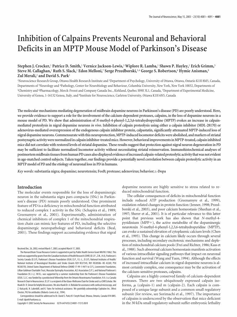
ResultsMPTP evokes sustained activation of calpains in the SNcWe first determined whether calpains may be activated in SNcneurons after chronic MPTP treatment in mice. Using an anti-body that selectively recognizes an epitope within ��spectrinthat has been exposed by calpain proteolysis (neoepitope)(Roberts-Lewis et al., 1994), midbrain sections from mice treatedwith MPTP were analyzed for evidence of increased calpain-mediated proteolysis by immunohistochemistry. Mice that weretreated with saline did not exhibit any detectable increase in neo-epitope labeling (Fig. 2a), whereas a noticeable and sustainedincrease in calpain-mediated proteolysis was observed in the ni-gral region of mice that had been treated with MPTP (Fig. 2b,c).Closer examination of this region revealed that the pattern ofcalpain-cleaved ��spectrin was located predominantly in thecytoplasm and was punctate in appearance (Fig. 2e,f). This punc-tate appearance resembled calpain translocation to intracellularmembrane compartments in hippocampal neurons after isch-emia in vivo (Yamashima et al., 1998). Intracerebroventricularinfusion of the pharmacological calpain inhibitor MDL-28170prevented the emergence of immunoreactivity for calpain-cleaved �-spectrin after chronic MPTP administration (Fig. 2d).
To demonstrate that the observed increase in calpain-relatedactivity indeed was in nigral dopamine neurons, we performedimmunofluorescent double-labeling experiments using TH asthe dopaminergic marker. In midbrain tissues from untreatedmouse brains, calpain-cleaved ��spectrin immunostaining wasweak and diffusely cytoplasmic in nigral dopaminergic neurons(Fig. 2g,i). However, in tissues from mice treated with MPTP, thiscytoplasmic immunolocalization within dopamine neurons ex-hibited a bright speckled appearance that became progressivelymore intense by 14 d (Fig. 2h,j) after cessation of MPTPadministration.
Table 1. Age and postmortem interval (PMI) of matched human nigral tissuesamples
Sample number Disease condition Age PMI (hr)
1 Parkinson’s disease 63 2.72 Parkinson’s disease 83 5.53 Parkinson’s disease 73 8.24 Parkinson’s disease 78 13.5
Average 74.3 Average 7.55 Control 85 46 Control 76 5.47 Control 68 14.88 Control 65 17.39 Control 70 18
Average 72.8 Average 11.9
Table 2. Summary of clinical case histories of Parkinson’s disease cases used in ourstudy based on available records from Harvard Brain Bank
Casenumber
Age ofdiagnosis Duration of illness Medications Cause of death
1 59 4 years a,b,c,d,e,f Cardiopulmonary arrest2 68 15 years a,h,n,p,q,r,s,t,u,v Congestive heart failure
w,x,y,z,aa,bb,cc,ddee,ff,gg
3 45 28 years c,g,h,l,j,k,l,m Dehydration4 66 12 years a,n,o Epidural hematoma
Medications: a, Sinemet; b, Propanolol; c, Parlodel; d, Eldepryl; e, Prozac; f, Diphenhydramine; g, L-Dopa; h, Zantac;i, Ditropan; j, Symmetrel; k, Xanax; l, Halclon; m, Deprenyl; n, Permax; o, Docusate calcium; p, Amitripyline; q, Ativan;r, Bactroban; s, Blephamide; t, Opthalmic cormax; u, Dicloxacillin; v, Digoxin; w, Fentany; x, Furosemide; y, Ibupro-fen; z, Lanoxin; aa, Lasix; bb, morphine; cc, Olanzepine; dd, Prednisone; ee, Salicyclic acid; ff, Vasotec; gg, Zaroxlolyn.
Crocker et al. • Calpains and Parkinson’s Disease J. Neurosci., May 15, 2003 • 23(10):4081– 4091 • 4083
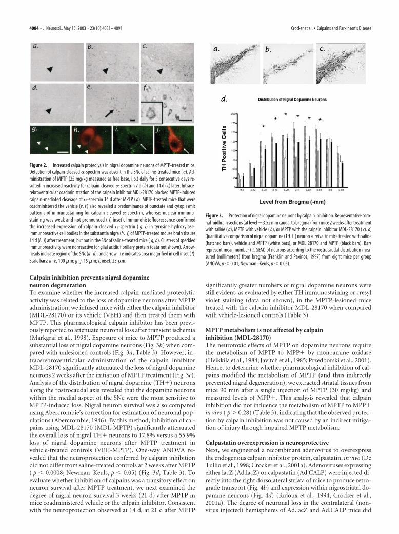
Calpain inhibition prevents nigral dopamineneuron degenerationTo examine whether the increased calpain-mediated proteolyticactivity was related to the loss of dopamine neurons after MPTPadministration, we infused mice with either the calpain inhibitor(MDL-28170) or its vehicle (VEH) and then treated them withMPTP. This pharmacological calpain inhibitor has been previ-ously reported to attenuate neuronal loss after transient ischemia(Markgraf et al., 1998). Exposure of mice to MPTP produced asubstantial loss of nigral dopamine neurons (Fig. 3b) when com-pared with unlesioned controls (Fig. 3a, Table 3). However, in-tracerebroventricular administration of the calpain inhibitorMDL-28170 significantly attenuated the loss of nigral dopamineneurons 2 weeks after the initiation of MPTP treatment (Fig. 3c).Analysis of the distribution of nigral dopamine (TH�) neuronsalong the rostrocaudal axis revealed that the dopamine neuronswithin the medial aspect of the SNc were the most sensitive toMPTP-induced loss. Nigral neuron survival was also comparedusing Abercrombie’s correction for estimation of neuronal pop-ulations (Abercrombie, 1946). By this method, inhibition of cal-pains using MDL-28170 (MDL-MPTP) significantly attenuatedthe overall loss of nigral TH� neurons to 17.8% versus a 55.9%loss of nigral dopamine neurons after MPTP treatment invehicle-treated controls (VEH-MPTP). One-way ANOVA re-vealed that the neuroprotection conferred by calpain inhibitiondid not differ from saline-treated controls at 2 weeks after MPTP( p � 0.0008; Newman–Keuls, p � 0.05) (Fig. 3d, Table 3). Toevaluate whether inhibition of calpains was a transitory effect onneuron survival after MPTP treatment, we next examined thedegree of nigral neuron survival 3 weeks (21 d) after MPTP inmice coadministered vehicle or the calpain inhibitor. Consistentwith the neuroprotection observed at 14 d, at 21 d after MPTP
significantly greater numbers of nigral dopamine neurons werestill evident, as evaluated by either TH immunostaining or cresylviolet staining (data not shown), in the MPTP-lesioned micetreated with the calpain inhibitor MDL-28170 when comparedwith vehicle-lesioned controls (Table 3).
MPTP metabolism is not affected by calpaininhibition (MDL-28170)The neurotoxic effects of MPTP on dopamine neurons requirethe metabolism of MPTP to MPP� by monoamine oxidase(Heikkila et al., 1984; Javitch et al., 1985; Przedborski et al., 2001).Hence, to determine whether pharmacological inhibition of cal-pains modified the metabolism of MPTP (and thus indirectlyprevented nigral degeneration), we extracted striatal tissues frommice 90 min after a single injection of MPTP (30 mg/kg) andmeasured levels of MPP�. This analysis revealed that calpaininhibition did not influence the metabolism of MPTP to MPP�in vivo ( p � 0.28) (Table 3), indicating that the observed protec-tion by calpain inhibition was not caused by an indirect mitiga-tion of injury through impaired MPTP metabolism.
Calpastatin overexpression is neuroprotectiveNext, we engineered a recombinant adenovirus to overexpressthe endogenous calpain inhibitor protein, calpastatin, in vivo (DeTullio et al., 1998; Crocker et al., 2001a). Adenoviruses expressingeither lacZ (Ad.lacZ) or calpastatin (Ad.CALP) were injected di-rectly into the right dorsolateral striata of mice to produce retro-grade transport (Fig. 4b) and expression within nigrostriatal do-pamine neurons (Fig. 4d) (Ridoux et al., 1994; Crocker et al.,2001a). The degree of neuronal loss in the contralateral (non-virus injected) hemispheres of Ad.lacZ and Ad.CALP mice did
Figure 2. Increased calpain proteolysis in nigral dopamine neurons of MPTP-treated mice.Detection of calpain-cleaved �-spectrin was absent in the SNc of saline-treated mice ( a). Ad-ministration of MPTP (25 mg/kg measured as free base, i.p.) daily for 5 consecutive days re-sulted in increased reactivity for calpain-cleaved �-spectrin 7 d ( b) and 14 d ( c) later. Intrace-rebroventricular coadministration of the calpain inhibitor MDL-28170 blocked MPTP-inducedcalpain-mediated cleavage of �-spectrin 14 d after MPTP ( d). MPTP-treated mice that werecoadministered the vehicle (e, f ) also revealed a predominance of punctate and cytoplasmicpatterns of immunostaining for calpain-cleaved �-spectrin, whereas nuclear immuno-staining was weak and not pronounced ( f, inset). Immunohistofluorescence confirmedthe increased expression of calpain-cleaved �-spectrin ( g, i) in tyrosine hydroxylase-immunoreactive cell bodies in the substantia nigra (h, j) of MPTP-treated mouse brain tissues14 d (i, j) after treatment, but not in the SNc of saline-treated mice ( g, h). Clusters of speckledimmunoreactivity were nonreactive for glial acidic fibrillary protein (data not shown). Arrow-heads indicate region of the SNc (a–d), and arrow in e indicates area magnified in cell inset ( f).Scale bars: a–e, 100 �m; g–j, 15 �m; f, inset, 25 �m.
Figure 3. Protection of nigral dopamine neurons by calpain inhibition. Representative coro-nal midbrain sections (at level�3.52 mm caudal to bregma) from mice 2 weeks after treatmentwith saline ( a), MPTP with vehicle ( b), or MPTP with the calpain inhibitor MDL-28170 ( c). d,Quantitative comparison of nigral dopamine (TH�) neuron survival in mice treated with saline(hatched bars), vehicle and MPTP (white bars), or MDL 28170 and MPTP (black bars). Barsrepresent mean number (�SEM) of neurons according to the rostrocaudal distribution mea-sured (millimeters) from bregma (Franklin and Paxinos, 1997) from eight mice per group(ANOVA, p � 0.01; Newman–Keuls, p � 0.05).
4084 • J. Neurosci., May 15, 2003 • 23(10):4081– 4091 Crocker et al. • Calpains and Parkinson’s Disease
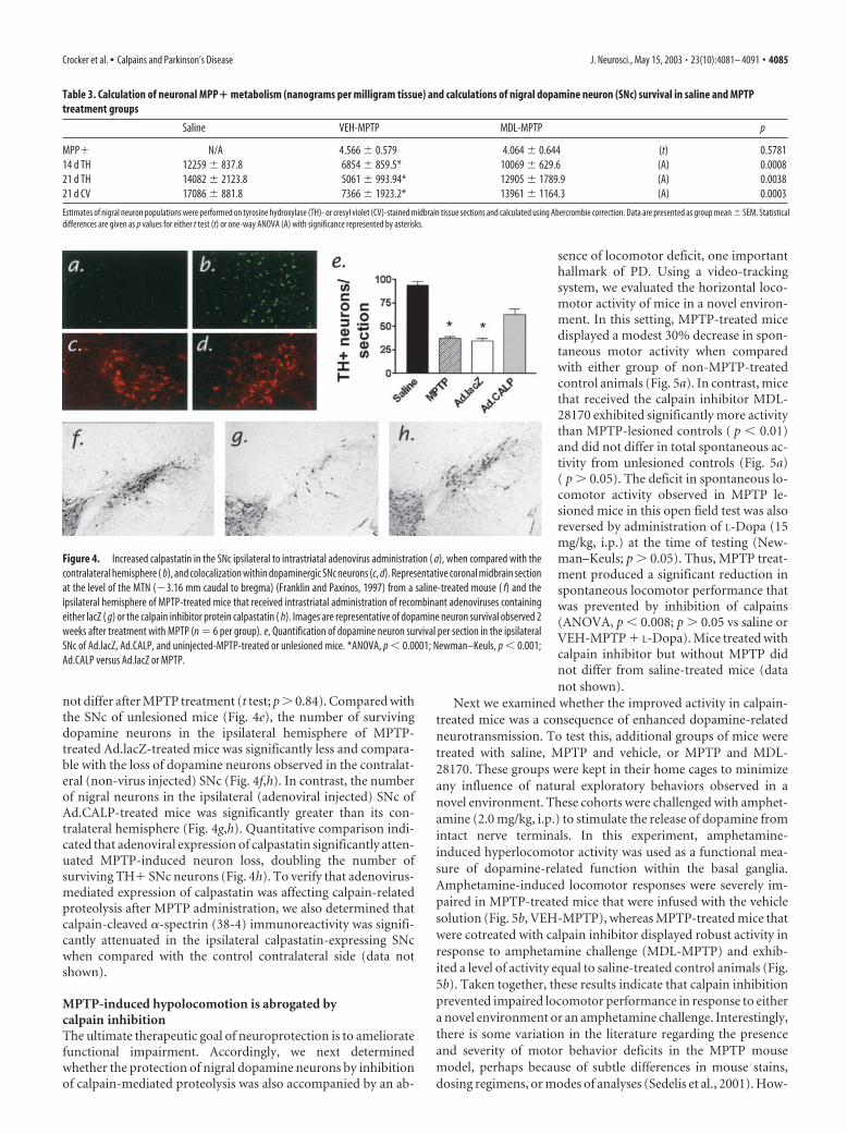
not differ after MPTP treatment (t test; p � 0.84). Compared withthe SNc of unlesioned mice (Fig. 4e), the number of survivingdopamine neurons in the ipsilateral hemisphere of MPTP-treated Ad.lacZ-treated mice was significantly less and compara-ble with the loss of dopamine neurons observed in the contralat-eral (non-virus injected) SNc (Fig. 4f,h). In contrast, the numberof nigral neurons in the ipsilateral (adenoviral injected) SNc ofAd.CALP-treated mice was significantly greater than its con-tralateral hemisphere (Fig. 4g,h). Quantitative comparison indi-cated that adenoviral expression of calpastatin significantly atten-uated MPTP-induced neuron loss, doubling the number ofsurviving TH� SNc neurons (Fig. 4h). To verify that adenovirus-mediated expression of calpastatin was affecting calpain-relatedproteolysis after MPTP administration, we also determined thatcalpain-cleaved �-spectrin (38-4) immunoreactivity was signifi-cantly attenuated in the ipsilateral calpastatin-expressing SNcwhen compared with the control contralateral side (data notshown).
MPTP-induced hypolocomotion is abrogated bycalpain inhibitionThe ultimate therapeutic goal of neuroprotection is to amelioratefunctional impairment. Accordingly, we next determinedwhether the protection of nigral dopamine neurons by inhibitionof calpain-mediated proteolysis was also accompanied by an ab-
sence of locomotor deficit, one importanthallmark of PD. Using a video-trackingsystem, we evaluated the horizontal loco-motor activity of mice in a novel environ-ment. In this setting, MPTP-treated micedisplayed a modest 30% decrease in spon-taneous motor activity when comparedwith either group of non-MPTP-treatedcontrol animals (Fig. 5a). In contrast, micethat received the calpain inhibitor MDL-28170 exhibited significantly more activitythan MPTP-lesioned controls ( p � 0.01)and did not differ in total spontaneous ac-tivity from unlesioned controls (Fig. 5a)( p � 0.05). The deficit in spontaneous lo-comotor activity observed in MPTP le-sioned mice in this open field test was alsoreversed by administration of L-Dopa (15mg/kg, i.p.) at the time of testing (New-man–Keuls; p � 0.05). Thus, MPTP treat-ment produced a significant reduction inspontaneous locomotor performance thatwas prevented by inhibition of calpains(ANOVA, p � 0.008; p � 0.05 vs saline orVEH-MPTP � L-Dopa). Mice treated withcalpain inhibitor but without MPTP didnot differ from saline-treated mice (datanot shown).
Next we examined whether the improved activity in calpain-treated mice was a consequence of enhanced dopamine-relatedneurotransmission. To test this, additional groups of mice weretreated with saline, MPTP and vehicle, or MPTP and MDL-28170. These groups were kept in their home cages to minimizeany influence of natural exploratory behaviors observed in anovel environment. These cohorts were challenged with amphet-amine (2.0 mg/kg, i.p.) to stimulate the release of dopamine fromintact nerve terminals. In this experiment, amphetamine-induced hyperlocomotor activity was used as a functional mea-sure of dopamine-related function within the basal ganglia.Amphetamine-induced locomotor responses were severely im-paired in MPTP-treated mice that were infused with the vehiclesolution (Fig. 5b, VEH-MPTP), whereas MPTP-treated mice thatwere cotreated with calpain inhibitor displayed robust activity inresponse to amphetamine challenge (MDL-MPTP) and exhib-ited a level of activity equal to saline-treated control animals (Fig.5b). Taken together, these results indicate that calpain inhibitionprevented impaired locomotor performance in response to eithera novel environment or an amphetamine challenge. Interestingly,there is some variation in the literature regarding the presenceand severity of motor behavior deficits in the MPTP mousemodel, perhaps because of subtle differences in mouse stains,dosing regimens, or modes of analyses (Sedelis et al., 2001). How-
Table 3. Calculation of neuronal MPP� metabolism (nanograms per milligram tissue) and calculations of nigral dopamine neuron (SNc) survival in saline and MPTPtreatment groups
Saline VEH-MPTP MDL-MPTP p
MPP� N/A 4.566 � 0.579 4.064 � 0.644 (t) 0.578114 d TH 12259 � 837.8 6854 � 859.5* 10069 � 629.6 (A) 0.000821 d TH 14082 � 2123.8 5061 � 993.94* 12905 � 1789.9 (A) 0.003821 d CV 17086 � 881.8 7366 � 1923.2* 13961 � 1164.3 (A) 0.0003
Estimates of nigral neuron populations were performed on tyrosine hydroxylase (TH)- or cresyl violet (CV)-stained midbrain tissue sections and calculated using Abercrombie correction. Data are presented as group mean � SEM. Statisticaldifferences are given as p values for either t test (t) or one-way ANOVA (A) with significance represented by asterisks.
Figure 4. Increased calpastatin in the SNc ipsilateral to intrastriatal adenovirus administration ( a), when compared with thecontralateral hemisphere ( b), and colocalization within dopaminergic SNc neurons (c, d). Representative coronal midbrain sectionat the level of the MTN (�3.16 mm caudal to bregma) (Franklin and Paxinos, 1997) from a saline-treated mouse ( f) and theipsilateral hemisphere of MPTP-treated mice that received intrastriatal administration of recombinant adenoviruses containingeither lacZ ( g) or the calpain inhibitor protein calpastatin ( h). Images are representative of dopamine neuron survival observed 2weeks after treatment with MPTP (n � 6 per group). e, Quantification of dopamine neuron survival per section in the ipsilateralSNc of Ad.lacZ, Ad.CALP, and uninjected-MPTP-treated or unlesioned mice. *ANOVA, p � 0.0001; Newman–Keuls, p � 0.001;Ad.CALP versus Ad.lacZ or MPTP.
Crocker et al. • Calpains and Parkinson’s Disease J. Neurosci., May 15, 2003 • 23(10):4081– 4091 • 4085
ever, our findings are consistent with the growing majority ofstudies reporting hypolocomotion.
Calpain inhibition and striatal denervationBecause calpain inhibition prevented the detrimental effects ofMPTP on locomotor performance, we next evaluated the statusof nigrostriatal function in groups of mice by assessing the ex-pression of various neurochemical and immunohistochemicalmarkers for dopaminergic neurotransmission. Immunohisto-chemical detection of TH fiber staining in striatal tissue sectionsfrom saline-treated (Fig. 6a), VEH-MPTP-treated (Fig. 6b), orMDL-MPTP-treated (Fig. 6c) mice revealed that MPTP-induceddepletion of nigrostriatal dopaminergic fibers was only slightlymitigated by treatment with the calpain inhibitor. Densitometricanalysis of striatal TH staining revealed a significant loss of THfibers in VEH-MPTP-treated mice and a lessened but significantloss in mice treated with calpain inhibitor (Fig. 6g). Althoughcalpain inhibition partially ameliorated the degree of loss of THfibrous staining, both groups of MPTP-treated mice were signif-icantly different from saline-treated controls. Finally, adjacenttissues sections assessed for expression of DAT revealed a similardegree of dopamine terminal loss as that detected by TH immu-nostaining in these groups (Fig. 6d–f).
To establish whether the modest DA terminal protection of-fered by calpain inhibition correlated with dopamine levels in the
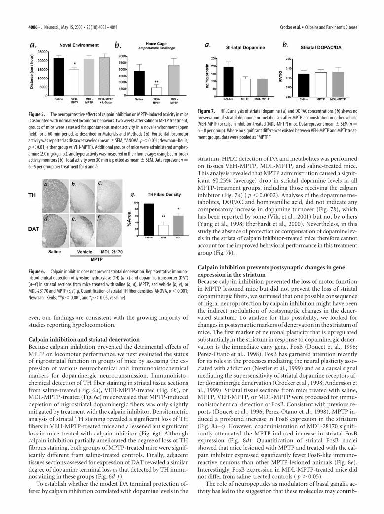
striatum, HPLC detection of DA and metabolites was performedon tissues VEH-MPTP, MDL-MPTP, and saline-treated mice.This analysis revealed that MPTP administration caused a signif-icant 60.25% (average) drop in striatal dopamine levels in allMPTP-treatment groups, including those receiving the calpaininhibitor (Fig. 7a) ( p � 0.0002). Analyses of the dopamine me-tabolites, DOPAC and homovanillic acid, did not indicate anycompensatory increase in dopamine turnover (Fig. 7b), whichhas been reported by some (Vila et al., 2001) but not by others(Yang et al., 1998; Eberhardt et al., 2000). Nevertheless, in thisstudy the absence of protection or compensation of dopamine lev-els in the striata of calpain inhibitor-treated mice therefore cannotaccount for the improved behavioral performance in this treatmentgroup (Fig. 7b).
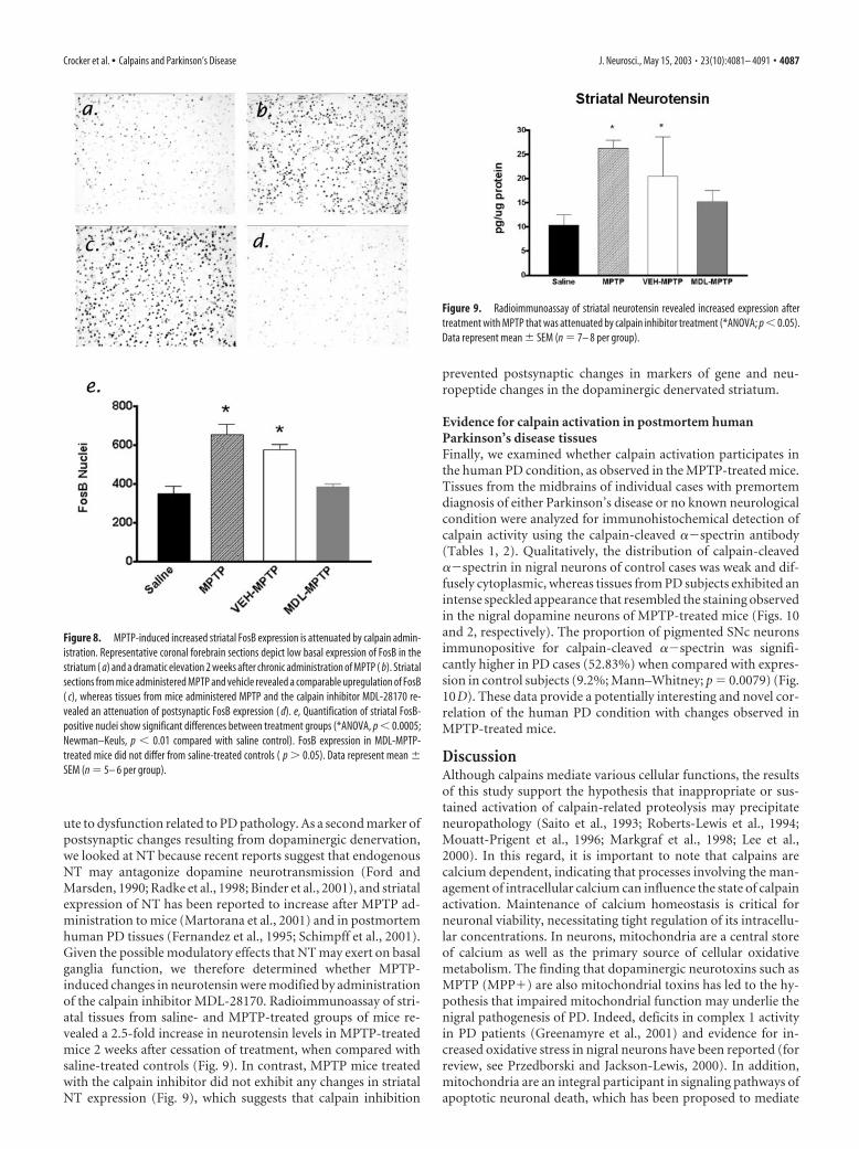
Calpain inhibition prevents postsynaptic changes in geneexpression in the striatumBecause calpain inhibition prevented the loss of motor functionin MPTP lesioned mice but did not prevent the loss of striataldopaminergic fibers, we surmised that one possible consequenceof nigral neuroprotection by calpain inhibition might have beenthe indirect modulation of postsynaptic changes in the dener-vated striatum. To analyze for this possibility, we looked forchanges in postsynaptic markers of denervation in the striatum ofmice. The first marker of neuronal plasticity that is upregulatedsubstantially in the striatum in response to dopaminergic dener-vation is the immediate early gene, FosB (Doucet et al., 1996;Perez-Otano et al., 1998). FosB has garnered attention recentlyfor its roles in the processes mediating the neural plasticity asso-ciated with addiction (Nestler et al., 1999) and as a causal signalmediating the supersensitivity of striatal dopamine receptors af-ter dopaminergic denervation (Crocker et al., 1998; Andersson etal., 1999). Striatal tissue sections from mice treated with saline,MPTP, VEH-MPTP, or MDL-MPTP were processed for immu-nohistochemical detection of FosB. Consistent with previous re-ports (Doucet et al., 1996; Perez-Otano et al., 1998), MPTP in-duced a profound increase in FosB expression in the striatum(Fig. 8a–c). However, coadministration of MDL-28170 signifi-cantly attenuated the MPTP-induced increase in striatal FosBexpression (Fig. 8d). Quantification of striatal FosB nucleishowed that mice lesioned with MPTP and treated with the cal-pain inhibitor expressed significantly fewer FosB-like immuno-reactive neurons than other MPTP-lesioned animals (Fig. 8e).Interestingly, FosB expression in MDL-MPTP-treated mice didnot differ from saline-treated controls ( p � 0.05).
The role of neuropeptides as modulators of basal ganglia ac-tivity has led to the suggestion that these molecules may contrib-
Figure 5. The neuroprotective effects of calpain inhibition on MPTP-induced toxicity in miceis associated with normalized locomotor behaviors. Two weeks after saline or MPTP treatment,groups of mice were assessed for spontaneous motor activity in a novel environment (openfield) for a 60 min period, as described in Materials and Methods ( a). Horizontal locomotoractivity was reported as distance traveled (mean� SEM; *ANOVA, p �0.001; Newman–Keuls,p � 0.01; either group vs VEH-MPTP). Additional groups of mice were administered amphet-amine (2.0 mg/kg, i.p.), and hyperactivity was measured in their home cages using beam-breakactivity monitors ( b). Total activity over 30 min is plotted as mean � SEM. Data represent n �6 –9 per group per treatment for a and b.
Figure 6. Calpain inhibition does not prevent striatal denervation. Representative immuno-histochemical detection of tyrosine hydroxylase (TH) (a–c) and dopamine transporter (DAT)(d–f ) in striatal sections from mice treated with saline (a, d), MPTP, and vehicle (b, e), orMDL-28170 and MPTP (c, f ). g, Quantification of striatal TH fiber densities (ANOVA, p � 0.001;Newman–Keuls, **p � 0.001, and *p � 0.05, vs saline).
Figure 7. HPLC analysis of striatal dopamine ( a) and DOPAC concentrations ( b) shows nopreservation of striatal dopamine or metabolism after MPTP administration in either vehicle(VEH-MPTP) or calpain inhibitor-treated (MDL-MPTP) mice. Data represent mean � SEM (n �6 – 8 per group). Where no significant differences existed between VEH-MPTP and MPTP treat-ment groups, data were pooled as “MPTP.”
4086 • J. Neurosci., May 15, 2003 • 23(10):4081– 4091 Crocker et al. • Calpains and Parkinson’s Disease
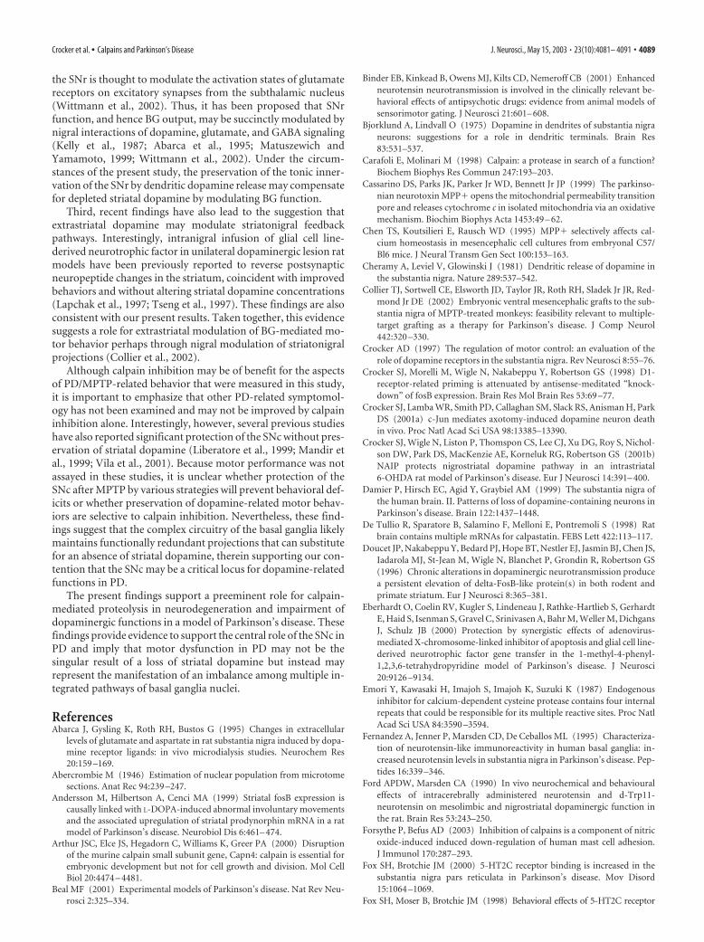
ute to dysfunction related to PD pathology. As a second marker ofpostsynaptic changes resulting from dopaminergic denervation,we looked at NT because recent reports suggest that endogenousNT may antagonize dopamine neurotransmission (Ford andMarsden, 1990; Radke et al., 1998; Binder et al., 2001), and striatalexpression of NT has been reported to increase after MPTP ad-ministration to mice (Martorana et al., 2001) and in postmortemhuman PD tissues (Fernandez et al., 1995; Schimpff et al., 2001).Given the possible modulatory effects that NT may exert on basalganglia function, we therefore determined whether MPTP-induced changes in neurotensin were modified by administrationof the calpain inhibitor MDL-28170. Radioimmunoassay of stri-atal tissues from saline- and MPTP-treated groups of mice re-vealed a 2.5-fold increase in neurotensin levels in MPTP-treatedmice 2 weeks after cessation of treatment, when compared withsaline-treated controls (Fig. 9). In contrast, MPTP mice treatedwith the calpain inhibitor did not exhibit any changes in striatalNT expression (Fig. 9), which suggests that calpain inhibition
prevented postsynaptic changes in markers of gene and neu-ropeptide changes in the dopaminergic denervated striatum.
Evidence for calpain activation in postmortem humanParkinson’s disease tissuesFinally, we examined whether calpain activation participates inthe human PD condition, as observed in the MPTP-treated mice.Tissues from the midbrains of individual cases with premortemdiagnosis of either Parkinson’s disease or no known neurologicalcondition were analyzed for immunohistochemical detection ofcalpain activity using the calpain-cleaved ��spectrin antibody(Tables 1, 2). Qualitatively, the distribution of calpain-cleaved��spectrin in nigral neurons of control cases was weak and dif-fusely cytoplasmic, whereas tissues from PD subjects exhibited anintense speckled appearance that resembled the staining observedin the nigral dopamine neurons of MPTP-treated mice (Figs. 10and 2, respectively). The proportion of pigmented SNc neuronsimmunopositive for calpain-cleaved ��spectrin was signifi-cantly higher in PD cases (52.83%) when compared with expres-sion in control subjects (9.2%; Mann–Whitney; p � 0.0079) (Fig.10D). These data provide a potentially interesting and novel cor-relation of the human PD condition with changes observed inMPTP-treated mice.
DiscussionAlthough calpains mediate various cellular functions, the resultsof this study support the hypothesis that inappropriate or sus-tained activation of calpain-related proteolysis may precipitateneuropathology (Saito et al., 1993; Roberts-Lewis et al., 1994;Mouatt-Prigent et al., 1996; Markgraf et al., 1998; Lee et al.,2000). In this regard, it is important to note that calpains arecalcium dependent, indicating that processes involving the man-agement of intracellular calcium can influence the state of calpainactivation. Maintenance of calcium homeostasis is critical forneuronal viability, necessitating tight regulation of its intracellu-lar concentrations. In neurons, mitochondria are a central storeof calcium as well as the primary source of cellular oxidativemetabolism. The finding that dopaminergic neurotoxins such asMPTP (MPP�) are also mitochondrial toxins has led to the hy-pothesis that impaired mitochondrial function may underlie thenigral pathogenesis of PD. Indeed, deficits in complex 1 activityin PD patients (Greenamyre et al., 2001) and evidence for in-creased oxidative stress in nigral neurons have been reported (forreview, see Przedborski and Jackson-Lewis, 2000). In addition,mitochondria are an integral participant in signaling pathways ofapoptotic neuronal death, which has been proposed to mediate
Figure 8. MPTP-induced increased striatal FosB expression is attenuated by calpain admin-istration. Representative coronal forebrain sections depict low basal expression of FosB in thestriatum ( a) and a dramatic elevation 2 weeks after chronic administration of MPTP ( b). Striatalsections from mice administered MPTP and vehicle revealed a comparable upregulation of FosB( c), whereas tissues from mice administered MPTP and the calpain inhibitor MDL-28170 re-vealed an attenuation of postsynaptic FosB expression ( d). e, Quantification of striatal FosB-positive nuclei show significant differences between treatment groups (*ANOVA, p � 0.0005;Newman–Keuls, p � 0.01 compared with saline control). FosB expression in MDL-MPTP-treated mice did not differ from saline-treated controls ( p � 0.05). Data represent mean �SEM (n � 5– 6 per group).
Figure 9. Radioimmunoassay of striatal neurotensin revealed increased expression aftertreatment with MPTP that was attenuated by calpain inhibitor treatment (*ANOVA; p � 0.05).Data represent mean � SEM (n � 7– 8 per group).
Crocker et al. • Calpains and Parkinson’s Disease J. Neurosci., May 15, 2003 • 23(10):4081– 4091 • 4087

neuronal loss in PD (Cassarino et al., 1999;Hartmann et al., 2000; Mochizuki et al.,2001; Vila et al., 2001; Viswanath et al.,2001).
Several lines of evidence support thehypothesis of calcium-mediated pathol-ogy in PD: (1) MPP� evokes sustained in-creases of intracellular calcium in neurons(Frei and Richter, 1986; Chen et al., 1995;Sherer et al., 2001); (2) mitochondria fromPD patients exhibit a diminished capacityto sequester calcium (Sheehan et al.,1997); (3) nigral neurons that express thecalcium-binding protein, calbindin D, areless vulnerable to degeneration in PD, per-haps because of the ability to buffer in-creased levels of intracellular calcium(Yamada et al., 1990; Lavoie and Parent, 1991; Hirsch et al., 1997;McMahon et al., 1998; Damier et al., 1999); (4) calcium-regulating genes, such as nitric oxide synthase, have been impli-cated in dopamine neuron degeneration in PD (Schulz et al.,1995; Liberatore et al., 1999); and (5) energy disruption alone isinsufficient to explain the toxicity of MPP� (Nakamura et al.,2000). It is important to note that although the sustainedcalcium-mediated activation of calpains in mice in our study islikely a consequence of the mitochondrial toxicity of MPTP, inthe human cases of PD, calcium may arise from mitochondria orother possible sites. For instance, mitochondrial dysfunction re-sults in a paucity of energy (ATP) that could evoke release fromendoplasmic reticular stores (Mattson et al., 2000; Paschen andFrandsen, 2001) or modulate influx of calcium from extracellularsources (Greenamyre et al., 1999). Finally, we cannot rule out thepossibility that calpain regulation may occur through mecha-nisms other than an abberant calcium response. This is impor-tant because the physiological regulation of calpains is complex,involving numerous factors including calcium, phosphorylation,and nitrosylation (Sato and Kawashima, 2001; Shiraha et al.,2002; Forsythe and Befus, 2003). Although our results validate arole for calpains in PD, the identity of the essential calpain speciesinvolved in the loss of nigral dopamine neurons after MPTP isalso unresolved. It is important to note that although previouswork on postmortem human tissues has reported increased ex-pression of m-calpain in PD, it not presently clear whether MPTPalso recapitulates this selectivity of calpain involvement in mice.
The manner by which calpains mediate dopaminergic deathremains to be elucidated. Putative calpain substrates that havebeen associated with neurodegeneration in PD include c-Jun(Hirai et al., 1991; Saporito et al., 1999; Xia et al., 2001) and p53(Trimmer et al., 1996; Gonen et al., 1997). In general, the shorthalf-life of calpain substrates coupled with the restricted nature ofcalpain cleavage of these target proteins has led to the suggestionthat calpains act to modify rather than completely catabolize sub-strate proteins (Suzuki et al., 1992; Carafoli and Molinari, 1998).For instance, the transcriptional activity of c-Jun is significantlyreduced in the presence of activated calpain, suggesting that cal-pains may influence signal transduction by modulating gene ex-pression (Suzuki et al., 1992). In addition, the cyclin-dependentkinase 5 (cdk5) activating protein p35 has been reported to becleaved by calpains into a p25 isoform that results in unregulatedcdk5 activity and neurodegeneration (Patrick et al., 1999; Lee etal., 2000). Hence, various potential calpain substrates may di-rectly or indirectly contribute to calpain-related degeneration ofdopamine neurons.
Nigral management of basal ganglia functionA second aspect of this study pertinent to the understanding ofPD is that calpain inhibition conferred nigral neuroprotectionwithout replenishment of striatal dopamine yet prevented defi-cits in locomotor behavior in MPTP-treated mice and normal-ized markers of BG activity. Historically, the symptoms of Par-kinson’s disease have been considered to be a consequence ofdiminished striatal dopamine. Indeed, dopamine replacementtherapies have supported this concept. However, previous workhas also suggested that the actions of L-Dopa may not be re-stricted to surviving dopamine terminals in the striatum, but thesubstantia nigra may too be a significant site for the actions ofL-Dopa and dopaminergic regulation of movement (Robertsonand Robertson, 1988, 1989; Crocker, 1997; Fox et al., 1998).
It has long been recognized that nigral dopaminergic neuronsrelease dopamine not only from their axons projecting to thestriatum but also from their dendrites (Bjorklund and Lindvall,1975; Cheramy et al., 1981). Therefore dendro-dendritic releaseof dopamine by SNc neurons may be of particular importance interms of the function of the BG in PD (Robertson and Robertson,1989; Tseng et al., 1997; Fox et al., 1998; Gainetdinov et al., 1999;Collier et al., 2002). Results of this study present evidence tosuggest that preservation of nigral integrity may provide preser-vation of dopaminergic-related motor function after loss of stri-atal nerve terminals. Although the precise nature of the func-tional benefit associated with nigral neuroprotection by calpaininhibition is presently not known, there are several possibilitiespertaining to nigral dopaminergic regulation of BG function.
The first possibility involves the counterbalancing influencesof serotoninergic inputs to the substantia nigra pars reticulata(SNr). There is increasing evidence that dopaminergic denerva-tion after degeneration of nigrostriatal neurons results in in-creased serotoninergic innervation of the reticulostriatal pathway(Thibaut et al., 1995; Fox and Brotchie, 2000). Interestingly, thisinfluence can be attenuated by direct administration of 5HT2C
receptor antagonists into the SN (Fox et al., 1998). The functionof serotonin in this context is thought to exacerbate hypoloco-motor activity. This notion is exemplified in DAT-deficient micein which basal hyperactivity can be modulated by enhancing se-rotonergic activity, without affecting levels of striatal dopamine(Gainetdinov et al., 1999). In the present study, the prevention ofSNc degeneration by calpain inhibition may have preserved theintegrity of dopaminergic innervation of the SNr and thereinacted to circumvent a serotonin-mediated hypolocomotion.
A second possibility relates to dopaminergic modulation ofexcitatory innervation of the SNr. Dopaminergic innervation of
Figure 10. Immunohistochemical evidence of enhanced calpain activity in nigral neurons in postmortem human tissues. A,Immunohistochemical detection of calpain-cleaved �-spectrin (38-4) viewed at low (20�) magnification revealed intenseclusters of immunopositive staining in PD cases that were rarely observed in control cases. B, C, Confocal optical sectioning (0.6�m) of calpain-cleaved �-spectrin immunoreactivity in pigmented dopamine neurons from a case without neurological condi-tion ( B) and from a case with Parkinson’s disease ( C) further revealed the cytoplasmic speckled appearance of 38-4-immunopositive staining that colocalized with neuromelanin (insets). D, Quantification of the proportion of pigmented neurons persection that displayed speckled calpain-cleaved �-spectrin immunoreactivity from PD and control tissues revealed a significant increasein calpain activity in subjects with PD (**52.83 � 7.5 vs 9.17 � 3.1%; Mann–Whitney rank-sum test; p � 0.0079). Positive immuno-staining disappeared when the primary antibody was omitted (data not shown). Scale bars: A, 25 �m; B, C, 50 �m.
4088 • J. Neurosci., May 15, 2003 • 23(10):4081– 4091 Crocker et al. • Calpains and Parkinson’s Disease
the SNr is thought to modulate the activation states of glutamatereceptors on excitatory synapses from the subthalamic nucleus(Wittmann et al., 2002). Thus, it has been proposed that SNrfunction, and hence BG output, may be succinctly modulated bynigral interactions of dopamine, glutamate, and GABA signaling(Kelly et al., 1987; Abarca et al., 1995; Matuszewich andYamamoto, 1999; Wittmann et al., 2002). Under the circum-stances of the present study, the preservation of the tonic inner-vation of the SNr by dendritic dopamine release may compensatefor depleted striatal dopamine by modulating BG function.
Third, recent findings have also lead to the suggestion thatextrastriatal dopamine may modulate striatonigral feedbackpathways. Interestingly, intranigral infusion of glial cell line-derived neurotrophic factor in unilateral dopaminergic lesion ratmodels have been previously reported to reverse postsynapticneuropeptide changes in the striatum, coincident with improvedbehaviors and without altering striatal dopamine concentrations(Lapchak et al., 1997; Tseng et al., 1997). These findings are alsoconsistent with our present results. Taken together, this evidencesuggests a role for extrastriatal modulation of BG-mediated mo-tor behavior perhaps through nigral modulation of striatonigralprojections (Collier et al., 2002).
Although calpain inhibition may be of benefit for the aspectsof PD/MPTP-related behavior that were measured in this study,it is important to emphasize that other PD-related symptomol-ogy has not been examined and may not be improved by calpaininhibition alone. Interestingly, however, several previous studieshave also reported significant protection of the SNc without pres-ervation of striatal dopamine (Liberatore et al., 1999; Mandir etal., 1999; Vila et al., 2001). Because motor performance was notassayed in these studies, it is unclear whether protection of theSNc after MPTP by various strategies will prevent behavioral def-icits or whether preservation of dopamine-related motor behav-iors are selective to calpain inhibition. Nevertheless, these find-ings suggest that the complex circuitry of the basal ganglia likelymaintains functionally redundant projections that can substitutefor an absence of striatal dopamine, therein supporting our con-tention that the SNc may be a critical locus for dopamine-relatedfunctions in PD.
The present findings support a preeminent role for calpain-mediated proteolysis in neurodegeneration and impairment ofdopaminergic functions in a model of Parkinson’s disease. Thesefindings provide evidence to support the central role of the SNc inPD and imply that motor dysfunction in PD may not be thesingular result of a loss of striatal dopamine but instead mayrepresent the manifestation of an imbalance among multiple in-tegrated pathways of basal ganglia nuclei.
ReferencesAbarca J, Gysling K, Roth RH, Bustos G (1995) Changes in extracellular
levels of glutamate and aspartate in rat substantia nigra induced by dopa-mine receptor ligands: in vivo microdialysis studies. Neurochem Res20:159 –169.
Abercrombie M (1946) Estimation of nuclear population from microtomesections. Anat Rec 94:239 –247.
Andersson M, Hilbertson A, Cenci MA (1999) Striatal fosB expression iscausally linked with L-DOPA-induced abnormal involuntary movementsand the associated upregulation of striatal prodynorphin mRNA in a ratmodel of Parkinson’s disease. Neurobiol Dis 6:461– 474.
Arthur JSC, Elce JS, Hegadorn C, Williams K, Greer PA (2000) Disruptionof the murine calpain small subunit gene, Capn4: calpain is essential forembryonic development but not for cell growth and division. Mol CellBiol 20:4474 – 4481.
Beal MF (2001) Experimental models of Parkinson’s disease. Nat Rev Neu-rosci 2:325–334.
Binder EB, Kinkead B, Owens MJ, Kilts CD, Nemeroff CB (2001) Enhancedneurotensin neurotransmission is involved in the clinically relevant be-havioral effects of antipsychotic drugs: evidence from animal models ofsensorimotor gating. J Neurosci 21:601– 608.
Bjorklund A, Lindvall O (1975) Dopamine in dendrites of substantia nigraneurons: suggestions for a role in dendritic terminals. Brain Res83:531–537.
Carafoli E, Molinari M (1998) Calpain: a protease in search of a function?Biochem Biophys Res Commun 247:193–203.
Cassarino DS, Parks JK, Parker Jr WD, Bennett Jr JP (1999) The parkinso-nian neurotoxin MPP� opens the mitochondrial permeability transitionpore and releases cytochrome c in isolated mitochondria via an oxidativemechanism. Biochim Biophys Acta 1453:49 – 62.
Chen TS, Koutsilieri E, Rausch WD (1995) MPP� selectively affects cal-cium homeostasis in mesencephalic cell cultures from embryonal C57/Bl6 mice. J Neural Transm Gen Sect 100:153–163.
Cheramy A, Leviel V, Glowinski J (1981) Dendritic release of dopamine inthe substantia nigra. Nature 289:537–542.
Collier TJ, Sortwell CE, Elsworth JD, Taylor JR, Roth RH, Sladek Jr JR, Red-mond Jr DE (2002) Embryonic ventral mesencephalic grafts to the sub-stantia nigra of MPTP-treated monkeys: feasibility relevant to multiple-target grafting as a therapy for Parkinson’s disease. J Comp Neurol442:320 –330.
Crocker AD (1997) The regulation of motor control: an evaluation of therole of dopamine receptors in the substantia nigra. Rev Neurosci 8:55–76.
Crocker SJ, Morelli M, Wigle N, Nakabeppu Y, Robertson GS (1998) D1-receptor-related priming is attenuated by antisense-meditated “knock-down” of fosB expression. Brain Res Mol Brain Res 53:69 –77.
Crocker SJ, Lamba WR, Smith PD, Callaghan SM, Slack RS, Anisman H, ParkDS (2001a) c-Jun mediates axotomy-induced dopamine neuron deathin vivo. Proc Natl Acad Sci USA 98:13385–13390.
Crocker SJ, Wigle N, Liston P, Thomspon CS, Lee CJ, Xu DG, Roy S, Nichol-son DW, Park DS, MacKenzie AE, Korneluk RG, Robertson GS (2001b)NAIP protects nigrostriatal dopamine pathway in an intrastriatal6-OHDA rat model of Parkinson’s disease. Eur J Neurosci 14:391– 400.
Damier P, Hirsch EC, Agid Y, Graybiel AM (1999) The substantia nigra ofthe human brain. II. Patterns of loss of dopamine-containing neurons inParkinson’s disease. Brain 122:1437–1448.
De Tullio R, Sparatore B, Salamino F, Melloni E, Pontremoli S (1998) Ratbrain contains multiple mRNAs for calpastatin. FEBS Lett 422:113–117.
Doucet JP, Nakabeppu Y, Bedard PJ, Hope BT, Nestler EJ, Jasmin BJ, Chen JS,Iadarola MJ, St-Jean M, Wigle N, Blanchet P, Grondin R, Robertson GS(1996) Chronic alterations in dopaminergic neurotransmission producea persistent elevation of delta-FosB-like protein(s) in both rodent andprimate striatum. Eur J Neurosci 8:365–381.
Eberhardt O, Coelin RV, Kugler S, Lindeneau J, Rathke-Hartlieb S, GerhardtE, Haid S, Isenman S, Gravel C, Srinivasen A, Bahr M, Weller M, DichgansJ, Schulz JB (2000) Protection by synergistic effects of adenovirus-mediated X-chromosome-linked inhibitor of apoptosis and glial cell line-derived neurotrophic factor gene transfer in the 1-methyl-4-phenyl-1,2,3,6-tetrahydropyridine model of Parkinson’s disease. J Neurosci20:9126 –9134.
Emori Y, Kawasaki H, Imajoh S, Imajoh K, Suzuki K (1987) Endogenousinhibitor for calcium-dependent cysteine protease contains four internalrepeats that could be responsible for its multiple reactive sites. Proc NatlAcad Sci USA 84:3590 –3594.
Fernandez A, Jenner P, Marsden CD, De Ceballos ML (1995) Characteriza-tion of neurotensin-like immunoreactivity in human basal ganglia: in-creased neurotensin levels in substantia nigra in Parkinson’s disease. Pep-tides 16:339 –346.
Ford APDW, Marsden CA (1990) In vivo neurochemical and behaviouraleffects of intracerebrally administered neurotensin and d-Trp11-neurotensin on mesolimbic and nigrostriatal dopaminergic function inthe rat. Brain Res 53:243–250.
Forsythe P, Befus AD (2003) Inhibition of calpains is a component of nitricoxide-induced induced down-regulation of human mast cell adhesion.J Immunol 170:287–293.
Fox SH, Brotchie JM (2000) 5-HT2C receptor binding is increased in thesubstantia nigra pars reticulata in Parkinson’s disease. Mov Disord15:1064 –1069.
Fox SH, Moser B, Brotchie JM (1998) Behavioral effects of 5-HT2C receptor
Crocker et al. • Calpains and Parkinson’s Disease J. Neurosci., May 15, 2003 • 23(10):4081– 4091 • 4089

antagonism in the substantia nigra zona reticulata of the 6-hydroxy-dopamine-lesioned rat model of Parkinson’s disease. Exp Neurol 151:35–49.
Franklin KBJ, Paxinos G (1997) The mouse brain in stereotaxic coordinates.Toronto: Academic.
Frei B, Richter C (1986) N-methyl-4-phenylpyridine (MPP�) togetherwith 6-hydroxydopamine of dopamine stimulates Ca 2� release from mi-tochondria. FEBS Lett 198:99 –102.
Gainetdinov RR, Wetsel WC, Jones SR, Levin ED, Jaber M, Caron MG(1999) Role of serotonin in the paradoxical calming effect of psycho-stimulants on hyperactivity. Science 283:397– 401.
Gonen H, Shkedy D, Barnoy S, Kosower NS, Ciechanover A (1997) On theinvolvement of calpains in the degradation of the tumor suppressor pro-tein p53. FEBS Lett 406:17–22.
Goto K, Iwamoto T, Kondo H (1994) Localization of mRNAs for calpainand calpastatin in the adult rat brain by in situ hybridization histochem-istry. Brain Res Mol Brain Res 23:40 – 46.
Greenamyre JT, MacKenzie G, Peng TI, Stephans SE (1999) Mitochondrialdysfunction in Parkinson’s disease. Biochem Soc Symp 66:85–97.
Greenamyre JT, Sherer TB, Betarbet R, Panov AV (2001) Complex I andParkinson’s disease. IUBMB Life 52:135–141.
Hardy S, Kitamura M, Harris-Stansil T, Dai Y, Phipps ML (1997) Construc-tion of adenovirus vectors through Cre-lox recombination. J Virol71:1842–1849.
Hartmann A, Hunot S, Michel PP, Muriel MP, Vyas S, Faucheux BA, Mouatt-Prigent A, Turmel H, Srinivasan A, Ruberg M, Evan GI, Agid Y, Hirsch EC(2000) Caspase-3: a vulnerability factor and final effector in apoptoticdeath of dopaminergic neurons in Parkinson’s disease. Proc Natl Acad SciUSA 97:2875–2880.
Hartmann A, Troadec JD, Hunot S, Kikly K, Faucheux BA, Mouatt-Prigent A,Ruberg M, Agid Y, Hirsch EC (2001) Caspase-8 is an effector in apopto-tic death of dopaminergic neurons in Parkinson’s disease but pathwayinhibition results in neuronal necrosis. J Neurosci 21:2247–2255.
Hayley S, Brebner K, Lacosta S, Merali Z, Anisman H (1999) Sensitization tothe effects of tumor necrosis factor-�: neuroendocrine, central mono-amine, and behavioral variations. J Neurosci 19:5654 –5665.
Heikkila RE, Manzino L, Cabbat FS, Duvoisin RC (1984) Protection against thedopaminergic neurotoxicity of 1-methyl-4-phenyl-1,2,5,6-tetrahydropyri-dine by monoamine oxidase inhibitors. Nature 311:467–469.
Hirai S, Kawasaki H, Yaniv M, Suzuki K (1991) Degradation of transcrip-tion factors, c-Jun and c-Fos, by calpain. FEBS Lett 287:57– 61.
Hirsch E, Graybiel AM, Agid YA (1988) Melanized dopaminergic neuronsare differentially susceptible to degeneration in Parkinson’s disease. Na-ture 334:345–348.
Hirsch EC, Faucheux B, Damier P, Mouatt-Prigent A, Agid Y (1997) Neu-ronal vulnerability in Parkinson’s disease. J Neural Transm [Suppl]50:79 – 88.
Javitch JA, D’Amato RJ, Strittmatter SM, Snyder SH (1985) Parkinsonism-inducing neurotoxin, N-methyl-4-phenyl-1,2,3,6-tetrahydropyridine:uptake of the metabolite N-methyl-4-phenylpyridine by dopamine neu-rons explains selective toxicity. Proc Natl Acad Sci USA 82:2173–2177.
Jenner P (1998) Oxidative mechanisms in nigral cell death in Parkinson’sdisease. Mov Disord 13[Suppl 1]:24 –34.
Kass GE, Wright JM, Nicotera P, Orrenius S (1988) The mechanism of1-methyl-4-phenyl-1,2,3,6-tetrahydropyridine toxicity: role of intracel-lular calcium. Arch Biochem Biophys 260:789 –797.
Kelly E, Jenner P, Marsden CD (1987) Comparison of changes in locomotoractivity with striatal homovanillic acid and 3,4-dihydroxyphenylaceticacid concentrations following the bilateral intranigral injection of dopa-mine agonist drugs in rats. J Pharm Pharmacol 39:196 –202.
Lapchak PA, Miller PJ, Collins F, Jiao S (1997) Glial cell line-derived neuro-trophic factor attenuates behavioural deficits and regulates nigrostriataldopaminergic and peptidergic markers in 6-hydroxydopamine-lesionedadult rats: comparison of intraventricular and intranigral delivery. Neu-roscience 78:61–72.
Lavoie B, Parent A (1991) Dopaminergic neurons expressing calbindin innormal and parkinsonian monkeys. NeuroReport 2:601– 604.
Lee MS, Kwon YT, Li M, Peng J, Friedlander RM, Tsai LH (2000) Neuro-toxicity induces cleavage of p35 to p25 by calpain. Nature 405:360 –364.
Li J, Grynspan F, Berman S, Nixon R, Bursztajn S (1996) Regional Differ-ences in gene expression for calcium activated neutral proteases (calpains)and their endogenous inhibitor calpastatin in mouse brain and spinalcord. J Neurobiol 30:177–191.
Liberatore GT, Jackson-Lewis V, Vukosavic S, Mandir AS, Vila M, McAuliffeWG, Dawson VL, Dawson TM, Przedborski S (1999) Inducible nitricoxide synthase stimulates dopaminergic neurodegeneration in the MPTPmodel of Parkinson disease. Nat Med 5:1403–1409.
Mandir AS, Przedborski S, Jackson-Lewis V, Wang ZQ, Simbulan-RosenthalCM, Smulson ME, Hoffman BE, Guastella DB, Dawson VL, Dawson TM(1999) Poly(ADP-ribose) polymerase activation mediates 1-methyl-4-phenyl-1,2,3,6-tetrahydropyridine (MPTP)-induced parkinsonism. ProcNatl Acad Sci USA 96:5774 –5779.
Markgraf CG, Velayo NL, Johnson MP, McCarthy DR, Medhi S, Koehl JR,Chmielewski PA, Linnik MD (1998) Six-hour window of opportunityfor calpain inhibition in focal cerebral ischemia in rats. Stroke 29:152–158.
Martorana A, Fusco FR, Picconi B, Massa R, Bernardi G, Sancesario G (2001)Dopamine denervation induces neurotensin immunoreactivity in GABA-parvalbumin striatal neurons. Synapse 41:360 –362.
Mattson MP, LaFeria FM, Chan SL, Leissring MA, Shepel PN, Geiger JD(2000) Calcium signaling in the ER: its role in neuronal plasticity andneurodegenerative disorders. Trends Neurosci 23:222–229.
Matuszewich L, Yamamoto BK (1999) Modulation of GABA release by do-pamine in the substantia nigra. Synapse 32:29 –36.
McMahon A, Wong BS, Iacopino AM, Ng MC, Chi S, German DC (1998)Calbindin-D28k buffers intracellular calcium and promotes resistance todegeneration in PC12 cells. Brain Res Mol Brain Res 54:56 – 63.
Melloni E, De Tullio R, Averna M, Tedesco I, Salamino F, Sparatore B, Pon-tremoli S (1998) Properties of calpastatin forms in rat brain. FEBS Lett431:55–58.
Merali Z, Piggins H (1990) Effects of dopamine D1 and D2 receptor agonistsand antagonists on bombesin-induced behaviours. Eur J Pharmacol191:281–293.
Mochizuki H, Hayakawa H, Migita M, Shibata M, Tanaka R, Suzuki A,Shimo-Nakanishi Y, Urabe T, Yamada M, Tamayose K, Shimada T,Miura M, Mizuno Y (2001) An AAV-derived Apaf-1 dominant negativeinhibitor prevents MPTP toxicity as antiapoptotic gene therapy for Par-kinson’s disease. Proc Natl Acad Sci USA 98:10918 –10923.
Mouatt-Prigent A, Karlsson JO, Agid Y, Hirsch EC (1996) IncreasedM-calpain expression in the mesencephalon of patients with Parkinson’sdisease but not in other neurodegenerative disorders involving the mes-encephalon: a role in nerve cell death? Neuroscience 73:979 –987.
Nakamura K, Bindokas VP, Marks JD, Wright DA, Frim DM, Miller RJ, KangUJ (2000) The selective toxicity of 1-methyl-4-phenylpyridinium to do-paminergic neurons: the role of mitochondrial complex I and reactiveoxygen species revisited. Mol Pharmacol 58:271–278.
Nestler EJ, Kelz MB, Chen J (1999) DeltaFosB: a molecular mediator oflong-term neural and behavioral plasticity. Brain Res 835:10 –17.
Palkovits M, Brownstein MJ (1988) Maps and guide to microdissection ofthe rat brain. New York: Elsevier.
Paschen W, Frandsen A (2001) Endoplasmic reticulum dysfunction—acommon denominator for cell injury in acute and degenerative diseases ofthe brain? J Neurochem 79:719 –725.
Patrick GN, Zukerberg L, Nikolic M, de la Monte S, Dikkes P, Tsai LH (1999)Conversion of p35 to p25 deregulates Cdk5 activity and promotes neuro-degeneration. Nature 402:615– 622.
Perez-Otano I, Mandelzys A, Morgan JI (1998) MPTP-parkinsonism is ac-companied by persistent expression of a delta-FosB-like protein in dopa-minergic pathways. Brain Res Mol Brain Res 53:41–52.
Przedborski S, Jackson-Lewis V (2000) ROS and Parkinson’s disease: a viewto a kill. In: Free radicals in brain pathophysiology (Poli G, Cadenas E,Packer L, eds), pp 273–290. New York: Marcel Dekker.
Przedborski S, Jackson-Lewis V, Yokoyama R, Shibata T, Dawson VL, Daw-son TM (1996) Role of neuronal nitric oxide in 1-methyl-4-phenyl-1,2,3,6-tetrahydropyridine (MPTP)-induced dopaminergic neurotoxic-ity. Proc Natl Acad Sci USA 93:4565– 4571.
Przedborski S, Jackson-Lewis V, Naini AB, Jakowec M, Petzinger G, Miller R,Akram M (2001) The parkinsonian toxin 1-methyl-4-phenyl-1,2,3,6-tetrahydropyridine (MPTP): a technical review of its utility and safety.J Neurochem 76:1265–1274.
Przedborski S, Jackson-Lewis V, Vila M, Wu du C, Teismann P, Tieu K, ChoiDK, Cohen O (2003) Free radical and nitric oxide toxicity in Parkinson’sdisease. Adv Neurol 91:83–94.
Radke JM, Owens MJ, Ritchie JC, Nemeroff CB (1998) Atypical antipsy-
4090 • J. Neurosci., May 15, 2003 • 23(10):4081– 4091 Crocker et al. • Calpains and Parkinson’s Disease

chotic drugs selectively increase neurotensin efflux in dopamine terminalregions. Proc Natl Acad Sci USA 95:11462–11464.
Ridoux V, Robert JJ, Zhang X, Perricaudet M, Mallet J, La Gal La Salle G(1994) Adenoviral vectors as functional retrograde neuronal tracers.Brain Res 648:171–175.
Roberts-Lewis JM, Savage MJ, Marcy VR, Pinsker LR, Siman R (1994) Im-munolocalization of calpain 1-mediated spectrin degradation to vulner-able neurons in the ischemic gerbil brain. J Neurosci 14:3934 –3944.
Robertson GS, Robertson HA (1988) Evidence that the substantia nigra is asite of action for L-DOPA. Neurosci Lett 89:204 –208.
Robertson GS, Robertson HA (1989) Evidence that L-dopa-induced rota-tional behavior is dependent on both striatal and nigral mechanisms.J Neurosci 9:3326 –3331.
Saito K, Elce JS, Hamos JE, Nixon RA (1993) Widespread activation ofcalcium-activated neutral proteinase (calpain) in the brain in Alzheimerdisease: a potential molecular basis for neuronal degeneration. Proc NatlAcad Sci USA 90:2628 –2632.
Saporito MS, Brown EM, Miller MS, Carswell S (1999) CEP-1347/KT-7515,an inhibitor of c-jun N-terminal kinase activation, attenuates the1-methyl-4-phenyl tetrahydropyridine-mediated loss of nigrostriatal do-paminergic neurons in vivo. J Pharmacol Exp Ther 288:421– 427.
Sato K, Kawashima S (2001) Calpain function in the modulation of signaltransduction molecules. Biol Chem 382:743–751.
Schapira AH, Cooper JM, Dexter D, Jenner P, Clark JB, Marsden CD (1989)Mitochondrial complex I deficiency in Parkinson’s disease. Lancet1:1269.
Schimpff RM, Avard C, Fenelon G, Lhiaubet AM, Tenneze L, Vidailhet M, Ros-tene W (2001) Increased plasma neurotensin concentrations in patientswith Parkinson’s disease. J Neurol Neurosurg Psychiatry 70:784–786.
Schulz JB, Matthews RT, Beal MF (1995) Role of nitric oxide in neurode-generative diseases. Curr Opin Neurol 8:480 – 486.
Sedelis M, Schwarting RK, Huston JP (2001) Behavioral phenotyping of theMPTP mouse model of Parkinson’s disease. Behav Brain Res125:109 –125.
Sheehan JP, Swerdlow RH, Parker WD, Miller SW, Davis RE, Tuttle JB(1997) Altered calcium homeostasis in cells transformed by mitochon-dria from individuals with Parkinson’s disease. J Neurochem68:1221–1233.
Sherer TB, Trimmer PA, Borland K, Parks JK, Bennett JP Jr, Tuttle JB (2001)Chronic reduction in complex I function alters calcium signaling in SH-SY5Y neuroblastoma cells. Brain Res 891:94 –105.
Shiraha H, Glading A, Chou J, Jia Z, Wells A (2002) Activation of m-calpain(calpain II) by epidermal growth factor is limited by protein kinase Aphosphorylation of m-calpain. Mol Cell Biol 22:2716 –2727.
Sorimachi H, Ishiura S, Suzuki K (1997) Structure and physiological func-tion of calpains. Biochem J 328:721–732.
Suzuki K, Saido TC, Hirai S (1992) Modulation of cellular signals by cal-pain. Ann NY Acad Sci 674:218 –227.
Tatton NA, Kish SJ (1997) In situ detection of apoptotic nuclei in the substantianigra compacta of 1-methyl-4-phenyl-1,2,3,6-tetrahydropyridine-treatedmice using terminal deoxynucleotidyl transferase labelling and acridine or-ange staining. Neuroscience 77:1037–1048.
Thibaut F, Faucheux BA, Marquez J, Villares J, Menard JF, Agid Y, Hirsch EC(1995) Regional distribution of monoamine vesicular uptake sites in themesencephalon of control subjects and patients with Parkinson’s disease:a postmortem study using tritiated tetrabenazine. Brain Res 692:233–243.
Trimmer PA, Smith TS, Jung AB, Bennett JP Jr (1996) Dopamine neuronsfrom transgenic mice with a knockout of the p53 gene resist MPTP neu-rotoxicity. Neurodegeneration 5:233–239.
Tseng JL, Baetge EE, Zurn AD, Aebischer P (1997) GDNF reduces drug-induced rotational behavior after medial forebrain bundle transection bya mechanism not involving striatal dopamine. J Neurosci 17:325–333.
Vila M, Jackson-Lewis V, Vukosavic S, Djaldetti R, Liberatore G, Offen D, Kors-meyer SJ, Przedborski S (2001) Bax ablation prevents dopaminergic neuro-degeneration in the 1-methyl-4-phenyl-1,2,3,6-tetrahydropyridine mousemodel of Parkinson’s disease. Proc Natl Acad Sci USA 98:2837–2842.
Viswanath V, Wu Y, Boonplueang R, Chen S, Stevenson FF, Yantiri F, Yang L,Beal MF, Andersen JK (2001) Caspase-9 activation results in down-stream caspase-8 activation and bid cleavage in 1-methyl-4-phenyl-1,2,3,6-tetrahydropyridine-induced Parkinson’s disease. J Neurosci21:9519 –9528.
Wang KKW, Yuen P-W (1994) Calpain inhibition: an overview of its ther-apeutic potential. Trends Pharmacol 15:412– 419.
Wittmann M, Marino MJ, Conn PJ (2002) Dopamine modulates the func-tion of group II and group III metabotropic glutamate receptors in thesubstantia nigra pars reticulata. J Pharmacol Exp Ther 302:433– 441.
Xia XG, Harding T, Weller M, Bieneman A, Uney JB, Schulz JB (2001) Genetransfer of the JNK interacting protein-1 protects dopaminergic neuronsin the MPTP model of Parkinson’s disease. Proc Natl Acad Sci USA98:10433–10438.
Yamada T, McGeer PL, Baimbridge KG, McGeer EG (1990) Relative spar-ing in Parkinson’s disease of substantia nigra dopamine neurons contain-ing calbindin-D28K. Brain Res 526:303–307.
Yamashima T, Kohda Y, Tsuchiya K, Ueno T, Yamashita J, Yoshioka T, Komi-nami E (1998) Inhibition of ischaemic hippocampal neuronal death inprimates with cathepsin B inhibitor CA-074: a novel strategy for neuro-protection based on “calpain-cathepsin hypothesis”. Eur J Neurosci10:1723–1733.
Yang L, Matthews RT, Schulz JB, Klockgether T, Liao AW, Martinou JC,Penney Jr JB, Hyman BT, Beal MF (1998) 1-Methyl-4-phenyl-1,2,3,6-tetrahydropyride neurotoxicity is attenuated in mice overexpressingBcl-2. J Neurosci 18:8145– 8152.
Zimmerman UJ, Boring L, Pak JH, Mukerjee N, Wang KK (2000) The cal-pain small subunit gene is essential: its inactivation results in embryoniclethality. IUBMB Life 50:63– 68.
Crocker et al. • Calpains and Parkinson’s Disease J. Neurosci., May 15, 2003 • 23(10):4081– 4091 • 4091