Theoretical models of the ion channel structure of amyloid beta-protein
9
Biophysical Journal Volume 67 December 1994 2137-2145 Theoretical Models of the Ion Channel Structure of Amyloid ,8-Protein Stewart R. Durell,* H. Robert Guy,* Nelson Arispe,* Eduardo Rojas,$ and Harvey B. Pollardt *Laboratory of Mathematical Biology, National Cancer Institute, and *Lab of Cell Biology and Genetics, National Institutes of Diabetes and Digestive and Kidney Diseases, National Institutes of Health, Bethesda, Maryland 20892 USA ABSTRACT Theoretical methods are used to develop models for the ion channel structure of the membrane-bound amyloid /-protein. This follows recent observations that the /-protein forms cation-selective channels in lipid bilayers in vitro. Amyloid ,/-protein is the main component of the extracellular plaques in the brain that are characteristic of Alzheimer's disease. Based on the amino acid sequence and the unique environment of the membrane, the secondary structure of the 40-residue /-protein is predicted to form a /-hairpin followed by a helix-turn-helix motif. The channel structures were designed as aggregates of peptide subunits in identical conformations. Three types of models were developed that are distinguished by whether the pore is formed by the /3-hairpins, the middle helices, or by the more hydrophobic C-terminal helices. The latter two types can be converted back and forth by a simple conformational change, which would explain the variable conduction states observed for a single channel. It is also demonstrated how lipid headgroups could be incorporated into the pore lining, and thus affect the ion selectivity. The atomic-scale detail of the models make them useful for designing experiments to determine the real structure of the channel, and thus further the understanding of peptide channels in general. In addition, if /3-protein-induced channel activity is found to be the cause of cell death in Alzheimer's disease, then the models may be helpful in designing counteracting drugs. INTRODUCTION The occurrence of Alzheimer's disease is associated with vas- cular and neuronal damage in the brain, involving the formation of intraneuronal neurofibrillary tangles and the accumulation of amyloid /3-protein (A/3P) in extracellular amyloid plaques (Blessed et al., 1968; Neve et al., 1990; Katzman and Saitoh, 1991; Selkoe, 1991; McKee et al., 1991; Hardy and Higgins, 1992; Kosik, 1992). Accordingly, it has been suggested that A,P is an important neurotoxic principle in the development of the disorder, and many experiments have been devoted to testing this hypothesis (Goldgaber et al., 1981; Tanzi et al., 1987; Joachim et al., 1988; Goate et al., 1991; Pike et al., 1991; Barger et al., 1993). Two concepts have emerged. The first is that A,BP tox- icity may be synergistic with other agents, such as with the ex- citatory amino acid glutamate (Koh et al., 1990; Mattson et al., 1992), or by the binding to tachykinin receptors (Yankner et al., 1990; Kimura and Schubert, 1993). The second, although not exclusive, possibility is that A/P is toxic by itself (Neve et al., 1990; Yankner et al., 1990), perhaps by raising the intracellular concentration of calcium ions (Joseph and Han, 1992). Consis- tent with the latter is our recent observation that A(BP[1-40] forms "giant" cation-selective channels in lipid bilayers, with conduction values as large as 5 nS (Arispe et al., 1993a, b; Pollard et al., 1993). Considerable effort has focused on determining the solu- tion and solid-state structure of APP and its fragments to better understand the extracellular plaques observed in vivo (Muller-Hill and Beyreuther, 1989; Kirschner et al., 1987; Halverson et al., 1990; Hilbich et al., 1991; Barrow and Zagorski, 1991; Barrow et al., 1992; Burdick et al., 1992; Inouye et al., 1993). In solution, A,BP assumes different pro- portions of helix, /3-sheet, and random coil and different de- grees of aggregation depending on the environmental con- ditions. In the solid state, however, A,BP is generally characterized by ,B-sheet structures. Unfortunately, the rel- evance of these data to the conformation of A,BP in the mem- brane is not clear, because of the stratified hydrophilic/ hydrophobic environment of the bilayer. Instead, we based our structure predictions on the amino acid sequence and its relation to patterns observed in other membrane-bound and channel-forming peptides and proteins. For example, the am- phipathic pattern and turn sequence in the N-terminal region suggest that this part of the sequence forms a /3-hairpin. Like- wise, the hydrophobic character and transmembrane position of the C-terminal region of A/3P in the precursor protein (Kang et al., 1987) suggest that this part of the sequence forms a membrane-embedded a-helix. Previously, it has been demonstrated that similar ion channels are formed when A/3P[1-40] is incorporated into artificial lipid bilayers by either the liposome fusion technique or directly from solution (Arispe et al., 1993a, b; Pollard et al., 1993). Single channels are observed to convert within and between two con- duction ranges: 40-400 pS and 0.4-5.0 nS. Both of these "large" and "giant" sized channels are cation-selective; however, the "giant" channel is considerably less susceptible to blockage by tromethamine. Because of the suspected large physical size of the channel structures, the models were designed as aggregates of many membrane-bound A/3P subunits. The process used to develop the models is an extension of our work with other chan- nel forming peptides, which includes cecropin, magainin, 8-hemolysin, and pardaxin (Durell et al., 1992; Cruciani et al., 1992; Raghunathan et al., 1990; Lazarovici et al., 1992) and is a combination of theoretical principles with experimental data on the conductance of the channel. Three basic types of channel models were developed. These are distinguished by whether the pore is formed by a /3-barrel (comprised of the N-terminal /3-hairpins) or by a grouping of the middle and/or C-terminal helices. The latter two channel types are interconvertible by a simple conformational change. This alters the proportions of the pore and thus could explain the observed "spontaneous" conduction changes. To examine the 2137
Transcript of Theoretical models of the ion channel structure of amyloid beta-protein

Biophysical Journal Volume 67 December 1994 2137-2145
Theoretical Models of the Ion Channel Structure of Amyloid ,8-Protein
Stewart R. Durell,* H. Robert Guy,* Nelson Arispe,* Eduardo Rojas,$ and Harvey B. Pollardt*Laboratory of Mathematical Biology, National Cancer Institute, and *Lab of Cell Biology and Genetics, National Institutes of Diabetes andDigestive and Kidney Diseases, National Institutes of Health, Bethesda, Maryland 20892 USA
ABSTRACT Theoretical methods are used to develop models for the ion channel structure of the membrane-bound amyloid/-protein. This follows recent observations that the /-protein forms cation-selective channels in lipid bilayers in vitro. Amyloid,/-protein is the main component of the extracellular plaques in the brain that are characteristic of Alzheimer's disease. Basedon the amino acid sequence and the unique environment of the membrane, the secondary structure of the 40-residue /-proteinis predicted to form a /-hairpin followed by a helix-turn-helix motif. The channel structures were designed as aggregates ofpeptide subunits in identical conformations. Three types of models were developed that are distinguished by whether the poreis formed by the /3-hairpins, the middle helices, or by the more hydrophobic C-terminal helices. The latter two types can beconverted back and forth by a simple conformational change, which would explain the variable conduction states observed fora single channel. It is also demonstrated how lipid headgroups could be incorporated into the pore lining, and thus affect theion selectivity. The atomic-scale detail of the models make them useful for designing experiments to determine the real structureof the channel, and thus further the understanding of peptide channels in general. In addition, if /3-protein-induced channel activityis found to be the cause of cell death in Alzheimer's disease, then the models may be helpful in designing counteracting drugs.
INTRODUCTION
The occurrence of Alzheimer's disease is associated with vas-cular and neuronal damage in the brain, involving the formationof intraneuronal neurofibrillary tangles and the accumulation ofamyloid /3-protein (A/3P) in extracellular amyloid plaques(Blessed et al., 1968; Neve et al., 1990; Katzman and Saitoh,1991; Selkoe, 1991; McKee et al., 1991; Hardy and Higgins,1992; Kosik, 1992). Accordingly, it has been suggested that A,Pis an important neurotoxic principle in the development of thedisorder, and many experiments have been devoted to testing thishypothesis (Goldgaber et al., 1981; Tanzi et al., 1987; Joachimet al., 1988; Goate et al., 1991; Pike et al., 1991; Barger et al.,1993). Two concepts have emerged. The first is that A,BP tox-icity may be synergistic with other agents, such as with the ex-citatory amino acid glutamate (Koh et al., 1990; Mattson et al.,1992), or by the binding to tachykinin receptors (Yankner et al.,1990; Kimura and Schubert, 1993). The second, although notexclusive, possibility is that A/P is toxic by itself (Neve et al.,1990; Yankner et al., 1990), perhaps by raising the intracellularconcentration of calcium ions (Joseph and Han, 1992). Consis-tent with the latter is our recent observation that A(BP[1-40]forms "giant" cation-selective channels in lipid bilayers, withconduction values as large as 5 nS (Arispe et al., 1993a, b;Pollard et al., 1993).
Considerable effort has focused on determining the solu-tion and solid-state structure of APP and its fragments tobetter understand the extracellular plaques observed in vivo(Muller-Hill and Beyreuther, 1989; Kirschner et al., 1987;Halverson et al., 1990; Hilbich et al., 1991; Barrow andZagorski, 1991; Barrow et al., 1992; Burdick et al., 1992;Inouye et al., 1993). In solution, A,BP assumes different pro-portions of helix, /3-sheet, and random coil and different de-grees of aggregation depending on the environmental con-ditions. In the solid state, however, A,BP is generallycharacterized by ,B-sheet structures. Unfortunately, the rel-evance of these data to the conformation ofA,BP in the mem-
brane is not clear, because of the stratified hydrophilic/hydrophobic environment of the bilayer. Instead, we basedour structure predictions on the amino acid sequence and itsrelation to patterns observed in other membrane-bound andchannel-forming peptides and proteins. For example, the am-phipathic pattern and turn sequence in the N-terminal regionsuggest that this part of the sequence forms a /3-hairpin. Like-wise, the hydrophobic character and transmembrane positionof the C-terminal region of A/3P in the precursor protein(Kang et al., 1987) suggest that this part of the sequenceforms a membrane-embedded a-helix.
Previously, it has been demonstrated that similar ion channelsare formed when A/3P[1-40] is incorporated into artificial lipidbilayers by either the liposome fusion technique or directly fromsolution (Arispe et al., 1993a, b; Pollard et al., 1993). Singlechannels are observed to convert within and between two con-duction ranges: 40-400 pS and 0.4-5.0 nS. Both of these "large"and "giant" sized channels are cation-selective; however, the"giant" channel is considerably less susceptible to blockage bytromethamine. Because of the suspected large physical size ofthe channel structures, the models were designed as aggregatesof many membrane-bound A/3P subunits. The process used todevelop the models is an extension of our work with other chan-nel forming peptides, which includes cecropin, magainin,8-hemolysin, and pardaxin (Durell et al., 1992; Cruciani et al.,1992; Raghunathan et al., 1990; Lazarovici et al., 1992) and isa combination oftheoretical principles with experimental data onthe conductance of the channel.
Three basic types of channel models were developed. Theseare distinguished by whether the pore is formed by a /3-barrel(comprised of the N-terminal /3-hairpins) or by a grouping of themiddle and/or C-terminal helices. The latter two channel typesare interconvertible by a simple conformational change. Thisalters the proportions of the pore and thus could explain theobserved "spontaneous" conduction changes. To examine the
2137

Volume 67 December 1994
possible modes of peptide-membrane binding and channel for-mation, some models were developed with lipid molecules in-corporated into the structures. In this case, the specific confor-mations and identities of the lipid headgroups would affect theproperties of the pore. It is hoped that the ideas embodied in thesemodels will prove useful in designing tests of AB3P and otherpeptide channel structures. The coordinates are available fromthe corresponding authors upon request.
NH3-+ A @F Y g V XgV @ L V F F
1 5 10 15 20
A(5gV IG N GA I I G L M V G G V V-C0_2(M)25 30 40
FIGURE 1 Amino acid sequence of AP3P[1-40]. The circles indicate thecharged residues, the underscores indicate the polar-neutral residues, and theremaining residues are considered hydrophobic. The rectangles indicate pre-dicted turn regions.
MATERIALS AND METHODS
Because current methods cannot calculate accurately the free energy of theion channel models, which would include interactions between the peptides,lipids, solvent, and ions, a number of principles are used to construct struc-tures that are expected to be thermodynamically stable. The main principlerequires positioning the polar and nonpolar amino acid residues in eitherhydrophilic or hydrophobic environments, respectively. For the polar resi-dues, this means limiting contact to other polar residues, the lipid head-groups, and/or the solvent. This also results in maximizing the number ofstabilizing hydrogen bonds and salt-bridges. Likewise, the nonpolar residuesare limited to contact with other nonpolar residues and/or the alkyl chainsof the lipids. A second principle concerns the packing of the peptide sub-units, such that unrealistic atomic overlaps and internal cavities are avoided.This is achieved by optimizing the van der Waals energy.
The first assumption used in designing the models was that the channelsare formed from an assembly of A(3P[1-40] subunits. Otherwise, it is hardto imagine how a single A(3P molecule, which is not quite long enough toform two transmembrane helices, could form a channel structure responsiblefor such large observed conductances (up to 5 nS). A second assumption wasthat each subunit assumes the same conformation and that they are arrangedsymmetrically around the axis of the pore. Although not unexpected on
energetic grounds, this restriction also simplifies the modeling procedure byreducing the number of possible inter-subunit arrangements.
Once a basic structural motif was devised and built on the computer, an
iterative procedure of direct manipulation and automated energy minimi-zation was used to refine the models in accord with the principles describedabove. In particular, the Quanta computer program (Molecular Simulations,Inc., Waltham, MA) was used to adjust the torsional angles into the desiredbackbone and side-chain conformations, and the Minimization facility of theCHARMM computer program (Brooks et al., 1983; Molecular Simulations)was used to optimize the packing for that specific conformation (whichincludes removing atomic overlap and tuning the salt-bridges and hydrogenbonds). The minimization procedure typically consisted of 100 steps ofSteepest Descent followed by 300 steps ofAdopted Basis Newton-Raphson,utilizing the residue topology and PARM30 parameters provided with thecommercial version of CHARMM. The Image facility was used for theminimization of multimeric channel structures. It must be emphasized thatthe purpose of the automated energy minimization was simply to optimizethe packing within the local minimum associated with the starting confor-mation. No attempt was made to search for the "correct" conformation ofthe protein with minimization and/or dynamics procedures, which wouldrequire accurately representing the membrane and solvent environments andsufficiently sampling the extremely large conformational space. For thisreason, only a simple distance-dependent dielectric constant was used forthe electrostatic interactions.
The concepts for the channel structure assemblies were developed fromthe particular arrangement of secondary structure elements that we predictedfor Af3P[1-40] in a membrane/solvent environment, which is describedbelow. The sequence of A,BP used in the channel measurements is shownin Fig. 1. Except for the histidines, the circles indicate those residues thatwould be formally charged at the experimental conditions ofpH 7.0 and 7.4(Arispe et al., 1993a, b; Pollard et al., 1993). Because this is only slightlyabove the intrinsic pK. of the peptide-bound imidazole ring (-6.5-7.0), theactual charged states of the histidines would be strongly influenced bythe local environment. The underscores indicate the polar-neutral residues.The remaining residues are considered hydrophobic.
As pointed out by Kirschner et al. (1987), there are two regions withinthe first 28 residues of the sequence that have a high propensity for pro-ducing a turn in the 3-D structure (Chou and Fasman, 1978). These areindicated by rectangular boxes in Fig. 1. The next most striking feature isthat the first turn segment is flanked on both sides by a short stretch ofalternating charged and hydrophobic residues. For a peptide bound in themixed hydrophilic/hydrophobic environment of a membrane, this type ofamphipathic pattern suggests that the first 13 residues of the sequence forma (3-hairpin structure. The three-residue linker of the hairpin would likelyform some type of fy-turn structure (Sibanda et al., 1989; Rose et al., 1985).After the second turn segment (second rectangle), the remaining residues[28-40] are mostly nonpolar and are thus likely to be embedded in thehydrophobic core of the membrane. In this case, and despite the extra-flexible glycines, the sequence would likely adopt a helical conformationto form hydrogen bonds and thus reduce the destabilizing effect of thepeptide backbone partial charges. It should be pointed out, however, that acircular dichroism analysis of the AP3P[29-42] fragment in reduced polaritysolvent failed to detect appreciable amounts of helical secondary structure(Barrow et al., 1992). However, the hydroxyl-group-containing trifluoro-ethanol and hexafluoro-2-propanol solvents used in that study are still morepolar than the alkyl-chain core of a membrane. Furthermore, this C-terminalregion of A,BP has been identified as forming the outer half of the singletransmembrane domain of the amyloid precursor protein (Kang et al., 1987),which would generally be believed to have a helical conformation. Theprediction for this region also corresponds with the occurrence of an as-paragine at position 27, which is the most commonly observed residue foundat the N-terminal cap of helices in known protein structures (Richardson andRichardson, 1988). Another interesting feature is that in a helical confor-mation the four glycine residues at positions 29, 33, 37, and 38 would resultin an exposed band along one side of the helix. Finally, residues 16-24 werepostulated to form a second helix, which is in the middle of the sequence.This was based on the Chou-Fasman prediction values (Chou andFassman, 1978) and the general observation that hydrophobic segmentsform helices in membranes. In addition, the glycine at position 25, inthe second turn region, corresponds with the fact that this is the mostlikely type of residue to terminate a helix in known protein structures(Richardson and Richardson, 1988).
RESULTS
As described in Materials and Methods, Fig. 2 shows the basicsecondary structure predicted for a membrane-bound A3P[l-40] molecule. The blue and red letters indicate the positive andnegatively charged residues, respectively. As can be seen fromthe alternating pattern, the ,3-hairpin structure would be am-phipathic, with the hydrophilic E3, R5, Eli, and H13 residuesprojecting from one side, and the hydrophobic A2, F4, Y10, andV12 residues projecting from the other side. Following a smalllinker is the relatively short middle helix. Although hydrophobicin the center, this helix has a positively charged residue (K16)at the N-terminal end and two negatively charged residues (E22and D23) at the C-terminal end. Finally, the N27 residue forms
Biophysical Journal2138
Ion Channel Models of Amyloid 3-Protein
middle helix
hairpin
C - term Ihelix
Cc(..
FIGURE 2 Secondary structure prediction for a membrane-bound A(3Pmolecule. The red and yellow rectangles indicate the 13-strands of the hair-pin, and the large cyan and blue cylinders indicate the middle and C-terminalhelices, respectively. The blue and red letters indicate the positive and nega-tively charged residues, respectively. The four glycines in the C-terminalhelix are highlighted in yellow. (Although not clear in the figure, the secondhalf of the A2 residue is actually part of the (3-hairpin structure.)
the N-terminal cap of the more hydrophobic C-tenninal helix,which comprises the remainder of the sequence. As also seen inthe figure, the four glycine residues (G29, 33, 37, and 38) are
stacked above each other. Because these residues are devoid ofside chains, they expose the backbone atoms along one side ofthe helix.
Type I models
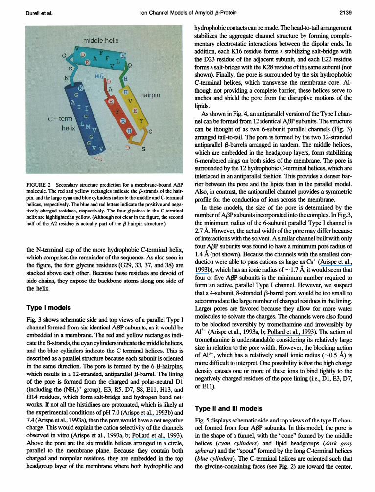
Fig. 3 shows schematic side and top views of a parallel Type Ichannel formed from six identical A(3P subunits, as it would beembedded in a membrane. The red and yellow rectangles indi-cate the (3-strands, the cyan cylinders indicate the middle helices,and the blue cylinders indicate the C-terminal helices. This isdescribed as a parallel structure because each subunit is orientedin the same direction. The pore is formed by the 6 (3-hairpins,which results in a 12-stranded, antiparallel (3-barrel. The liningof the pore is formed from the charged and polar-neutral Dl(including the (NH3)+ group), E3, R5, D7, S8, Ell, H13, andH14 residues, which form salt-bridge and hydrogen bond net-works. If not all the histidines are protonated, which is likely atthe experimental conditions ofpH 7.0 (Arispe et al., 1993b) and7.4 (Arispe et al., 1993a), then the pore would have a net negativecharge. This would explain the cation selectivity of the channelsobserved in vitro (Arispe et al., 1993a, b; Pollard et al., 1993).Above the pore are the six middle helices arranged in a circle,parallel to the membrane plane. Because they contain bothcharged and nonpolar residues, they are embedded in the topheadgroup layer of the membrane where both hydrophilic and
hydrophobic contacts can be made. The head-to-tail arrangementstabilizes the aggregate channel structure by forming comple-mentary electrostatic interactions between the dipolar ends. Inaddition, each K16 residue forms a stabilizing salt-bridge withthe D23 residue of the adjacent subunit, and each E22 residueforms a salt-bridge with the K28 residue of the same subunit (notshown). Finally, the pore is surrounded by the six hydrophobicC-tenminal helices, which transverse the membrane core. Al-though not providing a complete barrier, these helices serve toanchor and shield the pore from the disruptive motions of thelipids.As shown in Fig. 4, an antiparallel version of the Type I chan-
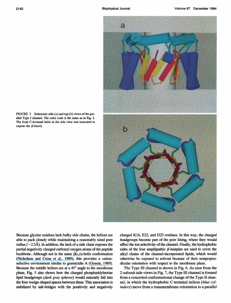
nel can be formed from 12 identical AA3P subunits. The structurecan be thought of as two 6-subunit parallel channels (Fig. 3)arranged tail-to-tail. The pore is formed by the two 12-strandedantiparallel (3-barrels arranged in tandem. The middle helices,which are embedded in the headgroup layers, form stabilizing6-membered rings on both sides of the membrane. The pore issurrounded by the 12 hydrophobic C-terminal helices, which areinterlaced in an antiparallel fashion. This provides a denser bar-rier between the pore and the lipids than in the parallel model.Also, in contrast, the antiparallel channel provides a symmetricprofile for the conduction of ions across the membrane.
In these models, the size of the pore is determined by thenumber ofA,BP subunits incorporated into the complex. In Fig.3,the minimum radius of the 6-subunit parallel Type I channel is2.7 A. However, the actual width of the pore may differ becauseof interactions with the solvent.A similar channel built with onlyfour A,BP subunits was found to have a minimum pore radius of1.4 A (not shown). Because the channels with the smallest con-duction were able to pass cations as large as Cs' (Arispe et al.,1993b), which has an ionic radius of -1.7 A, it would seem thatfour or five A,BP subunits is the minimum number required toform an active, parallel Type I channel. However, we suspectthat a 4-subunit, 8-stranded (-barrel pore would be too small toaccommodate the large number ofcharged residues in the lining.Larger pores are favored because they allow for more watermolecules to solvate the charges. The channels were also foundto be blocked reversibly by tromethamine and irreversibly byAl3+ (Arispe et al., 1993a, b; Pollard et al., 1993). The action oftromethamine is understandable considering its relatively largesize in relation to the pore width. However, the blocking actionof A13+, which has a relatively small ionic radius (-0.5 A) ismore difficult to interpret. One possibility is that the high chargedensity causes one or more of these ions to bind tightly to thenegatively charged residues of the pore lining (i.e., Dl, E3, D7,or Ell).
Type 11 and Ill models
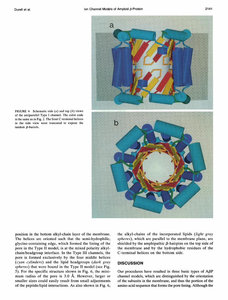
Fig. 5 displays schematic side and top views of the type II chan-nel formed from four A,BP subunits. In this model, the pore isin the shape of a funnel, with the "cone" formed by the middlehelices (cyan cylinders) and lipid headgroups (dark grayspheres) and the "spout" formed by the long C-terminal helices(blue cylinders). The C-terminal helices are oriented such thatthe glycine-containing faces (see Fig. 2) are toward the center.
Durell et al. 2139
Volume 67 December 1994
a
FIGURE 3 Schematic side (a) and top (b) views of the par-allel Type I channel. The color code is the same as in Fig. 2.The front C-terminal helix in the side view was truncated toexpose the ,3-barrel.
Because glycine residues lack bulky side chains, the helices areable to pack closely while maintaining a reasonably sized poreradius (-2.5A). In addition, the lack of a side chain exposes thepartial negatively charged carbonyl oxygen atoms of the peptidebackbone. Although not in the same KL,D)-helix confonnation(Nicholson and Cross et al., 1989), this provides a cation-selective environment similar to gramicidin A (Gennis, 1989).Because the middle helices are at a 450 angle to the membraneplane, Fig. 5 also shows how the charged phosphatidylserinelipid headgroups (dark gray spheres) would naturally fall intothe four wedge-shaped spaces between them. This association isstabilized by salt-bridges with the positively and negatively
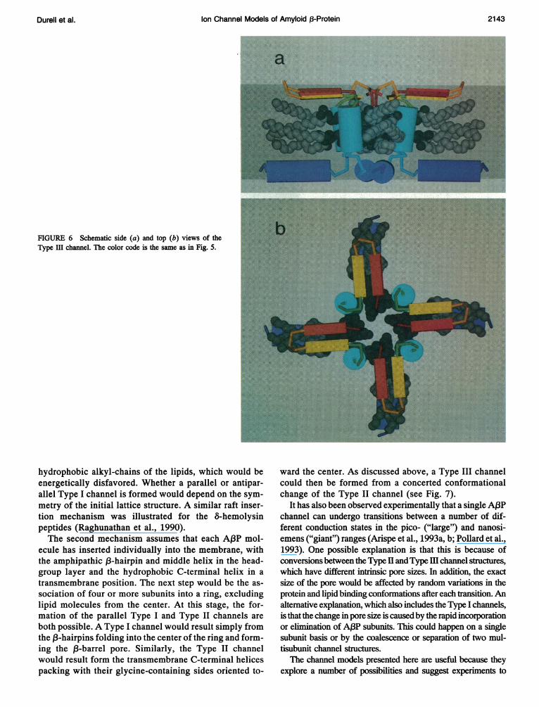
charged K16, E22, and D23 residues. In this way, the chargedheadgroups become part of the pore lining, where they wouldaffect the ion selectivity of the channel. Finally, the hydrophobicsides of the four amphipathic 13-hairpins are used to cover thealkyl chains of the channel-incorporated lipids, which wouldotherwise be exposed to solvent because of their nonperpen-dicular orientation with respect to the membrane plane.The Type III channel is shown in Fig. 6. As seen from the
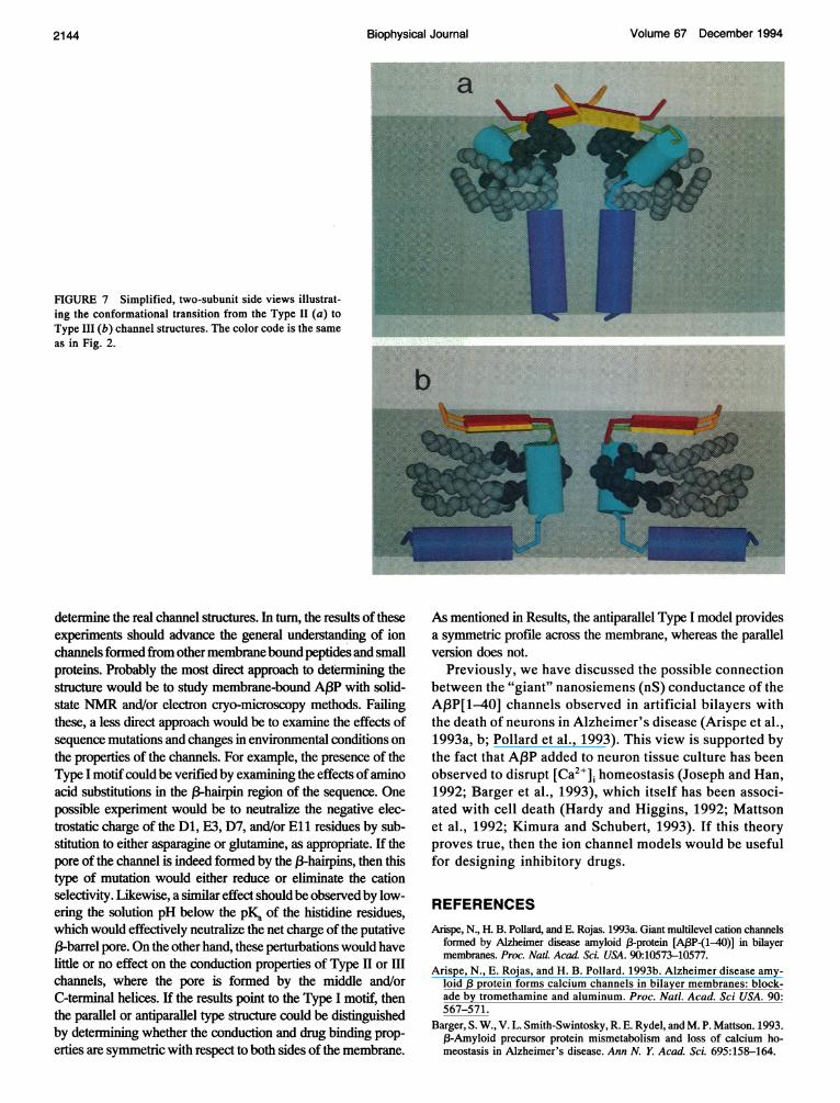
2-subunit side views in Fig. 7, the Type III channel is formedfrom a concerted conformational change of the Type II chan-nel, in which the hydrophobic C-terminal helices (blue cyl-inders) move from a transmembrane orientation to a parallel
2140 Biophysical Jlournal
Ion Channel Models of Amyloid 3-Protein
a
FIGURE 4 Schematic side (a) and top (b) viewsof the antiparallel Type I channel. The color codeis the same as in Fig. 2. The front C-terminal helicesin the side view were truncated to expose thetandem 1-barrels.
position in the bottom alkyl-chain layer of the membrane.The helices are oriented such that the semi-hydrophilic,glycine-containing edge, which formed the lining of thepore in the Type II model, is at the mixed polarity alkyl-chain/headgroup interface. In the Type III channels, thepore is formed exclusively by the four middle helices(cyan cylinders) and the lipid headgroups (dark grayspheres) that were bound in the Type II model (see Fig.5). For the specific structure shown in Fig. 6, the mini-mum radius of the pore is 3.0 A. However, larger orsmaller sizes could easily result from small adjustmentsof the peptide/lipid interactions. As also shown in Fig. 6,
the alkyl-chains of the incorporated lipids (light grayspheres), which are parallel to the membrane plane, areshielded by the amphipathic ,B-hairpins on the top side ofthe membrane and by the hydrophobic residues of theC-terminal helices on the bottom side.
DISCUSSION
Our procedures have resulted in three basic types of A,BPchannel models, which are distinguished by the orientationof the subunits in the membrane, and thus the portion of theamino acid sequence that forms the pore lining. Although the
2141Durell et al.
Volume 67 December 1994
a
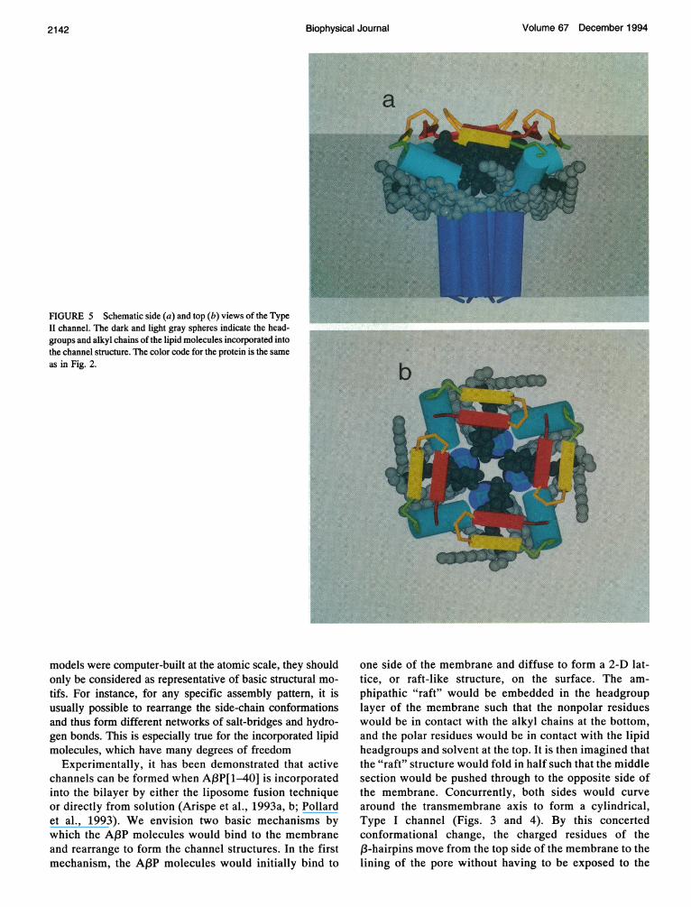
FIGURE 5 Schematic side (a) and top (b) views of the TypeII channel. The dark and light gray spheres indicate the head-groups and alkyl chains of the lipid molecules incorporated intothe channel structure. The color code for the protein is the sameas in Fig. 2.
models were computer-built at the atomic scale, they shouldonly be considered as representative of basic structural mo-tifs. For instance, for any specific assembly pattern, it isusually possible to rearrange the side-chain conformationsand thus form different networks of salt-bridges and hydro-gen bonds. This is especially true for the incorporated lipidmolecules, which have many degrees of freedom
Experimentally, it has been demonstrated that activechannels can be formed when AI3P[1-40] is incorporatedinto the bilayer by either the liposome fusion techniqueor directly from solution (Arispe et al., 1993a, b; Pollardet al., 1993). We envision two basic mechanisms bywhich the A3P molecules would bind to the membraneand rearrange to form the channel structures. In the firstmechanism, the Af3P molecules would initially bind to
one side of the membrane and diffuse to form a 2-D lat-tice, or raft-like structure, on the surface. The am-phipathic "raft" would be embedded in the headgrouplayer of the membrane such that the nonpolar residueswould be in contact with the alkyl chains at the bottom,and the polar residues would be in contact with the lipidheadgroups and solvent at the top. It is then imagined thatthe "raft" structure would fold in half such that the middlesection would be pushed through to the opposite side ofthe membrane. Concurrently, both sides would curvearound the transmembrane axis to form a cylindrical,Type I channel (Figs. 3 and 4). By this concertedconformational change, the charged residues of the3-hairpins move from the top side of the membrane to the
lining of the pore without having to be exposed to the
Biophysical Journal2142
Ion Channel Models of Amyloid j-Protein
FIGURE 6 Schematic side (a) and top (b) views of theType III channel. The color code is the same as in Fig. 5.
hydrophobic alkyl-chains of the lipids, which would beenergetically disfavored. Whether a parallel or antipar-allel Type I channel is formed would depend on the sym-metry of the initial lattice structure. A similar raft inser-tion mechanism was illustrated for the 8-hemolysinpeptides (Raghunathan et al., 1990).The second mechanism assumes that each Af3P mol-
ecule has inserted individually into the membrane, withthe amphipathic ,3-hairpin and middle helix in the head-group layer and the hydrophobic C-terminal helix in atransmembrane position. The next step would be the as-sociation of four or more subunits into a ring, excludinglipid molecules from the center. At this stage, the for-mation of the parallel Type I and Type II channels areboth possible. A Type I channel would result simply fromthe 13-hairpins folding into the center of the ring and form-ing the 13-barrel pore. Similarly, the Type II channelwould result form the transmembrane C-terminal helicespacking with their glycine-containing sides oriented to-
ward the center. As discussed above, a Type III channelcould then be formed from a concerted conformationalchange of the Type II channel (see Fig. 7).
It has also been observed experimentally that a single A13Pchannel can undergo transitions between a number of dif-ferent conduction states in the pico- ("large") and nanosi-emens ("giant") ranges (Arispe et al., 1993a, b; Pollard et al.,1993). One possible explanation is that this is because ofconversions between the TypeHandType HI channel structures,which have different intrinsic pore sizes. In addition, the exactsize of the pore would be affected by random variations in theprotein and lipid binding conformations after each transition. Analternative explanation, which also includes the Type I channels,is that the change in pore size is caused by the rapid incorporationor elimination of A1BP subunits. This could happen on a singlesubunit basis or by the coalescence or separation of two mul-tisubunit channel structures.The channel models presented here are useful because they
explore a number of possibilities and suggest experiments to
Durell et al. 2143
2144 Biophysical Journal Volume 67 December 1994
FIGURE 7 Simplified, two-subunit side views illustrat-ing the conformational transition from the Type 11 (a) to *l __Type III (b) channel structures. The color code is the sameas in Fig. 2.
determine the real channel structures. In turn, the results of theseexperiments should advance the general understanding of ionchannels formed from other membrane bound peptides and smallproteins. Probably the most direct approach to detenrining thestructure would be to study membrane-bound ABP with solid-state NMR and/or electron cryo-microscopy methods. Failingthese, a less direct approach would be to examine the effects ofsequence mutations and changes in environmental conditions onthe properties of the channels. For example, the presence of theType I motifcould be verified by examining the effects ofaminoacid substitutions in the 13-hairpin region of the sequence. Onepossible experiment would be to neutralize the negative elec-trostatic charge of the Dl, E3, D7, and/or Eli residues by sub-stitution to either asparagine or glutamine, as appropriate. If thepore of the channel is indeed formed by the 13-hairpins, then thistype of mutation would either reduce or eliminate the cationselectivity. Likewise, a similar effect should be observed by low-ering the solution pH below the pKa of the histidine residues,which would effectively neutralize the net charge of the putative13-barrel pore. On the other hand, these perturbations would havelittle or no effect on the conduction properties of Type H or IIIchannels, where the pore is formed by the middle and/orC-terminal helices. If the results point to the Type I motif, thenthe parallel or antiparallel type structure could be distinguishedby determining whether the conduction and drug binding prop-erties are symmetric with respect to both sides of the membrane.
As mentioned in Results, the antiparallel Type I model providesa symmetric profile across the membrane, whereas the parallelversion does not.
Previously, we have discussed the possible connectionbetween the "giant" nanosiemens (nS) conductance of theAPP[1-40] channels observed in artificial bilayers withthe death of neurons in Alzheimer's disease (Arispe et al.,1993a, b; Pollard et al., 1993). This view is supported bythe fact that Af3P added to neuron tissue culture has beenobserved to disrupt [Ca2"]i homeostasis (Joseph and Han,1992; Barger et al., 1993), which itself has been associ-ated with cell death (Hardy and Higgins, 1992; Mattsonet al., 1992; Kimura and Schubert, 1993). If this theoryproves true, then the ion channel models would be usefulfor designing inhibitory drugs.
REFERENCESArispe, N., H. B. Pollard, and E. Rojas. 1993a. Giant multilevel cation channels
formed by Alzheimer disease amyloid f3-protein [AIBP-(1-40)] in bilayermembranes. Proc. Natl. Acad. Sci. USA. 90:10573-10577.
Arispe, N., E. Rojas, and H. B. Pollard. 1993b. Alzheimer disease amy-loid ,B protein forms calcium channels in bilayer membranes: block-ade by tromethamine and aluminum. Proc. Natl. Acad. Sci USA. 90:567-571.
Barger, S. W., V. L. Smith-Swintosky, R. E. Rydel, and M. P. Mattson. 1993.13-Amyloid precursor protein mismetabolism and loss of calcium ho-meostasis in Alzheimer's disease. Ann N. Y. Acad. Sci. 695:158-164.
Durell et al. Ion Channel Models of Amyloid A-Protein 2145
Barrow, C. J., A. Yasuda, P. T. M. Kenny, and M. G. Zagorski. 1992.Solution conformations and aggregational properties of synthetic amyloid,B-peptides of Alzheimer's disease. J. MoL Bio. 225:1075-1093.
Barrow, C. J., and M. G. Zagorski. 1991. Solution structures of (3 peptideand its constituent fragments: Relation to amyloid deposition. Science.253:179-182.
Blessed, G., B. E. Tomlinson, and M. Roth. 1968. The association betweenquantitative measures of dementia and of senile change in the cerebralgrey matter of elderly subjects. Br. J. Psychiatry. 114:797-811.
Brooks, B. R., R. E. Bruccoleri, B. D. Olafson, D. J. States, S. Swaminathan,and M. Karplus. 1983. CHARMM: A program for macromolecular en-ergy, minimization, and dynamics calculations. J. Comp. Chem. 4:187-217.
Burdick, D., B. Soreghan, M. Kwon, J. Kosmoski, M. Knauer, A. Henschen,J. Yates, C. Cotman, and C. Glabe. 1992. Assembly and aggregationproperties of synthetic Alzheimer's A4/f3 amyloid peptide analogs.J. Biol. Chem. 267:546-554.
Chou, P. Y., and G. D. Fasman. 1978. Empirical predictions of proteinconformation. Ann. Rev. Biochem. 47:251-276.
Cruciani, R. A., J. L. Barker, S. R. Durell, G. Raghunathan, H. R. Guy, M.Zasloff, and E. F. Stanley. 1992. Magainin 2, a natural antibiotic from frogskin, forms ion channels in lipid bilayer membranes. Eur. J. Pharmacol.226:287-296.
Durell, S. R., G. Raghunathan, and H. R. Guy. 1992. Modeling the ionchannel structure of cecropin. Biophys. J. 63:1623-1631.
Gennis, R. B. 1989. Biomembranes: Molecular Structure and Function.Springer-Verlag, New York. 288-290.
Goate, A., M.-C. Chartier-Harlin, M. Mullan, J. Brown, F. Crawford, L.Fidani, L. Giuffra, A. Haynes, N. Irving, L. James, R. Mant, P. Newton,K. Rooke, P. Roques, C. Talbot, M. Pericak-Vance, A. Roses, R. Wil-liamson, M. Rossor, M. Owen, and J. Hardy. 1991. Segregation of amissense mutation in the amyloid precursor protein gene with familialAlzheimer's disease. Nature. 349:704-706.
Goldgaber, D., M. I. Lerman, 0. W. McBride, U. Saffiotti, and D. C. Ga-jdusek. 1987. Characterization and chromosomal localization of a cDNAencoding brain amyloid of Alzheimer's disease. Science. 235:877-880.
Halverson, K., P. E. Fraser, D. A. Kirschner, and P. T. Lansbury, Jr. 1990.Molecular determinants of amyloid deposition in Alzheimer's disease:conformational studies of synthetic (3-protein fragments. Biochemistry.29:2639-2644.
Hardy, J. A., and G. A. Higgins. 1992. Alzheimer's disease: the amyloidcascade hypothesis. Science. 256:184-185.
Hilbich, C., B. Kisters-Woike, J. Reed, C. L. Masters, and K. Beyreuther.1991. Aggregation and secondary structure of synthetic amyloid 13A4peptides of Alzheimer's disease. J. Mol. Biol. 218:149-163.
Inouye, H., P. E. Fraser, and D. A. Kirschner. 1993. Structure of (3-crystalliteassemblies formed by Alzheimer (3-amyloid protein analogues: analysisby x-ray diffraction. Biophys. J. 64:502-519.
Joachim, C. L., L K Duffy, J. H. Morris, and D. J. Selkoe. 1988. Protein chemi-cal and immunocytochemical studies of meningovascular ,3-amyloid proteinin Alzheimer's disease and normal aging. Brain Res. 474:100-111.
Joseph, R., and E. Han. 1992. Amyloid (3-protein fragment 25-35 causesactivation of cytoplasmic calcium in neurons. Biochem. Biophys. Res.Commun. 184:1441-1447.
Kang, J., H.-G. Lemaire, A. Unterbeck, J. M. Salbaum, C. L. Masters, K.-H.Grzeschik, G. Multhaup, K. Beyreuther, and B. Muller-Hill. 1987. Theprecursor of Alzheimer's disease amyloid A4 protein resembles a cell-surface receptor. Nature. 325:733-736.
Katzman, R., and T. Saitoh. 1991. Advances in Alzheimer's disease. FASEBJ. 5:278-286.
Kimura, H., and D. Schubert. 1993. Amyloid ,3-protein activates tachykininreceptors and inositol trisphosphate accumulation by synergy with glu-tamate. Proc. Natl. Acad. Sci. USA. 90:7508-7512.
Kirschner, D. A., H. Inouye, L. K. Duffy, A. Sinclair, M. Lind, and D. J.Selkoe. 1987. Synthetic peptide homologous to 13 protein from Alzheimerdisease forms amyloid-like fibrils in vitro. Proc. Natl. Acad. Sci. USA.84:6953-6957.
Koh, J.-Y., L. L. Yang, and C. W. Cotman. 1990. 13-amyloid protein in-creases the vulnerability of cultured cortical neurons to excitotoxic dam-age. Brain Res. 533:315-320.
Kosik, K. S. 1992. Alzheimer's disease: a cell biological perspective.Science. 256:780-783.
Lazarovici, P., C. Edwards, G. Raghunathan, and H. R. Guy. 1992. Sec-ondary structure, permeability and molecular modeling of pardaxin pores.J. Natural Toxins. 1:1-15.
Mattson, M. P., B. Cheng, D. Davis, K Bryant, I. Lieberburg, and R. E. Rydel.1992. 13-amyloid peptides destabilize calcium homeostasis and render humancortical neurons vulnerable to excitotoxicity. J. Neurosci. 12:376-389.
McKee, A. C., K. S. Kosik, and N. W. Kowall. 1991. Neuritic pathologyand dementia in Alzheimer's disease. Ann. Neurol. 30:156-165.
Muller-Hill, B., and K. Beyreuther. 1989. Molecular biology of Alzheimer'sdisease. Annu. Rev. Biochem. 58:287-307.
Neve, R. L., L. R. Dawes, B. A. Yankner, L. I. Benowitz, W. Rodriguez,and G. A. Higgins. 1990. Genetics and biology of the Alzheimer amyloidprecursor. Prog. Brain Res. 86:257-267.
Nicholson, L. K., and T. A. Cross. 1989. Gramicidin cation channel: anexperimental determination of the right-handed helix sense and verifi-cation of 13-type hydrogen bonding. Biochemistry. 28:9379-9385.
Pike, C. J., A. J. Walencewicz, C. G. Glabe, and C. W. Cotman. 1991. Invitro aging of 3-amyloid protein causes peptide aggregation and neuro-toxicity. Brain Res. 563:311-314.
Pollard, H. B., E. Rojas, and N. Arispe. 1993. A new hypothesis for themechanism of amyloid toxicity, based on the calcium channel activity ofamyloid 1 protein (A13P) in phospholipid bilayer membranes. Ann. N. Y.Acad. Sci. 695:165-168.
Raghunathan, G., P. Seetharamulu, B. R. Brooks, and H. R. Guy. 1990.Models of 8-hemolysin membrane channels and crystal structures.Proteins Struct. Funct. Genet. 8:213-225.
Richardson, J. S., and D. C. Richardson. 1988. Amino acid preferences forspecific locations at the ends of a helices. Science. 240:1648-1652.
Rose, G. D., L. M. Gierasch, and J. A. Smith. 1985. Turns in peptides andproteins. Adv. Protein Chem. 37:1-109.
Selkoe, D. J. 1991. The molecular pathology of Alzheimer's disease.Neuron. 6:487-498.
Sibanda, B. L., T. L. Blundell, and J. M. Thornton. 1989. Conformation of13-hairpins in protein structures: a systematic classification with appli-cations to modelling by homology, electron density fitting and proteinengineering. J. Mol. Biol. 206:759-777.
Tanzi, R. E., J. F. Gusella, P. C. Watkins, G. A. P. Bruns, P. St. George-Hyslop, M. L. Van Keuren, D. Patterson, S. Pagan, D. M. Kurnit, and R.L. Neve. 1987. Amyloid 13 protein gene: cDNA, mRNA distribution, andgenetic linkage near the Alzheimer locus. Science. 235:880-884.
Yankner, B. A., L. K. Duffy, and D. A. Kirschner. 1990. Neurotrophic andneurotoxic effects of amyloid 13 protein: reversal by tachykinin neuropep-tides. Science. 250:279-282.