Central oculomotor disturbances and nystagmus: a window into the brainstem and cerebellum
Pathology Associated with AAV Mediated Expression ofBeta Amyloid or C100 in Adult Mouse Hippocampus andCerebellumEleanor S. Drummond1,2*, Jill Muhling1, Ralph N. Martins2,3, Linda K. Wijaya3, Erich M. Ehlert4,
Alan R. Harvey1
1 School of Anatomy, Physiology and Human Biology, The University of Western Australia, Western Australia, Australia, 2 School of Psychiatry and Clinical Neurosciences,
The University of Western Australia, Western Australia, Australia, 3Centre of Excellence for Alzheimer’s Disease Research and Care, School of Exercise, Biomedical and
Health Sciences, Edith Cowan University, Western Australia, Australia, 4 Laboratory for Neuroregeneration, Netherlands Institute for Neuroscience, Royal Academy of Arts
and Sciences, Amsterdam, The Netherlands
Abstract
Accumulation of beta amyloid (Ab) in the brain is a primary feature of Alzheimer’s disease (AD) but the exact molecularmechanisms by which Ab exerts its toxic actions are not yet entirely clear. We documented pathological changes 3 and 6months after localised injection of recombinant, bi-cistronic adeno-associated viral vectors (rAAV2) expressing human Ab40-GFP, Ab42-GFP, C100-GFP or C100V717F-GFP into the hippocampus and cerebellum of 8 week old male mice. Injection of allrAAV2 vectors resulted in wide-spread transduction within the hippocampus and cerebellum, as shown by expression oftransgene mRNA and GFP protein. Despite the lack of accumulation of Ab protein after injection with AAV vectors, injectionof rAAV2-Ab42-GFP and rAAV2- C100V717F-GFP into the hippocampus resulted in significantly increased microgliosis andaltered permeability of the blood brain barrier, the latter revealed by high levels of immunoglobulin G (IgG) around theinjection site and the presence of IgG positive cells. In comparison, injection of rAAV2-Ab40-GFP and rAAV2-C100-GFP intothe hippocampus resulted in substantially less neuropathology. Injection of rAAV2 vectors into the cerebellum resulted insimilar types of pathological changes, but to a lesser degree. The use of viral vectors to express different types of Ab andC100 is a powerful technique with which to examine the direct in vivo consequences of Ab expression in different regions ofthe mature nervous system and will allow experimentation and analysis of pathological AD-like changes in a broader rangeof species other than mouse.
Citation: Drummond ES, Muhling J, Martins RN, Wijaya LK, Ehlert EM, et al. (2013) Pathology Associated with AAV Mediated Expression of Beta Amyloid or C100in Adult Mouse Hippocampus and Cerebellum. PLoS ONE 8(3): e59166. doi:10.1371/journal.pone.0059166
Editor: Gemma Casadesus, Case Western Reserve University, United States of America
Received April 29, 2012; Accepted February 13, 2013; Published March 13, 2013
Copyright: � 2013 Drummond et al. This is an open-access article distributed under the terms of the Creative Commons Attribution License, which permitsunrestricted use, distribution, and reproduction in any medium, provided the original author and source are credited.
Funding: This study was supported by funding from the School of Anatomy, Physiology and Human Biology, The University of Western Australia. ESD wassupported by an Australian Postgraduate Award and Jean Rogerson Postgraduate Scholarship. The funders had no role in study design, data collection andanalysis, decision to publish, or preparation of the manuscript.
Competing Interests: The authors have declared that no competing interests exist.
* E-mail: [email protected]
Introduction
There is a large body of evidence suggesting that beta amyloid
(Ab) accumulation in the brain may be a primary cause of
Alzheimer’s disease (AD). However, exactly how Ab contributes to
AD pathology and the mechanisms involved are still not yet fully
understood. The two main isoforms of Ab are Ab40 and Ab42.While it is widely accepted that Ab42 is more neurotoxic than
Ab40, surprisingly few studies have attempted to determine the
individual physiological and pathological effects directly attributed
to Ab40 and Ab42 in vivo and even fewer studies have directly
compared the effects of Ab40 and Ab42 expression [1,2]. It is
important to understand the physiological and pathological
functions of each Ab isoform as many proposed therapies for
AD act by modulating Ab levels [3,4,5,6].
It is difficult to determine the exact role of Ab in AD using the
current available animal models, which are predominantly trans-
genic mice. Therefore it was the aim of this study to develop an
alternative animal model for AD research using viral vectors to
initiate localised expression of human Ab. There are many benefits
associated with using viral vectors to develop animal models of
disease. For example, viral vectors can rapidly express transgene
proteins localised to desired brain regions, or even specific cell
types. Viral vectors can also induce transgene expression in many
species and at specific ages, hence preventing developmental or
other unwanted compensatory variables in response to life-long
transgene expression. Viral vectors also allow for the expression of
multiple genes with much greater ease than in transgenic mice,
a feature that is particularly important when studying a multi-
factorial disease such as AD.
To date, two studies have used viral vectors to directly express
Ab. The first study developed viral vectors that produced Ab40-BRI or Ab42-BRI fusion proteins [2]. The BRI protein is involved
in amyloid deposition in Familial British Dementia and Familial
Danish Dementia, and fusion of BRI to Ab results in enhanced
secretion of Ab-BRI fusion proteins. Expression of Ab42-BRIresulted in development of amyloid plaques at 3 months post-
injection and injection of a combination of BRI-Ab42 and BRI-
Ab40 vectors resulted in cognitive impairment. The second study
PLOS ONE | www.plosone.org 1 March 2013 | Volume 8 | Issue 3 | e59166
developed a LV-Ab42 vector, which was injected into the primary
motor cortex of rats [7]. While resulting brain pathology was only
examined at the early post-injection time point of 4 weeks, Ab42expression resulted in astrogliosis, increased TNF-a expression
and increased tau phosphorylation.
In the present study, recombinant bi-cistronic adeno-associated
viral vectors (rAAV2) were constructed, expressing either human
Ab40, Ab42, C100 or C100V717F. Direct expression of human
Ab40 and Ab42 was performed in an attempt to determine the
individual functions of each of these forms of Ab, while bypassingthe possible confounding functional effects of other APP fragments
such as sAPPa, N-APP, C100 and AICD, which are also produced
during APP processing [8,9,10,11,12]. Viral vectors expressing
C100, the immediate precursor to Ab, with and without the
Indiana mutation (V717F) were also developed in case Abexpression and/or functional effects were influenced by pro-
duction from a precursor protein. The Indiana mutation results in
preferential production of Ab42 rather than Ab40 [13,14], so this
strategy allowed for the indirect comparison of Ab40 and Ab42 by
comparing the downstream effects of C100 and C100V717F
expression. Each of these rAAV2 vectors was injected into the
adult mouse hippocampus and cerebellum, brain regions that are
vulnerable or relatively spared in AD respectively. rAAV2-GFP
and PBS injections served as controls. A targeted number of
changes associated with increased Ab were examined, including
glial reactivity (both astrogliosis and microgliosis), neuronal
degeneration, blood brain barrier disruption and expression of
apolipoprotein E (apoE), clusterin, synaptophysin and presenilin 1
(PS1).
Materials and Methods
Ethics StatementAll animal experimentation was approved by the UWA Animal
Ethics Committee and conformed to published NHMRC guide-
lines.
Plasmid and rAAV2 ConstructionrAAV2 vectors were generated from the pTRUF12 plasmid,
which contains two inverted terminal repeat sequences from AAV
serotype 2, the inserted transgene under the control of the
cytomegalovirus early enhancer-chicken beta actin (CMV-CAG)
promoter and an additional intron sequence with an enhancer
element. Four novel plasmids were generated containing transgene
sequences for Ab40, Ab42, C100 and C100V717F. The pTRUF12
plasmid without an inserted transgene was used as the GFP control
plasmid. An internal ribosome entry site (IRES) allowed the bi-
cistronic expression of the inserted transgene with enhanced GFP
(eGFP), resulting in transgene and GFP production as individual
proteins within each transduced cell. All plasmids were sequenced
to ensure that the transgene sequences were in the correct
orientation and that no unwanted mutations were present.
rAAV2 vectors were produced using a previously described
method [15]. An infection assay of HEK 293T cells was performed
to ensure infectivity and titres were determined by quantitative
PCR (qPCR) for viral genomic copies (GC) extracted from DNase-
treated viral particles. Titres ranged from 1.461012 to
4.161012 GC/ml.
In vitro TransfectionHEK 293T, PC12 (A.T.C.C. CRL-1721) and HeLa cells were
grown to approximately 90% confluency. HEK 293T and HeLa
cells were maintained in DMEM media (Sigma) containing 10%
fetal calf serum, 2 mM L-glutamine (Invitrogen), 100 Units/ml
penicillin (Invitrogen) and 100 mg/ml streptomycin (Invitrogen).
PC12 cells were maintained in DMEM media containing 5% fetal
calf serum, 10% horse serum, 2 mM L-glutamine, 100 Units/ml
penicillin, 100 mg/ml streptomycin and 0.1 mM non-essential
amino acids (Invitrogen). All cell types were transfected using 4 mgof plasmid DNA and 10 ml of Lipofectamine 2000 (Invitrogen)
according to the manufacturer’s instructions. Cells were examined
for GFP expression 24 and 48 hrs after transfection. Transfection
experiments were performed a minimum of three times for each
plasmid construct.
Media and cell lysates were collected for Western blot 48 hours
post-transfection. Cells were lysed with RIPA buffer (150 mM
NaCl, 50 mM Tris-HCl, 1% Triton-X100, 0.5% sodium deox-
ycholate, 0.1% SDS, 0.1 mM PMSF). Cell media was immuno-
precipitated for Ab using 6 E10 antibody (Signet) and Gamma-
Bind Plus Sepharose beads (Amersham/GE Healthcare). Western
blot was used to assay GFP, C100 and Ab levels in cell
homogenates and immunoprecipitated media. Briefly, between
25–200 mg of cell homogenate and immunoprecipitated media
was added to SDS loading buffer (166 mM Tris-HCl, 8% sodium
dodecylsulfate, 4% glycine, 2.5% 2-mercaptoethanol, pinch of
phenol red, pH 6.8), boiled at 95uC for 10 min and loaded onto
tris-trycine polyacrylamide gels. Proteins were separated using
electrophoresis and transferred to nitrocellulose membranes at
250 mA overnight at 4uC. Membranes were blocked with 5%
skim milk in TBS for 1 hr at room temperature and immuno-
blotted for GFP (1:3,000; Sigma) and C100/Ab (WO2; 1:2,500;
kindly provided by Professor Colin Masters at University of
Melbourne, Vic, Australia). Membranes were also blotted for b-actin (1:20,000; Abcam) to ensure consistent protein loading.
Protein bands were detected using enhanced chemiluminescence
(ECL; Amersham Biosciences) and developed onto film. 50 mg of
brain protein homogenate from APPSWE transgenic mouse brain
was used as a positive control for Ab detection.
Surgery and Tissue ProcessingEight week old male C57Bl6/J mice were anesthetised by an
intraperitoneal (i.p.) injection (10 ml/g) of a 1:1 mixture of
ketamine (100 mg/ml) and xylazine (20 mg/ml) that was diluted
1:10 in sterile saline. Mice were secured in a stereotaxic frame and
a small section of the skull was removed using a dental drill at the
co-ordinates of 1.9 mm rostral and 1.3 mm lateral relative to
lambda. For cerebellar injections a burr hole was drilled at
approximately 1 mm caudal to lambda and 2 mm lateral to the
midline. 1 ml of rAAV2 vector (1012 GC/ml) or vehicle (phosphate
buffered saline; PBS, pH 7.4) was directly injected into the
hippocampus (1.5 mm ventral from the dura) and cerebellum
using a pulled glass capillary at a rate of 0.2 ml/min. After each
injection, the glass capillary was left in place for an additional 2
minutes before withdrawal. The skull fragment was then replaced
and the skin closed using 4.0 sutures (Ethilon; Johnson & Johnson,
Australia). After each experimental procedure mice received
a subcutaneous injection of buprenorphine (0.02 mg/kg; Temge-
sic; Reckitt & Colman, Hull, UK) and an intramuscular injection
of Benacillin (25 ml, Troy Laboratories Pty. Ltd. Australia).
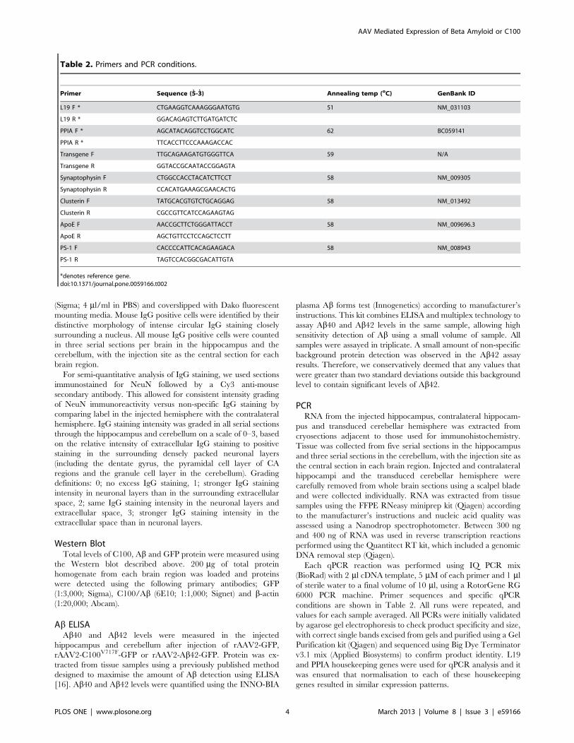
The number of animals included in each group and the type of
molecular analysis carried out at 3 months post-injection is shown
in Table 1. Analysis of cerebellar rAAV2-C100-GFP injections
was not possible due to problems with injection and thus consistent
transduction of neurons in the cerebellar cortex. rAAV2-GFP,
rAAV2-C100V717F-GFP and rAAV2-Ab42-GFP were used as
representative groups for further analysis at 6 months post-
injection.
AAV Mediated Expression of Beta Amyloid or C100
PLOS ONE | www.plosone.org 2 March 2013 | Volume 8 | Issue 3 | e59166

For cryosectioning, mice received an overdose of sodium
pentobarbitone (i.p.; Lethabarb, Virbac) and were transcardially
perfused with PBS (pH 7.4) containing 0.1% heparin followed by
4% paraformaldehyde in 0.1 M phosphate buffer (pH 7.4). Brains
were post-fixed in 4% paraformaldehyde for 2 hours, cryopro-
tected by sinking in 30% sucrose and incubated in increasing
concentrations of Jung freezing medium, all at 4uC. Brains werefrozen in 100% Jung Freezing medium by immersion into
isopentane on a bed of dry ice and stored at -80uC until
sectioning. Serial coronal cryosections were cut at a thickness of
25 mm for slides 1–13 of each series (for immunohistochemistry)
and a thickness of 14 mm for slides 14–15 of each series (for PCR),
resulting in each slide containing between 5–6 serial sections,
which were approximately 350 mm apart. Every section through
the hippocampus and cerebellum was collected.
For Western blot and ELISA, mice received an overdose of
sodium pentobarbitone (i.p.). The injected hippocampus, contra-
lateral hippocampus, cerebellum and rest of the brain were
collected individually, snap frozen in liquid nitrogen and stored at
280uC until homogenisation. Brain samples were homogenised in
a volume of ice-cold PBS containing protease inhibitor (Roche)
three times the weight of the tissue sample. Total protein was
quantified using the Micro BCA protein assay kit (Pierce) and
samples were stored at 280uC.
Immunohistochemistry and Density QuantificationPrimary antibodies used for immunohistochemistry were rabbit
anti-GFP (Millipore; 1:400), rabbit anti-GFAP (Sigma; 1:500),
mouse anti-NeuN (Chemicon; 1:500), rabbit anti-IBA-1 (Wako;
1:1000), rabbit anti-MAP2 (Millipore; 1:500) and rabbit CT20
(Calbiochem; 1:16,000). Secondary antibodies used were anti-
rabbit Alexa 488 (Invitrogen; 1:400), anti-mouse Cy3 (Jackson;
1:300) and anti-rabbit Cy3 (Jackson; 1:300).
For immunohistochemistry, cryosections were washed with
PBS, blocked for 1 hr in blocking buffer (PBS; 10% normal goat
serum; 0.2% Triton-X100) and incubated overnight (4uC) withprimary antibodies diluted in blocking buffer. Sections were
washed with PBS, incubated with secondary antibodies diluted in
blocking buffer for 2 hrs at room temperature, counterstained with
Hoechst 33342 (Sigma; 4 ml/ml in PBS) and coverslipped with
Dako fluorescent mounting media. Images were collected using
a multiphoton laser scanning confocal microscope.
Immunoreactivity for GFAP, IBA-1 and MAP2 was semi-
quantified as staining density in the injected hippocampus and
cerebellum. Low magnification confocal images were collected of
the injected and contralateral hippocampi and cerebellar hemi-
spheres. Three serial sections per brain in each brain region were
analysed, the central section containing the injection site to
maintain consistency between animals. Images were collected
using the lowest magnification that allowed for adequate
visualisation of immuno-positive staining. Thus, GFAP images
were collected at 56magnification, IBA-1 images were collected at
106magnification and MAP2 images were collected at 206mag-
nification. Because of the higher magnification necessary to image
MAP2 staining, images of the hippocampus were collected from
both the dentate gyrus and CA3 to ensure that the entire
transduced region was imaged. Every image for each immuno-
histochemistry stain was collected using the same imaging
conditions.
GFAP, IBA-1 and MAP2 staining density was quantified using
ImageJ analysis software (NIH). The area for quantification of
hippocampal staining was defined by the hippocampal anatomical
boundaries and density was measured in the injected and
contralateral hippocampi. In the cerebellum the analysis region
was defined by a rectangular area containing the GFP positive
transduced region in the injected hemisphere and the correspond-
ing region of the same area in the contralateral hemisphere. The
staining density of the injected hippocampus and cerebellar
hemisphere was calculated as a percentage of the corresponding
contralateral hippocampus or cerebellar hemisphere and averaged
across three sections per brain region. While it is acknowledged
that injection of rAAV2 vectors could cause distal changes in
contralateral brain regions, which would not be accounted for
using this quantification method, the staining density was
calculated this way in an attempt to standardise staining intensity
across multiple immunohistochemistry runs. Furthermore, general
observation of immunoreactivity in the contralateral hemisphere
showed minimal distal effects and no individual differences
between rAAV2 vectors and vehicle control, suggesting that
contralateral differences would not bias results.
Ab ImmunohistochemistryMultiple antibodies against Ab were trialled including; anti-
mouse 6E10 (Signet), anti-rabbit pan Ab (Zymed), anti-mouse 4G8
(Signet) and biotinylated Ab40 and Ab42 antibodies (gifts from Dr.
Pankaj Mehta, NYS Institute for Basic Research, New York,
USA). Extensive optimisation of the immunohistochemistry pro-
tocol was also performed, trialling heat and formic acid antigen
retrieval methods either alone or in combination, multiple
blocking reagents including 10% goat serum, 20% fetal bovine
serum, 0.2% bovine serum albumin and mouse-on-mouse
blocking reagent (M.O.M; Vector Laboratories) and both fluores-
cent and peroxidase immunohistochemistry. Positive control tissue
sections from 18 month old APPSWE transgenic mice were used in
every Ab immunohistochemistry trial.
IgG Staining and QuantificationMouse IgG was stained by incubation with anti-mouse
secondary antibodies. Briefly, sections were washed with PBS,
blocked for 1 hr in blocking buffer (PBS; 10% normal goat serum;
0.2% Triton-X100) and incubated with anti-mouse secondary
antibodies diluted in blocking buffer for 2 hrs at room temperature
(anti-mouse Cy3; 1:300; Jackson or anti-mouse FITC; 1:100; MP
Biochem). Sections were counterstained with Hoechst 33342
Table 1. Animal experimental groups used for differentanalysis techniques.
Vector injected n Post-injection time Analysis techniques
AAV2-C100-GFP 4 3 months Immunohistochemistry/PCR
AAV2-C100V717F-GFP 8 3 months Immunohistochemistry/PCR
AAV2- Ab40-GFP 4 3 months Immunohistochemistry/PCR
AAV2- Ab42-GFP 4 3 months Immunohistochemistry/PCR
AAV2-GFP 7 3 months Immunohistochemistry/PCR
PBS 4 3 months Immunohistochemistry/PCR
AAV2-C100V717F-GFP 4 3 months Western blot
AAV2- Ab42-GFP 4 3 months Western blot
AAV2-GFP 4 3 months Western blot
AAV2-C100V717F-GFP 4 6 months Immunohistochemistry/PCR
AAV2- Ab42-GFP 4 6 months Immunohistochemistry/PCR
AAV2-GFP 4 6 months Immunohistochemistry/PCR
doi:10.1371/journal.pone.0059166.t001
AAV Mediated Expression of Beta Amyloid or C100
PLOS ONE | www.plosone.org 3 March 2013 | Volume 8 | Issue 3 | e59166
(Sigma; 4 ml/ml in PBS) and coverslipped with Dako fluorescent
mounting media. Mouse IgG positive cells were identified by their
distinctive morphology of intense circular IgG staining closely
surrounding a nucleus. All mouse IgG positive cells were counted
in three serial sections per brain in the hippocampus and the
cerebellum, with the injection site as the central section for each
brain region.
For semi-quantitative analysis of IgG staining, we used sections
immunostained for NeuN followed by a Cy3 anti-mouse
secondary antibody. This allowed for consistent intensity grading
of NeuN immunoreactivity versus non-specific IgG staining by
comparing label in the injected hemisphere with the contralateral
hemisphere. IgG staining intensity was graded in all serial sections
through the hippocampus and cerebellum on a scale of 0–3, based
on the relative intensity of extracellular IgG staining to positive
staining in the surrounding densely packed neuronal layers
(including the dentate gyrus, the pyramidal cell layer of CA
regions and the granule cell layer in the cerebellum). Grading
definitions: 0; no excess IgG staining, 1; stronger IgG staining
intensity in neuronal layers than in the surrounding extracellular
space, 2; same IgG staining intensity in the neuronal layers and
extracellular space, 3; stronger IgG staining intensity in the
extracellular space than in neuronal layers.
Western BlotTotal levels of C100, Ab and GFP protein were measured using
the Western blot described above. 200 mg of total protein
homogenate from each brain region was loaded and proteins
were detected using the following primary antibodies; GFP
(1:3,000; Sigma), C100/Ab (6E10; 1:1,000; Signet) and b-actin(1:20,000; Abcam).
Ab ELISAAb40 and Ab42 levels were measured in the injected
hippocampus and cerebellum after injection of rAAV2-GFP,
rAAV2-C100V717F-GFP or rAAV2-Ab42-GFP. Protein was ex-
tracted from tissue samples using a previously published method
designed to maximise the amount of Ab detection using ELISA
[16]. Ab40 and Ab42 levels were quantified using the INNO-BIA
plasma Ab forms test (Innogenetics) according to manufacturer’s
instructions. This kit combines ELISA and multiplex technology to
assay Ab40 and Ab42 levels in the same sample, allowing high
sensitivity detection of Ab using a small volume of sample. All
samples were assayed in triplicate. A small amount of non-specific
background protein detection was observed in the Ab42 assay
results. Therefore, we conservatively deemed that any values that
were greater than two standard deviations outside this background
level to contain significant levels of Ab42.
PCRRNA from the injected hippocampus, contralateral hippocam-
pus and transduced cerebellar hemisphere was extracted from
cryosections adjacent to those used for immunohistochemistry.
Tissue was collected from five serial sections in the hippocampus
and three serial sections in the cerebellum, with the injection site as
the central section in each brain region. Injected and contralateral
hippocampi and the transduced cerebellar hemisphere were
carefully removed from whole brain sections using a scalpel blade
and were collected individually. RNA was extracted from tissue
samples using the FFPE RNeasy miniprep kit (Qiagen) according
to the manufacturer’s instructions and nucleic acid quality was
assessed using a Nanodrop spectrophotometer. Between 300 ng
and 400 ng of RNA was used in reverse transcription reactions
performed using the Quantitect RT kit, which included a genomic
DNA removal step (Qiagen).
Each qPCR reaction was performed using IQ PCR mix
(BioRad) with 2 ml cDNA template, 5 mM of each primer and 1 mlof sterile water to a final volume of 10 ml, using a RotorGene RG
6000 PCR machine. Primer sequences and specific qPCR
conditions are shown in Table 2. All runs were repeated, and
values for each sample averaged. All PCRs were initially validated
by agarose gel electrophoresis to check product specificity and size,
with correct single bands excised from gels and purified using a Gel
Purification kit (Qiagen) and sequenced using Big Dye Terminator
v3.1 mix (Applied Biosystems) to confirm product identity. L19
and PPIA housekeeping genes were used for qPCR analysis and it
was ensured that normalisation to each of these housekeeping
genes resulted in similar expression patterns.
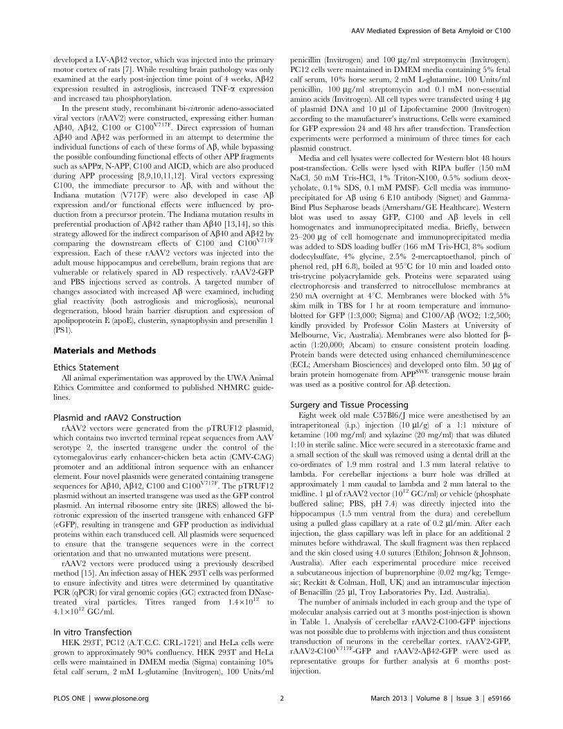
Table 2. Primers and PCR conditions.
Primer Sequence (5-3) Annealing temp (oC) GenBank ID
L19 F * CTGAAGGTCAAAGGGAATGTG 51 NM_031103
L19 R * GGACAGAGTCTTGATGATCTC
PPIA F * AGCATACAGGTCCTGGCATC 62 BC059141
PPIA R * TTCACCTTCCCAAAGACCAC
Transgene F TTGCAGAAGATGTGGGTTCA 59 N/A
Transgene R GGTACCGCAATACCGGAGTA
Synaptophysin F CTGGCCACCTACATCTTCCT 58 NM_009305
Synaptophysin R CCACATGAAAGCGAACACTG
Clusterin F TATGCACGTGTCTGCAGGAG 58 NM_013492
Clusterin R CGCCGTTCATCCAGAAGTAG
ApoE F AACCGCTTCTGGGATTACCT 58 NM_009696.3
ApoE R AGCTGTTCCTCCAGCTCCTT
PS-1 F CACCCCATTCACAGAAGACA 58 NM_008943
PS-1 R TAGTCCACGGCGACATTGTA
*denotes reference gene.doi:10.1371/journal.pone.0059166.t002
AAV Mediated Expression of Beta Amyloid or C100
PLOS ONE | www.plosone.org 4 March 2013 | Volume 8 | Issue 3 | e59166
StatisticsAll statistics were performed using SPSS (Version 17). Two-
tailed independent t-tests were initially used to determine if there
was a significant difference between PBS and AAV2-GFP control
groups in each brain region for all measures. The Levene’s test for
equality of variances was also performed to ensure normality of the
distribution and to test the equality of variances assumption. As
there was no difference observed between brain regions injected
with PBS and rAAV2-GFP for any measure, rAAV2-GFP was
used as the only control group for all further analysis, as this was
the most appropriate control for comparison with experimental
rAAV2 vectors. Univariate, one-way ANOVA was used when
comparing more than two groups and the Bonferroni post-hoc test
was used to determine individual significant differences between
groups. The non-parametric Kruskal-Wallis Test was used to
perform statistics on the intensity grading results from IgG
staining. In all cases p,0.05 was deemed significant.
Results
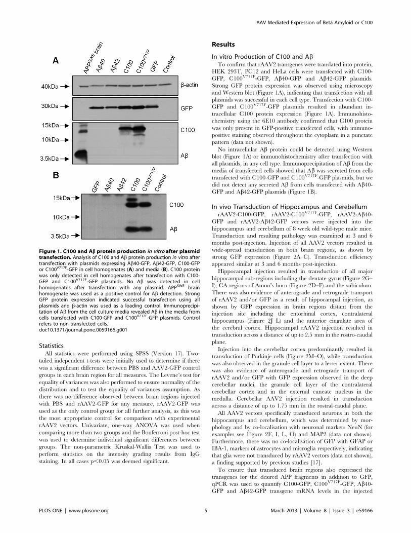
In vitro Production of C100 and AbTo confirm that rAAV2 transgenes were translated into protein,
HEK 293T, PC12 and HeLa cells were transfected with C100-
GFP, C100V717F-GFP, Ab40-GFP and Ab42-GFP plasmids.
Strong GFP protein expression was observed using microscopy
and Western blot (Figure 1A), indicating that transfection with all
plasmids was successful in each cell type. Transfection with C100-
GFP and C100V717F-GFP plasmids resulted in abundant in-
tracellular C100 protein expression (Figure 1A). Immunohisto-
chemistry using the 6E10 antibody confirmed that C100 protein
was only present in GFP-positive transfected cells, with immuno-
positive staining observed throughout the cytoplasm in a punctate
pattern (data not shown).
No intracellular Ab protein could be detected using Western
blot (Figure 1A) or immunohistochemistry after transfection with
all plasmids, in any cell type. Immunoprecipitation of Ab from the
media of transfected cells showed that Ab was secreted from cells
transfected with C100-GFP and C100V717F-GFP plasmids, but we
did not detect any secreted Ab from cells transfected with Ab40-GFP and Ab42-GFP plasmids (Figure 1B).
In vivo Transduction of Hippocampus and CerebellumrAAV2-C100-GFP, rAAV2-C100V717F-GFP, rAAV2-Ab40-
GFP and rAAV2-Ab42-GFP vectors were injected into the
hippocampus and cerebellum of 8 week old wild-type male mice.
Transduction and resulting pathology was examined at 3 and 6
months post-injection. Injection of all AAV2 vectors resulted in
wide-spread transduction in both brain regions, as shown by
strong GFP expression (Figure 2A–C). Transduction efficiency
appeared similar at 3 and 6 months post-injection.
Hippocampal injection resulted in transduction of all major
hippocampal sub-regions including the dentate gyrus (Figure 2G–
I), CA regions of Amon’s horn (Figure 2D–F) and the subiculum.
There was also evidence of anterograde and retrograde transport
of rAAV2 and/or GFP as a result of hippocampal injection, as
shown by GFP expression in brain regions distant from the
injection site including the entorhinal cortex, contralateral
hippocampus (Figure 2J–L) and the anterior cingulate area of
the cerebral cortex. Hippocampal rAAV2 injection resulted in
transduction across a distance of up to 2.5 mm in the rostro-caudal
plane.
Injection into the cerebellar cortex predominantly resulted in
transduction of Purkinje cells (Figure 2M–O), while transduction
was also observed in the granule cell layer to a lesser extent. There
was also evidence of anterograde and retrograde transport of
rAAV2 and/or GFP with GFP expression observed in the deep
cerebellar nuclei, the granule cell layer of the contralateral
cerebellar cortex and in the external cuneate nucleus in the
medulla. Cerebellar AAV2 injection resulted in transduction
across a distance of up to 1.75 mm in the rostral-caudal plane.
All AAV2 vectors specifically transduced neurons in both the
hippocampus and cerebellum, which was determined by mor-
phology and by co-localisation with neuronal markers NeuN (for
examples see Figure 2F, I, L, O) and MAP2 (data not shown).
Furthermore, there was no co-localisation of GFP with GFAP or
IBA-1, markers of astrocytes and microglia respectively, indicating
that glia were not transduced by rAAV2 vectors (data not shown),
a finding supported by previous studies [17].
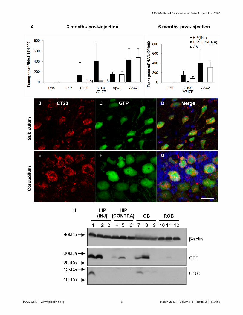
To ensure that transduced brain regions also expressed the
transgenes for the desired APP fragments in addition to GFP,
qPCR was used to quantify C100-GFP, C100V717F-GFP, Ab40-GFP and Ab42-GFP transgene mRNA levels in the injected
Figure 1. C100 and Ab protein production in vitro after plasmidtransfection. Analysis of C100 and Ab protein production in vitro aftertransfection with plasmids expressing Ab40-GFP, Ab42-GFP, C100-GFPor C100V717F-GFP in cell homogenates (A) and media (B). C100 proteinwas only detected in cell homogenates after transfection with C100-GFP and C100V717F-GFP plasmids. No Ab was detected in cellhomogenates after transfection with any plasmid. APPSWE brainhomogenate was used as a positive control for Ab detection. StrongGFP protein expression indicated successful transfection using allplasmids and b-actin was used as a loading control. Immunoprecipi-tation of Ab from the cell culture media revealed Ab in the media fromcells transfected with C100-GFP and C100V717F-GFP plasmids. Controlrefers to non-transfected cells.doi:10.1371/journal.pone.0059166.g001
AAV Mediated Expression of Beta Amyloid or C100
PLOS ONE | www.plosone.org 5 March 2013 | Volume 8 | Issue 3 | e59166
Figure 2. Transduction in the hippocampus and cerebellum as indicated by GFP expression. Low magnification images show widespreadGFP protein throughout the hippocampus (A) and cerebellar cortex (C). GFP protein was also observed in the contralateral hippocampus (B). Double
AAV Mediated Expression of Beta Amyloid or C100
PLOS ONE | www.plosone.org 6 March 2013 | Volume 8 | Issue 3 | e59166
hippocampus, contralateral hippocampus and the injected cere-
bellar hemisphere. RNA was extracted from fixed cryosections
adjacent to those used for immunohistochemistry and GFP
localisation. Strong mRNA expression of the human APP
fragments was observed in transduced regions of the hippocampus
and cerebellum at 3 and 6 months post-injection in all mice
injected with C100 and Ab transgenes (Figure 3A). There was also
lower, but detectable expression of C100 and Ab mRNA in the
contralateral hippocampus, providing evidence for retrograde
transport of rAAV2 vectors and not just GFP protein. This finding
supports previous studies that have shown neurons can transport
AAV particles by retrograde axonal transport and that AAV then
has the capability to express transgenes in distal brain regions
[18,19]. There was qualitatively similar transgene mRNA
expression in the hippocampus and cerebellum, indicating a similar
level of transduction in both brain regions. Importantly, no human
Ab or C100 mRNA expression was observed in any brain region
of mice injected with rAAV2-GFP or PBS.
Human C100 and Ab protein expression was also examined
using Western blot and immunohistochemistry. C100 immuno-
positive staining was observed throughout the cytoplasm and in
primary processes of transduced neurons in the hippocampus and
cerebellum (Figure 3B–G). C100 protein was also detected using
Western blot in hippocampal and cerebellar tissue injected with
rAAV2-C100V717F-GFP (Figure 3H). In contrast, Ab could not be
detected in either transduced brain region using immunohisto-
chemistry trialling multiple antibodies against Ab or Western blot
after injection with any rAAV2 vector (data not shown). These
results led to the hypothesis that Ab may be present at very low
levels and hence undetectable using Western blot or immunohis-
tochemistry. Therefore, the high sensitivity INNO-BIA plasma Abforms test was used to measure Ab40 and Ab42 levels in the
injected hippocampus and cerebellum. Ab42 was detected in
cerebellar tissue from every animal injected with rAAV2-
C100V717F-GFP, with Ab42 levels on average 5.964.3 (6
standard deviation) fold higher than baseline levels. Ab42 protein
was also present in the cerebellum of one of the four animals
injected with rAAV2-Ab42-GFP, this animal having Ab42 levels
that were 3.3 fold higher than baseline levels. Ab42 protein was
also detected in the hippocampus from animals injected with
either rAAV2-C100V717F-GFP or rAAV2-Ab42-GFP but Ab42protein levels did not reach the two standard deviation criterion
relative to baseline. It is important to note that, because the same
viral vectors were injected into each brain region, the lack of
definitive detection of Ab42 in the hippocampus suggests that
Ab42 may be processed differently in each brain region.
Significant levels of Ab40 were not found in the cerebellum or
hippocampus after injection of either rAAV2-C100V717F-GFP or
rAAV2-Ab42-GFP, although a relatively high level of Ab40protein expression was seen in hippocampal tissue from one
rAAV2-C100V717F-GFP injected animal. The inter-animal varia-
tion in the amount of Ab detected, and the differences in Ab levels
between the cerebellum and hippocampus, leads to speculation
that Ab was produced but was then degraded or cleared after
production, resulting in overall low levels available for detection in
the murine brain samples.
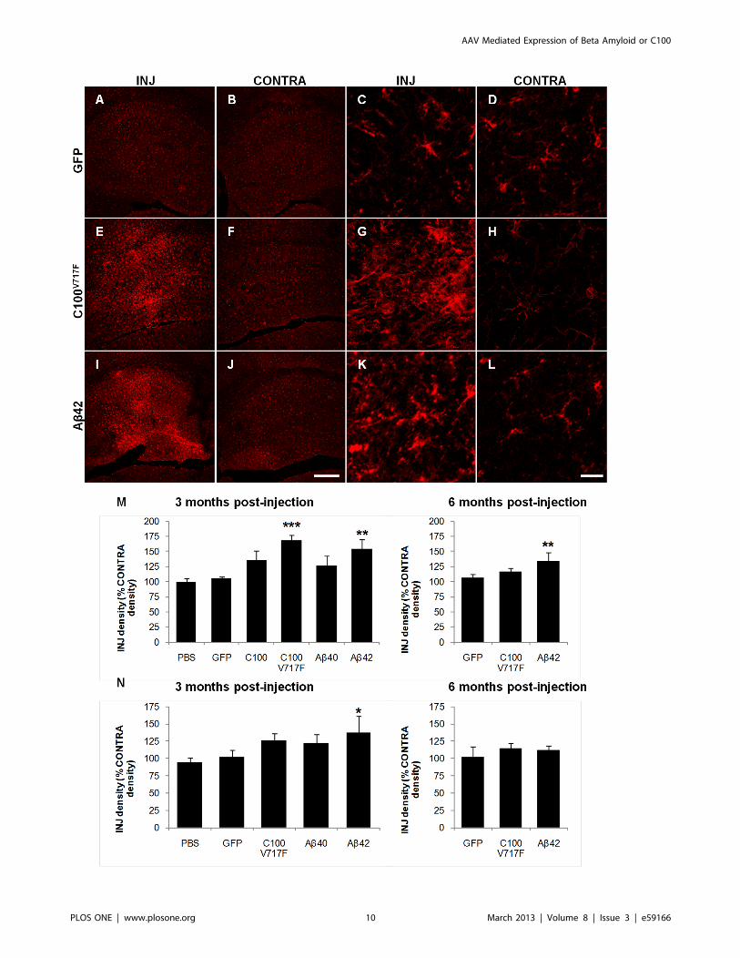
MicrogliosisImmuno-positive staining density of the microglial marker IBA-
1 was analysed to determine the extent of microgliosis following
injection of rAAV2 vectors. This analysis technique was designed
to examine wide-spread microgliosis across the whole transduced
region. IBA-1 positive microglia in areas of gliosis showed
morphological signs of activation including enlarged cell bodies,
ramified processes and increased IBA-1 staining within individual
microglia (Figure 4G, K).
In the hippocampus, injection of rAAV2-C100V717F-GFP and
rAAV2-Ab42-GFP resulted in significantly higher IBA-1 staining
density at 3 months post-injection than that observed after
rAAV2-GFP injection (Bonferroni; p,0.001 and p,0.01 re-
spectively), while there was no significant difference in IBA-1
staining density between hippocampi injected with rAAV2-C100-
GFP or rAAV2-Ab40-GFP and rAAV2-GFP (Figure 4A, E, I, M).
Microgliosis remained significantly higher in the hippocampus at 6
months post-injection with rAAV2-Ab42-GFP than after injection
of rAAV2-GFP (Bonferroni; p,0.01, Figure 4M).
In the cerebellum, injection of rAAV2-Ab42-GFP also resulted
in a significantly greater level of microgliosis at 3 months post-
injection than after injection of rAAV2-GFP (Bonferroni; p,0.05;
Figure 4N). Microgliosis was no longer elevated at 6 months post-
injection. Therefore, combined results from the hippocampus and
cerebellum suggest that microgliosis was strongly associated with
Ab42 rather than Ab40 expression, as it was most extensive after
injection with rAAV2-Ab42-GFP and rAAV2-C100V717F-GFP.
Astrogliosis and Neuronal DensityAb expression has been associated with astrogliosis and
neuronal death, therefore the extent of astrogliosis and neuronal
density were examined using immunohistochemistry for GFAP
and MAP2 respectively. Quantification of GFAP staining density
showed that while there was some evidence of increased astrocyte
activation surrounding the injection site in both brain regions
following injection of rAAV2-C100-GFP, rAAV2-C100V717F-
GFP, rAAV2-Ab40-GFP and rAAV2-Ab42-GFP in comparison
to after injection of rAAV2-GFP or PBS, this increase was not
extensive enough to be significantly different at either 3 or 6
months post-injection (data not shown). Quantification of MAP2
staining density showed that neuronal density was not altered in
the hippocampus or cerebellum as a result of injection of any
rAAV2 vector at either 3 or 6 months post-injection (data not
shown).
Permeability of the Blood Brain BarrierAn unexpected consequence of injection with rAAV2 vectors
was the presence of increased mouse IgG staining around the
injection site, first detected after immunohistochemistry using anti-
mouse secondary antibodies. Further testing showed that the
increased IgG staining was observed when sections were incubated
only with anti-mouse secondary antibodies, was localised to
transduced brain regions, and was not observed after injection
with PBS, therefore showing that it was not a technical artefact of
the immunohistochemistry procedure.
Increased IgG staining was observed in the hippocampus and
cerebellum after injection of rAAV2 vectors expressing APP
fragments, but not after injection of rAAV2-GFP or PBS (Figure 5).
The relative extracellular IgG staining intensity was graded in the
immunostaining for GFP (green) and NeuN (red) revealed that neurons were specifically transduced (D–O). Merged images (F, I, L, O) also showHoechst counterstaining. Neurons in many hippocampal sub-regions were transduced including Amon’s horn (D–F) and dentate gyrus (G–I). GFPwas observed in Purkinje cells and other neurons in the cerebellum (M–O). Scale bar = 500 mm (A–C) and 50 mm (D–O).doi:10.1371/journal.pone.0059166.g002
AAV Mediated Expression of Beta Amyloid or C100
PLOS ONE | www.plosone.org 7 March 2013 | Volume 8 | Issue 3 | e59166
AAV Mediated Expression of Beta Amyloid or C100
PLOS ONE | www.plosone.org 8 March 2013 | Volume 8 | Issue 3 | e59166

hippocampus and cerebellum after immunohistochemistry for
NeuN, using the IgG staining intensity of the surrounding densely
packed neuronal layers as the staining intensity comparison. IgG
staining intensity in the hippocampus at 3 months post-injection
was significantly higher after injection of rAAV2-C100-GFP
(Kruskal-Wallis; p = 0.009), rAAV2-C100V717F-GFP (Kruskal-
Wallis; p = 0.001) and rAAV2-Ab42-GFP (Kruskal-Wallis;
p = 0.016) than after injection of AAV2-GFP. There was also
a non-significant trend for increased IgG staining in the
hippocampus after injection of rAAV2-Ab40-GFP than after
injection of AAV2-GFP at 3 months post-injection (Kruskal-
Wallis; p = 0.056). Similarly, IgG staining intensity in the
cerebellum was significantly higher at 3 months post-injection
with rAAV2-C100V717F-GFP (Kruskal-Wallis; p = 0.014), rAAV2-
Ab40-GFP (Kruskal-Wallis; p = 0.014) and rAAV2-Ab42-GFP
(Kruskal-Wallis; p = 0.013) than after injection with rAAV2-GFP.
In contrast, IgG staining intensity was no longer significantly
elevated at 6 months post-injection with any rAAV2 vector in
either the hippocampus or cerebellum, indicating that the
increased IgG in the brain decreased with time.
Staining for mouse IgG also strongly labelled small, round cells
that were predominantly observed in transduced regions of the
hippocampus and cerebellum (Figure 6-A–C). Immunohistochem-
istry revealed that these IgG positive cells were also immunopo-
sitive for IBA-1 (Figure 6D–G), a calcium binding peptide
produced specifically by monocytes and activated microglia [20].
The number of IgG positive cells was counted and averaged across
three serial sections in the hippocampus and cerebellum. The vast
majority of IgG positive cells were observed throughout the
injected hippocampus and cerebellum and in the lateral ventricles
adjacent to the injected hippocampus, with a small number of cells
also observed in the contralateral hemispheres in both brain
regions. Almost no IgG positive cells were seen after control PBS
injections. 3 months post-injection, there were significantly more
IgG positive cells in the hippocampus after injection with rAAV2-
C100-GFP, rAAV2-C100V717F-GFP and rAAV2-Ab42-GFP than
after injection with rAAV2-GFP (Figure 6H; Bonferroni;
p,0.001). In comparison, while the number of IgG positive cells
in hippocampi at 3 months post-injection with rAAV2-Ab40-GFP
was higher than after rAAV2-GFP injection, this difference was
not statistically significant. There was no longer a significant
difference between groups in the number of IgG positive cells in
the hippocampus at 6 months post-injection (Figure 6H).
The number of IgG positive cells was also increased in the
cerebellum after injection with rAAV2 vectors containing APP
fragments, but high inter-animal variation meant that these
increases were not statistically significant at 3 months post-
injection (Bonferroni; Figure 6I). However, analysis using the less
stringent LSD post-hoc test found that injection with rAAV2-
C100V717F-GFP resulted in significantly more infiltrating cells than
injection with rAAV2-GFP (LSD; p= 0.049). This effect was still
observed at 6 months post-injection (Bonferroni; p,0.05).
Gene ExpressionPS1, apoE, clusterin and synaptophysin gene expression was
examined in the injected hippocampus, contralateral hippocampus
and injected cerebellar hemisphere at 3 and 6 months post-
injection. ApoE and clusterin expression was also examined to
determine if expression of genes involved in Ab clearance was
altered [21]. Synaptophysin is a synaptic protein marker and was
therefore examined as an additional marker of neurodegeneration
and PS1 expression was examined to determine if C100 mediated
endogenous PS1 expression, hence altering production of Ab.Altered levels of each of these genes and the resulting proteins have
also been previously associated with AD [22,23,24,25]. Differences
in gene expression between groups were only deemed significant if
the statistical difference was consistently observed after normal-
isation to two housekeeping genes; L19 and PPIA. It was found
that there was not a significant difference in expression of any gene
examined as a result of injection of any vector at 3 or 6 months
post-injection in either the hippocampus or cerebellum.
Discussion
Injection of rAAV2 vectors expressing transgenes for human
Ab40, Ab42, C100 and C100V717F into the mouse hippocampus
and cerebellum resulted in wide-spread transduction in both brain
regions and the development of some pathological changes
characteristic of AD, most notably increased microgliosis and
increased permeability of the blood brain barrier.
Injection of rAAV2-Ab42-GFP and rAAV2-C100V717F-GFP in
the hippocampus resulted in significantly increased IBA-1 density
at 3 months post-injection, while injection of rAAV2-Ab40-GFP
and rAAV2-C100-GFP did not, suggesting that Ab42 may be
a more potent mediator of microgliosis than Ab40. It is known that
Ab attracts and causes activation of microglia [26,27], but it has
been previously suggested that this may only occur in response to
fibrillar Ab [1,28,29,30]. The association between fibrillar Ab and
microgliosis is further supported by the fact that the onset of gliosis
in AD transgenic mice is closely linked to the onset of plaque
deposition [31,32], and manipulating the amount of plaque
deposition results in similar changes in the extent of microgliosis
[33,34,35]. However, the results from this study suggest that
microgliosis can also occur in response to soluble forms of Ab, asno plaques were observed after injection of any rAAV2 vector.
The rAAV2 vectors used in this study also resulted in pathology
indicative of increased permeability of the blood brain barrier,
which was most extensive in the hippocampus at 3 months post-
injection. Injection of rAAV2 vectors into the hippocampus
resulted in increased brain IgG and increased numbers of IgG/
IBA-1 positive cells. IgG cannot cross the blood brain barrier and
is only found in the brain under pathological conditions [36]. As
a result, IgG is a well established marker of blood brain barrier
permeabilisation and has been shown to be a good alternative to
other markers of blood brain barrier disruption such as Evans blue
dye staining [37,38,39,40,41]. Cells with similar morphology to
the IgG positive cells observed in this study have been
characterised previously and a large number of these cells is also
a common marker of blood brain barrier disruption [42]. Previous
studies have suggested that these cells are leukocytes and as the
cells observed in this study were also immuno-positive for IBA-1,
this suggests that they were leukocytes of monocyte-macrophage
lineage [43], in agreement with previous studies [37,44].
In the hippocampus at 3 months post-injection, blood brain
barrier disruption was more extensive after exposure to Ab42, via
Figure 3. Ab and C100 mRNA and protein expression in the hippocampus and cerebellum. mRNA for Ab and C100 transgenes wasdetected using PCR (A). Immunostaining for C100 using CT20 antibody revealed C100 protein in GFP positive neurons (B–G). C100 protein was alsodetected using western blot (H). HIP (INJ): injected hippocampus, HIP (CONTRA) contralateral hippocampus, CB: cerebellum, ROB: rest of the brain.Lanes 1, 4, 7, 10; representative brain injected with AAV2-C100V717F-GFP, lanes 2, 5, 8, 11; representative brain injected with AAV2-Ab42-GFP, lanes 3,6, 9, 12; representative brain injected with AAV2-GFP.doi:10.1371/journal.pone.0059166.g003
AAV Mediated Expression of Beta Amyloid or C100
PLOS ONE | www.plosone.org 9 March 2013 | Volume 8 | Issue 3 | e59166
AAV Mediated Expression of Beta Amyloid or C100
PLOS ONE | www.plosone.org 10 March 2013 | Volume 8 | Issue 3 | e59166

expression of Ab42 directly or the C100 and C100V717F
precursors. In comparison, while IgG staining intensity and
numbers of infiltrating cells after injection with rAAV2-Ab40-GFP were elevated, these changes were not significantly different
from that observed after injection with rAAV2-GFP. Blood brain
barrier disruption is a pathological feature of AD [45,46] and
previous studies have hypothesised that it may be directly caused
by Ab [42,47,48]. The results from this study not only support the
hypothesis that Ab expression may directly cause blood brain
barrier disruption, but also suggest that Ab42 may be a more
potent mediator of blood brain barrier disruption than Ab40.Injection of rAAV2 vectors did not induce widespread
astrogliosis or altered neuronal density in either the hippocampus
or cerebellum. Activation of astrocytes in response to Abexpression was observed to some extent, however it was primarily
localised to the injection site, in contrast to the more extensive
microgliosis. Previous studies have found Ab to cause activation
and migration of astrocytes [49,50,51]. However, it has also been
shown that the activation of astrocytes in AD is dependent upon
the conformation and aggregation state of Ab; astrocytes
surrounding dense core plaques become activated, while astrocytes
surrounding diffuse plaques or those not associated with aggre-
gated Ab do not show signs of activation and can often show signs
of atrophy [52,53]. Therefore, it is possible that the lack of
extensive astrogliosis observed in this study was due to the lack of
aggregated, fibrillar Ab following injection of viral vectors. The
lack of widespread neurodegeneration observed in this study is
consistent with previous studies that have shown that Ab is not
a potent mediator of neurodegeneration in vivo [54]. It is now
becoming more accepted that tau hyperphosphorylation and the
development of neurofibrillary tangles is more likely to be
a mediator of neurodegeneration in AD than Ab [55,56,57]. It
is interesting that synaptophysin mRNA levels were unaffected as
Ab, particularly soluble oligomers of Ab, has been shown to
decrease expression of synaptic markers, including synaptophysin
[32,58,59,60,61]. However, this finding is not consistent across all
studies [62,63], indicating that decreased synaptophysin expres-
sion may not be a direct consequence of Ab expression and that
other additional factors may be necessary. It is important to note
that only gross neurodegeneration would have been observed by
the quantification measures used in this study and that the use
other neuronal and cell death markers may have provided a more
specific indication of neurodegeneration.
Western blot and immunohistochemical processing failed to
detect significant Ab40 and Ab42 protein expression in transduced
brain regions. We do not believe that this was due to the use of
inefficient viral vectors because Ab42 was detected in tissues
injected with rAAV2-C100V717F-GFP or rAAV2-Ab42-GFP using
the more sensitive ELISA technique, thus proving that these
vectors were capable of producing Ab. Furthermore, there was
strong transduction of all bi-cistronic rAAV2 vectors in the
hippocampus and cerebellum, as shown by high expression levels
Figure 4. Immunostaining of microglia using IBA-1 antibody. Representative figures show microglial staining at low (A–B, E–F, I–J) and highmagnification (C–D, G–H, K–L) in injected (INJ) and contralateral (CONTRA) hippocampi at 3 months post-injection with AAV2-GFP (A–D), AAV2-C100V717F-GFP (E–H) and AAV2-Ab42-GFP (I–L). IBA-1 staining density was quantified in the hippocampus (M) and cerebellum (N) at 3 and 6 monthspost-injection. Data shown as density of injected region as a percentage of the corresponding contralateral region and is presented as mean 6standard deviation. ***p,0.001, **p,0.01, *p,0.05. Scale bar = 300 mm (A–B, E–F, I–J) and 20 mm (C–D, G–H, K–L).doi:10.1371/journal.pone.0059166.g004
Figure 5. Increased mouse IgG staining in the hippocampus and cerebellum. Example images show normal IgG staining followingimmunohistochemistry for NeuN after AAV2-GFP injection (A–D) and intense IgG staining after injection of AAV2-Ab42-GFP (E, G). Increased IgGstaining was only observed surrounding the injection site and not in the contralateral hemisphere (F, H). INJ; injected region, CONTRA; contralateralregion. Scale bar = 500 mm.doi:10.1371/journal.pone.0059166.g005
AAV Mediated Expression of Beta Amyloid or C100
PLOS ONE | www.plosone.org 11 March 2013 | Volume 8 | Issue 3 | e59166
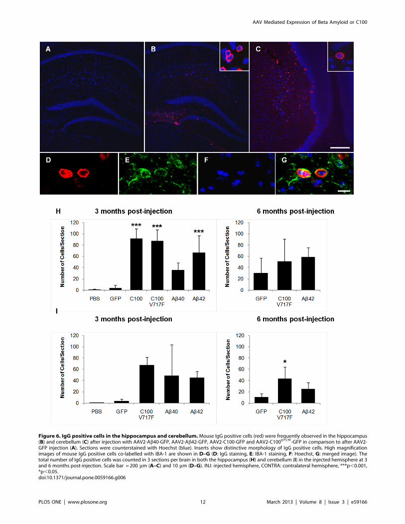
Figure 6. IgG positive cells in the hippocampus and cerebellum.Mouse IgG positive cells (red) were frequently observed in the hippocampus(B) and cerebellum (C) after injection with AAV2-Ab40-GFP, AAV2-Ab42-GFP, AAV2-C100-GFP and AAV2-C100V717F-GFP in comparison to after AAV2-GFP injection (A). Sections were counterstained with Hoechst (blue). Inserts show distinctive morphology of IgG positive cells. High magnificationimages of mouse IgG positive cells co-labelled with IBA-1 are shown in D–G (D: IgG staining, E: IBA-1 staining, F: Hoechst, G: merged image). Thetotal number of IgG positive cells was counted in 3 sections per brain in both the hippocampus (H) and cerebellum (I) in the injected hemisphere at 3and 6 months post-injection. Scale bar = 200 mm (A–C) and 10 mm (D–G). INJ: injected hemisphere, CONTRA: contralateral hemisphere, ***p,0.001,*p,0.05.doi:10.1371/journal.pone.0059166.g006
AAV Mediated Expression of Beta Amyloid or C100
PLOS ONE | www.plosone.org 12 March 2013 | Volume 8 | Issue 3 | e59166
of transgene mRNA and post-IRES GFP protein, the latter
produced only after C100 or Ab protein translation. Finally,
injection of rAAV2-Ab40-GFP and rAAV2-Ab42-GFP resulted in
the development of obvious brain pathology that was unique to
each Ab isoform and was not present after injection of vehicle or
rAAV2-GFP controls, strongly suggesting that the Ab produced as
a result of rAAV2 vector injection was responsible for the observed
pathologies. We hypothesise that the inability to detect Ab using
the less sensitive immunohistochemistry and western blotting
methods, and the observation of variance between animals in the
amount of Ab detected using ELISA, was due to rapid clearance
and/or degradation of Ab. Ab levels are highly regulated in vivo by
rapid clearance across the blood brain barrier, phagocytosis by glia
and degradation by multiple enzymes. Enhancement of any of
these mechanisms could result in low Ab levels. Indeed, some of
the pathology observed in vivo including microgliosis and in-
filtration of cells from the periphery indicate that Ab degradation
may have been increased in response to expression of Ab as both
microglia and infiltrating monocytes are capable of Ab phagocy-
tosis and enhance degradation [44,64,65,66,67,68]. Increased Abclearance may also explain the lack of Ab40 detected using the
ELISA-mutiplex assay, as this isoform is more readily cleared and
degraded than Ab42 [69,70]. Furthermore, the Indiana mutation
promotes the preferential production of Ab42 rather than Ab40,therefore the presence of this mutation and the hypothesised
increased clearance of Ab could account for the very low levels of
Ab40 observed after injection of rAAV2-C100V717F-GFP. Lack of
available tissue prevented ELISA-based quantification of Ab40levels after injection of rAAV2-Ab40-GFP.
An intriguing finding was that, while significant levels of Ab42protein were detected in the cerebellum but not in the
hippocampus, injection of rAAV2 vectors into the hippocampus
resulted in greater pathological changes than those seen in the
cerebellum. Why there was this apparent paradox of increased
Ab42 but reduced pathology in the cerebellum is not clear, but
these observations do suggest that there are differences in the way
different brain regions process and respond to C100 and/or Ab.Previous studies have tried to determine why the cerebellum is less
vulnerable to AD pathology. It has been shown that the
cerebellum contains all of the necessary proteins to produce Ab[71,72], and that plaques do eventually appear in the cerebellum
as AD pathology advances [73], indicating that the cerebellum is
capable of producing amyloid pathology. Nonetheless, the
cerebellum consistently has fewer plaques and lower levels of
insoluble Ab and intracellular Ab42 [73,74,75] than other brain
regions that are primarily affected in AD such as the hippocampus
and cortex. It seems that the cerebellum is better equipped to
prevent AD pathology from progressing. A recent study reported
that secreted metabolites produced from cerebellar neurons
reversed AD brain pathology in AD transgenic mice, while
metabolites from hippocampal neurons exacerbated pathology
[76]. The exact proteins or pathways involved in the protection of
the cerebellum in AD are not yet known, but it has been suggested
that this may be specifically due to enhanced clearance or
degradation of Ab [77,78]. The present data suggest an alternative
hypothesis, that cerebellar cells may be intrinsically less responsive
to the presence of Ab and/or C100. Future research is needed to
further examine why AD brain pathology develops differently in
different brain regions as this could help determine what initiates
the development of AD brain pathology.
Vectors expressing the C100 transgene were more effective at
consistently producing higher amounts of Ab than vectors directly
expressing Ab transgenes, both in vitro and in vivo. This most likely
resulted from the more physiological method of production of Abfrom C100, in comparison to the non-physiological production by
direct expression of either Ab40 or Ab42. Direct expression of Abmay not be optimal for Ab accumulation, possibly due to Abproduction occurring in the incorrect sub-cellular location.
Previous in vitro studies have shown that fusing Ab and C100 to
a signal protein that directs expression in the secretory pathway
greatly increases the amount of Ab detected after plasmid
transfection [79,80], hence suggesting that sub-cellular location
of Ab may be important for expression.
A further aim of this study was to determine if the effects of
transduction with rAAV2 vectors expressing APP fragments were
exacerbated at 6 months post-injection in comparison to 3 months
post-injection. This was not found to be the case in either brain
region as less extensive pathological changes were observed at 6
months post-injection. The level of transduction was similar at 3
and 6 months post-injection, therefore the less extensive pathology
observed at 6 months post-injection is unlikely to be a result of any
technical issues associated with long-term transduction. Instead, it
is possible that brain regions may have adapted to the long-term
expression of C100 and/or Ab and as a result became better
equipped to deal with the consequent pathology, such as by
increasing levels of Ab degrading enzymes or increasing anti-
inflammatory proteins. However, further studies are necessary to
confirm this hypothesis.
In conclusion, the use of viral vectors to over-express Ab and
C100 is a promising technique with which to examine the
consequences of Ab expression in mature CNS tissues in vivo.
Results from this study provide evidence that Ab42 causes greater
pathology than Ab40, particularly by promoting microgliosis and
inducing abnormal permeability changes in the blood brain
barrier.
Acknowledgments
The authors thank Karl De Ruyck for his assistance performing ELISA.
The authors acknowledge the facilities, scientific and technical assistance of
the Australian Microscopy & Microanalysis Research Facility at the Centre
for Microscopy, Characterisation & Analysis, The University of Western
Australia, a facility funded by the University, State and Commonwealth
Governments.
Author Contributions
Conceived and designed the experiments: ESD RNM ARH. Performed
the experiments: ESD JM EME LKW. Analyzed the data: ESD.
Contributed reagents/materials/analysis tools: ARH RNM. Wrote the
paper: ESD.
References
1. McGowan E, Pickford F, Kim J, Onstead L, Eriksen J, et al. (2005) Abeta42 is
essential for parenchymal and vascular amyloid deposition in mice. Neuron 47:
191–199.
2. Lawlor P, Bland R, Das P, Price R, Holloway V, et al. (2007) Novel rat
Alzheimer’s disease models based on AAV-mediated gene transfer to selectively
increase hippocampal Abeta levels. Molecular Neurodegeneration 2: 11.
3. Lemere CA, Masliah E (2010) Can Alzheimer disease be prevented by amyloid-
beta immunotherapy? Nat Rev Neurol 6: 108–119.
4. Lannfelt L, Blennow K, Zetterberg H, Batsman S, Ames D, et al. (2008) Safety,
efficacy, and biomarker findings of PBT2 in targeting Abeta as a modifying
therapy for Alzheimer’s disease: a phase IIa, double-blind, randomised, placebo-
controlled trial. Lancet Neurol 7: 779–786.
5. Siemers ER, Friedrich S, Dean RA, Gonzales CR, Farlow MR, et al. (2010)
Safety and changes in plasma and cerebrospinal fluid amyloid beta after a single
administration of an amyloid beta monoclonal antibody in subjects with
Alzheimer disease. Clin Neuropharmacol 33: 67–73.
AAV Mediated Expression of Beta Amyloid or C100
PLOS ONE | www.plosone.org 13 March 2013 | Volume 8 | Issue 3 | e59166
6. Bateman RJ, Siemers ER, Mawuenyega KG, Wen G, Browning KR, et al.
(2009) A gamma-secretase inhibitor decreases amyloid-beta production in the
central nervous system. Ann Neurol 66: 48–54.
7. Rebeck GW, Hoe HS, Moussa CE (2010) Beta-amyloid1–42 gene transfer
model exhibits intraneuronal amyloid, gliosis, tau phosphorylation, and neuronal
loss. J Biol Chem 285: 7440–7446.
8. Nikolaev A, McLaughlin T, O’Leary DD, Tessier-Lavigne M (2009) APP binds
DR6 to trigger axon pruning and neuron death via distinct caspases. Nature
457: 981–989.
9. Dewachter I, Reverse D, Caluwaerts N, Ris L, Kuiperi C, et al. (2002) Neuronal
deficiency of presenilin 1 inhibits amyloid plaque formation and corrects
hippocampal long-term potentiation but not a cognitive defect of amyloid
precursor protein [V717I] transgenic mice. Journal of Neuroscience 22: 3445–
3453.
10. Yu H, Saura C, Choi S, Sun L, Yang X, et al. (2001) APP processing and
synaptic plasticity in presenilin-1 conditional knockout mice. Neuron 31: 713–
726.
11. Cao X, Sudhof T (2001) A transcriptionally active complex of APP with Fe65
and histone acetyltransferase Tip60. Science 293: 115–120.
12. Hiltunen M, van Groen T, Jolkkonen J (2009) Functional roles of amyloid-beta
protein precursor and amyloid-beta peptides: evidence from experimental
studies. J Alzheimers Dis 18: 401–412.
13. Suzuki N, Cheung TT, Cai XD, Odaka A, Otvos L Jr, et al. (1994) An increased
percentage of long amyloid beta protein secreted by familial amyloid beta
protein precursor (beta APP717) mutants. Science 264: 1336–1340.
14. Johnson-Wood K, Lee M, Motter R, Hu K, Gordon G, et al. (1997) Amyloid
precursor protein processing and A beta42 deposition in a transgenic mouse
model of Alzheimer disease. Proc Natl Acad Sci U S A 94: 1550–1555.
15. Mason MR, Ehlert EM, Eggers R, Pool CW, Hermening S, et al. (2010)
Comparison of AAV serotypes for gene delivery to dorsal root ganglion neurons.
Mol Ther 18: 715–724.
16. Schmidt SD, Nixon RA, Mathews PM (2005) ELISA method for measurement
of amyloid-beta levels. Methods Mol Biol 299: 279–297.
17. Bartlett JS, Samulski RJ, McCown TJ (1998) Selective and rapid uptake of
adeno-associated virus type 2 in brain. Hum Gene Ther 9: 1181–1186.
18. Passini MA, Dodge JC, Bu J, Yang W, Zhao Q, et al. (2006) Intracranial delivery
of CLN2 reduces brain pathology in a mouse model of classical late infantile
neuronal ceroid lipofuscinosis. J Neurosci 26: 1334–1342.
19. Kaspar BK, Erickson D, Schaffer D, Hinh L, Gage FH, et al. (2002) Targeted
retrograde gene delivery for neuronal protection. Mol Ther 5: 50–56.
20. Imai Y, Ibata I, Ito D, Ohsawa K, Kohsaka S (1996) A novel gene iba1 in the
major histocompatibility complex class III region encoding an EF hand protein
expressed in a monocytic lineage. Biochem Biophys Res Commun 224: 855–
862.
21. DeMattos R, Cirrito J, Parsadanian M, May P, O’Dell M, et al. (2004) ApoE
and clusterin cooperatively suppress Abeta levels and deposition: evidence that
ApoE regulates extracellular Abeta metabolism in vivo. Neuron 41: 193–202.
22. Masliah E, Terry RD, DeTeresa RM, Hansen LA (1989) Immunohistochemical
quantification of the synapse-related protein synaptophysin in Alzheimer disease.
Neurosci Lett 103: 234–239.
23. Borghi R, Piccini A, Barini E, Cirmena G, Guglielmotto M, et al. (2010)
Upregulation of presenilin 1 in brains of sporadic, late-onset Alzheimer’s disease.
J Alzheimers Dis 22: 771–775.
24. Yamagata K, Urakami K, Ikeda K, Ji Y, Adachi Y, et al. (2001) High expression
of apolipoprotein E mRNA in the brains with sporadic Alzheimer’s disease.
Dement Geriatr Cogn Disord 12: 57–62.
25. Lidstrom AM, Bogdanovic N, Hesse C, Volkman I, Davidsson P, et al. (1998)
Clusterin (apolipoprotein J) protein levels are increased in hippocampus and in
frontal cortex in Alzheimer’s disease. Exp Neurol 154: 511–521.
26. Shaffer LM, Dority MD, Gupta-Bansal R, Frederickson RC, Younkin SG, et al.
(1995) Amyloid beta protein (A beta) removal by neuroglial cells in culture.
Neurobiol Aging 16: 737–745.
27. Meyer-Luehmann M, Spires-Jones TL, Prada C, Garcia-Alloza M, de Calignon
A, et al. (2008) Rapid appearance and local toxicity of amyloid-beta plaques in
a mouse model of Alzheimer’s disease. Nature 451: 720–724.
28. Weldon DT, Rogers SD, Ghilardi JR, Finke MP, Cleary JP, et al. (1998) Fibrillar
beta-amyloid induces microglial phagocytosis, expression of inducible nitric
oxide synthase, and loss of a select population of neurons in the rat CNS in vivo.
J Neurosci 18: 2161–2173.
29. Passos GF, Figueiredo CP, Prediger RD, Silva KA, Siqueira JM, et al. (2010)
Involvement of phosphoinositide 3-kinase gamma in the neuro-inflammatory
response and cognitive impairments induced by beta-amyloid 1–40 peptide in
mice. Brain Behav Immun 24: 493–501.
30. Jantaratnotai N, Ryu JK, Kim SU, McLarnon JG (2003) Amyloid beta peptide-
induced corpus callosum damage and glial activation in vivo. Neuroreport 14:
1429–1433.
31. Radde R, Bolmont T, Kaeser SA, Coomaraswamy J, Lindau D, et al. (2006)
Abeta42-driven cerebral amyloidosis in transgenic mice reveals early and robust
pathology. EMBO Rep 7: 940–946.
32. Oakley H, Cole SL, Logan S, Maus E, Shao P, et al. (2006) Intraneuronal beta-
amyloid aggregates, neurodegeneration, and neuron loss in transgenic mice with
five familial Alzheimer’s disease mutations: potential factors in amyloid plaque
formation. J Neurosci 26: 10129–10140.
33. Bales K, Verina T, Cummins D, Du Y, Dodel R, et al. (1999) Apolipoprotein Eis essential for amyloid deposition in the APP(V717F) transgenic mouse model of
Alzheimer’s disease. Proceedings of the National Academy of Sciences of the
United States of America 96: 15233–15238.
34. Schenk D, Barbour R, Dunn W, Gordon G, Grajeda H, et al. (1999)
Immunization with amyloid-beta attenuates Alzheimer-disease-like pathology in
the PDAPP mouse. Nature 400: 173–177.
35. Weiner HL, Lemere CA, Maron R, Spooner ET, Grenfell TJ, et al. (2000) Nasal
administration of amyloid-beta peptide decreases cerebral amyloid burden ina mouse model of Alzheimer’s disease. Annals of Neurology 48: 567–579.
36. Seitz RJ, Heininger K, Schwendemann G, Toyka KV, Wechsler W (1985) The
mouse blood-brain barrier and blood-nerve barrier for IgG: a tracer study by useof the avidin-biotin system. Acta Neuropathol 68: 15–21.
37. Wang T, Town T, Alexopoulou L, Anderson JF, Fikrig E, et al. (2004) Toll-like
receptor 3 mediates West Nile virus entry into the brain causing lethalencephalitis. Nat Med 10: 1366–1373.
38. Marchi N, Teng Q, Ghosh C, Fan Q, Nguyen MT, et al. (2010) Blood-brain
barrier damage, but not parenchymal white blood cells, is a hallmark of seizureactivity. Brain Res 1353: 176–186.
39. Nicaise C, Mitrecic D, Demetter P, De Decker R, Authelet M, et al. (2009)
Impaired blood-brain and blood-spinal cord barriers in mutant SOD1-linkedALS rat. Brain Res 1301: 152–162.
40. Baker EA, Tian Y, Adler S, Verbalis JG (2000) Blood-brain barrier disruption
and complement activation in the brain following rapid correction of chronichyponatremia. Exp Neurol 165: 221–230.
41. Fullerton SM, Shirman GA, Strittmatter WJ, Matthew WD (2001) Impairment
of the blood-nerve and blood-brain barriers in apolipoprotein e knockout mice.Exp Neurol 169: 13–22.
42. Farkas IG, Czigner A, Farkas E, Dobo E, Soos K, et al. (2003) Beta-amyloid
peptide-induced blood-brain barrier disruption facilitates T-cell entry into therat brain. Acta Histochem 105: 115–125.
43. Ito D, Imai Y, Ohsawa K, Nakajima K, Fukuuchi Y, et al. (1998) Microglia-specific localisation of a novel calcium binding protein, Iba1. Brain Res Mol
Brain Res 57: 1–9.
44. Town T, Laouar Y, Pittenger C, Mori T, Szekely CA, et al. (2008) BlockingTGF-beta-Smad2/3 innate immune signaling mitigates Alzheimer-like pathol-
ogy. Nat Med 14: 681–687.
45. Kalaria RN (1997) Cerebrovascular degeneration is related to amyloid-betaprotein deposition in Alzheimer’s disease. Ann N Y Acad Sci 826: 263–271.
46. Donahue JE, Johanson CE (2008) Apolipoprotein E, amyloid-beta, and blood-
brain barrier permeability in Alzheimer disease. J Neuropathol Exp Neurol 67:261–270.
47. Jancso G, Domoki F, Santha P, Varga J, Fischer J, et al. (1998) Beta-amyloid (1–
42) peptide impairs blood-brain barrier function after intracarotid infusion inrats. Neurosci Lett 253: 139–141.
48. Su GC, Arendash GW, Kalaria RN, Bjugstad KB, Mullan M (1999)
Intravascular infusions of soluble beta-amyloid compromise the blood-brainbarrier, activate CNS glial cells and induce peripheral hemorrhage. Brain Res
818: 105–117.
49. Pihlaja R, Koistinaho J, Malm T, Sikkila H, Vainio S, et al. (2008) Transplantedastrocytes internalize deposited beta-amyloid peptides in a transgenic mouse
model of Alzheimer’s disease. Glia 56: 154–163.
50. Canning DR, McKeon RJ, DeWitt DA, Perry G, Wujek JR, et al. (1993) beta-
Amyloid of Alzheimer’s disease induces reactive gliosis that inhibits axonal
outgrowth. Exp Neurol 124: 289–298.
51. Pike CJ, Cummings BJ, Monzavi R, Cotman CW (1994) Beta-amyloid-induced
changes in cultured astrocytes parallel reactive astrocytosis associated with senile
plaques in Alzheimer’s disease. Neuroscience 63: 517–531.
52. Kamphuis W, Mamber C, Moeton M, Kooijman L, Sluijs JA, et al. (2012)
GFAP isoforms in adult mouse brain with a focus on neurogenic astrocytes and
reactive astrogliosis in mouse models of Alzheimer disease. PLoS One 7: e42823.
53. Rodriguez JJ, Olabarria M, Chvatal A, Verkhratsky A (2009) Astroglia in
dementia and Alzheimer’s disease. Cell Death Differ 16: 378–385.
54. McGowan E, Eriksen J, Hutton M (2006) A decade of modeling Alzheimer’sdisease in transgenic mice. Trends Genet 22: 281–289.
55. Jaworski T, Dewachter I, Lechat B, Croes S, Termont A, et al. (2009) AAV-tau
mediates pyramidal neurodegeneration by cell-cycle re-entry without neurofi-brillary tangle formation in wild-type mice. PLoS One 4: e7280.
56. Santacruz K, Lewis J, Spires T, Paulson J, Kotilinek L, et al. (2005) Tau
suppression in a neurodegenerative mouse model improves memory function.Science 309: 476–481.
57. Ramsden M, Kotilinek L, Forster C, Paulson J, McGowan E, et al. (2005) Age-
dependent neurofibrillary tangle formation, neuron loss, and memory impair-ment in a mouse model of human tauopathy (P301L). J Neurosci 25: 10637–
10647.
58. Tomiyama T, Matsuyama S, Iso H, Umeda T, Takuma H, et al. (2010) A
mouse model of amyloid beta oligomers: their contribution to synaptic
alteration, abnormal tau phosphorylation, glial activation, and neuronal lossin vivo. J Neurosci 30: 4845–4856.
59. Ishibashi K, Tomiyama T, Nishitsuji K, Hara M, Mori H (2006) Absence of
synaptophysin near cortical neurons containing oligomer Abeta in Alzheimer’sdisease brain. J Neurosci Res 84: 632–636.
60. Shankar GM, Leissring MA, Adame A, Sun X, Spooner E, et al. (2009)
Biochemical and immunohistochemical analysis of an Alzheimer’s disease mouse
AAV Mediated Expression of Beta Amyloid or C100
PLOS ONE | www.plosone.org 14 March 2013 | Volume 8 | Issue 3 | e59166
model reveals the presence of multiple cerebral Abeta assembly forms
throughout life. Neurobiol Dis 36: 293–302.61. Buttini M, Masliah E, Barbour R, Grajeda H, Motter R, et al. (2005) Beta-
amyloid immunotherapy prevents synaptic degeneration in a mouse model of
Alzheimer’s disease. J Neurosci 25: 9096–9101.62. Jacobsen JS, Wu CC, Redwine JM, Comery TA, Arias R, et al. (2006) Early-
onset behavioral and synaptic deficits in a mouse model of Alzheimer’s disease.Proc Natl Acad Sci U S A 103: 5161–5166.
63. Irizarry MC, McNamara M, Fedorchak K, Hsiao K, Hyman BT (1997) APPSw
transgenic mice develop age-related A beta deposits and neuropil abnormalities,but no neuronal loss in CA1. J Neuropathol Exp Neurol 56: 965–973.
64. Bolmont T, Haiss F, Eicke D, Radde R, Mathis CA, et al. (2008) Dynamics ofthe microglial/amyloid interaction indicate a role in plaque maintenance.
J Neurosci 28: 4283–4292.65. Mandrekar S, Jiang Q, Lee CY, Koenigsknecht-Talboo J, Holtzman DM, et al.
(2009) Microglia mediate the clearance of soluble Abeta through fluid phase
macropinocytosis. J Neurosci 29: 4252–4262.66. Chung H, Brazil M, Soe T, Maxfield F (1999) Uptake, degradation, and release
of fibrillar and soluble forms of Alzheimer’s amyloid beta-peptide by microglialcells. Journal of Biological Chemistry 274: 32301–32308.
67. Paresce DM, Chung H, Maxfield FR (1997) Slow degradation of aggregates of
the Alzheimer’s disease amyloid beta-protein by microglial cells. J Biol Chem272: 29390–29397.
68. El Khoury J, Toft M, Hickman SE, Means TK, Terada K, et al. (2007) Ccr2deficiency impairs microglial accumulation and accelerates progression of
Alzheimer-like disease. Nat Med 13: 432–438.69. Vigo-Pelfrey C, Lee D, Keim P, Lieberburg I, Schenk D (1993) Characterization
of beta-amyloid peptide from human cerebrospinal fluid. Journal of Neuro-
chemistry 61: 1965–1968.70. Deane R, Wu Z, Sagare A, Davis J, Du Yan S, et al. (2004) LRP/amyloid beta-
peptide interaction mediates differential brain efflux of Abeta isoforms. Neuron43: 333–344.
71. Cribbs DH, Chen LS, Bende SM, LaFerla FM (1996) Widespread neuronal
expression of the presenilin-1 early-onset Alzheimer’s disease gene in the murine
brain. Am J Pathol 148: 1797–1806.
72. Mita S, Schon EA, Herbert J (1989) Widespread expression of amyloid beta-
protein precursor gene in rat brain. Am J Pathol 134: 1253–1261.
73. Joachim CL, Morris JH, Selkoe DJ (1989) Diffuse senile plaques occur
commonly in the cerebellum in Alzheimer’s disease. Am J Pathol 135: 309–319.
74. Fukumoto H, Cheung BS, Hyman BT, Irizarry MC (2002) Beta-secretase
protein and activity are increased in the neocortex in Alzheimer disease. Arch
Neurol 59: 1381–1389.
75. Hashimoto M, Bogdanovic N, Volkmann I, Aoki M, Winblad B, et al. (2010)
Analysis of microdissected human neurons by a sensitive ELISA reveals
a correlation between elevated intracellular concentrations of Abeta42 and
Alzheimer’s disease neuropathology. Acta Neuropathol 119: 543–554.
76. Du J, Sun B, Chen K, Zhang L, Liu S, et al. (2009) Metabolites of cerebellar
neurons and hippocampal neurons play opposite roles in pathogenesis of
Alzheimer’s disease. PLoS One 4: e5530.
77. Causevic M, Farooq U, Lovestone S, Killick R (2010) beta-Amyloid precursor
protein and tau protein levels are differently regulated in human cerebellum
compared to brain regions vulnerable to Alzheimer’s type neurodegeneration.
Neurosci Lett 485: 162–166.
78. Caccamo A, Oddo S, Sugarman MC, Akbari Y, LaFerla FM (2005) Age- and
region-dependent alterations in Abeta-degrading enzymes: implications for
Abeta-induced disorders. Neurobiol Aging 26: 645–654.
79. Maruyama K, Tagawa K, Kawamura Y, Asada H, Ishiura S, et al. (1995)
Secretion of Alzheimer beta/A4 protein (1–40) and intracellular retention of
beta/A4 protein (1–42) in transfected COS cells. Biochem Biophys Res
Commun 207: 971–977.
80. Magrane J, Smith R, Walsh K, Querfurth H (2004) Heat shock protein 70
participates in the neuroprotective response to intracellularly expressed beta-
amyloid in neurons. Journal of Neuroscience 24: 1700–1706.
AAV Mediated Expression of Beta Amyloid or C100
PLOS ONE | www.plosone.org 15 March 2013 | Volume 8 | Issue 3 | e59166
Copyright © 2022 FDOKUMEN