Hypothalamic gene expression following ghrelin therapy to gastrectomized rodents
Biophysical Journal Volume 73 July 1997 67-75
Alzheimer's Disease Amyloid (8-Protein Forms Zn2+-Sensitive,Cation-Selective Channels Across Excised Membrane Patches fromHypothalamic Neurons
M. Kawahara,* N. Arispe,# Y. Kuroda,* and E. Rojas#§¶*Department of Molecular and Cellular Neurobiology, Tokyo Metropolitan Institute for Neuroscience, Tokyo 183, Japan; #Department ofAnatomy and Cell Biology, Uniformed Services University of Health Sciences, Bethesda, Maryland 20814-4799, USA; §Laboratory of CellBiology and Biochemistry, National Institute of Arthritis, Diabetes, and Digestive and Kidney Diseases, National Institutes of Health,Bethesda, Maryland 20892-0151, USA; and '9Department of Physiology and Biophysics, Faculty of Medicine, University of Chile,Santiago, Chile
ABSTRACT We have previously shown that the 40-residue peptide termed amyloid 13-protein (A,3P[1-40]) in solution formscation-selective channels across artificial phospholipid bilayer membranes. To determine whether AP3P[1-40] also formschannels across natural membranes, we used electrically silent excised membrane patches from a cell line derived fromhypothalamic gonadotrophin-releasing hormone GnRH neurons. We found that exposing either the internal or the externalside of excised membrane patches to A,BP[1-40] leads to the spontaneous formation of cation-selective channels. With Cs'as the main cation in both the external as well as the internal saline, the amplitude of the A,3P[1-40] channel currents wasfound to follow the Cs+ gradient and to exhibit spontaneous conductance changes over a wide range (50-500 pS). We alsofound that free zinc (Zn2+), reported to bind to amyloid 13-protein in solution, can block the flow of Cs+ through the A13P[1-40]channel. Because the Zn2+ chelator o-phenanthroline can reverse this blockade, we conclude that the underlying mechanisminvolves a direct interaction between the transition element Zn2+ and sites in the A13P[1-40] channel pore. These propertiesof the A,BP[1-40] channel are rather similar to those observed in the artificial bilayer system. We also show here, byimmunocytochemical confocal microscopy, that amyloid 1-protein molecules form deposits closely associated with theplasma membrane of a substantial fraction of the GnRH neurons. Taken together, these results suggest that the interactionsbetween amyloid 13-protein and neuronal membranes also occur in vivo, lending further support to the idea that A,BP[1-40]channel formation might be a mechanism of amyloid 1-protein neurotoxicity.
INTRODUCTION
Alzheimer's disease (AD) is a chronic dementia affectingthe aging population and is characterized by the presence ofamyloid plaques in the brain and intraneuronal neurofibril-lary tangles (Neve et al., 1990; Hardy and Higgins, 1992;Selkoe, 1991). The major components of brain amyloidplaques are peptides, 39 to 42 residues long, termed amyloid13-proteins (Masters et al., 1985; Hass and Selkoe, 1993).These peptides are proteolytic products of the widely dis-tributed amyloid precursor glycoprotein (APP751), definedby a locus on chromosome 21 (Goldgaber et al., 1987; Tanziet al., 1987). In particular, amyloid ,B-protein (A,BP[1-40])has been linked to the neurotoxic principle causing neuronaldeath in the disease, although the mechanism has remainedelusive (Yankner et al., 1990; Malouf, 1992; Yankner,1992; Frazer et al., 1997).
Cortical neurons exposed to A,BP[1-40] exhibit elevatedlevels of intracellular free calcium ([Ca2+]i), suggesting thattoxicity may result from amyloid 13-protein-evoked Ca2+entry (Matson et al., 1992). We have previously shown thatwhen A,3P[1-40] was incorporated into lipid bilayer mem-branes, the amyloid formed cation-selective (including
Received for publication 3 February 1997 and in final form 2 April 1997.Address reprint requests to Dr. E. Rojas, LCBB, National Institutes ofHealth, NIDDK, 8 Center Drive, MSC 0840, Building 8, Room 326,Bethesda, MD 20892. Tel.: 301-594-2694; Fax: 301-402-1760.C 1997 by the Biophysical Society0006-3495/97/07/67/09 $2.00
Ca2+) channels (Arispe et al., 1993a,b; Durell et al., 1994),and we suggested that the ability of A,BP[1-40] to formthese cation channels could be the molecular basis of amy-loid neurotoxicity (Arispe et al., 1994). To determinewhether direct incorporation of A13P[1-40] molecules lead-ing to channel formation might also occur in neuronalmembranes, we studied the deposit of A,BP[1-40] mole-cules on the cell surface of cultured hypothalamic GnRH,GT1-7, neurons by immunocytochemical techniques andthe A,BP[1-40]-channel formation in excised membranepatches from these cells after exposure to amyloid 13-pro-tein. We found and report here that although A,BP[1-40]molecules can interact with the excised membrane patchfrom either side, peptide incorporation leading to channelformation was faster from the inner aspect of the excisedpatch. As expected, we found that the reversal potential ofthe A13P[I-40]-specific channel currents followed the Cs+gradient, indicating that the channel was permeable to Cs+over ClF. We also noted that the A,3P[1-40] channel in-corporated into excised patches of GnRH neuronal mem-brane exhibits spontaneous conductance changes over awide range (50-500 pS; Arispe et al., 1993a). To under-stand the mechanism of amyloid 13-protein actions, we ex-amined the ability of Zn2+, known to induce amyloid 13-pro-tein aggregation (Bush et al., 1994), to bind to specificA13P[1-40] sites (Bush et al., 1993), and to modulateA,BP[ 1-40] channel activity in planar bilayers (Arispe et al.,1996). We found that Zn2+ (50-500 AM) can block the
67
Volume 73 July 1997
A,BP[1-40] channel incorporated into the neuronal mem-brane from either the external or the internal side, and theblockade can be reversed by the addition of o-phenanthro-line, a specific Zn2+ chelator, to the side containing Zn2+.These properties of the Af3P[1-40] channel formed acrossnatural membranes are similar to those observed in artificialbilayer membranes (Arispe et al., 1993a,b, 1996), support-ing the idea that A,BP[1-40] channel formation might be themechanism of amyloid 3-protein neurotoxicity (Arispe,1994; Frautschy et al., 1991; Kowall et al., 1992; Fuson etal., 1996). Finally, to determine whether A,3PP[1-40] canalso interact with the plasma membrane of viable GT1-7neurons, and to gather information on the exact localizationof A,3P[1-40] molecules, we used confocal microscopy tolocalize two specific antibodies, one raised against a neu-ronal protein marker (MAP2), and the other against A,BP[1-40]. Both antibodies reacted with the target proteins dem-onstrating the deposition of aggregates of AB3P[1-40] on thesurface of GT1-7 cells. From these data we conclude thatfree A,3P[1-40] molecules in solution can spontaneouslyinteract with both intracellular and extracellular aspects ofthe plasma membrane of GnRH neurons to form cation-selective channels. In addition, our studies of Zn2+/AI3P[1-40] channel interactions reveal that the transition elementbinds to specific domains of the A,BP[1-40] channel mol-ecules, leading to a blockade of the channel. Because theblockade occurs when the amyloid (3-protein and Zn2+ areadded either to the same side or to opposite sides of themembrane, these interactions could explain recent datashowing that peptide deposition and Af3P[1-40]-associatedneurotoxicity are attenuated by Zn2+ (Fuson et al., 1996;May et al., 1996). A preliminary presentation of the data inan abstract form has been made to the Biophysical Society(Kawahara et al., 1996).
MATERIALS AND METHODS
Cell culture
Immortalized hypothalamic neurons (GT1-7; Mellon et al., 1990) werecultured under conditions described previously (Mellon et al., 1990; Sper-gel et al., 1995). Briefly, GT1-7 cells (provided by Dr. R. Weiner,University of California at San Francisco) were grown in a mixture of 50%Dulbecco's modified Eagle's medium and Ham's F-12 medium supple-mented with 2.4 g/liter NaHCO3, 0.1 g/liter gentamicin, and 100 mI/literheat-inactivated fetal calf serum. The culture medium was changed every2-3 days. Upon reaching confluence, the GTI-7 cells were dissociated byincubation for 5 min at 37°C in phosphate-buffered saline containing 0.5g/liter trypsin, 0.15 g/liter deoxyribonuclease I, 5.4 ,uM EDTA, 5.95 mMNaHCO3, 5-6 mM glucose, and 0.07 gentamicin. The cells were thencentrifuged again, resuspended, counted, and plated in 35-mm Petri dishesat a density of 5 X 10⁴ cells/dish. After plating, the cells were cultured for2-3 days before use in patch-clamp experiments.
Aj3P channel current recordingsPatch-clamp experiments were performed as previously described (Rojas et
al., 1990; Spergel et al., 1996), with minor modifications. Patch pipetteswere made from microhematocrit capillary glass by using a microcomput-er-controlled multistep puller (BB-CH; Mecanex, Geneva, Switzerland).
Pipettes were coated to the tip with Sylgard (Dow Corning). When filledwith a Cs' solution (in mM: 140 CsCl, 0.5 CaCI2, 0.5 MgCl2, 1 EGTA, 5NaHEPES, pH 7.4), pipette tip resistance ranged from 8 to 12 Mfl.Channel current recordings were made with the same Cs' solution, bothinside the pipette and in the bath, by means of an EPC-7 amplifier (ListElectronics, Darmstadt, Germany). A13P[I-40] solutions were always pre-pared fresh in distilled water. The amyloid 13-protein was added to the bathat a concentration of -4.7 ,uM.
Membrane currents under voltage clamp conditions were recordedthrough a low-pass filter set at 10 kHz (eight-pole Bessel, model 902 LPF;Frequency Devices, Haverhill, MA) to the input amplifier of a 16-bitresolution digital magnetic tape recorder (PCM-VCR; Unitrade, Data Ac-quisition and Storage Systems, Philadelphia, PA).
ImmunohistochemistryGTI-7 cells were incubated for 24 h in the presence of AP3P[1-40] (-4,.M) added to the culture medium. Next the GT1-7 cells were fixed by theaddition of paraformaldehyde (4%). After fixation cells were thoroughlywashed with medium. After this step specific antibodies against A/3P[l-40] (c4j342; kindly donated by Dr. Mori, Tokyo Institute of Psychiatry) andagainst microtubule associated protein of dividing cells (MAP2) wereadded. After a prolonged exposure to the protein-specific antibodies,secondary antibodies, conjugated to the fluorescent probes fluorescein-5-isothiocyanate (FITC) and Texas Red, respectively, were used to identifythe presence of the proteins in the cells using a confocal microscope.
Statistical evaluation and channelcurrent analysisOff-line analysis of the recorded A13P[I-40] channel activity was carriedout with the software package pClamp 5.51 (Axon Instruments, Burlin-game, CA). Data-base files were obtained from playbacks of the experi-mental records digitized with a 12-bit analog-to-digital converter (TL-1DMA interface; Axon Instruments), using the Fetchex subroutine. At least10 2048-point episodes were needed to achieve the minimum 2500 events
required to construct histograms. These data files were analyzed using thesubroutine TIP (Transit Interface Program; Baylor College of Medicine,Houston, TX). As a rule, the data base used for the open-time andclose-time distributions corresponded to recordings with a signal-to-noiseratio of 25:1.
Four experimental paradigms were carried out on excised inside-outmembrane patches:
Af3P[1-40] added to the bath solution
For these experiments the excised membrane patch was exposed to sym-metrical Cs' solution for a 10-30-min period before the addition ofamyloid 13-protein. 1) After the incorporation of A13P[1-40] channel ac-
tivity, Zn2+ was added to the bath solution. 2) After the blockade by Zn2+was apparent, the Zn2+ chelator, o-phenanthroline, was added to the bathsolution, which contained A13P[1-40] and Zn2+.
Af3P[1-40] added to the pipette solution
To allow for a control period in the absence of amyloid 13-protein, thepipette was first back-filled with a CsCl solution. Next, a small volume(-5 ,ll) of A,BP[1-40] water solution was added to the distal end of theCs' solution in the pipette. The time taken for the amyloid molecules toreach the outer aspect of the patch allowed us to verify that the membranepatch was electrically silent. 1) After the incorporation A13P[1-40] channelactivity, Zn2+ was added to the solution in the dish. 2) The Zn2+ chelator,o-phenanthroline, was added to the solution in the dish after the blockadeby Zn2+ was apparent.
68 Biophysical Journal
Model for Amyloid 3-PI
Each paradigm was repeated at least three times with different cellpreparations. The figures shown are representative of the records obtained.
Chemicals and reagents
Amyloid 13-protein was obtained from Bachem California (Torrance, CA),o-phenanthroline was from Sigma (St. Louis, MO), and ZnCl2 was fromFluka (Ronkonkoma, NY).
rotein Neurotoxicity 69
40] (extracellular side of the cell membrane), the minimumtime allowed for the control period was 5 min (see meth-ods). For bath applications of A,BP[1-40] (intracellular sideof the cell membrane), the minimum control time was 15min. Provided the membrane patch was electrically silent(i.e., endogenous channel current of amplitude smaller than0.1 pA) during this time, the amyloid (3-protein was addedto the solution in the dish to a final concentration of 4.7 ,uM.
RESULTS
To detect endogenous channels that might be present in the Af3P[1-40] but not AI8P[40-1] forms cation
... ~channels across neuronal membranespatch, our standard protocol included an initial control pe-
riod of recording at high gain (200 mV/pA). During this Channel activity always appeared minutes after the expo-
control period the membrane potential across the patch was sure of the inner aspect of the membrane patch to A,BP[1-held at different levels (from -40 to 40 mV) while the 40]. Records of such A,3P[1-40] channel activity incorpo-current was recorded. For pipette applications of Af3P[1- rated from the solution into an inside-out excised patch are
--I. r' J
f -46 pA
200 ms
20 m\/sovO.4 1,M-.t " 0%1
$.m%4i A
'' -' '''- -
r-U TM W o "i
^I_ ft
~~~~~~~* .4 4
Al o&Po ON --
1 pA
200 ms
40 mV
6j- "
.0 A%.
-" -
" 4 fv.,
'Im 0.4-
-P.- A-"Wl-r-
X r--r---.
.,W .I
"I,....,. _- 1
2 pA
200 ms
211
5 pA
200 ms
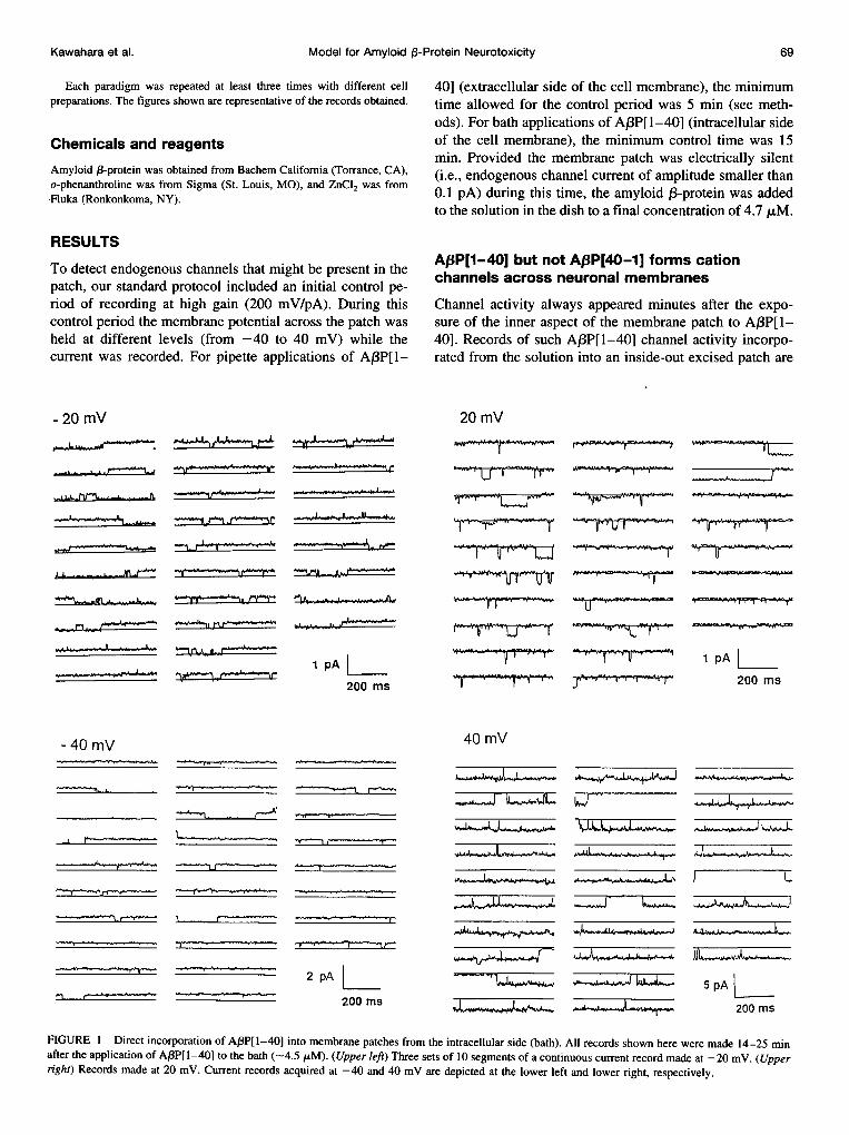
FIGURE 1 Direct incorporation of A,3P[1-40] into membrane patches from the intracellular side (bath). All records shown here were made 14-25 minafter the application of ABP[1-40] to the bath (-4.5 ,uM). (Upper left) Three sets of 10 segments of a continuous current record made at -20 mV. (Upper
right) Records made at 20 mV. Current records acquired at -40 and 40 mV are depicted at the lower left and lower right, respectively.
Kawahara et al.
- 20mV
-40OmV
_ _
,A ---I
-w- .
-0 .4 -n--
--- --. -
I-
I _r-
.1 N.
v .
Ll,-,"Ll
Ok. (i
Pr-P-PWGM WY

Volume 73 July 1997
shown in Fig. 1. The presence of spontaneous transitionsbetween different levels of the current, together with theappearance of complete channel closures from differentlevels, suggests the presence of a single channel with sev-eral levels of conductance. Open-channel current levels aswell as open- and closed-channel kinetics were analyzed asdescribed previously (Arispe et al., 1996). To calculate thefraction of time at a given conductance level from recordslike those shown in Fig. 1, we obtained first open- andclosed-time histograms (not shown). The fractional opentime (estimated from records like those depicted in Fig. 1,bottom) remained unchanged at -0.8, even though thetransmembrane potential was changed from -40 mV (left)to 40 mV (right). By contrast, changing the potential from-20 mV (top left) to 20 mV (top right) reduced the opentime of the channel from 0.75 to 0.25. Under the conditionsof symmetrical solutions used here (140 mM CsCl on bothsides of the membrane patch), the open-channel current wasfound to be close to zero, as expected, at a transmembranepotential of 0 mV. By contrast, in an asymmetrical CsClsolution system (pipette: 140 mM; bath: 70 mM), the re-versal potential was found to be about -16 mV. This isclose to the Nernst potential of -17.4 mV, assuming thatthe charge carrier is Cs+. As illustrated in Fig. 1, the currentchanged from positive (left) to negative (right). By contrast,exposure of either the intracellular or the extracellular sideof the plasma membrane to the peptide AP3P[40-1] with thereverse amino acid sequence had no effect. Indeed, mem-brane patches (n = 6) remained electrically silent afterprolonged (30-60 min) exposures to the peptide A,BP[40-1] (not shown).
Several traits of the ABP[I1-40] channel activity incor-porated into artificial bilayers are also expressed in naturalmembranes. These include the presence of different levelsof channel conductance, from -5 pS to 3 nS (Arispe et al.,1993a), and the sensitivity to the transition element Zn2+(Arispe et al., 1996), illustrated in the following section.
Interactions between Zn2+ andAI8PCI-40] channels
We have previously shown that AfP[1-40] channels incor-porated into artificial lipid bilayer membranes interact withZn2+ in a dose-dependent manner (Arispe et al., 1996). Atconcentrations less than 250 ,uM, Zn2+ may induce pro-found changes in channel kinetics, and at higher doses,Zn2+ may even block the A,BP[1-40] channel. To deter-mine whether the A,BP[1-40] channel activity in naturalmembranes can be modulated by Zn2+, amyloid j3-proteinwas added to the solution bathing either the extracellularside (Fig. 2) or the intracellular side (Fig. 3) of inside-outexcised membrane patches. After the direct incorporation ofA,BP[1-40] molecules from the solution, which was re-vealed by the appearance of channel activity, Zn2+ wasadded to the chamber facing the inner aspect of the patch.
Fig. 2 shows the activity of the A,BP[1-40] channel
(Af3P[1-40] in the pipette). Traces represent segments of a
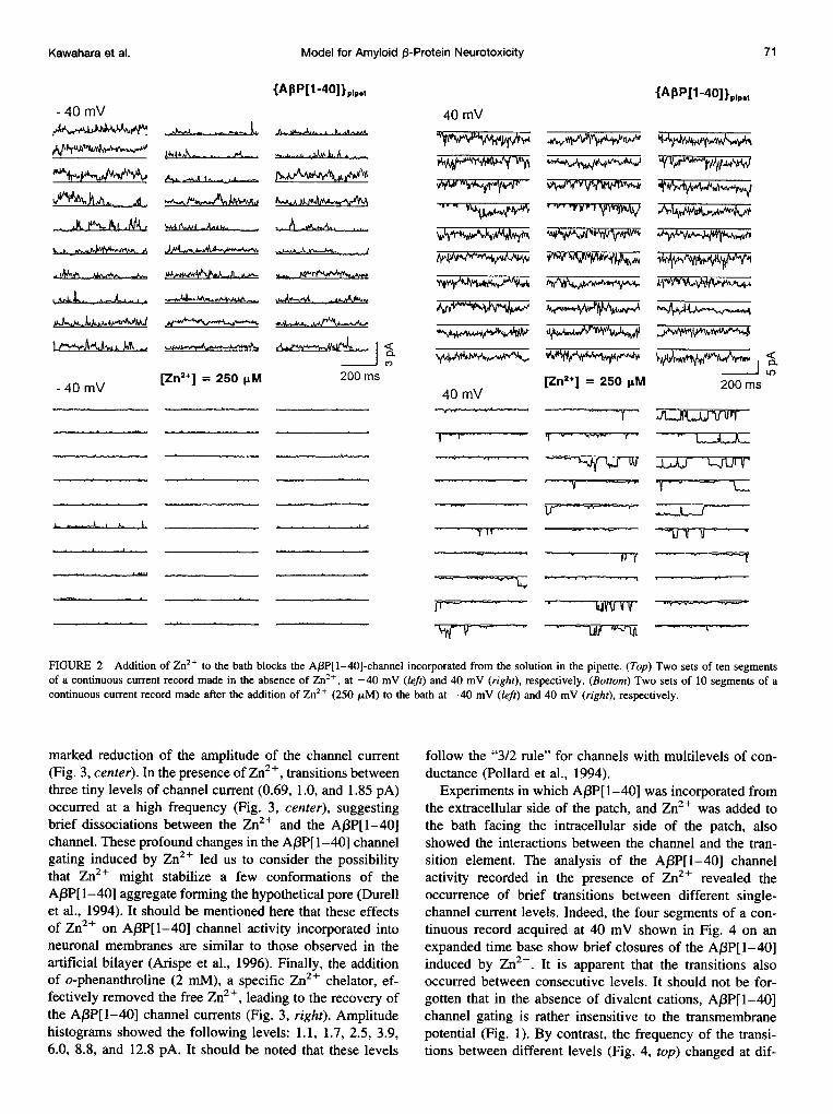
continuous record of A,BP channel activity recorded at dif-ferent transmembrane potentials in the absence (top) and inthe presence (bottom) of Zn2+. In the absence of Zn2+,regardless of the direction of the Cs+ current, channelactivity is rather irregular. At -40 mV, Cs+ is expected toflow through the open channel from the bath to the pipette(upper left), and in the opposite direction at 40 mV (upperright). A few minutes after the addition of Zn+ (250 ,uM) tothe solution facing the cytoplasmic aspect of the membranepatch, channel activity was profoundly inhibited (bottom). Itshould be noted that the extent of the blockade induced byZn2+ depends on the direction of the Cs+ flow. Indeed,while at a pipette potential of -40 mV, there is an almostcomplete inhibition in the A,BP[1-40] channel activity (Fig.2, lower left). In contrast, at 40 mV, for a substantialfraction of time, there is channel activity expressed as
well-defined events (Fig. 2, lower right). Because theserecords were made at a positive potential inside of thepipette, the electrical potential gradient should drive Cs+ions through the open channel. It is then possible that Cs+flowing along the pore, from the external side of the mem-brane, might displace Zn2+ from binding sites at the cyto-plasmic end of the pore. In addition, before the applicationof Zn2+, the fractional open time of the A,BP[1-40] channel
remained at -0.9, even though the transmembrane potentialwas changed from -40 mV to 40 mV (Fig. 2, top). Bycontrast, in the presence of Zn2+ (s250 ,uM), the fractionalopen time changed from -0.05 at -40 mV (Fig. 2, lowerleft) to -0.17 at 40 mV (Fig. 2, lower right).From these data we conclude that the A,3P[1-40] mole-
cule can insert itself into the plasma membrane from thesolution on the extracellular side. Because Zn2+ appliedfrom the intracellular side can modulate the channel, theunavoidable conclusion is that A,3P[1-40] molecules form-ing the channel (Durell et al., 1994) can span the hydropho-bic region of natural membranes, making it possible for thetransition element to interact with the channel from theintracellular side.
Zn2+ blockade is reversed by o-phenanthroline
We also studied the specificity of the interactions betweenZn2+ and amyloid ,3-protein channels incorporated directlyfrom the solution into the cytoplasmic side of the patch. Inthe symmetrical CsCl solution system, the potential across
the patch was set at -20 mV, and a few minutes after theaddition of the protein A,BP[1-40] to the bath (4.7 ,tM),channel activity began (Fig. 3, left). Because the character-istic multilevel channel activity was observed, we con-
cluded that incorporation of the amyloid 13-protein hadoccurred (Fig. 3, left). Amplitude histograms of the activityrevealed the multilevel character of the channel activitywith the following levels: 1.0, 1.7, 2.6, 4.1, 5.9, 9.3, and12.8 pA (not shown). Furthermore, exposure of the cyto-
incorporated from the extracellular side of the patch
70 Biophysical Journal
plasmic side of the patch to Zn2+ (250 ltM) induced a
Model for Amyloid 3-Protein Neurotoxicity
{A,BP[1 -40] plp.t
- 40 mV
%h,L)
.1i, 4A
L _sJh
40 mV
vyww-,Vj4r,II , I-Fh,
A.. ,J-..A& ..A...
OAk_&SiL
r"AM^~Aki.-O.A I.. -
vywpvw~1
.~~.A A ~ AkAAA4m,,
4~~~~~~~ ~~~~~~~t~~~~~~~±±~~~~~~~~~~ae~~~~~~
K ww'
- 40 mV[Zn2*] = 250 pM
C2m200 ms
40 mV[Zn2+] = 250 ILM
T '*-YW"
-y
iAMI,m <Ca
200 ms
_ -F L, _
T- ---
i.S-LA, I'I it --Vgp- -
-'id-I 11, I
-
-VT
FIGURE 2 Addition of Zn2+ to the bath blocks the AP3P[1-40]-channel incorporated from the solution in the pipette. (Top) Two sets of ten segmentsof a continuous current record made in the absence of Zn2+, at -40 mV (left) and 40 mV (right), respectively. (Bottom) Two sets of 10 segments of acontinuous current record made after the addition of Zn2+ (250 ,uM) to the bath at -40 mV (left) and 40 mV (right), respectively.
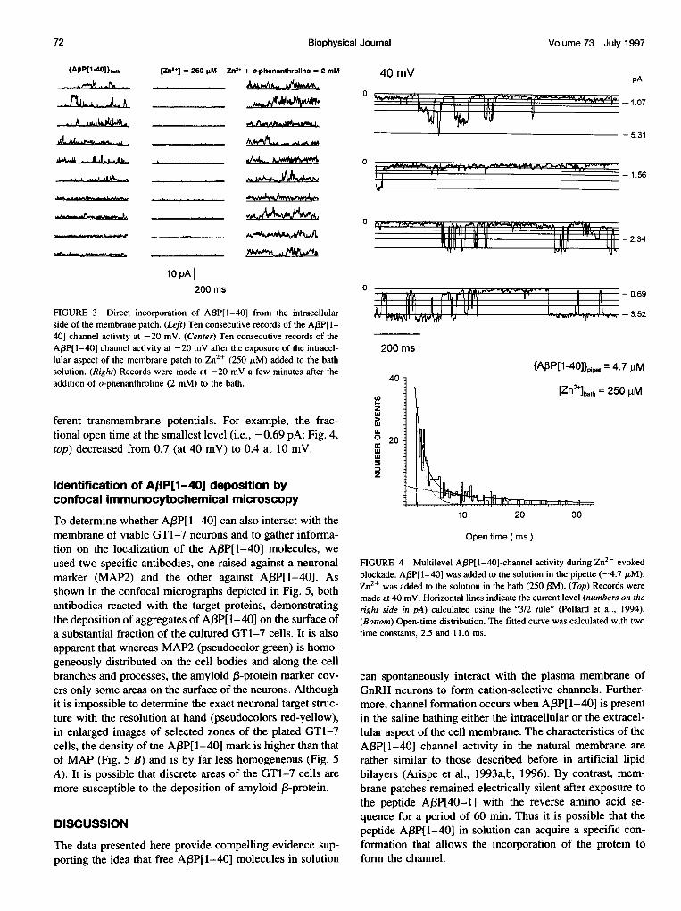
marked reduction of the amplitude of the channel current(Fig. 3, center). In the presence of Zn2+, transitions betweenthree tiny levels of channel current (0.69, 1.0, and 1.85 pA)occurred at a high frequency (Fig. 3, center), suggestingbrief dissociations between the Zn2+ and the A(3P[l-40]channel. These profound changes in the AB3P[l-40] channelgating induced by Zn2+ led us to consider the possibilitythat Zn2+ might stabilize a few conformations of theA3P[1-40] aggregate forming the hypothetical pore (Durellet al., 1994). It should be mentioned here that these effectsof Zn2+ on A,3P[I-40] channel activity incorporated intoneuronal membranes are similar to those observed in theartificial bilayer (Arispe et al., 1996). Finally, the additionof o-phenanthroline (2 mM), a specific Zn2+ chelator, ef-fectively removed the free Zn2+, leading to the recovery ofthe AfBP[1-40] channel currents (Fig. 3, right). Amplitudehistograms showed the following levels: 1.1, 1.7, 2.5, 3.9,6.0, 8.8, and 12.8 pA. It should be noted that these levels
follow the "3/2 rule" for channels with multilevels of con-ductance (Pollard et al., 1994).
Experiments in which A(P[1-40] was incorporated fromthe extracellular side of the patch, and Zn2+ was added tothe bath facing the intracellular side of the patch, alsoshowed the interactions between the channel and the tran-sition element. The analysis of the A,BP[1-40] channelactivity recorded in the presence of Zn2+ revealed theoccurrence of brief transitions between different single-channel current levels. Indeed, the four segments of a con-tinuous record acquired at 40 mV shown in Fig. 4 on anexpanded time base show brief closures of the Af3P[1-40]induced by Zn2+. It is apparent that the transitions alsooccurred between consecutive levels. It should not be for-gotten that in the absence of divalent cations, A/3P[l-40]channel gating is rather insensitive to the transmembranepotential (Fig. 1). By contrast, the frequency of the transi-tions between different levels (Fig. 4, top) changed at dif-
71Kawahara et al.
i
.
. -.,A,, - -L.A
- __q -I
fAPP[1-40]lplp,et
Volume 73 July 1997
{APP[140]}.th.A,.1£Ph.n.
J-ALl_LAA-M.""
[Zn2+] = 250 FM Zn2+ + o-phenanthroline = 2 mM. V Ln
0 ~~~~~~I'LOL A Al MIW q LW
101 11 n * X11| | 11U *J
Adhlba ~ ~ _'_._L,--A.
10 pA200 ms
FIGURE 3 Direct incorporation of A(3P[I-40] from the intracellularside of the membrane patch. (Left) Ten consecutive records of the A,BP[1-40] channel activity at -20 mV. (Center) Ten consecutive records of theA,BP[1-40] channel activity at -20 mV after the exposure of the intracel-lular aspect of the membrane patch to Zn2+ (250 ,M) added to the bathsolution. (Right) Records were made at -20 mV a few minutes after theaddition of o-phenanthroline (2 mM) to the bath.
ferent transmembrane potentials. For example, the frac-tional open time at the smallest level (i.e., -0.69 pA; Fig. 4,top) decreased from 0.7 (at 40 mV) to 0.4 at 10 mV.
Identification of Aj3P[1-40] deposition byconfocal immunocytochemical microscopy
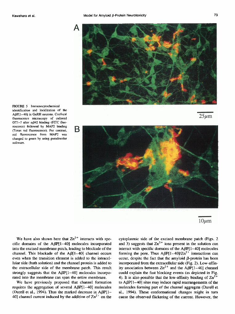
To determine whether A,BP[1-40] can also interact with themembrane of viable GT1-7 neurons and to gather informa-tion on the localization of the A,BP[1-40] molecules, weused two specific antibodies, one raised against a neuronalmarker (MAP2) and the other against A,BP[1-40]. Asshown in the confocal micrographs depicted in Fig. 5, bothantibodies reacted with the target proteins, demonstratingthe deposition of aggregates of A,BP[1-40] on the surface ofa substantial fraction of the cultured GT1-7 cells. It is alsoapparent that whereas MAP2 (pseudocolor green) is homo-geneously distributed on the cell bodies and along the cellbranches and processes, the amyloid ,B-protein marker cov-ers only some areas on the surface of the neurons. Althoughit is impossible to determine the exact neuronal target struc-ture with the resolution at hand (pseudocolors red-yellow),in enlarged images of selected zones of the plated GT1-7cells, the density of the A,3P[1-40] mark is higher than thatof MAP (Fig. 5 B) and is by far less homogeneous (Fig. 5A). It is possible that discrete areas of the GT1-7 cells aremore susceptible to the deposition of amyloid 3-protein.
DISCUSSION
The data presented here provide compelling evidence sup-porting the idea that free A,BP[1-40] molecules in solution
0
0
0
-1.07
- 5.31
- 1.56
-- 2.34
- 0.69
- 3.52
200 ms
{AP3P[1-4O]}0ppt = 4.7 ,uM40
enzwuJ
0 20w
z
[Zn2i]bath = 250 g1M
10 20 30
Open time ( ms )
FIGURE 4 Multilevel A,BP[1-40]-channel activity during Zn2+ evokedblockade. A,BP[1-40] was added to the solution in the pipette (-4.7 ,uM).Zn2+ was added to the solution in the bath (250 PM). (Top) Records weremade at 40 mV. Horizontal lines indicate the current level (numbers on theright side in pA) calculated using the "3/2 rule" (Pollard et al., 1994).(Bottom) Open-time distribution. The fitted curve was calculated with twotime constants, 2.5 and 11.6 ms.
can spontaneously interact with the plasma membrane ofGnRH neurons to form cation-selective channels. Further-more, channel formation occurs when AJ3P[1-40] is presentin the saline bathing either the intracellular or the extracel-lular aspect of the cell membrane. The characteristics of theA,BP[I-40] channel activity in the natural membrane arerather similar to those described before in artificial lipidbilayers (Arispe et al., 1993a,b, 1996). By contrast, mem-brane patches remained electrically silent after exposure tothe peptide AP3P[40-1] with the reverse amino acid se-quence for a period of 60 min. Thus it is possible that thepeptide A,BP[1-40] in solution can acquire a specific con-formation that allows the incorporation of the protein toform the channel.
72 Biophysical Journal
40 mV pA
Model for Amyloid ,3-Protein Neurotoxicity
A
FIGURE 5 Immunocytochemicalidentification and localization of theAP3P[I-40] in GnRH neurons. Confocalfluorescence microscopy of culturedGTl-7 after a,342 binding (FITC fluo-rescence) followed by MAP2 binding(Texas red fluorescence). For contrast,red fluorescence from MAP2 waschanged to green by using pseudocolorsoftware.
25jm
B
We have also shown here that Zn2+ interacts with spe-
cific domains of the A,3P[1-40] molecules incorporatedinto the excised membrane patch, leading to blockade of thechannel. This blockade of the A,3[1-40] channel occurs
even when the transition element is added to the intracel-lular side (bath solution) and the channel protein is added tothe extracellular side of the membrane patch. This resultstrongly suggests that the A,3P[1-40] molecules incorpo-rated into the membrane can span the entire membrane.We have previously proposed that channel formation
requires the aggregation of several A,BP[1-40] molecules(Durell et al., 1994). Thus the marked decrease in A,BP[1-40] channel current induced by the addition of Zn2+ on the
cytoplasmic side of the excised membrane patch (Figs. 2and 3) suggests that Zn2+ ions present in the solution can
interact with specific domains of the A/3P[I-40] moleculesforming the pore. Thus Af3P[l-40]/Zn2+ interactions can
occur, despite the fact that the amyloid ,3-protein has beenincorporated from the extracellular side (Fig. 2). Low-affin-ity association between Zn2+ and the ABP[I1-40] channelcould explain the fast blocking events (as depicted in Fig.4). It is also possible that the low-affinity binding of Zn2+to A,BP[1-40] sites may induce rapid rearrangements of themolecules forming part of the channel aggregate (Durell etal., 1994). These conformational changes might in turncause the observed flickering of the current. However, the
lOJm
73Kawahara et al.

74 Biophysical Journal Volume 73 July 1997
open time of the AJ3P[1-40] channel was found to be ratherinsensitive to the transmembrane potential under calcium-deficient conditions (Fig. 1). Thus voltage-dependentA,BP[1-40] channel gating (Fig. 4, top) can be observedonly under conditions that allow divalent cations, such asCa2+ (Arispe et al., 1993b) and Zn2+ (this work), to enterthe channel pore (Durell et al., 1994) and interact withspecific binding sites. On the basis of our molecular model(Durell et al., 1994), we conclude that Zn2+/A13P[1-40]interactions must occur in a high dielectric constant region,probably at the entrance of the A,BP[1-40] channel, wherethe electric field can be assumed to be relatively constant.
Application of o-phenanthroline induced a complete re-covery of the control A(3P[1-40] channel activity, whichwas characterized by infrequent transitions among differentlevels of conductance (not shown). It should be mentionedhere that o-phenanthroline can remove the blockade only ifapplied to the side containing Zn2+. These results are ingood agreement with recently published planar lipid bilayerdata (Arispe et al., 1996).
In conclusion, our channel data indicate that A,BP[1-40]molecules in solution can interact with both the external aswell as the internal aspect of the plasma membrane andform cation-selective channels. The addition of Zn2+ to thebath solution (cytoplasmic side of the patch) blocked themultilevel A,BP[1-40] channel activity, regardless of theside of incorporation of the Af3P[1-40] molecules. Thisresult suggests that Zn2+ can block the flow of Cs+ throughthe open channel from either end of the pore.
Finally, our preliminary immunocytochemical analysisshowed that whereas the neuronal membrane marker MAP2was homogeneously localized throughout the cell bodiesand dendritic tree, the antibody ca342 reacted with amyloid13-protein molecules deposited onto restricted regions of thecell bodies and processes (Fig. 5 B). Although we do nothave an explanation for this remarkable difference in thelocalization of the two antibodies, it is possible that discretemembrane areas of the GT1-7 cells are more susceptible tothe deposition of amyloid 1-protein. In this respect, it is notuncommon to find in different types of neurons specializedmembrane regions containing distinct membrane proteins.For example, immunolocalization studies in Purkinje neu-rons revealed a rather homogeneous localization of ryano-dine and inositol 1,4,5-triphosphate (1P3) receptor proteinsthroughout the cell body and dendritic tree. By contrast, IP3receptors were detected only in the dendritic spines (Ellis-man et al., 1990, 1991; Martone et al., 1993; Sharp et al.,1993).Taken together, the results presented here suggest that the
interactions between amyloid 13-protein and neuronal mem-branes also occur in vivo, lending further support to the ideathat A13P[1-40] channel formation might be a mechanismof amyloid 13-protein neurotoxicity (Arispe et al., 1994).The interactions of Zn2+ with amyloid 13-protein moleculesincorporated into neuronal membranes could even lead toattenuation of amyloid channel neurotoxicity. It should alsobe mentioned here that the concentration of amyloid 13-pro-
tein used in this work was higher than the levels detected incerebrospinal fluid (CSF) samples from normal and Alzhei-mer's patients (Seubert et al., 1992). However, analysis ofCSF in samples from these subjects showed identical con-centrations of soluble A,BP[1-40] (i.e., <1 ,tM) in bothgroups (Tabaton et al., 1994), suggesting that the presenceof soluble amyloid P-protein in the CSF cannot be consid-ered a marker of the disease.
Thanks are given to Dr. H. Mori (Tokyo Institute for Psychiatry) fordonating the antibody against A,BP[1-40].
This work was supported in part by the Japan Health Sciences Foundation,FONDECYT 1950774 and the 1996 Chilean Presidential Cathedra to ER.
REFERENCES
Arispe, N., H. B. Pollard, and E. Rojas. 1993a. Giant multilevel cationchannels formed by Alzheimer disease amyloid beta-protein (A,BP-[1-40]) in bilayer membranes. Proc. Natl. Acad. Sci. USA. 90:10573-10577.
Arispe, N., H. B. Pollard, and E. Rojas. 1994. ,B-amyloid Ca2+-channelhypothesis for neuronal death in Alzheimer disease. Mol. Cell. Biochem.140:119-125.
Arispe, N., H. B. Pollard, and E. Rojas. 1996. Zn2+ interaction withAlzheimer amyloid ,B-protein calcium channels. Proc. Natl. Acad. Sci.USA. 93:1710-1715.
Arispe, N., E. Rojas, and H. B. Pollard. 1993b. Alzheimer disease amyloid(3-protein forms calcium channels in bilayer membranes: blockade bythrometamine and aluminum. Proc. Natl. Acad. Sci. USA. 90:567-571.
Bush, A. I., G. Multhaup, R. D. Moir, T. G. Williamson, D. H. Small, B.Rumble, P. Pollwein, K. Beyreuther, and C. L. Masters. 1993. A novelzinc binding site modulates the function of the ,BA4 amyloid proteinprecursor of Alzheimer's disease. J. Biol. Chem. 268:16109-16112.
Bush, A. I., W. H. Pettingell, G. Multhaup, and M. D. Paradis. 1994. Rapidinduction of Alzheimer A beta amyloid formation by zinc. Science.265:1464-1467.
Durell, S. R., H. R. Guy, N. Arispe, E. Rojas, and H. B. Pollard. 1994.Theoretical models of the ion channel structure of amyloid (3-protein.Biophys. J. 67:2137-2145.
Ellisman, M. H., T. J. Deerinck, Y. Ouyang, C. F. Beck, S. J. Tanksley, P.D. Walton, J. A. Airey, and J. L. Sutko. 1990. Identification and locationof ryanodine binding proteins in the avian central nervous system.Neuron. 5:135-146.
Ellisman, M. H., J. D. Lindsey, B. 0. Carragher, S. H. Kiyonaga, L. R. McEwen, and B. F. McEwen. 1991. Ryanodine and inositol triphosphatereceptors coexist in avian cerebellar Purkinje neurons. J. Cell. Biol.113:1145-1157.
Frautschy, S. A., A. Baird, and G. M. Cole. 1991. Effects of injectedAlzheimer ,3-amyloid cores in rat brain. Proc. Natl. Acad. Sci. USA.88:8362-8366.
Frazer, S. P., Y-H. Suh, and M. B. A. Djamgoz. 1997. Ionic effects of theAlzheimer's disease ,3-amyloid precursor protein and its metabolic frag-ments. Trends Neurosci. 20:67-72.
Fuson, K. S., L. N. Boggs, and P. C. May. 1996. Zinc attenuates amyloid13-protein [1-40] neurotoxicity. Abstr. Soc. Neurosci. 22:195.
Goldgaber, D., M. I. Lerman, 0. W. McBride, U. Saffiotti, and C. Gaj-dusek. 1987. Characterization and chromosomal localization of a cDNAencoding brain amyloid of Alzheimer's disease. Science. 235:877-880.
Hardy, J. A., and G. A. Higgins. 1992. Alzheimer's disease: the amyloidcascade hypothesis. Science. 256:780-783.
Hass, C., and D. J. Selkoe. 1993. Cellular processing of (-amyloid pre-cursor protein and the genesis of amyloid ,B-peptide. Cell. 75:1039-1042.
Kawahara, M., Y. Kuroda, N. Arispe, and E. Rojas. 1996. Direct incorpo-ration of amyloid (3-protein cation channels into excised membranepatches from GnRH neurons. Biophys. J. 70:A241.
Kawahara et al. Model for Amyloid p3-Protein Neurotoxicity 75
Kowall, N. C., A. C. McKee, B. A. Yankner, and M. F. Beal. 1992. In vivoneurotoxicity of beta amyloid [,B(1-40)] and the 13(25-35) fragment.Neurobiol. Aging. 13:537-542.
Malouf, A. T. 1992. Effect of beta amyloid peptides on neurons in hip-pocampal slice cultures. Neurobiol. Aging. 13:543-55 1.
Martone, M. E., Y. Zhang, V. M. Simpliacino, B. 0. Carragher, and M. H.Ellisman. 1993. Three-dimensional visualization of the smooth endo-plasmic reticulum in Purkinje cell dendrites. J. Neurosci. 13:4636-4646.
Masters, C. L., G. Simms, N. A. Weinman, G. Multhaup, B. L. McDonald,and K. Beyreuther. 1985. Amyloid plaque core protein in Alzheimerdisease and Down syndrome. Proc. Natl. Acad. Sci. USA. 82:4245-4249.
Matson, M. P., B. Cheng, D. Davis, K. Bryant, I. Lieberberg, and R. E.Rydel. 1992. ,B-Amyloid peptides destabilize calcium homeostasis andrender human cortical neurons vulnerable to exocitotoxicity. J. Neurosci.12:376-389.
May, P. C., L. N. Boggs, Z. Zhang, G. J. Drsewiecki, P. A. Hyslop, and K.S. Fuson. 1996. Zinc interferes with A,3P[1-40] deposition onto cul-tured neurons: correlation with neuroprotection. Abstr. Soc. Neurosci.22:196.
Mellon, P. L., J. J. Windle, P. C. Goldsmith, J. L. Padula, and R. I. Weiner.1990. Immortalization of hypothalamic GnRH neurons by geneticallytargeted tumorigenesis. Neuron. 5:1-10.
Neve, R. L., L. R. Dawes, B. A. Yankner, L. L. Benowitz, W. Rodriguez,and G. A. Higgins. 1990. Genetics and biology of the Alzheimer amy-loid precursor. Prog. Brain Res. 86:257-267.
Pollard, J. R., N. Arispe, E. Rojas, and H. B. Pollard. 1994. A geometricsequence that accurately describes allowed multiple conductance levelsof ion channels: the "three-halves (3/2) rule." Biophys. J. 67:647-655.
Rojas, E., J. Hidalgo, P. B. Carroll, M-X. Xu Li, and I. Atwater. 1990. Anew class of calcium channels activated by glucose in human pancreatic,B-cells. FEBS Lett. 261:265-270.
Selkoe, D. J. 1991. The molecular pathology of Alzheimer's disease.Neuron. 6:487-498.
Seubert, P., C. Vigo-Pelfrey, F. Esh, M. Lee, L. Dovey, D. Davis, S. Sinha,M. Schlossmacher, J. Whaley, C. Swindlehurst, R. McCormack, D.Selkoe, I. Lieberburg, and D. Schenk. 1992. Isolation and quantificationof soluble Alzheimer's ,3-peptide from biological fluids. Nature. 259:325-327.
Sharp, A. H., T. M. Dawson, C. A. Ross, M. Fotuhi, R. J. Mourey, and S.Snyder. 1993. Inositol 1,4,5-triphosphate receptors: immunohistochem-ical localization to discrete areas of rat central nervous system. Neuro-science. 53:927-942.
Spergel, D. J., K. J. Catt, and E. Rojas. 1996. Immortalized GnRH neuronsexpress large-conductance calcium-activated potassium channels. Neu-roendocrinology. 63:101-111.
Spergel, D. J., L. Z. Krsmanovic, S. S. Stijilkovic, and K. J. Catt. 1995.L-type Ca2+-channels mediate joint modulation by y-aminobutyric acidand glutamate of [Ca2+]i and neuropeptide secretion in immortalizedgonadotropin-releasing hormone neurons. Neuroendocrinology. 61:499-508.
Tabaton, M., M. G. Nunzi, R. Xue, M. Usiak, L. Autilio-Gambetti, and P.Gambetti. 1994. Soluble amyloid 1B protein is a marker of Alzheimeramyloid in brain but not in cerebrospinal fluid. Biochem. Biophys. Res.Commun. 200:1598-1603.
Tanzi, R. E., J. F. Gusella, P. C. Watkins, G. A. P. Bruns, P. St. George-Hyslop, M. L. Van Keuren, D. Patterson, S. Pagan, D. M. Kurnit, and R.L. Neve. 1987. Amyloid 13-protein gene: cDNA, mRNA distribution, andgenetic linkage near the Alzheimer locus. Science. 235:880-882.
Yankner, B. A. 1992. Commentary and perspective on studies of betaamyloid neurotoxicity. Neurobiol. Aging. 13:615-616.
Yankner, B. L., L. K. Duffy, and D. A. Kirschner. 1990. Neurotropic andneurotoxic effects of amyloid beta protein: reversal by tachykinin neu-ropeptides. Science. 250:279-282.
Copyright © 2022 FDOKUMEN