In Vivo Reinsertion of Excised Episomes by the V(D)J Recombinase: A Potential Threat to Genomic...
16
In Vivo Reinsertion of Excised Episomes by the V(D)J Recombinase: A Potential Threat to Genomic Stability Katrina Vanura 1[ , Bertrand Montpellier 2,3,4[ , Trang Le 1 , Salvatore Spicuglia 2,3,4 , Jean-Marc Navarro 2,3,4 , Olivier Cabaud 2,3,4 , Sandrine Roulland 2,3,4 , Elodie Vachez 2,3,4¤a , Immo Prinz 2,3,4 , Pierre Ferrier 2,3,4 , Rodrig Marculescu 1¤b , Ulrich Ja ¨ ger 1 , Bertrand Nadel 2,3,4* 1 Department of Internal Medicine I, Division of Hematology, Medical University of Vienna, Vienna, Austria, 2 Centre d’Immunologie de Marseille-Luminy, Universite ´ de la Me ´diterrane ´ e, Marseille, France, 3 Institut National de la Sante ´ et de la Recherche Me ´dicale U631, Marseille, France, 4 Centre National de la Recherche Scientifique UMR6102, Marseille, France It has long been thought that signal joints, the byproducts of V(D)J recombination, are not involved in the dynamics of the rearrangement process. Evidence has now started to accumulate that this is not the case, and that signal joints play unsuspected roles in events that might compromise genomic integrity. Here we show both ex vivo and in vivo that the episomal circles excised during the normal process of receptor gene rearrangement may be reintegrated into the genome through trans-V(D)J recombination occurring between the episomal signal joint and an immunoglobulin/T-cell receptor target. We further demonstrate that cryptic recombination sites involved in T-cell acute lymphoblastic leukemia–associated chromosomal translocations constitute hotspots of insertion. Eventually, the identification of two in vivo cases associating episomal reintegration and chromosomal translocation suggests that reintegration events are linked to genomic instability. Altogether, our data suggest that V(D)J-mediated reintegration of episomal circles, an event likely eluding classical cytogenetic screenings, might represent an additional potent source of genomic instability and lymphoid cancer. Citation: Vanura K, Montpellier B, Le T, Spicuglia S, Navarro JM, et al. (2007) In vivo reinsertion of excised episomes by the V(D)J recombinase: A potential threat to genomic stability. PLoS Biol 5(3): e43. doi:10.1371/journal.pbio.0050043 Introduction V(D)J recombination is a unique mechanism of somatic recombination aimed to provide a large antigen receptor repertoire in T and B cells (for review, see [1] and references therein). During this process, the variable (V) diversity (D) and joining (J) gene segments present within the immuno- globulin (IG) and T-cell receptor (TCR) loci, are assembled to form a complete VDJ exon encoding the variable region of the IG/TCR (Figure 1A). The recombination requires the presence of specific motifs flanking all the V, D, and J gene segments (12– and 23–recombination signal sequences [RSSs]), and allowing the recruitment, binding, and proper positioning of the products of the recombination activating genes 1 and 2 (RAG-1/2). Recent data suggest that in vivo, RAG-1/2 proteins initiate the rearrangement by performing a first single-strand nick at the exact border between a 12- RSS and its adjacent coding gene segment [2]. This leads to the capture of a 23-RSS, the formation of a 12/23 synaptic complex in which the two DNA/protein structures are held in close juxtaposition, and the generation of another nick at the captured 23-RSS. Within this complex, RAG-1/2 catalyzes a trans-esterification reaction in which each liberated hydroxyl group attacks the opposite DNA strand [3]; this generates four broken ends held in a postcleavage synaptic complex: two blunt RSSs or signal ends (SEs), and two covalently sealed hairpin coding ends (CEs). The broken ends are then efficiently repaired by the nonhomologous end- joining (NHEJ) pathway [4–6]; on the one hand, hairpins present at the CEs are resolved through the Artemis endonuclease activity; when opened at bases off the apex, hairpin opening generates overhanging flaps, which, if filled in by a DNA polymerase activity, form palindromic (P) stretches. Nontemplated (N) nucleotides may be added de novo by the terminal deoxynucleotidyl transferase (TdT), and/or nucleotides may be deleted from the CEs. Ligation of the processed CEs forms a highly diversified coding joint (CJ). By contrast, the two SEs present in the synaptic complex undergo only limited processing (some N addition and rare nucleotide deletion) before joining into signal Academic Editor: David Nemazee, Scripps Research Institute, United States of America Received May 11, 2006; Accepted December 12, 2006; Published February 13, 2007 Copyright: Ó 2007 Nadel et al. This is an open-access article distributed under the terms of the Creative Commons Attribution License, which permits unrestricted use, distribution, and reproduction in any medium, provided the original author and source are credited. Abbreviations: CSJ, chromosomal SJ; CE, coding end; CJ, coding joint; EC, episomal circles; ESJ, episomal signal joint; HJ, hybrid joint; IG, immunoglobulin; NHEJ, nonhomologous end-joining; PE, primer extension; WHJ, pseudo-HJ; RSS, recombination signal sequence; SE, signal end; SJ, signal joint; T-ALL, T-cell acute lymphoblastic leukemia; TCR, T-cell receptor; TdT, terminal deoxynucleotidyl transferase * To whom correspondence should be addressed. E-mail: [email protected] [ These authors contributed equally to this work. ¤a Current address: Ozyme Biosciences, Parc Scientifique de Luminy, Marseille, France ¤b Current address: Department of Dermatology, Rudolfstiftung Hospital, Vienna, Austria PLoS Biology | www.plosbiology.org March 2007 | Volume 5 | Issue 3 | e43 0515 P L o S BIOLOGY
-
Upload
independent -
Category
Documents
-
view
0 -
download
0
Transcript of In Vivo Reinsertion of Excised Episomes by the V(D)J Recombinase: A Potential Threat to Genomic...

In Vivo Reinsertion of Excised Episomesby the V(D)J Recombinase: A Potential Threatto Genomic StabilityKatrina Vanura
1[, Bertrand Montpellier
2,3,4[, Trang Le
1, Salvatore Spicuglia
2,3,4, Jean-Marc Navarro
2,3,4,
Olivier Cabaud2,3,4
, Sandrine Roulland2,3,4
, Elodie Vachez2,3,4¤a
, Immo Prinz2,3,4
, Pierre Ferrier2,3,4
,
Rodrig Marculescu1¤b
, Ulrich Jager1
, Bertrand Nadel2,3,4*
1 Department of Internal Medicine I, Division of Hematology, Medical University of Vienna, Vienna, Austria, 2 Centre d’Immunologie de Marseille-Luminy, Universite de la
Mediterranee, Marseille, France, 3 Institut National de la Sante et de la Recherche Medicale U631, Marseille, France, 4 Centre National de la Recherche Scientifique UMR6102,
Marseille, France
It has long been thought that signal joints, the byproducts of V(D)J recombination, are not involved in the dynamics ofthe rearrangement process. Evidence has now started to accumulate that this is not the case, and that signal joints playunsuspected roles in events that might compromise genomic integrity. Here we show both ex vivo and in vivo that theepisomal circles excised during the normal process of receptor gene rearrangement may be reintegrated into thegenome through trans-V(D)J recombination occurring between the episomal signal joint and an immunoglobulin/T-cellreceptor target. We further demonstrate that cryptic recombination sites involved in T-cell acute lymphoblasticleukemia–associated chromosomal translocations constitute hotspots of insertion. Eventually, the identification of twoin vivo cases associating episomal reintegration and chromosomal translocation suggests that reintegration events arelinked to genomic instability. Altogether, our data suggest that V(D)J-mediated reintegration of episomal circles, anevent likely eluding classical cytogenetic screenings, might represent an additional potent source of genomicinstability and lymphoid cancer.
Citation: Vanura K, Montpellier B, Le T, Spicuglia S, Navarro JM, et al. (2007) In vivo reinsertion of excised episomes by the V(D)J recombinase: A potential threat to genomicstability. PLoS Biol 5(3): e43. doi:10.1371/journal.pbio.0050043
Introduction
V(D)J recombination is a unique mechanism of somaticrecombination aimed to provide a large antigen receptorrepertoire in T and B cells (for review, see [1] and referencestherein). During this process, the variable (V) diversity (D)and joining (J) gene segments present within the immuno-globulin (IG) and T-cell receptor (TCR) loci, are assembledto form a complete VDJ exon encoding the variable regionof the IG/TCR (Figure 1A). The recombination requires thepresence of specific motifs flanking all the V, D, and J genesegments (12– and 23–recombination signal sequences[RSSs]), and allowing the recruitment, binding, and properpositioning of the products of the recombination activatinggenes 1 and 2 (RAG-1/2). Recent data suggest that in vivo,RAG-1/2 proteins initiate the rearrangement by performinga first single-strand nick at the exact border between a 12-RSS and its adjacent coding gene segment [2]. This leads tothe capture of a 23-RSS, the formation of a 12/23 synapticcomplex in which the two DNA/protein structures are heldin close juxtaposition, and the generation of another nick atthe captured 23-RSS. Within this complex, RAG-1/2 catalyzesa trans-esterification reaction in which each liberatedhydroxyl group attacks the opposite DNA strand [3]; thisgenerates four broken ends held in a postcleavage synapticcomplex: two blunt RSSs or signal ends (SEs), and twocovalently sealed hairpin coding ends (CEs). The broken endsare then efficiently repaired by the nonhomologous end-joining (NHEJ) pathway [4–6]; on the one hand, hairpinspresent at the CEs are resolved through the Artemis
endonuclease activity; when opened at bases off the apex,hairpin opening generates overhanging flaps, which, if filledin by a DNA polymerase activity, form palindromic (P)stretches. Nontemplated (N) nucleotides may be added denovo by the terminal deoxynucleotidyl transferase (TdT),and/or nucleotides may be deleted from the CEs. Ligation ofthe processed CEs forms a highly diversified coding joint(CJ). By contrast, the two SEs present in the synapticcomplex undergo only limited processing (some N additionand rare nucleotide deletion) before joining into signal
Academic Editor: David Nemazee, Scripps Research Institute, United States ofAmerica
Received May 11, 2006; Accepted December 12, 2006; Published February 13,2007
Copyright: � 2007 Nadel et al. This is an open-access article distributed under theterms of the Creative Commons Attribution License, which permits unrestricteduse, distribution, and reproduction in any medium, provided the original authorand source are credited.
Abbreviations: CSJ, chromosomal SJ; CE, coding end; CJ, coding joint; EC,episomal circles; ESJ, episomal signal joint; HJ, hybrid joint; IG, immunoglobulin;NHEJ, nonhomologous end-joining; PE, primer extension; WHJ, pseudo-HJ; RSS,recombination signal sequence; SE, signal end; SJ, signal joint; T-ALL, T-cell acutelymphoblastic leukemia; TCR, T-cell receptor; TdT, terminal deoxynucleotidyltransferase
* To whom correspondence should be addressed. E-mail: [email protected]
[ These authors contributed equally to this work.
¤a Current address: Ozyme Biosciences, Parc Scientifique de Luminy, Marseille,France
¤b Current address: Department of Dermatology, Rudolfstiftung Hospital, Vienna,Austria
PLoS Biology | www.plosbiology.org March 2007 | Volume 5 | Issue 3 | e430515
PLoS BIOLOGY

joints (SJs). While CJs give rise to the functional recombina-tion products, the SJs are merely the byproduct of V(D)Jrecombination.
SJs have until recently been assumed to be ‘‘harmless’’ andirrelevant in the dynamics of the V(D)J recombinationprocess, but evidence now starts to accumulate that this isnot the case, and that they play unsuspected roles in eventswhich might compromise genomic integrity [7,8]. SJs areindeed constituted of two functional RSSs fused back toback, each of which therefore potentially capable of furtherV(D)J rearrangement in presence of RAG-1/2. The issue of SJreactivity was initially addressed ex vivo by the use ofintegrated minilocus and transient extrachromosomal re-combination substrates containing germline gene segmentsflanked by their RSSs, and undergoing rearrangement inculture [9–11]. Both integrative and extrachromosomalexperiments indicated that, following a first rearrangementby inversion, the SJ produced was indeed reactive, and couldengage into further cycles of rearrangement with RSSpartners in cis (similar to Figure 1C and 1D). In vivo andex vivo observations have revealed that the productsresulting from such secondary SJ rearrangements consist ofone new SJ and one hybrid RSS/coding-segment junction(hybrid joint [HJ]), albeit with the molecular features of a CJ(i.e., with N nucleotide insertion, and extensive nucleotidedeletion and P nucleotide addition at both the RSS andcoding segment sides; Figure 1D) [8–10,12]. This junction,which we refer to as a ‘‘pseudo-hybrid’’ joint (WHJ), isthereby morphologically distinguishable from CJs, SJs, and toa large extent from ‘‘genuine’’ HJs [13–18]. WHJs constitutetherefore specific signatures of such ongoing SJ rearrange-ment events. Interestingly, recent in vivo data suggest thatIGK/IGL rearrangement hierarchy and isotypic exclusionmight in part be achieved by ongoing SJ recombination [12].
Thus, SJ reactivity might have also evolved as part of thedynamics of the V(D)J rearrangement process. Eventually,the pathological counterpart of this possible physiologicalextension of the V(D)J recombination capability has alsobeen shown to occur in cases of oncogenic chromosomaltranslocation, in which ongoing rearrangement of theresulting chromosomal SJ (CSJ) constitutes the source ofoncogene activation [8].In the normal process of V(D)J recombination, the large
majority of SJs produced is however not retained on thechromosome, but excised on episomal circles (ECs; Figure1A). Because ex vivo RAG binding (or rebinding) alsoefficiently takes place on episomal SJs (ESJs), leading to SJrecleavage and, at least in vitro, to RAG transposition [7], wereasoned that ongoing trans-V(D)J recombination might alsoand concurrently occur (Figure 1B). This might result in thesame type of insertion of the whole circle into the genome aspreviously observed in vivo for RAG-mediated transposition[19], with the important difference that it would in this caseemploy trans-V(D)J recombination [20–23], a process poten-tially more efficient than RAG transposition [15,24–28]. Bothmechanisms might obviously lead to similar genomic insta-bility events, including oncogenic activation/deregulation. Inthis report, we investigated V(D)J-mediated ESJ insertion asan additional potent source of genomic instability andoncogenic deregulation in lymphoid cells.
Results
ESJs Undergo trans-V(D)J Recombination Ex VivoTo investigate the possibility that excised episomes can
reintegrate the genome through ongoing recombination ofthe SJ, we first assessed the ability of an ESJ to undergo trans-V(D)J recombination in an ex vivo trans-recombinationsubstrate assay [20] (Figure 2A). Three different human ESJsand two standard RSSs were cloned in separate extrachro-mosomal recombination substrates; two genuine SJs wereused as ‘‘donor’’ ESJ plasmids: Jd1/Dd3 and Ki/Jj3 [12];furthermore, an artificial Db1D ESJ was generated by muta-genesis deletion of the 12-bp Db1 coding sequence locatedbetween the Db1 59 and 39 RSSs; the human Jb2.7 and VjA27gene segments were used as 12-RSS ‘‘target’’ substrates.NIH3T3 fibroblasts were cotransfected with donor and targetplasmids either with or without RAG-1/2 and TdT expressionvectors. Bulk plasmid DNA was recovered after 48 h ofculture, and junctions resulting from trans-recombinationbetween the ESJ and the target RSS were amplified in a singleround PCR. Primer combinations were designed to detect the2 expected integration breakpoints complying with a 12/23synapsis: combination (3þ 2) was used to detect putative SJs,and combination (1 þ 4) was used to detect putative WHJs(Figure 2A). PCR products were revealed by an IRD800-labeled primer extension (PE) assay allowing a precise to-the-base resolution of the amplified species (see Materials andMethods).In absence of the RAGs, faint and nonrecurrent PCR
products scattered at various sizes were obtained for bothprimer combinations (e.g., Figure 2B, WHJ T2, ;160 bp;additional bands are also present outside the visualized partsof the gels). To assess the identity of such junctions, double-nested secondary PCR were performed, and the amplificationproducts were cloned and sequenced. Sequence analysis
Author Summary
Lymphoid cells recognize billions of pathogens as a result of generearrangements that generate pathogen-specific B- and T-cellreceptors. This genetic reshuffling, called V(D)J recombination,occasionally misfires and damages genomic integrity. When suchaberrations dysregulate proto-oncogenes, cancer ensues. It hasbecome increasingly clear that multiple oncogenes acting indifferent cellular pathways can cooperate to cause cancer. Never-theless, in the case of T-cell acute lymphoblastic leukemia, about athird of cases display oncogene activation in the absence ofidentified aberration, suggesting the presence of additionalmechanisms of chromosomal alteration. In the hunt for suchmechanisms, episomal circles (DNA segments that are excisedduring V(D)J recombination) have recently drawn attention. More-over, signal joints, short sequences formed after gene rearrange-ments, once considered harmless, now appear to take part in eventsthat might compromise genomic integrity. Using ex vivo recombi-nation assays and genetically modified mice, we demonstrate thatepisomal circles may be reintegrated into the genome throughrecombination occurring between the episomal signal joints and aT-cell receptor target. Furthermore, we show that cryptic recombi-nation sites located in the vicinity of oncogenes constitute hotspotsof episomal insertion. Altogether, our results suggest that reintegra-tion of excised episomal circles constitute a potential source ofgenomic instability and cancer in leukemia and lymphoma.
PLoS Biology | www.plosbiology.org March 2007 | Volume 5 | Issue 3 | e430516
Reinsertion of Episomal Circles by RAG
confirmed the occurrence of junctions that were notgenerated by RAG-mediated recombination, but rather dueto random breaks joined through NHEJs and/or homologousrecombination pathways (not shown). Such junctions, collec-tively referred to as ‘‘break/repair,’’ have been previouslydescribed, and represent the RAG-independent recombina-tion background of the trans-V(D)J assay [29].
In presence of RAGs, however, more intense and recurrentPCR products of the expected sizes were obtained for bothPCR combinations (typical examples are illustrated for theDb1D 3 Jb2.7 combination in Figure 2B). SJ primercombination (3 þ 2) usually displayed one major band andfew minor species around that position, as expected from astandard SJ, which generally presents limited nucleotideprocessing. WHJ primer combination (1 þ 4), on the otherhand, displayed a pattern of intense bands around theexpected size, representing the typical spectra of largelyprocessed junctions. Sequence analysis of cloned productsfrom double-nested secondary PCR confirmed the presenceof the two specific breakpoints expected from V(D)J-mediated ongoing ESJ recombination in most cases (Figures3 and 4; also, see below). Thus, and as anticipated fromprevious studies describing ongoing recombination of SJ withtargets in cis, our results demonstrate that ESJs are alsocapable of ongoing efficient RAG-mediated recombinationwith RSS targets in trans in the context of a 12/23 synapsis.However, as the ESJ is formed by a functional 12-RSS and a
functional 23-RSS, both potentially able to bind the RAGs, wenext wondered if this particular structure might allow tobypass the 12/23 rule for synapsis and give rise to additionalrecombination products that we would fail to detect with thetwo primer combinations used above. Double-nested PCRwith the two complementary primer combinations (1þ3) and(2 þ 4) (Figure 2A) corresponding to a 12/12 synapsis werethus performed on the same bulk DNA. Such combinations,however, gave rise to only weak amplification products.Cloning and sequencing confirmed in most cases theoccurrence of the symmetrical 12/12 SJ (1 þ 3) and WHJ (2 þ4) (Figure S1A). This suggests that although a fraction of thetrans-recombination can occur in violation of the 12/23 rule(as in normal V(D)J recombination), this represents a minorpopulation, and the large majority of the RAG-mediatedrecombinants are detected with primer combinations (3þ 2)and (1 þ 4), in accordance with a 12/23 synapsis between theESJ and its RSS target. Accordingly, the use of ESJ donorsmade of two 12-RSS in the context of a 12-RSS target did notgive rise to any specific amplified signal, while each of the twoRSS from an ESJ donor made of two 23-RSS gave rise toefficient recombination in the context of the same 12-RSStarget (Figure S1B). In addition, the use of ESJ donorconstructs made of one functional and one nonamerlessRSS through deletional mutagenesis showed that whiledeletion of the nonamer from the reactive 23-RSS completelyabolished recombination, deletion of the nonamer from the
Figure 1. Two Models of Illegitimate V(D)J Recombination
(A) V(D)J recombination generating an ESJ.(B) Ongoing V(D)J recombination of the ESJ with a RSS target in trans, leading to reintegration of the excised EC in the genome. The specific trans-CSJand WHJ breakpoint signatures of the episomal reintegration are depicted.(C) V(D)J-mediated translocation generating one trans-chromosomal CJ and one trans-CSJ.(D) Ongoing V(D)J recombination of a CSJ in cis. The illustration depicts a CSJ generated through V(D)J-mediated translocation, but the same principlestands for a CSJ generated through V(D)J recombination by inversion.White triangles, 12-RSS; black triangles, 23-RSS; dotted white triangles, IG/TCR or cryptic 12-RSS; rectangles, IG/TCR gene segment or oncogeneactivated through rearrangement; ellipses, RAG-1/2. Indicative chromosomes and derivative chromosomes are shown. Dented line in the WHJ and CJrepresents processing at the junction (D, nucleotide deletion; P, P nucleotide addition; N, N nucleotide addition).doi:10.1371/journal.pbio.0050043.g001
PLoS Biology | www.plosbiology.org March 2007 | Volume 5 | Issue 3 | e430517
Reinsertion of Episomal Circles by RAG

bystander 12-RSS did not modify the overall efficiency(Figure S1C). Altogether, our data clearly indicate thattrans-V(D)J recombination of ESJ obeys the 12/23 rule and isnot dependent on alternative mechanisms such as ESJopening by nick–nick (see below for a potential role ofnick–nick in the generation of some junctions).
trans-V(D)J Recombination Is the Major Pathway of RAG-Mediated ESJ Recombination
We next analyzed in details the sequences issued fromrecombination of the ESJ (Figure 3 and 4). SJs obtained weremorphologically undistinguishable from standard SJs, withthe presence of some N insertion and limited nucleotidedeletion (Figure 3; 3T3). Likewise, detailed analysis of theWHJ revealed that in most sequences, end processing similarto that of a standard CJ (nucleotide deletion, N and Pnucleotide addition) occurred at both the SE (the bystander
12-RSS of the ESJ) and the CE sides of the joint (Figure 4;3T3). This strongly suggests that once engaged in synapsis,and despite its ability to bind the RAGs, the bystander RSS ofthe ESJ mostly behaves as a coding segment, and undergoestherefore the hairpin formation step, followed by hairpinresolution and further processing (Figure S2, left panel).Nevertheless, the presence of sequences containing a full-sizeESJ bystander RSS (e.g., Jd1; Figure 4) together with theambiguity in the assignment of P nucleotides in presence ofTdT, suggested possible RAG binding on both ESJ RSSs, andinvolvement of alternative pathways of RAG-mediatedrecombination. To confirm the involvement of trans-V(D)Jrecombination and to test the potential contribution ofalternative pathways, the ESJ assay was repeated in theGUETEL Artemis-deficient cell line [30]. During standardV(D)J recombination, failure to resolve hairpin CEs inabsence of the Artemis endonuclease results in the absence
Figure 2. Ex Vivo trans-V(D)J Recombination Assay
(A) Schematic representation of trans-V(D)J recombination between an ESJ (donor) and a 12-RSS (target). The expected breakpoints (one SJ and oneWHJ) are shown. The various donor/target combinations assayed are boxed. PCR primers are depicted by arrows. Nested IR800-labeled PE primers areindicated by an asterisk.(B) Typical example of PCR/PE assays obtained from the (Db1D) ESJ 3 Jb2.7 12-RSS combination. The expected sizes of the PE products are indicated. Asthe resolution of the PE assay is to the base, WHJ patterns typically show multiple bands (corresponding to the spectra of nucleotide addition anddeletion), while SJ patterns are typically centered on a major band (corresponding to no or limited nucleotide processing). PE assays shown wereperformed on 0.75 ll PCR. Five independent PCRs from two independent transfections performed with (T3–T4) or without RAG-1/2 (T1–T2) are shownfor each junction.doi:10.1371/journal.pbio.0050043.g002
PLoS Biology | www.plosbiology.org March 2007 | Volume 5 | Issue 3 | e430518
Reinsertion of Episomal Circles by RAG
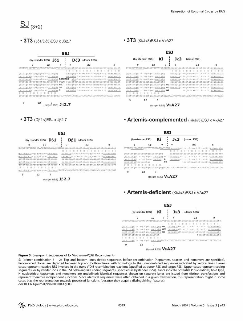
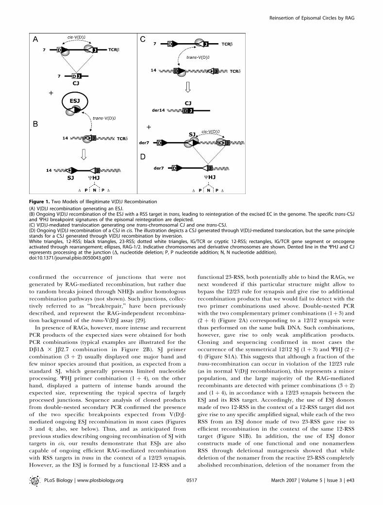
Figure 3. Breakpoint Sequences of Ex Vivo trans-V(D)J Recombinants
SJ (primer combination 3 þ 2). Top and bottom lanes depict sequences before recombination (heptamers, spacers and nonamers are specified).Recombined clones are depicted between top and bottom lanes, with homology to the unrecombined sequences indicated by vertical lines. Lowercases represent reactive RSS involved in the trans-V(D)J recombination reactions (specified as donor RSS and target RSS). Upper cases represent codingsegments, or bystander RSSs in the ESJ behaving like coding segments (specified as bystander RSSs). Italics indicate potential P nucleotides; bold type,N nucleotides; heptamers and nonamers are underlined; identical sequences shown on separate lanes are issued from distinct transfections andrepresent therefore independent junctions. Since identical sequences were often obtained in a given transfection, this representation might in somecases bias the representation towards processed junctions (because they acquire distinguishing features).doi:10.1371/journal.pbio.0050043.g003
PLoS Biology | www.plosbiology.org March 2007 | Volume 5 | Issue 3 | e430519
Reinsertion of Episomal Circles by RAG

of CJ formation without impeding SJ formation [31–33].Similarly, the absence of Artemis in the ESJ assay shouldprevent WHJ formation without hindering the generation ofSJs. Following cotransfection of the GUETEL cell line withthe (Ki/Jj3)ESJ/VjA27 couple, PCR was performed as aboveand the amplification products cloned and sequenced. Asexpected, SJs were obtained in absence of Artemis and wereundistinguishable from SJs obtained both in the Artemis-proficient 3T3 cells and in the GUETEL cell line comple-mented with Artemis (GUETEL-A; [30] and Figure 3). Despitethe absence of Artemis, weak amplification products werealso obtained for the WHJ PCR combination (1þ4). However,the sequenced junctions displayed virtual absence of Ninsertion and nucleotide processing (Figure 4), in sharpcontrast to the morphology of WHJs obtained in Artemis-proficient cells (3T3 and GUETEL-A). The features of theArtemis�/� junctions are however strongly reminiscent of HJsgenerated by the ‘‘RAG-mediated joining’’ pathway [15–18].RAG-mediated joining is an NHEJ-independent pathwayrelated to RAG transposition in which a direct attack of afree 39 hydroxyl group from the SE into the hairpinned CEbypasses the hairpin resolution step (illustrated in Figure S2,right panel); this usually results in the generation of a class ofHJs displaying a full-size RSS joined to a coding sequencewith limited processing (depending on the position of theattack in the hairpin). The presence of such junctions in theabsence of Artemis suggests that both trans-V(D)J recombi-nation and RAG-mediated joining concurrently occur togenerate WHJs and HJs, respectively. However, the virtualabsence of such junctions in Artemis-proficient cells (3T3,Artemis-complemented) strongly suggests that RAG-medi-ated joining constitutes a minor recombination pathwaycompared to trans-V(D)J recombination. Thus, the junctionswith a full-size ESJ bystander RSS initially observed inArtemis-proficient cells are Artemis dependent, and areeither WHJs in which processing is limited due to RAG-binding on the bystander RSS, or HJs generated through‘‘RSS swapping’’ (Figure S2, middle panel).
Altogether, our results indicate therefore that standardtrans-V(D)J recombination is the major pathway of RAG-mediated ESJs ongoing rearrangement.
ESJs Are as Efficient as Standard RSSs to Undergo trans-V(D)J Recombination Ex Vivo
How efficient is trans-V(D)J recombination of ESJs? In thecase of recombination between two standard coding-segmentRSSs, the frequency of trans-V(D)J recombination has beenpreviously shown to be reduced compared to cis-V(D)Jrecombination events [20,21]. In the present case of recombi-nation between an ESJ and its RSS target, the presence of thesecond RSS in the SJ could structurally impede—or on thecontrary stimulate—RAG fixation and/or activity on thereactive one, and modify the overall recombination effi-ciency. We therefore compared in the ex vivo assay theefficiency of trans-V(D)J recombination of an ESJ and a RSStarget with the efficiency of trans-V(D)J recombinationbetween two standard coding-segment RSSs (Figure 5A). Todo so, we tested two pairs of plasmids: (Jd1/Dd3) ESJ 3 Jb2.7versus Dd3 3 Jb2.7, and (Db1DESJ 3 Jb2.7 versus Db1 3 Jb2.7.Following transfection and harvesting carried out as above,semiquantitative primary PCR amplification of the break-points was performed, and serial dilutions were revealed by
PE. As illustrated in Figure 5B for the (Jd1/Dd3) ESJ 3 Jb2.7versus Dd33 Jb2.7 couples, no significant difference could beseen in the formation rate of SJs in the presence of an ESJ.Similarly, results showed comparable rates in the formationof a WHJ relative to a CJ. We conclude that ESJs are at least asefficient as standard RSSs to undergo trans-V(D)J recombina-tion. Altogether, this suggests that the presence of thebystander RSS does not impede the recombination process,whether structurally (through steric constraints) or function-ally (through nick–nick activity). From the mechanistic pointof view, this data predicts therefore that in vivo, V(D)J-mediated reintegration of ESJs (Figure 1B) should not bedifferent from V(D)J-mediated translocation (Figure 1C);most important, both processes should use the same RSStargets with the same efficiency.
Cryptic RSSs Involved in Oncogenic TranslocationsConstitute Hotspots of Episomal Reintegration throughOngoing V(D)J Recombination of an ESJV(D)J-mediated translocations have been shown to occur
not only between authentic RSSs from distinct IG/TCR loci,but also between authentic RSSs and fortuitous sequences inthe genome resembling a RSS (cryptic RSSs) [34]. Themistargeting of the RAGs towards cryptic RSSs located inthe vicinity of a silent proto-oncogene is a recurrent sourceof genomic instability and oncogenesis. Erroneous targetingof cryptic sites located near the LMO2 and TAL2 proto-oncogenes in t(11;14)(p13;q11) and t(7;9)(q34;q32) transloca-tions, respectively, represent prototypical examples of suchoncogenic translocations in T-cell acute lymphoblasticleukemia (T-ALL) [8,29,35–37]. Our data above suggest thatin vivo, such cryptic sites might provide efficient targets forESJ reintegration. To further define the potential oncogenicproperties of episomal reintegration, we next investigated inour ex vivo assay the capacity of ESJs to target oncogeniccryptic RSS. The human LMO2 and TAL2 cryptic RSSs andflanking sequences were cloned in a recombination substrateplasmid (Figure 6A) and assayed in parallel to the Jb2.7segment as a target for the (Jd1/Dd3) ESJ, using the PCR/PEassay described above. Our results show a similar consid-erable high rate of V(D)J-mediated recombination of the ESJwith the LMO2 and TAL2 cryptic RSSs than with the Jb2.7authentic RSS (Figure 6A). To further estimate the likelihoodin vivo of SJ insertion in such cryptic RSS, the LMO2 versusthe HPRT intron 1 region were assayed as competitive targetsfor the (Jd1/Dd3 ESJ (Figure 6B). The HPRT intron 1 regionwas chosen as a competitor because it contains a well-described cryptic RSS classically involved in illegitimateV(D)J-mediated deletion of exons 2–3 in vivo [38], and hasalso been identified as a site of ESJ reintegration in vivo ([39];see Discussion) as well as a region of RAG transposition invivo [19]. While recombination with the LMO2 cryptic RSSconstituted a hotspot of integration, no recombination couldbe observed in the HPRT region, neither specifically at thecryptic RSS, nor in the surrounding sequences (Figure 6B).Similar results were obtained when using a target plasmidcontaining a second copy of the HPRT fragment in a head-to-tail orientation, and forming a cruciform structure (notshown). Altogether, these data strongly suggest that crypticsites such as LMO2 with much higher recombinogenicpotential than the HPRT intron 1 cryptic RSS might also be
PLoS Biology | www.plosbiology.org March 2007 | Volume 5 | Issue 3 | e430520
Reinsertion of Episomal Circles by RAG

Figure 4. Breakpoint Sequences of Ex Vivo trans-V(D)J Recombinants
WHJ and/or HJ (primer combination 1þ 4). Nucleotides in parenthesis are ambiguous and could be assigned to either side of the joint. See also legendto Figure 3.doi:10.1371/journal.pbio.0050043.g004
PLoS Biology | www.plosbiology.org March 2007 | Volume 5 | Issue 3 | e430521
Reinsertion of Episomal Circles by RAG

targeted in vivo by ESJ insertion and could potentially lead tooncogenic activation.
trans-Chromosomal WHJ Breakpoints Are ReadilyDetectable In Vivo
We next investigated the physiological relevance of thereintegration of excised episomes through ongoing SJrecombination. As a first approach, we sought to assess iftrans-chromosomal WHJ breakpoints were present anddetectable in vivo, and if so, to estimate their formation rate.To do so, we designed primer combinations allowing theamplification of trans-TCR WHJ in mouse thymocyte DNA(Table 1). Additional combinations designed to amplify trans-TCR CJs and trans-TCR SJs were also performed as reference.To detect such relatively rare trans-TCR recombinationevents, we used a sensitive fluctuation PCR assay allowingthe detection of less than one recombination event in amillion cells (see Materials and Methods). Positive PCR-amplification replicates were obtained for all trans-TCRcombinations in the broad range of ;1 in 1,000,000 cells to1 in 10,000 cells. Sequence analysis of the PCR-amplificationproducts was carried out to assess the identity of thejunctions, and revealed the presence of WHJs (Figure S3),CJs, and SJs (not shown). Importantly, the WHJs obtained invivo displayed the same molecular features as observed in theex vivo assay, with extensive nucleotide processing on bothsides of the junctions, and N/P addition. These results clearlydemonstrate that WHJ breakpoints are indeed generated invivo. Furthermore, such junctions are readily detectable inmouse thymocytes at a range similar to that of equivalenttrans-CSJs and CJs. In line with our ex vivo results, thisindicates that in vivo, SJs undergo efficient ongoing cis- and/or trans-rearrangement with RSS targets in the genome withsurprisingly high frequency. This suggests further that inpresence of RAGs, CSJs and/or ESJs are indeed veryrecombinogenic structures.
Efficient In Vivo Reintegration of ECs through OngoingV(D)J Recombination of a SJ
Although we demonstrated above that trans-chromosomalWHJs are readily detectable in vivo, such breakpoints do notrepresent exclusive signatures of ESJ reintegration, as theymay also derive from ongoing recombination of trans-CSJswith neighboring gene segments in cis (compare Figure 1Band 1D). To ensure that the WHJs observed above did notexclusively derive from CSJs issued from trans-TCR trans-locations, and to estimate the rate of episomal reintegrationin vivo, we generated double mutant mice in which thegeneration of trans-CSJs is abolished (Figure 7). Eb�/� knock-out mice, in which deletion of the 560-bp Eb core generatesan .100-fold reduction in TCRb rearrangements [40] andTCRd/b translocations (Table 1), were crossed with a Db1GFPknockin mouse, in which the introduction of the GFP in theDb1 gene segment and flanking 23-RSS abolishes Db1-Jb1/2rearrangements (SS, OC, PF, unpublished data). In the(Db1GFP 3 Eb�) double-mutant (DE) mice, all TCRb chainsare consequently produced via a Vb-Db2-Jb2 rearrangementfrom the Db1GFP allele (unpublished data), and all TCRexcision circles issued from Db-Jb rearrangements carry a(Db2Jb2) ESJ (Figure 7A). Because one allele is knocked outfor Eb, and the other produces a TCRb chain, translocationsto TCRb cannot be present in TCRabþ cells from DE mice. In
absence of translocation to TCRb, no trans-TCRbd CSJ isproduced, and all Jb2.7/Jd1 WHJs are consequently issuedfrom episomal reintegration. As shown in Table 1, while therate of Jb2.7/Jd1 WHJs was decreased over ;40-fold in totalthymocytes from the Eb�/�mice compared to WT, the rate ofJb2.7/Jd1 WHJs was only decreased 1.2-fold in sorted TCRabþ
cells from DE mice compared to WT.Altogether, and in agreement with the ex vivo data, these
results clearly demonstrate that V(D)J-mediated episomalreintegration is indeed occurring in vivo, and at a ratecomparable to that of V(D)J-mediated chromosomal trans-location.
High Rates of Chromosomal Aberrations CombiningInsertion and TranslocationDuring our screen of trans-TCR SJs described above, we
noticed the presence of amplification products larger thanexpected. Out of a total of 310 PCR replicates of sevendistinct trans-TCRd/b SJ combinations, 120 were PCR positiveand four were of unexpected larger size (unpublished data).Sequencing of the cloned products revealed that largeamplicons resulted from cis-V(D)J recombination to a crypticsite in one case, and to a downstream gene segment inanother case; eventually, two of the four cases showed theinsertion of a sequence from the TCRd locus with featurescompatible with RAG-mediated ESJ reintegration. In one ofthese two cases, a ;900-bp Jd1-Dd2 fragment was insertedinto a Jb2.7/Vd2 CSJ target (Figure 8A). The breakpointsconsisted of a perfect SJ on the right arm of the insertion; onthe left arm, a junction compatible with a WHJ was found,displaying a deletion of three nucleotides on one side of thejoint, a deletion of one nucleotide on the other side, and oneN nucleotide addition; in the second case, the same ;900-bpJd1-Dd2 fragment was observed inserted into a Db1/Vd2 CSJtarget (Figure 8B). Similarly, one of the breakpoints displayeda perfect SJ, and the other breakpoint consisted of an SJ with2 N insertions. Considering the structure of the target, thislast junction was ambiguous to assign as an SJ or a WHJ, andcould have occurred through nick–nick, V(D)J, and/or RAG-mediated joining. Nevertheless, all junctions complied withthe 12/23 rule and with features of RAG-mediated breaks, andit is therefore very likely that both cases represent in vivoexamples of RAG-mediated reintegration of a Jd1-Dd2 ESJ. Inthe two cases, both ESJ reintegration and translocation eventsoccurred, and two mechanisms could therefore account fortheir formation: either the trans-TCR translocations occurredfirst, and provided a trans-TCR SJ target for ESJ reintegration;or, alternatively, ESJ reintegration in a standard RSS target(e.g., Vd2) could have occurred first, and provided a trans-TCRstructure and/or breakpoints prone to V(D)J-mediated trans-location. Intriguingly, the actual frequency of this doubletranslocation/insertion event (2/120 ¼ ;10�2 trans-recombi-nation events) was not compatible with the expectedcombined frequency of each independent event: (trans-TCRtranslocation ;10�4–10�6 3 reintegration ;10�4–10�6 ¼;10�8–10�12). Furthermore, as the assay is limited to PCR-amplifiable sizes of the inserted fragment (such as therelatively short Jd1-Dd2 episome), the apparent rate ofreintegration is probably vastly underestimated. This stronglysuggests that the two events are linked, either because CSJsconstitute preferential targets for ESJ reintegration, orbecause ESJ reintegration generates genomic instability
PLoS Biology | www.plosbiology.org March 2007 | Volume 5 | Issue 3 | e430522
Reinsertion of Episomal Circles by RAG

leading to further chromosomal abnormalities; such abnor-malities could consist of chromosomal translocations asdetected here, or potentially more complex events whichcould not be detected in the present in vivo assay.
Altogether, these data demonstrate that in vivo, SJs locatedon excised ECs may be reintegrated into the genome throughtrans-V(D)J recombination using standard and cryptic RSStargets, and further suggest that this event is associated withadditional genomic instability.
Discussion
During B- and T-cell ontogeny, ECs are sequentially excisedfrom the genome of lymphoid cells as a result of the
hierarchically regulated D-to-J, V-to-DJ, and V-to-J rear-rangement events. Such episomes are believed to be non-replicative, but have nevertheless been shown to besurprisingly stable structures, persisting until diluted out bycell divisions [41,42]. Such episomes carry an ESJ comprisedof two functional RSSs, each of which are susceptible to (re-)bind the RAG proteins when (re-)expressed at the variouscell maturation steps until final downregulation [7,41,43]. Onthe other hand, trans-V(D)J recombination occurring betweenRSSs or cryptic RSSs located on distinct chromosomes is arelatively common event in developing lymphocytes[22,23,44], which has been shown to lead to recurrentoncogenic translocations [34]. Here we show both ex vivoand in vivo that ECs may be reintegrated into the genome
Figure 5. Semiquantitative Ex Vivo trans-V(D)J Recombination Assay
(A) Schematic representation of the comparative trans-V(D)J recombination assay. Left panel: recombination of an ESJ 23-RSS (Jd1/Dd3 or Db1D) with a12-RSS target (Jb2.7); Right panel: recombination of a coding segment/23-RSS (Dd3 or Db1) with a 12-RSS target (Jb2.7). The expected breakpoints areshown for each case.(B) Typical example of semiquantitative PCR/PE assays obtained from the (Jd1/Dd3) ESJ 3 Jb2.7 versus Dd3 3 Jb2.7 comparisons. PE assays shown wereperformed on serial dilutions of PCR products.doi:10.1371/journal.pbio.0050043.g005
PLoS Biology | www.plosbiology.org March 2007 | Volume 5 | Issue 3 | e430523
Reinsertion of Episomal Circles by RAG

through trans-V(D)J recombination occurring between theESJ and an IG/TCR or one of the many cryptic RSS targetsscattered in the genome.
Molecular Mechanisms of ESJ Reintegration in Ex VivoAssays
We have demonstrated in ex vivo assays that the efficiencyof trans-V(D)J recombination of an ESJ with a RSS target isnot quantitatively different from the trans-V(D)J recombina-tion occurring between two RSSs. This is somehow unex-pected, because its particular structure confers additionalproperties to the ESJ. ESJs are efficiently cleaved ex vivo andin vitro by the nick–nick mechanism, a symmetrical nickoccurring 59 of each RSS (simultaneously or sequentially) andgenerating two flush SEs ending with 39 hydroxyl groups [7].Remarkably, this process bypasses both the formation of ahairpin intermediate and the need of synapsis with another
RSS. In the context of ongoing SJ recombination, one couldthink that efficient nick–nick opening of lone ESJs upon RAGbinding might prevent the occurrence of synapsis with a RSStarget, and thus considerably reduce the overall frequency oftrans-V(D)J rearrangement. Our ex vivo data clearly argueagainst this assumption, and suggest that trans-V(D)J rear-rangement is largely independent of nick–nick. This howeverdoes not preclude that ESJ opening by nick–nick mightoccasionally participate in the synapsis, and the recent in vivodemonstration of synapsis by capture [2] provides a plausibletwo-step scenario of the occurrence of nick–nick within a 12/23 synapse (Figure S4).Incidentally, our data provides additional evidence that HJs
can be formed ex vivo through both NHEJ-dependent andNHEJ-independent pathways (Figure S2). RAG-mediatedjoining has been initially proposed as an efficient alternativepathway of NHEJ-independent HJ formation [16–18]. How-
Figure 6. Ex Vivo SJ trans-V(D)J Recombination Assay with Cryptic RSS Targets
(A) Recombination of (Jd1/Dd3) ESJ with Jb2.7 12-RSS (white triangle with open dot), LMO2, or TAL2 cryptic 12-RSS (white triangle with filled dot). PEassays shown were performed on 1 ll primary PCR. Five independent PCRs from two independent transfections (T1–T2, T3–T4, and T5–T6) are shownfor each junction. Similar results were obtained when LMO2 and Jb2.7 RSSs were cloned on a single plasmid as competitive targets.(B) Competitive recombination of (Jd1/Dd3) ESJ with HPRT intron 1 cryptic RSS (black triangle with white dot) versus LMO2 cryptic RSS (white trianglewith filled dots). SJ, Jd1/HPRT; WHJ, Jd1/LMO2. PE assays shown were performed on 3 ll primary PCR using 30 cycles of PE. Five independent PCRs fromtwo independent transfections performed in the presence (T1–T2) or absence of RAG-1/2 (T3–T4) are shown for each junction. Similar results wereobtained when LMO2 and HPRT were cloned on separate plasmids and assayed in parallel. As described in the text, absence of specific V(D)Jrecombination (e.g., in absence of RAG, T3 ;485 bp) favors the emergence of nonspecific and nonrecurrent recombination events (‘‘break-repair’’).doi:10.1371/journal.pbio.0050043.g006
PLoS Biology | www.plosbiology.org March 2007 | Volume 5 | Issue 3 | e430524
Reinsertion of Episomal Circles by RAG

ever, conflicting data have been reported on the relevance ofthis pathway ex vivo and in vivo [15,25–27,45,46]. Inparticular, recent data indicated that at least ex vivo, RAG-mediated joining is seldom observed in presence of full-length RAGs, even in the absence of the concurrent NHEJpathway [15,46]. We find here the presence of HJ with featuresof RAG-mediated joining in the absence, but not in thepresence of Artemis, suggesting that at least ex vivo, HJformation is a mixed process consisting of efficient RSS‘‘swapping’’ and inefficient RAG-mediated joining. Interest-ingly, it is possible that the frequency of RAG-mediatedjoining would be favored in our assay, due to the participationof an ESJ. As mentioned above, nick–nick opening of the ESJin the context of a 12/23 synaptic complex would generate twoflush SEs ending with 39 hydroxyl groups. While the tworeactive RSSs might be sequestered by tight binding in the SEpostcleavage complex (Figure S4), this additional free 39 OHwould provide an available substrate for hairpin attack, atleast in absence of concurrent hairpin resolution by Artemis.Thus, nick–nick opening might participate in both trans-V(D)Jrecombination and RAG-mediated joining.
In Vivo Reintegration of Excised ECs by the V(D)JRecombinase
Using WT and DE double-mutant mice, we could estimatethat ESJ reintegration in authentic TCR RSS targets occurs invivo in the broad range of 1 in 1,000,000 to 1 in 10,000 mousethymocytes, depending on the integration site. In agreementwith our results, recent data provide further evidence for theexistence of V(D)J-mediated ESJ reintegration in vivo, andconcur with the idea that it constitutes a significant source ofgenomic instability. Using a screen for HPRT mutants,Finette and colleagues recently identified a case in which aVa/Ja episome was found inserted in the cryptic 23-RSS fromHPRT intron 1 [39]. This finding is all the more remarkablegiven that this HPRT cryptic site has been shown to exhibit a
very low recombinogenic potential in functional assays ([47],our unpublished data), and provides direct evidence that invivo, human cryptic RSSs constitute efficient targets for ESJreintegration. In the same line, using a double selection assayallowing for the recovery and quantification of excision/reinsertion events in a pre–B-cell line, Reddy et al. recentlyidentified three cases of V(D)J-mediated reintegration [48]. Infull agreement with our in vivo estimation, they evaluated arate of one reintegration out of every 100,000 V(D)Jrecombinations. Considering the average number of V(D)Jrecombination per B or T cell, these data suggest that thedaily lymphocyte output in human could be accompanied byas many as 5,000 ESJ reintegration events.
A Potential Threat for Genomic Stability and LymphoidNeoplasiaAuthentic RSSs from IG/TCR must without doubt con-
stitute preferential targets for such reintegration. However,there are an estimated 10 million functional cryptic sitesdispersed throughout the human genome [34,47], some ofthem displaying much higher recombinogenic potential thanthe HPRT intron 1 cryptic RSS in which ESJ reintegration wasfound [29,37]. Most important, some of them are alreadyknown to provide recurrent targets for oncogenic (type 1)V(D)J-mediated translocations. In T-ALL, for example, themistargeting of the RAGs towards such cryptic RSSs locatedin the vicinity of a silent proto-oncogene is a recurrentsource of genomic instability and oncogenesis [34]. Wedemonstrate in our ex vivo assay that cryptic sites involvedin oncogenic V(D)J-mediated translocations are also hotspotsfor ESJ recombination; it seems thus reasonable to think thatin vivo, such cryptic RSSs might provide as efficient targetsfor ESJ reintegration than for chromosomal translocations.ESJ reintegration could lead to similar oncogenic activation/deregulation, either through the insertion of active immuneregulatory elements (e.g., the TCRd enhancer excised during
Table 1. In Vivo Rates of trans-TCR Junctions
Mouse Model Thymocyte Source trans-TCR Combination Expected Junction Fluctuation PCR Ratea Calculated Frequencyb
WT Total Vd2/Jb2.7 CJ 72/80 (90%) ;0.8 3 10�5
WT Total Db1/Jd2 CJ 8/31 (26%) ;1.0 3 10�6
WT Total Dd2/Jb2.7 CJ 25/30 (83%) ;0.6 3 10�5
WT Total Jb2.7/Vd2 SJ 15/60 (25%) ;1.0 3 10�6
WT Total Db2/Vd2 SJ/WHJc 43/60 (72%) ;0.4 3 10�5
WT Total Db1/Vd2 SJ/WHJc 3/20 (15%) ;0.5 3 10�6
WT Total Jb2.7/Dd2 SJ/WHJc 43/70 (72%) ;0.3 3 10�5
WT Total Jb2.7/Dd1 SJ/WHJc 15/60 (25%) ;1.0 3 10�6
WT Total Jb2.7/Jd2 WHJ 10/40 (25%) ;1.0 3 10�6
WT Total Jb2.7/Jd1 WHJ 25/40 (62%)d ;1.3 3 10�4
Eb�/� Total Vd2/Jb2.7 CJ 0/20 (0%) , 1.0 3 10�7
Eb�/� Total Db1/Jd2 CJ 0/34 (0%) , 1.0 3 10�7
Eb�/� Total Dd2/Jb2.7 CJ 0/34 (0%) , 1.0 3 10�7
Eb�/� Total Jb2.7/Jd1 WHJ 1/40 (2.5%)d ;0.3 3 10�5
Eb�/� Total Jb2.7/Jd1 WHJ 1/40 (2.5%)d ;0.3 3 10�5
WT TCRabþ Jb2.7/Jd1 WHJ 14/40 (35%)d ;0.6 3 10�4
DE TCRabþ Jb2.7/Jd1 WHJ 12/40 (30%)d ;0.5 3 10�4
aFraction of positive PCR/total number of repeats (2 lg/replicate).bCalculated from the fluctuation PCR rate using Poisson’s assumption [62].cCombinations which could either represent trans-TCR WHJ or trans-TCR SJ.dFor positive replicates out of the fluctuation range, PCR was repeated with 50 ng/replicate, and frequencies calculated accordingly.doi:10.1371/journal.pbio.0050043.t001
PLoS Biology | www.plosbiology.org March 2007 | Volume 5 | Issue 3 | e430525
Reinsertion of Episomal Circles by RAG

DRec/WJa or Va/Ja rearrangements [42,49], or the IGKenhancer excised during KDE/Ki rearrangements [12,50]) orthrough the disruption of locus silencing (e.g., the negativeregulatory element upstream of LMO2 [51,52], WA Dik; BN etal., unpublished data).
If ESJs are at least as efficient as any standard coding-segment RSS to undergo V(D)J recombination with a RSStarget in trans, the two processes are nearly identicalmechanistically, and the RSS/cryptic RSS targets are thesame, why then has V(D)J-mediated oncogenic translocationbeen largely documented in the literature, but V(D)J-mediated reintegration never been reported up to now in
lymphoid malignancies? Higher-order spatial genome organ-ization is a contributing factor in the formation of recurrenttranslocations [53]. In contrast, increased mobility of the ECmight favor interactions and recombination of ESJs, withtargets located in parts of the genome generally segregated.Oncogenic targets might thus be distinct for reintegrationand translocation. However, due to the multiplicity and sizerange (from ,1 Kb to several Mb) of the excised episomes, aswell as the diversity of insertion targets, reintegration eventsare very unlikely to be detected by routine analysis.Particularly, and in contrast to chromosomal translocations,episomal reintegration is invisible by standard karyotype
Figure 7. Strategy for In Vivo Detection of Episomal Reintegration in the (Db1GFP 3 Eb�) double-mutant (DE) mouse
(A) TCRb allelic configuration of the DE mouse. The Eb knockout prevents efficient rearrangements on allele 1, and the GFP knockin in Db1 prevents D-Jcluster 1 rearrangements on allele 2. In this configuration, all productive TCRb chains are issued from Vb rearrangements to D-J cluster 2 of allele 2.Consequently, TCRb chain production and translocation to the TCRb locus are mutually exclusive. The generation of a TCR excision circle carrying a(Jb2/Db2)ESJ through Db2-Jb2 rearrangements from the Db1GFP allele is depicted.(B) Reintegration of (Jb2/Db2) ESJs in Jd1. In TCRabþ cell-sorted thymocytes from DE mice, the presence of a Jb2.7/Jd1 WHJ specifically signs thereintegration of (Jb2/Db2)ESJ in Jd1, and is assayed by fluctuation PCR.doi:10.1371/journal.pbio.0050043.g007
PLoS Biology | www.plosbiology.org March 2007 | Volume 5 | Issue 3 | e430526
Reinsertion of Episomal Circles by RAG

analysis [54,55]. Few examples of identified class-switchrecombination– and RAG-mediated episomal reintegrationin lymphoid neoplasia provide proof of principle thatepisomal reintegration can indeed lead to cancer, andillustrate the complexity and unlikelihood of identifying suchevents without specifically designed screens [55–57]. Forexample, using complex DNA fiber-FISH and three-colorinterphase FISH techniques on samples from follicularlymphoma patients devoid of the hallmark t(14;18)(q32;q21)translocation, Vaandrager et al. identified two cases in whichthe BCL2 gene was excised from 18q21 and inserted into theIGH locus at 14q32 [58]. The relatively high frequency (5%) ofsuch events, discovered relatively recently by Vaandrager etal. out of an abundantly studied and characterized pathology,illustrate well the extent to which insertional events might bemissed by routine cytogenetic and molecular analysis.Another possibility which could account for the rarity of
cases of episomal reintegration observed in lymphoid neo-plasia is that episomal reinsertions might generate unstabletransitory structures in the genome, leading to additionalaberration events and more complex genomic configuration.This possibility is supported by the unexpected highfrequency of two in vivo cases observed in this study,combining ESJ reintegration and translocation events.Similar examples of genomic instability following reintegra-tion of episomal structures have been previously docu-mented. In mouse plasmacytoma, Kovalchuk and colleagueshave shown that class-switch–mediated El/Sl episomalreintegration into c-MYC favors t(12;15) translocation [56].Although the ground of such instability is not yet clear, anobvious possibility is that a fraction of the recombinationevents might lead to incomplete insertion in vivo. Alter-natively, the reinserted episomes might in vivo retain theirinitial ‘‘open’’ chromatinized structures, and provide prefer-
Figure 8. Two In Vivo Cases of Combined Chromosomal Translocation and Reintegration of an Excised EC
The insertion of a (Jd1/Dd2) ESJ (red, upper cases) into (A) a Jb2.7/Vd2 trans-TCR or (B) a Db1/Vd2 trans-TCR CSJ target (black, lower cases) is depicted;alternatively, ESJ reintegration could have occurred first, and provided a trans-TCR structure and/or breakpoints prone to V(D)J-mediated translocation.Breakpoint junctions are boxed. Black bold upper case type: N nucleotides; heptamers and nonamers are underlined. PCR primers are indicated, andshown in italics in the sequence.doi:10.1371/journal.pbio.0050043.g008
PLoS Biology | www.plosbiology.org March 2007 | Volume 5 | Issue 3 | e430527
Reinsertion of Episomal Circles by RAG

ential accessible targets for the recombination machinery.The V(D)J-mediated instability of reinserted ESJs would beespecially relevant in neoplastic cells such as T-ALLs, inwhich arrest of differentiation at early stages of T-celldevelopment leads to sustained RAG levels.
Gene profiling studies have shown that a substantialfraction of the T-ALL cases display oncogenic activation inabsence of detectable chromosomal alterations [59], suggest-ing the presence of alternative pathways of oncogenesis.Some of them have indeed been recently discovered, andinvolve episomal structures [60]. Considering the largenumber of ESJs produced daily and the mechanisticsimilarities between ESJ reintegration and oncogenic trans-locations, our data suggest that reintegration of excised ECsby the V(D)J recombinase might also account for some ofthese cases, and constitute an additional potent source ofgenomic instability. Definitive answer to this open questionwill however await large-scale screens of human lymphoidcancer samples with specifically adapted strategies.
Materials and Methods
Ex vivo trans-recombination substrate assay. Recombinationsubstrates were derived from a series previously described [29], andthe constructs are summarized in Figure S5. The various genesegments containing the regions to recombine were amplified fromhuman DNA with appropriate tailed primers and cloned in therecombination substrate using unique restriction sites (Mlu1, Sac2,Not1). The (Ki/Jj3) ESJ was cloned (Not1/BamH1) in the pPCR-scriptAmp vector (Stratagene, http://www.stratagene.com). Someconstructs were flanked by ‘‘tag’’ sequences, which were previouslytested to be devoid of functional cryptic RSSs, and in which specificPCR primers were designed. NIH 3T3 Swiss mouse fibroblasts, andhuman Artemis-deficient GUETEL or Artemis-complemented GUE-TEL-A cell lines [30] (generously supplied by J.-P. de Villartay) werecultured in standard conditions (DMEM/10% FCS). Cells (2 3 106)were transfected with 3 lg of each recombination substrate, 2 lgpEBB-RAG1, 2 lg pEBB-RAG2 expression vectors (generouslysupplied by C. Roman and S. Cherry), and 2 lg pCDNA3TdTexpression vector (TdT cDNA from the pTDT expression vector [agenerous gift from N. Doyen] recloned into pcDNA3 [Invitrogen,http://www.invitrogen.com]) using Superfect (Qiagen, http://www.qiagen.com) according to the instructions recommended by themanufacturer. Transfected cells were transferred to 10 ml DMEMsupplemented with 10% FCS and cultured for 48 h. Cells weresubsequently trypsinized, and plasmids recovered by alkaline lysis andphenol-chloroform extraction as previously described [29]. To testthe possibility that trans-rearrangements could occur through a firststep of homologous recombination between the identical coreregions of the donor/acceptor plasmids, the trans-recombinationassay was also performed with ‘‘coreless’’ excised linear fragmentscarrying the RSS target, and gave rise to similar results (Figure S6).Although we cannot formally exclude the possibility that whenpresent, stretches of homology can facilitate the recombinationprocess both ex vivo and in vivo, our data suggests that recombina-tion efficiently occurs through direct trans-V(D)J synapsis aspreviously assumed for the assay [20].
PCR/sequencing. Breakpoints were PCR amplified from 1 ll (1/20)harvested bulk DNA with appropriate primers (summarized in TableS1) in the following conditions: 4 min at 948C for 25 cycles (30 s at948C, 30 s at 648C, and 30 s at 728C), and 7 min at 728C. Secondarydouble-nested PCRs were performed in the same conditions, and theamplification products cloned and sequenced as previously described[29].
Semiquantitative PCR/PE assays. Breakpoints were amplified in asingle round PCR from 1 ll (1/20) harvested bulk DNA withappropriate primers (summarized in Table S2) in the same conditionsas above, and otherwise undetectable amplification products wererevealed by PE. PE is a sensitive alternative to Southern blotconsisting of several cycles of DNA polymerization extending froma labeled primer until the end of a matrix DNA fragment (here a PCRproduct). This generates linear (nonexponential) accumulation oflabeled fragments of specific size. PE assays were performed on 1.5 ll,1.0 ll, and 0.75 ll of primary PCR using a nested IR800-labeled
primer in the following conditions: 5 min at 958C for 20 cycles (30 s at958C, 15 s at 608C and 1 min at 708C), and 1 min at 708C with theEXCEL II kit (Epicentre Biotechnologies, http://epicentre.com), usingan equal amount of all dNTPs and omitting ddNTPs. A portion (1.2ll) of the reaction was used for running on a Li-COR 4200 DNAsequencer (http://www.licor.com). A sequencing reaction was per-formed in parallel on the appropriate unrecombined purifiedplasmid, using the same IR800-labeled primer and in the samereaction conditions (at the exception of the dNTP/ddNTP mix), andwas run on the same gel, providing both a precise to-the-base sizemarker and a positive control for the reaction. Semiquantitativeconditions were calibrated on serial dilutions of bulk DNA and ofPCR amplifications (Figure S7A–S7C). Furthermore, to excludepossible bias due to difference in the efficiency of the PCR primercombinations used in the semiquantitative experiments, eachcomparison using different sets of primers was calibrated (FigureS7D–S7E). All PCR/PE assays were performed on at least fourindependent transfections with similar results.
Flow cytometry and cell-cycle analysis. Thymocyte preparation andcell sorting were performed as described previously [61]. Phycoery-thrin-conjugated mAb against TCRb (H57–597), purchased from BDPharMingen (http://www.bdbiosciences.com), was used for cell sortingof TCRbþ thymocytes. The sorting windows were defined in such away that only cells expressing high levels of TCRb were purified.
In vivo fluctuation PCR assay. The principle of the assay has beenpreviously described [29]. Detection of rare events by sensitivedouble-nested PCR gives rise to fluctuation in target amplification,depending on the presence or not of the event in the aliquot takenfrom the sample. Junctions were PCR amplified from multiplereplicates of 2 lg DNA (or 50 ng for positive replicates out of thefluctuation range) isolated either from total thymocytes of WT orEb�/� mice [40], or from TCRabþ sorted thymocytes from WT or DEdouble-mutant mice, using appropriate primers (see list in ProtocolS1), and in the following conditions: 4 min at 948C for 30 cycles (30s at 948C, 30 s at 648C, and 30 s at 728C), and 7 min at 728C.Secondary double-nested PCRs were performed on 1 ll of eachprimary PCR replicate in the same conditions, and amplificationproducts were cloned and sequenced as previously described [29].Frequencies were calculated using Poisson assumption as previouslydescribed [62].
Supporting Information
Figure S1. Ex Vivo trans-V(D)J Recombination of ESJs Obeys the 12/23Rule and Is Not Nick–Nick Dependent
(A) SJ and WHJ/HJ sequences obtained with the complementaryprimer combinations (1þ3) and (2þ4) (see Figure 2A) correspondingto a 12/12 synapsis.(B) Ex vivo trans-V(D)J recombination of ESJs made of two 12-RSSs(Db1/Dd1) or made of two 23-RSSs (Db1/Dd1) in the context of a 12-RSS target (Jb2.7). Constructs and primers are indicated in Figure S5.(C) Ex vivo trans-V(D)J recombination of nonamerless ESJs (indicatedas D9) in the context of a 12-RSS target. See Figure S5 for constructand primer details.
Found at doi:10.1371/journal.pbio.0050043.sg001 (3.7 MB PDF).
Figure S2. Three Possible Pathways of a RAG-Mediated HybridJunction Resulting from Ongoing Recombination of an ESJ in trans(Left) ESJ opens by standard RAG-mediated nick and trans-ester-ification in the context of a normal 12/23 synapsis; despite its abilityto bind the RAGs, the bystander 12-RSS of the ESJ behaves as acoding segment, and undergoes hairpin formation. Artemis-depend-ent hairpin resolution, further processing, and NHEJ-dependentrepair leads to a WHJ displaying processing on both sides of the joint;nevertheless, processing may be more limited at the RSS side, due toprotection conferred by RAG binding.(Middle) SJ opening by nick–nick in the context of a standard 12/23synapse (see Figure S4 for details) provides an ESJ 12-RSS with a free39 OH. As an alternative, a rare 12/12 synapsis might also provide anESJ 12-RSS with a free 39 OH. In an Artemis-dependent pathway,hairpins are resolved by Artemis, processing occurs, and intermediateproducts are mistakenly repaired by RSS ‘‘swapping,’’ leading to HJformation. Because the ESJ 12-RSS did not go through a hairpinformation, and is bound to the RAGs, limited processing occurs at theRSS side.(Right) In the Artemis-independent RAG-mediated joining pathway,a direct attack of the ESJ 12-RSS free 39 OH into the hairpinned CEbypasses the hairpin resolution step, and results in the generation of
PLoS Biology | www.plosbiology.org March 2007 | Volume 5 | Issue 3 | e430528
Reinsertion of Episomal Circles by RAG

a class of HJ displaying a full-size RSS joined to a coding sequencewith limited processing (depending on the position of the attack inthe hairpin). In sequences from Artemis-proficient cells, the virtualabsence of joints with features of RAG-mediated joining, the minorrepresentation of products from 12/12 synapsis, and the presence ofnucleotide deletion/addition at both sides in most of the joints,indicate that in the vast majority of cases, ESJs opened by standardRAG-mediated nick and trans-esterification in the context of anormal 12/23 synapsis.White triangles, 12-RSS; black triangles, 23-RSS. Dented linesrepresent nucleotide processing; D, nucleotide deletion; P, Pnucleotide addition; N, N nucleotide addition. For clarity, thegeneration of only one hybrid junction is represented.
Found at doi:10.1371/journal.pbio.0050043.sg002 (351 KB PDF).
Figure S3. In Vivo Detection of trans-TCR WHJs in Mouse Thymocytes
Top lanes depict germline sequences (heptamers, spacers, andnonamers are specified; heptamers and nonamers are underlined).Upper cases represent coding-segments. Lower cases represent(bystander) RSSs. Recombined clones are depicted underneath withhomology to the germline sequences indicated by vertical lines. Aschematic representation of the PCR assay used for the detection ofJb2.7/Dd(1/2) and Jb2.7/Jd2) WHJs is depicted. PCR primers used areindicated; for the Jb2.7/Dd combinations, some cases were ambiguousbecause the sequences could correspond either to WHJs with largedeletions or to processed SJs (sequences marked SJ/WHJ). Never-theless, at least half of the cases could be unambiguously assigned to aWHJ due to the presence of remaining nucleotides from the Ddcoding sequence. Italics indicate potential P nucleotides; bold type, Nnucleotides. Nucleotides in parenthesis are ambiguous and could beassigned to either side of the joint.
Found at doi:10.1371/journal.pbio.0050043.sg003 (1.4 MB PDF).
Figure S4. Hypothetical Two-Step Scenario of the Occurrence ofNick–Nick within a 12/23 Synapsis
In a first step, both the target 12-RSSs and the ESJ 12-RSSs wouldindependently undergo a RAG-specific nick; in a second step, theRAG-associated prenicked 12-RSS target would capture the 23-RSSsof the prenicked ESJ, forming a synaptic complex and initiating thesecond, symmetrical nick at the ESJ, eventually resulting to itsopening in absence of transesterification. Although flush ESJ 12-RSSscould also be provided by rare 12/12 synapses, the two-step nick–nickprocess provides a potential pathway by which HJs could begenerated in the context of a 12/23 synapse (Figure S2). Ellipsesrepresent RAG-1/2, regardless of stochiometry.
Found at doi:10.1371/journal.pbio.0050043.sg004 (1.2 MB PDF).
Figure S5. Schematic Representation of the Recombination Cassettes(to Scale) Inserted in the Donor (ESJ, RSS) and Target (RSS)Recombination Substrates
Cassettes and tags are delimited by lozenges, and PCR/PE primers areindicated. Broken line in the (Ki/Jj3) ESJ represents core vectorsequence. M, Mlu1; N, Not1; Sc, Sac2.
Found at doi:10.1371/journal.pbio.0050043.sg005 (1.5 MB PDF).
Figure S6. Ex Vivo trans-V(D)J Recombination Assay with CorelessTarget Plasmid
The 12-RSS target is located on a linear fragment excised from theplasmid core, and is devoid of large regions of homology with the ESJplasmid. See also legend to Figures 2 and 3 for PCR/PE assay andbreakpoint sequences.
Found at doi:10.1371/journal.pbio.0050043.sg006 (1.4 MB PDF).
Figure S7. Calibration of the PCR/PE Assay
The PCR/PE assay was tested by performing PCR amplification onserial dilutions of bulk DNA harvested from transfections.(A) A portion (10 ll) of the reaction was loaded on ethidiumbromide–stained 1% agarose gel (M, 100-bp marker). The expectedband (319 bp) is not detectable at any dilution.
(B) A portion (1 ll) of the PCR was then used for the PE assay, in thesame conditions as described in Materials and Methods. The PCR/PEassay for the (Jd1/Dd3) ESJ/Jb2.7 T1 transfection is shown, andillustrates the dynamic range of the assay. Bulk DNA (1 ll) wassubsequently used as the standard condition for semiquantitativePCR.(C) PE assay conditions were also tested on serial dilutions of PCRamplification products performed on 1 ll harvested bulk DNA. Threedilutions (1.5 ll, 1.0 ll, 0.75 ll) were used as the standard conditionfor the PE assay.(D) Primer calibrations. Triplex PCR amplifications were performedin the same conditions as the primary PCR described in Materials andMethods, using a 1:1:1 mix of the indicated primers, and on serialdilutions of a 1:1 mix of plasmid DNA constructs containing theappropriate tags (indicated in brackets); each primer coupleamplified its specific target with similar sensitivity, indicating thatthere is no large bias in the PCR detection of the rearrangements dueto the use of distinct primers.(E) Similarly, the efficiencies of the various labeled PE primers werealso compared on serial dilutions from primary PCR. PE assays wereperformed in the same conditions as described in Materials andMethods, using the indicated labeled primers (*), on serial dilutionsof the last dilution point (0.25 pg) of the corresponding triplex PCR.Again, primer comparison shows a similar sensitivity, indicating thatthere is no large bias in the PE detection of the rearrangements.
Found at doi:10.1371/journal.pbio.0050043.sg007 (4.4 MB PDF).
Protocol S1. List of PCR Primers
Found at doi:10.1371/journal.pbio.0050043.sd001 (29 KB DOC).
Table S1. Primer Combinations in Ex Vivo Cloning Experiments
Found at doi:10.1371/journal.pbio.0050043.st001 (17 KB XLS).
Table S2. Primer Combinations in Ex Vivo Semiquantitative Experi-ments
Found at doi:10.1371/journal.pbio.0050043.st002 (19 KB XLS).
Acknowledgments
We thank C. Roman and S. Cherry for the generous gift of pEBB-RAG1 and pEBB-RAG2 expression vectors, N. Doyen for the generousgift of the pTDT expression vector, and J. P. de Villartay for thegenerous gift of the GUETEL/ GUETEL-A cell lines. We are gratefulto D. Payet-Bornet, V. Asnafi, J. P. de Villartay, B. Finette, W. A. Dik,D. B. Roth, D. C. van Gent, R. W. Hendriks, S. Spicuglia, D.-M.Franchini, I. Prinz, and B. Malissen for helpful discussions; and to C.Verthuy for technical assistance.
Author contributions. KV, BM, SS, IP, RM, and BN conceived anddesigned the experiments. KV, BM, TL, JMN, OC, SR, and EVperformed the experiments. KV, BM, and BN analyzed the data. PFand UJ contributed reagents/materials/analysis tools. KV, BM, and BNwrote the paper.
Funding. This work was supported by the grant ID20010 from theCentre of Molecular Medicine (Austrian Academy of Science), thegrant P13984 from the Fonds zur Forderung der WissenschaftlichenForschung (FWF), the Austrian GEN-AU GZ 200.136/1-VI/1/2005project, the grant INE2003114116 from la Fondation pour laRecherche Medicale (FRM), a grant from Le Conseil General desBouches du Rhone, and institutional grants from Institut National dela Sante et de la Recherche Medicale (INSERM) and Centre NationalDe La Recherche Scientifique (CNRS). Work in the BN lab issupported by the AVENIR2003 grant from INSERM. The PF lab issupported by specific grants from the Association pour la Recherchesur le Cancer (ARC grant 3275) and the Foundation Princesse Gracede Monaco. BN is a recipient of a Contrat d’Interface INSERM/Assistance Publique-Hopitaux de Marseille. BM is a recipient of afellowship from la Ligue Nationale Contre le Cancer.
Competing interests. The authors have declared that no competinginterests exist.
References
1. Schatz DG (2004) V(D)J recombination. Immunol Rev 200: 5–11.2. Curry JD, Geier JK, Schlissel MS (2005) Single-strand recombination signal
sequence nicks in vivo: evidence for a capture model of synapsis. NatImmunol 6: 1272–1279.
3. McBlane JF, van Gent DC, Ramsden DA, Romeo C, Cuomo CA, et al. (1995)
Cleavage at a V(D)J recombination signal requires only RAG1 and RAG2proteins and occurs in two steps. Cell 83: 387–395.
4. Rooney S, Chaudhuri J, Alt FW (2004) The role of the non-homologous end-joining pathway in lymphocyte development. Immunol Rev 200: 115–131.
5. Buck D, Malivert L, de Chasseval R, Barraud A, Fondaneche MC, et al.(2006) Cernunnos, a novel nonhomologous end-joining factor, is mutatedin human immunodeficiency with microcephaly. Cell 124: 287–299.
PLoS Biology | www.plosbiology.org March 2007 | Volume 5 | Issue 3 | e430529
Reinsertion of Episomal Circles by RAG

6. Ahnesorg P, Smith P, Jackson SP (2006) XLF interacts with the XRCC4-DNA ligase IV complex to promote DNA nonhomologous end-joining. Cell124: 301–313.
7. Neiditch MB, Lee GS, Huye LE, Brandt VL, Roth DB (2002) The V(D)Jrecombinase efficiently cleaves and transposes signal joints. Mol Cell 9:871–878.
8. Marculescu R, Vanura K, Le T, Simon P, Jager U, et al. (2003) Distinctt(7;9)(q34;q32) breakpoints in healthy individuals and individuals with T-ALL. Nat Genet 33: 342–344.
9. Lewis S, Gifford A, Baltimore D (1984) Joining of V kappa to J kappa genesegments in a retroviral vector introduced into lymphoid cells. Nature 308:425–428.
10. Lewis S, Gifford A, Baltimore D (1985) DNA elements are asymmetricallyjoined during the site-specific recombination of kappa immunoglobulingenes. Science 228: 677–685.
11. Hesse JE, Lieber MR, Gellert M, Mizuuchi K (1987) Extrachromosomal DNAsubstrates in pre-B cells undergo inversion or deletion at immunoglobulinV-(D)-J joining signals. Cell 49: 775–783.
12. Langerak AW, Nadel B, De Torbal A, Wolvers-Tettero IL, Gastel-Mol EJ, etal. (2004) Unraveling the consecutive recombination events in the humanIGK locus. J Immunol 173: 3878–3888.
13. Lewis SM, Hesse JE, Mizuuchi K, Gellert M (1988) Novel strand exchanges inV(D)J recombination. Cell 55: 1099–1107.
14. Morzycka-Wroblewska E, Lee FE, Desiderio SV (1988) Unusual immuno-globulin gene rearrangement leads to replacement of recombinationalsignal sequences. Science 242: 261–263.
15. Sekiguchi JA, Whitlow S, Alt FW (2001) Increased accumulation of hybridV(D)J joins in cells expressing truncated versus full-length RAGs. Mol Cell8: 1383–1390.
16. Melek M, Gellert M, van Gent DC (1998) Rejoining of DNA by the RAG1and RAG2 proteins. Science 280: 301–303.
17. Bogue MA, Wang C, Zhu C, Roth DB (1997) V(D)J recombination in Ku86-deficient mice: Distinct effects on coding, signal, and hybrid jointformation. Immunity 7: 37–47.
18. Han JO, Steen SB, Roth DB (1997) Ku86 is not required for protection ofsignal ends or for formation of nonstandard V(D)J recombinationproducts. Mol Cell Biol 17: 2226–2234.
19. Messier TL, O’Neill JP, Hou SM, Nicklas JA, Finette BA (2003) In vivotransposition mediated by V(D)J recombinase in human T lymphocytes.EMBO J 22: 1381–1388.
20. Han JO, Steen SB, Roth DB (1999) Intermolecular V(D)J recombination isprohibited specifically at the joining step. Mol Cell 3: 331–338.
21. Tevelev A, Schatz DG (2000) Intermolecular V(D)J recombination. J BiolChem 275: 8341–8348.
22. Tycko B, Palmer JD, Sklar J (1989) T cell receptor gene trans-rearrange-ments: Chimeric gamma-delta genes in normal lymphoid tissues. Science245: 1242–1246.
23. Bailey SN, Rosenberg N (1997) Assessing the pathogenic potential of theV(D)J recombinase by interlocus immunoglobulin light-chain gene rear-rangement. Mol Cell Biol 17: 887–894.
24. Melek M, Gellert M (2000) RAG1/2-mediated resolution of transpositionintermediates: Two pathways and possible consequences. Cell 101: 625–633.
25. Elkin SK, Matthews AG, Oettinger MA (2003) The C-terminal portion ofRAG2 protects against transposition in vitro. EMBO J 22: 1931–1938.
26. Tsai CL, Schatz DG (2003) Regulation of RAG1/RAG2-mediated trans-position by GTP and the C-terminal region of RAG2. EMBO J 22: 1922–1930.
27. Swanson PC, Volkmer D, Wang L (2004) Full-length RAG-2, and not full-length RAG-1, specifically suppresses RAG-mediated transposition but nothybrid joint formation or disintegration. J Biol Chem 279: 4034–4044.
28. Chatterji M, Tsai CL, Schatz DG (2006) Mobilization of RAG-generatedsignal ends by transposition and insertion in vivo. Mol Cell Biol 26: 1558–1568.
29. Marculescu R, Le T, Simon P, Jaeger U, Nadel B (2002) V(D)J-mediatedtranslocations in lymphoid neoplasms: A functional assessment of genomicinstability by cryptic sites. J Exp Med 195: 85–98.
30. Poinsignon C, De Chasseval R, Soubeyrand S, Moshous D, Fischer A, et al.(2004) Phosphorylation of Artemis following irradiation-induced DNAdamage. Eur J Immunol 34: 3146–3155.
31. Moshous D, Callebaut I, De Chasseval R, Corneo B, Cavazzana-Calvo M, etal. (2001) Artemis, a novel DNA double-strand break repair/V(D)Jrecombination protein, is mutated in human severe combined immunedeficiency. Cell 105: 177–186.
32. Ma Y, Pannicke U, Schwarz K, Lieber MR (2002) Hairpin opening andoverhang processing by an Artemis/DNA-dependent protein kinasecomplex in nonhomologous end joining and V(D)J recombination. Cell108: 781–794.
33. Rooney S, Sekiguchi J, Zhu C, Cheng HL, Manis J, et al. (2002) Leaky Scidphenotype associated with defective V(D)J coding end processing inArtemis-deficient mice. Mol Cell 10: 1379–1390.
34. Marculescu R, Vanura K, Montpellier B, Roulland S, Le T, et al. (2006)Recombinase, chromosomal translocations and lymphoid neoplasia:Targeting mistakes and repair failures. DNA Repair (Amst) 5: 1246–1258.
35. Cheng JT, Yang CY, Hernandez J, Embrey J, Baer R (1990) The chromosome
translocation (11;14)(p13;q11) associated with T cell acute leukemia.Asymmetric diversification of the translocational junctions. J Exp Med171: 489–501.
36. Tycko B, Reynolds TC, Smith SD, Sklar J (1989) Consistent breakagebetween consensus recombinase heptamers of chromosome 9 DNA in arecurrent chromosomal translocation of human T cell leukemia. J Exp Med169: 369–377.
37. Raghavan SC, Kirsch IR, Lieber MR (2001) Analysis of the V(D)Jrecombination efficiency at lymphoid chromosomal translocation break-points. J Biol Chem 276: 29126–29133.
38. Fuscoe JC, Zimmerman LJ, Lippert MJ, Nicklas JA, O’Neill JP, et al. (1991)V(D)J recombinase-like activity mediates hprt gene deletion in human fetalT-lymphocytes. Cancer Res 51: 6001–6005.
39. Messier TL, O’Neill JP, Finette BA (2006) V(D)J recombinase mediatedinter-chromosomal HPRT alterations at cryptic recombination signalsequences in peripheral human T cells. Hum Mutat 8: 829.
40. Hempel WM, Stanhope-Baker P, Mathieu N, Huang F, Schlissel MS, et al.(1998) Enhancer control of V(D)J recombination at the TCRbeta locus:Differential effects on DNA cleavage and joining. Genes Dev 12: 2305–2317.
41. Livak F, Schatz DG (1996) T-cell receptor alpha locus V(D)J recombinationby-products are abundant in thymocytes and mature T cells. Mol Cell Biol16: 609–618.
42. Hazenberg MD, Verschuren MC, Hamann D, Miedema F, van Dongen JJ(2001) T cell receptor excision circles as markers for recent thymicemigrants: Basic aspects, technical approach, and guidelines for interpre-tation. J Mol Med 79: 631–640.
43. Ramsden DA, Gellert M (1995) Formation and resolution of double-strandbreak intermediates in V(D)J rearrangement. Genes Dev 9: 2409–2420.
44. Lipkowitz S, Stern MH, Kirsch IR (1990) Hybrid T cell receptor genesformed by interlocus recombination in normal and ataxia-telangiectasislymphocytes. J Exp Med 172: 409–418.
45. Lee GS, Neiditch MB, Sinden RR, Roth DB (2002) Targeted transposition bythe V(D)J recombinase. Mol Cell Biol 22: 2068–2077.
46. Raghavan SC, Tong J, Lieber MR (2006) Hybrid joint formation in humanV(D)J recombination requires nonhomologous DNA end joining. DNARepair (Amst) 5: 278–285.
47. Lewis SM, Agard E, Suh S, Czyzyk L (1997) Cryptic signals and the fidelity ofV(D)J joining. Mol Cell Biol 17: 3125–3136.
48. Reddy YVR, Perkins EJ, Ramsden DA (2006) Genomic instability due toV(D)J recombination-associated transposition. Genes Dev 20: 1575–1582.
49. de Villartay JP, Hockett RD, Coran D, Korsmeyer SJ, Cohen DI (1988)Deletion of the human T-cell receptor delta-gene by a site-specificrecombination. Nature 335: 170–174.
50. Siminovitch KA, Bakhshi A, Goldman P, Korsmeyer SJ (1985) A uniformdeleting element mediates the loss of kappa genes in human B cells. Nature316: 260–262.
51. Hammond SM, Crable SC, Anderson KP (2005) Negative regulatoryelements are present in the human LMO2 oncogene and may contributeto its expression in leukemia. Leuk Res 29: 89–97.
52. Nam CH, Rabbitts TH (2006) The role of LMO2 in development and in Tcell leukemia after chromosomal translocation or retroviral insertion. MolTher 13: 15–25.
53. Roix JJ, McQueen PG, Munson PJ, Parada LA, Misteli T (2003) Spatialproximity of translocation-prone gene loci in human lymphomas. NatGenet 34: 287–291.
54. Kuppers R, Dalla-Favera R (2001) Mechanisms of chromosomal trans-locations in B cell lymphomas. Oncogene 20: 5580–5594.
55. Corcoran LM, Cory S, Adams JM (1985) Transposition of the immunoglo-bulin heavy chain enhancer to the myc oncogene in a murine plasmacy-toma. Cell 40: 71–79.
56. Kovalchuk AL, Kim JS, Janz S (2003) E mu/S mu transposition into Myc issometimes a precursor for t(12;15) translocation in mouse B cells.Oncogene 22: 2842–2850.
57. Gabrea A, Bergsagel PL, Chesi M, Shou Y, Kuehl WM (1999) Insertion ofexcised IgH switch sequences causes overexpression of cyclin D1 in amyeloma tumor cell. Mol Cell 3: 119–123.
58. Vaandrager JW, Schuuring E, Philippo K, Kluin PM (2000) V(D)Jrecombinase-mediated transposition of the BCL2 gene to the IGH locusin follicular lymphoma. Blood 96: 1947–1952.
59. Ferrando AA, Neuberg DS, Staunton J, Loh ML, Huard C, et al. (2002) Geneexpression signatures define novel oncogenic pathways in T cell acutelymphoblastic leukemia. Cancer Cell 1: 75–87.
60. Graux C, Cools J, Melotte C, Quentmeier H, Ferrando A, et al. (2004) Fusionof NUP214 to ABL1 on amplified episomes in T-cell acute lymphoblasticleukemia. Nat Genet 36: 1084–1089.
61. Leduc I, Hempel WM, Mathieu N, Verthuy C, Bouvier G, et al. (2000) T celldevelopment in TCR beta enhancer-deleted mice: Implications for alphabeta T cell lineage commitment and differentiation. J Immunol 165: 1364–1373.
62. Fuscoe JC, Setzer RW, Collard DD, Moore MM (1996) Quantification oft(14;18) in the lymphocytes of healthy adult humans as a possible biomarkerfor environmental exposures to carcinogens. Carcinogenesis 17: 1013–1020.
PLoS Biology | www.plosbiology.org March 2007 | Volume 5 | Issue 3 | e430530
Reinsertion of Episomal Circles by RAG