
Hypothalamic gene expression following ghrelin therapy to gastrectomized rodents
Expression of Hypothalamic–Pituitary–Thyroid Axis RelatedGenes in the Human Skin
Andrzej Slominski, Jacobo Wortsman†, Leonard Kohn‡, Kenneth B. Ain§, GopalakrishnanM. Venkataraman‡, Alexander Pisarchik, Jae Hoon Chung‡, Cesidio Giuliani‡, MarkThornton‡, George Slugocki*, and Desmond J. Tobin¶
Departments of Pathology, and *Surgery, University of Tennessee Health Science Center, Memphis, TN38163, U.S.A.; †Department of Internal Medicine, Southern Illinois University, Springfield, IL 62794, U.S.A.;‡Edison Biotechnology Institute and Department of Biomedical Sciences, Ohio University College ofOsteopathic Medicine, Athens, OH 45701, U.S.A.; §Division of Endocrinology & Metabolism, Departmentof Internal Medicine, University of Kentucky Chandler Medical Center, and the Veterans AdministrationMedical Center, Lexington, KY, U.S.A.; ¶Department of Biomedical Sciences, University of Bradford,Bradford, West Yorks, U.K.
AbstractThe skin is commonly affected in thyroid diseases, but the mechanism for this association is stillunclear. As the skin expresses numerous neuroendocrine elements, we tested the additional cutaneousexpression of mediators operating in the hypothalamic–pituitary–thyroid axis. We found significantexpression of the thyroid-stimulating hormone receptor mRNA in cultured keratinocytes, epidermalmelanocytes, and melanoma cells. The presence of thyroid-stimulating hormone receptor wasconfirmed by northern analyses and the thyroid-stimulating hormone receptor was found to befunctionally active in cyclic adenosine monophosphate signal assays. Thyroid-stimulating hormonereceptor expressing cells also expressed the sodium iodide symporter and thyroglobulin genes. Wealso found expression of deiodinases 2 and 3 (mainly deiodinase 2) in whole skin biopsy specimens,and in the majority of epidermal and dermal cells by reverse transcription–polymerase chain reactionfollowed by sequencing of the amplified gene segments. There was selective expression of the genefor thyroid-stimulating hormone β; detection of the thyroid-releasing hormone gene was minimaland thyroid-releasing hormone receptor mRNA was not detected in most of the samples. Expressionof functional thyroid-stimulating hormone receptor in the skin may have significant physiologic andpathologic consequences, particularly in autoimmune conditions associated with production ofstimulating antibodies against the thyroid-stimulating hormone receptor. We conclude that theexpanding list of neuroendocrine elements expressed in the skin supports a strong role for this systemin cutaneous biology.
Keywordsdeiodinases; skin; sodium symporter; thyroid-releasing hormone receptor; thyroid-releasinghormone; thyroid-stimulating hormone receptor; thyroid-stimulating hormone
Reprint requests to: Andrzej T. Slominski, MD, PhD, Department of Pathology, 899 Madison Ave., Room 576M, Memphis, TN 38163,U.S.A. Email: [email protected] research was supported in part by intramural funding of the Department of Pathology, UTHSC and the NIHgrant AR047079-01A2to AS: “Neuroendocrinology of the Skin” and by a Technology Action Fund Grant from the State of Ohio to LK
NIH Public AccessAuthor ManuscriptJ Invest Dermatol. Author manuscript; available in PMC 2005 September 13.
Published in final edited form as:J Invest Dermatol. 2002 December ; 119(6): 1449–1455.
NIH
-PA Author Manuscript
NIH
-PA Author Manuscript
NIH
-PA Author Manuscript
AbbreviationsTRH, thyroid-releasing hormone; TRH-R, TRH receptor; TSH, thyroid-stimulating hormone; TSH-R, TSH receptor; D2, deiodinase 2; D3, deiodinase 3; T3, triiodothyronine; T4, deiodinatingthyroxine; NIS, sodium iodide symporter; BCC, basal cell carcinoma; TR, thyroid hormone receptors
The skin, the largest body organ, maintains internal homeostasis by serving as a barrier betweenthe external environment and the internal milieu. To maximize the maintenance of its physicaland electrical integrity, the skin is endowed with high sensing capabilities for stressful stimuliand extremely tight spatial control over the subsequent biologic responses (Slominski andWortsman, 2000). Those properties appear to be mediated by a local neuroendocrine responsesystem tightly coupled to regional homeostasis (Slominski and Wortsman, 2000;Slominski etal, 2000,2001). In this regard, fully functional elements of the hypothalamic–pituitary–adrenalaxis are expressed in the skin (Slominski and Wortsman, 2000;Slominski et al, 2000,2001).
The skin is also well recognized as a target for thyroid hormones (reviewed in Thiboutot,1995;Slominski and Wortsman, 2000). Thus, 3,5,3′-triiodothyronine (T3) is involved inepidermal differentiation, in enhancing local responsiveness to growth factors, in thephysiology of sebaceous, eccrine and apocrine glands, and in hair growth, besides itsstimulating effects on proteoglycan and glycosaminoglycan production by dermal fibroblasts(reviewed in Thiboutot, 1995;Slominski and Wortsman, 2000). These effects are probablymediated through interactions with the specific thyroid hormone receptors (TR), as TRβ andTRα mRNA are present in the skin (reviewed in Slominski and Wortsman, 2000). In thyroiddysfunction, the skin exhibits typical abnormalities in hyperthyroidism and hypothyroidism(reviewed in Thiboutot, 1995;Slominski and Wortsman, 2000). In hyperthyroidism, skinchanges include erythema, palmoplantar hyperhidrosis, acropathy, and infiltrativedermopathy; also, Graves’ disease may be associated with generalized pruritus, chronicurticaria, alopecia areata, vitiligo, and diffuse skin pigmentation. In hypothyroidism, the skinis cool, dry with a pasty appearance; the epidermis is thin and hyperkeratotic; alopecia maydevelop, and there is diffuse myxedema. Interestingly, the generalized myxedema ofhypothyroidism, but not the pretibial myxedema of Graves’ disease, is fully reversible withthyroid hormone therapy (reviewed in Thiboutot, 1995;Slominski and Wortsman, 2000).
Actual in situ production of thyroid hormones in human skin has not yet been demonstrated,although epidermal keratinocytes may be capable of deiodinating thyroxine (T4) and T3(reviewed in Slominski and Wortsman, 2000). Fibroblasts of human dermal and orbital originhave been reported to express thyroid-stimulating hormone receptors (TSH-R) (Wu et al,1996;Stadlmayr et al, 1997;Rapoport et al, 2000). Furthermore, keratinocytes have been shownto contain thyroid transcription factor-1, an important regulator of genes important in thyroidhormone formation, including the TSH-R (Suzuki et al, 1998a,b). In amphibian vertebrates(frogs), the skin produces thyroid-releasing hormone (TRH) in amounts far greater than anyother neuroendocrine organ; and this cutaneous TRH reaches the pituitary to stimulateproduction of α-melanoctye-stimulating hormone (Vaudry et al, 1999).
We now report skin expression of molecular elements of the hypothalamic–pituitary–thyroidaxis. We tested for an array of genes that included TRH,TRH-R,TSH,TSH-R, sodium iodidesymporter (NIS), thyroglobulin, and deiodinases 2 and 3 (D2, D3) in skin cells, using brain,pituitary, and selected peripheral organs as appropriate controls.
Slominski et al. Page 2
J Invest Dermatol. Author manuscript; available in PMC 2005 September 13.
NIH
-PA Author Manuscript
NIH
-PA Author Manuscript
NIH
-PA Author Manuscript
MATERIALS AND METHODSTissues
Specimens of skin [normal and pathologic containing basal cell carcinoma (BCC)], adrenalgland, thyroid, and myometrium were collected during surgery, whereas fragments of placenta,umbilical cord, and fetal membranes were obtained after normal delivery. Pituitaries wereobtained from the National Hormone and Pituitary Program, NIDDK; whole brain RNA waspurchased commercially. The use of human tissues was approved by the University ofTennessee Health Science Center (UTHSC) Committee on Research Involving HumanSubjects.
CellsNormal human skin cells tested included neonatal keratinocytes, adult epidermal and follicularmelanocytes and keratinocytes, and dermal and follicular papilla fibroblasts. Cells werecultured following standard protocols as previously described (Tobin and Bystryn, 1996).Established human skin-derived cell lines that were also tested included immortalizedkeratinocytes (HaCaT), squamous cell carcinoma (C4–1), and human melanoma lines. Thelatter were established from the radial growth phase (WM 35 and SBCE2), vertical growthphase (WM 98 and WM 1341D), metastasis (WM 164) (gift of Dr M. Herlyn,Wistar Institute,Philadelphia, PA), and SKMEL188 melanoma that expresses the melanotic phenotype whencultured in Dulbecco minimal Eagle’s medium (Slominski et al, 1998). Rodent cell linesincluded the melanomas, hamster AbC1 and mouse S91. The culture conditions have beendescribed in detail (Slominski et al, 1998,1998,1999). Rat FRTL-5 thyroid cells (ATCC CRL8305; Interthyr Research Foundation, Baltimore, MD) were a fresh subclone (F1) that had allthe functional properties previously detailed (Kohn et al, 1986;Suzuki et al, 1998a,b). Cellswere diploid and between their fifth and 25th passage. They were grown in 6H medium withadditions described previously (Kohn et al, 1986). To maximize TSH-R gene expression inthese experiments (Saji et al, 1992), cells were maintained in 5H medium that contains no TSHfor 5 d before use. Nonfunctioning FRT thyroid cells were grown in Coon’s modified F-12medium (Sigma, ST. Louis, MO) containing 5% heat-treated, mycoplasma-free calf serum(Gibco Laboratories Life Technologies, Inc., Grand Island, NY), glutamine, and 1 mM
nonessential amino acids (Suzuki et al, 1998b).
Reverse transcription–polymerase chain reaction (reverse transcription–PCR) assaysTotal RNA was extracted using Trizol isolation kit (Gibco-BRL, Gaithersburg, MD). Thesynthesis of first-strand cDNA was performed using the Superscript preamplification system(Gibco-BRL). Five micrograms of total RNA per reaction was reverse transcribed accordingto the manufacturer’s protocol using oligo(dT) as the primer. Expression of genes having onlyone coding exon was evaluated using cDNA samples that were synthesized from DNase-treatedpoly(A) mRNA. Lack of DNA contamination was confirmed by negative amplification of RNAwithout prior reverse transcription.
All samples were standardized by the amplification of housekeeping gene GAPDH as describedpreviously (Pisarchik and Slominski, 2001). The reaction mixture (25 μl) contained 2.5 mM
MgCl2, 2.5 mM of each deoxyribonucleoside triphosphate, 0.4 μM of each primer, 75 mM Tris–HCl (pH 8.8), 20 mM (NH4)2SO4, 0.01% Tween 20, and 0.25 U of Taq polymerase (Promega,Madison, WI). The mixture was heated to 94°C for 2.5 min and then amplified for 35, 30, or25 cycles as specified: 94°C for 30 s (denaturation), 60°C (TSHβ, TRH-R, D2, D3 genes, andfirst PCR round of TSH-R) or 55°C (TRH gene and second PCR round of TSH-R) for 40 s(annealing) and 72°C for 1 min (extension).
Slominski et al. Page 3
J Invest Dermatol. Author manuscript; available in PMC 2005 September 13.
NIH
-PA Author Manuscript
NIH
-PA Author Manuscript
NIH
-PA Author Manuscript
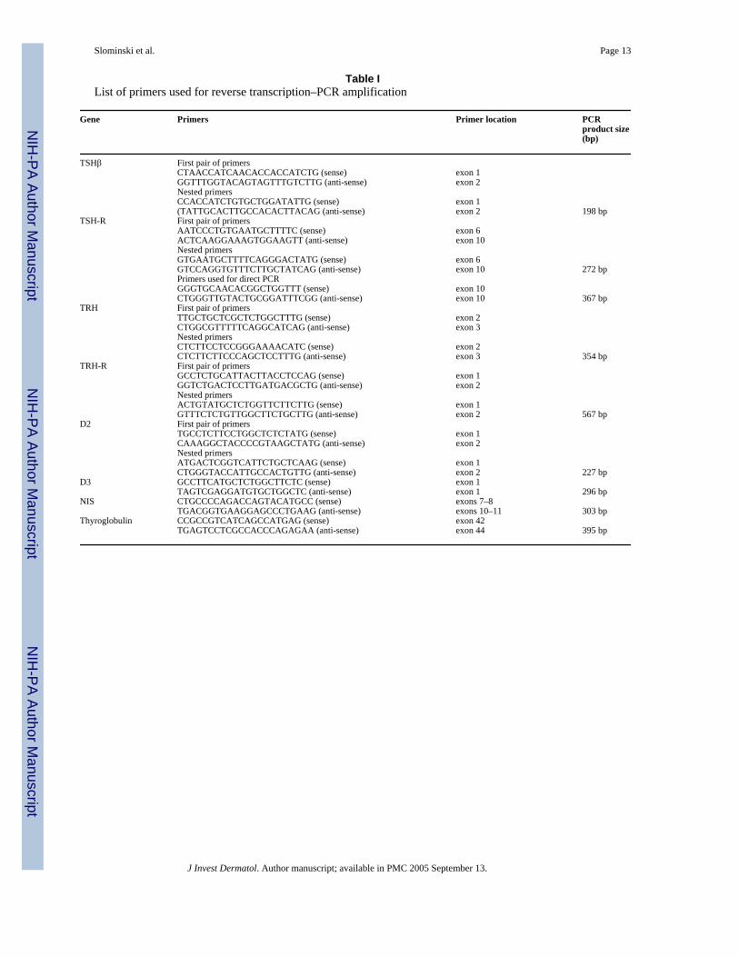
Human TSHβ (accession no. XM_002123), TSH-R (accession no. NM_000369), TRH(accession no. NM_007117), TRH-R (accession no. NM_003301), and D2 genes (accessionno. AF093774) were amplified by nested PCR. Gene D3 (accession no. NM–001362) wasamplified in one PCR reaction. We never amplified D3 gene for more than 25 cycles and usedan appropriate amount of RNA as a control for DNA contamination for each sample. Sequencesof the primers used for PCR amplifications are listed in Table I.
Amplification products were separated by agarose gel electrophoresis and visualized withethidium bromide staining. The identified PCR products were excised from the agarose geland purified by GFX PCR DNA and gel band purification kit (Amersham Pharmacia Biotech,Piscataway, NJ). PCR fragments were cloned in pGEM-T easy vector system (Promega) andpurified by plasmid purification kit (Qiagen, Valencia, CA). Sequencing was performed in theMolecular Resource Center at the University of Tennessee HSC (Memphis) using AppliedBiosystems 3100 Genetic Analyzer and BigDye™ Terminator Kit (Applied Biosystems, FosterCity, CA).
To detect NIS (accession no. NM_000453), thyroglobulin, and TSH-R (accession no.NM_000369) gene expression in HaCaT keratinocytes and human melanomas the RNA waspurified free of genomic DNA using DNA-Free (Ambion, Austin, TX) according torecommended conditions. cDNA was synthesized from 1.0 μg of total RNA using MMLVreverse transcriptase with random-hexamer primers (Clontech, Palo Alto, CA). Polymerasechain reaction (50 μl) was performed using 3% of the total cDNA in a reaction mixturecontaining 10 mM Tris–HCl, pH 8.3, 50 mM KCl, 3.5 mM MgCl2, 0.01% gelatin, 0.25 mM eachdeoxyribonucleoside triphosphate, 0.4 μM of each primer, 1.0 U AmpliTaq polymerase (Perkin-Elmer, Norwalk, CT), and 0.2 μg TaqStart antibody (Clontech). The primers used are in TableI. Amplifications conditions were: polymerase activation at 95°C for 5 min; 35 (thyroglobulinand TSH-R) or 40 (NIS) cycles of denaturation at 94°C for 20 s, annealing at 64°C for 1 min(thyroglobulin and TSH-R) or 70°C for 1 min (NIS), and extension at 72°C for 1 min; followedby 72°C for 7 min.
Northern blot analysesTotal RNA was prepared using a commercial kit (RNeasy Mini Kit, Qiagen). Fifteenmicrograms of RNA samples were run on denatured agarose gels, blotted on Nytran membranes(Schleicher & Schuell, Keene, NH) and subjected to hybridization as described (Saji et al,1992;Giuliani et al, 1995;Suzuki et al, 1998a,b). Full length rat or human TSH-R radiolabeledwith [α-32P]deoxycytidine triphosphate were used as probes (Tahara et al, 1991).
Cyclic adenosine monophosphate (cAMP) assaysThe noted concentration of bovine TSH, in units described by the manufacturer (Sigma), or100 μl (0.5 mg) of a standard anti-TSH-R stimulating antibodies were added with fresh mediumto cell samples cultured in 24 well plates then incubated for 2 h in the presence of 0.5 mM ofisobuthylmethylxanthine at 37°C in 5% CO2. The concentrations of extracellular cAMP weremeasured with a nonacetylation enzyme immunoassay using the BIOTRAK cAMP enzymeimmunoassay system as recommended by the manufacturer (Amersham Pharmacia Biotech).The cAMP concentration of samples was expressed as a level of stimulation relative to controlincubations with no TSH or stimulating TSH antibodies.
RESULTS AND DISCUSSIONExpression of functional TSH-R in skin cells
As previous reports suggested TSH-R expression in the human skin (Wu et al, 1996;Stadlmayret al, 1997;Rapoport et al, 2000), we tested expression of the gene by reverse transcription–
Slominski et al. Page 4
J Invest Dermatol. Author manuscript; available in PMC 2005 September 13.
NIH
-PA Author Manuscript
NIH
-PA Author Manuscript
NIH
-PA Author Manuscript
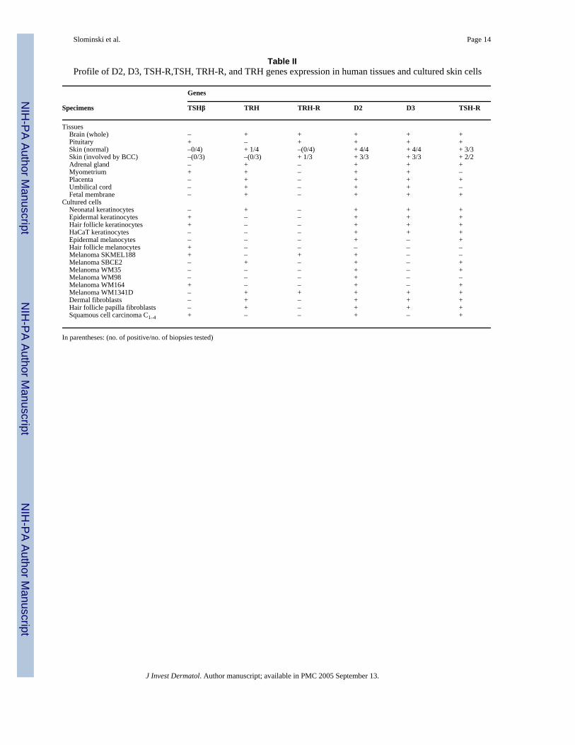
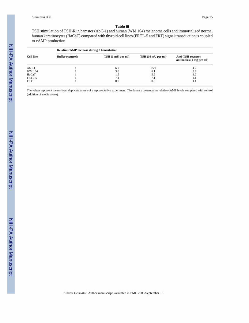
PCR in a broad panel of specimens. Expression of the TSH-R gene 272 bp product of nestedPCR (not shown) and the 367 bp product of direct PCR (Fig 1) was found in almost all tissuestested, including normal and pathologic skin; exceptions being the umbilical cord andmyometrium (Table II). TSH-R was also detected in cultured dermal and follicular papillafibroblasts, neonatal, follicular and immortalized HaCaT keratinocytes, squamous cellcarcinoma cells, epidermal melanocytes, and in four melanoma lines. TSH-R gene expressionwas below detectable levels only in follicular melanocytes and in two melanoma lines (TableII). Northern blot analysis was then performed in selected human and rodent melanoma lines,and in human squamous cell carcinoma cells and immortalized epidermal keratinocytes. Thisrevealed a TSH-R transcript of 3.3 kb, similar to a TSH-R transcript detected in control ratFRTL-5 thyroid cells (Fig 2). This transcript encoded a functional TSH-R as documented bystrong responses to TSH and antibodies against TSH-R, e.g., marked stimulation of cAMPproduction in human melanoma, in keratinocytes, in hamster melanoma (Table III) and inmouse melanoma (not shown).
The presence of TSH-R transcripts in dermal fibroblasts or in skin biopsies had previouslybeen shown (Rapoport et al, 2000;Wu et al, 1996;Stadlmayr et al, 1997), and expression ofthyroid transcription factor-1 (positive transcriptional regulator of TSH-R gene) (Shimura etal, 1994) had also been detected in keratinocytes (Suzuki et al, 1998a,b). What is novel is thedetection of TSH-R in the main cellular components of epidermis and hair follicles (with theexception of hair follicle melanocytes) with strong functional activity. This finding would haveclinical relevance to explain some of the cutaneous symptoms of Graves’ disease (Thiboutot,1995). Thus, the observed expression of TSH-R on epidermal melanocytes may be connectedwith the skin pigmentation of Graves’disease as its intracellular mediator, cAMP acts asstimulator of melanocytes proliferation and differentiation. Indeed, both human and rodentmalignant melanocytes and keratinocytes showed that TSH and anti-TSH-R antibodiesstimulated cAMP production (Table III). Alternatively, expression of the TSH-R may turnmelanocytes into targets for the destruction by TSH-R autoantibodies, thus explaining the highincidence of vitiligo in patients with Graves’ disease (Slominski et al, 1989). Also intriguingis the presence of TSH-R on hair follicle keratinocytes and papilla fibroblasts that might beinvolved in the association of Graves’ disease with alopecia areata. Finally, taken together withprevious findings (Wu et al, 1996;Stadlmayr et al, 1997;Rapoport et al, 2000), the presentobservations raise the tantalizing prospect that some forms of Graves’ disease could be due toan autoimmune response directed primarily against a cutaneous TSH-R antigen. Thus, thiscould result from abnormal expression of major histocompatibility complex proteins, andabnormal exposure of TSH-R antigen to immune cells (Shimojo et al, 1996;Suzuki et al,1999;Kohn et al, 2000), in response to exposure to environmental insults, e.g., solar radiationor skin infections.
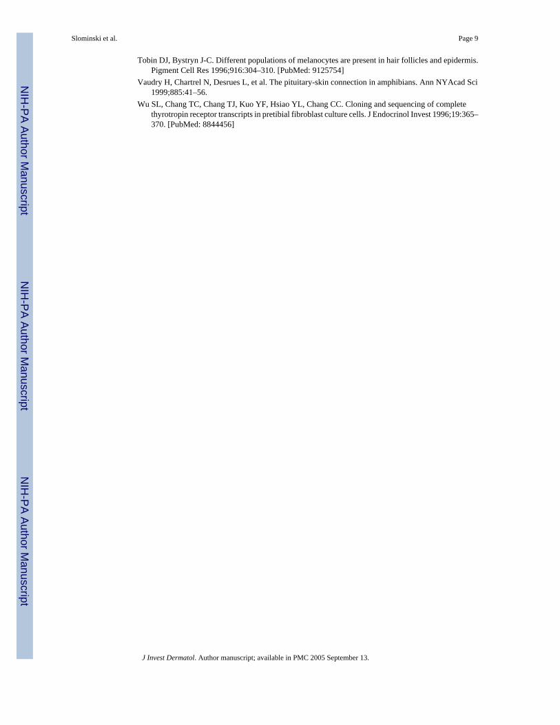
In the thyroid, TSH-R-stimulated pathways regulate most aspects of intracellular iodide andthyroglobulin metabolism, synthesis of NIS and production, and secretion of T4 (Kohn et al,2000). We therefore evaluated the possibility of similar effects in the skin. Using reversetranscription–PCR we tested skin expression of TSH-R-regulated genes in vitro in stable celllines positive for TSH-R (HaCaT keratinocytes and four different melanoma lines, e.g., SWM35, WM164, WM1341D), and also in one line negative for TSH-R (SKMEL188). The linesexpressing TSH-R also coexpressed thyroglobulin and NIS genes, whereas the SKMEL188line was negative for these genes (Fig 1). All of these lines were negative for thyroid peroxidasegene (not shown). Thus, expression of some, but not all molecular elements of the TSH-R-related pathways that are characteristic for the thyroid is conserved in cultured skin cells.
Recent studies have shown NIS expression in lactating breast tissue and in some breast cancers(Spitzweg et al, 2001). TSH-R has been found on fat cells and other nonthyroid tissues(Szkudlinski et al, 2001). Functional roles have not been fully clarified for NIS in breast tissue
Slominski et al. Page 5
J Invest Dermatol. Author manuscript; available in PMC 2005 September 13.
NIH
-PA Author Manuscript
NIH
-PA Author Manuscript
NIH
-PA Author Manuscript
or TSH-R in fat tissue, nor is it clear why such genes might be expressed in a pathologicsituation. The existence of TSH-R in melanoma cells might be exploited if the TSH-R isinvolved in the regulation of growth or function of these cells as might be the case for NIS inbreast cancer. Growth and function studies are in progress in our laboratories.
Reverse transcription–PCR assays for TSHβ, D2, D3, TRH-R, and TRHWe used the reverse transcription–PCR technique with sequencing of the amplified geneproducts together to define cutaneous expression of the genes coding for TSHβ, D2, D3, TRH-R, and TRH genes, in a combination of human tissue and cultured skin cells (Fig. 3; TableIV).
TSHβ—As we had found functional expression of TSH-R in the skin, we tested for its naturalligand (TSHβ) to investigate paracrine or autocrine control of TSH-R activity. Aside from itsexpression in the pituitary control, TSHβ gene expression was observed in epidermal and hairfollicle keratinocytes, hair follicle melanocytes, squamous cell carcinoma, and two melanomalines, as well as myometrium, being absent in all other cells or tissues tested that includedbiopsies of whole skin (Fig 3A; Table II). This pattern suggests that TSH expression is notconstitutive, but instead responds to environmental stimulation exemplified, in this case, bythe conditions of cell culture and/or by random depression during malignant progression.
D2 and D3 genes—The D2 gene product (227 bp) was expressed in almost all testedsamples; the sole exception was a line of hair follicle melanocytes where it was belowdetectable levels (Fig 3B; Table II). In some biopsies of normal skin (one of four) and skincontaining BCC (one of three), an additional second transcript of 335 bp was detected. Itcorresponds to the b isoform of D2 (D2b, accession no. AB041843). D2b contains an additionalexon (insertion of 108 bp) with an in-frame TGA codon that may encode an extra selenocysteine(Ohba et al, 2001). This second isoform (335 bp) was also present in neonatal, epidermal,follicular and HaCaT keratinocytes, dermal and follicular papilla fibroblasts, epidermalmelanocytes, squamous cell carcinoma cells, and the SBCE2 melanoma line (Fig 1B). Thedetection of the second isoform is consistent with the recent findings of two additional splicingvariants of human D2 differentially expressed in brain, kidney, lung, and trachea (Ohba et al,2001).
The D3 gene was expressed in all tissues tested that included brain, pituitary, adrenal gland,and intrauterine tissues. It was also detected in skin, where its expression was cell type specific(Fig 3C). It was expressed in all tested keratinocytes and fibroblasts lines, but was absent inmelanocytes and in the majority of melanoma lines (five of six) (Table II).
D2 catalyzes 5′ (outer ring) deiodination of T4 to T3, whereas D3 catalyzes 5 (inner ring)deiodination of T4 and T3 to inactive T3 (rT3) and 3,3′-diiodothyronine (T2) (St Germain,1999;Bianco et al, 2002). The structure of both genes is brown. D2 is predominantly expressedin pituitary, brain, brown fat, thyroid, heart, and skeletal muscle, whereas D3 is predominantlyexpressed in the brain, uterus, placenta, fetal membranes, and skin (rodents) (St Germain,1999). Actual transformation of T4 to T3, and of T3 to T2 have been demonstrated in culturedepidermal keratinocytes (Kaplan et al, 1988), although the pattern of expression of thecorresponding genes has not been evaluated in human skin. Our data thus represent the firstdemonstration of D2 and D3 gene expression in human skin. A differential pattern forexpression of the genes becomes clearly evident along the predominant cellular componentsof the epidermis and dermis. The absence of D2 in hair follicle melanocytes and of D3 in bothepidermal and hair follicle melanocytes, in the majority of melanoma cells and in the squamouscell carcinoma line suggest cell lineage specific gene expression and probably, ofcorrespondingly specific patterns of T3 degradation in the same populations. Future
Slominski et al. Page 6
J Invest Dermatol. Author manuscript; available in PMC 2005 September 13.
NIH
-PA Author Manuscript
NIH
-PA Author Manuscript
NIH
-PA Author Manuscript
biochemical studies on the differential pattern of cell type specific T3 production anddegradation should clarify the functional implications at the systemic level, and in epidermaland dermal homeostasis and activity of adnexal structures.
TRH and TRH-R—As TSH production is under the control of hypothalamic TRH, weevaluated local expression of the TRH-R gene. As expected, expression of TRH-R gene wasfound in brain and pituitary. In the skin, it was detected only in a single skin biopsy specimenof BCC, and in two melanoma lines SKMEL188 and WM1341D (Fig 3D; Table II). Thus,TRH-R may not participate directly in the physiology of the skin. Conversely, TRH that wasdetected in brain, adrenal gland, myometrium, placenta, umbilical cord, and fetal membrane,was also widely present in skin cells. It was found in normal skin (in one of four biopsies), infour melanoma lines, in neonatal keratinocytes and in both dermal and follicular papillafibroblasts (Fig 3E, Table II). Therefore, TRH is not coexpressed with TRH-R in most cells(with exception of SKMEL188 melanoma) suggesting that paracrine or autocrine mechanismsmay not operate locally in peripheral tissues, e.g., skin, placenta, fetal membranes, uterus, andadrenal gland. It can be speculated that expression of TRH mRNA by the above tissues andcell types may result in TRH production mainly for export, to regulate regional (skinfibroblasts, adrenal gland) or fetal (placenta, umbilical cord, fetal membranes) homeostasis.Of note, amphibian skin regulates pituitary function through local production of TRH that isexported to stimulate pituitary production and release of prolactin and α-melanoctye-stimulating hormone (Vaudry et al, 1999). Thus, this peptide gene expression may very wellrepresent an evolutionary continuum.
CONCLUSIONSThere is broad cutaneous expression of the gene and functional TSH-R, which is still ofundetermined physiologic and pathologic significance. Genes for elements of thehypothalamic–pituitary–thyroid axis are also expressed in the skin, but with selectivity in bothcell type and gene type. It appears that there may be important divergences between the rolesof the molecules at the central and peripheral levels.
ReferencesBianco AC, Salvatore D, Gereben B, Berry MJ, Larsen PR. Biochemistry, cellular and molecular biology,
and physiological roles of the iodothyronine selenodeiodinases. Endocr Rev 2002;23:38–89. [PubMed:11844744]
Giuliani C, Saji M, Napolitano G, et al. Hormonal modulation of MHC class I gene expression involvesan enhancer A-binding complex consisting of fra-2 and the p50 subunit of NF-κB. J Biol Chem1995;270:11453–11462. [PubMed: 7744783]
Kaplan MM, Pan C, Gordon PR, Lee J-K, Gilchrest BA. Human epidermal keratinocytes in cultureconvert thyroxine to 3,5,3′-triidothyronine by type II iodothyronine deiodination. A novel endocrinefunction of the skin. J Clin Endocrinol Metab 1988;66:815–822. [PubMed: 2450104]
Kohn LD, Valente WA, Grollman EF, Aloj SM, Vitti P: Clinical determination and/or quantification ofthyrotropin and a variety of thyroid stimulatory or inhibitory factors performed in vitro with animproved thyroid cell line. p. 4,609,622, assignee. Sept. 2, 1986.
Kohn LD, Napolitano G, Singer DS, et al. Graves’disease: a host defense mechanism gone awry. Int RevImmunol 2000;19:633–644. [PubMed: 11129119]
Ohba K, Yoshioka T, Muraki T. Identification of two novel splicing variants of human type IIiodothyronine deiodinase mRNA. Mol Cell Endocrinol 2001;172:169–175. [PubMed: 11165050]
Pisarchik A, Slominski A. Alternative splicing of CRH-R1 receptors in human and mouse skin:identification of new variants and their differential expression. FASEB J 2001;15:2754–2756.[PubMed: 11606483]
Slominski et al. Page 7
J Invest Dermatol. Author manuscript; available in PMC 2005 September 13.
NIH
-PA Author Manuscript
NIH
-PA Author Manuscript
NIH
-PA Author Manuscript
Rapoport B, Alsabeh R, Aftergood D, McLachlan SM. Elephantiasic pretibial myxedema. Insight intoand a hypothesis regarding the pathogenesis of the extrathyroidal manifestations of Graves’ disease.Thyroid 2000;10:685–692. [PubMed: 11014313]
Saji M, Akamizu T, Sanchez M, Obici S, Avvedimento E, Gottesman ME, Kohn LD. Regulation ofthyrotropin receptor gene expression in rat FRTL-5 thyroid cells. Endocrinology 1992;130:520–523.[PubMed: 1309347]
Shimojo N, Kohno Y, Yamaguchi K-I, et al. Induction of Graves’-like disease in mice by immunizationwith fibroblasts transfected with the thyrotropin receptor and a class II molecule. Proc Natl Acad SciUSA 1996;93:11074–11079. [PubMed: 8855311]
Shimura H, Okajima F, Ikuyama S, Shimura Y, Saji M, Kohn LD. Thyroid-specific expression of thethyrotropin receptor promoter and autoregulation of the thyrotropin receptor gene by cAMP involvesthyroid transcription factor-1 (TTF-1). Mol Endocrinol 1994;8:1049–1069. [PubMed: 7997232]
Slominski A, Wortsman J. Neuroendocrinology of the skin. Endocr Rev 2000;21:457–487. [PubMed:11041445]
Slominski A, Moellman G, Kuklinska E, Bomirski A, Pawelek J. Positive regulation of melaninpigmentation by two key substrates of the melanogenic pathway: L-tyrosine and L-dopa. J Cell Sci1988;89:287–296. [PubMed: 3143738]
Slominski A, Paus R, Bomirski A. Hypothesis: a possible role of the melatonin receptor in vitiligo. J RSoc Med 1989;82:539–541. [PubMed: 2552111]
Slominski A, Ermak G, Mazurkiewicz JE, Baker J, Wortsman J. Characterization of corticotropinreleasing hormone (CRH) in human skin. J Clin Endocrinol Metab 1998;83:1020–1024. [PubMed:9506767]
Slominski A, Ermak G, Wortsman J. Modification of melanogenesis in cultured human melanoma cells.InVitro Cell Dev Biol 1999;35:564–565.
Slominski A, Wortsman J, Luger T, Paus R, Salomon S. Corticotropin releasing hormone andproopiomelanocortin involvement in the cutaneous response to stress. Physiol Rev 2000;80:979–1020. [PubMed: 10893429]
Slominski A, Wortsman J, Pisarchik A, Zbytek A, Linton EA, Mazurkiewicz J, Wei ET. Cutaneousexpression of corticotropin releasing hormone (CRH), urocortin and CRH receptors. FASEB J2001;15:1678–1693. [PubMed: 11481215]
Spitzweg C, Harrington KJ, Pinke LA, Vile RG, Morris JC. The sodium iodide symporter and its potentialrole in cancer therapy. J Clin Endocrin Metab 2001;86:3327–3335.
St Germain DL. Development effects of thyroid hormone: the role of deiodinases in regulatory control.Biochem SocTrans 1999;27:83–88.
Stadlmayr W, Spitzweg C, Bichlmair AM, Heufelder AE. TSH receptor transcripts and TSH receptor-like immunoreactivity in orbital and pretibial fibroblasts of patients with Graves’ ophthalmopathyand pretibial myxedema. Thyroid 1997;7:3–12. [PubMed: 9086563]
Suzuki K, Kobayashi Y, Katoh R, Kohn LD, Kawaoi A. Identification of thyroid transcription factor-1in C cells and parathyroid cells. Endocrinology 1998a;139:3014–3017. [PubMed: 9607813]
Suzuki K, Lavaroni S, Mori A, et al. Thyroid transcription factor-1 is a calcium-modulated andcoordinately regulates genes involved in calcium homeostasis in C cells. Mol Cell Biol 1998b;18:7410–7422. [PubMed: 9819427]
Suzuki H, Mori A, Ishii KJ, et al. Activation of target tissue immune recognition molecules by doublestrand polynucleotides. Proc Natl Acad Sci USA 1999;96:2285–2290. [PubMed: 10051633]
Szkudlinski MW, Fremont V, Ronin C, Weintraub BD. Thyroid-stimulating hormone and thyroid-stimulating hormone receptor structure-function relationships. Physiol Rev 2001;82:473–502.[PubMed: 11917095]
Tahara K, Ban T, Minegishi T, Kohn LD. Immunoglobulins from Graves’disease patients interact withdifferent sites on TSH receptor/LH-CG receptor chimeras than either TSH or immunoglobulins fromidiopathic myxedema patients. Biochem Biophys Res Commun 1991;179:70–77. [PubMed:1883391]
Thiboutot DM. Dermatological manifestations of endocrine disorders. J Clin Endocrinol Metab1995;80:3082–3087. [PubMed: 7559901]
Slominski et al. Page 8
J Invest Dermatol. Author manuscript; available in PMC 2005 September 13.
NIH
-PA Author Manuscript
NIH
-PA Author Manuscript
NIH
-PA Author Manuscript
Tobin DJ, Bystryn J-C. Different populations of melanocytes are present in hair follicles and epidermis.Pigment Cell Res 1996;916:304–310. [PubMed: 9125754]
Vaudry H, Chartrel N, Desrues L, et al. The pituitary-skin connection in amphibians. Ann NYAcad Sci1999;885:41–56.
Wu SL, Chang TC, Chang TJ, Kuo YF, Hsiao YL, Chang CC. Cloning and sequencing of completethyrotropin receptor transcripts in pretibial fibroblast culture cells. J Endocrinol Invest 1996;19:365–370. [PubMed: 8844456]
Slominski et al. Page 9
J Invest Dermatol. Author manuscript; available in PMC 2005 September 13.
NIH
-PA Author Manuscript
NIH
-PA Author Manuscript
NIH
-PA Author Manuscript
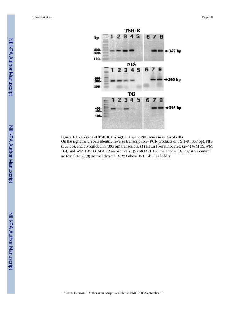
Figure 1. Expression of TSH-R, thyroglobulin, and NIS genes in cultured cellsOn the right the arrows identify reverse transcription– PCR products of TSH-R (367 bp), NIS(303 bp), and thyroglobulin (395 bp) transcripts. (1) HaCaT keratinocytes; (2–4) WM 35,WM164, and WM 1341D, SBCE2 respectively; (5) SKMEL188 melanoma; (6) negative controlno template; (7,8) normal thyroid. Left: Gibco-BRL Kb Plus ladder.
Slominski et al. Page 10
J Invest Dermatol. Author manuscript; available in PMC 2005 September 13.
NIH
-PA Author Manuscript
NIH
-PA Author Manuscript
NIH
-PA Author Manuscript
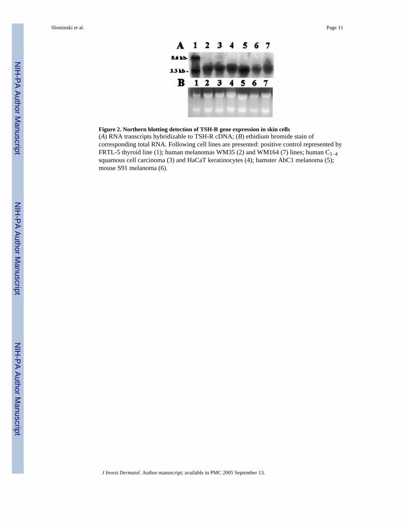
Figure 2. Northern blotting detection of TSH-R gene expression in skin cells(A) RNA transcripts hybridizable to TSH-R cDNA; (B) ethidium bromide stain ofcorresponding total RNA. Following cell lines are presented: positive control represented byFRTL-5 thyroid line (1); human melanomas WM35 (2) and WM164 (7) lines; human C1–4squamous cell carcinoma (3) and HaCaT keratinocytes (4); hamster AbC1 melanoma (5);mouse S91 melanoma (6).
Slominski et al. Page 11
J Invest Dermatol. Author manuscript; available in PMC 2005 September 13.
NIH
-PA Author Manuscript
NIH
-PA Author Manuscript
NIH
-PA Author Manuscript
Figure 3. Expression of thyroid-related genes in human tissues(A) Expression of TSHβ: (1) DNA ladder; (2) pituitary; (3) follicular melanocytes; (4)SKMEL188 melanoma; (5) WM164 melanoma; (6) follicular keratinocytes; (7) epidermalkeratinocytes; (8) dermal fibroblasts; (9) hair follicle papilla fibroblasts; (10) HaCaTkeratinocytes. (B) Expression of D2 in human skin samples: (1,13) DNA ladder; (2) normalskin; (3) BCC; (4) HaCaT keratinocytes; (5) neonatal keratinocytes; (6) follicularkeratinocytes; (7) epidermal keratinocytes; (8) neonatal keratinocytes; (9) dermal fibroblasts;(10) hair follicle papilla fibroblasts; (11) SKMEL188 melanoma; (12) SBCE2 melanoma.Arrows marked 227 bp (D2a) and 335 bp (D2b) transcripts. (C) Expression of D3 in humantissues and skin cells: (1,9) DNA ladder; (2) pituitary; (3,4) normal skin; (5) BCC; (6)myometrium; (7) follicular keratinocytes; (8) epidermal keratinocytes; (10) epidermalmelanocytes; (11–13) SBCE2,WM35, and WM98 melanomas, respectively. (D) Expressionof TRH-R: (1) DNA ladder; (2) pituitary; (3,4) SKMEL188 and WM1341D melanomas,respectively. (E) Expression of TRH: (1,10) DNA ladder; (2) nonlesional human skin; (3)dermal fibroblasts; (4) hair follicle papilla fibroblasts; (5) melanoma WM1341D; (6) neonatalkeratinocytes; (7) adrenal gland; (8) myometrium; (9) placenta.
Slominski et al. Page 12
J Invest Dermatol. Author manuscript; available in PMC 2005 September 13.
NIH
-PA Author Manuscript
NIH
-PA Author Manuscript
NIH
-PA Author Manuscript
NIH
-PA Author Manuscript
NIH
-PA Author Manuscript
NIH
-PA Author Manuscript
Slominski et al. Page 13
Table IList of primers used for reverse transcription–PCR amplification
Gene Primers Primer location PCRproduct size(bp)
TSHβ First pair of primersCTAACCATCAACACCACCATCTG (sense) exon 1GGTTTGGTACAGTAGTTTGTCTTG (anti-sense) exon 2Nested primersCCACCATCTGTGCTGGATATTG (sense) exon 1(TATTGCACTTGCCACACTTACAG (anti-sense) exon 2 198 bp
TSH-R First pair of primersAATCCCTGTGAATGCTTTTC (sense) exon 6ACTCAAGGAAAGTGGAAGTT (anti-sense) exon 10Nested primersGTGAATGCTTTTCAGGGACTATG (sense) exon 6GTCCAGGTGTTTCTTGCTATCAG (anti-sense) exon 10 272 bpPrimers used for direct PCRGGGTGCAACACGGCTGGTTT (sense) exon 10CTGGGTTGTACTGCGGATTTCGG (anti-sense) exon 10 367 bp
TRH First pair of primersTTGCTGCTCGCTCTGGCTTTG (sense) exon 2CTGGCGTTTTTCAGGCATCAG (anti-sense) exon 3Nested primersCTCTTCCTCCGGGAAAACATC (sense) exon 2CTCTTCTTCCCAGCTCCTTTG (anti-sense) exon 3 354 bp
TRH-R First pair of primersGCCTCTGCATTACTTACCTCCAG (sense) exon 1GGTCTGACTCCTTGATGACGCTG (anti-sense) exon 2Nested primersACTGTATGCTCTGGTTCTTCTTG (sense) exon 1GTTTCTCTGTTGGCTTCTGCTTG (anti-sense) exon 2 567 bp
D2 First pair of primersTGCCTCTTCCTGGCTCTCTATG (sense) exon 1CAAAGGCTACCCCGTAAGCTATG (anti-sense) exon 2Nested primersATGACTCGGTCATTCTGCTCAAG (sense) exon 1CTGGGTACCATTGCCACTGTTG (anti-sense) exon 2 227 bp
D3 GCCTTCATGCTCTGGCTTCTC (sense) exon 1TAGTCGAGGATGTGCTGGCTC (anti-sense) exon 1 296 bp
NIS CTGCCCCAGACCAGTACATGCC (sense) exons 7–8TGACGGTGAAGGAGCCCTGAAG (anti-sense) exons 10–11 303 bp
Thyroglobulin CCGCCGTCATCAGCCATGAG (sense) exon 42TGAGTCCTCGCCACCCAGAGAA (anti-sense) exon 44 395 bp
J Invest Dermatol. Author manuscript; available in PMC 2005 September 13.
NIH
-PA Author Manuscript
NIH
-PA Author Manuscript
NIH
-PA Author Manuscript
Slominski et al. Page 14
Table IIProfile of D2, D3, TSH-R,TSH, TRH-R, and TRH genes expression in human tissues and cultured skin cells
Genes
Specimens TSHβ TRH TRH-R D2 D3 TSH-R
Tissues Brain (whole) – + + + + + Pituitary + – + + + + Skin (normal) –0/4) + 1/4 –(0/4) + 4/4 + 4/4 + 3/3 Skin (involved by BCC) –(0/3) –(0/3) + 1/3 + 3/3 + 3/3 + 2/2 Adrenal gland – + – + + + Myometrium + + – + + – Placenta – + – + + + Umbilical cord – + – + + – Fetal membrane – + – + + +Cultured cells Neonatal keratinocytes – + – + + + Epidermal keratinocytes + – – + + + Hair follicle keratinocytes + – – + + + HaCaT keratinocytes – – – + + + Epidermal melanocytes – – – + – + Hair follicle melanocytes + – – – – – Melanoma SKMEL188 + – + + – – Melanoma SBCE2 – + – + – + Melanoma WM35 – – – + – + Melanoma WM98 – – – + – – Melanoma WM164 + – – + – + Melanoma WM1341D – + + + + + Dermal fibroblasts – + – + + + Hair follicle papilla fibroblasts – + – + + + Squamous cell carcinoma C1–4 + – – + – +
In parentheses: (no. of positive/no. of biopsies tested)
J Invest Dermatol. Author manuscript; available in PMC 2005 September 13.
NIH
-PA Author Manuscript
NIH
-PA Author Manuscript
NIH
-PA Author Manuscript
Slominski et al. Page 15
Table IIITSH stimulation of TSH-R in hamster (AbC-1) and human (WM 164) melanoma cells and immortalized normalhuman keratinocytes (HaCaT) compared with thyroid cell lines (FRTL-5 and FRT) signal transduction is coupledto cAMP production
Relative cAMP increase during 2 h incubation
Cell line Buffer (control) TSH (1 mU per ml) TSH (10 mU per ml) Anti-TSH receptorantibodies (1 mg per ml)
AbC-1 1 6.7 25.9 4.2WM 164 1 3.6 6.1 2.8HaCaT 1 1.5 5.2 3.2FRTL-5 1 7.1 7.1 4.1FRT 1 0.9 0.8 1.1
The values represent means from duplicate assays of a representative experiment. The data are presented as relative cAMP levels compared with control(addition of media alone).
J Invest Dermatol. Author manuscript; available in PMC 2005 September 13.
Copyright © 2022 FDOKUMEN