FDG-PET/CT can rule out malignancy in patients with vocal cord palsy
Upload
independentCategory
view
3download
0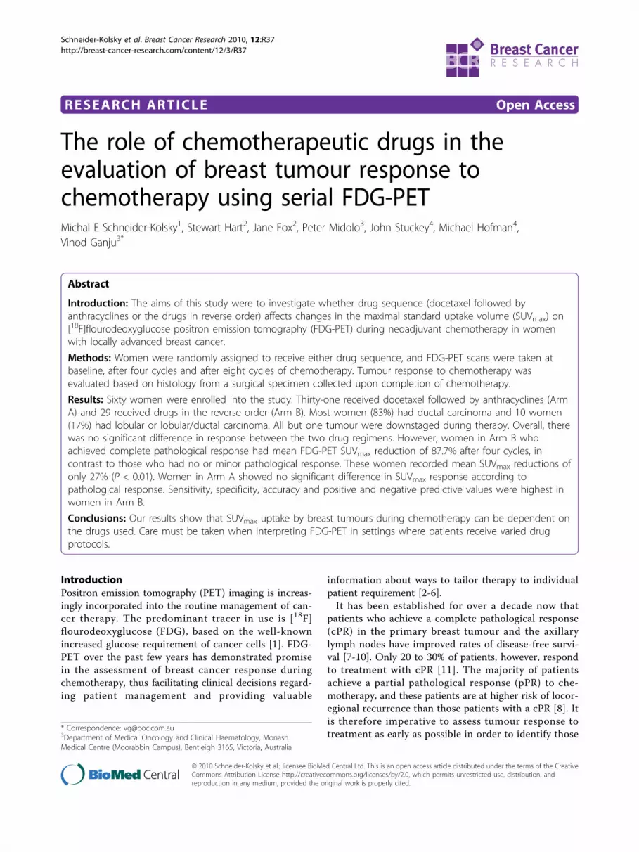
RESEARCH ARTICLE Open Access
The role of chemotherapeutic drugs in theevaluation of breast tumour response tochemotherapy using serial FDG-PETMichal E Schneider-Kolsky1, Stewart Hart2, Jane Fox2, Peter Midolo3, John Stuckey4, Michael Hofman4,Vinod Ganju3*
Abstract
Introduction: The aims of this study were to investigate whether drug sequence (docetaxel followed byanthracyclines or the drugs in reverse order) affects changes in the maximal standard uptake volume (SUVmax) on[18F]flourodeoxyglucose positron emission tomography (FDG-PET) during neoadjuvant chemotherapy in womenwith locally advanced breast cancer.
Methods: Women were randomly assigned to receive either drug sequence, and FDG-PET scans were taken atbaseline, after four cycles and after eight cycles of chemotherapy. Tumour response to chemotherapy wasevaluated based on histology from a surgical specimen collected upon completion of chemotherapy.
Results: Sixty women were enrolled into the study. Thirty-one received docetaxel followed by anthracyclines (ArmA) and 29 received drugs in the reverse order (Arm B). Most women (83%) had ductal carcinoma and 10 women(17%) had lobular or lobular/ductal carcinoma. All but one tumour were downstaged during therapy. Overall, therewas no significant difference in response between the two drug regimens. However, women in Arm B whoachieved complete pathological response had mean FDG-PET SUVmax reduction of 87.7% after four cycles, incontrast to those who had no or minor pathological response. These women recorded mean SUVmax reductions ofonly 27% (P < 0.01). Women in Arm A showed no significant difference in SUVmax response according topathological response. Sensitivity, specificity, accuracy and positive and negative predictive values were highest inwomen in Arm B.
Conclusions: Our results show that SUVmax uptake by breast tumours during chemotherapy can be dependent onthe drugs used. Care must be taken when interpreting FDG-PET in settings where patients receive varied drugprotocols.
IntroductionPositron emission tomography (PET) imaging is increas-ingly incorporated into the routine management of can-cer therapy. The predominant tracer in use is [18F]flourodeoxyglucose (FDG), based on the well-knownincreased glucose requirement of cancer cells [1]. FDG-PET over the past few years has demonstrated promisein the assessment of breast cancer response duringchemotherapy, thus facilitating clinical decisions regard-ing patient management and providing valuable
information about ways to tailor therapy to individualpatient requirement [2-6].It has been established for over a decade now that
patients who achieve a complete pathological response(cPR) in the primary breast tumour and the axillarylymph nodes have improved rates of disease-free survi-val [7-10]. Only 20 to 30% of patients, however, respondto treatment with cPR [11]. The majority of patientsachieve a partial pathological response (pPR) to che-motherapy, and these patients are at higher risk of locor-egional recurrence than those patients with a cPR [8]. Itis therefore imperative to assess tumour response totreatment as early as possible in order to identify those
* Correspondence: [email protected] of Medical Oncology and Clinical Haematology, MonashMedical Centre (Moorabbin Campus), Bentleigh 3165, Victoria, Australia
Schneider-Kolsky et al. Breast Cancer Research 2010, 12:R37http://breast-cancer-research.com/content/12/3/R37
© 2010 Schneider-Kolsky et al.; licensee BioMed Central Ltd. This is an open access article distributed under the terms of the CreativeCommons Attribution License http://creativecommons.org/licenses/by/2.0, which permits unrestricted use, distribution, andreproduction in any medium, provided the original work is properly cited.
tumours that demonstrate sensitivity to the chemo-therapy regimen, to direct appropriate treatment and tospare patients unnecessary chemotherapy. Conventionalimaging, such as mammography and ultrasound, provideonly information on anatomical changes in tumour mor-phology and size. Any residual masses present after com-pletion of chemotherapy may be composed of viabletumour cells and/or fibrotic tissue, but conventional ima-ging modalities are largely unable to differentiatebetween the different cell types. Histopathology is stillrequired in order to accurately characterise the remain-ing masses.Since tumours undergo changes in metabolism before
apparent changes in size, it is important to evaluatefunctional changes as early as possible during treatment.Several previous studies have shown that high baselineuptake of FDG by the tumour is associated with poorersurvival compared with tumours with lower uptake[12-14]. It has also been demonstrated that a significantreduction in glucose uptake on FDG-PET during orafter chemotherapy can predict cPR [4,5,15,16]. Theinformation obtained from FDG-PET studies to datefurther suggests that changes in glucose uptake by thetumour are independent of other, traditional, predictivebreast cancer markers [17]. The impetus now is to iden-tify chemosensitive tumours as early as possible duringtreatment. One study has recently demonstrated that areduction in glucose uptake after just one cycle ofneoadjuvant chemotherapy is predictive of cPR [4].Many of the reported studies, however, investigated
small samples of patients undergoing various differentchemotherapeutic regimens. We still know little aboutthe functional changes of tumours in response to speci-fic drug regimens. Hence, we designed the present pro-spective study to analyse serial changes in tumours ofwomen with locally advanced breast cancer undergoingneoadjuvant chemotherapy with docetaxel followed byanthracycline-based chemotherapy (FEC100), or ofwomen undergoing FEC100 treatment followed by doce-taxel using FDG-PET, mammography, ultrasound andclinical assessment - and to compare these changes withhistological changes in sequential biopsies.
Materials and methodsPatient selectionWomen presenting consecutively with locally advancedbreast cancer TMN stage T2 to T4, N0 to N3, M0between June 2004 and June 2008 were invited to parti-cipate in the study. The diagnosis was confirmed bycore needle biopsy and histopathology in all patientsprior to enrolment into the study.The inclusion criteria were: histologically or cytolo-
gically proven invasive adenocarcinoma, age ≥ 18,
Karnofsky performance status index ≥ 80%, andadequate haematology parameters, renal and hepaticfunction.Patients with prior history of breast cancer or other
neoplasms, with other serious medical conditions - suchas congestive heart failure, significant neurological orpsychiatric disorders, active uncontrolled infection,active ulcers, unstable diabetes mellitus - undergoingconcurrent treatment with other experimental drugs orhormonal agents, or who were pregnant were excludedfrom the study.The study was approved by the human research ethics
committee at our institution and all women gave writ-ten, informed consent.
Study designThe women were randomised using a computer-gener-ated random number allocation to receive either fourcycles of docetaxel (100 mg/m2), followed by four cyclesof combined fluorouracil (500 mg/m2), epirubicin(10 0 mg/m2) and cyclophosphamide (500 mg/m2)(FEC100) (Arm 1), or the reverse order of drugs(FEC100 followed by docetaxel) (Arm 2).At baseline, prior to commencement of chemotherapy,
all women underwent a clinical examination followed bymammography, ultrasound and FDG-PET. All examina-tions and imaging were carried out within 4 weeks ofcommencement of treatment. Imaging (all three modal-ities) was repeated after the initial four cycles of eitherdocetaxel or FEC100 and again upon completion of thechemotherapy. All imaging was carried out at the sameclinic using the same equipment. The pathologicaltumour response (gold standard) was assessed after sur-gical resection upon completion of the treatment.
Drug regimenAll patients received either docetaxel followed byFEC100 or the same drugs in reverse order prior to sur-gical resection. Each drug was delivered every 21 daysfor four cycles. Each subject received a total of eightcycles of chemotherapy over 24 weeks. Supportive careincluded antiemetics and granulocyte-stimulating factoras per American Society of Clinical Oncology guidelines.
Clinical assessmentTumours were measured with callipers, with the twolargest diameters of the tumour recorded and multi-plied. Tumour sizes at the second and third examina-tions were compared with baseline measurements andclassified into either complete clinical response (fullresolution of the palpable mass), partial resolutionof the mass (> 50%) or minimal resolution or clinicalprogression of the mass.
Schneider-Kolsky et al. Breast Cancer Research 2010, 12:R37http://breast-cancer-research.com/content/12/3/R37
Page 2 of 11
MammographyBidimensional tumour measurements were taken in twoplanes. When two or more tumours were present, thelargest tumour was used to calculate the tumour sizeand response to treatment. The area of the tumour wascalculated in each of the two projections (using the for-mula for an area of an ellipse) and the two values wereadded together. Changes in tumour size were recordedas the percentage change from baseline of the combinedarea measurement.
Ultrasound measurementsBoth breasts and axillas were scanned at each time pointat the same site using a Phillips ATL HDI 5000 with a12.5 MHz linear transducer. Tumours were visualised inthe transverse and sagittal planes. The tumour volumeswere calculated at each time point using three measure-ments for an ellipsoid volume [18]:
volume length width thickness= × × × / 6 (1)
When more than one tumour was present, the largesttumour was used and compared in subsequentexaminations.
FDG-PET protocol and imagingPET scans were performed using a dual-modality Sie-mens Biograph 2 PET/CT scanner (PET, Siemns MedicalImaging, Knoxville, Tenessee, USA; CT, Siemens MedicalImaging, Forchheim, Bavaria, Germany), which consistsof a two-row spiral CT and full-ring bismuth-germanatePET. Patients were advised to fast for at least 6 hoursprior to the scan. Blood glucose concentrations weremeasured before the tracer (FDG) was injected. If theblood glucose level was < 12 mmol/l, the patient wasgiven diluted contrast material orally and injected withbetween 8 and 10 mCi FDG via an intravenous cannula.The patient was then rested for 60 minutes prior to ima-ging as per our routine protocol. Variations in waitingtime due to technical or workflow factors were restrictedto between 45 and 75 minutes (60 ± 15 minutes).Imaging was performed from the base of skull to just
inferior of the ischial tuberosities. Images were acquiredin three-dimensional mode and were reconstructed withan iterative technique using an ordered subset expecta-tion-maximisation algorithm incorporating CT-basedattenuation correction. The following formula was usedfor the acquisition of the standardised uptake volume(SUV) in our study:
I scale I dose weightsuv Act Tstart= × ×( / ( / , ))1 000 (2)
where Isuv is the image pixel value (SUV units), IAct isthe image pixel value (Bq/cm3) corrected for the start
acquisition start time, doseTstart is the injected dose cor-rected for the acquisition time, weight is the patientweight (kg), and scale is the multiplier applied to SUVvalues.The patient volume of distribution is approximated by
the water equivalent to the patient weight. SUV valuestypically range from 0 to 10 after applying a scaling fac-tor suitable for the Syngo™ software system (SiemensMedical Imaging, Forchheim, Bavaria, Germany).All cases were reviewed by a team of five radiologists
with extensive experience in nuclear medicine, as wellas a nuclear medicine physician using a Siemen’s eSoftworkstation to review the PET, CT and fused images.All five radiologists and the nuclear medicine physicianformed a consensus agreement prior to the study tostandardise measurements of SUV uptake. Regions ofinterest were drawn using a three-dimensional contourencompassing axial, sagittal and coronal planes tracedby the eSoft software after selecting the area of increas-ing FDG uptake in the primary breast tumour andinvolved axillary nodes. The maximum standardiseduptake value (SUVmax) within this region of interest wascalculated, and standardised according to body weight,dose of administered FDG and time to imaging. Partialvolume correction or correction for blood sugar levelwas not performed. If more than one FDG avid breastlesion was identified, the lesion with the highest SUVmax
was chosen as the target lesion for serial comparison.This same procedure was performed for any axillarynodal involvement.The metabolic response of the tumour to chemother-
apy was analysed using the SUVmax values obtained onFDG-PET during and after treatment according to thefollowing formula:
Δ = × −SUV SUV baseline SUV post-treatment SUV ba(%) ( ) /max max max100 sseline (3)
All PET readers were blinded to the clinical informa-tion, as well as the mammography and ultrasoundreports of all patients. The only information provided tothe readers was the reports of previous FDG-PET ima-ging, which were required for the mid-treatment andpost-treatment reports.
Pathologic evaluationBiopsy samples collected at baseline and after fourcycles of chemotherapy were analysed by the consultantanatomical pathologist at our institution according tostandard protocols. Tumour samples were fixed in 10%formaldehyde and were embedded in paraffin. Sectionswere cut at 5 μm thickness and stained with H & E.The tumour type, grade and extent were reported, aswell as the oestrogen receptor, progesterone receptorand HER2 status.
Schneider-Kolsky et al. Breast Cancer Research 2010, 12:R37http://breast-cancer-research.com/content/12/3/R37
Page 3 of 11
The pathological assessment of the final resected spe-cimen was used for the definitive outcome measure.Patients were classified into three groups according totheir pathological response. Group 1 consisted ofpatients who were clear of tumour in surgical speci-mens, including nodes (cPR); Group 2 consisted ofpatients with either a small residual tumour mass(≥ 80% reduction) or clear nodes (pPR); and Group 3included patients that could not be classified intoGroups 1 or 2. All pathological assessments were carriedout blinded to the clinical and imaging findings.
Statistical analysisComparison of demographic and tumour characteristicsbetween the two groups of patients were carried outusing the Mann-Whitney U test. Comparison of theSUVmax tumour uptake of each group between baselineand at the midpoint or after treatment was made usingthe Kruskal-Wallis test. Differences in sample sizesbetween two groups were analysed using chi-squaretests or Fisher’s exact tests. Comparison of SUVmax
uptake values at each time point between the twogroups of patients was made using the Mann-WhitneyU test. Cutoff values ≥ 75% reduction in SUVmax frombaseline were used to assess sensitivity, specificity, posi-tive predictive value, negative predictive value and accu-racy in both treatment arms. This cutoff value waschosen to fit with the published literature and to over-ride any minor changes in SUV measurements due tovariation in technical factors. Significance was affordedwhen P < 0.05. All statistical analyses were carried outusing SPSS, version 16 (SPSS Inc., Chicago, IL, USA).
ResultsSixty-eight patients were recruited into the study. Ofthese patients, one withdrew from the study and sevenhad not completed treatment and evaluation at the timeof analysis. These patients were not included in the finalanalysis. The final number of participants thereforeincluded 31 patients who were randomised to receivedocetaxel followed by FEC100 and 29 patients whoreceived the same drugs in reverse order.Tables 1 and 2 demonstrate the demographic charac-
teristics of patients and tumour characteristics accordingto drug regimen used. There was no significant differ-ence in any of the patient or tumour parameters studiedbetween the two groups of patients. The majority ofpatients (50/60, 83%) presented with ductal carcinoma.Seven patients (11.6%) had lobular carcinoma and threepatients (5%) had mixed ductal/lobular carcinoma. Mostpatients (33/60, 55%) had high-grade tumours. Thirty-seven out of 60 patients (61.6%) were oestrogen recep-tor-positive. Although more of these patients were ran-domised to receive docetaxel initially rather than
FEC100 (71% compared with 52%, respectively), the dif-ference in proportion was not significant. Similarly, theproportions of women who were progesterone receptor-positive and HER2-positive were not significantly differ-ent between the two groups. The proportion of womenwho underwent mastectomy or a wide local excisionupon completion of chemotherapy was not statisticallydifferent between the two groups. The median (ranges)resting time prior to PET imaging was 60 minutes (45to 75 minutes).Tumour size on mammography and ultrasound and
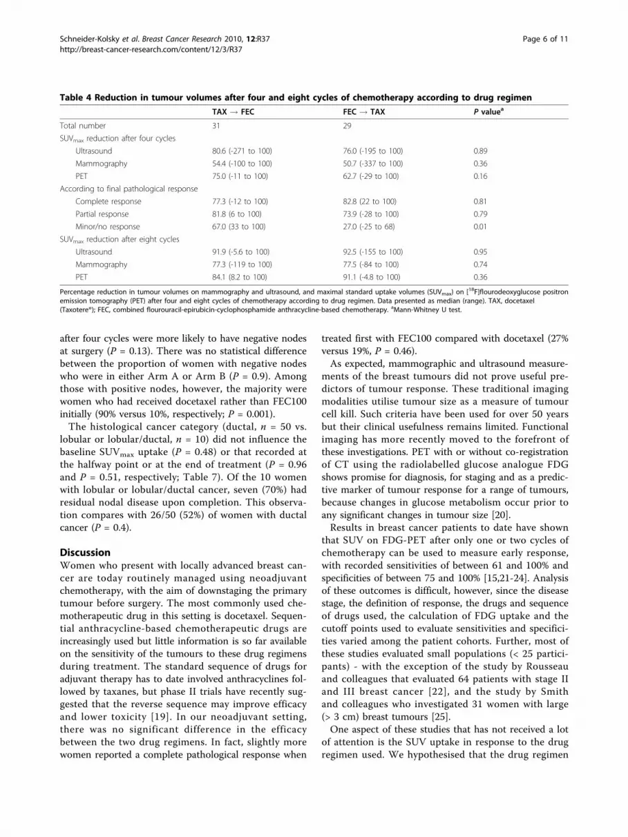
the SUV uptake on FDG-PET were not significantly dif-ferent between the two groups of patients at baseline(Table 2). The median (range) SUVmax uptake for allwomen was 6.5 (1.58 to 26.6). Higher baseline SUVmax
uptake was associated with higher rates of final patholo-gical response, although statistical significance was notreached (Figure 1).The numbers of tumours that were classified as cPR,
as pPR and as pathological response not included incPR or pPR are presented in Table 3. All but one
Table 1 Demographic and tumour characteristics ofwomen undergoing neoadjuvant chemotherapyaccording to drug regimen
Characteristic TAX ! FEC FEC ! TAX P valuea
Age 52 (37-70) 48 (30-66) 0.04b
Pathology type
Ductal 25/31 (80.6) 25/29 (86.2) 1.00
Ductal/lobular 2/31 (6.5) 1/29 (3.4) n/a
Lobular 4/31 (13.4) 3/29 (10.3) n/a
Pathology grade
Low 4/31 (12.9) 2/29 (6.9) 0.41
Medium 11/31 (35.5) 10/29 (34.5) 0.82
High 16/31 (51.6) 17/29 (58.6) 0.86
Oestrogen receptor status
Negative 10/31 (32.2) 13/29 (44.8) 0.29
Positive 21/31 (67.7) 16/29 (55.2) 0.25
Progesterone receptor status
Negative 16/31 (51.6) 18/29 (62.1) 0.60
Positive 15/31 (48.4) 11/29 (37.9) 0.33
HER2 statusc
Negative 17/29 (58.6) 16/29 (55.2) 1.00
Positive 12/29 (41.4) 13/29 (44.8) 1.00
Surgical procedured
Mastectomy 16/30 (53.3) 17/29 (58.6) 0.86
Wide local excision 13/30 (43.3) 12/29 (41.3) 0.84
Partial mastectomy 1/30 (3.3) 0/29 (0.0) n/a
Demographic and tumour characteristics of women undergoing neoadjuvantchemotherapy for locally advanced breast cancer according to drug regimen(n = 34/group). TAX, docetaxel (Taxotere®); FEC, combined flourouracil-epirubicin-cyclophosphamide anthracycline-based chemotherapy; n/a, notavailable. Data presented as median (range) or n/N (%). aChi-square test.bMann-Whitney U test. cNot available for two patients in Arm 1 (TAX ® FEC).dOne woman in Arm 1 declined surgery.
Schneider-Kolsky et al. Breast Cancer Research 2010, 12:R37http://breast-cancer-research.com/content/12/3/R37
Page 4 of 11
tumour were downstaged during neoadjuvant chemo-therapy. Overall, 23% of tumours (14/60) were classifiedas having undergone a complete pathological response.Thirty per cent of tumours (18/60) underwent a partialresponse, and the other tumours (28/60 (47%) recordedonly minor or no changes during chemotherapy. Therewas no significant difference in tumour responsebetween the two drug regimens (P > 0.05).Table 4 demonstrates the reduction in tumour
volumes after four and eight cycles of chemotherapy asobserved on mammography, ultrasound and FDG-PETaccording to the drug given. There was no statisticallysignificant difference between the two groups of patientswhen comparing tumour volume reduction or SUVmax
uptake on either imaging modality. The only significantdifference observed was associated with SUVmax uptakeafter four cycles of chemotherapy with anthracyclines.In this group of women, minor or no final pathologicalresponse was associated with minimal reduction of
SUVmax uptake compared with women receiving doce-taxel (P = 0.01). These outcomes are also highlighted inFigure 2. Women receiving FEC100 first who achieved acPR had a median reduction of SUVmax of 87.7% frombaseline (Figure 2a). In contrast, those on the same drugwhose tumour recorded no or only minor pathologicalresponse had a median reduction of only 27% (Figure2b). These data are significantly different from those bywomen who received docetaxel initially (P = 0.01). Thisgroup had no significant difference in the medianSUVmax reduction according to pathological response.The percentage reduction in SUVmax uptake after alleight cycles is shown in Figure 3. There were no signifi-cant differences between the two drug sequences interms of pathological response (P > 0.05).The sensitivity, specificity, positive predictive value,
negative predictive value and accuracy rates for bothtreatment arms are presented in Table 5. Highest sensi-tivity (0.87) was obtained for SUVmax reduction afterchemotherapy with FEC100 (Arm B). In contrast, sensi-tivity was 0.83 for women in Arm A. Specificity andoverall accuracy was best among women who receivedArm B (0.76 and 0.79, respectively). Similarly, both posi-tive and negative predictive values scored highest in thisgroup of women (0.58 and 0.94, respectively).The proportion of women who achieved SUVmax
reduction ≥ 75% after four cycles of chemotherapy ineither treatment arm was also compared according tonodal status at surgery (Table 6). There was no differ-ence between the proportion of women with ≥ 75%reduction of SUVmax in either Arm A or Arm B (P =0.63). Overall, women with ≥ 75% reduction of SUVmax
Table 2 Breast tumour characteristics on mammography, ultrasound and PET at baseline according to drug regimen
TAX ! FEC FEC ! TAX P valuea
Total number 31 29
Ultrasound (volume)b 10,669 (702 to 169,221) 11,344 (919 to 121,428) 0.49
Mammography (volume)c 1,685 (0 to 7,487) 1,739 (0 to 5,749) 0.95
PET (SUVmax) 6.76 (1.58 to 24.8) 6.26 (2.3 to 26.6) 0.98
Data presented as median (range). TAX, docetaxel (Taxotere®); FEC, combined flourouracil-epirubicin-cyclophosphamide anthracycline-based chemotherapy; PET,positron emission tomography; SUVmax, maximal standard uptake volume. aMann-Whitney U test. bVolume based on calculation of elliptical tumour shape.cVolume based on the sum of areas in the craniocaudal and mediolateral oblique appearance.
Figure 1 Standard uptake values prior to neoadjuvantchemotherapy according to pathological response uponcompletion of chemotherapy. The raw standardised uptakevolume (SUV) values are based on those provided by the baseline[18F]flourodeoxyglucose-positron emission tomography scan. Datadepict the median, interquartile ranges and total ranges(N/complete = 14, N/partial = 18, N/minor = 28).
Table 3 Number of tumours classified as complete,partial or minor pathological response according to drugregimen
Response TAX ! FEC FEC ! TAX Total (all women)
cPR 6/31 (19) 8/29 (28) 14/60 (23)
pPR 9/31 (29) 9/29 (31) 18/60 (30)
mPR 16/31 (52) 12/29 (41) 28/60 (47)
Number of tumours classified as a complete pathological response (cPR),partial pathological response (pPR) or minor pathological response (mPR) aftercompletion of neoadjuvant chemotherapy according to drug regimen. Datapresented as n/N (%). TAX, docetaxel (Taxotere®); FEC, combined flourouracil-epirubicin-cyclophosphamide anthracycline-based chemotherapy.
Schneider-Kolsky et al. Breast Cancer Research 2010, 12:R37http://breast-cancer-research.com/content/12/3/R37
Page 5 of 11
after four cycles were more likely to have negative nodesat surgery (P = 0.13). There was no statistical differencebetween the proportion of women with negative nodeswho were in either Arm A or Arm B (P = 0.9). Amongthose with positive nodes, however, the majority werewomen who had received docetaxel rather than FEC100initially (90% versus 10%, respectively; P = 0.001).The histological cancer category (ductal, n = 50 vs.
lobular or lobular/ductal, n = 10) did not influence thebaseline SUVmax uptake (P = 0.48) or that recorded atthe halfway point or at the end of treatment (P = 0.96and P = 0.51, respectively; Table 7). Of the 10 womenwith lobular or lobular/ductal cancer, seven (70%) hadresidual nodal disease upon completion. This observa-tion compares with 26/50 (52%) of women with ductalcancer (P = 0.4).
DiscussionWomen who present with locally advanced breast can-cer are today routinely managed using neoadjuvantchemotherapy, with the aim of downstaging the primarytumour before surgery. The most commonly used che-motherapeutic drug in this setting is docetaxel. Sequen-tial anthracycline-based chemotherapeutic drugs areincreasingly used but little information is so far availableon the sensitivity of the tumours to these drug regimensduring treatment. The standard sequence of drugs foradjuvant therapy has to date involved anthracyclines fol-lowed by taxanes, but phase II trials have recently sug-gested that the reverse sequence may improve efficacyand lower toxicity [19]. In our neoadjuvant setting,there was no significant difference in the efficacybetween the two drug regimens. In fact, slightly morewomen reported a complete pathological response when
treated first with FEC100 compared with docetaxel (27%versus 19%, P = 0.46).As expected, mammographic and ultrasound measure-
ments of the breast tumours did not prove useful pre-dictors of tumour response. These traditional imagingmodalities utilise tumour size as a measure of tumourcell kill. Such criteria have been used for over 50 yearsbut their clinical usefulness remains limited. Functionalimaging has more recently moved to the forefront ofthese investigations. PET with or without co-registrationof CT using the radiolabelled glucose analogue FDGshows promise for diagnosis, for staging and as a predic-tive marker of tumour response for a range of tumours,because changes in glucose metabolism occur prior toany significant changes in tumour size [20].Results in breast cancer patients to date have shown
that SUV on FDG-PET after only one or two cycles ofchemotherapy can be used to measure early response,with recorded sensitivities of between 61 and 100% andspecificities of between 75 and 100% [15,21-24]. Analysisof these outcomes is difficult, however, since the diseasestage, the definition of response, the drugs and sequenceof drugs used, the calculation of FDG uptake and thecutoff points used to evaluate sensitivities and specifici-ties varied among the patient cohorts. Further, most ofthese studies evaluated small populations (< 25 partici-pants) - with the exception of the study by Rousseauand colleagues that evaluated 64 patients with stage IIand III breast cancer [22], and the study by Smithand colleagues who investigated 31 women with large(> 3 cm) breast tumours [25].One aspect of these studies that has not received a lot
of attention is the SUV uptake in response to the drugregimen used. We hypothesised that the drug regimen
Table 4 Reduction in tumour volumes after four and eight cycles of chemotherapy according to drug regimen
TAX ! FEC FEC ! TAX P valuea
Total number 31 29
SUVmax reduction after four cycles
Ultrasound 80.6 (-271 to 100) 76.0 (-195 to 100) 0.89
Mammography 54.4 (-100 to 100) 50.7 (-337 to 100) 0.36
PET 75.0 (-11 to 100) 62.7 (-29 to 100) 0.16
According to final pathological response
Complete response 77.3 (-12 to 100) 82.8 (22 to 100) 0.81
Partial response 81.8 (6 to 100) 73.9 (-28 to 100) 0.79
Minor/no response 67.0 (33 to 100) 27.0 (-25 to 68) 0.01
SUVmax reduction after eight cycles
Ultrasound 91.9 (-5.6 to 100) 92.5 (-155 to 100) 0.95
Mammography 77.3 (-119 to 100) 77.5 (-84 to 100) 0.74
PET 84.1 (8.2 to 100) 91.1 (-4.8 to 100) 0.36
Percentage reduction in tumour volumes on mammography and ultrasound, and maximal standard uptake volumes (SUVmax) on [18F]flourodeoxyglucose positronemission tomography (PET) after four and eight cycles of chemotherapy according to drug regimen. Data presented as median (range). TAX, docetaxel(Taxotere®); FEC, combined flourouracil-epirubicin-cyclophosphamide anthracycline-based chemotherapy. aMann-Whitney U test.
Schneider-Kolsky et al. Breast Cancer Research 2010, 12:R37http://breast-cancer-research.com/content/12/3/R37
Page 6 of 11

may significantly affect the degree of reduction observedon serial PET scans because different drugs target differ-ent biological actions in the cancer cells. Most studies todate have all evaluated FDG-PET in cohorts of womenwho received various drugs with a range of dosage andsequences. It is thus difficult to assess the true useful-ness of PET as a predictor of tumour response totreatment. In general, however, the literature hasdemonstrated that tumours with high levels of FDGuptake at baseline are more likely to achieve a completepathological response [14,22,24,25]. These findings wereconfirmed in our study.
Women in our cohort who recorded SUVmax reduc-tions > 75% after four cycles of chemotherapy weremore likely to achieve a cPR of the tumour. Further,tumours with SUVmax reductions < 30% after four cyclesof FEC100 were more likely to remain insensitive to thetreatment regardless of the subsequent use of docetaxel.Sensitivity, specificity, positive and negative predictivevalues, as well as overall accuracy were highest (whenthe 75% cutoff value was used) among women receivingFEC100 initially. Berriolo-Riedinger and colleaguesachieved sensitivities and specificities of 0.91 and 0.86,
Figure 2 Reduction in maximal standardised uptake volumeafter four cycles of neoadjuvant chemotherapy. Percentagereduction in maximal standardised uptake volume (SUVmax) afterfour cycles of neoadjuvant chemotherapy according to pathologicalresponse obtained upon completion of chemotherapy. (a)Reductions in SUVmax after four cycles of docetaxel (Taxotere®)(TAX). (b) Reductions in SUVmax after four cycles of anthracycline-based fluorouracil, epirubicin and cyclophosphamide (FEC). Datadepict the median, interquartile ranges and total ranges. (a) N/complete = 6, N/partial = 9, N/minor = 16. (b) N/complete = 8, N/partial = 9, N/minor = 12.
Figure 3 Reduction in maximal standardised uptake volumeafter eight cycles of neoadjuvant chemotherapy. Percentagereduction in maximal standardised uptake volume (SUVmax) frombaseline after eight cycles of neoadjuvant chemotherapy accordingto pathological response obtained upon completion ofchemotherapy. (a) Reductions in SUVmax after four cycles withdocetaxel (Taxotere®) (TAX) followed by four cycles withanthracycline-based fluorouracil, epirubicin and cyclophosphamide(FEC). (b) Reductions in SUVmax after four cycles with FEC followedby four cycles with TAX. Data depict the median, interquartileranges and total ranges. (a) N/complete = 6, N/partial = 9, N/minor= 16. (b) N N/complete = 7, N/partial = 9, N/minor = 12).
Schneider-Kolsky et al. Breast Cancer Research 2010, 12:R37http://breast-cancer-research.com/content/12/3/R37
Page 7 of 11

respectively, after one treatment cycle [4]. In their study,however, women received four different treatmentprotocols.Sensitivity of 0.77 and specificity of 0.80 were achieved
in a study of 50 women with very large breast tumours(mean size 4.3 cm) who had FDG-PET before and aftertwo cycles of chemotherapy with epirubicin and cyclo-phosphamide and, if axillary nodes were positive, addi-tional taxanes [24]. Rousseau and colleagues achievedsensitivities and specificities of 0.61 and 0.96, respec-tively, in their cohort of women using the 60% cutoffvalue after two cycles [22]. Our comparative values(when calculating a 60% cutoff value) after four cyclesare 0.87 and 0.56 in women receiving FEC100 and are0.83 and 0.32 in women receiving docetaxel. Thesevalues are similar to those recently reported by Jung andcolleagues [26]. In their study, 66 women were dividedinto two groups, with 31 women receiving docetaxelplus capecitabine and the other 31 women receivingdoxorubicin plus cyclophosphamide. FDG-PET was car-ried out before and after four cycles of chemotherapy.The authors used an 84.8% cutoff value to achieve sensi-tivity and specificity values of 70% and 69.6%, respec-tively [26]. The patients were not analysed according todrugs used, but were evaluated as a whole group.
In an earlier study, patients receiving cyclophospha-mide, doxorubicin, vincristine and prednisolone with orwithout docetaxel were scanned using FGD-PET afterone cycle and four cycles of treatment [25]. There wasno difference in the SUVmax reduction between the twotreatment regimens used at either time point. Using a20% reduction in SUV as the cutoff value, this studyrecorded sensitivity and specificity values of 0.90 and0.74, respectively [25]. The patients included in thatstudy had locally advanced cancer > 3 cm, which mayhave contributed to the relatively high sensitivityachieved. In our study, patients were included if theircancer was > 2 cm in size. Our less restrictive inclusioncriteria may hence have resulted in the lower sensitivityand specificity values since the sensitivity of FDG-PETis known to be lower when the tumours are smaller[17].It is interesting to compare the different drug-induced
tumour responses observed on PET. The overall reduc-tion in tumour volume was similar in all imaging mod-alities investigated regardless of the drugs used(Table 3). The bulk of tumour reduction occurred dur-ing the first four cycles. Women who received FEC100and who had a median SUVmax reduction of only 27%or less, however, were significantly more likely to havetreatment-resistant tumours (P = 0.01). In contrast,tumours of women who had received docetaxel couldnot be easily stratified. Our results further show thatwomen who reported reductions of SUVmax uptake ≥75% halfway through treatment were significantly morelikely to have positive nodes upon completion of che-motherapy if they had received docetaxel initially (90%vs. 10%) (Table 5). This association has not previouslybeen reported. Since nodal status is an important prog-nostic indicator of overall survival and recurrence,further investigations in larger cohorts of women withadequate follow up of at least 5 years are required.Our results demonstrate that the usefulness of PET in
predicting tumour response can be influenced by thetype of drugs used. This is an important considerationin the evaluation of PET during chemotherapy. All
Table 5 Sensitivities, specificities, positive and negativepredictive values and accuracy rates after four cycles ofchemotherapy
All women TAX ! FEC FEC ! TAX
Total number 60 31 29
Sensitivity 0.78 0.66 0.87
Specificity 0.60 0.48 0.76
Positive predictive value 0.37 0.23 0.58
Negative predictive value 0.90 0.85 0.94
Accuracy 0.65 0.51 0.79
Sensitivities, specificities, positive and negative predictive values and accuracyrates for [18F]flourodeoxyglucose positron emission tomography based on a75% reduction of maximal standard uptake volume after four cycles ofchemotherapy. TAX, docetaxel (Taxotere®); FEC, combined flourouracil-epirubicin-cyclophosphamide anthracycline-based chemotherapy.
Table 6 Response of women to neoadjuvantchemotherapy according to final nodal status
All women TAX! FEC FEC! TAX P valuea
≥ 75% reductionin SUVmax
28/57 (49.1) 17/28 (60.7) 11/28 (39.3) 0.63
Node-negative 18/28 (64.2) 8/18 (44.4) 10/18 (55.5) 0.9
Node-positive 10/28 (35.7) 9/10 (90.0) 1/10 (10.0) 0.001
Response of women to neoadjuvant chemotherapy according to final nodalstatus (positive or negative nodes at surgery). Women were stratifiedaccording to drug sequence and > 75% reduction in maximal standarduptake volume (SUVmax) of [
18F]flourodeoxyglucose after four cycles. Datapresented as n/N (%) (total number = 57). TAX, docetaxel (Taxotere®); FEC,combined flourouracil-epirubicin-cyclophosphamide anthracycline-basedchemotherapy. aChi-square test (comparison of the two treatment arms).
Table 7 PET SUVmax at baseline and after four cycles ofchemotherapy according to histological cancer category
SUVmax Ductal(n = 50)
Lobular/lobular-ductal(n = 10)
P valuea
Baseline 6.96 (1.58 to 26.6) 6.42 (1.76 to 10.3) 0.48
After four cycles 1.85 (0 to 12.3) 1.87 (0 to 8.3) 0.96
After eight cycles 1.13 (0 to 8.7) 0.25 (0 to 5.6) 0.51
Positron emission tomography (PET) maximal standard uptake volume(SUVmax) at baseline and after four cycles of chemotherapy (> 75% reductionin SUVmax) according to histological cancer category. Data presented asmedian (range) (total number = 60). aMann-Whitney U test.
Schneider-Kolsky et al. Breast Cancer Research 2010, 12:R37http://breast-cancer-research.com/content/12/3/R37
Page 8 of 11

previous studies, with the exception of that by Smithand colleagues [25], evaluated groups of patients withmixed drug protocols. Given the various modes ofactions of these drugs on tumour cells, it is not surpris-ing that statistically significant differences in metabolicactivity of the tumours may be observed on PET oncetherapy is initiated. The mechanisms of drug responsive-ness/resistance are not fully understood. Docetaxel andFEC100 affect multiple pathways in tumour signallingand growth. Docetaxel disrupts microtubules, enhancesapoptosis and affects angiogenesis [27,28], while FEC100interferes with the synthesis of DNA and reduces mito-tic and metabolic activity. Today, most combinationneoadjuvant chemotherapies involve docetaxel withanthracyclines since this combination improves thepathological response rates in primary breast cancersand results in improved disease-free survival and overallsurvival, at least in the adjuvant setting [28-30]. Never-theless, randomised prospective studies are needed inorder to clarify how each drug protocol affects tumourglucose metabolism and the appearance/activity onserial FDG-PET studies.SUV uptake values can be influenced by a range of
factors. Core or fine-needle aspiration biopsies canpotentially cause local inflammatory reaction, whichcould affect SUV uptake and mask true tumour uptake.This can impact on subsequent results and interpreta-tion of the SUV values reported. In our study, the initial(baseline) PET studies were carried out at least 10 to 14days after the diagnostic core or fine-needle aspirationtook place, and are therefore unlikely to significantlyaffect the SUV uptake recorded for the baseline imaging.Once recruited, all women were biopsied only after thePET imaging took place in order to avoid influencingSUV measurements at the mid-treatment assessment.The final PET imaging took place before surgical inter-vention and hence did not have any impact on the thirdPET imaging report.Another potential influence on SUV measurements is
related to the variations of resting times that were per-mitted during the study. The protocol in our centre sti-pulates a 60 minute resting time prior to PET imaging.This was adhered to in the great majority of patients.On occasion, however, technical issues or delays inworkflow resulted in fluctuations of the resting time.This was kept to a minimum whenever possible (varia-tions of ± 15 minutes were permitted). Other factorspotentially resulting in SUV variation or inaccuracyinclude partial volume effects (in which there is under-estimation in small tumours), patient movementbetween PET and CT, including respiratory motion, andvariation in FDG uptake time between initial and resta-ging PET scans. Whilst variation in technique may
impact on the SUV measurements, it is very unlikelythat a marked reduction in SUV (75%) will occur purelydue to technical factors. Finally, we chose to reportSUVmax values rather than SUV mean values becausethese are the values that are routinely reported to thetreating oncologists and also to facilitate comparison ofour results with those reported in the literature[22-24,26].Our study assessed tumour response after four cycles
of chemotherapy. Recent studies have shown that opti-mal response evaluation exists as early as after twocycles [22,23]. It would have been useful to have carriedout this earlier scan in our cohort of patients and tocompare the sensitivity and specificity with other pub-lished studies. This should be investigated in future stu-dies in order to obtain drug-specific response rates at astage during treatment when patients are not exposed toseveral cycles of toxic and potentially ineffective therapy.Our study was also limited by the heterogeneity of thebreast tumours in our cohort of women. This hetero-geneity possibly contributed to the large variation ofSUVmax values observed at baseline among our patientcohort and may be reflective of the variable biologicalfactors inherent to breast cancer.We have previously observed a correlation between
PET SUV and a 12-gene signature [31]. Despite thebiological diversity of breast cancers in our cohort ofwomen, tumour type (ductal versus lobular) did notappear to influence SUVmax uptake at any stage oftreatment (Table 6). There was no associationbetween residual nodal disease and PET responseaccording to histological types of cancer. Neverthe-less, given the small number of women with lobularor lobular/ductal breast cancer in our study (n = 10),the next series of studies should investigate the rela-tionship between various histological breast cancertypes, drugs and SUV uptake on FDG-PET in a largercohort of women. Such information will provide clini-cians with better tools to direct individualised patientmanagement and predict treatment responses early onduring therapy.
ConclusionsOur results have shown that SUVmax uptake by breasttumours on FDG-PET during neoadjuvant chemo-therapy may be dependent on the drug regimen used.The predictive value of FDG-PET appears to be moreuseful in patients treated initially with anthracycline-based chemotherapy rather than taxane-basedchemotherapy. Care must be taken when evaluatingFDG-PET in settings where patients receive varied drugprotocols. More studies are needed to clarify and stan-dardise the relationships between tumour type,
Schneider-Kolsky et al. Breast Cancer Research 2010, 12:R37http://breast-cancer-research.com/content/12/3/R37
Page 9 of 11
chemotherapeutic drugs and SUV uptake on serialFDG-PET imaging.
AbbreviationscPR: complete pathological response; CT: computed tomography; FDG: [18F]flourodeoxyglucose; FEC100: anthracycline-based fluorouracil, epirubicin andcyclophosphamide; H & E: haematoxylin and eosin; PET: positron emissiontomography; pPR: partial pathological response; SUVmax: maximalstandardised uptake volume.
AcknowledgementsThe authors are grateful to Imaging Medical - Medical Imaging Australasiafor providing FDG-PET imaging for the present study.
Author details1Department of Medical Imaging and Radiation Sciences, Faculty ofMedicine, Nursing and Health Sciences, Monash University, Clayton,Wellington Road, Victoria 3800, Australia. 2Monash Breast Unit, MonashMedical Centre (Moorabbin Campus), Centre Road, Bentleigh 3165, Victoria,Australia. 3Department of Medical Oncology and Clinical Haematology,Monash Medical Centre (Moorabbin Campus), Bentleigh 3165, Victoria,Australia. 4Medical Imaging Australasia (MIA), Monash Medical Centre(Moorabbin Campus), Centre Road, Bentleigh 3165, Victoria, Australia.
Authors’ contributionsMES-K carried out the data analysis, some of the data collection and wrotethe manuscript. SH and JF carried out the surgical procedures (wide localexcision and mastectomy) and edited the manuscript. PM carried out thedata collection and edited the manuscript. JS and MH carried out the FDG-PET studies and interpreted the results. VG devised the study and co-wrotethe manuscript.
Competing interestsThe authors declare that they have no competing interests.
Received: 12 January 2010 Revised: 13 April 2010Accepted: 21 June 2010 Published: 21 June 2010
References1. Warburg O, Wind F, Neglers E: On the metabolism of tumors in the body.
Metabolism of Tumors London: ConstableWarburg O 1930, 264-270.2. McDermott GM, Welch A, Staff RT, Gilbert FJ, Schweiger L, Semple SIK,
Smith TAD, Hutcheon AW, Miller ID, Smith IC, Heys SD: Monitoring primarybreast cancer throughout chemotherapy using FDG-PET. Breast CancerRes Treat 2007, 102:75-84.
3. Krak NC, Hoekstra OS, Lammertsma AA: Measuring response tochemotherapy in locally advanced breast cancer: methodologicalconsiderations. Eur J Nucl Med Mol Imaging 2004, 31(Suppl 1):S103-S111.
4. Beriollo-Riedinger A, Touzery C, Riedinger J, Toubeau M, Coudert B,Arnould L, Biochet C, Cochet A, Fumoleau P, Brunotte F: [18F] FDG-PETpredicts complete pathological respionse of breast cancer toneoadjuvant chemotherapy. Eur J Nucl Med Mol Imaging 2007,34:1915-1924.
5. Emmering J, Krak NC, Van der Hoeven JJ, Spreeuwenberg MD, Twisk JW,Meijer S, Pinedo HM, Hoekstra OS: Preoperative [18F] FDG-PET afterchemotherapy in locally advanced breast cancer: prognostic value ascompared with histopathology. Ann Oncol 2008, 19:1573-1577.
6. Li D, Yao Q, Li L, Wang L, Chen J: Correlation between hybrid 18F-FDGPET/CT and apoptosis induced by neoadjuvant chemotherapy in breastcancer. Cancer Biol Ther 2007, 6:1442-1448.
7. Kuerer HM, Newman LA, Buzdar AU, Hunt KK, Dhingra K, Buchholz TA,Binkley SM, Ames FC, Feig BW, Ross MI, Hortobagyi GN, Singletary SE:Residual metastatic axillary lymph nodes following neoadjuvantchemotherapy predict disease-free survival in patients with locallyadvanced breast cancer. Am J Surg 1998, 176:502-509.
8. Machiavelli MR, Romero AO, Perez JE, Lacava JA, Domínguez ME,Rodríguez R, Barbieri MR, Romero Acuña LA, Romero Acuña JM, Langhi MJ,Amato S, Ortiz EH, Vallejo CT, Leone BA: Prognostic significance ofpathological response of primary tumor and metastatic axillary lymph
nodes after neoadjuvant chemotherapy for locally advanced breastcarcinoma. Cancer J Sci Am 1998, 4:125-131.
9. Kuerer HM, Newman LA, Smith TL, Ames FC, Hunt KK, Dhingra K,Theriault RL, Singh G, Binkley SM, Sneige N, Buchholz TA, Ross MI,McNeese MD, Buzdar AU, Hortobagyi GN, Singletary SE: Clinical course ofbreast cancer patients with complete pathologic primary tumor andaxillary lymph node response to doxorubicin-based neodajuvantchemotherapy. J Clin Oncol 1999, 17:460-469.
10. Honkop AH, van Diest PJ, de Jong JS, Linn SC, Giaccone G, Hoekman K,Wagstaff J, Pinedo HM: Prognostic role of clinical, pathological andbiological characteristics in patients with locally advanced breast cancer.Br J Cancer 1998, 77:621-626.
11. Heller W, Mazhar D, Ward R, Sinnett HD, Lowdell C, Phillips R, Shousha S,Fayaz A, Palmeri C, Coombes RC: Neodajuvant 5-flourouracil, epirubicinand cyclophosphamide chemotherapy followed by docetaxel inrefractory patients with locally advanced breast cancer. Oncol Rep 2007,17:253-259.
12. Oshida M, Uno K, Suzuki M, Nagashima T, Hashimoto H, Yagata H,Shishikura T, Imazeki K, Nakajima N: Predicting the prognosis of breastcarcinoma patients with positron emission tomography using 2-deoxy-2-[18F]flouro-D-glucose. Cancer 1998, 82:2227-2234.
13. Inoue T, Yutani K, Taguchi T, Tamaki Y, Shiba E, Noguchi S: Preoperativeevaluation of prognosis in breast cancer patients by [(18)F2-deoxy-2-flouro-D-glucose-positron emission tomography. J Cancer Res Clin Oncol2004, 130:273-278.
14. Mankoff DA, Dunnwald LK, Gralow JR, Ellis GK, Charlop A, Lawton TJ,Schubert EK, Tseng J, Livingston RB: Blood flow and metabolism in locallyadvanced breast cancer (LABC): relationship to response to therapy.J Nucl Med 2002, 43:500-509.
15. Schelling M, Avril N, Naehrig J, Kuhn W, Römer W, Sattler D, Werner M,Dose J, Jänicke F, Graeff H, Schwaiger M: Positron emission tomographyusing F-18 flourodeoxyglucose for monitoring primary chemotherapy inbreast cancer. J Clin Oncol 2000, 18:1689-1695.
16. Kim SJ, Kim SK, Lee ES, Ro J, Kang S: Predictive value of [18]FDG PET forpathological response of breast cancer to neo-adjuvant chemotherapy.Ann Oncol 2004, 15:1352-1357.
17. Eubank WB, Mankoff DA: Evolving role of positron emission tomographyin breast cancer imaging. Semin Nucl Med 2005, 35:84-99.
18. Riccabona Nelson TR, Pretorius DH: Three-dimensional ultrasound:accuracy of distance and volume measurements. Ultrasound ObstetGynecol 1996, 7:429-434.
19. Di Vici P, Rossi VS, Botti C, Vitucci C, Ferranti FR, Saracca E, Di Lauro L,Corsetti S, Foggi P, Fattoruso SIS, Lopez M: Optimal sequence ofanthracyclines and taxanes as adjuvant breast cancer treatment. Clin Ter2008, 159:453-456.
20. Alavi A, Lakhani P, Mavi A, Kung JW, Zhuang H: PET: a revolution inmedical imaging. Radiol Clin North Am 2004, 42:983-1001.
21. Wahl RL, Zasadny K, Helvie M, Hutchins GD, Weber B, Cody R: Metabolicmonitoring of breast cancer chemohormotherapy using positronemission tomography: Initial evaluation. J Clin Oncol 1993, 11:2101-2111.
22. Rousseau C, Devillers A, Sagan C, Ferrer L, Bridji B, Campion L, Ricaud M,Bourbuloux E, Doutriaux I, Clouet M, Berton-Rigaud D, Bouriel C,Delecroix V, Garin E, Rouquette S, Resche I, Kerbrat P: Monitoring of earlyresponse to neoadjuvant chemotherapy in stage II and III breast cancerby [18F] flourodeoxyglucose positron emission tomography. J Clin Oncol2006, 24:5366-5372.
23. Kumar A, Kumar R, Seenu V, Gupta SD, Chawla M, Malhotra A, Mehta SN:The role of 18F-FDG PET/CT in evaluation of early response toneoadjuvant chemotherapy in patients with locally advanced breastcancer. Eur Radiol 2009, 19:1347-1357.
24. Duch J, Fuster D, Munoz M, Fernandez P, Paredes P, Fontanillas M,Guzman F, Rubi S, Lomena F, Pons F: 18F-FDG-PET/CT ofr early predictionof response to neoadjuvant chemotherapy in breast cancer. Eur J NuclMed Mol Imaging 2009, 36:1551-1557.
25. Smith IC, Welch AE, Hutcheon AW, Miller ID, Payne S, Chilcott F, Waikar S,Whitaker T, Ah-See AK, Eremin O, Heys SD, Gilbert FJ, Sharp PF: Positronemission tomography using [(18)F]-flourodeoxy-D-glucose to predict thepathologic response of breast cancer to primary chemotherapy. J ClinOncol 2000, 18:1676-1688.
26. Jung SY, Kim S, Nam B, Min S, Lee S, Park C, Kwon Y, Kim E, Ko K, Park I,Lee K, Shin K, Lee S, Kim S, Kang H, Ro J: Prognostic impact of [18F] FDG-
Schneider-Kolsky et al. Breast Cancer Research 2010, 12:R37http://breast-cancer-research.com/content/12/3/R37
Page 10 of 11
PET in operable breast cancer treated with neoadjuvant chemotherapy.Ann Surg Oncol 2009, 17:247-253.
27. Herbst RS, Khuri FR: Mode of action of docetaxel - a basis forcombination with novel anticancer agents. Cancer Treat Rev 2003,29:407-415.
28. Noguchi A: Predictive factors for response to docetaxel in human breastcancers. Cancer Sci 2006, 97:813-820.
29. Baer HD, Anderson S, Smith RE, Geyer CE, Mamounas EP, Fisher B,Brown AM, Robidoux A, Margolese R, Kahlenberg MS, Paik S, Soran A,Wickerham DL, Wolmark N: Sequential preoperative or postoperativedicetaxel added to preoperative doxoribicin plus cyclophosphamide foroperable breast cancer: national surgical adjuvant breast and bowelproject protocol B-27. J Clin Oncol 2006, 24:2019-2026.
30. Mamounas EP: Can we approach zero relapse in breast cancer?Oncologist 2005, 10:9-17.
31. Ganju V, Strickland A, Hart S, Fox J, Stuckey J, Baldey A, Schneider-Kolsky ME, Susil B, Wu D, Rogers P: Identification of predictive markers fortumour response to neoadjuvant chemotherapy (NCT) treatment forlocally advanced breast cancer. Eur J Cancer 2007, 5 Suppl:104.
doi:10.1186/bcr2591Cite this article as: Schneider-Kolsky et al.: The role of chemotherapeuticdrugs in the evaluation of breast tumour response to chemotherapyusing serial FDG-PET. Breast Cancer Research 2010 12:R37.
Submit your next manuscript to BioMed Centraland take full advantage of:
• Convenient online submission
• Thorough peer review
• No space constraints or color figure charges
• Immediate publication on acceptance
• Inclusion in PubMed, CAS, Scopus and Google Scholar
• Research which is freely available for redistribution
Submit your manuscript at www.biomedcentral.com/submit
Schneider-Kolsky et al. Breast Cancer Research 2010, 12:R37http://breast-cancer-research.com/content/12/3/R37
Page 11 of 11
Copyright © 2022 FDOKUMEN