The Role of Cardiovascular Magnetic Resonance Imaging in ...
13
Citation: Sivalokanathan, S. The Role of Cardiovascular Magnetic Resonance Imaging in the Evaluation of Hypertrophic Cardiomyopathy. Diagnostics 2022, 12, 314. https:// doi.org/10.3390/diagnostics12020314 Academic Editors: Sophie Mavrogeni and Felix G. Meinel Received: 7 November 2021 Accepted: 25 January 2022 Published: 26 January 2022 Publisher’s Note: MDPI stays neutral with regard to jurisdictional claims in published maps and institutional affil- iations. Copyright: © 2022 by the author. Licensee MDPI, Basel, Switzerland. This article is an open access article distributed under the terms and conditions of the Creative Commons Attribution (CC BY) license (https:// creativecommons.org/licenses/by/ 4.0/). diagnostics Review The Role of Cardiovascular Magnetic Resonance Imaging in the Evaluation of Hypertrophic Cardiomyopathy Sanjay Sivalokanathan 1,2 1 Internal Medicine, Pennsylvania Hospital, University of Pennsylvania Health System, Philadelphia, PA 19107, USA; [email protected] 2 Cardiovascular Clinical Academic Group, St. George’s University of London and St George’s University Hospitals NHS Foundation Trust, London SW17 0RE, UK Abstract: Hypertrophic cardiomyopathy (HCM) is the most common inherited cardiac disorder, affecting 1 out of 500 adults globally. It is a widely heterogeneous disorder characterized by a range of phenotypic expressions, and is most often identified by non-invasive imaging that includes echocar- diography and cardiovascular magnetic resonance imaging (CMR). Within the last two decades, cardiac magnetic resonance imaging (MRI) has emerged as the defining tool for the characteriza- tion and prognostication of cardiomyopathies. With a higher image quality, spatial resolution, and the identification of morphological variants of HCM, CMR has become the gold standard imaging modality in the assessment of HCM. Moreover, it has been crucial in its management, as well as adding prognostic information that clinical history nor other imaging modalities may not provide. This literature review addresses the role and current applications of CMR, its capacity in evaluating HCM, and its limitations. Keywords: cardiac MRI; hypertrophic cardiomyopathy; sudden cardiac death 1. Introduction Hypertrophic cardiomyopathy (HCM) is an inherited (60–70%) cardiomyopathy, af- fecting 0.2–0.5% of the general population across the globe, and has been the focus of extensive research over the last five decades [1]. It is a highly heterogeneous disorder, attributed to the myriad of mutations involving the cardiac sarcomere proteins. Common mutations include genes encoding myosin heavy chain 7 (MYH7) and myosin-binding pro- tein c (MYBPC3). It is characterized by left ventricular hypertrophy (LVH), with or without signs of left ventricular (LV) outflow tract obstruction, diastolic dysfunction, myocardial ischemia, and mitral regurgitation. Although the annual mortality rate is 1%, with the estimated survival rates being 98% at 5 years and 95% at 10 years [2], a small subset of patients is subject to adverse complications that includes sudden cardiac death (SCD), heart failure, and tachyarrhythmias [3]. It is also the most common identified cause of SCD in athletes in North America [4]. It is evident that cardiovascular magnetic resonance (CMR) imaging is key towards the recognition of HCM, as well as aiding in management of the disorder. CMR can provide three-dimensional (3D) imaging, with high spatial and temporal resolution that does not involve any ionizing radiation. Furthermore, CMR is well suited to describe the various expressions of HCM, with an important feature being the ability to delineate the etiology of left ventricular hypertrophy from other causes that includes athlete’s heart, hypertension, valvular disease, and infiltrative diseases. This literature review discusses the role of CMR in HCM, its current applications, and its limitations. 2. Materials and Methods We performed a review of the literature on PubMed and Scopus, focusing on cardiovascu- lar magnetic resonance imaging and hypertrophic cardiomyopathy (Supplementary Table S1, Diagnostics 2022, 12, 314. https://doi.org/10.3390/diagnostics12020314 https://www.mdpi.com/journal/diagnostics
-
Upload
khangminh22 -
Category
Documents
-
view
1 -
download
0
Transcript of The Role of Cardiovascular Magnetic Resonance Imaging in ...
�����������������
Citation: Sivalokanathan, S. The Role
of Cardiovascular Magnetic
Resonance Imaging in the Evaluation
of Hypertrophic Cardiomyopathy.
Diagnostics 2022, 12, 314. https://
doi.org/10.3390/diagnostics12020314
Academic Editors: Sophie Mavrogeni
and Felix G. Meinel
Received: 7 November 2021
Accepted: 25 January 2022
Published: 26 January 2022
Publisher’s Note: MDPI stays neutral
with regard to jurisdictional claims in
published maps and institutional affil-
iations.
Copyright: © 2022 by the author.
Licensee MDPI, Basel, Switzerland.
This article is an open access article
distributed under the terms and
conditions of the Creative Commons
Attribution (CC BY) license (https://
creativecommons.org/licenses/by/
4.0/).
diagnostics
Review
The Role of Cardiovascular Magnetic Resonance Imaging in theEvaluation of Hypertrophic CardiomyopathySanjay Sivalokanathan 1,2
1 Internal Medicine, Pennsylvania Hospital, University of Pennsylvania Health System,Philadelphia, PA 19107, USA; [email protected]
2 Cardiovascular Clinical Academic Group, St. George’s University of London and St George’s UniversityHospitals NHS Foundation Trust, London SW17 0RE, UK
Abstract: Hypertrophic cardiomyopathy (HCM) is the most common inherited cardiac disorder,affecting 1 out of 500 adults globally. It is a widely heterogeneous disorder characterized by a range ofphenotypic expressions, and is most often identified by non-invasive imaging that includes echocar-diography and cardiovascular magnetic resonance imaging (CMR). Within the last two decades,cardiac magnetic resonance imaging (MRI) has emerged as the defining tool for the characteriza-tion and prognostication of cardiomyopathies. With a higher image quality, spatial resolution, andthe identification of morphological variants of HCM, CMR has become the gold standard imagingmodality in the assessment of HCM. Moreover, it has been crucial in its management, as well asadding prognostic information that clinical history nor other imaging modalities may not provide.This literature review addresses the role and current applications of CMR, its capacity in evaluatingHCM, and its limitations.
Keywords: cardiac MRI; hypertrophic cardiomyopathy; sudden cardiac death
1. Introduction
Hypertrophic cardiomyopathy (HCM) is an inherited (60–70%) cardiomyopathy, af-fecting 0.2–0.5% of the general population across the globe, and has been the focus ofextensive research over the last five decades [1]. It is a highly heterogeneous disorder,attributed to the myriad of mutations involving the cardiac sarcomere proteins. Commonmutations include genes encoding myosin heavy chain 7 (MYH7) and myosin-binding pro-tein c (MYBPC3). It is characterized by left ventricular hypertrophy (LVH), with or withoutsigns of left ventricular (LV) outflow tract obstruction, diastolic dysfunction, myocardialischemia, and mitral regurgitation. Although the annual mortality rate is 1%, with theestimated survival rates being 98% at 5 years and 95% at 10 years [2], a small subset ofpatients is subject to adverse complications that includes sudden cardiac death (SCD), heartfailure, and tachyarrhythmias [3]. It is also the most common identified cause of SCD inathletes in North America [4]. It is evident that cardiovascular magnetic resonance (CMR)imaging is key towards the recognition of HCM, as well as aiding in management of thedisorder. CMR can provide three-dimensional (3D) imaging, with high spatial and temporalresolution that does not involve any ionizing radiation. Furthermore, CMR is well suitedto describe the various expressions of HCM, with an important feature being the abilityto delineate the etiology of left ventricular hypertrophy from other causes that includesathlete’s heart, hypertension, valvular disease, and infiltrative diseases. This literaturereview discusses the role of CMR in HCM, its current applications, and its limitations.
2. Materials and Methods
We performed a review of the literature on PubMed and Scopus, focusing on cardiovascu-lar magnetic resonance imaging and hypertrophic cardiomyopathy (Supplementary Table S1,
Diagnostics 2022, 12, 314. https://doi.org/10.3390/diagnostics12020314 https://www.mdpi.com/journal/diagnostics
Diagnostics 2022, 12, 314 2 of 13
Appendix A). Reviews, meta-analyses, prospective, retrospective, interventional, and ob-servational studies were included in our search. The exclusion criteria included conferenceabstracts, case studies, or articles where there was no association between CMR and HCM.Key search terms included “hypertrophic cardiomyopathy”, “HCM”, “hypertrophic”,“inherited cardiomyopathy”, “left ventricular hypertrophy”, and “LVH” in combinationwith “cardiovascular magnetic resonance imaging”, “CMR”, “cardiac imaging”, “cardiacmagnetic resonance imaging”, “perfusion imaging”, and “cardiac MRI”.
3. Results3.1. Definition of HCM
HCM is a disorder characterized by inappropriate myocardial hypertrophy that occursin the absence of any systemic or detectable causes. Histopathologically, it is defined bymyocyte disarray, microvascular dysfunction, and interstitial and perivascular fibrosis. Itwas first described in the late 1950s, and whilst the initial diagnosis of HCM was made bypathologists, advancement in imaging has facilitated prompt diagnosis, risk-stratification,and earlier utilization of interventions to curb cardiovascular complications [5,6]. HCM isdiagnosed when one or more LV myocardial segments, at end-diastole, is ≥15 mm, or theseptal to lateral wall thickness ratio is higher than 1.3 in a non-dilated LV, in the absenceof a loading condition [7]. The three main types of HCM, typically identified throughCMR, include asymmetric, concentric, and apical. Asymmetric or septal HCM accounts fortwo-thirds of the spectrum, which may lead to systolic anterior motion (SAM) of the mitralvalve, and subsequent left ventricular outflow tract (LVOT) obstruction [8]. The apicalvariant accounts for less than 10% of HCM patients [9]. Complications of HCM includesudden death, heart failure, and arrhythmias. In HCM patients with heart failure, theannual mortality is ten-fold higher, with the increase in risk being attributed to progressivepump failure and SCD [10].
3.2. Cardiovascular Magnetic Resonance vs. Echocardiography
Whilst conventional echocardiography is considered the first line imaging modalityfor the clinical diagnosis of HCM, it is operator dependent and often limited by its acousticwindows and lack of tissue characterization [11]. Conversely, CMR allows a more com-prehensive and detailed evaluation of hypertrophy, with accurate measurements of wallthickness, chamber size, and distribution of hypertrophy (Table 1). It is also capable ofevaluating coronary flow reserve, contractility, and tissue perfusion, permitting the repro-ducible assessment of cardiac abnormalities [12]. One of the significant advantages of CMRincludes detailed anatomical assessment in different planes, providing a three-dimensionalrepresentation of anatomy [13]. Thus, contouring the epicardial and endocardial bordersallows the calculation of quantitative parameters, including consistent measurements ofatrial and ventricular function and volumes. A typical CMR examination involves electro-cardiographic (ECG) synchronized cine acquisitions, at a strength of 1.5 or 3T [14]. Thereare three main techniques used in clinical CMR, which include spin echo imaging, gradientecho imaging, and flow velocity encoding [13]. Steady-state free precession (SSFP), relatedto gradient echo imaging, involves generating high temporal and spatial resolution cineimages, which are important for functional assessment [15]. This offers a greater distinctionbetween muscle and blood, with different ratios of T2 and T1, which is suitable for cardiacimaging and more beneficial for segmentation algorithms.

Diagnostics 2022, 12, 314 3 of 13
Table 1. There are several advantages of cardiovascular magnetic resonance imaging (CMR) thatinclude functional evaluation, morphological visualization, and risk stratification. There is also theadded benefit of differentiating HCM from other similar processes that result in cardiac hypertrophy,such as amyloidosis, athlete’s heart, and hypertensive heart disease.
Advantages of Cardiovascular Magnetic Resonance Imaging
Identification of HCM phenotypesAccurate quantification of maximal wall thicknessAssessment of co-existing valvulopathiesVolume analysis and quantificationPerfusion and strain analysisAssessment of LVOT and causeOffers differential diagnosisRisk stratification through identification of fibrosis
Furthermore, with contrast, there have been additional capabilities of CMR that in-clude late gadolinium enhancement (LGE) assessment and myocardial T1 and T2 mapping.CMR also has the capability of distinguishing phenotypic expression, segmental LVH, andthe identification of fibrosis and structural abnormalities. For instance, assessing the extentof LVH of the lateral or basal anterior LV wall and the LV apex, or the assessment of LVoutflow and/or midflow obstruction, is better evaluated by CMR [16]. Moreover, CMRprovides a clear demarcation between the blood pool and the myocardium, which enablesa more accurate calculation of LV volumes and mass [17].
In addition, the advantage of CMR over echocardiography includes better characteri-zation of mitral valve abnormalities as well as identifying the mechanisms of LV outflowobstruction [17]. The discrepancy might be vast between the two imaging modalities, inwhich the difference may be as high as 70% in classifying massive LVH [18]. Other usefulinformation CMR provides includes the identification and quantification of right ventric-ular (RV) hypertrophy, microvascular dysfunction, and assessment of diastolic function.LVOT obstruction is another important manifestation of HCM that is better evaluated byCMR, whereby the identification and grading in severity of the obstruction is cardinalfor offering invasive treatment that includes myomectomy or alcohol septal ablation. Inthis sense, CMR has the added advantage of locating the site of flow obstruction, and anyspecific anomalies that contribute to it as well.
3.3. Late Gadolinium Enhancement
The identification and quantification of LGE is a valuable feature of CMR, changing theparadigm in how ischemic and non-ischemic myocardial diseases are assessed (Figure 1).It represents myocardial fibrosis and is a predictor of both a higher mortality rate andthe progression towards heart failure amongst HCM patients [19]. The mean reportedprevalence of LGE is 65%, but may be present in up to 86% of HCM patients [7,20]. Typicalpatterns include localization to the mid-wall, located in segments with the greatest LVH,and at RV insertion points. Thus, peculiar patterns of LGE, during the assessment of LVH,can be attributed to other diagnoses that may include, but are not limited to, Anderson–Fabry disease or cardiac amyloidosis (CA). The distinction between the location of LGEincludes the type of fibrosis, whereby intramural mid-wall LGE is considered a marker forreplacement fibrosis, whilst LGE at RV insertion points suggests interstitial fibrosis [21].Replacement fibrosis increases diastolic dysfunction and ventricular stiffness [10], andplays a role in the progression of heart failure. It is well acknowledged that the presenceand extent of LGE is associated with disease severity, which includes the extent of LVthickness, remodeling, and dysfunction [17,22]. It has also been suggested that it may actas a substrate for both arrhythmias and heart failure [23]. More importantly, LGE is anindependent predictor of sudden death, whereby if >15% occupies the LV wall, it results inadverse remodeling and a twofold increase in the risk of sudden death [24]. The presence

Diagnostics 2022, 12, 314 4 of 13
of LGE is thus proposed to be a substrate, accounting for a greater risk, in ventriculartachyarrhythmias [23,25].
Diagnostics 2022, 12, 314 4 of 12
LGE patterns in HCM patients do vary greatly, with a wide range of locations and distributions being described. The most common pattern, affecting 30% of patients, is de-scribed as patchy, affecting the septum and free LV wall [26]. Other locations include the isolated involvement of the apex, septum, lateral wall, and the right ventricular insertion points. Often, extensive LGE is located in the walls of apical aneurysms associated with HCM [27]. LGE is, however, frequently absent in HCM patients without LVH, suggesting the association between pathological hypertrophy and LGE enhancement [27]. In addi-tion, the presence of LGE has not shown a clear relationship with a reduction in ejection fraction (EF), but has been implicated in myocardial stiffness, regional wall motion abnor-malities, and magnitude of LVOT obstruction [26,28,29]. Nonetheless, it dictates follow up, as those with significant LGE do require closer monitoring given the risk of progres-sion in systolic dysfunction [30,31].
Figure 1. Short axis images demonstrating patchy late gadolinium enhancement (LGE) in hyper-trophic cardiomyopathy (HCM) patients. Common patterns identified in HCM patients include lo-calization in segments with greatest left ventricular hypertrophy (LVH), and right ventricular (RV) insertion points.
3.4. T1 and T2 Mapping. Early myocardial structural changes in cardiomyopathies are often elusive and indis-
tinguishable. LGE was initially viewed as the gold standard for assessment of fibrosis in HCM. However, with the development of more sensitive techniques, the European Soci-ety of Cardiology (ESC) has described a paradigm shift towards the role of parametric mapping in assessing myocardial integrity [32,33]. Therefore, T1, T2, and T2* mapping has become a routine part of the CMR exam, with changes in values showing early re-modeling changes. T1 mapping provides the assessment of the total extent of expanded extracellular space [34,35], and is thus emerging as a tool for the quantification of subtle fibrosis [36,37]. Longitudinal relaxation time, which depends on T1, varies between tis-sues and pathological conditions. Whilst LGE signifies focal fibrosis and myocardial scar, native and post-contrast T1 mapping detects diffuse interstitial fibrosis [38]. Moreover, subtle LGE enhancement may not be easily appreciable, and there may be errors in myo-cardial nulling during LGE assessment. Therefore, native and post-contrast T1 mapping has become a significant tool for prognostication in challenging cases. Extracellular vol-ume (ECV), derived from T1 mapping, measures the proportion of extracellular space be-tween myocytes. In HCM patients, both native T1 and ECV have been demonstrated to be prolonged, which correlates with the hypertrophied segments, suggesting interstitial fi-brosis [39]. More importantly, these findings were associated with an increased suscepti-bility to ventricular arrhythmias, and thus sudden death [40]. The values are also utilized
Figure 1. Short axis images demonstrating patchy late gadolinium enhancement (LGE) in hyper-trophic cardiomyopathy (HCM) patients. Common patterns identified in HCM patients includelocalization in segments with greatest left ventricular hypertrophy (LVH), and right ventricular (RV)insertion points.
LGE patterns in HCM patients do vary greatly, with a wide range of locations anddistributions being described. The most common pattern, affecting 30% of patients, isdescribed as patchy, affecting the septum and free LV wall [26]. Other locations include theisolated involvement of the apex, septum, lateral wall, and the right ventricular insertionpoints. Often, extensive LGE is located in the walls of apical aneurysms associated withHCM [27]. LGE is, however, frequently absent in HCM patients without LVH, suggestingthe association between pathological hypertrophy and LGE enhancement [27]. In addition,the presence of LGE has not shown a clear relationship with a reduction in ejection fraction(EF), but has been implicated in myocardial stiffness, regional wall motion abnormalities,and magnitude of LVOT obstruction [26,28,29]. Nonetheless, it dictates follow up, as thosewith significant LGE do require closer monitoring given the risk of progression in systolicdysfunction [30,31].
3.4. T1 and T2 Mapping
Early myocardial structural changes in cardiomyopathies are often elusive and in-distinguishable. LGE was initially viewed as the gold standard for assessment of fibrosisin HCM. However, with the development of more sensitive techniques, the EuropeanSociety of Cardiology (ESC) has described a paradigm shift towards the role of parametricmapping in assessing myocardial integrity [32,33]. Therefore, T1, T2, and T2* mapping hasbecome a routine part of the CMR exam, with changes in values showing early remodelingchanges. T1 mapping provides the assessment of the total extent of expanded extracellularspace [34,35], and is thus emerging as a tool for the quantification of subtle fibrosis [36,37].Longitudinal relaxation time, which depends on T1, varies between tissues and pathologicalconditions. Whilst LGE signifies focal fibrosis and myocardial scar, native and post-contrastT1 mapping detects diffuse interstitial fibrosis [38]. Moreover, subtle LGE enhancementmay not be easily appreciable, and there may be errors in myocardial nulling during LGEassessment. Therefore, native and post-contrast T1 mapping has become a significant toolfor prognostication in challenging cases. Extracellular volume (ECV), derived from T1 map-ping, measures the proportion of extracellular space between myocytes. In HCM patients,both native T1 and ECV have been demonstrated to be prolonged, which correlates with
Diagnostics 2022, 12, 314 5 of 13
the hypertrophied segments, suggesting interstitial fibrosis [39]. More importantly, thesefindings were associated with an increased susceptibility to ventricular arrhythmias, andthus sudden death [40]. The values are also utilized to differentiate from other causes ofcardiac hypertrophy that include amyloidosis [41], hypertensive cardiomyopathy [42], andFabry’s disease [43].
T2 mapping, however, demonstrates myocardial or interstitial water content withvariations in signal intensity being useful in differentiating between different cardiac dis-eases [44]. In HCM, higher T2 values are reported, with the differences being accountedfor on a structural level, which are purported to reflect myocyte degeneration, disarray,and replacement fibrosis [45]. In addition, it frequently corresponds with LGE, which mayexplain the process of initial dysfunction in HCM and the subsequent progression to fibro-sis [46,47]. Another technique is T2* blood oxygen level-dependent (BOLD) MRI, whichevaluates myocardial oxygenation. In HCM patients, the values are reduced, reflectingareas of reduced perfusion, and correlate with T1 mapping, showing a temporal associationbetween fibrosis and silent ischemia [2]. Similar to the presence of LGE, abnormalities withT2* values were also associated with ventricular arrhythmias [48].
3.5. Strain Measurement
HCM patients often exhibit a hyperdynamic LV, and therefore the assessment of trueLV function is of paramount importance. Similar to echocardiography, strain measurementsinclude measurements in radial, circumferential, and longitudinal directions, reflectingboth global and regional myocardial function. With a higher spatial resolution offered byCMR, it is argued that it displays a more superior and sensitive estimation of LV functionthan echocardiography. Here, LV strain is assessed using myocardial tissue tagging, wheretagged CMR can assess regional myocardial mechanics at different time-points during thecardiac cycle [49,50]. Several studies have reported HCM patients, despite a seeminglynormal LV ejection fraction, as having globally reduced strain, which is associated withboth increased mortality and heart failure events [51,52]. The dysfunction of the LV inHCM is silent, and given myocardial contraction is heterogeneous, the different markers ofstrain assessment are useful in delineating the type of contractile dysfunction. For instance,the hypertrophied segments often exhibit reduced early diastolic strain rates [53]. Suchaggravation in myocardial integrity correlates biochemically, with elevated NT-proBNP andtroponin T, and is also reflected by an increased likelihood of ventricular arrhythmias [18,54].Furthermore, the impairment in strain often correlates with the presence of LGE, and itsrole has been extended to the assessment of the right ventricle and left atrium [52,55]. Giventhat approximately 1 in 5 HCM patients are affected by atrial fibrillation (AF) [56], theevaluation of left atrial strain is becoming more essential, as it may predict the risk of AF,but also provide the clinician with a cause for closer monitoring [54,55,57,58].
A proponent of CMR is speckle tracking imaging. Speckle tracking echocardiography(STE) involves tracking patterns provided by speckles, which are stable acoustic mark-ers [59]. These are sites of positive interference with the ultrasound wave, and depend onthe orientation of the scattering sites and surrounding ultrasound field. The orientationof the myocardial architecture changes within each cardiac cycle, which is reflected bychanges in speckles. An additional advantage of STE includes having a lower signal-to-noise ratio than CMR. However, the tissue tracking technology is based on estimations oftissue displacement, and thus may not be representative of true myocardial motion, maybe affected by blood motion, and is often operator dependent [59].
3.6. Perfusion CMR
Microvascular dysfunction (MVD) is another common feature thought to be responsi-ble for ischemia-mediated myocyte death in HCM, which is argued to result in replacementfibrosis and adverse LV remodeling. This is corroborated in postmortem studies that revealthe presence of ischemic damage in HCM hearts, without any significant coronary arterydisease, that varies from acute to chronic fibrotic changes [60,61]; other noteworthy changes
Diagnostics 2022, 12, 314 6 of 13
include arteriolar dysplasia, without significant plaque formation [10]. In order to assessthe presence and impact of MVD, indirect measures such as myocardial perfusion reserve(MPR), derived by the ratio of myocardial blood flow (MBF) during maximal coronaryvasodilation to MBF at rest, provides the means of estimating MVD. Adenosine stressperfusion MRI is highly sensitive and is the method that may assess such myocardialperfusion defects in HCM patients with suspected MVD [62–65]. Perfusion defects arepresent in up to half of HCM patients, and is a predictor of clinical decline [10]. As a result,it has been viewed as a target, albeit unsuccessful thus far, for the prevention of diseaseprogression [62]. A frequent finding using this technique was stress perfusion defects alonghypertrophied segments, often correlating with the degree of maximal wall thickness inHCM patients [61]. Such findings were also associated with ventricular arrhythmias, thepresence of apical aneurysms, and increased LV mass index [61]. It was also common forHCM patients (30%) to demonstrate perfusion defects at rest, which often correlates withthe presence of LGE and a reduction in contractile function [66,67]. Thus, it is suspectedthat the presence of such findings are risk factors for HCM patients to develop adversecardiovascular effects that include sudden cardiac death.
More importantly, several studies have also shown a correlation between MVD andLGE burden [68]. However, these perfusion defects may manifest themselves prior tothe development of LGE [69–71]. This was complemented by a recent study showinggenotype-positive, phenotype-negative carriers of HCM in demonstrating both global andsegmental perfusion defects, prior to the development of LGE [72,73], therefore suggestingthat microvascular abnormalities may precede the development of cardiac hypertrophyand fibrosis.
3.7. Microstructural Dysfunction
Microstructural changes are argued to precede macroscopic abnormalities in HCM,and in patients with normal wall thickness, and without any discernable scar, it is importantto devise a technique that may appreciate these abnormalities and thus provide the viablemarkers necessary for the early detection of disease, screening of family members, and forprognostication. Cardiac diffusion tensor imaging (cDTI) is a method that allows the char-acterization of the myocardial microstructure and may provide such a solution. There areseveral parameters of measurement, including mean diffusivity (MD), fractional anisotropy(FA), voxel-wise helix angle (HA), and secondary eigenvector angle (E2A). MD measuresthe magnitude of diffusion in a given voxel, and is said to be increased in areas of interstitialfibrosis in HCM patients [74]. FA measures the directional variability of diffusion in a givenvoxel, and is a measure of myocyte disarray, with low values suggesting adverse clinicaloutcomes, such as ventricular arrhythmias [74–76]. HA describes myocyte orientation, andE2A reflects orientations of laminar sheetlets [57]. Studies have demonstrated that HCMpatients have increased MD, E2A, and reduced FA in comparison to normal subjects [76,77].Higher MD and E2A was seen to correlate with areas of scarring, fibrosis, increased ECV,and LV wall thickness, whereas a reduction in FA was a marker for areas of fibrosis [77].Whilst this may be an investigative tool, and its clinical applicability is still being explored,such in vivo biomarkers may provide an additional means of risk stratifying HCM.
3.8. Flow Imaging
The assessment of blood flow is important to the clinical evaluation of cardiovasculardisease. Phase-contrast imaging allows the visualization and quantification of flow, andis widely used in cardiac imaging for the functional assessment of regional blood flowin the heart, across the valves, and in great vessels [78–80]. It involves taking advantageof the direct relationship between blood flow velocity and phase of the MR signal, andthrough correction sequences and subtraction of unwanted signals, velocity encodedimages are generated [79,81]. Important basic functions of phase-contrast MRI include theestimation of cardiac output and evaluation of diastolic dysfunction. The measurement of
Diagnostics 2022, 12, 314 7 of 13
diastolic dysfunction may be done by measuring the flow across the mitral valve, yieldinginformation on early (E) and late or atrial (A) ventricular filling patterns [13,81].
One of the uses of phase-contrast MRI includes assessing obstructive HCM, a commondisease subtype, characterized by thickening of the interventricular septum and associatedwith systolic anterior motion (SAM) of the mitral valve, in which the leaflets, during mid-systole, contact or near contact the septum. A component of HCM that is also often nothighlighted as much in literature is aortic stiffness, with the LVOT-to-ascending aorta (AAo)diameter ratio correlating with the outflow gradient [82]. The degree of LVOT obstruction isnecessary to evaluate, as it is associated with structural disadvantages that include diastolicdysfunction and reduced LV compliance, leading to adverse cardiovascular events such asheart failure, stroke, and SCD [83]. The hemodynamic assessment of the LVOT gradient inHCM using echocardiography is limited, and with recent developments, four-dimensional(4D) flow MRI, which can visualize 3D flow patterns, may provide a more comprehensiveassessment of the LVOT pressure gradient and ascending aortic flow [15]. The findingsinclude altered pressure gradients and abnormal flow patterns in the LVOT and AAo,demonstrating that as the gradient increases, it leads to worsening outcomes [15,84].
4. Discussion
HCM is a widely heterogeneous disorder that is associated with sudden cardiacdeath and heart failure, but often presents without any symptoms. Thus, its variabilityin presentation makes it challenging to risk stratify the disorder and delineate its diseasecourse. Reassuringly, with CMR, there has been greater confidence in evaluating HCMand its associated disease severity. There are several applications of CMR that describeHCM well, both at a subclinical and molecular level. Methods include LGE assessment,strain analysis, and T1/T2 mapping. Whilst LGE detects replacement fibrosis, parametricmapping can detect interstitial fibrosis and changes prior to focal scar formation. Forinstance, T1 and T2 remodeling occurs even in normal appearing myocardial segments,suggesting that tissue remodeling precedes functional and morphological changes in HCMpatients [85]. There has been an overwhelming number of studies and evidence in the use ofLGE, and it is becoming a recognized arbitrator in assessing the risk of SCD. For instance, if>15% LGE occupies the LV wall, it dictates whether an implantable cardioverter-defibrillator(ICD) should be considered, as well as the length of follow-up. Undoubtedly, one of theadvantages of CMR over echocardiography is its better visualization and assessment of theLV wall and thickness. For instance, the identification of apical hypertrophy and aneurysmsmay only be identified through CMR. The other use of CMR includes preproceduralplanning, which allows the evaluation and follow-up of remodeling post interventionssuch as septal myectomy or ablation. The influence and potential of CMR is extensive,with new and emerging applications that include the use of cDTI, hyperpolarized 13CMRI, and CMR spectroscopy involving assessment of myocardial dysfunction and energystatus at a cellular level [86]. Moreover, as the incidence of unexplained LVH increases,novel quantitative markers have been developed. For instance, myocardial contractionfraction (MCF), calculated by dividing the LV stroke volume by LV myocardial volume,discriminates HCM from CA and hypertensive heart disease [87].
On the other hand, it should be noted that there are several limitations in the utility ofCMR. Much of the functional analyses require manual editing, which may result in over- orunderestimation. Other common barriers include longer examination times, reimbursementissues, and lack of availability and expertise. Furthermore, the interpretation of LGE inHCM patients can be challenging and may be overexaggerated. For instance, the LGEsignal may differ from one study to another, and is influenced by technical parametersthat include the threshold set to differentiate normal from fibrotic myocardium. However,this has been resolved with the use of semi-automated analysis for quantification. Here,using signal intensity in normal myocardium as a reference, five or six standard deviationsfrom the normal myocardial intensity seem to correlate the best with visual assessmentwhen identifying LGE [9]. Within multiparametric mapping, factors such as co-morbidities,
Diagnostics 2022, 12, 314 8 of 13
age, and gender play a role with the values obtained. More importantly, centers usedifferent vendors, mapping sequences, and field strength, which makes it difficult at timesto incorporate complex protocols and apply them to the general HCM population.
5. Conclusions
HCM is an inherited cardiomyopathy with a wide range of clinical presentations andlong-term sequelae. Whilst many patients have an indolent course, a considerable numberof patients are at an increased risk of SCD, heart failure, or tachyarrhythmias. Thus, imaginghas come to play a central role in the diagnosis and prognostication of HCM. CMR is awidely employed tool that aids in the diagnosis and clinical management of HCM patients.Its capability in providing unique information on cardiac function, morphology, and tissuecharacterization has surpassed its own expectations, and with continued technologicaladvancement, it is only a matter of time before pre-existing techniques are refined andnewer methods are devised to even further characterize HCM.
Supplementary Materials: The following supporting information can be downloaded at: https://www.mdpi.com/review/10.3390/12020314. Supplementary Table S1: PRISMA 2020 Checklist.
Author Contributions: S.S. contributed to the writing, elaboration, editing, and approval of themanuscript. S.S. designed the figures and tables. All authors have read and agreed to the publishedversion of the manuscript.
Funding: This research received no external funding.
Institutional Review Board Statement: Not applicable.
Informed Consent Statement: Not applicable.
Data Availability Statement: Data sharing is not applicable to this article.
Conflicts of Interest: The authors declare no conflict of interest.
Appendix A
Diagnostics 2022, 12, 314 8 of 12
sequences, and field strength, which makes it difficult at times to incorporate complex proto-cols and apply them to the general HCM population.
5. Conclusions HCM is an inherited cardiomyopathy with a wide range of clinical presentations and
long-term sequelae. Whilst many patients have an indolent course, a considerable number of patients are at an increased risk of SCD, heart failure, or tachyarrhythmias. Thus, im-aging has come to play a central role in the diagnosis and prognostication of HCM. CMR is a widely employed tool that aids in the diagnosis and clinical management of HCM patients. Its capability in providing unique information on cardiac function, morphology, and tissue characterization has surpassed its own expectations, and with continued tech-nological advancement, it is only a matter of time before pre-existing techniques are re-fined and newer methods are devised to even further characterize HCM.
Supplementary Materials: The following supporting information can be downloaded at: https://www.mdpi.com/review/10.3390/12020314. Supplementary Table S1: PRISMA 2020 Checklist.
Author Contributions: S.S. contributed to the writing, elaboration, editing, and approval of the manuscript. S.S. designed the figures and tables. All authors have read and agreed to the published version of the manuscript.
Funding: This research received no external funding.
Institutional Review Board Statement: Not applicable.
Informed Consent Statement: Not applicable.
Data Availability Statement: Data sharing is not applicable to this article.
Conflicts of Interest: The authors declare no conflict of interest.
Appendix A
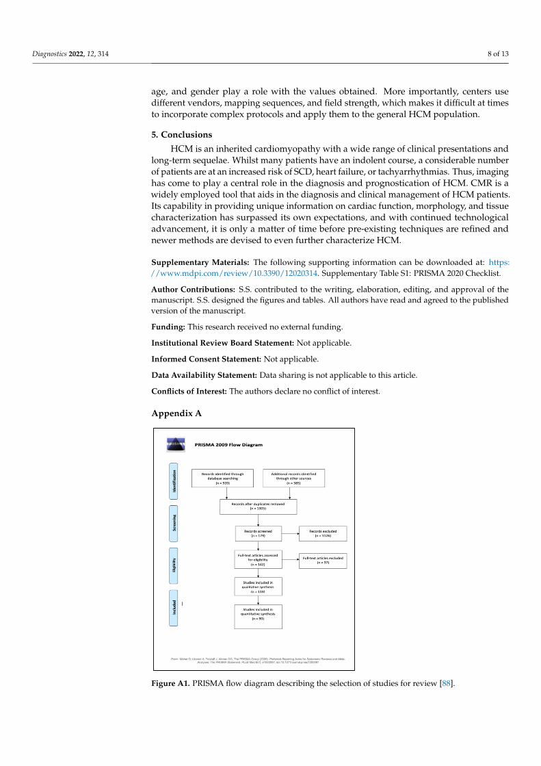
Figure A1. PRISMA flow diagram describing the selection of studies for review [88]. Figure A1. PRISMA flow diagram describing the selection of studies for review [88].
Diagnostics 2022, 12, 314 9 of 13
References1. Maron, B.J.; Gardin, J.M.; Flack, J.M.; Gidding, S.S.; Kurosaki, T.T.; Bild, D.E. Prevalence of hypertrophic cardiomyopathy in a
general population of young adults. Echocardiographic analysis of 4111 subjects in the CARDIA Study. Coron. Artery Risk Dev.Young Adults. Circ. 1995, 92, 785–789.
2. Ando, K.; Nagao, M.; Watanabe, E.; Sakai, A.; Suzuki, A.; Nakao, R.; Ishizaki, U.; Sakai, S.; Hagiwara, N. Association betweenmyocardial hypoxia and fibrosis in hypertrophic cardiomyopathy: Analysis by T2* BOLD and T1 mapping MRI. Eur. Radiol. 2020,30, 4327–4336. [CrossRef]
3. Cheng, S.; Fang, M.; Cui, C.; Chen, X.; Yin, G.; Prasad, S.K.; Dong, D.; Tian, J.; Zhao, S. LGE-CMR-derived texture features reflectpoor prognosis in hypertrophic cardiomyopathy patients with systolic dysfunction: Preliminary results. Eur. Radiol. 2018, 28,4615–4624. [CrossRef] [PubMed]
4. Malhotra, A.; Sivalokanathan, S. The transatlantic evolution in understanding sudden cardiac death in athletes. Trends Cardiovasc.Med. 2021, 21, S1050-173800071-2. [CrossRef] [PubMed]
5. Rowin, E.J.; Maron, B.J.; Carrick, R.T.; Patel, P.P.; Koethe, B.; Wells, S.; Maron, M.S. Outcomes in Patients with HypertrophicCardiomyopathy and Left Ventricular Systolic Dysfunction. J. Am. Coll. Cardiol. 2020, 75, 3033–3043. [CrossRef]
6. Brock, R. Functional obstruction of the left ventricle; acquired aortic subvalvar stenosis. Guy’s Hosp. Rep. 1957, 106, 221–238.7. Noureldin, R.A.; Liu, S.; Nacif, M.S.; Judge, D.P.; Halushka, M.K.; Abraham, T.P.; Ho, C.; Bluemke, D.A. The diagnosis of
hypertrophic cardiomyopathy by cardiovascular magnetic resonance. J. Cardiovasc. Magn. Reson. 2012, 14, 17. [CrossRef]8. Maron, M.S.; Olivotto, I.; Harrigan, C.; Appelbaum, E.; Gibson, C.M.; Lesser, J.R.; Haas, T.S.; Udelson, J.E.; Manning, W.J.; Maron,
B.J. Mitral valve abnormalities identified by cardiovascular magnetic resonance represent a primary phenotypic expression ofhypertrophic cardiomyopathy. Circulation 2011, 124, 40–47. [CrossRef]
9. Bogaert, J.; Olivotto, I. MR Imaging in Hypertrophic Cardiomyopathy: From Magnet to Bedside. Radiology 2014, 273, 329–348.[CrossRef]
10. Raphael, C.E.; Mitchell, F.; Kanaganayagam, G.S.; Liew, A.C.; Di Pietro, E.; Vieira, M.S.; Kanapeckaite, L.; Newsome, S.; Gregson,J.; Owen, R.; et al. Cardiovascular magnetic resonance predictors of heart failure in hypertrophic cardiomyopathy: The role ofmyocardial replacement fibrosis and the microcirculation. J. Cardiovasc. Magn. Reson. Off. J. Soc. Cardiovasc. Magn. Reson. 2021,23, 26. [CrossRef]
11. Grajewski, K.G.; Stojanovska, J.; Ibrahim, E.H.; Sayyouh, M.; Attili, A. Left Ventricular Hypertrophy: Evaluation with CardiacMRI. Curr. Probl. Diagn. Radiol. 2020, 49, 460–475. [CrossRef] [PubMed]
12. Waterhouse, D.F.; Ismail, T.F.; Prasad, S.K.; Wilson, M.G.; O’Hanlon, R. Imaging focal and interstitial fibrosis with cardiovascularmagnetic resonance in athletes with left ventricular hypertrophy: Implications for sporting participation. Br. J. Sports Med. 2012,46 (Suppl. S1), i69–i77. [CrossRef] [PubMed]
13. Markl, M.; Kilner, P.J.; Ebbers, T. Comprehensive 4D velocity mapping of the heart and great vessels by cardiovascular magneticresonance. J. Cardiovasc. Magn. Reson. Off. J. Soc. Cardiovasc. Magn. Reson. 2011, 13, 7. [CrossRef] [PubMed]
14. Scheffler, K.; Lehnhardt, S. Principles and applications of balanced SSFP techniques. Eur. Radiol. 2003, 13, 2409–2418. [CrossRef]15. Allen, B.D.; Choudhury, L.; Barker, A.J.; van Ooij, P.; Collins, J.D.; Bonow, R.O.; Carr, J.C.; Markl, M. Three-dimensional
haemodynamics in patients with obstructive and non-obstructive hypertrophic cardiomyopathy assessed by cardiac magneticresonance. Eur. Heart J. Cardiovasc. Imaging 2015, 16, 29–36. [CrossRef]
16. Brenes, J.C.; Doltra, A.; Prat, S. Cardiac magnetic resonance imaging in the evaluation of patients with hypertrophic cardiomyopa-thy. Glob. Cardiol. Sci. Pract. 2018, 2018, 22. [CrossRef]
17. Suinesiaputra, A.; Bluemke, D.A.; Cowan, B.R.; Friedrich, M.G.; Kramer, C.M.; Kwong, R.; Plein, S.; Schulz-Menger, J.; Westenberg,J.J.; Young, A.A.; et al. Quantification of LV function and mass by cardiovascular magnetic resonance: Multi-center variabilityand consensus contours. J. Cardiovasc. Magn. Reson. Off. J. Soc. Cardiovasc. Magn. Reson. 2015, 17, 63. [CrossRef]
18. Pu, C.; Fei, J.; Lv, S.; Wu, Y.; He, C.; Guo, D.; Mabombo, P.U.; Chooah, O.; Hu, H. Global Circumferential Strain by CardiacMagnetic Resonance Tissue Tracking Associated with Ventricular Arrhythmias in Hypertrophic Cardiomyopathy Patients. Front.Cardiovasc. Med. 2021, 8, 670361. [CrossRef]
19. Green, J.J.; Berger, J.S.; Kramer, C.M.; Salerno, M. Prognostic value of late gadolinium enhancement in clinical outcomes forhypertrophic cardiomyopathy. JACC Cardiovasc. Imaging 2012, 5, 370–377. [CrossRef]
20. O’Hanlon, R.; Grasso, A.; Roughton, M.; Moon, J.C.; Clark, S.; Wage, R.; Webb, J.; Kulkarni, M.; Dawson, D.; Sulaibeekh, L.; et al.Prognostic significance of myocardial fibrosis in hypertrophic cardiomyopathy. J. Am. Coll. Cardiol. 2010, 56, 867–874. [CrossRef]
21. Quarta, G.; Aquaro, G.D.; Pedrotti, P.; Pontone, G.; Dellegrottaglie, S.; Iacovoni, A.; Brambilla, P.; Pradella, S.; Todiere, G.; Rigo,F.; et al. Cardiovascular magnetic resonance imaging in hypertrophic cardiomyopathy: The importance of clinical context. Eur.Heart J. Cardiovasc. Imaging 2018, 19, 601–610. [CrossRef] [PubMed]
22. Liu, J.; Zhao, S.; Yu, S.; Wu, G.; Wang, D.; Liu, L.; Song, J.; Zhu, Y.; Kang, L.; Wang, J.; et al. Patterns of Replacement Fibrosis inHypertrophic Cardiomyopathy. Radiology 2021, 302, 210914. [CrossRef] [PubMed]
23. Elliott, P.M.; Gimeno, J.R.; Thaman, R.; Shah, J.; Ward, D.; Dickie, S.; Tome Esteban, M.T.; McKenna, W.J. Historical trends inreported survival rates in patients with hypertrophic cardiomyopathy. Heart Br. Card. Soc. 2006, 92, 785–791. [CrossRef] [PubMed]
24. Chan, R.H.; Maron, B.J.; Olivotto, I.; Pencina, M.J.; Assenza, G.E.; Haas, T.; Lesser, J.R.; Gruner, C.; Crean, A.M.; Rakowski,H.; et al. Prognostic value of quantitative contrast-enhanced cardiovascular magnetic resonance for the evaluation of suddendeath risk in patients with hypertrophic cardiomyopathy. Circulation 2014, 130, 484–495. [CrossRef]
Diagnostics 2022, 12, 314 10 of 13
25. Adabag, A.S.; Maron, B.J.; Appelbaum, E.; Harrigan, C.J.; Buros, J.L.; Gibson, C.M.; Lesser, J.R.; Hanna, C.A.; Udelson,J.E.; Manning, W.J.; et al. Occurrence and frequency of arrhythmias in hypertrophic cardiomyopathy in relation to delayedenhancement on cardiovascular magnetic resonance. J. Am. Coll. Cardiol. 2008, 51, 1369–1374. [CrossRef]
26. Rudolph, A.; Abdel-Aty, H.; Bohl, S.; Boyé, P.; Zagrosek, A.; Dietz, R.; Schulz-Menger, J. Noninvasive detection of fibrosisapplying contrast-enhanced cardiac magnetic resonance in different forms of left ventricular hypertrophy relation to remodeling.J. Am. Coll. Cardiol. 2009, 53, 284–291. [CrossRef]
27. Yang, K.; Song, Y.Y.; Chen, X.Y.; Wang, J.X.; Li, L.; Yin, G.; Zheng, Y.C.; Wei, M.D.; Lu, M.J.; Zhao, S.H. Apical hypertrophiccardiomyopathy with left ventricular apical aneurysm: Prevalence, cardiac magnetic resonance characteristics, and prognosis.Eur. Heart J. Cardiovasc. Imaging 2020, 21, 1341–1350. [CrossRef]
28. Moon, J.C.; McKenna, W.J.; McCrohon, J.A.; Elliott, P.M.; Smith, G.C.; Pennell, D.J. Toward clinical risk assessment in hypertrophiccardiomyopathy with gadolinium cardiovascular magnetic resonance. J. Am. Coll. Cardiol. 2003, 41, 1561–1567. [CrossRef]
29. Moon, J.C.; Reed, E.; Sheppard, M.N.; Elkington, A.G.; Ho, S.Y.; Burke, M.; Petrou, M.; Pennell, D.J. The histologic basis of lategadolinium enhancement cardiovascular magnetic resonance in hypertrophic cardiomyopathy. J. Am. Coll. Cardiol. 2004, 43,2260–2264. [CrossRef] [PubMed]
30. Huang, L.; Ran, L.; Zhao, P.; Tang, D.; Han, R.; Ai, T.; Xia, L.; Tao, Q. MRI native T1 and T2 mapping of myocardial segmentsin hypertrophic cardiomyopathy: Tissue remodeling manifested prior to structure changes. Br. J. Radiol. 2019, 92, 20190634.[CrossRef]
31. Gastl, M.; Gotschy, A.; von Spiczak, J.; Polacin, M.; Bönner, F.; Gruner, C.; Kelm, M.; Ruschitzka, F.; Alkadhi, H.; Kozerke, S.; et al.Cardiovascular magnetic resonance T2* mapping for structural alterations in hypertrophic cardiomyopathy. Eur. J. Radiol. Open2019, 6, 78–84. [CrossRef] [PubMed]
32. Snel, G.; van den Boomen, M.; Hernandez, L.M.; Nguyen, C.T.; Sosnovik, D.E.; Velthuis, B.K.; Slart, R.; Borra, R.; Prakken, N.Cardiovascular magnetic resonance native T2 and T2
* quantitative values for cardiomyopathies and heart transplantations: Asystematic review and meta-analysis. J. Cardiovasc. Magn. Reson. Off. J. Soc. Cardiovasc. Magn. Reson. 2020, 22, 34.
33. Messroghli, D.R.; Moon, J.C.; Ferreira, V.M.; Grosse-Wortmann, L.; He, T.; Kellman, P.; Mascherbauer, J.; Nezafat, R.; Salerno, M.;Schelbert, E.B.; et al. Clinical recommendations for cardiovascular magnetic resonance mapping of T1, T2, T2* and extracellularvolume: A consensus statement by the Society for Cardiovascular Magnetic Resonance (SCMR) endorsed by the EuropeanAssociation for Cardiovascular Imaging (EACVI). J. Cardiovasc. Magn. Reson. Off. J. Soc. Cardiovasc. Magn. Reson. 2017, 19, 75.
34. Rowin, E.J.; Maron, M.S. The Role of Cardiac MRI in the Diagnosis and Risk Stratification of Hypertrophic Cardiomyopathy.Arrhythmia Electrophysiol. Rev. 2016, 5, 197–202. [CrossRef]
35. Arcari, L.; Hinojar, R.; Engel, J.; Freiwald, T.; Platschek, S.; Zainal, H.; Zhou, H.; Vasquez, M.; Keller, T.; Rolf, A.; et al. Native T1and T2 provide distinctive signatures in hypertrophic cardiac conditions—Comparison of uremic, hypertensive and hypertrophiccardiomyopathy. Int. J. Cardiol. 2020, 306, 102–108. [CrossRef] [PubMed]
36. Kaldoudi, E.; Williams, C.R. Relaxation Time Measurements in NMR Imaging. Concepts Magn. Reson. 1993, 5, 217–242. [CrossRef]37. Amano, Y.; Takayama, M.; Kumita, S. Contrast-enhanced myocardial T1-weighted scout (Look-Locker) imaging for the detection
of myocardial damages in hypertrophic cardiomyopathy. J. Magn. Reson. Imaging JMRI 2009, 30, 778–784. [CrossRef]38. Hurtado-de-Mendoza, D.; Corona-Villalobos, C.P.; Pozios, I.; Gonzales, J.; Soleimanifard, Y.; Sivalokanathan, S.; Montoya-Cerrillo,
D.; Vakrou, S.; Kamel, I.; Mormontoy-Laurel, W.; et al. Diffuse interstitial fibrosis assessed by cardiac magnetic resonance isassociated with dispersion of ventricular repolarization in patients with hypertrophic cardiomyopathy. J. Arrhythmia 2017, 33,201–207. [CrossRef]
39. El-Rewaidy, H.; Neisius, U.; Nakamori, S.; Ngo, L.; Rodriguez, J.; Manning, W.J.; Nezafat, R. Characterization of interstitial diffusefibrosis patterns using texture analysis of myocardial native T1 mapping. PLoS ONE 2020, 15, e0233694. [CrossRef]
40. Xu, J.; Zhuang, B.; Sirajuddin, A.; Li, S.; Huang, J.; Yin, G.; Song, L.; Jiang, Y.; Zhao, S.; Lu, M. MRI T1 Mapping in HypertrophicCardiomyopathy: Evaluation in Patients Without Late Gadolinium Enhancement and Hemodynamic Obstruction. Radiology 2020,294, 275–286. [CrossRef]
41. Martinez-Naharro, A.; Treibel, T.A.; Abdel-Gadir, A.; Bulluck, H.; Zumbo, G.; Knight, D.S.; Kotecha, T.; Francis, R.; Hutt, D.F.;Rezk, T.; et al. Magnetic Resonance in Transthyretin Cardiac Amyloidosis. J. Am. Coll. Cardiol. 2017, 70, 466–477. [CrossRef][PubMed]
42. Hinojar, R.; Varma, N.; Child, N.; Goodman, B.; Jabbour, A.; Yu, C.Y.; Gebker, R.; Doltra, A.; Kelle, S.; Khan, S.; et al. T1 Mappingin Discrimination of Hypertrophic Phenotypes: Hypertensive Heart Disease and Hypertrophic Cardiomyopathy: Findings fromthe International T1 Multicenter Cardiovascular Magnetic Resonance Study. Circ. Cardiovasc. Imaging 2015, 8, e003285. [CrossRef][PubMed]
43. Karur, G.R.; Robison, S.; Iwanochko, R.M.; Morel, C.F.; Crean, A.M.; Thavendiranathan, P.; Nguyen, E.T.; Mathur, S.; Wasim, S.;Hanneman, K. Use of Myocardial T1 Mapping at 3.0 T to Differentiate Anderson-Fabry Disease from Hypertrophic Cardiomyopa-thy. Radiology 2018, 288, 398–406. [CrossRef] [PubMed]
44. Shi, R.Y.; An, D.A.; Chen, B.H.; Wu, R.; Du, L.; Jiang, M.; Xu, J.R.; Wu, L.M. Diffusion-weighted imaging in hypertrophiccardiomyopathy: Association with high T2-weighted signal intensity in addition to late gadolinium enhancement. Int. J.Cardiovasc. Imaging 2020, 36, 2229–2238. [CrossRef] [PubMed]

Diagnostics 2022, 12, 314 11 of 13
45. Gastl, M.; Lachmann, V.; Christidi, A.; Janzarik, N.; Veulemans, V.; Haberkorn, S.; Holzbach, L.; Jacoby, C.; Schnackenburg, B.;Berrisch-Rahmel, S.; et al. Cardiac magnetic resonance T2 mapping and feature tracking in athlete’s heart and HCM. Eur. Radiol.2021, 31, 2768–2777. [CrossRef]
46. Abdel-Aty, H.; Cocker, M.; Strohm, O.; Filipchuk, N.; Friedrich, M.G. Abnormalities in T2-weighted cardiovascular magneticresonance images of hypertrophic cardiomyopathy: Regional distribution and relation to late gadolinium enhancement andseverity of hypertrophy. J. Magn. Reson. Imaging JMRI 2008, 28, 242–245. [CrossRef]
47. Abdel-Aty, H.; Zagrosek, A.; Schulz-Menger, J.; Taylor, A.J.; Messroghli, D.; Kumar, A.; Gross, M.; Dietz, R.; Friedrich, M.G.Delayed enhancement and T2-weighted cardiovascular magnetic resonance imaging differentiate acute from chronic myocardialinfarction. Circulation 2004, 109, 2411–2416. [CrossRef]
48. Gastl, M.; Gruner, C.; Labucay, K.; Gotschy, A.; Von Spiczak, J.; Polacin, M.; Boenner, F.; Kelm, M.; Ruschitzka, F.; Alkadhi, H.; et al.Cardiovascular magnetic resonance T2* mapping for the assessment of cardiovascular events in hypertrophic cardiomyopathy.Open Heart 2020, 7, e001152. [CrossRef]
49. Cannon, C.P. (Ed.) Lamb BPPHJ: Assessment of Diastolic Function by Cardiac MRI; Cardiovascular Magnetic Resonance Imaging;Humana Press: Totowa, NJ, USA, 2008; pp. 415–428.
50. Giusca, S.; Steen, H.; Montenbruck, M.; Patel, A.R.; Pieske, B.; Erley, J.; Kelle, S.; Korosoglou, G. Multi-parametric assessmentof left ventricular hypertrophy using late gadolinium enhancement, T1 mapping and strain-encoded cardiovascular magneticresonance. J. Cardiovasc. Magn. Reson. Off. J. Soc. Cardiovasc. Magn. Reson. 2021, 23, 92. [CrossRef]
51. Pagourelias, E.D.; Mirea, O.; Duchenne, J.; Unlu, S.; Van Cleemput, J.; Papadopoulos, C.E.; Bogaert, J.; Vassilikos, V.P.; Voigt, J.U.Speckle tracking deformation imaging to detect regional fibrosis in hypertrophic cardiomyopathy: A comparison between 2Dand 3D echo modalities. Eur. Heart J. Cardiovasc. Imaging 2020, 21, 1262–1272. [CrossRef]
52. Tayal, B.; Malahfji, M.; Buergler, J.M.; Shah, D.J.; Nagueh, S.F. Hemodynamic determinants of left atrial strain in patients withhypertrophic cardiomyopathy: A combined echocardiography and CMR study. PLoS ONE 2021, 16, e0245934. [CrossRef][PubMed]
53. Li, Z.L.; He, S.; Xia, C.C.; Peng, W.L.; Li, L.; Liu, K.L.; Zhang, J.G.; Pu, J.; Guo, Y.K. Global longitudinal diastolic strain rate as anovel marker for predicting adverse outcomes in hypertrophic cardiomyopathy by cardiac magnetic resonance tissue tracking.Clin. Radiol. 2021, 76, 78.e19–78.e25. [CrossRef] [PubMed]
54. Cavus, E.; Muellerleile, K.; Schellert, S.; Schneider, J.; Tahir, E.; Chevalier, C.; Jahnke, C.; Radunski, U.K.; Adam, G.; Kirch-hof, P.; et al. CMR feature tracking strain patterns and their association with circulating cardiac biomarkers in patients withhypertrophic cardiomyopathy. Clin. Res. Cardiol. Off. J. Ger. Card. Soc. 2021, 110, 1757–1769. [CrossRef]
55. Sivalokanathan, S.; Zghaib, T.; Greenland, G.V.; Vasquez, N.; Kudchadkar, S.M.; Kontari, E.; Lu, D.Y.; Dolores-Cerna, K.; van derGeest, R.J.; Kamel, I.R.; et al. Hypertrophic Cardiomyopathy Patients with Paroxysmal Atrial Fibrillation Have a High Burden ofLeft Atrial Fibrosis by Cardiac Magnetic Resonance Imaging. JACC Clin. Electrophysiol. 2019, 5, 364–375. [CrossRef] [PubMed]
56. Siontis, K.C.; Geske, J.B.; Ong, K.; Nishimura, R.A.; Ommen, S.R.; Gersh, B.J. Atrial fibrillation in hypertrophic cardiomyopathy:Prevalence, clinical correlations, and mortality in a large high-risk population. J. Am. Heart Assoc. 2014, 3, e001002. [CrossRef]
57. Kowallick, J.T.; Kutty, S.; Edelmann, F.; Chiribiri, A.; Villa, A.; Steinmetz, M.; Sohns, J.M.; Staab, W.; Bettencourt, N.; Unterberg-Buchwald, C.; et al. Quantification of left atrial strain and strain rate using Cardiovascular Magnetic Resonance myocardialfeature tracking: A feasibility study. J. Cardiovasc. Magn. Reson. Off. J. Soc. Cardiovasc. Magn. Reson. 2014, 16, 60. [CrossRef]
58. Raman, B.; Smillie, R.W.; Mahmod, M.; Chan, K.; Ariga, R.; Nikolaidou, C.; Ormondroyd, E.; Thomson, K.; Harper, A.R.; Tan,G.; et al. Incremental value of left atrial booster and reservoir strain in predicting atrial fibrillation in patients with hypertrophiccardiomyopathy: A cardiovascular magnetic resonance study. J. Cardiovasc. Magn. Reson. Off. J. Soc. Cardiovasc. Magn. Reson.2021, 23, 109. [CrossRef]
59. Pedrizzetti, G.; Claus, P.; Kilner, P.J.; Nagel, E. Principles of cardiovascular magnetic resonance feature tracking and echocardio-graphic speckle tracking for informed clinical use. J. Cardiovasc. Magn. Reson. Off. J. Soc. Cardiovasc. Magn. Reson. 2016, 18, 51.[CrossRef]
60. Ismail, T.F.; Hsu, L.Y.; Greve, A.M.; Gonçalves, C.; Jabbour, A.; Gulati, A.; Hewins, B.; Mistry, N.; Wage, R.; Roughton, M.; et al.Coronary microvascular ischemia in hypertrophic cardiomyopathy—A pixel-wise quantitative cardiovascular magnetic resonanceperfusion study. J. Cardiovasc. Magn. Reson. Off. J. Soc. Cardiovasc. Magn. Reson. 2014, 16, 49. [CrossRef]
61. Varnava, A.M.; Elliott, P.M.; Sharma, S.; McKenna, W.J.; Davies, M.J. Hypertrophic cardiomyopathy: The interrelation of disarray,fibrosis, and small vessel disease. Heart Br. Card. Soc. 2000, 84, 476–482. [CrossRef]
62. Kim, E.K.; Lee, S.C.; Chang, S.A.; Jang, S.Y.; Kim, S.M.; Park, S.J.; Choi, J.O.; Park, S.W.; Jeon, E.S.; Choe, Y.H. Prevalence andclinical significance of cardiovascular magnetic resonance adenosine stress-induced myocardial perfusion defect in hypertrophiccardiomyopathy. J. Cardiovasc. Magn. Reson. Off. J. Soc. Cardiovasc. Magn. Reson. 2020, 22, 30. [CrossRef] [PubMed]
63. Basso, C.; Thiene, G.; Corrado, D.; Buja, G.; Melacini, P.; Nava, A. Hypertrophic cardiomyopathy and sudden death in the young:Pathologic evidence of myocardial ischemia. Hum. Pathol. 2000, 31, 988–998. [CrossRef] [PubMed]
64. Maron, M.S.; Olivotto, I.; Maron, B.J.; Prasad, S.K.; Cecchi, F.; Udelson, J.E.; Camici, P.G. The case for myocardial ischemia inhypertrophic cardiomyopathy. J. Am. Coll. Cardiol. 2009, 54, 866–875. [CrossRef] [PubMed]
65. Olivotto, I.; Girolami, F.; Sciagrà, R.; Ackerman, M.J.; Sotgia, B.; Bos, J.M.; Nistri, S.; Sgalambro, A.; Grifoni, C.; Torricelli, F.; et al.Microvascular function is selectively impaired in patients with hypertrophic cardiomyopathy and sarcomere myofilament genemutations. J. Am. Coll. Cardiol. 2011, 58, 839–848. [CrossRef]
Diagnostics 2022, 12, 314 12 of 13
66. Jablonowski, R.; Fernlund, E.; Aletras, A.H.; Engblom, H.; Heiberg, E.; Liuba, P.; Arheden, H.; Carlsson, M. Regional Stress-Induced Ischemia in Non-fibrotic Hypertrophied Myocardium in Young HCM Patients. Pediatric Cardiol. 2015, 36, 1662–1669.[CrossRef]
67. Chiribiri, A.; Leuzzi, S.; Conte, M.R.; Bongioanni, S.; Bratis, K.; Olivotti, L.; De Rosa, C.; Lardone, E.; Di Donna, P.; Villa, A.D.; et al.Rest perfusion abnormalities in hypertrophic cardiomyopathy: Correlation with myocardial fibrosis and risk factors for suddencardiac death. Clin. Radiol. 2015, 70, 495–501. [CrossRef]
68. Raman, B.; Ariga, R.; Spartera, M.; Sivalokanathan, S.; Chan, K.; Dass, S.; Petersen, S.E.; Daniels, M.J.; Francis, J.; Smillie,R.; et al. Progression of myocardial fibrosis in hypertrophic cardiomyopathy: Mechanisms and clinical implications. Eur. Heart J.Cardiovasc. Imaging 2019, 20, 157–167. [CrossRef]
69. Tezuka, D.; Kosuge, H.; Terashima, M.; Koyama, N.; Kishida, T.; Tada, Y.; Suzuki, J.I.; Sasano, T.; Ashikaga, T.; Hirao, K.; et al.Myocardial perfusion reserve quantified by cardiac magnetic resonance imaging is associated with late gadolinium enhancementin hypertrophic cardiomyopathy. Heart Vessel. 2018, 33, 513–520. [CrossRef]
70. Knaapen, P.; van Dockum, W.G.; Götte, M.J.; Broeze, K.A.; Kuijer, J.P.; Zwanenburg, J.J.; Marcus, J.T.; Kok, W.E.; van Rossum,A.C.; Lammertsma, A.A.; et al. Regional heterogeneity of resting perfusion in hypertrophic cardiomyopathy is related to delayedcontrast enhancement but not to systolic function: A PET and MRI study. J. Nucl. Cardiol. Off. Publ. Am. Soc. Nucl. Cardiol. 2006,13, 660–667. [CrossRef]
71. Petersen, S.E.; Jerosch-Herold, M.; Hudsmith, L.E.; Robson, M.D.; Francis, J.M.; Doll, H.A.; Selvanayagam, J.B.; Neubauer, S.;Watkins, H. Evidence for microvascular dysfunction in hypertrophic cardiomyopathy: New insights from multiparametricmagnetic resonance imaging. Circulation 2007, 115, 2418–2425. [CrossRef]
72. Hughes, R.K.; Camaioni, C.; Augusto, J.B.; Knott, K.; Quinn, E.; Captur, G.; Seraphim, A.; Joy, G.; Syrris, P.; Elliott, P.M.; et al.Myocardial Perfusion Defects in Hypertrophic Cardiomyopathy Mutation Carriers. J. Am. Heart Assoc. 2021, 10, e020227.[CrossRef] [PubMed]
73. Sipola, P.; Lauerma, K.; Husso-Saastamoinen, M.; Kuikka, J.T.; Vanninen, E.; Laitinen, T.; Manninen, H.; Niemi, P.; Peuhkurinen,K.; Jääskeläinen, P.; et al. First-pass MR imaging in the assessment of perfusion impairment in patients with hypertrophiccardiomyopathy and the Asp175Asn mutation of the alpha-tropomyosin gene. Radiology 2003, 226, 129–137. [CrossRef] [PubMed]
74. Das, A.; Kelly, C.; Teh, I.; Nguyen, C.; Brown, L.; Chowdhary, A.; Jex, N.; Thirunavukarasu, S.; Sharrack, N.; Gorecka, M.; et al.Phenotyping hypertrophic cardiomyopathy using cardiac diffusion magnetic resonance imaging: The relationship betweenmicrovascular dysfunction and microstructural changes. Eur. Heart J. Cardiovasc. Imaging 2021, jeab210. [CrossRef] [PubMed]
75. McGill, L.A.; Ismail, T.F.; Nielles-Vallespin, S.; Ferreira, P.; Scott, A.D.; Roughton, M.; Kilner, P.J.; Ho, S.Y.; McCarthy, K.P.; Gate-house, P.D.; et al. Reproducibility of in-vivo diffusion tensor cardiovascular magnetic resonance in hypertrophic cardiomyopathy.J. Cardiovasc. Magn. Reson. Off. J. Soc. Cardiovasc. Magn. Reson. 2012, 14, 86. [CrossRef]
76. Ariga, R.; Tunnicliffe, E.M.; Manohar, S.G.; Mahmod, M.; Raman, B.; Piechnik, S.K.; Francis, J.M.; Robson, M.D.; Neubauer, S.;Watkins, H. Identification of Myocardial Disarray in Patients with Hypertrophic Cardiomyopathy and Ventricular Arrhythmias. J.Am. Coll. Cardiol. 2019, 73, 2493–2502. [CrossRef]
77. Nielles-Vallespin, S.; Khalique, Z.; Ferreira, P.F.; de Silva, R.; Scott, A.D.; Kilner, P.; McGill, L.A.; Giannakidis, A.; Gatehouse, P.D.;Ennis, D.; et al. Assessment of Myocardial Microstructural Dynamics by In Vivo Diffusion Tensor Cardiac Magnetic Resonance. J.Am. Coll. Cardiol. 2017, 69, 661–676. [CrossRef]
78. Wymer, D.T.; Patel, K.P.; Burke, W.F., 3rd; Bhatia, V.K. Phase-Contrast MRI: Physics, Techniques, and Clinical Applications.Radiographics 2020, 40, 122–140. [CrossRef]
79. Srichai, M.B.; Lim, R.P.; Wong, S.; Lee, V.S. Cardiovascular applications of phase-contrast MRI. AJR Am. J. Roentgenol. 2009, 192,662–675. [CrossRef]
80. Nayak, K.S.; Nielsen, J.F.; Bernstein, M.A.; Markl, M.; DGatehouse, P.; MBotnar, R.; Saloner, D.; Lorenz, C.; Wen, H.; SHu, B.; et al.Cardiovascular magnetic resonance phase contrast imaging. J. Cardiovasc. Magn. Reson. Off. J. Soc. Cardiovasc. Magn. Reson. 2015,17, 71. [CrossRef]
81. Stankovic, Z.; Allen, B.D.; Garcia, J.; Jarvis, K.B.; Markl, M. 4D flow imaging with MRI. Cardiovasc. Diagn. Ther. 2014, 4, 173–192.82. Pruijssen, J.T.; Allen, B.D.; Barker, A.J.; Bonow, R.O.; Choudhury, L.; Carr, J.C.; Markl, M.; van Ooij, P. Hypertrophic Cardiomy-
opathy Is Associated with Altered Left Ventricular 3D Blood Flow Dynamics. Radiology. Cardiothorac. Imaging 2020, 2, e190038.[CrossRef] [PubMed]
83. She, J.Q.; Guo, J.J.; Yu, Y.F.; Zhao, S.H.; Chen, Y.Y.; Ge, M.Y.; Zeng, M.S.; Jin, H. Left Ventricular Outflow Tract Obstruction inHypertrophic Cardiomyopathy: The Utility of Myocardial Strain Based on Cardiac MR Tissue Tracking. J. Magn. Reson. Imaging:JMRI 2021, 53, 51–60. [CrossRef] [PubMed]
84. Van Ooij, P.; Allen, B.D.; Contaldi, C.; Garcia, J.; Collins, J.; Carr, J.; Choudhury, L.; Bonow, R.O.; Barker, A.J.; Markl, M. 4Dflow MRI and T1 -Mapping: Assessment of altered cardiac hemodynamics and extracellular volume fraction in hypertrophiccardiomyopathy. J. Magn. Reson. Imaging: JMRI 2016, 43, 107–114. [CrossRef] [PubMed]
85. Marstrand, P.; Han, L.; Day, S.M.; Olivotto, I.; Ashley, E.A.; Michels, M.; Pereira, A.C.; Wittekind, S.G.; Helms, A.; Saberi, S.; et al.Hypertrophic Cardiomyopathy with Left Ventricular Systolic Dysfunction: Insights from the SHaRe Registry. Circulation 2020,141, 1371–1383. [CrossRef]
86. Wang, Z.J.; Ohliger, M.A.; Larson, P.; Gordon, J.W.; Bok, R.A.; Slater, J.; Villanueva-Meyer, J.E.; Hess, C.P.; Kurhanewicz, J.;Vigneron, D.B. Hyperpolarized 13C MRI: State of the Art and Future Directions. Radiology 2019, 291, 273–284. [CrossRef]

Diagnostics 2022, 12, 314 13 of 13
87. Arenja, N.; Fritz, T.; Andre, F.; Riffel, J.H.; Aus dem Siepen, F.; Ochs, M.; Paffhausen, J.; Hegenbart, U.; Schönland, S.;Müller-Hennessen, M.; et al. Myocardial contraction fraction derived from cardiovascular magnetic resonance cine images-reference values and performance in patients with heart failure and left ventricular hypertrophy. Eur. Heart J. Cardiovasc. Imaging2017, 18, 1414–1422. [CrossRef]
88. Page, M.J.; McKenzie, J.E.; Bossuyt, P.M.; Boutron, I.; Hoffmann, T.C.; Mulrow, C.D.; Shamseer, L.; Tetzlaff, J.M.; Akl, E.A.;Brennan, S.E.; et al. The PRISMA 2020 statement: An updated guideline for reporting systematic reviews. BMJ 2021, 372, 71.[CrossRef]