Rapid and selective separation for mixed proteins with thiol functionalized magnetic nanoparticles
Upload
independentCategory
view
0download
0
PEG-Functionalized Magnetic Nanoparticles for Drug Deliveryand Magnetic Resonance Imaging Applications
Murali Mohan Yallapu1,2, Susan P Foy1, Tapan K Jain1,3, and Vinod Labhasetwar1,4,*1 Department of Biomedical Engineering, Lerner Research Institute, Cleveland Clinic, Cleveland,OH 44195, USA4 Taussig Cancer Center, Cleveland Clinic, Cleveland, OH 44195, USA
AbstractPurpose—Polyethylene glycol (PEG) functionalized magnetic nanoparticles (MNPs) were testedas a drug carrier system, magnetic resonance imaging (MRI) agent, and ability to conjugate to anantibody.
Methods—An iron oxide core coated with oleic acid (OA) and then with OA-PEG forms a waterdispersible MNP formulation. Hydrophobic doxorubicin partitions into the OA layer for sustaineddrug delivery. The T1 and T2 MRI contrast properties were determined in vitro and the circulationof the MNPs measured in mouse carotid arteries. An N-hydroxysuccinimide group (NHS) on theOA-PEG-80 was used to conjugate the amine functional group on antibodies for active targetingin the human MCF-7 breast cancer cell line.
Results—The optimized formulation had a mean hydrodynamic diameter of 184 nm with an 8nm iron-oxide core. The MNPs enhance the T2 MRI contrast, and have a long circulation time invivo with 30% relative concentration 50 min post-injection. Doxorubicin-loaded MNPs showedsustained drug release and dose-dependent antiproliferative effects in vitro; the drug effect wasenhanced with transferrin antibody conjugated MNPs.
Conclusion—PEG functionalized MNPs could be developed as a targeted drug delivery systemand MRI contrast agent.
KeywordsIron-oxide; Anticancer drugs; Anti-proliferative effect; Reticuloendothelial system; Transferrinantibody
INTRODUCTIONMagnetic nanoparticles (MNPs) have gained significant interest in recent years for variousbiomedical applications. Their intrinsic magnetic properties have been explored in celllabeling/cell separation, developing bioassays, in magnetofection, drug delivery, to inducelocal hyperthermia in response to an external alternating magnetic field, and to selectivelydestroy cancer cells (1). In addition, MNPs are used in clinical diagnosis as a magneticresonance imaging (MRI) contrast agent (2).
*Author for correspondence: Vinod Labhasetwar, Ph.D. Department of Biomedical Engineering/ND-20, Cleveland Clinic, 9500 EuclidAvenue, Cleveland, OH 44195, Tel: 216/445-9364, Fax 216/444-9198, [email protected] address: Cancer Biology Research Center, Sanford Research/University of South Dakota, Sioux Falls, SD 57105, USA3Present address: University School of Basic & Applied Sciences, Guru Gobind Singh Indraprastha University, Kashmere Gate,Delhi-110403, India
NIH Public AccessAuthor ManuscriptPharm Res. Author manuscript; available in PMC 2011 November 1.
Published in final edited form as:Pharm Res. 2010 November ; 27(11): 2283–2295. doi:10.1007/s11095-010-0260-1.
NIH
-PA Author Manuscript
NIH
-PA Author Manuscript
NIH
-PA Author Manuscript
Our interest is in developing MNPs with dual functional characteristics for drug delivery andas a MRI agent. These two applications are complimentary in many ways, as it provides theunique ability to assess the efficiency of drug delivery to the target site in real time, as wellas to monitor the progression of the disease in response to the therapy using a non-invasiveMRI technique (3). Drug-loaded MNPs can play roles in therapy and/or detection, andtherefore are of high clinical importance.
Although several MNP formulations have been developed to meet the specific needs of aparticular application, there have been limited efforts towards developing MNPs withcombined properties of drug delivery and imaging (2). This could, in part, be due to thedifferent sets of issues involved in achieving both the objectives with one formulation ofMNPs. The general approaches are to incorporate the drug of interest and MNPs inliposomes/emulsion/polymers, or to conjugate the drug of interest to the coating, usuallydextran, that is used for stabilizing the core material in an aqueous system. However, suchapproaches usually result in low efficiency of drug association with MNPs; the associateddrug rapidly dissociates (4), the process of drug conjugation often times results in the loss ofmagnetization of the core material, and/or causes significant increase in particle size. Thesechanges significantly affect the imaging property of MNPs. Further, the rapid clearance ofMNPs from the circulation and their sequestering by the reticuloendothelial system (RES)limit the drug targeting efficiency and imaging applications of MNPs to targets other thanthe organs of the RES such as the liver or lymphatic systems.
Thus, critical to the successful development of MNPs with multifunctional properties wouldbe to meet the needs of drug delivery without compromising their MRI characteristics.Ideally, such a formulation should have the following functional properties- i) surface thatwould prevent the rapid clearance of the MNPs by the RES following intravenousadministration; ii) ability to load drugs without influencing the physical properties of theMNP formulation or magnetization property of the core material; iii) ability to conjugateMNPs to a targeting ligand or antibody. With the above intention, we have been developinga MNP formulation that contains an iron-oxide core which is first coated with oleic acid(OA) and then with OA-conjugated poly(ethylene) glycol (PEG) (OA-PEG). It washypothesized that the OA of OA-PEG would anchor on to the OA coated on the iron-oxidecore, and the PEG would form a corona that would impart water dispersity and stericstability to the MNPs. In addition, functionalized PEG would prevent the rapid clearance ofMNPs by the RES, a known characteristic of PEG, and provide the functional groups forconjugation to a targeting ligand. The OA coat around the iron-oxide core could act as areservoir for loading of hydrophobic drugs. Further, functionalized groups on PEG can beused for conjugation to antibody or targeting ligands.
In this study, we have optimized the various formulation parameters and characterized thePEG-functionalized MNPs for physical properties and chemical analysis of the surface.Further, we have determined protein binding to MNPs and their uptake by macrophages invitro. To determine their drug delivery application, we have demonstrated loading ofdoxorubicin (base) as a model anticancer cancer drug and studied the antiproliferative effectof the drug-loaded MNPs in the MCF-7 breast cancer cell line. The imaging properties ofMNPs in phantom agar gels and clearance of the MNPs in the mouse carotid artery weredetermined using MRI. Magnetic resonance properties and clearance of our MNPs werecompared with Feridex IV, a dextran-coated iron-oxide formulation used in clinicaldiagnostic applications.
Yallapu et al. Page 2
Pharm Res. Author manuscript; available in PMC 2011 November 1.
NIH
-PA Author Manuscript
NIH
-PA Author Manuscript
NIH
-PA Author Manuscript

MATERIALS AND METHODSMaterials
Iron(III) chloride hexahydrate (FeCl3·6H2O) 99% pure granulated (Fe(III)), Iron(II) chloridetetrahydrate (FeCl2·4H2O) 99+% (Fe(II)), ammonium hydroxide (5 M), and oleic acid (OA)were purchased from Fisher Scientic (Pittsburgh, PA). SUNBRIGHT OE-020CS (OA-PEG-20), OE-040CS (OA-PEG-40) and OE-80CS (OA-PEG-80), namely (α-succinimidyloxysuccinyl-ω-oleyloxy) poly(oxyethylene)s differing in molecular weight ofPEG chain length (i.e. 2104, 4352, and 8525, respectively) were purchased from NOFCorporation (Tokyo, Japan). Feridex IV (11.2 mg Fe/mL, 5.6–9.1 mg dextran/mL) waspurchased from Berlex Laboratories, Inc. (Montville, NJ). Bacto nutrient agar dehydratedwas purchased from Difco Laboratories (Detroit, MI). Doxorubicin hydrochloride was a giftfrom Dabur Research Foundation (Ghaziabad, India). A primary transferrin antibody FITC(rabbit polyclonal to transferrin) was purchased from Abcam Inc. (Cambridge, MA).Transferrin was received from Sigma (St. Louis, MO). Sterile nitrogen purged water wasused in all steps in the synthesis of magnetic nanoparticles (MNPs).
Synthesis of Magnetic NanoparticlesAqueous solutions of 0.1 M Fe(III) (30 mL) and 0.1 M Fe(II) (15 mL) were combined in a100 mL beaker with magnetic stirring for 10 min. Ammonium hydroxide (5 M, 3 mL) wasadded drop wise over 5 min and stirring was continued for 30 min under nitrogen-gasatmosphere. The reaction was stirred for 24 h for bare magnetite particles.
For oleic acid coated MNPs (OA-MNPs), after 30 min of stirring under nitrogen-gasatmosphere, 100 mg OA was added to the above mixture, and the mixture was heated to 80°C while stirring for 30 min. The resulting mixture was cooled down to room temperatureand stirred for 24 h. Poly(ethylene) glycol (PEG) functionalized OA-MNPs (PEG-OA-MNPs) were prepared by cooling the OA-MNPs to room temperature, and adding anaqueous solution of OA-PEG-20, OA-PEG-40, or OA-PEG-80 polymers (25–300 mg)dissolved in 5 mL of water over 5 min with stirring. The stirring was continued for anadditional 24 h at room temperature. The PEG-OA-MNPs prepared with 200 mg of OA-PEG-20, OA-PEG-40 and OA-PEG-80 are designated as OE-20, OE-40, and OE-80 MNPsthroughout the manuscript (Figure 1).
Bare MNPs, OA-MNPs, and PEG-OA-MNPs were separated by magnetic separation byplacing a magnet (12,2000 G, Edmund Scientific, Tonawanda, NY) below the beaker andallowing the solution to clear. The excess iron salts, ammonium hydroxide, OA, and OA-PEG in the wash were discarded. The particles were washed with 40 mL nitrogen purgedsterile water four times using magnetic separation, dispersed in 25 mL of sterile water, andcentrifuged at 1000 rpm for 10 min to remove large and aggregated particles. PurifiedOE-80 MNPs to be used in antibody conjugation were washed with water and centrifuged asmentioned above but at 4°C.
Magnetic Nanoparticle Characterization—A 5–10 μL nanoparticle suspension (3–5mg/mL) was diluted to 2.5 mL with distilled water and the mean hydrodynamic particle sizeand zeta potential determined by dynamic laser light scattering (DLS, NICOMP 380™ ZLS,Particle sizing systems, Santa Barbara, CA). The particle size was also studied at the sameconcentration in mannitol citrate buffer, saline (0.9 wt.% sodium chloride), PBS (154 mM,pH 7.4), and 1mM HEPES buffer adjusted with 0.1 N sodium hydroxide or hydrochloricacid solutions to a final pH of 3 to 10. Particle stability was determined by storing 4 mg ofparticles in 1 mL of water, saline, PBS or mannitol buffer and measuring the size at varyingtime intervals.
Yallapu et al. Page 3
Pharm Res. Author manuscript; available in PMC 2011 November 1.
NIH
-PA Author Manuscript
NIH
-PA Author Manuscript
NIH
-PA Author Manuscript
Transmission electron microcopy (TEM) was used to determine the particle size of theformulations in dry state (Philips/FEI Inc., Briarcliff, Manor, NY). The particle size in TEMimages were measured using NIH ImageJ software (http://rsbweb.nih.gov/ij/). Samples oflyophilized MNPs were used for Fourier transform infrared (FTIR) spectroscopy(PerkinElmer spectrum 100 spectrophotometer, PerkinElmer, Shelton, CT),thermogravimetric analysis (TGA, Thermogravimetric analyzer pyres 1 TGA, PerkinElmer,PerkinElmer, Shelton, CT), X-ray photoelectron spectroscopy (XPS) (PerkinElmerPHI-5600, Physical Electronics, Eden Prairie, MN), and magnetization measurements(MicroMag 2900 alternating gradient field magnetometer, AGFM, Princeton MeasurementsCorp, USA).
Protein Binding—To determine the protein binding, 1 mL of Bovine Serum Albumin(BSA) at 330 μg/mL in mannitol buffer was titrated with 1 mg/mL of OE-20, OE-40, OE-80MNPs, or Feridex IV in mannitol buffer. The BSA binding onto MNPs was determinedusing the intrinsic fluorescence quenching of the tryptophan residue on BSA (5). Thedecrease in the fluorescence intensity of the tryptophan residue due to interaction withMNPs was recorded between 300–500 nm at λex = 295 nm at varying MNP concentrations.MNPs alone did not change the spectral intensity. The Lehrer and Fashman (6) and Chipman(7) methods were used in equation (1) to determine the binding constant (Kb) and number ofbinding sites or binding stochiometry (n).
(1)
where F0 and Fs are the relative fluorescence intensities of the BSA solution and BSAsolution saturated with MNPs, respectively. The relative fluorescence intensity (F) wasobtained from the area under the fluorescence curve. The magnetic nanoparticleconcentration [MNP] is in mg/mL. Number of binding sites (n) was obtained from the slopeof log [(F0−F)/(F−Fs)] vs log [MNP]. Logarithm of dissociation constant (Kdiss) equals log[MNP] at log [(F0−F)/(F−Fs)] = 0. Binding constant (Kb) is 1/Kdiss.
Macrophage Uptake—Macrophage cells (RAW 264.7 Cell Line murine, Macrophagefrom blood) were purchased from Sigma (St. Louis, MO) and maintained in RPMI 1640medium supplemented with 10% fetal bovine serum, 100 μg/mL streptomycin (Gibco BRL,Grand Island, NY) and 100 μg/mL penicillin G (Gibco BRL, Grand Island, NY). Cells werekept in a humidified, 5% CO2 atmosphere at 37 °C, seeded in 6 well plates and allowed toreach 80% confluence. The media was then replaced with 2 mL of 0.05 and 0.10 mg/mLOA-PEG-MNPs or Feridex IV suspensions in growth media, and incubated for 4 h. Thecells were washed three times with PBS, collected by scraping, and centrifuged at 1000 rpmat 4 °C. The pellets obtained were analyzed for iron levels using 1,10-phenanthrolinecolorimetric method (8) and the levels were normalized with respect to cell protein contentusing Pierce BCA Protein Assay (Pierce, Rockford IL/Fisher Scientific).
Preparation and Magnetic Resonance Imaging of Phantom Gels—A 2.5% w/vagar solution was prepared by heating 250 mg agar in 10 mL PBS at 80 °C for about 20 minand was maintained at 80 °C. Phantom gels were prepared by mixing 160 μL of the aboveagar solution with 840 μL dispersion of PEG-OA-MNPs with different concentrations of Feso that the final concentrations are in the range of 0–50 μg Fe/mL. Dispersions of PEG-OA-MNPs were preheated to 60 °C to prevent gelation while mixing with agar solution. A 250μL aliquot of each mixture was transferred quickly to 300 μL microcentrifuge tubes and
Yallapu et al. Page 4
Pharm Res. Author manuscript; available in PMC 2011 November 1.
NIH
-PA Author Manuscript
NIH
-PA Author Manuscript
NIH
-PA Author Manuscript
allowed to cool to room temperature. Similarly, agar gels with Feridex IV were prepared, aswell as a control phantom gel using an equivalent volume of 1X PBS.
Gel aliquots were kept inside the coil of the MRI (9.4 T MRI 94/20, Bruker BioSpinCorporation, Billerica, MA) scanner for simultaneous imaging. Paravision 3.0 software(Bruker BioSpin Corporation) was used for data acquisition, reconstruction andvisualization/analysis of images. Images were acquired at various echo time (TE) from 10ms to 340 ms with repetition time (TR) of 10,000 ms. Axial slice images of thickness 2.0mm were used to measure the signal intensity. Intensities were measured within themanually drawn regions of interest (ROI). Transverse relaxation rate R2 (R2=1/T2) wascalculated by curve fitting the plot of signal intensity vs. TE. The R2 was calculated for gelswith different iron concentration and slope (T2 relaxivity) was calculated to comparedifferent formulations for sensitivity of the contrast enhancing properties. Similarly, forcalculating R1 (the longitudinal relaxation rate 1/T1) for the corresponding samples, TE waskept fixed at 10 ms and TR was varied between 15.4 ms to 10,000 ms and T1 relaxivity wascalculated.
Clearance of Magnetic Nanoparticles In Vivo—All experimental proceduresinvolving live animals were approved by the Cleveland Clinic Foundation InstitutionalAnimal Care and Use Committee, Cleveland, Ohio. Athymic nude mice (male, nu/nu, 30–40g; Charles River Laboratories, Wilmington, MA) were used for in vivo experiments. Themice were maintained on isoflurane anesthesia throughout the experiments. Feridex IV andOE-80 MNP suspensions in mannitol citrate buffer were injected via tail vein over 40 susing a 30 gauge needle connected to PE-20 tubing at a dose of 7 mg Fe/kg mouse. Dynamicscanning of mice pre- and post-injection of the MNPs was carried out using the 9.4 T MRIto detect the changes in the signal intensities in the carotid arteries. A FLASH sequence(TR/TE = 12.4/3.5 ms, FOV = 2.0 × 2.0 cm, matrix = 128 × 128, α = 30°, slice thickness = 1mm) was employed to acquire axial images of the carotid arteries for each mouse. Signalintensities within ROIs drawn inside the carotid arteries were measured at different timeintervals. Relative concentrations of OE-80 MNPs and Feridex IV particles were calculatedfrom the signal intensity changes by the following equation, a simplified version of theBloch equations for the FLASH acquisitions with short TR and T2* changes caused by theparticle suspensions (9):
(2)
where So is the signal intensity before MNP injection, S(t) is the signal intensity at time “t”post injection. Echo time (TE) was kept constant for each acquisition.
Drug Loading of Magnetic Nanoparticles—Doxorubicin (DOX) was used as a modelanticancer drug. Doxorubicin hydrochloride was first converted to a water-insoluble base(DOX) (10), so that it could partition into the OA layer. An ethanolic solution of DOX (200μL, 5 mg/mL) was added drop-wise while stirring to an aqueous dispersion of PEG-OA-MNPs (10 mg of particles in 3 mL water) and stirred overnight (~16 h). The DOX loadedPEG-OA-MNPs (DOX-MNPs) were separated from the free drug by washing three timesusing magnetic separation, and dispersed in 2 mL of sterile distilled water. Doxorubicinloaded MNPs were characterized using DLS for size and zeta potential, and FTIR forchemical composition.
To determine DOX loading, a 200 μL aliquot of the suspension of DOX-MNPs waslyophilized for 2 days, and the weight of each lyophilized sample was determined. To eachlyophilized sample, 2 mL of 12.5% v/v methanol in chloroform was added to extract the
Yallapu et al. Page 5
Pharm Res. Author manuscript; available in PMC 2011 November 1.
NIH
-PA Author Manuscript
NIH
-PA Author Manuscript
NIH
-PA Author Manuscript
drug. The samples were kept on a shaker rotating at 100 rpm at room temperature for 24 h,then centrifuged for 10 min at 14,000 g. A 100 μL aliquot of the supernatant from eachsample was diluted to 2 mL with 12.5% v/v methanol in chloroform, and the DOXconcentration determined using a fluorescence spectrophotometer (LS55 FluorescenceSpectrophotometer, PerkinElmer LLC, Shelton, CT) at λex = 485 nm and λem = 591 nm. Astandard plot was prepared under identical conditions in the concentration range of 0.5–10μg/mL. The supernatant from the control, i.e., PEG-OA-MNPs without drug, was used as ablank.
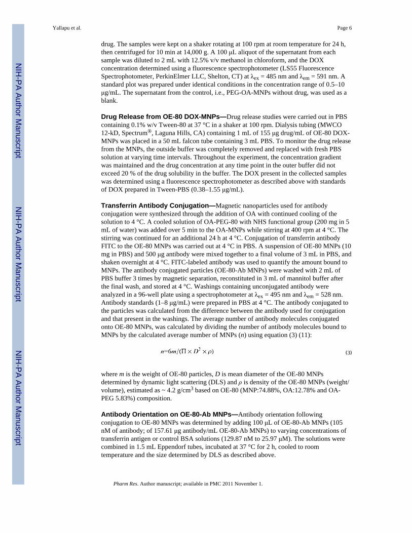
Drug Release from OE-80 DOX-MNPs—Drug release studies were carried out in PBScontaining 0.1% w/v Tween-80 at 37 °C in a shaker at 100 rpm. Dialysis tubing (MWCO12-kD, Spectrum®, Laguna Hills, CA) containing 1 mL of 155 μg drug/mL of OE-80 DOX-MNPs was placed in a 50 mL falcon tube containing 3 mL PBS. To monitor the drug releasefrom the MNPs, the outside buffer was completely removed and replaced with fresh PBSsolution at varying time intervals. Throughout the experiment, the concentration gradientwas maintained and the drug concentration at any time point in the outer buffer did notexceed 20 % of the drug solubility in the buffer. The DOX present in the collected sampleswas determined using a fluorescence spectrophotometer as described above with standardsof DOX prepared in Tween-PBS (0.38–1.55 μg/mL).
Transferrin Antibody Conjugation—Magnetic nanoparticles used for antibodyconjugation were synthesized through the addition of OA with continued cooling of thesolution to 4 °C. A cooled solution of OA-PEG-80 with NHS functional group (200 mg in 5mL of water) was added over 5 min to the OA-MNPs while stirring at 400 rpm at 4 °C. Thestirring was continued for an additional 24 h at 4 °C. Conjugation of transferrin antibodyFITC to the OE-80 MNPs was carried out at 4 °C in PBS. A suspension of OE-80 MNPs (10mg in PBS) and 500 μg antibody were mixed together to a final volume of 3 mL in PBS, andshaken overnight at 4 °C. FITC-labeled antibody was used to quantify the amount bound toMNPs. The antibody conjugated particles (OE-80-Ab MNPs) were washed with 2 mL ofPBS buffer 3 times by magnetic separation, reconstituted in 3 mL of mannitol buffer afterthe final wash, and stored at 4 °C. Washings containing unconjugated antibody wereanalyzed in a 96-well plate using a spectrophotometer at λex = 495 nm and λem = 528 nm.Antibody standards (1–8 μg/mL) were prepared in PBS at 4 °C. The antibody conjugated tothe particles was calculated from the difference between the antibody used for conjugationand that present in the washings. The average number of antibody molecules conjugatedonto OE-80 MNPs, was calculated by dividing the number of antibody molecules bound toMNPs by the calculated average number of MNPs (n) using equation (3) (11):
(3)
where m is the weight of OE-80 particles, D is mean diameter of the OE-80 MNPsdetermined by dynamic light scattering (DLS) and ρ is density of the OE-80 MNPs (weight/volume), estimated as ~ 4.2 g/cm3 based on OE-80 (MNP:74.88%, OA:12.78% and OA-PEG 5.83%) composition.
Antibody Orientation on OE-80-Ab MNPs—Antibody orientation followingconjugation to OE-80 MNPs was determined by adding 100 μL of OE-80-Ab MNPs (105nM of antibody; of 157.61 μg antibody/mL OE-80-Ab MNPs) to varying concentrations oftransferrin antigen or control BSA solutions (129.87 nM to 25.97 μM). The solutions werecombined in 1.5 mL Eppendorf tubes, incubated at 37 °C for 2 h, cooled to roomtemperature and the size determined by DLS as described above.
Yallapu et al. Page 6
Pharm Res. Author manuscript; available in PMC 2011 November 1.
NIH
-PA Author Manuscript
NIH
-PA Author Manuscript
NIH
-PA Author Manuscript

Antiproliferative Activity of Drug-Loaded MNPs—Breast cancer cells (MCF-7) fromAmerican Type Culture Collection (ATCC, Manassas, VA) were maintained in DMEMmedium supplemented with 10% fetal bovine serum, 100 μg/mL penicillin G, and 100 μg/mL streptomycin at 37 °C in a humidified and 5% CO2 atmosphere. Cells were seeded at3,000 cells per well in 96 well plates and were allowed to attach for 24 h. Doxorubicin insolution and DOX-MNPs were prepared and diluted to different concentrations in culturemedium to study the dose-response effect. Medium from wells was replaced with eitherDOX solution or the suspension of DOX-MNPs and the cells were returned to the incubatorfor 24 h. The medium in the wells was changed at 2 and 4 days after the treatment and nofurther dose of the drug was added. Medium and control MNPs (without drug) acted as therespective controls for DOX solution and DOX-MNPs. Cell viability was determined on day5 using an MTS assay (CellTiter 96 AQueous, Promega, Madison, WI). Prior to the assay,the medium from each well was replaced with 100 μL fresh medium and 20 μL of MTSreagent was added. The plates were incubated at 37 °C in CO2 incubator for 90 min, and theabsorbance was measured at 490 nm using a microplate reader (BioTek EL800, BioTekInstrument Inc., Winooski, VT). Percentage proliferation of cells in the presence of drug wascalculated with respect to the respective controls; i.e., medium control for cells treated withdrug in solution and cells treated with DOX-MNPs with control MNPs. The followingequation was used to calculate the IC50 values:
(4)
where y is % growth, x is the added concentration of Doxorubicin content, A1 is the %growth on the top plateau of the curve, A2 is the % growth on the bottom plateau of thecurve, x0 is the inflection point, and p is the slope factor. Experimental data points werefitted to this equation and the IC50 was determined by setting y equal to 50 in the aboveequation and solving for x.
Statistical Analysis—The least square method was used for linear curve fitting. Origin7.5 (OriginLab Corporation, Northampton, MA) was used to curve fit the dose-responseequation, and exponential equations to calculate the IC50 values and relaxation times (T1 andT2).
RESULTSParticle Characterization
The mean hydrodynamic diameter of the poly(ethylene) glycol coated oleic acid magneticnanoparticles (PEG-OA-MNPs) depends on the amount and molecular weight of the OA-PEG polymers used for coating, and the buffer used to disperse the MNPs. An optimalconcentration of 200 mg of the polymer produced the smallest particle diameter with a lowpolydispersity index of 0.009–0.11 for all three of the OA-PEG polymers tested in water(Figure 2A), with an iron oxide core of ~8 nm (Figure 2B). When coated with 200 mg ofOA-PEG polymer the particle size and aggregation increased when the particles weredispersed in saline or 1X PBS but did not change in water or mannitol citrate buffer (FigureS1).
Presence of the magnetic core, OA, and polymer coating of the MNPs was confirmed byFourier transform infrared (FTIR) spectroscopy. Bare MNPs had a strong FTIR peak in theregion of 550 cm−1 from the iron oxide skeleton (–O–Fe), and a broad peak between 3500–3000 cm−1 due to surface –OH and NH2 groups (Figure 3A). Intense bands appeared in theOA-MNPs at 2914/2848 and 1465 cm−1 due to -CH2 asymmetric/symmetric stretching and
Yallapu et al. Page 7
Pharm Res. Author manuscript; available in PMC 2011 November 1.
NIH
-PA Author Manuscript
NIH
-PA Author Manuscript
NIH
-PA Author Manuscript
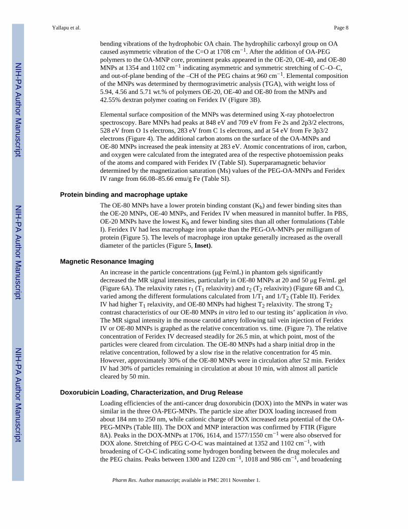
bending vibrations of the hydrophobic OA chain. The hydrophilic carboxyl group on OAcaused asymmetric vibration of the C=O at 1708 cm−1. After the addition of OA-PEGpolymers to the OA-MNP core, prominent peaks appeared in the OE-20, OE-40, and OE-80MNPs at 1354 and 1102 cm−1 indicating asymmetric and symmetric stretching of C–O–C,and out-of-plane bending of the –CH of the PEG chains at 960 cm−1. Elemental compositionof the MNPs was determined by thermogravimetric analysis (TGA), with weight loss of5.94, 4.56 and 5.71 wt.% of polymers OE-20, OE-40 and OE-80 from the MNPs and42.55% dextran polymer coating on Feridex IV (Figure 3B).
Elemental surface composition of the MNPs was determined using X-ray photoelectronspectroscopy. Bare MNPs had peaks at 848 eV and 709 eV from Fe 2s and 2p3/2 electrons,528 eV from O 1s electrons, 283 eV from C 1s electrons, and at 54 eV from Fe 3p3/2electrons (Figure 4). The additional carbon atoms on the surface of the OA-MNPs andOE-80 MNPs increased the peak intensity at 283 eV. Atomic concentrations of iron, carbon,and oxygen were calculated from the integrated area of the respective photoemission peaksof the atoms and compared with Feridex IV (Table SI). Superparamagnetic behaviordetermined by the magnetization saturation (Ms) values of the PEG-OA-MNPs and FeridexIV range from 66.08–85.66 emu/g Fe (Table SI).
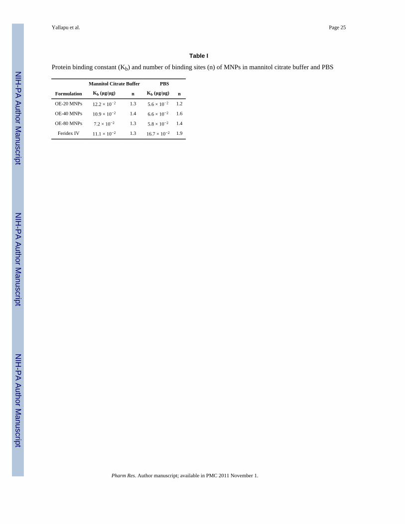
Protein binding and macrophage uptakeThe OE-80 MNPs have a lower protein binding constant (Kb) and fewer binding sites thanthe OE-20 MNPs, OE-40 MNPs, and Feridex IV when measured in mannitol buffer. In PBS,OE-20 MNPs have the lowest Kb and fewer binding sites than all other formulations (TableI). Feridex IV had less macrophage iron uptake than the PEG-OA-MNPs per milligram ofprotein (Figure 5). The levels of macrophage iron uptake generally increased as the overalldiameter of the particles (Figure 5, Inset).
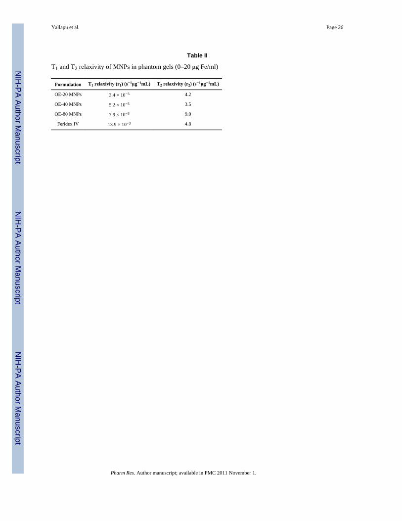
Magnetic Resonance ImagingAn increase in the particle concentrations (μg Fe/mL) in phantom gels significantlydecreased the MR signal intensities, particularly in OE-80 MNPs at 20 and 50 μg Fe/mL gel(Figure 6A). The relaxivity rates r1 (T1 relaxivity) and r2 (T2 relaxivity) (Figure 6B and C),varied among the different formulations calculated from 1/T1 and 1/T2 (Table II). FeridexIV had higher T1 relaxivity, and OE-80 MNPs had highest T2 relaxivity. The strong T2contrast characteristics of our OE-80 MNPs in vitro led to our testing its’ application in vivo.The MR signal intensity in the mouse carotid artery following tail vein injection of FeridexIV or OE-80 MNPs is graphed as the relative concentration vs. time. (Figure 7). The relativeconcentration of Feridex IV decreased steadily for 26.5 min, at which point, most of theparticles were cleared from circulation. The OE-80 MNPs had a sharp initial drop in therelative concentration, followed by a slow rise in the relative concentration for 45 min.However, approximately 30% of the OE-80 MNPs were in circulation after 52 min. FeridexIV had 30% of particles remaining in circulation at about 10 min, with almost all particlecleared by 50 min.
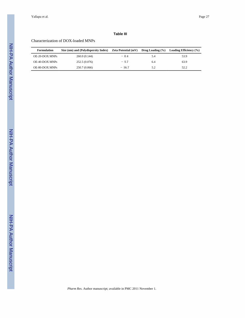
Doxorubicin Loading, Characterization, and Drug ReleaseLoading efficiencies of the anti-cancer drug doxorubicin (DOX) into the MNPs in water wassimilar in the three OA-PEG-MNPs. The particle size after DOX loading increased fromabout 184 nm to 250 nm, while cationic charge of DOX increased zeta potential of the OA-PEG-MNPs (Table III). The DOX and MNP interaction was confirmed by FTIR (Figure8A). Peaks in the DOX-MNPs at 1706, 1614, and 1577/1550 cm−1 were also observed forDOX alone. Stretching of PEG C-O-C was maintained at 1352 and 1102 cm−1, withbroadening of C-O-C indicating some hydrogen bonding between the drug molecules andthe PEG chains. Peaks between 1300 and 1220 cm−1, 1018 and 986 cm−1, and broadening
Yallapu et al. Page 8
Pharm Res. Author manuscript; available in PMC 2011 November 1.
NIH
-PA Author Manuscript
NIH
-PA Author Manuscript
NIH
-PA Author Manuscript
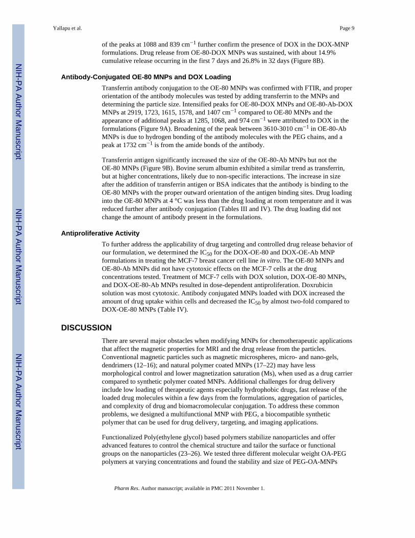
of the peaks at 1088 and 839 cm−1 further confirm the presence of DOX in the DOX-MNPformulations. Drug release from OE-80-DOX MNPs was sustained, with about 14.9%cumulative release occurring in the first 7 days and 26.8% in 32 days (Figure 8B).
Antibody-Conjugated OE-80 MNPs and DOX LoadingTransferrin antibody conjugation to the OE-80 MNPs was confirmed with FTIR, and properorientation of the antibody molecules was tested by adding transferrin to the MNPs anddetermining the particle size. Intensified peaks for OE-80-DOX MNPs and OE-80-Ab-DOXMNPs at 2919, 1723, 1615, 1578, and 1407 cm−1 compared to OE-80 MNPs and theappearance of additional peaks at 1285, 1068, and 974 cm−1 were attributed to DOX in theformulations (Figure 9A). Broadening of the peak between 3610-3010 cm−1 in OE-80-AbMNPs is due to hydrogen bonding of the antibody molecules with the PEG chains, and apeak at 1732 cm−1 is from the amide bonds of the antibody.
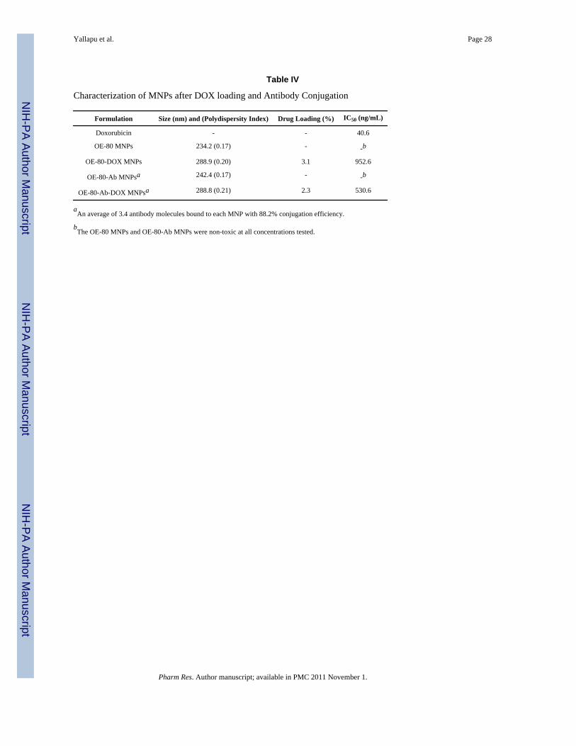
Transferrin antigen significantly increased the size of the OE-80-Ab MNPs but not theOE-80 MNPs (Figure 9B). Bovine serum albumin exhibited a similar trend as transferrin,but at higher concentrations, likely due to non-specific interactions. The increase in sizeafter the addition of transferrin antigen or BSA indicates that the antibody is binding to theOE-80 MNPs with the proper outward orientation of the antigen binding sites. Drug loadinginto the OE-80 MNPs at 4 °C was less than the drug loading at room temperature and it wasreduced further after antibody conjugation (Tables III and IV). The drug loading did notchange the amount of antibody present in the formulations.
Antiproliferative ActivityTo further address the applicability of drug targeting and controlled drug release behavior ofour formulation, we determined the IC50 for the DOX-OE-80 and DOX-OE-Ab MNPformulations in treating the MCF-7 breast cancer cell line in vitro. The OE-80 MNPs andOE-80-Ab MNPs did not have cytotoxic effects on the MCF-7 cells at the drugconcentrations tested. Treatment of MCF-7 cells with DOX solution, DOX-OE-80 MNPs,and DOX-OE-80-Ab MNPs resulted in dose-dependent antiproliferation. Doxrubicinsolution was most cytotoxic. Antibody conjugated MNPs loaded with DOX increased theamount of drug uptake within cells and decreased the IC50 by almost two-fold compared toDOX-OE-80 MNPs (Table IV).
DISCUSSIONThere are several major obstacles when modifying MNPs for chemotherapeutic applicationsthat affect the magnetic properties for MRI and the drug release from the particles.Conventional magnetic particles such as magnetic microspheres, micro- and nano-gels,dendrimers (12–16); and natural polymer coated MNPs (17–22) may have lessmorphological control and lower magnetization saturation (Ms), when used as a drug carriercompared to synthetic polymer coated MNPs. Additional challenges for drug deliveryinclude low loading of therapeutic agents especially hydrophobic drugs, fast release of theloaded drug molecules within a few days from the formulations, aggregation of particles,and complexity of drug and biomacromolecular conjugation. To address these commonproblems, we designed a multifunctional MNP with PEG, a biocompatible syntheticpolymer that can be used for drug delivery, targeting, and imaging applications.
Functionalized Poly(ethylene glycol) based polymers stabilize nanoparticles and offeradvanced features to control the chemical structure and tailor the surface or functionalgroups on the nanoparticles (23–26). We tested three different molecular weight OA-PEGpolymers at varying concentrations and found the stability and size of PEG-OA-MNPs
Yallapu et al. Page 9
Pharm Res. Author manuscript; available in PMC 2011 November 1.
NIH
-PA Author Manuscript
NIH
-PA Author Manuscript
NIH
-PA Author Manuscript
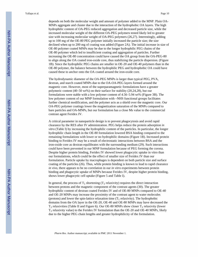
depends on both the molecular weight and amount of polymer added to the MNP. Plain OA-MNPs aggregate and cluster due to the interaction of the hydrophobic OA layers. The highhydrophilic content of OA-PEG reduced aggregation and decreased particle size, while theincreased molecular weight of the different OA-PEG polymers tested likely led to greatersize with increasing molecular weight of OA-PEG polymers (26,27). Interestingly, addingup to 100 mg of the OE-80 PEG polymer initially increased the particle size; the sizedeclined when up to 200 mg of coating was added (Figure 2A). The initial increase in size ofOE-80 polymer coated MNPs may be due to the longer hydrophilic PEG chains of theOE-80 polymer which led to insufficient coating and aggregation of particles. Furtherincreasing the OE-80 concentration could have caused the OA group from the OA-PEG-80to align along the OA coated iron-oxide core, thus stabilizing the particle dispersion. (Figure1B). Since the hydrophilic PEG chains are smaller in OE-20 and OE-40 polymers than in theOE-80 polymer, the balance between the hydrophilic PEG and hydrophobic OA could havecaused these to anchor onto the OA coated around the iron-oxide core.
The hydrodynamic diameter of the OA-PEG MNPs is larger than typical PEG, PVA,dextran, and starch coated MNPs due to the OA-OA-PEG layers formed around themagnetic core. However, most of the superparamagnetic formulations have a greaterpolymeric content (40–50 wt%) on their surface for stability (20,24,28), but ourformulations were stable with a low polymer content of 4.56–5.94 wt% (Figure 3B). Thelow polymer content of our MNP formulation with –NHS functional groups facilitatesfurther chemical modification, and the polymer acts as a shield over the magnetic core. OurOA-PEG polymer coatings lower the magnetization saturation of the MNPs compared tobare particles and OA-MNPs, but our formulation has a close Ms value to the commercialcontrast agent Feridex IV.
A critical parameter in nanoparticle design is to prevent phagocytosis and avoid rapidclearance by the RES after IV administration. PEG helps reduce the protein adsorption invitro (Table I) by increasing the hydrophilic content of the particles. In particular, the longerhydrophilic chain length in the OE-80 formulation lowered BSA binding compared to theremaining formulations with lower or no hydrophilic domains (Figure 1B). Increased proteinbinding to Feridex IV may be a result of electrostatic interactions between BSA and theiron-oxide core as dextran equilibrates with the surrounding medium (29). Such interactionscould have been prevented in our MNP formulation because of PEG forming the corona.Despite higher protein binding, Feridex IV showed lower phagocytic uptake in vitro thanour formulations, which could be the effect of smaller size of Feridex IV than ourformulation. Particle uptake by macrophages is dependent on both particle size and surfacecoating of the particles (26). Thus, while protein binding is known to lead to rapid clearancein vivo, there appears to be no correlation in our in vitro experiments between proteinbinding and phagocytic uptake of MNPs because Feridex IV, despite higher protein binding,shows lower phagocytic cell uptake (Figure 5 and Table I).
In general, the process of T1 shortening (T1 relaxivity) requires the direct interactionbetween protons and the magnetic component of the contrast agents (30). The greaterhydrophilic content of dextran coated Feridex IV and of OE-80 MNPs compared to OE-40and OE-20 MNPs may increase the proximity of the contrast agent to water molecules(protons) and lower the spin-lattice relaxation time (T1 relaxivity). The hydrophobicdomains from the OA layer in the OE-20, OE-40 and OE-80 MNPs may have decreased theT1 relaxivities (Table II and Figure 6). Our OE-80 MNPs show closer T1 relaxivity (lowerT1 relaxivity value) to the Feridex IV formulation than the OE-20 and OE-40 MNPs, likelydue to the higher PEG chain lengths and greater hydrophilicity of the formulation.
Yallapu et al. Page 10
Pharm Res. Author manuscript; available in PMC 2011 November 1.
NIH
-PA Author Manuscript
NIH
-PA Author Manuscript
NIH
-PA Author Manuscript
The large particle size, magnetization saturation (Ms), and long polymer chain length caneach enhance the T2 MRI contrast of magnetic nanoparticles. Particle aggregation changesthe magnetic relaxation properties of near by water protons, reducing the T2 relaxation times(31,32), while increasing Ms increases in the T2 relaxivities and contrast. Each of theseproperties may support the relative increase in T2 relaxivity and better contrast properties ofour OE-80 formulation compared with Feridex IV (Table II) and our similar formulationswith Pluronic® coated OA-MNPs (3.8 × 10⁻³ s−1μg−1mL) (33). The shorter chain lengths ofthe OE-20 and OE-40 may increased the aggregation altering the local magneticheterogeneity in the agarose gels and T2 contrast properties (34).
The significance of formulating MNPs with various polymer coatings is to minimizemacrophage uptake and improve blood circulation time (34–38). There are severalcommercial dextran or polymer coated MNP formulations with available for diagnostics, butthe low in vivo half life and rapid capture of the particles by cells and organs of the RESlimits their application to spleen and liver diagnostics (2). We observed an initial steep dropin contrast with our formulation followed by a gradual increase, compared to Feridex IV,which gradually decreased over time following injection. The initial sharp decline with ourOE-80 MNP formulation may be due to particles initially being removed from circulationinto body compartments such as the liver, and then returning to circulation instead of beingbroken down and cleared by the liver. The long chain length of our PEG coated MNPs maynot be recognized as foreign particles in vivo as easily as dextran coated Feridex, and ourMNPs may slowly equilibrate with the vascular compartment leading to a gradual increasein the signal intensity detected. We observed a similar pattern of change in the dynamics ofiron content in different tissue with time in our previous studies with similarly formulatedpluronic stabilized MNPs (39). Conversely, Feridex IV may decline gradually as thedextran, which is in equilibrium with the surrounding environment and weakly associatedwith the iron-oxide core (29), dissociates and the particles aggregate. This observation issimilar our in vitro observation of protein binding, where we saw greater protein binding onFeridex than our MNP formulation (Table I). The prolonged circulation time of our OE-80formulation in the mouse carotid arteries compared to both Feridex IV (Figure 7) andPluronic® based formulations may be due to the longer PEG chain length and highhydrophilic content of the OE-80 MNPs (40). We observed less macrophage uptake for thesmaller Feridex IV formulation in vitro, thus reducing the size further and increasing thehydrophilic content of the current formulation may result in an even longer circulation timein vivo (36,41).
A major advantage in using nanoparticles for chemotherapeutic applications is to preventsystemic drug toxicity by encapsulating the drug. In addition, the particles can allow forsustained drug release over time, potentially lowering the dosing frequency and improvingpatient compliance. The OA layer in the current formulation entraps the hydrophobic drugDoxorubicin through physical interaction. This physical interaction is a significantadvantage over nanoparticle-drug conjugates, magnetic liposomes, and drug encapsulatedformulations because easy drug loading, high loading efficiency, and sustained release asopposed to burst release. In particular, the release of hydrophilic drugs from hydrophiliccompounds is rapidly reversible and the drug dissociates within hours (4). The sustainedrelease of doxorubicin from the MNPs over several days likely occurs through twomechanisms, drug release from the surface of the MNPs and release through diffusion fromOA layers due to concentration gradient with respect to the surrounding environment.
The versatility of the OE-80 MNP formulation allowed us to investigate antibodyconjugation and thus targeted drug delivery in addition to the MRI. The OA-PEG-80 chainhas an active – NHS end group which offers high and direct conjugation capacity to theamine group on antibodies without requiring a cross-linker like some polymer coated MNP
Yallapu et al. Page 11
Pharm Res. Author manuscript; available in PMC 2011 November 1.
NIH
-PA Author Manuscript
NIH
-PA Author Manuscript
NIH
-PA Author Manuscript
formulations used for conjugation (42). For antibody conjugation, the reaction and drugloading was carried out at 4 °C in PBS. The hydrophilicity of the particle after antibodyconjugation and a the low temperature reduced the drug loading capacity of the formulationcompared to drug loading at room temperature because the parent OA freezing point is 16.4°C. In addition, drug loading in PBS may have further reduced the loading capacity due tosome aggregation of the particles which was not observed when drug was added to particlessuspended in water (Table S1 and Table III). However, the drug loading combined withantibody conjugation was still sufficient to provide a two-fold decrease in drug required toinhibit 50% of cell growth in vitro (Table IV). In addition, drug is released over severalweeks from the MNPs, whereas 100% of the free drug is available to inhibit cell growth.Based on the drug release profile (Figure 8B), about 13.68% of the drug is released over fivedays, the length of the in vitro study. Thus, the drug released may be around 72.6 ng/mL forOE-80-Ab-DOX MNPs, and 130.3 ng/mL OE-80-Ab MNPs, which is much closer to the40.6 ng/mL DOX required to inhibit 50% of the cell growth in vitro. Confocal scanninglaser microscopy of DOX MNPs confirms that drug is present within the cytoplasm andlikely still associated with the MNPs (43). There is a slow increase in accumulation of drugdetected in the nucleus, the site action, as the drug is released from the MNPs. Plain drug,alternatively, is visible almost exclusively in the nucleus; this likely accounts for the highertoxicity observed of free DOX compared to that of DOX MNPs. Our formulation can befurther optimized for the drug and also for specific targeting ligand and its density aroundMNPs to further improve the efficacy.
Our MNPs have potential to be developed as an imaged-guided drug therapy because thesecan be imaged using MRI and simultaneously deliver drugs to the target tissue. Image-guided drug therapy is particularly important in cancer therapy because of significantvariations in tumor vascularity, heterogeneous and diverse expression of cell surfacereceptors, and genetic make-up of cancer cells (44), as these factors could significantlyinfluence the efficacy of nanocarrier-mediated drug delivery as well as drug response.Nanocarriers with imaging capacity can ensure drug delivery to the target tissue.
CONCLUSIONWe show that our MNP formulation has dual functionality, i.e. drug delivery and MRIproperties. In the future, our MNPs can potentially be used as a theranostic agent for image-guided cancer therapy to maximize therapeutic effect while minimizing drug toxicity.
Supplementary MaterialRefer to Web version on PubMed Central for supplementary material.
AcknowledgmentsThe study reported here is funded by grant R01 EB005822 (to VL) from the National Institute of BiomedicalImaging and Bioengineering of the National Institutes of Health. SPF is a predoctoral student in Cleveland Clinic’sMolecular Medicine Ph.D. Program, which is funded by the “Med into Grad” initiative of the Howard HughesMedical Institute [http://www.lerner.ccf.org/molecmed/phd/].
References1. Bulte JW, Kraitchman DL. Monitoring cell therapy using iron oxide MR contrast agents. Curr
Pharm Biotechnol 2004;5:567–584. [PubMed: 15579045]2. Josephson, L. Magnetic Nanoparticles for MR Imaging. Springer; US: 2007.3. Bulte JW, Kraitchman DL. Iron oxide MR contrast agents for molecular and cellular imaging. NMR
Biomed 2004;17:484–499. [PubMed: 15526347]
Yallapu et al. Page 12
Pharm Res. Author manuscript; available in PMC 2011 November 1.
NIH
-PA Author Manuscript
NIH
-PA Author Manuscript
NIH
-PA Author Manuscript
4. Alexiou C, Arnold W, Klein RJ, Parak FG, Hulin P, Bergemann C, Erhardt W, Wagenpfeil S, LubbeAS. Locoregional cancer treatment with magnetic drug targeting. Cancer Res 2000;60:6641–6648.[PubMed: 11118047]
5. Beaven GH, Chen SH, d’Albis A, Gratzer WB. A spectroscopic study of the haemin--human-serum-albumin system. Eur J Biochem 1974;41:539–546. [PubMed: 4817561]
6. Lehrer SS, Fasman GD. The fluorescence of lysozyme and lysozyme substrate complexes. BiochemBiophys Res Commun 1966;23:133–138. [PubMed: 5928904]
7. Chipman DM, Grisaro V, Sharon N. The binding of oligosaccharides containing N-acetylglucosamine and N-acetylmuramic acid to lysozyme. The specificity of binding subsites. JBiol Chem 1967;242:4388–4394. [PubMed: 6070843]
8. Jeffery, GH.; Bassett, J.; Mendham, J.; Denny, RC. Vogel’s text book of quantitative chemicalanalysis. John Wiley & Sons Inc; New York: 1989.
9. Moffat BA, Reddy GR, McConville P, Hall DE, Chenevert TL, Kopelman RR, Philbert M,Weissleder R, Rehemtulla A, Ross BD. A novel polyacrylamide magnetic nanoparticle contrastagent for molecular imaging using MRI. Mol Imaging 2003;2:324–332. [PubMed: 14717331]
10. Yolles S, Aslund B, Morton JF, Olson OT, Rosenberg B. Timed-released depot for anticanceragents. II. Acta Pharm Suec 1978;15:382–388. [PubMed: 747096]
11. Olivier JC, Huertas R, Lee HJ, Calon F, Pardridge WM. Synthesis of pegylatedimmunonanoparticles. Pharm Res 2002;19:1137–1143. [PubMed: 12240939]
12. Gou ML, Qian ZY, Wang H, Tang YB, Huang MJ, Kan B, Wen YJ, Dai M, Li XY, Gong CY, TuMJ. Preparation and characterization of magnetic poly(epsilon-caprolactone)-poly(ethyleneglycol)-poly(epsilon-caprolactone) microspheres. J Mater Sci Mater Med 2007;19:1033–1041.[PubMed: 17701292]
13. Liu X, Kaminski MD, Chen H, Torno M, Taylor L, Rosengart AJ. Synthesis and characterizationof highly-magnetic biodegradable poly(d,l-lactide-co-glycolide) nanospheres. J Control Release2007;119:52–58. [PubMed: 17350131]
14. Okassa LN, Marchais H, Douziech-Eyrolles L, Herve K, Cohen-Jonathan S, Munnier E, Souce M,Linassier C, Dubois P, Chourpa I. Optimization of iron oxide nanoparticles encapsulation withinpoly(d,l-lactide-co-glycolide) sub-micron particles. Eur J Pharm Biopharm 2007;67:31–38.[PubMed: 17289360]
15. Hamoudeh M, Al Faraj A, Canet-Soulas E, Bessueille F, Leonard D, Fessi H. Elaboration ofPLLA-based superparamagnetic nanoparticles: characterization, magnetic behaviour study and invitro relaxivity evaluation. Int J Pharm 2007;338:248–257. [PubMed: 17317054]
16. Bhattacharya S, Eckert F, Boyko V, Pich A. Temperature-, pH-, and magnetic-field-sensitivehybrid microgels. Small 2007;3:650–657. [PubMed: 17340664]
17. Shen F, Poncet-Legrand C, Somers S, Slade A, Yip C, Duft AM, Winnik FM, Chang PL.Properties of a novel magnetized alginate for magnetic resonance imaging. Biotechnol Bioeng2003;83:282–292. [PubMed: 12783484]
18. Bonacchi D, Caneschi A, Dorignac D, Falqui A, Gatteschi D, Rovai D, Sangregorio C, Sessoli R.Nanosized iron oxide particles entrapped in pseudo-single crystals gamma-cyclodextrin.Chemistry of Materials 2004;16:2016–2020.
19. Bonacchi D, Caneschi A, Gatteschi D, Sangregorio C, Sessoli R, Falqui A. Synthesis andcharacterisation of metal oxides nanoparticles entrapped in cyclodextrin. Journal of Physics andChemistry of Solids 2004;65:719–722.
20. Mikhaylova M, Kim DK, Bobrysheva N, Osmolowsky M, Semenov V, Tsakalakos T, MuhammedM. Superparamagnetism of magnetite nanoparticles: dependence on surface modification.Langmuir 2004;20:2472–2477. [PubMed: 15835712]
21. Kim DK, Mikhaylova M, Wang FH, Kehr J, Bjelke B, Zhang Y, Tsakalakos T, Muhammed M.Starch-coated superparamagnetic nanoparticles as MR contrast agents. Chemistry of Materials2003;15:4343–4351.
22. Pardoe H, Chua-anusorn W, St Pierre TG, Dobson J. Structural and magnetic properties ofnanoscale iron oxide particles synthesized in the presence of dextran or polyvinyl alcohol. J MagnMagn Mater 2001;225:41–46.
Yallapu et al. Page 13
Pharm Res. Author manuscript; available in PMC 2011 November 1.
NIH
-PA Author Manuscript
NIH
-PA Author Manuscript
NIH
-PA Author Manuscript
23. Lee H, Yu MK, Park S, Moon S, Min JJ, Jeong YY, Kang HW, Jon S. Thermally cross-linkedsuperparamagnetic iron oxide nanoparticles: synthesis and application as a dual imaging probe forcancer in vivo. J Am Chem Soc 2007;129:12739–12745. [PubMed: 17892287]
24. Wan S, Huang J, Guo M, Zhang H, Cao Y, Yan H, Liu K. Biocompatible superparamagnetic ironoxide nanoparticle dispersions stabilized with poly(ethylene glycol)-oligo(aspartic acid) hybrids. JBiomed Mater Res A 2007;80:946–954. [PubMed: 17083116]
25. Lutz JF, Stiller S, Hoth A, Kaufner L, Pison U, Cartier R. One-pot synthesis of pegylatedultrasmall iron-oxide nanoparticles and their in vivo evaluation as magnetic resonance imagingcontrast agents. Biomacromolecules 2006;7:3132–3138. [PubMed: 17096542]
26. Xie J, Xu C, Kohler N, Hou Y, Sun S. Controlled PEGylation of monodisperse Fe3O4nanoparticles for reduced non-specific uptake by macrophage cells. Adv Mater 2007;19:3163–3166.
27. Ditsch A, Laibinis PE, Wang DI, Hatton TA. Controlled clustering and enhanced stability ofpolymer-coated magnetic nanoparticles. Langmuir 2005;21:6006–6018. [PubMed: 15952854]
28. Weissleder R, Elizondo G, Wittenberg J, Lee AS, Josephson L, Brady TJ. Ultrasmallsuperparamagnetic iron oxide: an intravenous contrast agent for assessing lymph nodes with MRimaging. Radiology 1990;175:494–498. [PubMed: 2326475]
29. McCarthy JR, Weissleder R. Multifunctional magnetic nanoparticles for targeted imaging andtherapy. Adv Drug Deliv Rev 2008;60:1241–1251. [PubMed: 18508157]
30. Okuhata Y. Delivery of diagnostic agents for magnetic resonance imaging. Adv Drug Deliv Rev1999;37:121–137. [PubMed: 10837731]
31. Yigit MV, Mazumdar D, Lu Y. MRI detection of thrombin with aptamer functionalizedsuperparamagnetic iron oxide nanoparticles. Bioconjugate Chem 2008;19:412–417.
32. Jun YW, Huh YM, Choi JS, Lee JH, Song HT, Kim S, Yoon S, Kim KS, Shin JS, Suh JS, Cheon J.Nanoscale size effect of magnetic nanocrystals and their utilization for cancer diagnosis viamagnetic resonance imaging. J Am Chem Soc 2005;127:5732–5733. [PubMed: 15839639]
33. Jain TK, Richey J, Strand M, Leslie-Pelecky DL, Flask CA, Labhasetwar V. Magneticnanoparticles with dual functional properties: Drug delivery and magnetic resonance imaging.Biomaterials 2008;29:4012–4021. [PubMed: 18649936]
34. Bulte JWM, Cuyper MD, Despres D, Frank JA. Preparation, relaxometry, and biokinetics ofPEGylated magnetoliposomes as MR contrast agent. J Magn Magn Mater 1999;194:204–209.
35. Zhang Y, Kohler N, Zhang MQ. Surface modification of superparamagnetic magnetitenanoparticles and their intracellular uptake. Biomaterials 2002;23:1553–1561. [PubMed:11922461]
36. Arruebo M, Fernandez-Pacheco R, Ibarra MR, Santamaria J. Magnetic nanoparticles for drugdelivery. Nano Today 2007;2:22–32.
37. Lewin M, Carlesso N, Tung CH, Tang XW, Cory D, Scadden DT, Weissleder R. Tat peptide-derivatized magnetic nanoparticles allow in vivo tracking and recovery of progenitor cells. NatBiotechnol 2000;18:410–414. [PubMed: 10748521]
38. Seo SB, Yang J, Hyung W, Cho EJ, Lee TI, Song YJ, Yoon HG, Suh JS, Huh YM, Haam S. Novelmultifunctional PHDCA/PEI nano-drug carriers for simultaneous magnetically targeted cancertherapy and diagnosis via magnetic resonance imaging. Nanotechnology 2007;18:1–8.
39. Jain TK, Reddy MK, Morales MA, Leslie-Pelecky DL, Labhasetwar V. Biodistribution, clearance,and biocompatibility of iron oxide magnetic nanoparticles in rats. Mol Pharmaceutics 2008;5:316–327.
40. Jain TK, Foy SP, Erokwu B, Dimitrijevic S, Flask CA, Labhasetwar V. Magnetic resonanceimaging of multifunctional pluronic stabilized iron-oxide nanoparticles in tumor-bearing mice.Biomaterials 2009;30:6748–6756. [PubMed: 19765817]
41. Neuberger T, Schopf B, Hofmann H, Hofmann M, von Rechenberg B. Superparamagneticnanoparticles for biomedical applications: Possibilities and limitations of a new drug deliverysystem. Journal of Magnetism and Magnetic Materials 2005;293:483–496.
42. Jun YW, Lee JH, Cheon J. Chemical design of nanoparticle probes for high-performance magneticresonance imaging. Angew Chem Int Ed Engl 2008;47:5122–5135. [PubMed: 18574805]
Yallapu et al. Page 14
Pharm Res. Author manuscript; available in PMC 2011 November 1.
NIH
-PA Author Manuscript
NIH
-PA Author Manuscript
NIH
-PA Author Manuscript
43. Jain TK, Morales MA, Sahoo SK, Leslie-Pelecky DL, Labhasetwar V. Iron oxide nanoparticles forsustained delivery of anticancer agents. Mol Pharmaceutics 2005;2:194–205.
44. Kosaka N, Ogawa M, Longmire MR, Choyke PL, Kobayashi H. Multi-targeted multi-color in vivooptical imaging in a model of disseminated peritoneal ovarian cancer. J Biomed Opt2009;14:014023. [PubMed: 19256711]
Yallapu et al. Page 15
Pharm Res. Author manuscript; available in PMC 2011 November 1.
NIH
-PA Author Manuscript
NIH
-PA Author Manuscript
NIH
-PA Author Manuscript
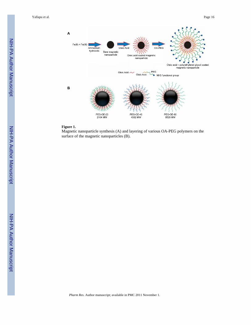
Figure 1.Magnetic nanoparticle synthesis (A) and layering of various OA-PEG polymers on thesurface of the magnetic nanoparticles (B).
Yallapu et al. Page 16
Pharm Res. Author manuscript; available in PMC 2011 November 1.
NIH
-PA Author Manuscript
NIH
-PA Author Manuscript
NIH
-PA Author Manuscript
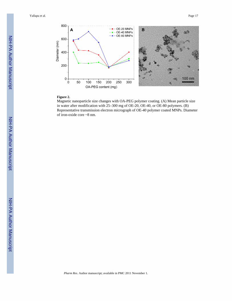
Figure 2.Magnetic nanoparticle size changes with OA-PEG polymer coating. (A) Mean particle sizein water after modification with 25–300 mg of OE-20, OE-40, or OE-80 polymers. (B)Representative transmission electron micrograph of OE-40 polymer coated MNPs. Diameterof iron-oxide core ~8 nm.
Yallapu et al. Page 17
Pharm Res. Author manuscript; available in PMC 2011 November 1.
NIH
-PA Author Manuscript
NIH
-PA Author Manuscript
NIH
-PA Author Manuscript
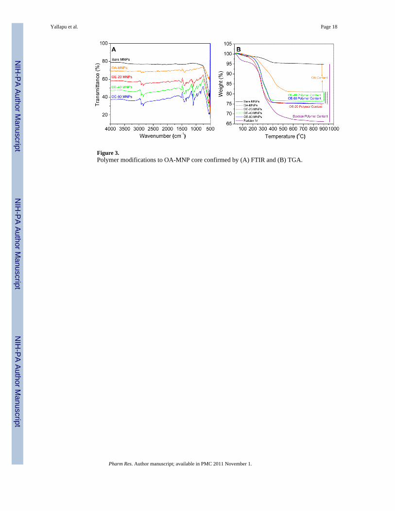
Figure 3.Polymer modifications to OA-MNP core confirmed by (A) FTIR and (B) TGA.
Yallapu et al. Page 18
Pharm Res. Author manuscript; available in PMC 2011 November 1.
NIH
-PA Author Manuscript
NIH
-PA Author Manuscript
NIH
-PA Author Manuscript
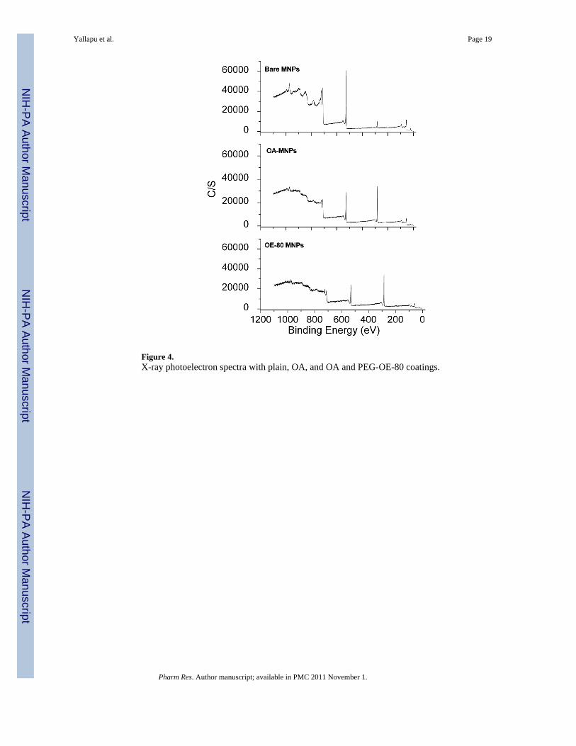
Figure 4.X-ray photoelectron spectra with plain, OA, and OA and PEG-OE-80 coatings.
Yallapu et al. Page 19
Pharm Res. Author manuscript; available in PMC 2011 November 1.
NIH
-PA Author Manuscript
NIH
-PA Author Manuscript
NIH
-PA Author Manuscript
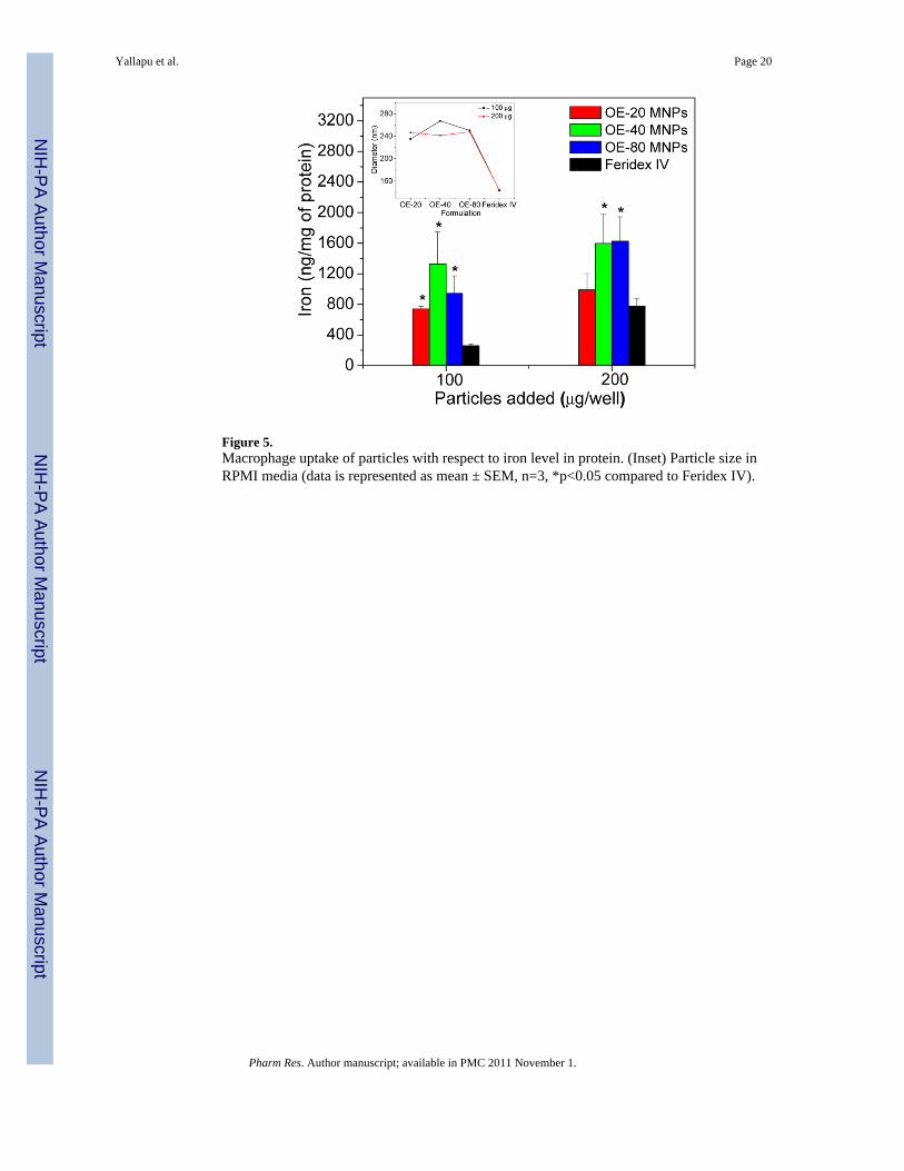
Figure 5.Macrophage uptake of particles with respect to iron level in protein. (Inset) Particle size inRPMI media (data is represented as mean ± SEM, n=3, *p<0.05 compared to Feridex IV).
Yallapu et al. Page 20
Pharm Res. Author manuscript; available in PMC 2011 November 1.
NIH
-PA Author Manuscript
NIH
-PA Author Manuscript
NIH
-PA Author Manuscript
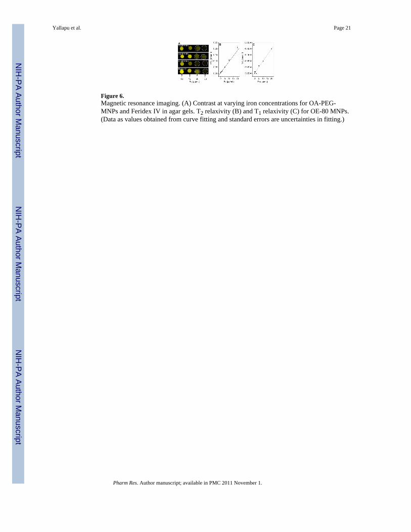
Figure 6.Magnetic resonance imaging. (A) Contrast at varying iron concentrations for OA-PEG-MNPs and Feridex IV in agar gels. T2 relaxivity (B) and T1 relaxivity (C) for OE-80 MNPs.(Data as values obtained from curve fitting and standard errors are uncertainties in fitting.)
Yallapu et al. Page 21
Pharm Res. Author manuscript; available in PMC 2011 November 1.
NIH
-PA Author Manuscript
NIH
-PA Author Manuscript
NIH
-PA Author Manuscript
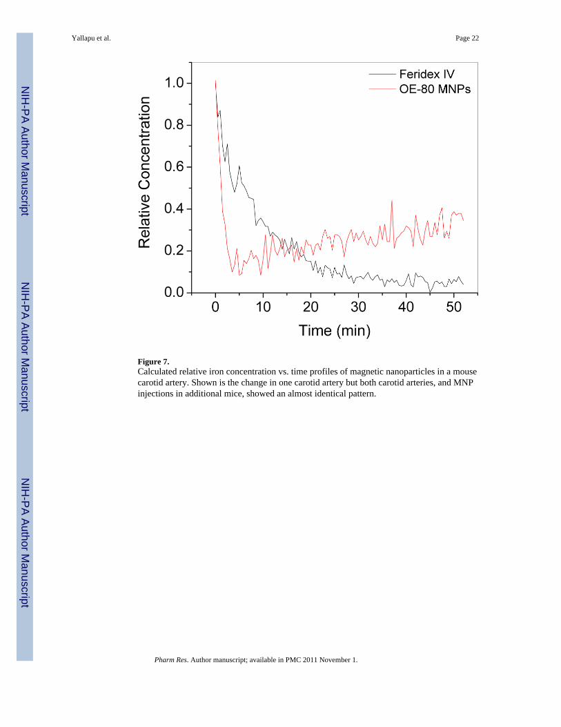
Figure 7.Calculated relative iron concentration vs. time profiles of magnetic nanoparticles in a mousecarotid artery. Shown is the change in one carotid artery but both carotid arteries, and MNPinjections in additional mice, showed an almost identical pattern.
Yallapu et al. Page 22
Pharm Res. Author manuscript; available in PMC 2011 November 1.
NIH
-PA Author Manuscript
NIH
-PA Author Manuscript
NIH
-PA Author Manuscript
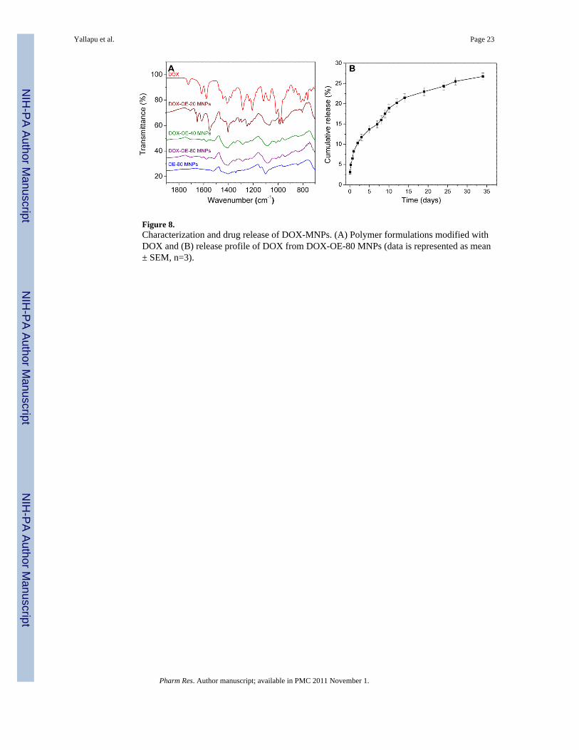
Figure 8.Characterization and drug release of DOX-MNPs. (A) Polymer formulations modified withDOX and (B) release profile of DOX from DOX-OE-80 MNPs (data is represented as mean± SEM, n=3).
Yallapu et al. Page 23
Pharm Res. Author manuscript; available in PMC 2011 November 1.
NIH
-PA Author Manuscript
NIH
-PA Author Manuscript
NIH
-PA Author Manuscript
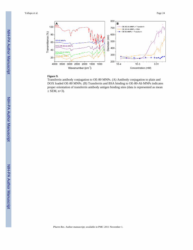
Figure 9.Transferrin antibody conjugation to OE-80 MNPs. (A) Antibody conjugation to plain andDOX loaded OE-80 MNPs. (B) Transferrin and BSA binding to OE-80-Ab MNPs indicatesproper orientation of transferrin antibody antigen binding sites (data is represented as mean± SEM, n=3).
Yallapu et al. Page 24
Pharm Res. Author manuscript; available in PMC 2011 November 1.
NIH
-PA Author Manuscript
NIH
-PA Author Manuscript
NIH
-PA Author Manuscript
NIH
-PA Author Manuscript
NIH
-PA Author Manuscript
NIH
-PA Author Manuscript
Yallapu et al. Page 25
Table I
Protein binding constant (Kb) and number of binding sites (n) of MNPs in mannitol citrate buffer and PBS
Formulation
Mannitol Citrate Buffer PBS
Kb (μg/μg) n Kb (μg/μg) n
OE-20 MNPs 12.2 × 10⁻² 1.3 5.6 × 10⁻² 1.2
OE-40 MNPs 10.9 × 10⁻² 1.4 6.6 × 10⁻² 1.6
OE-80 MNPs 7.2 × 10⁻² 1.3 5.8 × 10⁻² 1.4
Feridex IV 11.1 × 10⁻² 1.3 16.7 × 10⁻² 1.9
Pharm Res. Author manuscript; available in PMC 2011 November 1.
NIH
-PA Author Manuscript
NIH
-PA Author Manuscript
NIH
-PA Author Manuscript
Yallapu et al. Page 26
Table II
T1 and T2 relaxivity of MNPs in phantom gels (0–20 μg Fe/ml)
Formulation T1 relaxivity (r1) (s−1μg−1mL) T2 relaxivity (r2) (s−1μg−1mL)
OE-20 MNPs 3.4 × 10⁻³ 4.2
OE-40 MNPs 5.2 × 10⁻³ 3.5
OE-80 MNPs 7.9 × 10⁻³ 9.0
Feridex IV 13.9 × 10⁻³ 4.8
Pharm Res. Author manuscript; available in PMC 2011 November 1.
NIH
-PA Author Manuscript
NIH
-PA Author Manuscript
NIH
-PA Author Manuscript
Yallapu et al. Page 27
Table III
Characterization of DOX-loaded MNPs
Formulation Size (nm) and (Polydispersity Index) Zeta Potential (mV) Drug Loading (%) Loading Efficiency (%)
OE-20-DOX MNPs 260.0 (0.144) − 0.4 5.4 53.9
OE-40-DOX MNPs 252.5 (0.076) − 5.7 6.4 63.9
OE-80-DOX MNPs 250.7 (0.066) − 16.7 5.2 52.2
Pharm Res. Author manuscript; available in PMC 2011 November 1.
NIH
-PA Author Manuscript
NIH
-PA Author Manuscript
NIH
-PA Author Manuscript
Yallapu et al. Page 28
Table IV
Characterization of MNPs after DOX loading and Antibody Conjugation
Formulation Size (nm) and (Polydispersity Index) Drug Loading (%) IC50 (ng/mL)
Doxorubicin - - 40.6
OE-80 MNPs 234.2 (0.17) - -b
OE-80-DOX MNPs 288.9 (0.20) 3.1 952.6
OE-80-Ab MNPsa 242.4 (0.17) - -b
OE-80-Ab-DOX MNPsa 288.8 (0.21) 2.3 530.6
aAn average of 3.4 antibody molecules bound to each MNP with 88.2% conjugation efficiency.
bThe OE-80 MNPs and OE-80-Ab MNPs were non-toxic at all concentrations tested.
Pharm Res. Author manuscript; available in PMC 2011 November 1.
Copyright © 2022 FDOKUMEN