Biology, Prognosis, and Therapy of Waldenström Macroglobulinemia
Upload
independentCategory
view
2download
0
Catalano et al. Journal of Cardiovascular Magnetic Resonance 2012, 14:29http://www.jcmr-online.com/content/14/1/29
RESEARCH Open Access
Late gadolinium enhancement by cardiovascularmagnetic resonance is complementary to leftventricle ejection fraction in predicting prognosisof patients with stable coronary artery diseaseOronzo Catalano1*, Guido Moro2, Mariarosa Perotti1, Mauro Frascaroli2, Monica Ceresa1, Serena Antonaci3,Paola Baiardi4, Carlo Napolitano5,6, Maurizia Baldi2 and Silvia G Priori1,7
Abstract
Background: Late gadolinium enhancement (LGE) cardiovascular magnetic resonance (CMR) predicts adverseprognosis in patients with stable coronary artery disease (CAD). However, the interaction with conventional riskfactors remains uncertain. Our aim was to assess whether the extent of LGE is an independent predictor of adversecardiac outcome beyond conventional risk factors, including left ventricle ejection fraction (LVEF).
Methods: We enrolled 376 patients (88% males, 64 ± 11 years) with stable CAD, who underwent LGE assessmentand a detailed conventional evaluation (clinical and pharmacological history, risk factors, ECG, Echocardiography).During a follow-up of 38 ± 21 months, 56 events occurred (32 deaths, 24 hospitalizations for heart failure).
Results: LGE and LVEF showed the strongest univariate associations with end-points (HR: 13.61 [95%C.I.: 7.32-25.31] for LGE≥ 45% of LV mass; and 12.34 [6.80-22.38] for LVEF≤ 30%; p< 0.0001). Multivariate analysisidentified baseline LVEF, loop diuretic therapy, moderate-severe mitral regurgitation and pulmonary hypertensionas significant predictors among conventional risk factors. According to a step-wise approach, LGE showed strongassociation with prognosis as well (5.25 [2.64-10.43]; p< 0.0001). LGE significantly improved the modelpredictability (chi-square 239 vs 221, F-test p< 0.0001) with an additive effect on the prognostic power of LVEF,which however retained its prognostic power (4.89 [2.50-09.56]; p< 0.0001). Patients with LGE≥ 45% and/orLVEF≤ 30% had much worse prognosis compared to patients without risk factors (annual event rates of 43% vs3%; p< 0.0001). Interestingly LGE was a significant predictor when all cause mortality was analyzed as the onlyendpoint.
Conclusions: This study demonstrates that LGE assessed by CMR is a robust independent non-invasive marker ofprognosis in stable CAD patients. LGE can integrate the available metrics to substantially improve risk stratification.
BackgroundLate gadolinium enhancement (LGE), assessed with cardio-vascular magnetic resonance (CMR), has high sensitivityand specificity to detect and quantify fibrotic tissue due tomyocardial infarction (MI) [1-3].Previous studies suggested that LGE predicts adverse
prognosis in patients with stable coronary artery disease(CAD) [4-9]. Since it also predicts unfavourable left
* Correspondence: [email protected] di Cardiologia, IRCCS Fondazione Salvatore Maugeri, via Maugeri 6,Pavia, ItalyFull list of author information is available at the end of the article
© 2012 Catalano et al.; licensee BioMed CentraCommons Attribution License (http://creativecreproduction in any medium, provided the or
ventricle (LV) remodelling after acute MI [10], LGE mightbe considered a metric of LV pump dysfunction alternativeto ejection fraction (EF) or end-systolic volume (ESV).Pathophysiologic correlation between LGE and LVEF andequivalence of informative content seem to be supportedby studies showing that LGE inclusion in multivariatemodels often leads to substantial blunting of the wellknown prognostic power of LVEF [5,6]. Interestingly,however, this evidence has not been confirmed by otherstudies in which both scar extent and LVEF seem to havean independent prognostic value [9,11]. Moreover, LGEretains a prognostic significance in the subset of patients
l Ltd. This is an Open Access article distributed under the terms of the Creativeommons.org/licenses/by/2.0), which permits unrestricted use, distribution, andiginal work is properly cited.

Catalano et al. Journal of Cardiovascular Magnetic Resonance 2012, 14:29 Page 2 of 8http://www.jcmr-online.com/content/14/1/29
with reduced LVEF [7]. Finally, conflicting results havebeen found in patients with stable CAD and unrecognizedMI, with LGE prognostic power being incremental oralternative to LVEF at multivariate analysis [4,8]. Thus, theprognostic significance of LGE seems to be complex andnot yet completely elucidated.The aim of our study was to assess whether, in a large
well-characterized population of patients with knownor suspected stable CAD the extent of LGE is an inde-pendent predictor of adverse outcome in the long-termbeyond conventional risk factors, in particular LVEF.
MethodsStudy population and designWe performed a single centre observational prospectivestudy. Inclusion criteria: consecutive patients clinicallyreferred for CMR from January 2002 to December 2006,either with definite diagnosis or with a history suggestingstable CAD. Exclusion criteria: recent acute coronarysyndrome (within 6 weeks), previous hospitalization forheart failure (NYHA class IV or need of infusive therapy)and signs of myocarditis, infiltrative or hypertrophiccardiomyopathy and pericardial disease. Patients under-went detailed clinical and instrumental risk stratificationand were prospectively followed up. The study wasapproved by the Fondazione Maugeri ethical committeeand informed consent was obtained from patients.
Conventional risk assessmentBefore CMR execution patients’ clinical history wascollected, including anthropometric data, atheroscleroticrisk factors profile, any documented CAD history, coron-ary angiography, NYHA class and pharmacologicalrecords. An ECG was also recorded the same day of CMR.ECGs were automatically analysed about heart rate, PRinterval, QTc interval and QRS duration (E-Scribe System,Mortara Rangoni Europe), and interpreted by a singleblinded reader (O.C.) with regard to rhythm, signs of LVhypertrophy, left and right bundle branch block, STdepression, negative T waves and Q waves presence.Patients underwent an echocardiographic examinationwithin few days from CMR with up-to-date equipments(Sonos 5500, Hewlett Packard; Sequoia 512, Acuson; Vivid7, General Electric). We evaluated dimensions, mass,segmental/global contractility and diastolic function of LV,mitral regurgitation, RV dimension and function, andpulmonary artery pressure (evaluation criteria for ECGand Echo are provided in additional file 1).
CMR and LGE assessmentCMR was performed with a 1.0-T scanner with a20mTgradient (Magneton Harmony, Siemens, Erlangen,Germany) and a phased-array cardiac coil. LGE wasassessed by inversion-recovery turboFLASH sequences
(TE 2.6 msec, FA 8°, inversion time 260–360 msec,matrix 96 × 256; FOV 400 mm), 7–8 min after0.15–0.20 mmol/kg intravenous injection of gadolinium(Magnevist; Schering, Berlin, Germany; Multihance,Bracco, Milan, Italy). Multiple 8 mm thick short-axisslices (usually 8–10) with appropriate interslice space(usually 2 mm) were used for a full coverage of LV.Transmural extent of LGE was scored by consensus oftwo experienced readers (O.C., G.M.), using a five pointscale: 0 = no LGE, 1 = 1–25%, 2 = 26–50%, 3 = 51–75%and 4 = 76–100% of wall thickness [12]. Standard17-segments segmentation of LV was used and total LGEburden in percent of total LV mass was calculated ([totalscore*100]/[17*4]). Maximal transmural extent andspatial extent, that is maximal LGE score and thenumber of affected segments, were considered too.
Follow-upFollow up visits were performed at our Centre every1–24 months, depending on the clinical severity. Trans-telephonic follow-up was collected only for thosepatients whose last visit date was antecedent 6 monthdatabase closure (March 2009).Primary outcome measure was a composite clinical
end-point of all-cause mortality and new onset heartfailure (HF) requiring hospitalization (NYHA class IV orneed of infusive therapy). If the patients were admittedto a hospital other than our institution we retrieved thehospital record to confirm diagnosis, clinical parametersand outcome.Myocardial revascularization procedures occurring after
CMR were registered as well, to evaluate any modulatingeffect of myocardial revascularization on prognosticvalue of LGE.
StatisticsCategorical variables were expressed as counts andpercentage, continuous variables as mean ± standarddeviation. Two sided P< 0.05 was the significance levelfor hypothesis testing and SPSS Statistics 18.0 was thestatistical package we used.Differences at baseline between patients with and
without events were tested with Pearson Chi-Square orFisher's exact test in case of categorical variables andStudent's t-test or Mann–Whitney U-test in case ofcontinuous variables.Univariate hazard ratios were calculated by Cox analysis
after converting continuous variables into dichotomousvariables; cut-offs were taken from the literature. Specific-ally dichotomization of LVEF was made according to themore conservative SCDHeft cut-off (30%). If establishedcut-offs were lacking, we used the 75th and the 95thpercentiles of the entire study population. Proportional

Table 1 Baseline characteristics and differences between patients with and without events in the follow-up
All patients Event free (n = 320) With events (n = 56) P Value*
ANTHROPOMETRY
Age (y) 64 ± 11 63± 11 68 ± 10 0.003
Sex (m) 292 (78%) 248 (76%) 44 (79%) 0.859
Body mass index 26± 4 26 ± 4 26 ± 4 0.300
CAD RISK FACTORS
Familiar history of CAD 170 (45%) 143 (45%) 27 (48%) 0.625
Smoking habit 220 (59%) 179 (56%) 41 (73%) 0.015
Diabetes 77 (21%) 66 (21%) 11 (20%) 0.867
Hypertension 218 (58%) 185 (58%) 33 (59%) 0.876
Hypercholesterolemia 214 (57%) 181 (57%) 33 (59%) 0.742
# risk factors 2.4 ± 1.1 2.4 ± 1.1 2.6 ± 1.1 0.159
CLINIC HISTORY
Previous CAD diagnosis 332 (88%) 277 (87%) 55 (98%) 0.012
Previous myocardial infarction 246 (65%) 202 (63%) 44 (79%) 0.025
NYHA classification (III class) 22 (6%) 11 (3%) 11 (20%) <0.0001
Revascularization in the follow-up 79 (21%) 73 (23%) 6 (11%) 0.040
PHARMACOLOGICAL THERAPY
β-blockers 289 (77%) 448 (78%) 41 (73%) 0.483
Ca++−antagonist 76 (20%) 62 (19%) 14 (25%) 0.334
Nitrates 159 (42%) 136 (43%) 23 (41%) 0.842
Loop diuretics 135 (36%) 95 (30%) 40 (71%) <0.0001
Aldosterone antagonist 51 (14%) 30 (9%) 21 (38%) <0.0001
ACE-inhibitors/AT1-receptors antagonist 304 (81%) 257 (80%) 47 (84%) 0.526
ASA 319 (85%) 275 (86%) 44 (79%) 0.156
Statins 280 (75%) 240 (75%) 40 (71%) 0.572
Anticoagulant 33 (9%) 18 (6%) 15 (27%) <0.0001
ECG
Heart rate (bpm) 65 ± 13 64± 12 73 ± 14 <0.0001
Non sinusal rhythm 12 (3%) 7 (2%) 5 (9%) 0.021
QRS duration (msec) 105± 21 103 ± 19 112 ± 27 0.022
QTc interval (msec) 425± 34 421 ± 32 447 ± 37 <0.0001
LV hypertrophy 58 (15%) 50 (14%) 8 (16%) 0.942
LBB block 59 (16%) 44 (14%) 15 (27%) 0.013
RBB block 16 (12%) 12 (12%) 4 (14%) 0.612
ST segment depression 46 (8%) 38 (7%) 8 (13%) 0.176
Negative T waves 184 (49%) 151 (47%) 33 (59%) 0.105
Q waves 164 (44%) 135 (42%) 29 (52%) 0.181
ECHOCARDIOGRAPHY
LV EDV (ml/m2) 59 ± 22 57± 20 74 ± 30 <0.0001
LV ESV (ml/m2) 31 ± 20 28± 16 49 ± 28 <0.0001
LV EF (%) 51 ± 13 53± 12 39 ± 15 <0.0001
LV WMSI 1.4 ± 0.5 1.4 ± 0.4 1.9 ± 0.5 <0.0001
LV mass (g) 188± 59 186 ± 57 202 ± 70 <0.0001
LV diastolic function (≥ pseudo-normal) 44 (12%) 25 (8%) 19 (34%) <0.0001
Catalano et al. Journal of Cardiovascular Magnetic Resonance 2012, 14:29 Page 3 of 8http://www.jcmr-online.com/content/14/1/29

Table 1 Baseline characteristics and differences between patients with and without events in the follow-up (Continued)
Mitral regurgitation (≥ moderate) 56 (15%) 36 (11%) 20 (36%) <0.0001
Pulmonary hypertension 34 (9%) 19 (6%) 15 (27%) <0.0001
RVIT dilatation 17 (5%) 12 (4%) 5 (9%) 0.085
RV dysfunction 38 (10%) 28 (9%) 10 (18%) 0.037
LATE GADOLINIUM ENHANCEMENT
Total burden (% of LV mass) 13 ± 15 10± 12 28 ± 22 <0.0001
Spatial extent (% of LV surface) 22 ± 22 18± 19 42 ± 29 <0.0001
Max transmural extent (% of wall thickness) 55 ± 39 51± 39 73 ± 39 <0.0001
LV = left ventricle; LBB = left bundle branch; RBB = right bundle branch; MR=mitral regurgitation; RVIT = right ventricle inflow tract.* Pearson Chi-Square or Fisher's exact test (where appropriate) for categorical data; Student's t-test or Mann–Whitney test (# risk factors, LV WMSI, lateenhancement data) for numeric data.
Catalano et al. Journal of Cardiovascular Magnetic Resonance 2012, 14:29 Page 4 of 8http://www.jcmr-online.com/content/14/1/29
hazard assumption was graphically tested using plots ofthe log estimated cumulative baseline hazard against time.Conventional variables correlated with prognosis (p< 0.1)
at a first multivariate analysis (step-wise forward selection,forceful introduction of LVEF), were used to build the finalmodel in which LGE was introduced at the last step to testthe hypothesis of its independent prognostic value on topof a conventional risk stratification approach. F-testfor extra sum of square principle was applied to assessgoodness of fit of the final model with respect to theconventional nested model. Annual event rate and deathrate for patients at risk were calculated.
ResultsFour-hundred-ten patients were referred to our unit forCMR assessment during the period of interest. Twenty-seven (7%) were excluded according to the exclusioncriteria and seven (2%) patients were lost at follow-up.Thus, 376 patients entered the study, with a definitediagnosis of CAD at the enrolment in 332 cases (88%)and suspicion history in 44 (12%). Patients werefollowed-up for 38 ± 21 months, during which there were56 events (32 deaths, 24 new onset HF cases).Main baseline characteristics are reported in Table 1.
Overall the study cohort was characterized by highprevalence of male sex (88%). History of previous MIwas detected in two thirds of cases. Pharmacologicaltreatment was characterized by high rates of beta-blockers, ACE-inhibitors, ASA and statins administration(70-85%) with no significant differences among patientswith and without events.
Predictors of eventsSurvival Cox univariate analyses showed that outcomewas associated with several variables. As known fromprevious studies, the most powerful predictors of eventswere age, previous history of CAD, 3-vessel disease atcoronary angiography, NYHA class, need for diureticand anticoagulant therapy, heart rate, non sinus rhythm,
QRS complex duration, QTc interval, LV volumes, LVEF,LV wall motion score index, LV diastolic function, mitralregurgitation, pulmonary hypertension and rightventricle function. A revascularization procedure afterthe study enrolment was a protecting factor against theoutcome. All LGE indexes were also strongly associatedwith prognosis with total burden, that is the amount ofLGE in percent of total LV mass, showing the mostpowerful correlation. The 95th percentiles was the bestcut-off and that was considered for further analysis.Univariate hazard ratios with 95% confidence intervals ofall considered variables are shown in Table 2.The step-wise inclusion of variable reaching the prede-
fined univariate p value threshold (p< 0.1) into a multi-variate Cox model in which LVEF was forcefullyincluded, significantly improved the model predictability(chi-square 163 vs 157, F test: p= 0.033) with respect toconsidering LVEF alone. However, only loop diuretictherapy (HR: 3.20 [95%C.I.: 1.71 – 5.97]; p= 0.003),pulmonary hypertension (2.44 [1.27 – 4.70]; p= 0.008)and moderate-severe mitral regurgitation (2.02 [95% CI:1.06 – 3.85]; p= 0.028) were independently associatedwith an adverse prognosis after considering LVEF(5.54 [2.85 – 10.78]; p< 0.0001).
Prognostic role of late gadolinium enhancementSignificant conventional variables as from the multivariateanalysis were used to build the final model, that includedLGE and showed LGE to be significantly associatedwith an adverse prognosis in terms of death or new onsetHF. LGE introduction significantly improved the model fit(chi-square 238 vs 223, F-test p=0.0001) with respect toconventional variables alone. Moreover, LGE was thestrongest prognostic indicator with a 5.3 fold increaseof event risk at follow-up . However, LVEF retained acomparable prognostic power with a 4.9 fold increase ofevent risk (HRs shown in Table 3). Similar results wereobtained analysing only patients with MI history at theenrolment (n=246), with a definite diagnosis of CAD at the

Table 2 Unadjusted hazard ratios for death or new heartfailure
UnadjustedHR
95% ConfidenceInterval
P value
ANTHROPOMETRIC
Age (75 y) 1.70 0.92 – 3.17 0.093
Male sex 0.92 0.67 – 1.27 0.614
Body mass index >30 1.04 0.49 – 2.20 0.916
RISK FACTORS
Familiar history of CAD 1.16 0.68 – 1.96 0.588
Smoking habit 2.22 1.23 – 4.00 0.008
Diabetes 0.88 0.45 – 1.70 0.698
Hypertension 1.00 0.59 – 1.70 0.999
Hypercholesterolemia 1.13 0.66 – 1.92 0.664
# risk factors≥ 3 1.58 0.93 – 2.69 0.093
CLINIC
Previous CAD diagnosis 8.76 1.21 – 63.3 0.032
Previous myocardialinfarction
2.51 1.32 – 4.76 0.005
NYHA classification≥ 3 5.81 2.99 – 11.3 <0.0001
Revascularization in thefollow-up
0.36 0.15 – 0.85 0.019
THERAPY
β-blockers 1.00 0.55 – 1.81 0.997
Ca++−antagonist 1.15 0.63 – 2.12 0.644
Nitrates 0.92 0.54 – 1.56 0.753
Loop diuretics 5.26 2.94 – 9.40 <0.0001
Aldosterone antagonist 5.69 3.29 – 9.86 <0.0001
ACE-inhibitors/AT1-receptorsantagonist
1.45 0.71 – 2.96 0.310
ASA 0.69 0.36 – 1.30 0.251
Statins 0.94 0.53 – 1.68 0.837
Anticoagulant 6.38 3.49 – 11.7 <0.0001
ECG
Heart rate (≥75 bpm) 2.80 1.62 – 4.84 <0.001
Non sinusal rhythm 4.19 1.67 – 10.54 0.002
QRS duration 2.62 1.47 – 4.70 0.001
QTc interval 4.34 2.53 – 7.46 <0.0001
LV hypertrophy 1.00 0.47 – 2.11 0.995
LBB block 1.63 1.07 – 2.48 0.024
RBB block 1.30 0.62 – 2.76 0.489
ST segment depression 1.60 0.73 – 3.54 0.245
Negative T waves 1.76 1.03 – 3.00 0.039
Q waves 1.55 0.92 – 2.62 0.102
ECHOCARDIOGRAPHY
LV EDV (≥ 105 ml/m2) 4.66 2.34 – 9.29 <0.0001
LV ESV (≥ 75 ml/m2) 8.95 4.55 – 17.60 <0.0001
LV EF (≤ 30%) 12.34 6.80 – 22.38 <0.0001
Table 2 Unadjusted hazard ratios for death or new heartfailure (Continued)
LV WMSI (≥ 2.32) 10.94 5.53 – 21.62 <0.0001
LV mass (≥ 310 g) 4.89 2.05 – 11.70 <0.001
LV diastolic function (≥pseudo-normal)†
7.03 4.00 – 12.38 <0.0001
Mitral regurgitation (≥moderate){
4.67 2.68 – 8.13 <0.0001
Pulmonary hypertension 4.86 2.68 – 8.80 <0.0001
RVIT dilatation 1.45 0.92 – 2.30 0.112
RV dysfunction 5.17 2.48 – 10.80 <0.0001
CMR
LGE total burden≥ 45% } 13.61 7.32 – 25.31 <0.0001
LGE total burden≥ 20% # 6.62 3.86 – 11.38 <0.0001
LGE spatial Extent≥ 68% } 9.27 4.86 – 17.66 <0.0001
LGE spatial Extent ≥16% # 4.96 2.60 – 9.45 <0.0001
LGE transmurality 4.82 2.81 – 8.31 <0.0001
LV = left ventricle; LVEF = left ventricle ejection fraction; LBB = left bundlebranch; RBB = right bundle branch; MR=mitral regurgitation; RVIT = rightventricle inflow tract; LGE = late gadolinium enhancement.† based on trans-mitral diastolic flow and pulmonary vein flow evaluation.{ based on effective regurgitate orifice area.} cut-off equal to the 95% percentile of the entire population.# cut-off equal to the 75% percentile of the entire population.
Catalano et al. Journal of Cardiovascular Magnetic Resonance 2012, 14:29 Page 5 of 8http://www.jcmr-online.com/content/14/1/29
end of the study (n=344) or using CMR derived LVEF;HRs of these analyses are provided in additional file 2.Accordingly, LVEF less than 30% and LGE total burden
more than 45% of total LV mass (95th percentile of thestudy population) sharply stratified the population atrisk. The absence of both risk factors identified patientsat low risk (n = 340), with event-free survival of 95% at1 year, 92% at 3 years and 86% at 5 years, and meanannual event rate of 3%; conversely the presence of atleast 1 of these risk factors identified patients at veryhigh risk (n = 36) with event-free survival of 60% at 1 year,31% at 3 years and 21% at 5 years, and a mean annualrate of 43% (Log-Rank test: p< 0.0001). Ten out of 36(28%) patients at high risk would not have been identifiedwithout considering LGE, since their LVEF was greaterthan 30%.Finally we repeated the analysis using total mortality as
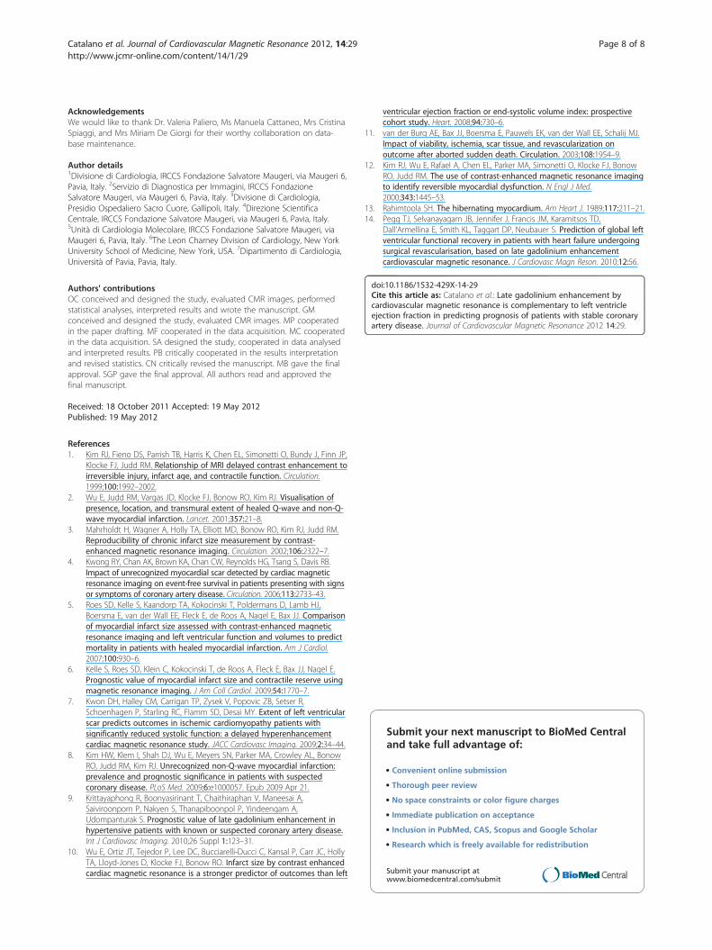
the only endpoint and we found similar results. In thisgroup LGE was associated with significantly increasedrisk of mortality (HR 3.78 [95% CI: 1.46 – 9.75];p= 0.006). All-cause mortality showed survival of 97% at1 year, 95% at 3 years and 92% 5 years, and mean annualdeath rate of 2% in the group without risk factors;compared to survival of 82% at 1 year, 63% at 3 yearsand 53% at 5 years, and mean annual death rate of14% in the group with risk factors (p< 0.0001). Thesefindings are graphically shown by Kaplan-Meyer curvesin the Figure 1.

Table 3 Adjusted hazard ratios for death or new heartfailure of the final model
AdjustedHR
95% ConfidenceInterval
P value
LGE total burden(≥ 45% of LV mass)
5.25 2.64 – 10.43 <0.0001
LVEF (≤ 30%) 4.89 2.50 – 9.56 <0.0001
Pulmonary hypertension(sPAP ≥ 35 mmHg)
2.89 1.56 – 5.36 <0.001
Loop diuretics therapy 2.92 1.54 – 5.55 0.001
LGE = late gadolinium enhancement; LVEF = left ventricle ejection fraction;sPAP = systolic pulmonary artery pressure.
Catalano et al. Journal of Cardiovascular Magnetic Resonance 2012, 14:29 Page 6 of 8http://www.jcmr-online.com/content/14/1/29
DiscussionIn the last decade LGE CMR has paved the way to a newera in the assessment of CAD [1]. Indeed, for the first time
Figure 1 Prognostic value of late gadolinium enhancement and left vcumulative incidence of death plus heart failure (panel a) or death alone (p
this technique allowed to pursue a direct detection andquantification of areas of irreversibly injured myocardium.Thus LGE enabled in vivo morphopathologic assessmentcomplementary to that of myocardium functionallyimpaired but still viable (hibernating myocardium),postulated from previous studies [13]. Accordingly, LGEhad the potential to become the technique of choice tostudy myocardial viability. Recent studies have confirmedthis hypothesis by showing that (a) the lesser transmuralextent of scar is, the more likely functional recovery of asegment will be after revascularization and (b) the amountof viable plus normal segments is the best predictor ofglobal functional recovery [12,14].In survival studies the extent of LGE has emerged as the
strongest prognostic factor in stable CAD patients withprevious MI [5,6]. This correlation further refines the
entricle ejection fraction. Kaplan-Meier survival analysis showing theanel b).

Catalano et al. Journal of Cardiovascular Magnetic Resonance 2012, 14:29 Page 7 of 8http://www.jcmr-online.com/content/14/1/29
relationship between LVEF and prognosis because LGEdirectly reflects the amount of irreversibly injured myocar-dium. Accordingly, these studies seem to support thehypothesis that LVEF is no more significant once LGE isincluded in multivariate models. On the other hand, theidea of the predictive value of LVEF to be enclosed andovertaken by LGE assessment, is in conflict with a previ-ous scintigraphy study in sudden death survivors and in arecent CMR study in hypertensive patients [9,11]. In thesetwo studies, indeed, LVEF remained an independentpredictor of events after the inclusion of scar dimension ina multivariate analysis. Thus the interaction between LGEand LVEF in stable CAD populations seems to be modu-lated by the selection of specific subsets of patients.
Conventional risk stratificationOur study was aimed at verifying these findings in alarge group of unselected patients with stable CAD. Forthis reason we intentionally avoid to enrol a highlyhomogeneous study cohort by defining relatively looseentry criteria. In this setting, we attempted to test thehypothesis whether LGE is an independent predictor ofadverse cardiac outcome in the context of a complexcohort undergoing a complete standard evaluation. Webelieve this study is representative of clinical referral ofmany outpatient CAD cardiology clinics.We studied a group of 376 consecutive patients with
stable CAD and optimized medical therapy, who werefollowed-up for an average time of 3 years. The conven-tional prognostic factor included in the study confirmedtheir association with adverse prognosis, thus furtherstrengthening the evidence that our study group is repre-sentative of a general population with stable CAD. Toremove redundant information a multivariate analysiswas performed, which confirmed the expected strongprognostic power of LVEF and showed a significant con-tribution by only few other conventional variables. Thelatter did not include myocardial revascularization, evenif it seemed to have a protective effect as from univariateanalysis.
Added value of late gadolinium enhancementOn top of relevant conventional variables, we found thatLGE was significantly and strongly associated with prog-nosis in a multivariate model (5.3 fold risk increase forLGE >45%). Even more importantly, the present studyshowed that LGE extent is complementary to LVEF instratifying the risk of patients with stable CAD. A largescar, replacing more than 45% of total LV mass, wellintegrates with the generally accepted criterion of LVEFlower than 30% in identifying patients at high risk ofdeath or HF. Accordingly, patients with either LGE ≥ 45%or LVEF ≤ 30%, have completely different prognosis ifcompared with patients without any of these risk factors,
showing annual event rates of 43% versus 3% and mor-tality rates of 14% versus 2%, respectively. Notably, about30% of patients at high risk would not have been identi-fied without including LGE into the stratificationprocess.The result of the present study seems to reinforce the
idea of LGE as a predictor of prognosis that adds toLVEF. In comparison to the studies of Roes et al. andKelle et al., that came to opposite results, our study con-sidered similar end-points but did not have scar presenceas an inclusion criterion (approximately 70% of ourpatients were LGE positive). Accordingly, on averagescar dimension was smaller (13% vs 19–20% of totalmass) and EF higher (51 vs 43–44%). Thus, it may behypothesized that the pre-selection of patients with ascar among those with stable CAD may influence theinteraction between LGE and LVEF in the prediction offuture events.Understanding the relative relevance of LGE and LVEF
is not a purely theoretical issue. Indeed, in patients withstable CAD reduced LVEF is a pre-requisite for importanttherapeutic decisions, such as cardiac resynchronizationtherapy (CRT) or automatic implantable cardioverter-defibrillator (AICD). Thus, will LGE be confirmed a stron-ger predictor than LVEF, the management of patients withstable CAD could be substantially changed
ConclusionsOur study confirms and refines the evidence of LGE asstrong prognostic factor in unselected patients with stableCAD, showing a complementary role with respect to LVEF.Although further studies are warranted to assess the useful-ness of LGE as selection criterion for major therapeuticdecision such as CRT or AICD, findings of the presentstudy promote the inclusion of LGE into current clinicalmanagement of patients with stable CAD, especially ofthose with reduced LVEF at echocardiography.
Additional files
Additional file 1: Table 1. Evaluation criteria of ECG andEchocardiography. LV = left ventricle; LBB = left bundle branch; RBB = rightbundle branch; MR =mitral regurgitation; sPAH = systolic pulmonary arteryhypertension; RV = right ventricle; RVIT = right ventricle inflow tract;TAPSE = tricuspid anular plane systolic excursion * based on trans-mitraldiastolic flow and pulmonary vein flow evaluation † based on proximalisovelocity surface area radius.
Additional file 2: Table 2. Adjusted hazard ratios for death or newheart failure of the final model for patients with myocardial infarctionhistory (panel a), with definite diagnosis of coronary artery disease (panelb), and using MR derived left ventricle ejection fraction (panel c).LGE = late gadolinium enhancement; LVEF = left ventricle ejection fraction;sPAP = systolic pulmonary artery pressure.
Competing interestsThe authors declare that they have no competing interests.
Catalano et al. Journal of Cardiovascular Magnetic Resonance 2012, 14:29 Page 8 of 8http://www.jcmr-online.com/content/14/1/29
AcknowledgementsWe would like to thank Dr. Valeria Paliero, Ms Manuela Cattaneo, Mrs CristinaSpiaggi, and Mrs Miriam De Giorgi for their worthy collaboration on data-base maintenance.
Author details1Divisione di Cardiologia, IRCCS Fondazione Salvatore Maugeri, via Maugeri 6,Pavia, Italy. 2Servizio di Diagnostica per Immagini, IRCCS FondazioneSalvatore Maugeri, via Maugeri 6, Pavia, Italy. 3Divisione di Cardiologia,Presidio Ospedaliero Sacro Cuore, Gallipoli, Italy. 4Direzione ScientificaCentrale, IRCCS Fondazione Salvatore Maugeri, via Maugeri 6, Pavia, Italy.5Unità di Cardiologia Molecolare, IRCCS Fondazione Salvatore Maugeri, viaMaugeri 6, Pavia, Italy. 6The Leon Charney Division of Cardiology, New YorkUniversity School of Medicine, New York, USA. 7Dipartimento di Cardiologia,Università of Pavia, Pavia, Italy.
Authors' contributionsOC conceived and designed the study, evaluated CMR images, performedstatistical analyses, interpreted results and wrote the manuscript. GMconceived and designed the study, evaluated CMR images. MP cooperatedin the paper drafting. MF cooperated in the data acquisition. MC cooperatedin the data acquisition. SA designed the study, cooperated in data analysedand interpreted results. PB critically cooperated in the results interpretationand revised statistics. CN critically revised the manuscript. MB gave the finalapproval. SGP gave the final approval. All authors read and approved thefinal manuscript.
Received: 18 October 2011 Accepted: 19 May 2012Published: 19 May 2012
References1. Kim RJ, Fieno DS, Parrish TB, Harris K, Chen EL, Simonetti O, Bundy J, Finn JP,
Klocke FJ, Judd RM. Relationship of MRI delayed contrast enhancement toirreversible injury, infarct age, and contractile function. Circulation.1999;100:1992–2002.
2. Wu E, Judd RM, Vargas JD, Klocke FJ, Bonow RO, Kim RJ. Visualisation ofpresence, location, and transmural extent of healed Q-wave and non-Q-wave myocardial infarction. Lancet. 2001;357:21–8.
3. Mahrholdt H, Wagner A, Holly TA, Elliott MD, Bonow RO, Kim RJ, Judd RM.Reproducibility of chronic infarct size measurement by contrast-enhanced magnetic resonance imaging. Circulation. 2002;106:2322–7.
4. Kwong RY, Chan AK, Brown KA, Chan CW, Reynolds HG, Tsang S, Davis RB.Impact of unrecognized myocardial scar detected by cardiac magneticresonance imaging on event-free survival in patients presenting with signsor symptoms of coronary artery disease. Circulation. 2006;113:2733–43.
5. Roes SD, Kelle S, Kaandorp TA, Kokocinski T, Poldermans D, Lamb HJ,Boersma E, van der Wall EE, Fleck E, de Roos A, Nagel E, Bax JJ. Comparisonof myocardial infarct size assessed with contrast-enhanced magneticresonance imaging and left ventricular function and volumes to predictmortality in patients with healed myocardial infarction. Am J Cardiol.2007;100:930–6.
6. Kelle S, Roes SD, Klein C, Kokocinski T, de Roos A, Fleck E, Bax JJ, Nagel E.Prognostic value of myocardial infarct size and contractile reserve usingmagnetic resonance imaging. J Am Coll Cardiol. 2009;54:1770–7.
7. Kwon DH, Halley CM, Carrigan TP, Zysek V, Popovic ZB, Setser R,Schoenhagen P, Starling RC, Flamm SD, Desai MY. Extent of left ventricularscar predicts outcomes in ischemic cardiomyopathy patients withsignificantly reduced systolic function: a delayed hyperenhancementcardiac magnetic resonance study. JACC Cardiovasc Imaging. 2009;2:34–44.
8. Kim HW, Klem I, Shah DJ, Wu E, Meyers SN, Parker MA, Crowley AL, BonowRO, Judd RM, Kim RJ. Unrecognized non-Q-wave myocardial infarction:prevalence and prognostic significance in patients with suspectedcoronary disease. PLoS Med. 2009;6:e1000057. Epub 2009 Apr 21.
9. Krittayaphong R, Boonyasirinant T, Chaithiraphan V, Maneesai A,Saiviroonporn P, Nakyen S, Thanapiboonpol P, Yindeengam A,Udompanturak S. Prognostic value of late gadolinium enhancement inhypertensive patients with known or suspected coronary artery disease.Int J Cardiovasc Imaging. 2010;26 Suppl 1:123–31.
10. Wu E, Ortiz JT, Tejedor P, Lee DC, Bucciarelli-Ducci C, Kansal P, Carr JC, HollyTA, Lloyd-Jones D, Klocke FJ, Bonow RO. Infarct size by contrast enhancedcardiac magnetic resonance is a stronger predictor of outcomes than left
ventricular ejection fraction or end-systolic volume index: prospectivecohort study. Heart. 2008;94:730–6.
11. van der Burg AE, Bax JJ, Boersma E, Pauwels EK, van der Wall EE, Schalij MJ.Impact of viability, ischemia, scar tissue, and revascularization onoutcome after aborted sudden death. Circulation. 2003;108:1954–9.
12. Kim RJ, Wu E, Rafael A, Chen EL, Parker MA, Simonetti O, Klocke FJ, BonowRO, Judd RM. The use of contrast-enhanced magnetic resonance imagingto identify reversible myocardial dysfunction. N Engl J Med.2000;343:1445–53.
13. Rahimtoola SH. The hibernating myocardium. Am Heart J. 1989;117:211–21.14. Pegg TJ, Selvanayagam JB, Jennifer J, Francis JM, Karamitsos TD,
Dall'Armellina E, Smith KL, Taggart DP, Neubauer S. Prediction of global leftventricular functional recovery in patients with heart failure undergoingsurgical revascularisation, based on late gadolinium enhancementcardiovascular magnetic resonance. J Cardiovasc Magn Reson. 2010;12:56.
doi:10.1186/1532-429X-14-29Cite this article as: Catalano et al.: Late gadolinium enhancement bycardiovascular magnetic resonance is complementary to left ventricleejection fraction in predicting prognosis of patients with stable coronaryartery disease. Journal of Cardiovascular Magnetic Resonance 2012 14:29.
Submit your next manuscript to BioMed Centraland take full advantage of:
• Convenient online submission
• Thorough peer review
• No space constraints or color figure charges
• Immediate publication on acceptance
• Inclusion in PubMed, CAS, Scopus and Google Scholar
• Research which is freely available for redistribution
Submit your manuscript at www.biomedcentral.com/submit
Copyright © 2022 FDOKUMEN