Rocky Mountain Conference on Magnetic Resonance
170
Rocky Mountain Conference on Magnetic Resonance Volume 58 58th Annual Rocky Mountain Conference on Magnetic Resonance Article 1 July 2016 58th Annual Rocky Mountain Conference on Magnetic Resonance Follow this and additional works at: hps://digitalcommons.du.edu/rockychem Part of the Chemistry Commons , Materials Science and Engineering Commons , and the Physics Commons is work is licensed under a Creative Commons Aribution 4.0 License. is Article is brought to you for free and open access by Digital Commons @ DU. It has been accepted for inclusion in Rocky Mountain Conference on Magnetic Resonance by an authorized editor of Digital Commons @ DU. For more information, please contact [email protected],dig- [email protected]. Recommended Citation (2016) "58th Annual Rocky Mountain Conference on Magnetic Resonance," Rocky Mountain Conference on Magnetic Resonance: Vol. 58 , Article 1. Available at: hps://digitalcommons.du.edu/rockychem/vol58/iss1/1
-
Upload
khangminh22 -
Category
Documents
-
view
3 -
download
0
Transcript of Rocky Mountain Conference on Magnetic Resonance

Rocky Mountain Conference on Magnetic ResonanceVolume 58 58th Annual Rocky Mountain Conferenceon Magnetic Resonance Article 1
July 2016
58th Annual Rocky Mountain Conference onMagnetic Resonance
Follow this and additional works at: https://digitalcommons.du.edu/rockychem
Part of the Chemistry Commons, Materials Science and Engineering Commons, and the PhysicsCommons
This work is licensed under a Creative Commons Attribution 4.0 License.This Article is brought to you for free and open access by Digital Commons @ DU. It has been accepted for inclusion in Rocky Mountain Conferenceon Magnetic Resonance by an authorized editor of Digital Commons @ DU. For more information, please contact [email protected],[email protected].
Recommended Citation(2016) "58th Annual Rocky Mountain Conference on Magnetic Resonance," Rocky Mountain Conference on Magnetic Resonance: Vol.58 , Article 1.Available at: https://digitalcommons.du.edu/rockychem/vol58/iss1/1

FINAL PROGRAM AND ABSTRACTS
Endorsed by:
Colorado Section – American Chemical Society &
Society for Applied Spectroscopy
July 17–21, 2016Beaver Run Resort & Conference Center
Breckenridge, Colorado
www.rockychem.com
1
et al.: 58th RMCMR Final Program and Abstracts
Published by Digital Commons @ DU, 2016

1
58TH ROCKY MOUNTAIN CONFERENCE ON MAGNETIC RESONANCEJuly 17–22, 2016 • July 18–20, 2016 (Exhibition)
Beaver Run Resort & Conference Center, Breckenridge, Colorado
Endorsed by: Colorado Section – American Chemical Society
& Society for Applied Spectroscopy
TABLE OF CONTENTSOrganizers and Chairpersons . . . . . . . . . . . . . . . . . . . . . . . . . . . . . . . . . . . . . . . . . . . . . . . . . . . . . . . . . . . . . . . . . . . . . . . . . . . . 2
Conference Supporters & Exhibitors . . . . . . . . . . . . . . . . . . . . . . . . . . . . . . . . . . . . . . . . . . . . . . . . . . . . . . . . . . . . . . . . . . . . . . 2
RMCMR Information . . . . . . . . . . . . . . . . . . . . . . . . . . . . . . . . . . . . . . . . . . . . . . . . . . . . . . . . . . . . . . . . . . . . . . . . . . . . . . . . . . . . . 3 Registration
Exhibition Schedule
Conference Reception
Conference Banquet & Awards Ceremony
Altitude
Messages
Social Media
Conference-at-a-Glance . . . . . . . . . . . . . . . . . . . . . . . . . . . . . . . . . . . . . . . . . . . . . . . . . . . . . . . . . . . . . . . . . . . . . . . . . . . . . . . . . . 3
Meeting Spaces . . . . . . . . . . . . . . . . . . . . . . . . . . . . . . . . . . . . . . . . . . . . . . . . . . . . . . . . . . . . . . . . . . . . . . . . . . . . . . . . . . . . . . . . . 4
Exhibitors . . . . . . . . . . . . . . . . . . . . . . . . . . . . . . . . . . . . . . . . . . . . . . . . . . . . . . . . . . . . . . . . . . . . . . . . . . . . . . . . . . . . . . . . . . . . . . . 5
RMCMR Technical Program Schedule
39TH INTERNATIONAL EPR SYMPOSIUM . . . . . . . . . . . . . . . . . . . . . . . . . . . . . . . . . . . . . . . . . . . . . . . . . . . . . . . . . . . . . . . . . . . . 7Sunday Oral Sessions . . . . . . . . . . . . . . . . . . . . . . . . . . . . . . . . . . . . . . . . . . . . . . . . . . . . . . . . . . . . . . . . . . . . . . . . . . . . . . . . . . . . . . . . . . . . . . . 8
Monday Oral Sessions . . . . . . . . . . . . . . . . . . . . . . . . . . . . . . . . . . . . . . . . . . . . . . . . . . . . . . . . . . . . . . . . . . . . . . . . . . . . . . . . . . . . . . . . . . . . . . 9
Tuesday Oral Sessions . . . . . . . . . . . . . . . . . . . . . . . . . . . . . . . . . . . . . . . . . . . . . . . . . . . . . . . . . . . . . . . . . . . . . . . . . . . . . . . . . . . . . . . . . . . . .10
Wednesday Oral Sessions . . . . . . . . . . . . . . . . . . . . . . . . . . . . . . . . . . . . . . . . . . . . . . . . . . . . . . . . . . . . . . . . . . . . . . . . . . . . . . . . . . . . . . . . . .11
Thursday Oral Sessions . . . . . . . . . . . . . . . . . . . . . . . . . . . . . . . . . . . . . . . . . . . . . . . . . . . . . . . . . . . . . . . . . . . . . . . . . . . . . . . . . . . . . . . . . . . .12
EPR Poster Sessions . . . . . . . . . . . . . . . . . . . . . . . . . . . . . . . . . . . . . . . . . . . . . . . . . . . . . . . . . . . . . . . . . . . . . . . . . . . . . . . . . . . . . . . . . . . . 13–15
SOLID-STATE NMR SYMPOSIUM . . . . . . . . . . . . . . . . . . . . . . . . . . . . . . . . . . . . . . . . . . . . . . . . . . . . . . . . . . . . . . . . . . . . . . . . . .16Sunday Oral Sessions . . . . . . . . . . . . . . . . . . . . . . . . . . . . . . . . . . . . . . . . . . . . . . . . . . . . . . . . . . . . . . . . . . . . . . . . . . . . . . . . . . . . . . . . . . . . . .17
Monday Oral Sessions . . . . . . . . . . . . . . . . . . . . . . . . . . . . . . . . . . . . . . . . . . . . . . . . . . . . . . . . . . . . . . . . . . . . . . . . . . . . . . . . . . . . . . . . . . . . .18
Tuesday Oral Sessions . . . . . . . . . . . . . . . . . . . . . . . . . . . . . . . . . . . . . . . . . . . . . . . . . . . . . . . . . . . . . . . . . . . . . . . . . . . . . . . . . . . . . . . . . . . . .19
Wednesday Oral Sessions . . . . . . . . . . . . . . . . . . . . . . . . . . . . . . . . . . . . . . . . . . . . . . . . . . . . . . . . . . . . . . . . . . . . . . . . . . . . . . . . . . . . . . . . . .20
Thursday Oral Sessions . . . . . . . . . . . . . . . . . . . . . . . . . . . . . . . . . . . . . . . . . . . . . . . . . . . . . . . . . . . . . . . . . . . . . . . . . . . . . . . . . . . . . . . . . . . .21
NMR Poster Sessions . . . . . . . . . . . . . . . . . . . . . . . . . . . . . . . . . . . . . . . . . . . . . . . . . . . . . . . . . . . . . . . . . . . . . . . . . . . . . . . . . . . . . . . . . . . 22–25
RMCMR Abstracts . . . . . . . . . . . . . . . . . . . . . . . . . . . . . . . . . . . . . . . . . . . . . . . . . . . . . . . . . . . . . . . . . . . . . . . . . . . . . . . . . 25–160
Index of Presenters . . . . . . . . . . . . . . . . . . . . . . . . . . . . . . . . . . . . . . . . . . . . . . . . . . . . . . . . . . . . . . . . . . . . . . . . . . . . . . . . 161–16
www.rockychem.com
Milestone Presentations, LLC • 4255 South Buckley Road, #118 • Aurora, CO 80013 Ph: 800-996-3233 or 303-690-3233 • Fax: 888-996-3296 or 303-690-3278
E-mail: [email protected] • Web: www.milestoneshows.com
2
Rocky Mountain Conference on Magnetic Resonance, Vol. 58 [2016], Art. 1
https://digitalcommons.du.edu/rockychem/vol58/iss1/1

2
ORGANIZERS AND CHAIRPERSONS
ENDORSED BY: Colorado Section — American Chemical Society
& Society for Applied Spectroscopy
CONFERENCE CHAIR:Kurt W. Zilm
Yale University Department of Chemistry, PO Box 20817
New Haven, CT 06520-8107 Ph: 203-432-3956 • Fax: 203-432-6144
EPR SCIENTIFIC COMMITTEE:John Morton – Chair
University College LondonJohn McCracken – Co-Chair 2016, Chair 2017
Michigan State UniversityAnia Bleszynski-Jayich
University of California Santa BarbaraChristoph Boehme University of UtahHoward Halpern
University of ChicagoFraser MacMillan
University of East AngliaStefan Stoll
University of WashingtonSusumu Takahashi
University of Southern California
CONFERENCE SUPPORTERS & EXHIBITORS (As of July 13, 2016)
Avanti Polar Lipids, Inc.
BlueSky NMR, LLC
Bruker BioSpin
CortecNet
Doty Scientific
Elsevier Journal Solid State Nuclear Magnetic Resonance
ExxonMobil
JEOL USA, Inc.
National High Magnetic Field Lab
Oxford Instruments NanoScience
PhoenixNMR LLC
Revolution NMR LLC
Springer Science & Business Media B.V.
Steppingstone MAgnetic Resonance Training
Wilmad-LabGlass
3
et al.: 58th RMCMR Final Program and Abstracts
Published by Digital Commons @ DU, 2016

3
REGISTRATIONAdmission to all technical sessions and the exhibition is by name badge only. Registration materials may be picked up at the RMCMR registration area located at Beaver Run Resort & Conference Center between 10:00 a.m. and 5:00 p.m. on Sunday, July 17 or 8:00 a.m. and 5:00 p.m. anytime Monday, July 18 through Thursday, July 21.
EXHIBITION SCHEDULE
Monday, July 1810:00 a.m. – 7:00 p.m. (Conference Reception 5:30 p.m. – 7:00 p.m.)
Tuesday, July 199:00 a.m. – 5:00 p.m.
Wednesday, July 209:00 a.m. – 2:00 p.m.
CONFERENCE RECEPTIONMonday evening from 5:30 p.m. to 7:00 p.m., all attendees are cordially invited to join in on beverages and hors d’oeuvres. Unwind from the day’s events and continue the “Rocky Mountain Conference” experience. Check out all of the latest products and services as the reception is held right in the exhibition area.
CONFERENCE BANQUET & AWARDS CEREMONY
Wednesday evening from 7:00 p.m. to 9:00 p.m. in the Breckenridge Ballroom. Enjoy an evening of comradeship, fine food and recognition of peers. Pre-registration required. Speech by Eiichi Fukushima, followed by EPR Awards and SSNMR Awards.
ALTITUDEBreckenridge is approximately 9,600 feet above sea level. The acclimatization process is inhibited by dehydration, over-exertion, alcohol and other depressant drugs. Please take the following precautions regarding high altitude:
• Take it easy; don’t over-exert yourself.
• Light activity during the day is better than sleeping because respiration decreases during sleep, exacerbating the symptoms.
• Avoid tobacco, alcohol and other depressant drugs including, barbiturates, tranquilizers, and sleeping pills.
• Eat a high carbohydrate diet
• Drink three to four times more water than usual.
MESSAGESMessages will be accepted and posted on the message board. Call 800-996-3233 or 303-690-3233 to leave messages.
SOCIAL MEDIAFollow us on Facebook (rockymtnconf) or Twitter (@rockymtnconf) and join in the conversation.
ROCKY MOUNTAIN CONFERENCE INFORMATION
CONFERENCE-AT-A-GLANCE
E VENT LOC ATIONSunday Monday Tuesday Wednesday Thursday
a .m . p .m . a .m . p .m . a .m . p .m . a .m . p .m . a .m . p .m .
Bruker EPR Users’ Meeting Coppertop II
Bruker NMR Symposium & Workshop Coppertop II
Get Into Shape Workshop Peak 5
EPR Lectures Peak 5
EPR Posters Coppertop III
ExhibitionColorado Ballrooom Foyer
SSNMR Lectures Peak 1-4
SSNMR Posters Imperial
4
Rocky Mountain Conference on Magnetic Resonance, Vol. 58 [2016], Art. 1
https://digitalcommons.du.edu/rockychem/vol58/iss1/1

4
CONFERENCE CENTER MEETING SPACE
119 12 1310
7
Speaker Ready Room
EPRMeeting
Room
SSNMRMeeting
Room
Catering
Catering
To P
oste
rs
18
COLORADO BALLROOM FOYER
5
et al.: 58th RMCMR Final Program and Abstracts
Published by Digital Commons @ DU, 2016

5
EXHIBITORS
Bruker BioSpin CorporationBooth 9, 10 & 11
15 Fortune Dr
Billerica, MA 01821
Phone: 978-667-950
E-mail: [email protected]
Web: www.bruker.com
Bruker BioSpin is the market leader in analytical research tools based on magnetic resonance. Our comprehensive portfolio includes NMR, EPR and TD-NMR, delivering a range of research tools to enable life science, materials science, analytical chemistry and process control.
CortecNet CorpBooth 8
Downstate Biotech Incubator
760 Parkside Ave
Brooklyn, NY 11226
Phone: 347-404-6810
Fax: 415-230-5796
E-mail: [email protected]
Web: www.cortecnet.com/us
Cortecnet has been proudly serving the NMR community worldwide for more than 20 years, providing stable isotopes and NMR consumables (Bruker MAS rotors, d-solvents, NMR tubes, 13C, 15N,D-labeled compounds , DNP radicals, and much more!)
Doty Scientific, Inc .Booth 7
700 Clemson Rd
Columbia, SC 29229
Phone: 803-788-6497
Fax: 803-736-5495
E-mail: [email protected]
Web: www.dotynmr.com
NMR Probes For All Spectrometers: Agilent, Varian, Chemagetics, JOEL, Bruker. Solids: MAS Magic Angle Gradient MAS, High Temp MAS, Wide Line. (B-MAX MAS probes: Low-E. Quad Resonance, Double broad-band, H-F, or Lock, Extended VT.)
JEOL USA, Inc .Booth 1
11 Dearborn Rd
Peabody, MA 01960
Phone: 978-535-5900
Fax: 978-536-2205
E-mail: [email protected]
Web: www.jeolusa.com
JEOL is a world leader in analytical instrumentation – including ESR and NMR. Stop by to learn more about our next generation JNM-ECZ series NMR spectrometers, the JES-X3 Series ESR spectrometers, NMR Probes for Solids NMR, and other ESR & NMR accessories.
PhoenixNMR LLCBooth 13
4921 Eagle Lake Dr
Fort Collins, CO 80524
Phone: 970-472-0613
Fax: 970-416-8896
E-mail: [email protected]
Web: www.revolutionnmr.com
PhoenixNMR provides unique modular 40 mm solid state NMR probes and probe heads enabling state of the art solids NMR in both wide bore and standard bore systems, and probe repair services for Agilent and Varian solids probes.
Revolution NMR LLCBooth 12
4921 Eagle Lake Dr
Fort Collins, CO 80524
Phone: 970-472-0613
Fax: 970-416-8896
E-mail: [email protected]
Web: www.revolutionnmr.com
Revolution NMR supplies spinning systems and components, specialty probes, and probe upgrade and repair services for solid state NMR.
6
Rocky Mountain Conference on Magnetic Resonance, Vol. 58 [2016], Art. 1
https://digitalcommons.du.edu/rockychem/vol58/iss1/1

6
39TH INTERNATIONAL EPR SYMPOSIUM
JULY 17-21, 2016 Breckenridge, Colorado, USA
CONFERENCE CHAIRKurt W. Zilm
EPR SYMPOSIUM COMMITTEEJohn Morton (Chair)
John McCracken (Co-Chair 2016, Chair 2017)
Ania Bleszynski-Jayich
Christoph Boehme
Howard Halpern
Fraser MacMillan
Stefan Stoll
Susumu Takahashi
EPR SYMPOSIUM SPONSORSAvanti Polar Lipids, Inc.
Bruker BioSpin
Doty Scientific
National High Magnetic Field Lab
Oxford Instruments NanoScience
REGISTRATIONRegister at www.rockychem.com
Admission to all technical sessions and the exhibition is by name badge only. Registration materials may be picked up at the RMCMR registration area located at Beaver Run Resort & Conference Center between 10:00 a.m. and 5:00 p.m. on Sunday, July 17 or 8:00 a.m. and 5:00 p.m. anytime Monday, July 18 through Thursday, July 21.
Complimentary lunches are being provided July 18, 19 and 20 to all registered symposia attendees. You will receive your luncheon ticket(s) upon check-in at the RMCMR registration desk. Tickets are date-specific and cannot be interchanged with any other day. Lost tickets cannot be replaced. Unused tickets cannot be redeemed for another day. The lunch will be served in the Event Tent each designated day from 12:00 noon – 1:00 p.m.
EVENTS Get Into Shape Workshop (Pulse Shaping)
Sunday, July 17 3:30 p.m. - 5:30 p.m. (Peak 5)
Led by Songi Han (UCSB), Gareth Eaton & Laura Buchanan (Denver) and Ralph Weber (Bruker)
Bruker EPR Users' Meeting:
Sunday, July 17 Starts at 6:30 p.m. followed by a mixer. (Coppertop II)
For information and registration access: https://www.bruker.com/events/mr/bruker-at-rocky-mountain/epr-users-meeting.html
Poster Sessions:
Monday, July 18 7:30 p.m. – 9:00 p.m. (Coppertop III) and Tuesday, July 19 7:30 p.m. – 9:00 p.m. (Coppertop III)
Conference Banquet & Awards Ceremony
Wednesday, July 20 7:00 p.m. – 9:00 p.m. (Breckenridge Ballroom)
Enjoy an evening of comradeship, fine food and recognition of peers. Pre-registration required.
— Banquet Speaker: Eiichi Fukushima, New Mexico Resonance
— EPR Awards
— SSNMR Awards
7
et al.: 58th RMCMR Final Program and Abstracts
Published by Digital Commons @ DU, 2016

7
EPR SYMPOSIUM ORAL SESSIONS AGENDA
SUNDAY, JULY 17, 2016Pre-Conference Activities
3:30–5:30 PM
Get Into Shape Workshop (Pulse Shaping). Songi Han (University of California Santa Barbara) Gareth Eaton (University of Denver) Laura Buchanan (University of Denver) Ralph Weber (Bruker BioSpin)
6:30–10:00 PMBruker EPR Users’ Meeting Meeting followed by Mixer
8
Rocky Mountain Conference on Magnetic Resonance, Vol. 58 [2016], Art. 1
https://digitalcommons.du.edu/rockychem/vol58/iss1/1

8
MONDAY, JULY 18, 20168:10 AM Welcoming Remarks. John Morton, EPR Symposium ChairSession I: Materials I. Christoph Boehme, Chair
8:15 AM 100 Time-dependent Photo-EPR Applied to Point Defects in Crystals: Limitations and Applications. Mary Ellen Zvanut, University of Alabama Birmingham
8:45 AM 101 Suppressing Spin-spin Relaxation in Silicon Carbide with Natural Isotope Abundance using Dynamic Decoupling. Andreas Sperlich, University of Würzburg
9:00 AM 102Silicon Carbide Magnetoresistive Magnetometer with Electrically Detected Magnetic Resonance Self-calibration Feature for Space Science Application. Corey Cochrane, California Institute of Technology, Jet Propulsion Laboratory
9:15 AM 103 Measurement of Paramagnetic Spin Concentration in a Solid-state System using Double Electron-electron Resonance. Viktor Stepanov, University of Southern California
9:30 AM 104 Hyperfine Interactions in Silicon. Chandrasekhar Ramanathan, Dartmouth College10:00 AM BreakSession II: Materials II. Christoph Boehme, Chair
10:40 AM 105 Probing Giant Magnetic Anisotropies in Mononuclear Single-molecule Magnets. Stephen Hill, National High Magnetic Field Laboratory and Florida State University
10:55 AM 106 Triplet Exciton Generation in Materials for Organic Solar Cells. Jan Behrends, Freie Universität Berlin
11:10 AM 107 Spin-orbit Coupling in Conjugated Polymers. Hans Malissa, University of Utah
11:25 AM 108 Effect of Spin-orbit Interaction and Topological Gap for ESR Spectra in Low-dimensional Organic Conductors. Toshikazu Nakamura, Institute for Molecular Science
11:40 AM 109 Spin Dynamics of TAPD-MP Ar-C60 Spin Correlated Radical Pair. Naitik Panjwani, University College London
12:00 PM Lunch (included with registration)Session III: Spin Centers in Biology and Chemistry I. Fraser MacMillan, Chair
1:30 PM 110 Structural Information from Hyperfine Couplings in Iron Catalysts. Inés Garcia-Rubio, Centro Universitario de la Defensa
2:00 PM 111263 GHz Pulse EPR Reports on Proton-coupled Electron Transfer Through the Subunit Interface of E. Coli Ribonucleotide Reductase Ia. Thomas Nick, Max Planck Institute for Biophysical Chemistry
2:15 PM 112 The Composition and Structure of the Inorganic Core of Intermediate X(WT) and X(Y1212F) of E. coli Ribonucleotide Reductase. Peter Doan, Northwestern University
2:30 PM 113 Mechanistic Investigations on Electron Bifurcation by EPR Spectroscopy. David Mulder, National Renewable Energy Laboratory
2:45 PM 114Out-of-phase ESEEM: Measuring Distances of Excited Radical-pair States to Identify the Final Electron Donor in Cryptochromes and Photolyases. Daniel Nohr, Albert-Ludwigs-University Freiburg
3:00 PM BreakSession IV: Spin Centers in Biology and Chemistry II. Fraser MacMillan, Chair
3:40 PM 115 Studying the Structure of Metalloproteins with RIDME Spectroscopy: Application to Nitric Oxide Synthase. Andre Astashkin, University of Arizona
4:10 PM 116 High-Field EPR Studies on Model Dimeric MnIV Complexes. Andrew Ozarowski, National High Magnetic Field Laboratory and Florida State University
4:25 PM 117 EPR-active Molecular pH Probes at a Protein-Lipid Interface: Turning Electrical Charges On and Off. Tatyana Smirnova, North Carolina State University
4:40 PM 118Free Energy Landscape and Protein Configurational Fluctuation Contributions to Radical Rearrangement Catalysis in B12 -dependent Ethanolamine Ammonia-Lyase. Meghan Kohne, Emory University
4:55 PM 119 Utilizing Novel 95 GHz 2D-ESR Spectroscopy to Study Nitroxide Partitioning into the Lipid Membranes at Room Temperatures. Siddarth Chandrasekaran, ACERT and Cornell University
5:10 PM 120 Using EPR, ENDOR, and HYSCORE to Elucidate the Structure of Copper and Cobalt Pre-Catalysts. Elizabeth Papish, University of Alabama
5:30–7:00 PM Conference ReceptionSession V: Posters7:30-9:00 PM Authors Present for Posters Labeled A
9
et al.: 58th RMCMR Final Program and Abstracts
Published by Digital Commons @ DU, 2016

9
TUESDAY, JULY 19, 2016Session VI: Spin Devices I. Ania Bleszynski-Jayich, Chair
8:15 AM 125 Nanowire-Based Magnetic Resonance Imaging and Spectroscopy. Raffi Budakian, University of Waterloo
8:45 AM 126 EPR Spectroscopy of using Nitrogen-vacancy Centers in Diamond. Chathuranga Abeywardana, University of Southern California
9:00 AM 127 Nanoliter Biological Electron Paramagnetic Resonance Spectroscopy on a Diamond Chip. Ilja Fescenko, University of New Mexico
9:15 AM 128 Improving Optical Collection Efficiency for Simultaneous Electrically and Optically Detected Magnetic Resonance on Thin Film Devices. Douglas Baird, University of Utah
9:30 AM 129 Toward Single Atom Qubits on a Surface: ESR in a Scanning Tunneling Microscope. William Paul, IBM Research
10:00 AM BreakSession VII: Spin Devices II. Ania Bleszynski-Jayich, Chair
10:40 AM 130 Spin Coherence and Spin Relaxation in Monolayer Semiconductors. Scott Crooker, National High Magnetic Field Laboratory
11:10 AM 131 Simultaneous Detection of Transient Electrically Detected and Transient Magnetic Resonance Signals from Organic Solar Cells. Felix Kraffert, Free University of Berlin
11:25 AM 132Separation of Hyperfine and Spin-Orbit Interactions in Organic Semiconductors by Multi-Frequency Electrically Detected Magnetic Resonance using Coplanar Waveguide Microresonators. Gajadhar Joshi, University of Utah
11:40 AM 133Estimation of Spin Diffusion Length and Spin-Orbit Coupling Strength in Organic Semiconductors by Means of pulsed Inverse Spin-Hall Effect Measurements. Marzieh Kavand, University of Utah
12:00 PM Lunch (included with registration)Session VIII: Biological Macromolecules I. Stefan Stoll, Chair
1:30 PM 134 Cu2+-ions as a ESR Probe of Protein Structure. Sunil Saxena, University of Pittsburgh
2:00 PM 135 Site-Specific Investigations of the Protein Dynamical Transition via Pulse EPR. Ryan Barnes, University of California Santa Barbara
2:15 PM 136 Bayesian Uncertainty Quantification for DEER Spectroscop. Thomas Edwards, University of Washington
2:30 PM 137 WavPDS: A Wavelet Approach in Denoising Pulsed Dipolar Spectroscopy. Madhur Srivastava, Cornell University
2:45 PM 138 Three Homologous TonB-dependent Transporters Utilize Different Mechanisms to Regulate Protein-Protein Interactions. Lishan Liu, University of Virginia
3:00 PM BreakSession IX: Biological Macromolecules II. Stefan Stoll, Chair
3:30 PM 138 Measuring Oxidation States in Exchange-Coupled Metal Clusters Using Ligand Hyperfine. Troy Stich, University of California Davis
4:00 PM 139 Optimization of Pulsed EPR Distance Measurements for Tau Protein Aggregation. Timothy Keller, University of California Santa Barbara
4:15 PM 140Conformational Transitions of Maltose Binding Protein in the Native State and as Molten Globule at pH 3 as Monitored by DEER and DQC EPR Spectroscopy. Wolfgang Trommer, TU Kaiserslautern
4:30 PM 141 Selective Membrane Disruption Mechanism of an Antibacterial γ-AApeptide Defined by EPR Spectroscopy. Likai Song, National High Magnetic Field Laboratory and Florida State University
4:45 PM 142 Distance Measurements Between Paramagnetic Ligands Bound to Parallel Stranded Guanine Quadruplexes. Matthew Donohue, National Institute of Standards and Technology
5:00 PM 143 Phenylalanine Hydroxylase: Providing Details of a Catalytic Cycle with EPR Spectroscopy. John McCracken, Michigan State University
Session X: Posters
7:30–9:00 PM Authors Present for Posters Labeled B
10
Rocky Mountain Conference on Magnetic Resonance, Vol. 58 [2016], Art. 1
https://digitalcommons.du.edu/rockychem/vol58/iss1/1

10
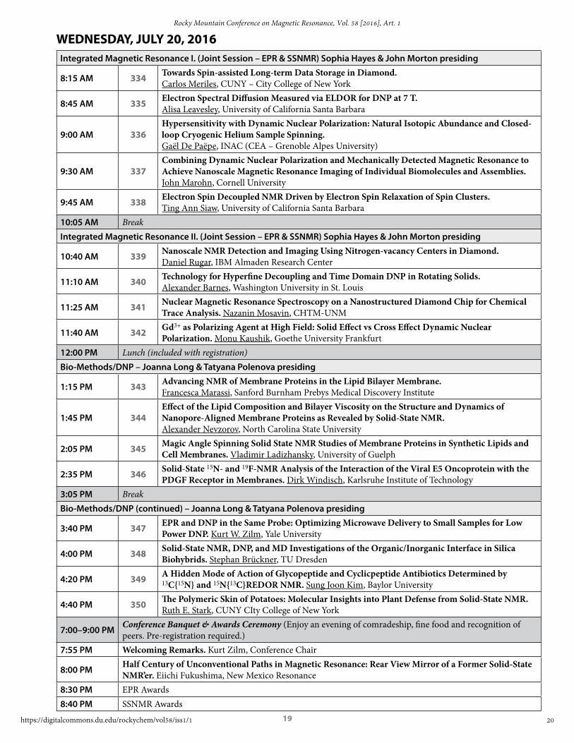
WEDNESDAY, JULY 20, 2016Session XI: Integrated Magnetic Resonance I. (Joint Session – EPR & SSNMR) Sophia Hayes & John Morton, Chairs
8:15 AM 149 Towards Spin-assisted Long-term Data Storage in Diamond. Carlos Meriles, CUNY–City College of New York
8:45 AM 150 Electron Spectral Diffusion Measured via ELDOR for DNP at 7 T. Alisa Leavesley, University of California Santa Barbara
9:00 AM 151 Hypersensitivity with Dynamic Nuclear Polarization: Natural Isotopic Abundance and Closed-loop Cryogenic Helium Sample Spinning. Gaël De Paëpe, INAC (CEA – Grenoble Alpes University)
9:30 AM 152Combining Dynamic Nuclear Polarization and Mechanically Detected Magnetic Resonance to Achieve Nanoscale Magnetic Resonance Imaging of Individual Biomolecules and Assemblies. John Marohn, Cornell University
9:45 AM 153 Electron Spin Decoupled NMR Driven by Electron Spin Relaxation of Spin Clusters. Ting Ann Siaw, University of California Santa Barbara
10:05 AM Break Session XII: Integrated Magnetic Resonance II. (Joint Session – EPR & SSNMR) Sophia Hayes & John Morton, Chairs
10:40 AM 154 Nanoscale NMR Detection and Imaging Using Nitrogen-vacancy Centers in Diamond. Daniel Rugar, IBM Almaden Research Center
11:10 AM 155 Technology for Hyperfine Decoupling and Time Domain DNP in Rotating Solids. Alexander Barnes, Washington University in St. Louis
11:25 AM 156 Nuclear Magnetic Resonance Spectroscopy on a Nanostructured Diamond Chip for Chemical Trace Analysis. Nazanin Mosavin, CHTM-UNM
11:40 AM 157 Gd3+ as Polarizing Agent at High Field: Solid Effect vs Cross Effect Dynamic Nuclear Polarization. Monu Kaushik, Goethe University Frankfurt
12:00 PM Lunch (included with registration)Session XIII: Methods I. Susumu Takahashi, Chair
1:30 PM 158 Quantum-Enhanced Nuclear Spin Imaging by an Electronic Spin Probe in Diamond. Paola Cappellaro, Massachusetts Institute of Technology
2:00 PM 159 Broadband Arbitrary Shaped Pulses for Pulsed EPR at 200 GHz. Ilia Kaminker, University of California Santa Barbara
2:15 PM 160 Pushing SIFTER Towards New Application. Philipp Schöps, Goethe University Frankfurt
2:30 PM 161 Frequency Swept Rapid Scan EDMR. Duane McCrory, National Institute of Standards and Technology
2:45 PM 162 A Rapid Scan Method to Measure T1 Relaxation Times. Laura Buchanan, University of Denver3:00 PM Break Session XIV: Methods II. Susumu Takahashi, Chair3:40 PM 163 EPR Spectroscopy at the Quantum Limit. Patrice Bertet, CEA Saclay
4:10 PM 164 Pulsed ENDOR with On-Chip Superconducting Resonators. Anthony Sigillito, Princeton University
4:25 PM 165 Millikelvin ESR With Superconducting Resonators at Magnetic Fields up to 170 mT. Christoph Zollitsch, London Centre for Nanotechnology
4:40 PM 166 Using CMOS Voltage-controlled Oscillators for Ultra-fast Rapid Scan ESR Experiments. Jens Anders, University of Ulm
Session XV: IES Awards. Alex Smirnov, Chair
5:00 PM 167 Coherent Pump Pulses in Double Electron Electron Resonance Spectroscopy. Claudia Tait, University of Washington
5:20 PM 168 Pulsed Electrically Detected Magnetic Resonance. Christoph Boehme, University of UtahRMC General Business Meeting5:40 PM John Morton, Chair
7:00–9:00 PMConference Banquet & Awards Ceremony (Enjoy an evening of comradeship, fine food and recognition of peers. Pre-registration required.)
7:55 PM Welcoming Remarks. Kurt Zilm, Conference Chair
8:00 PMHalf Century of Unconventional Paths in Magnetic Resonance: Rear View Mirror of a Former Solid-State NMR’er. Eiichi Fukushima, New Mexico Resonance
8:30 PM EPR Awards8:40 PM SSNMR Awards
11
et al.: 58th RMCMR Final Program and Abstracts
Published by Digital Commons @ DU, 2016

11
THURSDAY, JULY 21, 2016Session XVI: EPR Imaging / In-Vivo I . Howard Halpern, Chair
8:15 AM 175 Precise Delivery of Radiation Treatment to Hypoxic Areas Based on EPR Oxygen Images. Boris Epel, University of Chicago
8:45 AM 176Interstitial Inorganic Phosphate as an EPR Marker of Tumor Microenvironment and its Role in Tumorigenesis, Tumor Progression and Aggressiveness. Valery Khramtsov, West Virginia University
9:00 AM 177 Initial Results of Phase 1 Clinical Trial of OxyChip, an Implantable Probe for EPR Oximetry. Periannan Kuppusamy, Dartmouth College
9:30 AM 178 Feasibility Study of a CW-EPR-based Oxygen-mapping Technique Using a Pair of Isotopic Nitroxyl Radicals. Hiroshi Hirata, Hokkaido University
9:45 AM 179 Molecular Probes for Monitoring Thiol Redox Status In Vivo. Joe Kao, University of Maryland
10:15 AM Break
Session XVII: Methods III . Susumu Takahashi, Chair
10:45 AM 180 Tracking Field Fluctuations in Pulsed EPR. Abraham Asfaw, Princeton University
11:00 AM 181 Demagnetization Shifts in Very High Frequency Pulsed Electron Paramagnetic Resonance. Blake Wilson, University of California Santa Barbara
11:15 AM 182 Frequency-Domain EPR up to Several THz: Direct Observation of Large ZFS in CoII Clusters. Joscha Nehrkorn, University of Washington
11:30 AM 183 Multi-Extreme THz ESR: Development of Micro-Cantilever ESR up to the THz Region. Hitoshi Ohta, Kobe University
11:45 AM 184 High Sensitivity Transmission Mode Non-Resonant Stopped-Flow ESR. Pragya Shrestha, National Institute of Standards and Technology
12:00 PM Closing Remarks. John Morton, EPR Symposium Chair
12
Rocky Mountain Conference on Magnetic Resonance, Vol. 58 [2016], Art. 1
https://digitalcommons.du.edu/rockychem/vol58/iss1/1

12
58TH ROCKY MOUNTAIN CONFERENCE ON MAGNETIC RESONANCE
39TH INTERNATIONAL EPR SYMPOSIUM POSTER SESSIONS AGENDA
MONDAY, JULY 18 • 7:30–9:00 p .m . (Authors Present for Posters Labeled A)TUESDAY, JULY 19 • 7:30–9:00 p .m . (Authors Present for Posters Labeled B)
A 200 EPR for a Cu4S Model for Nitrous Oxide Reductase. William E Antholine, Medical College of Wisconsin
B 201 Chemical Influences on Quantum Coherence in Potential Molecular Qubits. Katharina Bader, University of Stuttgart
A 202 Triplet Exciton Generation in Materials for Organic Solar Cells. Jan Behrends, Freie Universität Berlin
B 203 A Digital Low-Field Dynamic Nuclear Polarization Spectrometer. Joshua R. Biller, National Institute of Standards and Technology
A 204Utilizing Novel 95 GHz 2D-ESR Spectroscopy to Study Nitroxide Partitioning into the Lipid Membranes at Room Temperatures. Siddarth Chandrasekaran, ACERT and Cornell University
B 205 T-jump and Freeze-quench EPR. Alexey V Cherepanov, Goethe University
A 206 Distance Measurements in Gd3+-labeled Proteorhodopsin Oligomers by 240 GHz CW EPR. Jessica Clayton, University of California Santa Barbara
B 207Highly Precise DEER Distance Measurements within Proteins using the Double Histidine Cu2+-Binding Motif. Timothy F. Cunningham, Hanover College
A 208Towards Understanding the Orientation Dependence of NV-Mediated Bulk Nuclear Hyperpolarization in Diamond at High Magnetic Fields. Melanie Drake, University of California Berkeley
B 209 Bayesian Uncertainty Quantification For DEER Spectroscopy. Thomas H. Edwards, University of Washington
A 210 SpecMan4EPR: The Second Generation of AWG Engine. Boris Epel, University of Chicago
B 211Interaction Between the Prion Protein’s Copper-Bound Octarepeat Domain and a Charged C-terminal Pocket Suggests a Mechanism for N-terminal Regulation. Eric GB Evans, University of Washington
A 212 Nanoliter Biological Electron Paramagnetic Resonance Spectroscopy on a Diamond Chip. Ilja Fescenko, University of New Mexico
B 213 The SHARED EPR Network. Gary J. Gerfen, Albert Einstein College of Medicine
A 214 NV Centers in Silicon Carbide (SiC): Identification, Modeling and Basic Properties. Uwe Gerstmann, University of Paderborn
B 215EPR Analysis of the Effects of Curcuminoids from Turmeric Spice on Superoxide Free Radicals Formed from a Xanthine-Xanthine Oxidase Reaction. Pranav Gopalakrishnan, Steppingstone MAgnetic Resonance Training Center
A 216 Room Temperature PELDOR Measurements with Rigid Nitroxide Spin Labels on Duplex DNA. Markus Gränz, Goethe University Frankfurt
B 217 Radical Intermediates in the Formation and Repair of Spore Photoproduct. Ellen C. Hayes, University of Washington
A 218 Electronic Structure of a CuII-Alkoxide Complex Modeling Intermediates in Copper-Catalyzed Alcohol Oxidations. Ellen C. Hayes, University of Washington
13
et al.: 58th RMCMR Final Program and Abstracts
Published by Digital Commons @ DU, 2016

13
B 219Feasibility Study of a CW-EPR-based Oxygen-mapping Technique Using a Pair of Isotopic Nitroxyl Radicals. Hiroshi Hirata, Hokkaido University
A 220 Spin Labelled Carbohydrates on Au Nanoparticles. Michael Hollas, University of Manchester
B 221Enhancing Nuclear Polarization for Nanoscale Imaging Using Magnetic Resonance Force Microscopy. Corinne E. Isaac, Cornell University
A 222Electrically Detected Magnetic Resonance Spectroscopy of Polymer Layers at B1 Exceeding B0 with Copper Microwire on Silicon Nitride/silicon Substrate. Shirin Jamali, University of Utah
B 223Characteristics of 14N- and 15N-labeled Dicarboxy-PROXYLs as Oxygen-sensitive Probes for CW-EPR-based Single-point Imaging (SPI). Harue Kubota, Hokkaido University
A 224 Electron Spectral Diffusion Measured via ELDOR for DNP at 7 T. Alisa Leavesley, University of California Santa Barbara
B 225
Determination of Zero Field Splitting Distribution and Rotational Correlation Times for Slow-motion Gd(III)-DOTA X-band cw-EPR Spectra Using a General Spin System Stochastic Liouville Equation Solver. Jeremy D. Lehner, University of Washington
A 226A Modular Low Frequency EPR Spectrometer for Studying Objects with Cultural Heritage Significance. Ken Liu, Symphotic
B 227Speciation of Vanadyl Porphyrin Complexes Through High Resolution Electron Paramagnetic Resonance. Donald Mannikko, University of Washington
A 228 Frequency Swept Rapid Scan EDMR. Duane McCrory, National Institute of Standards and Technology
B 229High-field/high-frequency Pulsed/CW EPR with Increased Concentration Sensitivity and High Power. Johannes E. McKay, National High Magnetic Field Laboratory
A 230An AWG-based Digital X-band Saturation Recovery Spectrometer for Spin Lattice Relaxation Measurements. Joseph McPeak, University of Denver
B 231 Spin-dependent Charge Carrier Interaction Processes in Polyfluorene Thin Films. Richards (Chad) Miller III, University of Utah
A 232Calculation of 2D-SECSY (Spin-Echo Correlation Spectroscopy) and 2D-ELDOR (Electron Double Resonance) Signal Using Stochastic- Liouville Equation (SLE). Sushil K. Misra, Concordia University
B 233 Mechanistic Investigations on Electron Bifurcation by EPR Spectroscopy. David Mulder, National Renewable Energy Laboratory
A 234 Frequency-Domain EPR up to Several THz: Direct Observation of Large ZFS in CoII Clusters. Joscha Nehrkorn, University of Washington
B 235Characterization of Solvent Dynamical Properties Around the B12-dependent Ethanolamine Ammonia-lyase by Using Spin Probe-EPR Spectroscopy. Benjamen Nforneh, Emory University
A 236Out-of-phase ESEEM: Measuring Distances of Excited Radical-pair States to Identify the Final Electron Donor in Cryptochromes and Photolyases. Daniel Nohr, Universität Freiburg
B 237Development of High-frequency Cantilever-detected ESR Technique and its Application to Metalloporphyrin Complexes. Tsubasa Okamoto, Kobe University
A 238 Probing the Membrane Binding of Alpha-Synuclein: One Spin Label at a Time. Jessica Sarver, Swarthmore College
14
Rocky Mountain Conference on Magnetic Resonance, Vol. 58 [2016], Art. 1
https://digitalcommons.du.edu/rockychem/vol58/iss1/1

14
B 239 Trityl Radical Relaxation and S/N at Frequencies Between 0.4-1 GHz. Yilin Shi, University of Denver
A 240Coplanar Waveguide Microresonators for High-Frequency Optically-Detected Magnetic Resonance. Muhandis Shiddig, TU Dortmund
B 241 Stop-Flow study of Nitroxide Reduction by Human Lymphocytes. Pragya R. Shrestha, National Institute of Standards and Technology
A 242Selective Membrane Disruption Mechanism of an Antibacterial γ-AApeptide Defined by EPR Spectroscopy. Likai Song, National High Magnetic Field Laboratory and Florida State University
B 243Oligomerization of Anabaena Sensory Rhodopsin Lipid Bilayers by DEER and Solid State NMR Methods. Alex I. Smirnov, North Carolina State University
A 244a WaDeESR: Wavelet Denoising for Continuous Wave-ESR. Madhur Srivastava, Cornell University
B 244b WavPDS: A Wavelet Approach in Denoising Pulsed Dipolar Spectroscopy. Madhur Srivastava, Cornell University
B 245Measurement of Paramagnetic Spin Concentration in a Solid-state System using Double Electron-electron Resonance. Viktor Stepanov, University of Southern California
A 246 Mapping the Conformational Landscape of Calmodulin with PELDOR Spectroscopy. Andrew Stewart, University of Manchester
B 247A Surface Resonator Array Based X-band EPR Instrument for Making In Vivo Measurements in Finger Nails for Rapid Dosimetry. Steven G. Swarts, University of Florida
A 248
Electrically Detected Magnetic Resonance Spectroscopy of Spin-dependent Charge Transitions in the Organic Semiconductor poly(3,4-ethylenedioxythiophene):poly(styrene-sulfonate) for Different Ethylene Glycol Doping Concentrations at Low Temperature. Mandefro Y. Teferi, University of Utah
B 249 2+1 artifact Suppression in DEER Traces using Gaussian Pulses. Markus Teucher, Ruhr University Bochum
A 250 Low-Frequency Spectroscopy of Nuclear Spin Dressed States via EPR Frequency Shifts. Edward F. Thenell, University of Utah
B 251Conformational Transitions of Maltose Binding Protein in the Native State and as Molten Globule at pH 3 as Monitored by DEER and DQC EPR Spectroscopy. Wolfgang Trommer, TU Kaiserslautern
A 252 In vivo EPR System From Scratch, Work-in-Progress. Mark Tseytlin, West Virginia University
B 253Structural Origins of the Temperature-Dependent Free Energy Landscape for Radical Rearrangement in B12-Dependent Ethanolamine Ammonia-Lyase. Umar Twahir, Emory University
A 254 Double-Ring Dielectric Resonators for Frequency-Tunable DEER Experiments. Alexei Tyryshkin, Princeton University
B 255 Sterilization by γ-Irradiation: Evaluating the Effects on Pharmaceutical Excipients. Claudio Vallotto, University of Warwick
A 256Effects of Gadolinium-based Fullerenes on Solid State DNP at Cryogenic Temperatures – EPR and DNP Studies. Xiaoling Wang, National High Magnetic Field Lab
B 257 The Rate at Which Free Radicals Form in Extra Virgin Olive Oil as a Function of Time and Heat. Jagjeet T. Wani, Steppingstone MAgnetic Resonance Training (SMART) Center
A 258Multiple Frequency Electrically Detected Magnetic Resonance and Near Zero Field Magneto-resistance Study of Transport Mechanisms in Dense a-SiOC:H Thin Films of Varying Thickness. Ryan J. Waskiewicz, University of Washington
B 259 Demagnetization Shifts in Very High Frequency Pulsed Electron Paramagnetic Resonance. Blake Wilson, University of California Santa Barbara
15
et al.: 58th RMCMR Final Program and Abstracts
Published by Digital Commons @ DU, 2016

15
58TH ROCKY MOUNTAIN CONFERENCE ON MAGNETIC RESONANCE
SOLID-STATE NMR SYMPOSIUMJuly 17–21, 2016
Beaver Run Resort & Conference Center Breckenridge, Colorado
CONFERENCE CHAIRKurt W. Zilm
SOLID-STATE NMR SYMPOSIUM COMMITTEEGillian Goward (Co-Chair)
Leonard Mueller (Co-Chair)
Gerard Harbison (Past Chair)
Ulrich Scheler (Past Chair)
Sharon Ashbrook
David Bryce
Sophia E. Hayes
Christopher Jaroniec
Joanna Long
Tatyana Polenova
Marek Pruski
SOLID-STATE NMR SYMPOSIUM SPONSORSBruker BioSpin
CortecNet
Doty Scientific
ExxonMobil
Elsevier Journal Solid State Nuclear Magnetic Resonance
National High Magnetic Field Lab
PhoenixNMR LLC
Revolution NMR LLC
REGISTRATIONRegister at www.rockychem.com
Admission to all technical sessions and the exhibition is by name badge only. Registration materials may be picked up at the RMCMR registration area located at Beaver Run Resort & Conference Center between 10:00 a.m. and 5:00 p.m. on Sunday, July 17 or 8:00 a.m. and 5:00 p.m. anytime Monday, July 18 through Thursday, July 21.
Complimentary lunches are being provided July 18, 19 and 20 to all registered symposia attendees. You will receive your luncheon ticket(s) upon check-in at the RMCMR registration desk. Tickets are date-specific and cannot be interchanged with any other day. Lost tickets cannot be replaced. Unused tickets cannot be redeemed for another day. The lunch will be served in the Event Tent each designated day from 12:00 p.m. – 1:00 p.m.
EVENTS Bruker NMR Symposium & Workshop
Sunday, July 17
9:00 a.m. – 1:00 p.m. (Coppertop II)
For information and registration access: https://www.bruker.com/events/mr/bruker-at-rocky-mountain/nmr-symposium.html
Poster Sessions:
Monday, July 18 7:30 p.m. – 9:30 p.m. (Imperial) and Tuesday, July 19 7:30 p.m. – 9:30 p.m. (Imperial)
Conference Banquet & Awards Ceremony
Wednesday, July 20 7:00 p.m. – 9:00 p.m. (Breckenridge Ballroom)
Enjoy an evening of comradeship, fine food and recognition of peers. Pre-registration required.
— Banquet Speaker: Eiichi Fukushima, New Mexico Resonance
— EPR Awards
— SSNMR Awards
16
Rocky Mountain Conference on Magnetic Resonance, Vol. 58 [2016], Art. 1
https://digitalcommons.du.edu/rockychem/vol58/iss1/1

16
SUNDAY, JULY 17, 2016
Pre-Conference Activities
9:00 AM–1:00 PM Bruker Solid-State NMR Workshop and Seminar
Materials/NMR Crystallography – Gillian Goward presiding
7:00 PM Opening Remarks. Gillian Goward and Leonard Mueller
7:10 PM 301 Solid-State NMR Analyses of Order and Disorder in Rare-earth-doped Oxide Phosphors. Bradley Chmelka, University of California Santa Barbara
7:40 PM 302 Higher Accuracy Solid-State NMR Chemical Shift Predictions at Lower Computational Cost. Gregory Beran, University of California Riverside
8:00 PM 303 Expanding the NMR Palette: Insights on Artificial Charge Separators. Brijith Thomas, Leiden University
8:20 PM 304 Distinguishing Faceted Oxide Nanocrystals with 17O Solid-State NMR Spectroscopy. Luming Peng, Nanjing University
8:40 PM 305 NMR Crystallography for Analyzing Selective Host-Guest Interactions in Metal-Organic Frameworks. Juergen Senker, Uinversity of Bayreuth
Stay in touch and raise your profile by adding your entry to the Who’s Who in
Solid-State NMR Network at http://www .rockychem .com/links/whos-who-in-ssnmr .html
17
et al.: 58th RMCMR Final Program and Abstracts
Published by Digital Commons @ DU, 2016

17
SSNMR SYMPOSIUM ORAL SESSIONS AGENDA
MONDAY, JULY 18, 2016Biomethods and Biomolecules – Leonard Mueller & Chris Jaroniec presiding
8:20 AM Opening Remarks
8:30 AM 306 Gaining More Systems to Solid-State NMR. Claudio Luchinat, CERM – University of Florence
9:00 AM 307 Analysis of Local Dynamics in Proteins Using CP-VC Under Ultra-fast MAS. Jean Paul Amoureux, Lille University
9:30 AM 308 Rapid Measurements of 15N Paramagnetic Relaxation Enhancements in Cu(II)–EDTA Tagged Proteins. Dwaipayan Mukhopadhyay, The Ohio State University
9:45 AM 309Insight into Dynamic Regulation of HIV-1 Maturation with an Integrated Magic Angle Spinning NMR and Molecular Dynamics Approach. Caitlin Quinn, University of Delaware
10:00 AM Break
10:30 AM 310Solid-State NMR Studies of Peroxidase-active Membrane-bound Cytochrome c – A Pivotal Trigger of Mitochondrial Apoptosis. Patrick van der Wel, University of Pittsburgh
11:00 AM 311Structural Virology of Filamentous Bacteriophages – The Effect of a Single Coat Protein Mutation Through Three Length Scales. Amir Goldbourt, Tel Aviv University
11:20 AM 312 High-Resolution Solid-State NMR Structure of a Pathogenic Fibril of α-Synuclein Fibrils. Marcus D. Tuttle, Yale University
11:40 AM 313 Structural Investigations of a Functional Amyloid Important for Long-term Memory. Ansgar Siemer, University of Southern California
12:00 PM Lunch (included with registration)Materials & Methods – Sharon Ashbrook & Marek Pruski presiding
1:30 PM 314 Topological Band Structures Probed by NMR. Louis Bouchard, UCLA
2:00 PM 315 Solid-State NMR Proves the Presence of 5-fold Coordinated Scandium in Metal- Organic Frameworks. Frédérique Pourpoint, UCCS – ENSCL – University of Lille
2:20 PM 316 Exploring Wadsleyite Hydration by Combining AIRSS and NMR Spectroscopy. Robert F. Moran, University of St. Andrews
2:35 PM 317 DNP Enhanced Solid-State NMR Spectroscopy of Heterogeneous Catalysts. David Gajan, ISA-CRMN
2:50 PM 318 Structural and Dynamics Investigation of new fast Li ion conductors using Solid-State NMR Spectroscopy. Kenneth K. Inglis, University of Liverpool
3:05 PM BreakMaterials & Methods (continued) – Sharon Ashbrook & Marek Pruski presiding
3:30 PM 319 Interfaces in Polymer Hybrid Materials. Ulrich Scheler, Leibniz-Institut für Polymersforschung Dresden e.V.
4:00 PM 3207Li MATPASS NMR Spectroscopy Combined with Monte Carlo Simulations for Structure Solution of Metal-Oxide Li Battery Cathodes. Kris Harris, McMaster University
4:15 PM 321 Charging Mechanisms and Dynamics in Supercapacitors. Alexander C. Forse, University of Cambridge
4:30 PM 322 Solid-State NMR Studies of Rechargeable Battery Materials. Yan-Yan Hu, Florida State University. National High Magnetic Field Laboratory
4:50 PM 323Studying the Effects of Metallic Nanoparticles on Conversion Negative Electrode Materials using Solid-State NMR. Karen E. Johnston, Durham University
5:30-7:00 PM Conference ReceptionPosters
7:30-9:30 PM Authors Present for Posters Labeled A
18
Rocky Mountain Conference on Magnetic Resonance, Vol. 58 [2016], Art. 1
https://digitalcommons.du.edu/rockychem/vol58/iss1/1

18
TUESDAY, JULY 19, 2016Morning Free time to explore the area
12:00 PM Lunch (included with registration)
Vaughan Symposium – Materials & Quadrupolar NMR. Gillian Goward & Leonard Mueller presiding
1:20 PM Introduction
1:30 PM 326Vaughan Lecture – Local and Medium Range Order and Disorder as Viewed by NMR: Concepts, Methods and Applications. Dominique Massiot, CNRS
2:15 PM 327On The Potential of Optically-pumped and Microwave-driven DNP of Diamonds in Solid-State and Dissolution13C NMR. Lucio Frydman, Weizmann Institute
2:45 PM 328Methodological Developments in Solid-State NMR with Applications in Catalysis and Energy Materials. Arno Kentgens, Radboud University
3:15 PM Break
4:00 PM 329Combined Solid-State NMR and Molecular Dynamics Investigation of the Structure of Sr-, Ba- or Zn-Aluminosilicate Glasses. Pierre Florian, CEMHTI-CNRS
4:20 PM 3302D NMR Measurement and Prediction of Full Paramagnetic Shift Tensors of Quadrupolar Nuclei. Philip J. Grandinetti, Ohio State University
4:50 PM 331Looking into the Structure and Reactivity of Hybrid Materials Involving Boronates and Benzoxaborolates. Danielle Laurencin, Université de Montpellier
5:20–7:20 PM CortecNet Reception
Posters
7:30-9:30 PM Authors Present for Posters Labeled B
19
et al.: 58th RMCMR Final Program and Abstracts
Published by Digital Commons @ DU, 2016
19
WEDNESDAY, JULY 20, 2016Integrated Magnetic Resonance I. (Joint Session – EPR & SSNMR) Sophia Hayes & John Morton presiding
8:15 AM 334 Towards Spin-assisted Long-term Data Storage in Diamond. Carlos Meriles, CUNY – City College of New York
8:45 AM 335 Electron Spectral Diffusion Measured via ELDOR for DNP at 7 T. Alisa Leavesley, University of California Santa Barbara
9:00 AM 336Hypersensitivity with Dynamic Nuclear Polarization: Natural Isotopic Abundance and Closed-loop Cryogenic Helium Sample Spinning. Gaël De Paëpe, INAC (CEA – Grenoble Alpes University)
9:30 AM 337Combining Dynamic Nuclear Polarization and Mechanically Detected Magnetic Resonance to Achieve Nanoscale Magnetic Resonance Imaging of Individual Biomolecules and Assemblies. John Marohn, Cornell University
9:45 AM 338 Electron Spin Decoupled NMR Driven by Electron Spin Relaxation of Spin Clusters. Ting Ann Siaw, University of California Santa Barbara
10:05 AM BreakIntegrated Magnetic Resonance II. (Joint Session – EPR & SSNMR) Sophia Hayes & John Morton presiding
10:40 AM 339 Nanoscale NMR Detection and Imaging Using Nitrogen-vacancy Centers in Diamond. Daniel Rugar, IBM Almaden Research Center
11:10 AM 340 Technology for Hyperfine Decoupling and Time Domain DNP in Rotating Solids. Alexander Barnes, Washington University in St. Louis
11:25 AM 341 Nuclear Magnetic Resonance Spectroscopy on a Nanostructured Diamond Chip for Chemical Trace Analysis. Nazanin Mosavin, CHTM-UNM
11:40 AM 342 Gd3+ as Polarizing Agent at High Field: Solid Effect vs Cross Effect Dynamic Nuclear Polarization. Monu Kaushik, Goethe University Frankfurt
12:00 PM Lunch (included with registration)Bio-Methods/DNP – Joanna Long & Tatyana Polenova presiding
1:15 PM 343 Advancing NMR of Membrane Proteins in the Lipid Bilayer Membrane. Francesca Marassi, Sanford Burnham Prebys Medical Discovery Institute
1:45 PM 344Effect of the Lipid Composition and Bilayer Viscosity on the Structure and Dynamics of Nanopore-Aligned Membrane Proteins as Revealed by Solid-State NMR. Alexander Nevzorov, North Carolina State University
2:05 PM 345 Magic Angle Spinning Solid State NMR Studies of Membrane Proteins in Synthetic Lipids and Cell Membranes. Vladimir Ladizhansky, University of Guelph
2:35 PM 346 Solid-State 15N- and 19F-NMR Analysis of the Interaction of the Viral E5 Oncoprotein with the PDGF Receptor in Membranes. Dirk Windisch, Karlsruhe Institute of Technology
3:05 PM BreakBio-Methods/DNP (continued) – Joanna Long & Tatyana Polenova presiding
3:40 PM 347 EPR and DNP in the Same Probe: Optimizing Microwave Delivery to Small Samples for Low Power DNP. Kurt W. Zilm, Yale University
4:00 PM 348 Solid-State NMR, DNP, and MD Investigations of the Organic/Inorganic Interface in Silica Biohybrids. Stephan Brückner, TU Dresden
4:20 PM 349 A Hidden Mode of Action of Glycopeptide and Cyclicpeptide Antibiotics Determined by 13C{15N} and 15N{13C}REDOR NMR. Sung Joon Kim, Baylor University
4:40 PM 350 The Polymeric Skin of Potatoes: Molecular Insights into Plant Defense from Solid-State NMR. Ruth E. Stark, CUNY CIty College of New York
7:00–9:00 PMConference Banquet & Awards Ceremony (Enjoy an evening of comradeship, fine food and recognition of peers. Pre-registration required.)
7:55 PM Welcoming Remarks. Kurt Zilm, Conference Chair
8:00 PMHalf Century of Unconventional Paths in Magnetic Resonance: Rear View Mirror of a Former Solid-State NMR’er. Eiichi Fukushima, New Mexico Resonance
8:30 PM EPR Awards8:40 PM SSNMR Awards
20
Rocky Mountain Conference on Magnetic Resonance, Vol. 58 [2016], Art. 1
https://digitalcommons.du.edu/rockychem/vol58/iss1/1

20
THURSDAY, JULY 21, 2016Bio-Molecules/Materials – Chris Jaronic & Marek Pruski presiding
8:30 AM 351 Energy Landscapes, Anisotropic Motions and Dynamics in Large Protein Complexes. Józef R. Lewandowski, University of Warwick
9:00 AM 352 Deuterium NMR Spectroscopy for Structure and Dynamics of Protein. Umit Akbey, Aarhus University
9:20 AM 353Quadruple-resonance 1H/13C/2H/15N MAS Probe for Structure Determination of Extensively Deuterated Biomolecular Solids. Rachel Martin, University of California Irvine
9:40 AM Break
10:10 AM 354Using 1H T1 Relaxation Times for Measuring Particle Size, Purity, and Stabilty of Crystalline Organic Compounds. Eric J. Munson, University of Kentucky
10:30 AM 355The Importance of Allowing Quadrupolar Polarization of the Core in the Computation of Electric Field Gradients. Gerard R. Harbison, University of Nebraska at Lincoln
11:00 AM 356 New Frontiers in 14N Solid-State NMR. Robert W. Schurko, University of Windsor
11:20 AM 357 100+ kHz MAS Solid-State NMR for Natural Abundance Samples. Yusuke Nishiyama, JEOL Resonance, Inc.
11:50 AM Closing remarks and 2018 Vaughan Lecturer Call for Nominations
21
et al.: 58th RMCMR Final Program and Abstracts
Published by Digital Commons @ DU, 2016

21
58TH ROCKY MOUNTAIN CONFERENCE ON MAGNETIC RESONANCE
SOLID-STATE NMR SYMPOSIUM POSTER PRESENTATIONS
MONDAY, JULY 18 • 7:30–9:30 p .m . (Authors Present for Posters Labeled A) TUESDAY, JULY 19 • 7:30–9:30 p .m . (Authors Present for Posters Labeled B)
A 400 Aluminum for Solution-processed Oxide Dielectrics. Yvonne Afriyie, Washington University in St. Louis
B 401 Cryogenic Technology for In-Cell Structural Biology with Dynamic Nuclear Polarization NMR. Nicholas Alaniva, Washington University in St. Louis
A 402 DNP MAS NMR with Novel Cryogenic Technology. Nicholas Alaniva, Washington University in St. Louis
B 403Ceramics for Waste Encapsulation: Insight into Composition, Structure and Disorder Using Solid-State NMR and DFT Calculations. Sharon Ashbrook, University of St. Andrews
A 404 Effects of Steric Hindrance and Electron Relaxation on DNP Enhancement at High Field. Claudia Avalos, Ecole Polytechnique Fédérale de Lausanne Institut des Sciences et Ingénierie Chimiques
B 405 Solving Crystal Structures from Powder NMR Crystallography. Maria Baias, New York University Abu Dhabi
A 406 A Sensitive Sample for a More Accurate NMR Thermometer. Guy Bernard, University of Alberta
B 407 Synthesis, Enrichment and Solid-State NMR Characterisation of ADORable Zeolites. Giulia Bignami, University of St. Andrews
A 408Structure and Sodium Ion Dynamics in Na doped SrSiO3 Investigated by Multinuclear Solid-State NMR. Frédéric Blanc, University of Liverpool
B 409 NMR Meets Dark Matter: The Cosmic Axion Spin Precession Experiment (CASPEr). John W. Blanchard, Helmholtz-Institut Mainz
A 410DNP MAS Applied to Natural Calcifications: the Study of Microgram-Samples and Revisiting GIPAW Calculations of Calcium Oxalates. Christian Bonhomme, Universite Pierre et Marie Curie
B 411Characterization of Elastic Interactions in GaAs/Si Composites by Optically Pumped Nuclear Magnetic Resonance. Clifford Bowers, University of Florida
A 412Application of Advanced Catalytic Nanomaterials Engineering to Parahydrogen Induced Polarization. Clifford Bowers, University of Florida
B 413Minimizing the Effects of RF Inhomogeneity and Phase Transients Allows Resolution of Two Peaks in the 1H CRAMPS NMR Spectrum of Adamantane. Darren Brouwer, Redeemer University College
A 414 Towards NMR Crystallography of Materials with Multispin Networks. Darren Brouwer, Redeemer University College
B 415Structure Elucidation of Amorphous Photocatalytic Active Polymers from Dynamic Nuclear Polarization Enhanced Solid State Nuclear Magnetic Resonance. Nick J. Brownbill, University of Liverpool
A 416 Novel Quasi-Optical Components for DNP and Frequency Swept EPR of Diamonds. Anne Carroll, Yale University
B 417 Direct Interrogation of a Quinonoid Intermediate in PLP-Dependent Tryptophan Synthase. Bethany G. Caulkins, University of California Riverside
22
Rocky Mountain Conference on Magnetic Resonance, Vol. 58 [2016], Art. 1
https://digitalcommons.du.edu/rockychem/vol58/iss1/1

22
A 418New Developments in Solid-State Dynamic Nuclear Polarization at High-Field, High-Temperature and Fast Magic Angle Spinning. Sachin Rama Chaudhari, Centre de RMN à Très Hauts Champs, Institut des Sciences Analytiques
A 41913CO2 chemisorption (for “Carbon Capture”) on Solid Amine Sorbents by 13C, 15N CPMAS and REDOR. Chia-Hsin Chen, Washington University in St. Louis
B 420Carbon Capture and Storage – Geosequestration of 13CO2 with Sintered Forsterite Sample Monitored by Solid-State NMR. Jinlei Cui, Washington University in St. Louis
A 421Study of Singlet Fission in Pentacene: C60 Photovoltaic Devices Using Magnetic Resonance Force Microscopy (MRFM). Elizabeth Curley, Cornell University
B 422Accessing the Structure of Well-defined Grafted Catalysts with Experimental and First Principles 17O Solid-State NMR Methodology. Laurent Delevoye, CNRS - UCCS UMR 8181
A 423Satellite Transition Selective 27Al/1H Proton-detected D-HMQC Experiment at Ultrafast MAS for the Determination of Quadrupolar Coupling Constants. Nghia Tuan Duong, RIKEN Yokohama
B 424 Flexibility and Solvation of Amyloid-β Hydrophobic Core. Isaac Falconer, University of Colorado Denver
A 425Quantifying Proton Dynamics in Phosphate Solid Acids Below the Superprotonic Transition Temperature. Gabrielle Foran, McMaster University
B 426 Thin Ice Under Pressure on Graphene: A Theoretical NMR Study. Uwe Gerstmann, University of Paderborn
A 427Visualization of Steady-State Ionic Concentration Profiles Formed in Electrolytes During Li-Ion Battery Operation a by In-Situ Magnetic Resonance Imaging. Gillian R. Goward, McMaster University
B 428Structure and Intermolecular Interface of CAP-Gly Domain on Polymeric Microtubules Determined by Magic Angle Spinning NMR Spectroscopy. Changmiao Guo, University of Delaware
A 429Surface Organometallic Chemistry and Dynamic Nuclear Polarization Surface Enhanced NMR Spectroscopy. When MCM41 is the Mediator! Andrei Gurinov, King Abdullah University of Science and Technology
B 430 New Frontiers in Solid-state NMR Spectroscopy and Synthesis of Group 13 Clusters and Thin Films. Blake A. Hammann, Washington University in St. Louis
A 431A Multinuclear Solid-State NMR and GIPAW DFT Approach Towards the Evaluation of the Proposed Structural Motifs of Vaterite. John V. Hanna, University of Warwick
B 432 Characterization of the Surface of Silicon Nanoparticles by Solid-State NMR. Michael P. Hanrahan, Iowa State University
A 433Distinguishing Between COOH, COO- and H Disordered COOH Moieties with 13C Shift Tensor and T1 Data. James K. Harper, University of Central Florida
B 434Fragment-Based Electronic Structure Approach for Computing Nuclear Magnetic Resonance Chemical Shifts in Molecular Crystals. Joshua D. Hartman, University of California Riverside
A 435Detection of Active Pharmaceutical Ingredients in Dosage Forms using DNP-Enhanced 35Cl Solid-State NMR Spectroscopy. David Hirsh, University of Windsor
B 436 Multinuclear Solid-State NMR Study of an Unknown Gallophosphate. Joseph E. Hooper, University of St. Andrews
A 437 Gd3+ as Polarizing Agent at High Field: Solid Effect vs Cross Effect Dynamic Nuclear Polarization. Monu Kaushik, Goethe University Frankfurt
23
et al.: 58th RMCMR Final Program and Abstracts
Published by Digital Commons @ DU, 2016

23
B 438 Design and Construction of ssNMR Probes for the Investigation of Oriented Solids and Liquids. John E. Kelly, University of California Irvine
A 439Room-Temperature in situ Nuclear Spin Hyperpolarization from Optically-Pumped Nitrogen Vacancy Centers in Diamond. Jonathan King, University of California Berkeley
B 440Atomic-scale Exploration of Catalyst Sufaces: Homonuclear Correlation Approach Using Dynamic Nuclear Polarization (DNP). Takeshi Kobayashi, U.S. DOE Ames Laboratory
A 44129Si and 17O NMR for Structural Analysis of Silicon Oxycarbide Ceramics: Computational Investigations Enhancing Experimental Studies. Peter Kroll, University of Texas Arlington
B 442Linking Microscopic Structural Rearrangement to Macroscopic Motion with NMR Crystallography. Ryan Kudla, University of California Riverside
A 443Study of Proton and Carbon Hyperpolarization at Low Temperature With Different Radicals and Spin Concentrations. Bimala Lama, University of Florida
B 444 Dynamic Allostery Governs Cyclophilin A - HIV-1 Capsid Interplay. Manman Lu, University of Delaware
A 445Multinuclear Solid-State NMR Structural and Dynamics Analyses of Modified Carbon Allotrope Systems. Adam R. MacIntosh, McMaster University
B 446Solid-State NMR as a Probe for CO2 Dynamics in Metal-Organic Framework (MOF) Materials and Characterization of Aluminum Carbide-Derived Carbons. Robert Marti, Washington University in St. Louis
A 447Coupling Powder Diffraction, Electron Microscopy, Solid-State NMR and GIPAW Calculations: Structure and Dynamics of Inorganic Fluorides. Charlotte Martineau-Corcos, ILV & CEMHTI
B 448 A Tiered Approach to Biophysical 17O Solid-State NMR: High Fields, Labelling and Dynamics. Vladimir Michaelis, University of Alberta
A 449 High Resolution Solid-State NMR Lighting of Alkali Borates Glasses Properties. Valerie Montouillout, CNRS-CEMHTI
B 450a Solid state NMR and NQR in Methylammonium Lead Iodide. Igor Moudrakovski, Max-Planck Institute for Solid State Research
A 450bMulti-nuclear Solid-State NMR in Photocatalytically Active Dion-Jacobson Triple-layered Perovskites. Igor Moudrakovski, Max-Planck Institute for Solid State Research
A 451 Design of an RF Isolated Multiple-Sample NMR Probe. Eric J. Munson, University of Kentucky
B 452Selective Excitation for Spectroscopic Assignment and Establishing Nearest-Neighbor Correlations in Solid-State NMR of Macroscopically Aligned Samples. Alexander Nevzorov, North Carolina State University
A 453 Characterizing Donor/acceptor Interfaces in Organic Photovoltaics via Solid-State NMR. Ryan Nieuwendaal, NIST
B 454Mechanistic Study of the Solid-State Synthesis of a Zeolitic Imidazolate Framework Using Multinuclear SSNMR. Christopher O’Keefe, University of Windsor
A 455The Study of PAH Aggregation of Compounds Using Relaxation, Cross Relaxation and Diffusion Coefficient Determined by NMR. Temidayo Amos Orimolade, University of Lethbridge
B 456NMR Investigations of the Interactions Between Liquid Adsorbates and Metal Organic Frameworks. Thomas Osborn Popp, University of California Berkeley
A 457 Fluorescent DNP Polarizing Agents for Optical Localization. Seong Ho Pahng, Washington University in St. Louis
24
Rocky Mountain Conference on Magnetic Resonance, Vol. 58 [2016], Art. 1
https://digitalcommons.du.edu/rockychem/vol58/iss1/1
24
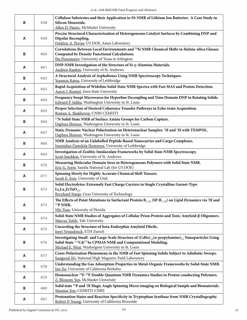
B 458Cellulose Substrates and their Application to SS-NMR of Lithium Ion Batteries: A Case Study in Silicon Monoxide. Allen D. Pauric, McMaster University
A 459Precise Structural Characterization of Heterogeneous Catalyst Surfaces by Combining DNP and Dipolar Recoupling. Frédéric A. Perras, US DOE, Ames Laboratory
B 460Correlations Between Local Environments and 29Si NMR Chemical Shifts in Hafnia-silica Glasses Computed by Density Functional Calculations. Ilia Ponomarev, University of Texas at Arlington
A 461 DNP-NMR Investigation of the Structure of Si-γ-Alumina Materials. Andrew Rankin, University of St. Andrews
B 462 A Structural Analysis of Asphaltenes Using NMR Spectroscopy Techniques. Yeasmin Ratna, University of Lethbridge
A 463 Rapid Acquisition of Wideline Solid-State NMR Spectra with Fast MAS and Proton Detection. Aaron J. Rossini, Iowa State University
B 464 Frequency Swept Microwaves for Hyperfine Decoupling and Time Domain DNP in Rotating Solids. Edward P. Saliba, Washington University in St. Louis
A 465 Proper Selection of Desired Coherence Transfer Pathways in Echo-train Acquisition. Roman A. Shakhovoy, CNRS CEMHTI
B 46615N Solid-State NMR of Surface Amine Groups for Carbon Capture. Daphna Shimon, Washington University in St. Louis
A 467 Static Dynamic Nuclear Polarization on Heteronuclear Samples: 1H and 2H with TEMPOL. Daphna Shimon, Washington University in St. Louis
B 468 NMR Analysis of an Unlabelled Peptide Based Nanocarrier and Cargo Complexes. Inumidun Damilola Shotonwa, University of Lethbridge
A 469 Investigation of Zeolitic Imidazolate Frameworks by Solid-State NMR Spectroscopy. Scott Sneddon, University of St. Andrews
B 470 Measuring Molecular Domain Sizes in Heterogeneous Polymers with Solid State NMR. Eric G. Sorte, Sandia National Lab (for US DOE)
A 471 Spinning Slowly for Highly Accurate Chemical Shift Tensors. Sarah E. Soss, University of Utah
B 472Solid Electrolytes: Extremely Fast Charge Carriers in Single Crystalline Garnet-Type Li6La3ZrTaO12. Bernhard Stanje, Graz University of Technology
A 473The Effects of Point Mutations in Surfactant Protein B1-25 (SP-B1-25) on Lipid Dynamics via 2H and 31P NMR. Nhi Tran, University of Florida
B 474 Solid-State NMR Studies of Aggregates of Cellular Prion Protein and Toxic Amyloid-β Oligomers. Marcus Tuttle, Yale University
A 475 Unraveling the Structure of beta-Endorphin Amyloid Fibrils. Joeri Verasdonck, ETH Zurich
B 476Investigating Small- and Large-Scale Structure of (CdSe)13(n-propylamine)13 Nanoparticles Using Solid-State 113Cd/77Se CPMAS NMR and Computational Modeling. Michael E. West, Washington University in St. Louis
A 477 Cross-Polarization Phenomena in the NMR of Fast Spinning Solids Subject to Adiabatic Sweeps. Sungsool Wi, National High Magnetic Field Laboratory
B 478 Understanding the Gas Adsorption Properties in Metal-Organic Frameworks by Solid-State NMR. Jun Xu, University of California Berkeley
A 479 Homonuclear 19F-19F Double Quantum NMR Dynamics Studies in Proton-conducting Polymers. Z. Blossom Yan, McMaster University
B 480 Solid state 31P and 1H Magic Angle Spinning Micro-imaging on Biological Sample and Biomaterials. Maxime Yon, CEMHTI-CNRS
A 481 Protonation States and Reaction Specificity in Tryptophan Synthase from NMR Crystallography. Robert P. Young, University of California Riverside
25
et al.: 58th RMCMR Final Program and Abstracts
Published by Digital Commons @ DU, 2016

25
EPR ABSTRACTS
100 Time-dependent Photo-EPR Applied to Point Defects in Crystals: Limitations and Applications. W.R. Willoughby1, J. Dashdorj1, M.E. Zvanut1, M. Bockowski2 1. University of Alabama at Birmingham, Birmingham AL 2. Institute of High Pressure Physics, Warsaw, Poland
Optical excitation is commonly used in concert with EPR to create the paramagnetic state of centers in a wide range of materials from complex polymers to highly ordered crystals. However, except for the pioneering studies in the early 80’s, there are few reports of time-dependent photo-EPR of point defects in crystalline semiconductors. The scant use is caused, in part, by the limited time response imposed by the commonly used 100 kHz magnetic field modulation and the small number of EPR-active centers inherent to semiconductors. Recently, however, we have analyzed time-dependent data in several types of semiconductors using standard phase-sensitive detection. The results of one such study will be summarized here, emphasizing both the utility and limitations of the technique. The system to be discussed is Be-doped GaN, in which the Be acts a deep acceptor providing electrical compensation as well as yellow luminescence. Charge trapping parameters such as defect level (ionization energy) and capture coefficients are critical to understanding the effectiveness of Be as a dopant. The samples are 0.1 um thick, 0.5 cm2 GaN platelets doped with 1017 cm-3 Be, and are measured at 3.5 K. The time-dependent photo-EPR data is analyzed using three coupled differential equations based on charge transfer among the Be acceptor, unintentionally added O donors, and the conduction and valence bands. Analysis of the results obtained during illumination with selected photon energies yields an optical threshold of 2.8 eV, which accounts for the effectiveness of the dopant as a compensator, and relaxation energy of 0.5 eV, which accounts for the yellow luminescence seen at 2.2 eV. The significance of these results and complications arising from more heavily Be-doped samples will be reviewed, along with consideration of the limitations imposed by the 100 kHz modulation detection system.
The work is supported by NSF/DMR-1308446.
EPR ORAL SESSIONMary Ellen Zvanut, University of Alabama Birmingham, 1300 University Blvd, Birmingham, AL, 35294-1170, US E-mail: [email protected]
101 Suppressing Spin-spin Relaxation in Silicon Carbide with Natural Isotope Abundance using Dynamic Decoupling. A. Sperlich,1 D. Simin,1 H. Kraus,1 T. Ohshima,2 V. Dyakonov,1,3 G. V. Astakhov1 1. Experimental Physics VI, Julius-Maximilian University of Würzburg, 97074 Würzburg, Germany 2. Japan Atomic Energy Agency, Takasaki, Gunma 370-1292, Japan 3. Bavarian Center for Applied Energy Research (ZAE Bayern), 97074 Würzburg, Germany
The vacancy-related color centers in the CMOS-compatible material silicon carbide (SiC) are perspective for chip-scale quantum technologies based on ensembles as well as on single centers. Similar to the spin S = 1 nitrogen-vacancy (NV) defect in diamond – which has become a standard solid-state system for quantum applications under ambient conditions – the silicon vacancy (VSi) in SiC possesses selectively addressable spin states through optically detected magnetic resonance (ODMR) [1,2]. In order to achieve long-lived electronic quantum memory in solid state, expensive and non-trivial engineering with spin-free nuclear isotopes, such as silicon-28 or carbon-12, is usually required. We investigate the coherence time properties of the Si-vacancies in a commercial 4H-SiC wafer with natural isotope abundance using the pulsed-ODMR technique [3]. Implementing the common Rabi-, Ramsey-, Spin-Echo- and CPMG-sequences, we can precisely measure spin-lattice (T1) and spin-spin (T2) relaxation times. The measurements are not only conducted at ambient conditions, but also at different temperatures and in different magnetic fields. In particular, the coherent spin properties of the VSi defect are investigated in the temperature range from 10K to 300K and at magnetic field strengths of up to 30mT. Remarkably long spin-spin relaxation times in the millisecond range are attained through the suppression of heteronuclear spin cross-talking by applying a magnetic field above ten millitesla in combination with dynamic decoupling (CPMG) from the nearly separated nuclear spin baths. The fundamental limit, given by the spin-lattice relaxation time, tends to ten seconds at cryogenic temperatures.
1. D. Riedel, F. Fuchs, H. Kraus, S. Väth, A. Sperlich, V. Dyakonov, A. A. Soltamova, P. G. Baranov, V. A. Ilyin, and G. V. Astakhov, Phys. Rev. Lett., 2012, 109, 226402.
2. H. Kraus, V. A. Soltamov, D. Riedel, S. Väth, F. Fuchs, A. Sperlich, P. G. Baranov, V. Dyakonov, and G. V. Astakhov, Nature Physics, 2013, 10, 157.
26
Rocky Mountain Conference on Magnetic Resonance, Vol. 58 [2016], Art. 1
https://digitalcommons.du.edu/rockychem/vol58/iss1/1

26
3. D. Simin, H. Kraus, A. Sperlich, T. Ohshima, G. V. Astakhov and V. Dyakonov, arXiv:1602.05775, 2016
EPR ORAL SESSIONAndreas Sperlich, University of Würzburg, Am Hubland, Würzburg, Bayern, 97074, DE E-mail: [email protected]
102 Silicon Carbide Magnetoresistive Magnetometer with Electrically Detected Magnetic Resonance Self-calibration Feature for Space Science Application. Corey J. Cochrane1, Jordana Blacksberg1, Philip G. Neudeck2, David J. Spry2, Mark A. Anders3, Patrick M. Lenahan3 1. NASA Jet Propulsion Laboratory, California Institute of Technology, Pasadena, CA 91109 2. NASA Glenn, Cleveland, OH 44135 3. Pennsylvania State University, University Park, PA 16802
It’s commonly known that the wide bandgap nature of the silicon carbide (SiC) semiconductor allows it to be leveraged in electronics exposed to harsh environments. The material therefore has much potential for space missions where very high temperature and high radiation environments are commonly encountered. This work entails the development of a SiC based magnetometer which leverages intrinsic defects to sense near zero magnetic fields in space. The deep level defects give rise to a magneto resistive response referred to as zero-field spin dependent recombination (SDR). The SDR phenomenon allows a change in device current to be measured with changes in external magnetic field. The magnetometer has the ability to self-calibrate either by measuring spacing of the symmetrically spaced zero-field spin interactions (spin-spin and or hyperfine) or by measuring the field/frequency SDR response induced by low-field electrically detected magnetic resonance (EDMR). Leveraging the pn junction of a SiC power MOSFET designed for high power applications, the magnetometer currently exhibits a sensitivity of about 400 nT/sqrt(Hz). However, a future design of the device using custom materials, optimized geometry and fabrication will allow sensitivities to be pushed below the 1nT/sqrt(Hz) threshold, making the technology competitive to heritage designs such as fluxgate and optically pumped He magnetometers flown on most missions in space.
C.J. Cochrane, P.M. Lenahan, JAP, 112, 12, 2012. W.J. Baker, et. al., Nat. Comm., 3, 898, (2012).
EPR ORAL SESSIONCorey J Cochrane, California Institute of Technology, Jet Propulsion Laboratory, 4800 Oak Grove Dr, Pasadena, CA, 91109, US E-mail: [email protected]
103 Measurement of Paramagnetic Spin Concentration in a Solid-state System using Double Electron-electron Resonance. Viktor Stepanov,1 Susumu Takahashi1,2 1. Department of Chemistry, University of Southern California, Los Angeles, CA 90089 2. Department of Physics, University of Southern California, Los Angeles, CA 90089
Diamond has been extensively investigated recently due to a wide range of potential applications of nitrogen-vacancy (NV) defect centers existing in a diamond lattice. The applications include magnetometry and quantum information technologies, and long decoherence time (T2) of NV centers is critical for those applications. Although it has been known that T2 highly depends on the concentration of paramagnetic impurities in diamond, precise measurement of the impurity concentration remains challenging. Here we demonstrate a method to determine a wide range of the nitrogen concentration (n) in diamond using a wide-band high-frequency electron spin resonance and double electron-electron resonance spectrometer. Moreover, we investigate T2 of the nitrogen impurities and show the relationship between T2 and n. The method developed in this work is applicable for various spin systems in solid and implementable in nanoscale magnetic resonance spectroscopy with NV centers to characterize the concentration of the paramagnetic spins within a microscopic volume.1
1. V. Stepanov and S. Takahashi, submitted (2016), arXiv:1603.07404
EPR POSTER SESSIONViktor Stepanov, University of Southern California, , 840 Downey Wat, Suite LJS 151, Los Angeles, CA,90089,USA Tel: 213-740-1793, E-mail: [email protected]
27
et al.: 58th RMCMR Final Program and Abstracts
Published by Digital Commons @ DU, 2016

27
104 Hyperfine Interactions in Silicon. M.L. Guy, L. Zhu, K. van Schooten, C. Ramanathan Department of Physics and Astronomy, Dartmouth College, Hanover NH 03755
Silicon is a technologically versatile material – ubiquitous in microelectronics and solar cells, a promising platform for spin-based quantum devices and computers, and in nanoparticle form a, viable contrast agent in magnetic resonance imaging. In this talk I will present two examples of recent work from our group studying hyperfine interactions of electron spins in silicon. First I will describe the use of frequency-modulated microwaves in W-band dynamic nuclear polarization experiments to characterize local hyperfine interactions between paramagnetic defects at the surface of silicon microparticles and local nuclear spins. Next, I will discuss the optical hyperpolarization of phosphorus donors in silicon and the optical frequency dependence of EDMR signals in silicon.
EPR ORAL SESSIONChandrasekhar Ramanathan, Dartmouth College, 6127 Wilder Laboratory, Hanover, NH, 03755, US E-mail: [email protected]
105 Probing Giant Magnetic Anisotropies in Mononuclear Single-molecule Magnets. Stephen Hill,1,2 Lakshmi Bhaskaran,1,2 Komalavalli Thirunavukkuarasu,2,3 Katie Marriott,4 Mark Murrie,4 Mohamed Saber,5 Kim Dunbar5 1. Department of Physics, Florida State University, Tallahassee, FL 32306, USA 2. National High Magnetic Field Laboratory, Florida State University, Tallahassee, FL 32310, USA 3. Department of Physics, Florida A&M University, Tallahassee, FL32307, USA 4. WestCHEM, School of Chemistry, University of Glasgow, Glasgow, G12 8QQ, UK 5. Department of Chemistry, Texas A&M University, College Station, TX 77842, USA
The development and study of molecular nanomagnets has witnessed tremendous progress in recent years, with potential applications on the horizon. Of particular interest are so-called single-molecule magnets (SMMs) that display slow magnetic relaxation below a characteristic blocking temperature, TB, due to a combination of a large magnetic moment and appreciable spin-orbit (SO) anisotropy. Early efforts aimed at increasing TB focused on polynuclear clusters and maximization of the molecular spin state, S. However, this becomes challenging for large clusters whilst simultaneously maintaining the molecular anisotropy. Thus, a more direct route to increasing TB involves optimization of the magnetic anisotropy, albeit for simpler molecules in which one can exert synthetic control over the ligand field (LF). In particular, certain transition metals residing in high-symmetry coordination environments can experience orbital degeneracies and very strong first-order contributions to their SO anisotropy. This talk will highlight recent work involving S = 1 NiII [1] and VIII [2] complexes subjected to rigid trigonal coordination environments in the solid state that are relatively stable against symmetry lowering Jahn-Teller distortions [3]. The resulting giant anisotropies associated with these species have been measured using very high-field (up to 35 T) EPR techniques. In particular, analysis of results for a trigonal bipyramidal (TBP) [NiIICl3(Me-dabco)2] complex on the basis of a spin-only Hamiltonian suggest an axial D parameter exceeding –400 cm-1, which is close to the SO coupling parameter, λ = 668 cm-1, for NiII, suggesting an orbitally degenerate ground state. However, the spin-only description cannot work in this limit, therefore necessitating the development of a model that includes the orbital moment. A qualitative theoretical approach will be described that takes into account a full description of crystal field, electron-electron repulsion and spin-orbit coupling effects on the ground state of NiII in a TBP coordination geometry. The model provides both qualitative and quantitative agreement with the high-field EPR experiments, hence validating its use for spectroscopic studies of orbitally degenerate molecular nanomagnets.
[1] K. E. R. Marriott, L. Bhaskaran, C. Wilson, M. Medarde, S. T. Ochsenbein, S. Hill and M. Murrie, Chem. Sci., 2015, 6, 6823-6828.
[2] M. R. Saber, M. Atanasov, L. Bhaskaran, K. Thirunavukkuarasu, S. Hill, F. Neese and K. R. Dunbar (in preparation).
[3] R. Ruamps, R. Maurice, L. Batchelor, M. Boggio-Pasqual, R. Guillot, A.-L. Barra, J. Liu, E.-E. Bendief, S. Pillet, S. Hill, T. Mallah and N. Guihery, J. Am. Chem. Soc., 2013, 135, 3017-3026.
EPR ORAL SESSIONStephen Hill, NHMFL and FSU Physics, 1800 E Paul Dirac Dr, Tallahassee, FL, 32310, US Tel: 850-644-1647, E-mail: [email protected]
28
Rocky Mountain Conference on Magnetic Resonance, Vol. 58 [2016], Art. 1
https://digitalcommons.du.edu/rockychem/vol58/iss1/1

28
106 Triplet Exciton Generation in Materials for Organic Solar Cells. Felix Kraffert, Robert Steyrleuthner, Jan Behrends Berlin Joint EPR Lab, Fachbereich Physik, Freie Universität Berlin, Berlin, Germany.
Using time-resolved EPR spectroscopy in conjunction with optical excitation we study charge separation in absorber materials for organic solar cells. When blended with the fullerene-based electron acceptor PCBM, two prominent derivatives of the low-bandgap polymer PCPDTBT differing by the bridging atom (carbon or silicon) exhibit different charge separation yields. While the EPR signatures of photogenerated positive polarons in C- and Si-bridged PCPDTBT are virtually identical, significant differences are observed with respect to the spin-relaxation behaviour. The spin-lattice relaxation time of positive polarons in C-PCPDTBT at low temperature (T = 80 K) is found to be more than two orders or magnitude longer than in the Si-bridged polymer derivative. This surprisingly slow relaxation can be rationalized by polarons trapped in defect states that seem to be absent (or are present in a substantially smaller concentration) in blends comprising Si-PCPDTBT. Transient EPR signals attributed to charge transfer (CT) states at the donor/acceptor interface and separated polarons are smaller in the blends with C-PCPDTBT as compared to those with the silicon-bridged polymer. We propose that triplet formation occurs via the CT state, thus diminishing the probability that the CT state forms free charge carriers in blends of C-PCPDTBT with PCBM. This hypothesis is confirmed by direct detection of triplet excitons in C-PCPDTBT:PCBM blends. The shape of the transient EPR spectra reveals that the triplet excitons are, in contrast to those formed in pristine polymer films, not generated by direct intersystem crossing, but result from back electron transfer through CT state recombination. The strong triplet signal is not observed in blends containing the Si-bridged polymer, indicating efficient singlet exciton splitting and subsequent charge carrier separation at the Si-PCPDTBT/PCBM interface.1
1. F. Kraffert, R. Steyrleuthner, S. Albrecht, D. Neher, M. C. Scharber, R. Bittl and J. Behrends, J. Phys. Chem. C, 2014, 118, 28482.
EPR ORAL SESSIONJan Behrends, Freie Universität Berlin, Arnimallee 14, Berlin, Berlin, D-14195, DE Tel: 0049 30 838 54266, E-mail: [email protected]
107 Spin-orbit Coupling in Conjugated Polymers. H. Malissa,1 R. Miller,1 D.L. Baird,1 S. Jamali,1 G. Joshi,1 P. Klemm,2 S. Bange,2 M. Bursch,3 S. Grimme,3 J. van Tol,4 J.M. Lupton,1,2 C. Boehme1 1. University of Utah, Department of Physics and Astronomy, Salt Lake City, UT 84112 2. Universität Regensburg, Institut für Experimentelle und Angewandte Physik, Regensburg, 93051 Germany 3. Universität Bonn, Institut für Physikalische und Theoretische Chemie, Bonn, 53115 Germany 4. National High Magnetic Field Laboratory, Tallahassee, FL 32310
Conjugated polymers consist of only light elements and are generally considered to have negligible spin-orbit coupling (SOC). However, even a small contribution of SOC can have a large impact on charge carrier pair spin statistics and thus on the magneto-opto-electronic properties of materials used for organic light emitting diodes (OLEDs) and other devices. The sole source of SOC is assumed to be inter-site variations of the g-factor due to structural disorder of the polymer and it is observed indirectly as the so-called ∆g mechanism in magneto-electroluminescence and magneto-resistance measurements.1 We observe the effects of SOC in electrically detected magnetic resonance (EDMR) measurements on OLEDs as a magnetic field dependent line broadening mechanism that is already detectable at fields below 700 mT where the line width is still dominated by the unresolved hyperfine couplings to the surrounding hydrogen nuclei.2,3 At much higher magnetic fields, up to 12 T, EDMR line widths are increasingly governed by the SOC. In this regime, line widths scale with the magnetic field and an increasingly asymmetric line shape emerges due to the rhombic g-tensors of both charge carriers. Preliminary quantum chemistry calculations of open-shell model systems implicitly produce g-tensors that are in excellent agreement with the measurements and suggest that the effects of disorder or g-tensor-strain are negligible in these systems.
This work was supported by the US Department of Energy, Office of Basic Energy Sciences, Division of Materials Sciences and Engineering under Award #DE-SC0000909. Part of this work was performed at the National High Magnetic Field Laboratory, which is supported by National Science Foundation Cooperative Agreement No. DMR-1157490 and the State of Florida.
1. Schellekens et al., Phys. Rev. B, 2011, 84, 075204.2. Baker et al., Nat. Commun., 2012, 3, 898.3. Joshi et al., arXiv:1603.03807 [cond-mat.mes-hall], 2016.
EPR ORAL SESSIONHans Malissa, University of Utah, 115 South 1400 East #201, Salt Lake City, UT, 84112, USA Tel: 801-585-0485, E-mail: [email protected]
29
et al.: 58th RMCMR Final Program and Abstracts
Published by Digital Commons @ DU, 2016

29
108 Effect of Spin-orbit Interaction and Topological Gap for ESR Spectra in Low-dimensional Organic Conductors. Mizue Asada,1 Toshikazu Nakamura1,2 1. Institute for Molecular Science 2. Graduate University for Advanced Studies
Recently theoretical investigation by Oshikawa proposed that strong spin-orbit interaction causes additional ESR satellite signal and g-shift in 1D metallic system. So, we reexamined detailed X-band ESR spectra (satellite, line-shape, line-width) for low-dimensional metallic systems. Firstly, we focused on a 1D organic conductor (TMTSF)2ClO4, which shows stable metallic state down to 1K. Since the TMTSF molecule contains heavy selenium elements, there are considerable spin-orbit interactions. When we apply the static magnetic field along 1D conducting direction (H0//a), a tiny satellite peak was appeared below 12K. In the case of H0//c*, we cannot observed any satellite peak. We also performed 2D metallic system, BEDT-TTF salts. A series of BEDT-TTF salts with low-symmetry shows anomalous g-shift at low-temperatures. We discuss relationship between the tiny band gap and anomalous ESR behavior observed in low-dimensional metallic systems.
1. M. Oshikawa, 54th SEST (Japan ESR society) annual meeting SB-05 (2015.11).2. M. Oshikawa and I. Affleck: Phys. Rev. Lett. 82 (1999) 5136, and references therein.
EPR ORAL SESSIONToshikazu Nakamura, Institute for Molecular Science, 38 Nishi-Gonaka, Myodaiji, Okazaki, Aichi, 444-8585, JP Tel: 81-564-55-7381, E-mail: [email protected]
109 Spin Dynamics of TAPD-MP Ar-C60 Spin Correlated Radical Pair. Naitik A. Panjwani,1 Julien B. Kelber,1,2 Di Wu2, Enrico Salvadori,1 Christopher W.M. Kay,1 Harry L. Anderson,2 John J.L. Morton1 1. London Centre for Nanotechnology, University College London, UK 2. Department of Chemistry, University of Oxford, UK
Molecular triads which undergo photo-induced electron transfer have a wide range of applications, from understanding and mimicking energy transfer in natural photosystems, to molecular spintronics and the understanding of magnetoreception in migratory birds such as the European robin1,2. Our work involves the study of optically generated molecular spin states as a tool to hyperpolarise, entangle and measure nuclear spins in molecules, while leaving the molecule in a diamagnetic ground state in order to minimise the long-term impact on nuclear spin decoherence3,4. Current work involves the study of the Donor-Bridge-Acceptor molecules TAPD-MP-C60 where M= H2 , Zn, Cd, for the primary purpose of using the charge-separated state (CSS) as source of a long-lived optically-generated electron spin, to interact with nearby nuclear spins and mediate coupling between them. We present studies on these molecules employing time-resolved and pulsed, electron paramagnetic resonance (EPR) and double resonance methods (ENDOR) combined with pulsed laser excitation, to extract the spin Hamiltonian parameters and to quantitatively understand the charge and spin dynamics.
1. T. Miura and M.R. Wasielewski. J. Am. Chem. Soc., 133, 2844–2847 (2011). 2. T. Ritz, S. Adem and K. Schulten. Biophys. J., 78, 707–718 (2000). 3. V. Filidou, S. Simmons, S.D. Karlen, F. Giustino, H.L. Anderson, and J.J.L. Morton. Nat. Phys., 8,
596–600 (2012). 4. M. Schaffry et al. Phys. Rev. Lett., 104, 200501 (2010).
EPR ORAL SESSIONNaitik A Panjwani, University College London, London Centre for Nanotechnology, 17-19 Gordon St, London, Greater London, WC1H 0AH, GB E-mail: [email protected]
30
Rocky Mountain Conference on Magnetic Resonance, Vol. 58 [2016], Art. 1
https://digitalcommons.du.edu/rockychem/vol58/iss1/1

30
110 Structural Information from Hyperfine Couplings in Iron Catalysts. Inés Garcia-Rubio, Centro Universitario de la Defensa, Ctra de Huesca s/n, 50090 Zaragoza, Spain. Laboratory of Physical Chemistry, ETH Zurich, CH-8093 Zurich, Switzerland
Iron(III) has very interesting catalytic properties that can be modulated by its molecular environment. For this reason, one can find in nature or synthesize in the laboratory a myriad of different iron-based catalysts,either monoatomic or forming iron clusters, in low-symmetry sites or coordinated to highly symmetric organic chelators. In all cases, the properties, shape and spin density of the d-orbitals are crucial in determining the catalytic properties of iron. In this contribution we will illustrate how the determination of the spin state and g-values through EPR methods yields useful electronic information using two examples of iron catalysts with very different characteristics. One of the catalysts is an artificial heterocubane with a [Fe4N4] core that can reversibly store up to four electrons at very negative potentials. The second system is the protein cytochrome c550, where a single iron ion is coordinated by a four-fold organic ring of porphyrin. This cofactor, called heme group, is ubiquitous in life systems where it performs very different functions (catalytic, transport…) In both cases the study of the hyperfine interactions with magnetic nuclei in close vicinity of the iron yields also useful structural information and allows mapping the electron spin density distribution. The strategy for experimental determination of the hyperfine couplings of nitrogen and/or hydrogen nuclei will be shown for both systems. In the case of the heterocubane, the interpretation of hyperfine data reveals that the unpaired electron is predominantly located at one Fe center with some delocalization to the coordinated olefin moiety (ρ ≈ 18%) and to the nitrogen atom in the same chelating ligand (ρ ≈ 0.8%). For cytochrome c550 hyperfine couplings reveal electronic details related to the structure that can possibly be discussed in terms of the biological function.
EPR ORAL SESSIONInés García-Rubio, Centro Universitario de la Defensa, Carretera de Huesca s/n, Zaragoza, Zaragoza, 50090, ES E-mail: [email protected]
111 263 GHz Pulse EPR Reports on Proton-coupled Electron Transfer Through the Subunit Interface of E. Coli Ribonucleotide Reductase Ia. T.U. Nick,1 K. Ravichandran,2 M. Kasanmascheff,1 J. Stubbe,2 M. Bennati1,3 1. Electron Spin Resonance Group, Max Planck Institute for Biophysical Chemistry, Am Fassberg 11, 37077 Göttingen, Germany 2. Dept. of Chemistry, Massachusetts Institute of Technology, 77 Massachusetts Avenue, Cambridge, MA 02139, USA 3. Department of Chemistry, University of Göttingen, Tammannstr. 2, 37077 Göttingen, Germany
Ribonucleotide reductases (RNR) connect the RNA and DNA world by reducing all four essential ribonucleotides to deoxyribonucleotides. In E. coli RNR Ia, the strictly controlled radical chemistry starts in the β subunit at the “stable” tyrosyl radical (Y122•) di-iron cofactor. A long-range radical transfer from subunit β to α generates a putative catalytic active cysteine radical (C439•) in the α subunit that initiates catalysis. Successive studies showed that Y356•(β), Y731•(α) and Y730•(α) are intermediate steps of this inter-subunit (α:β) proton-coupled electron-transfer (PCET). Conformational gating hinders the direct observation of these transient radicals. Therefore, site specific incorporation of the unnatural 3-amino-tyrosine (NH2Y) and 2,3,5-fluoro-tyrosine (F3Y) was used to trap all radical intermediates.1-2 Our 263 GHz pulsed-EPR spectroscopy delivers highly resolved g-values, which are correlated to the individual hydrogen (H) bond network based on ENDOR spectroscopy and DFT models of radical intermediates. Within the α subunit short PCET steps with moderate to strong H bonds perpendicular to the ring were found, consistent with a concerted and collinear PCET.3-4 At the structurally ill-defined interface EPR spectra of β-Y356• with different mutants of the α subunit (α-Y730F and α-Y731F) reveal that the presence of α-Y731 influences the electrostatic environment around β‑Y356•. This supports the proposal of an H bond network enabling the proton transfer between the two subunits.5 Furthermore, it demonstrates an indirect communication between the subunits.
1. Minnihan, Seyedsayamdost, Uhlin, Stubbe, Am. Chem. Soc. 2011, 133, 9430-9440.2. Minnihan, Young, Schultz, Stubbe, Am. Chem. Soc. 2011, 133, 15942-15945.3. Nick, Lee, Koßmann, Neese, Stubbe, Bennati, Am. Chem. Soc. 2015, 137, 289-298.4. Argirević, Riplinger, Stubbe, Neese, Bennati, Am. Chem. Soc. 2012, 134, 17661-17670.5. Kasanmascheff, Lee, Nick, Stubbe, Bennati, Chemical Science 2016, 7, 2170.
EPR ORAL SESSIONThomas U Nick, Max Planck Institute for Biophysical Chemistry, Am Fassberg 11, Goettingen, Lower Saxony, 37073, DE E-mail: [email protected]
31
et al.: 58th RMCMR Final Program and Abstracts
Published by Digital Commons @ DU, 2016

31
112 The Composition and Structure of the Inorganic Core of Intermediate X(WT) and X(Y1212F) of E. coli Ribonucleotide Reductase. Peter E. Doan,1 Muralidharan Shanmugam,2 Andrei Astachkine,3 Jo Anne Stubb,4 Brian M. Hoffman1 1. Northwestern University, Department of Chemistry, Evanston, IL 60208-3113 2. Manchester Institute of Biotechnology, The University of Manchester, Manchester, M1 7DN, UK 3. Department of Chemistry and Biochemistry, University of Arizona, Tucson, AZ 85721 4. Department of Chemistry, MIT, Cambridge, MA 02139-4307
Activation of the diferrous center of the β2 (R2) subunit of the class 1a Escherichia coli ribonucleotide reductases (RNR) by reaction with O2 followed by one-electron reduction yields a spin-coupled, paramagnetic Fe(III)/Fe(IV) intermediate, denoted X, whose identity has been sought by multiple investigators for over a quarter century. To determine the composition and structure of X, the present study has applied 57Fe, 14,15N, 17O and 1H ENDOR measurements combined with quantitative measurements of 17O and 1H EPR line broadening studies to WT X, which is very short-lived, and to X prepared with the Y122F mutant, which has a lifetime of many seconds. Previous studies have established that over several seconds the as-formed X(Y122F) relaxes to an equilibrium structure. This report focuses on the relaxed structure and the differences between this relaxed structure and the structure of both WT X and X(Y122F) at short quenching times that are revealed by EPR, ENDOR, and ESEEM techniques.
EPR ORAL SESSIONPeter E Doan, Northwestern University, Department of Chemistry, 2145 Sheridan Road, Evanston, IL, 60626, USA Tel: 847-491 4488, E-mail: [email protected]
113 Mechanistic Investigations on Electron Bifurcation by EPR Spectroscopy. David W. Mulder,1 Carolyn E. Lubner,1 Gerti J. Schut,2 Mike W. W. Adams,2 Oleg A. Zadvornyy,3 John W. Peters,3 David P. Jennings,4 Anne K. Jones,4 Paul W. King.1 1. National Renewable Energy Laboratory, Biosciences Center, Golden, CO 80401 2. University of Georgia, Department of Biochemistry and Molecular Biology, Athens, GA 30602 3. Montana State University, Department of Chemistry and Biochemistry, Bozeman, MT 59717 4. Arizona State University, School of Molecular Sciences, Tempe, AZ 85276
Electron bifurcation, a process for coupling endergonic and exergonic reactions to overcome thermodynamic barriers, is considered the third mechanism of biological energy conservation and results in the efficient coupling of electrochemical potential to chemical bond formation.1,2 Overall, the mechanism of bifurcation and how bifurcating enzymes function is poorly understood. We are investigating the mechanism of flavin-based electron bifurcation in the NADH-dependent ferredoxin-NADP+ oxidoreductases, (Nfn), which catalyze the reversible reduction of NADP+ with reduced ferredoxin and NADH. Nfn contains two electron-transfer pathways both of which are comprised of flavins and FeS clusters.3 EPR spectroscopy in conjunction with x-ray crystallographic and other biophysical techniques, are being used to investigate the oxidation-reduction properties of these centers and how they facilitate gating of electron-transfer to respective pathways. The results reveal that two unique, site-differentiated FeS clusters, Cys3Asp [2Fe-2S] and Cys3Glu[4Fe-4S], play key roles in the process through tuning of midpoint potentials and coupling with other redox centers. This presentation will summarize how these features work in concert with key structural properties of the enzyme to achieve bifurcation.
Supported by the Biological and Electron Transfer and Catalysis (BETCy) EFRC, an Energy Frontier Research Center funded by the U.S. Department of Energy, Office of Science (DE-SC0012518).
1. Peters et al. Curr. Opin. Chem., Biol., 2016, 31, 146.2. Buckel and Thauer. BBA Bioenerg., 2013, 1827, 94. 3. Demmer et al. J. Biol. Chem., 2015, 290, 21985.
EPR ORAL SESSIONDavid W Mulder, National Renewable Energy Laboratory, 16253 Denver West Pkwy, Biosciences Center, Golden, CO, 80401, USA Tel: 303-384-7486, E-mail: [email protected]
32
Rocky Mountain Conference on Magnetic Resonance, Vol. 58 [2016], Art. 1
https://digitalcommons.du.edu/rockychem/vol58/iss1/1
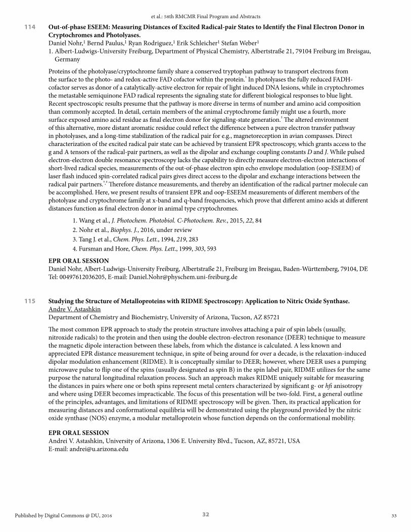
32
114 Out-of-phase ESEEM: Measuring Distances of Excited Radical-pair States to Identify the Final Electron Donor in Cryptochromes and Photolyases. Daniel Nohr,1 Bernd Paulus,1 Ryan Rodriguez,1 Erik Schleicher1 Stefan Weber1 1. Albert-Ludwigs-University Freiburg, Department of Physical Chemistry, Albertstraße 21, 79104 Freiburg im Breisgau,
Germany
Proteins of the photolyase/cryptochrome family share a conserved tryptophan pathway to transport electrons from the surface to the photo- and redox-active FAD cofactor within the protein.¹ In photolyases the fully reduced FADH- cofactor serves as donor of a catalytically-active electron for repair of light induced DNA lesions, while in cryptochromes the metastable semiquinone FAD radical represents the signaling state for different biological responses to blue light. Recent spectroscopic results presume that the pathway is more diverse in terms of number and amino acid composition than commonly accepted. In detail, certain members of the animal cryptochrome family might use a fourth, more surface exposed amino acid residue as final electron donor for signaling-state generation.² The altered environment of this alternative, more distant aromatic residue could reflect the difference between a pure electron transfer pathway in photolyases, and a long-time stabilization of the radical pair for e.g., magnetoreception in avian compasses. Direct characterization of the excited radical pair state can be achieved by transient EPR spectroscopy, which grants access to the g and A tensors of the radical-pair partners, as well as the dipolar and exchange coupling constants D and J. While pulsed electron-electron double resonance spectroscopy lacks the capability to directly measure electron-electron interactions of short-lived radical species, measurements of the out-of-phase electron spin echo envelope modulation (oop-ESEEM) of laser flash induced spin-correlated radical pairs gives direct access to the dipolar and exchange interactions between the radical pair partners.³,⁴ Therefore distance measurements, and thereby an identification of the radical partner molecule can be accomplished. Here, we present results of transient EPR and oop-ESEEM measurements of different members of the photolyase and cryptochrome family at x-band and q-band frequencies, which prove that different amino acids at different distances function as final electron donor in animal type cryptochromes.
1. Wang et al., J. Photochem. Photobiol. C-Photochem. Rev., 2015, 22, 842. Nohr et al., Biophys. J., 2016, under review3. Tang J. et al., Chem. Phys. Lett., 1994, 219, 283 4. Fursman and Hore, Chem. Phys. Lett., 1999, 303, 593
EPR ORAL SESSIONDaniel Nohr, Albert-Ludwigs-University Freiburg, Albertstraße 21, Freiburg im Breisgau, Baden-Württemberg, 79104, DE Tel: 00497612036205, E-mail: [email protected]
115 Studying the Structure of Metalloproteins with RIDME Spectroscopy: Application to Nitric Oxide Synthase. Andre V. Astashkin Department of Chemistry and Biochemistry, University of Arizona, Tucson, AZ 85721
The most common EPR approach to study the protein structure involves attaching a pair of spin labels (usually, nitroxide radicals) to the protein and then using the double electron-electron resonance (DEER) technique to measure the magnetic dipole interaction between these labels, from which the distance is calculated. A less known and appreciated EPR distance measurement technique, in spite of being around for over a decade, is the relaxation-induced dipolar modulation enhancement (RIDME). It is conceptually similar to DEER; however, where DEER uses a pumping microwave pulse to flip one of the spins (usually designated as spin B) in the spin label pair, RIDME utilizes for the same purpose the natural longitudinal relaxation process. Such an approach makes RIDME uniquely suitable for measuring the distances in pairs where one or both spins represent metal centers characterized by significant g- or hfi anisotropy and where using DEER becomes impracticable. The focus of this presentation will be two-fold. First, a general outline of the principles, advantages, and limitations of RIDME spectroscopy will be given. Then, its practical application for measuring distances and conformational equilibria will be demonstrated using the playground provided by the nitric oxide synthase (NOS) enzyme, a modular metalloprotein whose function depends on the conformational mobility.
EPR ORAL SESSIONAndrei V. Astashkin, University of Arizona, 1306 E. University Blvd., Tucson, AZ, 85721, USA E-mail: [email protected]
33
et al.: 58th RMCMR Final Program and Abstracts
Published by Digital Commons @ DU, 2016

33
116 High-Field EPR Studies on Model Dimeric MnIV Complexes. A. Ozarowski,1 D. Premužić,2 C. Pietzonka,2 M. Hołyńska2 1. National High Magnetic Field Laboratory, Tallahassee, FL 32310, USA 2. Fachbereich Chemie, Philipps-Universität, 35043 Marburg, Germany
MnIV complexes are important in research on natural Photosystem II (PSII) and in design of artificial PSII. Dimeric MnIV systems have been postulated as intermediates in the catalytic water oxidation relevant to artificial photosynthesis. In this work, weakly antiferromagnetic dimeric tetraazaadamantane MnIV complexes were studied by high-field EPR, up to 15 T at frequencies up to 640 GHz. In a dialkoxo-bridged complex, EPR spectra coming from the coupled spin states with S=1, 2 and 3 were initially interpreted in terms of the “giant spin” Hamiltonian (Figure 1). The D and E parameters in the S=1 state were -2.3 and -0.75 cm-1, respectively, while in the S=3 state +0.42 and +0.10 cm-1 were determined. Contributions to the zero field splitting (zfs) tensor due to the individual ions as well as to the anisotropic metal-metal interactions were extracted from the above experimental results and were subsequently used to simulate spectra using the full spin Hamiltonian expressed in spins of two Mn ions. Diagonalisation of the 16x16 spin Hamiltonian matrices was employed in these simulation procedures. The zero-field splitting in the dimers was found to be dominated by the single-ion zfs contribution. The anisotropic metal-metal interactions appear to be almost exclusively of the magnetic dipolar nature.
Supported by DFG HO 5055/3-1 project (Germany). NHMFL is funded by the NSF through the Cooperative Agreement No. DMR-1157490, the State of Florida and the DOE.
EPR ORAL SESSIONAndrew Ozarowski, National High Magnetic Field Laboratory, Florida State University, 1800 East Paul Dirac Drive, Tallahassee, Florida, 32310, USA Tel: 8506445996, E-mail: [email protected]
117 EPR-active Molecular pH Probes at a Protein-Lipid Interface: Turning Electrical Charges On and Off. Tatyana Smirnova, Erkang Ou, Matthew Donohue, Maxim A. Voynov, Alex I. Smirnov North Carolina State University, Department of Chemistry, Raleigh, NC 27695
The ionization states of individual amino acid residues of membrane proteins are difficult to decipher or assign directly in the lipid-bilayer membrane environment. The effective pK(a) values of protein groups are determined by a complex interplay between local polarity, Coulomb interactions, and a structural reorganization. The analysis is further complicated by the dearth of information about gradients in polarity, electric potentials, and hydration at the protein-membrane interface. In this work we report on developing pH-sensitive ionizable EPR labels and related methods to 1) profile a heterogeneous dielectric environment along the α-helix of a WALP peptide integrated in a lipid bilayer and 2) asses the effect of anionic lipid surface charge density on effective pK(a) of membrane-burred ionisable sidechains and 3) asses the effect of solid state support on effective pK(a) of membrane-burred ionisable sidechains The change in the protonation state of the pH-sensitive nitroxide was directly observed by EPR. Displacement of the EPR probe upon protonation, similar to that of the “snorkeling” of the charged sidechains of Lys and Arg, was directly observed by DEER and was shown to depend strongly upon the depth of the label with respect to the bilayer surface. Further, it was found that the experimentally observed dielectric constant at the membrane-protein interface is significantly higher than the values expected for the same location in the bulk membrane. The effects of the membrane surface charge density on the dielectric profile at the peptide-membrane interface and on “snorkeling” of the charged nitroxide sidechains was investigated. We have also shown that the effective pK(a) of the probe increases by 2.1 to 2.3 pK(a) units (depending on the depth of the probe) upon replacing zwitterionic PC lipids with anionic PG lipids, with almost 80% of that pK(a) shift is observed upon replacing only half of the PC lipid with PG lipids. Water penetration at the peptide-membrane interface was assessed by HYSCORE.
Supported by NSF 0843632 and 1508607 to TIS.
EPR ORAL SESSIONTatyana Smirnova, North Carolina State University, 2620 Yarbrough Dr, Raleigh, North Carolina, 27606, USA Tel: 919-513-4375, E-mail: [email protected]
34
Rocky Mountain Conference on Magnetic Resonance, Vol. 58 [2016], Art. 1
https://digitalcommons.du.edu/rockychem/vol58/iss1/1

34
118 Free Energy Landscape and Protein Configurational Fluctuation Contributions to Radical Rearrangement Catalysis in B12 -dependent Ethanolamine Ammonia-Lyase. Meghan Kohne, Chen Zhu, Umar Twahir, Kurt Warncke Department of Physics, Emory University, Atlanta, GA 30322
First-order rate constants (k) of the core chemical reaction step of substrate radical rearrangement in the B12 -dependent ethanolamine ammonia-lyase (EAL) from Salmonella typhimurium are determined by using temperature-step triggered decay of the cryotrapped substrate radical intermediate, and time-resolved, full-spectrum electron paramagnetic resonance (EPR) spectroscopy [1] over the temperature (T) range of 197≤ T ≤230 K, and from kcat values up to 295 K [2,3]. The piecewise-linear Eyring [ln(kT -1 ) versus T-1 ] dependence shows a kinetic bifurcation from native into fast and slow decay components at 220 K, and a kink in the fast decay component at 217 K. The bifurcation and kink are proposed to arise from the effective quenching of native stochastic, collective-atom protein configurational fluctuations that are coupled to the reaction. The reaction at T The native and non-native reaction channels represent two distinct dynamical paradigms for radical rearrangement catalysis in EAL, which are described by using a T-dependent free energy landscape (FEL) model. This model is cast in a deterministic kinetic model, that is used to simulate the observed decays and reproduce the Erying dependence. Electron spin-echo envelope modulation (ESEEM) spectroscopy of substrate radical- active site group interactions [4,5] are aimed at resolution of two model-predicted sub- states, and microscopic annotation of the kinetic mechanisms. The results reveal unique insights into the interplay of the FEL, activation enthalpy and entropy, and specific configurational fluctuations in the manifestation of enzyme catalysis in B12 -dependent EAL, that impact enzyme catalysis, in general.
Supported by NIH R01DK054514.
[1] M. Wang, C. Zhu, M. Kohne and K. Warncke, Meth. Enzymol. 2015, 563, Part A, 59.[2] C. Zhu and K. Warncke, Biophys. J., 2008, 95, 5890. [3] C. Zhu and K. Warncke J. Am. Chem. Soc.
2010, 132, 9610.[4] L. Sun, O. A. Groover, J. M. Canfield and K. Warncke, Biochemistry 2008 47, 5523.[5] A. M. Bovell and K. Warncke, Biochemistry 2013, 52, 1419.
EPR ORAL SESSIONMeghan Kohne, Emory University, 400 Dowman Dr, Atlanta, GA, 30307, USA E-mail: [email protected]
119 Utilizing Novel 95 GHz 2D-ESR Spectroscopy to Study Nitroxide Partitioning into the Lipid Membranes at Room Temperatures. Siddarth Chandrasekaran, Boris Dzikovski, Curt R. Dunnam, Jack H. Freed Department of Chemistry and Chemical Biology and National Biomedical Center for Advanced ESRTechnology, Cornell University, Ithaca, New York 14853, USA
Small-molecule spin probes like TEMPO have been used to study biological systems by ESR for many years. Their ESR parameters are very sensitive to the local environment and can report on its viscosity, polarity and accessibility for other paramagnetic species. In complex systems high ESR spectral resolution is crucial for obtaining this information by simultaneously observing multiple spectral components which are difficult to interpret when unresolved and overlapped. Using an example of TEMPO partitioning between the phospholipid membrane and aqueous phases we show how combining the benefits of high field (HF) ESR with two dimensional (2D) ESR provides spectral resolution which cannot be achieved by either HF-ESR or 2D-ESR alone. We present results obtained by the 2D-ELDOR (Two-Dimensional Electron-Electron Double Resonance) technique with our ACERT 95 GHz High Field High Power Pulse ESR spectrometer at biological temper! atures. We demonstrate complete separation of ESR signals from different membrane phases and show how our recent method of 2D data analysis is used to separately extract T1 & T 2 relaxation times from each phase. We utilize the advantages of the fullSc- code developed at ACERT to obtain the pure absorption spectra from the hypercomplex 2D-ELDOR data recorded. We also study by 2D-ELDOR the interaction of paramagnetic relaxants, such as oxygen and transition metal ions with spin-labeled membrane. We show that much lower concentration of the relaxant can be used to selectively remove one of the components than in CW-ESR. Finally, we discuss some new physico-chemical insights in the behavior of the system obtained by HF 2D-ELDOR such as the anomalous diffusion of oxygen in the membrane phase and the ion-membrane interactions.
EPR ORAL SESSIONSiddarth Chandrasekaran, ACERT and Cornell University, Dept of Chemistry & Chemical Biology, Baker laboratory, Ithaca, NY, 14853, USA Tel: 607-255-1153, E-mail: [email protected]
35
et al.: 58th RMCMR Final Program and Abstracts
Published by Digital Commons @ DU, 2016

35
120 Using EPR, ENDOR, and HYSCORE to Elucidate the Structure of Copper and Cobalt Pre-Catalysts. Elizabeth Papish, , Deidra L. Gerlach, Alex C. Cruce, Michael K. Bowman, Sopheavy Siek, Dalton B. Burks University of Alabama
Water oxidation can lead to a sustainable source of energy, but for water oxidation catalysts (WOCs) to be economical they must use earth abundant metals. We have reported that 6,6’-dihydroxybipyridine (6,6’-dhbp) based ligands form 2:1 copper complexes that are efficient at electrocatalytic water oxidation in aqueous base (pH = 10-14). The complexes formed were characterized by crystallography and acid dissociation constants were determined. EPR, ENDOR, and HYSCORE experiments also confirmed that the 6,6’-dhbp ligand is bound to copper over a wide pH range, and these experiments show how pH influences pre-catalyst structure. Water oxidation studies with analogous zinc and copper complexes have illustrated that both copper and proximal OH groups are necessary for water oxidation at a low overpotential and moderate rates. Furthermore, our combined work demonstrates the ways in which OH/O- groups on the bipyridine rings can hydrogen bond, support unusual binding modes, and potentially facilitate proton coupled electron transfer. This work has been published in Inorg. Chem. in 2014. We will also describe our recent efforts to make new cobalt and copper complexes as novel catalysts, and again these structures have been characterized by EPR methods.
EPR ORAL SESSIONElizabeth T Papish, University of Alabama, 250 Hackberry Lane, Tuscaloosa, AL, 35401, USA Tel: 205-348-5822, E-mail: [email protected]
125 Nanowire-Based Magnetic Resonance Imaging and Spectroscopy. Raffi Budakian Institute for Quantum Computing, University of Waterloo, University of Illinois at Urbana-Champaign
Magnetic resonance imaging (MRI) has had a profound impact on biology and medicine. Key to its success has been the unique ability to combine imaging with nuclear magnetic resonance spectroscopy—a capability that has led to a host of powerful modalities for imaging spins. Although it remains a significant challenge, there is considerable interest to extend these powerful spectroscopic and imaging capabilities to the nanometer scale. In this talk, I will discuss a new platform for force-detected magnetic resonance detection that allows us to bring many of the tools developed for NMR to the nanometer scale. In particular, I will focus on the development of optimal control theory (OCT) pulses, which incorporate average Hamiltonian theory and realize arbitrary unitary operations. I will present initial results demonstrating the use of OCT pulses for dynamical decoupling and chemical shift averaging of nanometer scale ensembles.
EPR ORAL SESSIONRaffi Budakian, University of Waterloo, 200 University Avenue West, Waterloo, ON, N2L 3G1, CA Tel: 519-888-4567, E-mail: [email protected]
126 EPR Spectroscopy of using Nitrogen-vacancy Centers in Diamond. Chathuranga Abeywardana,1 Viktor Stepanov,1 Susumu Takahashi1,2 1. Department of Chemistry, University of Southern California, Los Angeles CA 90089, USA 2. Department of Physics, University of Southern California, Los Angeles CA 90089, USA
Magnetic resonance (MR), such as nuclear magnetic resonance (NMR) and electron paramagnetic resonance (EPR), can probe the local structure and dynamic properties of various systems, making them among the most powerful and versatile analytical methods. However, their intrinsically low sensitivity precludes MR analyses of samples with very small volumes; e.g., more than 1010 electron spins are typically required to observe EPR signals at room temperature. A vast improvement in the current limits of MR will enable the imaging of structures and conformational changes of molecules in solution at the single molecule level. A nitrogen-vacancy (NV) center in diamond is a promising candidate for applications in room temperature magnetic sensing with single spin sensitivity. In this presentation, we will discuss EPR spectroscopy using NV centers in diamond. By employing EPR and double electron-electron resonance (DEER) techniques, we investigate impurities and coherence in diamond. We also demonstrate EPR of several electron spins using NV-based EPR spectroscopy.1
This work is supported by NSF and the Searle Scholar program.
1. Abeywardana, V. Stepanov, F. H. Cho and S. Takahashi, arXiv:1507.08744.
EPR ORAL SESSIONChathuranga Abeywardana, University of Southern California, 840 Downey way, LJS 151, Los Angeles, CA 90089, USA E-mail: [email protected]
36
Rocky Mountain Conference on Magnetic Resonance, Vol. 58 [2016], Art. 1
https://digitalcommons.du.edu/rockychem/vol58/iss1/1

36
127 Nanoliter Biological Electron Paramagnetic Resonance Spectroscopy on a Diamond Chip. I. Fescenko,1 N. Mosavian,1 F. Benito,1 P. Kehayias,1,2 A. Jarmola,2,3 V.M. Acosta,1 1. University of New Mexico, Center for High Technology Materials, Albuquerque, NM 87106 2. Department of Physics, University of California, Berkeley, California 94720 3. ODMR Technologies, Berkeley CA
Commercial X-band micro-EPR devices have detection thresholds of 1013 spins (40 μL, 0.3 μM) at room temperature1. Obtaining greater sensitivity typically requires cryogenics, large magnetic fields, and/or large amounts of analyte. Our lab is developing a new EPR platform based on diamond nanotechnology, capable of detecting ~109 spins which corresponds to minute (1 nL, 1 μM) quantities of biomolecules. The sensor’s working principle is analogous to Double-Electron-Electron Resonance (DEER) in traditional EPR, except here we detect an external spin species (the analyte) by Pulsed Optical Detection of another spin species inside the sensor (Nitrogen-Vacancy centers). These techniques rely on detecting statistical magnetization; this was recently demonstrated in landmark experiments using a single-NV sensor2. Remaining challenges include long measurement times and laborious sample prep, owing to stochastic placement of NV centers relative to the targets. The EPR detection sensitivity depends on the number of NV centers that are located sufficiently close to the diamond surface to sense external spins. To increase this number, we lithographically structure the diamond surface with high-aspect-ratio nanogratings, which enhances the sensor analyte contact area by more than an order of magnitude. We then dope the sidewalls of the nanostructures with a high density of NV centers. The result is that billions of NV centers come into contact with the analyte, boosting the EPR signal and reducing the signal acquisition time. We recently used a similar platform for detection of NMR and are now extending this work to EPR detection. We will report recent efforts to determine the sensor’s detection threshold to nitroxide-labeled proteins, such as MAD2. We will also discuss technical challenges such as analyte diffusion and photodamage, and will outline a path towards detection of label-free biomarkers such as malarial hemozoin nanocrystals.
1. White et al., Amer. Lab., 2011, 43, 5. 2. Shi et al., Science, 2015, 347(6226), 1135-1138.
EPR POSTER SESSIONIlja Fescenko, University of New Mexico, 1313 Goddard St SE, Albuquerque, NM, 87106, USA E-mail: [email protected]
128 Improving Optical Collection Efficiency for Simultaneous Electrically and Optically Detected Magnetic Resonance on Thin Film Devices. Douglas Baird,1 Marzieh Kavand,1 Kipp van Schooten,1 Hans Malissa,1 David P. Waters,1 M. Teferi1, Shirin Jamali,1 Gajadhar Joshi,1 John M. Lupton,1,2 Christoph Boehme1 1. Department of Physics and Astronomy, University of Utah, Salt Lake City, UT 84112, USA 2. Institut für Experimentelle und Angewandte Physik, Universität Regensburg, D-93040 Regensburg, Germany
Understanding the magneto-optical-electronic properties of organic semiconductors is essential for improving the efficiency of organic thin film devices such as organic light emitting diodes (OLEDs) and organic photovoltaics (OPVs). A technique capable of simultaneously measuring the electronic, magnetic and optical properties of these materials is required if one hopes to unambiguously establish interrelationships between them. We report here on several ways to simultaneously measure electrically (EDMR) and optically (ODMR) detected magnetic resonance from the same device. First, we present measurements made in a commercial resonator using a home-built sample rod equipped with optical fibers and a small prism to collect light together with electrical access to the thin-film wiring on sample templates described previously.1 The setup allows for simultaneous measurement of EDMR and ODMR experiments within a homogeneous B1 field in spite of the presence of the electrical connections as well as the optical detection setup within the resonator. In this configuration, however, the photon collecting efficiency is strongly limited by the resonator geometry. In order to improve the photon collection efficiency, we integrated a home-built coplanar waveguide resonator into a probe head2 which allows both electrical access and an unobstructed optical pathway to the sample. This eliminates the need to collect light with optical fibers and increases the photoluminescence we are able to collect by a factor greater than 50. The probe head is compatible for use in a Bruker spectrometer and easily accommodates measurements on several sample geometries at a variety of frequencies. This opens the door for extremely versatile experiments.
This work was supported by the US Department of Energy, Office of Basic Energy Sciences, Division of Materials Sciences and Engineering under Award #DE-SC0000909.
[1] D. R. McCamey et al., Nat Mater 7, 723 (2008)[2] Joshi et al., arXiv:1603.03807 (2016).
EPR ORAL SESSIONDouglas Baird, University of Utah, 201 James Fletcher Bldg. 115 S. 1400 E., Salt Lake City, UT, 84112, USA E-mail: [email protected]
37
et al.: 58th RMCMR Final Program and Abstracts
Published by Digital Commons @ DU, 2016
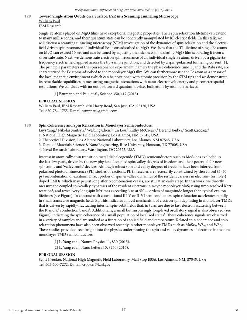
37
129 Toward Single Atom Qubits on a Surface: ESR in a Scanning Tunneling Microscope. William Paul IBM Research
Single Fe atoms placed on MgO films have exceptional magnetic properties: Their spin relaxation lifetime can extend to many milliseconds, and their quantum state can be coherently manipulated by RF electric fields. In this talk, we will discuss a scanning tunneling microscopy (STM) investigation of the dynamics of spin-relaxation and the electric-field-driven spin resonance of individual Fe atoms adsorbed to MgO. We show that the T1 lifetime of single Fe atoms on MgO can exceed 10 ms, and can be tuned by adjusting the thickness of insulating MgO film separating it from a silver substrate. Next, we demonstrate electron spin resonance of an individual single Fe atom, driven by a gigahertz-frequency electric field applied across the tip-sample junction, and detected by a spin-polarized tunneling current [1]. The principle parameters of the spin resonance experiment, namely the phase coherence time T2 and the Rabi rate, are characterized for Fe atoms adsorbed to the monolayer MgO film. We can furthermore use the Fe atom as a sensor of the local magnetic environment (which can be positioned with atomic precision by the STM tip) and we demonstrate its remarkable capabilities in measuring magnetic interactions with nano-electronvolt energy and picometer spatial resolutions. We conclude with an outlook toward quantum devices built atom-by-atom on surfaces.
[1] Baumann and Paul et al., Science 350, 417 (2015)
EPR ORAL SESSIONWilliam Paul, IBM Research, 650 Harry Road, San Jose, CA, 95120, USA Tel: 650-784-1755, E-mail: [email protected]
130 Spin Coherence and Spin Relaxation in Monolayer Semiconductors. Luyi Yang,1 Nikolai Sinitsyn,2 Weibing Chen,3 Jun Lou,3 Kathy McCreary,4 Berend Jonker,4 Scott Crooker1 1. National High Magnetic Field Laboratory, Los Alamos, NM 87545, USA 2. Theoretical Division, Los Alamos National Laboratory, Los Alamos, NM 87545, USA 3. Dept. of Materials Science & NanoEngineering, Rice University, Houston, TX 77005, USA 4. Naval Research Laboratory, Washington, DC 20375, USA
Interest in atomically-thin transition metal dichalcogenide (TMD) semiconductors such as MoS2 has exploded in the last few years, driven by the new physics of coupled spin/valley degrees of freedom and their potential for new spintronic and ‘valleytronic’ devices. Although robust spin and valley degrees of freedom have been inferred from polarized photoluminescence (PL) studies of excitons, PL timescales are necessarily constrained by short-lived (3–30 ps) recombination of excitons. Direct probes of spin & valley dynamics of the resident carriers in electron- (or hole-) doped TMDs, which may persist long after recombination ceases, are still at an early stage. In this work, we directly measure the coupled spin-valley dynamics of the resident electrons in n-type monolayer MoS2 using time-resolved Kerr rotation1, and reveal very long spin lifetimes exceeding 3 ns at 5K — orders of magnitude longer than typical exciton lifetimes (see Figure). In contrast with conventional III-V or II-VI semiconductors, spin relaxation accelerates rapidly in small transverse magnetic fields By. This indicates a novel mechanism of electron spin dephasing in monolayer TMDs that is driven by rapidly-fluctuating internal spin-orbit fields that, in turn, are due to fast electron scattering between the K and K’ conduction bands1. Additionally, a small but surprisingly long-lived oscillatory signal is also observed (see Figure), indicating the spin coherence of a small population of localized states2. These coherence signals are observed in a variety of samples and are studied as a function of applied field and temperature. Related spin coherence and spin relaxation phenomena have also been observed recently in other monolayer TMDs such as MoSe2, WS2, and WSe2. These studies provide direct insight into the physics underpinning the spin and valley dynamics of electrons in the new monolayer TMD semiconductors.
[1] L. Yang et al., Nature Physics 11, 830 (2015).[2] L. Yang et al., Nano Letters 15, 8250 (2015).
EPR ORAL SESSIONScott Crooker, National High Magnetic Field Laboratory, Mail Stop E536, Los Alamos, NM, 87545, USA Tel: 505-500-7272, E-mail: [email protected]
38
Rocky Mountain Conference on Magnetic Resonance, Vol. 58 [2016], Art. 1
https://digitalcommons.du.edu/rockychem/vol58/iss1/1

38
131 Simultaneous Detection of Transient Electrically Detected and Transient Magnetic Resonance Signals from Organic Solar Cells. Felix Kraffert, Robert Steyrleuthner, Christoph Meier, Robert Bittl, Jan Behrends Berlin Joint EPR Lab, Freie Universität Berlin, D-14195 Berlin, Germany
Techniques based on electron paramagnetic resonance spectroscopy can provide valuable insight into excitation transfer pathways in organic semiconductors used as absorber layers in solar cells.1,2 However, these measurements are usually performed on “model systems”, and the conclusions drawn from such experiments may not be valid under true solar cell operating conditions. Here we report on the development of a setup that allows for simultaneous detection of transient electron paramagnetic resonance as well as transient electrically detected magnetic resonance (trEDMR) signals from fully-processed and encapsulated solar cells. Combining both techniques provides a direct link between photoinduced triplet excitons, charge transfer states and free charge carriers as well as their influence on the photocurrent generated by organic photovoltaic devices. Our results obtained from solar cells based on poly(3-hexylthiophene) and the fullerene-based electron acceptor PCBM show that the resonant signals observed in low-temperature (T = 80 K) trEDMR spectra can be attributed to positive polarons in the polymer as well as negative polarons in the fullerene phase, indicating that both centers are involved in spin-dependent processes that directly influence the photocurrent.3 Furthermore, we will show how transient EPR measurements on blends comprising low-bandgap polymers and PCBM can help to disentangle complex charge transfer processes and identify loss mechanisms in organic solar cell materials.2
1. J. Behrends, A. Sperlich, A. Schnegg, T. Biskup, C. Teutloff, K. Lips, V. Dyakonov, and R. Bittl, Phys. Rev. B 2012, 85 , 125206.
2. F. Kraffert, R. Steyrleuthner, S. Albrecht, D. Neher, M. C. Scharber, R. Bittl, and J. Behrends, J. Phys. Chem. C 2014, 118, 28482.
3. F. Kraffert, R. Steyrleuthner, C. Meier, R. Bittl, and J. Behrends, Appl. Phys. Lett. 2015, 107, 043302.
EPR ORAL SESSIONFelix Kraffert, Free University of Berlin, Arnimallee 14, Berlin, Berlin, 14195, DE E-mail: [email protected]
132 Separation of Hyperfine and Spin-Orbit Interactions in Organic Semiconductors by Multi-Frequency Electrically Detected Magnetic Resonance using Coplanar Waveguide Microresonators. Gajadhar Joshi,1 Richards Miller,1 Lillie Ogden,1 Marzieh Kavand,1 Shirin Jamali,1 Kapildeb Ambal,1 Suresh Venkatesh,2 David Schurig,2 Hans Malissa,1 John M. Lupton,1,3 Christoph Boehme1 1. Department of Physics and Astronomy, University of Utah, Salt Lake City, UT 84112 2. Department of Electrical and Computer Engineering, University of Utah, Salt Lake City, UT 84112 3. Institut für Experimentelle und Angewandte Physik, Universität Regensburg, D-93040 Regensburg, Germany
Studies of magneto-optoelectronic properties of organic semiconductors, such as organic magnetoresistance and magneto-electroluminescence, have revealed a strong influence of hyperfine fields caused by protons and a weak but non-negligible influence of spin-orbit interactions1,2. In order to separate these influences of hyperfine fields and spin-orbit effects on charge carrier states in organic semiconductors we conduct continuous wave electrically detected magnetic resonance (cw-EDMR) spectroscopy over a wide range of frequencies between about 1-20 GHz using both fundamental and higher-harmonic modes of coplanar waveguide resonators3. Cw-EDMR spectra of bipolar injection devices (diodes) based on a π-conjugated polymer, poly[2-methoxy-5-(2-ethylhexyloxy)-1,4- phenylenevinylene] (MEH-PPV), reveal that the magnetic resonance spectra conducted at low magnetic fields and frequencies are only weakly dependent on the magnetic field and thus, are predominantly governed by the hyperfine fields. At higher magnetic fields, a significant broadening of the resonance lines is observed consistent with the presence of a spin-orbit induced g-factor distribution. In order to obtain the hyperfine field as well as g-factor distributions, we conduct a global fit of all measured spectra with two inhomogeneously broadened resonance lines, for the paramagnetic resonances lines of electrons and holes, respectively. We use a so-called ‘bootstrap’ procedure in order to obtain confidence limits for the fit results which reveals that for the charge carrier species experiencing higher hyperfine fields, only an upper limit can be placed on the magnitude of their g-factor distributions4.
This research has been supported by the US Department of Energy, Office of Basic Energy Sciences, Division of Materials Sciences and Engineering under Award #DE-SC0000909. L.O. and K. A. acknowledge support for their contributions through the Utah MRSEC center, grant #1121252.
[1] Nguyen et al., Nat. Mater. 2010, 9, 345[2] McCamey et al. Phys. Rev. Lett. 2010, 104, 017601
39
et al.: 58th RMCMR Final Program and Abstracts
Published by Digital Commons @ DU, 2016

39
[3] Malissa et al., Rev. Sci. Instrum. 2013, 84, 025116[4] Joshi et al.,arXiv:1603.03807 (2016).
EPR ORAL SESSIONGajadhar Joshi, University of Utah, 115 South 1400 East, Salt Lake City, Utah, 84112, USA Tel: 801-581-6904, E-mail: [email protected]
133 Estimation of Spin Diffusion Length and Spin-Orbit Coupling Strength in Organic Semiconductors by Means of pulsed Inverse Spin-Hall Effect Measurements. Marzieh Kavand, Kipp J. van Schooten, Dali Sun, Hans Malissa, Chuang Zhang, Matthew Groesbeck, Z. Valy Vardeny, Christoph Boehme Department of Physics and Astronomy, University of Utah, 115 South 1400 East, Salt Lake City, UT 84112-0830, USA
Spin-orbit coupling in condensed matter is crucial for spintronics applications, including generation, manipulation, and detection schemes for spin currents. We have succeeded in employing spin-pumping methods based on ferromagnetic resonance (FMR) to generate pure – meaning charge free – spin currents from a ferromagnetic (FM) substrate into organic semiconductor (OSEC) layers. When the FM is in resonance with pulsed microwave excitation, a strong, pure spin-current is injected into the OSEC, irrespective of the impedance mismatch between the ferromagnetic layer and the organic layer. Because of the weak spin-orbit coupling (SOC) in most OSECs, the inverse spin Hall effect (ISHE) resulting from this spin injection scheme is very subtle, yet with pulsed, high microwave power driving of the FMR, relatively strong ISHE signals can be measured, nevertheless. Here we report the measurement of the ISHE in a variety of OSECs having tunable SOC, ranging from strong SOC (pi-conjugated polymers that contain intrachain Pt atoms) to weak SOC polymers (such as the pi-conjugated polymer DOO-PPV). We find that the ISHE response in these compounds scales with SOC, in spite of the decrease in the spin diffusion length. Remarkably, thin film materials based on Fullerenes exhibit some of the most pronounced ISHE signals, owing to curvature-induced spin-orbit effects1 .
We acknowledge support by the National Science Foundation (DMR-1404634) for sample preparation, execution of the experiments and data processing of the ISHE experiments. We also acknowledge the NSF-Material Science & Engineering Center (DMR-1121252) for supporting Pt-polymer synthesis, execution of the OSV experiments and development of the ISHE device structures, as well as support for the device preparation facilities.
[1] Sun et al., Nat. Mat., DOI:10.1038/NMAT4618 (2016).
EPR ORAL SESSIONMarzieh Kavand, University of Utah, 201 James Fletcher Bldg, 115 South 1400 East, Salt Lake City, UT, 84112-0830, USA Tel: 385-226-7330, E-mail: [email protected]
134 Cu2+-ions as a ESR Probe of Protein Structure. Sunil Saxena Department of Chemistry, University of Pittsburgh, Pittsburgh, PA-15260
Pulsed -ESR techniques that reliably measure interspin separations in the order of 1.5-10 nm – even in non-crystalline samples – to ultimately provide an “amino-acid-level” picture of structure and structural transitions, have impacted biophysical research. The talk will discuss our efforts in developing Cu2+-ion based pulsed-ESR distance methods and illustrate how they can potentially be used to understand structure function relationships in proteins. The talk will focus on restriction endonuclease EcoRI, which binds to the specific DNA sequence GAATTC with an affinity that is 50,000-90,000-fold greater than that of a miscognate site that differs by only one base pair. In the presence divalent metal ions, such as magnesium, EcoRI the specific sequence of viral DNA with a high specificity. We will describe the insights gained in regard to the high specificity as well as cleavage chemistry from ESR distance measurements. Finally, the talk will describe recent efforts to bind Cu2+-ions site selectively at α-helical and β-sheet sites in protein. The spin probe is assembled in situ from natural amino acid residues and a metal salt, and requires no post-expression synthetic modification. Initial results show that the resultant Cu2+-probe potentially provides distance distributions that are five times narrower than the common protein spin label – the approach, thus, has the potential to significantly overcome the inherent limitation of the current technology which relies on a spin label with a highly flexible side-chain.
EPR ORAL SESSIONSunil K Saxena, University of Pittsburgh, 219 Parkman Ave, Pittsburgh, PA, 15260, USA Tel: 412-624-8680, E-mail: [email protected]
40
Rocky Mountain Conference on Magnetic Resonance, Vol. 58 [2016], Art. 1
https://digitalcommons.du.edu/rockychem/vol58/iss1/1

40
135 Site-Specific Investigations of the Protein Dynamical Transition via Pulse EPR. Ryan Barnes,1 Ilia Kaminker,1 Songi Han1,2 1. Department of Chemistry University of California, Santa Barbara 2. Department of Chemical Engineering University of California, Santa Barbara
The dynamical transition of a solvated protein contains necessary information about the protein solvent coupling and has major implications for water mediated protein-protein and protein-ligand interactions. We investigate the dynamical transition of the Trp-cage protein in a hydrated powder (h~0.2) at select sites along the peptide backbone with spin-label pulse EPR. The electron T2 serves as a probe for the local magnetic noise induced by the motion of nearby solvent and protein nuclei that is sensitive to noise on the 10-6 to 10-12 second timescales and thus is capable of probing the wide range of timescales present throughout the protein dynamical transition. In this report we show that measurements of the electron T2 by Hahn echo as a function of temperature reveal the protein dynamical transition as a strong inflection point at approximately 180 K. In addition we discuss preliminary results of a modified spectral filtering technique to recover local noise correlation time at each temperature of the measurement. We discuss the site-specific protein dynamics revealed by pulse EPR about the protein dynamic transition in context of the chemical properties of the Trp cage protein.
EPR ORAL SESSIONRyan P Barnes, University of California Santa Barbara, 1232 Laguna St Unit C, Santa Barbara, CA, 93101, USA E-mail: [email protected]
136 Bayesian Uncertainty Quantification for DEER Spectroscop. Thomas H. Edwards, Stefan Stoll Department of Chemistry, University of Washington, Seattle, WA 98195
DEER (Double Electron-Electron Resonance) spectroscopy is a solid-state pulse EPR (Electron Paramagnetic Resonance) experiment that measures distances between unpaired electrons, most commonly between protein-bound spin-labels separated by 1.5-8 nm. From the experimental data, a distance distribution P(r) is extracted using Tikhonov regularization. The disadvantage of this method is that it does not directly provide error bars for the resulting P(r), rendering correct interpretation difficult. Further, Tikhonov regularization requires the selection of a regularization parameter, and current methods employ heuristics and introduce bias. Here we introduce a Bayesian statistical approach that quantifies uncertainty in P(r) arising from time-domain signal noise and numerical regularization. This method provides credible intervals (error bars) of P(r) at each r. This allows practitioners to answer whether or not small features are significant, whether or not apparent shoulders are significant, and whether or not two distance distributions are significantly different from each other. In addition, the method quantifies uncertainty arising from the degree of regularization.
EPR ORAL SESSIONThomas H Edwards, University of Washington, Box 351700, Seattle, WA, 98195, USA E-mail: [email protected]
137 WavPDS: A Wavelet Approach in Denoising Pulsed Dipolar Spectroscopy. Madhur Srivastava,1,2 Elka R. Georgieva,1 Peter P. Borbat,1 Jack H. Freed1,3 1. Cornell University, National Biomedical Center for Advanced ESR Technology (ACERT), Ithaca, NY 14853 2. Cornell University, Meinig School of Biomedical Engineering, Ithaca, NY 14853 3. Cornell University, Department of Chemistry and Chemical Biology, Ithaca, NY 14853
Studying biological systems using Pulsed Dipolar Electron Spin Resonance Spectroscopy (PDS) is challenging due to the short relaxation times and low protein concentrations typically used. These frequently result in a low Signal to Noise Ratio (SNR), complicating the analysis. Even if the average distance between spin probes can be estimated, the determination of the distance distribution (DD) is likely to be corrupted by noise. To address the challenge of noise removal in Pulsed Dipolar ESR in order to obtain reliable information, we developed a new wavelet denoising method (WavPDS) to remove/reduce noise. Our method improves the stability and reliability of the DD reconstruction, and reduces the signal acquisition time by an order of magnitude. This enables the study of biomolecular structures at low SNR signals with accuracy. We believe that studies in a wide variety of disciplines will greatly benefit.
EPR ORAL SESSIONMadhur Srivastava, Cornell University, B16 Baker Lab, Ithaca, New York, 14853, USA E-mail: [email protected]
41
et al.: 58th RMCMR Final Program and Abstracts
Published by Digital Commons @ DU, 2016

41
138 Three Homologous TonB-dependent Transporters Utilize Different Mechanisms to Regulate Protein-Protein Interactions. Lishan Liu, Jessica L. Sarver, Michael Zhang, David S. Cafiso University of Virginia, Department of Chemistry, Charlottesville, VA 22903
Gram negative bacteria utilize a family of specific, high affinity transport proteins to scavenge rare nutrients such as iron, vitamin B12 and certain carbohydrates from their environment. These active transport proteins are localized to the bacterial outer-membrane and obtain energy for transport from cytoplasm membrane proton motive force by coupling to the inner membrane protein TonB. Although a number of high-resolution crystal structures have been obtained for TonB-dependent transport proteins (TBDT), the molecular mechanism by which transport takes place still remains unknown. TonB is stoicheometrically limited with respect to the outer membrane TDBT, and transport is thought to involve cycles of attachment and dissociation of TonB from the outer-membrane transporter. Previous studies have shown that the affinity of TonB for several transporters is enhanced by substrate binding. In both BtuB, the vitamin B12 transporter and FecA, the ferric citrate transporter, binding of substrate induces conformational changes in the energy-coupling segment termed Ton box. Upon substrate binding, Ton box of BtuB and FecA become less ordered and increase their exposure to the periplasm. Here, we studied the structure and dynamics of the FhuA Ton box in the presence or absence of substrate utilizing both CW and pulsed EPR techniques in isolated native outer membrane. Unlike BtuB and FecA, the Ton box of FhuA is disordered with or without substrate. However, substrate binding does change the position of Ton box relative to the barrel axis and appears to extend the Ton box further into the periplasm. The results suggest a mechanism that could enhance TonB affinity for FhuA in the presence of substrate. Remarkably, each of these three homologous transporter, BtuB, FecA and FhuA appears to regulate TonB affinity by a different mechanism.
EPR ORAL SESSIONLishan Liu, University of virginia, 1822 Sugar Maple Terrace, Charlottesville, VA, 22903, USA E-mail: [email protected]
138 Measuring Oxidation States in Exchange-Coupled Metal Clusters Using Ligand Hyperfine. Troy A. Stich,1 Paul H. Oyala,1 Gregory J. Yeagle,1 William K. Myers,1 Daniel L.M. Suess,1 Jon M. Kuchenreuther,2 Rachel J. Service,3 James W. Whittaker,4 Richard J. Debus,3 James R. Swartz,2 R. David Britt1 1. University of California-Davis, Department of Chemistry, Davis, CA 95616 2. Department of Chemical Engineering and Department of Bioengineering, Stanford University, Stanford, CA 94305 3. Department of Biochemistry, University of California, Riverside, Riverside, CA 92521 4. Department of Science and Engineering, School of Medicine, Oregon Health and Science University,, Beaverton, OR
97006
Some of the most important redox reactions in Nature (e.g., nitrogen fixation, water oxidation, proton reduction) take place at enzyme active sites composed of multiple metal ions. Understanding the electronic structures of these metal clusters has been the target of extensive research efforts. However, obtaining site-specific information on a particular metal ion’s oxidation state can be challenging when its electrons are coupled to the unpaired electrons on the other metal centers in the cluster via electron exchange. In this presentation, we provide several examples that illustrate how by measuring the hyperfine interaction with ligand nuclei, the oxidation state of the bonding partner metal ion can be determined. These results help to improve computational models of the enzyme active site and can provide insights into the reaction mechanism.
EPR ORAL SESSIONTroy Stich, University of California Davis, 1 Shields Ave, Davis, California, 95616, USA Tel: 530-220-5669, E-mail: [email protected]
42
Rocky Mountain Conference on Magnetic Resonance, Vol. 58 [2016], Art. 1
https://digitalcommons.du.edu/rockychem/vol58/iss1/1

42
139 Optimization of Pulsed EPR Distance Measurements for Tau Protein Aggregation. Timothy Keller, Ilia Kaminker, Ryan Barnes, Neil Eschmann, Zach Oberholtzer, Songi Han Department of Chemistry and Biochemistry, University of California Santa Barbara, Santa Barbara, CA, USA
Tau protein is an intrinsically disordered protein found in neurons where its primary function is to stabilize microtubules. Under certain conditions, tau has a propensity to aggregate into beta-sheet fibrils. The formation of these fibrils has been identified as an important characteristic in neurodegenerative diseases such as Alzheimer’s disease. Preliminary double electron-electron resonance (DEER) measurements have led to the discovery of an intermediate state which forms within minutes of initiating tau aggregation. Unfortunately, aggregating systems are particularly difficult to study with DEER due to: 1) dilution of spin labeled protein with analog protein is necessary to prevent interference from inter-protein distances and the total protein concentration is limited by the protein solubility 2) clustering of spins upon aggregation causes a reduction in T1 and T2 relaxation times which reduce the echo amplitude for the same DEER dipolar evolution times. Recent development of new DEER pulse sequences, including 5-pulse DEER [1] and CP-DEER [2], as well as the development of broadband SIFTER [3] have shown promise in improving the performance of pulsed EPR distance measurements; however these techniques have not yet been applied to many realistic biological systems including the study of protein aggregation. In addition to new pulse sequences, the addition of arbitrarily shaped pulses offers a great deal of flexibility in improving the signal to noise and/or reducing the time of DEER measurements which would otherwise be unrealistic. Our study investigates optimizing the performance of distance measurements on tau protein by comparing the performance (i.e. signal to noise) for a variety of pulse sequences and pulse shapes.
[1] Borbat et al., J. Phys. Chem. Lett., 2013, 4, 170. [2] Spindler et al.,J. Phys. Chem. Lett, 2015, 6, 4331. [3] Schöps et al., J. Magn. Reson., 2015, 250, 55.
EPR ORAL SESSIONTimothy J Keller, University of California Santa Barbara, Department of Chemistry and Biochemistry, Santa Barbara, CA, 93106-9510, USA E-mail: [email protected]
140 Conformational Transitions of Maltose Binding Protein in the Native State and as Molten Globule at pH 3 as Monitored by DEER and DQC EPR Spectroscopy. Benjamin Selmke,1 Chen Nickolaus,1 Peter P. Borbat,2 Jack H. Freed,2 Wolfgang E. Trommer1 1. Department of Chemistry, TU Kaiserslautern, D-67653 Kaiserslautern, Germany 2. ACERT, Cornell University, Ithaca, N.Y., 14850, USA
Maltose-binding protein (MBP) is a single chain protein composed of two domains that is in a molten globule state at pH 3 as characterized by ANS binding. DEER measurements of seven spin-labeled double mutants in the native state at pH 7 had shown excellent agreement with X-ray data. At pH 3 corresponding DEER measurements of all the mutants yielded a broad distribution of distances. This can be expected if there is no defined tertiary structure and the individual helices point into all possible directions. Depending on maltose binding in a cleft between the domains, MBP exhibits both, an open and a closed conformation with respect to these domains. We have followed this substrate-depending conformational change by means of additional spin-labeled mutants at or near the active site. In these experiments DQC spectroscopy has been particularly helpful as it allows for distance measurements of labels in close proximity. Data show, e.g., that there is a defined structure of the active site of MBP at both pH values even in the absence of substrate.
EPR ORAL SESSIONWolfgang Trommer, TU Kaiserslautern, P.O.Box 3049, Kaiserslautern, Rheinland-Pfalz, D-67653, DE Tel: 496312052045, E-mail: [email protected]
43
et al.: 58th RMCMR Final Program and Abstracts
Published by Digital Commons @ DU, 2016

43
141 Selective Membrane Disruption Mechanism of an Antibacterial γ-AApeptide Defined by EPR Spectroscopy. Pavanjeet Kaur,1 Yaqiong Li,2 Jianfeng Cai,2 Likai Song1 1. National High Magnetic Field Laboratory and Florida State University, Tallahassee, FL 32310 2. University of South Florida, Tampa, FL 33620
Antibiotic resistance is one of the major threats to public health. γ-AApeptides are a new class of antibacterial peptidomimetics that are not prone to antibiotic resistance and are highly resistant to protease degradation. It is not clear how γ-AApeptides interact with bacterial membranes and alter lipid assembly, but such information is essential to understanding their antimicrobial activities and guiding future design of more potent and specific antimicrobial agents. Using EPR techniques at 9 and 95 GHz, we characterized the membrane interaction and destabilizing mechanism of a lipo-cyclic-γ-AApeptide (AA1), which has broad-spectrum antibacterial activities1. The analyses revealed that AA1 binding increases the membrane permeability of POPC/POPG liposomes, which mimic negatively charged bacterial membranes. AA1 binding also inhibits membrane fluidity and reduces solvent accessibility around the lipid head-group region. Moreover, AA1 interacts strongly with POPC/POPG liposomes, inducing significant lipid lateral-ordering and membrane thinning. In contrast, minimal membrane property changes were observed upon AA1 binding for liposomes mimicking mammalian cell membranes, which consist of neutral lipids and cholesterol. Our findings suggest that AA1 interacts and disrupts bacterial membranes through a carpet-like mechanism. The results showed that the intrinsic features of γ-AApeptides are important for their ability to disrupt bacterial membranes selectively, the implications of which extend to developing new antibacterial biomaterials.
1. Kaur, P.; Li, Y.; Cai, J. and Song, L. Biophysical J., 2016, 110 (8), 1789-1799.
EPR ORAL SESSIONLikai Song, National High Magnetic Field Laboratory and Florida State University, 1800 E. Paul Dirac Dr., Tallahassee, FL 32310, USA Tel: 850-6446153, E-mail: [email protected]
142 Distance Measurements Between Paramagnetic Ligands Bound to Parallel Stranded Guanine Quadruplexes. M.P. Donohue,1,2 V.A. Szalai1 1. Center for Nanoscale Science and Technology, National Institute of Standards and Technology, Gaithersburg, MD 20899 2. Maryland NanoCenter, University of Maryland, College Park, MD 20742
Aside from a double helix, deoxyribonucleic acid (DNA) folds into non-canonical structures, one of which is the guanine quadruplex. Cationic porphyrins bind guanine quadruplexes, but the effects of ligand binding on the structure of guanine quadruplexes with more than four contiguous guanine quartets remains to be fully elucidated. Double electron electron resonance (DEER) spectroscopy conducted at 9.5 GHz (X-band) using broadband shaped excitation pulses was used to measure the distances between cationic copper porphyrins bound to model parallel-stranded guanine quadruplexes with increasing numbers of guanine quartets. A monotonic increase in the average Cu2+-Cu2+ distance was found as the poly-guanine tract increased within the oligonucleotide sequence, indicative of tetramolecular quadruplexes. A single Gaussian component was found to best model the time domain datasets, characteristic of a 2:1 binding stoichiometry between the porphyrins and each quadruplex, which is consistent with our previous work. Rather unexpectedly, the measured increase in Cu2+-Cu2+ distances was not linear with the number of guanine tracts, suggesting a conformational change in the quadruplex secondary structure upon an incremental increase of successive guanine quartets.
EPR ORAL SESSIONMatthew P. Donohue, National Institute of Standards and Technology, 100 Bureau Dr, Gaithersburg, Maryland, 20899, USA Tel: 301-975-8413, E-mail: [email protected]
143 Phenylalanine Hydroxylase: Providing Details of a Catalytic Cycle with EPR Spectroscopy. John McCracken,1 Gulbenk Anarat Cappillino,2 Nicole Dickson,1 Matthew D. Krzyaniak,1 John P. Caradonna2 1. Department of Chemistry, Michigan State University, East Lansing, MI 48824 2. Department of Chemistry, Boston University, Boston, MA 02215
Phenylalanine hydroxylase (PheH) is a liver enzyme that catalyzes the hydroxylation of the side-chain of phenylalanine to form tyrosine. This chemistry requires the binding of substrate, L-phe, a cofactor, tetrahydrobiopterin, and molecular oxygen to an Fe(II) active site. In the enzyme’s resting state, the metal ion features an octahedral coordination geometry with one coordination face, consisting of the side chains of two histidines and a glutamic acid, provided by the protein and the remaining three coordination sites occupied by water ligands. While the diverse chemistry catalyzed by non-heme iron enzymes is often attributed to the facial arrangement of these open coordination sites, the proposed chemical mechanism for phenylalanine hydroxylation calls for the initial binding of both substrate L-phe and the tetrahydrobiopterin cofactor as second coordination-sphere ligands. In our studies, we have used nitric oxide, NO, as
44
Rocky Mountain Conference on Magnetic Resonance, Vol. 58 [2016], Art. 1
https://digitalcommons.du.edu/rockychem/vol58/iss1/1
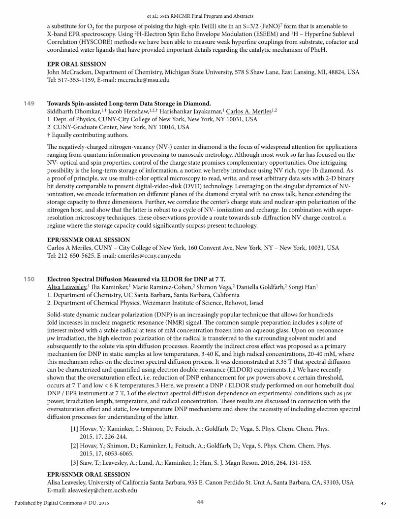
44
a substitute for O2 for the purpose of poising the high-spin Fe(II) site in an S=3/2 {FeNO}7 form that is amenable to X-band EPR spectroscopy. Using 2H-Electron Spin Echo Envelope Modulation (ESEEM) and 1H – Hyperfine Sublevel Correlation (HYSCORE) methods we have been able to measure weak hyperfine couplings from substrate, cofactor and coordinated water ligands that have provided important details regarding the catalytic mechanism of PheH.
EPR ORAL SESSIONJohn McCracken, Department of Chemistry, Michigan State University, 578 S Shaw Lane, East Lansing, MI, 48824, USA Tel: 517-353-1159, E-mail: [email protected]
149 Towards Spin-assisted Long-term Data Storage in Diamond. Siddharth Dhomkar,1,† Jacob Henshaw,1,2,† Harishankar Jayakumar,1 Carlos A. Meriles1,2 1. Dept. of Physics, CUNY-City College of New York, New York, NY 10031, USA 2. CUNY-Graduate Center, New York, NY 10016, USA † Equally contributing authors.
The negatively-charged nitrogen-vacancy (NV-) center in diamond is the focus of widespread attention for applications ranging from quantum information processing to nanoscale metrology. Although most work so far has focused on the NV- optical and spin properties, control of the charge state promises complementary opportunities. One intriguing possibility is the long-term storage of information, a notion we hereby introduce using NV rich, type-1b diamond. As a proof of principle, we use multi-color optical microscopy to read, write, and reset arbitrary data sets with 2-D binary bit density comparable to present digital-video-disk (DVD) technology. Leveraging on the singular dynamics of NV- ionization, we encode information on different planes of the diamond crystal with no cross talk, hence extending the storage capacity to three dimensions. Further, we correlate the center’s charge state and nuclear spin polarization of the nitrogen host, and show that the latter is robust to a cycle of NV- ionization and recharge. In combination with super-resolution microscopy techniques, these observations provide a route towards sub-diffraction NV charge control, a regime where the storage capacity could significantly surpass present technology.
EPR/SSNMR ORAL SESSIONCarlos A Meriles, CUNY – City College of New York, 160 Convent Ave, New York, NY – New York, 10031, USA Tel: 212-650-5625, E-mail: [email protected]
150 Electron Spectral Diffusion Measured via ELDOR for DNP at 7 T. Alisa Leavesley,1 Ilia Kaminker,1 Marie Ramirez-Cohen,2 Shimon Vega,2 Daniella Goldfarb,2 Songi Han1 1. Department of Chemistry, UC Santa Barbara, Santa Barbara, California 2. Department of Chemical Physics, Weizmann Institute of Science, Rehovot, Israel
Solid-state dynamic nuclear polarization (DNP) is an increasingly popular technique that allows for hundreds fold increases in nuclear magnetic resonance (NMR) signal. The common sample preparation includes a solute of interest mixed with a stable radical at tens of mM concentration frozen into an aqueous glass. Upon on-resonance µw irradiation, the high electron polarization of the radical is transferred to the surrounding solvent nuclei and subsequently to the solute via spin diffusion processes. Recently the indirect cross effect was proposed as a primary mechanism for DNP in static samples at low temperatures, 3-40 K, and high radical concentrations, 20-40 mM, where this mechanism relies on the electron spectral diffusion process. It was demonstrated at 3.35 T that spectral diffusion can be characterized and quantified using electron double resonance (ELDOR) experiments.1,2 We have recently shown that the oversaturation effect, i.e. reduction of DNP enhancement for µw powers above a certain threshold, occurs at 7 T and low < 6 K temperatures.3 Here, we present a DNP / ELDOR study performed on our homebuilt dual DNP / EPR instrument at 7 T, 3 of the electron spectral diffusion dependence on experimental conditions such as µw power, irradiation length, temperature, and radical concentration. These results are discussed in connection with the oversaturation effect and static, low temperature DNP mechanisms and show the necessity of including electron spectral diffusion processes for understanding of the latter.
[1] Hovav, Y.; Kaminker, I.; Shimon, D.; Feiuch, A.; Goldfarb, D.; Vega, S. Phys. Chem. Chem. Phys. 2015, 17, 226-244.
[2] Hovav, Y.; Shimon, D.; Kaminker, I.; Feituch, A.; Goldfarb, D.; Vega, S. Phys. Chem. Chem. Phys. 2015, 17, 6053-6065.
[3] Siaw, T.; Leavesley, A.; Lund, A.; Kaminker, I.; Han, S. J. Magn Reson. 2016, 264, 131-153.
EPR/SSNMR ORAL SESSIONAlisa Leavesley, University of California Santa Barbara, 935 E. Canon Perdido St. Unit A, Santa Barbara, CA, 93103, USA E-mail: [email protected]
45
et al.: 58th RMCMR Final Program and Abstracts
Published by Digital Commons @ DU, 2016

45
151 Hypersensitivity with Dynamic Nuclear Polarization: Natural Isotopic Abundance and Closed-loop Cryogenic Helium Sample Spinning. K. Märker,1 F. Mentink-Vigier,1 S. Paul,1 A. Purea,3 F. Engelke,3 E. Bouleau,1 Sabine Hediger,1,2 Daniel Lee,1 Gaël De Paëpe1 1. INAC, CEA & Univ. Grenoble Alpes, F-38000, France 2. INAC, CNRS, F-38000, France 3. Bruker Biospin GmbH, Rheinstetten, Germany
The ability to record correlation experiments for nuclei at low natural isotopic abundance (13C, 15N) using dynamic nuclear polarization (DNP) [1] shows great promise for the field of NMR crystallography. The low natural abundance statistically simplifies coupled systems to spin pairs, making the measurement of inter-nuclear distances more straightforward. Furthermore, these simplified 2-spin cases permit the measurements of long distances as dipolar truncation effects (seen in multi-spin systems) are not present. It will be shown that dipolar recoupling experiments can be used to obtain not only intra-molecular distance restraints but also inter-molecular distances. Examples will be shown on natural isotopic abundance self-assembled systems, such as certain peptides and guanosine derivatives, where p-stacking interactions and hydrogen-bonding play a large role in the crystal structure – which cannot be easily determined from conventional crystallography methods.[2] In order to further enhance the sensitivity, we will report on a strategy to push the limits of DNP-enhanced solid-state NMR beyond its current state-of-the-art. This leap-forward was made possible thanks to the employment of a closed-loop of cryogenic helium as the gas to power magic angle sample spinning (MAS) for DNP-enhanced NMR experiments. The experimental conditions reported here far exceed what is currently possible and allows reaching sample temperatures down to 30 K while conducting experiments with high spinning frequencies (up to 25 kHz @ 100 K for a 3.2 mm probe). Thanks to the impressive associated gains, which will be presented, sustainable cryogenic helium sample spinning significantly enlarges the realm and possibilities of the MAS-DNP technique and is the route to transform NMR into a versatile and sensitive atomic-level characterization tool.[3] Finally, we will describe our efforts towards understanding the origin of the polarization losses associated with radical doping. Notably, we will investigate NMR signal losses occurring during MAS-DNP experiments and specifically compare the (MAS-dependent) depolarization effect for several “gold-standard” (bi-)radicals currently in use in most MAS-DNP studies (e.g. Totapol/bTbK/Amupol/TEMTriPol/etc.). Using MAS-DNP simulations we will show that these observations can be rationalized and are consistent with the biradicals’ structure. Further insight into the depolarization mechanism (multi-parameters phenomenon) can be obtained comparing the result for each crystallite orientation with the result obtained on the powder average.[4]
[1] Takahashi H. et al., Angewandte Chemie International Edition, 51, 11766-11769, 2012[2] K. Märker et al., Journal of the American Chemical Society, 137, 13796-13799, 2015[3] Bouleau E. et al., Chemical Science, 6, 6806-6812, 2015[4] Mentink-Vigier F., et al., Physical Chemistry Chemical Physics, 17, 21824-21836, 2015
EPR/SSNMR ORAL SESSIONGaël De Paëpe, INAC (CEA – Grenoble Alpes University), 17, rue des martyrs, Grenoble, Rhône-Alpes, 38054, FR E-mail: [email protected]
152 Combining Dynamic Nuclear Polarization and Mechanically Detected Magnetic Resonance to Achieve Nanoscale Magnetic Resonance Imaging of Individual Biomolecules and Assemblies. Corinne E. Isaac, Hoang L. Nguyen, Pamela T. Nasr, Elizabeth A. Curley, Michael C. Boucher, John A. Marohn Department of Chemistry and Chemical Biology, Cornell University, Ithaca, New York 14853-1301 USA
A tool for imaging the proton envelope of an individual macromolecule and biological assembly would be a major advance. To realize this opportunity, we are pushing magnetic resonance imaging (MRI) to nanometer resolution using the sensitivity afforded by detecting magnetic resonance mechanically. We have developed attonewton-sensitivity cantilevers with integrated nanomagnet tips capable of detecting nuclear magnetic resonance from a polymer film at cryogenic temperatures with a sensitivity of a few hundred proton magnetic moments.1 These experiments observe magnetization fluctuations present in small, nanometer-scale volumes of spins. I will describe our efforts to significantly improve the per-spin sensitivity and acquisition time of “nano-MRI” experiments by using dynamic nuclear polarization (DNP) to create a measurable net nuclear spin polarization,2 pushing the experiment out of the spin-noise limit. Even with a noiseless detector (e.g., a zero-temperature cantilever or a quantum-limited nitrogen-vacancy center), stochastic spin fluctuations in the sample still limit the imaging resolution achievable in a nano-MRI experiment. We have performed numerical simulations of nano-MRI experiments with a number of image-encoding and detection protocols to identify the conditions under which DNP can get us beyond the limits imposed by spin-noise. Taken together, our experiments and simulations suggest that magnetic resonance force microscopy is on its way to becoming a powerful new route for obtaining a three-dimensional image of a single copy of a globular protein, macromolecular complex, and membrane protein.
46
Rocky Mountain Conference on Magnetic Resonance, Vol. 58 [2016], Art. 1
https://digitalcommons.du.edu/rockychem/vol58/iss1/1

46
[1] Longenecker, J. G.; et al.; Marohn, J. A. ACS Nano, 2012, 6, 9637 – 9645; URL http://dx.doi.org/10.1021/nn3030628
[2] Isaac, C. E.; et al.; Marohn, J. A. Phys. Chem. Chem. Phys., 2016, 18, 8806 – 8819; URL http://dx.doi.org/10.1039/C6CP00084C
EPR/SSNMR ORAL SESSIONJohn A Marohn, Cornell University, 150 Baker Laboratory, Ithaca, New York, 14853-1301, USA Tel: 607-255-2004, E-mail: [email protected]
153 Electron Spin Decoupled NMR Driven by Electron Spin Relaxation of Spin Clusters. Ting Ann Siaw,1 Blake Wilson,2 Songi Han1,3 1. Department of Chemistry and Biochemistry, University of California Santa Barbara, Santa Barbara, CA 2. Department of Physics, University of California Santa Barbara, Santa Barbara, CA 3. Department of Chemical Engineering, University of California Santa Barbara, CA
The dramatic benefit of dynamic nuclear polarization (DNP) to amplify nuclear magnetic resonance (NMR) signal is fundamentally countered by line broadening and signal loss due to paramagnetic effects exerted by DNP agents, compromising NMR spectral resolution and sensitivity. Electron spin decoupling would principally eliminate these paramagnetic effects, but has not been experimentally accessible at high magnetic fields. We show that continuous wave microwave excitation of electron spin clusters in a Li ion battery electrolyte system at ~4 K results simultaneously in electron spin decoupling and DNP enhancement, removing paramagnetic effects on 7Li NMR while providing signal enhancements of ~5-20 fold. EPR measurements reveal that favorable electron spin relaxation (long T1e, short TM) are responsible for a broad excitation of the EPR line, measured directly using electron double resonance (ELDOR) experiments. Additionally, concurrent paramagnetic NMR and DNP analysis concluded that the favorable electron spin relaxation properties originated from coordination of TEMPO-based nitroxides to the Li ion, generating spin clusters. The DNP study here implicates that DNP analysis could be a materials characterization method for detecting clustering in materials systems.
EPR/SSNMR ORAL SESSIONTing Ann Siaw, University of California Santa Barbara, Department of Chemistry and Biochemistry, UC Santa Barbara, Santa Barbara, CA, 93106, USA Tel: 805-893-2792, E-mail: [email protected]
154 Nanoscale NMR Detection and Imaging Using Nitrogen-vacancy Centers in Diamond. John Mamin, Mark Sherwood, Moonhee Kim, Daniel Rugar IBM Almaden Research Center, San Jose, CA 95120
We are exploring the use of individual, near-surface nitrogen-vacancy (NV) centers in diamond as atomic-size magnetometers to detect proton NMR in organic material located external to the diamond. Using a combination of electron spin echoes and proton spin manipulation, the NV center senses the nanotesla field fluctuations from the protons, enabling both time-domain and spectroscopic NMR measurements on the nanometer scale. By scanning a small polymer test object past a near-surface NV center, we have recently demonstrated proton magnetic resonance imaging (MRI) with spatial resolution on the order of 10 nm. One key issue in NV-NMR experiments is the loss of spin coherence when the NV center is located near the diamond surface. Although this loss of coherence is frequently attributed to the effect of magnetic noise emanating from unpaired spins on the diamond surface, we will show evidence that electric field noise from fluctuating surface charge can be a significant factor.
EPR/SSNMR ORAL SESSIONDaniel Rugar, IBM Almaden Research Center, 650 Harry Rd., San Jose, CA, 95120, USA E-mail: [email protected]
47
et al.: 58th RMCMR Final Program and Abstracts
Published by Digital Commons @ DU, 2016

47
155 Technology for Hyperfine Decoupling and Time Domain DNP in Rotating Solids. Edward Saliba, Nicholas Alaniva, Faith Scott, Brice Albert, Chukun Gao, Michael Mardini, Eric Choi, Seong Ho Pahng, Alexander B. Barnes Washington University in St. Louis, Department of Chemistry MO 63130, USA
Hyperfine decoupling and pulsed dynamic nuclear polarization (DNP) are promising techniques to improve DNP experiments.1-3 We explore experimental and theoretical considerations to implement them with magic angle spinning (MAS). Microwave field simulations using the high frequency structural simulator (HFSS) software suite are performed to characterize the inhomogeneous phase independent microwave field throughout a 198 GHz MAS DNP probe. Adiabatic electron spin inversions of stable organic radicals are simulated with SPINEVOLUTION using the inhomogeneous microwave fields calculated by HFSS. We calculate an electron spin inversion efficiency of 56% at a spinning frequency of 5 kHz. Voltage tunable gyrotron oscillators are proposed as a class of frequency agile microwave sources to generate microwave frequency sweeps required for time domain DNP transfers and hyperfine decoupling in rotating solids. We demonstrate gyrotron acceleration potentials and microwaves sweeps required for the hyperfine decoupling, and the integrated solid effect. In addition to designs and results of a new voltage tunable gyrotron microwave source, we also will describe novel instrumentation for cryogenic MAS including 1) a quadruple resonance transmission line MAS DNP probe capable of producing 320 kHz proton nutation fields within a 3.2 mm diameter solinoid.
This research was supported by the NIH Director’s New Innovator Award number DP2GM119131 and an NSF CAREER Award number DBI-1553577.
1. Corzilius, B., Andreas, L. B., Smith, A. A., Ni, Q. Z., & Griffin, R. G. (2014). Paramagnet induced signal quenching in MAS–DNP experiments in frozen homogeneous solutions. Journal of Magnetic Resonance, 240, 113-123.
2. Can, T. V., Walish, J. J., Swager, T. M., & Griffin, R. G. (2015). Time domain DNP with the NOVEL sequence. The Journal of chemical physics, 143(5), 054201.
3. Hoff, D. E., Albert, B. J., Saliba, E. P., Scott, F. J., Choi, E. J., Mardini, M., & Barnes, A. B. (2015). Frequency swept Microwaves for hyperfine decoupling and time domain dynamic nuclear polarization. Solid state nuclear magnetic resonance, 72, 79-89.
EPR/SSNMR ORAL SESSIONAlexander B Barnes, Washington University in St. Louis, One Brookings Dr., St. Louis, Missouri, 63130, USA E-mail: [email protected]
156 Nuclear Magnetic Resonance Spectroscopy on a Nanostructured Diamond Chip for Chemical Trace Analysis. Nazanin Mosavin CHTM-UNM, 1313 Goddard SE, Albuquerque, NM 87106
Studying trace quantities of analyte is challenging and time consuming using traditional coil based NMR, owing to poor signal to noise ratios. This motivates using larger and more expensive magnets and/or cooling samples which restricts general applicability. Non inductive magnetometry detection, when combined with nuclear hyperpolarization methods, promises an alternative low field, ambient temperature solution, but typical sensor standoffs are mm, limiting studies to mm3 or larger samples. Recently a new technique has emerged based on optically detected magnetic resonance of nitrogen vacancy (NV) centers in diamond which offers a path to highly sensitive NMR at ambient conditions. Rather than detecting the small net thermal magnetization of nuclear spins, these non inductive magnetometers detect the nanoscale variations in their magnetization. This produces an NMR signal which is independent of temperature and magnetic field and is orders of magnitude larger at ambient temperature. Early experiments using single NV sensors have demonstrated detection of multiple nuclear species in nm 3 volumes of liquids and thin films. A remaining challenge is that measurements typically take several hours and require laborious sample preparation, owing to stochastic placement of analyte relative to the single spin sensor. In this work we bridge the gap between nm3 and um3 . Employing a refined interference lithography method, high aspect ratio diamond nanogratings are fabricated with 400 nm pitch leading to an overall surface enhancement of >10. We then dope the sidewalls of the gratings with a high density of NV centers between 2 10 nm from the surface. The end result is that billions of NV centers come into nm scale contact with analyte and the ensemble averaged signal gives a corresponding boost in sensitivity. Using these sensors, we acquired NMR spectra of nL of liquid and powder analytes on minute timescales, orders of magnitude faster than previous diamond techniques. For liquid analytes, the spectral resolution is currently limited by translational diffusion of analyte which prohibits observation of chemical shifts. We are currently investigating whether nanostructures can be fabricated which restrict nuclear translational diffusion without requiring complicated surface tethering strategies.
EPR/SSNMR ORAL SESSIONNazanin Mosavin, CHTM-UNM, 1313 Goddard SE, Albuquerque, NM, 87106, USA E-mail: [email protected]
48
Rocky Mountain Conference on Magnetic Resonance, Vol. 58 [2016], Art. 1
https://digitalcommons.du.edu/rockychem/vol58/iss1/1

48
157 Gd3+ as Polarizing Agent at High Field: Solid Effect vs Cross Effect Dynamic Nuclear Polarization. Monu Kaushik,1 Mian Qi,2 Adelheid Godt,2 Björn Corzilius,1 1. Institute of Physical and Theoretical Chemistry, Institute of Biophysical Chemistry, and Center for Biomolecular Magnetic Resonance, Goethe University Frankfurt, Max-von- Laue-Str. 7-9, 60438 Frankfurt am Main, Germany 2. Faculty of Chemistry and Center
A variety of polarizing agents have been developed for efficient dynamic nuclear polarization (DNP) for high sensitivity gain. In this work we present studies based on a relatively new class of polarizing agents: high spin transition metal ions. Transition metal ions (Gd3+, Mn2+) can act as paramagnetic substitute of intrinsically bound diamagnetic ions in biomolecules. Doping with paramagnetic ions in this case has no (or insignificant) effect on the structure of the biomolecule. This gives an opportunity to obtain site-specific information about the biomolecule and further the research in structural biology. The polarization transfer mechanisms for these polarizing agents are yet to be understood. Here, we demonstrate DNP effects via Gd-DOTA, which invokes solid effect at low concentration owing to its narrow linewidth. Deviation from pure solid effect mechanism at shorter inter-metal distance in the uniform frozen solution matrix is observed. The properties of Gd3+ being a high spin 7/2 system featuring a relatively strong zero-field (electron quadrupolar) interaction lead to a non-trivial consequences. In our attempts to shed light on underlying polarization transfer mechanisms, bis-Gd rigid model complexes are investigated. By variation of the molecular tether length between the chelator moieties we are able to investigate the distance dependence of DNP field profiles and enhancements. This study enables us to comment on designing complexes for efficient CE DNP.
EPR/SSNMR ORAL SESSIONMonu Kaushik, Goethe University Frankfurt, Max-von-Laue-Str. 7, Frankfurt am Main, Hessen, 60439, DE E-mail: [email protected]
158 Quantum-Enhanced Nuclear Spin Imaging by an Electronic Spin Probe in Diamond. Paola Cappellaro Massachusetts Institute of Technology
Recent developments in materials fabrication and coherent control have brought quantum magnetometers based on electronic spin defects in diamond close to single nuclear spin sensitivity. These quantum sensors have the potential to be a revolutionary tool in proteomics, thus helping drug discovery: They can overcome some of the challenges plaguing other experimental techniques (x-ray and NMR) and allow single protein reconstruction in their natural conditions. While the sensitivity of diamond-based magnetometers approaches the single nuclear spin level, the outstanding challenge is to resolve contributions arising from distinct nuclear spins in a dense sample and use the acquired signal to reconstruct their positions. In this talk I will describe a set of strategies to boost the spatial resolution of NV-based magnetic resonance imaging, that take advantage of a quantum memory intrinsic to the NV system and of quantum interpolation by Hamiltonian engineering. These strategies promise to make diamond-based quantum sensors an invaluable technology for bioimaging, as they could achieve the reconstruction of biomolecules local structure without the need to crystallize them, to synthesize large ensembles or to alter their natural environment.
EPR ORAL SESSIONPaola Cappellaro, Massachusetts Institute of Technology, 77 Massachusetts Ave, Cambridge, MA, 02139, USA Tel: 617-253-8137, E-mail: [email protected]
159 Broadband Arbitrary Shaped Pulses for Pulsed EPR at 200 GHz. Ilia Kaminker,1 Ryan Barnes,1 Songi Han1,2 1. Department of Chemistry and Biochemistry, University of California Santa Barbara, Santa Barbara, CA 2. Department of Chemical Engineering, University of California Santa Barbara, Santa Barbara, CA
Major challenges in high field pulsed EPR are associated with limited available mw power at frequencies above W-band (>100 GHz). This together with the increased g- anisotropy due to the increased magnetic field results in the typical excitation bandwidth of few percent or less of the entire EPR spectrum. Major recent advances in pulsed EPR at X-band and Q-band frequencies are associated with introduction of phase and amplitude modulated pulses that became available with introduction of high frequency (>500MHz) Arbitrary Waveform Generators (AWG). The main advantage of shaped pulses lies in the possibility to excite larger bandwidth than is possible with conventional non-shaped pulses at given mw power. Consequently even larger relative gains associated with implementation of AWG are expected for high field EPR where mw power is scarce and spectra are broad. In this work we introduce our new homebuilt 200 GHz pulsed EPR / DNP spectrometer1 which was recently upgraded to include an AWG with 14bit vertical and 1ns temporal resolution. We demonstrate that high fidelity waveforms produced by AWG allow for efficient correction of many hardware imperfections
49
et al.: 58th RMCMR Final Program and Abstracts
Published by Digital Commons @ DU, 2016

49
associated with use of amplifier multiplier chains (AMC) and that phase and amplitude modulated pulses can be efficiently generated at 200 GHz using a ~150mW solid state source (VDI). We illustrate the performance of the AWG shaped pulses by showing that broadband inversion >10MHz was successfully achieved with only ~0.6MHz B1 field exceeding by more than an order of magnitude what is possible with non-shaped rectangular pulses.
1. Siaw, T. A., Leavesley, A., Lund, A., Kaminker, I. & Han, S. A versatile and modular quasi optics-based 200 GHz dual dynamic nuclear polarization and electron paramagnetic resonance instrument. J. Magn. Reson. 264, 131–153 (2016).
EPR ORAL SESSIONIlia Kaminker, University of California Santa Barbara, Department of Chemistry and Biochemistry, University of California Santa Barbara, Santa Barbara, California, 93106-9510, USA E-mail: [email protected]
160 Pushing SIFTER Towards New Application. P. Schöps, P.E. Spindler, A. Bowen, D. Akhmetzyanov, T.F. Prisner Institute of Physical and Theoretical Chemistry and Center for Biomolecular Magnetic Resonance, Goethe University Frankfurt, Germany
SIFTER (single-frequency technique for refocusing dipolar couplings)1 is a not often used pulsed EPR (electron paramagnetic resonance) technique to measure distances between spin labels. However, it is possible to achieve a higher signal to noise ratio with SIFTER compared to PELDOR (pulsed electron electron double resonance, also called DEER)2,3 and DQC (double-quantum coherence)4. Drawbacks such as small modulation depths, artifacts resulting from inefficient pulse inversion and an ambiguity in the definition of the background function, have made previous SIFTER experiments ineffective. Here we show that it is possible to overcome the first two drawbacks by utilizing broadband pulses with nitroxide spin labels at X/Q – band frequencies5 or by using spin labels with narrow spectral width, for example triarylmethyl based radicals (TAM or trityl)6. The ambiguity in the definition of the background function is a general problem for single frequency techniques. By applying a three pulse sequence we were able to measure the non-mono exponential part of the SIFTER background and hence obtain a more quantitative description. Furthermore in SIFTER the background can be minimized by using small concentrations and broadband pulses leading to a large modulation depth. The high excitation efficiency achievable with broadband-SIFTER for nitroxides at X-band frequencies, made it also possible to excite multi-spin effects in systems consisting of more than two nitroxide radicals, which might be useful to quantify oligomeric states of proteins.
1. G. Jeschke, M. Pannier, A. Godt and H. W. Spiess, Chem. Phys. Lett., 2000, 331, 243 2. A. D Milov, K. M. Salikhov and M. D. Shchirov, Sov. Phys. Solid State 1981, 23, 565. 3. M. Pannier, S. Veit, A. Godt, G. Jeschke and H. W. Spiess, J. Magn. Reson., 2000, 142, 331. 4. P. P. Borbat and J. H. Freed, Chem. Phys. Lett., 1999, 313, 145. 5. P. Schöps, P. E. Spindler, A. Marko and T. F. Prisner, J.Magn. 4.Reson., 2015, 250, 55. 6. D. Akhmetzyanov,P. Schöps , A. Marko, N. C. Kunjir, S. T. Sigurdsson, T. F. Prisner, Phys. Chem.
Chem. Phys. 2015, 17, 24446-24451.
EPR ORAL SESSIONPhilipp Schöps, Goethe University Frankfurt, Max-von-Laue-Str. 7, Frankfurt am Main, Hessen, 60438, DE E-mail: [email protected]
161 Frequency Swept Rapid Scan EDMR. Duane J. McCrory,1,2 Jason P. Campbell,1 Jason T. Ryan,1* Pragya Shrestha,1,3 Kin P. Cheung,1 Patrick M. Lenahan,2 Mark A. Anders2 1. Engineering Physics Division, National Institute of Standards and Technology, 100 Bureau Drive, MS 8120, Gaithersburg, MD 20899 2. Engineering Science Department, Pennsylvania State University, 101 EES Building, University Park, PA 16801 3. Department of Electrical and Computer Engineering, Old Dominion University, Norfolk, VA 23508 * [email protected]
We report on rapid scan frequency swept electrically-detected magnetic resonance (EDMR) with ≥ 100 GHz/s sweep rates (equivalent to ~3.6 T/s for a free electron). Our measurements forgo a microwave cavity or other resonator for a very small non-resonant near field microwave probe.1 This allows us to replace the standard electromagnet with a small permanent magnet and detect EDMR via frequency sweep. The entire apparatus is sufficiently compact that we integrated it into a standard probing station, allowing EDMR measurements to be made conveniently on a wide range of samples of interest. Rapid scan2 frequency swept EDMR was demonstrated on the recombination current
50
Rocky Mountain Conference on Magnetic Resonance, Vol. 58 [2016], Art. 1
https://digitalcommons.du.edu/rockychem/vol58/iss1/1

50
in a biased drain-substrate junction of a SiC MOSFET. “Slow” frequency-swept EDMR utilizing lock-in amplifier detection (amplitude or frequency modulation) on the same device was also performed. Compared to a standard field swept resonator-based EDMR acquisition,3 while not yet optimized, rapid scan demonstrated a modest level of boost in signal to noise ratio. We expect our sweep rate can increase substantially with a larger bandwidth current amplifier. The elimination of modulation in the rapid scan approach, coupled with the elimination of the resonance cavity and electromagnet, greatly simplifies the EDMR detection scheme and offers promise for more widespread EDMR adoption.
EPR ORAL SESSIONDuane J McCrory, National Institute of Standards and Technology, 100 Bureau Drive Mailstop 8120, Gaithersburg, Maryland, 20899, USA Tel: 301-975-2068, E-mail: [email protected]
162 A Rapid Scan Method to Measure T1 Relaxation Times. Laura A. Buchanan,1 George A. Rinard,2 Richard W. Quine,2 Sandra S. Eaton,1 Gareth R. Eaton1 1. Department of Chemistry and Biochemistry, University of Denver, Denver, CO 80210 2. School of Engineering and Computer Science, University of Denver, Denver, CO 80210
Electron paramagnetic resonance rapid scan is a method in which the magnetic field is swept through resonance in a time that is short compared to the relaxation times1. Spin-spin (T2) relaxation times can be extracted from simulating the “wiggles” that occur as a result of the fast passage2. T1 relaxation times are traditionally determined from a 3-pulse inversion recovery experiment or from a saturation recovery experiment3. Measurements of T1 are limited by instrumental dead-time and may reflect spectral diffusion. Fast adiabatic passage using frequency sweeps to measure T1 has previously been described4. In our new method, magnetic field is rapidly scanned though resonance to monitor the spin lattice recovery following a saturating period. As an example implementation, a home-built instrument utilizes an arbitrary waveform generator to produce both the excitation radiofrequency at 1.09 GHz and a 75 kHz waveform which is used to drive the rapid scan coils. Irradiated fused quartz and degassed Trityl-CD3 were selected to demonstrate this method. Relaxation times measured by this method are in good agreement with values obtained by traditional methods.
1. Joshi JP, Ballard JR, Rinard GA, Quine RW, Eaton SS, Eaton GR. Rapid-scan EPR with triangular scans and fourier deconvolution to recover the slow-scan spectrum. 2005;175:44-51.
2. Tseytlin M, Dhami A., Quine RW, Rinard GA, Eaton SS, Eaton GR. Radical N. Applied Magnetic Resonance Electron Spin T2 of a Nitroxyl Radical at 250 MHz Measured by Rapid-Scan EPR. Appl Magn Reson. 2006;30:651-656.
3. Eaton SS, Eaton GR. Biomedical EPR, Part B: Methodology, Instrumentation, and Dynamics. Vol 24.; 2005.4.
4. Czechowski T, Baranowski M, Jurga K, Jurga J, Ke P, Woz A. The Instrument Set for Generating Fast Adiabatic Passage. Appl Magn Reson. 2012;43:331-340.
EPR ORAL SESSIONLaura A Buchanan, University of Denver, 2803 S. Pennsylvania Street, Englewood, CO, 80113, USA E-mail: [email protected]
163 EPR Spectroscopy at the Quantum Limit. A.Bienfait,1 J.Pla,2 T. Schenkel,3 D. Vion,1 D. Esteve,1 J.J.L. Morton,2 P. Bertet1 1. Quantronics Group, SPEC, CEA, CNRS, Université Paris-Saclay, CEA-Saclay, 91191 Gif-sur-Yvette, France 2. London Centre for Nanotechnology, University College London, London WC1H 0AH, United Kingdom 3. Accelerator Technology and Applied Physics Division, Lawrence Berkeley National Laboratory, Berkeley, CA 94720, USA
Using the tools offered by circuit Quantum Electrodynamics (QED), namely high quality factor superconducting micro-resonators and Josephson parametric amplifiers that operate at the quantum limit when cooled at 20mK [1], we report an increase of the sensitivity of inductively detected Electron Spin Resonance spectroscopy by 4 orders of magnitude over the state-of-the-art, enabling the detection of 1700 Bismuth donor spins in silicon with a signal-to-noise ratio of 1 in a single echo [2]. We also demonstrate that the energy relaxation time of the spins is limited by spontaneous emission of microwave photons into the measurement line via the resonator [3], which opens the way to on-demand spin initialization via the Purcell effect.
[1] X. Zhou et al., Physical Review B 89, 214517 (2014).[2] A. Bienfait et al., Nature Nanotechnology 11, 253 (2015)[3] A. Bienfait et al., Nature 531, 74 (2016)
EPR ORAL SESSIONPatrice Bertet, CEA Saclay, Route de l’orme, Gif-sur-Yvette, Ile-de-France, 91191, FR E-mail: [email protected]
51
et al.: 58th RMCMR Final Program and Abstracts
Published by Digital Commons @ DU, 2016
51
164 Pulsed ENDOR with On-Chip Superconducting Resonators. A.J. Sigillito, A.M. Tyryshkin, S.A. Lyon Princeton University, Department of Electrical Engineering, Princeton, NJ 08540
Superconducting coplanar waveguide resonators (CPW) offer many advantages over conventional volume resonators in electron spin resonance applications. In particular they have a high spin number sensitivity and are compatible with dilution refrigerators. Unfortunately, current CPW resonator designs do not allow the application of radio frequency (RF) pulses necessary for electron-nuclear double resonance experiments (ENDOR). To allow the use of broadband RF pulses, we have designed new resonators using one-dimensional coplanar photonic bandgap structures. These resonators require very little power—100 nanoWatts to produce 500 mG microwave magnetic fields—and allow for broadband RF transmission below a lithographically designed bandgap as shown in the accompanying plot. Our photonic bandgap resonators consist of a ½ wavelength, 2-port superconducting CPW device coupled through stepped impedance Bragg reflectors. Both microwave and RF pulses are applied through the same transmission line. We will discuss the resonator design and show results from echo-detected ESR and Davies ENDOR experiments on phosphorus and arsenic donor electron spins in 28Si epitaxial layers. The role of RF and microwave field inhomogeneity will be discussed and we will demonstrate the use of adiabatic RF pulses to achieve uniform spin manipulation. These results demonstrate powerful new devices which can perform spectroscopy on both electronic and nuclear spin transitions at ultra-low temperatures.
EPR ORAL SESSIONAnthony Sigillito, Princeton University, B417 Equad, Princeton, NJ, 08544, USA Tel: 314-600-2468, E-mail: [email protected]
165 Millikelvin ESR With Superconducting Resonators at Magnetic Fields up to 170 mT. Christoph W. Zollitsch,1,2,3 John J.L. Morton,3,4 Kai Müller,1,2 David P. Franke,2,5 Sebastian T.B. Goennenwein,1,2,6 Martin S. Brandt,2,5 Rudolf Gross,1,2,6 Hans Huebl,1,2,6 1. Walther-Meissner-Institut, Bayerische Akademie der Wissenschaften, Garching, Germany 2. Physik-Department, Technische Universität München, Garching, Germany 3. London Centre for Nanotechnology, University College London, London WC1H 0AH, UK. 4. Dept. of Electronic and Electrical Engineering, University College London, London WC1E 7JE, UK 5. Walter Schottky Institut, Technische Universität München, Garching, Germany 6. Nanosystems Initiative Munich, München Germany
Performing ESR at millikelvin temperatures offers several opportunities to enhance both the spin number and concentration sensitivity of the measurement compared to experiments at, e.g., liquid helium temperatures. First, an electron spin polarisation approaching 100% can be achieved while using excitation frequencies in the GHz regime. Second, the use of low-temperatures and quantum-limited amplifiers permits measurements with noise levels limited only by vacuum fluctuations of the microwave field[1]. Finally, high Q-factor superconducting coplanar resonators can be used with small mode volumes[2], further enhancing the ESR signal strength. Aluminum resonators have been used at small magnetic fields for ESR on systems with large zero-field splittings (e.g. bismuth donors in silicon[1]). However, for more general applications in ESR, considering g-factors of about 2 and magnetic fields of several hundreds of millitesla, different superconducting materials are required to maintain high Q-factors. In this context, niobium resonators with Q-factors up to 3200 have been previously used in ESR at 1.7 K and 260 mT to demonstrate adiabatic techniques for high-fidelity spin control[3]. Here we show ESR on an ensemble of phosphorus donors in an isotopically enriched 28Si host lattice, using a superconducting coplanar niobium microwave resonator at 50 mK. At magnetic fields of more than 170 mT, we find the hyperfine split spin transitions of the phosphorus donor spin ensemble, while maintaining a Q-factor of more than 6000. We extract the ESR linewidth by investigating the interaction between resonator and spin ensemble. Furthermore, Hahn echo type experiments indicate that the coherence time of the spin system is larger than expected for the investigated phosphorus doping concentration of 1017 cm-3.
[1] A. Bienfait et al., Nature Nanotechnology 11, 253 (2016)[2] M. Göppl et al., Journal of Applied Physics 104, 113904 (2008)[3] A. J. Sigillito et al., Applied Physics Letters 104, 222407 (2014)
EPR ORAL SESSIONChristoph W. Zollitsch, London Centre for Nanotechnology, 17-19 Gordon Street, London, Greater London, WC1H 0AH, GB E-mail: [email protected]
52
Rocky Mountain Conference on Magnetic Resonance, Vol. 58 [2016], Art. 1
https://digitalcommons.du.edu/rockychem/vol58/iss1/1

52
166 Using CMOS Voltage-controlled Oscillators for Ultra-fast Rapid Scan ESR Experiments. Jens Anders,1 Jonas Handwerker,1 Benedikt Schlecker,1 Maurits Ortmanns,1 Jannik Möser,2 Alexander Schnegg,2 Klaus Lips2 1. Institute of Microelectronics, University of Ulm, Germany 2. Berlin Joint EPR Lab, Helmholtz Zentrum Berlin für Materialien und Energie, Germany
Oscillator-based spin detection has recently gained significant attention in the ESR community due to its excellent spin sensitivity in cw-ESR experiments with operating temperatures down to 4 K.1,2 In the proposed talk we will explain how a voltage-controlled oscillator (VCO), i.e. an oscillator whose oscillation frequency can be controlled by a tuning voltage VTUNE, can be used to perform frequency modulated rapid scan ESR experiments with very high repetition rates significantly beyond 1 MHz. Measured results from a prototype VCO realized in CMOS technology with an operating frequency around 14 GHz and a tuning range of approximately 1.5 GHz demonstrate the excellent performance achievable with the proposed approach.
Supported by the DFG through the SPP1601 (Ulm and Berlin).
1. Anders et al., J. Magn. Res., 2012, 217, p. 19-262. 2. Gualco et al., J. Magn. Res., 2014, 247, p. 96-103
EPR ORAL SESSIONJens Anders, University of Ulm, Albert-Einstein-Allee 43, Ulm, Baden-Wuerttemberg, 89081, DE Tel: 00497315026225, E-mail: [email protected]
167 Coherent Pump Pulses in Double Electron Electron Resonance Spectroscopy. Claudia E. Tait, Stefan Stoll University of Washington, Department of Chemistry, Seattle, WA 98195
The availability of arbitrary waveform generators (AWGs) with nanosecond resolution has led to significant advancements in the development of improved versions of the Double Electron Electron Resonance (DEER) or Pulsed ELectron DOuble Resonance (PELDOR) experiment.1-6 So far, these experiments have been performed mainly on spectrometers with two separate microwave sources for observer and pump pulses; however the current trend in EPR instrumentation is towards AWG-based spectrometers with a single microwave source.7,8 We have investigated the signals generated from coherence transfer pathways involving both observer and pump pulses in DEER experiments on nitroxide-labelled proteins. While these signals are averaged out in the absence of a fixed phase relationship between observer and pump frequencies, they can introduce artefacts in the DEER trace in case of coherent observer and pump pulses. These artefacts can lead to misinterpretation and increased uncertainty in the analysis of the DEER data and therefore strategies for their elimination need to be identified. The different types of echoes generated in experiments with coherent monochromatic or hyperbolic secant pulses at different microwave frequencies were analyzed. The observed echo shapes and relative intensities can be well reproduced by simple Hilbert space density matrix simulations taking the resonator profile and the frequency response of the receiver into account. The position and nature of the artefacts introduced in the DEER trace when these echoes cross the detection window were identified for the four-, five- and seven-pulse experiments and different phase cycling schemes for the elimination of the echo crossing artefacts are proposed, enabling the use of these advanced sequences on coherent AWG-based spectrometers.
1. Borbat, P. P., Georgieva, E. R., Freed, J. H. J. Phys. Chem. Lett. 2013, 4, 170–175.2. Spindler, P. E., Glaser, S. J., Skinner, T. E., Prisner, T. F. Angew. Chem. Int. Ed. 2013, 52, 3425–3429.3. Doll, A., Pribitzer, S., Tschaggelar, R., Jeschke, G. J. Magn. Reson. 2013, 230, 27–39. 4. Spindler, P. E.,
Waclawska, I., Endeward, B., Plackmeyer, J., Ziegler, C. M., Prisner, T. F. J. Phys. Chem. Lett. 2015, 6, 4331–4335.
5. Doll, A., Qui, M., Wili, N., Pribitzer, S., Godt, A., Jeschke, G. J. Magn. Reson. 2015, 259, 153–162.6. Doll, A., Qi, M., Pribitzer, S., Wili, N., Yulikov, M., Godt, A., Jeschke, G. Phys. Chem. Chem. Phys.
2015, 17, 7334–7344.7. Kaufmann, T., Keller, T. J., Franck, J. M., Barnes, R. P., Glaser, S. J., Martinis, J. M., Han, S. J. Magn.
Reson. 2013, 235, 95–108.8. Doll, A., Jeschke, G.J. Magn. Reson. 2014, 246, 18–26.
EPR ORAL SESSIONClaudia Tait, University of Washington, Department of Chemistry, Seattle, Washington, 98195, USA E-mail: [email protected]
53
et al.: 58th RMCMR Final Program and Abstracts
Published by Digital Commons @ DU, 2016

53
168 Pulsed Electrically Detected Magnetic Resonance. Christoph Boehme Department of Physics and Astronomy, University of Utah, 115S 1400E, Salt Lake City, UT 84122
While electrically detected magnetic resonance (EDMR) spectroscopy has been used since the mid-1960s, when Honig [1] conducted first EDMR measurements on phosphorus donor electron states in crystalline silicon, the implementation of pulsed EDMR spectroscopy remained unsuccessful until the early 2000s, mostly due to the dielectric relaxation time related inherent low bandwidths of many materials for which EDMR experiments are most relevant. These problems where eventually overcome by using spin pump-probe schemes [2,3] in which coherent spin excitation sequences are followed by current integration, collecting charge carriers that contribute to the current due to the resonantly manipulated spin ensemble. Following these developments, an increasingly broad range of pulsed spin resonance experiments and their associated pulse sequences were adapted for electrical detection. Examples include transient nutation spectroscopy [3,4], different spin-echo techniques [5,6], or electron nuclear double resonance spectroscopy [7]. Due to the spin-polarization independence of many EDMR signals, an expansion of this technique to a near continuous frequency range has taken place enabling electron spin-resonance spectroscopy from the low MHz range [8] to hundreds of GHz [6,7].
[1] A. Honig, Phys. Rev. Lett. 17, 186-188 (1966)[2] C. Boehme, K. Lips, Phys. Rev. B 68 (24), 245105 (2003)[3] C. Boehme, K. Lips, Phys. Rev. Lett. 91 (24), 246603 (2003)[4] K. J. van Schooten, et al., Nature Commun. 6, 6688 (2015)[5] H. Huebl, et al., Phys. Rev. Lett. 100, 177602 (2008)[6] G. W. Morley, et al., Phys. Rev. Lett. 101, 207602 (2008)[7] D. R. McCamey, et al., Science 330, 1652 (2010)[8] W. J. Baker, et al., Nature Commun. 3, 898 (2012).
EPR ORAL SESSIONChristoph Boehme, University of Utah, 115 S 1400 E, Salt Lake City, Utah, 84112, USA Tel: 801-581-6806, E-mail: [email protected]
175 Precise Delivery of Radiation Treatment to Hypoxic Areas Based on EPR Oxygen Images. Boris Epel,1,2 Matt Maggio,1,2 Charles Pelizzari,1,2 Howard J. Halpern1,2 1. Center for EPR Imaging In Vivo Physiology 2. Department of Radiation and Cellular Oncology, University of Chicago, USA
Precise and localized treatment of tumors is important for elimination of cancer cells and patient post-treatment quality of life. It is well-known that the treatment outcome strongly correlates with tumor oxygenation. Tumor areas with low oxygen, hypoxia, are more radiation resistant. Electron Paramagnetic Resonance oxygen imaging is capable to image oxygen with 1 torr accuracy and 1 mm spatial resolution. For pre-clinical validation of oxygen-guided radiation therapy we use mice models. The radiation treatment with sub-millimeter precision was delivered using XRAD225Cx micro-CT/radiation therapy system. For radiation beam shaping we developed custom tumor hypoxia-shaped blocks 3D-printed using tungsten infused ABS polymer. The calculation of beam geometry and production of blocks was performed at the place of experiment.
Supported by NIH grants P41 EB002034 and R01 CA98575.
EPR ORAL SESSIONBoris Epel, University of Chicago, UC Medical Center, Rad. Onc., MC1105, 5835 S. Cottage Grove Ave, ESB-05, Chicago, IL, 60637-1416, USA Tel: 773-834-1938, E-mail: [email protected]
54
Rocky Mountain Conference on Magnetic Resonance, Vol. 58 [2016], Art. 1
https://digitalcommons.du.edu/rockychem/vol58/iss1/1

54
176 Interstitial Inorganic Phosphate as an EPR Marker of Tumor Microenvironment and its Role in Tumorigenesis, Tumor Progression and Aggressiveness. Valery V. Khramtsov,1 Andrey A. Bobko,1 Benoit Driesschaert,1 Ilirian Dhimitruka,2 Jason Evans,2 Mikhail M. Dikov,2 Timothy D. Eubank1 1. West Virginia University, In Vivo Multifunctional Magnetic Resonance center, Robert C. Byrd Health Sciences Center, Morgantown, WV 26506 2. The Ohio State University, Department of Internal Medicine, Columbus, OH 43210
Recently designed trityl probe with spectral properties that allow for concurrent EPR monitoring of physiologically important chemical parameters such as oxygen, pO2, extracellular acidosis, pHe, and interstitial inorganic phosphate, Pi 1, was used in this work in vivo in various animal models of cancer. The comparison of the mean values of pO2 and pHe in tumor microenvironment (TME) of PyMT transgenic mice which spontaneously develop breast cancer vs. normal mammary glands shows lower values in tumors supporting existence of hypoxic and acidic areas in TME. The dramatic differences, more than 3-fold higher concentrations in tumors vs. normal mammary glands, were observed for interstitial [Pi]. In mouse tumor xenograft models the only parameter which allowed for discrimination between non-metastatic PC14 and highly metastatic PC14HM tumors was interstitial [Pi] (> 2 fold higher for PC14HM). A negative correlation found between [Pi] and pO2 in normal and tumor tissues is in agreement with association of high [Pi] (and low ATP/Pi ratio) with changes in bioenergetics status upon lower oxygen supply. A positive correlation was found between pO2 and pHe in normal mammary gland vs. absence of correlation in tumors supporting tumor reliance on glycolysis independently on oxygen concentration. In all tissues we did not observe correlation between pHe and Pi. Apparently it means that mechanisms of protons and Pi accumulation in TME can be different. A potential role of interstitial Pi in tumorigenesis, tumor progression and aggressiveness will be discussed including its role as metabolic intermediate, interstitial buffer and proton carrier, and a key factor in “biological carbon:phosphorus stoichiometry” associated with the elevated demand for phosphorus-rich ribosomal RNA, a requirement for rapid growth in the growth rate hypothesis 2.
Supported by NIH grants CA194013 and CA192064.
1. Bobko et al., Chem. Int. Edit. 2014, 53, 2735-2738.2. Kareva, I., PLoS One, 2013, 8(1), e51844.
EPR ORAL SESSIONValery V. Khramtsov, West Virginia University, 1 Medical Center Drive, Morgantown, WV, 26506, USA E-mail: [email protected]
177 Initial Results of Phase 1 Clinical Trial of OxyChip, an Implantable Probe for EPR Oximetry. L.A. Jarvis,1,2 B.B. Williams,1,2 P.E. Schaner,1,2 E.Y. Chen,1,2 C.V. Angeles,1 H. Hou,2 W. Schreiber,2 V.A. Wood,2 A.B. Flood,2 H.M. Swartz,2 P. Kuppusamy2 1. Dartmouth-Hitchcock Medical Center, Lebanon, NH 2. Geisel School of Medicine at Dartmouth, Hanover, NH
Clinical interventions to overcome the radiation resistance of hypoxic tumors has been stymied by our inability to gather patient-specific oxygen (pO2) data, and thus to selectively apply appropriate oxygen-enhancing strategies where they may have an impact on therapeutic potential. Most of the clinical trials aimed to increase tumor oxygen content to mitigate tumor hypoxia and improve tumor response to radiation have not demonstrated clear improvement in patient outcomes. Negative results of these trials may be attributed to a lack of data on the tumor response to hyperoxygenation due to technical inability to measure the oxygen levels in individual patients. The objective of this work was to establish a novel and robust technology, based on electron paramagnetic resonance (EPR) oximetry, as a practical clinical tool for measurement of tumor oxygen to help clinicians make patient-individualized and informed treatment-decisions based on the status of pre-, during, and post-treatment tumor oxygen status. EPR oximetry was performed using an innovative implantable oxygen sensor, called OxyChip, encapsulated in a biocompatible polymer matrix. Preclinical measurements established that the OxyChip is robust and capable of making direct and repeated measurements of pO2 for 12 months or longer without toxicity or change in oxygen sensitivity. Extensive measurements in animal models of human xenograft tumors of head and neck, pancreas, colon, and breast cancer showed severe hypoxia ( The OxyChips, which we designed for human studies, were of 0.6-mm diameter and 5-mm length, such that it can be implanted in the tumor using an 18-G brachytherapy needle. We have designed the first-ever clinical studies to establish the safety and efficacy of the new technology to obtain tumor pO2 data in cancer patients undergoing surgery, radiation, or chemotherapy treatments. Following implantation into tumor tissue within 20 mm from skin surface, pO2 measurements were performed noninvasively using an external RF coil working at 1,200 MHz. Repeated measurements of pO2 were
55
et al.: 58th RMCMR Final Program and Abstracts
Published by Digital Commons @ DU, 2016

55
performed after 2 to 31 days of OxyChip implantation. Where possible, measurements were also made on multiple days. In the Phase 1 clinical trial, the OxyChips were removed when the tumors were surgically resected, as is standard of care therapy for these patients. Post-operative assessment of explanted OxyChips and pathology evaluation of the implanted site in the tumor tissue were performed to establish the safety of the procedure. Initial results from the first cohort of surgical cancer patients with SCC and melanoma tumors demonstrated the ability of the chip to measure tumor pO2 for the first time in humans. The baseline pO2 values of room-air breathing patients in general were hypoxic (1-5 mmHg). The tumors showed variable responses to 100% O2 breathing, with the SCC tumor showing no response at all, while some melanoma tumors showed response. Histopathological evaluation of the excised tissues revealed no to minimal inflammation or hemorrhage along the needle tract or at the site of OxyChip. In conclusion, the ongoing EPR oximetry addresses a clinically relevant and timely need to enable reliable and repeated pO2 measurements in tumors to support the development and optimized application of hypoxia modifiers to improve treatment outcomes.
EPR ORAL SESSIONPeriannan Kuppusamy, Dartmouth College, 1 Medical Center Dr, Lebanon, New Hampshire, 03756, USA Tel: 603-443-7525, E-mail: [email protected]
178 Feasibility Study of a CW-EPR-based Oxygen-mapping Technique Using a Pair of Isotopic Nitroxyl Radicals. H. Hirata,1 H. Kubota,1 H. Yasui,2 S. Matsumoto,1 O. Inanami,3 I. A. Kirilyuk,4 V.V. Khramtsov5 1. Hokkaido University, Division of Bioengineering and Bioinformatics, Sapporo, Hokkaido 060-0814, Japan 2. Hokkaido University, Central Institute of Isotope Science, Sapporo, Hokkaido 060-0815, Japan 3. Hokkaido University, Graduate School of Veterinary Medicine, Laboratory of Radiation Biology, Sapporo, Hokkaido 060-0818, Japan 4. N. Vorozhtsov Novosibirsk Institute of Organic Chemistry, Novosibirsk, 630090, Russia 5. West Virginia University, Department of Biochemistry, Morgantown, WV 26506
A feasibility study of CW-EPR-based visualization of the partial pressure of oxygen (pO2) and the concentrations of nitroxyl radicals is reported. Since the concentration of a spin probe affects its EPR linewidth (or the relaxation time T2), measurements of pO2 have traditionally required simultaneous estimation of the probe concentration. Methods for measuring pO2 using monohydrogenated CTPO (mHCTPO) were developed in the 1990s to overcome this dependence on the concentration of the probe.1–3 We revisited this problem to visualize pO2 and the concentrations of spin probes in a three-dimensional subject. To simultaneously measure unknown parameters (pO2 and the concentrations of the probes), we used a pair of isotopic nitroxyl radicals, such as 14N- and 15N-labeled dicarboxy-PROXYLs (14N-DCP and 15N-DCP) as oxygen-sensitive spin probes.4 First, we established simultaneous equations to express the effects of the self-broadening of 14N- and 15N-DCPs, cross-broadening between 14N- and 15N-DCPs, and oxygen-broadening on the linewidths of the probes. To estimate the linewidths of the probes, we used a CW-EPR-based single-point imaging (SPI) modality.5 Linewidth maps could be obtained from T2
* maps measured from a mixture of 14N- and 15N-DCPs. The concentrations of the probes and pO2 could then be calculated simultaneously by solving the simultaneous equations. This approach might be useful for oxygen-mapping in biological tissues.
Supported by JSPS KAKENHI Grant Number 26249057.
1. H. J. Halpern et al., J. Magn. Reson., 1990, 90, 40.2. H. J. Halpern et al., J. Magn. Reson., 1993, A103, 13.3. H. J. Halpern et al., Proc. Natl. Acad. Sci. USA, 1994, 91, 13047.4. D. Komarov et al., Magn. Reson. Med., 2015, DOI: 10.1002/mrm.25867.5. K. Matsumoto et al., Magn. Reson. Med., 2003, 50, 865.
EPR ORAL SESSIONHiroshi Hirata, Hokkaido University, North 14, West 9, Kita-ku, Sapporo, Hokkaido, 060-0814, JP Tel: 81-11-706-6762, E-mail: [email protected]
56
Rocky Mountain Conference on Magnetic Resonance, Vol. 58 [2016], Art. 1
https://digitalcommons.du.edu/rockychem/vol58/iss1/1
56
179 Molecular Probes for Monitoring Thiol Redox Status In Vivo. Nathaniel D.A. Dirda,1 Eric A. Legenzov,1,2 Gerald M. Rosen,1,3 Joseph P.Y. Kao1,2 1. Center for Biomedical Engineering & Technology, University of Maryland School of Medicine, Baltimore, MD 21201 2. Department of Physiology, University of Maryland School of Medicine, Baltimore MD 21201 3. Department of Pharmaceutical Sciences, University University of Maryland School of Pharmacy, Baltimore, MD 21201
Intracellular thiol−disulfide redox balance is crucial to cell health, and may be a key determinant of a cancer’s response to chemotherapy and radiation therapy. The ability to assess intracellular thiol−disulfide balance may thus be useful not only in predicting responsiveness of cancers to therapy, but in assessing predisposition to disease. We and others had developed linear disulfide-dinitroxide probes that are cleaved by free thiols to generate monomeric nitroxides.1-4 The resulting change in coupling between the nitroxides gives rise to marked changes in EPR spectra, which could provide information on thiol redox status. Linear disulfide probes are flawed because in open systems (e.g., in vivo), cleavage by thiols causes the monomers to be at essentially infinite separation and thus they can never recombine to regenerate the disulfide form. Therefore a linear probe can never equilibrate with the in vivo redox environment. Here we report the design, synthesis, and validation of a cyclic disulfide-dinitroxide that can equilibrate with a thiol-disulfide pool and thus report an effective redox potential for that pool through its EPR spectrum.
Supported by NIH GM56481, DA036721, and the Nanobiology Fund of the University of Maryland Baltimore Foundation.
1. Weiner et al. FEBS Lett. 1991, 290, 243.2. Roshchupkina et al. Free Radic. Biol. Med. 2008, 45, 312.3. Bobko et al. Magn. Reson. Med. 2012, 67, 1827.4. Legenzov et al. Biochemistry. 2015, 54, 6973.
EPR ORAL SESSIONJoseph P. Y. Kao, University of Maryland School of Medicine, 111 S. Penn St, Baltimore, MD, 21201, USA Tel: 410-706-4167, E-mail: [email protected]
180 Tracking Field Fluctuations in Pulsed EPR. A.T. Asfaw, A.M. Tyryshkin, S.A. Lyon Princeton University, Department of Electrical Engineering, Princeton NJ 08544
Environmental magnetic field fluctuations are detrimental in electron paramagnetic resonance (EPR) studies of candidate systems for quantum computation applications. First, from a quantum computation perspective, these fluctuations randomize the phase of the spin ensemble with respect to the microwave reference, resulting in loss of quantum control at timescales longer than 1ms.1 Second, from a spectroscopy perspective, the randomized phase of the spin ensemble during quadrature detection prohibits signal averaging and therefore limits the types of samples that can be studied.
Traditional field-frequency lock schemes have been used in nuclear magnetic resonance (NMR) studies to stabilize slow magnetic field drifts.2,3,4 In our pulsed EPR studies, the requirement for magnetic field stability is more stringent, requiring fields stable to a part in 108 and with rates faster than a kHz. We discuss a dynamic field-frequency lock scheme in which we use phase-locked loops to track the NMR signal of protons in water at room temperature and 3He spins at 2K to generate X-band microwaves which will drive EPR transitions. At the NMR level, we demonstrate that this scheme can maintain field-frequency lock for over 1s in an inhomogeneous magnetic field that limits T2
* of the nuclei to only a few milliseconds. We discuss current progress in using this approach to follow field fluctuations in our EPR experiments.
Tyryshkin et al., J. Phys.: Condens. Matter, 2006, 18, S783-S794.R. Prigl et al., Nuclear Instruments and Methods in Physics Research A, 1996, 374, 118-126.S. Kan et al., Rev. Sci. Instrum., 1978, 49, 785.P.L. Kuhns et al., Rev. Sci. Instrum. 1991, 62, 2159.
EPR ORAL SESSIONAbraham T. Asfaw, Princeton University, Department of Electrical Engineering, Engineering Quadrangle, Princeton, New Jersey, 08544, USA Tel: 646-450-5223, E-mail: [email protected]
57
et al.: 58th RMCMR Final Program and Abstracts
Published by Digital Commons @ DU, 2016

57
181 Demagnetization Shifts in Very High Frequency Pulsed Electron Paramagnetic Resonance. Blake Wilson,1,2 Jessica Clayton,1,2 Devin Edwards,3 Nick Agladze,2 Songi Han,4 Mark S. Sherwin1,2 1. University of California, Department of Physics, Santa Barbara, Ca 93106 2. University of California, Institute for Terahertz Science and Technology, Santa Barbara, CA 93106 3. JILA, National Institute of Standards and Technology and University of Colorado, Boulder, CO 80309 4. University of California, Department of Chemistry and Biochemistry, Santa Barbara, CA 93106
At high magnetic fields, large spin magnetization effects become important, even at room temperature. We study these effects at 8.6 T and 240 GHz, using as a source the UCSB mm-wave Free Electron Laser (FEL)1. The high power afforded by the FEL can invert spin-1/2 electrons in 12-15 ns. We report that the frequency of free induction decays seen in BDPA crystals excited by these short pulses at room temperature is tip angle dependent. We characterize this effect by performing Rabi oscillation experiments on BDPA crystals, and demonstrate that the observed frequency shifts are proportional to sample magnetization. This nonlinear behavior can be explained by coupling between sample magnetization and geometry through the demagnetizing field, an effect we have termed paramagnetic demagnetization. We perform simulations accounting for the demagnetizing field, and reproduce the observed frequency shifts.
Figure 1. Top: Plots of free induction decay (FID) intensity vs frequency relative to 240 GHz as a function of pulse length. Off resonance and in the small tip-angle regime (upper left and upper right) there is no observed frequency shift, while on resonance (upper middle) we observe a tip-angle dependence in the FID frequency. Bottom: Simulations including the demagnetizing field.
1. Takahashi et. al., Nature, 487, 2012, 7416
EPR ORAL SESSIONBlake Wilson, University of California Santa Barbara, ITST, Building 937, Santa Barbara, California, 93107, USA E-mail: [email protected]
182 Frequency-Domain EPR up to Several THz: Direct Observation of Large ZFS in CoII Clusters. Joscha Nehrkorn,1,2 Sergey L. Veber,3,4 Elizaveta A. Suturina,4 Liudmila A. Zhukas,3,4 Stefan Stoll,2 Karsten Holldack,5 Alexander Schnegg1 1. Berlin Joint EPR Lab, Institute for Nanospectroscopy, Helmholtz-Zentrum Berlin für Materialien und Energie,12489 Berlin, Germany 2. Department of Chemistry, University of Washington, Seattle, WA 98195 3. United States International Tomography Center, SB RAS, 630090 Novosibirsk, Russia 4. Novosibirsk State University, 630090 Novosibirsk, Russia 5. Institute for Methods and Instrumentation for Synchrotron Radiation Research, Helmholtz-Zentrum Berlin für
Materialien und Energie, 12489 Berlin, Germany
The Zero-Field Spitting (ZFS) of molecular clusters is of great interest. It is a result of spin-spin and spin-orbit interactions and is therefore a sensitive probe of the electronic structure. Furthermore, the ZFS is considered as key ingredient for slow relaxation of the magnetization in single molecule magnets and single ion magnets. Increasing the ZFS might sustain slow relaxation of the magnetization up to higher temperatures. Therefore ions with inherently large magnetic anisotropy become more prevalent. High-spin CoII ions (d7, S = 3/2) have a large spin-orbit interaction and therefore potentially very large ZFS. The large spin-orbit interaction might invalidate the widely used effective spin
58
Rocky Mountain Conference on Magnetic Resonance, Vol. 58 [2016], Art. 1
https://digitalcommons.du.edu/rockychem/vol58/iss1/1
58
approach. Direct spectroscopic observation of ZFS is extremely challenging. Frequency-Domain Fourier-Transform THz-EPR as developed by us allows to measure ZFS in the range from 100 GHz to 5 THz.1,2 Simulation abilities for frequency-domain EPR were introduced into EasySpin.3,4 Several CoII molecular clusters were studied and we could directly observe the ZFS and furthermore detect field dependence up to 10 T. The ZFS was found to range from 1.5 to 5.2 THz. Combining these results with CW X-Band EPR, magnetic susceptibility and computational studies allowed us to probe different effective models for the magnetic properties of CoII clusters in great detail. We found that the effective spin model do surprisingly well. However, for the compound with the largest ZFS we found it that it does not reproduce experimental data. This might be due to low-lying excited states. Instead we propose to use a total angular momentum approach.
1. Schnegg et al., Phys. Chem. Chem. Phys. 2009, 11, 6820.2. Nehrkorn et al., Mol. Phys. 2013, 111, 2696. 3. Nehrkorn et al., Phys. Rev. Lett. 2015, 114, 010801. 4. Nehrkorn et al., J. Phys. Chem. B 2015, 119, 13816.
EPR ORAL SESSIONJoscha Nehrkorn, University of Washington, Box 351700, Seattle, Washington, WA 98195-1700, USA Tel: 206-685-6476, E-mail: [email protected]
183 Multi-Extreme THz ESR: Development of Micro-Cantilever ESR up to the THz Region. H. Ohta,1,2 S. Okubo,1,2 E. Ohmichi,2 T. Sakurai,3 H. Takahashi4 1. Kobe University, Molecular Photoscience Research Center, Kobe, 657-8501 Japan 2. Kobe University, Graduate School of Science, Kobe, 657-8501, Japan 3. Kobe University, Center for Support to Research and Education Activities, Kobe, 657-8501, Japan 4. Kobe University, Organization of Advanced Science and Technology, Kobe, 657-8501, Japan
We have been developing the THz ESR under multi-extreme conditions, such as high magnetic field, high pressure and low temperature in Kobe. It covers the frequency region between 0.03 and 7 THz,1 the temperature region between 1.8 and 300 K,1 the magnetic field region up to 55 T,1 and the pressure region is extended from 1.5 GPa2 to 2.7 GPa using the hybrid-type pressure cell.3 Moreover, our micro-cantilever ESR also enables the measurements of microgram sample using the torque and Faraday methods.4 Very recently the micro-cantilever ESR measurements of Co-Tutton salt has been extended up to 1.1 THz using the torque method and the super conducting magnet up to 15 T, which is the world record frequency for such mechanical detection of ESR.5
1. H. Ohta et al., J. Low Temp. Phys. 2013, 170, 511.2. T. Sakurai et al., Rev. Sci. Inst. 2007, 78, 065107; T. Sakurai, J. Phys.: Conf. Series, 2010, 215, 012184.3. K. Fujimoto et al., Appl. Mag. Res. 2013, 44, 893; H. Ohta et al., J. Phys. Chem. B 2015, 119, 13755;
T. Sakurai et al., J. Mag. Res., 2015, 259,108.4. H. Ohta et al., AIP Conf. Proceedings 2006, 850, 1643; E. Ohmichi et al., Rev. Sci. Instrum. 2008, 79,
103903; E. Ohmichi et al., Rev. Sci. Instrum. 2009, 80, 013904; H. Ohta and E. Ohmichi, Appl. Mag. Res. 2010, 37, 881; E. Ohmichi et al., J. Low Temp. Phys. 2010, 159, 276; Y. Tokuda et al., J. Phys.: Conf. Series 2012, 400, 032103; E. Ohmichi et al., J. Mag. Res. 2013, 227, 9.
5. H. Takahashi, E. Ohmichi, H. Ohta, Appl. Phys. Lett. 2015, 107, 182405.
EPR ORAL SESSIONHitoshi Ohta, Kobe University, Molecular Photoscience Research Center, 1-1 Rokkodai-cho, Nada, Kobe, Hyogo, 657-8501, JP Tel: 81788035646, E-mail: [email protected]
59
et al.: 58th RMCMR Final Program and Abstracts
Published by Digital Commons @ DU, 2016

59
184 High Sensitivity Transmission Mode Non-Resonant Stopped-Flow ESR. Pragya R. Shrestha,1,2 Jason P. Campbell,1 Jason T. Ryan,1 Joseph P. Y. Kao,3 Eric A. Legenzov,3 Vasileia Georgiou1,4 Joseph Robertson,1 Kin P. Cheung1* 1. National Institute of Standards and Technology, 100 Bureau Drive, MS 8120, Gaithersburg, MD 20899 2. Department of Electrical and Computer Engineering, Old Dominion University, Norfolk, VA 23508 3. Center for Biomedical Engineering & Technology, and Department of Physiology, University of Maryland School of Medicine, Baltimore, MD 21201 4. Department of Electrical and Computer Engineering, George Mason University, 4400 University Drive, Fairfax, Virginia 22030 *[email protected]
Stopped-flow ESR has been used extensively to monitor reactions involving the formation, decay, or transformation of radicals1,2. Typical experiments achieve fast time responses by monitoring the signal decay at a single magnetic field point (usually at the peak)1,3. In this approach, any subsequent peak shift or spectral broadening can easily mar the result with artifacts. Here, we demonstrate a microfluidic stopped-flow ESR method using a non-resonant4 transmission line ESR probe to measure full ESR spectra with high temporal resolution. This technique simultaneously achieves high time resolution and sensitivity. The ESR active volume of the system is 0.3 µl and is defined by the intersection of our transmission line and the fluidic channel. The total volume between the point of mixing to the active region is ~6 µl. This volume can be substantially reduced to 0.8 µl. We demonstrate two different methods to acquire dynamic ESR spectrum. The first is via a 6x10⁵ G/s rapid scan (15 G sweep width) measurement of 20 mM of TEMPO ((2,2,6,6-Tetramethyl-piperidin-1-yl)oxyl) solution with signal to noise ratio of >10 in 25 ms. The second is via “fast” field sweep with field modulation (100 kHz) observations of a 50 µM TEMPO solution quenched by a 50 µM Ascorbic acid solution in 4 min with a resolution of ~6 sec.
1. G. Lassmann, P. P. Schmidt and W. Lubitz, J Magn Reson 172 (2), 312-323 (2005)2. S. Hodaka, R. Komatsu-Watanabe, T. Ideguchi, et al., Chem Lett 36 (11), 1388-1389 (2007)3. A. Sienkiewicz, A.M. Da Costa Ferreira, B. Danner, C.P. Scholes, J. Mag. Res., 136, 137-142 (1999)4. J .P. Campbell, J.T. Ryan, P.R. Shrestha, et al., Anal. Chem., 87, pp. 4910-4916 (2015)
EPR ORAL SESSIONPragya R Shrestha, National Institute of Standards and Technology, 100 Bureau Dr, Gaithersburg, MD 20899, USA Tel: 301-975-6616, E-mail: [email protected]
200 EPR for a Cu4S Model for Nitrous Oxide Reductase. Brittany J. Johnson,1 William E. Antholine,2 Sergay V. Lindeman,3 Neil P. Mankad1 1. Department of Chemistry, University of Illinois at Chicago, Chicago, IL 60607 2. Department of Biophysics, Medical College of Wisconsin, Milwaukee, WI 53226 3. Department of Chemistry, Marquette University, Milwaukee, WI 53233
Nitrous oxide reductase (N2OR) converts nitrous oxide into dinitrogen and water. Two forms of the active site of N2OR, model complexes Cu3S2 and Cu4(µ4-S) with phosphorous, and the model complex in this work, Cu4(µ4-S), are reviewed. Electron paramagnetic resonance (EPR) spectra and simulations are obtained for the one electron reduced complex, the mixed-valence Cu4
1.25+S (alternatively, the sulfur radical). Resolved lines on the high- and low-field sides of the X-band spectrum are attributed to a copper hyperfine splitting of 100 MHz (36 G) (see figure below). The weakest lines from a 1:4:10:20:31:40:44:40:31:20:10:4:1 pattern from four equivalent coppers are best seen on the high- and-low field sides of the expanded spectrum (not shown). The g-values, 2.090 and 2.043, are not readily obtained from the X-band spectrum, but are obtained from the Q-band spectrum. The first harmonic (derivative) spectrum emphasizes the copper hyperfine structure (see figure below). The simulated spectrum fits well to the experimental spectrum. DFT calculations for Cu4S indicate the spin density is 13% on each of the four coppers and 25% on sulfur. We conclude that the electron is highly distributed over the four coppers and the sulfur, resulting in a hybrid between a copper site and a free radical with EPR parameters intermediate between a mixed-valence complex and a free radical.
The EPR facilities are supported by the National Biomedical EPR Center grant EB001980 from the National Institutes of Health.
EPR POSTER SESSIONWilliam E. Antholine, Medical College of Wisconsin, 8701 Watertown Plank Road, Milwaukee, WI, 53226, USA Tel: 414-955-4032, E-mail: [email protected]
60
Rocky Mountain Conference on Magnetic Resonance, Vol. 58 [2016], Art. 1
https://digitalcommons.du.edu/rockychem/vol58/iss1/1

60
201 Chemical Influences on Quantum Coherence in Potential Molecular Qubits. Katharina Bader, Samuel Lenz, Mario Winkler, Joris van Slageren Institut für Physikalische Chemie, Universität Stuttgart, Pfaffenwaldring 55, 70569 Stuttgart, Germany
The realization of a quantum computer would change our world by boosting computation periods and cracking nowadays highly save encryption algorithms.1,2 In contrast to classical bits, a quantum bit (qubit) cannot only be in the states |0> and |1> but also in a coherent superposition of them. Among different proposed systems, electron spin qubits in transition metal complexes are highly attractive. Here the qubit properties can be tuned easily by synthetic means3 and coherent manipulations can be performed by pulsed electron spin resonance. The critical parameter for quantum computation is the lifetime of the coherent superposition state, characterized by the phase memory time TM. Apart from a few examples,4-6 only rather short phase memory times of a few μs were reported for molecular qubits.7-9 We aim to understand the factors determining TMby systematic investigations on influences of molecular structure, sample matrix and experimental conditions on electron spin relaxation. The compound Cu_mnt, (d20-PPh4)2[Cu(mnt)2], exhibits extraordinarily slow electron spin dynamics and coherence up to room temperature.5 The electron spin relaxation properties of Cu_mnt in different matrices (frozen solution, doped powder) will be discussed. With an investigation of Ni(III)_mnt, (d20-PPh4)[Ni(mnt)2], we present coherence in Ni(III)-based molecular qubits, identifying novel building blocks for nuclear spin-free molecular qubits. Furthermore, we carefully investigated the impact of ligands, sample matrix and sample preparation on electron spin coherence in a range of transition metal phthalocyanines.10 The rigid phthalocyanine ligand in combination with a low number of nuclear spins enables long spin-lattice relaxation and coherence times. In addition we discovered that the nature of the SOMO plays a crucial role in dephasing. With this presentation and our systematic EPR investigations we aim to contribute towards rational qubit design.
[1] Grover, Pramana – J. Phys. 2001, 56, 333-348. [2] Shor, SIAM J. Sci. Statist. Comput. 1997, 26, 1484-1509. [3] Timco et al., Nature Nanotech. 2009, 4, 173-178. [4] Eaton and Eaton, J. Magn. Reson. 1999, 136, 63-68. [5] Bader et al., Nat. Commun. 2014, 5, 5304. [6] Zadrozny et al., ACS Cent. Sci. 2015, 1, 488-492. [7] van Slageren, Top. Curr. Chem. 2012, 321, 199-234. [8] Eaton and Eaton, in Distance Measurements in Biological Systems by EPR, Ch. 2, Biological Magnetic Resonance, Vol. 19 (Eds.: L. Berliner, G. Eaton, S. Eaton), Springer US, 2002. [9] Zadrozny et al., J. Am. Chem. Soc. 2014, 136, 15841-15844. [10] Bader et al., Chem.Commun. 2016, 52, 3623-3626.
EPR POSTER SESSIONKatharina Bader, University of Stuttgart, Pfaffenwaldring 55, Stuttgart, Baden-Wuerttemberg, 70569, DE Tel: 004971168564414, E-mail: [email protected]
202 Triplet Exciton Generation in Materials for Organic Solar Cells. Felix Kraffert,Robert Steyrleuthner, Jan Behrends,
Berlin Joint EPR Lab, Fachbereich Physik, Freie Universität Berlin, Berlin, Germany.
Using time-resolved EPR spectroscopy in conjunction with optical excitation we study charge separation in absorber materials for organic solar cells. When blended with the fullerene-based electron acceptor PCBM, two prominent derivatives of the low-bandgap polymer PCPDTBT differing by the bridging atom (carbon or silicon) exhibit different charge separation yields. While the EPR signatures of photogenerated positive polarons in C- and Si-bridged PCPDTBT are virtually identical, significant differences are observed with respect to the spin-relaxation behaviour. The spin-lattice relaxation time of positive polarons in C-PCPDTBT at low temperature (T = 80 K) is found to be more than two orders or magnitude longer than in the Si-bridged polymer derivative. This surprisingly slow relaxation can be rationalized by polarons trapped in defect states that seem to be absent (or are present in a substantially smaller concentration) in blends comprising Si-PCPDTBT. Transient EPR signals attributed to charge transfer (CT) states at the donor/acceptor interface and separated polarons are smaller in the blends with C-PCPDTBT as compared to those with the silicon-bridged polymer. We propose that triplet formation occurs via the CT state, thus diminishing the probability that the CT state forms free charge carriers in blends of C-PCPDTBT with PCBM. This hypothesis is confirmed by direct detection of triplet excitons in C-PCPDTBT:PCBM blends. The shape of the transient EPR spectra reveals that the triplet excitons are, in contrast to those formed in pristine polymer films, not generated by direct intersystem crossing, but result from back electron transfer through CT state recombination. The strong triplet signal is not observed in blends containing the Si-bridged polymer, indicating efficient singlet exciton splitting and subsequent charge carrier separation at the Si-PCPDTBT/PCBM interface.1
1. F. Kraffert, R. Steyrleuthner, S. Albrecht, D. Neher, M. C. Scharber, R. Bittl and J. Behrends, J. Phys. Chem. C, 2014, 118, 28482.
EPR POSTER SESSIONJan Behrends, Freie Universität Berlin, Arnimallee 14, Berlin, Berlin, D-14195, DE Tel: 0049 30 838 54266, E-mail: [email protected]
61
et al.: 58th RMCMR Final Program and Abstracts
Published by Digital Commons @ DU, 2016

61
203 A Digital Low-Field Dynamic Nuclear Polarization Spectrometer. Joshua R. Biller, Karl F. Stupic, John Moreland National Institute of Standards and Technology, Physical Measurement Laboratory, Division of Applied Physics, Magnetic Imaging Group
Since its discovery in 19531, dynamic nuclear polarization (DNP) has provided a powerful means for enhancing the proton resonance signal. The majority of recent research effort has focused on high magnetic fields, leading to many transformative experiments2. Initial research into solution DNP in low magnetic fields has also been very exciting3,4. Models predict the magnitude of DNP enhancement at low magnetic fields could be several times greater than the theoretical limit (ge/gH = 658x) at high magnetic fields5,6. We have constructed a digital DNP spectrometer with commercially available components designed to study the Overhauser (solution) effect at magnet fields from 230 mT – 50 mT (10 kHz-2 MHz/NMR, 6.6 MHz-1.3 GHz/EPR). The system is based on PXI architecture. An embedded controller removes the requirement of a separate PC, reducing the instrument footprint. We developed a magnetic resonance console and software package for single pulse NMR and DNP experiments using commercial instrumentation control programming language. An arbitrary waveform generator (AWG) PXI board is used to generate square pulses for 1H frequencies from 10 kHz to 2 MHz. A RF generator PXI board is used to generate a continuous wave signal from 500 kHz up to 1.3 GHz. The RF signal is cycled on for (5 x T1H), and cycled off before the NMR experiment commences. Single pulse saturation recovery experiments to measure T1H can be carried out in the presence/absence of the radical to assess the DNP leakage factor, f. The maximum frequency of operation for 1H with the current AWG card is 12.5 MHz. In the future, operational range of this instrument could be extended to 0.3 T by simply replacing the current RF generator card with one where maximum output is in the 8-9 GHz region.
1. Overhauser, A. Phys. Rev.,1953, 91, 476 2. Jeschke, G., Frydman, L. J. Magn. Reson.,2016, 264 1-2 3. Armstrong, B. D. & Han, S. J. Chem Phys., 2007, 127 (10), 1045084. Lingwood, M. D., Ivanov, I. A., Cote, A. R. & Han, S. J. Magn Reson., 2010, 204 (1), 56-635. Guiberteau, T. & Grucker, D. Phys. Med. Biol., 1998, 43 (7), 1887-1892 6. Halse, M. E. & Callaghan, P. T. J. Magn Reson., 2008, 195 (2), 162-168
EPR POSTER SESSIONJoshua R Biller, National Institute of Standards and Technology, 325 Broadway, Boulder, CO,80305,USA Tel: 303-497-6240, E-mail: [email protected]
204 Utilizing Novel 95 GHz 2D-ESR Spectroscopy to Study Nitroxide Partitioning into the Lipid Membranes at Room Temperatures. Siddarth Chandrasekaran, , Boris Dzikovski, Curt R Dunnam, Jack H. Freed Department of Chemistry and Chemical Biology and National Biomedical Center for Advanced ESRTechnology, Cornell University, Ithaca, New York 14853, USA
Small-molecule spin probes like TEMPO have been used to study biological systems by ESR for many years. Their ESR parameters are very sensitive to the local environment and can report on its viscosity, polarity and accessibility for other paramagnetic species. In complex systems high ESR spectral resolution is crucial for obtaining this information by simultaneously observing multiple spectral components which are difficult to interpret when unresolved and overlapped. Using an example of TEMPO partitioning between the phospholipid membrane and aqueous phases we show how combining the benefits of high field (HF) ESR with two dimensional (2D) ESR provides spectral resolution which cannot be achieved by either HF-ESR or 2D-ESR alone. We present results obtained by the 2D-ELDOR (Two-Dimensional Electron-Electron Double Resonance) technique with our ACERT 95 GHz High Field High Power Pulse ESR spectrometer at biological temper! atures. We demonstrate complete separation of ESR signals from different membrane phases and show how our recent method of 2D data analysis is used to separately extract T1 & T 2 relaxation times from each phase. We utilize the advantages of the fullSc- code developed at ACERT to obtain the pure absorption spectra from the hypercomplex 2D-ELDOR data recorded. We also study by 2D-ELDOR the interaction of paramagnetic relaxants, such as oxygen and transition metal ions with spin-labeled membrane. We show that much lower concentration of the relaxant can be used to selectively remove one of the components than in CW-ESR. Finally, we discuss some new physico-chemical insights in the behavior of the system obtained by HF 2D-ELDOR such as the anomalous diffusion of oxygen in the membrane phase and the ion-membrane interactions.
EPR POSTER SESSIONSiddarth Chandrasekaran, ACERT and Cornell University, Dept of Chemistry & Chemical Biology, Baker laboratory, Ithaca, NY, 14853, USA Tel: 607-255-1153, E-mail: [email protected]
62
Rocky Mountain Conference on Magnetic Resonance, Vol. 58 [2016], Art. 1
https://digitalcommons.du.edu/rockychem/vol58/iss1/1

62
205 T-jump and Freeze-quench EPR. Alexey V. Cherepanov, Florian Lehner, Harald Schwalbe Johann Wolfgang von Goethe University Frankfurt am Main, Institute for Organic Chemistry and Chemical Biology, Max-von-Laue-str. 7, 60438, Frankfurt am Main, Germany
Biosynthesis of the macromolecular assemblies in the living cell is a complex process, which ultimately involves activation of the individual macromolecules forming a functional conformation. This is achieved via the series of folding and binding events, often employing additional assisting components. The presented setup gives a unique opportunity for studying these processes, combining sudden changing of the reaction temperature with rapid sampling by cryofixation and the subsequent structural analyses by EPR and other spectroscopic techniques. Depending on the volumetric flow rate and the nozzle diameter, the heat up time typically varies between 6 – 90 ms; the temperature rise can reach 50 – 100 oC. The reaction solution leaves the T-jump enclosure as a narrow rapid jet with the diameter down to 5 μm and the supersonic velocities up to 350-370 m s-1. The time of flight typically varies between 5 and 15 μs. The sample is freeze-quenched by spraying in the liquid cryomedium at 77 K. Quenched samples contain amorphous glassy water, indicating the cooling rates above 107-108 K s-1. The EPR data on thermal unfolding of cytochrome c from bovine heart is presented.
EPR POSTER SESSIONAlexey V Cherepanov, Goethe University, Max-von-Laue-str. 7, Frankfurt, Hessen, 60438, DE E-mail: [email protected]
206 Distance Measurements in Gd3+-labeled Proteorhodopsin Oligomers by 240 GHz CW EPR. Jessica A. Clayton,1,2 Mian Qi3, Evelyn Chang4, Naomi Baxter4, Chung-ta Han5, Adelheid Godt3, Daniella Goldfarb,6 Songi Han,2,4,5 Mark S. Sherwin1,2 1. University of California, Department of Physics, Santa Barbara, CA 93106 2. University of California, Institute for Terahertz Science and Technology, Santa Barbara, CA 93106 3. University of Bielefeld, Department of Chemistry, 33615 Bielefeld, Germany 4. University of California, Department of Chemistry and Biochemistry, Santa Barbara, CA 93106 5. University of California, Department of Chemical Engineering, Santa Barbara, CA 93106 6. Weizmann Institute of Science, Department of Chemical Physics, Rehovot, Israel
EPR in combination with site-directed spin-labeling has proven to be a very powerful tool for elucidating the structure and organization of biomolecules in native-like environments. At high fields, S = 7/2 Gd3+ spin labels have been shown to be particularly effective in increasing the sensitivity of distance measurements via DEER at Q- and W-band frequencies and above, and are now being developed as distance probes for use with CW EPR lineshapae analysis at very high frequencies. Proof of concept experiments on random solutions of GdCl3 have shown that line broadening measurements of the central |-1/2> to |1/2> transition of Gd3+ with CW EPR at 240 GHz are sensitive to inter-spin distances up to ~3.8 nm, and at elevated temperatures. This greatly increased distance sensitivity persists in pairwise distance measurements in a series of model molecular rulers labeled with Gd-PyMTA. Dipolar broadening in these Gd-rulers follows a 1/r3 dependence and is resolvable up to inter-spin distances of at least 3.4 nm at 30 K, with similar line broadening trends observed at 215 K and up to room temperature. This extension of maximal resolvable distance from less than 2 nm with CW EPR at X-band with nitroxide labels to more than 3 nm with CW EPR at 240 GHz with Gd3+ labels affords much greater flexibility in the study of the structure and oligomerization of green-absorbing proteorhodopsin (G-PR), a seven-alpha helical transmembrane protein. Multiple inter-protein distance measurements made with 4MMDPA-Gd and Maleimide-DOTA-Gd spin labeled G-PR oligomers by W-band DEER reveal a penta- or hexameric organization which agrees with crystal structure of the homologous B-PR. We will present progress made with CW EPR lineshape based distance measurements at 240 GHz towards accessing the extended distances measured by W-band DEER of G-PR oligomers labeled with Maleimide-DOTA-Gd.
Supported by NSF MCB-1244651 and the USA-Israel Binational Science Foundation.
EPR POSTER SESSIONJessica A Clayton, University of California, Santa Barbara, Institute for Terahertz Science and Technology, Bldg. 937, Mail Code: 4170, Santa Barbara, California,93106-4170, USA Tel: 805-893-4707, E-mail: [email protected]
63
et al.: 58th RMCMR Final Program and Abstracts
Published by Digital Commons @ DU, 2016

63
207 Highly Precise DEER Distance Measurements within Proteins using the Double Histidine Cu2+-Binding Motif. Timothy F. Cunningham,1 Miriam R. Putterman,2 Astha Desai,3 W. Seth Horne,4 Sunil Saxena4 1. Hanover College, Department of Chemistry, Hanover, IN 47243 2. Drexel University, School of Biomedical Engineering, Philadelphia, PA 19104 3. Temple University, School of Dentistry, Philadelphia, PA 19122 4. University of Pittsburgh, Department of Chemistry, Pittsburgh, PA 15260
ESR is a highly attractive tool in the elucidation of protein structure and dynamics. In order to perform such measurements, stable unpaired electrons must be incorporated into a protein through a process known as spin labeling. The most commonly used label for these purposes is R1, which contains a nitroxide ring attached to the protein backbone via five rotatable bonds. These bonds make R1 highly flexible and due to this flexibility, the location of the nitroxide ring relative to the protein varies greatly. As a result, distance measurements performed with R1 are ambiguous when relating these distances back to protein structure. Here, an alternative labeling procedure is utilized which directly addresses these flexibility concerns. The double histidine (dHis) method uses two strategically placed histidine mutations which subsequently bind a single Cu2+ ion. Without any post-expression modifications, the label self assembles through the addition of a small organic chelator (IDA) and a metal salt. The motif was generated in a model system at both α-helical and β-sheet sites. The ESR data indicate similar binding environments for the Cu2+ ions in both helix and sheet environments. Additionally, comparable DEER distance measurements were performed using R1 and dHis as a means to assess the reduced flexibility of the motif. The measured width of the dHis distance distribution was starkly reduced as compared to R1, indicating greatly reduced flexibility. In addition, using simple modeling combined with an X-ray crystal structure of the mutant protein, a distance was predicted within 0.5 Å of the experimental most probable dHis distance. Taken together, these data illustrate the exciting potential for the dHis motif for resolving very precise and unambiguous structural and dynamic information for many proteins of interest.
EPR POSTER SESSIONTimothy F. Cunningham, Hanover College, 908 W Main St., Madison, IN,47250, USA Tel: 5184355585, E-mail: [email protected]
208 Towards Understanding the Orientation Dependence of NV-Mediated Bulk Nuclear Hyperpolarization in Diamond at High Magnetic Fields. Melanie Drake1, Eric Scott2, Jeffrey A. Reimer1 1. University of California, Berkeley, Department of Chemical and Biomolecular Engineering, Berkeley, CA 94720 2. University of California, Berkeley, Department of Chemistry, Berkeley, CA 94720
Room temperature optical illumination of NV− doped single crystal diamonds with a 532 nm laser produces 13C polarization enhancements up to 200 times greater than thermal equilibrium at 7.05 T1. Surprisingly, both positive and negative nuclear polarizations can be generated by manipulating the orientation of the diamond in the magnetic field. We discuss a possible mechanism for the polarization transfer between the NV centers and nuclear spins, which expands upon a previous model based on an NV dipolar energy reservoir. The model is further informed by EPR experiments through which we determined the orientation dependence of NV- polarization in the lab frame as well as the true EPR lineshape1,2. Through EPR, we also find the NV- defect polarization varies with the P1 defect concentration, and that the polarization of the ms = 0 state with optical pumping decreases from 46% to 36% in samples as P1 concentrations vary from 20 ppm to 100 ppm, respectively1.
1. E. Scott, M. Drake, J. A. Reimer, “The Phenomenology of Optically Pumped 13C NMR in Diamond at 7.05 T: Room Temperature Polarization, Orientation Dependence, and the Effect of Defect Concentration on Polarization Dynamics”, JMR, 264, 154 (2016)2. M. Drake, E. Scott, J. A. Reimer, “Nitrogen-Vacancy Polarization as a Function of Field Misalignment and P1 Defect Concentration”, New J. Phys. 18, 013011 (2016)
EPR POSTER SESSIONMelanie Drake, University of California Berkeley, 1338 Shattuck Ave, Berkeley, CA 94709, USA Tel: 2533356216, E-mail: [email protected]
209 Bayesian Uncertainty Quantification For DEER Spectroscopy. Thomas H. Edwards, Stefan Stoll Department of Chemistry, University of Washington, Seattle, WA 98195
DEER (Double Electron-Electron Resonance) spectroscopy is a solid-state pulse EPR (Electron Paramagnetic Resonance) experiment that measures distances between unpaired electrons, most commonly between protein-bound
64
Rocky Mountain Conference on Magnetic Resonance, Vol. 58 [2016], Art. 1
https://digitalcommons.du.edu/rockychem/vol58/iss1/1

64
spin-labels separated by 1.5-8 nm. From the experimental data, a distance distribution P(r) is extracted using Tikhonov regularization. The disadvantage of this method is that it does not directly provide error bars for the resulting P(r), rendering correct interpretation difficult. Further, Tikhonov regularization requires the selection of a regularization parameter, and current methods employ heuristics and introduce bias. Here we introduce a Bayesian statistical approach that quantifies uncertainty in P(r) arising from time-domain signal noise and numerical regularization. This method provides credible intervals (error bars) of P(r) at each r. This allows practitioners to answer whether or not small features are significant, whether or not apparent shoulders are significant, and whether or not two distance distributions are significantly different from each other. In addition, the method quantifies uncertainty in the regularization parameter.
EPR POSTER SESSIONThomas H Edwards, University of Washington, Box 351700, Seattle, WA 98195, USA E-mail: [email protected]
210 SpecMan4EPR: The Second Generation of AWG Engine. Boris Epel, Femi Instruments, LLC and Center for EPR Imaging In Vivo Physiology. Department of Radiation and Cellular Oncology, University of Chicago, USA
SpecMan4EPR is control software for home-built EPR instruments 1. One distinctive feature of SpecMan4EPR is its reconfigurable structure, which allows use of the same software for a variety of different instruments – from low frequency EPR imagers to high frequency DNP spectrometers. Widespread use of arbitrary waveform generators (AWGs) applications in EPR has inspired the development of the specialized SpecMan4EPR AWG engine. This engine enhances a pulse engine built around execution of pulse programming language (PPL) scripts that represent microwave pulses, delays and detection triggers. The second generation of AWG engine features a larger pattern library and support for IQ modulators. Although the instrumental part of using AWGs is relatively straightforward, design of the user interface requires consideration of many factors. AWG pulse patterns are multi-parametric and should be generated both inside and outside of the software. We manage to preserve the simplicity of PPL scripts and achieve the desired flexibility of control without compromising Specman4EPR program performance. This poster presents the details of three projects utilizing AWGs: a 700 MHz pulse EPR imager at the University of Chicago, a Q-band EPR spectrometer being developed in NIST, Gaithersburg, and a D-band spectrometer constructed at UCSB. All projects use different AWGs in different configurations, but utilize the same software and user interface.
Epel et al., Conc. Magn. Reson. B, 2005; 26B, 36.
EPR POSTER SESSIONBoris Epel, University of Chicago, UC Medical Center, Rad. Onc., MC1105, 5835 S. Cottage Grove Ave, ESB-05, Chicago, IL 60637-1416, USA Tel: 7738341938, E-mail: [email protected]
211 Interaction Between the Prion Protein’s Copper-Bound Octarepeat Domain and a Charged C-terminal Pocket Suggests a Mechanism for N-terminal Regulation. Eric G.B. Evans,1,3 M. Jake Pushie,2 Kate A. Markham,1 Hsiau-Wei Lee,1 Glenn L. Millhauser1 1. Department of Chemistry and Biochemistry, University of California, Santa Cruz, Santa Cruz, CA 95064, USA 2. Department of Geological Sciences, University of Saskatchewan, Saskatoon, Saskatchewan S7N 5E2, Canada 3. Departments of Chemistry and Physiology and Biophysics, University of Washington, Seattle, WA 98195, USA (current address)
Copper plays a critical role in prion protein (PrP) physiology. Cu2+ binds with high affinity to the PrP N-terminal octarepeat domain (OR), and intracellular copper promotes PrP expression. The molecular details of copper coordination within the OR are now well characterized. Here we examine how Cu2+ influences the interaction between the PrP N-terminal domain and the C-terminal globular domain. Using NMR and dipolar-based distance measurements from both continuous wave (CW) and copper-nitroxide double electron-electron resonance (DEER) EPR, we localize the position of Cu2+ in its high-affinity OR-bound state. Our results are supported by molecular dynamics simulations and reveal an interdomain cis interaction that is stabilized by a conserved, negatively charged pocket of the globular domain. Interestingly, this interaction surface overlaps an epitope recognized by the POM1 antibody, the binding of which drives rapid cerebellar degeneration mediated by the PrP N-terminus. The resulting structure suggests that the globular domain regulates the N-terminal domain by binding the Cu2+-occupied OR within a complementary pocket.
EPR POSTER SESSIONEric G.B. Evans, University of Washington, Dept of Chemistry, 36 Bagley Hall, Box 351700, Seattle, WA 98195, USA E-mail: [email protected]
65
et al.: 58th RMCMR Final Program and Abstracts
Published by Digital Commons @ DU, 2016

65
212 Nanoliter Biological Electron Paramagnetic Resonance Spectroscopy on a Diamond Chip. I. Fescenko,1 N. Mosavian,1 F. Benito,1 P. Kehayias,1,2 A. Jarmola,2,3 V.M. Acosta,1 1. University of New Mexico, Center for High Technology Materials, Albuquerque, NM 87106 2. Department of Physics, University of California, Berkeley, California 94720 3. ODMR Technologies, Berkeley CA
Commercial X-band micro-EPR devices have detection thresholds of 1013 spins (40 μL, 0.3 μM) at room temperature1. Obtaining greater sensitivity typically requires cryogenics, large magnetic fields, and/or large amounts of analyte. Our lab is developing a new EPR platform based on diamond nanotechnology, capable of detecting ~109 spins which corresponds to minute (1 nL, 1 μM) quantities of biomolecules. The sensor’s working principle is analogous to Double-Electron-Electron Resonance (DEER) in traditional EPR, except here we detect an external spin species (the analyte) by Pulsed Optical Detection of another spin species inside the sensor (Nitrogen-Vacancy centers). These techniques rely on detecting statistical magnetization; this was recently demonstrated in landmark experiments using a single-NV sensor2. Remaining challenges include long measurement times and laborious sample prep, owing to stochastic placement of NV centers relative to the targets. The EPR detection sensitivity depends on the number of NV centers that are located sufficiently close to the diamond surface to sense external spins. To increase this number, we lithographically structure the diamond surface with high-aspect-ratio nanogratings, which enhances the sensor analyte contact area by more than an order of magnitude. We then dope the sidewalls of the nanostructures with a high density of NV centers. The result is that billions of NV centers come into contact with the analyte, boosting the EPR signal and reducing the signal acquisition time. We recently used a similar platform for detection of NMR and are now extending this work to EPR detection. We will report recent efforts to determine the sensor’s detection threshold to nitroxide-labeled proteins, such as MAD2. We will also discuss technical challenges such as analyte diffusion and photodamage, and will outline a path towards detection of label-free biomarkers such as malarial hemozoin nanocrystals.
1. White et al., Amer. Lab., 2011, 43, 5. 2. Shi et al., Science, 2015, 347(6226), 1135-1138.
EPR POSTER SESSIONIlja Fescenko, University of New Mexico, 1313 Goddard St SE, Albuquerque, NM, 87106, USA E-mail: [email protected]
213 The SHARED EPR Network. Christoph Boehme,1 Gail E. Fanucci,2 Gary J. Gerfen,3 Stephen A. Lyon,4 Mark Sherwin5 Stefan Stoll6 1. Department of Physics and Astronomy, University of Utah, Salt Lake City, UT 84112-0830 2. Department of Chemistry, University of Florida, Gainesville, FL 32611 3. Department of Biophysics, Albert Einstein College of Medicine, Bronx, NY 10461 4. Department of Electrical Engineering, Princeton University, Princeton, NJ 08544 5. Department of Physics, UC Santa Barbara, Santa Barbara, CA 93106 6. Department of Chemistry, University of Washington, Seattle, WA 98195
This poster describes recent activities of the NSF-supported Research Coordination Network “Supporting, Highlighting and Advancing Recent Developments in Electron Paramagnetic Resonance” (SHARED EPR). The network is being implemented to promote the development and dissemination of innovative instrumentation and techniques in the area of EPR spectroscopy. The Primary Goals of the network are to: (1) facilitate the advancement of EPR methodology, instrumentation and techniques; (2) foster cross-fertilization and establish new collaborative research opportunities within the U.S. EPR community; and (3) establish international collaborations. During the past year, the network has established a web portal which will provide a centralized location for information regarding EPR research and resources and serve as a gateway to the EPR community for non-specialists. It has provided funds for high school students, graduate students and postdocs for the pursuit of EPR-related research and/or education. The network has sponsored the attendance of U.S. PIs at the annual meeting of the German EPR Priority Program Network titled “New Frontiers in Sensitivity for EPR Spectroscopy” in September of 2015, and it has organized the Grand Challenge Workshop titled “EPR on a Chip: Development and Applications of Micro EPR” at the 2015 Rocky Mountain Conference. SHARED EPR Network plans for the coming year will be discussed.
EPR POSTER SESSIONGary J Gerfen, Albert Einstein College of Medicine, 1300 Morris Park Avenue, Bronx, NY 10461, USA Tel: 718-430-2634, E-mail: [email protected]
66
Rocky Mountain Conference on Magnetic Resonance, Vol. 58 [2016], Art. 1
https://digitalcommons.du.edu/rockychem/vol58/iss1/1

66
214 NV Centers in Silicon Carbide (SiC): Identification, Modeling and Basic Properties. H.J. von Bardeleben,1 J.L. Cantin,1 A. Csoré,2 A. Gali,2 E. Rauls,3 U. Gerstmann.3 1. Université Pierre et Marie Curie, Institut des Nanosciences de Paris (INSP), 75005 Paris, France 2. Budapest University of Technology and Economics, Department of Atomic Physics, Budapest H-1111, Hungary 3. University of Paderborn, Department of Physics, D-33098 Paderborn, Germany
Whereas the NV center in diamond has been studied for decades and is still the object of intense research due to its potential applications in quantum computing and nano-sensoring the question of NV-like centers in other, technologically more relevant materials has been put forward recently. In particular Silicon Carbide (SiC) seems to be an excellent candidate for the search of NV centers [1]; here they are predicted to take the form of nitrogen-donor silicon-vacancy pair (NCVSi). SiC is a mature high-tech material which can be obtained in form of bulk material and epitaxial layers with well-controlled doping and defect properties. Further, SiC exists in many different polytypes giving rise to various NV centers with different optical and symmetry properties.
In this work we present recent results on NV- spin-triplet centers in the three polytypes 3C (the direct diamond equivalent), 4H and 6H. Our investigation is based on photo-EPR spectroscopy at cryogenic to room temperatures and DFT calculation whereby the full set of EPR parameters, g-tensors, hyperfine splittings as well as zero-field splittings (ZFS) is calculated for the relaxed defect structures. In all three polytypes a resolved 14N related hyperfine splitting of about |A|=1.2 MHz provides a clear fingerprint of these centers [2]. In the hexagonal polytypes the ground state ZFS parameters are in the range of 1.3 GHz and crucial to attribute the different spectra to the inequivalent lattice sites involved. We demonstrate that the NCVSi
- center can be optically manipulated with a preferential population of the ms=0 state. This optical initialization of the groundstate proceeds in particular for the axial symmetric pairs. Selective excitation of axial pairs in 4H/6H SiC suggests, thus, an easy preparation of unidirectional spin ensembles thereby coping one major problem of NV centers in diamond.
The work was funded by DFG (SPP-1601).
[1] L. Gordon et al., “Defects as qubits in 3C-and 4H-SiC”, Phys.Rev.B 92, 045208 (2015)[2] H.J. von Bardeleben et al., “Identification and magneto-optical properties of the NV center in
4H-SiC”, Phys.Rev.B 92, 064104 (2015)
EPR POSTER SESSIONUwe Gerstmann, University of Paderborn, Warburger Str. 100, Paderborn, NRW, 33098, DE E-mail: [email protected]
215 EPR Analysis of the Effects of Curcuminoids from Turmeric Spice on Superoxide Free Radicals Formed from a Xanthine-Xanthine Oxidase Reaction. Pranav Gopalakrishnan, Reef (Phillip D. II) Morse Steppingstone MAgnetic Resonance Training Center, Farmington Hills, MI 48336
Superoxide is a highly reactive oxygen free radical species produced by the human body by processes such as cellular respiration.1 In a previous Steppingstone MAgnetic Resonance Training Center experiment, it was shown that extracts of juice from fresh cinnamon, turmeric, and ginger reduced the signal of superoxide from a xanthine-xanthine oxidase reaction.2 However, spices like turmeric contain multiple major active components, such as the curcuminoids curcumin and demethoxycurcumin3. As a result, this work seeks to evaluate the efficiency of individual curcuminoids in turmeric. Our experiments measured the signal of the superoxide free radical in two scenarios: when it formed on its own via a xanthine-xanthine oxidase system in PBN, and the same system exposed to curcuminoids in an amount proportional to the amount of respective curcuminoids present in commercial turmeric. Kinetic experiments were carried out on a Bruker E-scan EPR spectrometer and both control and non-control sample were tested simultaneously under the same conditions.
Nohl, Natl. Cent. Biotech. Info., 1994, 52, 199-204Gopalakrishnan, Morse, Rocky Mountain Chemical Conference., 2014, Abstract #234Revathy et al., Journal of Experimental Sciences., 2011, 2(7), 21-25
EPR POSTER SESSIONPranav Gopalakrishnan, Steppingstone MAgnetic Resonance Training Center, 30250 Grand River Avenue, Farmington Hills, MI 48336, USA Tel: 848-248-3033, E-mail: [email protected]
67
et al.: 58th RMCMR Final Program and Abstracts
Published by Digital Commons @ DU, 2016

67
216 Room Temperature PELDOR Measurements with Rigid Nitroxide Spin Labels on Duplex DNA. Markus Gränz,*,1 Dnyaneshwar B. Gophane,2 Snorri Th. Sigurdsson,2 Thomas F. Prisner1 1. Institute of Physical and Theoretical Chemistry, Goethe University Frankfurt, Germany 2. Science Institute, University of Iceland, Reykjavik, Iceland * [email protected]
Although pulsed EPR techniques, such as Pulsed Electron Electron Double Resonance (PELDOR or DEER) provide highly accurate distance information in the nanometer range and enable measurements of conformal changes, these experiments are commonly carried out in frozen solutions at ~50 K. Pushing this to a physiological temperature requires immobilizing the biomolecule to avoid averaging of the dipolar interaction. This has been demonstrated by using trityl spin labels attached to lysozyme immobilized on a bead1 and to DNA immobilized by a solid support2. For the commonly used nitroxide spin labels, the internal rotational motion of the spin label also needs to be suppressed to achieve long transversal relaxation times, required for room temperature PELDOR. This has been demonstrated recently with spirocyclohexyl-nitroxides in a dry glassy trehalose matrix.3 Here we present room-temperature PELDOR data on double-stranded DNA molecules which are immobilized on nucleosil. The cytosine analog spin label Ç, used in our work, is rigidly attached to the DNA.4 Because it has no internal degree of motion, long enough relaxations times can be achieved in liquid solution and highly precise distances can be obtained. Moreover, the relative orientation between both spin labels can be achieved from PELDOR experiments with variable probe frequencies, allowing one to obtain detailed information on the structure and conformational dynamics of the DNA.
[1] Yang, Z.; Liu, Y.; Borbat, P.; Zweier, J.; Freed, J.; J. Am. Chem. Soc. 2012, 134, 9950-9952[2] Shevelev, G.; Krumkacheva, O.; Lomzov, A.; Kuzhelev, A.; Rogozhnikova, O.; Trukhin, D.; Troitskaya,
T.; Tormyshev, V.; Fedin, M.; Pyshnyi, D.; Bagryanskaya, E.; J. Am. Chem. Soc. 2014, 136, 9874-9877[3] Meyer, V.; Swanson, M.; Clouston, L.; Boratynski, P.; Stein, R.; Mchaourab, H.; Rajca, A.; Eaton, S.;
Eaton, G.; Biophy. J. 2015, 108, 1213-1219[4] Marko, A.; Denysenkov, V.; Margraf, D.; Cekan, P.; Schiemann, O.; Sigurdsson, S.; Prisner, T.; J. Am
.Chem. Soc. 2011, 133, 13375-79
EPR POSTER SESSIONMarkus Gränz, Goethe University Frankfurt, Burgstr. 4a, Eschborn, Hesse, 65760, DE E-mail: [email protected]
217 Radical Intermediates in the Formation and Repair of Spore Photoproduct. Ellen C. Hayes,1 Linlin Yang,2 Lei Li,2 Stefan Stoll1 1. Department of Chemistry, Box 351700, University of Washington, Seattle, WA 98195 2. Department of Chemistry, Indiana University – Purdue University Indianapolis, Indianapolis, IN 46202
Spore photoproduct lyase (SPL) is a 4Fe-4S cluster protein that utilizes the cofactor S-adenosylmethionine (SAM) to catalyze the repair of a specific photodamage product in DNA known as the spore photoproduct (SP). Both the mechanisms of formation and repair of SP are still actively under investigation and proposed to proceed through radical intermediates.1,2 The SP is a dimer of thymidine bases. Using a series of isotopically labeled thymidine molecules and electron paramagnetic resonance (EPR) spectroscopy, we show that thymidine exposed to ultra-violet (UV) radiation produces two radical species whose structures support a suggested radical pair intermediate in SP formation.3 The suggested repair mechanism of SP by SPL involves a series of hydrogen atom transfers through a SAM based radical, a tyrosyl radical, and a cysteinyl radical.4,5 We made a series of mutations to the relevant amino acid residues in the SPL active site. The changes in binding affinity of SAM due to the various mutations can be monitored through the extent of the spectral changes to the 4Fe-4S clusters’ EPR spectra. At 40 K, EPR spectra characteristic of a carbon centered radical appear. The identity of this radical is still under investigation. The unique structure of SP and its efficient repair make bacterial endospores resistant to the deleterious effects of UV light on DNA for extremely long periods of time. Given the utilization of UV light in sterilization processes, it is interesting to fully understand the mechanisms of formation and repair of the spore photoproduct.
1. Yang, L.; Li, L. J. Biol. Chem. 2015, 290, 4003.2. Setlow, P.; Li, L. Photochem. Photobiol. 2015, 91, 1263.3. Lin, G.; Li, L. Angew. Chem. Int. Ed. 2010, 49, 9926.4. Yang, L.; Nelson, R. S.; Benjdia, A.; Lin, G.; Telser, J.; Stoll, S.; Schlichting, I.; Li, L. Biochemistry
2013, 52, 3041.5. Benjdia, A.; Heil, K.; Barends, T. R. M.; Carell, T.; Schlichting, I. Nucleic Acids Res. 2012, 40, 9308.
EPR POSTER SESSIONEllen C Hayes, University of Washington, Chemistry Department Box 351700, Seattle, WA 98195, USA E-mail: [email protected]
68
Rocky Mountain Conference on Magnetic Resonance, Vol. 58 [2016], Art. 1
https://digitalcommons.du.edu/rockychem/vol58/iss1/1

68
218 Electronic Structure of a CuII-Alkoxide Complex Modeling Intermediates in Copper-Catalyzed Alcohol Oxidations. Ellen C. Hayes, Thomas R. Porter, Charles J. Barrows, Werner Kaminsky, James M. Mayer, Stefan Stoll Department of Chemistry, Box 351700, University of Washington, Seattle, WA 98195
In the copper-catalyzed oxidation of alcohols to aldehydes, a CuII-alkoxide (CuII-OR) intermediate is believed to modulate the αC-H bond strength of the deprotonated substrate to facilitate the oxidation. As a structural model for these intermediates, we characterized the electronic structure of the stable compound TptBuCuII(OCH2CF3) (TptBu = (hydro-tris (3-tert-butyl-pyrazolyl) borate) and investigated the influence of the trifluoroethoxide ligand on the electronic structure of the complex. The compound exhibits an electron paramagnetic resonance (EPR) spectrum with an unusually large gzz value of 2.44 and a small copper hyperfine coupling Azz of 40⋅10−4 cm−1
(120 MHz). Single-crystal electron nuclear double resonance (ENDOR) spectra show that the unpaired spin population is highly localized on the copper ion (≈ 68 %), with no more than 15 % on the ethoxide oxygen. Electronic absorption and magnetic circular dichroism (MCD) spectra show weak ligand-field transitions between 5000 and 12000 cm−1 and an intense ethoxide-to-copper charge transfer (LMCT) transition at 24000 cm−1, resulting in the red color of this complex. Resonance Raman (rR) spectroscopy reveals a Cu-O stretch mode at 592 cm−1. Quantum chemical calculations support the interpretation and assignment of the experimental data. Compared to known CuII-thiolate1 and CuII-alkylperoxo2 complexes from the literature, we found an increased σ interaction in the CuII-OR bond that results in the spectroscopic features. These insights lay the basis for further elucidating the mechanism of copper-catalyzed alcohol oxidations.
1. Randall, D. W.; George, S. D.; Hedman, B.; Hodgson, K. O.; Fujisawa, K.; Solomon, E. I. J. Am. Chem. Soc. 2000, 122, 11620–11631.
2. Chen, P.; Fujisawa, K.; Solomon, E. I. J. Am. Chem. Soc. 2000, 122, 10177–10193.
EPR POSTER SESSIONEllen C Hayes, University of Washington, Chemistry Department Box 351700, Seattle, Washington,98195, USA E-mail: [email protected]
219 Feasibility Study of a CW-EPR-based Oxygen-mapping Technique Using a Pair of Isotopic Nitroxyl Radicals. H. Hirata,1 H. Kubota,1 H. Yasui,2 S. Matsumoto,1 O. Inanami,3 I. A. Kirilyuk,4 V.V. Khramtsov5 1. Hokkaido University, Division of Bioengineering and Bioinformatics, Sapporo, Hokkaido 060-0814, Japan 2. Hokkaido University, Central Institute of Isotope Science, Sapporo, Hokkaido 060-0815, Japan 3. Hokkaido University, Graduate School of Veterinary Medicine, Laboratory of Radiation Biology, Sapporo, Hokkaido 060-0818, Japan 4. N. Vorozhtsov Novosibirsk Institute of Organic Chemistry, Novosibirsk, 630090, Russia 5. West Virginia University, Department of Biochemistry, Morgantown, WV 26506
A feasibility study of CW-EPR-based visualization of the partial pressure of oxygen (pO2) and the concentrations of nitroxyl radicals is reported. Since the concentration of a spin probe affects its EPR linewidth (or the relaxation time T2), measurements of pO2 have traditionally required simultaneous estimation of the probe concentration. Methods for measuring pO2 using monohydrogenated CTPO (mHCTPO) were developed in the 1990s to overcome this dependence on the concentration of the probe.1–3 We revisited this problem to visualize pO2 and the concentrations of spin probes in a three-dimensional subject. To simultaneously measure unknown parameters (pO2 and the concentrations of the probes), we used a pair of isotopic nitroxyl radicals, such as 14N- and 15N-labeled dicarboxy-PROXYLs (14N-DCP and 15N-DCP) as oxygen-sensitive spin probes.4 First, we established simultaneous equations to express the effects of the self-broadening of 14N- and 15N-DCPs, cross-broadening between 14N- and 15N-DCPs, and oxygen-broadening on the linewidths of the probes. To estimate the linewidths of the probes, we used a CW-EPR-based single-point imaging (SPI) modality.5 Linewidth maps could be obtained from T2
* maps measured from a mixture of 14N- and 15N-DCPs. The concentrations of the probes and pO2 could then be calculated simultaneously by solving the simultaneous equations. This approach might be useful for oxygen-mapping in biological tissues.
Supported by JSPS KAKENHI Grant Number 26249057.
1. H. J. Halpern et al., J. Magn. Reson., 1990, 90, 40.2. H. J. Halpern et al., J. Magn. Reson., 1993, A103, 13.3. H. J. Halpern et al., Proc. Natl. Acad. Sci. USA, 1994, 91, 13047.4. D. Komarov et al., Magn. Reson. Med., 2015, DOI: 10.1002/mrm.25867.5. K. Matsumoto et al., Magn. Reson. Med., 2003, 50, 865.
EPR POSTER SESSIONHiroshi Hirata, Hokkaido University, North 14, West 9, Kita-ku, Sapporo, Hokkaido, 060-0814, JP Tel: 81-11-706-6762, E-mail: [email protected]
69
et al.: 58th RMCMR Final Program and Abstracts
Published by Digital Commons @ DU, 2016

69
220 Spin Labelled Carbohydrates on Au Nanoparticles. Michael Hollas1,2, Alistair J. Fielding1, Sabine L. Flitsch2, Simon J. Webb2 1. The Photon Science Institute, The University of Manchester, UK. 2. The Manchester Institute of Biotechnology, The University of Manchester, UK.
Carbohydrates play a vast number of key roles in biological functions, ranging from immune response regulation [1] to cell recognition [2], making them great targets for investigating treatments for cancer (and other diseases), new treatments for bacterial infections, and to gain a greater understanding of the immune response. Model membrane studies have shown that ligand density has a dramatic effect on binding to a surface, with some showing an improvement in binding [3], while others decrease in activity with greater ligand density, e.g. Concanavalin A has an affinity for clustered membrane bound mannose 3-fold weaker than it does in solution [4]. Self-assembled monolayers (SAMs) on nanoparticles provide a convenient model system for controlling the interfacial properties of surfaces, allowing for a flexible and simple model for surface reactions. SAMs can be applied to both flat surfaces and nanoparticles [5], are easily modified and are a useful tool for probing multivalent binding systems. Using bi-functional spin labels, SAMs have been functionalised with sugar moieties and radical spin labels (Figure 1), allowing investigation of enzymatic reactions, controlling and quantifying the degree of clustering on the surface of gold nanoparticles and allowing insight into the effect of substrate density on enzymatic dynamics.
1. T. Feizi, Demonstration by monoclonal antibodies that carbohydrate structures of glycoproteins and glycolipids are onco-developmental antigens, Nature, 1985, 314, 53–57.
2. R. Schauer, Sialic acids and their role as biological masks. Trends Biochem, Sci., 1985, 10, 357–360.3. G. T. Noble, F. L. Craven, J. Voglmeir, R. Šardzík, S. L. Flitsch, S. J. Webb, Accelerated Enzymatic
Galactosylation of N-Acetylglucosaminolipids in Lipid Microdomains, J. Am. Chem. Soc., 2012, 134, 13010–13017.
4. G. T. Noble, S. L. Flitsch, K. P. Liem, S. J. Webb, Assessing the cluster glyco-side effect during the binding of concanavalin A to mannosylated artificial lipid rafts, Org. Biomol. Chem., 2009, 7, 5245.
5. M. Brust, J. Fink, D. Bethell, D. J. Schiffrin, C. Kiely, Synthesis and reactions of functionalised gold nanoparticles, J. Chem. Soc. Chem. Commun., 1995, 1655–1656.
EPR POSTER SESSIONMichael Hollas, University of Manchester, 131 Princess Street, Manchester, Greater Manchester, M1 7DN, GB Tel: 07794802128, E-mail: [email protected]
221 Enhancing Nuclear Polarization for Nanoscale Imaging Using Magnetic Resonance Force Microscopy. Corinne E. Isaac1, Christine M. Gleave1, Pamela T. Nasr1, Hoang L. Nguyen1, Elizabeth A. Curley1, Jonilyn L. Yoder2, Eric W. Moore3, Lei Chen4, Ehsan Afshari5, John A. Marohn1 1. Cornell University, Department of Chemistry, Ithaca, NY 14853 2. Massachusetts Institute of Technology, Lincoln Laboratory, Lexington, MA 02420 3. Lehigh University, Department of Chemistry, Bethlehem, PA 18015 4. Chinese Academy of Sciences, Shanghai Institute of Microsystem and Information Technology, China 5. Cornell University, Department of Electrical and Computer Engineering, Ithaca, NY 14853
The ability to image the proton envelope of a near-native, flash-frozen, single copy of a macromolecule or macromolecular complex would be an enabling advance. Studying interesting biomolecules by nanoscale magnetic resonance imaging (nano-MRI) requires near single-spin sensitivity and a depth-of-view of 20 nm or more. Magnetic resonance force microscopy (MRFM) offers the sensitivity and non-invasive 3-dimensional scanning abilities required for nano-MRI by mechanically detecting resonant spins as a force or force-gradient on an attonewton sensitivity microcantilever. Exciting recent advances include the < 10 nm resolution viral imaging experiment of Degen et al.1 and the integrated nanomagnet tipped cantilevers of Longenecker and Marohn2 that have a demonstrated sensitivity of a few hundred proton magnetic moments. These experiments detected statistical fluctuations in nuclear magnetization. We will describe our efforts to decrease signal averaging time and increase imaging resolution by moving MRFM out of the spin-noise limit. We use dynamic nuclear polarization (DNP) to generate a measurable net nuclear spin polarization. We report the design, fabrication, and implementation of micrometer-scale coplanar waveguides to simultaneously deliver oscillating radiowave and microwave magnetic fields. We use these waveguides to detect electron spin resonance (at 0.6 tesla), observe nuclear magnetic resonance (at 4 to 6 tesla), and acquire the first microwave-enhanced 1H signal in an MRFM experiment (0.6 T, via DNP). These experiments observe magnetic resonance signal from a nitroxide-doped polystyrene film, at 4.2 kelvin, and in a large magnetic field gradient from the micrometer-scale magnet tipped-cantilever3. We will furthermore discuss our work toward integrating 40 GHz microwave chip-scale sources into our cryogenic apparatus to increase the operating field for electron spin resonance and further enhance nuclear spin polarization through DNP.
70
Rocky Mountain Conference on Magnetic Resonance, Vol. 58 [2016], Art. 1
https://digitalcommons.du.edu/rockychem/vol58/iss1/1

70
1. Degen et al., PNAS, 2009, 106, 1313. Longenecker et al., ACS Nano, 2012, 6, 9637. Isaac et al., PCCP, 2016, 18, 8806.
EPR POSTER SESSIONCorinne E. Isaac, Cornell University, 37 Uptown Road #24B, Ithaca, New York,14850, USA E-mail: [email protected]
222 Electrically Detected Magnetic Resonance Spectroscopy of Polymer Layers at B1 Exceeding B0 with Copper Microwire on Silicon Nitride/silicon Substrate. Shirin Jamali, Gajadhar Joshi, Hans Malissa, Kapildeb Ambal, Douglas L. Baird, Christoph Boehme Department of Physics and Astronomy, University of Utah, Salt Lake City, 84112, USA
In contrast to inductively detected magnetic resonance spectroscopy, the sensitivity of electrically detected magnetic resonance spectroscopy (EDMR) of charge carrier states in polymer layers does not depend on ensemble polarization and thus can be carried out on very small sample sizes at room temperature and at very low magnetic field conditions.1-3 These circumstances allow us to investigate very peculiar parameter domains including a regime where the amplitude of the resonant driving field B1 is of the same order of magnitude as the static magnetic field, B0. These are highly non-linear magnetic resonance conditions which for inductively detected magnetic resonance are technically hard to achieve. Theory predicts 4 that under these conditions a collective macroscopic spin phase emerges, which is a magnetic manifestation of what has been known from spectroscopy with electric dipoles as the Dicke effect. The onset of this spin-Dicke effect was recently observed experimentally 5 with B1 ≈ 1 mT being approximately 3 times smaller than B0 ≈ 3 mT at an excitation frequency of 85MHz. This experiment was achieved by use of conventional RF copper coils for which the excitation strength is limited by sample heating. In order to go beyond this regime, we have fabricated 1 µm thick copper wires to generate large B1 underneath polymer thin-film devices with bipolar injection contacts with small circular active areas (diameter = 57um). The entire device stack is fabricated on a 1 µm insulation layer that separates the polymer layers from the microwire and the SiN/Si substrate. A microwire, silicon substrate and brass sample holder combination provides a good heat sink and we succeeded to generate an RF field with B1 exceeding 6.7 mT at ω/2π ≈ 85MHz and room temperature.
1. R. McCamey, et al., Nature Mat., 2008, 7, 723.2. R. McCamey, et al., Phys. Rev. Lett., 2010, 104, 017601.3. W. J. Baker, et al., Nature Commun., 2012, 3, 898. R. C.4. Roundy, M. E. Raikh, Phys. Rev. B., 2013, 88, 125206.5. D. P. Waters, et al., Nature Phys., 2015, 11, 910.
EPR POSTER SESSIONShirin Jamali, University of Utah, Physics and Astronomy Dept, 115 S 1400 E #201, Salt Lake City, UT,84112, USA E-mail: [email protected]
223 Characteristics of 14N- and 15N-labeled Dicarboxy-PROXYLs as Oxygen-sensitive Probes for CW-EPR-based Single-point Imaging (SPI). H. Kubota,1 H. Yasui,2 S. Matsumoto,1 O. Inanami,3 I. A. Kirilyuk,4 V.V. Khramtsov,5 H. Hirata1 1. Hokkaido University, Division of Bioengineering and Bioinformatics, Sapporo, Hokkaido 060-0814, Japan 2. Hokkaido University, Central Institute of Isotope Science, Sapporo, Hokkaido 060-0815, Japan 3. Hokkaido University, Graduate School of Veterinary Medicine, Laboratory of Radiation Biology, Sapporo, Hokkaido 060-0818, Japan 4. N. Vorozhtsov Novosibirsk Institute of Organic Chemistry, Novosibirsk, 630090, Russia 5. West Virginia University, Department of Biochemistry, Morgantown, WV 26506
We describe both the oxygen- and concentration-dependence of the relaxation time T2* for 14N- and 15N-labeled
dicarboxy-PROXYLs, i.e., 3,4-dicarboxy-2,2,5,5-tetra(methyl-d3)pyrrolidine-3,4-d2-1-15N-1-oxyl (15N-DCP) and 3,4-dicarboxy-2,2,5,5-tetra(methyl-d3)pyrrolidine-3,4-d2-1-oxyl (14N-DCP).1 We prepared aqueous samples of 14N-DCP and 15N-DCP, and then bubbled nitrogen into the solution to change the oxygen partial pressure. Next, we measured the transverse relaxation time T2
* of the samples. To visualize the relaxation time T2* in three-dimensional space,
single-point imaging (SPI) was applied to a mixture of 14N-DCP and 15N-DCP.2,3 In EPR image scanning, we obtained the spectra of both probes simultaneously and then separated the spectra. We also investigated the cytotoxicity and pharmacokinetics of 14N-DCP and 15N-DCP with murine squamous cell carcinoma (SCC VII) cells and C3H/HeJ mice. No significant cytotoxicity was observed at concentrations below 10 mM, and the EPR signals of 14N-DCP and 15N-DCP persisted long enough after a single intravenous injection to conduct in vivo oxygen measurements. The in vivo half-lives of 14N-DCP and 15N-DCP were approximately 25 min.
71
et al.: 58th RMCMR Final Program and Abstracts
Published by Digital Commons @ DU, 2016

71
Supported by JSPS KAKENHI Grant Number 26249057.
1. Komarov et al., Magn. Reson. Med., 2015, DOI: 10.1002/mrm.25867. 2. Subramanian et al., Magn. Reson. Med., 2002, 48, 370. 3. Matsumoto et al., Magn. Reson. Med., 2003, 50, 865.
EPR POSTER SESSIONHarue Kubota, Hokkaido University, North 14, West 9, Kita-ku, Sapporo, Hokkaido, 060-0814, JP Tel: 011 706 6802, E-mail: [email protected]
224 Electron Spectral Diffusion Measured via ELDOR for DNP at 7 T. Alisa Leavesley,1 Ilia Kaminker,1 Marie Ramirez-Cohen,2 Shimon Vega,2 Daniella Goldfarb,2 Songi Han1 1. Department of Chemistry, UC Santa Barbara, Santa Barbara, California 2. Department of Chemical Physics, Weizmann Institute of Science, Rehovot, Israel
Solid-state dynamic nuclear polarization (DNP) is an increasingly popular technique that allows for hundreds fold increases in nuclear magnetic resonance (NMR) signal. The common sample preparation includes a solute of interest mixed with a stable radical at tens of mM concentration frozen into an aqueous glass. Upon on-resonance µw irradiation, the high electron polarization of the radical is transferred to the surrounding solvent nuclei and subsequently to the solute via spin diffusion processes. Recently the indirect cross effect was proposed as a primary mechanism for DNP in static samples at low temperatures, 3-40 K, and high radical concentrations, 20-40 mM, where this mechanism relies on the electron spectral diffusion process. It was demonstrated at 3.35 T that spectral diffusion can be characterized and quantified using electron double resonance (ELDOR) experiments.1,2 We have recently shown that the oversaturation effect, i.e. reduction of DNP enhancement for µw powers above a certain threshold, occurs at 7 T and low < 6 K temperatures.3 Here, we present a DNP / ELDOR study performed on our homebuilt dual DNP / EPR instrument at 7 T, 3 of the electron spectral diffusion dependence on experimental conditions such as µw power, irradiation length, temperature, and radical concentration. These results are discussed in connection with the oversaturation effect and static, low temperature DNP mechanisms and show the necessity of including electron spectral diffusion processes for understanding of the latter.
[1] Hovav, Y.; Kaminker, I.; Shimon, D.; Feiuch, A.; Goldfarb, D.; Vega, S. Phys. Chem. Chem. Phys. 2015, 17, 226-244.
[2] Hovav, Y.; Shimon, D.; Kaminker, I.; Feituch, A.; Goldfarb, D.; Vega, S. Phys. Chem. Chem. Phys. 2015, 17, 6053-6065.
[3] Siaw, T.; Leavesley, A.; Lund, A.; Kaminker, I.; Han, S. J. Magn Reson. 2016, 264, 131-153.
EPR POSTER SESSIONAlisa Leavesley, University of California Santa Barbara, 935 E. Canon Perdido St. Unit A, Santa Barbara, CA, 93103, USA E-mail: [email protected]
225 Determination of Zero Field Splitting Distribution and Rotational Correlation Times for Slow-motion Gd(III)-DOTA X-band cw-EPR Spectra Using a General Spin System Stochastic Liouville Equation Solver. Jeremy D Lehner, Stefan Stoll University of Washington, Department of Chemistry, Seattle, WA 98195
A general stochastic Liouville equation (SLE) solver has been implemented in EasySpin to simulate slow-motion cw-EPR spectra for general spin systems. This solver can simulate spectra of complex high-spin systems and systems with multiple nuclear spins. We applied the general solver to investigate the high-spin S = 7/2 Gd(III)-DOTA system with electron Zeeman and zero field splitting interactions in the spin Hamiltonian. A distribution in the zero field splitting parameter D is determined for Gd(III)-DOTA in frozen 40:60 glycerol:water solution using an iterative model-free minimization procedure[1] to fit frozen X-band cw-EPR spectra. The SLE solver is then used to determine rotational correlation times for Gd(III)-DOTA in the same glycerol:water solution at various temperatures from slow-motion X-band cw-EPR spectra, given the D distribution obtained from the frozen spectra.
[1] M. Azarkh, E. J. J. Groenen, J. Mag. Res., 2015, 255, 106-113.
EPR POSTER SESSIONJeremy D Lehner, University of Washington, 1424 18th Ave, Seattle, WA,98122, USA Tel: 2064503038, E-mail: [email protected]
72
Rocky Mountain Conference on Magnetic Resonance, Vol. 58 [2016], Art. 1
https://digitalcommons.du.edu/rockychem/vol58/iss1/1

72
226 Newly Improved ADANI SPINSCAN EPR/ESR Benchtop Spectrometer. Liliya Bui1, Ken Liu2, Eric Wu2, Jack Roberts3 1. ADANI, Minsk, Belarus 2. Irvine, CA 3. Symphotic Tii, Camarillo, CA
Several important features have been upgraded for the ADANI EPR/ESR ergonomic benchtop spectrometer, the SPINSCAN. The detection limit can be as low as 10 nM. Based on CMS-8400, the magnet cooling is improved and the electronic components are completely re-designed, which enables the signal resolution enhancement and reliable data acquisitions.
An electronic unit based on high-speed ADCs allows the 1st and 2nd harmonics measurements of EPR signals. The extended modulation frequency range (10 to 500 kHz) allows the exploration of any EPR line shape for different type of samples. The different signal phase shifting can be informative for resolution enhancement and saturation transfer EPR. The cavity critical coupling is accomplished automatically with higher reliability and reproducibility, especially for liquid samples. There is also the capability to use Ethernet for device control.
A new system software program, e-SPINOZA, has been carefully developed with modern interfaces to provide an environment that is easy to learn, yet powerful enough for experienced users. It has many new features, including (but not limited to) Q-factor measurement, microwave power setting, 2D experiments vs microwave power, vs temperature etc., spectrum fragmentation, spectrum parameter calculations (line-width, g-factor, concentration etc.), data processing (integration, differentiation, smoothing etc.), and control of accessories (temperature control systems, etc.)
The SPINSCAN system, intended for EPR spectra determination in liquid or solid phases to detect paramagnetic species, including free radicals, can be used for a wide range of applications in physics, chemistry, biophysics, geology, medicine, and more. With its high quality and low cost, it is an ideal instrument for routine measurements in any laboratory, especially for education and postgraduate research. Utilizing additional options and scripting programs, it can be a high quality research instrument in commercial applications, such as medicine and pharmacology, dosimetry and food control, controlling petroleum products, and in other disciplines.
EPR POSTER SESSIONKen Liu, Symphotic, P.O. Box 14112, Irvine, CA,92623, USA E-mail: [email protected]
227 Speciation of Vanadyl Porphyrin Complexes Through High Resolution Electron Paramagnetic Resonance. D. Mannikko, S. Stoll University of Washington, Department of Chemistry, Seattle, WA 98195-1700
Vanadium occurs naturally in the world’s crude oil reserves on the order of several hundred ppm. Vanadium poisons the catalysts used in the refining process of crude oil, therefore there is much interest in the separation of vanadium from the crude oil matrix. The exact nature of vanadium in crude oil is as of yet unknown, however most if not all is in the form of vanadyl ions complexed with various porphyrin families (etioporphyrins, benzoporphyrins, etc. ). The separation of vanadyl porphyrins from crude oil is non-trivial and speciation of vanadyl porphyrins in crude oil is an ongoing area of research. The goal of this project is to characterize the magnetic properties of the ligand structure of vanadyl porphyrins; ultimately differentiating the various types of vanadyl porphyrins in crude oil. The variation between vanadyl porphyrin ligands is small (on the order of 0.05 MHz) and therefore high resolution experiments will be required. Characterization of the vanadyl porphyrin ligand structure is achieved by combining pulsed Electron Paramagnetic Resonance experimental techniques (Electron Nuclear Double Resonance (ENDOR), Electron Spin Echo Envelope Modulation (ESEEM) and Hyperfine Sub-level Correlation (HYSCORE)) with Density Functional Theory (DFT) and spectral simulations. Thus far we have fully resolved all of the proton couplings in vanadyl tetraphenylporphyrin and can differentiate between the ethyl and methyl protons on the ligands of octaethylporphyrin and etioporphyrin.
EPR POSTER SESSIONDonald Mannikko, University of Washington, 36 Bagley Hall, Seattle, WA,98195, USA E-mail: [email protected]
73
et al.: 58th RMCMR Final Program and Abstracts
Published by Digital Commons @ DU, 2016

73
228 Frequency Swept Rapid Scan EDMR. Duane J. McCrory,1,2 Jason P. Campbell,1 Jason T. Ryan,1* Pragya Shrestha,1,3 Kin P. Cheung,1 Patrick M. Lenahan,2 Mark A. Anders2 1. Engineering Physics Division, National Institute of Standards and Technology, 100 Bureau Drive, MS 8120, Gaithersburg, MD 20899 2. Engineering Science Department, Pennsylvania State University, 101 EES Building, University Park, PA 16801 3. Department of Electrical and Computer Engineering, Old Dominion University, Norfolk, VA 23508 * [email protected]
We report on rapid scan frequency swept electrically-detected magnetic resonance (EDMR) with ≥ 100 GHz/s sweep rates (equivalent to ~3.6 T/s for a free electron). Our measurements forgo a microwave cavity or other resonator for a very small non-resonant near field microwave probe.1 This allows us to replace the standard electromagnet with a small permanent magnet and detect EDMR via frequency sweep. The entire apparatus is sufficiently compact that we integrated it into a standard probing station, allowing EDMR measurements to be made conveniently on a wide range of samples of interest. Rapid scan2 frequency swept EDMR was demonstrated on the recombination current in a biased drain-substrate junction of a SiC MOSFET. “Slow” frequency-swept EDMR utilizing lock-in amplifier detection (amplitude or frequency modulation) on the same device was also performed. Compared to a standard field swept resonator-based EDMR acquisition,3 while not yet optimized, rapid scan demonstrated a modest level of boost in signal to noise ratio. We expect our sweep rate can increase substantially with a larger bandwidth current amplifier. The elimination of modulation in the rapid scan approach, coupled with the elimination of the resonance cavity and electromagnet, greatly simplifies the EDMR detection scheme and offers promise for more widespread EDMR adoption.
EPR POSTER SESSIONDuane J McCrory, National Institute of Standards and Technology, 100 Bureau Drive Mailstop 8120, Gaithersburg, Maryland 20899, USA Tel: 301-975-2068, E-mail: [email protected]
229 High-field/high-frequency Pulsed/CW EPR with Increased Concentration Sensitivity and High Power. Johannes E. McKay1, Likai Song1, Graham Smith2, Stephen Hill1 1. National High Magnetic Field Laboratory, Florida State University, Tallahassee, FL 32310, USA 2. School of Physics and Astronomy, University of St. Andrews, North Haugh, St. Andrews KY16 9SS, UK
High-field high-power pulsed electron paramagnetic resonance (EPR) can offer insight into phenomenon which are inaccessible or obscured in traditional continuous-wave (CW) EPR spectra, particularly when acquired at lower magnetic fields. Despite the significant advantages of high-field pulsed EPR and the ready availability of higher magnetic fields, there are increasing technical challenges in generating high-power microwaves above 10 GHz limiting the availability of such instruments. However, a recently installed pulsed high-power (1 kW) w-band (94 GHz) EPR spectrometer “HiPER”1 at the National High Magnetic field Laboratory (MagLab) has been made available as part of its user program and has been rapidly applied to investigate a range of problems in the fields of Biology, Chemistry and Physics. In this talk, an overview of the instrumentation and application development program will be presented through a small selection of experiments which highlight the unique capabilities of the spectrometer and significant advantages offered to scientists as part of the MagLab user program. We will illustrate the unique concentration sensitivity of the instrument for biological applications. We have so far shown the high concentration sensitivity of CW EPR measurements of aqueous samples and it has been observed to be 2-20 µM with sample volumes of 50 µL2, offering significant advantage for characterizing spin-labeled biological systems in solution in a low concentration environment. High sensitivity has also been used for orientation-dependent pulsed electron-electron double resonance (PELDOR) measurements of the bipedal spin label Rx3 offering a general strategy for the measurement of spin-label orientation in proteins. Applications developed around the large 1 GHz bandwidth and high power of the HiPER spectrometer for dynamic nuclear polarization (DNP), inorganic and organic chemistry, and single crystal studies will also be shown.
1. Cruickshank, P. A. S. et al. A kilowatt pulsed 94 GHz electron paramagnetic resonance spectrometer with high concentration sensitivity, high instantaneous bandwidth, and low dead time. Rev. Sci. Instrum. 80, 103102 (2009). 2. Song, L. et al. Toward increased concentration sensitivity for continuous wave EPR investigations of spin-labeled biological macromolecules at high fields. J. Magn. Reson. 265, 188–196 (2016). 3. Stevens, M. A. et al. The use of the Rx spin label in orientation measurement on proteins, by EPR. Phys. Chem. Chem. Phys. 18, 5799–5806 (2016).
EPR POSTER SESSIONJohannes E McKay, National High Magnetic Field Laboratory, 1800 Paul Dirac Drive, Tallahassee, FL,32310, USA Tel: 8506442600, E-mail: [email protected]
74
Rocky Mountain Conference on Magnetic Resonance, Vol. 58 [2016], Art. 1
https://digitalcommons.du.edu/rockychem/vol58/iss1/1

74
230 An AWG-based Digital X-band Saturation Recovery Spectrometer for Spin Lattice Relaxation Measurements. Joseph McPeak1, Zhelin Yu1, Richard W. Quine2, Sandra S. Eaton1, Gareth R. Eaton1 1. Department of Chemistry and Biochemistry, University of Denver, Denver, CO 80210 2. Department of Engineering and Computer Science, University of Denver, Denver, CO 80210
An arbitrary waveform generator (AWG) forms the basis for an X-band saturation recovery (SR) spectrometer. The outputs of the AWG provide the X-band frequencies for the excitation and observe paths, the reference frequency for detection of the EPR signal in a mixer, trigger the digitizer, and control switches that select amplifier paths. The excitation (saturating) pulse path can be up to 30 dBm (1 W), and the observe paths can be selected from -10 dBm to +15 dBm output after the selecting switch. There is also an attenuator to reduce power in the EPR observe path. In the initial demonstrations, a Bruker ER4118X-MD-5 dielectric resonator was used. Samples were in standard 4 mm o.d. quartz tubes. The observed EPR signal is detected in a quadrature mixer and both channels are amplified to provide signals with appropriate amplitude for digitization. The digitizer can be a digital oscilloscope or a Bruker SpecJet under control of Bruker Xepr software. For convenience, there is a phase shifter in the reference arm path from the AWG to the LO of the mixer. Examples will be shown of T1 relaxation measurements of sample whose relaxation times are known from previous measurements with the SR spectrometer described previously [1]. By replacing the resonator and a few frequency-sensitive parts, the AWG-based spectrometer could be implemented at any frequency below X-band.
[1] R. W. Quine, S. S. Eaton, and G. R. Eaton, A saturation recovery electron paramagnetic resonance spectrometer, Rev. Sci. Instrum. 63, 4251-4262 (1992).
EPR POSTER SESSIONJoseph McPeak, University of Denver, Dept of Chemistry and Biochemistry, 2102 E Wesley Ave, Denver, Colorado,80210, USA E-mail: [email protected]
231 Spin-dependent Charge Carrier Interaction Processes in Polyfluorene Thin Films. Richards (Chad) Miller III,1 K. J. van Schooten,1 H. Malissa,1 G. Joshi,1 S. Jamali,1 J. M. Lupton,1,2 C. Boehme1 1. University of Utah, Department of Physics and Astronomy, Salt Lake City, Utah 84112-0830 2. Institut für Experimentelle und Angewandte Physik, Universität Regensburg, 93053 Regensburg, Germany
Owing to weak spin-orbit coupling, the dynamics of charge carrier interaction processes in conducting polymer films such as charge transport and recombination can be strongly influenced by spin-selection rules, significantly affecting the magneto-opto-electronic properties of these materials.1 We have studied the role of spin-dependent processes on conductivity in polyfluorene (PFO) thin films by conducting continuous wave (cw) electrically detected magnetic resonance (EDMR) spectroscopy at temperatures between 10 K and 290 K using microwave frequencies between about 100 MHz and 20 GHz and pulsed EDMR at X-band (about 10 GHz). We used PFO for this study in order to allow for the investigation of microscopic order effects since it can exist in two distinct solid state morphologies: an amorphous (glassy) and an ordered (beta) phase.2 The phases can be controlled in thin films by deposition parameters and are verified by electroluminescence spectroscopy.3 Under bipolar charge carrier injection conditions, achieved with a Ca electrode for electrons and a poly(3,4-ethylenedioxythiophene)-poly(styrenesulfonate) electrode for holes (as routinely for organic light emitting diodes), we conducted multi-frequency cw EDMR, electrically detected Rabi spin-beat experiments, Hahn-echo and inversion-recovery measurements, as well as electrically detected electron spin-echo envelope modulation. Our results indicate that, while disorder can influence the observed EDMR signals, including the sign of the observed current changes as well as the magnitudes of local hyperfine fields and charge carrier spin-orbit interaction, they do not qualitatively affect the nature of spin-dependent transitions in this material. In both morphologies, we observe the presence of at least two different spin-dependent recombination processes. At both high and low-temperatures, polaron-pair recombination through weakly spin-spin coupled intermediate charge carrier pair states is dominant, while only at low-temperatures, signatures of spin-dependent charge transport through the interaction of polarons with triplet excitons are clearly seen in the half-field resonance of the triplet spin-1 species.
This work was supported by the US Department of Energy, Office of Basic Energy Sciences, Division of Materials Sciences and Engineering under Award #DE-SC0000909.
1. C. Boehme, J.M. Lupton, Nature Nanotechnol. 2013, 8, 612. 2. A. Khan et al. Phys. Rev. B 2004, 69, 085201. 3. T.-H. Jen, K.-K. Wang, and S.-A. Chen, Polymer, 2012, 53, 5850.
EPR POSTER SESSIONRichards Miller III, University of Utah, 115 S 1400 E, 201 James Fletcher Bldg , Salt Lake City, UT 84112-0830, USA Tel: 8023735663, E-mail: [email protected]
75
et al.: 58th RMCMR Final Program and Abstracts
Published by Digital Commons @ DU, 2016

75
232 Calculation of 2D-SECSY (Spin-Echo Correlation Spectroscopy) and 2D-ELDOR (Electron Double Resonance) Signal Using Stochastic- Liouville Equation (SLE). Lin Li, Sushil K. Misra Physics Department, Concordia University, 7141 Sherbrooke St. West, Montreal, Quebec H4B 1R6
A general technique for numerical calculation of pulsed electron paramagnetic resonance (EPR) by the use of stochastic Liouville equation (SLE) in Liouville space is developed. In this method, the operators in the SLE equation are transformed from the Hilbert space to the corresponding superoperator in Liouville space, including the Hamiltonian operator, the pulse propagator and the relaxation superoperator. The method is quite general, in that it can handle pulses of arbitrary duration and intensity, and can be applied to any pulse sequences. The calculated echo signal is a sum over the signals generated by coherent electronic pathways. For each pathway, the time domain signal is obtained, from which the frequency domain signal is calculated by Fourier transformation. The method can also be used for large electron spins associated with a significant zero-field splitting, as well as for pulse electron-nucleon double (ENDOR) resonance experiments. This method, as coded here in MATLAB, is illustrated numerically to two-dimensional spin echo correlation spectroscopy (2D-SECSY) and two-dimension electron-electron double resonance (2D-ELDOR) signals obtained on a malonic acid crystal.
EPR POSTER SESSIONSushil K. Misra, Physics Department, Concordia University, 7141 Sherbrooke St. West, Montreal, Quebec,H4B 1R6,CA Tel: 514-482-3690, E-mail: [email protected]
233 Mechanistic Investigations on Electron Bifurcation by EPR Spectroscopy. David W. Mulder,1 Carolyn E. Lubner,1 Gerti J. Schut,2 Mike W. W. Adams,2 Oleg A. Zadvornyy,3 John W. Peters,3 David P. Jennings,4 Anne K. Jones,4 Paul W. King.1 1. National Renewable Energy Laboratory, Biosciences Center, Golden, CO 80401 2. University of Georgia, Department of Biochemistry and Molecular Biology, Athens, GA 30602 3. Montana State University, Department of Chemistry and Biochemistry, Bozeman, MT 59717 4. Arizona State University, School of Molecular Sciences, Tempe, AZ 85276
Electron bifurcation, a process for coupling endergonic and exergonic reactions to overcome thermodynamic barriers, is considered the third mechanism of biological energy conservation and results in the efficient coupling of electrochemical potential to chemical bond formation.1,2 Overall, the mechanism of bifurcation and how bifurcating enzymes function is poorly understood. We are investigating the mechanism of flavin-based electron bifurcation in the NADH-dependent ferredoxin-NADP+ oxidoreductases, (Nfn), which catalyze the reversible reduction of NADP+ with reduced ferredoxin and NADH. Nfn contains two electron-transfer pathways both of which are comprised of flavins and FeS clusters.3 EPR spectroscopy in conjunction with x-ray crystallographic and other biophysical techniques, are being used to investigate the oxidation-reduction properties of these centers and how they facilitate gating of electron-transfer to respective pathways. The results reveal that two unique, site-differentiated FeS clusters, Cys3Asp [2Fe-2S] and Cys3Glu[4Fe-4S], play key roles in the process through tuning of midpoint potentials and coupling with other redox centers. This presentation will summarize how these features work in concert with key structural properties of the enzyme to achieve bifurcation.
Supported by the Biological and Electron Transfer and Catalysis (BETCy) EFRC, an Energy Frontier Research Center funded by the U.S. Department of Energy, Office of Science (DE-SC0012518).
1. Peters et al. Curr. Opin. Chem., Biol., 2016, 31, 146.2. Buckel and Thauer. BBA Bioenerg., 2013, 1827, 94. 3. Demmer et al. J. Biol. Chem., 2015, 290, 21985.
EPR POSTER SESSIONDavid W Mulder, National Renewable Energy Laboratory, 16253 Denver West Pkwy, Biosciences Center, Golden, CO, 80401, USA Tel: 303-384-7486, E-mail: [email protected]
234 Frequency-Domain EPR up to Several THz: Direct Observation of Large ZFS in CoII Clusters. Joscha Nehrkorn,1,2 Sergey L. Veber,3,4 Elizaveta A. Suturina,4 Liudmila A. Zhukas,3,4 Stefan Stoll,2 Karsten Holldack,5 Alexander Schnegg1 1. Berlin Joint EPR Lab, Institute for Nanospectroscopy, Helmholtz-Zentrum Berlin für Materialien und Energie, 12489 Berlin, Germany 2. Department of Chemistry, University of Washington, Seattle, WA 98195 3. United States International Tomography Center, SB RAS, 630090 Novosibirsk, Russia
76
Rocky Mountain Conference on Magnetic Resonance, Vol. 58 [2016], Art. 1
https://digitalcommons.du.edu/rockychem/vol58/iss1/1

76
4. Novosibirsk State University, 630090 Novosibirsk, Russia 5. Institute for Methods and Instrumentation for Synchrotron Radiation Research, Helmholtz-Zentrum Berlin für
Materialien und Energie, 12489 Berlin, Germany
The Zero-Field Spitting (ZFS) of molecular clusters is of great interest. It is a result of spin-spin and spin-orbit interactions and is therefore a sensitive probe of the electronic structure. Furthermore, the ZFS is considered as key ingredient for slow relaxation of the magnetization in single molecule magnets and single ion magnets. Increasing the ZFS might sustain slow relaxation of the magnetization up to higher temperatures. Therefore ions with inherently large magnetic anisotropy become more prevalent. High-spin CoII ions (d7, S = 3/2) have a large spin-orbit interaction and therefore potentially very large ZFS. The large spin-orbit interaction might invalidate the widely used effective spin approach. Direct spectroscopic observation of ZFS is extremely challenging. Frequency-Domain Fourier-Transform THz-EPR as developed by us allows to measure ZFS in the range from 100 GHz to 5 THz.1,2 Simulation abilities for frequency-domain EPR were introduced into EasySpin.3,4 Several CoII molecular clusters were studied and we could directly observe the ZFS and furthermore detect field dependence up to 10 T. The ZFS was found to range from 1.5 to 5.2 THz. Combining these results with CW X-Band EPR, magnetic susceptibility and computational studies allowed us to probe different effective models for the magnetic properties of CoII clusters in great detail. We found that the effective spin model do surprisingly well. However, for the compound with the largest ZFS we found it that it does not reproduce experimental data. This might be due to low-lying excited states. Instead we propose to use a total angular momentum approach.
1. Schnegg et al., Phys. Chem. Chem. Phys. 2009, 11, 6820.2. Nehrkorn et al., Mol. Phys. 2013, 111, 2696. 3. Nehrkorn et al., Phys. Rev. Lett. 2015, 114, 010801. 4. Nehrkorn et al., J. Phys. Chem. B 2015, 119, 13816.
EPR POSTER SESSIONJoscha Nehrkorn, University of Washington, Box 351700, Seattle, Washington, WA 98195-1700, USA Tel: 206-685-6476, E-mail: [email protected]
235 Characterization of Solvent Dynamical Properties Around the B12-dependent Ethanolamine Ammonia-lyase by Using Spin Probe-EPR Spectroscopy. Benjamen Nforneh, Kurt Warncke Department of Physics, Emory University, Atlanta, GA 30322
The effect of dimethyl sulfoxide (DMSO) on the solvent dynamical properties around the B12- dependent ethanolamine ammonia-lyase (EAL) from Salmonella typhimurium was studied over the temperature (T) range of 190-265 K in frozen aqueous solutions. Spin probe electron paramagnetic resonance (EPR) was used with 4-hydroxy-TEMPO (TEMPOL), and the added DMSO concentration was 0-4 %v/v. The aim is to identify and characterize protein hydration and bulk solvent behavior with the goal of resolving the effects of solvent dynamics on enzyme activity. The rotational dynamics of the TEMPOL as a function of T was revealed by the EPR line shape and was quantified by the rotational correlation time (τc) obtained from EPR simulations. Three motional regions were identified in all samples: (A) low T; τc >10-7.5 s; one rigid component. (B) Intermediate T, τc ≤10-7.5 s, two mobile components (Wf=55±5 %, Ws=45±5 %). (C), Wf begins to rise in proportion to Ws and eventually becomes dominant. The rigid to mobile transition T of the spin probe decreased with increased DMSO concentration. The lowering of the mobility transition T with increased DMSO concentration is consistent with the freezing point depression of aqueous solutions by the DMSO. 1 Arrhenius parameters (Ea, A) from the τc values indicated common, DMSO concentration-independent component phase compositions for Region B, whereas in Region C, the viscosity of the phase corresponding to Ws increased concomitant without significant change in Wf phase viscosity. The results indicate that TEMPOL occupies two phases, and suggest that the Ws and Wf components correspond to the protein hydration layer2 and the “bulk” solvent mesodomain.3 The variation of DMSO concentration and T systematically controls solvent dynamics around EAL, and provides the basis for an approach for investigating the influence of solvent-protein dynamical coupling on catalysis in EAL.
Supported by NIH R01DK054514.
1. Lovelock, J.E. & Bishop, H., Nature 1959, 183, 1394. 2. Timasheff, S.N., Proc. Natl. Acad. Sci., 2002, 99, 9721. 3. Chen, H., Sun, L., & Warncke, K., Langmuir 2013, 29, 4357.
EPR POSTER SESSIONBenjamen Nforneh, Emory University, 400 Dowman Drive, Atlanta, GA 30322, USA Tel: (404)-727-1506, E-mail: [email protected]
77
et al.: 58th RMCMR Final Program and Abstracts
Published by Digital Commons @ DU, 2016

77
236 Out-of-phase ESEEM: Measuring Distances of Excited Radical-pair States to Identify the Final Electron Donor in Cryptochromes and Photolyases. Daniel Nohr, Bernd Paulus, Ryan Rodriguez, Erik Schleicher, Stefan Weber
Albert-Ludwigs-Universität Freiburg, Institut für Physikalische Chemie Albertstraße 21, 79104 Freiburg / Germany
Proteins of the photolyase/cryptochrome family share a conserved tryptophan pathway to transport electrons from the surface to the photo- and redox-active FAD cofactor within the protein.¹ In photolyases the fully reduced FADH- cofactor serves as donor of a catalytically-active electron for repair of light induced DNA lesions, while in cryptochromes the metastable semiquinone FAD radical represents the signaling state for different biological responses to blue light. Recent spectroscopic results presume that the pathway is more diverse in terms of number and amino acid composition than commonly accepted. In detail, certain members of the animal cryptochrome family might use a fourth, more surface exposed amino acid residue as final electron donor for signaling-state generation.² The altered environment of this alternative, more distant aromatic residue could reflect the difference between a pure electron transfer pathway in photolyases, and a long-time stabilization of the radical pair for e.g., magnetoreception in avian compasses. Direct characterization of the excited radical pair state can be achieved by transient EPR spectroscopy, which grants access to the g and A tensors of the radical-pair partners, as well as the dipolar and exchange coupling constants D and J. While pulsed electron-electron double resonance spectroscopy lacks the capability to directly measure electron-electron interactions of short-lived radical species, measurements of the out-of-phase electron spin echo envelope modulation (oop-ESEEM) of laser flash induced spin-correlated radical pairs gives direct access to the dipolar and exchange interactions between the radical pair partners.³,⁴ Therefore distance measurements, and thereby an identification of the radical partner molecule can be accomplished. Here, we present results of transient EPR and oop-ESEEM measurements of different members of the photolyase and cryptochrome family at x-band and q-band frequencies, which prove that different amino acids at different distances function as final electron donor in animal type cryptochromes.
The work presented here has been performed in collaboration with: K. Hitomi and E. D. Getzoff (Scribbs Research Institute La Jolla, CA, USA) S. Franz and L.-O. Essen (Philipps University Marburg, Germany)
1. Wang et al., J. Photochem. Photobiol. C-Photochem. Rev., 2015, 22, 842. Nohr et al., Biophys. J., 2016, under review3. Tang J. et al., Chem. Phys. Lett., 1994, 219, 2834. Fursman and Hore, Chem. Phys. Lett., 1999, 303, 593
EPR POSTER SESSIONDaniel Nohr, Universität Freiburg, Albertstraße 21, Freiburg im Breisgau, Baden-Württemberg, 79104, DE E-mail: [email protected]
237 Development of High-frequency Cantilever-detected ESR Technique and its Application to Metalloporphyrin Complexes. Tsubasa Okamoto,1 Hideyuki Takahashi,2 Eiji Ohmichi,1 Hitoshi Ohta3 1. Kobe University, Graduate School of Science, Kobe, 657-8501, Japan 2. Kobe University, Organization for Advanced and Integrated Research, Kobe, 657-8501, Japan 3. Kobe University, Molecular Photoscience Research Center, Kobe, 657-8501, Japan
High frequency ESR (HFESR) is a powerful method for studying microscopic properties of samples. HFESR allows high g-value resolution and observation of the large zero-field-splitting. However, because of a lack of intense light sources, spin sensitivity of apparatus is very low. Thus it has been difficult to apply HFESR to biological systems with a low spin density such as metalloprotein. To solve this problem, we developed a cantilever-detected high sensitivity HFESR technique. In this technique, a tiny sample (micro-gram order) is mounted on a cantilever end, and a field gradient is applied by a gradient magnet which is located 200 micro-meter under the sample. When a magnetization change associated with ESR absorption is induced, the sample is pulled by the field gradient force, and ESR signals are detected as a cantilever bending. So far, we could detect HFESR signals in terahertz region up to 400 GHz and attained a high spin sensitivity (108 spins/gauss).
In this study, we applied our apparatus to protein samples. For example, hemoproteins have a large zero-field-splitting value exceeding 100 GHz. So HFESR measurement is needed for studying more detailed electronic structure of hemoproteins. So far, we used a ferrite magnet to produce a gradient magnet. In this study, we used a dysprosium magnet whose gradient field was two order of magnitude stronger than that for the ferrite magnet. We chose metalloporphyrin (hemin-chloride) as a test sample. Hemin-chloride is one of the model samples of hemoprotein such as hemoglobin or myoglobin and is known to have the large zero-field-splitting. In this study, we succeeded in ESR
78
Rocky Mountain Conference on Magnetic Resonance, Vol. 58 [2016], Art. 1
https://digitalcommons.du.edu/rockychem/vol58/iss1/1

78
observations at multiple frequencies up to 160 GHz. However, the signal-noise-ratio is not high, and we need further improvement for observing ESR signals of hemoprotein samples. In this symposium, we will show our apparatus and experimental results on metalloporphyrin.
EPR POSTER SESSIONTsubasa Okamoto, Kobe University, Graduate School of Science, 1-1 Rokkodai, Nada, Kobe, Hyogo, 657-8501, JP E-mail: [email protected]
238 Probing the Membrane Binding of Alpha-Synuclein: One Spin Label at a Time. Jessica Sarver, Aditi Kulkarni, Jacob Kirsh, Zain Hannan, Grace Kim, Terry Yu Swarthmore College, Department of Chemistry & Biochemistry, Swarthmore, PA 19081
Alpha-synuclein (aSyn) is a membrane-binding protein that is intrinsically disordered in solution, but is known to form an α-helical structure upon binding membranes. Due to its implications in Parkinson’s disease, aSyn has been a topic of research in order to better understand not only its role in the disease state, but also its physiological role in the cell. Through this work it has been shown that the membrane binding of aSyn is sensitive to both membrane curvature and charge as well as to the N-terminal acetylation of aSyn, a common post- translational modification. We are interested in further probing, in a site-specific manner, how the membrane binding of aSyn is altered by membrane size and charge as well as the effect that N-terminal acetylation of the protein has on membrane binding using electron paramagnetic resonance (EPR). In this work, aSyn was transformed and expressed in E. coli cells in the absence and presence of the fission yeast NatB complex, which enables the N-terminal acetylation of aSyn. Several cysteine mutants throughout the membrane binding region (residues 1-95) were generated and spin labeled. Spin labeled aSyn was then added to POPC/POPG lipid vesicles of varying curvature and charge, by altering the vesicle size and concentration of POPG, respectively. Through the use of continuous wave and power saturation EPR we can site-specifically measure the change in dynamics and membrane accessibility of aSyn as a function of membrane character and N-terminal acetylation to help elucidate the membrane binding behavior of this protein.
This work is supported by Swarthmore College.
EPR POSTER SESSIONJessica Sarver, Swarthmore College, 604 Elm Avenue, Swarthmore, Pennsylvania 19081, USA Tel: 6106906893, E-mail: [email protected]
239 Trityl Radical Relaxation and S/N at Frequencies Between 0.4-1 GHz. Yilin Shi, Gareth R. Eaton, Sandra S. Eaton University of Denver, Department of Chemistry and Biochemistry, Denver, CO 80208
Oxygen concentration (pO2) is among the most important parameters in physiology and pathophysiology of living organisms. Triarylmethyl-based radicals are promising probes to monitor pO2 in vivo, because of their sharp, single-line electron paramagnetic resonance (EPR) spectra and better stability in vivo compared to the traditional nitroxide-based radicals [1,2]. Changes in relaxation times are more sensitive and accurate measures of pO2 than are linewidths. Low observation frequency is used to maximize the depth of penetration into tissue. In this work we seek to find the optimum frequency.
Relaxation times and signal-to-noise (S/N) of the OX63 radical at frequencies between 0.4 – 1 GHz were studied. The experiments were carried out on a locally designed and constructed pulse instrument with a cross-loop resonator (CLR) [3] built specifically for these experiments. The spin-lattice relaxation times T1 and spin-spin relaxation times T2 were measured at 3 radical concentrations for several frequencies at room temperature. S/N was measured for 2 radical concentrations at room temperature. T1, T2 and S/N also were measured for 2 radical concentrations, 3 salt concentrations and 2 temperatures (19 and 37 oC) at 700 MHz.
In the frequency range 0.4 – 1 GHz, T1 and T2 increase as the frequency increases, consistent with motional models. Q-normalized S/N values increase as frequency increases. For the same frequency, relaxation time decreases as the OX63 concentration increases. Addition of NaCl to the solution to mimic in vivo ionic strength decreases T2 more than T1. Adding the salt to the radical solution decreases S/N at 700 MHz because salt lowers the resonator Q. When temperature increases, T1 decreases while T2 increases. Changing the temperature did not cause much change in S/N at 700 MHz.
[1] A. Bobko, I. Dhimitruka, J. Zweier, V. Khramtsov, Trityl Radicals as Persistent Dual Function pH and Oxygen Probes for in Vivo Electron Paramagnetic Resonance Spectroscopy and Imaging: Concept and Experiment, J. Am. Chem. Soc. (2007) 129, 7240-7241
79
et al.: 58th RMCMR Final Program and Abstracts
Published by Digital Commons @ DU, 2016

79
[2] B. Epel, M. K. Bowman, C. Mailer, and H. J. Halpern, Absolute Oxygen R1e Imaging In Vivo with Pulse Electron Paramagnetic Resonance, Magn. Reson. Med. 72, 362 – 368 (2014).
[3] R. W. Quine, G. A. Rinard, S. S. Eaton, G. R. Eaton, A Pulsed and Continuous Wave 250 MHz Electron Paramagnetic Resonance Spectrometer, Magn. Reson. Engineer. 15, 59-91 (2002).
EPR POSTER SESSIONYilin Shi, University of Denver, 2101 E. Wesley Ave, Denver, CO,80208, USA Tel: 7203823287, E-mail: [email protected]
240 Coplanar Waveguide Microresonators for High-Frequency Optically-Detected Magnetic Resonance. Muhandis Shiddiq, Dieter Suter
Fakultät Physik, Universität Dortmund, 44221 Dortmund, Germany
It has been shown that planar microresonators are better than conventional resonators in terms of power handling properties and sensitivity for Electron Spin Resonance (ESR) of small samples1,2. However, the existing planar microresonator designs are based on the microstrip model that requires metallization on the rear side of the substrate. This back-metallization makes incorporating laser in Optically-Detected Magnetic Resonance (ODMR) experiments difficult. A possible solution to this problem is to build coplanar waveguide microresonators that require metallization only on one side. We discuss several designs of coplanar waveguide microresonator for 29 GHz ODMR. We show that with our design, it is possible to separate the microwave magnetic field from the electric component and concentrate the magnetic field at the sample position.
1. R. Narkowicz et al., J. Magn. Reson. 2005, 175, 275.2. R. Narkowicz et al., Rev. Sci. Inst. 2008, 79, 8.
EPR POSTER SESSIONMuhandis Shiddiq, TU Dortmund, Fachbereich Physik Universität Dortmund, Dortmund, Nordrhein-Westfalen, 44139, DE Tel: 4915207833180, E-mail: [email protected]
241 Stop-Flow study of Nitroxide Reduction by Human Lymphocytes. Eric A. Legenzov1, Joseph P. Y. Kao1, Pragya R. Shrestha2,3, Jason P. Campbell2, Jason T. Ryan2, Vasileia Georgiou2,4, Joseph Robertson2, Kin P. Cheung2 1. Center for Biomedical Engineering & Technology, and Department of Physiology, University of Maryland School of Medicine, Baltimore, MD 21201 2. National Institute of Standards and Technology, 100 Bureau Drive, MS 8120, Gaithersburg, MD 20899 3. Department of Electrical and Computer Engineering, Old Dominion University, Norfolk, VA 23508 4. Dept. of Electrical and Computer Engineering, George Mason University, 4400 University Drive, Fairfax, VA 22030
Here we use a non-resonant transmission mode stopped flow ESR spectrometer with a sensitive RF bridge1 to observe reduction of nitroxide radicals by human lymphocytes with high sensitivity.
The cavity-less ESR consists of a transmission line for the microwave frequency. The active area for this setup is a channel carved between the signal line and the ground of the transmission line. The ESR-active volume of the system is 0.3 µL. The field was swept at ~400 Gauss/s. Signal-to-noise ratio (S/N) was 10 after 6s averaging. Immediately after mixing, the nitroxide concentration was 200 µM and the cell density was 2 ×10⁸ mL-1. The dead volume and time for the setup were ~12 µL and ~1.4 s, respectively. The time constant obtained from data shown is about 4 min.
1. J. P. Campbell, J. T. Ryan, P. R. Shrestha, Z. L. Liu, C. Vaz, J. H. Kim, V. Georgiou and K. P. Cheung, Anal Chem 87 (9), 4910-4916 (2015).
EPR POSTER SESSIONPragya R Shrestha, National Institute of Standards and Technology, 100 Bureau Dr, Gaithersburg, MD,20899, USA Tel: 301-975-6616, E-mail: [email protected]
80
Rocky Mountain Conference on Magnetic Resonance, Vol. 58 [2016], Art. 1
https://digitalcommons.du.edu/rockychem/vol58/iss1/1

80
242 Selective Membrane Disruption Mechanism of an Antibacterial γ-AApeptide Defined by EPR Spectroscopy. Pavanjeet Kaur,1 Yaqiong Li,2 Jianfeng Cai,2 Likai Song1 1. National High Magnetic Field Laboratory and Florida State University, Tallahassee, FL 32310 2. University of South Florida, Tampa, FL 33620
Antibiotic resistance is one of the major threats to public health. γ-AApeptides are a new class of antibacterial peptidomimetics that are not prone to antibiotic resistance and are highly resistant to protease degradation. It is not clear how γ-AApeptides interact with bacterial membranes and alter lipid assembly, but such information is essential to understanding their antimicrobial activities and guiding future design of more potent and specific antimicrobial agents. Using EPR techniques at 9 and 95 GHz, we characterized the membrane interaction and destabilizing mechanism of a lipo-cyclic-γ-AApeptide (AA1), which has broad-spectrum antibacterial activities1. The analyses revealed that AA1 binding increases the membrane permeability of POPC/POPG liposomes, which mimic negatively charged bacterial membranes. AA1 binding also inhibits membrane fluidity and reduces solvent accessibility around the lipid head-group region. Moreover, AA1 interacts strongly with POPC/POPG liposomes, inducing significant lipid lateral-ordering and membrane thinning. In contrast, minimal membrane property changes were observed upon AA1 binding for liposomes mimicking mammalian cell membranes, which consist of neutral lipids and cholesterol. Our findings suggest that AA1 interacts and disrupts bacterial membranes through a carpet-like mechanism. The results showed that the intrinsic features of γ-AApeptides are important for their ability to disrupt bacterial membranes selectively, the implications of which extend to developing new antibacterial biomaterials.
1. Kaur, P.; Li, Y.; Cai, J. and Song, L. Biophysical J., 2016, 110 (8), 1789-1799.
EPR POSTER SESSIONLikai Song, National High Magnetic Field Laboratory and Florida State University, 1800 E. Paul Dirac Dr., Tallahassee, FL, 32310, USA Tel: 850-6446153, E-mail: [email protected]
243 Oligomerization of Anabaena Sensory Rhodopsin Lipid Bilayers by DEER and Solid State NMR Methods. Sergey Milikisiyants,1 Shenlin Wang,2 David Bolton,3 Rachel A. Munro,3 Matthew Donohue,1 Meaghan E. Ward,3 Leonid S. Brown,3 Tatyana I. Smirnova,1 Vladimir Ladizhansky,3 Alex I. Smirnov1 1. Dept of Chemistry, College of Sciences, 2620 Yarbrough Dr., North Carolina State University, Raleigh, NC 27695-8204 2. Beijing Nuclear Magnetic Resonance Center and College of Chemistry and Molecular Engineering, Peking University, 5 Yiheyuan Road, Haidian, Beijing, 100871, People’s Republic of China 3. Department of Physics and Biophysics Interdepartmental Group, University of Guelph, 50 Stone Rd E., Guelph,
Ontario, N1G 2W1, Canada
Structure-function studies of membrane proteins in native lipid bilayer environment and, in particular, organization of protein oligomers, are challenging even for the modern spectroscopic and biophysical methods. While DEER in combination with nitroxide labeling is well suited for determination of long-range distance constraints and oligomeric order, high local concentration of electronic spins in oligomers and lipid environment shorten electronic relaxation time and, often give rise to multispin artifacts. Here, we describe optimization of DEER experiments to determine the oligomeric order and obtain intermonomer distance restraints for an integral membrane protein Anabaena Sensory Rhodopsin (ASR) reconstituted in the lipid environment, using essentially the same preparation of spin-labeled ASR samples as employed in paramagnetic relaxation enhancement (PRE) NMR experiments. Magnetic dilutions as well as experiments with model lipid vesicles have been carried out to improve deconvolution of the DEER signal arising from defined spin clusters from the one due to random spin-pairs. Further, we show that the oligomeric order can be determined from the direct modeling of the multispin effects. The later approach could be useful when the efficiency of spin-labeling is not known with sufficient accuracy. Such an approach allows for an unambiguous differentiation of the ASR trimers from other types of symmetric oligomers. We then combine long-range DEER data with NMR restraints to refine ASR structure: addition of long-range intermonomer DEER restraints to the previously determined short- and medium-range NMR restraints resulted in a more compact packing of helices and refined positions of side chains at the intermonomer interface compared to the structure determined using the NMR data alone.
DEER experiments were supported by U.S. DOE Contract DE-FG02-02ER15354 to AIS.
EPR POSTER SESSIONAlex I Smirnov, North Carolina State University, 2620 Yarbrough Drive, Campus Box 8204, Raleigh, NC,27695, USA Tel: 919-513-4377, E-mail: [email protected]
81
et al.: 58th RMCMR Final Program and Abstracts
Published by Digital Commons @ DU, 2016

81
244a WaDeESR: Wavelet Denoising for Continuous Wave-ESR. Madhur Srivastava1,2, Boris Dzikovski1, Jack H. Freed1,3 1. Cornell University, National Biomedical Center for Advanced ESR Technology (ACERT), Ithaca, NY 14853 2. Cornell University, Meinig School of Biomedical Engineering, Ithaca, NY 14853 3. Cornell University, Department of Chemistry and Chemical Biology, Ithaca, NY 14853
Continuous-wave Electron Spin Resonance (cw-ESR) spectroscopy is the most commonly used ESR technique. It is extensively applied to study the dynamics and structure of biomolecules. To obtain the desired spectrum, signal averaging is used. This reduces noise by averaging multiple measurements on the same sample. This is typically time consuming especially for samples with low SNR, and can be limited sample degradation. Also, it is mainly effective in canceling white (random) noise. We developed a novel wavelet denoising method for cw-ESR (WaDeESR) that substantially reduces the signal averaging required by about an order of magnitude (to obtain good SNR). Our extensive studies on model and experimental spectra have shown that our method performs significantly better than other denoising methods. An important feature of our method is its ability to identify and eliminate noise at and near the peaks of the spectrum, and to recover small satellite details.
EPR POSTER SESSIONMadhur Srivastava, Cornell University, B16 Baker Lab, Cornell University, Ithaca, New York,14853, USA E-mail: [email protected]
244b WavPDS: A Wavelet Approach in Denoising Pulsed Dipolar Spectroscopy. Madhur Srivastava,1,2 Elka R. Georgieva,1 Peter P. Borbat,1 Jack H. Freed1,3 1. Cornell University, National Biomedical Center for Advanced ESR Technology (ACERT), Ithaca, NY 14853 2. Cornell University, Meinig School of Biomedical Engineering, Ithaca, NY 14853 3. Cornell University, Department of Chemistry and Chemical Biology, Ithaca, NY 14853
Studying biological systems using Pulsed Dipolar Electron Spin Resonance Spectroscopy (PDS) is challenging due to the short relaxation times and low protein concentrations typically used. These frequently result in a low Signal to Noise Ratio (SNR), complicating the analysis. Even if the average distance between spin probes can be estimated, the determination of the distance distribution (DD) is likely to be corrupted by noise. To address the challenge of noise removal in Pulsed Dipolar ESR in order to obtain reliable information, we developed a new wavelet denoising method (WavPDS) to remove/reduce noise. Our method improves the stability and reliability of the DD reconstruction, and reduces the signal acquisition time by an order of magnitude. This enables the study of biomolecular structures at low SNR signals with accuracy. We believe that studies in a wide variety of disciplines will greatly benefit.
EPR ORAL SESSIONMadhur Srivastava, Cornell University, B16 Baker Lab, Ithaca, New York, 14853, USA E-mail: [email protected]
245 Measurement of Paramagnetic Spin Concentration in a Solid-state System using Double Electron-electron Resonance. Viktor Stepanov,1 Susumu Takahashi1,2 1. Department of Chemistry, University of Southern California, Los Angeles, CA 90089 2. Department of Physics, University of Southern California, Los Angeles, CA 90089
Diamond has been extensively investigated recently due to a wide range of potential applications of nitrogen-vacancy (NV) defect centers existing in a diamond lattice. The applications include magnetometry and quantum information technologies, and long decoherence time (T2) of NV centers is critical for those applications. Although it has been known that T2 highly depends on the concentration of paramagnetic impurities in diamond, precise measurement of the impurity concentration remains challenging. Here we demonstrate a method to determine a wide range of the nitrogen concentration (n) in diamond using a wide-band high-frequency electron spin resonance and double electron-electron resonance spectrometer. Moreover, we investigate T2 of the nitrogen impurities and show the relationship between T2 and n. The method developed in this work is applicable for various spin systems in solid and implementable in nanoscale magnetic resonance spectroscopy with NV centers to characterize the concentration of the paramagnetic spins within a microscopic volume.1
1. V. Stepanov and S. Takahashi, submitted (2016), arXiv:1603.07404
EPR POSTER SESSIONViktor Stepanov, University of Southern California, , 840 Downey Wat, Suite LJS 151, Los Angeles, CA,90089,USA Tel: 213-740-1793, E-mail: [email protected]
82
Rocky Mountain Conference on Magnetic Resonance, Vol. 58 [2016], Art. 1
https://digitalcommons.du.edu/rockychem/vol58/iss1/1

82
246 Mapping the Conformational Landscape of Calmodulin with PELDOR Spectroscopy. Andrew Stewart1, Muralidharan Shanmugam1, Alistair Fielding2, Janet Lovett3, Sam Hay1 1. University of Manchester, Manchester Institute of Biotechnology, Manchester, M1 7DN, UK 2. University of Manchester, Photon Science Institute, Manchester, M13 9PL, UK 3. University of St. Andrews, Department of Physics and Astronomy, St Andrews, KY16 9AJ, Fife, Scotland, UK
Calmodulin (CAM) is a promiscuous protein which binds hundreds of proteins, regulating a wide array of functions, in a Ca2+ dependent fashion. CaM has two globular domains, each with two EF hands utilized in binding Ca2+, separated by a flexible linker. In the absence of Ca2+CaM adopts a semi-compact structure1, while binding Ca2+ forces a conformational change elongating the protein2 allowing CaM to bind its target sequence, whereupon CaM adopts a compact conformation. Nitric Oxide Synthase (NOS) is one binding target for CaM. NOS is a multi-domain homo-dimer responsible for the synthesis of NO from arginine and molecular oxygen. Electron transfer from the reductase domain to the oxygenase domain, where catalysis occurs, is regulated by the conformational flexibility of the system. The allowed conformational states of NOS are restricted by CaM binding3, promoting electron transfer from the reductase domain to the oxidase domain. In order to elucidate the mechanism of binding to NOS, we seek to map the conformational landscape of CaM, both in solution and when bound to neuronal NOS. PELDOR spectroscopy and molecular dynamics (MD) simulations were utilized to explore the conformational states of CaM under a range of conditions. PELDOR studies of doubly spin-labeled CaM T34C-T110C were performed at both Q-band and X-band. PELDOR data shows distances corresponding to the expected distances from the solution and crystal structures of CaM. MD simulations show CaM collapsing into a compact conformation, upon the addition of Ca2+. which is similar to the conformation observed upon binding of a NOS-derived peptide. These data will be discussed in the context of a model where CaM samples a large conformational space, which becomes restricted upon the addition of Ca2+ and/or protein binding partner. Possible experimental artefacts including crystal packing constraints and problems with population biasing during rapid freezing are also considered.
[1] Kuoniwa et al., Nat Struct Biol 1995 2 768[2] Chattopadhyaya et al., J Mol Biol 1992 228 1177[3] Campbell et al., PNAS 2014 111 E3614
EPR POSTER SESSIONAndrew Stewart, University of Manchester, 315 Asia House 82 Princess St, Manchester, Greater Manchester, M1 6BD, GB E-mail: [email protected]
247 A Surface Resonator Array Based X-band EPR Instrument for Making In Vivo Measurements in Finger Nails for Rapid Dosimetry. Steven G. Swarts,2 Jason W. Sidabras,3 Oleg Grinberg,1 Dmitriy S. Tipikin,1 Matthew Feldman,1 Michael Mariani,1 Maciej Kmiec,1 Sergey Petryakov,1 Spencer Brugger,1 Shiv Varanasi,3 Wilson Schreiber,1 Holly Boyle,1 Victoria Satinsky,1 Benjamin B. Williams,1 Ann B. Flood,1 Harold M. Swartz1 1. Geisel School of Medicine at Dartmouth, Hanover, New Hampshire, 03755 2. Department of Radiation Oncology, University of Florida, Gainesville, Florida, 32610 3. Medical College of Wisconsin, Milwaukee, Wisconsin, 53226
Electron Paramagnetic Resonance (EPR) spectrometry measurement of the radiation-induced signal (RIS) in finger/toe nails is being developed as a method to rapidly and accurately determine individual radiation dose for triage in a radiological/nuclear event. Instrumentation and methodology are being developed for an in vivo nail X-band EPR dosimetry method to directly measure RIS in finger/toe nails in the field. Key components under development are resonators with unique geometries that allow for large sampling volumes but limiting the measurements to the nail plate. One resonator under development is a Surface Array Resonator (SRA) consisting of parallel elements which restricts the electric field component of the microwave from penetrating the nail plate and limits the depth sensitivity of the RIS measurements to within the nail plate. Several SRA geometries have been tested in tissue-equivalent nail models and in vivo nail measurements of simulated RIS in fingernails of healthy volunteers, where the simulated RIS signals are obtained by applying thin plastic films (containing an EPR active singlet signal) to the surface of nails. The 9-element SRA was found to provide the best detection sensitivity of the nail background and simulated RIS in in vivo measurements of the nail plate while minimizing losses due to the lossiness of the soft tissues underlying the nail plate. With the integration of the 9-element SRA within an ergonomic platform for secure positioning of the nail and finger performance testing of the in vivo EPR spectrometer in healthy volunteers is underway. Current results show that X-band EPR in vivo measurements of the RIS in nails is approaching the detection sensitivities within a clinically relevant range.
EPR POSTER SESSIONSteven G Swarts, University of Florida, 2033 Mowry Road, Box 103633, Gainesville, FL 32610, USA Tel: 352-273-9157, E-mail: [email protected]
83
et al.: 58th RMCMR Final Program and Abstracts
Published by Digital Commons @ DU, 2016

83
248 Electrically Detected Magnetic Resonance Spectroscopy of Spin-dependent Charge Transitions in the Organic Semiconductor poly(3,4-ethylenedioxythiophene):poly(styrene-sulfonate) for Different Ethylene Glycol Doping Concentrations at Low Temperature. M. Teferi1, J. Ogle2, D. L. Baird1, S. Jamali1, G. Joshi1, H. Malissa1, L. Whittaker Brooks2, C. Boehme1 1. Department of Physics & Astronomy, University of Utah, Salt Lake City, UT, USA 2. Department of Chemistry, University of Utah, Salt Lake City, UT, USA
Poly(3, 4-ethylenedioxythiophene):poly(styrene-sulfonate) (PEDOT:PSS) is a thin-film polymer material that finds wide applications as a transparent thin-film conductor as used for anti-static coatings, as well as electrode material in organic light emitting diodes and display applications. It has been shown in the past that the conductivity of PEDOT:PSS films can be enhanced by adding ethylene glycol (EG) to the PEDOT:PSS aqueous solution before the film is deposited1. This study is focused on the microscopic origin of these conductivity enhancements. In particular, we question whether the observed conductivity increase is influenced by changes of the physical nature of the electronic states that conduct electric charge, the so-called polaron states which are typically localized on chromophore size scales.
In order to address this question, we have carried out electrically magnetic resonance (EDMR) experiments on PEDOT:PSS layers with asymmetric electron–hole injection at low temperature (5K) for which PEDOT:PSS is known to show strong spin-dependent recombination currents which produce a single-inhomogenously broadened EDMR signal that is caused by weakly spin-spin coupled recombining charge carrier pairs2. For the experiments, EMDR measuurements3 as well as current voltage functions were measured on Al/PEDOT:PSS/ITO sandwich structures for various EG-doping concentrations (0%, 0.05%, and 0.1% by weight). All devices showed nearly ohmic relationship at room temperature. However, at a temperature of 5K these diodes revealed clearly nonlinear double-diode behavior as well as pronounced EDMR signals which decreased in magnitude with increasing dopant concentration. The EDMR spectra showed that the characteristic resonance features of polaron pair recombination4, namely the double Gaussian spectral lines display a decreasing line width with increasing EG doping concentration. We conclude that doping with EG causes weaker local hyperfine fields in the material indicative of less localized electronic charge carrier states.
EPR POSTER SESSIONMandefro Y Teferi, University of Utah, 115 South 1400 East, Salt Lake City, UT 84112-0830, USA Tel: 8015124854, E-mail: [email protected]
249 2+1 artifact Suppression in DEER Traces using Gaussian Pulses. Markus Teucher, , Enrica Bordignon
Ruhr University Bochum, Department of Chemistry and Biochemistry, 44801 Bochum, Germany
Double Electron-Electron Resonance (DEER) is a versatile technique for obtaining high precision distance information in proteins. The sensitivity of this technique is strongly dependent on the achievable excitation bandwidth of the microwave pulses with respect to the available spectral width of the spin probes. However, in optimized X-band, as well as in high power Q-band1 DEER setups, the excitation bandwidths of rectangular pump and observer pulses slightly overlap due to the large side bands of rectangular pulses in Fourier space. This causes an artifact at the end of each time trace since the spins within the overlap are subjected to a single frequency experiment called “2+1” pulse train ESE2. For proper data analysis, it is necessary to exclude the last part of the time domain data from background fitting. Otherwise, the artifact causes problems in background correction and leads to artificially narrower widths in distance distributions, especially for long distances. Here, we present an optimized DEER setup for AWG-equipped spectrometers that exclusively uses Gaussian pulses. This allows almost complete suppression of the 2+1 artifact without significant sensitivity losses, which makes the entire length of the DEER time trace accessible for data evaluation.
1. Polyhach et al., Phys. Chem. Chem. Phys., 2012, 14, 107622. Kurshev et al., J. Magn. Res., 1989, 81, 441.
EPR POSTER SESSIONMarkus Teucher, Ruhr University Bochum, Universitaetsstrasse 150, Bochum, Nordrhein-Westfalen, 44801, DE E-mail: [email protected]
84
Rocky Mountain Conference on Magnetic Resonance, Vol. 58 [2016], Art. 1
https://digitalcommons.du.edu/rockychem/vol58/iss1/1

84
250 Low-Frequency Spectroscopy of Nuclear Spin Dressed States via EPR Frequency Shifts. E.F. Thenell, Z.L. Ma, B. Saam Dept. of Physics & Astronomy, University of Utah, 115 South 1400 East, Salt Lake City, Utah, 84112
The dynamics of resonantly driven spins have been studied intently since the seminal paper by Redfield, which introduced field-modulated NMR, rotary saturation and rotary echoes.[1] Additionally, recent advances in ultrafast atomic spectroscopy and unconventional resonance detection schemes, such as EDMR and ODMR,[2] have renewed interest in resonance dynamics outside of the weak-drive limit (i.e. B0 >> B1). Spin-exchange optical pumping can enhance noble gas nuclear spin polarization by five orders of magnitude through collisional spin exchange with optically-pumped alkali metal atoms and functions at arbitrarily low quantizing fields, making it a particularly well-suited system for observation of such dynamics. In a sealed glass cell with 129Xe, 87Rb, and buffer gases (He, N2, etc), optically pumped with a 60-watt, 795 nm diode laser, we drive the Rb spins continuously in the weak-limit, allowing the Rb Larmor frequency ω0,Rb to be monitored via the Faraday angle of a low-power, near-resonant probe laser, oriented perpendicular to the pump laser. The strong contact-hyperfine interaction present during collisions between Xe and Rb atoms can cause a significant shift in ω0,Rb,[3] allowing us to observe xenon Rabi oscillations in real-time as a modulation of ω0,Rb at the xenon Rabi frequency, typically on order 100 Hz – 10 kHz. By further implementing a sinusoidal modulation of the xenon driving frequency (which is analogous to implementing a modulation of the quantizing field) we utilize our observation of real-time nuclear Rabi oscillations to perform rotating-frame spectroscopy on these dressed nuclear spin states at almost arbitrarily low frequencies.
Funded by the NSF Materials Research Science and Engineering Center (MRSEC) at the University of Utah (grant #DMR11-21252).
[1] A.G. Redfield, Phys. Rev., 98, 1787 (1955)[2] K.J. van Schooten, et. al., Nat. Commun., 6, 6688 (2015)[3] Z.L. Ma, et. al., Phys. Rev. Lett. 106, 193005 (2011)
EPR POSTER SESSIONEdward F Thenell, University of Utah, 328 W 200 S, Apt #408, Salt Lake City, UT,84101, USA Tel: 2069143836, E-mail: [email protected]
251 Conformational Transitions of Maltose Binding Protein in the Native State and as Molten Globule at pH 3 as Monitored by DEER and DQC EPR Spectroscopy. Benjamin Selmke,1 Chen Nickolaus,1 Peter P. Borbat,2 Jack H. Freed,2 Wolfgang E. Trommer1 1. Department of Chemistry, TU Kaiserslautern, D-67653 Kaiserslautern, Germany 2. ACERT, Cornell University, Ithaca, N.Y., 14850, USA
Maltose-binding protein (MBP) is a single chain protein composed of two domains that is in a molten globule state at pH 3 as characterized by ANS binding. DEER measurements of seven spin-labeled double mutants in the native state at pH 7 had shown excellent agreement with X-ray data. At pH 3 corresponding DEER measurements of all the mutants yielded a broad distribution of distances. This can be expected if there is no defined tertiary structure and the individual helices point into all possible directions. Depending on maltose binding in a cleft between the domains, MBP exhibits both, an open and a closed conformation with respect to these domains. We have followed this substrate-depending conformational change by means of additional spin-labeled mutants at or near the active site. In these experiments DQC spectroscopy has been particularly helpful as it allows for distance measurements of labels in close proximity. Data show, e.g., that there is a defined structure of the active site of MBP at both pH values even in the absence of substrate.
EPR POSTER SESSIONWolfgang Trommer, TU Kaiserslautern, P.O.Box 3049, Kaiserslautern, Rheinland-Pfalz, D-67653, DE Tel: 496312052045, E-mail: [email protected]
85
et al.: 58th RMCMR Final Program and Abstracts
Published by Digital Commons @ DU, 2016

85
252 In vivo EPR System From Scratch, Work-in-Progress. Dmitriy Tipikin, Andrey Bobko, Oxana Tseytlin, Valery Khramtsov, Mark Tseytlin In Vivo Multifunctional Magnetic Resonance center, Health Sciences Center, Biochemsitry Department, West Virginia University, Morgantown, WV USA
A new In Vivo Multifunctional Magnetic Resonance center was established in January 2016 in the Health Sciences Center at West Virginia University. Development of novel in vivo EPR spectroscopy and imaging methodologies will be an integral part of the research at the center. A new EPR system for in vivo spectroscopy and imaging is being designed and built that will incorporate all recent developments in continuous-wave (CW) multi-harmonic and rapid-scan EPR, as well as a newly developed field-modulated pulsed EPR methodology. The system is designed to operate in the wide range of frequencies from approximately 150 to 1100 MHz, enabled by utilizing the modern digital electronics (1, 2). A low noise constant frequency source is used as a master clock. It is frequency mixed with waveforms generated by an arbitrary waveform generator to cover the wide range of frequencies for CW and pulsed EPR. Bi-modal and reflection types of resonators are being designed and evaluated that allow positioning the animal along the external magnetic field, mimicking MRI designs (3). The resonator is inserted into a cylindrical RF shield. The rapid scan coils made with Litz wire were winded on a cylinder of a larger diameter coaxial to the shield to achieve approximately 70 G/A efficiency. The coils can safely produce about 100 G peak-to-peak magnetic field scans. This coaxial design permits bringing the coils closer to the sample and provides better isolation from the RF field. Narrow EPR spectra of multifunctional pO2-, pH-, and Pi-sensitive monophosphonated trityl probes were measured to test the performance of the spectrometer.
1. Tseitlin M, Quine RW, Rinard GA, Eaton SS, Eaton GR. Digital EPR with an arbitrary waveform generator and direct detection at the carrier frequency. Journal of magnetic resonance. 2011;213(1):119-25. doi: 10.1016/j.jmr.2011.09.024. PubMed PMID: 21968420; PMCID: 3236097.2. Tseitlin M, Eaton SS, Eaton GR. Reconstruction of the first-derivative EPR spectrum from multiple harmonics of the field-modulated continuous wave signal. Journal of magnetic resonance. 2011;209(2):277-81. doi: 10.1016/j.jmr.2011.01.027. PubMed PMID: 21349750; PMCID: PMC3107683.3. Tian C, Magill AW, Comment A, Gruetter R, Hongxia L. Ultra-high field birdcage coils: a comparison study at 14.1T. Conf Proc IEEE Eng Med Biol Soc. 2014;2014:2360-3. doi: 10.1109/EMBC.2014.6944095. PubMed PMID: 25570463.
The authors are grateful for invaluable advices provided by Subramanian Sundramoorthy band Boris Epel from University of Chicago. The support of this work by NIH K25 EB016040 and NIH/NIGMS U54GM104942 is gratefully acknowledged. The content is solely the responsibility of the authors and does not necessarily represent the official views of the National Institutes of Health
EPR POSTER SESSIONMark Tseytlin, West Virginia University, 1 Medical Center Dr; HSC 5906, Morgantown, WV,26506, USA Tel: 3042936425, E-mail: [email protected]
253 Structural Origins of the Temperature-Dependent Free Energy Landscape for Radical Rearrangement in B12-Dependent Ethanolamine Ammonia-Lyase. Umar Twahir, Meghan Kohne, Kurt Warncke Department of Physics, Emory University, Atlanta, GA 30322
Ethanolamine ammonia-lyase (EAL) isolated from Salmonella typhimurium catalyzes the conversion of ethanolamine to acetaldehyde and ammonia. The kinetics of the chemical step of aminoethanol substrate radical rearrangement, measured by time-resolved continuous-wave electron paramagnetic resonance (CW-EPR) from 295 to 197 K1,2, reveal a bifurcation from the native pathway into fast and slow decay phases below 220 K. The divergence arises from a temperature-dependent free energy landscape, and is quantified in a 3-state, 2-step kinetic model, in which step 1 represents a reaction-enabling reconfiguration of the substrate radical. Thus, two distinct substrate radical sub-states are predicted at T3,4. Herein, electron spin-echo envelope modulation (ESEEM) spectroscopy is used to identify structural differences in samples, in which the two substrate radical states that lead to the kinetic components are judiciously varied by using controlled decays. The substrate radical state formed with 2H-aminoethanol allows for distinguishing altered substrate C1-C2 rotameric states and displacements relative to the in situ hydrogen exchange sites on C5’ of the deoxyadenosyl group. Subtle, decay level-dependent alterations in the 2H-ESEEM waveforms evidence a geometric structure change in the substrate radical. This provides support for the kinetic model, and a T-dependent free energy barrier to substrate radical reconfiguration as the origin of the kinetic bifurcation.
Supported by NIH R01DK054514.
[1] C. Zhu and K. Warncke, Biophys. J., 2008, 95, 5890.
86
Rocky Mountain Conference on Magnetic Resonance, Vol. 58 [2016], Art. 1
https://digitalcommons.du.edu/rockychem/vol58/iss1/1

86
[2] M. Wang, C. Zhu, M. Kohne and K. Warncke, Meth. Enzymol. 2015, 563, Part A, 59.[3] J. M. Canfield, and K. Warncke, J. Phys. Chem. B 2002, 106, 8831.[4] L. Sun, O. A. Groover, J. M. Canfield and K. Warncke, Biochemistry 2008 47, 5523.
EPR POSTER SESSIONUmar Twahir, Emory University, 400 Dowman Drive, Atlanta , Georgia,30322, USA E-mail: [email protected]
254 Double-Ring Dielectric Resonators for Frequency-Tunable DEER Experiments. A.M. Tyryshkin, A.J. Sigillito, B.C. Rose, S.A. Lyon Princeton University
Double electron-electron resonance (DEER) technique uses two distinct microwave frequencies to selectively address two interacting spin species. Thus their dipole-dipole interaction and the distance between the spins can be measured. Single-mode resonators are most commonly used in DEER experiments. The Q-factor of the resonator must be tuned very low (often below 100) to accommodate both spin resonance frequencies. The low Q can lead to a considerable drop in the ESR signal. To address this issue, we have designed a tunable, dual-frequency dielectric resonator for DEER experiments. The resonator consists of two stacked dielectric (sapphire) rings with a tunable gap between the rings. The two resonant frequencies (quasi-TE011 and TE012) can be tuned over a broad frequency range by adjusting the vertical separation between the dielectric rings. We demonstrate that the splitting between the two resonant frequencies can be changed from 40 MHz to 1.2 GHz (Figure 1) while maintaining very high quality factors (Q>10,000) at both resonances. Thus, in a DEER experiment one resonant frequency of our resonator can be easily tuned on resonance with one spin species while the other resonant frequency can be tuned to other spin species. By maintaining high Q-factors, the signal-to-noise is greatly improved as compared to using a single broad resonator mode. We will discuss the resonator design and show results from DEER experiments on phosphorus donors in silicon and other systems.
EPR POSTER SESSIONAlexei Tyryshkin, Princeton University, Olden Str. Dept. Electrical Engineering, Princeton, NJ,08544, USA Tel: 609-258-1632, E-mail: [email protected]
87
et al.: 58th RMCMR Final Program and Abstracts
Published by Digital Commons @ DU, 2016

87
255 Sterilization by γ-Irradiation: Evaluating the Effects on Pharmaceutical Excipients. Claudio Vallotto,1 Helen E. Williams,2 Damien M. Murphy,3 Christopher J. Wedge,1 Mark E. Newton1 1. University of Warwick, Department of Physics, Coventry, CV4 7AL, UK 2. AstraZeneca, Product Development, Silk Road Business Park, Macclesfield, Cheshire, SK10 2NA, UK 3. Cardiff University, School of Chemistry, Main Building, Park Place, Cardiff CF10 3AT, UK
Sterilization by γ-irradiation is emerging as an alternative technique to classic sterilization methods that are inapplicable to heat- or moisture-sensitive products, as a result of being easy to control, secure, reliable, fast, and having a high penetrating power.1 However, the radiolytic effect of such ionizing radiation is difficult to predict and can lead to the formation of radical species.2 The process can so induce degradation of the product, hence affecting the efficacy of sterilized pharmaceuticals. Excipients are substances other than the pharmacologically active drug or prodrug which are included in the manufacturing process or are contained in a finished pharmaceutical product dosage form3 to improve the properties of the drug, such as enhancing the therapeutic effect of Active Pharmaceutical Ingredients (APIs) or facilitating the manufacturing process.4 Not only could direct degradation of the APIs diminish the action of the product, but also degradation of pharmaceutical excipients included in the formulation can affect the efficacy of the drug by either altering its chemico-physical properties or reacting with APIs. EPR can provide both qualitative and quantitative information on irradiated pharmaceutical products, allowing the identification and quantification of the radical species formed. In this work we analyse the effect of γ- and X-irradiation on the pharmaceutical excipient histidine by means of EPR techniques, confirming the identity of the main radical species generated and evaluating their reactivity in solution. Our studies represent the first step in the evaluation of γ-sterilization effects on complete pharmaceutical products, providing an increased mechanistic understanding of the sterilization process which will allow radical induced degradation to be avoided.
We acknowledge funding from the European Union under a Marie Curie Initial Training Network FP7-PEOPLE-2012-ITN Grant Agreement Number 316630 CAS IDP.
1. Abuhanoglu et al., J. Pharm. Sci., 2010, 35, 203–217. 2. Bushell, et al., J. Controlled Release, 2005, 110, 49-57. 3. Costantino and Pikal, Lyophilization of biopharmaceuticals, 2004. Arlington, VA: AAPS Press. 4. García-Arieta, Eur. J. Pharm. Sci., 2014, 65, 89-97.
EPR POSTER SESSIONClaudio Vallotto, The University of Warwick, Gibbet Hill Road, Coventry, United Kingdom,CV4 7AL,GB Tel: (0044)07463513721, E-mail: [email protected]
256 Effects of Gadolinium-based Fullerenes on Solid State DNP at Cryogenic Temperatures – EPR and DNP Studies. Xiaoling Wang1, Johannes Erik McKay1, Johan van Tol1, Tinghui Li2, Ivan Hung1, Zhehong Gan1, Stephen Hill1, Harry C. Dorn2 1. National High Magnetic Field Laboratory, Tallahassee, FL 32310 2. Virginia Polytechnic Institute and State University, Department of Chemistry, Blacksburg, VA 24061
The use of Gd(III) compounds in combination with radicals such as trityl can notably increase nuclear polarization at low temperatures, which has been shown to enhance dissolution-DNP efficiency.1 Recently theoretical frameworks proposed that the addition of Gd(III) causes a decrease of relaxation times of mixed radicals, which further alters the strength of electron spectral diffusion (eSD). According to this theory, a large part of the DNP enhancement is attributed to the impact of Gd (III) on eSD that is reflected by the narrowing of electron-electron double resonance (ELDOR) spectra.2 Compared to common Gd-chelates, Gd (III)-based fullerenes are more effective relaxation agents originally designed for MRI with biological compatibility. In this report, we investigated two novel gadofullerenes3,4 for potential applications in dissolution-DNP. Electron relaxation times, ELDOR and DNP experiments have been done at high fields and cryogenic temperatures. At 95 GHz and 6.5 K, with the addition of Gd2@C79N3, of one percent of concentration of TEMPO, T1e of TEMPO is shortened from 45 ms to 1.6 ms, and T2e is shortened by 43%. ELDOR spectra of TEMPO under same experimental conditions show significant narrowing by adding Gd2@C79N, which is an indication of electron polarization gradient, according to reference 2, and hence should result in enhanced nuclear polarization. At 200 GHz and 4 K, 1H DNP enhancement using TEMPO is increased by a factor of 5 by Gd2@C79N. Furthermore, 1H DNP buildup time is shortened from 1188 s to 9 s with the addition of Gd2@C79N to TEMPO.
1. Ardenkjaer-Larsen et al., Appl. Magn. Reson., 2008, 34, 509. 2. Vega et al.,Phys. Chem. Chem. Phys., 2015, 17, 26969. 3. Dorn et al. J. Amer. Chem. Soc., 2011, 133, 9741. 4. Dorn et al. J. Amer. Chem. Soc., 2015, 137, 7881.
EPR POSTER SESSIONXiaoling Wang, National High Magnetic Field Lab, 1800 E Paul Dirac Dr, Tallahassee, FL 32310, USA E-mail: [email protected]
88
Rocky Mountain Conference on Magnetic Resonance, Vol. 58 [2016], Art. 1
https://digitalcommons.du.edu/rockychem/vol58/iss1/1

88
257 The Rate at Which Free Radicals Form in Extra Virgin Olive Oil as a Function of Time and Heat. Jagjeet Wani, Reef (Philip D., II) Morse 30250 Grand River, Farmington Hills, Michigan 48336
Free radicals form in Extra Virgin Olive Oil with the passage of time. These free radicals form at an even faster rate at higher temperatures. Currently, the standard method to measure this rate is spin trapping, where the intensity of the resultant EPR signal is measured as a function of time. Unless the spectrometer is able to maintain a constant temperature in the cavity, the typical method to study a sample held at a higher temperature over time is to heat the sample in a different environment, remove it from that environment, run it in the spectrometer and continue this cycle each time a measurement is needed. However, this method produces variable results, so we attempted to create a more reliable method to get consistent results. In my experiment, seven 5O μL capillary tubes were filled with 2M PBN and oil (5 μL of PBN was diluted with 90 μL of oil) and were placed in a water bath at 90⁰C. All of the ethanol in the diluted PBN was evaporated to prevent the ethanol from boiling and pushing the solution out of the capillary tubes. Our results show that the rate at which free radicals form increases as the time the capillary tube spent immersed in the water bath increases. The kinetics of this reaction needs to be further studied.
EPR POSTER SESSIONJagjeet T Wani, Steppingstone MAgnetic Resonance Training (SMART) Center, 30250 Grand River , Farmington Hills, MI 48336, USA Tel: (248) 982-8969, E-mail: [email protected]
258 Multiple Frequency Electrically Detected Magnetic Resonance and Near Zero Field Magnetoresistance Study of Transport Mechanisms in Dense a-SiOC:H Thin Films of Varying Thickness. Ryan J. Waskiewicz,1 Michael J. Mutch,2 Patrick M. Lenahan,1,2 Sean W. King3 1. Pennsylvania State University, Department of Engineering Science and Mechanics, University Park, PA 16802 2. Pennsylvania State University, Intercollege Program of Materials, University Park, PA 16802 3. Intel Corporation, Logic Technology Development, Hillsboro, OR 97124
Low dielectric constant materials, so called low-k dielectrics, are utilized in state of the art microprocessors for back end of the line interlayer dielectrics to insulate between metal interconnects while reducing capacitive coupling between those interconnects. The most important low-k materials are various compositions of a-SiOC:H, with dielectric constants varying from 3.2 down to approximately 2.0 depending on porosity. Although electronic transport in these films is of great technological importance, little is known about the mechanisms involved. In this study, we utilize near zero field magnetoresistance and electrically detected magnetic resonance (EDMR) at multiple biasing conditions and frequencies to explore point defects in 14-56 nm dense a-SiOC:H thin films grown via plasma enhanced chemical vapor deposition. Multiple frequency measurements (arguably) allow us to identify the breadth of the g tensor components of essentially featureless EDMR spectra.1 EDMR amplitude versus bias measurements analyzed in terms of energy band diagrams allow us to crudely determine density of states information about the defects involved in transport. In addition, comparisons between EDMR and near zero field magnetoresistance measurements, which generally show close correspondence, provide insight into the physical phenomena involved in the magnetoresistance. Our results indicate that electronic transport in these systems is due to variable range hopping.2
1. Mutch et al., J. Appl. Phys. 119, 094102 (2016).2. Mott, Conduction in Non-Crystalline Materials Ch.3, Oxford Science Publications (1987).
EPR POSTER SESSIONRyan J Waskiewicz, Pennsylvania State University, 114 Leigh Street, Clinton, NJ,08809, USA Tel: 9083437428, E-mail: [email protected]
89
et al.: 58th RMCMR Final Program and Abstracts
Published by Digital Commons @ DU, 2016

89
259 Demagnetization Shifts in Very High Frequency Pulsed Electron Paramagnetic Resonance. Blake Wilson,1,2 Jessica Clayton,1,2 Devin Edwards,3 Nick Agladze,2 Songi Han,4 Mark S. Sherwin1,2 1. University of California, Department of Physics, Santa Barbara, CA 93106 2. University of California, Institute for Terahertz Science and Technology, Santa Barbara, CA 93106 3. JILA, National Institute of Standards and Technology and University of Colorado, Boulder, CO 80309 4. University of California, Department of Chemistry and Biochemistry, Santa Barbara, CA 93106
At high magnetic fields, large spin magnetization effects become important, even at room temperature. We study these effects at 8.6 T and 240 GHz, using as a source the UCSB mm-wave Free Electron Laser (FEL)1. The high power afforded by the FEL can invert spin-1/2 electrons in 12-15 ns. We report that the frequency of free induction decays seen in BDPA crystals excited by these short pulses at room temperature is tip angle dependent. We characterize this effect by performing Rabi oscillation experiments on BDPA crystals, and demonstrate that the observed frequency shifts are proportional to sample magnetization. This nonlinear behavior can be explained by coupling between sample magnetization and geometry through the demagnetizing field, an effect we have termed paramagnetic demagnetization. We perform simulations accounting for the demagnetizing field, and reproduce the observed frequency shifts.
Figure 1. Top: Plots of free induction decay (FID) intensity vs frequency relative to 240 GHz as a function of pulse length. Off resonance and in the small tip-angle regime (upper left and upper right) there is no observed frequency shift, while on resonance (upper middle) we observe a tip-angle dependence in the FID frequency. Bottom: Simulations including the demagnetizing field.
1. Takahashi et. al., Nature, 487, 2012, 7416
EPR POSTER SESSIONBlake Wilson, University of California Santa Barbara, ITST, Building 937, Santa Barbara, CA 93107, USA E-mail: [email protected]
90
Rocky Mountain Conference on Magnetic Resonance, Vol. 58 [2016], Art. 1
https://digitalcommons.du.edu/rockychem/vol58/iss1/1

90
SSNMR ABSTRACTS
301 Solid-State NMR Analyses of Order and Disorder in Rare-earth-doped Oxide Phosphors. Bradley F. Chmelka,1 Nathan C. George,1,2 Jakoah Brgoch,2 Andrew J. Pell,3 Guido Pintacuda,3 Ram Seshadri2 1. Dept. of Chemical Engineering, Univ. of California, Santa Barbara, USA 2. Materials Department, Univ. of California, Santa Barbara, USA 3. Centre de RMN à Très Hauts Champs, CNRS/ENS Lyon, France
The local environments of dilute rare-earth ions are known to have crucial influences on the macroscopic luminescence properties of doped oxides, though these have been challenging to characterize and understand. Solid-state 27Al, 89Y, 45Sc, and 43Ca NMR measurements conducted at very high magnetic fields (19-23 Tesla) exploit the local effects of paramagnetic rare-earth dopant ions on the peak positions and spin-lattice relaxation times of nearby NMR-active nuclei, yielding enhanced spectral resolution. A combination of NMR and scattering analyses establish distinct atomic environments near the rare-earth ions over a range of material compositions that are correlated with the macroscopic luminescence properties of cerium-doped Y3Al5O12 (YAG) and CaSc2O4. These materials exhibit interesting and complicated extents of local and long-range order and disorder that can be understood and optimized to enhance their properties for solid-state white lighting applications.
SSNMR ORAL SESSIONBradley F Chmelka, University of California Santa Barbara, Department of Chemical Engineering, Mail Code 5080, Santa Barbara, CA 93106, USA Tel: 805-893-3673, E-mail: [email protected]
302 Higher Accuracy Solid-State NMR Chemical Shift Predictions at Lower Computational Cost. Joshua Hartman, Gregory Beran Department of Chemistry, University of California, Riverside, CA 92521
First-principles chemical shift prediction plays an increasingly important role in nuclear magnetic resonance (NMR) crystallography. Accurate predictions are needed to resolve the often subtle ways in which chemical shift varies with crystal packing/structure. We have developed simple and computationally efficient fragment-based approach for chemical shift prediction which predicts chemical shifts with accuracy rivaling the widely used plane wave density functional theory (DFT) gauge-including projector augmented wave (GIPAW) method at often lower computational cost. The key advantage of this fragment approach stems from the ability to routinely use hybrid density functionals which are computationally prohibitive with plane wave DFT. Detailed benchmarking of these fragment techniques over many molecular crystals demonstrates that one can predict 1H, 13C, 15N, and 17O isotropic chemical shifts with root-mean-square errors of 0.3, 1.5, 4.5, and 7.1 ppm, respectively, when using the hybrid PBE0 functional. These errors are up to a third smaller than one obtains with the widely used PBE functional, as demonstrated in the sulfanilamide and testosterone 13C NMR examples shown below. Critically, these higher-accuracy predictions can provide increased discrimination between correct and incorrect chemical shift assignments. We will discuss a number of applications of these techniques, including NMR spectral assignment, crystal polymorph discrimination, and NMR crystallography in both molecular crystal and biological systems.
SSNMR ORAL SESSIONGregory Beran, University of California Riverside, Dept of Chemistry, 501 Big Springs Rd, Riverside, CA 92521, USA Tel: 9518277869, E-mail: [email protected]
303 Expanding the NMR Palette: Insights on Artificial Charge Separators. Brijith Thomas1, Jeroen Rombouts2, Rajeev K. Dubey4, Karthick Babu Sai Sankar Gupta1, Gert T. Oostergetel1, Max Clabbers1, Ute Baumeister3, Wolter F. Jager4, Francesco Buda1, Romano Orru2, Jan Pieter Abrahams1, Huub J.M. de Groot1 1. Leiden Institute of Chemistry, Einsteinweg 55, 2333 CC Leiden 2. Department of Chemistry, Vrije University Amsterdam, De Boelelaan 1083, NL-1081 HV, Amsterdam 3. Institut for Chemie, Martin-Luther-Universitat Halle-Wittenberg, Muhlpforte 1, 06108 Halle, Germany 4. Delft University of Technology, Julianalaan 136, 2628 BL,
Spurred by worries over climate change, there is increasing interest in mimicking natural photosynthesis for the conversion of solar energy into fuel. The molecular structure and packing of self-assembled Zinc Salphen/NDI dyad and Perylene-based molecules, which are potential, charge separators were studied in detail in the solid state.
91
et al.: 58th RMCMR Final Program and Abstracts
Published by Digital Commons @ DU, 2016

91
The computational integration of MAS NMR, TEM, Powder XRD and molecular modeling provide a powerful methodology that can be of use to investigate molecular geometry (and properties) of larger unlabeled - aggregated supramolecular systems. Systematic absence observed in the diffraction pattern and symmetry constraints from SSNMR were used to converge on a reasonable packing. DFT calculations were performed using the CASTEP module in the material studio with GIPAW wave function. Quantum mechanical calculations allow experimental 1H and 13C solid-state NMR spectra to be assigned in a quantitative manner to a specific molecular packing arrangement, starting from the chemical structure of a moderately sized molecule. Proposed packing is confirmed by selective NMR distance constraints and simulation of LGCP build up curve. To confirm the model we simulated the powder XRD pattern using Reflex module in the material studio. Observed diffraction pattern were reproduced using crystal maker from the proposed packing.
A protocol was developed in which the computational integration of MicroED, Powder XRD and SSNMR were used to propose a model for a molecule with high molecular mass, with less ambiguity. One of the biggest challenges with smarter crystallography is that it is limited to small molecules but here we proposed structures for molecules with higher atomic weight, which is around 1000gm/mol. This methodology could be extended to understand the surface deposition on electrode surface to understand the mechanism of battery in the near future.
SSNMR ORAL SESSIONBrijith Thomas, Leiden University, Gorlaeus Laboratories, Leiden, Den Haague, 2333 CC, NL Tel: 31659190929, E-mail: [email protected]
304 Distinguishing Faceted Oxide Nanocrystals with 17O Solid-State NMR Spectroscopy. Yuhong Li,1,3 Xin-Ping Wu,2 Ningxin Jiang,1 Ming Lin,4 Li Shen,1 Haicheng Sun,1 Meng Wang,1 Xiaokang Ke,1 Zhiwu Yu,5 Fei Gao,1 Lin Dong,1 Xuefeng Guo,1 Weiping Ding,1 Xue-Qing Gong,2 Luming Peng1 1. Key Laboratory of Mesoscopic Chemistry of Ministry of Education and Collaborative Innovation Center of Chemistry for Life Sciences, School of Chemistry and Chemical Engineering, Nanjing University, Nanjing 210023, China 2. Key Laboratory for Advanced Materials, Centre for Computational Chemistry and Research Institute of Industrial Catalysis, East China University of Science and Technology, Shanghai 200237, China 3. Jiangsu Laboratory of Advanced Functional Materials, School of Chemistry and Material Engineering, Changshu Institute of Technology, Changshu, 215500, China 4. Institute of Materials Research & Engineering, A*STAR (Agency for Science, Technology and Research), 3 Research Link, Singapore 117602, Singapore 5. High Magnetic Field Laboratory of the Chinese Academy of Sciences, Hefei 230031, China
Inorganic oxide nanocrystals with tailored facets show distinct physical properties and have potential applications in catalysis, gas sensing, laser emission and energy storage owing to their specific surface structure. It is crucial to distinguish and identify faceted nanosized oxides in order to develop structure-property relationships and rationally design nanostructures with desired properties. Although electron microscopy techniques have been powerful to visualize the surface of oxide nanocrystals1, these methods are only able to analyze a very limited sample volume. Here we develop a convenient 17O NMR strategy based on selective surface isotopic labelling2 to distinguish oxide nanocrystals exposing different facets. In combination with DFT calculations, we show that the oxygen ions on the {001} and {101} surfaces of anatase titania nanocrystals are associated with distinct 17O chemical shifts. Furthermore, the NMR data show the nature of water adsorption (molecular vs. dissociative) on these facets and their surface structural details such as surface reconstruction and step edge defects. The results presented here open up methods for characterizing faceted nanocrystalline oxides and related materials.
1. Begtrup et al., Phys. Rev. B, 2009, 79, 205409.2. Wang et al., Sci. Adv., 2015, 1, e1400133
SSNMR ORAL SESSIONLuming Peng, Nanjing University, School of Chemistry and Chemical Engineering, 163 Xianlin Road, Nanjing, Jiangsu, 210023, CN E-mail: [email protected]
92
Rocky Mountain Conference on Magnetic Resonance, Vol. 58 [2016], Art. 1
https://digitalcommons.du.edu/rockychem/vol58/iss1/1

92
305 NMR Crystallography for Analyzing Selective Host-Guest Interactions in Metal-Organic Frameworks. W. Kemnitzer,1 C. Tschense,1 J. Wack,1 Y.A. Avadhut,1 C.D. Keenan,2 T. Wittmann,1 K. Bärwinkel,1 J. Senker1 1. Inorganic Chemistry III, University of Bayreuth, Universitätsstr. 30, 95447 Bayreuth, Germany 2. Chemistry Department, Carson-Newman University, Jefferson City, Tennessee 37830, USA
Porous materials offer potential for applications like drug delivery, gas storage and separation as well as sensor design. In particular, within the context of current efforts for the realization of a sustainable energy future, porous materials are of relevance. Most applications rely crucially on the interactions between the framework and the incorporated guests. The lecture will provide an overview of our recent results about introducing and analyzing selective host-guest interactions in series of functionalized metal-organic frameworks based on MIL-53 and MIL-101 topologies based on NMR crystallographic strategies. Postsynthetic modification (PSM), allows us to make use of supramolecular principles like the lock-key concept based on amino, amide and urea functionalities, respectively [1,2]. Using xenon, carbon dioxide and acetone as local probes we have been able to study the porosity and interpore connectivities, the structural and dynamical disorder of anchor groups and guest molecules as well as preferred binding sites. This requires an integral approach combining different techniques like powder X-ray diffraction, sorption measurements, solid-state NMR spectroscopy and computational chemistry. We make use of techniques to hyperpolarize 129Xe gas to speed up the NMR experiments and apply modern multinuclear and multidimensional NMR techniques to unravel homo- and heteronuclear connectivities and distances. In this way we could follow the reversible breathing mode of MIL53 as a function of temperature and Xe partial pressure, which includes a volume change of about 30 %. The adsorption of carbon dioxide and acetone in MIL-53-X with X= NH2 and NHCHO was shown to be correlated to strong preferred alignments of the anchor groups based on Rietveld refinements, 13C-13C spin-diffusion and double-quantum correlation experiments. Finally, based on a combination of 1D 129Xe and 2D1H-27Al HETCOR spectra we investigated the mechanism for the remarkable stabilization of MIL-101 upon PSM with phenyl isocyanate.
[1] J. Wack, R. Siegel, T. Ahnfeldt, N. Stock, L. Mafra, J. Senker, J. Phys. Chem. C 117 (2013), 19991. [2] T. Wittmann, R. Siegel, N. Reimer, W. Milius, N. Stock, J. Senker, Chem. Eur. J. 21 (2015), 314.
SSNMR ORAL SESSIONJuergen Senker, Uinversity of Bayreuth, Inorganic Chemsitry, Universitätsstr. 30, Bayreuth, Bavaria, 995447, DE Tel: 0049921552532, E-mail: [email protected]
306 Gaining More Systems to Solid-State NMR. Claudio Luchinat CERM/CIRMMP and Department of Chemistry, University of Florence, Via L. Sacconi 6, 50019 Sesto Fiorentino, Italy
Not all samples of biological molecules yield good SSNMR spectra. Significant progress has been made recently, and SSNMR can now tackle a larger variety and types of biologically relevant samples. An overview will be presented of these new types of samples, from sedimented1,2 to silica entrapped3,4 to pegylated5 biomolecules, that we have shown to be amenable to SSNMR, including DNP-SSNMR6-8.
1. Solid-state NMR of proteins sedimented by ultracentrifugation, I Bertini, C Luchinat, G Parigi, E Ravera, B Reif, P Turano, Proceedings of the National Academy of Sciences 108 (26), 10396-10399, 2011
2. SedNMR: on the edge between solution and solid-state NMR, I Bertini, C Luchinat, G Parigi, E Ravera, Accounts of chemical research 46 (9), 2059-2069, 2013
3. SSNMR of biosilica-entrapped enzymes permits an easy assessment of preservation of native conformation in atomic detail, M Fragai, C Luchinat, T Martelli, E Ravera, I Sagi, I Solomonov, Y Udi, Chemical Communications 50 (4), 421-423, 2014
4. Atomic-Level Quality Assessment of Enzymes Encapsulated in Bioinspired Silica, T Martelli, E Ravera, A Louka, L Cerofolini, M Hafner, M Fragai, ... Chemistry–A European Journal 22 (1), 425-432, 2016
5. Solid-State NMR of PEGylated Proteins, E Ravera, S Ciambellotti, L Cerofolini, T Martelli, T Kozyreva, ... Angewandte Chemie 128, 2492-2495, 2016
6. Dynamic nuclear polarization of sedimented solutes, E Ravera, B Corzilius, VK Michaelis, C Rosa, RG Griffin, C Luchinat, ... Journal of the American Chemical Society 135 (5), 1641-1644, 2013
7. DNP-enhanced MAS NMR of bovine serum albumin sediments and solutions, E Ravera, B Corzilius, VK Michaelis, C Luchinat, RG Griffin, I Bertini, The Journal of Physical Chemistry B 118 (11), 2957-2965, 2014
8. Biosilica-Entrapped Enzymes Studied by Using Dynamic Nuclear-Polarization-Enhanced
93
et al.: 58th RMCMR Final Program and Abstracts
Published by Digital Commons @ DU, 2016

93
High-Field NMR Spectroscopy, E Ravera, VK Michaelis, TC Ong, EG Keeler, T Martelli, M Fragai, ... ChemPhysChem 16 (13), 2751-2754, 2015
SSNMR ORAL SESSIONClaudio Luchinat, CERM - University of Florence, Via L. Sacconi, 6, Sesto Fiorentino, Florence, 50019, IT Tel: 0039055457429, E-mail: [email protected]
307 Analysis of Local Dynamics in Proteins Using CP-VC Under Ultra-fast MAS. P. Paluch,1 J. Trébosc,2 M. Potrzebowski,1 T. Polenova,3 A. Vega,3 Y. Nishiyama,4 O. Lafon,2 J.P. Amoureux,2 1. Polish Academy of Sciences, Molecular and Macromolecular Studies, Lodz 90363, Poland 2. UCCS, University Lille North of France, Villeneuve d’Ascq 59652, France 3. Department of Chemistry and Biochemistry, University of Delaware, Newark, Delaware, USA 4. RIKEN CLST-JEOL collaboration center, Yokohama, Kanagawa 230-0045, Japan
Functional properties of most molecules are encoded in their motion. While several NMR methods are used to study dynamics in solution, there is a demand for high-throughput approaches to measure dynamics in solid-state. Since heteronuclear dipolar couplings are highly sensitive to local motions, we propose several ultrafast-MAS nD sequences to measure dynamics based on heteronuclear dipolar couplings. Previous studies have shown that a simple experiment, Cross-Polarization with a Variable Contact-time (CP-VC), is efficient at ultra-fast MAS to accurately measure the dipolar interactions corresponding to C-H and N-H short distances.1 CP-VC with indirect 1H detection allows a large gain in experimental time in case of small or perdeuterated molecules.2 CP-VC is robust with respect to (i) offsets, (ii) CSA, (iii) Hartmann-Hahn mismatch, and (iv) RF-inhomogeneity.3 These characteristics are related to the small rotor diameter allowing ultra-fast MAS (> 60 kHz) and large RF fields. CP-VC methods have been demonstrated in 2D with small unlabeled molecules and in 3D with labelled proteins, and the results have been compared with quantum mechanical calculations.4 Further studies are focused on the evaluation of local dynamics of each building unit of proteins including main skeleton (N-H and Ca), Cb and side-groups (both aliphatic and aromatic). For this purpose, we present several nD extensions of our previous 3D experiment on proteins.4 Experimental results obtained from several solids under fast spinning speeds (> 60 kHz) will be discussed. We will also show the way to extract the dynamics by fitting the dipolar line-shape with taking into account the dead-time, the apodization, the rf-inhomogeneity and the dynamic model.
1. Paluch, Pawlak, Amoureux, Potrzebowski, JMR, 233 (2013) 56.2. Nishiyama, Malon, Potrzebowski, Paluch, Amoureux, SS-NMR, 73 (2015) 15.3. Paluch, Trébosc, Nishiyama, Potrzebowski, Malon, Amoureux, JMR, 252 (2015) 67.4. Paluch, Pawlak, Jeziorna, Trébosc, Hou, Vega, Amoureux, Dracinsky, Polenova, Potrzebowski,
PCCP, 17 (2015) 28789.
SSNMR ORAL SESSIONJean Paul Amoureux, Lille University, Avenue Mendeleiv, Villeneuve d’Ascq, France, 59650, FR E-mail: [email protected]
308 Rapid Measurements of 15N Paramagnetic Relaxation Enhancements in Cu(II)–EDTA Tagged Proteins. Dwaipayan Mukhopadhyay, Philippe S. Nadaud, Matthew D. Shannon, Christopher P. Jaroniec Department of Chemistry and Biochemistry, The Ohio State University, Columbus, OH 43210
We present multidimensional MAS solid-state NMR experiments aimed at improving the sensitivity and drastically reducing the total experimental time associated with measurements of backbone amide 15N longitudinal paramagnetic relaxation enhancements (PREs) in proteins by combining proton detection at MAS rates greater than 60 kHz, perdeuteration and short recycle delays. Using the uniformly 2H, 13C,15N-labeled K28C-EDTA-Cu2+ mutant of the model protein GB1, back exchanged with H2O, we find that high resolution and sensitivity 2D and 3D chemical shift correlation spectra can be obtained in several minutes for samples containing as little as ~60 nanomoles of labeled protein. Most critically, acquisition of complete 15N longitudinal relaxation trajectories that enable accurate mapping of residue-specific PREs could be achieved within ~3 hours and ~14 hours via a series of 2D 15N-1H and 3D 13CO-15N-1H spectra, respectively. These dramatic reductions in sample amount and experiment time are key for applications of this paramagnetic solid-state NMR methodology to challenging biological systems, where multiple paramagnetic samples are required for structural studies.
This research was supported by NSF.
SSNMR ORAL SESSIONDwaipayan Mukhopadhyay, The Ohio State University, 151 West Woodruff Avenue, Columbus, Ohio,43210, USA E-mail: [email protected]
94
Rocky Mountain Conference on Magnetic Resonance, Vol. 58 [2016], Art. 1
https://digitalcommons.du.edu/rockychem/vol58/iss1/1

94
309 Insight into Dynamic Regulation of HIV-1 Maturation with an Integrated Magic Angle Spinning NMR and Molecular Dynamics Approach. Caitlin M. Quinn1,2, Mingzhang Wang1,2, Juan R. Perilla3, Huilan Zhang1,2, Guangjin Hou1, Rupal Gupta1,2, Sherimay Ablan4, Jinwoo Ahn1,2, In-Ja Byeon2, Christopher Aiken2,5, Klaus Schulten3, Angela M. Gronenborn2, Eric O. Freed4, Tatyana Polenova1,2 1. University of Delaware, Department of Chemistry and Biochemistry, Newark, DE 19711 2. Pittsburgh Center for HIV Protein Interactions, University of Pittsburgh, Pittsburgh, PA 15260 3. University of Illinois at Urbana-Champaign, Theoretical and Computational Biophysics Group, Urbana, IL 61801 4. HIV Dynamics and Replication Program, Center for Cancer Research, National Cancer Institute, Frederick, MD 21702 5. Dept. of Pathology, Microbiology and Immunology, Vanderbilt University School of Medicine, Nashville, TN 37232
HIV-1 viral maturation is an attractive target for therapeutic intervention. The final step of HIV-1 maturation is cleavage of the SP1 peptide from CA to form the mature capsid. Dynamic allostery is a critical feature of the HIV-1 capsid and maturation intermediates.1 The structure of CA is well known, but the secondary structure of the SP1 peptide in the CA-SP1 maturation intermediate and immature particle remains unclear. In wild type CA-SP1 constructs, the C-terminal SP1 tail is highly dynamic according to our previous studies, and chemical shifts cannot be ascribed to a well-defined secondary structure, albeit random coil appears to be a predominant structure type. 2 The T8I mutation in the SP1 region mimics the action of the small molecule retroviral maturation inhibitor Bevirimat. This mutation was proposed to stabilize the SP1 structure and inhibit SP1 peptide cleavage. 3 To gain in-depth insights into the structure and dynamics of CA-SP1 assemblies of the wild type and T8I mutant, we have integrated molecular dynamics simulations and MAS NMR spectroscopy. We demonstrate that in WT CA-SP1, chemical shifts calculated as averages over the course of a 20-us MD trajectory accurately reproduce experimental shifts. The results reveal that the SP1 peptide is in a dynamic helix - coil equilibrium with coil being the most populated structure. This result is supported by dynamic nuclear polarization experiments, which reveal weak but narrow lines arising from a helical SP1 sub-population at cryogenic temperatures. 4 In contrast, in CA-SP1 assemblies of the T8I mutant, the structure of the SP1 peptide is stabilized and is found to be predominantly helical. Allosteric structural perturbations are also observed throughout the CA molecule, including functionally critical regions, such as Cyclophilin A (CypA) loop, major homology region (MHR), and the N-terminal β-hairpin. Furthermore, the mutation renders CA-SP1 assemblies more rigid. Reduction in mobility is observed in the SP1 peptide. Attenuated dynamics are also found in the CypA-binding loop as well as the MHR. We propose that structural and dynamic changes observed in the T8I mutant are relevant for the inhibition of HIV-1 maturation and may guide future development of anti-retroviral therapeutics.
1. Lu et al. P Natl Acad Sci USA 2015, 112, 146172. Han et al. J Am Chem Soc 2013, 135, 177933. Fontana et al. J Virol 2016, 90, 9724. Gupta et al. J Phys Chem B 2016, 120, 329.
SSNMR ORAL SESSIONCaitlin M Quinn, University of Delaware, 041 Brown Lab, Dept of Chemistry, Newark, DE, 19716, USA Tel: 302-831-8624, E-mail: [email protected]
95
et al.: 58th RMCMR Final Program and Abstracts
Published by Digital Commons @ DU, 2016

95
310 Solid-State NMR Studies of Peroxidase-active Membrane-bound Cytochrome c – A Pivotal Trigger of Mitochondrial Apoptosis. Abhishek Mandal1, Cody L Hoop1, Maria DeLucia1, Ravindra Kodali1, Valerian E Kagan2, Jinwoo Ahn1, Patrick CA van der Wel1 1. Department of Structural Biology, University of Pittsburgh School of Medicine, Pittsburgh PA 15123 2. Departments of Environmental and Occupational Health, Chemistry, Pharmacology, and Chemical Biology, and the
Center for Free Radical and Antioxidant Health, University of Pittsburgh, Pittsburgh, PA 15213
Mitochondrial apoptosis plays a key role in the degeneration of neurons in Huntington’s Disease and other neurodegenerative diseases. An increased level of reactive oxygen species (ROS) is implicated in this process, with mitochondrially targeted redox scavengers giving promising results in model animal studies. The mitochondrial protein cytochrome c catalyzes the ROS-mediated chemical modification of specific mitochondrial lipids, by gaining a lipid peroxidase activity. This gain in function has been traced to specific lipid-protein interactions involving the mitochondrial lipid cardiolipin. Conflicting models exist for the molecular events that enable this apoptosis-inducing gain-of-function, due to a lack of structural data on the membrane-bound peroxidase-active protein. Via a combination of functional, structural and biophysical experiments we have gained new insights into this process and the underlying protein-lipid interactions1. We have reconstituted both wild-type (WT) and mutant cytochrome c with cardiolipin-containing lipid vesicles, and determined the lipid-mediated peroxidase activity. We correlate this pro-apoptotic activity to structural studies of 13C,15N-labeled membrane-bound cyt-c, enabled via multidimensional magic-angle-spinning (MAS) solid-state NMR spectroscopy. We also probe the effect of the protein on the lipid structure and dynamics, via both static and MAS ssNMR experiments. We find a remarkable and unexpected preservation of cytochrome c’s native conformation, even after membrane binding and peroxidase activity induction. The extent of membrane-induced peroxidase activity differs in WT and mutant proteins but is consistently dependent on the binding to cardiolipin. Molecular insights are obtained that contradict existing models assuming a membrane-induced unfolding of the protein. Instead, a novel, more regulated, molecular mechanism is proposed that is consistent with our data, and may facilitate the design of pro- or anti-apoptotic intervention strategies of value in the treatment of neurodegenerative disease and cancer.
1. Mandal et al. (2015) Biophys. J., 109(9), 1873–84.
SSNMR ORAL SESSIONPatrick C. A. van der Wel, University of Pittsburgh, BST3, room 2044, 3501 Fifth Ave, Pittsburgh, PA,15213, USA Tel: 4123839896, E-mail: [email protected]
311 Structural Virology of Filamentous Bacteriophages – The Effect of a Single Coat Protein Mutation Through Three Length Scales. Amir Goldbourt1, Gili Abramov2, Rona Shaharabani3, Roy Beck3, Nig Sgourakis4, David Baker5, Omry Morag1 1. School of Chemistry, Tel Aviv University 2. Department of Chemistry, NYU 3. School of Physics, Tel Aviv University 4. Department of Chemistry and Biochemistry, University of California, Santa Cruz 5. Department of Biochemistry, University of Washington, Seattle
Viral mutations are a main source of resistance to treatment. Recent efforts from our group using MAS NMR and Rosetta modeling resulted in the complete structure determination of the intact M13 bacteriophage1, a semi-flexible filamentous virus that contains a circular ssDNA genome wrapped by thousands of copies of a single, mostly-helical, coat protein. The virus spans a length of approximately one micron and a diameter of close to seven nanometers. Based on our studies and on past studies of mutated phage particles, we show that a single, natural, charge mutation (N12D) on the surface of the coat protein has a negligible effect on its structure however, an induced mutation in the hydrophobic packing region (Y21M) has a significant impact: it was shown to significantly alter the pitch of cholesteric liquid-crystals formed by the mutated form, it facilitated high-resolution studies by fiber-diffraction and static NMR and our MAS NMR data indicate that it changes the symmetry, the subunit structure and the packing of the phage. Despite the fact that changes occurring in the atomic-scale propagate to the micron (macroscopic) scale, those changes skip the nano-scale; SAXS measurements we employed on several forms of the virus at different salt and osmotic pressure conditions suggest that inter-particle interactions, representatives of the nano-scale, are not significantly affected by the hydrophobic Y21M mutation. Modelling of the SAXS data also allowed us to re-estimate the effective particle charge and show that the non-stoichiometric nucleotide-to-subunit ratio produces a net charge on the virus.
1. Morag O, Sgourakis NG, Baker D, Goldbourt A, PNAS (2015) 27, 971-976.
SSNMR ORAL SESSIONAmir Goldbourt, Tel Aviv University, Ramat Aviv, Tel Aviv Tel: 36408437, E-mail: [email protected]
96
Rocky Mountain Conference on Magnetic Resonance, Vol. 58 [2016], Art. 1
https://digitalcommons.du.edu/rockychem/vol58/iss1/1

96
312 High-Resolution Solid-State NMR Structure of a Pathogenic Fibril of α-Synuclein Fibrils. Marcus D. Tuttle,1,11 Gemma Comellas,2 Andrew J. Nieuwkoop,1,11 Dustin J. Covell,3,4,5 Deborah A. Berthold,1 Kathryn Kloepper,1,11 Joseph M. Courtney,1 Jae K. Kim,1 Alexander M. Barclay,2 Amy Kendall,6,7 William Wan,6,7,11 Gerald Stubbs,6,7 Charles D. Schwieters,3 Virginia M Y Lee,3,4,5 Julia M. George,9 Chad M. Rienstra1,2,10 1. Department of Chemistry, University of Illinois at Urbana-Champaign 2. Center for Biophysics and Computational Biology, University of Illinois at Urbana-Champaign 3. Dept. of Pathology and Laboratory Medicine, University of Pennsylvania School of Medicine, Philadelphia, PA, USA 4. Institute on Aging, University of Pennsylvania School of Medicine, Philadelphia, PA, USA. 5. Center for Neurodegenerative Disease Research, University of Pennsylvania School of Medicine, Philadelphia, PA, USA 6. Department of Biological Sciences, Vanderbilt University, Nashville, TN, USA 7. Center for Structural Biology, Vanderbilt University, Nashville, TN, USA 8. Division of Computational Bioscience, National Institutes of Health, Bethesda, MD 9. Division of Biological and Experimental Psychology, School of Biological and Chemical Sciences, Queen Mary University of London, London, UK 10. Department of Biochemistry, University of Illinois at Urbana-Champaign, Urbana, IL 11. Current Location: Department of Chemistry, Yale University, New Haven, Connecticut, USA (M.D.T.), Leibniz-
Institut Für Molekulare Pharmakologie, Berlin, Germany (A.J.N.), Department of Chemistry, Mercer University, Macon, GA, USA (K.D.K.) and Structural and Computational Biology Unit, European Molecular Biology Laboratory, Heidelberg, Germany (W.W.).
Parkinson’s disease (PD) is the second most common neurodegenerative disease and is pathologically characterized by Lewy Bodies (LBs) and Lewy neurites (LNs),1 intracytoplasmic aggregates primarily composed of α-synuclein (α-syn) in a misfolded fibrillar state.2 A growing body of evidence shows that the formation of LB- and LN-like aggregates from in vitro α-syn fibrils3,4 and the subsequent transmission of these aggregates between neurons can induce PD-like pathology.5 Additionally, inoculation of non-transgenic mice with these pre-formed α-syn fibrils recruits native mouse α-syn and begins a PD-like neurodegenerative cascade.6 Despite the immense interest in α-syn fibrils as a putative target for the development of biomarkers and treatments for PD, no atomic-resolution structure of the fibrils have been reported to aid in these endeavors. Using multiple homogenous uniformly 13C,15N, 1,3-13C-glycerol, 15N, and 2-13C-glycerol, 15N labeled α-syn fibrils of identical form, we were able to acquire high-sensitivity and resolution solid-state NMR data that enabled us to uniquely assign over 7000 crosspeaks, over 300 of which were long-range structural restraints. We then utilized these structural restraints in Xplor-NIH calculations to reach a unique structure consistent will all of the available experimental data. This structure adopts a central Greek-key topology with stabilizing hydrophobic interactions, a salt-bridge, and intermolecular side chain interactions. We envision that this structure will provide insights into the key structural features of α-syn fibrils and provide a starting point for the determination of additional fibril forms and as a target for development of biomarkers and treatments for PD.
1. F. H. Lewy, Pathologische anotomie In: Lewandowsky M Hundbuch der Neurologie III. Berlin. Springer., 920-933 (1912).M. G. Spillantini et al., α-Synuclein in Lewy bodies. Nature 388, 839-840 (1997).
2. K. C. Luk et al., Exogenous α-synuclein fibrils seed the formation of Lewy body-like intracellular inclusions in cultured cells. Proc. Natl. Acad. Sci. U.S.A. 106, 20051-20056 (2009).L. A.
3. Volpicelli-Daley et al., Exogenous α-synuclein fibrils induce Lewy body pathology leading to synaptic dysfunction and neuron death. Neuron 72, 57-71 (2011). 4. P. Desplats et al., Inclusion formation and neuronal cell death through neuron-to-neuron transmission of alpha-synuclein. Proc. Natl. Acad. Sci. U.S.A. 106, 13010-13015 (2009).
5. K. C. Luk et al., Pathological α-synuclein transmission initiates Parkinson-like neurodegeneration in nontransgenic mice. Science 338, 949-953 (2012).
SSNMR ORAL SESSIONMarcus D Tuttle, Yale University, Department of Chemistry, 275 Winchester Ave #117, New Haven, CT 06511, USA Tel: 315-276-5931, E-mail: [email protected]
313 Structural Investigations of a Functional Amyloid Important for Long-term Memory. Silvia A. Cervantes, Alexander S. Falk, Maria A. Conrad-Soria, Thalia H. Bajakian, Ralf Langen, Ansgar B. Siemer Department of Biochemistry and Molecular Biology, Zilkha Neurogenetic Institute, Keck School of Medicine of USC. 1501 San Pablo St, Los Angeles, CA 90033, USA
Orb2 is a functional amyloid essential for long-term memory in Drosophila melanogaster. Aggregation of Orb2 switches it from a repressor to an activator of mRNA polyadenylation. This activation allows synapse-specific protein
97
et al.: 58th RMCMR Final Program and Abstracts
Published by Digital Commons @ DU, 2016

97
expression. The N-terminus of Orb2 has a glutamine-rich (Q-rich), low complexity sequence reminiscent of huntingtin exon-1 and asparagine and glutamine rich sequences found in yeast prion proteins. The amphiphilic sequence in Orb2 isoform A that precedes the Q-rich sequence has been shown to be important for aggregation in cell culture and long-term memory in vivo. Using a combined solid-state NMR and EPR approach, we identified the location of Orb2A’s N-terminal amyloid fibril core and found that it adopts an in-register parallel β structure. These data show why the sequence preceding the Q-rich region is so important for amyloid formation. We also present solid-state NMR data comparing fibrils formed by the two Orb2 isoforms A and B. The comparison of Orb2 with other Q-rich or polyQ fibril forming proteins reveals a surprising amount of differences that give clues to why Orb2A is a functional amyloid while other poteins can form toxic amyloid.
SSNMR ORAL SESSIONAnsgar B Siemer, University of Southern California, 1501 San Pablo St, Los Angeles, California,90033, USA Tel: 323 442 2720, E-mail: [email protected]
314 Topological Band Structures Probed by NMR. Dimitrios Koumoulis1, Louis-S. Bouchard1,2 1. UCLA, Department of Chemistry and Biochemistry, Los Angeles, CA 90095 2. California NanoSystems Institute, Los Angeles, CA 90095
In recent years, the emergence of gapless topologically protected edge states in the solid state without the need to apply an external field has led to searches for new phases of condensed matter in new and existing materials.1 For example, some thermoelectrics and Kondo insulators have been shown to be topological insulators (TIs). The edge states give rise to exotic phenomena include the quantum anomalous Hall effect, fractional quantum anomalous Hall effect, topological superconductor, fractional time-reversal invariance, topological crystalline insulator and the topological magneto-electric effect. Because the interesting properties of TIs are found at edges and interfaces, they are challenging to study experimentally. In this talk, I will present new experimental approaches to study the electronic and magnetic properties of such topological materials based on nuclear spin interactions.2,3,4,5,6 Among the techniques, we shall discuss a type of radioactive ion beam spectroscopy to resolve properties as function of depth, and with nanoscale resolution.3 Such studies not only reveal substantial modulations of the material properties at these length scales, but also reveal new parameters such as s-d exchange integrals which cannot be obtained by other means. Because they do not rely on transport, NMR techniques may offer new and less ambiguous ways to separate bulk from surface contributions.3,4 Unlike ARPES, the method is not limited to n-type materials and one can easily probe p-type materials. NMR is also useful in the case of materials with high defect content even up to room temperature.5,6 I will discuss the distinct responses of nuclear spins based on dipolar and quadrupolar moments. The new methods could have implications in the design of devices, in the search for novel physics and in the optimization of material properties.
1. Hasan MZ, Kane CL, Rev. Mod. Phys., 2010, 82, 3045 2. Koumoulis D et al., Adv. Electr. Mater., 2015, 1, 1500117 3. Koumoulis D et al., Proc. Natl. Acad. Sci. USA, 2015, 112, E3645-E3650 4. Koumoulis D et al., Phys. Rev. Lett., 2013, 110, 026602 5. Chasapis TC et al., APL Materials, 2015, 3, 083601 6. Koumoulis D et al., Adv. Func. Mater., 2014, 24, 1519-1528
SSNMR ORAL SESSIONLouis Bouchard, UCLA, 607 Charles E Young Drive East, Los Angeles, CA - California,90095, USA Tel: 3108251764, E-mail: [email protected]
315 Solid-State NMR Proves the Presence of 5-fold Coordinated Scandium in Metal- Organic Frameworks. Frédérique Pourpoint1, Raynald Giovine1, Christophe Volkringer1,2, Thierry Loiseau1, Julien Trébosc1, Jean-Paul Amoureux1, Sharon E. Ashbrook3, Olivier Lafon1 1. Lille, CNRS, ENSCL, UMR 8181 – UCCS – Unité de Catalyse et de Chimie du Solide, 59000 Lille, France 2. Institut Universitaire de France, 1 rue Descartes, 75231 Paris Cedex 5, France 3. EaSt CHEM School of Chemistry, University of St. Andrews, Purdie Building, North Haugh, St. Andrews, Fife, KY16
9ST, Scotland
Metal-Organic Frameworks (MOFs) offer rich physical and chemical properties due to their adjustable architectures and porosity. Thus, they present possible applications in multiple domains including gas storage, capture of radioactive elements and drug delivery. In particular, Al, Cr or Sc-containing activated MIL-100 materials are good candidates
98
Rocky Mountain Conference on Magnetic Resonance, Vol. 58 [2016], Art. 1
https://digitalcommons.du.edu/rockychem/vol58/iss1/1

98
for heterogeneous catalysis due to their Lewis acidity. For MIL-100(Al or Cr), the Lewis acidity has been ascribed to the presence of pentacoordinated metal sites.[1,2] Such sites are generated upon thermal activation, which removes the physisorbed molecules in the pores (water, organic solvent and uncoordinated ligands) as well as some aqua ligands connected to the metals. For the first time, we demonstrate by a combined study of high field solid-state NMR and ab initio calculations the formation of pentacoordinated scandium sites in MIL-100(Sc). The structural modifications of MIL-100(Sc) during thermal activation are also probed by 1H and 13C NMR studies as well as 45Sc-{1H} through-space HMQC experiments. Furthermore, we report the first 13C-45Sc double-resonance experiments. The acquisition of these experiments has so far been prevented by the close Larmor frequencies (CLF) of these isotopes and the specifications of common NMR probes. Recently we have shown for the pair of CLF spins, such as 13C-27Al[3] and 13C-51V[4], that this instrumental limitation can be circumvented by the use of a frequency splitter and suitable RESPDOR and through-space HMQC pulse sequences. With this method, we observed the proximity between the linker and the scandium, and notably the shrinkage of the structure at higher temperature.
[1] M. Haouas et al., J. Phys. Chem. C 2011, 115, 17934.[2] A. Vimont et al., J. Am. Chem. Soc. 2006, 128, 3218.[3] F. Pourpoint et al., ChemPhysChem. 2012, 13, 3205; F. Pourpoint et al., J. Phys. Chem. C 2013, 117,
18091; F. Pourpoint et al., Chem. Commun. 2014, 50, 933.[4] F. Pourpoint et al., ChemPhysChem., 2015, 16, 1619
SSNMR ORAL SESSIONFrédérique Pourpoint, UCCS - ENSCL - University of Lille, Avenue Mendeleiev, Lille, France, 59000, FR E-mail: [email protected]
316 Exploring Wadsleyite Hydration by Combining AIRSS and NMR Spectroscopy. Robert F. Moran,1 David McKay,1 Chris J. Pickard,2 John M. Griffin3 Sharon E. Ashbrook.1 1. University of St Andrews, School of Chemistry, EaStCHEM, Centre of Magnetic Resonance, St Andrews, KY16 9ST, UK 2. University of Cambridge, Department of Material Science and Metallurgy, Cambridge, CB3 0FS, UK 3. Lancaster University, Department of Chemistry, Lancaster, LA1 4YB, UK
Wadsleyite makes up most of the Earth’s transition zone and has the potential to act as a vast hydrogen reservoir. The inclusion of hydrogen, as hydroxyl groups, is charge balanced by the removal of Mg cations (ratio of 2:1 H+:Mg2+). With three distinct Mg sites in the structure, in addition to a number of possible substitution positions, despite extensive previous experimental studies,1,2 the exact mechanism of hydrogen incorporation in wadsleyite remains elusive. Though theoretical studies have also proved inconclusive,3,4 ab initio random structure searching (AIRSS)5 represents a novel approach for the structural investigation of hydrous wadsleyite, particularly when combined with solid-state NMR parameters, from both computation, and experiment. Here, we present an AIRSS-based investigation into the structure of Fe-free wadsleyite containing the equivalent of 1.6 wt% H2O. Initially, three series of structures were generated using AIRSS, with one Mg1, Mg2 or Mg3, respectively, being replaced by two hydrogen atoms per unit cell. The lowest energy structures were those with an Mg3 vacancy. In the lowest-energy structures, both hydrogen atoms were bonded to O1 sites close to the vacant Mg3 site, however, a series of alternative substitution sites, only slightly higher in energy (thus potentially observable experimentally) were also generated. The ‘metastable’ structures consisted of one protonated O1 site, alongside the formation of a silanol (Si–OH) group on an O3 or O4 site. Only high-energy (unstable) structures contained multiple silanol groups, suggesting it would be unlikely to observe such structures experimentally. The DFT-predicted NMR parameters for these model structures were compared to previous experimental work, correlating well with a study of hydrous Fe-free wadsleyite containing 3.3 wt% H2O, where structures with an Mg3 vacancy were also found to be the most energetically stable.1
1. J. M. Griffin et al., Chem. Sci., 2013, 4, 1523.2. C. M. Holl et al., Am. Mineral., 2008, 93, 598.3. J. R. Smyth, Am. Mineral., 1994, 79, 1021.4. J. Tsuchiya and T. Tsuchiya, J. Geophys. Res.: Atmos., 2009, 114, B02206.5. C. J. Pickard and R. J. Needs, Phys. Rev. Lett, 2006, 97, 045504.
SSNMR ORAL SESSIONRobert F Moran, University of St Andrews, School of Chemistry, University of St Andrews, North Haugh, St Andrews, Fife, KY16 9ST, GB Tel: 07969141146, E-mail: [email protected]
99
et al.: 58th RMCMR Final Program and Abstracts
Published by Digital Commons @ DU, 2016

99
317 DNP Enhanced Solid-State NMR Spectroscopy of Heterogeneous Catalysts. David Gajan,1 Pierrick Berruyer,1 Ta-Chung Ong,2 Wei-Chih Liao,2 Victor Mougel,2 Matthew P. Conley,2 Daniel Silverio,2 Cory Widdifield,3 Moreno Lelli,4 Chloé Thieuleux,5 Anne Lesage,1 Christophe Copéret,2 Lyndon Emsley6 1. Institut des Sciences Analytiques, Centre de RMN à Très Hauts Champs (CNRS/ENS Lyon/UCB Lyon 1), Université de Lyon, Villeurbanne, France 2. Department of Chemistry, Laboratory of Inorganic Chemistry, ETH Zürich, Switzerland 3. Department of Chemistry, Durham University DH1 3LE Durham, United Kingdom 4. University of Florence, Chemistry Department, Magnetic Resonance Center, 50019 Sesto Fiorentino (FI), Italy 5. Université de Lyon, Institut de Chimie de Lyon, LC2P2, UMR 5265 CNRS-CPE Lyon-UCBL, Villeurbanne, France 6. Institut des Sciences et Ingénierie Chimiques, Ecole Polytechnique Fédérale de Lausanne (EPFL), CH-1015 Lausanne,
Switzerland.
Heterogeneous catalysts are key to efficient processes in the chemical industry. However, they are difficult to improve because of the lack of access to their active-site structures. Although solid-state NMR is the method of choice to describe at atomic level the structure of catalysts, it is plagued by its intrinsic low sensitivity. This limitation is further exacerbated by the small fraction of active sites on the materials and by their often disordered and multi-site nature. Recently it has been demonstrated that this limit can be overcome by using dynamic nuclear polarization (DNP) which allows enhancement factors of up to 250 in solutions, at a magnetic field of 9.4 T and sample temperatures of ca. 100 K. Key to transposing the high enhancement factors observed for bulk frozen solutions to materials is the use of incipient wetness impregnation. In this approach, the materials are wetted by a minimal amount of radical solution. If the first proof of concepts was reported on model mesoporous silica materials, recent applications by our group and others concern a diverse range of chemical systems such as nanoparticles, mixed oxides, cementitious materials or microcrystalline solids.
Here we will present new applications of DNP SENS in heterogeneous catalysis. We will show that DNP SENS allows to directly measure structural information of surface reaction intermediates in alkene metathesis catalysts, namely by obtaining C-C connectivities and bond distances. We will also show how the gain in sensitivity provided by DNP allows us to determine the full three-dimensional atomic-scale structure of a catalytically relevant organometallic complex anchored on a silica surface. This is done through a series of multi-dimensional and multi-nuclear NMR experiments producing several inter-nuclear distance constraints and the implementation of sophisticated NMR structure determination protocols.
(1) A. Rossini, et al., “Dynamic Nuclear Polarization Surface Enhanced NMR Spectroscopy,” Acc. Chem. Res. 2013, 46, 1942-1951.
(2) T.-C. Ong, et al., “Atomistic Description of Reaction Intermediates for Supported Metathesis Catalysts Enabled by DNP SENS”, Angew. Chem. 2016, 128, 4821–4825.
(3) I. Romanenko, et al., “Iridium(I)/N-Heterocyclic Carbene Hybrid Materials: Surface Stabilization of Low-Valent Iridium Species for High Catalytic Hydrogenation Performance”, Angew. Chem. 2015, 127, 13129 –13133.
SSNMR ORAL SESSIONDavid Gajan, ISA-CRMN, 5 rue de la Doua, Villeurbanne, France, 69100, FR E-mail: [email protected]
318 Structural and Dynamics Investigation of new fast Li ion conductors using Solid-State NMR Spectroscopy. Kenneth K. Inglis,1 Michael J. Pitcher,1 Matthew S. Dyer,1 Matthew J. Rosseinsky,1 Frédéric Blanc1,2 1. Department of Chemistry, University of Liverpool, UK 2. Stevenson Institute of Renewable Energy, University of Liverpool, UK
Multinuclear solid-state NMR spectroscopy combined with X-Ray diffraction (XRD), neutron diffraction (ND), AC impedance spectroscopy, density functional theory (DFT) calculations are playing an important role in the understanding of the dynamics of ions.1–7 This approach has enabled the identification of new fast ionic conductors, for example for use as solid electrolytes in solid state batteries.8 We will present structural and Li dynamics data of the distorted double perovskite La3Li3W2O12, the first of its kind to show Li present in both the A-site and B-site of a crystal structure. The 6Li MAS NMR spectra shows the presence the two distinct Li environments – the LiO6 octahedral B-site and Li inside the A-site cage made up of 8 B-site octahedron – with integral A:B ratio of 1:2, supporting XRD, ND and DFT results. 17O MAS NMR spectra reveals the heavily distorted nature of the material, supported by the large distribution of shifts seen in GIPAW calculations. {17O} 7Li heteronuclear multiple quantum correlation (HMQC) NMR using a slightly modified pulse program9 show close proximity of the two Li sites, expected with DFT and diffraction
100
Rocky Mountain Conference on Magnetic Resonance, Vol. 58 [2016], Art. 1
https://digitalcommons.du.edu/rockychem/vol58/iss1/1

100
results. Investigation of Li atom mobility was done using variable temperature 6,7Li NMR experiments under static conditions and analysing their line shapes and spin-lattice relaxation (SLR) rates. Although the extracted hopping rates suggested poor conductivity, possibly due to the lack of vacancies in this material, the presence of Li in both A and B sites makes this phase interesting.
1. T. Liu, M. Leskes, W. Yu, A. J. Moore, L. Zhou, P. M. Bayley, G. Kim and C. P. Grey, Science, 2015, 350, 530–533.
2. Y. Deng, C. Eames, J.-N. Chotard, F. Lalère, V. Seznec, S. Emge, O. Pecher, C. P. Grey, C. Masquelier and M. S. Islam, J. Am. Chem. Soc., 2015, 137, 9136–9145.3. A. Dunst, V. Epp, I. Hanzu, S. A. Freunberger and M. Wilkening, Energy Environ. Sci., 2014, 7, 2739.4. H. Buschmann, J. Dölle, S. Berendts, A. Kuhn, P. Bottke, M. Wilkening, P. Heitjans, A. Senyshyn, H.
Ehrenberg, A. Lotnyk, V. Duppel, L. Kienle and J. Janek, Phys. Chem. Chem. Phys., 2011, 13, 19378.5. K. Arbi, M. G. Lazarraga, D. Ben, J. M. Rojo and J. Sanz, Chem. Mater., 2004, 16, 255–262. (6) K. Arbi, J. M. Rojo and J. Sanz, J. Eur. Ceram. Soc., 2007, 27, 4215–4218.7. K. Arbi, A. Kuhn, J. Sanz and F. García-Alvarado, J. Electrochem. Soc., 2010, 157, A654–A659.8. P. Heitjans, A. Schirmer and S. Indris, in Diffusion in Condensed Matter: Methods, Materials, Models,
eds. P. Heitjans and J. Kärger, Springer-Verlag Berlin Heidelberg, The Netherlands, 2005, p. 367.9. D. Iuga, C. Morais, Z. Gan, D. R. Neuville, L. Cormier and D. Massiot, J. Am. Chem. Soc., 2005, 127,
11540–11541.
SSNMR ORAL SESSIONKenneth K. Inglis, University of Liverpool, Crown Street, Liverpool, Merseyside, L69 7ZD, GB Tel: 00447935050290, E-mail: [email protected]
319 Interfaces in Polymer Hybrid Materials. Ulrich Scheler Leibniz-Institut für Polymerforschung Dresden e.V.
The interface is the crucial part in composite materials, natural or man-made. In particuar 27Al provides a very efficient tool to monitor modifcations in inorganic fillers during teh incorporation in polymer nanocomposites. To study molecular dynamics in thin polymer systems like polymer brushes relaxation NMR has been combined with high-resolution solid-state NMR (CRAMPS). This provides sufficient resolution to identify functional groups and to separate solvent signals in swelling experiments and shows severe restrictions in the molecular dynamics in polymer brushes compared to bulk polymers. Special experiments permit the selective excitation at the interface between the organic and the inorganic phase by frequency selection combined with a relaxation filter. In nanoparticles from hydroxyapatite the OH signal is selectively excited by a chemical-shift selective spin echo, which benefits from the narrow linewidth. Then the magnetization is spread out by a spin diffusion period. The particles had been coated by polyelectrolyte multilayers from poly(maleic anhydride-co- ethylene) and poly(diallyldimethylamonium chloride and the outermost layer from poly(styrene sulfonate) (PSS). The aromatic signal from the PSS is identified in the MAS spectrum can thus be used as a ruler to correlate the spin diffusion time with the independently measured layer thickness. After establishing the techniques on model systems, it has been applied to realistic particle-filled polymer systems and biomimetic hydroxyapatite-gelatine nanoparticles. A similar approach has been combined with solid-state DNP on spin labelled polyelectrolytes providing a localized source of the magnetization. The experiments are complemented with X-band EPR and lineshape analysis for the molecular mobility.
SSNMR ORAL SESSIONUlrich Scheler, Leibniz-Institut für Polymersforschung Dresden e.V., Hohe Str. 6, Dresden, Saxony,01069, DE Tel: 011493514658275, E-mail: [email protected]
101
et al.: 58th RMCMR Final Program and Abstracts
Published by Digital Commons @ DU, 2016

101
320 7Li MATPASS NMR Spectroscopy Combined with Monte Carlo Simulations for Structure Solution of Metal-Oxide Li Battery Cathodes. K.J. Harris1, J.M. Foster2, M.Z. Tessaro1, B. Protas2, G.R. Goward1 1. McMaster University, Chemistry, L8S 4M1, Hamilton, Canada 2. McMaster University, Mathematics and Statistics, L8S 4L8, Hamilton, Canada
The most common commercial EV batteries contain Li[Ni1/3Mn1/3Co1/3O2] as positive electrode material; furthermore, varying the ratios of transition metals (TMs), Li atoms, and vacancies can provide capacity increases of up to 40 %. The empirical formulae of this large class of materials are simple, yet their exact structures and breakdown pathways are unknown. Here, we propose a method for determining the TM patterning using a combination of Monte Carlo (MC) methods and experimental 7Li NMR.
In order to generate structures using an MC method, we propose a Hamiltonian that is based on local electroneutrality. The close-packed 2D TM sheet is partitioned according to valence-bonding principles and a state of local charge balance—for the TM atoms with respect to the neighboring (fixed) 2D oxygen sheet—is sought. This simple Hamiltonian allows rapid, yet realistic, sampling of the configuration space of the large sheets (up to 10,000 TM atoms) necessary to properly capture the (often) complex arrangements.
The unpaired electrons of the TM atoms generate large paramagnetic chemical shifts in the neighboring Li atoms.1 The isotropic portion of these shifts is sensitive to the identity of the 12 TM atoms neighboring each Li: 6 in the TM sheet above, 6 in the sheet below.1 Because the interfering anisotropic portion of the interaction is too large to remove via MAS, its effects are separated using 7Li MATPASS NMR spectroscopy under 60 kHz MAS.2
A series of samples with compositions Li[NixMnxCo1-2xO2] are investigated, where x = 2%, 10%, and 33%. In each case, structures generated by the Monte Carlo calculations are verified through an extremely accurate matching between predicted and experimental 7Li NMR spectra.
Zeng, D. et al. Chem. Mater. 2007, 19 (25), 6277. Hung, I. et. al. J. Am. Chem. Soc. 2012, 134, 1898.
SSNMR ORAL SESSIONKris J Harris, McMaster University, 1280 Main Street, Hamilton, Ontario, N1R 2G6, Canada E-mail: [email protected]
321 Charging Mechanisms and Dynamics in Supercapacitors. Alexander C. Forse,1 Céline Merlet,1 John M. Griffin,2 Clare P. Grey1 1. University of Cambridge, Department of Chemistry, Cambridge, U.K. 2. Lancaster University, Department of Chemistry, Lancaster, U.K.
Supercapacitors are high power energy storage devices that can complement batteries in a more sustainable future. A detailed understanding of the mechanism of charge storage in these devices is key to the optimisation of the energy and power that they can provide.1,2 However, disordered carbon electrode structures and the dynamic nature of the electrode-electrolyte interface present challenges for the characterisation of these important systems. NMR spectroscopy is emerging as a powerful experimental probe of the interface between porous carbons and electrolytes. NMR allows charge-storing ions to be distinguished from those in bulk electrolyte, as the former experience a ring current shift at the surface of the carbon electrodes. In situ NMR measurements on working devices allow the ionic composition of the carbon pores to be measured at different cell voltages. We show that a number of different charge storage mechanisms can operate, depending on the chosen electrolyte and the polarisation of the electrode.3,4 We also show how pulsed field gradient NMR can be used to track the motion of the charge storing species in working supercapacitors, and how this can explain their charging rates. Our experiments offer new insights into the molecular mechanisms that underpin electrochemical double-layer capacitance, and can facilitate the development of new devices with improved performances.
(1) Forse, A. C.; Merlet, C. M.; Griffin, J. M.; Grey, C. P. J. Am. Chem. Soc. 2016, in-press, 10.1021/jacs.6b02115.
(2) Griffin, J. M.; Forse, A. C.; Grey, C. P. Solid State Nucl. Magn. Reson. 2016, 74, 16.(3) Griffin, J. M.; Forse, A. C.; Tsai, W.-Y.; Taberna, P.-L.; Simon, P.; Grey, C. P. Nature Mater. 2015, 14,
812.(4) Forse, A. C.; Griffin, J. M.; Merlet, C.; Bayley, P. M.; Wang, H.; Simon, P.; Grey, C. P. J. Am. Chem.
Soc. 2015, 137, 7231.
SSNMR ORAL SESSIONAlexander C Forse, University of Cambridge, Department of Chemistry, Cambridge, Cambridgeshire, CB21EW, GB E-mail: [email protected]
102
Rocky Mountain Conference on Magnetic Resonance, Vol. 58 [2016], Art. 1
https://digitalcommons.du.edu/rockychem/vol58/iss1/1
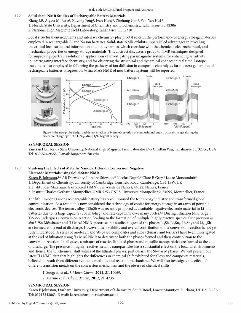
102
322 Solid-State NMR Studies of Rechargeable Battery Materials. Xiang Li1, Alyssa M. Rose1, Xuyong Feng1, Ivan Hung2, Zhehong Gan2, Yan-Yan Hu12 1. Florida State University, Department of Chemistry and Biochemistry, Tallahassee, FL 32306 2. National High Magnetic Field Laboratory, Tallahassee, FL32310
Local structural environments and interface chemistry play pivotal roles in the performance of energy storage materials employed in rechargeable Li and Na ion batteries. Solid-state NMR exhibits unparalleled advantages in revealing the critical local structural information and ion dynamics, which correlate with the chemical, electrochemical, and mechanical properties of energy storage materials. This abstract discusses a group of NMR techniques designed for improving spectral resolution in applications of investigating paramagnetic systems, for enhancing sensitivity in interrogating interface chemistry, and for observing the structural and dynamical changes in real time. Isotope tracking is also employed in following the pathway of ion diffusion in composite electrolytes for the next generation of rechargeable batteries. Progress on in situ MAS NMR of new battery systems will be reported.
Figure 1: the new probe design and demonstration of in situ observation of compositional and structural changes during the discharge-charge cycle of a LiNi0.5Mn1.5O4/Li bagcell battery.
SSNMR ORAL SESSIONYan-Yan Hu, Florida State University, National High Magnetic Field Laboratory, 95 Chieftan Way, Tallahassee, FL 32306, USA Tel: 850-524-9568, E-mail: [email protected]
323 Studying the Effects of Metallic Nanoparticles on Conversion Negative Electrode Materials using Solid-State NMR. Karen E. Johnston,1,2 Ali Darwiche,3 Lorenzo Stievano,3 Nicolas Dupré,2 Clare P. Grey,1 Laure Monconduit3 1. Department of Chemistry, University of Cambridge, Lensfield Road, Cambridge, CB2 1EW, UK 2. Institut des Matériaux Jean Rouxel (IMN), Université de Nantes, 44322, Nantes, France 3. Institut Charles Gerhardt Montpellier-UMR 5253 CNRS, Université Montpellier 2, 34095, Montpellier, France
The lithium-ion (Li-ion) rechargeable battery has revolutionised the technology industry and transformed global communication. As a result, it is now considered the technology of choice for energy storage in an array of portable electronic devices. The ternary alloy TiSnSb was recently proposed as a suitable negative electrode material in Li-ion batteries due to its large capacity (550 mA∙h/g) and rate capability over many cycles.1,2 During lithiation (discharge), TiSnSb undergoes a conversion reaction, leading to the formation of multiple, highly reactive species. Our previous in situ 119Sn Mössbauer and 7Li MAS NMR spectroscopic studies suggested the phases Li3Sb, Li7Sn2, Li7Sn3 and Li2−xSb are formed at the end of discharge. However, their stability and overall contribution to the conversion reaction is not yet fully understood. A series of model Sn and Sb-based composites and alloys (binary and ternary) have been investigated at the end of lithiation using 7Li MAS NMR to determine both the phases formed and their contribution to the conversion reaction. In all cases, a mixture of reactive lithiated phases and metallic nanoparticles are formed at the end of discharge. The presence of highly reactive metallic nanoparticles has a substantial effect on the local Li environments and, hence, the 7Li chemical shift values of the lithiated phases, particularly the Sb-based phases. We will present our latest 7Li NMR data that highlights the differences in chemical shift exhibited for alloys and composite materials, believed to result from different synthetic methods and reaction mechanisms. We will also investigate the effect of different transition metals on the conversion mechanism and the observed chemical shifts.
1. Sougrati et al., J. Mater. Chem., 2011, 21, 10069.2. Marino et al., Chem. Mater., 2012, 24, 4735.
SSNMR ORAL SESSIONKaren E Johnston, Durham University, Department of Chemistry, South Road, Lower Mountjoy, Durham, DH1 3LE, GB Tel: 01913342063, E-mail: [email protected]
103
et al.: 58th RMCMR Final Program and Abstracts
Published by Digital Commons @ DU, 2016

103
326 Vaughan Lecture – Local and Medium Range Order and Disorder as Viewed by NMR: Concepts, Methods and Applications. Dominique Massiot CNRS, Paris, France and CEMHTI CNRS, Orléans, France
Amorphous or disordered materials are intrinsically defined by a lack of order, which typically refers to an idealized description of a corresponding crystalline phase that seldom exists in real materials. Moreover the qualification of long-range ordering strongly depends upon the characterization techniques (e.g. cross section contrast in diffraction experiments).
When many materials properties (mechanical, optical, etc…) are recognized to depend upon the controlled introduction of either disorder in ordered structure or order in disordered structure (solid-solutions, defects, phase separation, chemical or structural heterogeneity), Nuclear Magnetic Resonance [NMR] spectroscopy has this unique ability to describe the actual order starting at the local scale of the coordination sphere of the observed family of atoms that are selectively observed. Starting from this local viewpoint, the further use of through-space and through-bond interactions allows extending the description of local-order to larger scales relevant to medium- or long-range ordering and to evidence structural or topological motifs that can hardly be addressed through other techniques. These new bias of observation can be used together with other experimental or computational approaches to reach a more comprehensive multi-scale description of materials.
SSNMR ORAL SESSIONDominique Massiot, CNRS, 3 rue Michel Ange, Paris, N/A, 75016, FR E-mail: [email protected]
327 On The Potential of Optically-pumped and Microwave-driven DNP of Diamonds in Solid-State and Dissolution13C NMR. Lucio Frydman Chemical Physics Department, Weizmann Institute of Science, 76100 Rehovot, Israel
Dynamic nuclear polarization (DNP) is a versatile option to improve the sensitivity of NMR and MRI. This versatility has elicited interest in overcoming potential limitations of the technique, including the achievement of solid-state polarization enhancement at room temperature, and the maximization of 13C signal lifetimes for performing in vivo MRI scans. This study explores whether diamond’s 13C behavior in powders and single crystals, could be used to achieve these ends. The characteristics of diamond’s nuclear polarization enhancement by both optical pumping of NV centers and by microwave-driven saturation of P1 electronic defects, were analyzed for different environments ranging from cryogenic to ambient temperatures. An approach for achieving efficient electron-13C spin-alignment transfers, compatible with a broad range of magnetic field strengths and field orientations with respect to the diamond crystal, was devised by combining coherent microwave- and incoherent laser-induced transitions between selected energy states of the coupled electron–nuclear spin manifold. It was also found that 13C NMR signals could be boosted by orders of magnitude in either low- or room-temperature solid-state experiments, utilizing conventional microwave-driven DNP. We attribute this behavior to the unusually long electronic/nuclear spin-lattice relaxation times characteristic of diamond, coupled to a time-independent cross-effect-like polarization transfer mechanism facilitated by a matching of the nitrogen-related hyperfine coupling and the 13C Zeeman splitting. The efficiency of this solid-state polarization process, however, is harder to exploit in dissolution DNP-enhanced MRI contexts. The prospects for utilizing polarized diamonds for both solid and solution applications will be briefly discussed.
SSNMR ORAL SESSIONLucio Frydman, Weizmann Institute, 234 Herzl Street, Rehovot, 76100, Israel Tel: 505109654, E-mail: [email protected]
104
Rocky Mountain Conference on Magnetic Resonance, Vol. 58 [2016], Art. 1
https://digitalcommons.du.edu/rockychem/vol58/iss1/1

104
328 Methodological Developments in Solid-State NMR with Applications in Catalysis and Energy Materials. A.P.M. Kentgens, M. Blaakmeer, R. Dervisoglu, E.R.H. van Eck. W. Franssen, M. Goswami, J.W.G. Janssen, G.A. de Wijs Institute for Molecules and Materials, Radboud University, Heyendaalseweg 135, 6525 AJ Nijmegen, the Netherlands
The high external magnetic fields that have become available for NMR, combined with small diameter fast MAS rotors, have made it possible to acquire well-resolved MAS NMR spectra of quadrupolar nuclei, even for very distorted sites with large quadrupolar interactions, particularly when combined with sensitivity enhancements. It will be shown how these methods are used for the study of Ziegler-Natta Catalysts. Ziegler-Natta catalysts are complex multicomponent systems which, besides the MgCl 2 support and the active titanium centres, contain electron donors and aluminium co-catalysts.
For cases were very large quadrupolar frequencies are encountered, i.e. when the available spinning speed becomes significantly less than the residual MAS line width, spectral resolution is lost. We have been exploring nutation NMR using micro-coil technology allowing rf-field strengths of hundreds of kHz to MHz. Pulse transients have very detrimental effects on the spectra, however. We will present approaches to circumvent these problems by appropriate pulse shaping. High rf-field nutation spectroscopy may bridge the gap between MAS NMR and NQR.
Materials in relation to energy storage and conversion are of prime interest. Here local order, kinetics and structural integrity as a function of composition play a major role. We study the order/disorder in traditional III-V semiconductor materials and hybrid organic-inorganic halide perovskites. These have become the focus of attention because of the astonishing performance of solar cells based on these materials.
In devices these materials find their application as thin films. NMR studies of thin films have been severely restricted. Therefore we introduced the stripline geometry which has a favourable sensitivity and scalability to the actual sample dimensions[3]. In addition to static wide-line studies of quadrupolar nuclei and protons we are now working on a setup that allows thin-film MAS studies of individual films piggybacked onto a regular rotor.
SSNMR ORAL SESSIONArno P.M. Kentgens, Radboud University , Heyendaalseweg 135, Nijmegen, Gld, 6525AJ, NL E-mail: [email protected]
329 Combined Solid-State NMR and Molecular Dynamics Investigation of the Structure of Sr-, Ba- or Zn-Aluminosilicate Glasses. P. Florian1, T. Charpentier2, K. Okhotnikov2, A.Novikov1,3 D. Neuville3 1. CNRS, CEMHTI UPR3079, Univ. Orléans, F-45071 Orléans, France 2. CEA, IRAMIS, NIMBE, CEA-CNRS UMR3685, 91191 Gif-sur-Yvette, France 3. CNRS-IPGP, Paris Sorbonne Cité, 1 rue Jussieu, 75005 Paris, France
If most technological glasses, glass-ceramics or geological materials have properties dependent upon their molten state, our present knowledge of their structure (and dynamics…) remains limited due to both technical difficulties and theoretical complexities. This is particularly true for aluminosilicates widely used in the glass industry (cover glass for high-end display devices, high strength material with moderate expansion for radomes, zero expansion glass-ceramics …) mainly on the ground of empirical knowledge.
We have started the investigation of largely unknown SiO2-Al2O3-SrO, -ZnO and -BaO based glass compositions, looking at the impact of “network-former” composition ([SiO2]/[Al2O3] ratio) as well as the nature of the “modifier” cation (Ba, Sr or Zn). Various types of 1D and 2D Solid-State NMR experiments were performed to clarify the type of species present in those glasses, their connectivity and cation location within this network. Those include 27Al high-field (20.0 T) MAS and MQMAS, 29Si CPMG, 27Al/27Al INADEQUATE, 27Al/29Si INEPT and D-HMQC. For composition on the compensation line, aluminum environments are found to be only lightly impacted by the composition whereas silicon spectra show great variations, due to the competition between Si-O-Si and Si-O-Al linkages. Both types of spectra do not resolve all of the individual species, but MD simulations allow us to disentangle those and get a detailed insight into their structure. 2D Al/Si correlations show preferential linkages between silicon and aluminum species which are dependent upon the nature of the cation involved (e.g. Sr or Ba).
Our methodology allows quantifying the local and medium-range order of those amorphous materials by comparison of the 1D and 2D NMR results with first-principle NMR parameters calculations performed on Molecular Dynamic derived structures.
SSNMR ORAL SESSIONPierre Florian, CEMHTI-CNRS, 1D av. Recherche Scientifique, CS 90055, Orleans, Centre, 45071, FR E-mail: [email protected]
105
et al.: 58th RMCMR Final Program and Abstracts
Published by Digital Commons @ DU, 2016

105
330 2D NMR Measurement and Prediction of Full Paramagnetic Shift Tensors of Quadrupolar Nuclei. Brennan Walder, Alex Peterson, Philip J. Grandinetti Department of Chemistry, Ohio State University, Columbus, OH, USA
Using the symmetry pathways formalism, we analyzed various 2D NMR methods for separating and correlating the paramagnetic shift and quadrupolar coupling frequency contributions in static polycrystalline samples. We illustrate our approach on a series of polycrystalline MCl2•2 D2O samples (M=Cu, Ni, Co, Fe, Mn), using a simple motional model of rapid hopping of D2O about their C2 axes for relating the observed motionally averaged quadrupolar and paramagnetic shift tensors to their instantaneous tensors. For the quadrupolar coupling a simple model for the efg tensor orientation along the O--D bond predicts a coupling in excellent agreement with previously measured values. Our analysis of the full deuterium paramagnetic shift tensor reveals that a delocalized point dipole model can be sufficient for predicting the paramagnetic shift tensor given an appropriate model for the molecular magnetic susceptibility tensor of the paramagnetic site.
SSNMR ORAL SESSIONPhilip J Grandinetti, Ohio State University, 100 West 18th Avenue, Columbus, OH 43212, USA Tel: 614-216-6232, E-mail: [email protected]
331 Looking into the Structure and Reactivity of Hybrid Materials Involving Boronates and Benzoxaborolates. Christel Gervais,1 Christian Bonhomme,1 Dorothée Berthomieu,2 Saad Sene,2 Marie-Alix Pizzoccaro,2 Mark E. Smith,3 Danielle Laurencin2 1. LCMCP, CNRS UMR 7574, Université Pierre et Marie Curie, Paris, France 2. ICGM, CNRS UMR 5253, Université de Montpellier, Montpellier, France 3. Vice chancellor’s office, University of Lancaster, UK.
Due to the unique reactivity of their organoboron group, boronic acids and benzoxaboroles have become essential molecules for both molecular chemistry and materials science. Their range of applications spans from organic synthesis (Suzuki coupling, organocatalysis…), to the design of sensors for saccharides and the development of new drugs.
In recent years, we have focused on hybrid materials involving boronates and benzoxaborolates. We have shown that these anions can be used as ligands for the construction of coordination polymers,1,2 as charge-balancing species in layered-double hydroxides,2 or even as functional groups grafted at the surface of hydroxyapatite.3 For each family of materials, solid state NMR has been an essential tool of analysis, unveiling not only the local structure around the organoboron anions, but also several aspects of their reactivity. The purpose of this presentation will be to describe the NMR studies performed, looking at 11B, 13C, 19F, 17O, 27Al, 25Mg, 87Sr and/or 43Ca (depending on the material), and working in some cases at ultra-high magnetic fields (20 T). The importance of carrying out computational modeling in conjunction with GIPAW calculations of NMR parameters will also be discussed, especially in the case of the intercalated layered double hydroxide phases.
1- S. Sene et al, Chem. Eur. J. 2013, 19, 880.2- S. Sene et al, Chem. Mater. 2015, 27, 1242.3- M. A. Pizzoccaro et al, 2016, submitted.
SSNMR ORAL SESSIONDanielle Laurencin, Institut Charles Gerhardt - CNRS UMR 5253, Université de Montpellier, Montpellier, Languedoc-Roussillon, 34095, FR E-mail: [email protected]
106
Rocky Mountain Conference on Magnetic Resonance, Vol. 58 [2016], Art. 1
https://digitalcommons.du.edu/rockychem/vol58/iss1/1

106
334 Towards Spin-assisted Long-term Data Storage in Diamond. Siddharth Dhomkar1,†, Jacob Henshaw1,2,†, Harishankar Jayakumar1, Carlos A. Meriles1,2 1Dept. of Physics, CUNY-City College of New York, New York, NY 10031, USA 2CUNY-Graduate Center, New York, NY 10016, USA † Equally contributing authors.
The negatively-charged nitrogen-vacancy (NV-) center in diamond is the focus of widespread attention for applications ranging from quantum information processing to nanoscale metrology. Although most work so far has focused on the NV- optical and spin properties, control of the charge state promises complementary opportunities. One intriguing possibility is the long-term storage of information, a notion we hereby introduce using NV rich, type-1b diamond. As a proof of principle, we use multi-color optical microscopy to read, write, and reset arbitrary data sets with 2-D binary bit density comparable to present digital-video-disk (DVD) technology. Leveraging on the singular dynamics of NV- ionization, we encode information on different planes of the diamond crystal with no cross talk, hence extending the storage capacity to three dimensions. Further, we correlate the center’s charge state and nuclear spin polarization of the nitrogen host, and show that the latter is robust to a cycle of NV- ionization and recharge. In combination with super-resolution microscopy techniques, these observations provide a route towards sub-diffraction NV charge control, a regime where the storage capacity could significantly surpass present technology.
SSNMR/EPR ORAL SESSIONCarlos A Meriles, CUNY - City College of New York, 160 Convent Ave, New York, NY - New York,10031, USA Tel: 212-650-5625, E-mail: [email protected]
335 Electron Spectral Diffusion Measured via ELDOR for DNP at 7 T. Alisa Leavesley,1 Ilia Kaminker,1 Marie Ramirez-Cohen,2 Shimon Vega,2 Daniella Goldfarb,2 Songi Han1 1. Department of Chemistry, UC Santa Barbara, Santa Barbara, California 2. Department of Chemical Physics, Weizmann Institute of Science, Rehovot, Israel
Solid-state dynamic nuclear polarization (DNP) is an increasingly popular technique that allows for hundreds fold increases in nuclear magnetic resonance (NMR) signal. The common sample preparation includes a solute of interest mixed with a stable radical at tens of mM concentration frozen into an aqueous glass. Upon on-resonance µw irradiation, the high electron polarization of the radical is transferred to the surrounding solvent nuclei and subsequently to the solute via spin diffusion processes. Recently the indirect cross effect was proposed as a primary mechanism for DNP in static samples at low temperatures, 3-40 K, and high radical concentrations, 20-40 mM, where this mechanism relies on the electron spectral diffusion process. It was demonstrated at 3.35 T that spectral diffusion can be characterized and quantified using electron double resonance (ELDOR) experiments.1,2 We have recently shown that the oversaturation effect, i.e. reduction of DNP enhancement for µw powers above a certain threshold, occurs at 7 T and low < 6 K temperatures.3 Here, we present a DNP / ELDOR study performed on our homebuilt dual DNP / EPR instrument at 7 T, 3 of the electron spectral diffusion dependence on experimental conditions such as µw power, irradiation length, temperature, and radical concentration. These results are discussed in connection with the oversaturation effect and static, low temperature DNP mechanisms and show the necessity of including electron spectral diffusion processes for understanding of the latter.
[1] Hovav, Y.; Kaminker, I.; Shimon, D.; Feiuch, A.; Goldfarb, D.; Vega, S. Phys. Chem. Chem. Phys. 2015, 17, 226-244.
[2] Hovav, Y.; Shimon, D.; Kaminker, I.; Feituch, A.; Goldfarb, D.; Vega, S. Phys. Chem. Chem. Phys. 2015, 17, 6053-6065.
[3] Siaw, T.; Leavesley, A.; Lund, A.; Kaminker, I.; Han, S. J. Magn Reson. 2016, 264, 131-153.
SSNMR/EPR ORAL SESSIONAlisa Leavesley, University of California Santa Barbara, 935 E. Canon Perdido St. Unit A, Santa Barbara, CA,93103, USA E-mail: [email protected]
107
et al.: 58th RMCMR Final Program and Abstracts
Published by Digital Commons @ DU, 2016

107
336 Hypersensitivity with Dynamic Nuclear Polarization: Natural Isotopic Abundance and Closed-loop Cryogenic Helium Sample Spinning. K. Märker,1 F. Mentink-Vigier,1 S. Paul,1 A. Purea,3 F. Engelke,3 E. Bouleau,1 Sabine Hediger,1,2 Daniel Lee,1 Gaël De Paëpe1 1. INAC, CEA & Univ. Grenoble Alpes, F-38000, France 2. INAC, CNRS, F-38000, France 3. Bruker Biospin GmbH, Rheinstetten, Germany
The ability to record correlation experiments for nuclei at low natural isotopic abundance (13C, 15N) using dynamic nuclear polarization (DNP) [1] shows great promise for the field of NMR crystallography. The low natural abundance statistically simplifies coupled systems to spin pairs, making the measurement of inter-nuclear distances more straightforward. Furthermore, these simplified 2-spin cases permit the measurements of long distances as dipolar truncation effects (seen in multi-spin systems) are not present. It will be shown that dipolar recoupling experiments can be used to obtain not only intra-molecular distance restraints but also inter-molecular distances. Examples will be shown on natural isotopic abundance self-assembled systems, such as certain peptides and guanosine derivatives, where p-stacking interactions and hydrogen-bonding play a large role in the crystal structure – which cannot be easily determined from conventional crystallography methods.[2]
In order to further enhance the sensitivity, we will report on a strategy to push the limits of DNP-enhanced solid-state NMR beyond its current state-of-the-art. This leap-forward was made possible thanks to the employment of a closed-loop of cryogenic helium as the gas to power magic angle sample spinning (MAS) for DNP-enhanced NMR experiments. The experimental conditions reported here far exceed what is currently possible and allows reaching sample temperatures down to 30 K while conducting experiments with high spinning frequencies (up to 25 kHz @ 100 K for a 3.2 mm probe). Thanks to the impressive associated gains, which will be presented, sustainable cryogenic helium sample spinning significantly enlarges the realm and possibilities of the MAS-DNP technique and is the route to transform NMR into a versatile and sensitive atomic-level characterization tool.[3]
Finally, we will describe our efforts towards understanding the origin of the polarization losses associated with radical doping. Notably, we will investigate NMR signal losses occurring during MAS-DNP experiments and specifically compare the (MAS-dependent) depolarization effect for several “gold-standard” (bi-)radicals currently in use in most MAS-DNP studies (e.g. Totapol/bTbK/Amupol/TEMTriPol/etc.). Using MAS-DNP simulations we will show that these observations can be rationalized and are consistent with the biradicals’ structure. Further insight into the depolarization mechanism (multi-parameters phenomenon) can be obtained comparing the result for each crystallite orientation with the result obtained on the powder average.[4]
[1] Takahashi H. et al., Angewandte Chemie International Edition, 51, 11766-11769, 2012[2] K. Märker et al., Journal of the American Chemical Society, 137, 13796-13799, 2015 [3] Bouleau E. et al., Chemical Science, 6, 6806-6812, 2015 [4] Mentink-Vigier F., et al., Physical Chemistry Chemical Physics, 17, 21824-21836, 2015
SSNMR/EPR ORAL SESSIONGaël De Paëpe, INAC (CEA - Grenoble Alpes University), 17, rue des martyrs, Grenoble, Rhône-Alpes, 38054, FR E-mail: [email protected]
337 Combining Dynamic Nuclear Polarization and Mechanically Detected Magnetic Resonance to Achieve Nanoscale Magnetic Resonance Imaging of Individual Biomolecules and Assemblies. Corinne E. Isaac, Hoang L. Nguyen, Pamela T. Nasr, Elizabeth A. Curley, Michael C. Boucher, John A. Marohn Department of Chemistry and Chemical Biology, Cornell University, Ithaca, New York 14853-1301 USA
A tool for imaging the proton envelope of an individual macromolecule and biological assembly would be a major advance. To realize this opportunity, we are pushing magnetic resonance imaging (MRI) to nanometer resolution using the sensitivity afforded by detecting magnetic resonance mechanically. We have developed attonewton-sensitivity cantilevers with integrated nanomagnet tips capable of detecting nuclear magnetic resonance from a polymer film at cryogenic temperatures with a sensitivity of a few hundred proton magnetic moments.1 These experiments observe magnetization fluctuations present in small, nanometer-scale volumes of spins. I will describe our efforts to significantly improve the per-spin sensitivity and acquisition time of “nano-MRI” experiments by using dynamic nuclear polarization (DNP) to create a measurable net nuclear spin polarization,2 pushing the experiment out of the spin-noise limit. Even with a noiseless detector (e.g., a zero-temperature cantilever or a quantum-limited nitrogen-vacancy center), stochastic spin fluctuations in the sample still limit the imaging resolution achievable in a nano-MRI experiment. We have performed numerical simulations of nano-MRI experiments with a number of image-encoding and detection protocols
108
Rocky Mountain Conference on Magnetic Resonance, Vol. 58 [2016], Art. 1
https://digitalcommons.du.edu/rockychem/vol58/iss1/1

108
to identify the conditions under which DNP can get us beyond the limits imposed by spin-noise. Taken together, our experiments and simulations suggest that magnetic resonance force microscopy is on its way to becoming a powerful new route for obtaining a three-dimensional image of a single copy of a globular protein, macromolecular complex, and membrane protein.
[1] Longenecker, J. G.; et al.; Marohn, J. A. ACS Nano, 2012, 6, 9637 – 9645; URL http://dx.doi.org/10.1021/nn3030628
[2] Isaac, C. E.; et al.; Marohn, J. A. Phys. Chem. Chem. Phys., 2016, 18, 8806 – 8819; URL http://dx.doi.org/10.1039/C6CP00084C
SSNMR/EPR ORAL SESSIONJohn A. Marohn, Cornell University, 150 Baker Laboratory, Ithaca, NY 14853-1301, USA Tel: 607-255-2004, E-mail: [email protected]
338 Electron Spin Decoupled NMR Driven by Electron Spin Relaxation of Spin Clusters. Ting Ann Siaw,1 Blake Wilson,2 Songi Han1,3 1. Department of Chemistry and Biochemistry, University of California Santa Barbara, Santa Barbara, CA 2. Department of Physics, University of California Santa Barbara, Santa Barbara, CA 3. Department of Chemical Engineering, University of California Santa Barbara, CA
The dramatic benefit of dynamic nuclear polarization (DNP) to amplify nuclear magnetic resonance (NMR) signal is fundamentally countered by line broadening and signal loss due to paramagnetic effects exerted by DNP agents, compromising NMR spectral resolution and sensitivity. Electron spin decoupling would principally eliminate these paramagnetic effects, but has not been experimentally accessible at high magnetic fields. We show that continuous wave microwave excitation of electron spin clusters in a Li ion battery electrolyte system at ~4 K results simultaneously in electron spin decoupling and DNP enhancement, removing paramagnetic effects on 7Li NMR while providing signal enhancements of ~5-20 fold. EPR measurements reveal that favorable electron spin relaxation (long T1e, short TM) are responsible for a broad excitation of the EPR line, measured directly using electron double resonance (ELDOR) experiments. Additionally, concurrent paramagnetic NMR and DNP analysis concluded that the favorable electron spin relaxation properties originated from coordination of TEMPO-based nitroxides to the Li ion, generating spin clusters. The DNP study here implicates that DNP analysis could be a materials characterization method for detecting clustering in materials systems.
SSNMR/EPR ORAL SESSIONTing Ann Siaw, University of California Santa Barbara, Department of Chemistry and Biochemistry, UC Santa Barbara, Santa Barbara, CA, 93106, USA Tel: 805-893-2792, E-mail: [email protected]
339 Nanoscale NMR Detection and Imaging Using Nitrogen-vacancy Centers in Diamond. John Mamin, Mark Sherwood, Moonhee Kim, Daniel Rugar IBM Almaden Research Center, San Jose, CA 95120
We are exploring the use of individual, near-surface nitrogen-vacancy (NV) centers in diamond as atomic-size magnetometers to detect proton NMR in organic material located external to the diamond. Using a combination of electron spin echoes and proton spin manipulation, the NV center senses the nanotesla field fluctuations from the protons, enabling both time-domain and spectroscopic NMR measurements on the nanometer scale. By scanning a small polymer test object past a near-surface NV center, we have recently demonstrated proton magnetic resonance imaging (MRI) with spatial resolution on the order of 10 nm. One key issue in NV-NMR experiments is the loss of spin coherence when the NV center is located near the diamond surface. Although this loss of coherence is frequently attributed to the effect of magnetic noise emanating from unpaired spins on the diamond surface, we will show evidence that electric field noise from fluctuating surface charge can be a significant factor.
SSNMR/EPR ORAL SESSIONDaniel Rugar, IBM Almaden Research Center, 650 Harry Rd., San Jose, CA 95120, USA E-mail: [email protected]
109
et al.: 58th RMCMR Final Program and Abstracts
Published by Digital Commons @ DU, 2016
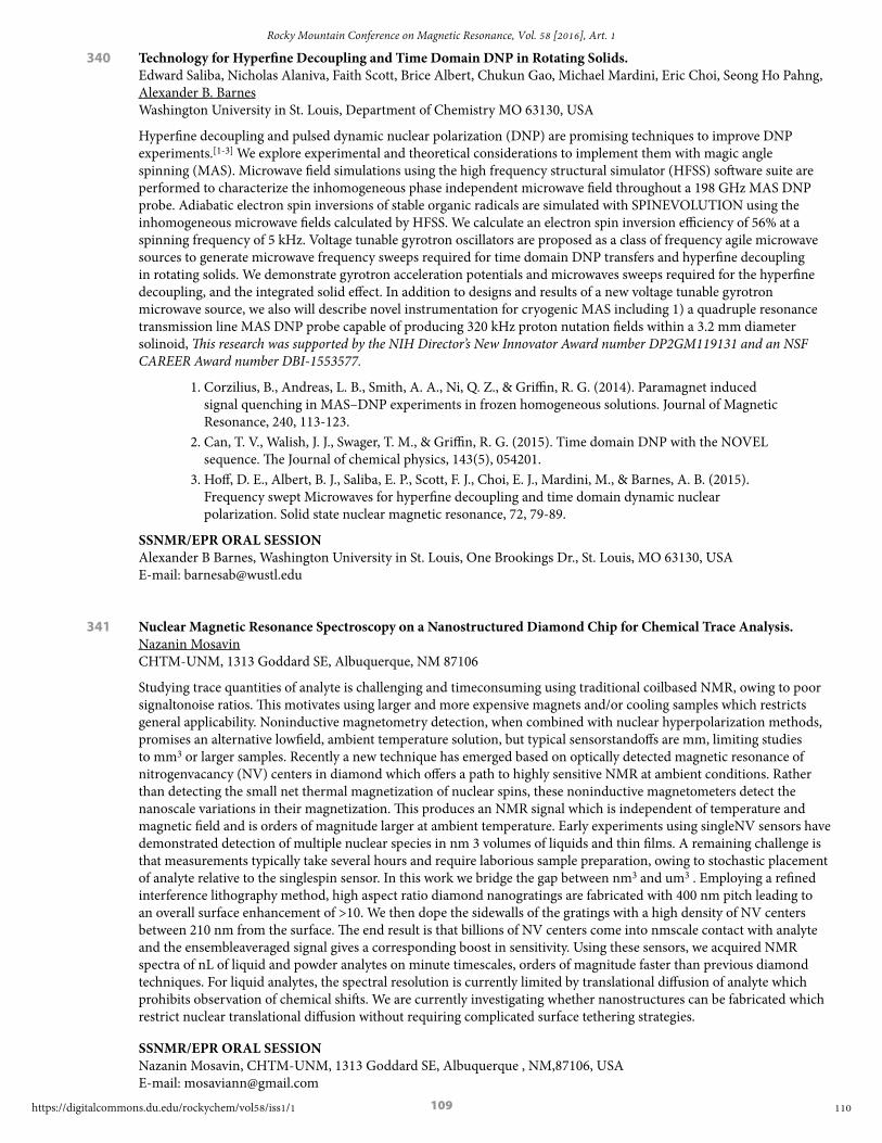
109
340 Technology for Hyperfine Decoupling and Time Domain DNP in Rotating Solids. Edward Saliba, Nicholas Alaniva, Faith Scott, Brice Albert, Chukun Gao, Michael Mardini, Eric Choi, Seong Ho Pahng, Alexander B. Barnes Washington University in St. Louis, Department of Chemistry MO 63130, USA
Hyperfine decoupling and pulsed dynamic nuclear polarization (DNP) are promising techniques to improve DNP experiments.[1-3] We explore experimental and theoretical considerations to implement them with magic angle spinning (MAS). Microwave field simulations using the high frequency structural simulator (HFSS) software suite are performed to characterize the inhomogeneous phase independent microwave field throughout a 198 GHz MAS DNP probe. Adiabatic electron spin inversions of stable organic radicals are simulated with SPINEVOLUTION using the inhomogeneous microwave fields calculated by HFSS. We calculate an electron spin inversion efficiency of 56% at a spinning frequency of 5 kHz. Voltage tunable gyrotron oscillators are proposed as a class of frequency agile microwave sources to generate microwave frequency sweeps required for time domain DNP transfers and hyperfine decoupling in rotating solids. We demonstrate gyrotron acceleration potentials and microwaves sweeps required for the hyperfine decoupling, and the integrated solid effect. In addition to designs and results of a new voltage tunable gyrotron microwave source, we also will describe novel instrumentation for cryogenic MAS including 1) a quadruple resonance transmission line MAS DNP probe capable of producing 320 kHz proton nutation fields within a 3.2 mm diameter solinoid, This research was supported by the NIH Director’s New Innovator Award number DP2GM119131 and an NSF CAREER Award number DBI-1553577.
1. Corzilius, B., Andreas, L. B., Smith, A. A., Ni, Q. Z., & Griffin, R. G. (2014). Paramagnet induced signal quenching in MAS–DNP experiments in frozen homogeneous solutions. Journal of Magnetic Resonance, 240, 113-123.
2. Can, T. V., Walish, J. J., Swager, T. M., & Griffin, R. G. (2015). Time domain DNP with the NOVEL sequence. The Journal of chemical physics, 143(5), 054201.
3. Hoff, D. E., Albert, B. J., Saliba, E. P., Scott, F. J., Choi, E. J., Mardini, M., & Barnes, A. B. (2015). Frequency swept Microwaves for hyperfine decoupling and time domain dynamic nuclear polarization. Solid state nuclear magnetic resonance, 72, 79-89.
SSNMR/EPR ORAL SESSIONAlexander B Barnes, Washington University in St. Louis, One Brookings Dr., St. Louis, MO 63130, USA E-mail: [email protected]
341 Nuclear Magnetic Resonance Spectroscopy on a Nanostructured Diamond Chip for Chemical Trace Analysis. Nazanin Mosavin CHTM-UNM, 1313 Goddard SE, Albuquerque, NM 87106
Studying trace quantities of analyte is challenging and time consuming using traditional coil based NMR, owing to poor signal to noise ratios. This motivates using larger and more expensive magnets and/or cooling samples which restricts general applicability. Non inductive magnetometry detection, when combined with nuclear hyperpolarization methods, promises an alternative low field, ambient temperature solution, but typical sensor standoffs are mm, limiting studies to mm3 or larger samples. Recently a new technique has emerged based on optically detected magnetic resonance of nitrogen vacancy (NV) centers in diamond which offers a path to highly sensitive NMR at ambient conditions. Rather than detecting the small net thermal magnetization of nuclear spins, these non inductive magnetometers detect the nanoscale variations in their magnetization. This produces an NMR signal which is independent of temperature and magnetic field and is orders of magnitude larger at ambient temperature. Early experiments using single NV sensors have demonstrated detection of multiple nuclear species in nm 3 volumes of liquids and thin films. A remaining challenge is that measurements typically take several hours and require laborious sample preparation, owing to stochastic placement of analyte relative to the single spin sensor. In this work we bridge the gap between nm3 and um3 . Employing a refined interference lithography method, high aspect ratio diamond nanogratings are fabricated with 400 nm pitch leading to an overall surface enhancement of >10. We then dope the sidewalls of the gratings with a high density of NV centers between 2 10 nm from the surface. The end result is that billions of NV centers come into nm scale contact with analyte and the ensemble averaged signal gives a corresponding boost in sensitivity. Using these sensors, we acquired NMR spectra of nL of liquid and powder analytes on minute timescales, orders of magnitude faster than previous diamond techniques. For liquid analytes, the spectral resolution is currently limited by translational diffusion of analyte which prohibits observation of chemical shifts. We are currently investigating whether nanostructures can be fabricated which restrict nuclear translational diffusion without requiring complicated surface tethering strategies.
SSNMR/EPR ORAL SESSIONNazanin Mosavin, CHTM-UNM, 1313 Goddard SE, Albuquerque , NM,87106, USA E-mail: [email protected]
110
Rocky Mountain Conference on Magnetic Resonance, Vol. 58 [2016], Art. 1
https://digitalcommons.du.edu/rockychem/vol58/iss1/1

110
342 Gd3+ as Polarizing Agent at High Field: Solid Effect vs Cross Effect Dynamic Nuclear Polarization. Monu Kaushik,1 Mian Qi,2 Adelheid Godt,2 Björn Corzilius1 1. Institute of Physical and Theoretical Chemistry, Institute of Biophysical Chemistry, and Center for Biomolecular Magnetic Resonance, Goethe University Frankfurt, Max-von- Laue-Str. 7-9, 60438 Frankfurt am Main, Germany 2. Faculty of Chemistry and Center for Molecular Materials, Bielefeld University, Universitätsstr. 25, 33615 Bielefeld, Germany
A variety of polarizing agents have been developed for efficient dynamic nuclear polarization (DNP) for high sensitivity gain. In this work we present studies based on a relatively new class of polarizing agents: high spin transition metal ions. Transition metal ions (Gd3+, Mn2+) can act as paramagnetic substitute of intrinsically bound diamagnetic ions in biomolecules. Doping with paramagnetic ions in this case has no (or insignificant) effect on the structure of the biomolecule. This gives an opportunity to obtain site-specific information about the biomolecule and further the research in structural biology. The polarization transfer mechanisms for these polarizing agents are yet to be understood. Here, we demonstrate DNP effects via Gd-DOTA, which invokes solid effect at low concentration owing to its narrow linewidth. Deviation from pure solid effect mechanism at shorter inter-metal distance in the uniform frozen solution matrix is observed. The properties of Gd3+ being a high spin 7/2 system featuring a relatively strong zero-field (electron quadrupolar) interaction lead to a non-trivial consequences. In our attempts to shed light on underlying polarization transfer mechanisms, bis-Gd rigid model complexes are investigated. By variation of the molecular tether length between the chelator moieties we are able to investigate the distance dependence of DNP field profiles and enhancements. This study enables us to comment on designing complexes for efficient CE DNP.
SSNMR/EPR ORAL SESSIONMonu Kaushik, Goethe University Frankfurt, Max-von-Laue-Str. 7, Frankfurt am Main, Hessen,60439, DE E-mail: [email protected]
343 Advancing NMR of Membrane Proteins in the Lipid Bilayer Membrane. Francesca Marassi Sanford Burnham Prebys Medical Discovery Institute
The molecular structures of bacterial membrane proteins are critical for developing new treatments. Through their interactions with human host ligands, bacterial membrane proteins mediate first-line processes that are critical for virulence. However, notwithstanding the valuable insights they offer, studies in crystals and detergents are limited; they do not provide either complete or accurate views of structure and function, and preclude examination of Ail-ligand interactions. By contrast, NMR can provide structural information in detergent-free lipid bilayer membranes. Here we present recent results obtained for a membrane protein critical for Yersinia pestis virulence. Use of the full range of NMR methods - solution NMR, solid-state magic angle spinning NMR, and solid-state oriented sample NMR - widens the range of conformational dynamics that can be characterized. Optimized samples enable high resolution solid-state NMR spectra to be obtained for Ail incorporated in liposomes, sedimented nanodiscs and macroscopically aligned bilayers. The spectra have resolution comparable to the solution NMR spectra of Ail in small nanodiscs, enabling resonance assignments to be made and structural restraints to be measured.
Supported by NIH grants: GM100265, GM110658, CA179087, EB002031.
SSNMR ORAL SESSIONFrancesca M Marassi, Sanford Burnham Prebys Medical Discovery Institute, 10901 North Torrey Pines Rd, La Jolla, CA 92037, USA Tel: 858-795-5282, E-mail: [email protected]
111
et al.: 58th RMCMR Final Program and Abstracts
Published by Digital Commons @ DU, 2016

111
344 Effect of the Lipid Composition and Bilayer Viscosity on the Structure and Dynamics of Nanopore-Aligned Membrane Proteins as Revealed by Solid-State NMR. Alex I. Smirnov, Morteza Jafarabadi, Antonin Marek, Deanna M. Tesch, Alexander A. Nevzorov Department of Chemistry, North Carolina State University, 2620 Yarbrough Drive, Raleigh, NC 27695-8204
Lipid bilayers immobilized within anodic aluminum oxide (AAO) nanopores exhibit an exceptionally high degree of alignment and can be made of virtually any lipid composition [1]. This makes it possible to study lipid-induced conformational changes of membrane proteins over a wide range of environmental conditions such as temperature, pH, ionic strength, as well as bilayer viscosity. Further improvement of spectral resolution for membrane proteins reconstituted in lipid bilayers was made possible by optimizing the sample preparation techniques and the use of the sensitivity-enhancement REP-CP sequence [2]. We present spectra of Pf1 coat protein reconstituted in AAO-supported lipid bilayers that exhibit the quality comparable to magnetically aligned DMPC/DHPC bicelles and allowing for a detailed analysis of the protein conformation and dynamics. 2D SAMPI4 spectra of Pf1 coat protein reconstituted in DOPC, POPC and DMPC bilayers aligned within AAO nanopores revealed marked differences attributed to the changes in the overall helix tilt (while preserving the kinked helix structure). In addition, we report on the effect of lipid saturation on the bilayer viscosity and Pf1 dynamics as evidenced by the changes in the linewidths in the Pf1 spectra, which are directly affected by the uniaxial rotational diffusion of the protein within the membrane. A quantitative model that extracts the diffusion coefficients from the experimental linewidths has been created.
[1] Marek, A.; Tang, W. X.; Milikisiyants, S.; Nevzorov, A. A.; Smirnov, A. I. Nanotube Array Method for Studying Lipid-Induced Conformational Changes of a Membrane Protein by Solid-State NMR, Biophysical Journal 108 (2015), 5-9.[2] Koroloff, S. N.; Nevzorov, A. A. Optimization of Cross-polarization at Low Radiofrequency Fields for Sensitivity Enhancement in Solid-state NMR of Membrane Proteins Reconstituted in Magnetically Aligned Bicelles, Journal of Magnetic Resonance 256 (2015), 14-22.
Figure. Left. REP-CP enhanced SAMPI4 spectra of ca. 1.5 mg of Pf1 reconstituted in POPC and DMPC bilayers aligned by AAO nanopores obtained at 500 MHz 1H frequency and 45 oC; 1 k scans and 80 t1 points were acquired. Right. Calculated structures showing a preserved kink and different tilts with respect to the bilayer normal.
SSNMR POSTER SESSIONAlexander Nevzorov, North Carolina State University, 2620 Yarbrough Drive, Raleigh, NC,27513, USA E-mail: [email protected]
112
Rocky Mountain Conference on Magnetic Resonance, Vol. 58 [2016], Art. 1
https://digitalcommons.du.edu/rockychem/vol58/iss1/1

112
345 Magic Angle Spinning Solid State NMR Studies of Membrane Proteins in Synthetic Lipids and Cell Membranes. Vladimir Ladizhansky,1 Shenlin Wang,1,a Daryl Good,1 Meaghan Ward,1 I. Kawamura,1,b I. Hung,2 P. Gor’kov,2 J. Lewandowski,3 Leonid S. Brown.1 1. Department of Physics and Biophysics Interdepartmental Group, University of Guelph, Ontario, Canada 2. National High Magnetic Field Laboratory, Florida State University, Tallahassee, FL, USA 3. University of Warwick, Warwick, UK a. Current address: Beijing Nuclear Magnetic Resonance Center and College of Chemistry and Molecular Engineering, Peking University, Beijing, China 100871 b. Current address: Faculty of Engineering, Yokohama National University, Yokohama, Japan
Membrane proteins play critical roles in cells and are involved in many physiological processes. Many important aspects of their structural and dynamic organization have been revealed by X-ray crystallography and solution NMR. More challenging for these methods are the studies of membrane proteins under nearly physiological conditions of a lipid bilayer or in cell membranes. Recent technological and methodological developments in solid-state NMR have led to the possibility of conducting structural analysis of large polytopic membrane proteins. In this presentation, I will discuss our studies of a 27 kDa retinal-binding seven-helical (7TM) photoreceptor Anabaena Sensory Rhodopsin (ASR). ASR initiates a phototransduction cascade involving a soluble transducer, which regulates the expression of several proteins responsible for photosynthesis and circadian clock in cyanobacterium Anabaena sp. PCC 7120. I will discuss structural hierarchical organization of ASR in in synthetic lipids and in cellular membranes, conformational dynamics of ASR, and its possible role in the modulation of interactions with the transducer.
SSNMR ORAL SESSIONVladimir Ladizhansky, University of Guelph, 50 Stone Rd E, Guelph, Ontario, N1L 1R5, Canada E-mail: [email protected]
346 Solid-State 15N- and 19F-NMR Analysis of the Interaction of the Viral E5 Oncoprotein with the PDGF Receptor in Membranes. Dirk Windisch,1 Stephan Grage,1 Xiaojun Xu,1 Colin Ziegler,1 Parvesh Wadhwani,1 Anne S. Ulrich1,2 1. Institute of Biological Interfaces (IBG-2), Karlsruhe Institute of Technology (KIT), P.O.B. 3640, 76021 Karlsruhe, Germany 2. Institute of Organic Chemistry (IOC), Karlsruhe Institute of Technology (KIT), Fritz-Haber-Weg 6, 76131 Karlsruhe, Germany
The platelet-derived growth factor receptor (PDGFR) is a receptor tyrosine kinase that gets constitutively activated by the oncogenic E5 protein from papillomavirus, leading to uncontrolled proliferation and cancer. Bovine E5 with a length of only 44 amino acids consists largely of a transmembrane helix that can engage in specific helix-helix interactions with the transmembrane segment of PDGFR.1,2 Our aim was to elucidate the structural criteria by which these transmembrane segments recognize each other and to describe the oligomeric bundle formed in the membrane. We used solid-state 15N-NMR to characterize the structure and alignment of E5 and PDGFR in lipid bilayers, each one alone and together in the hetero-oligomeric complex.3,4 When reconstituted alone in lipid bilayers, we observed that the E5 helix is inserted almost upright in thick membranes, but it starts to tilt and gets slightly deformed in moderately thinner bilayers, and it becomes aggregated in very thin membranes due to hydrophobic mismatch. On the other hand, when reconstituted together with the receptor, E5 can compensate for the hydrophobic mismatch by binding to the transmembrane segment of the receptor. This hydrophobic mismatch behaviour may be responsible for driving the two interacting partners together within the thin membrane of the Golgi compartment (before reaching the thicker membrane environments of the plasma membrane). They can recognize each other by forming a closely packed bundle of transmembrane aligned helices. As E5 is supposed to be present as a dimer to bind to the receptor, we performed solid-state 19F-NMR CODEX and CPMG experiments to characterize the homo-oligomerization interface of this protein. We were able to detect inter-molecular distance-dependent dipolar couplings between certain pairs of 19F-labels, namely for positions 6, 17, 28 in E5. These data providing direct evidence for E5 dimerization and allowed to construct a viable model for the E5 dimer in lipid membranes.
1. Petti and DiMaio, EMBO J. 1991, 10, 845.2. Petti and DiMaio, Proc. Natl. Acad. Sci USA 1992, 89, 6736.3. Windisch et al., Biophys. J., 2015, 109 (4), 737.4. Muhle-Goll et al., J. Biol. Chem., 2012, 287 (31), 26178.
SSNMR ORAL SESSIONDirk Windisch, Karlsruhe Institute of Technology, P.O.B 3640, Karlsruhe, Baden-Wuerttemberg,76021, DE Tel: 004972160847223, E-mail: [email protected]
113
et al.: 58th RMCMR Final Program and Abstracts
Published by Digital Commons @ DU, 2016

113
347 EPR and DNP in the Same Probe: Optimizing Microwave Delivery to Small Samples for Low Power DNP. Anne M. Carroll, Catalina A. Espinosa, Darien Lee, Kurt W. Zilm Department of Chemistry, Yale University, P.O. Box 208107, New Haven, CT 06520-8107
Combining DNP with fast MAS is an attractive proposition for addressing some of the shortcomings of both techniques. By adopting a small sample, the total power needed for DNP can be reduced if it can be focused into the sample. With the addition of DNP, the low net sensitivity that accompanies microliter size samples in fast MAS rotors can potentially be offset. In this talk we will describe our approach to solving the challenge of delivering and focusing mm-wave power into small MAS samples by development of EPR and DNP NMR capability in the same probe. Two distinct systems will be described on our 7T instrument using a 60 mW diode based mm-wave source. The first is a multinuclear solution NMR probe modified for frequency swept EPR. This probe employs a dielectric waveguide to transmit mm-waves into the sensitive volume, and is used to compare how different rotor orientations and materials affect mm-wave sample penetration. The second is a triple resonance MAS and EPR probe where we demonstrate the use of a dielectric antenna as an alternative to a microwave horn for delivering mm-waves into the MAS sample. Results will be presented that demonstrate how EPR can be used to make real time adjustments to focus mm-waves into the sample and thereby optimize DNP performance. The ability to record both DNP NMR and EPR in the same configuration provides many insights into DNP NMR, especially how enhancements are affected by microwave polarization and MAS. Using this capability to optimize mm-wave delivery, we have achieved DNP enhancements over 380-fold in 13C MAS spectra of diamonds.
Support for this work by the NSF Chemical Measurement and Imaging program under grant CHE-1413096 is gratefully acknowledged.
SSNMR ORAL SESSIONKurt W Zilm, Yale University Chemistry Department, 350 Edwards Street, New Haven, CT,06511, USA Tel: 203-432-3956, E-mail: [email protected]
348 Solid-State NMR, DNP, and MD Investigations of the Organic/Inorganic Interface in Silica Biohybrids. Stephan I. Brückner,1 Sergii Donets,2 Maryna Abacilar,3 Anne Jantschke,1 Eline Koers,4 Armin Geyer,3 Rafael Gutiérrez,2 Marc Baldus,4 Eike Brunner1 1. Chair for Bioanalytical Chemistry, Department of Chemistry and Food Chemistry, TU Dresden, 01062, Dresden, Germany 2. Institute for Materials Science and Max Bergmann Center of Biomaterials, TU Dresden, 01062 Dresden, Germany 3. Faculty of Chemistry, Philipps-Universität Marburg, 35032 Marburg, Germany 4. Bijvoet Center for Biomolecular Research, Utrecht University, Padualaan 8, 3584 CH Utrecht, The Netherlands
Diatom biosilica is an inorganic/ organic hybrid with outstanding materials properties. The molecular architecture of the organic material at atomic and nanometer scale has remained unknown. A dynamic nuclear polarization (DNP)-supported solid-state NMR approach assisted by molecular dynamics (MD) simulations was applied to study the structural organization of fully 13C, 15N, and 29Si-enriched biosilica [1]. For the first time, in situ insight into the secondary structure elements of tightly biosilica-associated native proteins in diatom biosilica was obtained. Our data suggest that these proteins are rich in a limited set of amino-acids and adopt a mixture of random coil and β-strand conformations. Furthermore, biosilica-associated long-chain polyamines (LCPAs) were characterized leading to a model for the supramolecular organization of intact biosilica. LCPAs are embedded into the silica whereas proteins are located at the surface. Close LCPA-silica contact was confirmed by 1H-13C-{29Si}-rotational echo double resonance (REDOR) and 1H-13C-29Si double cross polarization (DCP) [2]. Functional groups in contact with silica were identified. However, accurate distance determination by REDOR is impossible for fully isotope-labeled biosilica with its complicated biomolecular composition [2]. Distances and spin system geometries can now be determined using well defined synthetic model systems with selective isotope labeling. For example, nanocomposites containing silica and selectively 13C and 15N labeled polyamines of structure as found in diatoms could meanwhile be synthesized. Precise REDOR curves with maximum REDOR fractions exceeding 90 % allow determination of the spin system geometry beyond the simple 2-spin-approximation. Choline-silica nanocomposites exhibit 1H-13C-{29Si}-REDOR fractions up to 30 % and 13C chemical shift changes for 13C1-choline. This indicates the formation of hydrogen bonds between the choline C1-OH and ionized silanols at the silica surface, which is verified by MD simulations and 1H-29Si-1H DCP experiments.
1. Jantschke et al. Angew. Chem. Int. Ed. 2015 54 15069-150732. Wisser et al. Solid State Nucl. Magn. Reson. 2015, 66/67 33-39
SSNMR ORAL SESSIONStephan I Brückner, TU Dresden, Chair for Bioanalytical Chemistry, Department of Chemistry and Food Chemistry, TU Dresden, Dresden, Saxony 01062, DE Tel: 004935146332507, E-mail: [email protected]
114
Rocky Mountain Conference on Magnetic Resonance, Vol. 58 [2016], Art. 1
https://digitalcommons.du.edu/rockychem/vol58/iss1/1

114
349 A Hidden Mode of Action of Glycopeptide and Cyclicpeptide Antibiotics Determined by 13C{15N} and 15N{13C}REDOR NMR. Manmilan Singh,1 James Chang,2 Lauryn Coffman,2 Sung Joon Kim2 1. Department of Chemistry, Washington University, One Brookings Drive, St. Louis, MO 63130 2. Department of Chemistry and Biochemistry, Baylor University, One Bear Place #97348, Waco, TX 76798
Glycopeptide and cyclicpeptide antibiotics are important therapeutic agents for the treatment of serious infections by multi-drug resistant Gram-positive pathogens. These antibiotics exhibit potent bactericidal activities by targeting transglycosylation step of peptidoglycan biosynthesis and by preventing the regeneration of lipid transporter C55. Since C55 is a shared transporter required for wall teichoic acid (WTA) biosynthesis, we investigate the effect of glycopeptide and cyclicpeptide antibiotics on WTA biosynthesis in intact whole cells of Staphylococcus aureus using13C{15N} and 15N{13C} REDOR NMR. S. aureus were grown in a defined media containing 13C and 15N-labeled amino acids for selective 13C and 15N incorporations into peptidoglycan and WTA. S. aureus treated with antibiotics at sub-MIC exhibited rapid WTA inhibition without detectable changes to peptidoglycan cross-link or stem-link densities. The results are consistent with antibiotics inhibition of WTA biosynthesis (a hidden mode of action) prior to the inhibition of peptidoglycan biosynthesis. We conclude that the combined WTA and peptidoglycan inhibitions are responsible for the potent bactericidal activities exhibited by glycopeptide and cyclicpeptide antibiotics.
SSNMR ORAL SESSIONSung Joon Kim, Baylor University, 101 Bagby Ave, Waco, TX,76706, USA E-mail: [email protected]
350 The Polymeric Skin of Potatoes: Molecular Insights into Plant Defense from Solid-State NMR. Ruth E. Stark City University of New York
Successful cultivation, storage, and marketing of potato food crops require protection of the edible internal tissues from desiccation and microbial attack by an outer periderm (skin). The barrier function of the periderm is attributed to suberin, a fatty acyl-, glycerol-, and phenylpropanoid-based biopolyester that is deposited within a supporting polysaccharide cell-wall to form a matrix in which waxes, composed principally of long-chain fatty acids, are embedded. The protective capacity of the plant periderm is highly sensitive to wounding, cultivar breeding, and metabolic engineering, prompting us to seek a fundamental understanding of these trends in terms of macromolecular development, assembly, and dynamics. Because these periderm materials are insoluble, amorphous, heterogeneous polymer composites, solid-state NMR offers a unique strategy for molecular-level investigations. First, magic-angle spinning with either direct- or multiple-cross polarization of natural-abundance 13C by 1H has been used to identify and quantify the biopolymer functional groups that control hydrophilic-hydrophobic balance and crosslink capabilities: for differently russeted and RNAi-silenced potato cultivars, for native or wound periderms at contrasting developmental stages.1,2 Secondly, complementary 13C-detected assessments of molecular flexibility on several timescales and tensile strength challenges help to define the resiliency requirements for robust protective function.1 Thirdly, more comprehensive structural information has been obtained from through-space and through-bond connectivities in periderm materials that are enriched with 13C and/or 15N for the suberized fatty acyl, phenylpropanoid, or polysaccharide cell-wall regions. Signal enhancements of ~15 have been achieved using dynamic nuclear polarization methods. Together, these measurements allow us to formulate hypotheses regarding the development and design principles of this essential hydrophobic barrier in terrestrial plant tissues.
Supported by U.S. NSF MCB-1411984, NIH G12MD007603.
Serra,* Chatterjee,* Figueras, Molinas, Stark. Biomacromolecules, 15, 799-811 (2014).Dastmalchi, Kallash, Wang, Phan, Huang, Serra, Stark. J. Agric. Food Chem., 63, 6810–6822 (2015).
SSNMR ORAL SESSIONRuth E Stark, CUNY CIty College of New York, MR-1024, 160 Convent Avenue, New York, New York,10031, USA Tel: 212-650-8916, E-mail: [email protected]
115
et al.: 58th RMCMR Final Program and Abstracts
Published by Digital Commons @ DU, 2016

115
351 Energy Landscapes, Anisotropic Motions and Dynamics in Large Protein Complexes. Józef R. Lewandowski University of Warwick, Department of Chemistry, Coventry CV4 7AL, UK
Solid-state NMR relaxation measurements yield unique, complementary to other techniques, insights into protein dynamics. In particular, they can, in principle, inform not only on the time scales (between picosecond and millisecond1) and amplitudes of motions but also on their directions.2 In addition, the wide range of temperatures accessible for measurements in the solid state helps to tease out details about the protein conformational energy landscapes.3 In this presentation we discuss several recent developments in solid-state NMR methodology enabled by magic angle spinning at cryogenic temperatures and fast, 50-100 kHz, magic angle spinning that provide us with a detailed quantitative view of a hierarchy of anisotropic backbone protein motions from picoseconds to microseconds in crystalline protein GB1. We also show a few applications of solid state NMR to study dynamics of other systems, e.g. protein-protein complexes.4-5
1. Lamley et al. Chem. Chem. Phys. 2015, 17 (34), 21997.2. Lewandowski Acc. Chem. Res. 2013, 46 (9), 2018.3. Lewandowski et al. Science 2015, 348 (6234), 578.4. Lamley et al. Angew. Chem. 2015, 54 (51), 15374.5. Lamley et al. J. Am. Chem. Soc. 2014, 136 (48), 16800.
SSNMR ORAL SESSIONJózef R Lewandowski, University of Warwick, Gibbet Hill Road, Coventry, West Midlands, CV4 7AL, GB Tel: 0044247615 1355, E-mail: [email protected]
352 Deuterium NMR Spectroscopy for Structure and Dynamics of Protein. Umit Akbey Aarhus Institute of Advanced Studies (AIAS), Aarhus University, Høegh-Guldbergs Gade 6B, Building: 1630, Room: 106, 8000 Aarhus C, Denmark and Interdisciplinary Nanoscience Center (iNANO), Aarhus University, Gustav Wieds Vej 14, 8000 Aarhus C, Denmark
Solid state MAS NMR spectroscopy is a powerful technique for structure determination of biological solids. Many examples have been shown to demonstrate a routine-like application of this technique on various different sample preparations. Moreover, determination of site-specific dynamics of those materials can be accessed, as an increasing interest in the field.
Along these lines, using deuterated proteins opened new direction in structure-dynamics studies of proteins, by increasing the sensitivity and resolution of NMR spectra. We have been developing a “deuterium tool-package” that is very useful when dealing and manipulating large 2H quadrupolar coupling efficiently. This package included the use of DOuble Nucleus Enhanced Recoupling (DONER), Triple-resonance CP (TCP), 2H-13C optimal-control (OC) CP, 2H RESPIRATION excitation, 2H-13C RESPIRATION CP, quadruple-resonance NMR spectroscopy and RADIP acquisition techniques.
Here, I present the use of these techniques to understand further details of protein properties. Moreover, I will show new approaches to tackle the deuterium in a site-specific manner for obtaining both structure and dynamics information.
SSNMR ORAL SESSIONUmit Akbey, Aarhus University, Høegh-Guldbergs Gade 6B, Aarhus, Høegh-Guldbergs Gade 6B, 8000, DK E-mail: [email protected]
116
Rocky Mountain Conference on Magnetic Resonance, Vol. 58 [2016], Art. 1
https://digitalcommons.du.edu/rockychem/vol58/iss1/1

116
353 Quadruple-resonance 1H/13C/2H/15N MAS Probe for Structure Determination of Extensively Deuterated Biomolecular Solids. Kelsey A Collier, Catalina A. Espinosa, John E. Kelly, Suvrajit Sengupta, Rachel W. Martin Department of Chemistry, University of California, Irvine
Extensive deuteration is often used to improve resolution in solid-state NMR of proteins. Substantially deuterating the sample dramatically reduces the homonuclear (1H-1H) and heteronuclear (1H-13C and 1H-15N) heteronuclear dipolar interactions. This improves resolution, reduces the magnitude of the RF decoupling needed, and enables 1H-detected experiments, even in rigid solids. However, this enhanced resolution is obtained at the cost of information loss due to the less abundant protons. Although it is not frequently applied in the context of protein structure determination, the deuterium quadrupole interaction can be used in both solid-state and solution-state NMR as a sensitive probe of local order and sample mobility. This has been productively applied to determine order and dynamics in a variety of liquid crystals, structurally interesting lipids, and biological membranes. In particular, 2H NMR has often been used to probe perturbations in deuterated membranes upon binding of peptides and proteins, or to determine the alignment of peptide bonds and planes relative to the bilayer normal in deuterated peptides. In membranes and other oriented systems, the 2H signal from bulk D2O can be used as a measure of overall orientation. Alternatively, in a deuterated protein this kind of measurement can potentially be done in the context of 2H-13C or 2H-15N correlations. In semi-solid samples with significant mobility, specific orientational and dynamic information obtained can be correlated with structure by assigning the 13C and 15N resonances from more conventional 2D and 3D experiments.
In this presentation, I will describe the design, construction, and testing of a 1H/13C/2H/15N quadruple resonance MAS probe cfor use at 800 MHz proton resonance frequency. The probe utilizes a coaxial cross-coil design with the 1H circuit on a modified Alderman-Grant coil and the13C/2H/15N circuit on a solenoid coil. Probe perfromance benchmarks and experimental results on peptide and protein samples will also be presented.
SSNMR ORAL SESSIONRachel W. Martin, University of California Irvine, 4136 Natural Sciences 1, Department of Chemistry, Irvine, CA 92697-2025, USA Tel: 9498247959, E-mail: [email protected]
354 Using 1H T1 Relaxation Times for Measuring Particle Size, Purity, and Stabilty of Crystalline Organic Compounds. Eric Munson, Ashley Lay, Elodie Dempah University of Kentucky, 789 South Limestone, Lexington, KY 40536
Relaxation times in the solid state are usually used to measure local mobility in a sample. However, we have found that 1H T1 relaxation times are sensitive to long-range effects for crystalline organic compounds having a substantially-long relaxation time (tens to thousands of seconds). For particle size, we have found that the rate of spin diffusion to the particle surface matches particle sizes up to ~100 nm, at which point the rate no longer obeys the relationship of particle size being proportional to time squared. Particle sizes from submicron to several hundred microns have been correlated back to relaxation times. NMR relaxation times correlate well to particle size, unless there are crystal defects present caused by milling or grinding the sample, which result in a shorter than predicted relaxation time for a given particle size. These crystal defects also are initiation sites for chemical reactions, and we have shown that shorter relaxation times correlate well with reduced chemical stability of aspirin and gabapentin. Finally, increasing amounts of chemical impurities in a compound, such as other sugars in trehalose or salicylic acid in acetylsalicylic acid, have been correlated with shorter NMR relaxation times.
Eric Munson is a partial owner of Kansas Analytical Services, a company that provides solid-state NMR services to the pharmaceutical industry. The results presented here are from his academic work at the University of Kentucky, and no data from Kansas Analytical Services are presented here.
Funding was provided by the Center for Pharmaceutical Development, an NSF Industry-University Cooperative Research Center.
SSNMR ORAL SESSIONEric J Munson, University of Kentucky, 789 South Limestone, Lexington, KY,40536, USA Tel: 859-323-3107, E-mail: [email protected]
117
et al.: 58th RMCMR Final Program and Abstracts
Published by Digital Commons @ DU, 2016

117
355 The Importance of Allowing Quadrupolar Polarization of the Core in the Computation of Electric Field Gradients. Gerard S. Harbison University of Nebraska at Lincoln, Department of Chemistry, Lincoln, NE 68588-0304
Recently we1 showed that adding tight d functions to large valence ntuple zeta basis sets greatly improves the accuracy of compulation of nuclear quadrupole coupling constants (CQ) for first row diatomics. We have now extended this work in three ways (1) Use of first order perturbation theory to compute the distortion of core orbitals by the nuclear quadrupole. (2) Fitting the resulting perturbed wavefunction using one or several generic d- or f-functions. (3) Extending the work beyond the 1s orbital and to the second row.
The work yields a series of exponents and, if needed, contraction coefficients, that can simply be appended to a normal basis set for improved computational accuracy. Because core orbitals are relatively invariant for any individual element, regardless of chemical state, a single set of functions can be used for each orbital. The impact of this work on recommended nuclear quadrupole moments of first and secon-row nuclei will also be discussed.
1. G.S. Harbison J. Magn. Reson. 2015, 257, 24-31.
SSNMR ORAL SESSIONGerard S Harbison, University of Nebraska at Lincoln, 723 Hamilton Hall, Lincoln, NE 68588-0304, USA Tel: (402) 472-9346, E-mail: [email protected]
356 New Frontiers in 14N Solid-State NMR. Stanislav L. Veinberg,1 Karen E. Johnston,2 Michael J. Jaroszewicz,1 Brianna M. Kispal,1 Christopher R. Mireault,1 Takeshi Kobayashi,3 Marek Pruski3,4,* Robert W. Schurko1
1. Department of Chemistry and Biochemistry, University of Windsor, Windsor, Ontario, N9B 3P4, Canada 2. Department of Chemistry, Durham University, Durham, DH1 3L3, United Kingdom 3. U.S. DOE Ames Laboratory, Iowa State University, Ames, Iowa, 50011-3020, USA 4. Department of Chemistry, Iowa State University, Ames, IA 50011-3020, USA
Nitrogen is an important element in all areas of chemistry, biochemistry and materials science. However, it is notoriously difficult to probe with solid-state NMR (SSNMR). 15N (I = 1/2) is the most commonly studied isotope; however, it has a low gyromagnetic ratio (γ), very low natural abundance (0.36%), and often requires isotopic enrichment for study with NMR. 14N (I = 1) is quadrupolar, has an even lower γ, but has a natural abundance of 99.64%. 14N SSNMR is rare in comparison to 15N SSNMR because 14N powder patterns are often very broad (hundreds of kHz to several MHz in breadth); as a result, 14N SSNMR spectra usually have very low signal-to-noise (S/N), and are extremely challenging to acquire.[1] This is unfortunate, since there are many systems that could benefit from characterization of structure and dynamics with 14N SSNMR (especially those for which isotopic enrichment is prohibitive or impossible).
In this lecture, I will describe a series of recent developments (including new unpublished results) that increase the efficiency 14N SSNMR experiments and have the potential to inspire the widespread use of 14N SSNMR spectroscopy, including (i) practical guidelines for rapid acquisition of ultra-wideline 14N SSNMR spectra that exploit the positions of discontinuities in 14N Pake doublets, (ii) the use of WURST-CPMG (direct excitation) and BRAIN-CP (broadband adiabatic inversion CP) pulse sequences,[2],[3] (iii) temperature-dependent signal enhancements in 14N CPMG spectra, and (iv) a preliminary investigation of 2D relaxation-assisted separation (RAS) techniques for resolving overlapping 14N powder patterns. I will also present a series of applications of 14N SSNMR, including studies of polymorphs of crystalline organic materials,[4] transition metal compounds,[5] pharmaceuticals (in tandem with 1H-15N idHETCOR experiments), and amino acids.
[1] L. A. O’Dell, Prog. Nucl. Magn. Reson. Spectrosc., 2011, 59, 295.[2] L. A. O’Dell, R. W. Schurko, K. J. Harris, J. Autschbach and C. I. Ratcliffe, J. Am. Chem. Soc., 2011,
133, 527.[3] K. J. Harris, S. L. Veinberg, C. R. Mireault, A. Lupulescu, L. Frydman and R. W. Schurko, Chem.
Eur. J., 2013, 19, 16469.[4] S. L. Veinberg, Z. W. Friedl, K. J. Harris, L. A. O’Dell and R. W. Schurko, Crystengcomm, 2015, 17,
5225.[5] B. E. G. Lucier, K. E. Johnston, W. Xu, J. C. Hanson, S. D. Senanayake, S. Yao, M. W. Bourassa, M.
Srebro, J. Autschbach and R. W. Schurko, J. Am. Chem. Soc., 2014, 136, 1333.
SSNMR ORAL SESSIONRobert W Schurko, University of Windsor, 401 Sunset Ave., Windsor, ON N9B3P4, CA Tel: 5199921962, E-mail: [email protected]
118
Rocky Mountain Conference on Magnetic Resonance, Vol. 58 [2016], Art. 1
https://digitalcommons.du.edu/rockychem/vol58/iss1/1

118
357 100+ kHz MAS Solid-State NMR for Natural Abundance Samples. Yusuke Nishiyama JEOL RESONANCE Inc., Tokyo 186-8558, Japan, and RIKEN CLST-JEOL Collaboration Center, Kanagawa 230-0045, Japan
Recent advances in fast MAS technology achieve 100+ kHz MAS for solid-state NMR. Such a fast MAS is especially beneficial in resolution enhancements for 1H NMR of rigid solids due to better suppression of 1H-1H dipolar interactions. At the same time, the limited sample volume introduces the sensitivity issues, since such a fast MAS is exclusively realized using a very tiny rotor of 0.75 mm in diameter. This could be troublesome especially in natural abundance samples. In this presentation, we will discuss how to avoid the sensitivity issues and show that fast MAS is beneficial for natural abundance samples.
The use of 1H indirect detection achieves dramatic sensitivity enhancements to measure X nuclei. CP-based HSQC allows us to measure 1H-13C 2D correlation spectra within hours and even minutes in the favorable cases. The indirect detection also allows to access to 14N, whose abundance is 99.6%. Because of high natural abundance of 14N, we can also measure 14N-14N homonuclear correlation spectra. The indirect detection of half-integer quadrupolar nuclei is also interesting. The use of soft and hard pulse in 1H-35Cl HMQC experiments allows us to determine the quadrupolar interactions through the central and satellite transition lines.
The 1H CSA is also attractive measure for natural abundance samples. The 1H CSA had been difficult to measure due to the presence of strong 1H-1H dipolar interactions. However, fast MAS eliminate this difficulty and the robust 1H CSA sequences are developed. 1H CSA/1H CSA correlation gives the relative orientation of two 1H CSA tensors, giving the direct information on structures. The amide protons are one of the interesting targets for 1H CSA, since amide protons are involved in hydrogen bonding in many cases. However, the simultaneous recoupling of1H-14N dipolar interactions hampers us to determine CSA of amide protons. We show that the efficient 14N decoupling can be achieved by applying continuous 14N irradiation at exact on-resonance to 14N signals.
SSNMR ORAL SESSIONYusuke Nishiyama, JEOL Resonance, Inc., Musashino 3-1-2, Akishima, Tokyo 1968558, JP Tel: 81425422236, E-mail: [email protected]
400 Aluminum for Solution-processed Oxide Dielectrics. Yvonne Afriyie, 1 Blake Hammann, 1 Juan Carlos Ramos, 2 Cory Perkins, 2 Deok-Hie Park, 2 Matt G. Kast, 3 Lisa Enman, 3 Shannon Boettcher, 3 Douglas A. Keszler, 2 Sophia E. Hayes1 1. Washington University in St. Louis, Department of Chemistry, St. Louis, MO 63130 2. Oregon State University, Department of Chemistry, Corvallis, OR 97331 University of Oregon 3. Department of Chemistry and Biochemistry, Eugene, OR, 97403
Aluminum is an ideal metal for solution-processed oxide dielectrics because it can form polymerized hydroxo networks in aqueous solution and dense amorphous oxide dielectrics by vacuum methods. Atomic layer deposition (ALD) is the traditional vacuum method for thin film deposition, however, ALD is not the most economically feasible method for fabrication due to the high operational cost and limitations in large surface-area applications.1 Solution deposition is a more economical deposition method which is more cost-saving and ideal for large surface area thin film fabrication.2
The behavior of the solution-solid conversion remains an enigma thus the project seeks to understand the thin film transformation from solution to solid in order to fabricate films with optimal properties.1
Aluminum oxide (AlxOy) thin films prepared from aqueous solution-deposited cluster precursors have been proposed for use in devices such as high-k dielectrics in solar cell materials.1The films are fabricated with different Al precursors, spin-coated on a substrate and annealed to temperatures as high as 750 °C. As a result, these films are amorphous and lack long range order which complicates the analysis by traditional means; however, solid-state nuclear magnetic resonance (ssNMR) can be used to determine the structure of these materials. Herein, a combination of x-ray photoelectron spectroscopy (XPS), and NMR techniques are used to elucidate the phase transformation of these thin films as they are annealed to high temperatures. ssNMR is capable of resolving coordination environments of the Al at different annealing temperatures. The ultimate goal of this study is to demonstrate a structure-property relationship, and NMR yields important information regarding metal coordination.
SSNMR POSTER SESSIONYvonne Afriyie, Washington University in St. Louis, 1 Brookings Dr., St. Louis, Missouri, 63130, USA Tel: 314-935-5031, E-mail: [email protected]
119
et al.: 58th RMCMR Final Program and Abstracts
Published by Digital Commons @ DU, 2016

119
401 Cryogenic Technology for In-Cell Structural Biology with Dynamic Nuclear Polarization NMR. Nicholas Alaniva, Faith J. Scott, Brice J. Albert, Edward P. Saliba, Eric J. Choi, Michael Mardini, Alexander B. Barnes Washington University in Saint Louis
Eradication of the HIV virus cannot be accomplished solely by Highly Active Antiretroviral Therapy (HAART). A small percentage of T-cells can hold onto latent reservoirs of the viral DNA, concealing them from HAART and allowing the virus to resurface. To express these latent reservoirs and eradicate the virus, these T-cells need to be activated. A major enzyme in the activation pathway of the T-cell is Protein Kinase C. We are specifically probing the interaction of the Protein Kinase C’s C1b domain with the cell membrane, as well as its activating ligand, bryostatin. Observing these fine, structural interactions will require a high level of resolution. To accomplish this, we are implementing Dynamic Nuclear Polarization NMR and novel cryogenics-delivery instrumentation. With combination of Dynamic Nuclear Polarization NMR and cryogenic sample temperature (8K), we are expecting sensitivity gains of at least a factor of 10,000. This poster reports on the specifics of the cryogenics-delivery system, the effect of cryogenic cooling on Dynamic Nuclear Polarization NMR, and the application of these systems and techniques to in-cell structural biology.
SSNMR POSTER SESSIONNicholas H Alaniva, Washington University in Saint Louis, 414 North 23rd St, Unit 109, Saint Louis, MO 63103, USA Tel: 734-233-5838, E-mail: [email protected]
402 DNP MAS NMR with Novel Cryogenic Technology. Nicholas Alaniva, Faith J. Scott, Brice J. Albert, Edward P. Saliba, Eric J. Choi, Seong Ho Pahng, Michael Mardini, Alexander B. Barnes Washington University in St. Louis
Running DNP experiments at cryogenic temperatures (77 K to 25 K) yield greater enhancements due to the extended relaxation times for electrons and nuclei. Pulsed DNP techniques and hyperfine electron decoupling are easier to implement with these longer spin relaxation times. To reach these conditions we have designed our own probe, heat exchanger, and fast-frequency tuning gyrotron. The heat exchanger recycles cold nitrogen exhaust from the probe to cool input nitrogen gas and greatly reduce liquid nitrogen consumption (60 L/day while spinning cold). A system of custom-designed, brass and stainless steel vacuum-jacketed lines efficiently transfers cryogenic nitrogen and helium gases to the sample. In addition to these transfer lines, we also designed a corrugated waveguide to transmit the 200 GHz microwaves from our custom DNP gyrotron directly to the sample. With these advances in instrumentation, we have been able to successfully run experiments at 74 K using only nitrogen gas, and are developing pulsed DNP NMR and electron decoupling experiments at 25 K. We are utilizing these gains in NMR sensitivity to understand the structure and molecular dynamics of Protein Kinase C (PKC) activation. PKC agonists activate latent reservoirs of HIV viral DNA, making them a powerful therapeutic tool in HIV cure research.
SSNMR POSTER SESSIONNicholas H Alaniva, Washington University in St. Louis, 1 Brookings Drive, St. Louis, Missouri,63130, USA Tel: 734-233-5838, E-mail: [email protected]
120
Rocky Mountain Conference on Magnetic Resonance, Vol. 58 [2016], Art. 1
https://digitalcommons.du.edu/rockychem/vol58/iss1/1

120
403 Ceramics for Waste Encapsulation: Insight into Composition, Structure and Disorder Using Solid-State NMR and DFT Calculations. Sharon Ashbrook,1 Arantxa Fernandes,1 Robert F. Moran,1 Martin R. Mitchell,1 Scott Sneddon,1 David McKay1 Karl R. Whittle.2 1. School of Chemistry and EaStCHEM, University of St Andrews, St Andrews, KY16 9ST UK 2. School of Engineering, University of Liverpool, Liverpool, L69 3GH UK
The chemical flexibility of pyrochlore-based (A2B2O7) oxide materials has resulted in a wide range of applications, including energy materials, nuclear waste encapsulation and catalysis. There is, therefore, considerable interest in understanding the structure-property relationships in these materials, i.e., investigating how cation/anion disorder and local structure vary with composition. The pyrochlore phase is predicted to be stable when the relative ratio of cation radii, i.e., rA/rB is between 1.46 and 1.78. A substitution on either the A- or B-site cation results in a change of structure with a defect fluorite formed when rA/rB is below 1.46 and a layered perovskite formed when it takes a value above 1.78. Here, we combine 89Y, 17O and 119Sn NMR (MAS, CPMG, amplified PASS) with DFT calculations to investigate the number, nature and composition of the phases formed in Y2(Sn,Zr,Hf)2O7 and La2(Sn,Ti)2O7 ceramics. NMR shows that in many cases two-phase mixtures are observed. A detailed analysis of peak intensities is able to determine the composition of each phase present, and provide information on cation and anion/vacancy disorder. Preferential substitution of cations onto specific sites within the layered-perovskite structure is also demonstrated.
SSNMR POSTER SESSIONSharon E Ashbrook, University of St. Andrews, North Haugh, St Andrews, Fife, KY16 9ST, GB E-mail: [email protected]
404 Effects of Steric Hindrance and Electron Relaxation on DNP Enhancement at High Field. Claudia E. Avalos1, Dominik J. Kubicki1, Bálint Náfrádi2, Maxim Yulikov3, Gilles Casano4, Sébastien Abel4, Claire Sauvée4, Karthikeyan Ganesan4, Gunnar Jeschke3, Paul Tordo4, Olivier Ouari4, Anne Lesage5 Lyndon Emsley1 1. Institut des Sciences et Ingénierie Chimiques, Ecole Polytechnique Fédérale de Lausanne (EPFL), CH-1015 Lausanne, Switzerland 2. Laboratoire de physique de la matière complexe, Ecole Polytechnique Fédérale de Lausanne (EPFL), CH-1015 Lausanne, Switzerland 3. ETH Zurich, Department of Chemistry, Laboratory of Physical Chemistry, 8093 Zurich, Switzerland 4. Aix-Marseille Université, CNRS, ICR UMR 7273, 13397 Marseille, France 5. Institut de Sciences Analytiques (CNRS/ENS de Lyon/UCB-Lyon 1), Centre de RMN à Très Hauts Champs, Université
de Lyon, 69100 Villeurbanne, France
Dynamic nuclear polarization (DNP) methods can boost the sensitivity of magic-angle spinning (MAS) solid-state NMR experiments at low temperatures by several orders of magnitude. For experiments at 80 K or above, typically the samples are dissolved or impregnated with a solution of stable biradicals, acting as polarizing agents capable of transferring the electron polarization to surrounding nuclei through the cross-effect. In order to maximize sensitivity it is important to understand the factors that affect the magnitude of the DNP enhancement. From previous work, we found that a two-fold improvement in DNP enhancements can be achieved by simply incorporating solid particles into the sample, with enhancements up to εH = 515.1 We also found that the DNP enhancement appears to be strongly correlated with the electron and nuclear spin relaxation times, with longer longitudinal relaxation times (T1) leading to better enhancements. Heavier, more bulky radicals have longer electronic relaxation times, and we have shown that this leads directly to better DNP enhancements with the introduction of TEKPol and AMUPOL2. However, we guessed that the extent of crowding created by functional groups adjacent to the unpaired electron(s) could also significantly affect the DNP enhancement. We recently measured the electron relaxation times of `Open’ and `Closed’ forms of AMUPOL at 94 GHz and found that the electron relaxation times of both species are similar, yet the DNP enhancement using these radicals is an order of magnitude different. In this presentation we will review the origin of some of these enhancement effects as well as present some new high field relaxation data of commonly used DNP radicals at temperatures of 100 K and magnetic fields up to 16 T.
SSNMR POSTER SESSIONClaudia E Avalos, Ecole Polytechnique Fédérale de Lausanne Institut des Sciences et Ingénierie Chimiques, EPFL SB ISIC LRM BCH 1530 (Batochime) , Lausanne, VD,1015, CH E-mail: [email protected]
121
et al.: 58th RMCMR Final Program and Abstracts
Published by Digital Commons @ DU, 2016

121
405 Solving Crystal Structures from Powder NMR Crystallography. Maria Baias,1 Jean-Nicolas Dumez,2 Anne Lesage,3 Cory M. Widdifield,4 Graeme M. Day,5 Lyndon Emsley6 1. New York University Abu Dhabi, Science Division, Abu Dhabi, UAE 2. CNRS, Chemistry Department, Gif-sur-Yvette, France 3. Université de Lyon, Centre de RMN à Très Hauts Champs, Villeurbanne, France 4. University of Durham, Chemistry Department, Durham, United Kingdom 5. University of Southampton, Chemistry Department, Southampton, United Kingdom 6. Ecole Polytechnique Fédérale de Lausanne (EPFL), Institut des Sciences et Ingénierie Chimiques, Lausanne, Switzerland
Structural characterization is one of the key challenges for modern chemistry. For solids, single crystal diffraction methods are capable of characterizing systems as diverse as membrane proteins, whole virus particles, complex inorganic materials, or supramolecular nanostructures. In contrast, if the sample is a powder, structural characterization represents an enormous challenge. We established a protocol for natural abundance NMR crystallography for crystal structure elucidation of powdered solids, particularly of pharmaceutical relevance. Towards this end we explore the possibility of complete ab initio structure determination in molecular crystals using combined NMR and computationally based structure prediction techniques. We combine molecular modeling and DFT calculations of NMR parameters with high-resolution solid-state NMR experiments and powder X-ray diffraction. We illustrate the feasibility of this method in several examples including cocaine and several pharmaceutical drugs.1,2 This method opens the door to structure determination of microcrystalline powdered compounds when the powder under study is not suitable for other structure determination, or can undergo changes between polymorphs during sample preparation. It should be of wide spread interest in many areas, and particularly in pharmaceutical chemistry. Combining 1H solid-state NMR spectroscopy with DFT calculations can also be applied to the crystal structure determination of metal organic frameworks (MOF). We demonstrate this by reporting the discovery of the previously unknown crystal structure of a novel porous imidazolate substituted metal organic framework with possible applications as a gas storage material. 1H NMR experiments provided a description of the proton environments within the MOF, which in combination with DFT chemical shift calculations and powder X-ray diffraction led to the elucidation of the complete crystal structure.3
1. M. Baias et al., Phys. Chem. Chem. Phys., 2013, 15, 8069.2. M. Baias et al., J. Am. Chem. Soc., 2013, 135, 17501.3. M. Baias et al., Angew. Chem. Int. Ed., 2015, 54, 5971.
SSNMR POSTER SESSIONMaria Baias, New York University Abu Dhabi, Saadiyat Island, ERB #112, Abu Dhabi, Abu Dhabi, 129188, AE E-mail: [email protected]
406 A Sensitive Sample for a More Accurate NMR Thermometer. Guy M. Bernard, Atul Goyal, Mark Miskolzie, Ryan McKay, Qichao Wu, Roderick E. Wasylishen, Vladimir K. Michaelis University of Alberta, Department of Chemistry, Edmonton, Alberta, T6G 2G2 Canada
Molecular dynamics often give rise to distinct solid-state NMR line shapes; hence NMR spectroscopy is a powerful tool for the investigation of such motion. A proper interpretation of the resulting data requires accurate knowledge of the temperature experienced by the sample. However, determining this value can be challenging. For example, samples subject to magic-angle spinning cannot be assumed to be at ambient temperature, a consequence of frictional heating, which is not necessarily uniform across the sample. In addition, temperatures must be calibrated to correct for inaccuracies in temperature regulation, and knowledge of potential gradients introduced into the sample by the variable-temperature hardware is important. An ideal nucleus for such a calibration is one in a molecule that is stable over a wide temperature range and that is particularly sensitive to small changes in molecular structure introduced by variations in temperature. Although numerous methods have been proposed for temperature calibrations in solid-state NMR spectroscopy, the preferred method has been to monitor the response of the 207Pb NMR signal of Pb(NO3)2 to temperature.1,2 Here we show that 207Pb NMR spectroscopy of the organic-inorganic lead chloride perovskite CH3NH3PbCl3 offers advantages as a solid-state NMR thermometer. It is significantly more sensitivity to variations in temperature, 0.92 ppm K−1, than is the corresponding signal for Pb(NO3)2, 0.75 ppm K−1.2 Although broadened (ν½ ≈ 3 kHz), the 207Pb NMR peak of CH3NH3PbCl3 is not subject to anisotropic magnetic shielding and hence a symmetric peak is obtained for stationary samples, avoiding complications from the field-dependent asymmetric powder pattern observed for Pb(NO3)2. The sensitivity of this sample has been used to investigate the effects of frictional heating on magic-angle spinning samples in various rotors and at different spinning frequencies.
1. van Gorkom et al. Reson. Chem. 1995, 33, 791.2. Bielecki and Burum, J. Magn. Reson. A, 1995, 116, 215.
SSNMR POSTER SESSIONGuy M. Bernard, University of Alberta, Department of Chemistry, 11227 Saskatchewan Dr. NW, Edmonton, Alberta,T6G 2G2 CA Tel: 780-492-5732, E-mail: [email protected]
122
Rocky Mountain Conference on Magnetic Resonance, Vol. 58 [2016], Art. 1
https://digitalcommons.du.edu/rockychem/vol58/iss1/1
122
407 Synthesis, Enrichment and Solid-State NMR Characterisation of ADORable Zeolites. Giulia P. M. Bignami, Daniel M. Dawson, Valerie R. Seymour, Paul S. Wheatley, Russell E. Morris, Sharon E. Ashbrook School of Chemistry, EaStCHEM and Centre of Magnetic Resonance, University of St Andrews, North Haugh, St Andrews, Fife, KY16 9ST, UK
The vast success of zeolites has brought, in the last few years, the elusive goal of targeting new framework types to the forefront of research. The ADOR (assembly-disassembly-organisation-reassembly) method represents a feasible approach to achieving such a goal, transforming the way new, stable and active materials with targeted structures can be synthesised. In this contribution, we report the ADOR synthesis of 17O- and 29Si-enriched UTL-derived zeolitic frameworks and their subsequent characterisation mainly through 17O and 29Si solid-state NMR. By exploiting the different stages that characterise the ADOR process the final hydrolysed products have been successfully 17O- and doubly 17O- and 29Si-enriched, providing new kinetic insights into the mechanism and a complete spectroscopic investigation of the resulting zeolitic structures. 17O MAS NMR was able to demonstrate the success of this enrichment process, pointing to 17O incorporation during hydrolysis and the evolution over time of the 17O spectra. It has also been possible to selectively excite the elusive Si-OH interlayer species with standard cross-polarisation (CP) as well as a multiple-contact CP pulse sequence on enriched samples. Moreover, to resolve the intrinsically-broad 17O lineshapes, decoupled MQMAS spectra were collected on the 17O-enriched sample at 20.0 T, achieving a good resolution of 17O resonances in the isotropic dimension. For the doubly-enriched sample, 17O-29Si correlation experiments were carried out at 20.0 T, highlighting the spatial relationships between the 17O and 29Si species. Furthermore, 29Si MAS NMR spectra have been used to follow structural changes of silicon sites in the various experimental conditions during hydrolysis. In conclusion, we prove how 17O and 29Si NMR-based structural investigation is extremely helpful to gain insight into the ADOR mechanism, thus shedding light on the way new and targeted zeolitic structures could be achieved.
SSNMR POSTER SESSIONGiulia P. M. Bignami , University of St. Andrews, North Haugh , St Andrews, Fife,KY16 9ST, GB E-mail: [email protected]
408 Structure and Sodium Ion Dynamics in Na doped SrSiO3 Investigated by Multinuclear Solid-State NMR. Kenneth K. Inglis,1 John P. Corley,1 Pierre Florian,2 Jordi Cabana,3 Ryan D. Bayliss,3 Frédéric Blanc1,4 1. Department of Chemistry, University of Liverpool, Liverpool L69 7ZD, United Kingdom 2. Conditions Extrême et Matériaux: Haute Température and Irradiation, CNRS-UPR, 3079, 1 d Avenue de la Recherche Scientifique, 45071 Orléans, Cedex 2, France 3. Department of Chemistry, University of Illinois at Chicago, 845 West Taylor Street, Chicago, Illinois 60607, USA 4. Stephenson Institute for Renewable Energy, University of Liverpool, Crown Street, L69 7ZD, United Kingdom
The high oxide ion conductivity of the proposed sodium strontium silicate ion conductors Sr0.55Na0.45SiO2.775 (> 10-2 S.cm-1 at 525 °C) and its unusual alkali metal substitution strategy have been extensively questioned in the literature. Here, we present a comprehensive understanding of the structure of this material using a combination of XRD and multinuclear 17O, 23Na and 29Si solid-state NMR spectroscopy data and a detailed investigation of the Na ion dynamics by high temperature 23Na NMR line shape analysis and relaxation rates measurements. 1 Both 23Na and 29Si NMR spectra demonstrate the absence of Na doping in strontium silicate SrSiO3 and the presence of an amorphous phase identified as Na2O. 2SiO2 glass as the Na-containing product. Devitrification at 800 °C yields crystallisation of the Na2O. 2SiO2 glass into the known crystalline α-Na2Si2O5 phase which was positively identified by its XRD pattern and the extensive and clear 17O, 23Na and 29Si NMR fingerprints. High temperature 23Na NMR reveals that the Na ions are mobile in the Na2O. 2SiO2 amorphous component below its glass transition temperature (~ 450 °C). In contrast, 23Na NMR data obtained on the crystalline α-Na2Si2O5 shows limited Na dynamics below ~ 650 °C and this result explains the large discrepancy in the conductivity observed in the literature which strongly depends on the thermal history of the Sr0.55Na0.45SiO2.775 material. These insights demonstrate that the high conductivity observed in Sr0.55Na0.45SiO2.775 are due to Na conduction in the Na2O. 2SiO2 glass and this motivates the quest for the discovery of low temperature fast ion conductors in noncrystalline solids.
1. Inglis et al. Chem. Mater., 2016, DOI: 10.1021/acs.chemmater.6b00941
SSNMR POSTER SESSIONFrédéric Blanc, University of Liverpool, Department of Chemistry, Crown Street, Liverpool, Merseyside,L69 7ZD, GB E-mail: [email protected]
123
et al.: 58th RMCMR Final Program and Abstracts
Published by Digital Commons @ DU, 2016

123
409 NMR Meets Dark Matter: The Cosmic Axion Spin Precession Experiment (CASPEr). S. Afach,1 D. Aybas,2 J.W. Blanchard,3 P. Blümler,1 D. Budker,1,3,4,5 M. Engler,1 N.L. Figueroa,1 M.Gil,1 P.W. Graham,6 D.F. Jackson Kimball,7 S. Rajendran,4 A.O. Sushkov,2 T. Wang,4 A. Wickenbrock1 1. Johannes Gutenberg-Universität Mainz, 55099 Mainz, Germany 2. Department of Physics, Boston University, Boston, MA 02215 3. Helmholtz-Institut Mainz, 55099 Mainz, Germany 4. Department of Physics, University of California at Berkeley, Berkeley, CA, 94720 5. Nuclear Science Division, Lawrence Berkeley National Laboratory, Berkeley, CA, 94720 6. Stanford Institute for Theoretical Physics, Stanford University, Stanford, CA 94305 7. Department of Physics, California State University, East Bay, Hayward, CA 94542-3084
The nature of dark matter is one of the most important open problems in modern physics. Axions (originally introduced to resolve the strong CP problem, related to the imbalance between matter and antimatter in the universe), or axion-like particles are strongly motivated dark matter candidates, but are difficult to detect experimentally. The Cosmic Axion Spin Precession Experiment (CASPEr) uses NMR techniques to detect spin precession induced by background axion dark matter.1 CASPEr is naturally divided into two main e orts, based on the two relevant couplings between axions and nuclear spins (see Fig. 1A): CASPEr-Wind searches for the “axion wind” effect the direct coupling of nuclear spins to the relative velocity of the axion field, and CASPEr-Electric searches for the oscillating nuclear electric dipole moment caused by the QCD axion.2 A general picture of the CASPEr concept is shown in Fig. 1B. Under appropriate experimental conditions, both axion couplings behave analog sly to RF magnetic fields, in that they induce measurable spin precession if the frequency of oscillation of the axion fi ing to the axion mass) is equal to the nuclear Larmor frequency. As such, CASPEr is essentially a CW-NMR experiment where the field is swept from 0–14.1 T in order to search for transverse nuclear magnetization produced by the axion pseudo-RF field. In this presentation, we will discuss the experiment and technical developments from an NMR perspective. In particular, we will consider methods for maximizing experimental sensitivity via sample hyperpolarization and the impleme tation of highly sensitive low-noise detection techniques. We will also address applications of the experimental design to co entional (“non-exotic”) NMR.
1. D. Budker, P.W. Graham, M.P. Ledbetter, S. Rajendran and A.O Sushkov. Phys. Rev. X, 2014, 4, 021030
2. P.W. Graham, S. Rajendran. Phys. Rev. D, 2013, 88, 035023.
SSNMR POSTER SESSIONJohn W Blanchard, Helmholtz-Institut Mainz, Johann-Joachim-Becher-Weg 36, Mainz, RLP,55099, DE E-mail: [email protected]
124
Rocky Mountain Conference on Magnetic Resonance, Vol. 58 [2016], Art. 1
https://digitalcommons.du.edu/rockychem/vol58/iss1/1

124
410 DNP MAS Applied to Natural Calcifications: the Study of Microgram-Samples and Revisiting GIPAW Calculations of Calcium Oxalates. Christian Bonhomme,1 César Leroy,1 Cristina Coelho,1 Christel Gervais,1 Florence Babonneau,1 Dominique Bazin,1 Emmanuel Letavernier,2 Michel Daudon,2 Fabien Aussenac3 1. Laboratoire de Chimie de la Matière Condensée de Paris, Université Pierre et Marie Curie Sorbonne Universités, 4 place Jussieu, 75252 Paris Cedex 05, France 2. Laboratoire des Analyses Fonctionnelles, Hôpital Tenon, 4 rue de la Chine, 75020 Paris, France 3. Bruker Biospin, Wissembourg, France
Pathological calcifications (kidney stones) represent currently a major health problem in Western countries (in France, the related costs are evaluated to 600 millions of euro per year!). The chemistry of stones involves calcium containing mineral phases (such as apatite, HAp, and oxalates, CaOx) and organic moieties (proteins, triglycerides). It is claimed that the growth of CaOx stones is initiated by a very small nucleus of HAp (the Randall’s plaque). Its structure is still controversial and difficult to characterize in-depth. Indeed, its intrinsic small mass (<< 100 μg for a singleplaque) is a strong drawback when considering advanced solid state NMR techniques. In a first step, we show that 13C and 31P 1D and 2D DNP CP MAS experiments can be successfully performed on a single plaque. After optimizing the nature of the “DNP juice”, gains in experimental time were estimated to ∼ 625. This demonstrates the crucial role of DNP when the mass of the sample is an intrinsic limitation factor. For the first time, 1H-31P 2D HETCOR experiment demonstrated: (i) the apatitic nature of the plaque, (ii) the strong local disorder at the atomic level. Randall’s plaque can be considered as “much more disordered” than HAp found in bones and biomimetic samples. Moreover, 13C DNP experiments allowed us to specify the protein and carbonates content of an individual plaque. In a second step, the structures of hydrated CaOx phases were fully reinvestigated by combining GIPAW and DNP MAS experiments. As DNP experiments were performed at low temperature (∼ 100K), all 1H and 13C resonances were safely assigned in combination with GIPAW calculations (performed at 0K). The extreme sensitivity of the chemical shift parameters vs temperature allowed interpreting as well the spectra of amorphous calcium oxalate, which appears as a key intermediate in biomimetic processes.
SSNMR POSTER SESSIONChristian Bonhomme, Universite Pierre et Marie Curie, 4 place Jussieu, Paris, Ile de France,75252, FR Tel: 33144276295, E-mail: [email protected]
411 Characterization of Elastic Interactions in GaAs/Si Composites by Optically Pumped Nuclear Magnetic Resonance. Ryan M. Wood,1 John T. Tokarski III,1 Lauren A. McCarthy,1 Christopher J. Stanton,2 Clifford R. Bowers1 1 Department of Chemistry, University of Florida, Gainesville, FL USA 32611 2 Department of Physics, University of Florida, Gainesville, FL USA 32611
Elastic interactions in GaAs/Si bilayer composite structures were studied by optically pumped nuclear magnetic resonance (OPNMR). The composites were fabricated by epoxy bonding a single crystal of GaAs to a single crystal of Si at 373 K followed by selective chemical etching of the GaAs at room temperature to obtain a series of samples with GaAs thickness varying from 25 µm to 635 µm while the thickness of the Si support thickness remained fixed at 650 µm. Upon cooling to below 10 K, a biaxial tensile stress developed in the GaAs film due to differential thermal contraction. The strain perpendicular to the plane of the bilayer and localized near the surface of the GaAs was deduced from the quadrupolar splitting of the Gallium-71 OPNMR resonance. Strain relaxation by bowing of the composite was observed to an extent that depended on the relative thickness of the GaAs and Si layers. The variation of the strain with GaAs layer thickness was found to be in good agreement with a general analytical model for the elastic relationships in composite media.
SSNMR POSTER SESSIONClifford R Bowers, University of Florida, 2001 Museum Road, Physics, Gainesville, Florida,32611, USA Tel: 3528460839, E-mail: [email protected]
125
et al.: 58th RMCMR Final Program and Abstracts
Published by Digital Commons @ DU, 2016

125
412 Application of Advanced Catalytic Nanomaterials Engineering to Parahydrogen Induced Polarization. E.W. Zhao,1 R. Maligal-Ganesh,2 H. Zheng,3 W.Y. Huang,2 H.E. Hagelin-Weaver3 C.R. Bowers1 1. Department of Chemistry, University of Florida, Gainesville, FL, USA 2. Department of Chemistry, Iowa State University, Ames, IA, USA 3. Department of Chemical Engineering, University of Florida, Gainesville, FL, USA
Parahydrogen induced polarization (PHIP) is the conversion of the hidden scalar proton spin order inherent to parahydrogen (p-H2) into NMR-observable nuclear spin Zeeman order.1 In the hydrogenative forms of PHIP (i.e. the PASADENA and ALTADENA effects), maximum NMR signals are achieved when the two protons that are transferred to the unsaturated substrate originate from the same p-H2 molecule – i.e. the hydrogenation occurs by pairwise selective addition. Here we present three examples demonstrating how advanced catalytic nanomaterials engineering were used to optimize pairwise selectivity in heterogeneous hydrogenation catalysis. 1) Shaped CeO2 nanocrystals (cubes, rods, octahedra) were synthesized to examine the facet dependence of the total conversion and pairwise selectivity using alkene or alkyne as substrate.3,6 2) Strong metal-support interactions (SMSI) were shown to increase the pairwise selectivity by a factor of 20 due to reduced rate of dissociation of H2 on the SMSI-modified metal nanoparticle surface.5
3) An increase in the signal enhancement by a factor of >103 was achieved by adjusting the composition of Pt/Sn intermetallic nanoparticles.7 These examples illustrate how advanced nanomaterials design and synthesis can be applied to the optimization of the PHIP NMR signal enhancement by heterogeneous hydrogenation catalysis. Heterogeneous catalysis offers rapid production of contaminant free hyperpolarized liquids and gases for biomedical magnetic resonance imaging.
Supported by NSF-CHE 1507230.
1. C.R. Bowers, D.P. Weitekamp, JACS 109 (1987) 5541. 2. R.H. Zhou, E.W. Zhao, W. Cheng, L.M. Neal, H.B. Zheng, R.E. Quinones, H.E. Hagelin-Weaver,
C.R. Bowers, JACS, 2015, 137, 1938.3. E.W. Zhao, H. Zheng, R. Zhou, H.E. Hagelin-Weaver, C.R. Bowers, Angew. Chem. Int. Ed. 2015, 54,
14270.4. R. Zhou, W. Cheng, L.M. Neal, E.W. Zhao, K. Ludden, H.E. Hagelin-Weaver, C.R. Bowers, PCCP,
2015 17, 26121-26129.5. E.W. Zhao, H. Zheng, K. Ludden, Y. Xin, H.E. Hagelin-Weaver, C.R. Bowers, ACS Catalysis, 2016, 6,
974.6. E.W. Zhao, Y. Xin, H.E. Hagelin-Weaver, C.R. Bowers, ChemCatChem, doi: 10.1002/cctc.2016002707. E.W. Zhao, R. Maligal-Ganesh, H.E. Hagelin-Weaver, W.Y. Huang, C.R. Bowers, submitted.
SSNMR POSTER SESSIONClifford R Bowers, University of Florida, 2001 Museum Road, Physics Building, Gainesville, Florida,32611, USA Tel: 352 846-0839, E-mail: [email protected]
413 Minimizing the Effects of RF Inhomogeneity and Phase Transients Allows Resolution of Two Peaks in the 1H CRAMPS NMR Spectrum of Adamantane. Darren H Brouwer, Matthew Horvath Department of Chemsitry, Redeemer University College, Ancaster ON, Canada
One of the limiting factors to achieving highly resolved 1H NMR spectra with 1H homonuclear decoupling sequences is imperfections in the applied radiofrequency (RF) pulses, most notably phase transients and RF inhomogeneity. Through a series of simulations and solid-state NMR experiments, it is demonstrated that the combined effects of phase transients and RF inhomogeneity can be minimized by a combination of (i) restricting the sample to a small volume of the rotor, (ii) by employing a super-cycled version of the DUMBO decoupling sequence, and (iii) most importantly, by carefully adjusting the probe tuning such that the asymmetric component of phase transients is minimized. Under these optimal conditions, it was possible to clearly resolve two signals in the 1H CRAMPS NMR spectrum of adamantane arising from the CH and CH2protons in the molecule. It is proposed that adamantane could be a very useful setup sample for 1H CRAMPS NMR as the two peaks are only resolved when the effects of RF inhomogeneity and phase transients are minimized. Supported by the Natural Sciences and Engineering Research Council of Canada.
DH Brouwer and M Hovath, Solid State Nucl. Magn. Reson. 2015, 71, 30-40.
SSNMR POSTER SESSIONDarren H Brouwer, Redeemer University College, 777 Garner Rd East, Ancaster, ON,L8P2R5, CA Tel: 905-648-2131, E-mail: [email protected]
126
Rocky Mountain Conference on Magnetic Resonance, Vol. 58 [2016], Art. 1
https://digitalcommons.du.edu/rockychem/vol58/iss1/1

126
414 Towards NMR Crystallography of Materials with Multispin Networks. Darren H Brouwer, Chelsey L Hurst, J Alyssa Tuinstra, Brydon Eastman Redeemer University College, Ancaster ON, Canada
Experimental measurements of distance-dependent dipolar coupling constants are important for NMR crystallography strategies that seek to elucidate the structures of materials through a combination of solid-state NMR, diffraction, and computational chemistry techniques. For materials having substantial amounts of high-natural abundance nuclei (e.g. 1H, 19F, 31P, 23Na, 27Al, etc) or isotopically enriched nuclei (e.g. 13C, 15N), quantitative measurements of dipolar coupling constants is challenging due to “multispin networks” that lead to measurements of dipolar interactions having strong dependence on the multispin geometries. By normalizing 1D and 2D dipolar recoupling curves and fitting the initial rise of these normalized recoupling curves, we have been able to extract geometry-independent “apparent dipolar coupling constants” that provide quantitative distance information about a given structure. Simulations and experimental results will be presented for a variety of homonuclear (1H/1H, 31P/31P) and heteronuclear (1H/31P, 1H/23Na) symmetry-based recoupling experiments on model compounds. A genetic algorithm for solving crystal structures from these experimentally measured apparent dipolar coupling constants will also be outlined.
SSNMR POSTER SESSIONDarren H Brouwer, Redeemer University College, 777 Garner Road East, Ancaster, ON,L8K 1J4, CA E-mail: [email protected]
415 Structure Elucidation of Amorphous Photocatalytic Active Polymers from Dynamic Nuclear Polarization Enhanced Solid State Nuclear Magnetic Resonance. Nick J. Brownbill,1 Reiner Sebastian Sprick,1,2 Shane Pawsey,3 Fabien Aussenac,4 Alistair J. Fielding,5 Andrew I. Cooper,1,2 Frédéric Blanc1,6 1. Department of Chemistry, University of Liverpool, Crown Street, Liverpool, L69 7ZD, United Kingdom; 2. Centre for Materials Discovery, University of Liverpool, Crown Street, Liverpool, L69 7ZD, United Kingdom; 3. Bruker BioSpin, Billerica, Massachusetts 01821, United States; 4. Bruker BioSpin SA, Wissembourg, France; 5. Photon Science Institute, the University of Manchester, Oxford Road, M13 9PL, United Kingdom; 6. Stephenson Institute for Renewable Energy, University of Liverpool, Crown Street, Liverpool, L69 7ZD, United
Kingdom.
Photocatalytic polymers for water splitting under visible light are a fast developing area of research, with the potential to revolutionize the way in which we harvest energy.1 These polymers are virtually impossible to characterize by x-ray diffraction and infra-red spectroscopy. Nuclear magnetic resonance (NMR) is a very powerful technique in polymer chemistry2, however it is limited by low sensitivity. Dynamic nuclear polarization (DNP) enables the enhancement of the NMR signals by multiple orders of magnitude and we have shown that it offers the possibility to quickly characterize libraries of polymers.3 Here we have exploited these findings and the capability of DNP as a high-throughput technique to obtain detailed structural insights into a library of a new class of amorphous photocatalytically active conjugated microporous polymers (CMPs).4 13C cross polarization (CP) DNP magic angle spinning (MAS) NMR signal enhancements as high as 212 (at 9.4 T for CPCMP-11) and 29 (at 14.1 T for CPCMP-9) can be obtained with TEKPOL5 polarizing agent and C2Cl4H2 matrix using a glass forming technique6. These enhancements allow differences in the polymeric structure as a result of monomer feed ratio to be revealed across an entire polymer library (Figure 1a). Figure 1b shows the natural abundance through-bond 13C-13C correlation spectrum7 of one CMP demonstrating that such insensitive experiments (only 0.01 % of 13C-13C correlation exist at the natural abundance of 13C) could be recorded quickly on amorphous polymers. We concluded that the polymeric structure was directly related to monomer feed, and photocatalytic activity variations were not a result of structure, but likely photoelectric properties such as bandgap.
127
et al.: 58th RMCMR Final Program and Abstracts
Published by Digital Commons @ DU, 2016
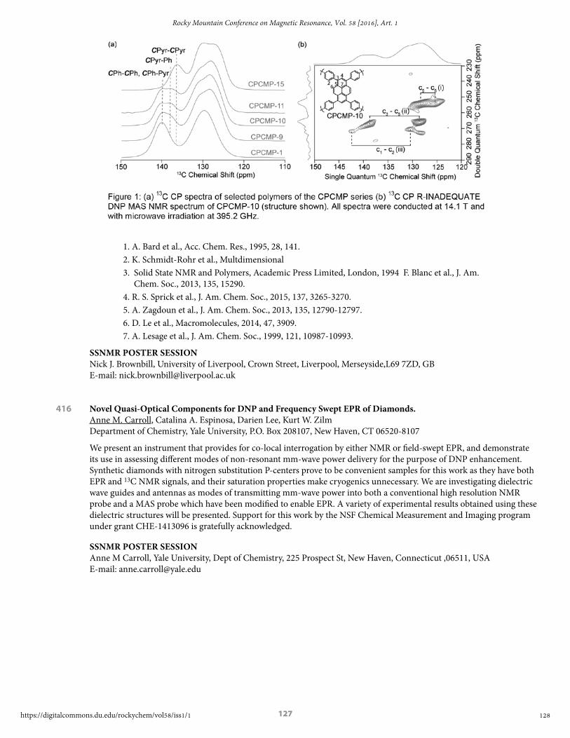
127
1. A. Bard et al., Acc. Chem. Res., 1995, 28, 141.2. K. Schmidt-Rohr et al., Multdimensional 3. Solid State NMR and Polymers, Academic Press Limited, London, 1994 F. Blanc et al., J. Am.
Chem. Soc., 2013, 135, 15290. 4. R. S. Sprick et al., J. Am. Chem. Soc., 2015, 137, 3265-3270. 5. A. Zagdoun et al., J. Am. Chem. Soc., 2013, 135, 12790-12797. 6. D. Le et al., Macromolecules, 2014, 47, 3909. 7. A. Lesage et al., J. Am. Chem. Soc., 1999, 121, 10987-10993.
SSNMR POSTER SESSIONNick J. Brownbill, University of Liverpool, Crown Street, Liverpool, Merseyside,L69 7ZD, GB E-mail: [email protected]
416 Novel Quasi-Optical Components for DNP and Frequency Swept EPR of Diamonds. Anne M. Carroll, Catalina A. Espinosa, Darien Lee, Kurt W. Zilm Department of Chemistry, Yale University, P.O. Box 208107, New Haven, CT 06520-8107
We present an instrument that provides for co-local interrogation by either NMR or field-swept EPR, and demonstrate its use in assessing different modes of non-resonant mm-wave power delivery for the purpose of DNP enhancement. Synthetic diamonds with nitrogen substitution P-centers prove to be convenient samples for this work as they have both EPR and 13C NMR signals, and their saturation properties make cryogenics unnecessary. We are investigating dielectric wave guides and antennas as modes of transmitting mm-wave power into both a conventional high resolution NMR probe and a MAS probe which have been modified to enable EPR. A variety of experimental results obtained using these dielectric structures will be presented. Support for this work by the NSF Chemical Measurement and Imaging program under grant CHE-1413096 is gratefully acknowledged.
SSNMR POSTER SESSIONAnne M Carroll, Yale University, Dept of Chemistry, 225 Prospect St, New Haven, Connecticut ,06511, USA E-mail: [email protected]
128
Rocky Mountain Conference on Magnetic Resonance, Vol. 58 [2016], Art. 1
https://digitalcommons.du.edu/rockychem/vol58/iss1/1

128
417 Direct Interrogation of a Quinonoid Intermediate in PLP-Dependent Tryptophan Synthase. Bethany G. Caulkins,1 Robert P. Young,1 Michael F. Dunn,2 Leonard J. Mueller1 1. UC Riverside, Department of Chemistry, Riverside, CA 92521 2. UC Riverside, Department of Biochemistry, Riverside, CA 92521
One hypothesis for reaction specificity in the extensive class of pyridoxal-5’-phosphate (PLP)-dependent enzymes suggests that protonation/deprotonation at ionizable sites in the active site prejudice the reaction for one pathway over another. X-ray crystallography, optical spectroscopy, and physical-organic studies act as a framework for defining possible protonation states, yet direct characterization remains elusive as these techniques cannot specifically identify proton locations or report unambiguously on the local chemical environment of individual atoms. In solid-state NMR spectroscopy, the interactions of chemical shift (both isotropic and anisotropic) and dipolar coupling are extremely sensitive probes of the chemical microenvironment, and here we report 13C and 15N isotropic chemical shifts, chemical shift tensors, and temperature dependent line shapes for the 2AP-quinonoid intermediate formed during catalysis in tryptophan synthase (TS). TS is a 143 kDa, PLP-dependent enzyme that catalyzes the synthesis of L-Trp from indole and L-serine. 2-aminophenol (2AP) serves as an analogue of indole, stabilizing the intermediate for extended periods under conditions of active catalysis. Our results indicate a dynamic environment that ensures the proper ionization state to direct the reaction to the necessary elimination pathway and guarantee the correct product. This involves some ionizable sites, such as the pyridine nitrogen, playing different roles during catalysis than previously assumed. Furthermore, a fast proton exchange between the phenolic oxygen of the cofactor and the Schiff base from the L-Ser substrate aids in ensuring the correct charge distribution for the correct catalytic outcome. These significant results help in furthering our understanding of the transformation of substrate to product in this enzyme.
SSNMR POSTER SESSIONBethany G Caulkins, University of California Riverside, Department of Chemistry, Riverside, California,92521, USA Tel: 951-827-7365, E-mail: [email protected]
418 New Developments in Solid-State Dynamic Nuclear Polarization at High-Field, High-Temperature and Fast Magic Angle Spinning. Sachin R. Chaudhari,1 Pierrick Berruyer,1 David Gajan,1 Christian Reiter,2 Frank Engelke,2 Daniel L. Silverio,3 Christophe Copéret,3 Moreno Lelli,4 Anne Lesage,1 Lyndon Emsley5 1. Institut de Sciences Analytiques, Centre de RMN à Très Hauts Champs, Université de Lyon (CNRS/ENS Lyon/UCB Lyon 1), France 2. Bruker Biospin, 76287 Rheinstetten, Germany 3. ETH Zürich, Department of Chemistry, Vladimir Prelog Weg 1-5, CH–8093 Zürich, Switzerland 4. University of Florence, Chemistry Department, Magnetic Resonance Centre, 50019 Sesto Fiorentino (FI), Italy Institut des Sciences et Ingénierie Chimiques 5. Ecole Polytechnique Fédérale de Lausanne (EPFL), CH-1015 Lausanne, Switzerland.
Dynamic nuclear polarization (DNP) has recently developed into a powerful analytical technique to enhance the sensitivity of magic angle spinning (MAS) solid-state NMR spectroscopy of biomolecules and materials.1,2 Most developments and applications of DNP MAS NMR were so far reported at moderate spinning frequencies (up to 14 kHz using 3.2 mm rotors) and temperatures of ~100 K or lower.
We will present recent results using a 1.3 mm MAS DNP probe operating at 18.8 T (800 MHz) and ~100 K. We will show that signal amplification factors can be increased by up to a factor two when using smaller volume rotors as compared to 3.2 mm rotors, and report enhancements of around 60 over a range of sample spinning rates from 10 to 40 kHz. The contribution of quenching effects to the overall sensitivity gain at very fast MAS will be discussed, and applications on functionalised mesostructured organic-inorganic materials presented.
In particular, we will show that spinning the sample at 40 kHz leads to a significant increase of coherence lifetimes, substantially increasing the sensitivity of CPMG experiments.
We will also show how high enhancements can be preserved at room temperature by using solvents having a high glass transition temperature. In particular, by using ortho-terphenyl (OTP), enhancement factors of around were obtained at 240 K (i.e. at the glass transition temperature of OTP) at 400 MHz. Enhancements of up to 20 were observed in the metastable phase at ambient temperature.3 The method was successfully applied to monitor molecular dynamic transitions in pharmaceutically relevant solid samples, including Ambroxol and Ibuprofen.
SSNMR POSTER SESSIONSachin Rama Chaudhari, Centre de RMN à Très Hauts Champs, Institut des Sciences Analytiques,, UMR 5280 CNRS/Université Lyon1/ENS-Lyon 5 rue de la Doua, Villeurbanne, France., Rhone,69100, FR E-mail: [email protected]
129
et al.: 58th RMCMR Final Program and Abstracts
Published by Digital Commons @ DU, 2016

129
419 13CO2 chemisorption (for “Carbon Capture”) on Solid Amine Sorbents by 13C, 15N CPMAS and REDOR. Chia-Hsin Chen1, Jason J. Lee2, Carsten Sievers2, Christopher W. Jones2, Anil K. Mehta3, Sophia E. Hayes1 1. Washington University in St. Louis, Department of Chemistry, St. Louis, MO 63130-4899 2. Georgia Institute of Technology, School of Chemical & Biomolecular Engineering, Atlanta, GA 30332 3. Emory University, Department of Chemistry, Atlanta, GA 30322
Amine grafted mesoporous silica is one of promising materials being evaluated for the capture of CO2 released from power plants. Here, we use solid state 13C NMR to identify chemisorbed products of CO2 and aminopropylsilane (APS) on SBA15 zeolite. 13C {1H} cross polarization magic angle spinning (CPMAS) NMR of 13CO2 loaded APS shows multiple chemisorbed products. To have a better understanding of chemisorbed products, 15N enriched APS was synthesized. 15N {1H} CPMAS of 15N enriched APS is analyzed. Rotational echo double resonance (REDOR) of 15N enriched APS was conducted and will be shown.
SSNMR POSTER SESSIONChia-Hsin Chen, Washington University in St. Louis, 1 Brookings Dr, St. louis, Missouri (MO),63130, USA E-mail: [email protected]
420 Carbon Capture and Storage – Geosequestration of 13CO2 with Sintered Forsterite Sample Monitored by Solid-State NMR. Jinlei Cui1, Erika L. Sesti1, Rachel K. Wells2, Mark S. Conradi3,1, Daniel Giammar4, Sophia Hayes1 1. Chemistry, Washington University, St. Louis, MO, United States 2. Earth and Planetary Sciences, Washington University, St. Louis, MO, United States 3. Physics, Washington University, St. Louis, MO, United States 4. Energy, Environmental and Chemical Engineering, Washington University, St. Louis, MO, United States
We have explored NMR spectroscopy (In situ and ex situ) to monitor mineralization reactions with 13CO2 in relevant conditions for geological sequestration. A custom-built high pressure NMR probe was used to collect 13C NMR in samples of pure forsterite pellets submerged in water at pressures of up to 100 bar and temperatures up to 100°C. Conversion of CO2 into stable solid-state carbonates was observed in situ by 13C NMR. Raman, SEM and ex situ NMR were used to confirm the presence of magnesium carbonates. Our preliminary data indicate that magnesite fills the fracture in the absence of flow and only magnesite was formed in the reaction. We have demonstrated that solid state NMR is an analytical tool for monitoring the formation of carbonates from carbon dioxide, and we are currently studying additional minerals and artificial rock samples.
SSNMR POSTER SESSIONJinlei Cui, Washington University in St.Louis, 710 Limit Ave 2S, St.Louis, MO 63130, USA E-mail: [email protected]
421 Study of Singlet Fission in Pentacene: C60 Photovoltaic Devices Using Magnetic Resonance Force Microscopy (MRFM). Elizabeth A. Curley, John A. Marohn
Cornell University, Department of Chemistry and Chemical Biology, Ithaca, NY 14853-1301
The mechanism of charge generation in organic electronics is highly controversial. The model that tightly bound excitons in organics cannot split spontaneously and thus must diffuse to a junction for charge separation to occur is used by current researchers[1,2]. Additionally, polyacenes undergo a singlet fission process where a singlet exciton splits into a pair of triplet excitons. Current studies probe the optically dark triplet states using transient absorption spectroscopy or indirectly by observing a few percent changes in the photocurrent when a magnetic field is applied[1]. Femtosecond two-photon photoemission experiments by X.Y. Zhu and coworkers present evidence that triplet excitons decay to the charge transfer state through a multiexciton state faster than from the triplet exciton state. These experiments the lack spatial resolution to confirm that charge separation occurs at the junction[2]. In this work, the nuclear spin-lattice relaxation time, T1, is used to study the triplet excitons. The paramagnetic, spin 1, triplet states give rise to additional fluctuating magnetic fields altering the T1 time of neighboring protons. By mechanically detecting the nuclear magnetic resonance, using a magnet on tip cantilever, magnetic resonance force microscopy (MRFM), achieves 10 nm resolution[3]. Furthermore, MRFM’s inherent depth resolution enables the detection of spins beneath a top contact. Our recent studies in hyperthermal spin polarization can also be expanded through this work, by implementing optical pumping dynamic nuclear polarization [4]. MRFM presents a unique opportunity to spatially map the nuclear T1 on a fully functional device and provide corroboration into the charge separation mechanism from singlet fission in donor-acceptor devices.
1. Baldo et al., Science, 2013, 340, 334.2. Zhu et al., Science, 2011, 334, 1541.
130
Rocky Mountain Conference on Magnetic Resonance, Vol. 58 [2016], Art. 1
https://digitalcommons.du.edu/rockychem/vol58/iss1/1

130
3. Longenecker et al., ACS Nano, 2012, 6, 9637.4. Isaac, Curley, Marohn et al. Chem. Chem. Phys., 2016, 18, 8806.
SSNMR POSTER SESSIONElizabeth A Curley, Cornell University, 150 Baker Laboratory, Ithaca, NY 14850, USA Tel: 607-254-4685, E-mail: [email protected]
422 Accessing the Structure of Well-defined Grafted Catalysts with Experimental and First Principles 17O Solid-State NMR Methodology. Grekov,1 Y. Bouhoute,2 I. Del Rosal,3 M. Taoufik,2 L. Maron,3 R. Gauvin,1 L. Delevoye1 1. UCCS, CNRS UMR 8181, Université de Lille 1, F-59655 Villeneuve d’Ascq 2. C2P2, CNRS UMR 5265, ESCPE Lyon, F-69616 Villeurbanne 3. LPCNO, CNRS UMR 5215, Université de Toulouse, INSA, UPS, F-31077 Toulouse
Solid State NMR is recognized as one of the most efficient characterization techniques for accessing the structure of well-defined grafted catalysts. The presence of oxygen in the first coordination sphere of the metal center in these systems makes 17O MAS NMR an interesting tool to elucidate the structure of such species at the molecular level. In most cases, the 17O MAS NMR approach benefits not only from the wide chemical shift (CS) range but also from the high sensitivity of the quadrupolar coupling constant (CQ) and Chemical Shift Anisotropy (CSA) to the local environment. Combined with the most advanced DFT methods to calculate these NMR parameters, it is now possible to determine the structure of complex systems. Recent advances in surface organometallic chemistry led to the generation of new catalysts bearing well-defined active sites, such as bipodal metal-carbene oxo species. These exhibit high efficiency in olefin metathesis, a major industrial process. In the present poster, selected examples will illustrate the input of 17O solid-state MAS NMR to the field of supported complexes, used as intermediates in the synthesis of olefin metathesis catalysts. The interactions between grafted organometallic fragments and the support will be addressed by means of surface-selective 17O MAS NMR spectroscopy. Comparing the sets of experimental NMR parameters of these complexes, extracted from best fit simulations, with theoretical data from DFT calculations, will allow for local structure around the metal centre to be determined. Terminal oxygens which reveal strong anisotropic interactions (CSA, CQ) will also be studied with this methodology to reveal site configurations on several grafted species (WOCl4 or WOCl(CH2SiMe3)3).
SSNMR POSTER SESSIONLaurent Delevoye, CNRS - UCCS UMR 8181, Avenue Mendeleev, Villeneuve d’Ascq, Nord, 59655 FR E-mail: [email protected]
423 Satellite Transition Selective 27Al/1H Proton-detected D-HMQC Experiment at Ultrafast MAS for the Determination of Quadrupolar Coupling Constants. Nghia Tuan Duong, 1 Manoj Kumar Pandey,2 Yusuke Nishiyama1,2 1. RIKEN CLST-JEOL collaboration center, RIKEN, Yokohama, Kanagawa 230-0045, Japan 2. JEOL RESONANCE Inc., Musashino, Akishima, Tokyo 196-8558, Japan
Contemporary progresses on ultrafast Magic Angle Spinning (MAS) technology enables the observation of heteronuclear correlation via 1H detection. By utilizing this approach, the advantages are multifold, including the larger sensitivity; better resolved correlation peaks between 1H and nearby X nuclei; and probe ringing avoidance. Recent study has demonstrated the application of the proton-detected dipolar heteronuclear multi-quantum coherence (D-HMQC)1 at 70 kHz of MAS to probe 1H/35Cl (I = 3/2) on pharmaceutical compounds.2 Regarding this D-HMQC experiment, the synchronized indirect dimension allows the overlap of spinning sidebands to the center band of satellite transition (ST), yielding higher intensity for this transition. From the relative shift difference between ST and the central transition (CT) peaks, the quadrupolar coupling constant (CQ) could be precisely calculated. The applicability of this technique is further investigated on spin I = 5/2, namely 27Al. However this is not a straightforward study since this nucleus consists of three transitions, including CT and inner (ST1) and outer (ST2) ST, in which CT and ST1 are partially unresolved no matter how “soft” the radiofrequency (rf) pulse is. In the current work we employed the selective excitation of ST1 and ST2 by applying the rf field on spinning sidebands of STs, achieving a spectrum where no distinguishable CT peak was observed. Comparing this ST selective with the conventional D-HMQC, not only the spatial proximity between 1H and 27Al nuclei was investigated but also the CQ of 27Al nucleus was determined.
1. Tricot et al., Annu. Rep. NMR Spectrosc., 2014, 81, 145.2. Kumar Pandey et al., Phys. Chem. Chem. Phys., 2016, 18, 6209.
SSNMR POSTER SESSIONNghia Tuan Duong, RIKEN Yokohama, W121 1-7-22 Suehiro-cho, Tsurumi-ku, Yokohama, Kanagawa, 230-0045 JP E-mail: [email protected]
131
et al.: 58th RMCMR Final Program and Abstracts
Published by Digital Commons @ DU, 2016

131
424 Flexibility and Solvation of Amyloid-β Hydrophobic Core. Liliya Vugmeyster,1 Matthew A. Clark,2 Isaac B. Falconer,1 Dmitry Ostrovsky,1 Donald Gantz,3 Wei Qiang,4 Gina L. Hoatson5 1. University of Colorado at Denver, Department of Chemistry, Denver, CO 80204 2. University of Alaska Anchorage, Department of Chemistry, Anchorage, AK 99508 3. Boston University School of Medicine, Department of Physiology and Biophysics, Boston, MA 02118 4. Binghamton University, Department of Chemistry, Binghamton, NY 13902 5. College of William and Mary, Department of Physics, Williamsburg, VA 23187
Amyloid fibril deposits found in Alzheimer’s disease patients are comprised of Amyloid-β (Aβ) protein forming a number of hydrophobic interfaces which are believed to be mostly rigid. We have investigated the μs-ms time scale dynamics of the intra-strand hydrophobic core and interfaces of the fibrils comprised of Aβ1-40 protein. Using solid-state 2H NMR line shape experiments performed on selectively deuterated methyl groups, we probed the three-fold symmetric and two-fold symmetric polymorphs of native Aβ as well as the protofibrils of D23N Iowa mutant, associated with an early onset of Alzheimer’s disease. The dynamics of the hydrophobic regions probed at L17, L34, V36, and M35 side chains were found to be very pronounced at all sites and in all polymorphs of Aβ, with methyl axis motions persisting down to 230-200K for most of the sites. The dominant mode of motions is the rotameric side chain jumps, with M35 displaying the most complex multi-modal behavior. There are distinct differences in the dynamics among the three protein variants, with the V36 site displaying the most variability. Solvation of the fibrils does not affect methyl group motions within the hydrophobic core of individual cross-beta subunits, but has a clear effect on the motions at the hydrophobic interface between the cross-beta subunits which is defined by M35 contacts. In particular, hydration activates transitions between additional rotameric states which are not sampled in the dry protein. Thus, these results support the existence of a water-accessible cavity recently predicted by MD simulations and suggested by cryo-EM studies.
SSNMR POSTER SESSIONIsaac B Falconer, University of Colorado Denver, 1201 5th St SI 4118, Denver, Colorado,80204, USA E-mail: [email protected]
425 Quantifying Proton Dynamics in Phosphate Solid Acids Below the Superprotonic Transition Temperature. Gabrielle Foran1, Darren Brouwer2, Gillian Goward1 1. McMaster University, Hamilton, Ontario, Canada 2. Redeemer University College, Ancaster, Ontario, Canada
Phosphate solid acids have been identified as alternative membrane materials for use in proton electrolyte membrane fuel cells as they conduct protons anhydrously via the Grotthuss mechanism above 100 °C.1 These materials have been shown to exhibit increased proton conductivity as a function of temperature by impedance spectroscopy between room temperature and 110 °C. Proton mobility in this temperature range, which falls below the superprotonic transition regime1, was investigated via solid state proton MAS NMR. Symmetry-based dipolar recoupling experiments, which have previously been used to quantify homonuclear dipolar coupling in systems with isolated spin pairs2, were employed to measure 1H-1H dipolar coupling in these multi spin systems. The strength of the dipolar couplings was found to decrease with increasing temperature which correlated with the observed changes in proton conductivity. Changes in proton-proton dipolar coupling could be tracked using the dipolar recoupling pulse sequence in tetragonal KH2PO4 which is a single site system. Proton-proton dipolar coupling was also investigated via the R264
11 pulse sequence in the following two-site systems: CsH2PO4, RbH2PO4, LiH2PO4 and Ca10(PO4)6(OH)2. The two-site systems were found to require additional characterization via 1H EXSY experiments3 to quantify the rate of proton exchange between sites and the activation energy for the proton exchange process. It was found that samples with higher proton conductivity exhibited greater rates of proton exchange and had lower activation energies.
Traer, J.; Soo, K.; Vijayakumar, M.; Goward, G. Elucidating the Time Scale and Geometry of Phosphate and Phosphonate Rotation in Solid Acid Electrolytes Using Multinuclear NMR. J. Phys. Chem. C 2011, 115, 6064-6072De Paepe, G. Dipolar Recoupling in Magic Angle Spinning Solid State Nuclear Magnetic Resonance. Annu. Rev. Phys. Chem. 2012, 63, 661-684Kim, G.; Blanc, F.; Hu, Y.; Grey, C. Understanding the Conduction Mechanism of the Protonic Conductor CsH2PO4 by Solid-State NMR Spectroscopy. J. Phys. Chem. C 2013, 117, 6504-6515
SSNMR POSTER SESSIONGabrielle Y Foran, McMaster University, 1280 Main St West, Hamilton, Ontario,L8S 4L8, CA E-mail: [email protected]
132
Rocky Mountain Conference on Magnetic Resonance, Vol. 58 [2016], Art. 1
https://digitalcommons.du.edu/rockychem/vol58/iss1/1

132
426 Thin Ice Under Pressure on Graphene: A Theoretical NMR Study. Achraf Jaadouni, Eva Rauls, Wolf Gero Schmidt, Uwe Gerstmann University of Paderborn, Department of Physics, D-33098 Paderborn, Germany
Bulk water can exist in many forms, liquid, vapour and at least 16 crystalline phases, including the famous hexagonal ice [1]. Theory suggests, that many further phases can occur, if water is adsorbed on surfaces or confined on nanoscopic pores. Since the discovery of graphene, the adsorption of water has been discussed as a possibility for doping [2], but the electronic properties strongly depend on details of the microscopoic structure [3]. A determination of the microscopic structure of the adsorbed water, however, provides a major challenge for experiment. Recently, locked between two graphene sheets, a new high-density phase of water has been reported using transmission electron microscopy (TEM) [4]. The so-called ’square ice’ provides a symmetry qualitatively different from both hexagonal ice as well as graphene. Modelling bilayer and trilayer ice lattices within density functional theory (DFT), we show that the pressure-induced phase transition from hexagonal to square ice is accompanied (i) by a shift a the Fermi-level thereby suppressing doping effects in graphene and (ii) by a characteristic change of the NMR chemical shifts for the included protons. Hence, the detection via NMR spectroscopy appears as a promising alternative to electron imaging, in particular in case of nanostructures with a high amount of disorder, e.g. hydrophobic nanocapillaries.
[1] G. Malenkov, J. Phys. Condens. Matter 21, 283101 (2009).[2] K.S. Novoselov et al., Science 306, 666 (2004).[3] T. Wehling et al., Appl. Phys. Lett. 93, 202110 (2008).[4] G. Algara-Siller et al., Nature 519, 443 (2015).
SSNMR POSTER SESSIONUwe Gerstmann, University of Paderborn, Warburger Str. 100, Paderborn, NRW,33098, DE E-mail: [email protected]
427 Visualization of Steady-State Ionic Concentration Profiles Formed in Electrolytes During Li-Ion Battery Operation a by In-Situ Magnetic Resonance Imaging. Sergey A. Krachkovskiy,1 J. David Bazak,1 Peter Werhun,1 Bruce J. Balcom,2 Ion C. Halalay,3 Gillian R. Goward1 1. Department of Chemistry, McMaster University, Hamilton, Ontario, Canada L8S 4L8 2. Department of Physics, University of New Brunswick, Fredericton, NB, Canada E3B 5A3 3. General Motors Global R & D, Warren, Michigan, United States 48090-9055.
An accurate modelling of Li-ion battery (LIB) performance requires knowledge of all relevant ion transport parameters, including diffusion coefficients and transference numbers. Taking into account that electrolytes experience a large concentration polarization. In particular, during operation of LIB cells at high rates or low temperatures, not just average values of transport parameters but their concentration dependences should be measured. A pseudo-3D MRI experiment in which magnetisation is prepared using NMR diffusion measurement technique was designed to address this challenge. The 90° pulse in the spin-echo chemical shift imaging was substituted with the one-shot diffusion-ordered NMR spectroscopy pulse sequence, which utilizes asymmetric gradients rather than a full phase cycle, to more rapidly acquire the diffusion data sets. In this experiment, the three orthogonal projections correspond to the chemical shift, the diffusion coefficient and the spatial distribution of the ionic species in the solution. Using this technique under the steady state conditions of electrolyte under applied constant current, when the diffusion flux against the formed concentration gradient compensates for the migration flux of the ions, one can simultaneously measure distributions of the salt concentration (c) and the salt diffusivity (D(c)) in the cell, and then calculate Li+ transference number (t+). The method was tested on the standard electrolyte used in LIBs, consisted of lithium hexafluorophosphate (LiPF6) solution in a binary mixture of ethylene carbonate (EC) and dimethyl carbonate (DMC) with 1:1 volume ratio. It was shown that the values of D measured at opposite ends of the cell varying by more than 60%, while the t+ varies less with salt concentration.
1. Krachkovskiy, S.A., Bazak, J.D., Werhun, P., Balcom, B., Halalay, I.C., Goward, G.R. JACS 2016, DOI: 10.1021/jacs.6b04226
SSNMR POSTER SESSIONGillian R. Goward, McMaster University, 1280 Main St. West, Hamilton, Ontario,L8S 4M1, CA Tel: 9055259140, E-mail: [email protected]
133
et al.: 58th RMCMR Final Program and Abstracts
Published by Digital Commons @ DU, 2016

133
428 Structure and Intermolecular Interface of CAP-Gly Domain on Polymeric Microtubules Determined by Magic Angle Spinning NMR Spectroscopy. Changmiao Guo1, Si Yan1, Guangjin Hou1, Huilan Zhang1, Xingyu Lu1, John C. Williams2, Tatyana Polenova1 1. Department of Chemistry and Biochemistry, University of Delaware, Newark, Delaware 19716, United States 2. Department of Molecular Medicine, Beckman Research Institute of City of Hope, 1500 East Duarte Road, Duarte,
California 91010, United States
Microtubules and their associated proteins perform a broad array of essential physiological functions, including mitosis, polarization and differentiation, cell migration, and vesicle and organelle transport.[1] As such, they have been extensively studied at multiple levels of resolution (e.g., from structural biology to cell biology). Despite these efforts, there remain significant gaps in our knowledge concerning how microtubule-binding proteins bind to microtubules, how dynamics connect different conformational states and how these interactions and dynamics affect the cellular processes. Magic angle spinning NMR spectroscopy is uniquely posed to bridge this gap and provide atomic-resolution insights on microtubule-associated proteins bound to polymeric MTs. However, structures of microtubule-associated proteins assembled on polymeric microtubules are not known at atomic resolution.[2-4]
Here we report a structure of CAP-Gly domain of dynactin motor on polymeric microtubules, solved by magic angle spinning NMR spectroscopy. We present the intermolecular interface of CAP-Gly with microtubules, derived by recording direct dipolar contacts between CAP-Gly and tubulin using double REDOR (dREDOR)-filtered experiments. Our results indicate that the structure adopted by CAP-Gly varies, particularly around its loop regions, permitting its interaction with multiple binding partners and with the microtubules. To our knowledge, this is the first atomic-resolution structure of a microtubule-associated protein on polymeric microtubules. Our approach laid the foundation for atomic-resolution structural analysis of other microtubule-associated motors.[5]
This work was supported by the National Institutes of Health (NIH Grant R01GM085306 from NIGMS). We acknowledge the support of the NSF Grant CHE0959496 for the acquisition of the 850 MHz NMR spectrometer and of the NIH Grants P30GM103519 and P30GM110758 for the support of core instrumentation infrastructure at the University of Delaware.
1. R. D. Vale (2003) Cell 112(4):467-480.2. S. Sun, A. Siglin, J. C. Williams, T. Polenova (2009) J Am Chem Soc 131(29):10113-10126.3. S. Yan, G. Hou, C. D. Schwieters, S. Ahmed,
J. C. Williams, T. Polenova (2013) J Mol Biol 425(22):4249-4266.
4. S. Yan, H. Zhang, G. Hou, S. Ahmed, J. C. Williams, T. Polenova (2015) J Biol Chem 290(3):1607-1622.
5. S. Yan, C. Guo, G. Hou, H. Zhang, X. Lu, J. C. Williams, T. Polenova (2015) Proc Natl Acad Sci USA 112(47):14611-14616.
SSNMR POSTER SESSIONChangmiao Guo, University of Delaware, 041 Brown Lab, Newark, Delaware,19716, USA Tel: 3028318624, E-mail: [email protected]
134
Rocky Mountain Conference on Magnetic Resonance, Vol. 58 [2016], Art. 1
https://digitalcommons.du.edu/rockychem/vol58/iss1/1

134
429 Surface Organometallic Chemistry and Dynamic Nuclear Polarization Surface Enhanced NMR Spectroscopy. When MCM41 is the Mediator! Eva Pump,1 Anissa Bendjeriou-Sedjerari,1 Edy Abou-Hamad,1 Andrei Gurinov,2 Jean-Marie Basset1 1. King Abdullah University of Science and Technology, KAUST Catalysis Center, Thuwal, 23955-6900 Saudi Arabia 2. King Abdullah University of Science and Technology, Advanced Nanofabrication Imaging and Characterization Core
Lab, Thuwal, 23955-6900 Saudi Arabia
In recent years, surface-related processes in energy and catalysis related applications have seen tremendous efforts to characterize chemical species involved at an atomic resolution. Solid state NMR spectroscopy proved to be an essential, powerful and versatile technique to probe surface structures. However, its low sensitivity leads to limitations in the precise understanding of structure of surface complexes. Dynamic nuclear polarization surface enhanced NMR spectroscopy (DNP SENS) has been recently introduced to solve the NMR sensitivity concerns and showed to be capable of fulfilling these requirements as the sensitivity can be increased by several orders of magnitude as compared to conventional solid-state NMR. So far, DNP SENS has been successfully applied to the investigation of various samples in materials science such as functionalized mesoporous silica, metalorganic frameworks or some particular stable immobilized catalysts. Herein, we present novel strategies to characterize highly sensitive and catalytically active early transition metal surface organometallic chemistry catalysts by DNP-SENS. It has been shown that avoiding a direct contact between the exogenous polarizing radical and the immobilized catalyst is crucial for a successful acquisition of DNP SENS experiments.
SSNMR POSTER SESSIONAndrei Gurinov, King Abdullah University of Science and Technology, 4700 KAUST, Thuwal, Makkah Province, 23955-6900 SA E-mail: [email protected]
430 New Frontiers in Solid-state NMR Spectroscopy and Synthesis of Group 13 Clusters and Thin Films. Blake A. Hammann,1 Zayd L. Ma,1 Katherine M. Wentz1 Maisha K. Kamunde-Devonish,2 Matt G. Kast,2 Paul P. Plassmeyer,2 Deok-Hie Park,3 Juan Carlos Ramos,3 Keenan N. Woods,2 Cory Perkins,3 Shannon Boettcher,2 Darren W. Johnson,2 Cathy J. Page,2 Douglas A. Keszler,3 Sophia E. Hayes1 1. Washington University in St. Louis, Department of Chemistry, St. Louis, MO 63130 2. University of Oregon, Department of Chemistry and Biochemistry, Eugene, OR, 97403 3. Oregon State University, Department of Chemistry, Corvallis, OR 97331
Nanomaterials have become a popular subject matter in solid-state nuclear magnetic resonance (NMR) due to advancements in high magnetic fields and ultra-fast magic angle spinning (MAS) probes. These nanomaterials are often composed of quadrupolar species (such as gallium or aluminum, nuclear spin I = 3/2 and 5/2, respectively), which presents a unique difficulty in NMR experiments due to the coupling of the quadrupolar moment with the electric field gradients. Here we present a solid-state NMR study of various nanoscale materials in which we show the characterization of the different metal sites within the molecular cluster precursors and the resulting thin films prepared from these precursors. These molecular clusters self-assemble in aqueous solution through pH control, concentration effects, influence of counterions, and the Group 13 metals (e.g. Al and Ga) form stable polyoxocations.1 The precise formula for the molecular clusters studied is [M13-xInx(μ3-OH)6(μ2-OH)18(H2O)24]+15, where M is either 69Ga, 71Ga, or 27Al and x = 0-6.2-4 The metal sites are held together by bridging hydroxyl groups (-OH) with aquo (H2O) ligands surrounding the outer periphery sites. These clusters once deposited onto a substrate and heat treated form very smooth and dense films.5 Utilizing high magnetic field and ultra-fast MAS probes, the metal sites in the cluster precursors and resulting films are capable of being resolved with solid-state NMR. Monitoring the coordination environment and characterizing the metal sites allows for important structural information to be gained on the transformation of the cluster precursors to thin films. Ultimately, materials scientists will be able to effectively tune the properties of the resulting films for future devices. This material is based on work in the Center for Sustainable Materials Chemistry, which is supported by the U.S. National Science Foundation under Grant CHE-1102637.
L. Mensinger; W. Wang; D. A. Keszler; D. W. Johnson. Chem. Soc. Rev., 2012, 41, 1019. Wang; K.M. Wentz; S.E. Hayes; D.W. Johnson; D.A. Keszler. Inorg. Chem., 2011, 50, 4683-4685. L. Ma; K.M. Wentz; B.A. Hammann; I-Ya Chang; M.K. Kamunde-Devonish; P.H. Cheong; D.W. Johnson; V.V. Terskikh; S.E. Hayes. Chem. Mater. 2014, 26 (17), 4978-4983. A. Hammann; Z.L. Ma; K.M. Wentz; M.K. Kamunde-Devonish; D.W. Johnson; S.E. Hayes. Dalton Trans. 2015, 44, 17652-17659. K. Kamunde-Devonish; M.N. Jackson, Jr.; Z.L. Mensinger; L.N. Zakharov; D.W. Johnson. Inorg. Chem. 2014, 53, 7101-7105.
SSNMR POSTER SESSIONBlake A Hammann, Washington University in St. Louis, Dept. of Chemistry, 1 Brookings Dr., #1134, St. Louis, MO,63130, USA Tel: 314-935-5031, E-mail: [email protected]
135
et al.: 58th RMCMR Final Program and Abstracts
Published by Digital Commons @ DU, 2016

135
431 A Multinuclear Solid-State NMR and GIPAW DFT Approach Towards the Evaluation of the Proposed Structural Motifs of Vaterite. Stephen P. Day1, Kevin M. Burgess2, David L. Bryce2, David Quigley1, John V. Hanna1 1. Department of Physics, University of Warwick, Gibbet Hill Rd., Coventry CV4 7AL, UK 2. Department of Chemistry, University of Ottawa, 10 Marie Curie St., Ottawa, Ontario K1N 6N5, Canada
The nature of the disorder characterising the metastable vaterite polymorph of the CaCO3 family has been debated in the literature for many years. This phase is extremely important as it is naturally produced on the surface of many living organisms to form a ‘semi-amorphous’ nanoparticle interface between the exterior of the organism and specific active biomolecules. Furthermore, recent computational and synthetic studies have suggested that CaCO3 polymorphs can grow under both biogenic and abiotic conditions, and this has challenged the models of classical nucleation theory that have underpinned the conventional understanding of the formation of CaCO3.1-4 The biggest problem confronting our understanding of the vaterite structure is the clear lack of agreement between the structures proposed by characterisation techniques (such as X-ray diffraction, transmission electron microscopy and Raman spectroscopy) and those which have been demomstrated to be energetically favoured by ab initio computational methods. The short range information provided by the solid state NMR technique should provide sufficient information to initiate the unravelling the complexities of the vaterite structure; however, until recently few significant results have been reported. A contemporary study by Bryce et al. used 43Ca MAS and DOR methodologies coupled with GIPAW DFT calculations to focus on a short-range characterisation of the polytypes that may exist, although some ambiguities and inconsistencies still remain.5 This work will report 1D and 2D 17O and 13C MAS NMR measurements, that have been agumented with additional GIPAW DFT computation, to further clarify the complex nature of the vaterite system and shed some light on additional complications.
1. Freeman C. L., Harding J. H., Quigley D., Rodger P. M. Angew. Chem. Int. Ed. 49, 5135-5137 (2010)2. Rodriguez-Blanco J. D., Shaw S., Benning L. G. Nanoscale 3, 265-271 (2011)3. Ogino T., Suzuki T., Sawada K. Geochin Cosmochim Acta 51, 2757-2767 (1987)4. Day S. J., Thompson S. P., Parker J. E., Evans A. Astron. Astrophys. 553, 1-10 (2013)5. Burgess K. M., Bryce D. L. Solid State Nucl. Magn. Reson. 65, 75-83 (2104)
SSNMR POSTER SESSIONJohn V Hanna, University of Warwick, Gibbet Hill Rd, Coventry, West Midlands,CV4 7AL, GB Tel: 44-7824-541149, E-mail: [email protected]
432 Characterization of the Surface of Silicon Nanoparticles by Solid-State NMR. Michael P. Hanrahan1, Lance M. Wheeler2, Nicholas C. Anderson2, Nathan R. Neale2, Aaron J. Rossini1 1. Department of Chemistry, Iowa State University, Ames, IA 2. National Renewable Energy Laboratory, Golden, CO
Silicon nanoparticles (Si NPs) are potentially useful in a wide range of applications such as solar cells, catalysts, LEDs and batteries.1 However, in order to tailor the properties of Si NPs and enable their application it is necessary to control and understand their surface chemistry.1 The atomic level characterization of the disordered surface of silicon NPs is challenging. Solid-state NMR should be an ideal probe of structure in as synthesized hydride terminated and functionalized Si NPs. For this reason, solid-state NMR has previously been applied in ca. 10-15 previous studies to probe the structure of hydrogen passivated and alkyl functionalized Si NPs.2-4 However, previous studies performed resonance assignments and obtained structural information with 1D solid-state NMR spectra. Here we demonstrate the detailed characterization of the surface of hydrogen terminated and dodecene passivated Si NPs by MAS 1H, 13C and 29Si solid-state NMR. Notably, MAS > 20 kHz combined with indirect detection enables the rapid acquisition of 2D scalar (INEPT) and dipolar (CP) 1H-29Si and 1H-13C HETCOR NMR spectra. These spectra allow definitive resonance assignments to be made for the different possible surface species. We show that surface mono-, di- and tri-hydride species can be detected and their populations on the surface can be estimated by performing INEPT 1H-29Si coherence transfers with different scalar coupling evolution times. Based on the INEPT experiments mono-hydride surface species are demonstrated to be the main hydride surface species on the surface of both the hydrogen terminated and dodecene passivated Si NPs.
1. Dasog, M., et al., Angew Chem Int Ed Engl 2016, 55 (7), 2322-39.2. Lee, D., et al. Chem. Eur. J., 2015, 21 (45), 16047-58.3. Mayeri, D., et al., Chem. Mater. 2001, 13 (3), 765-770.4. Giuliani, J. R., et al., Solid State Nucl. Magn. Reson. 2007, 32 (1), 1-10.
SSNMR POSTER SESSIONMichael P Hanrahan, Iowa State University, 2438 Pammel Dr, 0211 Hach Hall, Ames, IA,50011, USA Tel: 9702326866, E-mail: [email protected]
136
Rocky Mountain Conference on Magnetic Resonance, Vol. 58 [2016], Art. 1
https://digitalcommons.du.edu/rockychem/vol58/iss1/1

136
433 Distinguishing Between COOH, COO- and H Disordered COOH Moieties with 13C Shift Tensor and T1 Data. James K. Harper, Jacob Powell Department of Chemistry, University of Central Florida, 4111 Libra Drive, Physical Sciences Building, Orlando, FL, 32816-2366
Since 1993 is has been known that COOH and COO- groups can be readily distinguished by their 13C chemical shift tensors.1 Recently it has been demonstrated that, at least in one case, an H disordered COOH exhibits 13C tensors that are indistinguishable from typical COO- moieties.2Here, it is shown that H disordered COOH groups are not uncommon and that, in general, COO- and H disordered COOH are indistinguishable based on 13C tensors. Data from n-alky fatty acids and certain amino acids are employed to illustrate the similarities. Differentiating between COO- from H disordered COOH is found to be possible based on T1 values (1H and 13C). The influence of H disorder on COOH hydrogen bond strength has also been explored and theoretical considerations suggest that H disorder creates stronger hydrogen bonds than are found in similar COOH groups with localized hydrogens. Supported by NSF CAREER CHE-1455159.
1. Gu and McDermott, J. Am. Chem. Soc., 1993, 115, 4282.2. Powell, Kalakewich, Uribe-Romo, and Harper, Phys. Chem. Chem. Phys., 2016, 18, 12541.
SSNMR POSTER SESSIONJames K. Harper, University of Central Florida, 4111 Libra Drive, Orlando, Florida,32816-2366, USA Tel: 801-910-0265, E-mail: [email protected]
434 Fragment-Based Electronic Structure Approach for Computing Nuclear Magnetic Resonance Chemical Shifts in Molecular Crystals. Joshua Hartman, Greg Beran, Lenoard Mueller, Graeme M. Day, Ryan Kudla UC, Riverside Dept. of Chemistry, School of Chemistry, University of Southampton, Highfield.
Ab initio chemical shielding tensor predictions play a critical role in studying molecular crystal structures using nuclear magnetic resonance. Here we present a many-body expansion fragment approach for the calculation of chemical shielding tensors in molecular crystals. The performance of fragment-based 1H, 13C, 15N and 17O isotropic chemical shift predictions are assessed against benchmark sets of molecular crystals employing a variety of commonly used density functionals (PBE0, B3LYP, TPSSh, TPSS, PBE and OPBE). For each density functional we provide linear regression parameters intended for general application. Fragment-based calculations using the hybrid density functionals PBE0 and B3LYP are shown to provide higher accuracy relative to the GGA-based density functionals. Further, fragment methods demonstrate highly favorable performance relative to existing cluster and GIPAW/PBE-based calculations, improving the accuracy of 1H, 13C and 15N isotropic chemical shift predictions by 22%, 40% and 37%, respectively. In the case of 17O, the pronounced influence of local many-body effects necessitate the use of a combined cluster/fragment model for performance comparable to that of GIPAW. Finally, we assess the accuracy of both fragment-based NMR chemical shielding calculations as well as test set derived linear regression parameters for a collection of biologically and pharmaceutically relevant polymorphic crystals: testosterone, acetaminophen and phenobarbital.
SSNMR POSTER SESSIONJoshua D. Hartman, University of California Riverside, 501 Big Springs Road, Riverside, California,92507, USA E-mail: [email protected]
137
et al.: 58th RMCMR Final Program and Abstracts
Published by Digital Commons @ DU, 2016

137
435 Detection of Active Pharmaceutical Ingredients in Dosage Forms using DNP-Enhanced 35Cl Solid-State NMR Spectroscopy. David A. Hirsh,1 Aaron J. Rossini,2 Lyndon Emsley,3 Robert W. Schurko1 1. University of Windsor, Department of Chemistry & Biochemistry, Windsor, ON, Canada N9B3P4 2. Iowa State University, Department of Chemistry, Ames, IA 50011-3111 3. Ecole Polytechnique Fédérale de Lausanne (EPFL), Institut des Sciences et Ingénierie Chimiques, Lausanne, Switzerland CH-1015
Characterization of active pharmaceutical ingredients (APIs) is critical in the development and manufacture of dosage forms. APIs can crystallize in a variety of different solid forms (e.g., polymorphs, hydrates, cocrystals, and salts) that have different physicochemical properties, which affect qualities of the final formulation like the shelf-life and bioavailability. Common methods for characterizing APIs in the bulk phase (e.g., X-ray diffraction and 13C SSNMR) cannot be applied to the study of many dosage forms, due to the presence of interfering signals from excipients that obscure those from the API. 35Cl SSNMR is a powerful technique for characterizing APIs that are crystalized as HCl salts – more than half of all solid APIs are produced in this manner. 35Cl NMR spectra provide unique spectral fingerprints of each form of an API without interference from the excipient (which does not contain Cl). Given the importance of identifying trace amounts of APIs in dosage forms (e.g., low wt% APIs, polymorphs and/or impurities), there has been a focus on improving the lower detection limit of 35Cl SSNMR spectra. Recently, dynamic nuclear polarization (DNP) has become a popular method for achieving high gains in S/N, but its application to wideline spectra has been limited. Herein, I present the use of DNP to enhance static wideline (> 100 kHz) 35Cl patterns of APIs, with a focus on spectral quality, and a novel approach to increasing the DNP enhancement using periodic sample spinning (i.e., the spinning-on spinning-off, SOSO, technique). I demonstrate the application of 35Cl DNP SSNMR for the characterization of APIs in their bulk forms, as well as in dosage forms with low Cl contents. The potential uses of these techniques for polymorph differentiation, impurity identification, and the discovery of new solid phases are also discussed.
SSNMR POSTER SESSIONDavid Hirsh, University of Windsor, 401 Sunset Avenue, Windsor, Ontario,N9B 3P4, CA E-mail: [email protected]
436 Multinuclear Solid-State NMR Study of an Unknown Gallophosphate. Joseph E. Hooper1, Daniel M. Dawson1, Lucy Broom2, Mahrez Amri2, Richard I. Walton,2 Sharon E. Ashbrook1 1. School of Chemistry, University of St Andrews, St Andrews, Fife, KY16 9ST 2. Department of Chemistry, University of Warwick, Coventry, CV4 7AL
Gallophosphates (GaPOs) are a relatively underexplored family of zeolitic framework materials whose structures comprise alternating corner-sharing GaO4 and PO4 tetrahedra, with network topologies closely related to the better-known aluminosilicates and aluminophosphates. It is possible to prepare many such GaPOs, typically in the presence of fluoride and an organic structure-directing agent (SDA). The use of solid-state NMR for the characterisation of GaPOs can provide much structural information about the material, including the number of crystallographic species, the coordination number of Ga, the protonation state of the SDA and the types of fluoride-containing motifs present.
An unknown gallophosphate phase had been observed as a competing phase in the synthesis of GaPO-34, with both N-methylimidazole and pyridine as SDAs.1 A multinuclear solid-state NMR study of this unknown gallophosphate phase, termed GaPO-X, has been undertaken to provide information complementary to powder XRD and other characterisation techniques. To date, Rietveld refinement of the powder XRD pattern has yielded structures that fit the powder pattern, but are chemically unfeasible. A multinuclear NMR study should provide element-specific information on the local environment and connectivity in the unknown phase that could be key to solving its structure. The data acquired so far suggests that GaPO-X has a structure quite unlike, and much more complicated than, that of any gallophosphate or other zeolite analogue phase currently known.
C. Schott-Darie et al. Stud. Surf. Sci. Catal. 1994, 84, 101.
SSNMR POSTER SESSIONJoseph E Hooper, University of St. Andrews, School of Chemistry, Purdie Building, St. Andrews, Fife,KY16 9ST, GB Tel: 07538935863, E-mail: [email protected]
138
Rocky Mountain Conference on Magnetic Resonance, Vol. 58 [2016], Art. 1
https://digitalcommons.du.edu/rockychem/vol58/iss1/1

138
437 Gd3+ as Polarizing Agent at High Field: Solid Effect vs Cross Effect Dynamic Nuclear Polarization. Monu Kaushik,1 Mian Qi,2 Adelheid Godt,2 Björn Corzilius1 1. Institute of Physical and Theoretical Chemistry, Institute of Biophysical Chemistry, and Center for Biomolecular Magnetic Resonance, Goethe University Frankfurt, Max-von- Laue-Str. 7-9, 60438 Frankfurt am Main, Germany 2. Faculty of Chemistry and Center for Molecular Materials, Bielefeld University, Universitätsstr. 25, 33615 Bielefeld,
Germany.
A variety of polarizing agents have been developed for efficient dynamic nuclear polarization (DNP) for high sensitivity gain. In this work we present studies based on a relatively new class of polarizing agents: high spin transition metal ions. Transition metal ions (Gd3+, Mn2+) can act as paramagnetic substitute of intrinsically bound diamagnetic ions in biomolecules. Doping with paramagnetic ions in this case has no (or insignificant) effect on the structure of the biomolecule. This gives an opportunity to obtain site-specific information about the biomolecule and further the research in structural biology. The polarization transfer mechanisms for these polarizing agents are yet to be understood. Here, we demonstrate DNP effects via Gd-DOTA, which invokes solid effect at low concentration owing to its narrow linewidth. Deviation from pure solid effect mechanism at shorter inter-metal distance in the uniform frozen solution matrix is observed. The properties of Gd3+ being a high spin 7/2 system featuring a relatively strong zero-field (electron quadrupolar) interaction lead to a non-trivial consequences. In our attempts to shed light on underlying polarization transfer mechanisms, bis-Gd rigid model complexes are investigated. By variation of the molecular tether length between the chelator moieties we are able to investigate the distance dependence of DNP field profiles and enhancements. This study enables us to comment on designing complexes for efficient CE DNP.
SSNMR POSTER SESSIONMonu Kaushik, Goethe University Frankfurt, Max-von- Laue-Str. 7-9, 60438 Frankfurt am Main, Germany
438 Design and Construction of ssNMR Probes for the Investigation of Oriented Solids and Liquids. John E. Kelly1, Megha H. Unhelkar1, Kelsey A. Collier2, Rachel W. Martin1,3 1. Department of Chemistry, University of California at Irvine, Irvine, CA, 92697 2. Department of Physics & Astronomy, University of California at Irvine, Irvine, CA, 92697 3. Department of Molecular Biology and Biochemistry, University of California at Irvine, Irvine, CA, 92697
This poster describes the design of solid-state NMR probes for the investigation of oriented samples. The first probe is a three-channel (1H/13C/15N) switched-angle spinning cross-coil solid-state NMR probe for a 500 MHz (11.7 T) magnet ideal for studies of membrane-associated proteins in native-like environments. This probe is the next generation of the pneumatic SAS probe designed previously1. The new probe keeps the angle switching mechanism from the previous generation, while using a new coxial coil design and adding a third channel to enable triple resonance experiments. The channels utilize transmission line segments that act as tunable reactances, with each frequency network contained within an outer ground plane2. This probe uses two coils, a Helmholtz coil array inductively coupled to a microcoil insert and a double saddle coil. The double saddle coil will be outside the rotor, but inside the Helmholtz array. The microcoil will be inside the rotor. The fields generated by these coils are orthogonal, preventing interference between the coils. Both the Helmholtz array and the double saddle coil will be capacitively coupled to the channels. Using two sample coils reduces the need for isolation elements in the circuit and allows for more precise tuning. The Helmholtz array will be used to decouple 1H, while the higher powered double saddle coil will be used to tune the lower frequencies of 13C and 15N. The second probe utilizes a double saddle coil with a goniometer attached to the stator to allow for precise adjustment of single-crystal samples in the x-y plane. This coil allows for signal detection with the sample axis parallel to B 0. This probe is designed to be doubly resonant to 1H and either 15N or 13C. This probe design strategy represents a general paradigm for investigating oriented samples, whether in solid or liquid form.
1. I.M. Litvak, et al., J. Mag. Res. 2010, 206, 183–189.2. R. W. Martin, et al., Rev. Sci. Instrum. 2003, 74 (6) 3045–3061.
SSNMR POSTER SESSIONJohn E Kelly, University of California Irvine, 6306 Adobe Cir S, Irvine, California,92617, USA E-mail: [email protected]
139
et al.: 58th RMCMR Final Program and Abstracts
Published by Digital Commons @ DU, 2016

139
439 Room-Temperature in situ Nuclear Spin Hyperpolarization from Optically-Pumped Nitrogen Vacancy Centers in Diamond. Jonathan P. King1,2, Keunhong Jeong1,2, Christophoros C. Vassiliou1,2, Chang S. Shin1,2, Ralph H. Page1, Claudia E. Avalos1,2, Hai-Jing Wang1,2, Alexander Pines1,2 1. Department of Chemistry, University of California, Berkeley, California 94720 2. Materials Sciences Division, Lawrence Berkeley National Laboratory, Berkeley, California 94720
Nitrogen vacancy (NV-) centers in diamond, with their optically polarized spin states and optical spin readout, have been the focus of much work in the field of quantum information and high-resolution sensing. More recently, attention has shifted to the possibility of using optically polarized NV- centers to generate nuclear spin hyperpolarization. Particularly desirable would be a general method to produce hyperpolarization in situ under the same magnetic field and temperature conditions as the NMR experiment using an inert, non-toxic, and easily separated source. We report bulk, room-temperature hyperpolarization of 13C nuclear spins observed via high-field nuclear magnetic resonance (NMR). The hyperpolarization is achieved by optical pumping (OP) of nitrogen vacancy defect centers in diamond accompanied by dynamic nuclear polarization (DNP). The technique harnesses the large optically-induced spin polarization of NV- centers at room temperature, which is many orders of magnitude greater than thermal equilibrium polarization and typically achievable only at sub-Kelvin temperatures. Transfer of the spin polarization to the 13C nuclear spins is accomplished via a combination of OP and microwave irradiation. The OP/DNP is performed at 420 mT, where inductive detection of NMR is feasible, in contrast to the typically exploited level anticrossing regimes at 100 mT and 50 mT. Here, we report a bulk nuclear spin polarization of 6%. This polarization was generated in situ and detected with a standard, inductive NMR probe without the need for sample shuttling or precise crystal orientation. Hyperpolarization via OP/DNP should operate at arbitrary magnetic fields, enabling orders of magnitude sensitivity enhancement for NMR of solids and liquids at ambient conditions.
a) NMR spectra of 13C at natural abundance in diamond after the accumulation of 60 scans under DNP for 60 seconds at 8895 MHz (blue) and 8907 MHz (red). b) NMR spectrum of 99% 13C-enriched acetonitrile after accumulating 12676 scans. The diamond DNP signal corresponds to a polarization on the order of 6%, an enhancement of ~170,000 over thermal equilibrium. 13C nuclear polarization as a function of applied microwave frequency at the c) ms=0 to ms=-1 and d) ms=0 to ms=+1 NV- transitions. The opposite signs of these two curves are consistent with the opposite electron spin polarizations of the two NV- transitions. Data were acquired with a laser intensity of 16 W/cm2 and microwave power of 1.3 W.
SSNMR POSTER SESSIONJonathan King, University of California Berkeley, 208C Stanley Hall, UC Berkeley, Berkeley, California,94705, USA Tel: 785-770-0088, E-mail: [email protected]
140
Rocky Mountain Conference on Magnetic Resonance, Vol. 58 [2016], Art. 1
https://digitalcommons.du.edu/rockychem/vol58/iss1/1

140
440 Atomic-scale Exploration of Catalyst Sufaces: Homonuclear Correlation Approach Using Dynamic Nuclear Polarization (DNP). Takeshi Kobayashi1, Frederic A. Perras1, Robert .L. Johnson2, Dilini Singappuli-Arachchige3, Igor, I. Slowing1,3, Marek Pruski1,3 1. U.S. DOE Ames Laboratory, and Department of Chemistry, Iowa State University, Ames, Iowa 50011, USA 2. Department of Chemical and Biological Engineering, Iowa State University, Ames, Iowa 50011, USA 3. Department of Chemistry, Iowa State Univesity, Ames, Iowa 50011, USA
Owing to the technological developments during the past decades [1], modern DNP solid-state (SS)NMR offers unprecedented enhancements of SSNMR signals. The hyperpolarization of nuclear spins by DNP is transforming various “exotic” applications, which are far beyond the limits of conventional SSNMR methods, into routine studies [2]. One of the attractive opportunities which DNP brings to the materials research is selective observation of insensitive nuclei and/or minor constituents on the surface of a catalyst. For example, since the discovery of surfactant micelle-templated synthesis of mesoporous silica materials [3], many research efforts have focused on preparing the organic/inorganic hybrids through surface functionalization [4]. However, studies of the spatial distribution of surface-bound molecules posed an insurmountable challenge. Here, we explored the distribution of organic functional groups using DNP-enhanced two-dimensional 29Si-29Si homonuclear correlation experiments. Surprisingly, our studies revealed that the functional groups attached by post-synthesis grafting are more homogeneously distributed than those incorporated by co-condensation method [5]. We will also present the applications of DNP-enhanced 13C-13C correlation spectroscopy to the studies of surface species on supported metal catalysts [6].
[1] a) Becerra, L. R.; Gerfen, G. J.; Bellew, B. F.; Bryant, J. A.; Hall, D.A.; Inati, S. J.; Weber, R. T.; Un, S.; Prisner, T. F.; McDermott, A. E.;Fishbein, K. W.; Kreischer, K. E.; Temkin, R. J.; Singel, D. J.; Griffin, R.G. J. Magn. Reson., Ser. A 1995, 117, 28 − 40. b) Rosay, M.; Lansing, J. C.; Haddad, K. C.; Bachovchin, W. W.; Herzfeld, J.; Temkin, R. J.; Griffin, R. G. J. Am. Chem. Soc. 2003, 125, 13626 − 13627. c) Song, C.; Hu, K.-N.; Joo, C.-G.; Swager, T. M.; Griffin, R. G. J. Am. Chem. Soc. 2006, 128, 11385 − 11390.
[2] Kobayashi, T.; Perras, F.A.; Slowing, I.I.; Sadow, A.D.; Pruski, M. ACS Catal. 2015, 5, 7055, and references therein.
[3] Kresge, C. T.; Leonowicz, M. E.; Roth, W. J.; Vartuli, J. C.; Beck, J. S. Nature, 1992, 359, 710.[4] Huh, S.; Wiench, J.W.; Yoo, J.-C.; Pruski, M.; Lin, V. S.-Y. Chem. Mater, 2003, 15, 4247.[5] Kobayashi, T.; Singappuli-Arachchige, D.; Slowing, I.I.; Pruski, M. in preparation.[6] Johnson, R.L.; Perras, F.A.; Kobayashi, T.; Schwartz, T.J.; Dumesic, J.A.; Shanks, B.H., Pruski, M.
Chem. Commun. 2015, 52, 1862.
SSNMR POSTER SESSIONTakeshi Kobayashi, U.S. DOE Ames Laboratory, 229 Spedding Hall, Iowa State University, Ames, Iowa 50011, USA Tel: 515-294-6823, E-mail: [email protected]
441 29Si and 17O NMR for Structural Analysis of Silicon Oxycarbide Ceramics: Computational Investigations Enhancing Experimental Studies. Peter Kroll, John Paul Nimmo II Dept of Chemistry and Biochemistry, The University of Texas at Arlington, 700 Planetarium Pl, Arlington, TX 76001
Experimental data of 29Si NMR chemical shifts of silicon oxycarbide (SiCO) ceramics show broad peaks for so-called mixed {Si)CnO(4-n) (n=0-4) tetrahedra. To extend the analytical capabilities of NMR investigations, we provided extensive modeling and GIPAW calculations and derived relations between Si-O-Si angles and various bonding environments of C surrounding {Si}CxO(4-x) tetrahedra on one side, and 29Si NMR chemical shifts found at this center on the other side. [1] These relations are now used to analyze in more detail several experimental 29Si NMR spectra, from which we obtain additional structure information. Si-O-Si bond angle distributions of SiO2 nanodomains in SiCO indicate higher local strain in the amorphous ceramics in comparison to silica glass. Furthermore, average Si-O-Si angles in rings are the smaller the more C atoms the ring contains.
Most intriguing, however, all SiCO materials with high content of “free” carbon exhibit a pronounced signature, approximately 10-15 ppm shifted to lower field than typical {Si}O4 units in silica. This signature scales linearly with the amount of “free” carbon, and has not been observed in any silica-containing material before. We hypothesize that the signature is either related to wide Si-O-Si angles indicative of internal surfaces or large cages, as evidenced from NMR studies of zeolites. An alternative explanation relates the signal to 5-fold coordinated {Si}O5 or {Si}O4C-units. Analyzing {Si)CO3-peaks we find no evidence for significant bonding between Si of the glass matrix and embedded C of the “free”
141
et al.: 58th RMCMR Final Program and Abstracts
Published by Digital Commons @ DU, 2016

141
carbon phase. These findings support, via experimental data, that in SiCO ceramics “free” carbon units are incorporated into voids and cages surrounded by a glassy SiCO matrix.
[1] John P. Nimmo, Peter Kroll, “First-Principles Calculations and Analysis of 29Si Nuclear Magnetic Resonance Chemical Shifts in Silicon Oxycarbide Ceramics”, J. Phys. Chem. C 2014, 118, 29952−29961.
SSNMR POSTER SESSIONPeter Kroll, University of Texas Arlington, 700 Planetarium Pl, Arlington, TX,76019, USA Tel: 817-272-3814, E-mail: [email protected]
442 Linking Microscopic Structural Rearrangement to Macroscopic Motion with NMR Crystallography. Ryan A. Kudla, Chen Yang, Lingyan Zhu, Joshua D. Hartman, Gregory J.O. Beran, Christopher J. Bardeen, Leonard J. Mueller Department of Chemistry, University of California, Riverside, California 92521
The photodimerization of 9-tertbutyl-anthracene ester in molecular crystal nanorods is a single-crystal to single-crystal reaction that can cause expansions of up to 15%. This expansion results from the formation of a metastable crystalline intermediate termed the solid-state reacted dimer (SSRD). Photoreaction of bulk single crystal monomer invariably leads to strain that shatters the crystal, prohibiting direct characterization with single crystal X-ray diffraction. Here, the combination of powder X-ray diffraction, solid-state nuclear magnetic resonance, and first principles computational modeling is used to determine the crystal structure of the SSRD intermediate and establish a microscopic model for the macroscopic expansion. We find that the SSRD crystal unit cell and volume are quite similar to those of the monomer crystal, leading to the conclusion that gross changes in the volume or unit cell parameters of the SSRD are not responsible for the expansion. To link the macroscopic motion to microscopic structural rearrangement, we directly observed monomer and photoreacted nanorods aligned in anodic aluminum oxide templates via solid-state NMR. These data show the generation of new lattice orientations within the nanorod. Based on these observations, the nanorods expand not due to a change in the volume of the unit cell, but rather due to a rotation of the unit cells. These results demonstrate that while most photomechanical materials rely on the generation of a mixed phase bimorph structure, reconfiguration of the product phase can likewise generate a large mechanical response.
SSNMR POSTER SESSIONRyan Kudla, University of California Riverside, 501 Big Springs Rd, Riverside, CA,92521, USA E-mail: [email protected]
443 Study of Proton and Carbon Hyperpolarization at Low Temperature With Different Radicals and Spin Concentrations. Bimala Lama,1 Malathy Elumalai,2 Daniel Downes,1 James H.P. Colins,1 Matthew E. Merritt,1 Joanna R. Long1 1. University of Florida, Department of Biochemistry and Molecular Biology, Gainesville, FL, 32610 2. University of Florida, Mcknight Brain Institute, Gainesville, FL, 32610
Dynamic nuclear polarization enhances nuclear spin polarization by transferring the higher polarization from electron spins in paramagnetic centers. There is considerable interest in using DNP for in vivo applications via dissolution DNP as well as in structural biology applications using ssNMR and/or neutron diffraction. At present, considerable attention is given to 13C hyperpolarization because the long relaxation times of unprotonated 13C nuclei enable sufficient preservation of the polarization to study metabolism in real time. Nonetheless, 1H hyperpolarization has been successfully demonstrated for dissolution DNP enhanced angiography, in the study of protein structure and in polarizing neutron targets. It has also been demonstrated that 1H hyperpolarization can be transferred via cross polarization to 13C nuclei, increasing the net 13C polarization and reducing polarization buildup times. We have systematically studied 1H and 13C hyperpolarization using a variety of stable organic radicals at
SSNMR POSTER SESSIONBimala Lama, University of Florida, Dept of Biochemistry & Molecular Biology, 1200 Newell Dr, Gainesville, Florida,32610, USA Tel: 330-983-3350, E-mail: [email protected]
142
Rocky Mountain Conference on Magnetic Resonance, Vol. 58 [2016], Art. 1
https://digitalcommons.du.edu/rockychem/vol58/iss1/1

142
444 Dynamic Allostery Governs Cyclophilin A - HIV-1 Capsid Interplay. Manman Lu1,2, Guangjin Hou1,2, Huilan Zhang1,2, Christopher L. Suiter1,2, Jinwoo Ahn2,3, In-Ja L. Byeon2,3, Juan R. Perilla4, Christopher J. Langmead5, Ivan Hung6, Peter L. Gor’kov6, Zhehong Gan6, William Brey6, Christopher Aiken2,7, Peijun Zhang2,3, Klaus Schulten4, Angela M. Gronenborn2,3, Tatyana Polenova1,2 1. Department of Chemistry and Biochemistry, University of Delaware, Newark, DE 19716 2. Pittsburgh Center for HIV Protein Interactions, University of Pittsburgh School of Medicine, Pittsburgh, PA 15260 3. Department of Structural Biology, University of Pittsburgh School of Medicine, 3501 Fifth Ave., Pittsburgh, PA 15260 4. Center for Biophysics and Computational Biology and Beckman Institute for Advanced Science and Technology and Departments of Physics, Chemistry, and Biochemistry, University of Illinois at Urbana-Champaign, Urbana, IL 61801 5. School of Computer Science, Computational Biology Department, Carnegie Mellon University, 5000 Forbes Ave., Pittsburgh, PA 15213 6. National High Magnetic Field Laboratory, Florida State University, Tallahassee, FL, 32310 7. Department of Pathology, Microbiology and Immunology, Vanderbilt University Medical Center, Nashville, TN 37232
In a mature HIV-1 virion, the viral CA protein assembles into a conical capsid, enclosing the viral genome1. The host cell protein cyclophilin A (CypA) binds the capsid directly and regulates viral infectivity by an unknown mechanism2. CA protein forms tubular assemblies in the presence of NaCl (0.5-2.4 M), yielding magic angle spinning (MAS) NMR spectra exhibiting outstanding high resolution, and permitting their structural and dynamics investigations at atomic level. We have addressed the role of conformational dynamics on the nanosecond to millisecond timescales in the escape from CypA dependence by MAS NMR and molecular dynamics (MD)3. 1H-15N and 1H-13C dipolar order parameters (S) obtained from MAS NMR experiments on CA assemblies, CypA escape mutants A92E and G94D, and CA/CypA assemblies are in quantitative agreement with those calculated from MD trajectories3. Our data demonstrate that CA assemblies are dynamic on multiple timescales, especially in the CypA binding loop3. These motions are significantly reduced in CA/CypA assemblies3. Remarkably, the CypA escape mutant assemblies exhibit dynamic behavior similar to that in the CA/CypA assemblies3. Together, these findings suggest that dynamic allostery mechanism may govern the CA escape from CypA dependence3. To study the interfaces of interaction between CA and CypA, we examined a series of CA/CypA assemblies, where either CA or CypA were uniformly 13C, 15N labeled4. Multiple chemical shift perturbations and intensity changes were observed upon formation of CA/CypA assemblies at different CA:CypA ratios4. Interestingly, while many spectral changes map onto CA and CypA residues comprising the canonical binding sites, a large number of perturbations are associated with residues distal to these canonical binding sites, indicating either additional binding modes, allosteric effects, or both4. CryoEM and MD studies reveal that CypA binds to CA by selectively bridging the CA dimer along the direction of highest curvature4.
1. Zhao, G., Perilla, J. R., Yufenyuy, E. L., Meng, X., Chen, B., Ning, J., Ahn, J., Gronenborn, A. M., Schulten, K., Aiken, C. and Zhang, P. (2013). Nature 497(7451): 643-646.
2. Howard, B. R., Vajdos, F. F., Li, S., Sundquist, W. I. and Hill, C. P. (2003). Nat. Struct. Biol. 10(6): 475-481.
3. Lu, M., Hou, G., Zhang, H., Suiter, C. L., Ahn, J., Byeon, I. J. L., Perilla, J. R., Langmead, C. J., Hung, I., Gor’kov, P. L., Gan, Z., Brey, W., Aiken, C., Zhang, P., Schulten, K., Gronenborn, A. M. and Polenova, T. (2015). Natl. Acad. Sci. U.S.A. 112(47): 14617-14622.
4. Liu, C., Perilla, J. R., Ning, J., Lu, M., Hou, G., Ramalho, R., Himes, B. A., Zhao, G., Bedwell, G. J., Byeon, I. J., Ahn, J., Gronenborn, A. M., Prevelige, P. E., Rousso, I., Aiken, C., Polenova, T., Schulten, K. and Zhang, P. (2016). Nat. Commun 7, 10714.
SSNMR POSTER SESSIONManman Lu, University of Delaware, 041 Brown Lab, Newark, Delaware,19716, USA E-mail: [email protected]
143
et al.: 58th RMCMR Final Program and Abstracts
Published by Digital Commons @ DU, 2016

143
445 Multinuclear Solid-State NMR Structural and Dynamics Analyses of Modified Carbon Allotrope Systems. Adam R. MacIntosh1, Zhongxin Song2, Adam Riese2, Andy Sun2, Gillian R. Goward1 1. McMaster University, Department of Chemistry and Chemical Biology, Hamilton, ON, L8S4L8 2. Western University, Faculty of Engineering, London, ON, N6A 3K7
Many examples of the newest allotropes of carbon have enjoyed significant research attention since their introduction. Graphene sheets, carbon nanotubes, and even the relatively simple high surface area carbon black are constantly being investigated thanks to their interesting physicochemical properties. More recently, a number of chemically functionalized or heteroatomically doped carbon allotrope analogues have been developed, opening the door to fine-tuning of the aforementioned characteristics and broadening the applications of this fascinating class of materials.1-3
Notably absent from many of these investigations is a corroborating solid-state NMR (ssNMR) analysis. The routine exclusion of an analytical method with such high specificity and sensitivity from the study of such important systems may seem surprising, until one considers the high degree of difficulty associated with their ssNMR analysis. For instance, many of these materials contain delocalized electrons from residual conductive π-electron regimes or paramagnetic ions, making for extremely rapid relaxation and unwanted interactions with the external magnetic field.4
Despite these challenges, recently our group has demonstrated through ssNMR the functionalization and proton dynamics of graphene oxide sheets with acidic alkyl chains.5 1H-13C CPMAS spectra were used to resolve acidic functional groups grafted to the sheets at very low loading, and 1,2H experiments revealed slow ionic exchange, even in high-temperature or acidic conditions. These results indicate the materials exist as a tightly bound stack of sheets with surface acidic groups in the solid state. This poster will describe continuing work in this vein, focusing on the refined characterization of acidic, graphitic materials, as well as an analysis of CVD-synthesized phosphorus and nitrogen co-doped carbon nanotubes as well. The 31P signals obtained from these samples relax extremely rapidly, and, coupled with an uncharacteristically high chemical shift, seem to verify heteroatomic incorporation into the walls of the nanotubes themselves.
1) Lu, Y.; Jiang, Y.; Wu, H.; Chen, W. Electrochim. Acta, 2015, 156, 267-273.2) Sun, Q. et Al. Nano Energy,2015, 2, 698-708.3) Song, J. et Al. Angew. Chem. 2015, 127, 4399-4403.4) Panich, A.M. et Al. Solid State Commun., 2012, 152, 466-468.5) MacIntosh, A. R.; Harris, K.J.; Goward, G.R. Chem. Mater. 2016, 28, 360-367.
SSNMR POSTER SESSIONAdam R MacIntosh, McMaster University, 1280 Main St. West, Hamilton, Ontario, L8S4L8 CA Tel: 9055259140 , E-mail: [email protected]
446 Solid-State NMR as a Probe for CO2 Dynamics in Metal-Organic Framework (MOF) Materials and Characterization of Aluminum Carbide-Derived Carbons. Robert Marti,1 Cody Morelock,2 Colton Moran,2 Krista Walton,2 Sophia Hayes1 1. Washington University in Saint Louis, Department of Chemistry, St. Louis, MO 63130 2. Georgia Institute of Technology, School of Chemical & Biomolecular Engineering, Atlanta, GA 30332-0100
Some metal-organic frameworks (MOFs), such as MOF-74, have open metal sites that bind CO2. The choice of metal determines CO2 binding strength. We have investigated the effect of substituting Cd at Mg sites in Mg-MOF-74. Static variable temperature (VT) 13C NMR was used to investigate 13CO2 dynamics in Mg-MOF-74 and Mg0.77Cd0.23-MOF-74 loaded with 1 atm of 13CO2. Temperatures ranging from 8.5 to 295 K were used to explore the molecular motions of CO2 in the MOF-74 structure. The NMR spectra report an axially symmetric CSA for both MOF-74 hosts. At the lowest temperatures, in both MOF-74 hosts, CO2 becomes completely immobile, yielding powder patterns that exceed 300 ppm.
Aluminum carbide-derived carbons also can be used for CO2 capture and are synthesized by etching aluminum carbide (Al4C3). Incomplete etching of Al4C3 results in residual aluminum. The amount and type of residual aluminum is a function of time and etching temperature. Using27Al MAS NMR, we have been able to elucidate the residual metal species (such as hydrogen-bearing aluminum) and confirm the formation of α-Al2O3.
SSNMR POSTER SESSIONRobert Marti, Washington University in St. Louis, 1 Brookings Dr., St. Louis, MO 63130, USA Tel: 405-204-2160, E-mail: [email protected]
144
Rocky Mountain Conference on Magnetic Resonance, Vol. 58 [2016], Art. 1
https://digitalcommons.du.edu/rockychem/vol58/iss1/1

144
447 Coupling Powder Diffraction, Electron Microscopy, Solid-State NMR and GIPAW Calculations: Structure and Dynamics of Inorganic Fluorides. Charlotte Martineau-Corcos,1,2 Mathieu Allix,2 Franck Fayon2 1. ILV, UMR CNRS 8180, Université de Versailles, France 2. CEMHTI, UP CNRS 3079, Orléans, France
The combination of complementary physical measurements such as powder diffraction, electron microscopy or solid-state NMR with computation modelling of structure or NMR parameters is a strategy that is receiving growing interest, since it allows solving the structure of compounds for which diffraction only fails or leads to incomplete solution. This is particularly useful for powdered samples in which several nuclei can be observed by NMR.
Here we illustrate the whole process step-by-step with case study examples of barium-aluminum fluorides. Powder synchrotron X-ray and electron diffraction data are first used to find an initial structural model. However, the localization of the fluoride atoms is impeded by the large electron density of the surrounding barium atoms, and the confrontation of the GIPAW calculated 19F and 27Al NMR parameters with the experimental NMR data indicate some imperfections in the structural model. Additional contrast is therefore searched from neutron diffraction data. Refined model is obtained, for which the GIPAW calculated NMR parameters are in better agreement with the experimental data. Finally, the dynamics of the fluorine sub-lattice is described at the atomic level by variable temperature measurements and dipolar-based NMR experiments, used as filter for the rigid framework. This combined strategy, generalizable to numerous materials, offers a description of the structure with a much higher degree of details than what would have been achieved using these techniques individually.
CM thanks the ANR (12-JS08-0008-01) and the Institut Universitaire de France (IUF) for financial support.
SSNMR POSTER SESSIONCharlotte Martineau-Corcos, ILV & CEMHTI, 45 Avenue des Etats-Unis, Versailles, Ile de France, 78035 FR E-mail: [email protected]
448 A Tiered Approach to Biophysical 17O Solid-State NMR: High Fields, Labelling and Dynamics. Vladimir K. Michaelis1,2, Eric G. Keeler2, Michael T. Colvin2, Robert G. Griffin2 1. Department of Chemistry, University of Alberta, Edmonton, Alberta, Canada, T6G 2G2 2. Francis Bitter Magnet Laboratory and Department of Chemistry, Massachusetts Institute of Technology, Cambridge MA 02139
Many diseases affecting humans are associated with protein structural transformations. Over the past few decades, developments in solid-state NMR spectroscopy have profoundly enhanced the ability to characterize proteins, making it an extremely attractive tool for studying local and medium-range structures and dynamics of disordered biophysical chemical systems.
The revolutionary abilities offered through well-established 1H, 13C and 15N MAS NMR techniques continue to address complex chemical problems, although oxygen, the biologically relevant species associated with key hydrogen bonding, ion shuttling and metal coordination, is scarcely discussed. The low natural abundance (<0.04%) and quadrupolar (I = 5/2) nature of 17O has stymied its reliable use for structural studies within biophysical research. The magnitude of the quadrupolar coupling is often large in biological systems (CQ ~ 7-11 MHz) further complicating the ability to acquire NMR spectra due to poor resolution and hampered sensitivity. Despite these difficulties, its sensitive nature to local and secondary structure via anisotropic electric field gradient (EFG) and magnetic shielding make 17O one of the most important biologically relevant NMR nuclei under development in tackling chemical biology.
To this end we describe efficient labeling (~90-100% efficiency) of FMOC-protected amino acids, using a multiple turnover reaction proposed by Luedtke et al.1 This highly efficient and broad application to labelling key amino acids offers unparalleled sensitivity, diversity and control. We demonstrate this using a combination of small molecules and peptides with the aid of ultra high-field NMR further supporting our 17O NMR research efforts. Building further upon this theme we describe the 17O environment of bound water molecules,2 bringing to light the sensitive structural parameters, insight in the discrepancy between experimental and GIPAW-determined 17O EFG parameters and the effect of dynamics on the experimentally measured quadrupolar and dipolar coupling.
1. Seyfried et al. Org. Lett., 2010, 12,104, 2. Michaelis et al., J. Phys. Chem. B, 2015, 119, 8024.
SSNMR POSTER SESSIONVladimir Michaelis, University of Alberta, 11227 Saskatchewan Drive, Edmonton, Alberta,T6G 2G2, CA Tel: 780-248-5893, E-mail: [email protected]
145
et al.: 58th RMCMR Final Program and Abstracts
Published by Digital Commons @ DU, 2016

145
449 High Resolution Solid-State NMR Lighting of Alkali Borates Glasses Properties. V. Montouillout, H. Fan, L. del Campo, M. Malki, F. Fayon CEMHTI, CNRS, CEMHTI UPR3079, Univ. Orléans, F-45071 Orléans, France
Borate glasses are electrical insulating, however, combined with alkali metals, the great mobility of cations gives them interesting ionic conductivity properties. Structural characterization of these glasses and study of their evolution with alkaline oxide content are essential for understanding the mechanism of ionic conduction. In particular, a full understanding of network glasses requires broad coverage of a wide compositional range. The presence of boroxol rings and their break-up surely has an influence on the ionic mobility in the network. Moreover. boron anomaly induced by the transformation of BO3 units into BO4
- units is well known as a unique feature of borate glasses which is the origin of non-monotonic evolution of physicochemical properties.
In this work, we propose a short-range structural characterization of borate glasses under the lighting of Solid State NMR. Lithium borate glasses (xLi2O-(1-x)B2O3) and sodium borate glasses (yNa2O-(1-y)B2O3) were prepared at concentrations ranging from x=5 to 50mol% and y=5 to 40mol% . Beyond the easy quantification of three and four-coordinate boron species, new structural information have been obtained thanks to 11B MQMAS and 17O measurements
SSNMR POSTER SESSIONValerie Montouillout, CNRS-CEMHTI, 1D Avenue de la Recherche Scientifique, Orléans, cedex 1,45071, FR Tel: 33238255511, E-mail: [email protected]
450a Solid state NMR and NQR in Methylammonium Lead Iodide. Igor Moudrakovski, Alessandro Senocrate, Tae-Youl Yang, Giuliano Gregori, Joachim Maier Max-Planck Institute for Solid State Research, Heisenbergstraße 1, 70569 Stuttgart, Germany
Hybrid organic-inorganic perovskites such as methylammonium lead halides have recently attracted great attention as an effective and inexpensive material for solar cells, with reported photo conversion efficiencies exceeding 20 %. With all current interest, it is surprising that only very limited information is available on the nuclear magnetic resonance properties of these materials. Here we report preliminary results of our recent studies in methylammonium lead iodide (MAPbI 3) with 1H, 13C, 15N, 207Pb solid state NMR, along with the results of 127I NQR. 1H,13C and 15N MAS NMR are all in an agreement with the structure of stoichiometric MAPbI3. The nuclear relaxation times measurements suggest fast intramolecular reorientation of methylammonium cation with the rotational correlation time on the order of 5x10-13 s, while the translational motion of the ions is estimated to be slow. 207Pb NMR shows a signal with small chemical shift anisotropy and the isotropic shift being somewhat outside of the range commonly observed for Pb(II) halides. Short spin-spin relaxation time T2 of about 2x10⁻⁵ s makes detection of the signal very difficult, and is most likely a result of a strong dipole-dipole interactions with 127I. The most challenging, however, is the detection of 127I NMR, as the quadrupolar coupling constant for MAPbI3 is in excess of 500 MHz. Although we did manage to detect the allusive127I NMR signal, the 127I NQR has turned out to be a substantially more suitable technique for this system. Variable temperature NQR spectra allow for accurate detection of the phase transitions, while the NQR spin-lattice relaxation time measurements offer information on the mobility of iodine ions in the lattice.
SSNMR POSTER SESSIONIgor Moudrakovski, Max-Planck Institute for Solid State Research, Heisenbergstraße 1, Stuttgart, Baden-Württemberg,70569 , DE Tel: (49)7116891410, E-mail: [email protected]
146
Rocky Mountain Conference on Magnetic Resonance, Vol. 58 [2016], Art. 1
https://digitalcommons.du.edu/rockychem/vol58/iss1/1

146
450b Multi-nuclear Solid-State NMR in Photocatalytically Active Dion-Jacobson Triple-layered Perovskites. Igor Moudrakovski,1 Christian Ziegler,1,2,4 Daniel Weber,1 Christina Scheu,3 Bettina V. Lotsch1,2,4 1. Max Planck Institute for Solid State Research, Heisenbergstraße 1, 70569 Stuttgart, Germany 2. Department of Chemistry, University of Munich (LMU), Butenandtstraße 5-13, 81377 Munich, Germany 3. Max-Planck-Institut für Eisenforschung GmbH, Max-Planck-Straße 1, 40237 Düsseldorf, Germany 4. Nanosystems Initiative Munich (NIM) and Center for Nanoscience (CeNS), Schellingstraße 4, 80799 Munich,
Germany
The Dion-Jacobson layered niobium perovskite RbCa2Nb3O10 possesses a variety of interesting properties, among which is the photocatalytic activity in splitting of water under UV irradiation. Substitution of Ca with Pb in the structure produces a substantial reduction in the band gap, leading to a visible light sensitization of the material. Replacement of Ca with Pb, however, has a severe negative effect on the exfoliation properties of the catalyst, which can be further used to improve the catalytic performance. Good compromise between the narrowing in band gap and exfoliation properties can be achieved through preparation of solid solutions RbCa2-xPbxNb3O10, specifically for an initial ratio of Ca:Pb ≥ 1:1. Solid state NMR of 93Nb, 87Rb and 207Pb at 9.4 and 21.1T has been applied to study the environment of metal cations and structural transformations in a series of lead-sensitized layered perovskite solid solutions RbCa2-xPbxNb3O10, 0≤x≤2. 93Nb NMR results for the end members of the series are in good general agreement with an accepted for both materials P4/mmm space group, where both Nb atoms in the structure are located on the 4-fold axis. Based on 87Rb NMR, similar conclusion was also made for Rb site in RbPb2Nb3O10. In RbCa2Nb3O10, however, 87Rb NMR points into a lower symmetry of the Rb site, revealing a disagreement with the proposed space group. Perhaps, the explanation of the observed discrepancy can be found in the displacement of the perovskite slabs against the separating layers of alkali metal cations. Solid state NMR spectra of 87Rb, 93Nb, and 207Pb of the solid solutions all indicate a rather homogeneous distribution of Pb and Ca throughout the lattice, with no separate phases been detected throughout the full composition of Ca and Pb. While the quadrupolar interactions dominated both 87Rb and93Nb spectra, it was also possible to detect the contribution from the chemical shift anisotropy.
SSNMR POSTER SESSIONIgor Moudrakovski, Max-Planck Institute for Solid State Research, Heisenbergstraße 1, Stuttgart, Baden-Württemberg,70569, DE Tel: (49)7116891410, E-mail: [email protected]
451 Design of an RF Isolated Multiple-Sample NMR Probe. Eric Munson1, Sean Delaney1, Julie Calahan1, Matthew Nethercott1, 2 1. University of Kentucky, 789 South Limestone, Lexington, KY 40536 2. Kansas Analytical Services, Fort Collins, CO 80526
In a standard CPMAS NMR experiment, the acquisition time is typically 1-3 orders of magnitude less than the recovery time needed for the sample to return to equilibrium. We have previously designed probes that expoited this fact to acquire the signal from more than one sample during this recovery time by either moving samples sequentially into the homogeneous region of the magnet or acquiring data from two samples without probe movement. Both designs had limitations with respect to RF isolation and potential eddy currents at high magnetic fields. We have now designed a probe that overcomes these limitations by totally encompassing the RF circuit in shielded material, thereby avoiding RF isolation issues and also being able to place four or more modules in the homogeneous region of the magnet. The primary focus of our research is to design a multiple-sample double-resonance HF probe, as that has become the nucleus of choice for many pharmaceutical formulations, but also has numerous challenges in isolating the RF for multiple modules. The latest progress in developing this probe technology will be described, such as the construction of a high-efficiency RF filter based upon a multiple-wavelength filter.
Eric Munson is a partial owner of Kansas Analytical Services, a company that provides solid-state NMR services to the pharmaceutical industry. The results presented here are from his academic work at the University of Kentucky, and no data from Kansas Analytical Services are presented here. Funding was provided by the Center for Pharmaceutical Development, an NSF Industry-University Cooperative Research Center and a grant from the Kentucky Science and Engineering Foundation.
SSNMR POSTER SESSIONEric J Munson, University of Kentucky, 789 S Limestone, Lexington, KY,40536, USA Tel: 859-323-3107, E-mail: [email protected]
147
et al.: 58th RMCMR Final Program and Abstracts
Published by Digital Commons @ DU, 2016

147
452 Selective Excitation for Spectroscopic Assignment and Establishing Nearest-Neighbor Correlations in Solid-State NMR of Macroscopically Aligned Samples Sophie N. Koroloff, Alexander A. Nevzorov Department of Chemistry, North Carolina State University 2620 Yarbrough Drive, Raleigh, NC 27695-8204
Oriented-sample NMR makes it possible to study membrane proteins in planar, completely hydrated bilayers within the physiological temperature range and at high lipid-to-protein ratios (>100). However, spectral resolution and peak crowding is often a problem. Here we present a method based on selective pulses that allows one to excite only a selected region in the spectrum and then transfer magnetization from the excited peak to its nearest-neighbor spins via the dilute spin exchange of non-evolved polarization. Owing to the large chemical shift dispersion in oriented samples (100’s ppm), selective excitation becomes particularly efficient. After such a preparation period, standard two-dimensional separated local-field experiments can be performed, which evolves the dipolar couplings of only the nearest-neighbor residues, thus alleviating the problem of spectral crowding. The technique can be used as a block for building other multidimensional pulse sequences where cross peaks are evolved in the locations of main peaks. It is also useful for the initiation of spectroscopic assignment. This method is demonstrated for n-acetyl Leucine single crystal and Pf1 coat protein reconstituted in magnetically aligned bicelles.
FIGURE. Left. A control experiment showing selectively excited peaks for residues S41 and G37 of Pf1 coat protein reconstituted in magnetically aligned bicelles. Right. Overlay of fully excited SAMPI4 spectrum (gray) with selectively evolved spectrum (colors) showing spectral excitations of the residues that are spatially proximal to S41 and G37. Data were obtained at the 900MHz spectrometer at the National High Magnetic Field Lab.
SSNMR ORAL SESSIONAlexander Nevzorov, North Carolina State University, 2620 Yarbrough Drive, Raleigh, NC,27695-8204, USA E-mail: [email protected]
453 Characterizing Donor/acceptor Interfaces in Organic Photovoltaics via Solid-State NMR. Ryan Nieuwendaal NIST, Materials Science and Engineering Division
Robust relationships between structure and function are generally lacking in organic photovoltaic (OPV) thin film bulk heterojunction (BHJ) active layers. This is partially due to the fact that there are currently no measurement tools capable of unveiling details at fine enough length scales so as to be relatable to inter-molecular energy transfer. Common analytical methods such as optical spectroscopy, microscopy (AFM, TEM), and scattering techniques do not have sufficient spatial resolution. In this contribution, I will discuss the results of solid state NMR measurements towards characterizing the donor/acceptor interface in thin films of poly(3-hexylthiophene) (P3HT) and phenyl-C61-butyric acid methyl ester (PCBM).
SSNMR POSTER SESSIONRyan Nieuwendaal, NIST, 100 Bureau Drive, Gaithersburg, MD,20899, USA Tel: 301-975-6766, E-mail: [email protected]
148
Rocky Mountain Conference on Magnetic Resonance, Vol. 58 [2016], Art. 1
https://digitalcommons.du.edu/rockychem/vol58/iss1/1
148
454 Mechanistic Study of the Solid-State Synthesis of a Zeolitic Imidazolate Framework Using Multinuclear SSNMR. C.A. O’Keefe,1 C. Mottillo,2 T. Friščić,2 R.W. Schurko1 1. Department of Chemistry and Biochemistry, University of Windsor, Windsor, ON, Canada N9B 3P4 2. Department of Chemistry and FRQNT Centre for Green Chemistry and Catalysis, McGill University, Montréal, QC,
Canada H3A 0B8
Mechanochemistry (MC, the use of mechanical forces to provide the activation energy for a reaction) and accelerated aging (AA, generating hybrid metal-organic materials under conditions of high humidity and slight heating) are two synthetic approaches that are consistent with the philosophy of green chemistry, as they use little solvent and non-toxic starting materials, and afford quantitative yields. Recently, these approaches have been applied to the synthesis of zeolitic imidazolate frameworks (ZIFs), which are a class of hybrid metal-organic compounds that have garnered great interest due to their uses in catalysis and gas storage. The mechanisms and factors affecting ZIF synthesis are largely unknown and likely very different from their solvothermal analogues. Recent mechanistic studies utilized both in- and ex-situ X-ray diffraction (XRD) experiments to identify products and intermediate phases with known structures; however, the identification of short-lived intermediate phases in low concentrations was not possible as their signals are obscured by those of the starting materials. Herein, we present a mechanistic study of the formation of a ZIF using both MC and AA synthetic techniques. First, we describe the use of multinuclear SSNMR (111Cd, 1H, 13C and 14N) to characterize a series of cadmium-containing ZIFs with known structures. This information is then used to elucidate the structure of a ZIF that can be isolated from both MC and AA reactions. MC and AA reactions forming a cadmium-containing ZIF are monitored ex situ using 111Cd CP/MAS SSNMR. Using this technique, it is possible to observe signals corresponding to both the intermediates and products of the reactions, which could potentially provide information on mechanisms of ZIF formation. The elucidation of the reaction mechanisms, and the determination of the factors that control the rates of formation and the final topologies of the ZIFs, all allow for the fine-tuning of these reactions to rapidly and cleanly produce the desired products.
SSNMR POSTER SESSIONChristopher O’Keefe, University of Windsor, 401 Sunset Ave., Windsor, Ontario,N9B 3P4, CA E-mail: [email protected]
455 The Study of PAH Aggregation of Compounds Using Relaxation, Cross Relaxation and Diffusion Coefficient Determined by NMR. Temidayo Amos Orimolade, Paul Hazendonk Department of Chemistry and Biochemistry, University of Lethbridge, 4401 University Drive, Lethbridge, AB T1K 3M4m Canada
Crude oils are complex mixtures of millions of different compounds. The Asphaltene fraction of crude oil represents the most polar and heaviest aromatic compounds. Asphaltenes tend to aggregate, then form clusters which subsequently lead to precipitation from crude oil. Precipitation is known to cause operational problems such as plugging, fouling on heated surfaces and catalyst deactivation. Understanding asphaltenes structure on the nanoscale is crucial for developing strategies to improve crude oil production and to minimize operational problems. There are two proposed structural models for asphaltenes: 1) The island model; predominantly one large polycyclic aromatic hydrocarbon (PAH) core with pendant aliphatic chains and 2) The archipelago model; with several small PAHs bridged by aliphatic chains forming a network. The present study aims to investigate aggregation dynamics using simple model PAHs where the interactions are governed by π – π π – π stacking and London forces. The influence of concentration of PAH on particle size and size distribution is studied for a series of peri- and circularly-condensed PAHs covering a wide range in melting points. Relaxation (T1 and T2), cross relaxation, and diffusion order spectroscopy (DOSY) nuclear magnetic spectroscopy (NMR) techniques were employed to observe aggregation in PAH solution. Solutions of PAHs were prepared in a “good” solvent (CDCl3) and subsequently diluted by a “poor” solvent (D2O) to probe the influence of solvent-solute interactions on aggregation.Early results on Pyrene in CDCl3 showed significant relaxation dispersion at high concentration, indicating dramatic increase in correlation time resulting from aggregation. Results from relaxation dispersion and diffusion measurements on the remaining PAH’s will be reported along with those of their mono-alkyl substituted counter parts.
SSNMR POSTER SESSIONTemidayo Amos Orimolade, University of Lethbridge, 4401 University Dr, Lethbridge, AB,T1K 3M4, CA Tel: 403-317-2802, E-mail: [email protected]
149
et al.: 58th RMCMR Final Program and Abstracts
Published by Digital Commons @ DU, 2016

149
456 NMR Investigations of the Interactions Between Liquid Adsorbates and Metal Organic Frameworks. Thomas M. Osborn Popp, , Erick A. Andrews, Jeffrey A. Reimer University of California Berkeley
Metal organic frameworks (MOF) are a fascinating class of solid materials, known for their high degree of crystallinity, surface area, and porosity. Understanding their interactions with gaseous and liquid adsorbates is critical to improving their properties and to the design of new MOF materials. As a target for NMR studies, they can be studied using solid state NMR, but imbibed liquids may also be studied using NMR spectroscopic and relaxometric techniques. For example, we have performed variable temperature relaxometry measurements on benzene in MOF-5 which suggest the existence of benzene in two exchanging phases within the MOF crystals. These phases may be described as liquid-like and gas-like, with a phase diagram that differs significantly from the phase diagram of bulk benzene. The most significant finding is that we observe a supercritical-like phase of benzene inside the MOF crystals at temperatures nearly 200 K below the bulk critical point of benzene.
SSNMR POSTER SESSIONThomas M Osborn Popp, University of California Berkeley, 1720 Hearst Ave E, Berkeley, California,94703, USA E-mail: [email protected]
457 Fluorescent DNP Polarizing Agents for Optical Localization. Seong Ho Pahng, Edward Saliba, Nicholas Alaniva, Faith Scott, Brice Albert, Alexander B. Barnes Washington University in St. Louis, Department of Chemistry, St. Louis, MO 63130-4899
The dramatic sensitivity gain from dynamic nuclear polarization(DNP) makes solid-state NMR an excellent method to study molecular structures and dynamics of membrane proteins and amyloid fibrils. Often, solid-state DNP samples preparation includes a cryoprotecting glassy matrix of D2O, H2O, and organic polarizing agents in order to homogeneously dope a sample. However, if polarizing agents can be specifically targeted to analytes’ location, not as many polarizing agents will be required to achieve the same magnitude of polarization as in the homogenous doping scheme. In addition, DNP enhancement will no longer be leveraged by the spin diffusion process. The targeted DNP approach will be especially advantageous for experiments involving intracellular analytes, where the spin diffusion from an extracellular space is not efficient.
As a proof-of-concept study, the polarizing agent, TOTAPOL, is conjugated to a fluorescein-labelled peptide for an electrostatic localization. The fluorescent tag and polarizing agent, when tethered to a highly basic peptide, will localize on negatively-charged phosphotidylserine(PS) vesicles in D2O/glycerol mixture. The fluorescent dye is incorporated as a means of visualizing the localization of the polarizing agents. The peptide also functions as a spacer betwen two moieties, preventing the quenching of fluorescence. Optical and fluorescent microscope images will confirm the surface localization of the polarizing agents. 13C cross-polarization(CP) DNP experiment will show that DNP enhancement is specific to PS carbons.
SSNMR POSTER SESSIONSeong Ho Pahng, Washington University in St. Louis, 1 Brookings Drive , St. Louis, MO,63130, USA E-mail: [email protected]
150
Rocky Mountain Conference on Magnetic Resonance, Vol. 58 [2016], Art. 1
https://digitalcommons.du.edu/rockychem/vol58/iss1/1

150
458 Cellulose Substrates and their Application to SS-NMR of Lithium Ion Batteries: A Case Study in Silicon Monoxide. Allen D. Pauric, Kieran Doyle-Davis, Gillian R. Goward McMaster University, Canada (all authors)
Lithium ion battery materials are typically prepared as thin slurry coatings atop a metallic current collector. This geometry is problematic for NMR measurements, both in terms of the amount of sample which can be cycled and as a barrier to radio-frequency penetration for in-situ experiments. Attempts to address this issue for in-situ experiments have included the use of copper mesh current collectors.1 Presented here is an alternative where the metallic current collector is replaced with a cellulosic substrate (e.g. a KimWipe). Using a carbon rich electrode slurry, sufficient electrical conductivity is maintained while allowing for greater RF penetration. Furthermore, the highly porous nature of the substrate allows for increased active material loadings. To demonstrate the applicability of this approach we describe its use in the characterization of the silicon monoxide anode. This material is lithiated as a lithium-silicon alloy where the pristine material is believed to be comprised of discrete SiO2 and metallic silicon domains.2 The silicate domains function as an expansion buffer upon lithation, mitigating the volume expansion of the material to 100%, as opposed to 300% for pure silicon.3 The advantages of cellulosic substrates are realized in the SiO system to acquire ex-situ 29Si MAS-NMR and in-situ 7Li of cycled anode material. Additionally, extended cycling studies of the cellulosic SiO electrode are presented and discussed with reference to traditional electrode geometries. Implications for other electrode materials and avenues for further exploration are also discussed.
(1) Key, B.; Bhattacharyya, R.; Morcrette, M.; Seznéc, V.; Tarascon, J.-M.; Grey, C. P. J. Am. Chem. Soc. 2009, 131 (26), 9239–9249.(2) Friede, B.; Jansen, M. J. Non-Cryst. Solids 1996, 204 (2), 202–203.(3) Choi, N.-S.; Yew, K. H.; Lee, K. Y.; Sung, M.; Kim, H.; Kim, S.-S. J. Power Sources 2006, 161 (2), 1254–1259.
SSNMR POSTER SESSIONAllen D Pauric, McMaster University, 762 Mohawk Rd. East, Hamilton, Ontario,L8T2R1, CA Tel: 289-440-4665, E-mail: [email protected]
459 Precise Structural Characterization of Heterogeneous Catalyst Surfaces by Combining DNP and Dipolar Recoupling. Frédéric A. Perras,1 Takeshi Kobayashi,1 Marek Pruski1,2 1. US DOE, Ames Laboratory, Ames, IA, 50011, USA 2. Department of Chemistry, Iowa State University, Ames, IA, 50011, USA
Dynamic nuclear polarization (DNP) has revolutionized solid-state NMR spectroscopy and has had a considerable impact on the characterization of surfaces, particularly in the case of heterogeneous catalysts. Numerous studies have, for example, used DNP to elicit 13C and 29Si signals from surfaces and enabled the acquisition of heteronuclear correlation (HETCOR) spectra based on cross-polarization (CP). Such experiments have allowed the conformations and binding modes of heterogeneous catalysts to be elucidated. We will show that much more detailed information can be obtained by instead employing heteronuclear dipolar recoupling techniques in order to quantitatively describe the configurations and dynamics of surface species. This is particularly true in the case of quadrupolar nuclei for which CP-based approaches have a lessened reliability and information content. We first show that 1H-17O dipolar recoupling can be performed at silica surfaces at natural abundance, enabling the measurement of HETCOR spectra and dipolar coupling strengths.1,2 This allowed the distinction between hydrogen-bonded and lone silanol sites in mesoporous silicas. In a related study, we show that 13C-27Al dipolar recoupling can be performed on alumina-supported noble metal catalysts.3 Specifically, we elucidate the intermolecular interactions between organic molecules, such as a PVA coating or methionine, and the surface of a Pd/Al2O3 catalyst. In a broader perspective, this methodology can be applied to study coordination geometries and conformations of other dilute surface species, and to establish structure-activity relationships in other important heterogeneous catalyst systems.
1. Perras, F. A.; Kobayashi, T.; Pruski, M. J. Am. Chem. Soc. 2015, 137, 8336.2. Perras, F. A.; Chaudhary, U.; Slowing, I. I.; Pruski, M. J. Phys. Chem. C 2016, submitted.3. Perras, F. A.; Johnson, R. L.; Wang, L.-L.; Schwartz, T. J.; Kobayashi, T.; Dumesic, J. A.; Shanks, B.
H.; Johnson, D. D.; Pruski, M. J. Am. Chem. Soc. 2016, submitted.
SSNMR POSTER SESSIONFrédéric A Perras, US DOE, Ames Laboratory, 213 Spedding Hall, Ames, IA,50011, USA Tel: 515-294-4992, E-mail: [email protected]
151
et al.: 58th RMCMR Final Program and Abstracts
Published by Digital Commons @ DU, 2016

151
460 Correlations Between Local Environments and 29Si NMR Chemical Shifts in Hafnia-silica Glasses Computed by Density Functional Calculations. Ilia Ponomarev, Peter Kroll The University of Texas at Arlington
We investigate 29Si-NMR chemical shifts of hafnia-soda-silica (HfO2-Na2O-SiO2) and sol-gel derived hafnia-silica (HfO2-SiO2) glasses by density functional calculations using the gauge-including projector augmented wave (GIPAW) method. A great variety of periodic models with up to 10 mol% HfO2 and more than 100 atoms are generated using ab-initio molecular dynamics simulations via a melt-quench procedure and augmented by annealing sequences to reach robust local minima. Subsequently, we compute 29Si-NMR chemical shifts and gauge them to standards. More than 5,000 different Si centers contribute to our analysis of the impact of local environments on 29Si-NMR chemical shifts.
We compute angular correlation functions for Si in Q4, Q3, and Q2 units surrounded by Si atoms only, and our results agree nicely with literature data.1 Substitution of Na by H and selective optimization of H positions allows us to analyze the impact of Na in the environment on the 29Si-NMR as well. We then set out and extract the impact of Hf atoms as second nearest neighbor to Si. In a mixed environment with bond angles at O of 140-150°, presence of Hf causes a change of 3-5 ppm in the 29Si-NMR chemical shift. Our analysis shows that the impact of Hf is linear and uncorrelated, thus facilitating the derivation of independent angular correlation functions for Si-O-Hf angles.
We use our computed correlations to analyze experimental 29Si-NMR data of sol-gel derived hafnia-silica glasses2, which are important in microelectronics and in development of optical wave-guides. We provide guidelines to interpret correctly Q4, Q3, and Q2 units in glasses with different composition, and we provide an approach for analyzing quantitatively the degree of condensation in these networks.
1. S. Ispas et al, Solid State Sciences 2010, 12, 183–1922. L.A. O’Dell et al., Solid State Nuclear Magnetic Resonance 2008, 33, 16–24
SSNMR POSTER SESSIONIlia Ponomarev, University of Texas at Arlington, 700 Planetarium Place, Arlington, Texas,76019, USA Tel: 682-252-5078, E-mail: [email protected]
461 DNP-NMR Investigation of the Structure of Si-γ-Alumina Materials. Andrew G.M. Rankin1, Paul B. Webb1, Daniel M. Dawson1, Jasmine Viger-Gravel2, Brennan J. Walder2, Lyndon Emsley2, Sharon E. Ashbrook1 1. School of Chemistry, EaStCHEM and Centre of Magnetic Resonance, University of St Andrews, North Haugh, St Andrews, Fife, KY16 9ST, UK 2. Institut des Sciences et Ingénierie Chimiques, Ecole Polytechnique Fédérale de Lausanne (EPFL), CH-1015 Lausanne, Switzerland
Aluminium oxides are among the most ubiquitous oxides in heterogeneous catalysis. The structure of these aluminas, and their modified analogues, varies extensively and is highly complex, and a detailed understanding of the relationship between structure and catalytic behaviour is essential to the development of new catalytic materials. This applies not only to applications where alumina is itself a catalyst, but also when it functions as a support material. Modification of γ-alumina with silicon results in materials with high surface area that possess Bronsted acidity, opening up their potential for catalytic applications. Such solid acid catalysts are employed in several key industrial processes, including fluid catalytic cracking and the synthesis of petrol from methanol. As a result, they have attracted significant scientific interest. Solid-state NMR is ideally suited to determining the local environment of Si and Al in Si-alumina materials, but suffers from low sensitivity (particularly for 29Si). Preparation of 29Si-enriched Si-γ-Al2O3 has facilitated acquisition of 29Si NMR spectra via single pulse excitation (SPE) and cross-polarisation (CP). However, as Si comprises only ~1.5% of the total material, long acquisition times and reduced sensitivity continue to present challenges, particularly for the implementation of more complex multinuclear and multidimensional experiments. Dynamic nuclear polarisation (DNP) is well-known for its ability to enhance the sensitivity of solid-state NMR experiments. Recent studies have demonstrated the utility of DNP-NMR in structural investigation of both amorphous aluminosilicates and SiOx/Al2O3 catalysts, allowing for the acquisition of previously unfeasible experiments. Here this signal enhancement, in combination with 29Si isotopic enrichment, provides valuable insights into the structure of Si-γ-Al2O3. Specifically, 29Si isotopic enrichment has enabled Si/Si and Si/Al correlation experiments to be performed, even at 1.5% Si doping. Comparison of SPE and DNP/CP spectra has provided information on the distribution of Si in the surface/bulk of the material.
SSNMR POSTER SESSIONAndrew Rankin, University of St. Andrews, School of Chemistry, Purdie Building, North Haugh, St. Andrews, Fife,KY16 9ST, GB E-mail: [email protected]
152
Rocky Mountain Conference on Magnetic Resonance, Vol. 58 [2016], Art. 1
https://digitalcommons.du.edu/rockychem/vol58/iss1/1

152
462 A Structural Analysis of Asphaltenes Using NMR Spectroscopy Techniques. Yeasmin Ratna, Paul Hazendonk Department of Chemistry, University of lethbridge, AB, Canada
Asphaltenes represent the heaviest, structurally the most complex fraction of petroleum. They cause difficulties in the extraction, transportation and refining of crude oil due to precipitation. Under the geological conditions, at a high temperature and pressure, asphaltenes are stable in solution; however, under ambient conditions, they are unstable forming large aggregates and clusters leading to precipitation. In order to develop mitigation strategies, we need a better understanding of asphaltenes structure and dynamics relate to their phase behavior.
Two structural models of asphaltenes have been proposed. First, the “island” or Yen-Mullins model suggests that they contain a single large PAH (polyaromatic hydrocarbon) core with pendant aliphatic chains. These molecules form nanoaggregates with increasing concentration, which subsequently form clusters at higher concentration. This model predicts a structural hierarchy of three distinct phases. Second, the “archipelago” model, is best described as a network structure of smaller PAH’s bridged by aliphatic linkages, where no structural hierarchy inferred. The 1H T1 and T2 relaxation behavior of specific signals in the asphaltene spectrum were studied at 700 MHz using standard CPMG and inversion recovery sequences. All spectra were subjected to deconvolution analysis where the signals fell into three different linewidth categories. Very long relaxation delays were required owing to components with extremely long T1 (>100 Sec), and correspondingly very short T2s were seen (<1ms). The relaxation dispersion observed indicate that there are three distinct time scales of motion consistent with the sizes of the single molecule (T1/T2~1), the nano-aggregate (T1/T2~10-100), and the clusters (T1/T2~1000-10,000).
SSNMR POSTER SESSIONYeasmin Ratna, University of Lethbridge, 4401 University Drive West, Lethbridge, AB,T1K 3M4, CA Tel: 4038927338, E-mail: [email protected]
463 Rapid Acquisition of Wideline Solid-State NMR Spectra with Fast MAS and Proton Detection. Aaron J. Rossini, Michael P. Hanrahan
Iowa State University, Department of Chemistry, Ames, Iowa, USA
Many NMR active nuclei give rise to extremely broad solid-state NMR spectra due to broadening by large chemical shift anisotropy and/or the quadrupolar interaction. This necessitates the application of special wideline NMR techniques,1 which usually consist of frequency stepped acquisition combined with CPMG pulse sequences. Here we will describe how fast MAS combined with proton detection can enable rapid acquisition of high resolution wideline solid-state NMR spectra of spin ½ and quadrupolar nuclei (I > ½).
(1) Schurko, R. W. Accounts Chem. Res., 2013, 46, 1985-1995.
SSNMR POSTER SESSIONAaron J Rossini, Iowa State University, 2438 Pammel Drive, 0205 Hach Hall, Ames, Iowa,50011, USA Tel: 515-294-8952, E-mail: [email protected]
464 Frequency Swept Microwaves for Hyperfine Decoupling and Time Domain DNP in Rotating Solids. Edward Saliba, Faith Scott, Brice Albert, Nicholas Alaniva, Eric Choi, Michael Mardini, Seong-Ho Pahng, Alexander B. Barnes Washington University in St. Louis
Decoupling of hyperfine interactions and pulsed dynamic nuclear polarization (DNP) are promising techniques to improve high field DNP NMR. We explore experimental and theoretical considerations to implement them with magic angle spinning (MAS). Microwave field simulations using the high frequency structural simulator (HFSS) software suite are performed to characterize the inhomogeneous microwave field throughout a 197 GHz MAS DNP probe. Tunable gyrotron oscillators are proposed as a class of frequency agile sources to generate microwave frequency sweeps required for time-domain DNP transfers and electron adiabatic inversions as a means of hyperfine decoupling. Electron adiabatic inversions of stable organic radicals are simulated with SPINEVOLUTION using the inhomogeneous microwave fields calculated by HFSS.
SSNMR POSTER SESSIONEdward P. Saliba, Washington University in St. Louis, Barnes Lab, 5295 Waterman Blvd. Apt. A-14, St. Louis, Missouri,63108, USA Tel: 815-370-0211, E-mail: [email protected]
153
et al.: 58th RMCMR Final Program and Abstracts
Published by Digital Commons @ DU, 2016

153
465 Proper Selection of Desired Coherence Transfer Pathways in Echo-train Acquisition. R.A. Shakhovoy, V. Sarou-Kanian, F. Fayon CNRS, CEMHTI UPR 3079, Univ. Orléans, F-45071 Orléans, France
For the first time, we present a way to implement the cogwheel phase cycling to Carr-Purcell-Meiboom-Gill (CPMG) acquisition1,2. Our approach, called cogwheely cycled echo train acquisition (COGCETA), is based on the solution of the master equations for several π pulses extendedad hoc to any number of pulses in the train. COGCETA approach provides a highly robust method of the echo-train acquisition that keeps the main advantage of the cogwheel phase cycling, i.e. employs small number of phase cycle transients even in the case of tens of pulses, and, in addition, does not require a numerical search of the “cogwheel parameters” for a given length of the CPMG train. Due to these advantages, developed method could replace conventional CPMG in every instance providing a rival to multidimensional phase incremented echo train acquisition (PIETA) experiment3. Possible application of the COGCETA approach to Periodic Refocusing of J Evolution by Coherence Transfer (PROJECT) acquisition4 will be also discussed.
1. Carr and Purcell, Phys. Rev., 1954, 94, 630.2. Meiboom and Gill, Rev. Sci. Instrum., 1958, 29, 688.3. Baltisberger, Walder, Keeler, Kaseman, Sanders, and Grandinetti, J. Chem. Phys. 2012, 136, 211104.4. Aguilar, Nilsson, Bodenhausen, and Morris, Chem. Commun., 2012, 48, 811.
SSNMR POSTER SESSIONRoman A Shakhovoy, CNRS CEMHTI, 1D Avenue de la Recherche Scientifique Cedex 2, Orleans, Centre,45071, FR E-mail: [email protected]
466 15N Solid-State NMR of Surface Amine Groups for Carbon Capture. Daphna Shimon,1 Chia-Hsin Chen,1 Jason Lee,2 Carsten Sievers,2 Christopher. W. Jones,2 Sophia E. Hayes1 1. Department of Chemistry, Washington University in St. Louis, St. Louis, MO, U.S.A. 2. School of Chemical & Biomolecular Engineering, Georgia Institute of Technology, Atlanta, GA, U.S.A.
The negative consequences of high concentrations of carbon dioxide (CO2) in the atmosphere are well established, prompting research on carbon capture and storage (CCS) worldwide. One method of minimizing CO2 emissions is by reacting it with amine groups on the surface of mesoporous materials, which can be subsequently desorbed and concentrated for secondary use or storage. Understanding the adsorption and chemical reactions during these processes is critical to help make CCS more efficient. In this work, we study a candidate material for CCS that consists of aminopropylsilane (APS) grafted on the surface of mesoporous SBA-15. The grafting results in primary amine groups (in this case 15N-labeled) tethered to the surface such that these can react with CO2 gas introduced to the sample. 15N{1H} solid state MAS and CPMAS NMR experiments were conducted to understand the nature of the 15N groups on the surface of the sample, before reacting it with CO2 gas. Experiments were conducted on two samples with different APS surface concentrations, to see how the concentration of these pendant species affects the amine mobility and chemisorption of gases.
SSNMR POSTER SESSIONDaphna Shimon, Washington University in St. Louis, 1 Brookings Drive, St. Louis, MO,63130, USA E-mail: [email protected]
154
Rocky Mountain Conference on Magnetic Resonance, Vol. 58 [2016], Art. 1
https://digitalcommons.du.edu/rockychem/vol58/iss1/1

154
467 Static Dynamic Nuclear Polarization on Heteronuclear Samples: 1H and 2H with TEMPOL. D. Shimon,1 I. Kaminker,2 Y. Hovav,3 A. Feintuch,1 S. Vega1 1. Chemical Physics Department, Weizmann Institute of Science, Rehovot, Israel 2. Department of Chemistry and Biochemistry, University of California, Santa Barbara, U.S.A. 3. Department of Applied Physics, Hebrew University of Jerusalem, Israel
Dynamic nuclear polarization (DNP) experiments on samples containing free radicals and several types of magnetic nuclei sometimes exhibit polarization “cross-talk” between the nuclei. For example, different nuclei sometimes have DNP spectra with the same shape and enhancement, and sometimes exchange polarization between themselves. These types of effects are normally attributed to the Thermal Mixing (TM) mechanism, as they can be explained by formation of a single electron non-Zeeman spin-temperature. In this work we show results of static 1H- and 2H-DNP on a sample of 50% v/v H2O/DMSO-d6 with 40mM TEMPOL. At 6K we demonstrate “cross-talk” effects resulting in identical shapes of the steady state 1H- and 2H-DNP spectra but a different shape of the 2H-DNP spectrum with a microwave (MW) irradiation length of tMW=1sec. Other experiments show polarization exchange between the nuclei without MW irradiation. At 20K these effects are not seen; the DNP spectra of the nuclei are different from each other and can be simulated using the 1H- and 2H-indirect cross effect (iCE) mechanisms, respectively. At 6K we are only able to fit the steady state 1H-DNP and the tMW=1sec 2H-DNP spectra using the appropriate iCE mechanism. We are unable to explain these observations using the standard TM formalism as in our sample the electron spin reservoir cannot be described by a single non-Zeeman spin temperature, as concluded from analysis of electron-electron double resonance experiments. Based on simulations of the spin evolution in small model systems, we showed that the heteronuclear CE mechanism exhibits “cross-talk” effects that are similar to the experimental observations at 6K.1 Thus this mechanism must play an important role heteronuclear DNP and should be one of the building blocks for the necessary extension and modification of the TM formalism to explain observed effects.
1. Shimon, et al., Phys. Chem. Chem. Phys., 2015, 17, 11868-11883.
SSNMR POSTER SESSIONDaphna Shimon, Washington University in St. Louis, 1 Brookings Drive, St. Louis, Missouri,63130, USA E-mail: [email protected]
468 NMR Analysis of an Unlabelled Peptide Based Nanocarrier and Cargo Complexes. Inumidun Damilola Shotonwa, Paul Hazendonk. Francois Eudes University of Lethbridge, Lethbridge, Alberta, Canada T1J 3M4
Peptides although smaller than proteins still exhibit complex behaviour owing to their dynamics. Cell penetrating peptides, are characterized by containing clusters of cationic side chains that allow them to interact directly with the polar membrane surface, which enables them to enter the cell. Various biotechnologies exploit this property to transfer cargo molecules, such as DNA and RNA into the cells. Most application have been developed for animal cells; however, none currently exist for plants, which is of particular interest to the Agriculture Biotechnology sector as few transformation methods exist for plants.
Recently Nano carriers have been developed for agricultural application, which are composed to complexes of DNA/RNA and CPP’s; however, their structures are not well characterized and their translocation mechanism is not well understood.
In this work detailed structural information is sought on DNA/RNA CPP Complex formation, particle size and distribution. Towards this end NMR spectroscopy is employed to investigate the model complexes of DNA/RNA with CPP’s that are not isotopically enriched. Standard 1H, 31P and 13C 1D and 2D experiments are used for structural elucidation. Relaxation method are used to measure particle size and distribution.
The single stranded 5’-AGTCC-3’, its complementary strand 3’-GGAGT-5’ and the corresponding double-stranded DNA are studied. Complexes with Arginine, Tri-Arginine, Nona- Arginine peptide and TAT were prepared. Observations on the small complexes indicate that measurements on signals from α and δ-H, H1’ of ribose and the base signals were sufficient to observe and describe the complexation process. In this manner neither time consuming 2-D experiments nor expensive isotopic labelling was required to complexes preparation on large scale.
SSNMR POSTER SESSIONInumidun Damilola Shotonwa, University of Lethbridge, 2-143 Mount Sundance Crescent West, Lethbridge, Alberta, T1J 0L4, Canada E-mail: [email protected]
155
et al.: 58th RMCMR Final Program and Abstracts
Published by Digital Commons @ DU, 2016

155
469 Investigation of Zeolitic Imidazolate Frameworks by Solid-State NMR Spectroscopy. Scott Sneddon, Angelica Orsi, Jurgen Khar, David Price, Paul A. Wright, Sharon E. Ashbrook School of Chemistry, EaStCHEM and Centre of Magnetic Resonance, University of St Andrews, North Haugh, St Andrews, Fife, Scotland, UK, KY16 9ST.
Zeolitic imidazolate frameworks (ZIFs) are a relatively new subclass of metal-organic frameworks (MOFs) with extended 3D networks with transition metal nodes (e.g., Zn2+/Co2+) that are bridged by rigid imidazolate organic linkers, which line the pores and windows of the material giving rise not only to a range of unique properties, but also a specific framework topology.1 ZIFs have attracted much attention owing to their potential applications for gas storage and separation, fluid separation and the controlled delivery of drug molecules.2 The characterisation of ZIFs is typically performed by diffraction-based experiments, where solving powder X-ray diffraction (XRD) data can be more challenging than single-crystal XRD refinement, particularly if the sample is poorly crystalline. As many of the interesting properties in the solid state arise as a result of a variation in long-range order, spectroscopic techniques, such as NMR, that probe the atomic-scale structure can be a vital tool for understanding the structure of such materials and investigating guest-host interactions. Here, we focus on the acquisition and complete assignment of 13C and 15N NMR spectra of single- and dual-linker ZIFs in order to gain a better understanding of how chemical shifts are influenced by the structural topology. This information is then used to assign the NMR spectra of novel ZIFs and to gain insight into any changes observed in the chemical shifts upon loading the material with guest molecules. Furthermore, exploitation of the chemical shift anisotropy (CSA) using recoupling techniques is shown to provide a deeper understanding of the local chemical environment. Finally, first-principles DFT calculations are utilised to help assign and understand the solid-state NMR spectra.
1. K. S. Park et al., Proc. Natl. Acad. Sci., 2006, 103, 10186.2. A. Phan et al., Acc. Chem. Res., 2010, 43, 58.
SSNMR POSTER SESSIONScott Sneddon, University of St. Andrews, School of Chemistry, EaStCHEM and Centre of Magnetic Resonance, University of St Andrews, North Haugh, St. Andrews, Fife,KY16 9ST, GB E-mail: [email protected]
470 Measuring Molecular Domain Sizes in Heterogeneous Polymers with Solid State NMR. Eric G. Sorte, Cy Fujimoto, Todd M. Alam Department of Organic Material Sciences, Sandia National Laboratories, Albuquerque, NM 87185
Many important polymers with industrial applications such as fuel cells and batteries are multicomponent systems whose functional properties are determined by microscopic properties such as phase segregation, domain size, and molecular morphology, which can be difficult to characterize. In addition to providing spectroscopic and molecular identification data, NMR can yield information about spatial relations between different phases. In this work, we investigate two co-block polymers composed of differentiated regions: a hydrophilic block of sulfonated phenyl groups and a hydrophobic block composed of different fluorinated backbones that are being developed for fuel cell applications. We use 19F, 1H, and 13C solid state MAS NMR techniques to characterize the polymer and identify regions of distinct molecular mobility within the co-blocks. Using T2 and DQ filtered pulse sequences, we can select magnetization for different regions of the polymer based on relaxation (mobility) or functional group type, and then measure the spin-diffusion response to obtain information about intermolecular distances, phase geometries, and domain sizes in the polymer. Finally, we developed a numerical simulation of the spin diffusion process to translate the spin-diffusion data into reliable information about the spatial phase structure and domain sizes in the polymer. We have extended previous examples of approximating the polymer structure with simple well-defined geometries (lamellar, dispersed nanodomains etc.) to include more complicated structural models that have been proposed to describe the electrolyte membranes. For example, we used structural details from computational molecular dynamics simulations of the co-block polymers as input polymer structures for the simulations. Fits of the simulation results to the experimental NMR spin diffusion data yield valuable quantitative morphological information that may inaccessible by other characterization methods.
Sandia National Laboratories is a multi-program laboratory managed and operated by Sandia Corporation, a wholly owned subsidiary of Lockheed Martin Corporation, for the U.S. Department of Energy’s National Nuclear Security Administration under contract DE-AC04-94AL85000. SAND2016-3800 A
SSNMR POSTER SESSIONEric G. Sorte, Sandia National Lab (for US DOE), 1515 Eubank, Albuquerque, New Mexico,87123, USA Tel: 917-446-2326, E-mail: [email protected]
156
Rocky Mountain Conference on Magnetic Resonance, Vol. 58 [2016], Art. 1
https://digitalcommons.du.edu/rockychem/vol58/iss1/1

156
471 Spinning Slowly for Highly Accurate Chemical Shift Tensors. Sarah E. Soss12, James K. Harper3, Peter F. Flynn1,2 1. University of Utah, D.M Grant NMR Center, Salt Lake City, UT, 84112 2. University of Utah, Department of Chemistry, Salt Lake City, UT, 84112 3. University of Central Florida, Department of Chemistry, Orlando, Florida, 32816-8026
Recently, it has been established that in the absence of single crystal data, accurate measurement of the chemical shift anisotropy (CSA) by solid-state NMR can be combined with computational prediction to determine structure. Six experimental methods are widely used to overcome complications in CSA measurement involving the overlap of powder or spinning sidebands patterns of resonances. Among these, the FIREMAT experiment allows for analysis of nearly all resonances in a single experiment. Another advantage of the method is the ability to extract distinct sideband patterns from isotropically degenerate resonances. We have recently updated this experiment to include SPINAL decoupling, and in addition to the expected improvements to line widths, we observe significant improvements in the accuracy of the measured shift tensors. In this poster we demonstrate that these improvements are best achieved at slow spinning speeds of < 1 kHz, and that the accuracy of the measured tensors rivals those measured using single crystal NMR methods. The accuracy of FIREMAT-SPINAL is demonstrated by measuring 15N CSA tensors at natural abundance for di-glycine and histidine HCl H2O. In each case, the measured CSA lies within the uncertainty of previously published single crystal NMR tensor data. The improvement in accuracy is also observed in 13C, and measurements for organic structures are described. Work supported by NSF CAREER CHE-1455159 (University of Central Florida).
SSNMR POSTER SESSIONSarah E Soss, University of Utah, 315 S. 1400 E., Salt Lake City, Utah,84112, USA Tel: 801-587-3697, E-mail: [email protected]
472 Solid Electrolytes: Extremely Fast Charge Carriers in Single Crystalline Garnet-Type Li6La3ZrTaO12. B. Stanje1,2, D. Rettenwander3, S. Berendts4, R. Uecker5, G. Redhammer3, M. Wilkening1,2,6 1. Christian Doppler Laboratory for Lithium Batteries, and Institute for Chemistry and Technology of Materials, Graz University of Technology (NAWI Graz), Stremayrgasse 9, 8010 Graz, Austria 2. DFG Research Unit “Mobility of Lithium Ions in Solids”, Graz University of Technology, Stremayrgasse 9, 8010 Graz, Austria 3. Department of Materials Research and Physics, University of Salzburg, 5020 Salzburg, Austria 4. Technische Universität Berlin, Institut für Chemie, Straße des 17. Juni 135, 10623 Berlin, Germany 5. Leibniz Institute for Crystal Growth (Forschungsverbund Berlin e.V.), Max-Born-Straße 2, 12489 Berlin, Germany 6. Alistore-ERI European Research Institute, 33 rue Saint Leu, 80039 Amiens, France
The development of all-solid-state electrochemical energy storage systems, such as lithium-ion batteries with solid electrolytes, requires stable, electronically insulating compounds with exceptionally high ionic conductivities. Considering oxides, garnet-type Li6La3Zr2O12 and derivatives, see Zr-exchanged Li6La3ZrTaO12 (LLZTO), have attracted great attention because of its high Li+ ionic conductivity of up to 10-3 Scm-1. Despite numerous studies focusing on conductivities of powder samples, only a few use time-domain NMR methods to probe Li ion diffusion parameters in single crystals. Here we report, for the first time, on temperature-variable 7Li NMR relaxometry measurements using both laboratory and spin-lock techniques to probe Li jump rates in single crystalline Li-bearing garnets with high ion mobility. Time-domain NMR offers the possibility to study Li ion dynamics on both the short-range and long-range length scale. The techniques applied yield a fully consistent picture of correlated Li ion jump diffusion in LLZTO; the data perfectly mirror a modified BPP-type relaxation response being based on a Lorentzian-shaped relaxation function. The rates measured could be parameterized with a single set of diffusion parameters. Dynamic information about the elementary jump processes, such as jump rates and activation energies, was extracted from complete diffusion-induced rate peaks that are obtained when the relaxation rate is plotted vs inverse temperature.
SSNMR POSTER SESSIONBernhard Stanje, Graz University of Technology, Stremayrgasse 9, Graz, Steiermark, 8010 AT Tel: 00436803289050, E-mail: [email protected]
157
et al.: 58th RMCMR Final Program and Abstracts
Published by Digital Commons @ DU, 2016

157
473 The Effects of Point Mutations in Surfactant Protein B1-25 (SP-B1-25) on Lipid Dynamics via 2H and 31P NMR. N.T. Tran,1 Adam N. Smith,1 Gail E. Fanucci,1 Joanna R. Long2 1. University of Florida, Department of Chemistry, Gainesville, FL 32611-7200 2. University of Florida, Department of Biochemistry and Molecular Biology, Gainesville, FL 32610-0245
Pulmonary surfactant (PS) is a lipoprotein mixture found in the alveoli of the lungs. Primarily, PS allows for proper lung function by lowering the surface tension at the alveolar air-water interface. This reduction in surface tension is required for the expansion and contraction of alveoli during respiration. Of the four surfactant proteins, surfactant protein B (SP-B) is the only one that demonstrates the ability to reduce surface tension to minimal values necessary for respiration and consequently, is the only surfactant protein required for survival.1 Intriguingly, only the first 25 residues of SP-B (SP-B1-25) are required to recapture most of the activity of full length SP-B, implying a critical role in the highly conserved N-terminus region. To date, it is widely suggested that SP-B1-25 is responsible for trafficking PS lipids from the aqueous hypophase to the air-water interface, however its mechanism of action is poorly understood.
Figure 1. Rat alveolar surfactant, with arrows showing the air/water interface and aqueous hypophase.4 (box) Cartoon model of SP-B1-25 trafficking DPPC from the aqueous hypophase to air/water interface
Previous lipid dynamics studies in our group, using 2H and 31P NMR, have shown that SP-B1-25 selectively traffics 1,2-dipalmitoyl-sn-glycerol-3-phosphocholine (DPPC) in PS lipid mixtures through the induction of non-lamellar lipid morphologies.2,3 Our model suggests SP-B1-25 preferentially interacts and directly traffics DPPC to the air-water interface and may explain how the alveolar air-water interface is specifically enriched with DPPC (Figure 1). Here we will present the effects of point mutations in SP-B1-25 on lipid dynamics and morphologies in various lipid systems using 2H and 31P NMR. Our objective is to elucidate the sequence dependence of SP-B1-25 and its interaction with PS lipids.
1. Lopez-Rodriguez, E. BBA. 2014;1838:1568-1585 2. Farver S.R. Biophys. J. 2010;99:1773-1782 3. Farver, S. BBA. 2015;1848:203-210 4. Fehrenbach H. Respir Res; 2001;2:33-46
SSNMR POSTER SESSIONNhi Tran, University of Florida, 1716 NW 3rd ave, Gainesville, Florida,32603,US Tel: 9548027897, E-mail: [email protected]
474 Solid-State NMR Studies of Aggregates of Cellular Prion Protein and Toxic Amyloid-β Oligomers. Marcus D. Tuttle1, Mikhail A. Kostylev2, Anne Carroll1, Stephen M. Strittmatter2, Kurt W. Zilm1 1. Department of Chemistry, Yale University, New Haven, CT 2. Department of Neurology, Yale University School of Medicine, New Haven, CT
Protein aggregation is implicated across a wide spectrum of neurodegenerative diseases, including Alzheimer’s (AD), Parkinson’s, and Prion diseases. It has been shown that protein aggregates in neurodegenerative diseases have specific structure that have been hypothesized to be important for their pathogenicity and for their proliferation. Recently, it has been discovered that in AD specific and high affinity amyloid-β oligomer (Aβo) binding to cellular prion protein (PrPC) is required for cell death in vitro and deleting PrPC expression in mice results in partial rescue of cognitive performance(1,2). It is thus critical that we gain a clearer understanding of the specific structural interactions that drive the organization of this Aβo-PrPC complex to develop better treatments for AD. In this study we compare solution
158
Rocky Mountain Conference on Magnetic Resonance, Vol. 58 [2016], Art. 1
https://digitalcommons.du.edu/rockychem/vol58/iss1/1

158
and ssNMR data and, with several important exceptions, we show that nearly every residue in the PrP(23-111) Aβo globuomer complex is nearly indistinguishable by 13C NMR when compared to the expected solution shifts, suggesting only local conformational changes in PrPC to residues proximal to the interaction interface. Of particular interest are several chemical perturbations suggest a binding interface between the Aβo-PrPc complex that is consistent with mutagenisis studies, suggesting key interactions in this aggregate. We envision that these results combined with future experiments will lead to a greater understanding of the structure and formation of this complex and aid in the design of compounds that could be used to rescue cognitive performance in AD.
Um JW, Strittmatter SM. Prion. 2013;7(1):37-41. PMCID: 3609048. Kostylev MA, Kaufman AC, Nygaard HB, et al. J. Biol. Chem. 2015;290(28):17415-38. PMCID: 4498078. Liu A, Riek R, Wider G, et al. J. Biomol.NMR. 2000;16(2):127-38.
SSNMR POSTER SESSIONMarcus Tuttle, Yale University, 275 Winchester Ave Apt 117, New Haven, Connecticut,06511, USA Tel: 315-276-5931, E-mail: [email protected]
475 Unraveling the Structure of beta-Endorphin Amyloid Fibrils. Joeri Verasdonck,1 Carolin Seuring1, Maxim Yulikov1, Julia Gath1, Riccardo Cadalbert1, Thomas Wiegand1, Gunnar Jeschke1, Beat H. Meier1, Anja Böckmann2, Roland Riek1 1. ETH Zurich, Laboratory for Physical Chemistry, Vladimir-Prelog-Weg 2, 8093 Zurich, Switzerland 2. Institut de Biologie et Chimie des Protéines, UMR 5086 CNR/Université de Lyon 1, 7 passage du Vercors, 69367 Lyon,
France
Amyloid fibrils are most commonly associated with neurodegenerative diseases. However, there exist functional amyloid fibrils as well. It has been proposed that many hormones form such functional fibrils when stored in the secretory granules of the pituitary gland.1 Here they are stored in their fibrilar dormant state, ready to be released quickly in their active form when needed. One of these hormones is the 31 amino-acid polypeptide beta-endorphin.
We have performed a full assignment of beta-endorphin amyloid fibrils. There is a rigid fibrilar core stretching from residue 3 to 28. Analysis of the secondary chemical shifts indicates the structure contains three beta-sheets per monomer. They are found between residues 4-10, 14-19 and 21-24. In CP based experiments, the first two N-terminal residues are not observed and the last three C-terminal residues give rather weak signals. Therefore it is assumed that these terminal parts have some degree of flexibility.
Currently we are working towards obtaining the structure of these fibrils at atomic resolution. Solid-state NMR distance restraints will be combined with EPR DEER measurements and mass-per-length data.
1. Maji et al., Science, 2009, 325, 328
SSNMR POSTER SESSIONJoeri Verasdonck, ETH Zurich, Vladimir-Prelog-Weg 2-10, Zurich, Zurich,8093, CH E-mail: [email protected]
476 Investigating Small- and Large-Scale Structure of (CdSe)13(n-propylamine)13 Nanoparticles Using Solid-State 113Cd/77Se CPMAS NMR and Computational Modeling. Michael E West, Zayd Ma, Robert Marti, Sophia Hayes
Department of Chemistry, Washington University in St. Louis
Ultra-stable cadmium selenide (CdSe) quantum dots with diameters under a few nanometers have been studied and isolated, and due to quantum confinement of electrons, they exhibit unique size-dependent electrical properties, thereby showing potential for wide-ranging nanomaterial applications.1 (CdSe)13 , one of these ultra-stable CdSe quantum dots, can be synthesized with high purity with passivating amine ligands.2 The structure of these dots when ligated with propylamine was investigated using 1H-113Cd and 1H-77Se CP NMR. A single, axially-symmetric (η=0.01) Cd site and a single, axially-asymmetric (η=0.59) Se site were detected. These results were compared to CASTEP calculations of chemical shifts and asymmetry parameters of candidate structures.
1. Berrettini, Mia G., et al.; J. Am. Chem. Soc., 126, 7063-7070 (2004).2. Wang, Yuanyuan, et al.; Inorganic Chemistry 52, 2933-2938 (2013).
SSNMR POSTER SESSIONMichael E West, Washington University in St. Louis, 1 Brookings Dr, Campus Box 1134, St. Louis, MO 63130, USA Tel: 405-269-3294, E-mail: [email protected]
159
et al.: 58th RMCMR Final Program and Abstracts
Published by Digital Commons @ DU, 2016

159
477 Cross-Polarization Phenomena in the NMR of Fast Spinning Solids Subject to Adiabatic Sweeps. Sungsool Wi1, Zhehong Gan1, Robert Schurko2, Lucio Frydman1,3 1. National High Magnetic Field Laboratory, Tallahassee, Florida 32304 2. Department of Chemistry and Biochemistry, University of Windsor, 401 Sunset Avenue, Windsor, N9B 3P4, Ontario, Canada, NPB 3P4 3. Department of Chemical Physics, Weizmann Institute of Sciences, Rehovot, 76100 Israel
Cross-polarization magic-angle spinning (CPMAS) experiments employing frequency-swept pulses are explored within the context of obtaining broadband signal enhancements for rare spins S = ½, 1, and half-integer quadrupolar nuclei at ultrahigh spinning rates. These experiments employ adiabatic inversion pulses on the S-channel (13C, 2H, 23Na, 11B, 27Al, etc.) to cover a wide frequency offset range, while simultaneously applying conventional spin-locking pulse on the I-channel (1H). Conditions are explored where the adiabatic frequency sweep width, Dn, is changed from selectively irradiating a single MAS spinning centerband or sideband, to sweeping over multiple sidebands. A number of new physical features emerge upon assessing the swept-CP method under these conditions, including multiple zero- and double-quantum CP transfers. Conditions avoiding MAS-driven rotary resonance phenomenon and adiabatic level crossings among energy levels were discussed. These were examined using an average Hamiltonian theory specifically designed to tackle these experiments, extensive numerical simulations, and experiments on model compounds. Ultrawide CP profiles spanning frequency ranges of nearly were predicted and observed utilizing this new approach. Potential extensions and applications of this new approach are briefly discussed.
SSNMR POSTER SESSIONSungsool Wi, National High Magnetic Field Laboratory, 1800 E. Paul Dirac Drive, Tallahassee, FL,32310, USA Tel: 850-645-2770, E-mail: [email protected]
478 Understanding the Gas Adsorption Properties in Metal-Organic Frameworks by Solid-State NMR. Jun Xu,1 Jinxing Ye,1 Thomas M. McDonald,2 Andrew S. Lipton,3 Juncong Jiang,2 Jeffrey A. Reimer,1,4 Jeffrey R. Long,2,5 Nancy M. Washton,3 Omar M. Yaghi2,5 1. Department of Chemical and Biomolecular Engineering, University of California, Berkeley 2. Department of Chemistry, University of California, Berkeley 3. Environmental Molecular Sciences Laboratory, Pacific Northwest National Laboratory 4. Environmental Energy Technologies Division, Lawrence Berkeley National Laboratory 5. Materials Sciences Division, Lawrence Berkeley National Laboratory
As an important class of porous materials, metal-organic frameworks (MOFs) have been shown to be suitable for a broad range of industrial applications, in particular gas adsorption and separation. MOF-74 or related MOFs (also known as M2DOBDC or CPO-27) are promising candidates for this application due to the open metal centers. It is believed that strong and site-specific interactions between open metal centers and gas molecules are responsible to the large gas uptake and high selectivity. Unfortunately such open metal centers also have high affinity to water, leading to the decreasing of adsorption performances in the presence of water vapor. Several approaches have been proposed to develop new MOF-74 based materials that are more tolerant of humid conditions. One approach is to append alkyldiamines such as N,N’-dimethylethylenediamine (mmen) inside of the channels, showing promising CO2 adsorption behaviors.1 Another approach is to prepare mixed metal MOFs, by which some open metal centers can preferentially bind water while the others might retain their adsorption capacities.2 It is thus of fundamental importance to understand questions including how appended alkyldiamines interact with the framework and gas molecules and how the mixed metal cations are distributed. However, it is challenging to characterize these materials at the molecular level by diffraction-based techniques due to the lack of long-range ordering and rapid motions of alkyldiamines. Solid-state NMR spectroscopy is very sensitive to the local environment around the nucleus of interest and is also capable of probing motions. In this work, we reported 13C and 15N solid-state NMR data of mmen-appended MOF with an extended MOF-74 structure. 15N solid-state NMR results unambiguously reveals that one nitrogen of mmen binds to open metal center while the other one is free. The two nitrogens undergo rapid exchange at ambient conditions. When CO2 is present, it inserts into the metal-nitrogen bond and forms a carbamate anion. The insertion-desorption of CO2 is reversible and can be in-situ monitored by solid-state NMR experiments. We also reported 1H and 13C solid-state NMR data of mixed Mg/Ni-MOF-74. The paramagnetic effects induced by unpaired electrons of Ni2+ cations can be detected by observing the change of isotropic chemical shifts and spinning sideband patterns of framework 1H and 13C. Such effects are related to the spin-delocalization degree of unpaired electrons, which are different when Ni2+ are isolated, in small clusters, or in large clusters.
1. McDonald, T. M.; Lee, W. R.; Mason, J. A.; Wiers, B. M.; Hong, C. S.; Long, J. R. Am. Chem. Soc. 2012, 134, 7056-7065.
160
Rocky Mountain Conference on Magnetic Resonance, Vol. 58 [2016], Art. 1
https://digitalcommons.du.edu/rockychem/vol58/iss1/1

160
2. Wang, L. J.; Deng, H.; Furukawa, H.; Gándara, F.; Cordova, K. E.; Peri, D.; Yaghi, O. M. Chem. 2014, 53, 5881-5883.
SSNMR POSTER SESSIONJun Xu, University of California Berkeley, 3465 Carlson Blvd Apt 2, El Cerrito, California,94530, USA E-mail: [email protected]
479 Homonuclear 19F-19F Double Quantum NMR Dynamics Studies in Proton-conducting Polymers. Z. Blossom Yan,1 Darren H. Brouwer,2 Gillian R. Goward1 1. Department of Chemistry and Chemical Biology, McMaster University, Hamilton, Ontario, Canada L8S 4M1 2. Department of Chemistry, Redeemer University College, Ancaster, Ontario, Canada L9K 1J4
Perfluorosulfonic acid (PFSA) polymers are widely used in polymer electrolyte membrane fuel cell applications as electrolyte materials because of their mechanical durability and high proton conductivity. Nafion® the benchmark PFSA material has been commercialized for decades; its structure and physical properties have been investigated and resolved using ssNMR.[1,2] To link fundamental chemistry at a molecular level and the superior material performance and furthermore to improve material designs, dynamics studies using ssNMR were performed for these types of materials. 1H double quantum filter (DQF) NMR is a well-established strategy to probe local dynamics.[3-5] The concept has here been extended to characterize fluorinated ionomer materials for the first time. 19F DQ recoupling NMR experiments, where an R-symmetry sequence is applied, are conducted in this study to investigate the site-specific local dynamics of Nafion® at various conditions with respect to temperature and humidity. The initial build-up of the normalized double quantum (nDQ) curves generated is used as a measurement of local mobility. The effective ‘Dapp’ (apparent dipolar coupling constant) can be extracted as a measurement of local mobility in a quantitative manner. The order parameter, ST, which is essentially the ratio between the experimental value and the theoretical value in a rigid model, is introduced to regulate the dynamics measurements. The side chain and backbone local dynamics profiles can be ascertained. The side chain has a more sensitive response towards the temperature and humidity changes, which indicates the side chain possesses higher local dynamics. Alternative fluorinated electrolyte materials, such as Aquivion® and an alternative PFSA from 3M are also investigated in parallel. Therefore, a link between material performance and dynamics properties can be established.
(1) Chen, Q.; Schmidt-Rohr, K. Macromolecules 2004, 37, 5995.(2) Chen, Q.; Schmidt-Rohr, K. Macromol. Chem. Phys. 2007, 208, 2189.(3) Ghassemzadeh, L.; Kreuer, K. D.; Maier, J.; Müller, K. J. Power Sources 2011, 196, 2490.(4) an, Z. B.; De Almeida, N. E.; Traer, J. W.; Goward, G. R. Phys. Chem. Chem. Phys. 2013, 15, 17983.(5) Ye, G.; Janzen, N.; Goward, G. R. Macromolecules 2006, 39, 3283.
SSNMR POSTER SESSIONZ. Blossom Yan, McMaster University, 1280 Main St. West, Hamilton, Ontario,L8S4L8, CA E-mail: [email protected]
161
et al.: 58th RMCMR Final Program and Abstracts
Published by Digital Commons @ DU, 2016

161
480 Solid state 31P and 1H Magic Angle Spinning Micro-imaging on Biological Sample and Biomaterials. Maxime Yon1, Vincent Sarou-Kanian1, Dominique Massiot1, Ulrich Sheler2, Franck Fayon1 1. CEMHTI, CNRS, 1D av. Recherche Scientifique, 45071 Orléans Cedex2, France 2. Leibniz-Institut für Polymerforschung Dresden, Hohe Str. 6, Dresden, Germany
The main applications of Magnetic Resonance Imaging (MRI) concern the soft tissues using the resonance of mobile species. In rigid solid the short transverse relaxation time prohibits the use of spin echo MRI sequences and the wide resonances decrease both the sensitivity and the resolution obtained with frequency encoding.
Magic Angle Spinning (MAS) averages anisotropic interactions through a macroscopic rotation of the sample. Here we show the potentiality of the combination of MAS and MRI to carry out three dimensional imaging of phosphorus (31P) and protons (1H) in rigid solid, at very high field (17.6 T). The images have been recorded at MAS frequencies up to 20 000 Hz using classical MRI echo and Zero Echo Time (ZTE) sequences. In the experiments, the gradients were designed to follow the rotation of the rotor, allowing to obtain static images of the rotating sample.
Moreover, we show that MAS-MRI allows performing chemical imaging with a spectral selectivity unrealizable without spinning due to the overlap of the resonances in static conditions.
We also demonstrate that solid state interactions such as the dipolar coupling between 1H and 31P can be used to create contrast in 31P imaging between protonated and unprotonated materials with completely overlapping 31P lines.
This contrast method can potentially be used to individually image the bone and a protonated biocompatible cement complement from a single system.
SSNMR POSTER SESSIONMaxime Yon, CEMHTI-CNRS, 1D Avenue de la Recherche Scientifique, Orléans, France,45071, FR E-mail: [email protected]
481 Protonation States and Reaction Specificity in Tryptophan Synthase from NMR Crystallography. Robert P. Young1, Bethany G. Caulkins1, Michael F. Dunn2, Leonard J. Mueller1 1. Department of Chemistry, University of California, Riverside, CA 92521 2. Department of Biochemistry, University of California, Riverside, CA 92521
NMR crystallography – the synergistic combination of solid-state NMR spectroscopy, X-ray crystallography, and first-principles calculations – has been applied to a quinonoid intermediate in the active site of the 143kDa, pyridoxal-5’-phosphate (PLP) dependent enzyme tryptophan synthase. Quinonoid intermediates play a central role in the catalytic transformations of amino acids performed by PLP-dependent enzymes. A refined cluster-based DFT approach (including over 600 atoms, multi-tier basis-set assignments, and calibrated linear-rescaling of 15N, 13C, and 17O shieldings) indicates an equilibrium between two tautomeric forms of the intermediate, the phenolic and protonated Schiff-base species, with populations consistent with the temperature dependent chemical shifts measured in the catalytically active microcrystalline samples. These results find that a deprotonated PLP pyridine-ring nitrogen precludes formation of a true quinonoid intermediate in tryptophan synthase, giving instead a carbanionic species. Natural bond orbital (NBO) charge calculations of the full active site cluster reveal important mechanistic implications for these protonation states. Specifically, the combination of the deprotonated pyridine-ring nitrogen and protonated Schiff-base linkage found in the minor tautomeric form uniquely leads to a buildup of negative charge at the substrate Cα and positive charge at C4’ of the cofactor, setting up the partial charges necessary to progress down the correct catalytic pathway. These results support the hypothesis that reaction specificity in PLP-dependent enzymes is dictated in part by control of the protonation states of ionizable groups on PLP and the reacting substrate.
SSNMR POSTER SESSIONRobert P Young, University of California Riverside, Department of Chemistry, Riverside, CA,92521, USA Tel: 951-827-7365, E-mail: [email protected]
162
Rocky Mountain Conference on Magnetic Resonance, Vol. 58 [2016], Art. 1
https://digitalcommons.du.edu/rockychem/vol58/iss1/1

162
Abacilar, Maryna . . . . . . . . . . . . . . . . . . . . . . . . . . . . . . . . . . . . . . . . . . . . . . . . . . . . . . . .348Abel, Sébastien . . . . . . . . . . . . . . . . . . . . . . . . . . . . . . . . . . . . . . . . . . . . . . . . . . . . . . . . . . .404Abeywardana, Chathuranga . . . . . . . . . . . . . . . . . . . . . . . . . . . . . . . . .126Ablan, Sherimay . . . . . . . . . . . . . . . . . . . . . . . . . . . . . . . . . . . . . . . . . . . . . . . . . . . . . . . . .309Abou-Hamad, Edy . . . . . . . . . . . . . . . . . . . . . . . . . . . . . . . . . . . . . . . . . . . . . . . . . . . . .429Abrahams, Jan Pieter . . . . . . . . . . . . . . . . . . . . . . . . . . . . . . . . . . . . . . . . . . . . . . . .303Abramov, Gili . . . . . . . . . . . . . . . . . . . . . . . . . . . . . . . . . . . . . . . . . . . . . . . . . . . . . . . . . . . . . . .311Acosta, V .M . . . . . . . . . . . . . . . . . . . . . . . . . . . . . . . . . . . . . . . . . . . . . . . . . . . . . . . . . 127, 212Adams, Mike W .W . . . . . . . . . . . . . . . . . . . . . . . . . . . . . . . . . . . . . . . . . . . . 113, 233Afach, S . . . . . . . . . . . . . . . . . . . . . . . . . . . . . . . . . . . . . . . . . . . . . . . . . . . . . . . . . . . . . . . . . . . . . . . . . .409Afriyie, Yvonne . . . . . . . . . . . . . . . . . . . . . . . . . . . . . . . . . . . . . . . . . . . . . . . . . . . . . . . . . . . .400Afshari, Ehsan . . . . . . . . . . . . . . . . . . . . . . . . . . . . . . . . . . . . . . . . . . . . . . . . . . . . . . . . . . . . . .221Agladze, Nick . . . . . . . . . . . . . . . . . . . . . . . . . . . . . . . . . . . . . . . . . . . . . . . . . . . . . 181, 259Ahn, Jinwoo . . . . . . . . . . . . . . . . . . . . . . . . . . . . . . . . . . . . . . . . . . . . . .309, 310, 444Aiken, Christopher . . . . . . . . . . . . . . . . . . . . . . . . . . . . . . . . . . . . . . . . . . 309, 444Akbey, Umit . . . . . . . . . . . . . . . . . . . . . . . . . . . . . . . . . . . . . . . . . . . . . . . . . . . . . . . . . . . . . . . . .352Akhmetzyanov, D . . . . . . . . . . . . . . . . . . . . . . . . . . . . . . . . . . . . . . . . . . . . . . . . . . . . . .160Alam, Todd M . . . . . . . . . . . . . . . . . . . . . . . . . . . . . . . . . . . . . . . . . . . . . . . . . . . . . . . . . . . . . .470Alaniva, Nicholas . . . . . . . .155, 340, 401, 402, 457, 464Albert, Brice . . . . . . . . . . . . . . . . . .155, 340, 401, 402, 457, 464Allix, Mathieu . . . . . . . . . . . . . . . . . . . . . . . . . . . . . . . . . . . . . . . . . . .447, 132, 222Amoureux, J .P . . . . . . . . . . . . . . . . . . . . . . . . . . . . . . . . . . . . . . . . . . . . . . . . . . . . 307, 315Amri, Mahrez . . . . . . . . . . . . . . . . . . . . . . . . . . . . . . . . . . . . . . . . . . . . . . . . . . . . . . . . . . . . . . .436Anders, Jens . . . . . . . . . . . . . . . . . . . . . . . . . . . . . . . . . . . . . . . . . . . . . . . . . . . . . . . . . . . . . . . . .166Anders, Mark A . . . . . . . . . . . . . . . . . . . . . . . . . . . . . . . . . . . . . . . .102, 161, 228Anderson, Harry L . . . . . . . . . . . . . . . . . . . . . . . . . . . . . . . . . . . . . . . . . . . . . . . . . . . . . .109Anderson, Nicholas C . . . . . . . . . . . . . . . . . . . . . . . . . . . . . . . . . . . . . . . . . . . . . . .432Andrews, Erick A . . . . . . . . . . . . . . . . . . . . . . . . . . . . . . . . . . . . . . . . . . . . . . . . . . . . . . . .456Angeles, C .V . . . . . . . . . . . . . . . . . . . . . . . . . . . . . . . . . . . . . . . . . . . . . . . . . . . . . . . . . . . . . . . . .177Antholine, William E . . . . . . . . . . . . . . . . . . . . . . . . . . . . . . . . . . . . . . . . . . . . . . . . . .200Asada, Mizue . . . . . . . . . . . . . . . . . . . . . . . . . . . . . . . . . . . . . . . . . . . . . . . . . . . . . . . . . . . . . . .108Asfaw, A .T . . . . . . . . . . . . . . . . . . . . . . . . . . . . . . . . . . . . . . . . . . . . . . . . . . . . . . . . . . . . . . . . . . . . . .180Ashbrook, Sharon E . . . . . . . . . . . . . . . . . . . . . . . . . . . . . . . . . . . . . . . . . . . . . . . . . . . . . . . . . . . . . . . . . . . . . . . . . . . . . . . . . . . . . . . . . . 315, 316, 403, 407, 436, 461, 469Astachkine, Andrei . . . . . . . . . . . . . . . . . . . . . . . . . . . . . . . . . . . . . . . . . . . . . . . . . . . .112Astakhov, G .V . . . . . . . . . . . . . . . . . . . . . . . . . . . . . . . . . . . . . . . . . . . . . . . . . . . . . . . . . . . . . .101Astashkin, Andre V . . . . . . . . . . . . . . . . . . . . . . . . . . . . . . . . . . . . . . . . . . . . . . . . . . . .115Aussenac, Fabien . . . . . . . . . . . . . . . . . . . . . . . . . . . . . . . . . . . . . . . . . . . . . 410, 415Avadhut, Y .A . . . . . . . . . . . . . . . . . . . . . . . . . . . . . . . . . . . . . . . . . . . . . . . . . . . . . . . . . . . . . . . . .305Avalos, Claudia E . . . . . . . . . . . . . . . . . . . . . . . . . . . . . . . . . . . . . . . . . . . . . . 404, 439Aybas, D . . . . . . . . . . . . . . . . . . . . . . . . . . . . . . . . . . . . . . . . . . . . . . . . . . . . . . . . . . . . . . . . . . . . . . . .409Babonneau, Florence . . . . . . . . . . . . . . . . . . . . . . . . . . . . . . . . . . . . . . . . . . . . . . .410Bader, Katharina . . . . . . . . . . . . . . . . . . . . . . . . . . . . . . . . . . . . . . . . . . . . . . . . . . . . . . . . .201Baias, Maria . . . . . . . . . . . . . . . . . . . . . . . . . . . . . . . . . . . . . . . . . . . . . . . . . . . . . . . . . . . . . . . . . .405Baird, D .L . . . . . . . . . . . . . . . . . . . . . . . . . . . . . . . . . . . . . . . . . . . . . . . . . . . . . . . . . . . . . . . . . . . . . . .107Baird, Douglas . . . . . . . . . . . . . . . . . . . . . . . . . . . . . . . . . . . . . . . . . .128, 222, 248Bajakian, Thalia H . . . . . . . . . . . . . . . . . . . . . . . . . . . . . . . . . . . . . . . . . . . . . . . . . . . . . . .313Baker, David . . . . . . . . . . . . . . . . . . . . . . . . . . . . . . . . . . . . . . . . . . . . . . . . . . . . . . . . . . . . . . . . .311Balcom, Bruce J . . . . . . . . . . . . . . . . . . . . . . . . . . . . . . . . . . . . . . . . . . . . . . . . . . . . . . . . . .427Baldus, Marc . . . . . . . . . . . . . . . . . . . . . . . . . . . . . . . . . . . . . . . . . . . . . . . . . . . . . . . . . . . . . . . .348Bange, S . . . . . . . . . . . . . . . . . . . . . . . . . . . . . . . . . . . . . . . . . . . . . . . . . . . . . . . . . . . . . . . . . . . . . . . .107Barclay, Alexander M . . . . . . . . . . . . . . . . . . . . . . . . . . . . . . . . . . . . . . . . . . . . . . . .312Bardeen, Christopher J . . . . . . . . . . . . . . . . . . . . . . . . . . . . . . . . . . . . . . . . . . . .442Barnes, Ryan . . . . . . . . . . . . . . . . . . . . . . . . . . . . . . . . . . . . . . . . . . . . .135, 139, 159Barnes, Alexander B . . .155, 340, 401, 402, 457, 464Barrows, Charles J . . . . . . . . . . . . . . . . . . . . . . . . . . . . . . . . . . . . . . . . . . . . . . . . . . . . . .218Bärwinkel, K . . . . . . . . . . . . . . . . . . . . . . . . . . . . . . . . . . . . . . . . . . . . . . . . . . . . . . . . . . . . . . . . .305Basset, Jean-Marie . . . . . . . . . . . . . . . . . . . . . . . . . . . . . . . . . . . . . . . . . . . . . . . . . . . . .429Baumeister, Ute . . . . . . . . . . . . . . . . . . . . . . . . . . . . . . . . . . . . . . . . . . . . . . . . . . . . . . . . . .303Baxter, Naomi . . . . . . . . . . . . . . . . . . . . . . . . . . . . . . . . . . . . . . . . . . . . . . . . . . . . . . . . . . . . . .206Bayliss, Ryan D . . . . . . . . . . . . . . . . . . . . . . . . . . . . . . . . . . . . . . . . . . . . . . . . . . . . . . . . . . . .408Bazak, J . David . . . . . . . . . . . . . . . . . . . . . . . . . . . . . . . . . . . . . . . . . . . . . . . . . . . . . . . . . . . .427Bazin, Dominique . . . . . . . . . . . . . . . . . . . . . . . . . . . . . . . . . . . . . . . . . . . . . . . . . . . . . .410
Beck, Roy . . . . . . . . . . . . . . . . . . . . . . . . . . . . . . . . . . . . . . . . . . . . . . . . . . . . . . . . . . . . . . . . . . . . . .311Behrends, Jan . . . . . . . . . . . . . . . . . . . . . . . . . . . . . . . . . . . . . . . . . . . . . . . . . . . . 106, 131Bendjeriou-Sedjerari, Anissa . . . . . . . . . . . . . . . . . . . . . . . . . . . . . . . . .429Benito, F . . . . . . . . . . . . . . . . . . . . . . . . . . . . . . . . . . . . . . . . . . . . . . . . . . . . . . . . . . . . . . . 127, 212Bennati, M . . . . . . . . . . . . . . . . . . . . . . . . . . . . . . . . . . . . . . . . . . . . . . . . . . . . . . . . . . . . . . . . . . . .111Beran, Gregory . . . . . . . . . . . . . . . . . . . . . . . . . . . . . . . . . . . . . . . . .302, 434, 442Berendts, S . . . . . . . . . . . . . . . . . . . . . . . . . . . . . . . . . . . . . . . . . . . . . . . . . . . . . . . . . . . . . . . . . . .472Bernard, Guy M . . . . . . . . . . . . . . . . . . . . . . . . . . . . . . . . . . . . . . . . . . . . . . . . . . . . . . . . . . .406Berruyer, Pierrick . . . . . . . . . . . . . . . . . . . . . . . . . . . . . . . . . . . . . . . . . . . . . . 317, 418Bertet, P . . . . . . . . . . . . . . . . . . . . . . . . . . . . . . . . . . . . . . . . . . . . . . . . . . . . . . . . . . . . . . . . . . . . . . . . .163Berthold, Deborah A . . . . . . . . . . . . . . . . . . . . . . . . . . . . . . . . . . . . . . . . . . . . . . . .312Berthomieu, Dorothée . . . . . . . . . . . . . . . . . . . . . . . . . . . . . . . . . . . . . . . . . . . .331Bhaskaran, Lakshmi . . . . . . . . . . . . . . . . . . . . . . . . . . . . . . . . . . . . . . . . . . . . . . . . . .105Bienfait, A . . . . . . . . . . . . . . . . . . . . . . . . . . . . . . . . . . . . . . . . . . . . . . . . . . . . . . . . . . . . . . . . . . . . .163Bignami, Giulia P .M . . . . . . . . . . . . . . . . . . . . . . . . . . . . . . . . . . . . . . . . . . . . . . . . . . . .407Biller, Joshua R . . . . . . . . . . . . . . . . . . . . . . . . . . . . . . . . . . . . . . . . . . . . . . . . . . . . . . . . . . . .203Bittl, Robert . . . . . . . . . . . . . . . . . . . . . . . . . . . . . . . . . . . . . . . . . . . . . . . . . . . . . . . . . . . . . . . . . .131Blaakmeer, M . . . . . . . . . . . . . . . . . . . . . . . . . . . . . . . . . . . . . . . . . . . . . . . . . . . . . . . . . . . . . . .328Blacksberg, Jordana . . . . . . . . . . . . . . . . . . . . . . . . . . . . . . . . . . . . . . . . . . . . . . . . . .102Blanc, Frédéric . . . . . . . . . . . . . . . . . . . . . . . . . . . . . . . . . . . . . . . . .318, 408, 415Blanchard, J .W . . . . . . . . . . . . . . . . . . . . . . . . . . . . . . . . . . . . . . . . . . . . . . . . . . . . . . . . . . . . .409Blümler, P . . . . . . . . . . . . . . . . . . . . . . . . . . . . . . . . . . . . . . . . . . . . . . . . . . . . . . . . . . . . . . . . . . . . . .409Bobko, Andrey A . . . . . . . . . . . . . . . . . . . . . . . . . . . . . . . . . . . . . . . . . . . . . . 176, 252Böckmann, Anja . . . . . . . . . . . . . . . . . . . . . . . . . . . . . . . . . . . . . . . . . . . . . . . . . . . . . . . . .475Bockowski, M . . . . . . . . . . . . . . . . . . . . . . . . . . . . . . . . . . . . . . . . . . . . . . . . . . . . . . . . . . . . . .100Boehme, Christoph . . . . . . . . . . . . . . . . . . . . . . . . . . . . . . . . . . . . . . . . . . . . . . . . . . . . . . . . . . . . . . . . . . . . . . . . 107 ,128, 132, 133, 168, 213, 222, 231, 248Boettcher, Shannon . . . . . . . . . . . . . . . . . . . . . . . . . . . . . . . . . . . . . . . . 400, 430Bolton, David . . . . . . . . . . . . . . . . . . . . . . . . . . . . . . . . . . . . . . . . . . . . . . . . . . . . . . . . . . . . . . .243Bonhomme, Christian . . . . . . . . . . . . . . . . . . . . . . . . . . . . . . . . . . . . 331, 410Borbat, Peter P . . . . . . . . . . . . . . . . . . . . . . . . . . . . . .137, 140, 251, 244bBordignon, Enrica . . . . . . . . . . . . . . . . . . . . . . . . . . . . . . . . . . . . . . . . . . . . . . . . . . . . . .249Bouchard, Louis-S . . . . . . . . . . . . . . . . . . . . . . . . . . . . . . . . . . . . . . . . . . . . . . . . . . . . . .314Boucher, Michael C . . . . . . . . . . . . . . . . . . . . . . . . . . . . . . . . . . . . . . . . . 152, 337Bouhoute, Y . . . . . . . . . . . . . . . . . . . . . . . . . . . . . . . . . . . . . . . . . . . . . . . . . . . . . . . . . . . . . . . . . .422Bouleau, E . . . . . . . . . . . . . . . . . . . . . . . . . . . . . . . . . . . . . . . . . . . . . . . . . . . . . . . . . . . 151, 336Bowen, A . . . . . . . . . . . . . . . . . . . . . . . . . . . . . . . . . . . . . . . . . . . . . . . . . . . . . . . . . . . . . . . . . . . . . . .160Bowers, Clifford R . . . . . . . . . . . . . . . . . . . . . . . . . . . . . . . . . . . . . . . . . . . . . 411, 412Bowman, Michael K . . . . . . . . . . . . . . . . . . . . . . . . . . . . . . . . . . . . . . . . . . . . . . . . . .120Boyle, Holly . . . . . . . . . . . . . . . . . . . . . . . . . . . . . . . . . . . . . . . . . . . . . . . . . . . . . . . . . . . . . . . . . .247Brandt, Martin S . . . . . . . . . . . . . . . . . . . . . . . . . . . . . . . . . . . . . . . . . . . . . . . . . . . . . . . . .165Brey, William . . . . . . . . . . . . . . . . . . . . . . . . . . . . . . . . . . . . . . . . . . . . . . . . . . . . . . . . . . . . . . . .444Brgoch, Jakoah . . . . . . . . . . . . . . . . . . . . . . . . . . . . . . . . . . . . . . . . . . . . . . . . . . . . . . . . . . .301Britt, R . David . . . . . . . . . . . . . . . . . . . . . . . . . . . . . . . . . . . . . . . . . . . . . . . . . . . . . . . . . . . . . . .138Brooks, L . Whittaker . . . . . . . . . . . . . . . . . . . . . . . . . . . . . . . . . . . . . . . . . . . . . . . . . .248Broom, Lucy . . . . . . . . . . . . . . . . . . . . . . . . . . . . . . . . . . . . . . . . . . . . . . . . . . . . . . . . . . . . . . . . .436Brouwer, Darren H . . . . . . . . . . . . . . . . . . . . . . . . .413, 414, 425, 479Brown, Leonid S . . . . . . . . . . . . . . . . . . . . . . . . . . . . . . . . . . . . . . . . . . . . . . . 243, 345Brownbill, Nick J . . . . . . . . . . . . . . . . . . . . . . . . . . . . . . . . . . . . . . . . . . . . . . . . . . . . . . . . .415Brückner, Stephan I . . . . . . . . . . . . . . . . . . . . . . . . . . . . . . . . . . . . . . . . . . . . . . . . . . .348Brugger, Spencer . . . . . . . . . . . . . . . . . . . . . . . . . . . . . . . . . . . . . . . . . . . . . . . . . . . . . . .247Brunner, Eike . . . . . . . . . . . . . . . . . . . . . . . . . . . . . . . . . . . . . . . . . . . . . . . . . . . . . . . . . . . . . . .348Bryce, David L . . . . . . . . . . . . . . . . . . . . . . . . . . . . . . . . . . . . . . . . . . . . . . . . . . . . . . . . . . . . . .431Buchanan, Laura A . . . . . . . . . . . . . . . . . . . . . . . . . . . . . . . . . . . . . . . . . . . . . . . . . . . .162Buda, Francesco . . . . . . . . . . . . . . . . . . . . . . . . . . . . . . . . . . . . . . . . . . . . . . . . . . . . . . . . .303Budakian, Raffi . . . . . . . . . . . . . . . . . . . . . . . . . . . . . . . . . . . . . . . . . . . . . . . . . . . . . . . . . . . .125Budker, D . . . . . . . . . . . . . . . . . . . . . . . . . . . . . . . . . . . . . . . . . . . . . . . . . . . . . . . . . . . . . . . . . . . . . .409Bui, Liliya . . . . . . . . . . . . . . . . . . . . . . . . . . . . . . . . . . . . . . . . . . . . . . . . . . . . . . . . . . . . . . . . . . . . . . .226Burgess, Kevin M . . . . . . . . . . . . . . . . . . . . . . . . . . . . . . . . . . . . . . . . . . . . . . . . . . . . . . . .431Burks, Dalton B . . . . . . . . . . . . . . . . . . . . . . . . . . . . . . . . . . . . . . . . . . . . . . . . . . . . . . . . . . .120Bursch, M . . . . . . . . . . . . . . . . . . . . . . . . . . . . . . . . . . . . . . . . . . . . . . . . . . . . . . . . . . . . . . . . . . . . . .107Byeon, In-Ja . . . . . . . . . . . . . . . . . . . . . . . . . . . . . . . . . . . . . . . . . . . . . . . . . . . . . . . . 309, 444Cabana, Jordi . . . . . . . . . . . . . . . . . . . . . . . . . . . . . . . . . . . . . . . . . . . . . . . . . . . . . . . . . . . . . . .408
Cadalbert, Riccardo . . . . . . . . . . . . . . . . . . . . . . . . . . . . . . . . . . . . . . . . . . . . . . . . . .475Cafiso, David S . . . . . . . . . . . . . . . . . . . . . . . . . . . . . . . . . . . . . . . . . . . . . . . . . . . . . . . . . . . .138Cai, Jianfeng . . . . . . . . . . . . . . . . . . . . . . . . . . . . . . . . . . . . . . . . . . . . . . . . . . . . . . 141, 242Calahan, Julie . . . . . . . . . . . . . . . . . . . . . . . . . . . . . . . . . . . . . . . . . . . . . . . . . . . . . . . . . . . . . .451Campbell, Jason P . . . . . . . . . . . . . . . . . . . . . . . . . .161, 184, 228, 241Cantin, J .L . . . . . . . . . . . . . . . . . . . . . . . . . . . . . . . . . . . . . . . . . . . . . . . . . . . . . . . . . . . . . . . . . . . . .214Cappellaro, Paola . . . . . . . . . . . . . . . . . . . . . . . . . . . . . . . . . . . . . . . . . . . . . . . . . . . . . . .158Cappillino, Gulbenk Anarat . . . . . . . . . . . . . . . . . . . . . . . . . . . . . . . . . . . .143Caradonna, John P . . . . . . . . . . . . . . . . . . . . . . . . . . . . . . . . . . . . . . . . . . . . . . . . . . . . .143Carroll, Anne M . . . . . . . . . . . . . . . . . . . . . . . . . . . . . . . . . . . . . . . .347, 416, 474Casano, Gilles . . . . . . . . . . . . . . . . . . . . . . . . . . . . . . . . . . . . . . . . . . . . . . . . . . . . . . . . . . . . . .404Caulkins, Bethany G . . . . . . . . . . . . . . . . . . . . . . . . . . . . . . . . . . . . . . . . 417, 481Cervantes, Silvia A . . . . . . . . . . . . . . . . . . . . . . . . . . . . . . . . . . . . . . . . . . . . . . . . . . . . .313Chandrasekaran, Siddarth . . . . . . . . . . . . . . . . . . . . . . . . . . . . 119, 204Chang, Evelyn . . . . . . . . . . . . . . . . . . . . . . . . . . . . . . . . . . . . . . . . . . . . . . . . . . . . . . . . . . . . .206Chang, James . . . . . . . . . . . . . . . . . . . . . . . . . . . . . . . . . . . . . . . . . . . . . . . . . . . . . . . . . . . . . .349Charpentier, T . . . . . . . . . . . . . . . . . . . . . . . . . . . . . . . . . . . . . . . . . . . . . . . . . . . . . . . . . . . . . .329Chaudhari, Sachin R . . . . . . . . . . . . . . . . . . . . . . . . . . . . . . . . . . . . . . . . . . . . . . . . . .418Chen, Chia-Hsin . . . . . . . . . . . . . . . . . . . . . . . . . . . . . . . . . . . . . . . . . . . . . . . . 419, 466Chen, E .Y . . . . . . . . . . . . . . . . . . . . . . . . . . . . . . . . . . . . . . . . . . . . . . . . . . . . . . . . . . . . . . . . . . . . . . .177Chen, Lei . . . . . . . . . . . . . . . . . . . . . . . . . . . . . . . . . . . . . . . . . . . . . . . . . . . . . . . . . . . . . . . . . . . . . . .221Chen, Weibing . . . . . . . . . . . . . . . . . . . . . . . . . . . . . . . . . . . . . . . . . . . . . . . . . . . . . . . . . . . .130Cherepanov, Alexey V . . . . . . . . . . . . . . . . . . . . . . . . . . . . . . . . . . . . . . . . . . . . . . .205Cheung, Kin P . . . . . . . . . . . . . . . . . . . . . . . . . . . . . . . . . .161, 184, 228, 241Chmelka, Bradley F . . . . . . . . . . . . . . . . . . . . . . . . . . . . . . . . . . . . . . . . . . . . . . . . . . . .301Choi, Eric . . . . . . . . . . . . . . . . . . . . . . . . . . . . . . . . . 155, 340, 401, 402, 464Clabbers, Max . . . . . . . . . . . . . . . . . . . . . . . . . . . . . . . . . . . . . . . . . . . . . . . . . . . . . . . . . . . . .303Clark, Matthew A . . . . . . . . . . . . . . . . . . . . . . . . . . . . . . . . . . . . . . . . . . . . . . . . . . . . . . .424Clayton, Jessica . . . . . . . . . . . . . . . . . . . . . . . . . . . . . . . . . . . . . . .181, 206, 259Cochrane, Corey J . . . . . . . . . . . . . . . . . . . . . . . . . . . . . . . . . . . . . . . . . . . . . . . . . . . . . .102Coelho, Cristina . . . . . . . . . . . . . . . . . . . . . . . . . . . . . . . . . . . . . . . . . . . . . . . . . . . . . . . . . .410Coffman, Lauryn . . . . . . . . . . . . . . . . . . . . . . . . . . . . . . . . . . . . . . . . . . . . . . . . . . . . . . . .349Colins, James H .P . . . . . . . . . . . . . . . . . . . . . . . . . . . . . . . . . . . . . . . . . . . . . . . . . . . . . . . .443Collier, Kelsey A . . . . . . . . . . . . . . . . . . . . . . . . . . . . . . . . . . . . . . . . . . . . . . . . 353, 438Colvin, Michael T . . . . . . . . . . . . . . . . . . . . . . . . . . . . . . . . . . . . . . . . . . . . . . . . . . . . . . . .448Comellas, Gemma . . . . . . . . . . . . . . . . . . . . . . . . . . . . . . . . . . . . . . . . . . . . . . . . . . . . .312Conley, Matthew P . . . . . . . . . . . . . . . . . . . . . . . . . . . . . . . . . . . . . . . . . . . . . . . . . . . . .317Conrad-Soria, Maria A . . . . . . . . . . . . . . . . . . . . . . . . . . . . . . . . . . . . . . . . . . . . . .313Conradi, Mark S . . . . . . . . . . . . . . . . . . . . . . . . . . . . . . . . . . . . . . . . . . . . . . . . . . . . . . . . . .420Cooper, Andrew I . . . . . . . . . . . . . . . . . . . . . . . . . . . . . . . . . . . . . . . . . . . . . . . . . . . . . . .415Copéret, Christophe . . . . . . . . . . . . . . . . . . . . . . . . . . . . . . . . . . . . . . . 317, 418Corley, John P . . . . . . . . . . . . . . . . . . . . . . . . . . . . . . . . . . . . . . . . . . . . . . . . . . . . . . . . . . . . . .408Corzilius, Björn . . . . . . . . . . . . . . . . . . . . . . . . . . . . . . . . . . . . . . . . .157, 342, 437Courtney, Joseph M . . . . . . . . . . . . . . . . . . . . . . . . . . . . . . . . . . . . . . . . . . . . . . . . . .312Covell, Dustin J . . . . . . . . . . . . . . . . . . . . . . . . . . . . . . . . . . . . . . . . . . . . . . . . . . . . . . . . . . .312Crooker, Scott . . . . . . . . . . . . . . . . . . . . . . . . . . . . . . . . . . . . . . . . . . . . . . . . . . . . . . . . . . . . . .130Cruce, Alex C . . . . . . . . . . . . . . . . . . . . . . . . . . . . . . . . . . . . . . . . . . . . . . . . . . . . . . . . . . . . . . .120Csoré, A . . . . . . . . . . . . . . . . . . . . . . . . . . . . . . . . . . . . . . . . . . . . . . . . . . . . . . . . . . . . . . . . . . . . . . . . .214Cui, Jinlei . . . . . . . . . . . . . . . . . . . . . . . . . . . . . . . . . . . . . . . . . . . . . . . . . . . . . . . . . . . . . . . . . . . . . . .420Cunningham, Timothy F . . . . . . . . . . . . . . . . . . . . . . . . . . . . . . . . . . . . . . . . . .207Curley, Elizabeth A . . . . . . . . . . . . . . . . . . . . . . . .152, 221, 337, 421Darwiche, Ali . . . . . . . . . . . . . . . . . . . . . . . . . . . . . . . . . . . . . . . . . . . . . . . . . . . . . . . . . . . . . . .323Dashdorj, J . . . . . . . . . . . . . . . . . . . . . . . . . . . . . . . . . . . . . . . . . . . . . . . . . . . . . . . . . . . . . . . . . . . .100Daudon, Michel . . . . . . . . . . . . . . . . . . . . . . . . . . . . . . . . . . . . . . . . . . . . . . . . . . . . . . . . . .410Dawson, Daniel M . . . . . . . . . . . . . . . . . . . . . . . . . . . . . . . . . .407, 436, 461Day, Graeme M . . . . . . . . . . . . . . . . . . . . . . . . . . . . . . . . . . . . . . . . . . . . . . . . . 405, 434Day, Stephen P . . . . . . . . . . . . . . . . . . . . . . . . . . . . . . . . . . . . . . . . . . . . . . . . . . . . . . . . . . . .431de Groot, Huub J .M . . . . . . . . . . . . . . . . . . . . . . . . . . . . . . . . . . . . . . . . . . . . . . . . . . .303De Paëpe, Gaël . . . . . . . . . . . . . . . . . . . . . . . . . . . . . . . . . . . . . . . . . . . . . . . . . . 151, 336de Wijs, G .A . . . . . . . . . . . . . . . . . . . . . . . . . . . . . . . . . . . . . . . . . . . . . . . . . . . . . . . . . . . . . . . . . .328Debus, Richard J . . . . . . . . . . . . . . . . . . . . . . . . . . . . . . . . . . . . . . . . . . . . . . . . . . . . . . . . .138del Campo, L . . . . . . . . . . . . . . . . . . . . . . . . . . . . . . . . . . . . . . . . . . . . . . . . . . . . . . . . . . . . . . . .449Del Rosal, I . . . . . . . . . . . . . . . . . . . . . . . . . . . . . . . . . . . . . . . . . . . . . . . . . . . . . . . . . . . . . . . . . . . .422
Name Abstract No. Name Abstract No. Name Abstract No.
INDEX OF PRESENTERS
163
et al.: 58th RMCMR Final Program and Abstracts
Published by Digital Commons @ DU, 2016

163
Name Abstract No. Name Abstract No. Name Abstract No.
INDEX OF PRESENTERS
Delaney, Sean . . . . . . . . . . . . . . . . . . . . . . . . . . . . . . . . . . . . . . . . . . . . . . . . . . . . . . . . . . . . .451Delevoye, L . . . . . . . . . . . . . . . . . . . . . . . . . . . . . . . . . . . . . . . . . . . . . . . . . . . . . . . . . . . . . . . . . . .422DeLucia, Maria . . . . . . . . . . . . . . . . . . . . . . . . . . . . . . . . . . . . . . . . . . . . . . . . . . . . . . . . . . . .310Dempah, Elodie . . . . . . . . . . . . . . . . . . . . . . . . . . . . . . . . . . . . . . . . . . . . . . . . . . . . . . . . . .354Dervisoglu, R . . . . . . . . . . . . . . . . . . . . . . . . . . . . . . . . . . . . . . . . . . . . . . . . . . . . . . . . . . . . . . .328Desai, Astha . . . . . . . . . . . . . . . . . . . . . . . . . . . . . . . . . . . . . . . . . . . . . . . . . . . . . . . . . . . . . . . . .207Dhimitruka, Ilirian . . . . . . . . . . . . . . . . . . . . . . . . . . . . . . . . . . . . . . . . . . . . . . . . . . . . . .176Dhomkar, Siddharth . . . . . . . . . . . . . . . . . . . . . . . . . . . . . . . . . . . . . . . 149, 334Dickson, Nicole . . . . . . . . . . . . . . . . . . . . . . . . . . . . . . . . . . . . . . . . . . . . . . . . . . . . . . . . . . .143Dikov, Mikhail M . . . . . . . . . . . . . . . . . . . . . . . . . . . . . . . . . . . . . . . . . . . . . . . . . . . . . . . . .176Ding, Weiping . . . . . . . . . . . . . . . . . . . . . . . . . . . . . . . . . . . . . . . . . . . . . . . . . . . . . . . . . . . . .304Dirda, Nathaniel D .A . . . . . . . . . . . . . . . . . . . . . . . . . . . . . . . . . . . . . . . . . . . . . . . . .179Doan, Peter E . . . . . . . . . . . . . . . . . . . . . . . . . . . . . . . . . . . . . . . . . . . . . . . . . . . . . . . . . . . . . . .112Donets, Sergii . . . . . . . . . . . . . . . . . . . . . . . . . . . . . . . . . . . . . . . . . . . . . . . . . . . . . . . . . . . . . .348Dong, Lin . . . . . . . . . . . . . . . . . . . . . . . . . . . . . . . . . . . . . . . . . . . . . . . . . . . . . . . . . . . . . . . . . . . . . .304Donohue, Matthew . . . . . . . . . . . . . . . . . . . . . . . . . . . . . . . . . . . . . . . . . 117, 243Donohue, M .P . . . . . . . . . . . . . . . . . . . . . . . . . . . . . . . . . . . . . . . . . . . . . . . . . . . . . . . . . . . . . .142Dorn, Harry C . . . . . . . . . . . . . . . . . . . . . . . . . . . . . . . . . . . . . . . . . . . . . . . . . . . . . . . . . . . . . .256Downes, Daniel . . . . . . . . . . . . . . . . . . . . . . . . . . . . . . . . . . . . . . . . . . . . . . . . . . . . . . . . . .443Doyle-Davis, Kieran . . . . . . . . . . . . . . . . . . . . . . . . . . . . . . . . . . . . . . . . . . . . . . . . . . .458Drake, Melanie . . . . . . . . . . . . . . . . . . . . . . . . . . . . . . . . . . . . . . . . . . . . . . . . . . . . . . . . . . . .208Driesschaert, Benoit . . . . . . . . . . . . . . . . . . . . . . . . . . . . . . . . . . . . . . . . . . . . . . . . .176Dubey, Rajeev K . . . . . . . . . . . . . . . . . . . . . . . . . . . . . . . . . . . . . . . . . . . . . . . . . . . . . . . . .303Dumez, Jean-Nicolas . . . . . . . . . . . . . . . . . . . . . . . . . . . . . . . . . . . . . . . . . . . . . . . .405Dunbar, Kim . . . . . . . . . . . . . . . . . . . . . . . . . . . . . . . . . . . . . . . . . . . . . . . . . . . . . . . . . . . . . . . . .105Dunn, Michael F . . . . . . . . . . . . . . . . . . . . . . . . . . . . . . . . . . . . . . . . . . . . . . . . 417, 481Dunnam, Curt R . . . . . . . . . . . . . . . . . . . . . . . . . . . . . . . . . . . . . . . . . . . . . . . . 119, 204Duong, Nghia Tuan . . . . . . . . . . . . . . . . . . . . . . . . . . . . . . . . . . . . . . . . . . . . . . . . . . .423Dupré, Nicolas . . . . . . . . . . . . . . . . . . . . . . . . . . . . . . . . . . . . . . . . . . . . . . . . . . . . . . . . . . . .323Dyakonov, V . . . . . . . . . . . . . . . . . . . . . . . . . . . . . . . . . . . . . . . . . . . . . . . . . . . . . . . . . . . . . . . . .101Dyer, Matthew S . . . . . . . . . . . . . . . . . . . . . . . . . . . . . . . . . . . . . . . . . . . . . . . . . . . . . . . . .318Dzikovski, Boris . . . . . . . . . . . . . . . . . . . . . . . . . . . . . . . . . . . . . .119, 204, 244aEastman, Brydon . . . . . . . . . . . . . . . . . . . . . . . . . . . . . . . . . . . . . . . . . . . . . . . . . . . . . . . .414Eaton, Gareth R . . . . . . . . . . . . . . . . . . . . . . . . . . . . . . . . . . . . . . . .162, 230, 239Eaton, Sandra S . . . . . . . . . . . . . . . . . . . . . . . . . . . . . . . . . . . . . . . .162, 230, 239Edwards, Devin . . . . . . . . . . . . . . . . . . . . . . . . . . . . . . . . . . . . . . . . . . . . . . . . . 181, 259Edwards, Thomas H . . . . . . . . . . . . . . . . . . . . . . . . . . . . . . . . . . . . . . . . . 136, 209Elumalai, Malathy . . . . . . . . . . . . . . . . . . . . . . . . . . . . . . . . . . . . . . . . . . . . . . . . . . . . . .443Emsley, Lyndon . . . . . . . . . . .317, 404, 405, 418, 435, 461Engelke, F . . . . . . . . . . . . . . . . . . . . . . . . . . . . . . . . . . . . . . . . . . . . . . . . . . . . . . . . . . . . 151, 336Engelke, Frank . . . . . . . . . . . . . . . . . . . . . . . . . . . . . . . . . . . . . . . . . . . . . . . . . . . . . . . . . . . . .418Engler, M . . . . . . . . . . . . . . . . . . . . . . . . . . . . . . . . . . . . . . . . . . . . . . . . . . . . . . . . . . . . . . . . . . . . . . .409Enman, Lisa . . . . . . . . . . . . . . . . . . . . . . . . . . . . . . . . . . . . . . . . . . . . . . . . . . . . . . . . . . . . . . . . . .400Epel, Boris . . . . . . . . . . . . . . . . . . . . . . . . . . . . . . . . . . . . . . . . . . . . . . . . . . . . . . . . . . . 175, 210Eschmann, Neil . . . . . . . . . . . . . . . . . . . . . . . . . . . . . . . . . . . . . . . . . . . . . . . . . . . . . . . . . . .139Espinosa, Catalina A . . . . . . . . . . . . . . . . . . . . . . . . . . . . . . .347, 353, 416Esteve, D . . . . . . . . . . . . . . . . . . . . . . . . . . . . . . . . . . . . . . . . . . . . . . . . . . . . . . . . . . . . . . . . . . . . . . . .163Eubank, Timothy D . . . . . . . . . . . . . . . . . . . . . . . . . . . . . . . . . . . . . . . . . . . . . . . . . . . .176Eudes, Francois . . . . . . . . . . . . . . . . . . . . . . . . . . . . . . . . . . . . . . . . . . . . . . . . . . . . . . . . . . .468Evans, Eric G .B . . . . . . . . . . . . . . . . . . . . . . . . . . . . . . . . . . . . . . . . . . . . . . . . . . . . . . . . . . . . .211Evans, Jason . . . . . . . . . . . . . . . . . . . . . . . . . . . . . . . . . . . . . . . . . . . . . . . . . . . . . . . . . . . . . . . . .176Falconer, Isaac B . . . . . . . . . . . . . . . . . . . . . . . . . . . . . . . . . . . . . . . . . . . . . . . . . . . . . . . . .424Falk, Alexander S . . . . . . . . . . . . . . . . . . . . . . . . . . . . . . . . . . . . . . . . . . . . . . . . . . . . . . . .313Fan, H . . . . . . . . . . . . . . . . . . . . . . . . . . . . . . . . . . . . . . . . . . . . . . . . . . . . . . . . . . . . . . . . . . . . . . . . . . . . .449Fanucci, Gail E . . . . . . . . . . . . . . . . . . . . . . . . . . . . . . . . . . . . . . . . . . . . . . . . . . . 213, 473Fayon, Franck . . . . . . . . . . . . . . . . . . . . . . . . . . . . . . . . . .447, 449, 465, 480Feintuch, A . . . . . . . . . . . . . . . . . . . . . . . . . . . . . . . . . . . . . . . . . . . . . . . . . . . . . . . . . . . . . . . . . . .467Feldman, Matthew . . . . . . . . . . . . . . . . . . . . . . . . . . . . . . . . . . . . . . . . . . . . . . . . . . . .247Feng, Xuyong . . . . . . . . . . . . . . . . . . . . . . . . . . . . . . . . . . . . . . . . . . . . . . . . . . . . . . . . . . . . . .322Fernandes, Arantxa . . . . . . . . . . . . . . . . . . . . . . . . . . . . . . . . . . . . . . . . . . . . . . . . . . .403Fescenko, I . . . . . . . . . . . . . . . . . . . . . . . . . . . . . . . . . . . . . . . . . . . . . . . . . . . . . . . . . . 127, 212Fielding, Alistair J . . . . . . . . . . . . . . . . . . . . . . . . . . . . . . . . . . . .220, 246, 415Figueroa, N .L . . . . . . . . . . . . . . . . . . . . . . . . . . . . . . . . . . . . . . . . . . . . . . . . . . . . . . . . . . . . . . .409
Flitsch, Sabine L . . . . . . . . . . . . . . . . . . . . . . . . . . . . . . . . . . . . . . . . . . . . . . . . . . . . . . . . . .220Flood, A .B . . . . . . . . . . . . . . . . . . . . . . . . . . . . . . . . . . . . . . . . . . . . . . . . . . . . . . . . . . . . 177, 247Florian, P . . . . . . . . . . . . . . . . . . . . . . . . . . . . . . . . . . . . . . . . . . . . . . . . . . . . . . . . . . . . . . 329, 408Flynn, Peter F . . . . . . . . . . . . . . . . . . . . . . . . . . . . . . . . . . . . . . . . . . . . . . . . . . . . . . . . . . . . . . .471Foran, Gabrielle . . . . . . . . . . . . . . . . . . . . . . . . . . . . . . . . . . . . . . . . . . . . . . . . . . . . . . . . . .425Forse, Alexander C . . . . . . . . . . . . . . . . . . . . . . . . . . . . . . . . . . . . . . . . . . . . . . . . . . . . .321Foster, J .M . . . . . . . . . . . . . . . . . . . . . . . . . . . . . . . . . . . . . . . . . . . . . . . . . . . . . . . . . . . . . . . . . . . . .320Franke, David P . . . . . . . . . . . . . . . . . . . . . . . . . . . . . . . . . . . . . . . . . . . . . . . . . . . . . . . . . . . .165Franssen, W . . . . . . . . . . . . . . . . . . . . . . . . . . . . . . . . . . . . . . . . . . . . . . . . . . . . . . . . . . . . . . . . . .328Freed, Jack H . . . . . . . . . . . . . . . . . . . . . . . . . . . . . . . . . . . . . . . . . . . . . . . . . . . . . . . . . . . . . . . . . . . . . . . . . . . . . . . . . . . . . . . . . .119, 137, 140, 204, 251, 309, 244a, 244bFriščić, T . . . . . . . . . . . . . . . . . . . . . . . . . . . . . . . . . . . . . . . . . . . . . . . . . . . . . . . . . . . . . . . . . . . . . . . . .454Frydman, Lucio . . . . . . . . . . . . . . . . . . . . . . . . . . . . . . . . . . . . . . . . . . . . . . . . . 327, 477Fujimoto, Cy . . . . . . . . . . . . . . . . . . . . . . . . . . . . . . . . . . . . . . . . . . . . . . . . . . . . . . . . . . . . . . . . .470Gajan, David . . . . . . . . . . . . . . . . . . . . . . . . . . . . . . . . . . . . . . . . . . . . . . . . . . . . . . 317, 418Gali, A . . . . . . . . . . . . . . . . . . . . . . . . . . . . . . . . . . . . . . . . . . . . . . . . . . . . . . . . . . . . . . . . . . . . . . . . . . . . .214Gan, Zhehong . . . . . . . . . . . . . . . . . . . . . . . . . . . . . . . . .256, 322, 444, 477Ganesan, Karthikeyan . . . . . . . . . . . . . . . . . . . . . . . . . . . . . . . . . . . . . . . . . . . . . .404Gantz, Donald . . . . . . . . . . . . . . . . . . . . . . . . . . . . . . . . . . . . . . . . . . . . . . . . . . . . . . . . . . . . .424Gao, Chukun . . . . . . . . . . . . . . . . . . . . . . . . . . . . . . . . . . . . . . . . . . . . . . . . . . . . . . 155, 340Gao, Fei . . . . . . . . . . . . . . . . . . . . . . . . . . . . . . . . . . . . . . . . . . . . . . . . . . . . . . . . . . . . . . . . . . . . . . . . . .304Garcia-Rubio, Inés . . . . . . . . . . . . . . . . . . . . . . . . . . . . . . . . . . . . . . . . . . . . . . . . . . . . . .110Gath, Julia . . . . . . . . . . . . . . . . . . . . . . . . . . . . . . . . . . . . . . . . . . . . . . . . . . . . . . . . . . . . . . . . . . . . .475Gauvin, R . . . . . . . . . . . . . . . . . . . . . . . . . . . . . . . . . . . . . . . . . . . . . . . . . . . . . . . . . . . . . . . . . . . . . . .422George, Julia M . . . . . . . . . . . . . . . . . . . . . . . . . . . . . . . . . . . . . . . . . . . . . . . . . . . . . . . . . . .312George, Nathan C . . . . . . . . . . . . . . . . . . . . . . . . . . . . . . . . . . . . . . . . . . . . . . . . . . . . . .301Georgieva, Elka R . . . . . . . . . . . . . . . . . . . . . . . . . . . . . . . . . . . . . . . . . . .137, 244bGeorgiou, Vasileia . . . . . . . . . . . . . . . . . . . . . . . . . . . . . . . . . . . . . . . . . . . . 184, 241Gerfen, Gary J . . . . . . . . . . . . . . . . . . . . . . . . . . . . . . . . . . . . . . . . . . . . . . . . . . . . . . . . . . . . . .213Gerlach, Deidra L . . . . . . . . . . . . . . . . . . . . . . . . . . . . . . . . . . . . . . . . . . . . . . . . . . . . . . .120Gerstmann, U . . . . . . . . . . . . . . . . . . . . . . . . . . . . . . . . . . . . . . . . . . . . . . . . . . . . 214, 426Gervais, Christel . . . . . . . . . . . . . . . . . . . . . . . . . . . . . . . . . . . . . . . . . . . . . . . 331, 410Geyer, Armin . . . . . . . . . . . . . . . . . . . . . . . . . . . . . . . . . . . . . . . . . . . . . . . . . . . . . . . . . . . . . . . .348Giammar, Daniel . . . . . . . . . . . . . . . . . . . . . . . . . . . . . . . . . . . . . . . . . . . . . . . . . . . . . . . .420Gil, M . . . . . . . . . . . . . . . . . . . . . . . . . . . . . . . . . . . . . . . . . . . . . . . . . . . . . . . . . . . . . . . . . . . . . . . . . . . . . .409Giovine, Raynald . . . . . . . . . . . . . . . . . . . . . . . . . . . . . . . . . . . . . . . . . . . . . . . . . . . . . . . .315Gleave, Christine M . . . . . . . . . . . . . . . . . . . . . . . . . . . . . . . . . . . . . . . . . . . . . . . . . . .221Godt, Adelheid . . . . . . . . . . . . . . . . . . . . . . . . . . . . . . .157, 206, 342, 437Goennenwein, Sebastian T .B . . . . . . . . . . . . . . . . . . . . . . . . . . . . . . . . .165Goldbourt, Amir . . . . . . . . . . . . . . . . . . . . . . . . . . . . . . . . . . . . . . . . . . . . . . . . . . . . . . . . .311Goldfarb, Daniella . . . . . . . . . . . . . . . . . . . . . . . . . .150, 206, 224, 335Gong, Xue-Qing . . . . . . . . . . . . . . . . . . . . . . . . . . . . . . . . . . . . . . . . . . . . . . . . . . . . . . . . .304Good, Daryl . . . . . . . . . . . . . . . . . . . . . . . . . . . . . . . . . . . . . . . . . . . . . . . . . . . . . . . . . . . . . . . . . .345Gopalakrishnan, Pranav . . . . . . . . . . . . . . . . . . . . . . . . . . . . . . . . . . . . . . . . . .215Gophane, Dnyaneshwar B . . . . . . . . . . . . . . . . . . . . . . . . . . . . . . . . . . . . . .216Gor’kov, P . . . . . . . . . . . . . . . . . . . . . . . . . . . . . . . . . . . . . . . . . . . . . . . . . . . . . . . . . . . . 345, 444Goswami, M . . . . . . . . . . . . . . . . . . . . . . . . . . . . . . . . . . . . . . . . . . . . . . . . . . . . . . . . . . . . . . . . .328Goward, G .R . . . . . . . . . . . . . . . . . .320, 425, 427, 445, 458, 479Goyal, Atul . . . . . . . . . . . . . . . . . . . . . . . . . . . . . . . . . . . . . . . . . . . . . . . . . . . . . . . . . . . . . . . . . . . .406Grage, Stephan . . . . . . . . . . . . . . . . . . . . . . . . . . . . . . . . . . . . . . . . . . . . . . . . . . . . . . . . . . .346Graham, P .W . . . . . . . . . . . . . . . . . . . . . . . . . . . . . . . . . . . . . . . . . . . . . . . . . . . . . . . . . . . . . . . . .409Grandinetti, Philip J . . . . . . . . . . . . . . . . . . . . . . . . . . . . . . . . . . . . . . . . . . . . . . . . . .330Gränz, Markus . . . . . . . . . . . . . . . . . . . . . . . . . . . . . . . . . . . . . . . . . . . . . . . . . . . . . . . . . . . . .216Gregori, Giuliano . . . . . . . . . . . . . . . . . . . . . . . . . . . . . . . . . . . . . . . . . . . . . . . . . . . . . .450aGrekov, . . . . . . . . . . . . . . . . . . . . . . . . . . . . . . . . . . . . . . . . . . . . . . . . . . . . . . . . . . . . . . . . . . . . . . . . .422Grey, Clare P . . . . . . . . . . . . . . . . . . . . . . . . . . . . . . . . . . . . . . . . . . . . . . . . . . . . . . . 321, 323Griffin, John M . . . . . . . . . . . . . . . . . . . . . . . . . . . . . . . . . . . . . . . . . . . . . . . . . . 316, 321Griffin, Robert G . . . . . . . . . . . . . . . . . . . . . . . . . . . . . . . . . . . . . . . . . . . . . . . . . . . . . . . . .448Grimme, S . . . . . . . . . . . . . . . . . . . . . . . . . . . . . . . . . . . . . . . . . . . . . . . . . . . . . . . . . . . . . . . . . . . . .107Grinberg, Oleg . . . . . . . . . . . . . . . . . . . . . . . . . . . . . . . . . . . . . . . . . . . . . . . . . . . . . . . . . . . .247Groesbeck, Matthew . . . . . . . . . . . . . . . . . . . . . . . . . . . . . . . . . . . . . . . . . . . . . . . .133Gronenborn, Angela M . . . . . . . . . . . . . . . . . . . . . . . . . . . . . . . . . . 309, 444Gross, Rudolf . . . . . . . . . . . . . . . . . . . . . . . . . . . . . . . . . . . . . . . . . . . . . . . . . . . . . . . . . . . . . . .165
Guo, Changmiao . . . . . . . . . . . . . . . . . . . . . . . . . . . . . . . . . . . . . . . . . . . . . . . . . . . . . . . .428Guo, Xuefeng . . . . . . . . . . . . . . . . . . . . . . . . . . . . . . . . . . . . . . . . . . . . . . . . . . . . . . . . . . . . . .304Gupta, Karthick Babu Sai Sankar . . . . . . . . . . . . . . . . . . . . . . . . . .303Gupta, Rupal . . . . . . . . . . . . . . . . . . . . . . . . . . . . . . . . . . . . . . . . . . . . . . . . . . . . . . . . . . . . . . . .309Gurinov, Andrei . . . . . . . . . . . . . . . . . . . . . . . . . . . . . . . . . . . . . . . . . . . . . . . . . . . . . . . . . .429Gutiérrez, Rafael . . . . . . . . . . . . . . . . . . . . . . . . . . . . . . . . . . . . . . . . . . . . . . . . . . . . . . . . .348Guy, M .L . . . . . . . . . . . . . . . . . . . . . . . . . . . . . . . . . . . . . . . . . . . . . . . . . . . . . . . . . . . . . . . . . . . . . . . . .104Hagelin-Weaver, H .E . . . . . . . . . . . . . . . . . . . . . . . . . . . . . . . . . . . . . . . . . . . . . . . . .412Halalay, Ion C . . . . . . . . . . . . . . . . . . . . . . . . . . . . . . . . . . . . . . . . . . . . . . . . . . . . . . . . . . . . . . .427Halpern, Howard J . . . . . . . . . . . . . . . . . . . . . . . . . . . . . . . . . . . . . . . . . . . . . . . . . . . . .175Hammann, Blake . . . . . . . . . . . . . . . . . . . . . . . . . . . . . . . . . . . . . . . . . . . . . . 400, 430Han, Chung-ta . . . . . . . . . . . . . . . . . . . . . . . . . . . . . . . . . . . . . . . . . . . . . . . . . . . . . . . . . . . .206Han, Songi . . . . . . . . . . . . . . . . . . . . .135, 139,150, 153, 159, 181, . . . . . . . . . . . . . . . . . . . . . . . . . . . . . . . . . . . . . . . . . . . . . . . . . . . . 206, 224, 259, 335, 338Handwerker, Jonas . . . . . . . . . . . . . . . . . . . . . . . . . . . . . . . . . . . . . . . . . . . . . . . . . . . .166Hanna, John V . . . . . . . . . . . . . . . . . . . . . . . . . . . . . . . . . . . . . . . . . . . . . . . . . . . . . . . . . . . . .431Hannan, Zain . . . . . . . . . . . . . . . . . . . . . . . . . . . . . . . . . . . . . . . . . . . . . . . . . . . . . . . . . . . . . . .238Hanrahan, Michael P . . . . . . . . . . . . . . . . . . . . . . . . . . . . . . . . . . . . . . . . . . . . . . . . .432Harbison, Gerard S . . . . . . . . . . . . . . . . . . . . . . . . . . . . . . . . . . . . . . . . . . . . . . . . . . . .355Harper, James K . . . . . . . . . . . . . . . . . . . . . . . . . . . . . . . . . . . . . . . . . . . . . . . . 433, 471Harris, K .J . . . . . . . . . . . . . . . . . . . . . . . . . . . . . . . . . . . . . . . . . . . . . . . . . . . . . . . . . . . . . . . . . . . . . .320Hartman, Joshua . . . . . . . . . . . . . . . . . . . . . . . . . . . . . . . . . . . . .302, 434, 442Hay, Sam . . . . . . . . . . . . . . . . . . . . . . . . . . . . . . . . . . . . . . . . . . . . . . . . . . . . . . . . . . . . . . . . . . . . . . .246Hayes, Ellen C . . . . . . . . . . . . . . . . . . . . . . . . . . . . . . . . . . . . . . . . . . . . . . . . . . . . 217, 218Hayes, Sophia E . . . . . . . . . . . . . . . . . . . . . . . . . . . . . . . . . . . . . . . . . . . . . . . . . . . . . . . . . . . . . . . . . . . . . . . . . . . . . . . . . . . . . . . . . . . . . . . . . . 400, 419, 420, 430, 446, 466, 476Hazendonk, Paul . . . . . . . . . . . . . . . . . . . . . . . . . . . . . . . . . . . . .455, 462, 468Hediger, Sabine . . . . . . . . . . . . . . . . . . . . . . . . . . . . . . . . . . . . . . . . . . . . . . . . 151, 336Henshaw, Jacob . . . . . . . . . . . . . . . . . . . . . . . . . . . . . . . . . . . . . . . . . . . . . . . 149, 334Hill, Stephen . . . . . . . . . . . . . . . . . . . . . . . . . . . . . . . . . . . . . . . . . . . . .105, 229, 256Hirata, H . . . . . . . . . . . . . . . . . . . . . . . . . . . . . . . . . . . . . . . . . . . . . . . . . . . . . . . . . . . . . . 178, 223Hirsh, David A . . . . . . . . . . . . . . . . . . . . . . . . . . . . . . . . . . . . . . . . . . . . . . . . . . . . . . . . . . . . .435Hoatson, Gina L . . . . . . . . . . . . . . . . . . . . . . . . . . . . . . . . . . . . . . . . . . . . . . . . . . . . . . . . . .424Hoffman, Brian M . . . . . . . . . . . . . . . . . . . . . . . . . . . . . . . . . . . . . . . . . . . . . . . . . . . . . . .112Hollas, Michael . . . . . . . . . . . . . . . . . . . . . . . . . . . . . . . . . . . . . . . . . . . . . . . . . . . . . . . . . . .220Holldack, Karsten . . . . . . . . . . . . . . . . . . . . . . . . . . . . . . . . . . . . . . . . . . . . . 182, 234Hołyńska, M . . . . . . . . . . . . . . . . . . . . . . . . . . . . . . . . . . . . . . . . . . . . . . . . . . . . . . . . . . . . . . . . .116Hoop, Cody L . . . . . . . . . . . . . . . . . . . . . . . . . . . . . . . . . . . . . . . . . . . . . . . . . . . . . . . . . . . . . . .310Hooper, Joseph E . . . . . . . . . . . . . . . . . . . . . . . . . . . . . . . . . . . . . . . . . . . . . . . . . . . . . . .436Horne, W . Seth . . . . . . . . . . . . . . . . . . . . . . . . . . . . . . . . . . . . . . . . . . . . . . . . . . . . . . . . . . . .207Horvath, Matthew . . . . . . . . . . . . . . . . . . . . . . . . . . . . . . . . . . . . . . . . . . . . . . . . . . . . .413Hou, Guangjin . . . . . . . . . . . . . . . . . . . . . . . . . . . . . . . . . . . . . . . . . .309, 428, 444Hou, H . . . . . . . . . . . . . . . . . . . . . . . . . . . . . . . . . . . . . . . . . . . . . . . . . . . . . . . . . . . . . . . . . . . . . . . . . . . .177Hovav, Y . . . . . . . . . . . . . . . . . . . . . . . . . . . . . . . . . . . . . . . . . . . . . . . . . . . . . . . . . . . . . . . . . . . . . . . . .467Hu, Yan-Yan . . . . . . . . . . . . . . . . . . . . . . . . . . . . . . . . . . . . . . . . . . . . . . . . . . . . . . . . . . . . . . . . . .322Huang, W .Y . . . . . . . . . . . . . . . . . . . . . . . . . . . . . . . . . . . . . . . . . . . . . . . . . . . . . . . . . . . . . . . . . . .412Huebl, Hans . . . . . . . . . . . . . . . . . . . . . . . . . . . . . . . . . . . . . . . . . . . . . . . . . . . . . . . . . . . . . . . . .165Hung, Ivan . . . . . . . . . . . . . . . . . . . . . . . . . . . . . . . . . . . . . . . .256, 322, 345, 444Hurst, Chelsey L . . . . . . . . . . . . . . . . . . . . . . . . . . . . . . . . . . . . . . . . . . . . . . . . . . . . . . . . . .414Inanami, O . . . . . . . . . . . . . . . . . . . . . . . . . . . . . . . . . . . . . . . . . . . . . . . . . . . . . . . . . . 178, 223Inglis, Kenneth K . . . . . . . . . . . . . . . . . . . . . . . . . . . . . . . . . . . . . . . . . . . . . . 318, 408Isaac, Corinne E . . . . . . . . . . . . . . . . . . . . . . . . . . . . . . . . . . . . . . .152, 221, 337Jaadouni, Achraf . . . . . . . . . . . . . . . . . . . . . . . . . . . . . . . . . . . . . . . . . . . . . . . . . . . . . . . .426Jafarabadi, Morteza . . . . . . . . . . . . . . . . . . . . . . . . . . . . . . . . . . . . . . . . . . . . . . . . . .344Jager, Wolter F . . . . . . . . . . . . . . . . . . . . . . . . . . . . . . . . . . . . . . . . . . . . . . . . . . . . . . . . . . . . .303Jamali, S . . . . . . . . . . . . . . . . . . . . . . . . . . . . . . . . . . . . . . . . . . . . . . . . . . . . . . . . . . . . . . . . . . . . . . . .107Jamali, Shirin . . . . . . . . . . . . . . . . . . . . . . . . . 128, 132, 222, 231, 248Janssen, J .W .G . . . . . . . . . . . . . . . . . . . . . . . . . . . . . . . . . . . . . . . . . . . . . . . . . . . . . . . . . . . . .328Jantschke, Anne . . . . . . . . . . . . . . . . . . . . . . . . . . . . . . . . . . . . . . . . . . . . . . . . . . . . . . . . .348Jarmola, A . . . . . . . . . . . . . . . . . . . . . . . . . . . . . . . . . . . . . . . . . . . . . . . . . . . . . . . . . . . 127, 212Jaroniec, Christopher P . . . . . . . . . . . . . . . . . . . . . . . . . . . . . . . . . . . . . . . . . . . .308Jaroszewicz, Michael J . . . . . . . . . . . . . . . . . . . . . . . . . . . . . . . . . . . . . . . . . . . . .356Jarvis, L .A . . . . . . . . . . . . . . . . . . . . . . . . . . . . . . . . . . . . . . . . . . . . . . . . . . . . . . . . . . . . . . . . . . . . . .177
164
Rocky Mountain Conference on Magnetic Resonance, Vol. 58 [2016], Art. 1
https://digitalcommons.du.edu/rockychem/vol58/iss1/1

164
Jayakumar, Harishankar . . . . . . . . . . . . . . . . . . . . . . . . . . . . . . . . 149, 334Jennings, David P . . . . . . . . . . . . . . . . . . . . . . . . . . . . . . . . . . . . . . . . . . . . . 113, 233Jeong, Keunhong . . . . . . . . . . . . . . . . . . . . . . . . . . . . . . . . . . . . . . . . . . . . . . . . . . . . . .439Jeschke, Gunnar . . . . . . . . . . . . . . . . . . . . . . . . . . . . . . . . . . . . . . . . . . . . . . . 404, 475Jiang, Juncong . . . . . . . . . . . . . . . . . . . . . . . . . . . . . . . . . . . . . . . . . . . . . . . . . . . . . . . . . . . .478Jiang, Ningxin . . . . . . . . . . . . . . . . . . . . . . . . . . . . . . . . . . . . . . . . . . . . . . . . . . . . . . . . . . . . .304Johnson, Brittany J . . . . . . . . . . . . . . . . . . . . . . . . . . . . . . . . . . . . . . . . . . . . . . . . . . . .200Johnson, Darren W . . . . . . . . . . . . . . . . . . . . . . . . . . . . . . . . . . . . . . . . . . . . . . . . . . . .430Johnson, Robert L . . . . . . . . . . . . . . . . . . . . . . . . . . . . . . . . . . . . . . . . . . . . . . . . . . . . .440Johnston, Karen E . . . . . . . . . . . . . . . . . . . . . . . . . . . . . . . . . . . . . . . . . . . . 323, 356Jones, Anne K . . . . . . . . . . . . . . . . . . . . . . . . . . . . . . . . . . . . . . . . . . . . . . . . . . . . 113, 233Jones, Christopher W . . . . . . . . . . . . . . . . . . . . . . . . . . . . . . . . . . . . . . 419, 466Jonker, Berend . . . . . . . . . . . . . . . . . . . . . . . . . . . . . . . . . . . . . . . . . . . . . . . . . . . . . . . . . . . .130Joshi, G . . . . . . . . . . . . . . . . . . . . . . . . . . .107, 128, 132, 222, 231, 248Kagan, Valerian E . . . . . . . . . . . . . . . . . . . . . . . . . . . . . . . . . . . . . . . . . . . . . . . . . . . . . . .310Kaminker, Ilia . . . . . . 135, 139, 150, 159, 224, 335, 467Kaminsky, Werner . . . . . . . . . . . . . . . . . . . . . . . . . . . . . . . . . . . . . . . . . . . . . . . . . . . . . .218Kamunde-Devonish, Maisha K . . . . . . . . . . . . . . . . . . . . . . . . . . . . .430Kao, Joseph P .Y . . . . . . . . . . . . . . . . . . . . . . . . . . . . . . . . . . . . . . . . .179, 184, 241Kasanmascheff, M . . . . . . . . . . . . . . . . . . . . . . . . . . . . . . . . . . . . . . . . . . . . . . . . . . . . . .111Kast, Matt G . . . . . . . . . . . . . . . . . . . . . . . . . . . . . . . . . . . . . . . . . . . . . . . . . . . . . . . 400, 430Kaur, Pavanjeet . . . . . . . . . . . . . . . . . . . . . . . . . . . . . . . . . . . . . . . . . . . . . . . . . 141, 242Kaushik, Monu . . . . . . . . . . . . . . . . . . . . . . . . . . . . . . . . . . . . . . . . .157, 342, 437Kavand, Marzieh . . . . . . . . . . . . . . . . . . . . . . . . . . . . . . . . . . . . .128, 132, 133Kawamura, I . . . . . . . . . . . . . . . . . . . . . . . . . . . . . . . . . . . . . . . . . . . . . . . . . . . . . . . . . . . . . . . . .345Kay, Christopher W .M . . . . . . . . . . . . . . . . . . . . . . . . . . . . . . . . . . . . . . . . . . . . . . .109Ke, Xiaokang . . . . . . . . . . . . . . . . . . . . . . . . . . . . . . . . . . . . . . . . . . . . . . . . . . . . . . . . . . . . . . . .304Keeler, Eric G . . . . . . . . . . . . . . . . . . . . . . . . . . . . . . . . . . . . . . . . . . . . . . . . . . . . . . . . . . . . . . . .448Keenan, C .D . . . . . . . . . . . . . . . . . . . . . . . . . . . . . . . . . . . . . . . . . . . . . . . . . . . . . . . . . . . . . . . . . .305Kehayias, P . . . . . . . . . . . . . . . . . . . . . . . . . . . . . . . . . . . . . . . . . . . . . . . . . . . . . . . . . . 127, 212Kelber, Julien B . . . . . . . . . . . . . . . . . . . . . . . . . . . . . . . . . . . . . . . . . . . . . . . . . . . . . . . . . . .109Keller, Timothy . . . . . . . . . . . . . . . . . . . . . . . . . . . . . . . . . . . . . . . . . . . . . . . . . . . . . . . . . . . .139Kelly, John E . . . . . . . . . . . . . . . . . . . . . . . . . . . . . . . . . . . . . . . . . . . . . . . . . . . . . . . 353, 438Kemnitzer, W . . . . . . . . . . . . . . . . . . . . . . . . . . . . . . . . . . . . . . . . . . . . . . . . . . . . . . . . . . . . . . . .305Kendall, Amy . . . . . . . . . . . . . . . . . . . . . . . . . . . . . . . . . . . . . . . . . . . . . . . . . . . . . . . . . . . . . . .312Kentgens, A .P .M . . . . . . . . . . . . . . . . . . . . . . . . . . . . . . . . . . . . . . . . . . . . . . . . . . . . . . . . . .328Keszler, Douglas A . . . . . . . . . . . . . . . . . . . . . . . . . . . . . . . . . . . . . . . . . . . 400, 430Khar, Jurgen . . . . . . . . . . . . . . . . . . . . . . . . . . . . . . . . . . . . . . . . . . . . . . . . . . . . . . . . . . . . . . . . .469Khramtsov, Valery V . . . . . . . . . . . . . . . . . . . . . . .176, 178, 223, 252Kim, Grace . . . . . . . . . . . . . . . . . . . . . . . . . . . . . . . . . . . . . . . . . . . . . . . . . . . . . . . . . . . . . . . . . . . .238Kim, Jae K . . . . . . . . . . . . . . . . . . . . . . . . . . . . . . . . . . . . . . . . . . . . . . . . . . . . . . . . . . . . . . . . . . . . .312Kim, Moonhee . . . . . . . . . . . . . . . . . . . . . . . . . . . . . . . . . . . . . . . . . . . . . . . . . . 154, 339Kim, Sung Joon . . . . . . . . . . . . . . . . . . . . . . . . . . . . . . . . . . . . . . . . . . . . . . . . . . . . . . . . . .349Kimball, D .F . Jackson . . . . . . . . . . . . . . . . . . . . . . . . . . . . . . . . . . . . . . . . . . . . . . . . .409King, Jonathan P . . . . . . . . . . . . . . . . . . . . . . . . . . . . . . . . . . . . . . . . . . . . . . . . . . . . . . . . .439King, Paul W . . . . . . . . . . . . . . . . . . . . . . . . . . . . . . . . . . . . . . . . . . . . . . . . . . . . . . . 113, 233King, Sean W . . . . . . . . . . . . . . . . . . . . . . . . . . . . . . . . . . . . . . . . . . . . . . . . . . . . . . . . . . . . . . . .258Kirilyuk, I .A . . . . . . . . . . . . . . . . . . . . . . . . . . . . . . . . . . . . . . . . . . . . . . . . . . . . . . . . . 178, 223Kirsh, Jacob . . . . . . . . . . . . . . . . . . . . . . . . . . . . . . . . . . . . . . . . . . . . . . . . . . . . . . . . . . . . . . . . . .238Kispal, Brianna M . . . . . . . . . . . . . . . . . . . . . . . . . . . . . . . . . . . . . . . . . . . . . . . . . . . . . . .356Klemm, P . . . . . . . . . . . . . . . . . . . . . . . . . . . . . . . . . . . . . . . . . . . . . . . . . . . . . . . . . . . . . . . . . . . . . . .107Kloepper, Kathryn . . . . . . . . . . . . . . . . . . . . . . . . . . . . . . . . . . . . . . . . . . . . . . . . . . . . .312Kmiec, Maciej . . . . . . . . . . . . . . . . . . . . . . . . . . . . . . . . . . . . . . . . . . . . . . . . . . . . . . . . . . . . . .247Kobayashi, Takeshi . . . . . . . . . . . . . . . . . . . . . . . . . . . . . . . . .356, 440, 459Kodali, Ravindra . . . . . . . . . . . . . . . . . . . . . . . . . . . . . . . . . . . . . . . . . . . . . . . . . . . . . . . . .310Koers, Eline . . . . . . . . . . . . . . . . . . . . . . . . . . . . . . . . . . . . . . . . . . . . . . . . . . . . . . . . . . . . . . . . . . .348Kohne, Meghan . . . . . . . . . . . . . . . . . . . . . . . . . . . . . . . . . . . . . . . . . . . . . . . . 118, 253Koroloff, Sophie N . . . . . . . . . . . . . . . . . . . . . . . . . . . . . . . . . . . . . . . . . . . . . . . . . . . . .452Kostylev, Mikhail A . . . . . . . . . . . . . . . . . . . . . . . . . . . . . . . . . . . . . . . . . . . . . . . . . . . .474Koumoulis, Dimitrios . . . . . . . . . . . . . . . . . . . . . . . . . . . . . . . . . . . . . . . . . . . . . . . .314Krachkovskiy, Sergey A . . . . . . . . . . . . . . . . . . . . . . . . . . . . . . . . . . . . . . . . . . . .427Kraffert, Felix . . . . . . . . . . . . . . . . . . . . . . . . . . . . . . . . . . . . . . . . . . . . . . . . . . . . . 106, 131Kraus, H . . . . . . . . . . . . . . . . . . . . . . . . . . . . . . . . . . . . . . . . . . . . . . . . . . . . . . . . . . . . . . . . . . . . . . . . .101
Kroll, Peter . . . . . . . . . . . . . . . . . . . . . . . . . . . . . . . . . . . . . . . . . . . . . . . . . . . . . . . . . . 441, 460Krzyaniak, Matthew D . . . . . . . . . . . . . . . . . . . . . . . . . . . . . . . . . . . . . . . . . . . . . .143Kubicki, Dominik J . . . . . . . . . . . . . . . . . . . . . . . . . . . . . . . . . . . . . . . . . . . . . . . . . . . . .404Kubota, H . . . . . . . . . . . . . . . . . . . . . . . . . . . . . . . . . . . . . . . . . . . . . . . . . . . . . . . . . . . . 178, 223Kuchenreuther, Jon M . . . . . . . . . . . . . . . . . . . . . . . . . . . . . . . . . . . . . . . . . . . . . .138Kudla, Ryan . . . . . . . . . . . . . . . . . . . . . . . . . . . . . . . . . . . . . . . . . . . . . . . . . . . . . . . . 434, 442Kulkarni, Aditi . . . . . . . . . . . . . . . . . . . . . . . . . . . . . . . . . . . . . . . . . . . . . . . . . . . . . . . . . . . . . .238Kuppusamy, P . . . . . . . . . . . . . . . . . . . . . . . . . . . . . . . . . . . . . . . . . . . . . . . . . . . . . . . . . . . . . .177Ladizhansky, Vladimir . . . . . . . . . . . . . . . . . . . . . . . . . . . . . . . . . . . . 243, 345Lafon, O . . . . . . . . . . . . . . . . . . . . . . . . . . . . . . . . . . . . . . . . . . . . . . . . . . . . . . . . . . . . . . . 307, 315Lama, Bimala . . . . . . . . . . . . . . . . . . . . . . . . . . . . . . . . . . . . . . . . . . . . . . . . . . . . . . . . . . . . . . .443Langen, Ralf . . . . . . . . . . . . . . . . . . . . . . . . . . . . . . . . . . . . . . . . . . . . . . . . . . . . . . . . . . . . . . . . .313Langmead, Christopher J . . . . . . . . . . . . . . . . . . . . . . . . . . . . . . . . . . . . . . . .444Laurencin, Danielle . . . . . . . . . . . . . . . . . . . . . . . . . . . . . . . . . . . . . . . . . . . . . . . . . . .331Lay, Ashley . . . . . . . . . . . . . . . . . . . . . . . . . . . . . . . . . . . . . . . . . . . . . . . . . . . . . . . . . . . . . . . . . . . .354Leavesley, Alisa . . . . . . . . . . . . . . . . . . . . . . . . . . . . . . . . . . . . . . . .150, 224, 335Lee, Daniel . . . . . . . . . . . . . . . . . . . . . . . . . . . . . . . . . . . . . . . . . . . . . . . . . . . . . . . . . . 151, 336Lee, Darien . . . . . . . . . . . . . . . . . . . . . . . . . . . . . . . . . . . . . . . . . . . . . . . . . . . . . . . . . 347, 416Lee, Hsiau-Wei . . . . . . . . . . . . . . . . . . . . . . . . . . . . . . . . . . . . . . . . . . . . . . . . . . . . . . . . . . . .211Lee, Jason J . . . . . . . . . . . . . . . . . . . . . . . . . . . . . . . . . . . . . . . . . . . . . . . . . . . . . . . . 419, 466Lee, Virginia M .Y . . . . . . . . . . . . . . . . . . . . . . . . . . . . . . . . . . . . . . . . . . . . . . . . . . . . . . . . .312Legenzov, Eric A . . . . . . . . . . . . . . . . . . . . . . . . . . . . . . . . . . . . . .179, 184, 241Lehner, Florian . . . . . . . . . . . . . . . . . . . . . . . . . . . . . . . . . . . . . . . . . . . . . . . . . . . . . . . . . . . .205Lehner, Jeremy D . . . . . . . . . . . . . . . . . . . . . . . . . . . . . . . . . . . . . . . . . . . . . . . . . . . . . . .225Lelli, Moreno . . . . . . . . . . . . . . . . . . . . . . . . . . . . . . . . . . . . . . . . . . . . . . . . . . . . . . 317, 418Lenahan, Patrick M . . . . . . . . . . . . . . . . . . . . . . . .102, 161, 228, 258Lenz, Samuel . . . . . . . . . . . . . . . . . . . . . . . . . . . . . . . . . . . . . . . . . . . . . . . . . . . . . . . . . . . . . . .201Leroy, César . . . . . . . . . . . . . . . . . . . . . . . . . . . . . . . . . . . . . . . . . . . . . . . . . . . . . . . . . . . . . . . . . .410Lesage, Anne . . . . . . . . . . . . . . . . . . . . . . . . . . . . . . . . . . .317, 404, 405, 418Letavernier, Emmanuel . . . . . . . . . . . . . . . . . . . . . . . . . . . . . . . . . . . . . . . . . . .410Lewandowski, J . . . . . . . . . . . . . . . . . . . . . . . . . . . . . . . . . . . . . . . . . . . . . . . . 345, 351Li, Lei . . . . . . . . . . . . . . . . . . . . . . . . . . . . . . . . . . . . . . . . . . . . . . . . . . . . . . . . . . . . . . . . . . . . . . . . . . . . . .217Li, Lin . . . . . . . . . . . . . . . . . . . . . . . . . . . . . . . . . . . . . . . . . . . . . . . . . . . . . . . . . . . . . . . . . . . . . . . . . . . . . .232Li, Tinghui . . . . . . . . . . . . . . . . . . . . . . . . . . . . . . . . . . . . . . . . . . . . . . . . . . . . . . . . . . . . . . . . . . . . .256Li, Xiang . . . . . . . . . . . . . . . . . . . . . . . . . . . . . . . . . . . . . . . . . . . . . . . . . . . . . . . . . . . . . . . . . . . . . . . . .322Li, Yuhong . . . . . . . . . . . . . . . . . . . . . . . . . . . . . . . . . . . . . . . . . . . . . . . . . . . . . . . . . . . . . . . . . . . . .304Li, Yaqiong . . . . . . . . . . . . . . . . . . . . . . . . . . . . . . . . . . . . . . . . . . . . . . . . . . . . . . . . . . 141, 242Liao, Wei-Chih . . . . . . . . . . . . . . . . . . . . . . . . . . . . . . . . . . . . . . . . . . . . . . . . . . . . . . . . . . . . .317Lin, Ming . . . . . . . . . . . . . . . . . . . . . . . . . . . . . . . . . . . . . . . . . . . . . . . . . . . . . . . . . . . . . . . . . . . . . . .304Lindeman, Sergay V . . . . . . . . . . . . . . . . . . . . . . . . . . . . . . . . . . . . . . . . . . . . . . . . . .200Lips, Klaus . . . . . . . . . . . . . . . . . . . . . . . . . . . . . . . . . . . . . . . . . . . . . . . . . . . . . . . . . . . . . . . . . . . . .166Lipton, Andrew S . . . . . . . . . . . . . . . . . . . . . . . . . . . . . . . . . . . . . . . . . . . . . . . . . . . . . . .478Liu, Ken . . . . . . . . . . . . . . . . . . . . . . . . . . . . . . . . . . . . . . . . . . . . . . . . . . . . . . . . . . . . . . . . . . . . . . . . . .226Liu, Lishan . . . . . . . . . . . . . . . . . . . . . . . . . . . . . . . . . . . . . . . . . . . . . . . . . . . . . . . . . . . . . . . . . . . .138Loiseau, Thierry . . . . . . . . . . . . . . . . . . . . . . . . . . . . . . . . . . . . . . . . . . . . . . . . . . . . . . . . . .315Long, Jeffrey R . . . . . . . . . . . . . . . . . . . . . . . . . . . . . . . . . . . . . . . . . . . . . . . . . . . . . . . . . . . .478Long, Joanna R . . . . . . . . . . . . . . . . . . . . . . . . . . . . . . . . . . . . . . . . . . . . . . . . . 443, 473Lotsch, Bettina V . . . . . . . . . . . . . . . . . . . . . . . . . . . . . . . . . . . . . . . . . . . . . . . . . . . . . . 450bLou, Jun . . . . . . . . . . . . . . . . . . . . . . . . . . . . . . . . . . . . . . . . . . . . . . . . . . . . . . . . . . . . . . . . . . . . . . . . .130Lovett, Janet . . . . . . . . . . . . . . . . . . . . . . . . . . . . . . . . . . . . . . . . . . . . . . . . . . . . . . . . . . . . . . . .246Lu, Xingyu . . . . . . . . . . . . . . . . . . . . . . . . . . . . . . . . . . . . . . . . . . . . . . . . . . . . . . . . . . . . . . . . . . . .428Lu, Manman . . . . . . . . . . . . . . . . . . . . . . . . . . . . . . . . . . . . . . . . . . . . . . . . . . . . . . . . . . . . . . . . .444Lubner, Carolyn E . . . . . . . . . . . . . . . . . . . . . . . . . . . . . . . . . . . . . . . . . . . . . 113, 233Luchinat, Claudio . . . . . . . . . . . . . . . . . . . . . . . . . . . . . . . . . . . . . . . . . . . . . . . . . . . . . . .306Lupton, J .M . . . . . . . . . . . . . . . . . . . . . . . . . . . . . . . . . . . . . .107, 128, 132, 231Lyon, S .A . . . . . . . . . . . . . . . . . . . . . . . . . . . . . . . . . . . . . . . . . . .164, 180, 213, 254Ma, Z .L . . . . . . . . . . . . . . . . . . . . . . . . . . . . . . . . . . . . . . . . . . . . . . . . . . . . . . . .250, 430, 476MacIntosh, Adam R . . . . . . . . . . . . . . . . . . . . . . . . . . . . . . . . . . . . . . . . . . . . . . . . . . .445Maggio, Matt . . . . . . . . . . . . . . . . . . . . . . . . . . . . . . . . . . . . . . . . . . . . . . . . . . . . . . . . . . . . . . .175Maier, Joachim . . . . . . . . . . . . . . . . . . . . . . . . . . . . . . . . . . . . . . . . . . . . . . . . . . . . . . . . . .450aMaligal-Ganesh, R . . . . . . . . . . . . . . . . . . . . . . . . . . . . . . . . . . . . . . . . . . . . . . . . . . . . . .412Malissa, H . . . . . . . . . . . . . 107, 128, 132, 133, 222, 231, 248Malki, M . . . . . . . . . . . . . . . . . . . . . . . . . . . . . . . . . . . . . . . . . . . . . . . . . . . . . . . . . . . . . . . . . . . . . . . . .449
Mamin, John . . . . . . . . . . . . . . . . . . . . . . . . . . . . . . . . . . . . . . . . . . . . . . . . . . . . . . 154, 339Mandal, Abhishek . . . . . . . . . . . . . . . . . . . . . . . . . . . . . . . . . . . . . . . . . . . . . . . . . . . . . .310Mankad, Neil P . . . . . . . . . . . . . . . . . . . . . . . . . . . . . . . . . . . . . . . . . . . . . . . . . . . . . . . . . . . .200Mannikko, D . . . . . . . . . . . . . . . . . . . . . . . . . . . . . . . . . . . . . . . . . . . . . . . . . . . . . . . . . . . . . . . . .227Marassi, Francesca . . . . . . . . . . . . . . . . . . . . . . . . . . . . . . . . . . . . . . . . . . . . . . . . . . . . .343Mardini, Michael . . . . . . . . . . . . . . . . . . 155, 340, 401, 402, 464Marek, Antonin . . . . . . . . . . . . . . . . . . . . . . . . . . . . . . . . . . . . . . . . . . . . . . . . . . . . . . . . . . .344Mariani, Michael . . . . . . . . . . . . . . . . . . . . . . . . . . . . . . . . . . . . . . . . . . . . . . . . . . . . . . . . .247Märker, K . . . . . . . . . . . . . . . . . . . . . . . . . . . . . . . . . . . . . . . . . . . . . . . . . . . . . . . . . . . . . 151, 336Markham, Kate A . . . . . . . . . . . . . . . . . . . . . . . . . . . . . . . . . . . . . . . . . . . . . . . . . . . . . . .211Marohn, John A . . . . . . . . . . . . . . . . . . . . . . . . . . . . . .152, 221, 337, 421Maron, L . . . . . . . . . . . . . . . . . . . . . . . . . . . . . . . . . . . . . . . . . . . . . . . . . . . . . . . . . . . . . . . . . . . . . . . .422Marriott, Katie . . . . . . . . . . . . . . . . . . . . . . . . . . . . . . . . . . . . . . . . . . . . . . . . . . . . . . . . . . . . .105Marti, Robert . . . . . . . . . . . . . . . . . . . . . . . . . . . . . . . . . . . . . . . . . . . . . . . . . . . . . 446, 476Martin, Rachel W . . . . . . . . . . . . . . . . . . . . . . . . . . . . . . . . . . . . . . . . . . . . . . 353, 438Martineau-Corcos, Charlotte . . . . . . . . . . . . . . . . . . . . . . . . . . . . . . . . .447Massiot, Dominique . . . . . . . . . . . . . . . . . . . . . . . . . . . . . . . . . . . . . . . 326, 480Matsumoto, S . . . . . . . . . . . . . . . . . . . . . . . . . . . . . . . . . . . . . . . . . . . . . . . . . . . . 178, 223Mayer, James M . . . . . . . . . . . . . . . . . . . . . . . . . . . . . . . . . . . . . . . . . . . . . . . . . . . . . . . . . .218McCarthy, Lauren A . . . . . . . . . . . . . . . . . . . . . . . . . . . . . . . . . . . . . . . . . . . . . . . . . .411McCracken, John . . . . . . . . . . . . . . . . . . . . . . . . . . . . . . . . . . . . . . . . . . . . . . . . . . . . . . .143McCreary, Kathy . . . . . . . . . . . . . . . . . . . . . . . . . . . . . . . . . . . . . . . . . . . . . . . . . . . . . . . . .130McCrory, Duane J . . . . . . . . . . . . . . . . . . . . . . . . . . . . . . . . . . . . . . . . . . . . . 161, 228McDonald, Thomas M . . . . . . . . . . . . . . . . . . . . . . . . . . . . . . . . . . . . . . . . . . . . . .478McKay, David . . . . . . . . . . . . . . . . . . . . . . . . . . . . . . . . . . . . . . . . . . . . . . . . . . . . . 316, 403McKay, Johannes E . . . . . . . . . . . . . . . . . . . . . . . . . . . . . . . . . . . . . . . . . . 229, 256McKay, Ryan . . . . . . . . . . . . . . . . . . . . . . . . . . . . . . . . . . . . . . . . . . . . . . . . . . . . . . . . . . . . . . . .406McPeak, Joseph . . . . . . . . . . . . . . . . . . . . . . . . . . . . . . . . . . . . . . . . . . . . . . . . . . . . . . . . . .230Mehta, Anil K . . . . . . . . . . . . . . . . . . . . . . . . . . . . . . . . . . . . . . . . . . . . . . . . . . . . . . . . . . . . . . .419Meier, Christoph . . . . . . . . . . . . . . . . . . . . . . . . . . . . . . . . . . . . . . . . . . . . . . . . . . . . . . . . .131Meier, Beat H . . . . . . . . . . . . . . . . . . . . . . . . . . . . . . . . . . . . . . . . . . . . . . . . . . . . . . . . . . . . . . .475Mentink-Vigier, F . . . . . . . . . . . . . . . . . . . . . . . . . . . . . . . . . . . . . . . . . . . . . . 151, 336Meriles, Carlos A . . . . . . . . . . . . . . . . . . . . . . . . . . . . . . . . . . . . . . . . . . . . . . . 149, 334Merlet, Céline . . . . . . . . . . . . . . . . . . . . . . . . . . . . . . . . . . . . . . . . . . . . . . . . . . . . . . . . . . . . . .321Merritt, Matthew E . . . . . . . . . . . . . . . . . . . . . . . . . . . . . . . . . . . . . . . . . . . . . . . . . . . .443Michaelis, Vladimir K . . . . . . . . . . . . . . . . . . . . . . . . . . . . . . . . . . . . . . 406, 448Milikisiyants, Sergey . . . . . . . . . . . . . . . . . . . . . . . . . . . . . . . . . . . . . . . . . . . . . . . . .243Miller, R . . . . . . . . . . . . . . . . . . . . . . . . . . . . . . . . . . . . . . . . . . . . . . . . . . . . . . .107, 132, 231Millhauser, Glenn L . . . . . . . . . . . . . . . . . . . . . . . . . . . . . . . . . . . . . . . . . . . . . . . . . . . .211Mireault, Christopher R . . . . . . . . . . . . . . . . . . . . . . . . . . . . . . . . . . . . . . . . . . .356Miskolzie, Mark . . . . . . . . . . . . . . . . . . . . . . . . . . . . . . . . . . . . . . . . . . . . . . . . . . . . . . . . . . .406Misra, Sushil K . . . . . . . . . . . . . . . . . . . . . . . . . . . . . . . . . . . . . . . . . . . . . . . . . . . . . . . . . . . . .232Mitchell, Martin R . . . . . . . . . . . . . . . . . . . . . . . . . . . . . . . . . . . . . . . . . . . . . . . . . . . . . .403Monconduit, Laure . . . . . . . . . . . . . . . . . . . . . . . . . . . . . . . . . . . . . . . . . . . . . . . . . . .323Montouillout, V . . . . . . . . . . . . . . . . . . . . . . . . . . . . . . . . . . . . . . . . . . . . . . . . . . . . . . . . . . .449Moore, Eric W . . . . . . . . . . . . . . . . . . . . . . . . . . . . . . . . . . . . . . . . . . . . . . . . . . . . . . . . . . . . . .221Morag, Omry . . . . . . . . . . . . . . . . . . . . . . . . . . . . . . . . . . . . . . . . . . . . . . . . . . . . . . . . . . . . . . .311Moran, Robert F . . . . . . . . . . . . . . . . . . . . . . . . . . . . . . . . . . . . . . . . . . . . . . . . 316, 403Moran, Colton . . . . . . . . . . . . . . . . . . . . . . . . . . . . . . . . . . . . . . . . . . . . . . . . . . . . . . . . . . . . .446Moreland, John . . . . . . . . . . . . . . . . . . . . . . . . . . . . . . . . . . . . . . . . . . . . . . . . . . . . . . . . . .203Morelock, Cody . . . . . . . . . . . . . . . . . . . . . . . . . . . . . . . . . . . . . . . . . . . . . . . . . . . . . . . . . .446Morris, Russell E . . . . . . . . . . . . . . . . . . . . . . . . . . . . . . . . . . . . . . . . . . . . . . . . . . . . . . . . . .407Morse, Reef (Phillip D . II) . . . . . . . . . . . . . . . . . . . . . . . . . . . . . . . . 215, 257Morton, John J .L . . . . . . . . . . . . . . . . . . . . . . . . . . . . . . . . . . . . . .109, 163, 165Mosavian, N . . . . . . . . . . . . . . . . . . . . . . . . . . . . . . . . . . . . . . . . . . . . . . . . . . . . . . . 127, 212Mosavin, Nazanin . . . . . . . . . . . . . . . . . . . . . . . . . . . . . . . . . . . . . . . . . . . . 156, 341Möser, Jannik . . . . . . . . . . . . . . . . . . . . . . . . . . . . . . . . . . . . . . . . . . . . . . . . . . . . . . . . . . . . . .166Mottillo, C . . . . . . . . . . . . . . . . . . . . . . . . . . . . . . . . . . . . . . . . . . . . . . . . . . . . . . . . . . . . . . . . . . . . .454Moudrakovski, Igor . . . . . . . . . . . . . . . . . . . . . . . . . . . . . . . . . . . . 450a, 450bMougel, Victor . . . . . . . . . . . . . . . . . . . . . . . . . . . . . . . . . . . . . . . . . . . . . . . . . . . . . . . . . . . .317Mueller, Leonard J . . . . . . . . . . . . . . . . . . . . . . . . .417, 434, 442, 481Mukhopadhyay, Dwaipayan . . . . . . . . . . . . . . . . . . . . . . . . . . . . . . . . . .308Mulder, David W . . . . . . . . . . . . . . . . . . . . . . . . . . . . . . . . . . . . . . . . . . . . . . . 113, 233
Name Abstract No. Name Abstract No. Name Abstract No.
INDEX OF PRESENTERS
165
et al.: 58th RMCMR Final Program and Abstracts
Published by Digital Commons @ DU, 2016

165
Name Abstract No. Name Abstract No. Name Abstract No.
INDEX OF PRESENTERS
Müller, Kai . . . . . . . . . . . . . . . . . . . . . . . . . . . . . . . . . . . . . . . . . . . . . . . . . . . . . . . . . . . . . . . . . . . . .165Munro, Rachel A . . . . . . . . . . . . . . . . . . . . . . . . . . . . . . . . . . . . . . . . . . . . . . . . . . . . . . . . .243Munson, Eric . . . . . . . . . . . . . . . . . . . . . . . . . . . . . . . . . . . . . . . . . . . . . . . . . . . . . . 354, 451Murphy, Damien M . . . . . . . . . . . . . . . . . . . . . . . . . . . . . . . . . . . . . . . . . . . . . . . . . . .255Murrie, Mark . . . . . . . . . . . . . . . . . . . . . . . . . . . . . . . . . . . . . . . . . . . . . . . . . . . . . . . . . . . . . . . .105Mutch, Michael J . . . . . . . . . . . . . . . . . . . . . . . . . . . . . . . . . . . . . . . . . . . . . . . . . . . . . . . .258Myers, William K . . . . . . . . . . . . . . . . . . . . . . . . . . . . . . . . . . . . . . . . . . . . . . . . . . . . . . . . .138Nadaud, Philippe S . . . . . . . . . . . . . . . . . . . . . . . . . . . . . . . . . . . . . . . . . . . . . . . . . . . .308Náfrádi, Bálint . . . . . . . . . . . . . . . . . . . . . . . . . . . . . . . . . . . . . . . . . . . . . . . . . . . . . . . . . . . . .404Nakamura, Toshikazu . . . . . . . . . . . . . . . . . . . . . . . . . . . . . . . . . . . . . . . . . . . . . . .108Nasr, Pamela T . . . . . . . . . . . . . . . . . . . . . . . . . . . . . . . . . . . . . . . . . .152, 221, 337Neale, Nathan R . . . . . . . . . . . . . . . . . . . . . . . . . . . . . . . . . . . . . . . . . . . . . . . . . . . . . . . . . .432Nehrkorn, Joscha . . . . . . . . . . . . . . . . . . . . . . . . . . . . . . . . . . . . . . . . . . . . . 182, 234Nethercott, Matthew . . . . . . . . . . . . . . . . . . . . . . . . . . . . . . . . . . . . . . . . . . . . . . . .451Neudeck, Philip G . . . . . . . . . . . . . . . . . . . . . . . . . . . . . . . . . . . . . . . . . . . . . . . . . . . . . .102Neuville, D . . . . . . . . . . . . . . . . . . . . . . . . . . . . . . . . . . . . . . . . . . . . . . . . . . . . . . . . . . . . . . . . . . . .329Nevzorov, Alexander A . . . . . . . . . . . . . . . . . . . . . . . . . . . . . . . . . . 344, 452Newton, Mark E . . . . . . . . . . . . . . . . . . . . . . . . . . . . . . . . . . . . . . . . . . . . . . . . . . . . . . . . . .255Nforneh, Benjamen . . . . . . . . . . . . . . . . . . . . . . . . . . . . . . . . . . . . . . . . . . . . . . . . . . .235Nguyen, Hoang L . . . . . . . . . . . . . . . . . . . . . . . . . . . . . . . . . . . .152, 221, 337Nick, T .U . . . . . . . . . . . . . . . . . . . . . . . . . . . . . . . . . . . . . . . . . . . . . . . . . . . . . . . . . . . . . . . . . . . . . . . . .111Nickolaus, Chen . . . . . . . . . . . . . . . . . . . . . . . . . . . . . . . . . . . . . . . . . . . . . . . . 140, 251Nieuwendaal, Ryan . . . . . . . . . . . . . . . . . . . . . . . . . . . . . . . . . . . . . . . . . . . . . . . . . . .453Nieuwkoop, Andrew J . . . . . . . . . . . . . . . . . . . . . . . . . . . . . . . . . . . . . . . . . . . . . .312Nimmo II, John Paul . . . . . . . . . . . . . . . . . . . . . . . . . . . . . . . . . . . . . . . . . . . . . . . . . .441Nishiyama, Y . . . . . . . . . . . . . . . . . . . . . . . . . . . . . . . . . . . . . . . . . . . . . .307, 357, 423Nohr, Daniel . . . . . . . . . . . . . . . . . . . . . . . . . . . . . . . . . . . . . . . . . . . . . . . . . . . . . . . 114, 236Novikov, A . . . . . . . . . . . . . . . . . . . . . . . . . . . . . . . . . . . . . . . . . . . . . . . . . . . . . . . . . . . . . . . . . . . .329O’Keefe, C .A . . . . . . . . . . . . . . . . . . . . . . . . . . . . . . . . . . . . . . . . . . . . . . . . . . . . . . . . . . . . . . . . .454Oberholtzer, Zach . . . . . . . . . . . . . . . . . . . . . . . . . . . . . . . . . . . . . . . . . . . . . . . . . . . . . .139Ogden, Lillie . . . . . . . . . . . . . . . . . . . . . . . . . . . . . . . . . . . . . . . . . . . . . . . . . . . . . . . . . . . . . . . .132Ogle, J . . . . . . . . . . . . . . . . . . . . . . . . . . . . . . . . . . . . . . . . . . . . . . . . . . . . . . . . . . . . . . . . . . . . . . . . . . . .248Ohmichi, E . . . . . . . . . . . . . . . . . . . . . . . . . . . . . . . . . . . . . . . . . . . . . . . . . . . . . . . . . . 183, 237Ohshima, T . . . . . . . . . . . . . . . . . . . . . . . . . . . . . . . . . . . . . . . . . . . . . . . . . . . . . . . . . . . . . . . . . . . .101Ohta, H . . . . . . . . . . . . . . . . . . . . . . . . . . . . . . . . . . . . . . . . . . . . . . . . . . . . . . . . . . . . . . . . 183, 237Okamoto, Tsubasa . . . . . . . . . . . . . . . . . . . . . . . . . . . . . . . . . . . . . . . . . . . . . . . . . . . . .237Okhotnikov, K . . . . . . . . . . . . . . . . . . . . . . . . . . . . . . . . . . . . . . . . . . . . . . . . . . . . . . . . . . . . .329Okubo, S . . . . . . . . . . . . . . . . . . . . . . . . . . . . . . . . . . . . . . . . . . . . . . . . . . . . . . . . . . . . . . . . . . . . . . . .183Ong, Ta-Chung . . . . . . . . . . . . . . . . . . . . . . . . . . . . . . . . . . . . . . . . . . . . . . . . . . . . . . . . . . . .317Oostergetel, Gert T . . . . . . . . . . . . . . . . . . . . . . . . . . . . . . . . . . . . . . . . . . . . . . . . . . . .303Orimolade, Temidayo . . . . . . . . . . . . . . . . . . . . . . . . . . . . . . . . . . . . . . . . . . . . . . .455Orru, Romano . . . . . . . . . . . . . . . . . . . . . . . . . . . . . . . . . . . . . . . . . . . . . . . . . . . . . . . . . . . . .303Orsi, Angelica . . . . . . . . . . . . . . . . . . . . . . . . . . . . . . . . . . . . . . . . . . . . . . . . . . . . . . . . . . . . . .469Ortmanns, Maurits . . . . . . . . . . . . . . . . . . . . . . . . . . . . . . . . . . . . . . . . . . . . . . . . . . . .166Osborn, Thomas M . . . . . . . . . . . . . . . . . . . . . . . . . . . . . . . . . . . . . . . . . . . . . . . . . . . .456Ostrovsky, Dmitry . . . . . . . . . . . . . . . . . . . . . . . . . . . . . . . . . . . . . . . . . . . . . . . . . . . . . .424Ou, Erkang . . . . . . . . . . . . . . . . . . . . . . . . . . . . . . . . . . . . . . . . . . . . . . . . . . . . . . . . . . . . . . . . . . . .117Ouari, Olivier . . . . . . . . . . . . . . . . . . . . . . . . . . . . . . . . . . . . . . . . . . . . . . . . . . . . . . . . . . . . . . .404Oyala, Paul H . . . . . . . . . . . . . . . . . . . . . . . . . . . . . . . . . . . . . . . . . . . . . . . . . . . . . . . . . . . . . . .138Ozarowski, A . . . . . . . . . . . . . . . . . . . . . . . . . . . . . . . . . . . . . . . . . . . . . . . . . . . . . . . . . . . . . . . .116Page, Cathy J . . . . . . . . . . . . . . . . . . . . . . . . . . . . . . . . . . . . . . . . . . . . . . . . . . . . . . . . . . . . . . .430Page, Ralph H . . . . . . . . . . . . . . . . . . . . . . . . . . . . . . . . . . . . . . . . . . . . . . . . . . . . . . . . . . . . . .439Pahng, Seong Ho . . . . . . . . . . . . . . . . . 155, 340, 402, 457, 464Paluch, P . . . . . . . . . . . . . . . . . . . . . . . . . . . . . . . . . . . . . . . . . . . . . . . . . . . . . . . . . . . . . . . . . . . . . . . .307Pandey, Manoj Kumar . . . . . . . . . . . . . . . . . . . . . . . . . . . . . . . . . . . . . . . . . . . . . .423Panjwani, Naitik A . . . . . . . . . . . . . . . . . . . . . . . . . . . . . . . . . . . . . . . . . . . . . . . . . . . . . .109Papish, Elizabeth . . . . . . . . . . . . . . . . . . . . . . . . . . . . . . . . . . . . . . . . . . . . . . . . . . . . . . . .120Park, Deok-Hie . . . . . . . . . . . . . . . . . . . . . . . . . . . . . . . . . . . . . . . . . . . . . . . . . . 400, 430Paul, S . . . . . . . . . . . . . . . . . . . . . . . . . . . . . . . . . . . . . . . . . . . . . . . . . . . . . . . . . . . . . . . . . . . 151, 336Paul, William . . . . . . . . . . . . . . . . . . . . . . . . . . . . . . . . . . . . . . . . . . . . . . . . . . . . . . . . . . . . . . . .129Paulus, Bernd . . . . . . . . . . . . . . . . . . . . . . . . . . . . . . . . . . . . . . . . . . . . . . . . . . . . 114, 236Pauric, Allen D . . . . . . . . . . . . . . . . . . . . . . . . . . . . . . . . . . . . . . . . . . . . . . . . . . . . . . . . . . . . .458Pawsey, Shane . . . . . . . . . . . . . . . . . . . . . . . . . . . . . . . . . . . . . . . . . . . . . . . . . . . . . . . . . . . .415
Pelizzari, Charles . . . . . . . . . . . . . . . . . . . . . . . . . . . . . . . . . . . . . . . . . . . . . . . . . . . . . . . .175Pell, Andrew J . . . . . . . . . . . . . . . . . . . . . . . . . . . . . . . . . . . . . . . . . . . . . . . . . . . . . . . . . . . . . .301Peng, Luming . . . . . . . . . . . . . . . . . . . . . . . . . . . . . . . . . . . . . . . . . . . . . . . . . . . . . . . . . . . . . .304Perilla, Juan R . . . . . . . . . . . . . . . . . . . . . . . . . . . . . . . . . . . . . . . . . . . . . . . . . . . . 309, 444Perkins, Cory . . . . . . . . . . . . . . . . . . . . . . . . . . . . . . . . . . . . . . . . . . . . . . . . . . . . . . 400, 430Perras, Frederic A . . . . . . . . . . . . . . . . . . . . . . . . . . . . . . . . . . . . . . . . . . . . . 440, 459Peters, John W . . . . . . . . . . . . . . . . . . . . . . . . . . . . . . . . . . . . . . . . . . . . . . . . . . . 113, 233Peterson, Alex . . . . . . . . . . . . . . . . . . . . . . . . . . . . . . . . . . . . . . . . . . . . . . . . . . . . . . . . . . . . .330Petryakov, Sergey . . . . . . . . . . . . . . . . . . . . . . . . . . . . . . . . . . . . . . . . . . . . . . . . . . . . . .247Pickard, Chris J . . . . . . . . . . . . . . . . . . . . . . . . . . . . . . . . . . . . . . . . . . . . . . . . . . . . . . . . . . . .316Pietzonka, C . . . . . . . . . . . . . . . . . . . . . . . . . . . . . . . . . . . . . . . . . . . . . . . . . . . . . . . . . . . . . . . . .116Pines, Alexander . . . . . . . . . . . . . . . . . . . . . . . . . . . . . . . . . . . . . . . . . . . . . . . . . . . . . . . .439Pintacuda, Guido . . . . . . . . . . . . . . . . . . . . . . . . . . . . . . . . . . . . . . . . . . . . . . . . . . . . . . .301Pitcher, Michael J . . . . . . . . . . . . . . . . . . . . . . . . . . . . . . . . . . . . . . . . . . . . . . . . . . . . . . .318Pizzoccaro, Marie-Alix . . . . . . . . . . . . . . . . . . . . . . . . . . . . . . . . . . . . . . . . . . . . . .331Pla, J . . . . . . . . . . . . . . . . . . . . . . . . . . . . . . . . . . . . . . . . . . . . . . . . . . . . . . . . . . . . . . . . . . . . . . . . . . . . . . . .163Plassmeyer, Paul P . . . . . . . . . . . . . . . . . . . . . . . . . . . . . . . . . . . . . . . . . . . . . . . . . . . . . .430Polenova, T . . . . . . . . . . . . . . . . . . . . . . . . . . . . . . . . . . . . . . .307, 309, 428, 444Ponomarev, Ilia . . . . . . . . . . . . . . . . . . . . . . . . . . . . . . . . . . . . . . . . . . . . . . . . . . . . . . . . . . .460Porter, Thomas R . . . . . . . . . . . . . . . . . . . . . . . . . . . . . . . . . . . . . . . . . . . . . . . . . . . . . . . .218Potrzebowski, M . . . . . . . . . . . . . . . . . . . . . . . . . . . . . . . . . . . . . . . . . . . . . . . . . . . . . . . . .307Pourpoint, Frédérique . . . . . . . . . . . . . . . . . . . . . . . . . . . . . . . . . . . . . . . . . . . . . .315Powell, Jacob . . . . . . . . . . . . . . . . . . . . . . . . . . . . . . . . . . . . . . . . . . . . . . . . . . . . . . . . . . . . . . .433Premužić, D . . . . . . . . . . . . . . . . . . . . . . . . . . . . . . . . . . . . . . . . . . . . . . . . . . . . . . . . . . . . . . . . . .116Price, David . . . . . . . . . . . . . . . . . . . . . . . . . . . . . . . . . . . . . . . . . . . . . . . . . . . . . . . . . . . . . . . . . .469Prisner, T .F . . . . . . . . . . . . . . . . . . . . . . . . . . . . . . . . . . . . . . . . . . . . . . . . . . . . . . . . . . . 160, 216Protas, B . . . . . . . . . . . . . . . . . . . . . . . . . . . . . . . . . . . . . . . . . . . . . . . . . . . . . . . . . . . . . . . . . . . . . . . . .320Pruski, Marek . . . . . . . . . . . . . . . . . . . . . . . . . . . . . . . . . . . . . . . . . . . .356, 440, 459Pump, Eva . . . . . . . . . . . . . . . . . . . . . . . . . . . . . . . . . . . . . . . . . . . . . . . . . . . . . . . . . . . . . . . . . . . . .429Purea, A . . . . . . . . . . . . . . . . . . . . . . . . . . . . . . . . . . . . . . . . . . . . . . . . . . . . . . . . . . . . . . . 151, 336Pushie, M . Jake . . . . . . . . . . . . . . . . . . . . . . . . . . . . . . . . . . . . . . . . . . . . . . . . . . . . . . . . . . . .211Putterman, Miriam R . . . . . . . . . . . . . . . . . . . . . . . . . . . . . . . . . . . . . . . . . . . . . . . .207Qi, Mian . . . . . . . . . . . . . . . . . . . . . . . . . . . . . . . . . . . . . . . . . . . . .157, 206, 342, 437Qiang, Wei . . . . . . . . . . . . . . . . . . . . . . . . . . . . . . . . . . . . . . . . . . . . . . . . . . . . . . . . . . . . . . . . . . . .424Quigley, David . . . . . . . . . . . . . . . . . . . . . . . . . . . . . . . . . . . . . . . . . . . . . . . . . . . . . . . . . . . .431Quine, Richard W . . . . . . . . . . . . . . . . . . . . . . . . . . . . . . . . . . . . . . . . . . . . . 162, 230Quinn, Caitlin M . . . . . . . . . . . . . . . . . . . . . . . . . . . . . . . . . . . . . . . . . . . . . . . . . . . . . . . . .309R ., Pragya . . . . . . . . . . . . . . . . . . . . . . . . . . . . . . . . . . . . . . . . . . . . . . . . . . . . . . . . . . . . . . . . . . . . . .241Rajendran, S . . . . . . . . . . . . . . . . . . . . . . . . . . . . . . . . . . . . . . . . . . . . . . . . . . . . . . . . . . . . . . . . .409Ramanathan, C . . . . . . . . . . . . . . . . . . . . . . . . . . . . . . . . . . . . . . . . . . . . . . . . . . . . . . . . . . .104Ramirez-Cohen, Marie . . . . . . . . . . . . . . . . . . . . . . . . . .150, 224, 335Ramos, Juan Carlos . . . . . . . . . . . . . . . . . . . . . . . . . . . . . . . . . . . . . . . . . 400, 430Rankin, Andrew G .M . . . . . . . . . . . . . . . . . . . . . . . . . . . . . . . . . . . . . . . . . . . . . . . . .461Ratna, Yeasmin . . . . . . . . . . . . . . . . . . . . . . . . . . . . . . . . . . . . . . . . . . . . . . . . . . . . . . . . . . .462Rauls, E . . . . . . . . . . . . . . . . . . . . . . . . . . . . . . . . . . . . . . . . . . . . . . . . . . . . . . . . . . . . . . . . . 214, 426Ravichandran, K . . . . . . . . . . . . . . . . . . . . . . . . . . . . . . . . . . . . . . . . . . . . . . . . . . . . . . . . .111Redhammer, G . . . . . . . . . . . . . . . . . . . . . . . . . . . . . . . . . . . . . . . . . . . . . . . . . . . . . . . . . . . .472Reimer, Jeffrey A . . . . . . . . . . . . . . . . . . . . . . . . . . . . . . . . . . . . .208, 456, 478Reiter, Christian . . . . . . . . . . . . . . . . . . . . . . . . . . . . . . . . . . . . . . . . . . . . . . . . . . . . . . . . . .418Rettenwander, D . . . . . . . . . . . . . . . . . . . . . . . . . . . . . . . . . . . . . . . . . . . . . . . . . . . . . . . .472Riek, Roland . . . . . . . . . . . . . . . . . . . . . . . . . . . . . . . . . . . . . . . . . . . . . . . . . . . . . . . . . . . . . . . . .475Rienstra, Chad M . . . . . . . . . . . . . . . . . . . . . . . . . . . . . . . . . . . . . . . . . . . . . . . . . . . . . . . .312Riese, Adam . . . . . . . . . . . . . . . . . . . . . . . . . . . . . . . . . . . . . . . . . . . . . . . . . . . . . . . . . . . . . . . . .445Rinard, George A . . . . . . . . . . . . . . . . . . . . . . . . . . . . . . . . . . . . . . . . . . . . . . . . . . . . . . . .162Roberts, Jack . . . . . . . . . . . . . . . . . . . . . . . . . . . . . . . . . . . . . . . . . . . . . . . . . . . . . . . . . . . . . . .226Robertson, Joseph . . . . . . . . . . . . . . . . . . . . . . . . . . . . . . . . . . . . . . . . . . 184, 241Rodriguez, Ryan . . . . . . . . . . . . . . . . . . . . . . . . . . . . . . . . . . . . . . . . . . . . . . . 114, 236Rombouts, Jeroen . . . . . . . . . . . . . . . . . . . . . . . . . . . . . . . . . . . . . . . . . . . . . . . . . . . . .303Rose, Alyssa M . . . . . . . . . . . . . . . . . . . . . . . . . . . . . . . . . . . . . . . . . . . . . . . . . . . . . . . . . . . . .322Rose, B .C . . . . . . . . . . . . . . . . . . . . . . . . . . . . . . . . . . . . . . . . . . . . . . . . . . . . . . . . . . . . . . . . . . . . . . . .254Rosen, Gerald M . . . . . . . . . . . . . . . . . . . . . . . . . . . . . . . . . . . . . . . . . . . . . . . . . . . . . . . . .179Rosseinsky, Matthew J . . . . . . . . . . . . . . . . . . . . . . . . . . . . . . . . . . . . . . . . . . . . .318Rossini, Aaron J . . . . . . . . . . . . . . . . . . . . . . . . . . . . . . . . . . . . . . . .432, 435, 463
Rugar, Daniel . . . . . . . . . . . . . . . . . . . . . . . . . . . . . . . . . . . . . . . . . . . . . . . . . . . . . 154, 339Ryan, Jason T . . . . . . . . . . . . . . . . . . . . . . . . . . . . . . . . . . .161, 184, 228, 241Saam, B . . . . . . . . . . . . . . . . . . . . . . . . . . . . . . . . . . . . . . . . . . . . . . . . . . . . . . . . . . . . . . . . . . . . . . . . . .250Saber, Mohamed . . . . . . . . . . . . . . . . . . . . . . . . . . . . . . . . . . . . . . . . . . . . . . . . . . . . . . . .105Sakurai, T . . . . . . . . . . . . . . . . . . . . . . . . . . . . . . . . . . . . . . . . . . . . . . . . . . . . . . . . . . . . . . . . . . . . . . .183Saliba, Edward . . . . . . . . . . . . .155, 340, 401, 402, 457, 464Salvadori, Enrico . . . . . . . . . . . . . . . . . . . . . . . . . . . . . . . . . . . . . . . . . . . . . . . . . . . . . . . .109Sarou-Kanian, V . . . . . . . . . . . . . . . . . . . . . . . . . . . . . . . . . . . . . . . . . . . . . . . . 465, 480Sarver, Jessica . . . . . . . . . . . . . . . . . . . . . . . . . . . . . . . . . . . . . . . . . . . . . . . . . . . 138, 238Satinsky, Victoria . . . . . . . . . . . . . . . . . . . . . . . . . . . . . . . . . . . . . . . . . . . . . . . . . . . . . . . .247Sauvée, Claire . . . . . . . . . . . . . . . . . . . . . . . . . . . . . . . . . . . . . . . . . . . . . . . . . . . . . . . . . . . . . .404Saxena, Sunil . . . . . . . . . . . . . . . . . . . . . . . . . . . . . . . . . . . . . . . . . . . . . . . . . . . . . 134, 207Schaner, P .E . . . . . . . . . . . . . . . . . . . . . . . . . . . . . . . . . . . . . . . . . . . . . . . . . . . . . . . . . . . . . . . . . .177Scheler, Ulrich . . . . . . . . . . . . . . . . . . . . . . . . . . . . . . . . . . . . . . . . . . . . . . . . . . . . . . . . . . . . .319Schenkel, T . . . . . . . . . . . . . . . . . . . . . . . . . . . . . . . . . . . . . . . . . . . . . . . . . . . . . . . . . . . . . . . . . . .163Scheu, Christina . . . . . . . . . . . . . . . . . . . . . . . . . . . . . . . . . . . . . . . . . . . . . . . . . . . . . . 450bSchlecker, Benedikt . . . . . . . . . . . . . . . . . . . . . . . . . . . . . . . . . . . . . . . . . . . . . . . . . . .166Schleicher, Erik . . . . . . . . . . . . . . . . . . . . . . . . . . . . . . . . . . . . . . . . . . . . . . . . . . 114, 236Schmidt, Wolf Gero . . . . . . . . . . . . . . . . . . . . . . . . . . . . . . . . . . . . . . . . . . . . . . . . . . .426Schnegg, Alexander . . . . . . . . . . . . . . . . . . . . . . . . . . . . . .166, 182, 234Schöps, P . . . . . . . . . . . . . . . . . . . . . . . . . . . . . . . . . . . . . . . . . . . . . . . . . . . . . . . . . . . . . . . . . . . . . . .160Schreiber, W . . . . . . . . . . . . . . . . . . . . . . . . . . . . . . . . . . . . . . . . . . . . . . . . . . . . . . . 177, 247Schulten, Klaus . . . . . . . . . . . . . . . . . . . . . . . . . . . . . . . . . . . . . . . . . . . . . . . . . 309, 444Schurig, David . . . . . . . . . . . . . . . . . . . . . . . . . . . . . . . . . . . . . . . . . . . . . . . . . . . . . . . . . . . . .132Schurko, Robert W . . . . . . . . . . . . . . . . . . . . . . . . .356, 435, 454, 477Schut, Gerti J . . . . . . . . . . . . . . . . . . . . . . . . . . . . . . . . . . . . . . . . . . . . . . . . . . . . . 113, 233Schwalbe, Harald . . . . . . . . . . . . . . . . . . . . . . . . . . . . . . . . . . . . . . . . . . . . . . . . . . . . . . .205Schwieters, Charles D . . . . . . . . . . . . . . . . . . . . . . . . . . . . . . . . . . . . . . . . . . . . . . .312Scott, Eric . . . . . . . . . . . . . . . . . . . . . . . . . . . . . . . . . . . . . . . . . . . . . . . . . . . . . . . . . . . . . . . . . . . . . .208Scott, Faith . . . . . . . . . . . . . . . . . . . .155, 340, 401, 402, 457, 464Selmke, Benjamin . . . . . . . . . . . . . . . . . . . . . . . . . . . . . . . . . . . . . . . . . . . . 140, 251Sene, Saad . . . . . . . . . . . . . . . . . . . . . . . . . . . . . . . . . . . . . . . . . . . . . . . . . . . . . . . . . . . . . . . . . . . .331Sengupta, Suvrajit . . . . . . . . . . . . . . . . . . . . . . . . . . . . . . . . . . . . . . . . . . . . . . . . . . . . .353Senker, J . . . . . . . . . . . . . . . . . . . . . . . . . . . . . . . . . . . . . . . . . . . . . . . . . . . . . . . . . . . . . . . . . . . . . . . .305Senocrate, Alessandro . . . . . . . . . . . . . . . . . . . . . . . . . . . . . . . . . . . . . . . . . . .450aService, Rachel J . . . . . . . . . . . . . . . . . . . . . . . . . . . . . . . . . . . . . . . . . . . . . . . . . . . . . . . . .138Seshadri, Ram . . . . . . . . . . . . . . . . . . . . . . . . . . . . . . . . . . . . . . . . . . . . . . . . . . . . . . . . . . . . .301Sesti, Erika L . . . . . . . . . . . . . . . . . . . . . . . . . . . . . . . . . . . . . . . . . . . . . . . . . . . . . . . . . . . . . . . . .420Seuring, Carolin . . . . . . . . . . . . . . . . . . . . . . . . . . . . . . . . . . . . . . . . . . . . . . . . . . . . . . . . . .475Seymour, Valerie R . . . . . . . . . . . . . . . . . . . . . . . . . . . . . . . . . . . . . . . . . . . . . . . . . . . . .407Sgourakis, Nig . . . . . . . . . . . . . . . . . . . . . . . . . . . . . . . . . . . . . . . . . . . . . . . . . . . . . . . . . . . . .311Shaharabani, Rona . . . . . . . . . . . . . . . . . . . . . . . . . . . . . . . . . . . . . . . . . . . . . . . . . . . .311Shakhovoy, R .A . . . . . . . . . . . . . . . . . . . . . . . . . . . . . . . . . . . . . . . . . . . . . . . . . . . . . . . . . . .465Shanmugam, Muralidharan . . . . . . . . . . . . . . . . . . . . . . . . . 112, 246Shannon, Matthew D . . . . . . . . . . . . . . . . . . . . . . . . . . . . . . . . . . . . . . . . . . . . . . .308Sheler, Ulrich . . . . . . . . . . . . . . . . . . . . . . . . . . . . . . . . . . . . . . . . . . . . . . . . . . . . . . . . . . . . . . .480Shen, Li . . . . . . . . . . . . . . . . . . . . . . . . . . . . . . . . . . . . . . . . . . . . . . . . . . . . . . . . . . . . . . . . . . . . . . . . . .304Sherwin, Mark S . . . . . . . . . . . . . . . . . . . . . . . . . . . . .181, 206, 213, 259Sherwood, Mark . . . . . . . . . . . . . . . . . . . . . . . . . . . . . . . . . . . . . . . . . . . . . . . 154, 339Shi, Yilin . . . . . . . . . . . . . . . . . . . . . . . . . . . . . . . . . . . . . . . . . . . . . . . . . . . . . . . . . . . . . . . . . . . . . . . . .239Shiddiq, Muhandis . . . . . . . . . . . . . . . . . . . . . . . . . . . . . . . . . . . . . . . . . . . . . . . . . . . .240Shimon, Daphna . . . . . . . . . . . . . . . . . . . . . . . . . . . . . . . . . . . . . . . . . . . . . . 466, 467Shin, Chang S . . . . . . . . . . . . . . . . . . . . . . . . . . . . . . . . . . . . . . . . . . . . . . . . . . . . . . . . . . . . . .439Shotonwa, Inumidun Damilola . . . . . . . . . . . . . . . . . . . . . . . . . . . .468Shrestha, Pragya . . . . . . . . . . . . . . . . . . . . . . . . . . . . . . . . . . . . .161, 184, 228Siaw, Ting Ann . . . . . . . . . . . . . . . . . . . . . . . . . . . . . . . . . . . . . . . . . . . . . . . . . . 153, 338Sidabras, Jason W . . . . . . . . . . . . . . . . . . . . . . . . . . . . . . . . . . . . . . . . . . . . . . . . . . . . . .247Siek, Sopheavy . . . . . . . . . . . . . . . . . . . . . . . . . . . . . . . . . . . . . . . . . . . . . . . . . . . . . . . . . . .120Siemer, Ansgar B . . . . . . . . . . . . . . . . . . . . . . . . . . . . . . . . . . . . . . . . . . . . . . . . . . . . . . . .313Sievers, Carsten . . . . . . . . . . . . . . . . . . . . . . . . . . . . . . . . . . . . . . . . . . . . . . . . 419, 466Sigillito, A .J . . . . . . . . . . . . . . . . . . . . . . . . . . . . . . . . . . . . . . . . . . . . . . . . . . . . . . . . . 164, 254Sigurdsson, Snorri Th . . . . . . . . . . . . . . . . . . . . . . . . . . . . . . . . . . . . . . . . . . . . . . .216Silverio, Daniel . . . . . . . . . . . . . . . . . . . . . . . . . . . . . . . . . . . . . . . . . . . . . . . . . . 317, 418
166
Rocky Mountain Conference on Magnetic Resonance, Vol. 58 [2016], Art. 1
https://digitalcommons.du.edu/rockychem/vol58/iss1/1

166
Simin, D . . . . . . . . . . . . . . . . . . . . . . . . . . . . . . . . . . . . . . . . . . . . . . . . . . . . . . . . . . . . . . . . . . . . . . . . .101Singappuli-Arachchige, Dilini . . . . . . . . . . . . . . . . . . . . . . . . . . . . . . .440Singh, Manmilan . . . . . . . . . . . . . . . . . . . . . . . . . . . . . . . . . . . . . . . . . . . . . . . . . . . . . . . .349Sinitsyn, Nikolai . . . . . . . . . . . . . . . . . . . . . . . . . . . . . . . . . . . . . . . . . . . . . . . . . . . . . . . . . .130Slowing, Igor I . . . . . . . . . . . . . . . . . . . . . . . . . . . . . . . . . . . . . . . . . . . . . . . . . . . . . . . . . . . . .440Smirnov, Alex I . . . . . . . . . . . . . . . . . . . . . . . . . . . . . . . . . . . . . . . . .117, 243, 344Smirnova, Tatyana . . . . . . . . . . . . . . . . . . . . . . . . . . . . . . . . . . . . . . . . . . . 117, 243Smith, Adam N . . . . . . . . . . . . . . . . . . . . . . . . . . . . . . . . . . . . . . . . . . . . . . . . . . . . . . . . . . . .473Smith, Graham . . . . . . . . . . . . . . . . . . . . . . . . . . . . . . . . . . . . . . . . . . . . . . . . . . . . . . . . . . . .229Smith, Mark E . . . . . . . . . . . . . . . . . . . . . . . . . . . . . . . . . . . . . . . . . . . . . . . . . . . . . . . . . . . . . .331Sneddon, Scott . . . . . . . . . . . . . . . . . . . . . . . . . . . . . . . . . . . . . . . . . . . . . . . . . 403, 469Song, Likai . . . . . . . . . . . . . . . . . . . . . . . . . . . . . . . . . . . . . . . . . . . . . . . . .141, 229, 242Song, Zhongxin . . . . . . . . . . . . . . . . . . . . . . . . . . . . . . . . . . . . . . . . . . . . . . . . . . . . . . . . . .445Sorte, Eric G . . . . . . . . . . . . . . . . . . . . . . . . . . . . . . . . . . . . . . . . . . . . . . . . . . . . . . . . . . . . . . . . .470Soss, Sarah E . . . . . . . . . . . . . . . . . . . . . . . . . . . . . . . . . . . . . . . . . . . . . . . . . . . . . . . . . . . . . . . .471Sperlich, A . . . . . . . . . . . . . . . . . . . . . . . . . . . . . . . . . . . . . . . . . . . . . . . . . . . . . . . . . . . . . . . . . . . .101Spindler, P .E . . . . . . . . . . . . . . . . . . . . . . . . . . . . . . . . . . . . . . . . . . . . . . . . . . . . . . . . . . . . . . . . . .160Sprick, Reiner Sebastian . . . . . . . . . . . . . . . . . . . . . . . . . . . . . . . . . . . . . . . . . .415Spry, David J . . . . . . . . . . . . . . . . . . . . . . . . . . . . . . . . . . . . . . . . . . . . . . . . . . . . . . . . . . . . . . . .102Srivastava, Madhur . . . . . . . . . . . . . . . . . . . . . . . . . . .137, 244a, 244bStanje, B . . . . . . . . . . . . . . . . . . . . . . . . . . . . . . . . . . . . . . . . . . . . . . . . . . . . . . . . . . . . . . . . . . . . . . . .472Stanton, Christopher J . . . . . . . . . . . . . . . . . . . . . . . . . . . . . . . . . . . . . . . . . . . . .411Stark, Ruth E . . . . . . . . . . . . . . . . . . . . . . . . . . . . . . . . . . . . . . . . . . . . . . . . . . . . . . . . . . . . . . . .350Stepanov, Viktor . . . . . . . . . . . . . . . . . . . . . . . . . . . . . . . . . . . . . .103, 126, 245Stewart, Andrew . . . . . . . . . . . . . . . . . . . . . . . . . . . . . . . . . . . . . . . . . . . . . . . . . . . . . . . .246Steyrleuthner, Robert . . . . . . . . . . . . . . . . . . . . . . . . . . . . . . . . . . . . . 106, 131Stich, Troy A . . . . . . . . . . . . . . . . . . . . . . . . . . . . . . . . . . . . . . . . . . . . . . . . . . . . . . . . . . . . . . . . .138Stievano, Lorenzo . . . . . . . . . . . . . . . . . . . . . . . . . . . . . . . . . . . . . . . . . . . . . . . . . . . . . .323Stoll, Stefan . . . . . . . . . . . . . . . . . . . . . . . . . . . . . . . . . . . . . . . . . . . . . .136, 167, 182, . . . . . . . . . . . . . . . . . . . . . . . . . . . . . . . . . 209, 213, 217, 218, 225, 227 . 234Strittmatter, Stephen M . . . . . . . . . . . . . . . . . . . . . . . . . . . . . . . . . . . . . . . . . . .474Stubb, Jo Anne . . . . . . . . . . . . . . . . . . . . . . . . . . . . . . . . . . . . . . . . . . . . . . . . . . . . . . . . . . .112Stubbe, J . . . . . . . . . . . . . . . . . . . . . . . . . . . . . . . . . . . . . . . . . . . . . . . . . . . . . . . . . . . . . . . . . . . . . . .111Stubbs, Gerald . . . . . . . . . . . . . . . . . . . . . . . . . . . . . . . . . . . . . . . . . . . . . . . . . . . . . . . . . . . .312Stupic, Karl F . . . . . . . . . . . . . . . . . . . . . . . . . . . . . . . . . . . . . . . . . . . . . . . . . . . . . . . . . . . . . . . .203Suess, Daniel L .M . . . . . . . . . . . . . . . . . . . . . . . . . . . . . . . . . . . . . . . . . . . . . . . . . . . . . . .138Suiter, Christopher L . . . . . . . . . . . . . . . . . . . . . . . . . . . . . . . . . . . . . . . . . . . . . . . . .444Sun, Andy . . . . . . . . . . . . . . . . . . . . . . . . . . . . . . . . . . . . . . . . . . . . . . . . . . . . . . . . . . . . . . . . . . . . .445Sun, Dali . . . . . . . . . . . . . . . . . . . . . . . . . . . . . . . . . . . . . . . . . . . . . . . . . . . . . . . . . . . . . . . . . . . . . . . .133Sun, Haicheng . . . . . . . . . . . . . . . . . . . . . . . . . . . . . . . . . . . . . . . . . . . . . . . . . . . . . . . . . . . . .304Sushkov, A .O . . . . . . . . . . . . . . . . . . . . . . . . . . . . . . . . . . . . . . . . . . . . . . . . . . . . . . . . . . . . . . . .409Suturina, Elizaveta A . . . . . . . . . . . . . . . . . . . . . . . . . . . . . . . . . . . . . . . 182, 234Swarts, Steven G . . . . . . . . . . . . . . . . . . . . . . . . . . . . . . . . . . . . . . . . . . . . . . . . . . . . . . . .247Swartz, H .M . . . . . . . . . . . . . . . . . . . . . . . . . . . . . . . . . . . . . . . . . . . . . . . . . . . . . . . . 177, 247Swartz, James R . . . . . . . . . . . . . . . . . . . . . . . . . . . . . . . . . . . . . . . . . . . . . . . . . . . . . . . . . .138Szalai, V .A . . . . . . . . . . . . . . . . . . . . . . . . . . . . . . . . . . . . . . . . . . . . . . . . . . . . . . . . . . . . . . . . . . . . . .142Tait, Claudia E . . . . . . . . . . . . . . . . . . . . . . . . . . . . . . . . . . . . . . . . . . . . . . . . . . . . . . . . . . . . . .167Takahashi, H . . . . . . . . . . . . . . . . . . . . . . . . . . . . . . . . . . . . . . . . . . . . . . . . . . . . . . . 183, 237Takahashi, Susumu . . . . . . . . . . . . . . . . . . . . . . . . . . . . . . . .103, 126, 245Taoufik, M . . . . . . . . . . . . . . . . . . . . . . . . . . . . . . . . . . . . . . . . . . . . . . . . . . . . . . . . . . . . . . . . . . . . .422Teferi, M . . . . . . . . . . . . . . . . . . . . . . . . . . . . . . . . . . . . . . . . . . . . . . . . . . . . . . . . . . . . . . . 128, 248Tesch, Deanna M . . . . . . . . . . . . . . . . . . . . . . . . . . . . . . . . . . . . . . . . . . . . . . . . . . . . . . . .344Tessaro, M .Z . . . . . . . . . . . . . . . . . . . . . . . . . . . . . . . . . . . . . . . . . . . . . . . . . . . . . . . . . . . . . . . . .320Teucher, Markus . . . . . . . . . . . . . . . . . . . . . . . . . . . . . . . . . . . . . . . . . . . . . . . . . . . . . . . . .249Thenell, E .F . . . . . . . . . . . . . . . . . . . . . . . . . . . . . . . . . . . . . . . . . . . . . . . . . . . . . . . . . . . . . . . . . . .250Thieuleux, Chloé . . . . . . . . . . . . . . . . . . . . . . . . . . . . . . . . . . . . . . . . . . . . . . . . . . . . . . . .317Thirunavukkuarasu, Komalavalli . . . . . . . . . . . . . . . . . . . . . . . . . .105Thomas, Brijith . . . . . . . . . . . . . . . . . . . . . . . . . . . . . . . . . . . . . . . . . . . . . . . . . . . . . . . . . . . .303Tipikin, Dmitriy S . . . . . . . . . . . . . . . . . . . . . . . . . . . . . . . . . . . . . . . . . . . . . . 247, 252Tokarski III, John T . . . . . . . . . . . . . . . . . . . . . . . . . . . . . . . . . . . . . . . . . . . . . . . . . . . . . .411Tordo, Paul . . . . . . . . . . . . . . . . . . . . . . . . . . . . . . . . . . . . . . . . . . . . . . . . . . . . . . . . . . . . . . . . . . .404Tran, N .T . . . . . . . . . . . . . . . . . . . . . . . . . . . . . . . . . . . . . . . . . . . . . . . . . . . . . . . . . . . . . . . . . . . . . . . .473Trébosc, J . . . . . . . . . . . . . . . . . . . . . . . . . . . . . . . . . . . . . . . . . . . . . . . . . . . . . . . . . . . . 307, 315
Trommer, Wolfgang E . . . . . . . . . . . . . . . . . . . . . . . . . . . . . . . . . . . . . 140, 251Tschense, C . . . . . . . . . . . . . . . . . . . . . . . . . . . . . . . . . . . . . . . . . . . . . . . . . . . . . . . . . . . . . . . . . .305Tseytlin, Mark . . . . . . . . . . . . . . . . . . . . . . . . . . . . . . . . . . . . . . . . . . . . . . . . . . . . . . . . . . . . . .252Tseytlin, Oxana . . . . . . . . . . . . . . . . . . . . . . . . . . . . . . . . . . . . . . . . . . . . . . . . . . . . . . . . . . .252Tuinstra, J . Alyssa . . . . . . . . . . . . . . . . . . . . . . . . . . . . . . . . . . . . . . . . . . . . . . . . . . . . . . .414Tuttle, Marcus D . . . . . . . . . . . . . . . . . . . . . . . . . . . . . . . . . . . . . . . . . . . . . . . 312, 474Twahir, Umar . . . . . . . . . . . . . . . . . . . . . . . . . . . . . . . . . . . . . . . . . . . . . . . . . . . . . 118, 253Tyryshkin, A .M . . . . . . . . . . . . . . . . . . . . . . . . . . . . . . . . . . . . . . . . . .164, 180, 254Uecker, R . . . . . . . . . . . . . . . . . . . . . . . . . . . . . . . . . . . . . . . . . . . . . . . . . . . . . . . . . . . . . . . . . . . . . . .472Ulrich, Anne S . . . . . . . . . . . . . . . . . . . . . . . . . . . . . . . . . . . . . . . . . . . . . . . . . . . . . . . . . . . . . .346Unhelkar, Megha H . . . . . . . . . . . . . . . . . . . . . . . . . . . . . . . . . . . . . . . . . . . . . . . . . . .438Vallotto, Claudio . . . . . . . . . . . . . . . . . . . . . . . . . . . . . . . . . . . . . . . . . . . . . . . . . . . . . . . . .255van der Wel, Patrick CA . . . . . . . . . . . . . . . . . . . . . . . . . . . . . . . . . . . . . . . . . . . .310van Eck, E .R .H . . . . . . . . . . . . . . . . . . . . . . . . . . . . . . . . . . . . . . . . . . . . . . . . . . . . . . . . . . . . . .328van Schooten, K . . . . . . . . . . . . . . . . . . . . . . . . . . . . .104, 128, 133, 231van Slageren, Joris . . . . . . . . . . . . . . . . . . . . . . . . . . . . . . . . . . . . . . . . . . . . . . . . . . . . .201van Tol, J . . . . . . . . . . . . . . . . . . . . . . . . . . . . . . . . . . . . . . . . . . . . . . . . . . . . . . . . . . . . . . 107, 256Varanasi, Shiv . . . . . . . . . . . . . . . . . . . . . . . . . . . . . . . . . . . . . . . . . . . . . . . . . . . . . . . . . . . . . .247Vardeny, Z . Valy . . . . . . . . . . . . . . . . . . . . . . . . . . . . . . . . . . . . . . . . . . . . . . . . . . . . . . . . . .133Vassiliou, Christophoros C . . . . . . . . . . . . . . . . . . . . . . . . . . . . . . . . . . . . . .439Veber, Sergey L . . . . . . . . . . . . . . . . . . . . . . . . . . . . . . . . . . . . . . . . . . . . . . . . . 182, 234Vega, A . . . . . . . . . . . . . . . . . . . . . . . . . . . . . . . . . . . . . . . . . . . . . . . . . . . . . . . . . . . . . . . . . . . . . . . . . . .307Vega, S . . . . . . . . . . . . . . . . . . . . . . . . . . . . . . . . . . . . . . . . . . . . . . .150, 224, 335, 467Veinberg, Stanislav L . . . . . . . . . . . . . . . . . . . . . . . . . . . . . . . . . . . . . . . . . . . . . . . .356Venkatesh, Suresh . . . . . . . . . . . . . . . . . . . . . . . . . . . . . . . . . . . . . . . . . . . . . . . . . . . . .132Verasdonck, Joeri . . . . . . . . . . . . . . . . . . . . . . . . . . . . . . . . . . . . . . . . . . . . . . . . . . . . . . .475Viger-Gravel, Jasmine . . . . . . . . . . . . . . . . . . . . . . . . . . . . . . . . . . . . . . . . . . . . . . .461Vion, D . . . . . . . . . . . . . . . . . . . . . . . . . . . . . . . . . . . . . . . . . . . . . . . . . . . . . . . . . . . . . . . . . . . . . . . . . . .163Volkringer, Christophe . . . . . . . . . . . . . . . . . . . . . . . . . . . . . . . . . . . . . . . . . . . . .315von Bardeleben, H .J . . . . . . . . . . . . . . . . . . . . . . . . . . . . . . . . . . . . . . . . . . . . . . . . . .214Voynov, Maxim A . . . . . . . . . . . . . . . . . . . . . . . . . . . . . . . . . . . . . . . . . . . . . . . . . . . . . . .117Vugmeyster, Liliya . . . . . . . . . . . . . . . . . . . . . . . . . . . . . . . . . . . . . . . . . . . . . . . . . . . . .424Wack, J . . . . . . . . . . . . . . . . . . . . . . . . . . . . . . . . . . . . . . . . . . . . . . . . . . . . . . . . . . . . . . . . . . . . . . . . . . .305Wadhwani, Parvesh . . . . . . . . . . . . . . . . . . . . . . . . . . . . . . . . . . . . . . . . . . . . . . . . . .346Walder, Brennan . . . . . . . . . . . . . . . . . . . . . . . . . . . . . . . . . . . . . . . . . . . . . . . 330, 461Walton, Krista . . . . . . . . . . . . . . . . . . . . . . . . . . . . . . . . . . . . . . . . . . . . . . . . . . . . . . . . . . . . . .446Walton, Richard I . . . . . . . . . . . . . . . . . . . . . . . . . . . . . . . . . . . . . . . . . . . . . . . . . . . . . . . .436Wan, William . . . . . . . . . . . . . . . . . . . . . . . . . . . . . . . . . . . . . . . . . . . . . . . . . . . . . . . . . . . . . . . .312Wang, Hai-Jing . . . . . . . . . . . . . . . . . . . . . . . . . . . . . . . . . . . . . . . . . . . . . . . . . . . . . . . . . . . .439Wang, Meng . . . . . . . . . . . . . . . . . . . . . . . . . . . . . . . . . . . . . . . . . . . . . . . . . . . . . . . . . . . . . . . .304Wang, Mingzhang . . . . . . . . . . . . . . . . . . . . . . . . . . . . . . . . . . . . . . . . . . . . . . . . . . . . .309Wang, Shenlin . . . . . . . . . . . . . . . . . . . . . . . . . . . . . . . . . . . . . . . . . . . . . . . . . . . 243, 345Wang, T . . . . . . . . . . . . . . . . . . . . . . . . . . . . . . . . . . . . . . . . . . . . . . . . . . . . . . . . . . . . . . . . . . . . . . . . . .409Wang, Xiaoling . . . . . . . . . . . . . . . . . . . . . . . . . . . . . . . . . . . . . . . . . . . . . . . . . . . . . . . . . . .256Wani, Jagjeet . . . . . . . . . . . . . . . . . . . . . . . . . . . . . . . . . . . . . . . . . . . . . . . . . . . . . . . . . . . . . . .257Ward, Meaghan E . . . . . . . . . . . . . . . . . . . . . . . . . . . . . . . . . . . . . . . . . . . . . 243, 345Warncke, Kurt . . . . . . . . . . . . . . . . . . . . . . . . . . . . . . . . . . . . . . . . . . .118, 235, 253Washton, Nancy M . . . . . . . . . . . . . . . . . . . . . . . . . . . . . . . . . . . . . . . . . . . . . . . . . . . .478Waskiewicz, Ryan J . . . . . . . . . . . . . . . . . . . . . . . . . . . . . . . . . . . . . . . . . . . . . . . . . . . .258Wasylishen, Roderick E . . . . . . . . . . . . . . . . . . . . . . . . . . . . . . . . . . . . . . . . . . . .406Waters, David P . . . . . . . . . . . . . . . . . . . . . . . . . . . . . . . . . . . . . . . . . . . . . . . . . . . . . . . . . . .128Webb, Paul B . . . . . . . . . . . . . . . . . . . . . . . . . . . . . . . . . . . . . . . . . . . . . . . . . . . . . . . . . . . . . . . .461Webb, Simon J . . . . . . . . . . . . . . . . . . . . . . . . . . . . . . . . . . . . . . . . . . . . . . . . . . . . . . . . . . . .220Weber, Daniel . . . . . . . . . . . . . . . . . . . . . . . . . . . . . . . . . . . . . . . . . . . . . . . . . . . . . . . . . . . 450bWeber, Stefan . . . . . . . . . . . . . . . . . . . . . . . . . . . . . . . . . . . . . . . . . . . . . . . . . . . . 114, 236Wedge, Christopher J . . . . . . . . . . . . . . . . . . . . . . . . . . . . . . . . . . . . . . . . . . . . . . .255Wells, Rachel K . . . . . . . . . . . . . . . . . . . . . . . . . . . . . . . . . . . . . . . . . . . . . . . . . . . . . . . . . . . .420Wentz, Katherine M . . . . . . . . . . . . . . . . . . . . . . . . . . . . . . . . . . . . . . . . . . . . . . . . . .430Werhun, Peter . . . . . . . . . . . . . . . . . . . . . . . . . . . . . . . . . . . . . . . . . . . . . . . . . . . . . . . . . . . . .427West, Michael E . . . . . . . . . . . . . . . . . . . . . . . . . . . . . . . . . . . . . . . . . . . . . . . . . . . . . . . . . .476Wheatley, Paul S . . . . . . . . . . . . . . . . . . . . . . . . . . . . . . . . . . . . . . . . . . . . . . . . . . . . . . . . .407Wheeler, Lance M . . . . . . . . . . . . . . . . . . . . . . . . . . . . . . . . . . . . . . . . . . . . . . . . . . . . . .432Whittaker, James W . . . . . . . . . . . . . . . . . . . . . . . . . . . . . . . . . . . . . . . . . . . . . . . . . . .138
Whittle ., Karl R . . . . . . . . . . . . . . . . . . . . . . . . . . . . . . . . . . . . . . . . . . . . . . . . . . . . . . . . . . . . .403Wi, Sungsool . . . . . . . . . . . . . . . . . . . . . . . . . . . . . . . . . . . . . . . . . . . . . . . . . . . . . . . . . . . . . . . .477Wickenbrock, A . . . . . . . . . . . . . . . . . . . . . . . . . . . . . . . . . . . . . . . . . . . . . . . . . . . . . . . . . .409Widdifield, Cory . . . . . . . . . . . . . . . . . . . . . . . . . . . . . . . . . . . . . . . . . . . . . . . . 317, 405Wiegand, Thomas . . . . . . . . . . . . . . . . . . . . . . . . . . . . . . . . . . . . . . . . . . . . . . . . . . . . . .475Wilkening, M . . . . . . . . . . . . . . . . . . . . . . . . . . . . . . . . . . . . . . . . . . . . . . . . . . . . . . . . . . . . . . . .472Williams, B .B . . . . . . . . . . . . . . . . . . . . . . . . . . . . . . . . . . . . . . . . . . . . . . . . . . . . . . . 177, 247Williams, Helen E . . . . . . . . . . . . . . . . . . . . . . . . . . . . . . . . . . . . . . . . . . . . . . . . . . . . . . . .255Williams, John C . . . . . . . . . . . . . . . . . . . . . . . . . . . . . . . . . . . . . . . . . . . . . . . . . . . . . . . . .428Willoughby, W .R . . . . . . . . . . . . . . . . . . . . . . . . . . . . . . . . . . . . . . . . . . . . . . . . . . . . . . . . .100Wilson, Blake . . . . . . . . . . . . . . . . . . . . . . . . . . . . . . . . . . .153, 181, 259, 338Windisch, Dirk . . . . . . . . . . . . . . . . . . . . . . . . . . . . . . . . . . . . . . . . . . . . . . . . . . . . . . . . . . . . .346Winkler, Mario . . . . . . . . . . . . . . . . . . . . . . . . . . . . . . . . . . . . . . . . . . . . . . . . . . . . . . . . . . . . .201Wittmann, T . . . . . . . . . . . . . . . . . . . . . . . . . . . . . . . . . . . . . . . . . . . . . . . . . . . . . . . . . . . . . . . . . .305Wood, V .A . . . . . . . . . . . . . . . . . . . . . . . . . . . . . . . . . . . . . . . . . . . . . . . . . . . . . . . . . . . . . . . . . . . . .177Wood, Ryan M . . . . . . . . . . . . . . . . . . . . . . . . . . . . . . . . . . . . . . . . . . . . . . . . . . . . . . . . . . . . .411Woods, Keenan N . . . . . . . . . . . . . . . . . . . . . . . . . . . . . . . . . . . . . . . . . . . . . . . . . . . . . .430Wright, Paul A . . . . . . . . . . . . . . . . . . . . . . . . . . . . . . . . . . . . . . . . . . . . . . . . . . . . . . . . . . . . .469Wu, Di . . . . . . . . . . . . . . . . . . . . . . . . . . . . . . . . . . . . . . . . . . . . . . . . . . . . . . . . . . . . . . . . . . . . . . . . . . . . .109Wu, Eric . . . . . . . . . . . . . . . . . . . . . . . . . . . . . . . . . . . . . . . . . . . . . . . . . . . . . . . . . . . . . . . . . . . . . . . . . .226Wu, Qichao . . . . . . . . . . . . . . . . . . . . . . . . . . . . . . . . . . . . . . . . . . . . . . . . . . . . . . . . . . . . . . . . . . .406Wu, Xin-Ping . . . . . . . . . . . . . . . . . . . . . . . . . . . . . . . . . . . . . . . . . . . . . . . . . . . . . . . . . . . . . . . .304Xu, Jun . . . . . . . . . . . . . . . . . . . . . . . . . . . . . . . . . . . . . . . . . . . . . . . . . . . . . . . . . . . . . . . . . . . . . . . . . . .478Xu, Xiaojun . . . . . . . . . . . . . . . . . . . . . . . . . . . . . . . . . . . . . . . . . . . . . . . . . . . . . . . . . . . . . . . . . . .346Yaghi, Omar M . . . . . . . . . . . . . . . . . . . . . . . . . . . . . . . . . . . . . . . . . . . . . . . . . . . . . . . . . . . .478Yan, Si . . . . . . . . . . . . . . . . . . . . . . . . . . . . . . . . . . . . . . . . . . . . . . . . . . . . . . . . . . . . . . . . . . . . . . . . . . . . .428Yan, Z . Blossom . . . . . . . . . . . . . . . . . . . . . . . . . . . . . . . . . . . . . . . . . . . . . . . . . . . . . . . . . . .479Yang, Chen . . . . . . . . . . . . . . . . . . . . . . . . . . . . . . . . . . . . . . . . . . . . . . . . . . . . . . . . . . . . . . . . . . .442Yang, Linlin . . . . . . . . . . . . . . . . . . . . . . . . . . . . . . . . . . . . . . . . . . . . . . . . . . . . . . . . . . . . . . . . . . .217Yang, Luyi . . . . . . . . . . . . . . . . . . . . . . . . . . . . . . . . . . . . . . . . . . . . . . . . . . . . . . . . . . . . . . . . . . . . .130Yang, Tae-Youl . . . . . . . . . . . . . . . . . . . . . . . . . . . . . . . . . . . . . . . . . . . . . . . . . . . . . . . . . . .450aYasui, H . . . . . . . . . . . . . . . . . . . . . . . . . . . . . . . . . . . . . . . . . . . . . . . . . . . . . . . . . . . . . . . . 178, 223Ye, Jinxing . . . . . . . . . . . . . . . . . . . . . . . . . . . . . . . . . . . . . . . . . . . . . . . . . . . . . . . . . . . . . . . . . . . . .478Yeagle, Gregory J . . . . . . . . . . . . . . . . . . . . . . . . . . . . . . . . . . . . . . . . . . . . . . . . . . . . . . .138Yoder, Jonilyn L . . . . . . . . . . . . . . . . . . . . . . . . . . . . . . . . . . . . . . . . . . . . . . . . . . . . . . . . . . .221Yon, Maxime . . . . . . . . . . . . . . . . . . . . . . . . . . . . . . . . . . . . . . . . . . . . . . . . . . . . . . . . . . . . . . . .480Young, Robert P . . . . . . . . . . . . . . . . . . . . . . . . . . . . . . . . . . . . . . . . . . . . . . . . 417, 481Yu, Terry . . . . . . . . . . . . . . . . . . . . . . . . . . . . . . . . . . . . . . . . . . . . . . . . . . . . . . . . . . . . . . . . . . . . . . . . .238Yu, Zhelin . . . . . . . . . . . . . . . . . . . . . . . . . . . . . . . . . . . . . . . . . . . . . . . . . . . . . . . . . . . . . . . . . . . . . .230Yu, Zhiwu . . . . . . . . . . . . . . . . . . . . . . . . . . . . . . . . . . . . . . . . . . . . . . . . . . . . . . . . . . . . . . . . . . . . . .304Yulikov, Maxim . . . . . . . . . . . . . . . . . . . . . . . . . . . . . . . . . . . . . . . . . . . . . . . . . . 404, 475Zadvornyy, Oleg A . . . . . . . . . . . . . . . . . . . . . . . . . . . . . . . . . . . . . . . . . . . 113, 233Zhang, Chuang . . . . . . . . . . . . . . . . . . . . . . . . . . . . . . . . . . . . . . . . . . . . . . . . . . . . . . . . . . .133Zhang, Huilan . . . . . . . . . . . . . . . . . . . . . . . . . . . . . . . . . . . . . . . . . .309, 428, 444Zhang, Michael . . . . . . . . . . . . . . . . . . . . . . . . . . . . . . . . . . . . . . . . . . . . . . . . . . . . . . . . . . .138Zhang, Peijun . . . . . . . . . . . . . . . . . . . . . . . . . . . . . . . . . . . . . . . . . . . . . . . . . . . . . . . . . . . . . .444Zhao, E .W . . . . . . . . . . . . . . . . . . . . . . . . . . . . . . . . . . . . . . . . . . . . . . . . . . . . . . . . . . . . . . . . . . . . . .412Zheng, H . . . . . . . . . . . . . . . . . . . . . . . . . . . . . . . . . . . . . . . . . . . . . . . . . . . . . . . . . . . . . . . . . . . . . . .412Zhu, Chen . . . . . . . . . . . . . . . . . . . . . . . . . . . . . . . . . . . . . . . . . . . . . . . . . . . . . . . . . . . . . . . . . . . . .118Zhu, L . . . . . . . . . . . . . . . . . . . . . . . . . . . . . . . . . . . . . . . . . . . . . . . . . . . . . . . . . . . . . . . . . . . 104, 442Zhukas, Liudmila A . . . . . . . . . . . . . . . . . . . . . . . . . . . . . . . . . . . . . . . . . . 182, 234Ziegler, Christian . . . . . . . . . . . . . . . . . . . . . . . . . . . . . . . . . . . . . . . . . . . . . . . . . . . . . 450bZiegler, Colin . . . . . . . . . . . . . . . . . . . . . . . . . . . . . . . . . . . . . . . . . . . . . . . . . . . . . . . . . . . . . . .346Zilm, Kurt W . . . . . . . . . . . . . . . . . . . . . . . . . . . . . . . . . . . . . . . . . . . . . .347, 416, 474Zollitsch, Christoph W . . . . . . . . . . . . . . . . . . . . . . . . . . . . . . . . . . . . . . . . . . . . . .165Zvanut, M .E . . . . . . . . . . . . . . . . . . . . . . . . . . . . . . . . . . . . . . . . . . . . . . . . . . . . . . . . . . . . . . . . . .100
Name Abstract No. Name Abstract No. Name Abstract No.
INDEX OF PRESENTERS
167
et al.: 58th RMCMR Final Program and Abstracts
Published by Digital Commons @ DU, 2016

168
Rocky Mountain Conference on Magnetic Resonance, Vol. 58 [2016], Art. 1
https://digitalcommons.du.edu/rockychem/vol58/iss1/1

169
et al.: 58th RMCMR Final Program and Abstracts
Published by Digital Commons @ DU, 2016