Blunted Hypoxic Pulmonary Vasoconstriction in Experimental Neonatal Chronic Lung Disease

Early Human Development 86 (2010) 351–360
Contents lists available at ScienceDirect
Early Human Development
j ourna l homepage: www.e lsev ie r.com/ locate /ear lhumdev
Best practice guideline
Magnetic resonance imaging in hypoxic-ischaemic encephalopathy
Mary Rutherford a,⁎, Christina Malamateniou a, Amy McGuinness a, Joanna Allsop a,Miriam Martinez Biarge b,1, Serena Counsell c,2
a Perinatal Imaging Group, Robert Steiner MR Unit, MRC Clinical Sciences Centre, Imperial College, Hammersmith Hospital, Du Cane Road London W12 OHS, United Kingdomb La Paz University Hospital, Department of Neonatology, Paseo de la Castellana, 261, E-28046 Madrid, Spainc Neonatal Medicine Group, Robert Steiner MR Unit, MRC Clinical Sciences Centre, Imperial College, Hammersmith Hospital, Du Cane Road London W12 OHS, United Kingdom
Abbreviations: ADC, apparent diffusion coefficient; Bfast field echo; FOV, field of view; HIE, hypoxic-ischaemithe internal capsule; PVL, periventricular white matterrepetition time; TE, echo time; TSE, turbo spin echo; W⁎ Corresponding author. Tel.: +44 208 383 3298; fax
E-mail addresses: [email protected] (M. [email protected] (J. Allsop), Miriam.mbiarg
1 Tel.: +34 91 7277416; fax: +34 91 7277480.2 Tel.: +44 208 383 3298; fax: +44 208 383 3038.
0378-3782/$ – see front matter © 2010 Published by Edoi:10.1016/j.earlhumdev.2010.05.014
a b s t r a c t
a r t i c l e i n f oKeywords:
NeonateBrainHypoxic-ischaemic encephalopathyMagnetic resonance imagingMagnetic resonance imaging of the brain is invaluable in assessing the neonate who presents withencephalopathy. Successful imaging requires adaptations to both the hardware and sequences used foradults. Knowledge of the perinatal and postnatal details are essential for the correct interpretation of theimaging findings. Perinatal lesions are at their most obvious on conventional imaging between 1 and 2 weeksfrom delivery. Very early imaging is useful to guide management in ventilated neonates but abnormalitiesmay be subtle on conventional sequences. Diffusion-weighted imaging (DWI) is clinically useful for the earlyidentification of ischaemic tissue in the neonatal brain, the pattern of which can predict outcome. DWI mayunderestimate the final extent of injury, particularly basal ganglia and thalamic lesions. Serial imaging withquantification of both tissue damage and structure size provides invaluable insights into the effects ofperinatal injury on the developing brain.
GT, basal ganglia and thalami; DWI, diffusion-weighted imc encephalopathy; MR, magnetic resonance; NAA, N-Acety; SENSE, sensitivity encoding; SI, signal intensity; SNR,M, white matter.: +44 208 383 3038.utherford), [email protected] (C. Malamateniou), [email protected] (M.M. Biarge), Serena.counsell@imperia
lsevier Ireland Ltd.
© 2010 Published by Elsevier Ireland Ltd.
Contents
1. Introduction . . . . . . . . . . . . . . . . . . . . . . . . . . . . . . . . . . . . . . . . . . . . . . . . . . . . . . . . . . . . . . 3512. Patient preparation (Table 1) . . . . . . . . . . . . . . . . . . . . . . . . . . . . . . . . . . . . . . . . . . . . . . . . . . . . . . 352
2.1. Acquisition . . . . . . . . . . . . . . . . . . . . . . . . . . . . . . . . . . . . . . . . . . . . . . . . . . . . . . . . . . . 3522.2. Motion correction . . . . . . . . . . . . . . . . . . . . . . . . . . . . . . . . . . . . . . . . . . . . . . . . . . . . . . . . 3542.3. Image interpretation . . . . . . . . . . . . . . . . . . . . . . . . . . . . . . . . . . . . . . . . . . . . . . . . . . . . . . 355
3. Clinical history . . . . . . . . . . . . . . . . . . . . . . . . . . . . . . . . . . . . . . . . . . . . . . . . . . . . . . . . . . . . . 3554. Timing of scans . . . . . . . . . . . . . . . . . . . . . . . . . . . . . . . . . . . . . . . . . . . . . . . . . . . . . . . . . . . . 3575. Interpretation of scans . . . . . . . . . . . . . . . . . . . . . . . . . . . . . . . . . . . . . . . . . . . . . . . . . . . . . . . . . 3576. Patterns of injury and prediction of outcome . . . . . . . . . . . . . . . . . . . . . . . . . . . . . . . . . . . . . . . . . . . . . . 3597. Postmortem imaging . . . . . . . . . . . . . . . . . . . . . . . . . . . . . . . . . . . . . . . . . . . . . . . . . . . . . . . . . . 3608. Key guidelines . . . . . . . . . . . . . . . . . . . . . . . . . . . . . . . . . . . . . . . . . . . . . . . . . . . . . . . . . . . . . 360Acknowledgements . . . . . . . . . . . . . . . . . . . . . . . . . . . . . . . . . . . . . . . . . . . . . . . . . . . . . . . . . . . . . 360References . . . . . . . . . . . . . . . . . . . . . . . . . . . . . . . . . . . . . . . . . . . . . . . . . . . . . . . . . . . . . . . . . 360
aging; EEG, electroencephalogram; EPI, echo planar imaging; FFE,laspartate; NSA, number of signal averages; PLIC, posterior limb ofsignal to noise ratio; SVR, snapshot volume reconstruction; TR,
[email protected] (A. McGuinness),l.ac.uk (S. Counsell).

Table 2Ideal imaging protocol.
• T1 and T2 weighted axial images thick 4 mm (use “neonatal” angle—from inferiorfrontal lobe to torcula)
• T1 weighted sagittal, with thinner slices 1.5–3 mm• Diffusion-weighted imaging• Sinus venogram• MR angiography• MR proton spectroscopy• Use IV contrast if suspect additional acquired infection.• Be prepared with suitable sequences to use in presence of motion e.g. fast imagingsequences, BLADE, PROPELLER or MULTIVANE approaches, single shot sequences
352 M. Rutherford et al. / Early Human Development 86 (2010) 351–360
1. Introduction
MR imaging of the brain provides invaluable information inneonates with hypoxic-ischaemic encephalopathy (HIE). Imaging inthe neonatal period will provide information which can:
• Explain neurological symptoms• Confirm the presence or absence of acquired brain lesions• Ascertain the timing and aetiology of any brain abnormalities• Inform clinical management and guide diagnostic investigations• Predict outcome and help counsel parents• Inform critical incident proceedings• Prevent expensive medico legal proceedings
To achieve these ends it is important to obtain both good qualityimaging and a correct interpretation of the imaging findings. Imagequality is often impaired by motion artefact, poor signal to noiseor inappropriate sequence choice. Good preparation is essential toachieve a successful examination and we would therefore recom-mend the following procedures prior to scanning a neonate.
Table 3Suggested sequence parameters.
1.5 T scanner
Sequence T2 TSE 3DT1 TFE DWI EPI
Plane trans sag trans
TR (ms) 4500 30 6000TE (ms) 210 4.5 90Flip angle 90 30 90
2. Patient preparation (Table 1)
We routinely use sedation with chloral hydrate 25–50 mg/kgorally, via a nasogastric tube or rectally to keep motion to a minimum.Whilst occasionally images may be obtained in a naturally sleepinginfant these are often of poor quality. Severely encephalopathic neo-nates and those on anticonvulsants may not need extra sedation. AsMR imaging is noisy neonatal ear protection should be used, this willalso help keep the infant asleep. We use mouldable dental putty asindividualised earplugs and neonatal earmuffs (Natus minimuffs.www.natus.com). Infants may move even when asleep, naturally orwith sedation: moulded air bags or foam placed snugly around theinfant's head will keep this to a minimum. Swaddling the infants willalso reduce movements. Full metal checks need to be carried out withparticular attention, in this population, to the presence of intravenousscalp lines, long lines, EEG electrodes, intraventricular shunts andmetal fasteners on baby clothes. It is advisable to have staff that aretrained in neonatal resuscitation present throughout both theexamination and the transfer to and from the ward and to have allnecessary equipment for neonatal resuscitation readily available.Resuscitation should not be performed in the scanner room and in anemergency it is advisable to remove the neonate from the scannerroom. In sick neonates oxygen may be required; this can be deliveredvia extended tubing. Oxygen and suction should be available in thescanner room. For ventilation anMR compatible ventilator is required.It is however possible to hand bag a neonate with extended tubing forshort periods of time and this may be useful to obtain images toinform withdrawal of care decisions. In any neonate it is important tomonitor at least oxygen saturation and heart rate from the time ofsedation until fully woken up and fed. We also monitor unsedatedneonates whilst they are in the scanner (Table 1).
Table 1Patient preparation.
• Perform metal check prior to sedation• Sedate infant• Use ear protection• Perform metal check prior to entry into scanner room• Connect to MR compatible monitoring• Swaddle infants to decrease effects of motion• Ensure trained staff and equipment available for neonatal resuscitation• Monitor at least oxygen saturation and heart rate throughout the examination• Make a neonatal imaging protocol folder on the scanner consul
2.1. Acquisition
Image quality is governed by the signal to noise ratio (SNR). This ismaximised by using a closely fitting coil. In the absence of a dedicatedneonatal head coil an adult knee coil may be used. Phased array coilsmay provide improved benefit in terms of SNR although lack ofhomogeneity may be an issue. It is important to place the neonatalhead in the centre of the coil to avoid image signal inhomogeneity.Infants with severe HIE may be ventilator dependent in the first fewdays from delivery and it may therefore be necessary to perform theimaging examination using MR compatible ventilator equipment. Inthe absence of this, a neonate can be safely hand bagged during ashort MR examination if monitoring is in place. In ventilated neo-nates a larger adult head coil may be necessary to accommodate theendotracheal tube.
Imaging of the neonatal brain may be performed on a 1, 1.5 or 3 Tmagnet system, but some MR sequences need to be adapted fromadult protocols as the neonatal brain has higher water content.
We would routinely perform the following sequences (Table 2)and suggest parameters as documented (Table 3):
• T1 weighted sequence acquired in the transverse plane. This is idealfor assessing the basal ganglia and thalami (BGT) and provides thebest views of the posterior limb of the internal capsule (PLIC).(Fig. 1). Appearances of the PLIC provide an excellent predictor ofmotor outcome [1].
• T2 weighted sequence acquired in the transverse plane. This isbetter than T1 weighted imaging for identifying early ischaemicchange and provides excellent grey/white matter contrast in thevery immature brain (Fig. 1).
Slice thick (mm) 4 1.6 5NSA 2 1 1Time 4:30 5:45 0:30
3 T scanner
Sequence T2 TSE 3DT1 FFE DTI EPI
Plane trans sag trans
TR (ms) 8000 17 8000TE (ms) 160 4.6 49Flip angle 90 13 90Slice thick (mm) 2 0.8 2NSA 1 1 1SENSE ×2 ×2time 4:40 4:20 3:0+

Fig. 1. Normal appearances to the term neonatal brain. Top row: T1 weighted, T2 weighted and diffusion-weighted ADC map (from left to right) images in the transverse plane.Myelination within the posterior limb of the internal capsule (PLIC) is high signal intensity on T1 (arrow) and low signal intensity on T2 weighted images. Bottom row: T1 weighted,thin sliced image in midsagittal plane (left). A venogram shows normal filling of all the major sinuses (middle). An angiogram demonstrates the proximal cerebral arteries and thecircle of Willis (right).
353M. Rutherford et al. / Early Human Development 86 (2010) 351–360
• T1 weighted sequence acquired in the sagittal plane. A volumeacquisition is ideal as it provides thin slices and can be reformattedinto any plane. It can be used for absolute quantification of brainstructures (Fig. 1).
Fig. 2. Lesion sites following an acute hypoxic-ischaemic insult. Term born neonate with hypweighted images, in the form of an ADCmap, acquired on day 5, show abnormal low signal inbrainstem (from left to right). These are characteristic sites for an acute hypoxic-ischaemicbecome obvious as high signal intensity on conventional T1 weighted images.
• Diffusion-weighted imaging (DWI), which is ideal for early(b1 week) identification of ischaemic tissue. Ideally an apparentdiffusion coefficient (ADC) map should be generated postacquisi-tion (Fig. 1).
oxic-ischaemic encephalopathy, following an acute sentinel event. Top row: Diffusion-tensity in the cortex of the central sulcus, central grey matter, medial temporal lobe andinjury. Bottom row: By day 11 in the majority of sites these abnormalities have now

Fig. 3. Spectrum of basal ganglia and thalamic lesions. Term born neonates with hypoxic-ischaemic encephalopathy. T1 weighted images showing focal regions of abnormal highsignal intensity. The severity of basal ganglia and thalamic injury can be graded into mild (a) moderate (b) and severe (c). There is already atrophy of the basal ganglia and thalamiwith flattening of the border of the lateral lentiform nucleus (arrow) in the neonate with severe lesions. The more severe the lesions the more severe the motor impairment in theform of cerebral palsy.
354 M. Rutherford et al. / Early Human Development 86 (2010) 351–360
In addition we may add the following:
• A venogram to exclude the presence of sinus thrombosis anddifferentiate this from subdural haemorrhage (Fig. 1).
• Intravenous contrast in suspected central nervous system infection.• MR angiography to look at both cerebral and neck vessels, whichmay be implicated in focal stroke (Fig. 1).
• MR proton spectroscopy to obtain metabolic information fromboth basal ganglia and thalami and white matter. An increasedlactate and a decreased N-acetyl-aspartate (NAA) peak are asso-ciated with severe tissue injury and ratios of lactate/creatine or
Fig. 4. The posterior limb of the internal capsule. T1 weighted images (top row) with T2 weigcapsule (PLIC) (arrows). (b, c, d) Abnormal appearances of paired T1 and T2 weighted sequesequences to assess the PLIC.
lactate /choline or lactate /NAA within the BGT predict a pooroutcome [2].
2.2. Motion correction
If the infant is unsettled it may be necessary to repeat the sequenceacquisition or to image in a slightly different way, whilst this maysacrifice some signal to noise it will allow some interpretable imagesto be acquired. Fast imaging sequences from fast brain protocols maybe used or motion “correction” sequences e.g. PROPELLER and BLADE.Alternatively a single shot technique such as the SVR which has been
hted images (bottom row). (a) Normal appearances of the posterior limb of the internalnces in three different infants with HIE. It is important to use both T1 and T2 weighted

Fig. 5. Cortical and subcortical white matter lesions. Term born neonate with hypoxic-ischaemic encephalopathy. T1 weighted images level of the central sulcus (a, c) and ADC map(b). At 3 days there is minimal highlighting of the cortex of the central sulcus on conventional images (a) butmarked restriction of diffusionwith low signal intensity on the ADCmap(b). By day 14 there is overt cortical highlighting and abnormal low signal intensity in the adjacent subcortical white matter. (c) This pattern of injury is usually present in neonateswith moderate and severe basal ganglia lesions.
355M. Rutherford et al. / Early Human Development 86 (2010) 351–360
developed for imaging the mobile fetal brain will provide adequatequality images to assess anatomy and detect major pathology [3].
2.3. Image interpretation
The neonatal brain has a very different appearance from that of theadult in addition to acquiring a distinctly different set of pathologies. Itis essential that image interpretation is performed by an experiencedperson, with knowledge of the range of lesions seen in HIE and theeffect of timing from injury on their appearance. If in doubt obtain asecond opinion. The accurate interpretation of image findings alsorequires a thorough knowledge of the antenatal, perinatal and neo-natal details.
Fig. 6. Widespread lesions. Term born neonate with HIE. Top row: T1 (left) and T2 (middle)throughout the brain on the T1 weighted image but the changes are quite subtle. There is soareas of increased signal intensity mainly in the cortex but no larger focal lesions of altered sigbrain showed marked reduction in ADC. Values are shown with normal average value in bhemispheres with abnormal signal intensity and some atrophy of the BGT seen on both T1
3. Clinical history
The pattern of injury sustained by a neonate may be influenced bytheir gestation and predicted by the clinical history and the clinicalpresentation [4–8]. In a minority of neonates with HIE there will be awell documented sentinel event such as a uterine rupture or cordprolapsed but in the majority fetal distress develops without obviouscause [6].
Neonates with a history of a sentinel event are likely to sustain BGTlesions (Figs. 2, 3). This is usually accompanied by abnormalities in theappearanceof the interveningposterior limbof the internal capsule PLIC(Fig. 4) In severe cases theremay also be brainstem involvement. This istrue irrespective of gestational age [7]. Neonates with this imagingsignature of an acute hypoxic-ischaemic insult will also develop
, diffusion-weighted (right) images acquired on day 2. There is loss of anatomical detailme white matter high signal intensity on the T2 weighted image. The DWI shows linearnal intensity. However on an apparent diffusion coefficient (ADC)map all regions of therackets. Bottom row. At 15 days there has been widespread infarction throughout the(left) and T2 weighted (right) images.

Fig. 7.Whitematter lesions. Term born neonate with hypoxic-ischaemic encephalopathy (HIE) following a history of decreasedmovements after 48 h. Images taken on day 2with T1weighted, T2 weighted and diffusion-weighted sequences and an ADC map (left to right). There is some loss of grey white matter differentiation on the conventional T1 and T2weighted images. There is widespread abnormal signal intensity throughout the white matter and some thalamic involvement on the diffusion-weighted imaging and the ADCmap.Extensive white matter involvement with no basal ganglia injury is an uncommon pattern of injury in neonates fulfilling criteria for HIE and is very unusual in infants with HIE and ahistory of a sentinel event e.g. uterine rupture.
356 M. Rutherford et al. / Early Human Development 86 (2010) 351–360
abnormal signal intensity in specific cortical regions, most commonlyaround the central sulcus, the interhemispheric fissure and the insula.These cortical changes are accompanied by abnormal intensity in theadjacent subcortical WM (Fig. 5c). In those neonates who fulfil theclinical criteria for HIE but without such an acute event BGT are still the
Fig. 8. Evolution of basal ganglia and thalamic lesions. Term born neonate who hypoxic-ischThere are subtle changes within the basal ganglia and thalami. The posterior limb of the intereduction in the high signal intensity frommyelin for a term infant. On the T2 weighted imagusually more obvious on T1 weighted than on T2 weighted sequences. The basal gangliaabnormalities within the BGT are now more prominent with obvious foci of increased sigprobably to a reduction in initial swelling but they may also have started to atrophy. Theadditional regions of high SI (arrow).
most prevalent lesion but this central greymatter injury is accompaniedby white matter injury in approximately half of cases (Fig. 6) Isolatedwhite matter and cortical injury is rare in HIE and is more likely to bedetected in neonates with an atypical clinical course without the needformajor rescusitation. Itmay take the formof a focal infarct or stroke or
aemic encephalopathy (HIE). T1 (left) and T2 (right) weighted images. Top row. Day 4.rnal capsule (PLIC) does not have a normal appearance. The T1 weighted images show ae there is an excessive amount of low signal intensity. Normal myelination in the PLIC isare also slightly swollen and homogenous in appearance. Bottom row. Day 14. The
nal intensity (SI) on T1 weighted images (left) (arrows). The BGT appear smaller duere are corresponding foci of abnormal low SI on the T2 weighted image (right) with
357M. Rutherford et al. / Early Human Development 86 (2010) 351–360
bilateral parasagittal infarction (Fig. 7). Within this group there are alsomore likely to beneonateswith alternative or additional pathologies e.g.cerebral malformations, infection, inherited metabolic disorders andhypoglycaemia. 8 The clinical presentation therefore serves as a guide tothe lesions thatwill have been sustained. To this end it is vital to provideall relevant details to the radiologist. This should includegestational age,antenatal history, type of delivery, Apgar scores, resuscitation, neonatalcourse and family history.
4. Timing of scans
Perinatal brain lesions are at their most visually obvious onconventional sequences between 1 and 2 weeks from delivery (Figs. 2,8, 9). This is useful as neonates with HIE are usually off assistedventilation, clinically stable and with hypothermia discontinued bythis time.
In some infants earlier imaging may be required to clarify thediagnosis or to assist on clinical management. Imaging within thefirst couple of days from delivery may show only subtle abnormali-ties on conventional imaging in the presence of significant braininjury, particularly to the inexperienced radiologist. Early imageexaminations should always therefore include a diffusion-weightedsequence. Diffusion-weighted imaging (DWI), which is available onmost modern scanners, should identify any infarcted white matter(Fig. 7) but is not so reliable at detecting significant injury to the BGT,where it may underestimate the extent of injury (Fig. 10).
The DWI visual appearances of infarcted tissue are obvious veryearly and last for 7–14 days (Fig. 11). By this time conventionalimaging should be obviously abnormal (Fig. 2, 6). During the firstweek from injury ADC values are usually decreased in the presence ofischaemic white matter (Fig. 7) but may be reduced, normal orelevated in clinically significant BGT lesions (Fig. 10). In additionabnormalities consistent with restricted diffusion appear to involvedifferent regions within the BGT over time (Fig. 11). Any singlediffusion examination may therefore underestimate the total lesionload in the BGT (Fig. 10). Occasionally visual analysis of the DWI is
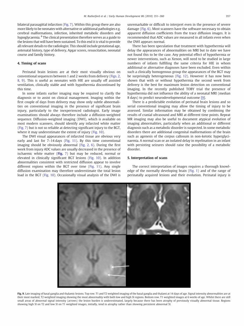
Fig. 9. Late imaging of basal ganglia and thalamic lesions. Top row. T1 and T2weighted imagintheir most marked, T2 weighted imaging showing the most abnormality with both low and hsmall areas of abnormal signal intensity (arrows) the lesion burden is underestimated, lashowing high SI on T2 and low SI on T1 weighted images, initially, tend to atrophy rather t
unremarkable or difficult to interpret even in the presence of severedamage (Fig. 6). Most scanners have the software necessary to obtainapparent diffusion coefficients from the trace diffusion images. It isrecommended that ADC values are measured in all infants even whenthe DWI appears normal.
There has been speculation that treatment with hypothermia willdelay the appearances of abnormalities on MRI but to date we havenot found this to be the case. Any potential effect of hypothermia ornewer interventions, such as Xenon, will need to be studied in largenumbers of infants fulfilling the same criteria for HIE in whomadditional or alternative diagnoses have been excluded. Even withinsuch a clinically homogenous group the appearances of the BGT maybe surprisingly heterogeneous (Fig. 12). However it has now beenshown that with or without hypothermia the second week fromdelivery is the best for maximum lesion detection on conventionalimaging. In the recently published TOBY trial the presence ofhypothermia did not influence the ability of a neonatal MRI (median8 days) to predict neurodevelopmental outcome [9].
There is a predictable evolution of perinatal brain lesions and soserial conventional imaging may allow the timing of injury to beassessed. Similar information may be obtained by combining theresults of cranial ultrasound and MRI at different time points. RepeatMR imaging may also be useful to document atypical evolution ofimaging abnormalities, particularly when an additional or differentdiagnosis such as ametabolic disorder is suspected. In somemetabolicdisorders there are additional congenital malformations of the brainsuch as agenesis of the corpus callosum in non-ketotic hyperglyci-naemia. A normal scan or an isolated delay in myelination in an infantwith persisting seizures should raise the possibility of a metabolicdisorder.
5. Interpretation of scans
The correct interpretation of images requires a thorough knowl-edge of the normally developing brain (Fig. 1) and of the range ofperinatally acquired lesions and their evolution. Perinatal injury is
g of the basal ganglia and thalami at 14 days of age. Signal intensity abnormalities are atigh SI regions. Bottom row. T1 weighted images at 6 weeks of age. Whilst there are stillrgely because there has been atrophy of previously visually abnormal tissue. Regionshan showing persistent abnormal SI.

Fig. 10. Diffusion-weighted imaging in the basal ganglia and thalami. Top row. T1 (left) and T2 (middle) weighted images and ADC map (right) acquired on day 3. The ADC mapshows several regions of abnormal low signal intensity within the basal ganglia and thalami (BGT) that look relatively subtle (circular region of interest). Bottom row. Day 22. T1(left) and T2 (right) weighted images. The BGT abnormalities are very severe with no normal looking tissue within the BGT (circular region of interest). Any abnormality within theBGT on an early ADC map is likely to be clinically significant and any one diffusion-weighted examination is likely to underestimate the final lesion load. By day 22 the white mattersignal intensity (arrow) has become abnormally high and there has been no head growth in this neonate.
Fig. 11. Evolution of abnormalities on diffusion ADCmaps. Top row. Day 5. There is abnormal low signal intensity in the cortex of central sulcus, central grey matter, medial temporallobe and brainstem (from left to right) Bottom row. Day 11. Areas of abnormal low signal intensity consistent with restricted diffusion and ischaemia disappear in many areas buthave become more obvious in the globus pallidus (circle). Appearances on an ADC map evolve over time between days 5 and day 11. Within the BGT, abnormalities may move fromone region to another.
358 M. Rutherford et al. / Early Human Development 86 (2010) 351–360

Fig. 12. Heterogeneity of basal ganglia and thalamic lesions. Appearances of the basal ganglia and thalami (BGT) in three neonates with HIE imaged during the second week fromdelivery. All lesions are severe but are very different in appearance despite similar age at scanning and similar clinical history. The heterogeneity of lesions within the BGT on MRimaging make it difficult to study the evolution of lesions and the relationship of diffusion imaging with conventional imaging.
359M. Rutherford et al. / Early Human Development 86 (2010) 351–360
often symmetrical andmay be confusedwith normal appearances andvice versa by those not experienced in neonatal pathology. It may beappropriate to send images to a centre that regularly performsneonatal examinations for a confirmatory report or second opinion.Since a good quality set of neonatal images contains valuable clinicalinformation that benefits infant, parents and medical staff, it is worth,therefore, to ensure that they have been correctly interpreted.
6. Patterns of injury and prediction of outcome
MR imaging provides detailed information about the pattern oflesions following perinatal brain injury and conventional sequencesmay provide excellent predictions of outcome. In neonates with aglobal hypoxic-ischaemic insult lesions are usually detected withinthe basal ganglia and thalami with abnormal signal intensity in theintervening PLIC (Figs. 2–4). Abnormal signal intensity within the PLICis an excellent predictor of abnormal motor outcome in term infantswith HIE [1]. BGT lesions give rise to motor impairment in the form ofcerebral palsy. The severity of the BGT lesions dictates the severity andnature of the cerebral palsy (Fig. 3) [9]. Lesions within the brainstemare usually found in those neonates with the most severe form of BGTinjury and are often associated with early death (Fig. 13). In neonatesthat survive persisting feeding difficulties and a necessity forgastrostomy are common. Lesions in the BGT are often accompaniedby injury to the cortex and subcortical white matter, most typically
Fig. 13. Appearances of the brainstem. T1 weighted images at the level of the mesencephaloseverely abnormal appearances with widespread low signal intensity (c).
around the central sulcus. These changes are most obvious after thefirst week from injury (Fig. 5). In the presence of severe BGT there areusually secondary WM changes with subsequent atrophy (Fig. 10).This is reflected in poor head growth and the development of asecondary microcephaly. In approximately 50% of neonates with BGTlesions there will be more extensive white matter abnormalities seenon the initial scan and consistent with a primary injury (Fig. 6). Themotor outcome for these children is still dictated by the BGT lesionsbut WM involvement may exacerbate any cognitive deficit. Howeverinfants with severe BGT lesions have severe cognitive impairmentregardless of the severity of initial additionalwhitematter involvement.
In some infants who present with what is thought to be HIEthere is no BGT involvement but only white matter lesions (Fig. 7).These may be haemorrhagic. These lesions give rise to tissueatrophy and later cognitive impairment. The more severe the WMlesions the worse the cognitive outcome [10]. It is important to notethat in some children with isolated cognitive impairment this mayhave arisen as a result of a perinatal injury. However there wouldnormally be an abnormal perinatal history usually with seizures andthe child is likely to be microcephalic. In addition in children whohave a term perinatally acquired white matter injury later follow upimages may be indistinguishable from what would normally becalled periventricular leucomalacia (PVL). The injury may then bewrongly attributed to antenatal damage at an earlier gestation. Onceagain in an infant with signs of PVL, on imaging and/or clinically,
n showing normal (a) moderately abnormal with low signal intensity regions (b) and

360 M. Rutherford et al. / Early Human Development 86 (2010) 351–360
who was born at term it is probably only reasonable to implicateperinatal events if there were neonatal symptoms such asencephalopathy or seizures.
7. Postmortem imaging
Infants with severe encephalopathy should always have brainimaging. This may be difficult and it may not be possible to performbefore an infant dies. In such circumstances and particularly if noautopsy is performed postmortem MR imaging should be considered.It may allow confirmation of abnormalities consistent with a hypoxic-ischaemic insult or suggest an alternative diagnosis [11].This wouldclearly have important genetic and medicolegal considerations.
8. Key guidelines
• Magnetic resonance imaging of the brain is invaluable in assessingthe neonate who presents with encephalopathy. Successful imagingrequires adaptations to both the hardware and sequences used foradults or older children.
• The perinatal and postnatal details often allow prediction of thepattern of lesions sustained and are essential for the correct inter-pretation of the imaging findings.
• Always consider additional or alternative diagnoses in infants withpresumed hypoxic-ischaemic encephalopathy.
• Perinatal lesions are usually at their most obvious on conventionalimaging between 1 and 2 weeks from delivery.
• Very early imaging during the first week may be useful to makemanagement decisions in ventilated neonates but abnormalitiesmay be subtle on conventional imaging.
• Diffusion-weighted imaging (DWI) is clinically useful for the earlyidentification of ischaemic tissue in the neonatal brain, the patternof which can predict outcome.
• Diffusion-weighted imaging may underestimate the final extent ofinjury, particularly basal ganglia and thalamic lesions.
• Serial imaging with quantification of both tissue damage andstructure size provides invaluable insights into the effects ofperinatal injury on the developing brain.
Acknowledgements
We would like to thank all the staff of the Robert Steiner MR Unit,Hammersmith Hospital, the Biomedical Research Centre and theneonatal units of Hammersmith and Queen Charlottes Hospital. Weare also grateful to The Medical Research Council and Philips MedicalSystems for their support.
References
[1] Rutherford MA, Pennock JM, Counsell SJ, Mercuri E, Cowan FM, Dubowitz LM,Edwards AD. Abnormal magnetic resonance signal in the internal capsule predictspoor neurodevelopmental outcome in infants with hypoxic-ischemic encepha-lopathy. Pediatrics 1998;102:323–8.
[2] Cheong JL, Cady EB, Penrice J, Wyatt JS, Cox IJ, Robertson NJ. Proton MRspectroscopy in neonates with perinatal cerebral hypoxic-ischemic injury:metabolite peak-area ratios, relaxation times, and absolute concentrations. AJNRAm J Neuroradiol 2006;27(7):1546–54.
[3] Jiang S, Xue H, Glover A, Rutherford M, Rueckert D, Hajnal JV. MRI of movingsubjects using multislice snapshot images with volume reconstruction (SVR):application to fetal, neonatal, and adult brain studies. IEEE Trans Med Imaging2007;26:967–80.
[4] Mercuri E, Cowan F, Rutherford M, Acolet D, Pennock J, Dubowitz L. Ischaemic andhaemorrhagic brain lesions in newborns with seizures and normal Apgar scores.Arch Dis Child Fetal Neonatal Ed 1995;73:67–74.
[5] Rutherford MA, Counsell S, Allsop Joanna, Boardman James, Kapellou Olga,Larkman David, Hajnal Jo, Edwards David, Cowan PhD* Frances. Diffusionweighted MR imaging in term perinatal brain injury: a comparison with site oflesion and time from birth. Pediatrics 2004;114(4):1004–14.
[6] Okereafor A, Allsop J, Counsell SJ, Fitzpatrick J, Azzopardi D, Rutherford MA, CowanFM. Patterns of brain injury in neonates exposed to perinatal sentinel events.Pediatrics 2008;121(5):906–14.
[7] Logitharajah P, Rutherford MA, Cowan FM. Hypoxic-ischemic encephalopathy inpreterm infants: antecedent factors, brain imaging, and outcome. Pediatr Res2009;66(2):222–9.
[8] Burns CM, Rutherford MA, Boardman JP, Cowan FM. Patterns of cerebral injury andneurodevelopmental outcomes after symptomatic neonatal hypoglycemia. Pedi-atrics 2008;122(1):65–74.
[9] Rutherford M, Ramenghi LA, Edwards AD, Brocklehurst P, Halliday H, Levene M,Strohm B, Thoresen M,Whitelaw A, Azzopardi D. Assessment of brain tissue injuryafter moderate hypothermia in neonates with hypoxic-ischaemic encephalopa-thy: a nested substudy of a randomised controlled trial. Lancet Neurol 2010(1):39–45 (Electronic publication ahead of print).
[10] Cowan F, Dubowitz L, Mercuri E, Counsell S, Rutherford M. White matter injurycan lead to cognitive without major motor deficits following perinatal asphyxiaand early encephalopathy. Dev Med Child Neurol Suppl 2003;93(45):14.
[11] Nicholl RM, Balasubramaniam VP, Urquhart DS, Sellathurai N, Rutherford MA.Postmortem brain MRI with selective tissue biopsy as an adjunct to autopsyfollowing neonatal encephalopathy. Eur J Paediatr Neurol 2007;11(3):167–74.
Copyright © 2022 FDOKUMEN