Phospholipidome of Candida : Each Species of Candida Has Distinctive Phospholipid Molecular Species
Upload
univ-paris7Category
view
1download
0
The Phospholipid Scramblases 1 and 4 Are CellularReceptors for the Secretory Leukocyte Protease Inhibitorand Interact with CD4 at the Plasma MembraneBenedicte Py1,2., Stephane Basmaciogullari1,2., Jerome Bouchet1,2, Marion Zarka1,2, Ivan C. Moura3,4,
Marc Benhamou3,4, Renato C. Monteiro3,4, Hakim Hocini5, Ricardo Madrid1,2, Serge Benichou1,2*
1 Institut Cochin, Universite Paris-Descartes, CNRS, UMR 8104, Paris, France, 2 INSERM, U567, Paris, France, 3 INSERM U699, Paris, France, 4 Universite Paris 7-Denis Diderot,
site Bichat, Paris, France, 5 INSERM U743, Paris, France
Abstract
Secretory leukocyte protease inhibitor (SLPI) is secreted by epithelial cells in all the mucosal fluids such as saliva, cervicalmucus, as well in the seminal liquid. At the physiological concentrations found in saliva, SLPI has a specific antiviral activityagainst HIV-1 that is related to the perturbation of the virus entry process at a stage posterior to the interaction of the viralsurface glycoprotein with the CD4 receptor. Here, we confirm that recombinant SLPI is able to inhibit HIV-1 infection ofprimary T lymphocytes, and show that SLPI can also inhibit the transfer of HIV-1 virions from primary monocyte-deriveddendritic cells to autologous T lymphocytes. At the molecular level, we show that SLPI is a ligand for the phospholipidscramblase 1 (PLSCR1) and PLSCR4, membrane proteins that are involved in the regulation of the movements ofphospholipids between the inner and outer leaflets of the plasma membrane. Interestingly, we reveal that PLSCR1 andPLSCR4 also interact directly with the CD4 receptor at the cell surface of T lymphocytes. We find that the same region of thecytoplasmic domain of PLSCR1 is involved in the binding to CD4 and SLPI. Since SLPI was able to disrupt the associationbetween PLSCR1 and CD4, our data suggest that SLPI inhibits HIV-1 infection by modulating the interaction of the CD4receptor with PLSCRs. These interactions may constitute new targets for antiviral intervention.
Citation: Py B, Basmaciogullari S, Bouchet J, Zarka M, Moura IC, et al. (2009) The Phospholipid Scramblases 1 and 4 Are Cellular Receptors for the SecretoryLeukocyte Protease Inhibitor and Interact with CD4 at the Plasma Membrane. PLoS ONE 4(3): e5006. doi:10.1371/journal.pone.0005006
Editor: Linqi Zhang, Comprehensive AIDS Reseach Center, China
Received January 13, 2009; Accepted February 22, 2009; Published March 31, 2009
Copyright: � 2009 Py et al. This is an open-access article distributed under the terms of the Creative Commons Attribution License, which permits unrestricteduse, distribution, and reproduction in any medium, provided the original author and source are credited.
Funding: This work was supported in part by INSERM, CNRS, Universite Paris-Descartes, the French National Agency for AIDS Research (ANRS), and Sidaction. Thefunders had no role in study design, data collection and analysis, decision to publish, or preparation of the manuscript.
Competing Interests: The authors have declared that no competing interests exist.
* E-mail: [email protected]
. These authors contributed equally to this work.
Introduction
Secretory leukocyte protease inhibitor (SLPI) is a polypeptide of
132 residues (11.7 kDa) secreted by epithelial cells in all the
mucous liquids such as saliva, bronchial and nasal secretions,
cervical mucus, as well as in the seminal liquid [1]. SLPI is a
powerful serine-protease inhibitor, and its main biological role is to
ensure protection of tissues from degradation by the leukocyte
proteolytic enzymes produced during local inflammatory reactions
[2]. Found in the extracellular medium, SLPI is able to cross the
biological membranes and to penetrate into the cell where it will
exert some of its biological functions [3,4]. At the structural level,
the primary amino acid (a.a.) sequence of SLPI reveals the
presence of two so-called «whey-acidic-protein» (WAP) motifs, a
domain of about fifty a.a. with eight highly conserved cysteine
residues that form four disulphide bonds. WAP motifs are
specifically found in a family of inhibitors of serine-proteases,
such as elastase, trypsin and chymotrypsin, whith SLPI and elafin
(or trappin-2) being the most characterized members (for review,
[5]) displaying both anti-inflammatory and antimicrobial activities.
Interestingly, several groups have shown that SLPI also displays,
at physiological concentrations found in saliva (20–150 mg/ml)
[6,7], a specific antiviral activity against human immunodeficiency
virus (HIV-1) [7–13]. The high salivary concentrations of SLPI
may be responsible for the absence of oral transmission of HIV-1
[14–16], and for the reduced mother-to-child HIV-1 transmission
by the mother’s milk [6]. Similarly, high concentrations of SLPI in
vaginal fluids have been associated with reduced rates of perinatal
HIV-1 transmission [17]. The inhibition of HIV-1 replication by
SLPI is independent of its anti-protease activity, but is related to a
perturbation of the virus entry process at a stage posterior to the
interaction of the viral surface glycoprotein gp120 with the CD4
receptor at the surface of HIV-1 target cells [9,11]. The SLPI
antiviral activity is indeed observed in various cell culture models,
including CD4-positive lymphoid and monocytoid cell lines as well
as primary lymphocytes and monocyte-derived macrophages. It is
exerted, independently of the chemokine coreceptor usage, on
viral strains with tropism for either lymphocytes (64 strains) or
macrophages (R5 strains) [7,8,13].
SLPI appears to block HIV-1 entry by interacting with a non-
CD4 cell membrane receptor protein [9,11]. Only two mem-
brane-associated proteins able to interact with SLPI have been
identified so far, the phospholipid (PL) binding protein annexin II
and the phospholipid scramblase 1 (PLSCR1) [9,18]. While
annexin II was proposed as a cofactor specifically involved in the
SLPI antiviral activity observed on macrophages [9], this finding
PLoS ONE | www.plosone.org 1 March 2009 | Volume 4 | Issue 3 | e5006
does not explain the ability of SLPI to block HIV-1 entry both in
CD4-positive transformed T-cell lines and primary peripheral
blood lymphocytes [7,8,10,13,19] where annexin II does not seem
to be expressed [11,20].
Therefore, we further explored the specific role of the
interaction of SLPI with the host cell PLSCR1 protein in the
anti-HIV-1 inhibitory activity of SLPI. PLSCR1 is a membrane
protein of 318 amino acids (a.a.) that is widely expressed at the
surface of many cell types [21–23]. It is one of the four members of
the scramblase family identified in humans, which share between
46 and 59% a.a. identity in their primary sequences [21]. Whereas
PLSCR1, 3 (295 a.a.) and 4 (329 a.a.) are widely expressed in most
human tissues, expression of PLSCR2 (224 a.a.) is restricted to
testis. Like other human and murine scramblases, PLSCR1 shows
a type-II membrane organization, and comprises a long
cytoplasmic domain of 290 a.a., followed by a single transmem-
brane helix (a.a. 291–309) and a short C-terminal extracellular
domain corresponding to the last 9 a.a. [22,23]. Scramblases are
membrane proteins allowing the bidirectional and nonspecific
transbilayer movement of phospholipids across the plasma
membrane ([22,24,25], for reviews).
Here, we confirm that recombinant SLPI is able to inhibit HIV-
1 infection of primary T lymphocytes when used at the
physiological concentrations found in saliva, and we show that
SLPI can also inhibit the transfer of HIV-1 virions from dendritic
cells derived from primary monocytes towards autologous T
lymphocytes, a process which is important in vivo for virus
spreading after mucosal transmission (for review, [26]). At the
molecular level, we show that SLPI is a ligand for both PLSCR1
and PLSCR4, and we reveal that PLSCR1 and PLSCR4 interact
directly with the CD4 receptor at the cell surface of T
lymphocytes. Since the same region of the cytoplasmic domain
of PLSCR1 is involved in the binding with CD4 and SLPI, our
data suggest a model in which the inhibitory effect of SLPI results
from its ability to modulate the interaction of the CD4 receptor
with scramblases. The disruption of these interactions between
CD4 and scramblases may thus represent new targets for antiviral
intervention.
Results
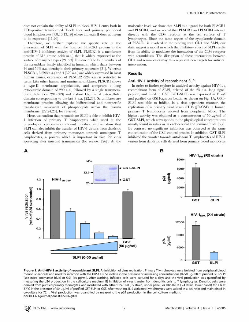
Anti-HIV-1 activity of recombinant SLPIIn order to further explore its antiviral activity against HIV-1, a
recombinant form of SLPI, deleted of the 25 a.a. long signal
peptide, and fused to GST (GST-SLPI) was expressed in E. coli
and purified on GSH-agarose beads. As shown on Fig. 1A, GST-
SLPI was able to inhibit, in a dose-dependent manner, the
replication of a primary viral strain (HIV-1JR-CSF) in human
primary T lymphocytes isolated from peripheral blood. The
highest activity was obtained at a concentration of 50 mg/ml of
GST-SLPI, which corresponds to the physiological concentrations
usually found in saliva or in endocervical and seminal fluids [6,7].
By contrast, no significant inhibition was observed at the same
concentration of the GST control protein. In addition, GST-SLPI
inhibited the transfer towards autologous T lymphocytes of HIV-1
virions from dendritic cells derived from primary blood monocytes
Figure 1. Anti-HIV-1 activity of recombinant SLPI. A) Inhibition of virus replication. Primary T lymphocytes were isolated from peripheral bloodmononuclear cells and used for infection with the HIV-1JR-CSF isolate in the presence of increasing concentrations (0–50 mg/ml) of purified GST-SLPI(see inset, coomassie blue) or GST (50 mg/ml). After washing, infected cells were cultured for 6 days and the viral production was quantified bymeasuring the p24 production in the cell-culture medium. B) Inhibition of virus transfer from dendritic cells to T lymphocytes. Dentritic cells werederived from purified primary monocytes, and incubated with either HIV-1Bal (R5 strain, upper panel) or HIV-1NDK (64 strain, lower panel) for 1 h at37uC in the presence of 50 mg/ml of purified GST-SLPI or GST. After washing, IL-2 activated-lymphocytes were added in a 1/5 ratio and maintained inco-culture for 72 h. Viral production was quantified by measuring the p24 production in the cell culture medium.doi:10.1371/journal.pone.0005006.g001
CD4-PLSCR-SLPI Interactions
PLoS ONE | www.plosone.org 2 March 2009 | Volume 4 | Issue 3 | e5006

(Fig. 1B). Interestingly, this inhibitory activity was exerted on viral
strains with tropism for either lymphocytes (HIV-1NDK64 strain)
or macrophages (HIV-1Bal R5 strain). These results show that
recombinant SLPI inhibits replication of HIV-1 in primary T
lymphocytes, but also cell-to-cell transfer of the virus from
dendritic cells to T lymphocytes.
SLPI directly binds to the phospholipid scramblases 1and 4
Since it has been previously suggested that the ubiquitous
PLSCR1 membrane protein was a receptor for SLPI [18], we
further explored this possibility by using several interaction assays.
The interaction between SLPI and PLSCR1 was initially analyzed
in the yeast two-hybrid system. The SLPI sequence was fused to
LexA and assessed for interaction with PLSCR1 fused to the
Gal4AD in the L40 yeast strain containing the two LexA-inducible
reporter genes, HIS3 and LacZ. In contrast to the results published
previously [18], the full length 132 a.a. long form of SLPI,
containing the N-terminal signal peptide (see on Fig. 2A), was not
able to interact with PLSCR1 in this assay (data not shown),
whereas a specific interaction could be detected with a form of
SLPI lacking the first 25 residues, as evidenced by growth of yeast
cells on medium without histidine and expression of the b-gal
activity (Fig. 2B). This form of SLPI (a.a. 26–132) corresponds to
the mature secreted protein after cleavage of the N-terminal signal
sequence. Binding of SLPI to PLSCR1 was direct, since we were
able to recapitulate in vitro this interaction in an ELISA test where
immobilized SLPI specifically captured the purified GST-
PLSCR1 fusion protein but not the GST control (Fig. 2C).
The specificity of the interaction was also confirmed by
biochemical approaches. GST-SLPI(26-132) was first immobilized
on GSH-sepharose beads, and then incubated with a lysate from
cells expressing GFP-PLSCR1. Bound proteins were analyzed by
Western blotting with anti-GFP (Fig. 2D). GFP-PLSCR1 specifi-
cally bound to GST-SLPI, but not to GST or to GST-Arf1 used as
negative controls. As shown in Fig. 2D, the two WAP1 (GST-SLPI
W1) and WAP2 (GST-SLPI W2) motifs of SLPI were separately
able to recruit GFP-PLSCR1 from tranfected cells, but to a lower
extent compared with the mature form of SLPI(26-132), suggesting
that both motifs cooperate to allow an optimal interaction with
PLSCR1. A weak interaction was also detected in the same pull-
down assay with a recombinant form of the mature elafin (GST-
Elafin) (Fig. 2D), another member of the family of serine protease
inhibitors containing a single WAP motif [5].
Similarly, we analyzed whether SLPI was also able to interact
with PLSCR3 and PLSCR4, the two other human scramblases
that are widely expressed in all tissues. As shown in Fig. 2E, both
GFP-PLSCR1 and GFP-PLSCR4, but not GFP-PLSCR3, were
specifically pulled-down on GST-SLPI-immobilized beads. These
results indicate that SLPI is a specific ligand for both PLSCR1 and
PLSCR4.
Co-distribution of scramblases and CD4 at the plasmamembrane
Molecular modeling predicted that PLSCR1 is a type II
membrane protein with a single transmembrane helix and a short
C-terminal extracellular region (see on Fig. 3A). To determine
whether PLSCR1 was expressed at the plasma membrane in CD4-
positive T cells, we first analyzed the cellular distribution of the
wild type or truncated forms of PLSCR1 fused to the C-terminus
of GFP in Jurkat T cells. After transfection, the cells were fixed and
directly examined by fluorescence microscopy (Fig. 3B). As
expected, both the full length GFP-PLSCR1 fusion and the (1–
310) truncated form, lacking the last 8 C-terminal a.a., were
predominantly localized as a rim staining at the plasma membrane
(left and central panels, respectively), but a consistent fraction was
also observed in an intracellular membrane compartment which
largely co-localized with Golgi markers (not shown). By contrast,
the truncated form (1–290) deleted of the last 28 C-terminal a.a.
including the transmembrane domain showed a diffuse nucleo-
cytoplasmic distribution (right panel). Therefore, PLSCR1, as well
as PLSCR3 and PLSCR4 (Fig. 3C), are transmembrane proteins
which are largely localized at the plasma membrane in CD4-
positive T lymphoid cells. As expected, GFP-PLSCR1 and the
endogenous CD4 antigen were both distributed at the plasma
membrane in T cells (Fig. 3D).
Figure 2. Characterization of the PLSCR1/SLPI interaction. A) Schematic representation of SLPI. SLPI contains an N-terminal signal peptide(black) and two WAP motifs (grey); a.a. are numbered according to Tseng et al. [18]. B) Interaction in the two-hybrid system. L40 yeast strainexpressing the indicated Gal4AD and LexA hybrids was analyzed for histidine auxotrophy and b-gal activity. Transformants were patched on mediumwith histidine (left panels) and then replica-plated on medium without histidine (central panels) and on Whatman filter for b-gal assay (right panels).Growth in the absence of histidine and expression of b-gal activity indicate the interaction between hybrids. C) ELISA interaction analysis. 96-wellplates were coated with non-tagged SLPI at a final concentration of 0.25 mM. After washings, GST-PLSCR1 (black curve) or GST (red curves) wasincubated at concentrations ranging from 1 nM to 1 mM. Binding was then revealed with an anti-GST antibody and a secondary peroxidase-conjugated anti-mouse IgG. The peroxidase substrate solution was incubated for 10 min and the optical density was measured at 450 nm. D and E) Invitro interactions. Lysates from 293T cells expressing GFP-PLSCR1, GFP-PLSCR3 or GFP-PLSCR4 were incubated with equal amounts of GST fusions(lower panels, Ponceau red) immobilized on GSH-sepharose beads as indicated at the top. Bound proteins were then analyzed by immunoblottingwith anti-GFP (upper panels).doi:10.1371/journal.pone.0005006.g002
CD4-PLSCR-SLPI Interactions
PLoS ONE | www.plosone.org 3 March 2009 | Volume 4 | Issue 3 | e5006

PLSCR1 and PLSCR4 interact directly with the CD4receptor
Since it was previously reported that the inhibition of HIV-1
replication by SLPI resulted from a direct action on the virus entry
process [9,11], we asked whether PLSCR1 could directly associate
with CD4, the main receptor of HIV-1 at the cell surface of T
lymphocytes. This hypothesis was first challenged in vitro by pull-
down assay. Recombinant PLSCR1 fused to GST was expressed in
E. coli, immobilized on GSH-sepharose beads and then tested for its
ability to retain full length CD4 from a lysate of Jurkat T-cells. Bound
proteins were analyzed by Western blot with anti-CD4 antibody
(Fig. 4A). Endogenous CD4 was specifically pulled-down by GST-
PLSCR1, but not by the GST control. Similarly, GST-PLSCR1 was
also able to retain in vitro translated 35S-CD4 (data not shown),
indicating a direct interaction between the two cellular proteins.
The interaction between endogenous PLSCR1 and CD4
proteins was finally documented by a co-immunoprecipitation
assay performed on CD4-positive T-lymphocytes. Jurkat cells were
lyzed and CD4 was precipitated with an anti-CD4 antibody.
Precipitates were then analyzed by Western blotting with anti-
CD4 (Fig. 4B, upper panel) and anti-PLSCR1 (lower panel).
Endogenous PLSCR1 was detected as a 35-kDa protein only from
cell lysate immunoprecipitated with anti-CD4 and not from lysate
treated with the control isotypic antibody, demonstrating that
PLSCR1 interacts with CD4 in human CD4-positive T-lympho-
cytes. Interestingly, co-immunoprecipitation experiments per-
formed on 293T cells expressing HA-tagged PLSCR1 in
combination with wild type CD4 (WT) or a CD4 mutant lacking
the cytoplasmic domain (DCT) revealed that the CD4 mutant
failed to associate with PLSCR1 (Fig. 4C). In addition, similar
experiments performed in 293T cells showed that PLSCR4 was
also able to interact with CD4, and this interaction was again
dependant of the cytoplasmic domain of the receptor (Fig. 4D).
Since the results reported in Figs 4C and 4D indicated that the C-
terminal cytoplasmic tail of CD4 (CD4c) was absolutely required
to maintain the interactions, we analyzed whether this CD4c
domain could be sufficient to associate with PLSCR1 in the two-
hybrid system. The 40 a.a. long cytoplasmic domain of CD4 (see
on Fig. 5A) was thus expressed in the L40 yeast reporter strain as a
protein fused with LexA and in combination with the full length
PLSCR1 fused to Gal4AD. As shown in Fig. 4E, the LexA-CD4c
hybrid conferred on the reporter strain the ability to grow on
medium without histidine and to express b-gal activity in the
presence of the Gal4AD-PLSCR1 hybrid, but not in cells co-
expressing an irrelevant Gal4AD-Raf hybrid. Used as control, we
detected the well-characterized interaction between CD4c and the
p56Lck kinase in this system (Fig. 4E).
Altogether, these results indicate that both PLSCR1 and
PLSCR4 can interact with the CD4 receptor, and these
interactions are mediated by the cytoplasmic domain of the
CD4 receptor.
Determinants of the CD4 cytoplasmic domain requiredfor binding to PLSCR1
In order to characterize the determinants of the CD4c domain
that participated in the interaction, a set of CD4 mutants with
Figure 3. Co-distribution of scramblases and CD4 at the plasma membrane. A) Schematic representation of PLSCR1. PLSCR1 is a type-IItransmembrane protein with a long N-terminal cytoplasmic domain (a.a. 1–290, grey box) containing 3 Cys-rich motifs (red boxes), and atransmembrane domain (a.a. 291–310, black box). Amino acids are numbered according to Zhou et al. [23]. B) Localization of wild type and deletedGFP-PLSCR1 forms. Jurkat CD4-positive T cells expressing the full length (left panel) or deleted forms (1–310 and 1–290, central and right panels,respectively) of GFP-PLSCR1 were fixed and directly examined. Cells were analyzed by epifluorescence microscopy, and images were acquired using aCCD camera. C) Localization of GFP-PLSCR3 and GFP-PLSCR4. Jurkat cells expressing GFP-PLSCR1 (left panel), GFP-PLSCR3 (central panel) or GFP-PLSCR4 (right panel) were fixed and directly examined as in (B). D) Subcellular distribution of CD4 and PLSCR1. Jurkat cells expressing wild type GFP-PLSCR1 (middle panel) were fixed, permeabilized and subsequently stained with an anti-CD4 (left panel). Scale bars, 10 mm.doi:10.1371/journal.pone.0005006.g003
CD4-PLSCR-SLPI Interactions
PLoS ONE | www.plosone.org 4 March 2009 | Volume 4 | Issue 3 | e5006

deletions and/or point mutations within the cytoplasmic domain
(see Fig. 5A) were assessed for PLSCR1 binding by co-
immunoprecipitation assay. CD4 mutants were expressed in
293T cells in combination with HA-PLSCR1, and then immuno-
precitated with anti-CD4. As deduced from the experiments
shown in Fig. 5B, the determinants required for PLSCR1 binding
were localized in the distal portion of the cytoplasmic domain,
since the CD4 423,3R/A mutant, lacking the last 10 a.a., failed to
interact with PLSCR1 (left panel). However, mutations of the
HRF residues found in position 425 to 427 (mutant CD4 HRF/A)
did not affect binding to PLSCR1, suggesting that the last 6
residues are essential to maintain the interaction. We could note
that the two cysteine residues, that are critical for binding to
p56Lck ([27], and data not shown), are not required for binding to
PLSCR1 (right panel, mutant CD4 C/S), indicating that the
determinants required for interaction with PLSCR1 are different
from those involved in the interaction with p56Lck.
To confirm that the distal region of CD4c was important to
support PLSCR1 binding, we developed an ELISA to recapitulate
the CD4-PLSCR1 interaction in vitro. Two peptides spanning the
regions 405–433 and 405–426 of CD4 (Fig. 6A) were synthesized
and then coated on microtiter plates. After incubation with
Figure 4. Interaction of PLSCR1 and PLSCR4 with the CD4 receptor. A) In vitro interaction. Lysates from Jurkat CD4-positive T cells wereincubated with equal amounts of GST or GST-PLSCR1 (upper panel, coomassie blue) immobilized on GSH-sepharose beads. Bound proteins werethen analyzed by immunoblotting with anti-CD4 (lower panel). B) Co-immunoprecipitation of endogenous proteins from T lymphocytes. Jurkat T cellswere lyzed and CD4 was precipitated with either anti-CD4 (OKT4) or a control isotypic antibody. Precipitates were analyzed by Western blot with anti-CD4 (upper panel) or anti-PLSCR1 (lower panel). C) Co-precipitation of overexpressed CD4 and PLSCR1 proteins. 293T cells expressing HA-taggedPLSCR1 (lower panel, Cell lysate) in combination with wild-type CD4 (WT) or a mutant of CD4 deleted of its cytoplasmic domain (DCT) were lyzed andthe CD4 forms were precipitated with anti-CD4. Precipitates were then analyzed by Western blot with anti-CD4 (upper panel) or anti-HA (middlepanel). D) Co-precipitation of overexpressed CD4 and PLSCR4 proteins. 293T cells expressing GFP- PLSCR1, GFP-PLSCR3 or GFP-PLSCR4 (lower panels,Cell lysate) in combination with CD4 WT (right panels) or CD4 DCT (left panels) were lyzed and the CD4 forms were precipitated with anti-CD4.Precipitates were then analyzed as in (C). Of note, the differences in migration observed between PLSCR1 (318 a.a.), PLSCR3 (295 a.a.) and PLSCR4(329 a.a.) is likely related to the respective amino acid lengths of the GFP fusion proteins [23]. E) Interactions in the two-hybrid system. L40 yeast strainexpressing the LexA-CD4c hybrid in combination with the indicated Gal4AD hybrids was analyzed for histidine auxotrophy and b-gal activity.Transformants were patched on medium with histidine (upper panels) and then replica-plated on medium without histidine (middle panels) and onWhatman filter for b-gal assay (lower panels).doi:10.1371/journal.pone.0005006.g004
Figure 5. Mapping of the CD4 determinants required for PLSCR1 binding by co-immunoprecipitation assay. A) Schematicrepresentation of the human CD4 mutants. The extracellular and transmembrane (TM) domains of CD4 are represented in grey and hatched,respectively. The a.a. sequences of the cytoplasmic tail of the CD4 mutants are aligned with that of the wild-type CD4 (CD4 WT). Dashes (2) indicatea.a. identities with the wild type protein and a.a. substitutions are identified. B) Co-immunoprecipitation assay. Protein extracts were prepared 48 hpost transfection from 293T cells co-expressing HA-PLSCR1 together with wild type or mutated CD4 as indicated at the top. Crude extracts were thensubjected to CD4 immunoprecipitation followed by Western blot analysis with anti-CD4 (upper panels) and anti-HA (lower panels).doi:10.1371/journal.pone.0005006.g005
CD4-PLSCR-SLPI Interactions
PLoS ONE | www.plosone.org 5 March 2009 | Volume 4 | Issue 3 | e5006

purified GST-PLSCR1 or GST, binding was revealed with an
anti-GST antibody. As shown in Fig. 6B, a positive signal was
observed in a large range of GST-PLSCR1 concentrations with
the long CD4c peptide (left panel), but not with the short CD4c
peptide lacking the last 5 a.a. (right panel). This signal was specific,
since no binding to the CD4c peptide was detected with the GST-
Arf1 (not shown) and GST controls at concentrations ranging
from 1 to 300 nM. Again, the cysteine residues directly involved in
p56Lck binding were not required for PLSCR1 binding, since the
CD4c peptides had alanine at these positions (Fig. 6A). These data
definitively demonstrate that the interaction between PLSCR1
and CD4 is direct, and requires the last 5 residues of the CD4
cytoplasmic domain.
SLPI binds to the cytoplasmic domain of PLSCR1 andimpairs CD4/PLSCR1 association
Since our results demonstrated that both CD4 and SLPI
interacted with PLSCR1, we first investigated if the binding of
SLPI to PLSCR1 could have any effect on CD4/PLSCR1
interaction. The impact of SLPI on the recruitment of CD4 by
recombinant GST-PLSCR1 was analyzed using the pull-down
assay described previously (Fig. 7A). At a concentration of 100 mg/
ml, recombinant GST-SLPI (left panel) dramatically reduced the
interaction of GST-PLSCR1 with native CD4 from a Jurkat cell
lysate (right panel). By contrast, no significant inhibition of CD4
binding was observed with the GST-Arf1 (right panel) and GST
(not shown) controls at the concentration of 120 mg/ml.
To determine the molecular nature of such an inhibition, we
mapped the region of PLSCR1 involved in the interaction with the
mature form of SLPI and with CD4c as well. Deletion mutants of
PLSCR1 were generated and analyzed in the two-hybrid system
for their capacity to interact with either the cytoplasmic domain of
CD4 or the mature form (26–132) of SLPI. The results
recapitulated in Fig. 7B show that CD4 and SLPI bound to the
same region of PLSCR1 located in the cytoplasmic domain of the
protein. The minimal sequence of PLSCR1 able to interact with
both partners was mapped between residues 155 and 233 in a
region containing the Cys-rich motif required for palmitoylation of
the protein [28]. However, this motif did not seem to participate in
these interactions, since Ala substitution of the 5 Cys residues
(mutant PLSCR1 C181/189A) did not disturb the capacity of
PLSCR1 to interact with either CD4 or SLPI. Interestingly, these
results show that the interaction between CD4 and PLSCR1 is
mediated by the respective cytoplasmic domains of these two
membrane proteins.
Altogether, the results reported in Fig. 7 strongly suggest that
SLPI modulates, at physiological concentrations (20–150 mg/ml)
[6,7], the CD4-PLSCR1 interaction by competitive binding to the
CD4-binding region of PLSCR1.
Discussion
In the present study, we document and highlight that the innate
host defense SLPI polypeptide, secreted in all mucosal liquids and
displaying an inhibitory activity against HIV-1, is a natural ligand
for the ubiquitously expressed plasma membrane PLSCR1
protein. Initially revealed from a genetic two-hybrid screening
performed to identify SLPI binding proteins [18], we confirm the
specificity of the interaction with PLSCR1 by biochemical
approaches, and reveal that SLPI also binds to PLSCR4, another
member of the scramblase family. Since PLSCR1 is a type II
integral membrane protein, we show that SLPI directly binds to a
region of PLSCR1 located in the central part of its long
cytoplasmic domain. Moreover, we demonstrate that PLSCR1,
and also PLSCR4, can interact directly with CD4, the main
receptor required for HIV-1 entry into its target cells, such as T
lymphocytes and macrophages. These interactions are mediated
Figure 6. Mapping of the CD4 determinants required for PLSCR1 binding by ELISA. A) Primary a.a. sequences of the CD4 long (405–433)and short (405–426) peptides used in the ELISA test. The primary sequence of the entire cytoplasmic domain of CD4 is shown at the top; the 2 Cysresidues required for p56Lck binding and mutated in Ala in both peptides are indicated in red. B) ELISA interaction test. 96-well plates were coatedwith the CD4c long (left panel) or short (right panel) peptides at a final concentration of 0.5 mM. After washings and blocking of non-specific bindingsites, GST or GST-PLSCR1 was incubated at concentrations ranging from 1 nM to 300 nM. Binding was then revealed with an anti-GST antibody and asecondary peroxidase-conjugated anti-mouse IgG. The peroxidase substrate solution was incubated for 10 min and the optical density was measuredat 450 nm.doi:10.1371/journal.pone.0005006.g006
CD4-PLSCR-SLPI Interactions
PLoS ONE | www.plosone.org 6 March 2009 | Volume 4 | Issue 3 | e5006

by determinants found in the cytoplasmic domains of these
integral membrane proteins. Interestingly, we demonstrate that
CD4 and SLPI bind to the same region of the cytoplasmic domain
of PLSCR1 (a.a. 155–233). Finally, we show that SLPI competes
with CD4 thus modulating its interaction with PLSCR1.
While recent works focused on the biological functions of
PLSCR1, nothing is known regarding the subcellular localization
and the functions of PLSCR4 ([29], for review). PLSCR1 was
originally identified as a membrane protein that promoted
acceleration of transbilayer phospholipid movements in response
to intracellular calcium, but increasing evidence indicates that
PLSCR1 also has biological roles in cell signaling pathways.
PLSCR1 has been shown to be engaged when cells are activated
through several plasma membrane receptors, including the
epidermal growth factor receptor and the high-affinity IgE
receptor [30]. The long cytoplasmic domain of PLSCR1 contains
multiple proline-rich motifs within its N-terminal distal region, and
these motifs may constitute Src-homology 3 (SH3) domain binding
sites, as revealed by the interaction of PLSCR1 with the SH3
domain of the c-Abl tyrosine kinase that in turn phosphorylates
PLSCR1 [31]. However, the determinants of PLSCR1 required
for SLPI binding are located in the central region of the
cytoplasmic tail containing cysteine-rich motifs. The minimal part
of PLSCR1 sufficient to mediate interaction with SLPI, and with
the CD4 receptor (see below), was mapped within a 79 a.a. long
region comprised between residues 155 and 233. This region of
PLSCR1 contains the cysteine cluster that forms a site for
palmitoylation involved in the stabilization of the membrane
anchoring of PLSCR1 [28], and this cluster is conserved in
PLSCR4. However, substitution of the 6 cysteine residues (Cys184
to Cys189) did not disturb the capacity of PLSCR1 to interact with
either SLPI or CD4, indicating that the palmitoylation motif does
not directly participate in these interactions. These cysteine
residues may indirectly participate since palmitoylation is required
for membrane expression of PLSCR1 which, when mutated, is
found in the nucleus [32]. Similarly, we found that both PLSCR3
and PLSCR4 are primarily localized at the plasma membrane of
T cells when they are expressed as GFP fusions. SLPI, which is
secreted in the extracellular medium, therefore needs to cross the
plasma membrane for efficient binding to the endofacial domain
of PLSCR1 and PLSCR4. It has been recently documented that
recombinant SLPI can be translocated through the plasma
membrane to reach the cytoplasm and nucleus of cells such as
monocytes, macrophages and B lymphocytes where it interacts
with cellular effectors [3,4,33]; similarly, we confirmed that the
purified GST-SLPI fusion protein used in the present study was
also able to penetrate into the cells from the culture medium (data
not shown).
While elafin is formed by a single WAP motif and interacted
weakly with PLSCR1, SLPI contains two WAP motifs and each
motif was able to support binding to PLSCR1, indicating that both
domains may cooperate in an additive manner leading to an
Figure 7. Disruption of the CD4/PLSCR1 interaction by SLPI. A) In vitro inhibition of the CD4/PLSCR1 interaction by SLPI. GST or GST-PLSCR1(left panel, coomassie blue) was incubated with equal amounts of lysates from Jurkat CD4-positive T cells in the presence of the indicatedconcentrations of either GST-SLPI or GST-ARF1 (left panel) used as a control. Bound proteins were analyzed by Western blot with anti-CD4 (rightpanel). B) Mapping of the PLSCR1 determinants required for binding to CD4 and SLPI. L40 yeast strain expressing either the cytoplasmic domain ofCD4 (CD4c) or SLPI fused to LexA in combination with each of the deleted forms of the Gal4AD-PLSCR1 hybrids indicated on the left was analyzed forhistidine auxotrophy and beta-gal activity. The interactions between hybrid proteins were scored as follows: (+), cell growth on medium withouthistidine and development of a b-gal activity; (2), no growth on medium without histidine and no b-gal activity.doi:10.1371/journal.pone.0005006.g007
CD4-PLSCR-SLPI Interactions
PLoS ONE | www.plosone.org 7 March 2009 | Volume 4 | Issue 3 | e5006

optimal interaction with PLSCR1. This also suggests that
PLSCR1 binding is not mediated by a linear motif contained
within the SLPI primary sequence, but is rather related to the
recognition of the conformational structure of the WAP domains.
Like other members of the WAP family proteins, SLPI is secreted
by epithelial cells and is thus found in all mucosal fluids ([1], for
review). However, more recent studies have revealed that SLPI
can be also produced and secreted by cells of the immune system,
including neutrophils, B lymphocytes, macrophages and dendritic
cells [33–36]. This suggests that exogenous SLPI functions as more
than a protease inhibitor of leukocyte serine proteases for the
protection of tissues at sites of inflammation, but it also participates
as an intracellular modulator in some signaling transduction
pathways. Similarly, our observations suggest that SLPI is an
intracellular ligand which may participate in the modulation of the
PLSCR1 and PLSCR4 functions by interacting directly with their
cytoplasmic tail.
The antiviral activity of SLPI against HIV-1 has been
documented in several reports [7–11,13]. SLPI is able to inhibit
the replication of primary viral strains both in human primary
CD4-positive T lymphocytes and in monocyte-derived macro-
phages at the physiological concentrations found in saliva (20–
120 mg/ml). In addition, we show here that SLPI is also able to
inhibit the capture of HIV-1 virions from dendritic cells derived
from primary blood monocytes and then the transfer of virions
towards autologous T lymphocytes, a process which is important in
vivo for spreading of the virus after mucosal transmission (for
review, [26]). This inhibitory activity is exerted on viral strains
with tropism for either lymphocytes or macrophages. The
inhibition of HIV-1 replication by SLPI is independent of its
anti-protease activity, does not involve a direct action on virus
particles, and is related to a block of the virus entry process at a
stage that is posterior to the interaction of the viral gp120 envelope
glycoprotein with the CD4 receptor [9,11]. This antiviral activity
is rather due to an interaction with the host cell surface molecule,
and annexin II, a membrane-associated protein that is specifically
expressed in macrophages, has been recently proposed as a
receptor for SLPI that could participate in HIV-1 infection of
macrophages [9]. However, this finding does not explain the
ability of SLPI to block HIV-1 replication both in CD4-positive
transformed T-cell lines and in primary peripheral blood
lymphocytes [7,8,10,13,19] where annexin II does not seem to
be expressed [11,20]. In addition, it was recently reported that
annexin II was rather involved in the late stage of HIV-1 assembly
through a direct interaction with the viral Pr55Gag precursor on
endosomal membranes of macrophages [20]. In this context, our
findings raise a new hypothesis concerning the molecular
mechanism of antiviral activity of SLPI. Our data suggest that
SLPI may disrupt the interaction of CD4, the main receptor of
HIV-1, at least with the PLSCR1 membrane protein at the cell
surface of CD4-expressing cells. Although the functions of the
interactions of CD4 with PLSCR1 and PLSCR4 in the biology of
lymphoid or myeloid cells expressing CD4, as well as in the HIV-1
infection process, remain to be elucidated, the demonstration that
the CD4 receptor associates directly with PLSCR1 and PLSCR4
may represent a critical point for the understanding of the antiviral
activity of SLPI. While we cannot exclude that SLPI could act as a
modulator on some other cellular signaling pathways leading to
inhibition of HIV-1 replication [3,4,9], we propose that the
antiviral activity of SLPI is related, at least in part, to the
disruption of the interactions of CD4 with PLSCR1 and PLSCR4.
As mentioned above, it is now clearly established that SLPI is
indeed capable to cross the biological membranes to access the
cytoplasm of the cell ([3,4], and data not shown) where it could
disrupt the interaction between the cytoplasmic domains of CD4
and PLSCR1 and PLSCR4.
In summary, the characterization of a direct interaction
between CD4 and scramblases, proteins regulating the movement
of phospholipids in the plasma membrane, as well as our finding
that SLPI, an antiviral polypeptide, disrupts the interaction
between CD4 and PLSCR1, open new directions for therapeutic
interventions. We hypothesize that the data generated herein will
help to the development of new antiviral strategies, and we are
currently looking to identify chemical compounds disrupting the
interaction between CD4 and PLSCR1 by conducting a high
throughput screen using the ELISA test developed in the present
study (see Fig. 6) as a primary interaction assay that recapitulates in
vitro the association of CD4 with scramblases.
Materials and Methods
PlasmidsYeast expression vectors. The vector for expression of the
cytoplasmic domain of CD4 fused to LexA has been described
[37]. The DNA sequence coding for PLSCR1 was amplified by
PCR from a cDNA library [38] and cloned into the pGAD3S2X
plasmid (Clontech) to generate the pGAD-PLSCR1 vector for
expression of PLSCR1 fused to the Gal4 activation domain
(Gal4AD). The deleted forms of PLSCR1 (see on Fig. 7) were
amplified by PCR using pGAD-PLSCR1 as a template and
appropriate primers. The PCR products were then inserted into
the EcoRI-NotI sites of pGAD3S2X for expression of the Gal4AD
fusions. The DNA sequence coding for the truncated form (aa 21–
132) of the human SLPI deleted of the signal peptide sequence was
synthesized in vitro by PCR using a set of specific primers, and
then cloned into the EcoRI-BamHI sites of the pLex10 plasmid
(Clontech) to generate the pLex-SLPI vector for expression of the
LexA-SLPI hybrid.
Bacterial expression vectors. The vectors for expression of
the full length PLSCR1 and the truncated form fused to the
glutathione-S-tranferase (GST) were made by subcloning
restriction fragments from the pGAD-PLSCR1 and pLex-SLPI
into the BamHI-XhoI and EcoRI-XhoI sites of the plasmids
pGEX4T-2 and pGEX4T-1 (Invitrogen), respectively. For
expression of the GST-SLPI W1 and GST-SLPI W2 fusions,
PCR fragments obtained using pLex-SLPI as a template and
appropriate primers were inserted into the EcoRI-XhoI sites of the
pGEX4T-1 plasmid. The vector for expression of GST-elafin (a.a.
61 to 117) was obtained by PCR amplification of the DNA coding
sequence of trappin-2 (pre-elafin) with specific primers and using
the pGE-SKAP plasmid (gift of Thierry Moreau, Tours, France) as
a template, and the PCR product was inserted into pGEX4T-1 as
indicated above.
Mammalian expression vectors. The vectors for
expression of the wild type and deleted forms of CD4 have been
described previously [37]. The vector for expression of the
hemaglutinin(HA)-tagged form of PLSCR1 was made by PCR
amplification using pGAD-PLSCR1 as a template and
appropriate primers, and the PCR product was subcloned in the
BamHI-XhoI sites of the pAS1B plasmid [39]. Vectors for
expression of the full length and truncated forms (1–310 and 1–
290, see on Fig. 3) of PLSCR1 fused to GFP (GFP-PLSCR1) were
made by PCR amplification using pGAD-PLSCR1 as a template;
the PCR products were inserted into the XhoI-EcoRI sites of the
pEGFPC1 plasmid (Clontech). Vectors for expression of GFP-
PLSCR3 and GFP-PLSCR4 were made by PCR amplification
using pENTR221-PLSCR3 and pENTR221-PLSCR4, purchased
from Invitrogen (clones ID IOH13045 and IOH55049,
CD4-PLSCR-SLPI Interactions
PLoS ONE | www.plosone.org 8 March 2009 | Volume 4 | Issue 3 | e5006

respectively), as templates; the PCR products were then inserted as
previously in pEGFPC1 (Clontech).
Commercial antibodies and reagentsThe monoclonal antibody used to immunoprecipitate CD4,
OKT4 (ATCC), is specific for the extracellular domain of CD4. In
western blot analysis, CD4 was detected using rabbit anti-CD4
polyclonal antibody H370 (Santa Cruz), while GFP-PLSCR1, -
PLSCR3 and -PLSCR4 fusions were detected using a pool of two
mouse anti-GFP monoclonal antibodies (7.1 and 13.1, Roche).
The mouse anti-GST antibody (G1160) used in ELISA was from
Sigma. Secondary reagents were horseraddish peroxidase-coupled
anti-mouse and anti-rabbit IgG (Sigma), both used at 1:2,000
dilution. Immunofluorescence staining was performed with anti-
CD4 (SK3, Becton Dickinson) followed by goat anti-mouse
Alexa594-coupled antibody (Invitrogen). Non-tagged SLPI was
purchased from R&D, while the CD4 long (a.a. 405–433) and
short (a.a. 405–426) synthetic peptides (.90% pure) were
purchased from NeoMPS (Strasbourg, France).
Generation of anti-PLSCR1 antibodiesMonoclonal antibodies against human PLSCR1 were generated
as follows: a 6 week-old BALB/c mouse was subcutaneously
immunized with GST-PLSCR1 (10 mg) emulsified in 100 ml
Freund’s complete adjuvant. Immunizations were repeated at 7-
day intervals in incomplete Freud’s adjuvant (3 times) and in PBS
one day before cell fusion. Lymph node cells were fused with
Ag8.653 mouse myeloma cell line (1:1 ratio) [40]. Hybridoma
supernatants specificity was tested by ELISA comparing GST and
GST-PLCR1 recombinant proteins. All positive specific clones
were then tested by immunoprecipitation and immunofluores-
cence. One hybridoma (clone 314A10, IgG1) was obtained after
subcloning by limited dilution. Polyclonal antibodies were
obtained by immunizing rabbits with the PLSCR1 C-terminal
peptide (encompassing the 306–318 a.a. sequence CESTGS-
QEQSSGVW) as described elsewhere [23].
Cell culture and transfectionCD4-positive HPB-ALL and Jurkat T-cells (clone 20) were
kindly provided by G. Bismuth (Institut Cochin, Paris, France) and
C. Hivroz (Institut Curie, Paris, France), respectively. These T-cell
lines were maintained in RPMI 1640 medium with Glutamax-1
(Invitrogen) supplemented with 10% fetal calf serum (FCS) and
100 U/ml penicillin/streptomycin. HPB-ALL and Jurkat cell lines
were electroporated using Biorad electroporator (Gene pulser II) at
250 V and 950 mF in complete media supplemented with 40 mM
NaCl. 293T cells were grown in Dulbecco modified Eagle’s
medium (DMEM) supplemented with 10% FCS, 100 IU/ml
penicillin and 0.1 mg/ml streptomycin. 293T cells were transfect-
ed using the calcium phosphate precipitation technique. Periph-
eral blood mononuclear cells (PBMC) were isolated from blood of
healthy donors by Ficoll (MSL) density gradient centrifugation.
Monocytes were isolated from PBMC by plastic adherence, and
immature dendritic cells were obtained from monocytes differ-
enciated in the presence of GM-CSF and IL-4 (both at 10 ng/ml)
for 6 days of culture as described previously [41]. Contamination
of immature dendritic cells with CD3+ T lymphocytes was ,1%
as checked by flow cytometry (data not shown). Autologous
lymphocytes, corresponding to the non adherent fraction after the
adherence step, were cultured in RPMI, 10% FCS, 1% antibiotics
supplemented with PHA (2.5 mg/ml) and IL-2 (10 ng/ml) for
48 h. Cells were then washed and cultured for additional 24 h in
the presence of IL-2. All cell lines were maintained in a humidified
atmosphere at 37uC with 5% CO2.
GST pull-down assayWild-type or truncated GST-SLPI fusions, as well as GST-
PLSCR1, GST-Arf1 and GST-elafin fusions were produced in
BL21 E. coli and then purified on glutathione(GSH)-sepharose
beads (GE Healthcare) as described previously [42]. The lysates
from 16107 Jurkat cells or 56107 293T cells previously transfected
with vectors for expression of either GFP-PLSCR1, GFP-PLSCR3
or GFP-PLSCR4 were incubated overnight at 4uC in the lysis
buffer (Tris-HCl pH 7.6 20 mM, NaCl 140 mM, EDTA 1 mM,
NP-40 1%, and antiprotease mixture from Sigma) with 2 mg of
immobilized GST fusions. After 5 washes in the lysis buffer, bound
proteins were separated by SDS-PAGE and analyzed by Western
blotting using anti-CD4 H370 and anti-GFP antibodies. For
inhibition experiments, GST-SLPI or GST-Arf1 (used as control)
were initially eluted from beads with a GSH 50 mM, Tris 50 mM
pH 8 buffer and dialyzed against PBS for 16 h at 4uC; lysates from
Jurkat cells were then incubated with immobilized GST-PLSCR1
in the presence of 10 or 100 mg/ml of purified GST-SLPI, or in
the presence of 120 mg/ml of purified GST-Arf1.
Co-immunoprecipitation assayFor co-immunoprecipitation of endogenous CD4 and PLSCR1
proteins, Jurkat T cells were suspended in lysis buffer containing
100 mM (NH4)2SO4, 20 mM Tris (pH 7.5), 10% glycerol, 1%
Igepal CA-630 and 16complete protease inhibitor mixture (Roche
Diagnostics GmbH). After 30-min incubation at 4uC under gentle
agitation, cell lysates were centrifuged at 14,000 g for 30 min at
4uC. The soluble fraction was assayed for protein content with the
DC protein assay kit (Bio-Rad). CD4 was immunoprecipitated from
cleared lysates containing ,2.5 mg of total proteins with 10 mg of
OKT4, on protein A-Sepharose beads (Sigma-Aldrich). Either 20 or
80% of the precipitated material was resolved by SDS-PAGE and
analyzed by Western blotting with an anti-CD4 (H370, Santa-Cruz,
CA) and polyclonal antibodies generated against the C-terminal
domain of PLSCR1, respectively.
The interactions between CD4 and either PLSCR1, PLSCR3
and PLSCR4 were also analyzed in 293T cells co-expressing CD4
and HA- or GFP-tagged forms of scramblases. CD4 was
immunoprecipitated from cleared lysates containing ,400 mg of
total proteins with 2 mg of OKT4, on protein A-Sepharose beads
and analyzed by Western blotting with anti-CD4 and either anti-
HA or anti-GFP antibodies.
ELISA interaction testMicrotiter plates (Fisher Scientific) were coated for 2 h at 37uC
with the long or short CD4c synthetic peptides or with non-tagged
SLPI at a final concentration of 1 mM and 0.25 mM in PBS,
respectively. The wells were then washed 3 times in PBS/0.1%
Tween 20 (PBS-T), and blocked for 16 h at 4uC with PBS-Tween
supplemented with 0.1% bovine serum albumin (PBS-TB). After
washings, the GST-PLSCR1 or GST-SLPI fusions were incubated
for 2 h at 37uC at concentrations ranging from 1 nM to 1 mM in
PBS-TB. The wells were then washed as previously, and an anti-
GST was added to each well at a final concentration of 100 nM in
PBS-TB. After 1 h at room temperature, the wells were washed
again and incubated for 45 min at room temperature with a
peroxidase-conjugated anti-mouse IgG Ab at a 1:2,000 dilution in
PBS-TB. The wells were washed and the TMB peroxidase
substrate solution (BioRad) was added for a 10-min incubation at
room temperature. The reaction was stopped by adding 50 ml of
2 M HCl per well. The optical density was measured at 450 nm
with a VICTOR-V spectrophotometer from Perkin Elmer.
CD4-PLSCR-SLPI Interactions
PLoS ONE | www.plosone.org 9 March 2009 | Volume 4 | Issue 3 | e5006

Analysis of SLPI antiviral activityFor inhibition of HIV-1 infection of primary lymphocytes,
lymphocytes were pre-incubated with purified GST-SLPI (6.25,
12.5, 25 and 50 mg/ml) for 30 min at room temperature before
addition of HIV-1JR-CSF (1 ng/ml p24) for 1 h at 37uC. Cells
were then extensively washed and cultured for 6 days. Supernatant
was then collected and virus lysed with 0.5% Triton X-100 before
determination of p24 levels by ELISA (Ingen). For inhibition of
HIV-1 transmission in trans from dendritic cells to T lymphocytes,
dendritic cells (16105 cells) were incubated with 1 ng/ml of either
HIV-1Bal or HIV-1NDK p24 for 1 h at 37uC. Cells were
extensively washed and IL-2-activated lymphocytes (56105 cells)
were added and maintained in coculture in RPMI, 10% FCS, 1%
antibiotics and supplemented with IL-2 (10 ng/ml) for 72 h.
Supernatants were collected and HIV p24 levels in culture
medium measured by ELISA. In inhibition experiments, cells were
pre-incubated with purified GST-SLPI (50 mg/ml) for 30 min at
room temperature before HIV-1 addition.
Yeast two-hybrid systemAll the procedures used to analyze interactions in the two-
hybrid system were performed in the L40 yeast reporter strain as
previously described in detail [37,42].
Immunofluorescence staining24 h after transfection, HPB-ALL T cells (26106) were
concentrated in 300 ml of culture medium without FCS and put
on cover glass pretreated with poly-L-lysine (Sigma). After
adhesion, cells were washed in PBS, fixed with 4% paraformal-
dehyde, and treated with 0.1 M glycine. Cells were then
permeabilized with 0.1% Triton X-100 for 10 min. CD4 was
detected with FITC-conjugated anti-CD4 (SK3, Becton Dick-
inson). Cells were then mounted in PBS containing 50% glycerol.
Images were acquired with a Zeiss Axiophot microscope 806equipped with a CCD camera controlled by Metaview software
(Universal Imaging).
Acknowledgments
We thank Thierry Moreau (Tours, France) for the kind gift of the Trappin-
2 cDNA.
Author Contributions
Conceived and designed the experiments: BP SB JB HH RM SB.
Performed the experiments: BP SB JB MZ ICM HH RM. Analyzed the
data: BP SB MB RCM RM SB. Contributed reagents/materials/analysis
tools: RCM. Wrote the paper: MB SB.
References
1. Thompson RC, Ohlsson K (1986) Isolation, properties, and complete amino
acid sequence of human secretory leukocyte protease inhibitor, a potentinhibitor of leukocyte elastase. Proc Natl Acad Sci U S A 83: 6692–6696.
2. Rice WG, Weiss SJ (1990) Regulation of proteolysis at the neutrophil-substrateinterface by secretory leukoprotease inhibitor. Science 249: 178–181.
3. Taggart CC, Cryan SA, Weldon S, Gibbons A, Greene CM, et al. (2005)Secretory leucoprotease inhibitor binds to NF-kappaB binding sites in
monocytes and inhibits p65 binding. J Exp Med 202: 1659–1668.
4. Xu W, He B, Chiu A, Chadburn A, Shan M, et al. (2007) Epithelial cells trigger
frontline immunoglobulin class switching through a pathway regulated by theinhibitor SLPI. Nat Immunol 8: 294–303.
5. Bouchard D, Morisset D, Bourbonnais Y, Tremblay GM (2006) Proteins withwhey-acidic-protein motifs and cancer. Lancet Oncol 7: 167–174.
6. Farquhar C, VanCott TC, Mbori-Ngacha DA, Horani L, Bosire RK, et al.(2002) Salivary secretory leukocyte protease inhibitor is associated with reduced
transmission of human immunodeficiency virus type 1 through breast milk.
J Infect Dis 186: 1173–1176.
7. Skott P, Lucht E, Ehnlund M, Bjorling E (2002) Inhibitory function of secretory
leukocyte proteinase inhibitor (SLPI) in human saliva is HIV-1 specific andvaries with virus tropism. Oral Dis 8: 160–167.
8. Hocini H, Becquart P, Bouhlal H, Adle-Biassette H, Kazatchkine MD, et al.(2000) Secretory leukocyte protease inhibitor inhibits infection of monocytes and
lymphocytes with human immunodeficiency virus type 1 but does not interferewith transcytosis of cell-associated virus across tight epithelial barriers. Clin
Diagn Lab Immunol 7: 515–518.
9. Ma G, Greenwell-Wild T, Lei K, Jin W, Swisher J, et al. (2004) Secretory
leukocyte protease inhibitor binds to annexin II, a cofactor for macrophage
HIV-1 infection. J Exp Med 200: 1337–1346.
10. McNeely TB, Dealy M, Dripps DJ, Orenstein JM, Eisenberg SP, et al. (1995)
Secretory leukocyte protease inhibitor: a human saliva protein exhibiting anti-human immunodeficiency virus 1 activity in vitro. J Clin Invest 96: 456–464.
11. McNeely TB, Shugars DC, Rosendahl M, Tucker C, Eisenberg SP, et al. (1997)Inhibition of human immunodeficiency virus type 1 infectivity by secretory
leukocyte protease inhibitor occurs prior to viral reverse transcription. Blood 90:1141–1149.
12. Shine NR, Wang SC, Konopka K, Burks EA, Duzgunes N, et al. (2002)Secretory leukocyte protease inhibitor: inhibition of human immunodeficiency
virus-1 infection of monocytic THP-1 cells by a newly cloned protein. Bioorg
Chem 30: 249–263.
13. Shugars DC, Sauls DL, Weinberg JB (1997) Secretory leukocyte protease
inhibitor blocks infectivity of primary monocytes and mononuclear cells withboth monocytotropic and lymphocytotropic strains of human immunodeficiency
virus type I. Oral Dis 3 Suppl 1: S70–72.
14. Baqui AA, Meiller TF, Falkler WA Jr (1999) Enhanced secretory leukocyte
protease inhibitor in human immunodeficiency virus type 1-infected patients.Clin Diagn Lab Immunol 6: 808–811.
15. Jana NK, Gray LR, Shugars DC (2005) Human immunodeficiency virus type 1stimulates the expression and production of secretory leukocyte protease
inhibitor (SLPI) in oral epithelial cells: a role for SLPI in innate mucosalimmunity. J Virol 79: 6432–6440.
16. Wahl SM, McNeely TB, Janoff EN, Shugars D, Worley P, et al. (1997) Secretoryleukocyte protease inhibitor (SLPI) in mucosal fluids inhibits HIV-I. Oral Dis 3
Suppl 1: S64–69.
17. Pillay K, Coutsoudis A, Agadzi-Naqvi AK, Kuhn L, Coovadia HM, et al. (2001)Secretory leukocyte protease inhibitor in vaginal fluids and perinatal human
immunodeficiency virus type 1 transmission. J Infect Dis 183: 653–656.
18. Tseng CC, Tseng CP (2000) Identification of a novel secretory leukocyteprotease inhibitor-binding protein involved in membrane phospholipid move-
ment. FEBS Lett 475: 232–236.
19. Kazmi SH, Naglik JR, Sweet SP, Evans RW, O’Shea S, et al. (2006)Comparison of human immunodeficiency virus type 1-specific inhibitory
activities in saliva and other human mucosal fluids. Clin Vaccine Immunol
13: 1111–1118.
20. Ryzhova EV, Vos RM, Albright AV, Harrist AV, Harvey T, et al. (2006)
Annexin 2: a novel human immunodeficiency virus type 1 Gag binding protein
involved in replication in monocyte-derived macrophages. J Virol 80:2694–2704.
21. Wiedmer T, Zhou Q, Kwoh DY, Sims PJ (2000) Identification of three new
members of the phospholipid scramblase gene family. Biochim Biophys Acta1467: 244–253.
22. Zhao J, Zhou Q, Wiedmer T, Sims PJ (1998) Level of expression of phospholipid
scramblase regulates induced movement of phosphatidylserine to the cellsurface. J Biol Chem 273: 6603–6606.
23. Zhou Q, Zhao J, Stout JG, Luhm RA, Wiedmer T, et al. (1997) Molecular
cloning of human plasma membrane phospholipid scramblase. A proteinmediating transbilayer movement of plasma membrane phospholipids. J Biol
Chem 272: 18240–18244.
24. Bevers EM, Comfurius P, Dekkers DW, Zwaal RF (1999) Lipid translocationacross the plasma membrane of mammalian cells. Biochim Biophys Acta 1439:
317–330.
25. Devaux PF (1992) Protein involvement in transmembrane lipid asymmetry.Annu Rev Biophys Biomol Struct 21: 417–439.
26. Wilflingseder D, Banki Z, Dierich MP, Stoiber H (2005) Mechanisms promoting
dendritic cell-mediated transmission of HIV. Mol Immunol 42: 229–237.
27. Zamoyska R (1998) CD4 and CD8: modulators of T-cell receptor recognition of
antigen and of immune responses? Curr Opin Immunol 10: 82–87.
28. Wiedmer T, Zhao J, Nanjundan M, Sims PJ (2003) Palmitoylation ofphospholipid scramblase 1 controls its distribution between nucleus and plasma
membrane. Biochemistry 42: 1227–1233.
29. Sahu SK, Gummadi SN, Manoj N, Aradhyam GK (2007) Phospholipidscramblases: an overview. Arch Biochem Biophys 462: 103–114.
30. Pastorelli C, Veiga J, Charles N, Voignier E, Moussu H, et al. (2001) IgE
receptor type I-dependent tyrosine phosphorylation of phospholipid scramblase.J Biol Chem 276: 20407–20412.
31. Sun J, Zhao J, Schwartz MA, Wang JY, Wiedmer T, et al. (2001) c-Abl tyrosine
kinase binds and phosphorylates phospholipid scramblase 1. J Biol Chem 276:28984–28990.
32. Chen MH, Ben-Efraim I, Mitrousis G, Walker-Kopp N, Sims PJ, et al. (2005)
Phospholipid scramblase 1 contains a nonclassical nuclear localization signalwith unique binding site in importin alpha. J Biol Chem 280: 10599–10606.
CD4-PLSCR-SLPI Interactions
PLoS ONE | www.plosone.org 10 March 2009 | Volume 4 | Issue 3 | e5006

33. Samsom JN, van der Marel AP, van Berkel LA, van Helvoort JM, Simons-
Oosterhuis Y, et al. (2007) Secretory leukoprotease inhibitor in mucosal lymphnode dendritic cells regulates the threshold for mucosal tolerance. J Immunol
179: 6588–6595.
34. Sallenave JM, Si Tahar M, Cox G, Chignard M, Gauldie J (1997) Secretoryleukocyte proteinase inhibitor is a major leukocyte elastase inhibitor in human
neutrophils. J Leukoc Biol 61: 695–702.35. Li J, Peet GW, Balzarano D, Li X, Massa P, et al. (2001) Novel NEMO/
IkappaB kinase and NF-kappa B target genes at the pre-B to immature B cell
transition. J Biol Chem 276: 18579–18590.36. Jin FY, Nathan C, Radzioch D, Ding A (1997) Secretory leukocyte protease
inhibitor: a macrophage product induced by and antagonistic to bacteriallipopolysaccharide. Cell 88: 417–426.
37. Py B, Bouchet J, Jacquot G, Sol-Foulon N, Basmaciogullari S, et al. (2007) TheSiva protein is a novel intracellular ligand of the CD4 receptor that promotes
HIV-1 envelope-induced apoptosis in T-lymphoid cells. Apoptosis 12:
1879–1892.
38. Peytavi R, Hong SS, Gay B, d’Angeac AD, Selig L, et al. (1999) HEED, the
product of the human homolog of the murine eed gene, binds to the matrix
protein of HIV-1. J Biol Chem 274: 1635–1645.
39. Selig L, Pages JC, Tanchou V, Preveral S, Berlioz-Torrent C, et al. (1999)
Interaction with the p6 domain of the gag precursor mediates incorporation into
virions of Vpr and Vpx proteins from primate lentiviruses. J Virol 73: 592–600.
40. Kearney JF, Radbruch A, Liesegang B, Rajewsky K (1979) A new mouse
myeloma cell line that has lost immunoglobulin expression but permits the
construction of antibody-secreting hybrid cell lines. J Immunol 123: 1548–1550.
41. Bouhlal H, Latry V, Requena M, Aubry S, Kaveri SV, et al. (2005) Natural
antibodies to CCR5 from breast milk block infection of macrophages and
dendritic cells with primary R5-tropic HIV-1. J Immunol 174: 7202–7209.
42. Benichou S, Bomsel M, Bodeus M, Durand H, Doute M, et al. (1994) Physical
interaction of the HIV-1 Nef protein with beta-COP, a component of non-
clathrin-coated vesicles essential for membrane traffic. J Biol Chem 269:
30073–30076.
CD4-PLSCR-SLPI Interactions
PLoS ONE | www.plosone.org 11 March 2009 | Volume 4 | Issue 3 | e5006
Copyright © 2022 FDOKUMEN