Preparation and characterization of phospholipid complexes of naringenin for effective drug delivery
9
This article was published in the above mentioned Springer issue. The material, including all portions thereof, is protected by copyright; all rights are held exclusively by Springer Science + Business Media. The material is for personal use only; commercial use is not permitted. Unauthorized reproduction, transfer and/or use may be a violation of criminal as well as civil law. ISSN 0923-0750, Volume 67, Combined 3-4
-
Upload
independent -
Category
Documents
-
view
0 -
download
0
Transcript of Preparation and characterization of phospholipid complexes of naringenin for effective drug delivery

This article was published in the above mentioned Springer issue.The material, including all portions thereof, is protected by copyright;all rights are held exclusively by Springer Science + Business Media.
The material is for personal use only;commercial use is not permitted.
Unauthorized reproduction, transfer and/or usemay be a violation of criminal as well as civil law.
ISSN 0923-0750, Volume 67, Combined 3-4
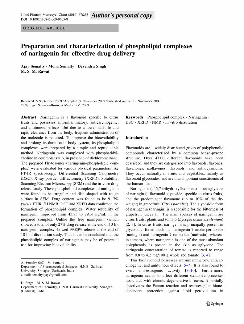
ORIGINAL ARTICLE
Preparation and characterization of phospholipid complexesof naringenin for effective drug delivery
Ajay Semalty • Mona Semalty • Devendra Singh •
M. S. M. Rawat
Received: 5 September 2009 / Accepted: 9 November 2009 / Published online: 19 November 2009
� Springer Science+Business Media B.V. 2009
Abstract Naringenin is a flavonoid specific to citrus
fruits and possesses anti-inflammatory, anticarcinogenic,
and antitumour effects. But due to a lower half-life and
rapid clearance from the body, frequent administration of
the molecule is required. To improve the bioavailability
and prolong its duration in body system, its phospholipid
complexes were prepared by a simple and reproducible
method. Naringenin was complexed with phosphatidyl-
choline in equimolar ratio, in presence of dichloromethane.
The prepared Phytosomes (naringenin–phospholipid com-
plex) were evaluated for various physical parameters like
FT-IR spectroscopy, Differential Scanning Calorimetry
(DSC), X-ray powder diffractometry (XRPD), Solubility,
Scanning Electron Microscopy (SEM) and the in vitro drug
release study. These phospholipid complexes of naringenin
were found to be irregular and disc shaped with rough
surface in SEM. Drug content was found to be 91.7%
(w/w). FTIR, 1H NMR, DSC and XRPD data confirmed the
formation of phospholipid complex. Water solubility of
naringenin improved from 43.83 to 79.31 lg/mL in the
prepared complex. Unlike the free naringenin (which
showed a total of only 27% drug release at the end of 10 h),
naringenin complex showed 99.80% release at the end of
10 h of dissolution study. Thus it can be concluded that the
phospholipid complex of naringenin may be of potential
use for improving bioavailability.
Keywords Phospholipid complex � Naringenin �DSC � XRPD � NMR � In vitro dissolution
Introduction
Flavonoids are a widely distributed group of polyphenolic
compounds characterized by a common benzo-pyrone
structure. Over 4,000 different flavonoids have been
described, and they are categorized into flavonols, flavones,
flavanones, isoflavones, flavonols, and anthocyanidins.
They occur naturally in fruits and vegetables, mainly as
flavonoid glycosides, and are thus important constituents of
the human diet.
Naringenin (40,5,7-trihydroxyflavanone) is an aglycone
of naringin (a flavonoid glycoside, specific to citrus fruits)
and the predominant flavanone (up to 10% of the dry
weight) in grapefruit (Citrus paradisi). The glycosidic form
of naringenin (naringin) is responsible for the bitterness of
grapefruit juices [1]. The main sources of naringenin are
citrus fruits, plants and tomato (Lycopersicum esculentum)
[2, 3]. In citrus fruits, naringenin is principally present in
glycosidic forms such as naringenin-7-neohesperidoside
(naringin) and naringenin-7-rutinoside (narirutin), whereas
in tomato, where naringenin is one of the most abundant
polyphenols, is present in the skin as aglycone. The
naringenin concentration of tomato is reported to range
from 0.8 to 4.2 mg/100 g whole red tomato [3, 4].
This bioflavonoid possesses anti-inflammatory, anticar-
cinogenic, and antitumour effects [5–7]. It is also found to
exert anti-estrogenic activity [8–10]. Furthermore,
naringenin seems to affect different oxidative processes
associated with chronic degenerative diseases. It partially
deactivates the Fenton reaction and restores glutathione-
dependent protection against lipid peroxidation in
A. Semalty (&) � M. Semalty
Department of Pharmaceutical Sciences, H.N.B. Garhwal
University, Srinagar (Garhwal), India
e-mail: [email protected]
D. Singh � M. S. M. Rawat
Department of Chemistry, H.N.B. Garhwal University, Srinagar
(Garhwal), India
123
J Incl Phenom Macrocycl Chem (2010) 67:253–260
DOI 10.1007/s10847-009-9705-8 Author's personal copy
a-tocopherol- deficient liver microsomes [11, 12]. It pro-
duces inhibitory activity in malonaldehyde production and
inhibits cytochrome P450 enzymes [13–15].
Despite this wide range of therapeutic activity, its
unfavourable pharmacokinetics associated with a lower
half-life and rapid clearance from the body restricts its use
as a potent phyto molecule. It was reported that the elim-
ination half-lives of naringenin from orange juice and
grapefruit juice (when taken orally) were 1.3 and 2.2 h,
respectively [16]. Therefore, to maintain steady plasma
concentration so as to exert the desired therapeutic activity,
frequent administration of the molecule is required, which
necessitates the need for development of a dosage form that
can maintain the concentration of naringenin in blood for a
longer period.
Phospholipids play a major role in drug delivery tech-
nology. It is an important carrier for those drug molecules
which require sustained/controlled release in vivo due to
faster elimination from the body. Developing the drugs as
lipid complexes may prove to be a potential approaches to
improve solubility and to minimize the GI toxicity of
drugs. These amphiphilic drug–lipid complexes, are stable
and more bioavailable drug delivery systems with low
interfacial tension between the system and the GI fluid
thereby facilitating membrane, tissue, or cell wall transfer,
in the organism [17].
Thus naringenin–phospholipid complex were prepared
and evaluated for various physical parameters (FTIR, DSC,
XRD, Solubility, SEM, etc.) and the in vitro dissolution
study of naringenin–phospholipid complex in comparison
with pure naringenin.
Materials and methods
Materials
Naringenin was purchased from Sigma–Aldrich Mumbai.
Soya phosphatidylcholine (LIPOID S-80) was obtained as
a gift sample from LIPOID GmbH Germany. All other
chemicals were of analytical grade.
Methods
Naringenin–PC complex was prepared by taking naringe-
nin with an equimolar concentration of phosphatidylcho-
line (PC). The equimolar concentration of PC and
naringenin were placed in a 100 mL round bottom flask
and refluxed in dichloromethane for 3 h. On concentrating
the solution to 5–10 mL, 30 mL of n-hexane was added to
get the complex as a precipitate followed by filtration. The
precipitate was collected and placed in vacuum desiccators.
Drug content
To determine the drug content in the complex, complex
equivalent to 100 mg were weighed and added in
100 mL of methanol taken in a 100 mL volumetric flask.
The volumetric flask was stirred continuously for 24hr
on a magnetic stirrer. Dilutions were made suitably and
measured for the drug content UV spectrophotometrically
by Lambda25 Perkin Elmer UV/Visible Spectropho-
tometer.
Solubility
To determine the change in solubility due to complexation,
solubility of drug and the complex was determined in
buffer/water and n-octanol by shake flask method. 50 mg
of drug (and 50 mg equivalent in case of complex) was
taken in a 100 mL conical flask. 50 mL of distilled water
was added and then stirred for 15 min. The suspension was
then transferred to 250 mL separating funnel with 50 mL
of n-octanol and was shaken well for 2 h. Then the sepa-
rating funnel was allowed to stand for about 30 min.
Concentration of the drug was determined from the aque-
ous layer spectrophotometrically.
Scanning electron microscopy (SEM)
To detect the surface morphology of the prepared complex,
SEM of complex was performed at UGC-DAE consortium
Indore and IIT Roorkee by Scanning Electron Microscope
(JEOL JSM 5600).
Fourier transform infrared spectroscopy (FT-IR)
FTIR spectra for the various powders were obtained on a
Perkin Elmer FTIR spectrometer (Perkin Elmer Life and
Analytical Sciences, MA, USA) in the transmission mode
with the wave number region 4,000–500 cm-1. KBr pellets
were prepared by gently mixing 1 mg sample powder with
100 mg KBr.
Differential scanning calorimetry (DSC)
Thermograms of naringenin, phosphatidylcholine (80%)
and the naringenin–PC complex were recorded using a
differential scanning calorimeter (2910 Modulated DSC
V4.4E, TA Instruments, US). The thermal behavior was
studied by heating 2.0 ± 0.2 mg of each individual sample
in a covered sample pan under nitrogen gas flow. The
investigations were carried out over the temperature range
25–250 �C with a heating rate of 10 �C min-1.
254 J Incl Phenom Macrocycl Chem (2010) 67:253–260
123
Author's personal copy

X-ray powder diffractometry (XRPD)
The crystalline state of drugs in the different samples was
evaluated with X-ray powder diffraction. Diffraction pat-
terns were obtained on a Bruker Axs-D8 Discover Powder
X-ray diffractometer (Germany). The X-ray generator was
operated at 40 KV tube voltages and 40 mA of tube cur-
rent, using the Ka lines of copper as the radiation source.
The scanning angle ranged from 1 to 60� of 2h in step scan
mode (step width 1�/min). Drug, phosphatidylcholine
(80%), and drug–PC complex were analyzed with X-ray
diffractions.
Nuclear magnetic resonance (NMR)
Proton NMR of drug, phospholipids and their complexes
were recorded on Bruker 400 MHz NMR (Germany), using
TMS as internal standard.
Dissolution study
In vitro dissolution studies for drug complex as well as
plain drug were performed in triplicate in a USP XXIII six
station dissolution test apparatus (Veego Model No. 6DR,
India) at 100 rpm and at 37 �C. An accurately weighed
amount of the complex equivalent to 100 mg of drug was
put into 900 mL distilled water (media). Samples (3 mL
each) of dissolution fluid were withdrawn at different
intervals and replaced with the equal volume of fresh
media, to maintain sink conditions. Withdrawn samples
were filtered (through a 0.45 mm membrane filter) and
diluted suitably and then analyzed spectrophotometrically.
Statistical analysis
Results were expressed as mean values and standard
deviations (±SD).
Results and discussion
In the present experiment naringenin–phospholipid com-
plex were prepared by a simple and reproducible method.
Content (percent loading) of naringenin in the complex,
as estimated by UV spectrophotometry (at 288 nm in
methanol), was found to be 91.7% (w/w). Phyto–
phospholipid complex of herbal drugs showed a high per-
centage of drug loading. Like phospholipid complexes
(pharmacosomes) of synthetic drugs, complexation is giv-
ing a good percent loading of the drug which makes the
delivery of drug clinically feasible.
Solubility of the naringenin complex was found to be
much higher (in water and n-octanol) than the naringenin.
Table 1 provides the solubility data. Naringenin is practi-
cally insoluble in water. But the complex of naringenin
with the phospholipid increased the solubility of naringenin
in water as well as n-octanol. This increase of solubility of
the complex may be explained by the solubilization of the
drug and by the amorphous characteristics of the complex.
As an amphiphilic surfactant, phospholipids could increase
the solubility of the drug by the action of wetting and
dispersion. So the complex showed an amphiphilic nature,
which in turn may show the improved bioavailability of the
drug.
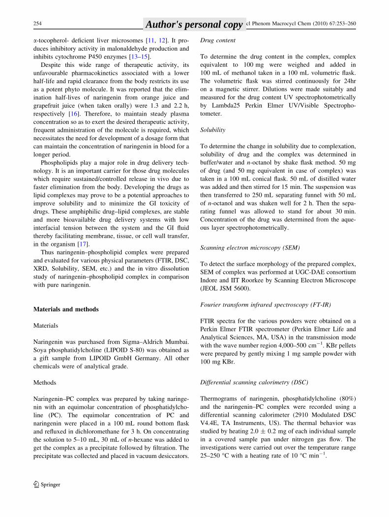
Scanning Electron Micrographs of the complex are
shown in Fig. 1. Unlike the needle shaped structure of
naringenin, its phospholipid complex were found to be of
irregular shaped with rough surface morphology. Com-
plexes were found to be as free flowing particles. The
average particle size of phospholipid complex was found to
be 100 lm.
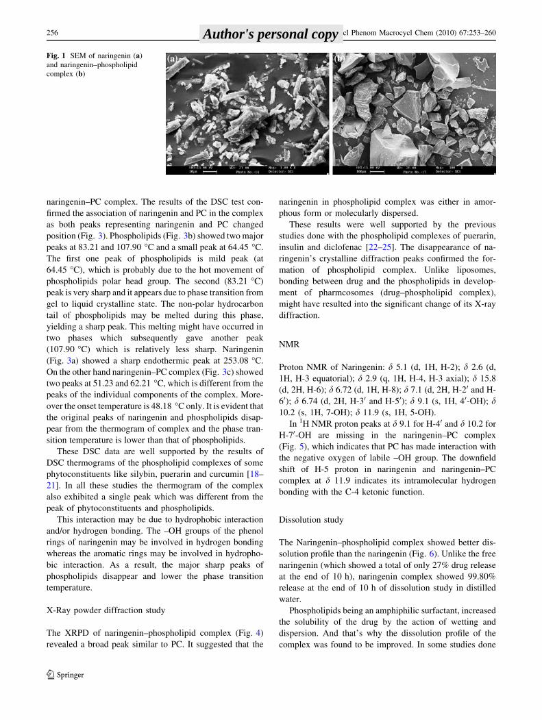
The formation of the complex can be confirmed by the
FT-IR spectroscopy comparing the spectrum of the com-
plex with the spectrum of the individual components and
their mechanical mixtures. FTIR spectra showed the
changes in peaks in complexes and positions from that of
naringenin (Fig. 2a) and PC (Fig. 2b). FT-IR spectra
of complex (Fig. 2c) were significantly different from that
of components and that of physical mixtures (Fig. 2d).
Naringenin showed the characteristic IR (KBr) peaks at
3285.79, 3117.36, 3035.80, 1629.76, 1602.09, 1519.88,
1498.10, 1463.39 cm-1. PC showed a broad peak at
3,437.05; sharp peaks at 2,917.93 and 2,849.91; and several
sharp peaks below 1,800 cm-1. IR spectra of naringenin–
phospholipid complex showed disappearance of peaks of
phenolic –OH of (of naringenin), which indicates that the
phospholipid has interacted with naringenin through these
phenolic –OH groups in the process of complexation. On
the other hand the physical mixture of PC and naringenin
showed broad peak like PC at 3,301.98 cm-1 while it
shows all the sharp peaks at about the same positions as
that of naringenin.
DSC studies
In order to substantiate the association of naringenin with
PC, DSC analysis was performed on naringenin, PC, and the
Table 1 Solubility study of naringenin and its complex
Drug Aqueous solubility
(lg/mL)an-Octanol solubility
(lg/mL)a
Naringenin 43.83 ± 0.039 440.163 ± 2.641
Naringenin–PC complex 79.31 ± 0.718 468.13 ± 7.590
a Data expressed as mean values and standard deviations (±SD);
n = 3
J Incl Phenom Macrocycl Chem (2010) 67:253–260 255
123
Author's personal copy
naringenin–PC complex. The results of the DSC test con-
firmed the association of naringenin and PC in the complex
as both peaks representing naringenin and PC changed
position (Fig. 3). Phospholipids (Fig. 3b) showed two major
peaks at 83.21 and 107.90 �C and a small peak at 64.45 �C.
The first one peak of phospholipids is mild peak (at
64.45 �C), which is probably due to the hot movement of
phospholipids polar head group. The second (83.21 �C)
peak is very sharp and it appears due to phase transition from
gel to liquid crystalline state. The non-polar hydrocarbon
tail of phospholipids may be melted during this phase,
yielding a sharp peak. This melting might have occurred in
two phases which subsequently gave another peak
(107.90 �C) which is relatively less sharp. Naringenin
(Fig. 3a) showed a sharp endothermic peak at 253.08 �C.
On the other hand naringenin–PC complex (Fig. 3c) showed
two peaks at 51.23 and 62.21 �C, which is different from the
peaks of the individual components of the complex. More-
over the onset temperature is 48.18 �C only. It is evident that
the original peaks of naringenin and phospholipids disap-
pear from the thermogram of complex and the phase tran-
sition temperature is lower than that of phospholipids.
These DSC data are well supported by the results of
DSC thermograms of the phospholipid complexes of some
phytoconstituents like silybin, puerarin and curcumin [18–
21]. In all these studies the thermogram of the complex
also exhibited a single peak which was different from the
peak of phytoconstituents and phospholipids.
This interaction may be due to hydrophobic interaction
and/or hydrogen bonding. The –OH groups of the phenol
rings of naringenin may be involved in hydrogen bonding
whereas the aromatic rings may be involved in hydropho-
bic interaction. As a result, the major sharp peaks of
phospholipids disappear and lower the phase transition
temperature.
X-Ray powder diffraction study
The XRPD of naringenin–phospholipid complex (Fig. 4)
revealed a broad peak similar to PC. It suggested that the
naringenin in phospholipid complex was either in amor-
phous form or molecularly dispersed.
These results were well supported by the previous
studies done with the phospholipid complexes of puerarin,
insulin and diclofenac [22–25]. The disappearance of na-
ringenin’s crystalline diffraction peaks confirmed the for-
mation of phospholipid complex. Unlike liposomes,
bonding between drug and the phospholipids in develop-
ment of pharmcosomes (drug–phospholipid complex),
might have resulted into the significant change of its X-ray
diffraction.
NMR
Proton NMR of Naringenin: d 5.1 (d, 1H, H-2); d 2.6 (d,
1H, H-3 equatorial); d 2.9 (q, 1H, H-4, H-3 axial); d 15.8
(d, 2H, H-6); d 6.72 (d, 1H, H-8); d 7.1 (d, 2H, H-20 and H-
60); d 6.74 (d, 2H, H-30 and H-50); d 9.1 (s, 1H, 40-OH); d10.2 (s, 1H, 7-OH); d 11.9 (s, 1H, 5-OH).
In 1H NMR proton peaks at d 9.1 for H-40 and d 10.2 for
H-70-OH are missing in the naringenin–PC complex
(Fig. 5), which indicates that PC has made interaction with
the negative oxygen of labile –OH group. The downfield
shift of H-5 proton in naringenin and naringenin–PC
complex at d 11.9 indicates its intramolecular hydrogen
bonding with the C-4 ketonic function.
Dissolution study
The Naringenin–phospholipid complex showed better dis-
solution profile than the naringenin (Fig. 6). Unlike the free
naringenin (which showed a total of only 27% drug release
at the end of 10 h), naringenin complex showed 99.80%
release at the end of 10 h of dissolution study in distilled
water.
Phospholipids being an amphiphilic surfactant, increased
the solubility of the drug by the action of wetting and
dispersion. And that’s why the dissolution profile of the
complex was found to be improved. In some studies done
Fig. 1 SEM of naringenin (a)
and naringenin–phospholipid
complex (b)
256 J Incl Phenom Macrocycl Chem (2010) 67:253–260
123
Author's personal copy
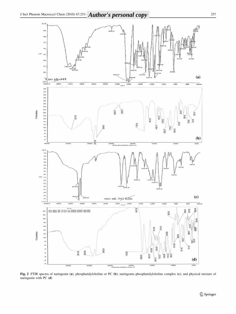
Fig. 2 FTIR spectra of naringenin (a); phosphatidylcholine or PC (b); naringenin–phosphatidylcholine complex (c); and physical mixture of
naringenin with PC (d)
J Incl Phenom Macrocycl Chem (2010) 67:253–260 257
123
Author's personal copy
Fig. 3 DSC thermograms of
naringenin (a); PC (b) and
naringenin–PC complex (c)
258 J Incl Phenom Macrocycl Chem (2010) 67:253–260
123
Author's personal copy

with silybin, the in vitro drug release from the complexes
was found to be pH dependent and with the increase of the
pH of media the dissolution amount of drug was increased
[18, 20].
0
500
1000
1500
20000
500
1000
1500
2000
0
500
1000
1500
2000
Inte
nsi
ty(a
.u)
2θ
PC
Ne
NeC
Fig. 4 High resolution X-ray diffraction (HRXRD) study of naringe-
nin (Ne) and its complex (NeC) and its components
Fig. 5 1H NMR of a naringenin (in DMSO) and b naringenin–phospholipid complex (in CDCl3)
0
20
40
60
80
100
120
0 2 4 6 8 10 12 14 16 18 20 22 24 26
Time (h)
% C
um
ula
tive
dru
g r
elea
se
Ne
NeC
Fig. 6 Dissolution study of naringenin (Ne) and naringenin–
phospholipid complex (NeC)
J Incl Phenom Macrocycl Chem (2010) 67:253–260 259
123
Author's personal copy

Conclusions
In the present study naringenin–phospholipid complex
were prepared by a simple and reproducible method and
evaluated for various physicochemical parameters. The
physicochemical investigations showed that naringenin
formed a stoichiometric complex with phosphatidylcholine
with better solubility and dissolution profile. The 1H NMR,
DSC and XRPD studies confirmed the formation of the
complex. The dissolution profile of the complex was found
to be improved. Thus it can be concluded that the
phospholipid complex of naringenin may be of potential
use for improving its bioavailability.
Acknowledgments Authors acknowledge the grant provided by the
Department of Science and Technology, Govt. of India for the
research work. Authors are also thankful to LIPOID GmbH Germany
for providing the gift sample of phosphatidylcholine for the research
work. Facilities provided by the UGC-DAE Consortium for Scientific
Research, Indore (M.P.) and Department of Chemistry, University of
Delhi are thankfully acknowledged.
References
1. Ortuno, A., Garcia-Puig, D., Fuster, M.D., Perez, M.L., Sabater,
F., Porras, I., Garcia-Lidon, A., Del Rio, A.J.: Flavanone and
nootkatone levels in different varieties of grapefruit and pum-
melo. J. Agric. Food Chem. 43, 1–5 (1995). doi:10.1021/
jf00049a001
2. Kawaii, S., Tomono, Y., Katase, E., Ogawa, K., Yano, M.:
Quantitation of flavonoids constituents in citrus fruits. J. Agric.
Food Chem. 47, 3565–3571 (1999). doi:10.1021/jf990153?
3. Davies, J.N., Graeme, E.H.: The constituents of tomato fruit: the
influence of environment, nutrition, and genotype. Crit. Rev.
Food Sci. Nutr. 15, 205–280 (1981)
4. Paganga, G., Miller, N., Rice-Evans, C.: The polyphenolic con-
tent of fruit and vegetables and their antioxidant activities: what
does a serving constitute? Free Radic. Res. 30, 153–162 (1999).
doi:10.1080/10715769900300161
5. Middleton, E., Kandaswami, C.: The impact of plant flavonoids
on mammalian biology: implications for immunity, inflammation
and cancer. In: Harborne, J.B. (ed.) The flavonoids: advances in
research since 1986, pp. 619–652. Chapman and Hall, London
(1994)
6. Benavente-Garcia, O., Castillo, J., Marin, F.R., Ortuno, A., Del
Rio, J.A.: Uses and properties of citrus flavonoids. J. Agric. Food
Chem. 45, 4505–4515 (1997). doi:10.1021/jf970373s
7. Montanari, A., Chen, J., Widmer, W.: Citrus flavonoids: a review
of past biological activity against disease. Discovery of new
flavonoids from Dancy tangerine cold pressed peel oil solids and
leaves. In: Manthey, J., Buslig, B. (eds.) Flavonoids in the living
system, pp. 103–116. Plenum, New York (1998)
8. Ruh, M.F., Zacharewsky, T., Connor, K., Howell, J., Chen, I.,
Safe, S.: Naringenin: a weakly estrogenic bioflavonoid that
exhibits antiestrogenic activity. Biochem. Phamacol. 50, 1485–
1493 (1995). doi:10.1016/0006-2952(95)02061-6
9. Miksicek, R.J.: Commonly occurring plant flavonoids have
estrogenic activity. Mol. Pharmacol. 44, 37–43 (1993)
10. Kuiper, J.M., Lemmen, J.G., Carlsson, B., Corton, J.C., Safe,
S.H., Van der Saag, P.T., Van der Burg, B., Gustafsson, J.A.:
Interaction of estrogenic chemicals and phytoestrogens with
estrogen receptor beta. Endocrinolology 139, 4252–4263 (1998).
doi:10.1210/en.139.10.4252
11. Cheng, F., Breen, K.: On the ability of four flavonoids, baicilein,
luteolin, naringenin and quercetin, to suppress the fenton reaction
of the iron–ATP complex. Biometals 13, 77–83 (2000). doi:
10.1023/A:1009229429250
12. Van Acker, F.A.A., Schouten, O., Haenen, G.R., Van der Vijgh,
W.J.F., Bast, A.: Flavonoids can replace a-tocopherol as an
antioxidant. FEBS Lett. 473, 145–148 (2000). doi:10.1016/
S0014-5793(00)01517-9
13. Lee, K.G., Shibamoto, T., Takeoka, G.R., Lee, S.E., Kim, J.H.,
Park, B.S.: Inhibitory effects of plant-derived flavonoids and
phenolic acids on malonaldehyde formation from ethyl arachid-
onate. J. Agric. Food Chem. 51, 7203–7207 (2003). doi:10.1021/
jf0345447
14. Saija, A., Scalese, M., Lanza, M., Marzullo, D., Bonina, F.,
Castelli, F.: Flavonoids as antioxidant agents: importance of their
interaction with biomembranes. Free Radic. Biol. Med. 19, 481–
486 (1995). doi:10.1016/0891-5849(94)00240-K
15. Ueng, Y.F., Chang, Y.L., Oda, Y., Park, S.S., Liao, J.F., Lin,
M.F., Chen, C.F.: In vitro and in vivo effects of naringin on
cytochrome P450-dependent monooxygenase in mouse liver. Life
Sci. 65, 2591–2602 (1999). doi:10.1016/S0024-3205(99)00528-7
16. Erlund, I., Meririnne, E., Alfthan, G., Aro, A.: Plasma kinetics
and urinary excretion of the flavanones naringenin and hesperetin
in humans after ingestion of orange juice and grapefruit juice. J.
Nutr. 131, 235–241 (2001)
17. Semalty, A., Semalty, M., Rawat, B.S., Singh, D., Rawat,
M.S.M.: Pharmacosomes: the lipid based novel drug delivery
system. Expert Opin. Drug Deliv. 6, 599–612 (2009). doi:
10.1517/17425240902967607
18. Yanyu, X., Yunmei, S., Zhipeng, C., Quineng, P.: The prepara-
tion of silybin–phospholipid complex and the study on its phar-
macokinetics in rats. Int. J. Pharm. 307, 77–82 (2006). doi:
10.1016/j.ijpharm.2005.10.001
19. Li, Y., Yang, D.J., Chen, S.L., Chen, S.B., Chan, A.S.C.: Com-
parative physicochemical characterization of phospholipids
complex of puerarin formulated by conventional and supercritical
methods. Pharm. Res. 25, 563–577 (2007). doi:10.1007/s11095-
007-9418-x
20. Maiti, K., Mukherjee, K., Gantait, A., Saha, B.P., Mukherjee,
P.K.: Curcumin–phospholipid complex: preparation, therapeutic
evaluation and pharmacokinetic study in rats. Int. J. Pharm. 330,
155–163 (2007). doi:10.1016/j.ijpharm.2006.09.025
21. Kumar, M., Ahuja, M., Sharma, S.K.: Hepatoprotective study of
curcumin–soya lecithin complex. Sci. Pharm. 76, 761–774
(2008). doi:10.3797/scipharm.0808-09
22. Shi, K., Cui, F., Yu, Y., Zhang, L., Tao, A., Cun, D.: Preparation
and characterization of a novel insulin phospholipid complex.
Asian J. Pharm. Sci. 1(3–4), 168–174 (2006)
23. Cui, F., Shi, K., Zhang, L., Tao, A., Kawashima, Y.: Biode-
gradable nanoparticles loaded with insulin–phospholipid complex
for oral delivery: preparation, in vitro characterization and
in vivo evaluation. J. Control. Release 114, 242–250 (2006). doi:
10.1016/j.jconrel.2006.05.013
24. Yoo, H.S., Park, T.G.: Biodegradable nanoparticles containing
protein–fatty acid complex for oral delivery of salmon calcitonin.
J. Pharm. Sci. 93, 488–495 (2003)
25. Semalty, A., Semalty, M., Singh, D., Rawat, M.S.M.: Develop-
ment and physicochemical evaluation of pharmacosomes of
diclofenac. Acta Pharma 59, 335–344 (2009). doi:10.2478/
v10007-009-0023-x
260 J Incl Phenom Macrocycl Chem (2010) 67:253–260
123
Author's personal copy