Influence of Rutin, Sinapic Acid, and Naringenin on Binding
15
Citation: Wani, T.A.; Bakheit, A.H.; Zargar, S.; Khayyat, A.I.A.; Al-Majed, A.A. Influence of Rutin, Sinapic Acid, and Naringenin on Binding of Tyrosine Kinase Inhibitor Erlotinib to Bovine Serum Albumin Using Analytical Techniques Along with Computational Approach. Appl. Sci. 2022, 12, 3575. https://doi.org/ 10.3390/app12073575 Academic Editor: Antony C. Calokerinos Received: 17 March 2022 Accepted: 30 March 2022 Published: 31 March 2022 Publisher’s Note: MDPI stays neutral with regard to jurisdictional claims in published maps and institutional affil- iations. Copyright: © 2022 by the authors. Licensee MDPI, Basel, Switzerland. This article is an open access article distributed under the terms and conditions of the Creative Commons Attribution (CC BY) license (https:// creativecommons.org/licenses/by/ 4.0/). applied sciences Article Influence of Rutin, Sinapic Acid, and Naringenin on Binding of Tyrosine Kinase Inhibitor Erlotinib to Bovine Serum Albumin Using Analytical Techniques Along with Computational Approach Tanveer A. Wani 1, * , Ahmed H. Bakheit 1 , Seema Zargar 2 , Arwa Ishaq A. Khayyat 2 and Abdulrahman A. Al-Majed 1 1 Department of Pharmaceutical Chemistry, College of Pharmacy, King Saud University, P.O. Box 2457, Riyadh 11451, Saudi Arabia; [email protected] (A.H.B.); [email protected] (A.A.A.-M.) 2 Department of Biochemistry, College of Science, King Saud University, P.O. Box 22452, Riyadh 11451, Saudi Arabia; [email protected] (S.Z.); [email protected] (A.I.A.K.) * Correspondence: [email protected] Abstract: Flavonoid-containing food supplements are widely used as antioxidants, and the continu- ous use of these supplements with other drugs can lead to clinically significant interactions between these and other drugs. The medications in systemic circulation are mainly transported by serum albumin, a major transport protein. This study evaluated the interactions of rutin (RUT), naringenin (NAR), and sinapic acid (SIN) with the most abundant transport protein, bovine serum albumin (BSA), and the anticancer drug, the tyrosine kinase inhibitor Erlotinib (ETB), using various analytical methods. Interaction between multiple types of ligands with the transport proteins and competition between themselves can lead to the bound ETB’s displacement from the BSA-binding site, leading to elevated ETB concentrations in the systemic circulation. These elevated drug fractions can lead to adverse events and lower tolerance, and increased resistance to the therapeutic regimen of ETB. The experimental and computational methods, including molecular-docking studies, were used to understand the molecular interactions. The results suggested that the complexes formed were utterly different in the binary and the ternary system. Furthermore, comparing the ternary systems amongst themselves, the spectra differed from each other. They thus inferred that complexes formed between BSA-ETB in the presence of each RUT, NAR, and SIN separately were also different, with the highest value of the reduction in the binding energy in RUT, followed by SIN and then NAR. Thus, we conclude that a competitive binding between the ETB and these flavonoids might influence the ETB pharmacokinetics in cancer patients by increasing ETB tolerance or resistance. Keywords: Erlotinib; bovine serum albumin; fluorescence quenching; binding interaction; rutin; naringenin; sinapic acid 1. Introduction Serum albumin protein is the abundant constituent of the plasma and plays a pivotal role in transporting endogenous and/or exogenous ligands [1]. Ligands bind to the albumin, which influences both the pharmacodynamics and pharmacokinetics of the ligand [2,3]. All the drugs reversibly bind to the plasma proteins, and their effect generally is dependent on the free drug fraction present in the systemic circulation. The binding capacity of the drug to plasma proteins is a major consideration for selecting the dosage of the drug. Thus, the free drug fraction present in the plasma is generally responsible for the drug’s therapeutic effect [4–6]. The BSA is structurally homologous and presents a 75.6% sequence similarity to human serum albumin (HSA). Therefore, BSA was chosen in the present study as a sub- stituent to HSA since it has the same ligand-binding properties and is readily available and Appl. Sci. 2022, 12, 3575. https://doi.org/10.3390/app12073575 https://www.mdpi.com/journal/applsci
-
Upload
khangminh22 -
Category
Documents
-
view
3 -
download
0
Transcript of Influence of Rutin, Sinapic Acid, and Naringenin on Binding
�����������������
Citation: Wani, T.A.; Bakheit, A.H.;
Zargar, S.; Khayyat, A.I.A.; Al-Majed,
A.A. Influence of Rutin, Sinapic Acid,
and Naringenin on Binding of
Tyrosine Kinase Inhibitor Erlotinib to
Bovine Serum Albumin Using
Analytical Techniques Along with
Computational Approach. Appl. Sci.
2022, 12, 3575. https://doi.org/
10.3390/app12073575
Academic Editor: Antony
C. Calokerinos
Received: 17 March 2022
Accepted: 30 March 2022
Published: 31 March 2022
Publisher’s Note: MDPI stays neutral
with regard to jurisdictional claims in
published maps and institutional affil-
iations.
Copyright: © 2022 by the authors.
Licensee MDPI, Basel, Switzerland.
This article is an open access article
distributed under the terms and
conditions of the Creative Commons
Attribution (CC BY) license (https://
creativecommons.org/licenses/by/
4.0/).
applied sciences
Article
Influence of Rutin, Sinapic Acid, and Naringenin on Bindingof Tyrosine Kinase Inhibitor Erlotinib to Bovine SerumAlbumin Using Analytical Techniques Alongwith Computational ApproachTanveer A. Wani 1,* , Ahmed H. Bakheit 1 , Seema Zargar 2 , Arwa Ishaq A. Khayyat 2
and Abdulrahman A. Al-Majed 1
1 Department of Pharmaceutical Chemistry, College of Pharmacy, King Saud University, P.O. Box 2457,Riyadh 11451, Saudi Arabia; [email protected] (A.H.B.); [email protected] (A.A.A.-M.)
2 Department of Biochemistry, College of Science, King Saud University, P.O. Box 22452,Riyadh 11451, Saudi Arabia; [email protected] (S.Z.); [email protected] (A.I.A.K.)
* Correspondence: [email protected]
Abstract: Flavonoid-containing food supplements are widely used as antioxidants, and the continu-ous use of these supplements with other drugs can lead to clinically significant interactions betweenthese and other drugs. The medications in systemic circulation are mainly transported by serumalbumin, a major transport protein. This study evaluated the interactions of rutin (RUT), naringenin(NAR), and sinapic acid (SIN) with the most abundant transport protein, bovine serum albumin(BSA), and the anticancer drug, the tyrosine kinase inhibitor Erlotinib (ETB), using various analyticalmethods. Interaction between multiple types of ligands with the transport proteins and competitionbetween themselves can lead to the bound ETB’s displacement from the BSA-binding site, leadingto elevated ETB concentrations in the systemic circulation. These elevated drug fractions can leadto adverse events and lower tolerance, and increased resistance to the therapeutic regimen of ETB.The experimental and computational methods, including molecular-docking studies, were used tounderstand the molecular interactions. The results suggested that the complexes formed were utterlydifferent in the binary and the ternary system. Furthermore, comparing the ternary systems amongstthemselves, the spectra differed from each other. They thus inferred that complexes formed betweenBSA-ETB in the presence of each RUT, NAR, and SIN separately were also different, with the highestvalue of the reduction in the binding energy in RUT, followed by SIN and then NAR. Thus, weconclude that a competitive binding between the ETB and these flavonoids might influence the ETBpharmacokinetics in cancer patients by increasing ETB tolerance or resistance.
Keywords: Erlotinib; bovine serum albumin; fluorescence quenching; binding interaction; rutin;naringenin; sinapic acid
1. Introduction
Serum albumin protein is the abundant constituent of the plasma and plays a pivotalrole in transporting endogenous and/or exogenous ligands [1]. Ligands bind to thealbumin, which influences both the pharmacodynamics and pharmacokinetics of theligand [2,3]. All the drugs reversibly bind to the plasma proteins, and their effect generallyis dependent on the free drug fraction present in the systemic circulation. The bindingcapacity of the drug to plasma proteins is a major consideration for selecting the dosage ofthe drug. Thus, the free drug fraction present in the plasma is generally responsible for thedrug’s therapeutic effect [4–6].
The BSA is structurally homologous and presents a 75.6% sequence similarity tohuman serum albumin (HSA). Therefore, BSA was chosen in the present study as a sub-stituent to HSA since it has the same ligand-binding properties and is readily available and
Appl. Sci. 2022, 12, 3575. https://doi.org/10.3390/app12073575 https://www.mdpi.com/journal/applsci

Appl. Sci. 2022, 12, 3575 2 of 15
cost-effective [7,8]. BSA has three primary ligand-binding sites in subdomain IIA, IIIA, andIB [9–12]. The intrinsic fluorescence of BSA, which is associated with two tryptophan aminoacids (Trp-134 and Trp-213), is generally used for studies on drug–protein interactions.
Drugs reversibly bind to the plasma proteins, and the free drug fraction present inthe systemic circulation at a given time is considered responsible for its therapeutic effect.Therefore, a competition between two or more drugs can occur upon their coadministrationfor the same BSA-binding site. This phenomenon leads to the competition or displacementof bound drugs to their binding sites, resulting in altered pharmacokinetics [13,14]. Inaddition, ligands binding to BSA might also cause conformational changes in the proteinmolecule, thus altering their ligand-binding phenomenon [15,16].
Coadministration of drugs might lead to elevated plasma concentrations of the dis-placed drug because of competition or displacement from the binding site. Thus, interactionat the serum albumin-binding level might cause toxic effects in the case of narrow therapeu-tic index drugs. These interactions may also lead to a higher incidence of adverse events,higher volume of distribution, or a subtherapeutic response due to shorter eliminationhalf-life [17].
Phenolic-compound flavonoids are commonly used as antioxidants in nutritionalsupplements [18]. A citrus flavonoid glycoside, RUT, is a potent antioxidant with anti-inflammatory activities [18,19]. RTN is believed to have an anticancer potential and isrecommended as a dietary supplement to prevent and manage cancer [20]. Another citrusflavonoid glycoside, NAR, which has high antioxidant potential, is also used as a dietarysupplement [21] and has anticancer properties [22]. The hydroxycinnamic-acid derivativesinapic acid (SIN) has shown anticancer activity by various mechanisms, including enzymebiotransformation (Figure 1) [23].
Appl. Sci. 2022, 12, x FOR PEER REVIEW 2 of 15
The BSA is structurally homologous and presents a 75.6% sequence similarity to hu‐
man serum albumin (HSA). Therefore, BSA was chosen in the present study as a substit‐
uent to HSA since it has the same ligand‐binding properties and is readily available and
cost‐effective [7,8]. BSA has three primary ligand‐binding sites in subdomain IIA, IIIA,
and IB [9–12]. The intrinsic fluorescence of BSA, which is associated with two tryptophan
amino acids (Trp‐134 and Trp‐213), is generally used for studies on drug–protein interac‐
tions.
Drugs reversibly bind to the plasma proteins, and the free drug fraction present in
the systemic circulation at a given time is considered responsible for its therapeutic effect.
Therefore, a competition between two or more drugs can occur upon their coadministra‐
tion for the same BSA‐binding site. This phenomenon leads to the competition or displace‐
ment of bound drugs to their binding sites, resulting in altered pharmacokinetics [13,14].
In addition, ligands binding to BSA might also cause conformational changes in the pro‐
tein molecule, thus altering their ligand‐binding phenomenon [15,16].
Coadministration of drugs might lead to elevated plasma concentrations of the dis‐
placed drug because of competition or displacement from the binding site. Thus, interac‐
tion at the serum albumin‐binding level might cause toxic effects in the case of narrow
therapeutic index drugs. These interactions may also lead to a higher incidence of adverse
events, higher volume of distribution, or a subtherapeutic response due to shorter elimi‐
nation half‐life [17].
Phenolic‐compound flavonoids are commonly used as antioxidants in nutritional
supplements [18]. A citrus flavonoid glycoside, RUT, is a potent antioxidant with anti‐
inflammatory activities [18,19]. RTN is believed to have an anticancer potential and is rec‐
ommended as a dietary supplement to prevent and manage cancer [20]. Another citrus
flavonoid glycoside, NAR, which has high antioxidant potential, is also used as a dietary
supplement [21] and has anticancer properties [22]. The hydroxycinnamic‐acid derivative
sinapic acid (SIN) has shown anticancer activity by various mechanisms, including en‐
zyme biotransformation (Figure 1) [23].
Figure 1. The chemical structures of (A): ETB; (B): RUT; (C): NAR; (D): SIN.
The ETB (Figure 1) is an ATP-competitive quinazoline derivative that works by bindingto the epidermal growth factor receptor (EGFR), disabling its phosphorylation abilityleading to apoptosis [24]. It is a tyrosine kinase inhibitor used to treat non-small-cell lung
Appl. Sci. 2022, 12, 3575 3 of 15
cancer (NSCLC). In 2013, ETB was approved by FDA for use as a first-line treatment ofNSCLC in patients whose tumors harbor EGFR exon-19 in-frame deletions or exon-21(L858R) mutations. (20-b) [25]. Coadministering RTN, NAR, and SIN either as dietarysupplements or as their presence in fruits or vegetables and ETB to treat NSCLC mightcause some interactions, resulting in altered therapeutic benefit or higher incidence ofadverse events to ETB [24,26].
ETB’s pharmacokinetics might be influenced by simultaneous administration of ETBalong with these flavonoids [26,27]. The present study investigates the effect of RUT, NAR,and SIN on the binding of ETB and BSA as a model protein. Since the drugs might competefor the same binding site, the studied flavonoids might displace ETB from its BSA-bindingsite, causing a higher free ETB concentration. The ETB side effects hinder the adherence tothe therapeutic regimen, and these in turn are related to the plasma concentration of ETB.Thus, our study explored the in vitro influence of RUT, NAR, and SIN on ETB binding toserum albumin.
2. Materials and Methods2.1. Chemicals
ETB was procured from Weihua Pharma Co., Limited (Hangzhou, Zhejiang, China)and the fatty-acid-free BSA from Sigma-Aldrich (St. Louis, MO, USA). RUT, NAR, and SINwere obtained through the National Scientific company (Riyadh, Saudi Arabia). The stocksolutions for the standard drugs were prepared in dimethyl sulfoxide and then dilutedwith (phosphate buffer saline) PBS. The water used to prepare phosphate buffer was Type Iwater obtained from the Millipore system (Burlington, MA, USA).
2.2. Fluorescence Measurements
The fluorescence spectral measurements were carried out with an FP-8200 spectrofluo-rometer (JASCO, Hachioji, Tokyo, Japan). The instrument was set at the slit width of 5 nmquartz cell holder. The required spectra after the correction of the inner filter effects wereevaluated by the equation [28]:
Fcor = Fobs × e(Aex+Aem)/2
where Fobs, Fcor are the observed and the corrected fluorescences; and Aex and Aem are theexcitation and emission wavelengths, respectively.
The spectra were obtained for the ETB and BSA for the binary and the ternary systemseparately. The BSA-ETB was termed the binary system and the ternary system when itconsisted of BSA-ETB and three flavonoids: RUT, NAR, and SIN.
All the spectra were obtained at a constant BSA concentration of 1.5 µM. The con-centration for ETB was varied from 0.00 to 27.5 µM in both the systems. However, in theternary system, a fixed concentration 5.5 µM of RUT, NAR, and SIN was added to the BSAand was followed by ETB interaction. The BSA-ETB system spectra were obtained at threedifferent temperatures (298, 303, and 310 K) to explore the interaction mechanism. Thebinary-system interaction at 298 K was compared to the ternary system.
2.3. U.V. Absorption Measurements
The UV-Visible spectrophotometer 1800 (Shimadzu, Kyoto, Japan) was used to obtainthe spectra for the BSA-ETB binary system and the ternary system. The BSA concentrationwas kept constant for all the experiments (1.5 µM) while the ETB concentration ranged from0.00–10 µM for both binary and ternary systems. The RUT, NAR, and SIN concentrationswere 5.5 µM in the ternary system.
2.4. Molecular Docking
The software (MOE-2015) used was a molecular operating environment (MOE, Montreal,QC, Canada) to evaluate the docking between BSA, ETB and RUT, NAR, and SIN. The BSA
Appl. Sci. 2022, 12, 3575 4 of 15
crystalline structure (PDB ID: 4F5S) was retrieved from the protein data bank (rcsb.org).The ligand structures ETB (PUBCHEM ID: 176870), RUT (PUBCHEM ID: 5280805), NAR(PUBCHEM ID: 932), and SIN (PUBCHEM ID: 637775), were retrieved form the PubChemin SDF format and then converted to PDB format. The docking parameters were set asdefault and included the triangle matcher for the replacement procedure, and for thescoring function, London dG was used. Finalization of the scoring was performed with theGBV1/WSA dG algorithm. The ligands ETB, RUT, NAR, and SIN were allowed to dockwith the BSA at the three binding regions in the subdomain IIA, IIIA, and IB for the binarysystem. For the docking of the ternary system, the flavonoids were first docked with BSAto obtain a right conformation of the protein and flavonoids (RUT/NAR/SIN). The BSA-flavonoid system was then docked with ETB, where BSA-RUT, BSA-NAR, and BSA-SINacted as the protein receptors. The most suitable protein-ligand docking conformation wasselected for evaluation.
3. Results and Discussion3.1. Fluorescence Quenching and Enhancement
The binary- and the ternary-system spectra were obtained at an excitation wavelengthof 280 nm, while the emission wavelength ranged between 300–500 nm (Figure 2A–D). Thebinary-system spectra showed a decline in the fluorescence intensity with the additionof ETB to BSA. The fluorescence intensity declined further with the increase in the ETBconcentration (Figure 2A). The decline in fluorescence intensity in the ternary system,including the flavonoids RUT, NAR, and SIN, decreased further than the binary system(Figure 2B–D). The spectra for the BSA-ETB system showed a redshift of 7 nm in theemission, suggesting increased polarity and reduced hydrophobicity of the fluorophoreamino acids [29,30]. The mechanism of quenching behavior of the BSA-ETB system wasobtained from the Stern Volmer plot using the Stern Volmer equation [31]:
F0
F= 1 + Ksv[Q] = 1 + kqτ0[Q]
kq = Ksv/τ0
The fluorescence intensity of BSA is F0, and the fluorescence intensity of the protein-ligand (BSA-ETB) is F. Ksv is the Stern Volmer constant, and [Q] is the quencher concentra-tion. The biomolecular quenching constant is kq, and the fluorophore lifetime τ0 for BSA inquencher absence is 6 ns.
The Stern Volmer plot of the BSA-ETB system showed declined quenching constant Ksvvalues with the elevated temperature (Table 1). The Stern Volmer plots at the three studiedtemperatures were linear (Figure 3A). In static or dynamic quenching, Ksv decreases orincreases, respectively, with a temperature rise. Since the BSA-ETB system showed reducedKsv values with increased temperature, the BSA-ETB system followed a static-quenchingmechanism [31,32]. Further, the static quenching also suggests the formation of a stablecomplex between the two of them. The biomolecular quenching constant values provide atool to establish the quenching mechanism. The static-quenching behavior of the BSA-ETBcomplex for the binary system is predicted based on the biomolecular quenching constantvalues that were higher than 2 × 10¹⁰ L mol−1 s−1 (Table 1). It is already well-known that kqvalues more than the 2 × 10¹⁰ L mol−1 s−1 suggest a static quenching, whereas the valuesbelow indicate a dynamic quenching between binary systems [31,32].

Appl. Sci. 2022, 12, 3575 5 of 15
Appl. Sci. 2022, 12, x FOR PEER REVIEW 5 of 15
quenching, whereas the values below indicate a dynamic quenching between binary sys‐
tems [31,32].
Figure 2. The fluorescence spectra of the (A): BSA‐ETB binary system and BSA‐ETB ternary system
in presence of flavonoids, (B): RUT; (C): NAR; and (D): SIN). BSA (1.5 μM) with a: ETB (0.00–27.5
μM); and (RUT, NAR, and SIN) = 5.5 μM at (λex = 280 nm and λem = 300–500 nm).
Table 1. Stern Volmer Ksv and bimolecular quenching constant kq.
T (K) R Ksv × 104 ± SD (M−1) kq × 1012 (M−1 S−1)
298 0.9890 1.88 3.14
303 0.9888 1.78 2.97
307 0.9966 1.74 2.90
Figure 2. The fluorescence spectra of the (A): BSA-ETB binary system and BSA-ETB ternary system inpresence of flavonoids, (B): RUT; (C): NAR; and (D): SIN). BSA (1.5 µM) with a: ETB (0.00–27.5 µM);and (RUT, NAR, and SIN) = 5.5 µM at (λex = 280 nm and λem = 300–500 nm).
Table 1. Stern Volmer Ksv and bimolecular quenching constant kq.
T (K) R Ksv × 104 ± SD (M−1) kq × 1012 (M−1 S−1)
298 0.9890 1.88 3.14303 0.9888 1.78 2.97307 0.9966 1.74 2.90
Appl. Sci. 2022, 12, x FOR PEER REVIEW 6 of 15
Figure 3. (A): Stern Volmer Plot (B): binding constant from double reciprocal plot (C): van’t Hoff
plots for thermodynamic parameters for BSA (1.5 μM)‐ETB (0.00–27.5 μM) system at 298/303/310 K.
The Ksvand kqwere also evaluated for the ternary system for the BSA‐ETB in the pres‐
ence of RUT, NAR, and SIN (Table 2). The effect of the ternary system was determined by
Figure 3. Cont.

Appl. Sci. 2022, 12, 3575 6 of 15
Appl. Sci. 2022, 12, x FOR PEER REVIEW 6 of 15
Figure 3. (A): Stern Volmer Plot (B): binding constant from double reciprocal plot (C): van’t Hoff
plots for thermodynamic parameters for BSA (1.5 μM)‐ETB (0.00–27.5 μM) system at 298/303/310 K.
The Ksvand kqwere also evaluated for the ternary system for the BSA‐ETB in the pres‐
ence of RUT, NAR, and SIN (Table 2). The effect of the ternary system was determined by
Figure 3. (A): Stern Volmer Plot (B): binding constant from double reciprocal plot (C): van’t Hoffplots for thermodynamic parameters for BSA (1.5 µM)-ETB (0.00–27.5 µM) system at 298/303/310 K.
The Ksv and kq were also evaluated for the ternary system for the BSA-ETB in thepresence of RUT, NAR, and SIN (Table 2). The effect of the ternary system was determinedby the Stern Volmer constant and biomolecular quenching constant values. These valueswere later compared to the binary BSA-ETB system.
Table 2. Quenching constants of the binary and ternary systems at room temperature.
System kq (M−1 S−1) Kb
BSA-ETB 3.14 × 10¹² 2.02 × 10⁴
(BSA-RUT)-ETB 4.60 × 10¹² 1.12 × 10²
(BSA-NAR)-ETB 6.21 × 10¹² 1.61 × 10³
(BSA-SIN)-ETB 4.35 × 10¹² 1.02 × 10³
3.2. Binding Constant and Number of Binding Sites
The Hill equation was applied to calculate the binding constant and cooperativity ofligand binding to BSA for both the binary and ternary systems.
log(F0 − F)
F= log Kb + n log[Q]

Appl. Sci. 2022, 12, 3575 7 of 15
Here in this equation, Kb represents the binding constant, and n is the cooperativityfactor. There is no cooperativity when n equals 1; thus, n < 1 shows negative cooperativity,n = 1, n > 1 shows positive cooperativity. The binding constants of the BSA-ETB systemat the respective temperatures are shown in Table 3 and Figure 3B. The binding constantsfor the BSA-ETB decreased with the temperature rise. The binding cooperativity n = 1was found for the BSA-ETB system at 273 K, which suggests no influence on the bindingcooperativity for the binary system at 298 K. Further, it was also observed for the BSA-ETBsystem that as the temperature increased, the binding cooperativity decreased (n < 1) and,therefore, negative cooperativity. A medium binding affinity between ETB and BSA issuggested based on the obtained values of 104 M−1 [33]. The binding constant values ofthe ternary system, BSA-ETB, in the presence of the three flavonoids RUT, NAR, and SIN,showed a decline in the values compared to the binary system. The BSA-ETB interactionwas further studied at different temperatures to determine the thermodynamics of thereactions by the following:
lnKb = −∆H◦
RT+
∆S◦
R∆G◦ = ∆H◦ − T∆S◦
where ∆H◦ is enthalpy change, ∆S◦ is entropy change, ∆G◦ is Gibbs free energy, R is theuniversal gas constant, and T is the temperature in kelvins (K). A van’t Hoff plot betweenln(Kb) vs. 1/T (Figure 3C) predicted ∆H◦, ∆S◦, and ∆G◦ values (Table 3). The negative∆G◦ suggested a spontaneous complex formation between ETB and BSA. Furthermore, thenegative values of enthalpy, as well as entropy, indicated van der Waals interaction andhydrogen bonds playing a role in complex formation between BSA and ETB.
Table 3. Binding parameters and thermodynamic parameters for BSA-ETB system.
T (K) Kb ± SD n ∆G◦ ± SD(kJ mol−1)
∆H◦ ± SD(kJ mol−1)
∆S◦ ± SD(J mol−1·K−1)
298 2.02 × 10⁴ 1.0073 −24.59−144.69 −403.02303 7.91 × 10³ 0.9871 −22.57
307 2.12 × 10³ 0.9833 −20.96
3.3. Comparison of Binary and Ternary-System Interactions
The binding constants were compared to each other for both the ternary and binarysystems. A decrease in the binding constants was observed with a temperature rise in thebinary system compared to the ternary system. It was observed that the addition of theflavonoids in the BSA-ETB system interfered in the binding and quenching constants ofthe BSA-ETB system (Table 2). The interaction with flavonoids (RUT, NAR, and SIN) andBSA was previously reported to decrease albumin fluorescence, and the binding constantsfor the three flavonoids RUT, NAR, and SIN were of the magnitude of >106, >104, and>105, respectively [34–37]. The presence of RUT, NAR, and SIN increased the quenchingconstants in the ternary system. In the binary system, ETB quenched the fluorescence ofthe serum albumin by 27%, while as in the ternary system, RUT, NAR, and SIN in theETB quenched the fluorescence of serum albumin by 29%, 33%, and 37%, respectively(Figure 4). Therefore, the influence of the studied flavonoids varied, with RUT affecting theleast followed by NAR and the highest with SIN. The elevated quenching constants for theternary system suggest that RUT, NAR, and SIN might have increased the accessibility ofthe ETB to the BSA, improving its quenching efficiency or the flavonoids had a synergisticeffect. Hence, the quenching increased (Table 2) [38].
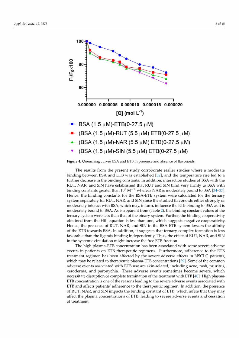
Appl. Sci. 2022, 12, 3575 8 of 15
Appl. Sci. 2022, 12, x FOR PEER REVIEW 8 of 15
binary system compared to the ternary system. It was observed that the addition of the
flavonoids in the BSA‐ETB system interfered in the binding and quenching constants of
the BSA‐ETB system (Table 2). The interaction with flavonoids (RUT, NAR, and SIN) and
BSA was previously reported to decrease albumin fluorescence, and the binding constants
for the three flavonoids RUT, NAR, and SIN were of the magnitude of >106, >104, and
>105, respectively [34–37]. The presence of RUT, NAR, and SIN increased the quenching
constants in the ternary system. In the binary system, ETB quenched the fluorescence of
the serum albumin by 27%, while as in the ternary system, RUT, NAR, and SIN in the ETB
quenched the fluorescence of serum albumin by 29%, 33%, and 37%, respectively (Figure
4). Therefore, the influence of the studied flavonoids varied, with RUT affecting the least
followed by NAR and the highest with SIN. The elevated quenching constants for the
ternary system suggest that RUT, NAR, and SIN might have increased the accessibility of
the ETB to the BSA, improving its quenching efficiency or the flavonoids had a synergistic
effect. Hence, the quenching increased (Table 2) [38].
Figure 4. Quenching curves BSA and ETB in presence and absence of flavonoids.
The results from the present study corroborate earlier studies where a moderate
binding between BSA and ETB was established [32], and the temperature rise led to a
further decrease in the binding constants. In addition, interaction studies of BSA with the
RUT, NAR, and SIN have established that RUT and SIN bind very firmly to BSA with
binding constants greater than 105 M−1, whereas NAR is moderately bound to BSA [34–
37]. Hence, the binding constants for the BSA‐ETB system were calculated for the ternary
system separately for RUT, NAR, and SIN since the studied flavonoids either strongly or
moderately interact with BSA, which may, in turn, influence the ETB binding to BSA as it
is moderately bound to BSA. As is apparent from (Table 2), the binding constant values
of the ternary system were less than that of the binary system. Further, the binding coop‐
erativity obtained from the Hill equation is less than one, which suggests negative coop‐
erativity. Hence, the presence of RUT, NAR, and SIN in the BSA‐ETB system lowers the
Figure 4. Quenching curves BSA and ETB in presence and absence of flavonoids.
The results from the present study corroborate earlier studies where a moderatebinding between BSA and ETB was established [32], and the temperature rise led to afurther decrease in the binding constants. In addition, interaction studies of BSA with theRUT, NAR, and SIN have established that RUT and SIN bind very firmly to BSA withbinding constants greater than 105 M−1, whereas NAR is moderately bound to BSA [34–37].Hence, the binding constants for the BSA-ETB system were calculated for the ternarysystem separately for RUT, NAR, and SIN since the studied flavonoids either strongly ormoderately interact with BSA, which may, in turn, influence the ETB binding to BSA as it ismoderately bound to BSA. As is apparent from (Table 2), the binding constant values of theternary system were less than that of the binary system. Further, the binding cooperativityobtained from the Hill equation is less than one, which suggests negative cooperativity.Hence, the presence of RUT, NAR, and SIN in the BSA-ETB system lowers the affinityof the ETB towards BSA. In addition, it suggests that ternary-complex formation is lessfavorable than the ligands binding independently. Thus, the effect of RUT, NAR, and SINin the systemic circulation might increase the free ETB fraction.
The high plasma-ETB concentration has been associated with some severe adverseevents in patients on ETB therapeutic regimens. Furthermore, adherence to the ETBtreatment regimen has been affected by the severe adverse effects in NSCLC patients,which may be related to therapeutic plasma-ETB concentrations [39]. Some of the commonadverse events associated with ETB use are skin-related, including acne, rash, pruritus,xeroderma, and paronychia. These adverse events sometimes become severe, whichnecessitate disruption or complete termination of the treatment with ETB [40]. High plasma-ETB concentration is one of the reasons leading to the severe adverse events associated withETB and affects patients’ adherence to the therapeutic regimen. In addition, the presenceof RUT, NAR, and SIN impacts the binding constant of ETB, which infers that they mayaffect the plasma concentrations of ETB, leading to severe adverse events and cessationof treatment.

Appl. Sci. 2022, 12, 3575 9 of 15
3.4. UV-Absorption Studies
The protein-ligand interaction gives information about the structural changes in thecomplex formed during the interaction and the protein alone. Static quenching leads tochanges in the structure, and hence, the protein spectra on interaction with its ligand. Basedon our results, the static quenching was predicted between BSA and ETB. The λmax ofBSA was at 280 nm because of π − π* transitions in the aromatic amino acids (tryptophan,tyrosine, and phenylalanine). On the interaction of the BSA with ETB, the λmax shiftedto 330 nm (Figure 5A) [31,41]. The hyperchromicity at 280 nm also suggested complexformation between ETB and BSA.
Appl. Sci. 2022, 12, x FOR PEER REVIEW 9 of 15
affinity of the ETB towards BSA. In addition, it suggests that ternary‐complex formation
is less favorable than the ligands binding independently. Thus, the effect of RUT, NAR,
and SIN in the systemic circulation might increase the free ETB fraction.
The high plasma‐ETB concentration has been associated with some severe adverse
events in patients on ETB therapeutic regimens. Furthermore, adherence to the ETB treat‐
ment regimen has been affected by the severe adverse effects in NSCLC patients, which
may be related to therapeutic plasma‐ETB concentrations [39]. Some of the common ad‐
verse events associated with ETB use are skin‐related, including acne, rash, pruritus, xe‐
roderma, and paronychia. These adverse events sometimes become severe, which neces‐
sitate disruption or complete termination of the treatment with ETB [40]. High plasma‐
ETB concentration is one of the reasons leading to the severe adverse events associated
with ETB and affects patients’ adherence to the therapeutic regimen. In addition, the pres‐
ence of RUT, NAR, and SIN impacts the binding constant of ETB, which infers that they
may affect the plasma concentrations of ETB, leading to severe adverse events and cessa‐
tion of treatment.
3.4. UV‐Absorption Studies
The protein‐ligand interaction gives information about the structural changes in the
complex formed during the interaction and the protein alone. Static quenching leads to
changes in the structure, and hence, the protein spectra on interaction with its ligand.
Based on our results, the static quenching was predicted between BSA and ETB. The λmax
of BSA was at 280 nm because of π − π* transitions in the aromatic amino acids (trypto‐
phan, tyrosine, and phenylalanine). On the interaction of the BSA with ETB, the λmax
shifted to 330 nm (Figure 5A) [31,41]. The hyperchromicity at 280 nm also suggested com‐
plex formation between ETB and BSA.
Figure 5. Ultraviolet‐visible spectra for (A): BSA (1.5 μM) in presence and absence of ETB (0.00–10
μM) and in presence of (B): RUT; (C): NAR; (D): SIN.
Figure 5. Ultraviolet-visible spectra for (A): BSA (1.5 µM) in presence and absence of ETB (0.00–10 µM)and in presence of (B): RUT; (C): NAR; (D): SIN.
The BSA-ETB spectra were also recorded for each RUT, NAR, and SIN to observe if anydifference in the spectra of BSA occurs in the presence of these flavonoids (Figure 5B–D).The BSA showed hyperchromicity on interaction with ETB in the presence of RUT, NAR,and SIN. The spectra for the ternary system of BSA-ETB were significantly different fromthose of the BSA-ETB spectra binary system. Hence, the binary and the ternary-systemcomplexes formed were completely different from one another. Further comparing theternary systems amongst themselves, the spectra differed from each other and thus inferredthat complexes formed between BSA-ETB in the presence of RUT, NAR, and SIN werealso different.
3.5. Molecular Docking
The experimental results were supplemented with molecular-docking analysis usingMOE. Experimental results suggested that the binding site for ETB binding on BSA to beSite I (subdomain IIA) and Site III (subdomain IIB) [26,42] (Figure 6A,B). Blind docking insilico was performed for the BSA-ETB system, and the results of the binary system werecompared to the ternary-system docking for the BSA-ETB in the presence of RUT, NAR,

Appl. Sci. 2022, 12, 3575 10 of 15
and SIN. It was observed that ETB binds to subdomain IIA and subdomain IB of BSA incorroboration with previous studies. The molecular docking results are presented in Table 4and suggest high binding energy for the BSA-ETB system at subdomain IIA. The bindingenergy showed a decline in the presence of all the flavonoids RUT, NAR, and SIN, with thehighest value of reduction in the binding energy in RUT, followed by SIN and then NAR.Thus, the presence of the three drugs RUT, NAR, and SIN influenced the binding energy ofETB with BSA.
Appl. Sci. 2022, 12, x FOR PEER REVIEW 11 of 15
Figure 6. Two‐dimensional molecular‐docking conformation for BSA ETB system (A): Site I and (B):
Site III.
Further, the comparison was made in the binding energies at subdomain IB, and it
was observed that the binding energies for the BSA‐ETB system in the presence of RUT,
NAR, and SIN were lower than the BSA‐ETB system in their absence, as shown in Table
4.
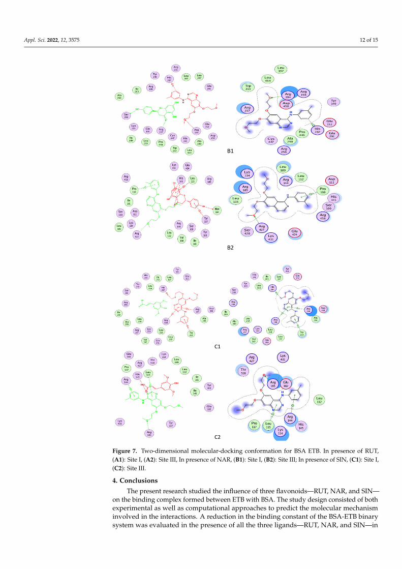
The amino acids Glu291, Leu218, Glu152, Arg198, Arg256, Arg217, Ser286, Leu237,
Leu259, Ile289, Tyr149, Ile263, Ala290, Trp213, Ala260, Val240, and Arg194 in the vicinity
of the ETB at the binding site in subdomain IIA surrounded the binding pocket at subdo‐
main IB with amino acids Lys 116, Pro117, Leu115, Ile181, Leu122, Glu182, Tyr137, Lys136,
Arg185, Ile141, Tyr160, Val188, Glu140, Asp118, and Thr121. A hydrogen bond and pi‐
cation bonds were involved in the formation of the binary complex between BSA and ETB
at Site I, whereas one hydrogen bond was involved in the interaction between BSA and
ETB at site III. The changes in the bonding in the ternary complex due to RUT, NAR, and
SIN are given in Table 4 and Figure 7A–C. The three‐dimensional figures for the interac‐
tion studies have been given in the Supplementary Materials (Figure S1). Molecular dy‐
namic simulation studies conducted for the BSA‐STB system suggested no fluctuation in
the RMSD and RMSF plots of BSA‐ETB and concluded a stable complex formation be‐
tween BSA and ETB [26].
Figure 6. Two-dimensional molecular-docking conformation for BSA ETB system (A): Site I and (B):Site III.
Table 4. Molecular-docking results for the binary and ternary BSA-ETB systems.
Complex Ligand Receptor Interaction Distance ScoreKcal mol−1 Subdomain
ETB-BSAn33 CD ARG 217 (A) H-acceptor 3.39
−9.255
IIA
6-ring NE ARG 256 (A) pi-cation 3.8
ETB(BSA-NRN)O14 OG SER 286 (A) H-donor 3.14
−7.684O1 NE ARG 217 (A) H-acceptor 3.04
ETB(BSA-RTN)N38 OD2 ASP 450 (A) H-donor 3.18
−6.331N33 CE LYS 439 (A) H-acceptor 3.51
ETB(BSA-SPA)
C29 5-ring HIS 287 (A) H-pi 4.15
−7.4286-ring CD ARG 194 (A) pi-H 3.15
6-ring NH1 ARG 198 (A) pi-cation 4.31
ETB-BSA N33 CA TYR 137 (A) H-acceptor 3.49 −7.495
IB
ETB(BSA-NRN)O28 NE ARG 427 (A) H-acceptor 3.21
−6.8366-ring CD PRO 110 (A) pi-H 4.53
ETB(BSA-RTN)
O5 NZ LYS 114 (A) H-acceptor 2.87
−7.272O28 NH1 ARG 185 (A) H-acceptor 3.13
O28 NH2 ARG 185 (A) H-acceptor 2.97
ETB(BSA-SPA)
O5 NH2 ARG 185 (A) H-acceptor 2.93
−7.0566-ring N LEU 115 (A) pi-H 4.41
6-ring CB ARG 144 (A) pi-H 4.22
6-ring CD ARG 144 (A) pi-H 4.04

Appl. Sci. 2022, 12, 3575 11 of 15
Further, the comparison was made in the binding energies at subdomain IB, and itwas observed that the binding energies for the BSA-ETB system in the presence of RUT,NAR, and SIN were lower than the BSA-ETB system in their absence, as shown in Table 4.
The amino acids Glu291, Leu218, Glu152, Arg198, Arg256, Arg217, Ser286, Leu237,Leu259, Ile289, Tyr149, Ile263, Ala290, Trp213, Ala260, Val240, and Arg194 in the vicinity ofthe ETB at the binding site in subdomain IIA surrounded the binding pocket at subdomainIB with amino acids Lys 116, Pro117, Leu115, Ile181, Leu122, Glu182, Tyr137, Lys136,Arg185, Ile141, Tyr160, Val188, Glu140, Asp118, and Thr121. A hydrogen bond and pi-cation bonds were involved in the formation of the binary complex between BSA and ETBat Site I, whereas one hydrogen bond was involved in the interaction between BSA andETB at site III. The changes in the bonding in the ternary complex due to RUT, NAR, andSIN are given in Table 4 and Figure 7A–C. The three-dimensional figures for the interactionstudies have been given in the Supplementary Materials (Figure S1). Molecular dynamicsimulation studies conducted for the BSA-STB system suggested no fluctuation in theRMSD and RMSF plots of BSA-ETB and concluded a stable complex formation betweenBSA and ETB [26].
Appl. Sci. 2022, 12, x FOR PEER REVIEW 12 of 16
Figure 7. Cont.
Appl. Sci. 2022, 12, 3575 12 of 15
Appl. Sci. 2022, 12, x FOR PEER REVIEW 13 of 16
Figure 7. Two-dimensional molecular-docking conformation for BSA ETB. In presence of RUT, (A1): Site I, (A2): Site III, In presence of NAR, (B1): Site I, (B2): Site III; In presence of SIN, (C1): Site I, (C2): Site III.
4. Conclusions The present research studied the influence of three flavonoids—RUT, NAR, and
SIN—on the binding complex formed between ETB with BSA. The study design consisted
Figure 7. Two-dimensional molecular-docking conformation for BSA ETB. In presence of RUT,(A1): Site I, (A2): Site III, In presence of NAR, (B1): Site I, (B2): Site III; In presence of SIN, (C1): Site I,(C2): Site III.
4. Conclusions
The present research studied the influence of three flavonoids—RUT, NAR, and SIN—on the binding complex formed between ETB with BSA. The study design consisted of bothexperimental as well as computational approaches to predict the molecular mechanisminvolved in the interactions. A reduction in the binding constant of the BSA-ETB binarysystem was evaluated in the presence of all the three ligands—RUT, NAR, and SIN—in

Appl. Sci. 2022, 12, 3575 13 of 15
the ternary system. The free drug fraction of ETB might be altered in the presence ofRUT, NAR, and SIN. Furthermore, the nonadherence to the treatment regimen of ETBis associated with some severe dermatological side effects, which are further associatedwith the systemic concentration of ETB. Hence, coadministration of these flavonoids mightinfluence the pharmacokinetic behavior of ETB, and their further knowledge needs to beexplored in vivo.
Supplementary Materials: The following supporting information can be downloaded at: https://www.mdpi.com/article/10.3390/app12073575/s1, Figure S1: Three dimensional conformations forBSA-ETB in absence and in presence of RUT, NAR and SIN at Site I and Site III of BSA.
Author Contributions: Conceptualization: T.A.W., S.Z.; Methodology: T.A.W., S.Z.; Software: A.H.B.;Formal analysis: A.I.A.K., S.Z.; Investigation: A.H.B., T.A.W., S.Z., A.A.A.-M.; Resources: T.A.W.;Writing: S.Z., T.A.W.; Review & Editing: A.I.A.K., A.A.A.-M., T.A.W. Project administration: T.A.W.All authors have read and agreed to the published version of the manuscript.
Funding: Researchers Supporting Project number (RSP-2021/357), King Saud University, Riyadh,Saudi Arabia.
Institutional Review Board Statement: Not Applicable.
Informed Consent Statement: Not Applicable.
Data Availability Statement: Data will be available on request to corresponding author.
Acknowledgments: The authors extend their appreciation to Researchers Supporting Project number(RSP-2021/357), King Saud University, Riyadh, Saudi Arabia, for funding this work.
Conflicts of Interest: The authors declare no conflict of interest.
References1. Peters, T., Jr. All about Albumin: Biochemistry, Genetics, and Medical Applications; Academic Press: Cambridge, MA, USA, 1995.2. Wang, B.-L.; Kou, S.-B.; Lin, Z.-Y.; Shi, J.-H. Investigation on the binding behavior between BSA and lenvatinib with the help of
various spectroscopic and in silico methods. J. Mol. Struct. 2020, 1204, 127521. [CrossRef]3. Koch-Weser, J.; Sellers, E.M. Binding of drugs to serum albumin. N. Engl. J. Med. 1976, 294, 311–316. [CrossRef] [PubMed]4. Keller, F.; Maiga, M.; Neumayer, H.-H.; Lode, H.; Distler, A. Pharmacokinetic effects of altered plasma protein binding of drugs in
renal disease. Eur. J. Drug Metab. Pharmacokinet. 1984, 9, 275–282. [CrossRef] [PubMed]5. Ni, Y.; Su, S.; Kokot, S. Spectrofluorimetric studies on the binding of salicylic acid to bovine serum albumin using warfarin and
ibuprofen as site markers with the aid of parallel factor analysis. Anal. Chim. Acta 2006, 580, 206–215. [CrossRef]6. Wani, T.A.; Alsaif, N.; Bakheit, A.H.; Zargar, S.; Al-Mehizia, A.A.; Khan, A.A. Interaction of an abiraterone with calf thymus
DNA: Investigation with spectroscopic technique and modelling studies. Bioorgan. Chem. 2020, 100, 103957. [CrossRef]7. Fan, J.; Sun, W.; Wang, Z.; Peng, X.; Li, Y.; Cao, J. A fluorescent probe for site I binding and sensitive discrimination of HSA from
BSA. Chem. Commun. 2014, 50, 9573–9576. [CrossRef]8. Zhang, Y.-F.; Zhou, K.-L.; Lou, Y.-Y.; Pan, D.-q.; Shi, J.-H. Investigation of the binding interaction between estazolam and bovine
serum albumin: Multi-spectroscopic methods and molecular docking technique. J. Biomol. Struct. Dyn. 2017, 35, 3605–3614.[CrossRef]
9. Fanali, G.; Di Masi, A.; Trezza, V.; Marino, M.; Fasano, M.; Ascenzi, P. Human serum albumin: From bench to bedside. Mol. Asp.Med. 2012, 33, 209–290. [CrossRef]
10. Sudlow, G.; Birkett, D.J.; Wade, D.N. Spectroscopic techniques in the study of protein binding. A fluorescence technique for theevaluation of the albumin binding and displacement of warfarin and warfarin-alcohol. Clin. Exp. Pharmacol. Physiol. 1975, 2,129–140. [CrossRef]
11. He, X.M.; Carter, D.C. Atomic structure and chemistry of human serum albumin. Nature 1992, 358, 209–215. [CrossRef]12. Wani, T.A.; Alsaif, N.; Alanazi, M.M.; Bakheit, A.H.; Zargar, S.; Bhat, M.A. A potential anticancer dihydropyrimidine derivative
and its protein binding mechanism by multispectroscopic, molecular docking and molecular dynamic simulation along with itsin-silico toxicity and metabolic profile. Eur. J. Pharm. Sci. 2021, 158, 105686. [CrossRef]
13. Wani, T.A. Highly sensitive ultra-performance liquid chromatography–tandem mass spectrometry method for the determinationof abiraterone in human plasma. Anal. Methods 2013, 5, 3693–3699. [CrossRef]
14. Khalil, N.Y.; Wani, T.A.; Abunassif, M.A.; Darwish, I.A. Sensitive HPLC method with fluorescence detection and on-linewavelength switching for simultaneous determination of valsartan and amlodipine in human plasma. J. Liq. Chromatogr. Rel.Technol. 2011, 34, 2583–2595. [CrossRef]

Appl. Sci. 2022, 12, 3575 14 of 15
15. Jing, J.J.; Liu, B.; Wang, X.; Wang, X.; He, L.L.; Guo, X.Y.; Xu, M.L.; Li, Q.Y.; Gao, B.; Dong, B.Y. Binding of fluphenazine withhuman serum albumin in the presence of rutin and quercetin: An evaluation of food-drug interaction by spectroscopic techniques.Luminescence 2017, 32, 1056–1065. [CrossRef]
16. Alsaif, N.A.; Al-Mehizia, A.A.; Bakheit, A.H.; Zargar, S.; Wani, T.A. A spectroscopic, thermodynamic and molecular dockingstudy of the binding mechanism of dapoxetine with calf thymus DNA. S. Afr. J. Chem. 2020, 73, 44–50. [CrossRef]
17. Benet, L.Z.; Bowman, C.M.; Sodhi, J.K. How transporters have changed basic pharmacokinetic understanding. AAPS J. 2019,21, 103. [CrossRef]
18. Zargar, S.; Wani, T.A. Protective Role of Quercetin in Carbon Tetrachloride Induced Toxicity in Rat Brain: Biochemical, Spec-trophotometric Assays and Computational Approach. Molecules 2021, 26, 7526. [CrossRef]
19. Frutos, M.J.; Rincón-Frutos, L.; Valero-Cases, E. Rutin. In Nonvitamin and Nonmineral Nutritional Supplements; Elsevier: Amsterdam,The Netherlands, 2019; pp. 111–117.
20. Farha, A.K.; Gan, R.-Y.; Li, H.-B.; Wu, D.-T.; Atanasov, A.G.; Gul, K.; Zhang, J.-R.; Yang, Q.-Q.; Corke, H. The anticancer potentialof the dietary polyphenol rutin: Current status, challenges, and perspectives. Crit. Rev. Food Sci. Nutr. 2022, 62, 832–859.[CrossRef]
21. Cavia-Saiz, M.; Busto, M.D.; Pilar-Izquierdo, M.C.; Ortega, N.; Perez-Mateos, M.; Muñiz, P. Antioxidant properties, radicalscavenging activity and biomolecule protection capacity of flavonoid naringenin and its glycoside naringin: A comparative study.J. Sci. Food Agric. 2010, 90, 1238–1244. [CrossRef]
22. Kumar, S.P.; Birundha, K.; Kaveri, K.; Devi, K.R. Antioxidant studies of chitosan nanoparticles containing naringenin and theircytotoxicity effects in lung cancer cells. Int. J. Biol. Macromol. 2015, 78, 87–95. [CrossRef]
23. Pandi, A.; Kalappan, V.M. Pharmacological and therapeutic applications of Sinapic acid—An updated review. Mol. Biol. Rep.2021, 48, 3733–3745. [CrossRef] [PubMed]
24. Park, J.H.; Liu, Y.; Lemmon, M.A.; Radhakrishnan, R. Erlotinib binds both inactive and active conformations of the EGFR tyrosinekinase domain. Biochem. J. 2012, 448, 417. [CrossRef] [PubMed]
25. Zhou, X.; Shi, K.; Hao, Y.; Yang, C.; Zha, R.; Yi, C.; Qian, Z. Advances in nanotechnology-based delivery systems for EGFRtyrosine kinases inhibitors in cancer therapy. Asian J. Pharm. Sci. 2020, 15, 26–41. [CrossRef] [PubMed]
26. Wani, T.A.; Alanazi, M.M.; Alsaif, N.A.; Bakheit, A.H.; Zargar, S.; Alsalami, O.M.; Khan, A.A. Interaction Characterization of aTyrosine Kinase Inhibitor Erlotinib with a Model Transport Protein in the Presence of Quercetin: A Drug–Protein and Drug–DrugInteraction Investigation Using Multi-Spectroscopic and Computational Approaches. Molecules 2022, 27, 1265. [CrossRef]
27. Wani, T.A.; Bakheit, A.H.; Al-Majed, A.A.; Altwaijry, N.; Baquaysh, A.; Aljuraisy, A.; Zargar, S. Binding and drug displacementstudy of colchicine and bovine serum albumin in presence of azithromycin using multispectroscopic techniques and moleculardynamic simulation. J. Mol. Liq. 2021, 333, 115934. [CrossRef]
28. Sengupta, B.; Sengupta, P.K. The interaction of quercetin with human serum albumin: A fluorescence spectroscopic study. Biochem.Biophys. Res. Commun. 2002, 299, 400–403. [CrossRef]
29. Xie, L.; Wehling, R.L.; Ciftci, O.; Zhang, Y. Formation of complexes between tannic acid with bovine serum albumin, eggovalbumin and bovine beta-lactoglobulin. Food Res. Int. 2017, 102, 195–202. [CrossRef]
30. Al-Mehizia, A.A.; Bakheit, A.H.; Zargar, S.; Bhat, M.A.; Asmari, M.M.; Wani, T.A. Evaluation of biophysical interaction betweennewly synthesized pyrazoline pyridazine derivative and bovine serum albumin by spectroscopic and molecular docking studies.J. Spectrosc. 2019, 2019, 3848670. [CrossRef]
31. Lakowicz, J.R. Principles of Fluorescence Spectroscopy; Springer Science & Business Media: Cham, Switzerland, 2013.32. Rasoulzadeh, F.; Asgari, D.; Naseri, A.; Rashidi, M.R. Spectroscopic studies on the interaction between erlotinib hydrochloride
and bovine serum albumin. DARU J. Pharm. Sci. 2010, 18, 179.33. Wani, T.A.; Bakheit, A.H.; Zargar, S.; Bhat, M.A.; Al-Majed, A.A. Molecular docking and experimental investigation of new
indole derivative cyclooxygenase inhibitor to probe its binding mechanism with bovine serum albumin. Bioorgan. Chem. 2019,89, 103010. [CrossRef]
34. Zargar, S.; Alamery, S.; Bakheit, A.H.; Wani, T.A. Poziotinib and bovine serum albumin binding characterization and influence ofquercetin, rutin, naringenin and sinapic acid on their binding interaction. Spectrochim. Acta Part A Mol. Biomol. Spectrosc. 2020,235, 118335. [CrossRef]
35. Sengupta, P.; Sardar, P.S.; Roy, P.; Dasgupta, S.; Bose, A. Investigation on the interaction of Rutin with serum albumins: Insightsfrom spectroscopic and molecular docking techniques. J. Photochem. Photobiol. B Biol. 2018, 183, 101–110. [CrossRef]
36. Hu, Y.-J.; Wang, Y.; Ou-Yang, Y.; Zhou, J.; Liu, Y. Characterize the interaction between naringenin and bovine serum albuminusing spectroscopic approach. J. Lumin. 2010, 130, 1394–1399. [CrossRef]
37. Sengupta, P.; Pal, U.; Mondal, P.; Bose, A. Multi-spectroscopic and computational evaluation on the binding of sinapic acid andits Cu (II) complex with bovine serum albumin. Food Chem. 2019, 301, 125254. [CrossRef]
38. Kameníková, M.; Furtmüller, P.G.; Klacsová, M.; Lopez-Guzman, A.; Toca-Herrera, J.L.; Vitkovská, A.; Devínsky, F.; Mucaji,P.; Nagy, M. Influence of quercetin on the interaction of gliclazide with human serum albumin–spectroscopic and dockingapproaches. Luminescence 2017, 32, 1203–1211. [CrossRef]
39. Timmers, L.; Boons, C.C.; Moes-Ten Hove, J.; Smit, E.F.; van de Ven, P.M.; Aerts, J.G.; Swart, E.L.; Boven, E.; Hugtenburg, J.G.Adherence, exposure and patients’ experiences with the use of erlotinib in non-small cell lung cancer. J. Cancer Res. Clin. Oncol.2015, 141, 1481–1491. [CrossRef]
Appl. Sci. 2022, 12, 3575 15 of 15
40. Kiyohara, Y.; Yamazaki, N.; Kishi, A. Erlotinib-related skin toxicities: Treatment strategies in patients with metastatic non-smallcell lung cancer. J. Am. Acad. Dermatol. 2013, 69, 463–472. [CrossRef]
41. Rabbani, G.; Khan, M.J.; Ahmad, A.; Maskat, M.Y.; Khan, R.H. Effect of copper oxide nanoparticles on the conformation andactivity of β-galactosidase. Colloids Surf. B. Biointerfaces 2014, 123, 96–105. [CrossRef]
42. Taghipour, P.; Zakariazadeh, M.; Sharifi, M.; Dolatabadi, J.E.N.; Barzegar, A. Bovine serum albumin binding study to erlotinibusing surface plasmon resonance and molecular docking methods. J. Photochem. Photobiol. B Biol. 2018, 183, 11–15. [CrossRef]