The interaction of small molecules with phospholipid ... - Nature
15
35 Acta Pharmacol Sin 2008 Jan; 29 (1): 35–49 ©2008 CPS and SIMM Invited review The interaction of small molecules with phospholipid membranes studied by 1 H NOESY NMR under magic-angle spinning 1 Holger A SCHEIDT 2 , Daniel HUSTER 3,4 2 Junior Research Group “Structural Biology of Membrane Proteins”, Institute of Biochemistry/Biotechnology, Martin Luther University Halle-Wittenberg, D-06120 Halle, Germany; 3 Institute of Medical Physics and Biophysics, Univeristy of Leipzig,D-04107 Leipzig, Germany Abstract The interaction of small molecules with lipid membranes and the exact knowledge of their binding site and bilayer distribution is of great pharmacological impor- tance and represents an active field of current biophysical research. Over the last decade, a highly resolved 1 H solid-state NMR method has been developed that allows measuring localization and distribution of small molecules in membranes. The classical solution 1 H NMR NOESY technique is applied to lipid membrane samples under magic-angle spinning (MAS) and NOESY cross-relaxation rates are determined quantitatively. These rates are proportional to the contact probability between molecular segments and therefore an ideal tool to study intermolecular interactions in membranes. Here, we review recent 1 H MAS NOESY applications that were carried out to study lateral lipid organization in mixed membranes and the interaction of membranes with water, ethanol, small aromatic compounds, peptides, fluorescence labels, and lipophilic nucleosides. Key words MAS NMR; lipid bilayers; lipid-water interface; drug partitioning; fluorescent probes; peptide- membrane interaction 1 The study was supported by the “Exzellenz- netzwerk Biowissenschaften” funded by the federal state of Sachsen-Anhalt. 4 Correspondence to Dr Daniel HUSTER. Phn 49-345-552-4942. Fax 49-345-552-7013. E-mail [email protected] halle.de Received 2007-06-12 Accepted 2007-07-24 doi: 10.1111/j.1745-7254.2008.00726.x Introduction Pharmacologically active drugs are usually small mol- ecules that target a specific protein, for instance an enzyme, a receptor, or a channel. Often, these molecules target mem- brane-stemming proteins, which represent about one third of all proteins in the human genome. It is estimated that on the order of 50%–70% of the current targets for drug devel- opment are proteins that are embedded in the cellular mem- brane [1,2] . The plasma membrane represents an important biological interface, where many essential processes for the cell function take place. Most importantly, it represents a barrier separating the cytosol from the extracellular medium. For specific functions, proteins are embedded in the membrane, where they are involved in signal generation and transduction, cellular communication, both active and pas- sive transport of ions and a number of different molecules, and the generation of energy. In order to fulfill its function, a drug molecule has to find its specific protein interaction partner and specifically bind to it. Although about 60% of the dry weight of the membrane is constituted of proteins, due to the large variety of these molecules the actual con- centration of a specific protein in the membrane is typically relatively low, which decreases the likelihood for a drug to find its target protein in the membrane. However, the bind- ing of a drug to its target can be facilitated by initial parti- tioning into the membrane and subsequent binding to its tar- get protein [3] . Such a scenario can have several advantages. First, pharmacological drugs are often lipophilic molecules that preferentially partition into a lipid membrane or its interface. Second, the diffusion of a drug in the membrane is constraint to two dimensions, which increases the interac- tion probability between membrane protein and its ligand by approximately a factor of 1000 [4] . Third, the area of the mem- brane is much larger than that of the specific protein. Therefore, unspecific binding of a drug molecule to the mem- brane will bring it in close proximity to its target. Since the binding constant of the drug to the target protein is usually
-
Upload
khangminh22 -
Category
Documents
-
view
0 -
download
0
Transcript of The interaction of small molecules with phospholipid ... - Nature

35
Acta Pharmacol Sin 2008 Jan; 29 (1): 35–49
©2008 CPS and SIMM
Invited review
The interaction of small molecules with phospholipid membranes studiedby 1H NOESY NMR under magic-angle spinning1
Holger A SCHEIDT2, Daniel HUSTER3,4
2Junior Research Group “Structural Biology of Membrane Proteins”, Institute of Biochemistry/Biotechnology, Martin Luther University
Halle-Wittenberg, D-06120 Halle, Germany; 3Institute of Medical Physics and Biophysics, Univeristy o f Leipzig,D-04107 Leipzig,
Germany
Abstract
The interaction of small molecules with lipid membranes and the exact knowledgeof their binding site and bilayer distribution is of great pharmacological impor-tance and represents an active field of current biophysical research. Over the lastdecade, a highly resolved 1H solid-state NMR method has been developed thatallows measuring localization and distribution of small molecules in membranes.The classical solution 1H NMR NOESY technique is applied to lipid membranesamples under magic-angle spinning (MAS) and NOESY cross-relaxation ratesare determined quantitatively. These rates are proportional to the contact probabilitybetween molecular segments and therefore an ideal tool to study intermolecularinteractions in membranes. Here, we review recent 1H MAS NOESY applicationsthat were carried out to study lateral lipid organization in mixed membranes andthe interaction of membranes with water, ethanol, small aromatic compounds,peptides, fluorescence labels, and lipophilic nucleosides.
Key words
MAS NMR; lipid bilayers; lipid-water interface;drug partitioning; fluorescent probes; peptide-membrane interaction
1The study was supported by the “Exzellenz-netzwerk Biowissenschaften” funded by thefederal state of Sachsen-Anhalt.4 Correspondence to Dr Daniel HUSTER.Ph n 49-345-552-4942.Fax 49-345-552-7013.E-ma il daniel.hu [email protected] ni-halle.de
Received 2007-06-12Accepted 2007-07-24
doi: 10.1111/j.1745-7254.2008.00726.x
Introduction
Pharmacologically active drugs are usually small mol-ecules that target a specific protein, for instance an enzyme,a receptor, or a channel. Often, these molecules target mem-brane-stemming proteins, which represent about one thirdof all proteins in the human genome. It is estimated that onthe order of 50%–70% of the current targets for drug devel-opment are proteins that are embedded in the cellular mem-brane[1,2]. The plasma membrane represents an importantbiological interface, where many essential processes for thecell function take place. Most importantly, it represents abarrier separating the cytosol from the extracellular medium.For specific functions, proteins are embedded in themembrane, where they are involved in signal generation andtransduction, cellular communication, both active and pas-sive transport of ions and a number of different molecules,and the generation of energy. In order to fulfill its function,a drug molecule has to find its specific protein interaction
partner and specifically bind to it. Although about 60% ofthe dry weight of the membrane is constituted of proteins,due to the large variety of these molecules the actual con-centration of a specific protein in the membrane is typicallyrelatively low, which decreases the likelihood for a drug tofind its target protein in the membrane. However, the bind-ing of a drug to its target can be facilitated by initial parti-tioning into the membrane and subsequent binding to its tar-get protein[3]. Such a scenario can have several advantages.First, pharmacological drugs are often lipophilic moleculesthat preferentially partition into a lipid membrane or itsinterface. Second, the diffusion of a drug in the membrane isconstraint to two dimensions, which increases the interac-tion probability between membrane protein and its ligand byapproximately a factor of 1000[4]. Third, the area of the mem-brane is much larger than that of the specific protein.Therefore, unspecific binding of a drug molecule to the mem-brane will bring it in close proximity to its target. Since thebinding constant of the drug to the target protein is usually

36
Acta Pharmacologica Sinica ISSN 1671-4083Scheidt HA et al
larger than to the membrane, subsequent protein bindingwill necessarily occur. These arguments may suggest amechanism of membrane partitioning of a drug molecule priorto binding to its target protein.
To date, only a few mammalian membrane proteins areknown in their structure to high resolution[5,6]. As for mem-brane proteins, the current understanding of the interactionof small molecules with the plasma membrane is also limited.Because of the complicated membrane structure, membraneproteins are difficult to crystallize in their natural environment.A biological membrane is characterized by a versatile mo-lecular dynamics that takes place in a broad time windowfrom picoseconds to hours[7-10]. Further, biological mem-branes feature a highly specialized lipid-water interface thatallows for numerous intermolecular interactions to take placeto stabilize a protein in its correct structure[11-13]. Likewise,the interactions of small lipophilic molecules such as drugswith a membrane are diverse and complicated usually givingrise to a broad and dynamic distribution of them. A sketch ofthe complicated membrane structure and the distribution ofmolecules within the lipid-water interface is provided inFigure 1. Considering the complicated nature of a biologicalmembrane and its lipid-water interface, simple partition ex-periment in water/octanol biphasic systems do not provide
good models for drug partitioning into membranes and morestructure-based biophysical methods are required to under-stand binding of small molecules to the membrane interface.
In lipid membranes, all molecules are affected by verycomplex interaction patterns brought about by basicelectrostatic, hydrophobic, and van der Waals interactionsarising from the physical and chemical features of the inter-acting molecules. Also, hydrogen bond formation, lipid in-duced interactions, and a multitude of entropic contribu-tions play an important role for the interaction between theconstituents in the bilayer. At any given depth of a membrane,different interaction patterns will dominate leading to veryspecific physical properties on a scale of only several Ang-stroms along the membrane normal. A small molecule thatpartitions into the bilayer is subject to large gradients of thebasic physical parameters such as dielectric constant, watercontent and so on giving rise to very specific and site-re-solved interactions. The average location of a molecule inthe membrane is consequently determined by a minimum inthe Gibbs free energy that contains both entropic andenthalpic contributions. But it needs to be stressed, that inmost cases membrane embedded molecules are broadly dis-tributed around their average location and subject to highdynamics on different time scales[14,15]. In such a complex
Figure 1. Schematic sketch of a phospholipid membrane in the liquid crystalline phase state with an (aromatic) drug molecule bound to thelipid-water interface. All molecular segments are subject to broad distributions functions parallel to the membrane normal as schematicallyindicated for the lipid headgroup, the chain end, and the drug molecule. In the lower leaflet of the membrane, the broad lipid-water interfaceis highlighted and typical correlation times for trans gauche isomerization, lateral lipid diffusion, and motions of the entire lipid molecules aregiven. In addition, slower membrane undulations and collective motions as well as lipid flip-flops take place.

Http://www.chinaphar.com Scheidt HA et al
37
environment a high spatial resolution for the membrane po-sition and orientation of a specific molecular segment is re-quired to understand the interactions between different mol-ecules and to relate this to the molecular function. Com-bined neutron and x-ray scattering techniques may achievesuch a resolution, which requires isotopic labeling and thepreparation of oriented samples. Typical, scattering experi-ments perform better with dehydrated samples, which couldbe intrusive[15,16]. Over the last 20 years, an 1H NMR techniquebecame available that allows to measure intermolecular con-tact probabilities in lipid membranes allowing to determine thedistribution profiles of small molecules[17].
This review will focus on 1H solid-state NMR applica-tions to study the interaction of small molecules with mem-branes and to determine their distribution within the bilayer.Solid-state NMR can not only provide important contribu-tions for solving the structure of membrane proteins[8,18-20]
but also represents a very useful technique to study thelipid membrane itself[10,21-24]. In particular, the interaction ofsmall molecules such as natural and synthetic drugs withmembranes can be very successfully studied by solid-stateNMR techniques[25-27]. Studies about membrane binding,the response of the lipid membrane to the presence of suchmolecules, the interactions between different molecules, thelateral membrane organization, and the membrane diffusionare possible. Such information is crucial to understand thefunction and the complicated interplay of membrane mol-ecules and the entire membrane and may help to identifytarget structures for drug research or to provide insight intothe mode of action of the investigated molecules.
Usually, solid-state NMR spectra are characterized bybroad anisotropic line shapes. However, the magic-anglespinning (MAS) technique can convert broad anisotropicsignals into well-resolved sharp lines[28]. For this technique,the NMR sample is contained in a cylindrical MAS rotor thatis oriented at an angle of 54.73º with respect to the externalmagnetic field and spun about its long axis at an angularfrequency of up to tens of kHz. Usually, for well-resolved 1HNMR spectra the MAS frequency has to exceed the width ofthe 1H-1H dipolar tensor interaction. However, for lipid mol-ecules in the fluid phase of the membrane, well-resolved 1HMAS spectra could be recorded at low MAS frequenciesalthough the homogeneous broadenings are on the order of10 kHz and larger[17]. This effect has been explained by therapid axially symmetric reorientation of the lipids in the mem-brane with a correlation time on the order of 1 ns or less[29].Thus, the dipolar broadening only depends on the orienta-tion of the local bilayer normal relative to the external mag-
netic field scaling with the second Legendré polynomial P2
(cosθ). In this manner, the homogenous dipolar broadeningis converted into an inhomogeneous broadening, which canbe averaged out by MAS even if the rotational frequency islower than the width of the anisotropic spectrum[29]. Typicalline widths of 1H MAS NMR spectra of lipid membranes areon the order of 0.01–0.1 ppm. An example of a 1D 1H MASNMR spectrum of an aqueous suspension of POPC with theassignments of all lipid signals is shown in Figure 2.
The good resolution of 1H MAS NMR spectra of lipidmembranes and their long spin-lattice relaxation times al-lows acquisition of two dimensional correlation spectra forthe study of intermolecular interactions. This review is de-voted to 1H MAS nuclear Overhauser enhancement spec-troscopy (NOESY) NMR investigations of lipid membranesand their interaction with small molecules -from water to pep-tides- incorporated in lipid membranes. While the NOESYtechnique in solution NMR is mostly applied to determineintramolecular distances in soluble protein or DNA molecules,for the application to lipid bilayers the intermolecular inter-actions are most interesting. Determination of intermolecu-lar cross-relaxation rates allows to study membrane binding,membrane organization, membrane localization and membraneorientation of membrane embedded molecules. In severalstudies 1H MAS NOESY NMR has shown its high potentialin such investigations.
Figure 2 . One dimensional 1H MAS NMR spectrum of POPCmultilamellar membrane vesicles hydrated to 50% water at a tem-perature of 30 ºC. The MAS rotational frequency was 10 kHz. Thespectrum was recorded at a Larmor frequency of 500.1 MHz. Theassignment of all peaks in the MAS spectrum is given in the struc-tural sketch shown above. Reproduced from ref[36] with permission.

38
Acta Pharmacologica Sinica ISSN 1671-4083Scheidt HA et al
Theory
Interpretation of intermolecular cross-relaxation rates
in membranes The simple and robust three pulse NOESYpulse sequence is shown in Figure 3[30,31]. The first pulsecreates transversal magnetization, which is allowed to evolveunder the influence of the isotropic chemical shift during t1.Thus, each spin is labelled with its characteristic resonancefrequency. The second pulse flips the magnetization intothe z-direction, where magnetization exchange takes placeduring the mixing time tm. This leads to the build up of crosspeaks in the 2D spectra provided the interacting spins are inclose proximity during mixing (< 5 Å). Since chemical ex-change typically does not contribute to the magnetizationexchange in lipid membranes the cross peaks are solelycaused by dipolar cross-relaxation between two proton spinsin close proximity. Such a process can be described by thecross-relaxation rate σ, which is strongly dependent on thedistance r between the two spins and the correlation time ofthe molecular motion τc according to[32,33]:
++−=
2C
20
6IS
2
C42
02
IS 41
61
π40 τω
τγµσ
r
η (1)
In equation (1), η is Planck’s constant divided by 2π, µ0
is the magnetic constant, ω0 is the Larmor frequency, and γthe gyromagnetic ratio. Due to this very strong distancedependence only interactions contribute to the cross-relax-ation rate that occur within 5 Å. If NOESY experiments areused to study the interaction of small molecules with lipidmembranes, only intermolecular cross-relaxation is of interest.This is different from the situation encountered for solubleproteins, where cross-relaxation rates provide a direct mea-sure of the inter-proton distance provided the correlationtime that modulates the interaction is known. In contrast,intermolecular cross-relaxation rates determined in lipid mem-branes in the fluid phase state, can not be used to measure awell defined fixed distance between the regarding protons.Due to the high mobility and molecular disorder in lipid mem-branes[14] the cross-relaxation rates provide a measure of thecontact probability between the interacting spins[34,35].
Therefore, in a lipid membrane the cross-relaxation rate is pro-portional to the contact frequency between the two nuclei, ahigh contact frequency results in large cross-relaxation ratesand a low contact frequency yields small cross-relaxation rates.
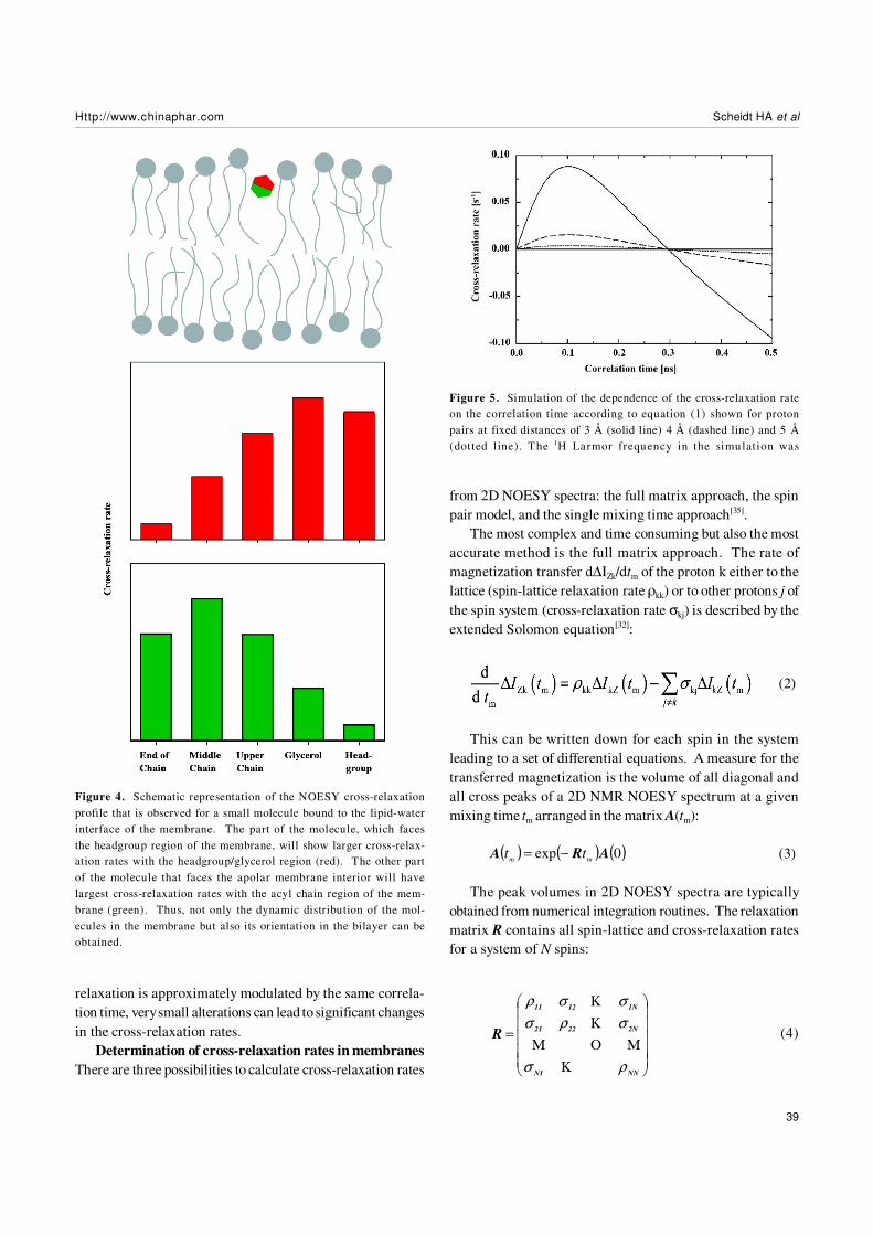
The contact probability, ie, the cross-relaxation rate be-tween two nuclei, is directly related to the structural arrange-ment in the membrane. If we assume a broad distributionfunction for each molecular segment, it becomes obviousthat for some of those segments the distribution functionsoverlap and for others they do not. A strong overlap in thedistribution functions leads to large cross-relaxation rates,small overlap creates small cross-relaxation rates, and if thereis no overlap in these distribution functions, no cross-relax-ation rate is measured. Thus, cross-relaxation rates in mem-branes represent a measure for the interaction strength be-tween molecular segments, which can be used to localizemolecules in the membrane and to study the lateral molecu-lar interaction. For small membrane embedded moleculescross peaks with more than just one lipid segment areobserved, often even all lipid segments exchange magneti-zation with the molecule of interest. This is related to thehigh degree of molecular disorder and the high mobility ofthe molecules in the membrane[35]. Therefore, a plot of thecross-relaxation rates of a particular molecular segment vs.all lipid segments along the membrane normal describes thedistribution function of this segment in the lipid bilayer assketched in Figure 4. Further, if the distribution functions oftwo or more spins of the membrane embedded molecule areknown, one can typically obtain the membrane orientationof this compound.
It should be mentioned that this interpretation of NOESYcross-relaxation rates is only correct if the correlation timethat modulates the magnetization exchange is equal for allthe different molecular contacts. First estimations of thecorrelation times that are responsible for the magnetizationexchange in a NOESY experiment in membranes revealed thatonly relatively slow processes have to be considered. Theseslow processes describe motions of entire molecules[7] andlateral lipid diffusion[36], which suggests that all molecularcontacts can be treated equally and the NOESY cross-relax-ation rate indeed represents the contact probability betweenmolecular segments.
To avoid over-interpretation of the NOESY data careshould be taken in comparing absolute cross-relaxation ratesfor different molecules in lipid membranes. The dependenceof the cross-relaxation rate σ on the correlation time of mo-tion τc in equation (1) may lead to remarkable changes in thecross-relaxation rates even for small variations of the corre-lation time as shown in Figure 5. Even though all cross-
Figure 3. Schematic representation of the pulse sequence of a two-dimensional NOESY experiment consisting of three 90º radio fre-quency pulses.
Http://www.chinaphar.com Scheidt HA et al
39
relaxation is approximately modulated by the same correla-tion time, very small alterations can lead to significant changesin the cross-relaxation rates.
Determination of cross-relaxation rates in membranes
There are three possibilities to calculate cross-relaxation rates
from 2D NOESY spectra: the full matrix approach, the spinpair model, and the single mixing time approach[35].
The most complex and time consuming but also the mostaccurate method is the full matrix approach. The rate ofmagnetization transfer d∆IZk/dtm of the proton k either to thelattice (spin-lattice relaxation rate ρkk) or to other protons j ofthe spin system (cross-relaxation rate σkj) is described by theextended Solomon equation[32]:
(2)
This can be written down for each spin in the systemleading to a set of differential equations. A measure for thetransferred magnetization is the volume of all diagonal andall cross peaks of a 2D NMR NOESY spectrum at a givenmixing time tm arranged in the matrix A(tm):
(3)
The peak volumes in 2D NOESY spectra are typicallyobtained from numerical integration routines. The relaxationmatrix R contains all spin-lattice and cross-relaxation ratesfor a system of N spins:
Figure 4. Schematic representation of the NOESY cross-relaxationprofile that is observed for a small molecule bound to the lipid-waterinterface of the membrane. The part of the molecule, which facesthe headgroup region of the membrane, will show larger cross-relax-ation rates with the headgroup/glycerol region (red). The other partof the molecule that faces the apolar membrane interior will havelargest cross-relaxation rates with the acyl chain region of the mem-brane (green). Thus, not only the dynamic distribution of the mol-ecules in the membrane but also its orientation in the bilayer can beobtained.
Figure 5. Simulation of the dependence of the cross-relaxation rateon the correlation time according to equation (1) shown for protonpairs at fixed distances of 3 Å (solid line) 4 Å (dashed line) and 5 Å(dotted line). The 1H Larmor frequency in the simulation was
( ) ( ) ( )0exp ARAmm
tt −=
=
NNN1
2N2221
1N1211
R
ρσ
σρσ
σσρ
Κ
ΜΟΜ
Κ
Κ
(4)

40
Acta Pharmacologica Sinica ISSN 1671-4083Scheidt HA et al
Most accurately, the relaxation matrix is calculated byfitting equation (3) to the experimental peak volume matricesobtained for different mixing times. The resulting relaxationmatrix contains all parameters to determine the localization andthe orientation of a given molecule in the lipid membrane.
Alternatively, the spin-pair interaction model assumesthat the spin system is reasonably well decoupled and re-duces the multi-spin system to an isolated spin pair. Thevolume of a cross peak between the two spin I and S is thenobtained by[37]:
(5)
where AIS(tm) represents the cross peak volume at themixing time tm, AII(0) the volume of the diagonal peak at tm=0,and T1,IS defines the magnetization leakage to the lattice. Byanalyzing the cross peak volumes for different mixing times(and the diagonal peak volumes for a very short mixing time)the cross-relaxation rate can be obtained from a simple fit tothis equation. The advantage of the spin-pair-model is thatonly the cross peaks between the investigated molecule andthe molecular segments of the lipid matrix need to be inte-grated to calculate the respective cross-relaxation rates.Since the determination of the complete peak volume matri-ces is tedious and time consuming, this approach is moreefficient if only a few cross-relaxation rates are required for acertain application. Though the information of the completerelaxation matrix is lost, it is not necessary to monitor allmagnetization transfer processes within the lipid matrix, ifone is only interested in the localization of a molecule in themembrane.
The most simplistic single mixing time approach deter-mines cross-relaxation rates from a single NOESY experimentconducted at just one mixing time. In this approach, equa-tion (5) is expanded into a truncated Taylor series that onlycontains the linear term, which leads to[35]:
(6)
This simplification represents the most efficient approachsince only a single NOESY spectrum needs to be acquired todetermine cross-relaxation rates. However, the cross-relax-ation rates determined by this approach may not be veryaccurate, in particular small rates can be highly biased whenthere is also strong (intramolecular) magnetization exchangeof the particular segment with other molecular sites.
The three approaches for the determination the cross-relaxation rates from 2D NOESY spectra were compared in an
DMPC lipid matrix[35]. For the majority of the cross-relax-
ation rates, reasonable agreement between the three modelswas reported. However, the cross-relaxation rates betweenneighboring protons in lipid hydrocarbon chains are veryhigh, which interferes with the calculation of magnetizationtransfer between chain protons, which are more distant. Thissituation is only correctly reflected in the full matrix approach.
1H NOESY MAS NMR of pure lipid membranes
Before discussing 1H MAS NOESY NMR investigationsof the interaction of small molecules with lipid membranes,we will first briefly review NOESY data on pure phospholipidbilayers that provided insight into their highly dynamic or-ganization and molecular disorder. The most extensive studieswere done by the Gawrisch group, who investigated the lat-eral lipid organization in pure DMPC bilayers[35] and complexphospholipid mixtures in the presence of cholesterol[38]. Atypical 1H MAS NOESY spectrum of lipid bilayers is shownin Figure 6. Interestingly, cross peaks between all lipid seg-ments of the phospholipid molecules were observed due tothe high mobility and molecular disorder in lipid membranes.This could only be understood from a quantitative magneti-zation transfer analysis within the lipid membrane using thefull matrix approach[35]. Experiments in mixed perdeuterated/protiated phospholipid membranes indicated that the cross-relaxation in lipid membranes is almost entirely of intermo-lecular origin. Therefore, the occurrence of NOESY crosspeaks between all lipid segments can only be explained by a
( ) ( )[ ]
−⋅−−=
IS,1
mmIS
IImIS exp2exp1
2
)0(
T
tt
AtA σ
( )( )
mIIm
mIS
IStAt
tA
⋅=σ
Figure 6. Contour plot of a 2D 1H MAS NOESY NMR spectrum ofPOPC membranes containing 8.75 H2O per POPC at a temperatureof 10°C and a mixing time of 300 ms. The spectrum was recorded ata Larmor frequency of 500.1 MHz. All lipid-lipid cross peaks havepositive intensity while the lipid water cross peaks are negative. Thespectrum was reproduced from ref[46] with permission.

Http://www.chinaphar.com Scheidt HA et al
41
membrane model that is characterized by a high degree ofmotional disorder. Indeed, if per proton based cross-relax-ation rates were compared, the most likely contacts betweenmolecular segments also showed the highest cross-relax-ation rates. Less frequent contacts, for instance those be-tween lipid headgroups and the ends of the acyl chains pro-duced smaller cross-relaxation rates. Although such con-tacts are by far less frequent, they occur with a relative highlikelihood in a liquid-crystalline membrane indicating the highamount of disorder and mobility in biological membranes.
1H NOESY cross-relaxation rates were also determined inmixed membranes of polyunsaturated SDPC/SDPE/SDPS/Cholesterol (4/4/1/1, mol/mol) membranes[38]. Since cross-relaxation rates can be determined for intermolecular lipid-lipid contacts, they are ideal tools to study lateral lipidorganization. In particular, the interaction of cholesterol withthe unsaturated lipid species was studied. The most impor-tant results of this study were that (i) cholesterol preferen-tially interacts with the SDPC in the mixture, while lateralcontacts between cholesterol and SDPE and SDPS are lessfrequent and (ii) cholesterol prefers contacts with saturatedchains, where favorable van der Waals interactions betweenthe chains and the rigid sterol ring systems are formed. Incontrast, interactions of cholesterol and the polyunsaturatedchains were less frequently observed. These results allowedto determine a quantitative model of the lateral lipid organi-zation in this complex mixture, which revealed the existence ofmicrodomains that are enriched in SDPC and cholesterol[38].
So far, the issue of spin diffusion has not been discussed.Spin diffusion represents an undesired mechanism that re-lays magnetization over several steps to yield a cross peakfor molecular segments that are not in close proximity. Sincethis relayed transfer of magnetization is typically slow, spindiffusion can be neglected if the mixing time is kept to aminimum. Since the occurrence of head-to-tail cross peaksin NOESY spectra of lipid membranes was previously attrib-uted to spin diffusion[39,40], this mechanism was investigatedin more detail[41]. In particular, special phospholipid mol-ecules were employed that contained two deuterated meth-ylene groups in the middle of the chain to block spin diffusion.The quantitative analysis of cross-relaxation rates in thisstudy proved that the cross peaks between lipid headgroupsand hydrocarbon chains are not caused by spin diffusionbut are the result of a direct approach (within 5 Å) ofheadgroup and chain protons[41]. Again, only highly disor-dered and dynamic membranes can explain this result.
NOESY MAS NMR on lipid membranes contain-ing small molecules
Several aspects of the interaction of small molecules withlipid membranes were investigated by 1H MAS NOESY NMRstudies over the last years. Starting with the question ofhow deep water penetrates a lipid membrane; further studiesinvestigated the membrane interaction of ethanol and differ-ent aromatic molecules. Finally, 1H MAS NOESY NMR wasused to study the membrane location and interaction of pep-tides and lipophilic nucleosides. In addition, several fluo-rescent probes like lipid and cholesterol analoga were inves-tigated in a membrane environment.
Water The water distribution in a lipid bilayer is an inter-esting problem that was one of the first issues investigatedby 1H NOESY MAS NMR. Specific interactions betweenwater and the lipids are essential for the formation of differ-ent lipid phases and in particular planar lipid membranes[27].At the same time, the water permeation across bilayers is veryhigh[42]. Therefore, the main questions concerned the depthwater penetration into the bilayer and the thickness of thelipid-water interface of the membrane. A number of studiesmeasured intermolecular cross-peaks between water and theindividual lipid segments[43-46].
Phase-sensitive NOESY experiments were applied underMAS conditions to show that the water molecules have ac-cess to the headgroup, the glycerol backbone and the upperchain region of a POPC bilayer[43]. The loss of some of thesecross peaks in the presence of a non-ionic detergent (C12EO4)indicated a process of dehydration due to the presence ofthe surfactant.
Further NOESY studies on lipid hydration of MeDOPEand DOPC bilayers[35] showed that the expected cross peaksbetween water and lipid headgroup/glycerol are detected forMeDOPE but are absent for DOPC membranes and even inmixed MeDOPE/DOPC bilayers[45]. Also for the hexagonaland the metastable isotropic phase of MeDOPE the lipid-water cross peaks were not observable, indicating that in thisphases the water is diffusing freely and no longer involved inthe hydrogen bonding pattern at the lipid headgroups[43].However, the presence or absence of cross-peaks betweenlipids and water should be interpreted with great caution.Since the lifetime of water-lipid associations is likely to beshorter than of lipid-lipid associations, those cross-peaksare often weak and have negative intensity. They are easilymissed if spectra are not recorded at proper experimentalconditions. This is underlined by the fact that in the samestudy heteronuclear HOESY experiments indicated contactsbetween the DOPC headgroups with the phosphate buffer(used as reporter for the water location)[45]. Therefore, wa-ter-lipid NOESY cross peaks could have been below the de-tection limit due to their weak intensities. On the other hand,

42
Acta Pharmacologica Sinica ISSN 1671-4083Scheidt HA et al
molecules that exchange protons with water via hydroxyland amino groups may show strong cross-peaks to water.However, those peaks may reflect chemical exchange ofprotons.
In the most recent study the cross-relaxation rates be-tween water and phospholipid segments were determined ina full matrix approach for POPC[46]. While cross-relaxationrates in lipid membranes are always negative, lipid-watercross-relaxation rates were calculated to be positive indicat-ing faster correlation times for the interaction on the order of100 ps. This value agrees well with the short lifetime of wa-ter in the first hydration layer[47]. Water showed weak cross-relaxation rates with the signals from the lipid headgroupand the glycerol. Only very low cross-relaxation rates weredetermined for the acyl chain region of the membrane sug-gesting that the water concentration in the bilayer core isvery low.
Taken together, these results indicate that there are anumber of water molecules in the lipid-water interface thatare in close proximity to the lipid headgroups, the glycerolregion, and the carbonyl groups of the acyl chains. Here,favorable hydrogen bonds are formed that give rise to theobserved NOESY cross peaks. All studies exhibited thatthere is no water detectable in the hydrophobic core ofmembrane. The water permeation across bilayers is veryhigh[42]. However, the individual act of a water moleculepassing through the bilayer is likely to require only a frac-tion of a microsecond. Therefore, water content in the hy-drophobic core of a bilayer at any given time is rather low,which is in agreement with observations by neutronscattering.
Ethanol The interaction of ethanol with lipid membraneshas been a controversy in anesthesia for several years. Somestudies report that the effect of ethanol on membrane pro-teins is attributed to changes in the lipid packing of the mem-brane[48], while others emphasize it is the result of ethanolbinding to the membrane protein itself [49]. While the mol-ecules could interact at multiple sites, 1H MAS NMR hasbeen used to determine the exact localization and distribu-tion of ethanol in the lipid membrane by Gawrisch and co-workers[50,51]. In these studies, relative cross peak intensi-ties and later cross-relaxation rates were for the first timeinterpreted in terms of a distribution function of the mol-ecule relative to various molecular segments of the lipidsand over the lipid bilayer[50]. In these profiles, ethanolshowed contacts to all phospholipid segments due to thehigh disorder and mobility in liquid-crystalline membranes.Being a small amphiphilic molecule, ethanol was localized inthe lipid-water interface of the membrane, with the largest
cross-relaxation rates (ie, contact probabilities) to the glyc-erol backbone. The degree of chain unsaturation in the lipidmatrix only had a slight effect on the membrane distributionof ethanol as shown for saturated DMPC, monounsaturatedSOPC, and polyunsaturated SDPC bilayers[50]. A further studyof the ethanol distribution in POPC membranes revealed anidentical position of ethanol in the bilayer and showed excel-lent agreement between the measured cross-relaxation rateprofile and this profile calculated from a molecular dynamicssimulation[51]. In addition, a comparison of the ethanol dis-tribution profiles exhibited that the methyl group of ethanolis more deeply inserted into the membrane then its methyl-ene group, revealing that the hydroxyl group as the mostpolar part of the molecule is pointing towards the membranesurface.
In conclusion, ethanol is localized in the chemically andphysically very heterogeneous region of the lipid-water in-terface of the membrane interacting with the lipid moleculesmainly by hydrogen bonds and smaller hydrophobiccontributions. It is expected that ethanol interacts via simi-lar mechanisms with protein surfaces and influences proteinfunction.
Small aromatic compounds One big challenge in theanalysis of 2D NOESY spectra for the localization of smallmolecules in the lipid membrane is to unambiguously distin-guish between cross peaks that are caused by the interac-tions of lipids with the small molecules and the less interest-ing lipid-lipid cross peaks. Usually, the latter are much higherin intensity rendering a quantitative interpretation of thecross-relaxation rates challenging. The above mentionedlipid-ethanol mixtures are an example for these problems,because lipid-ethanol cross peaks are superimposed withthe lipid-lipid signals[50]. For this reason, aromatic com-pounds interacting with lipid membranes are much easier tostudy. The chemical shift dispersion of the aromatic reso-nances lies outside that of the phospholipids (> 6 ppm) lead-ing to NOESY NMR spectra that can be easily analyzed. Avery clear example is given in Figure 7 for a luteolin/POPCpreparation. Here, the risk of misreading cross-peak vol-umes because of signal superposition with lipids is abolished.This renders the method very well suited for the determina-tion of pharmaceutical drugs, as these compounds are oftenaromatic.
The membrane interaction of a selection of flavonoids(flavone, chrysin, luteolin, myricetin) were investigated byScheidt et al[52]. Flavonoids are small aromatic compoundsconsidered to be beneficial for human health. A long list ofbiological activities is ascribed to these molecules, such asan antioxidative effect, which is depending on their interac-

Http://www.chinaphar.com Scheidt HA et al
43
tions and the penetration depth in lipid membranes. By ex-tensive 1H MAS NOESY NMR studies, the distribution pro-files of the flavonoid molecules in the membrane weredetermined. Again these molecules exhibited relative broaddistribution profiles in the membrane, which was explainedby the high mobility and molecular disorder. Subtle differ-ences in the membrane localization of the flavonoids studiedwere revealed by the technique. These differences origi-nated from polarity alterations in the molecules due to a vary-ing number of hydroxyl groups. In the complex interactionpattern of lipid membranes even the small changes in thechemical structure between the molecules led to changes inthe physical interactions and influence the membraneposition. Also, the orientation of a molecule within the mem-brane could be examined by comparison of the cross-relax-ation rate profiles of different protons on one flavonoidmolecule. These led to distribution functions of the fla-vonoid relative to the lipid bilayer as shown for luteolin in aPOPC membrane in Figure 8. These orientations of the fla-vonoid molecules in the complex chemical and physical en-vironment of a lipid membrane are also determined by thelocalization of the OH-groups in the molecule -the most po-lar part of the molecule is exposed to the aqueous phase. Inaddition, such changes were shown for luteolin-7-glucoside.
Flavonoid glucosylation is very common in nature, this pro-cess modifies the molecular properties (polarity, size) quitesignificantly and lead in this case to a reverse membrane
Figure 7. Contour plot of the aromatic region of a 2D 1H MASNOESY spectrum of a POPC/luteolin mixture at a mixing time of300 ms and a water content of 40% (wt). The spectrum was recordedat a Larmor frequency of 600.1 MHz. All cross peaks have positiveintensity. A projection of the spectrum with the assignment [43] tothe lipid segment is given on the right. HDO denotes the remainingwater in D 2O. The spectrum was reproduced from ref[52] withpermission.
Figure 8. The figure shows cross-relaxation rates (s-1) between dif-ferent luteolin protons (highlighted in blue, red, and green) and thelipid segments of POPC in the presence of 25% (m/m) luteolin. Thecross-relaxation rates were determined from 600.1 MHz 1H MASNOESY spectra acquired at several mixing times using the spin pairmodel. The cross-relaxation rates provide the distribution functionsof the marked protons in the luteolin molecule. Above, a sketch ofthe approximate membrane location of luteolin is shown. The dataare reproduced from ref[52] with permission.

44
Acta Pharmacologica Sinica ISSN 1671-4083Scheidt HA et al
insertion of luteolin-7-glucoside compared to luteolin. Theseresults indicated that flavonoid molecules could access alllipid segments and have the ability to contact different partsof the membrane (especially the double bonds of the lipidchains) to exert their antioxidative effects.
Aromatic molecules with a rather similar chemical struc-ture were investigated by 1H MAS NOESY NMR in theGlaubitz group[53]. Here, nine substrates and modulators ofthe ABC transporter P-Glycoprotein (PGP) (including peni-cillin G and the flavonoid quercetin) were studied systemati-cally in DMPC membranes to gain insight into their mem-brane location and interaction with lipid molecules. Thesemolecules belong to distinct classes such as Ca2+ channelblockers, cytotoxic agents, antihypertensives, anthracyclines,or bacterisostatics. Due to their lipophilic nature these drugsare accumulated in the membrane. By analyzing the NOESYcross peaks between signals of the aromatic finger print re-gion of the substrates and the lipid resonances a membranedistribution in the glycerol and the upper chain region wasfound for all investigated molecules. Again, the small differ-ences in the distribution profiles were caused by alterationsin the polarity between these molecules. The location of allstructurally different molecules in the membrane interfaceindicated that the PGP binding site, which is assumed to bein the transmembrane region of PGP, is likely accessible. Drugbinding could take place by cation-π[54] and π-π-stacking inter-actions with the aromatic and polar residues of PGP. Thehigh similarity between the distribution profiles led to theconclusion that the molecules use similar pathways of inter-action with PGP.
A number of aromatic indole and indole analogues werestudied by 1H MAS NOESY NMR to determine their mem-brane distribution [55]. The study was motivated by the factthat aromatic amino acids such as tyrosine and tryptophaneare enriched in those parts of membrane proteins that arelocated in the lipid-water interface of the membrane. In orderto study the preference of these compounds for the mem-brane interface four indole and indene molecules were stud-ied as analogues for trytophane. For all compounds, themaximum of the cross-relaxation rate profiles and thereforethe membrane distribution was found in the glycerol region.These results were supported by induced chemical shift dataand neutron diffraction results[55] and could be confirmedfor indole in different phospholipid matrixes [56]. The com-parison of the molecules, which were systematically variedin their dipole moment and their ability to form hydrogenbonds, showed that the cation-π-interaction plays an impor-tant role in the interfacial interactions of such molecules. Inthe complex environment of a lipid membrane the balance of
electrostatic, hydrogen bonding and cation-π-interactiondetermine the location of Trp in the interface influencing themembrane properties of peptides and proteins containingaromatic residues.
Peptides About one third of the human genome encodesmembrane proteins, which are the major targets in currentpharmaceutical research. Since structural data for mamma-lian membrane proteins is scarce, any information about themembrane interaction and location is of great interest. Inparticular for small segments of larger membrane proteins,1H MAS NOESY NMR experiments can provide insights intostructural and dynamical aspects of these molecules. Again,the strength of this experimental approach lies in the deter-mination of the distribution profile of certain peptide seg-ments with respect to the lipid membrane[57-61].
The model for the pore structure of gramicidin A in mem-brane environment was confirmed by observing NOESY crosspeaks between the trytophane residues of gramicidin A, whichare located at the C-terminal end of the peptide, and the lipidheadgroups, the glycerol region and the upper chain of DLPCmembranes. Additionally the formyl moiety exhibited theexpected cross peaks with the methyl and methylene groupsof the acyl chains due to its location in the center of thebilayer[57]. This study proved that the spatial selectivity of1H MAS NOESY experiments is sufficient to gain informa-tion about membrane-peptide interactions.
Unstructured clusters of aromatic and basic residues arewidely found in membrane proteins involved in signaltransduction. The membrane interaction of such proteins isoften mediated by these polybasic clusters. As an examplefor such proteins, a phenylalanine rich effector domain ofthe MARCKS protein (amino acids 151–175) was investi-gated in membrane environment using 1H MAS NOESY ex-periments by Zhang and co-workers[58]. The strong crosspeaks between the aromatic resonances and the acyl chainprotons of the lipids showed that the five Phe rings ofMARCKS(151–175) are penetrating the membrane and arelocated below the acyl chain carbonyl groups. In thisconfiguration, the positively charged amino acids ofMARCKS(151–175) came also in close proximity to the nega-tively charged lipid phosphates, implying an important in-fluence of electrostatic interactions on the membrane inter-action of this peptide. These results could confirm previousEPR data that showed a membrane surface localization ofMARCKS(151–175) with the Phe residues penetrating the mem-brane interface using saturation transfer EPR techniques[62].
Epand and co-workers used the relative strength of NOEcross peaks between aromatic residues and the phospholip-ids to study the modulation of the peptide-membrane inter-

Http://www.chinaphar.com Scheidt HA et al
45
actions in the absence and in the presence of cholesterol[59-61].This is of outstanding interest with regard to the raft hy-pothesis[63] and the influence of cholesterol on protein-lipid-interaction[64] and protein function[65,66]. In all studies, aninfluence of the cholesterol content in the lipid membrane onthe 1H MAS NOESY cross peaks was shown. For the pep-tide LWYIK (from the HIV-1 fusion protein gp41)[59] and apeptide corresponding to the N-terminus of the neuronalprotein NAP-22[60] a stronger peptide-membrane interactionand a deeper insertion of the aromatic residues were deter-mined from of stronger cross peak intensities in the pres-ence of cholesterol.
The 1H MAS NOESY technique was also used to deter-mine a model for the membrane insertion of the C-terminus ofthe lipidated human N-Ras protein[67,68]. Ras is involved inone of the major signal transduction cascades in biologystimulating cell proliferation and differentiation. Only in themembrane bound state the Ras protein is active. In a seriesof papers, a doubly lipid modified heptapeptide constitutingthe membrane anchor of Ras was studied[67,68]. The observa-tion of cross peaks between peptide and lipid signals notonly proved the membrane binding of the Ras peptide - acareful analysis of the cross-relaxation rate profiles of differ-ent peptide signals led to a structural model for the insertionof the Ras peptide into a lipid membrane. According to thismodel, the peptide backbone is located in the lipid-waterinterface of the membrane, where it strongly interacts withthe lipid molecules by dipole-dipole interactions and hydro-gen bond formation. The hydrophobic peptide side chainsand the two lipid modified Cys residues insert deeply intothe apolar membrane interior. This model could be confirmedby FTIR and neutron diffraction measurements[68] and helpedbuilding a structural model of the membrane anchor of theentire Ras protein[69].
Fluorescent lipid and cholesterol analogs Another in-teresting application of 1H NOESY MAS NMR measurementsis the study of the membrane interaction and localization offluorescence labels and fluorescent lipid analogues. Thesemolecules are used in several biophysical and biochemicalstudies to investigate intracellular trafficking or membraneorganization. Such phospholipid or sterol molecules eithercarry a fluorescent probe that is covalently attached (mostlyNBD) or in the case of sterol molecules become fluorescentdue to changes in the molecular ring structure. For the appli-cation of such molecules it is very important to know exactlyhow well they mimic the behavior of their unmodified coun-terparts to interpret the data achieved using these analoguescorrectly. As seen above, 1H MAS NOESY NMR can deliververy useful information about the membrane localization and
orientation of such molecules.By analyzing the cross-relaxation rates between NBD
groups covalently attached to the acyl chain end of phos-pholipid analoga and different molecular segments of thephospholipid matrix the distribution profile of the NBD groupin the lipid bilayer was determined[70,71]. These profilesproofed that the NBD groups are located in the lipid-waterinterface and not in the hydrophobic core of the membraneas may be expected for a NBD group attached to the acylchain end of a phospholipid molecule. The backfolding ofthe NBD moiety attached to the sn-2 chain of a PC moleculeis illustrated in Figure 9. Various interactions between thecharged bulky NBD group and the phospholipids(Coulombic, dipole-dipole, cation-π, and hydrogen bonds)lead to a backfolding of the acyl chain and place the NBDgroup in the membrane interface region, where it experiencesa more favorable free energy. Therefore, the data achievedwith such NBD labeled phospholipids has to be inter-preted carefully. Further, due to the broad distribution ofthe NBD label in the membrane interface, the probe is notwell suited for distance measurements with sub-Angstromresolution. The same applies for the distribution of EPRprobes[72].
In analogy to this study, the membrane orientation ofcholesterol and its naturally fluorescent analogues
Figure 9. Schematic sketch of the refolding of the sn-2 chain of aphospholipid molecule with a covalently attached fluorescent probeNBD. The polar NBD moiety is broadly distributed in the lipid-waterinterface of the membrane as determined by 1H MAS NOESY NMR[70,71].On the lipid structure, oxygen atoms are shown in blue, phosphorousin yellow, and nitrogen in green.

46
Acta Pharmacologica Sinica ISSN 1671-4083Scheidt HA et al
dehydroergosterol and cholestatrienol were investigated[73].For these molecules cross peaks between a methyl groupat the ring structure and the different segments of the phos-pholipid molecules were observed. Very similar cross-re-laxation rate profiles across the lipid bilayer were measuredfor all three sterols, where the hydroxyl group, as the mostpolar part of the molecule, is localized to the lipid-waterinterface of the membrane. Therefore, it was concludedthat the membrane localization and orientation of cholesterol,dehydroergosterol and cholestatrienol is comparable.
Lipophilic nucleotides and nucleosides Lipophilic nucle-otides and nucleosides are designed for the potential use innanobiotechnology by combining the molecular recognitionproperties of nuclear acids with the self-assembly character-istics of lipids in planer surfaces. By linking nucleosideswith a hydrophobic molecule, lipophilic molecules are cre-ated that can be used to functionalize membrane surfacesthat display certain recognition patterns. By using comple-mentary single stranded DNA molecules, the membrane canbe functionalized to bind enzymes, drugs, or other moleculesof interest that are covalently attached to the DNA. For thispurpose, the lipophilic nucleotides have to be incorporatedinto the lipid membrane, they should be stably anchored,and the nuclear acid moiety should be easily accessible fromthe aqueous phase.
To confirm these properties and to optimize the design ofthe molecules 1H MAS NOESY NMR was used[74-77]. Byobserving intermolecular cross peaks between resonancesof the lipophilic nucleotide and the phospholipid the mem-brane incorporation of such designed molecules wasdemonstrated. Further, the analysis of the cross-relaxationrates between different segments of the lipophilic nucleotide[77]
or nucleoside[74,75] and the phospholipid segments revealedthe distribution profiles of the molecules in the lipidmembrane. For the lipophilic nucleosides, the nucleobaseswere found to be localized in the lipid-water interface of themembrane, which represents an unfavorable location ofthese groups because their accessibility for base pairingwith complementary single stranded DNA would be lim-ited[74].
Concluding remarks
The examples discussed in this article show that 1H MASNOESY NMR is a useful method for the investigation of thelipid membrane structure, lateral lipid organization, and thelocalization of small molecules and molecular segments inlipid bilayers with atomic resolution. Taken together, themost surprising results of 1H MAS NOESY studies are thebroad distributions that are determined for small molecules
localized in lipid membranes. However, these effects areconfirmed by recent x-ray studies[14,15,78] and molecular dy-namics simulations[9,79] that picture lipid membranes as highlydynamic and thermally disordered two-dimensional liquids.Consequently, molecules that bind to the membrane and allsegments of the molecules constituting a biological mem-brane are subject to broad distribution functions and are byno means constraint to a single fixed position. This highlydynamic view of molecular processes that take place in thelipid environment should be adapted to the models that areused to explain complex processes such as lipid-proteininteraction, membrane protein structure, and the interac-tion of membrane proteins with their ligands and drugmolecules.
Abbreviations
DMPC 1,2-dimyristoyl-sn-glycero-3-phosphocholine
DLPC 1,2-dilauroyl-sn-glycero-3-phosphocholine
DOPC 1,2-di-oleoyl-sn-glycero-3-phosphocholine
HOESY heteronuclear Overhauser enhancement spectros
copy
MAS magic-angle spinning
MeDOPE monomethyl-1,2-dioleoyl-sn-glycero-3-
phosphoethanolamine
NBD (7-nitro-2-1,3-benzoxadiazol-4-yl)amino
NOE nuclear Overhauser effect
NOESY nuclear Overhauser enhancement spectroscopy
Phe phenylalanine
POPC 1-palmitoyl-2-oleoyl-sn-glycero-3-phosphocholine
SDPE 1-stearoyl-2-docosahexaenoyl-sn-glycero-3-
phosphoethanolamine
SDPC 1-stearoyl-2- docosahexaenoyl -sn-glycero-3-
phosphocholine
SDPS 1-stearoyl-2-docosahexaenoyl-sn-glycero-3-
phosphoserine
SOPC 1-stearoyl-2- oleoyl -sn-glycero-3-phosphocholine
Trp tryptophane
References
1 Watts A. Solid-state NMR in drug design and discovery for mem-brane-embedded targets. Nature 2005; 4: 555–68.
2 Drews J. Drug discovery: a historical perspective. Science 2000;287: 1960–4.
3 Kaiser ET, Kezdy FJ. Amphiphilic secondary structure: design ofpeptide hormones. Science 1984; 223: 249–55.
4 Murray D, Ben-Tal N, Honig B, McLaughlin S. Electrostaticinteraction of myristoylated proteins with membranes: simplephysics, complicated biology. Structure 1997; 5: 985–9.
5 Torres J, Stevens TJ, Samso M. Membrane proteins: the ‘WildWest’ of structural biology. Trends Biochem Sci 2003; 28: 137–

Http://www.chinaphar.com Scheidt HA et al
47
44.6 http://blanco.biomol.uci.edu/Membrane_Proteins_xtal.html7 Feller SE, Huster D, Gawrisch K. Interpretation of NOESY cross-
relaxation rates from molecular dynamics simulation of a lipidbilayer. J Am Chem Soc 1999; 121: 8963–4.
8 Huster D. Investigations of the structure and dynamics of mem-brane-associated peptides by magic angle spinning NMR. ProgNucl Magn Reson Spectrosc 2005; 46: 79–107.
9 Pastor RW, Feller SE. Time scales of lipid dynamics and molecu-lar dynamics. In: Merz KM, Roux B, editors. BiologicalMembanes. A molecular perspective from computation andexperiment. Boston: Birkhäuser; 1996. p3–30.
1 0 Huster D, Gawrisch K. New insights into biomembrane structure fromtwo-dimensional nuclear overhauser enhancement spectroscopy. In:Katsaras J, Gutberlet T, editors. Lipid bilayers-structure and interactions.Berlin: Springer; 2000. p109–25.
1 1 White SH, Wimley WC. Membrane protein folding and stability:physical principles. Annu Rev Biophys Biomol Struct 1999; 28:319–65.
1 2 White SH, Wimley WC. Hydrophobic interactions of peptideswith membrane interfaces. Biochim Biophys Acta 1998; 1376:339–52.
1 3 White SH, Ladokhin AS, Jayasinghe S, Hristova K. How mem-branes shape protein structure. J Biol Chem 2001; 276: 32395–8.
1 4 Wiener MC, White SH. Structure of a fluid dioleoylphosphatidyl-choline bilayer determined by joint refinement x-ray and neutrondiffraction data III. Complete structure. Biophys J 1992; 61: 434–47.
1 5 White SH, Wiener MC. The liquid-crystallographic structure offluid lipid bilayer membranes. In: Merz KM, Roux B, editors.Biological Membranes. A Molecular Perspective from Computa-tion and Experiment. Boston: Birkhäuser; 1996. p 127–44.
1 6 Nagle JF, Tristram-Nagle S. Structure of lipid bilayers. BiochimBiophys Acta 2000; 1469: 159–95.
1 7 Forbes J, Husted C, Oldfield E. High-field, high-resolution pro-ton “magic-angle” sample-spinning nuclear magnetic resonancespectroscopic studies of gel and liquid crystalline lipid bilayersand the effects of cholesterol. J Am Chem Soc 1988; 110: 1059–65.
1 8 Yeagle PL, Lee DA. Membrane protein structure. BiochimBiophys Acta 2002; 1565: 143.
1 9 Luca S, Heise H, Lange A, Baldus M. Investigation of ligand-receptor systems by high-resolution solid-state NMR: recentprogress and perspectives. Arch Pharm (Weinheim) 2005; 338:217–28.
2 0 Opella SJ, Marassi FM. Structure determination of membraneproteins by NMR spectroscopy. Chem Rev 2004; 104: 3587–606.
2 1 Brown MF. Membrane structure and dynamics studied with NMRspectroscopy. In: Merz Jr. K, Roux B, editors. BiologicalMembranes. Boston: Birkhäuser; 1996. p 175–252.
2 2 Auger M. Membrane structure and dynamics as viewed by solid-state NMR spectroscopy. Biophys Chem 1997; 68: 233–41.
2 3 Gawrisch K, Eldho NV, Polozov IV. Novel NMR tools to studystructure and dynamics of biomembranes. Chem Phys Lipids2002; 116: 135–51.
2 4 Lindblom G, Grobner G. NMR on lipid membranes and their
proteins. Curr Opin Coll Int Sci 2006; 11: 24–9.2 5 Davis JH. The description of membrane lipid conformation,
order and dynamics by 2H NMR. Biochim Biophys Acta 1983;737: 117–71.
2 6 Seelig J. Deuterium magnetic resonance: theory and applicationto lipid membranes. Q Rev Biophys 1977; 10: 353–418.
2 7 Seelig J. 31P nuclear magnetic resonance and the head groupstructure of phospholipids in membranes. Biochim Biophys Acta1978; 515: 105–40.
2 8 Andrew ER, Bradbury A, Eades RG. Nuclear magnetic resonancespectra from a crystal rotated at high speed. Nature 1958; 182:1659.
2 9 Davis JH, Auger M, Hodges RS. High resolution 1H nuclear mag-netic resonance of a transmembrane peptide. Biophys J 1995;69: 1917–32.
3 0 Jeener J, Meier BH, Bachmann P, Ernst RR. Investigation ofexchange processes by two-dimensional NMR spectroscopy. JChem Phys 1979; 71: 4546–53.
3 1 Wagner G, Wüthrich K. Sequential resonance assignments inprotein 1H nuclear magnetic resonance spectra. J Mol Biol 1982;155: 347–66.
3 2 Cavanagh J, Fairbrother WJ, Palmer AGI, Skelton NJ. Nuclearoverhauser effect. In: Protein NMR Spectroscopy-Principlesand Practice. San Diego: Academic Press; 1995. p 287–90.
3 3 Wüthr ich K. Nuclear overhauser enhancement (NO E) inbiopolymers. In: NMR of Proteins and Nucleic Acids. NewYork: Wiley & Sons; 1986. p 93–113.
3 4 Gawrisch K, Eldho NV, Polozov IV. Novel NMR tools to studystructure and dynamics of biomembranes. Chem Phys Lipids2002; 116: 135–51.
3 5 Huster D, Arnold K, Gawrisch K. Investigation of lipid organiza-tion in biological membranes by two-dimensional nuclearoverhauser enhancement spectroscopy. J Phys Chem B 1999;103: 243–51.
3 6 Yau WM, Gawrisch K. Lateral lipid diffusion dominates NOESYcross-relaxation in membranes. J Am Chem Soc 2000; 122:3971–2.
3 7 Macura S, Ernst RR. Elucidation of cross relaxation in liquids bytwo-dimensional NMR spectroscopy. Mol Phys 1980; 41: 95–117.
3 8 Huster D, Arnold K, Gawrisch K. Influence of docosahexaenoicacid and cholesterol on lateral lipid organization in phospholipidmixtures. Biochemistry 1998; 37: 17299–308.
3 9 Chen ZJ, Stark RE. Evaluating spin diffusion in MAS-NOESYspectra of phospholipid multibilayers. Solid State Nucl MagnReson 1996; 7: 239–46.
4 0 Forbes J, Bowers J, Shan X, Moran L, Oldfield E, Moscarello MA.Some new developments in solid-state nuclear magnetic reso-nance spectroscopic studies of lipids and biological membranes,including the effect of cholesterol in model and natural systems.J Chem Soc Faraday Trans 1 1988; 84: 3821–49.
4 1 Huster D, Gawrisch K. NOESY NMR Crosspeaks between lipidheadgroups and hydrocarbon chains: Spin diffusion or moleculardisorder. J Am Chem Soc 1999; 121: 1992–3.
4 2 Huster D, Jin AJ, Arnold K, Gawrisch K. Water permeability ofpolyunsaturated lipid membranes measured by 17O NMR. BiophysJ 1997; 73: 855–64.
4 3 Volke F, Pampel A. Membrane hydration and structure on a

48
Acta Pharmacologica Sinica ISSN 1671-4083Scheidt HA et al
subnanometer scale as seen by high resolution solid state nuclearmagnetic resonance: POPC and POPC/C12EO4 model membranes.Biophys J 1995; 68: 1960–5.
4 4 Chen ZJ, Van Gorkom LC, Epand RM, Stark RE. Nuclear mag-netic resonance studies of lipid hydration in monomethyldioleoyl-phosphatidylethanolamine dispersions. Biophys J 1996; 70:1412–8.
4 5 Zhou Z, Sayer BG, Hughes DW, Stark RE, Epand RM. Studies ofphospholipid hydration by high-resolution magic-angle spinningnuclear magnetic resonance. Biophys J 1999; 76: 387–99.
4 6 Gawrisch K, Gaede HC, Mihailescu M, White SH. Hydration ofPOPC bilayers studied by 1H-PFG-MAS-NOESY and neutrondiffraction. Eur Biophys J 2007; 36: 281–91.
4 7 Volke F, Eisenblätter S, Galle J, Klose G. Dynamic properties ofwater at phosphatidylcholine lipid-bilayer surfaces as seen bedeuterium and pulsed field gradient proton NMR. Chem PhysLipids 1994; 70: 121–31.
4 8 Mitchell DC, Litman BJ. Effect of ethanol and osmotic stress onreceptor conformation. Reduced water activity amplifies theeffect of ethanol on metarhodopsin II formation. J Biol Chem2000; 275: 5355–60.
4 9 Mihic SJ, Ye Q, Wick MJ, Koltchine VV, Krasowski MD, FinnSE, et al. Sites of alcohol and volatile anaesthetic action onGABA(A) and glycine receptors. Nature 1997; 389: 385–9.
5 0 Holte LL, Gawrisch K. Determining ethanol distribution in phos-pholipid multilayers with MAS- NOESY spectra. Biochemistry1997; 36: 4669–74.
5 1 Feller SE, Brown CA, Nizza DT, Gawrisch K. Nuclear overhauserenhancement spectroscopy cross-relaxation rates and ethanoldistribution across membranes. Biophys J 2002; 82: 1396–40 4.
5 2 Scheidt HA, Pampel A, Nissler L, Gebhardt R, Huster D. Investi-gation of the membrane localization and distribution of flavonoidsby high-resolution magic angle spinning NMR spectroscopy.Biochim Biophys Acta 2004; 1663: 97–107.
5 3 Siarheyeva A, Lopez JJ, Glaubitz C. Localization of multidrugtransporter substrates within model membranes. Biochemistry2006; 45: 6203–11.
5 4 Dougherty DA. Cation-π Interactions in chemistry and biology:a new view of Benzene, Phe, Tyr, and Trp. Science 1996; 271:163–8.
5 5 Yau WM, Wimley WC, Gawrisch K, White SH. The preferenceof tryptophan for membrane interfaces. Biochemistry 1998;37: 14713–8.
5 6 Gaede HC, Yau WM, Gawrisch K. Electrostatic contributionsto indole-lipid interactions. J Phys Chem B 2005; 109: 13014–23.
5 7 Le GC, Seigneuret M. High-resolution mono- and multidimen-sional magic angle spinning 1H nuclear magnetic resonance ofmembrane peptides in nondeuterated lipid membranes and H2O.Biophys J 1996; 71: 2633–44.
5 8 Zhang W, Crocker E, McLaughlin S, Smith SO. Binding of pep-tides with basic and aromatic residues to bilayer membranes: phe-nylalanine in the myristoylated alanine-rich C kinase substrateeffector domain penetra tes into the hydrophobic core of thebilayer. J Biol Chem 2003; 278: 21459–66.
5 9 Epand RM, Sayer BG, Epand RF. Peptide-induced formationof cholesterol-rich domains. Biochemistry 2003; 42: 14677–
8 9 .6 0 Epand RF, Sayer BG, Epand RM. Induction of raft-like domains
by a myristoylated NAP-22 peptide and its Tyr mutant. FEBS J2005; 272: 1792–803.
6 1 Epand RM, Sayer BG, Epand RF. Caveolin scaffolding regionand cholesterol-rich domains in membranes. J Mol Biol 2005;345: 339–50.
6 2 Victor K, Jacob J, Cafiso DS. Interactions controlling the mem-brane binding of basic protein domains: phenylalanine and theattachment of the myristoylated alanine-rich C- kinase sub-strate protein to interfaces. Biochemistry 1999; 38: 12527–36.
6 3 Simons K, Ikonen E. Functional rafts in cell membranes. Nature1997; 387: 569–72.
6 4 Ayala-Sanmartin J. Cholesterol enhances phospholipid bindingand aggregation of annexins by their core domain. BiochemBiophys Res Commun 2001; 283: 72–9.
6 5 Klein U, Gimpl G, Fahrenholz F. Alteration of the myometrialplasma membrane cholesterol content with beta-cyclodextrinmodulates the binding affinity of the oxytocin receptor. Bio-chemistry 1995; 34: 13784–93.
6 6 Albert AD, Young JE, Yeagle PL. Rhodopsin-cholesterol inter-actions in bovine rod outer segment disk membranes. BiochimBiophys Acta 1996; 1285: 47–55.
6 7 Huster D, Kuhn K, Kadereit D, Waldmann H, Arnold K. 1H high-resolution magic angle spinning NMR spectroscopy for the in-vestigation of a Ras lipopeptide in a lipid membrane. AngewChem Int Ed Engl 2001; 40: 1056–8.
6 8 Huster D, Vogel A, Katzka C, Scheidt HA, Binder H, Dante S, et
al. Membrane insertion of a lipidated Ras peptide studied byFTIR, solid- state NMR, and neutron diffraction spectroscopy. JAm Chem Soc 2003; 125: 4070–9.
6 9 Reuther G, Tan KT, Köhler J, Nowak C, Pampel A, Arnold K, et
al. Structural model of the membrane-bound C terminus of lipid-modified human N-ras protein. Angew Chem Int Ed Engl 2006;45: 5387–90.
7 0 Huster D, Müller P, Arnold K, Herrmann A. Dynamics of mem-brane penetra tion of the fluorescent 7-nitrobenz-2-oxa- 1,3-dia zol -4 -yl (N BD ) grou p a tt a ched to a n a cyl cha in ofphosphatidylcholine. Biophys J 2001; 80: 822–31.
7 1 Huster D, Müller P, Arnold K, Herrmann A. The distribution ofchain attached NBD in acidic membranes determined by 1H MASNMR spectroscopy. Eur Biophys J 2003; 32: 47–54.
7 2 Vogel A, Scheidt HA, Huster D. The distribution of lipid attachedEPR probes in bilayers. Application to membrane proteintopology. Biophys J 2003; 85: 1691–701.
7 3 Scheidt HA, Müller P, Herrmann A, Huster D. The potential offluorescent and spin labeled steroid analogs to mimic naturalcholesterol. J Biol Chem 2003; 278: 45563–9.
7 4 Scheidt HA, Flasche W, Cismas C, Rost M, Herrmann A, LiebscherJ, et al. Design and application of lipophilic nucleosides as build-ing blocks to obtain highly functional biological surfaces. J PhysChem B 2004; 108: 16279–87.
7 5 Cruciani O, Mannina L, Sobolev AP, Segre A, Luisi PL. Multilamellarliposomes formed by phosphatidyl nucleosides: an NMR-HR-MAS characterization. Langmuir 2004; 20: 1144–51.
7 6 Kurz A, Bunge A, Windeck AK, Rost M, Flasche W, Arbuzova A,et al. Lipid-anchored oligonucleotides for stable double-helix
Http://www.chinaphar.com Scheidt HA et al
49
formation in distinct membrane domains. Angew Chem Int EdEngl 2006; 45: 4440–4.
7 7 Bunge A, Kurz A, Windeck AK, Korte T, Flasche W, LiebscherJ, et al. Lipophilic oligonucleotides spontaneously insert intolipid membranes, bind complementary DNA strands, and se-quester into lipid-disordered domains. Langmuir 2007; 23:4455–64.
7 8 Petrache HI, Gouliaev N, Tristram-Nagle S, Zhang R, Suter RM,Nagle JF. Interbilayer interactions from high resolution x-rayscattering. Phys Rev E 1998; 57: 7014–24.
7 9 Venable RM, Zhang Y, Hardy BJ, Pastor RW. Molecular dynam-ics simulations of a lipid bilayer and of hexadecane: an investiga-tion of membrane fluidity. Science 1993; 262: 223–6.
2008 World Cancer Congress
Shanghai, China
June 12-17, 2008
Info: Annie SunOrganizing Committee of The World Cancer Congress26 Gaoneng St, R401Dalian Hightech ZoneDalian, LN 116025, ChinaPhn 86-411-8479-9479Fax 86-411-8479-9629E-mail [email protected]://www.bitlifesciences.com/cancer2008