The coupling among electron transfer, deformation, screening and binding in electrochemically active...
9
The coupling among electron transfer, deformation, screening and binding in electrochemically active macromolecules Waldemar A. Marmisolle´, M. Ine´s Florit and Dionisio Posadas* Received 3rd November 2009, Accepted 9th April 2010 First published as an Advance Article on the web 8th June 2010 DOI: 10.1039/b922973f Experimental data are presented demonstrating that electrochemically active macromolecules show a coupling among electron transfer, deformation, screening and binding. The work includes dependence of the redox potential of synthetic and natural electrochemically active polymers on the electrolyte pH (electron transfer-binding coupling), the changes in volume during the redox switching of synthetic electrochemically active polymers (deformation-electron transfer coupling) and the changes in the macromolecular conformation during the acid–base titration of polyelectrolytes and proteins (deformation-binding coupling). A simple equilibrium statistical thermodynamic model is presented that allows explaining these couplings effects. The model is based on the assumption that a macromolecule is composed of segments of different length that may bind species present in the external solution and that also contain redox centers that may be oxidized and reduced. The partition function of the system is obtained, and from it the expressions for the redox potentials, the total length and the chemical potential of the bound species are obtained. Simple calculations shows that the model satisfactorily explains the qualitative behavior of the experimental results. 1. Introduction Electrochemically active macromolecules are substances that can be oxidized and reduced in a reversible way. These macro- molecules, both natural and synthetic, have received a great deal of attention: natural macromolecules, mostly metalloproteins, because of their obvious importance in biochemical reactions, 1 and synthetic macromolecules, mostly polymers, because of their potential applications in several fields. 2–5 In this work we propose that, in electrochemically active macromolecules, there is a coupling among electron transfer, deformation or state of tension, binding and the ionic screening of the charged sites. By electron transfer we are referring to the ability of the macromolecule to transfer electrons to a suitable redox couple in the same solution or to an electrode submitted to a suitable potential. By deformation we are referring to the appearance of a state of tension in the macromolecule as a consequence of which the chemical bonds of different parts of the macromolecules become stretched. The deformation embodies several interaction forces in the macromolecule such as interaction with the solvent, conformations states, ionic screening, coulombic interaction between the charges sites in the macromolecule, hydrogen bonding, etc. The state of binding refers to the amount of specifically bound ionic or neutral species on the macromolecule, or parts of it. A typical example could be the binding of protons and hydroxyls on the amino acid residues present in different metalloproteins. The state of screening refers to the weakening of the coulombic inter- actions between the fixed charged sites of the macromolecule by the ionic atmosphere surrounding them. Obviously, this state of screening depends on the ionic strength of the external solution. These couplings manifest themselves in the sense that changing the state of one of them causes all the others to change. Thus, for instance the decrease of the pH of the external solution produces proton binding and this makes all the other mentioned states change. These couplings have a wide variety of consequences on the equilibrium and kinetic behavior of electrochemically active macromolecules. At this stage, it is worth noting that in macromolecular substances the characteristic magnitudes measuring the deformation, the redox potentials, the binding constants, etc., do not have a unique value, as happens with single site substrates, but a distribution of them. The choice of magnitudes to describe the state of a macro- molecule, whether in solution or as an amorphous solid or a gel, is not arbitrary; it is in agreement with the accepted thermo- dynamic and molecular descriptions of polyelectrolytes, both natural and synthetic. 6–8 On the other hand, the change of these states can be effec- tively monitored by different experimental techniques. 7 Thus, the average state of deformation of a single macromolecule in solution can be monitored by measuring the viscosity or by light scattering, or by NMR shifts or, in the case of gels, by simply measuring the volume. The state of binding can be determined by some kind of titration. The state of screening is generally ascertained determining the dependence of some binding constant (acid–base or electron) on the ionic strength of the solution. The influence of the binding and screening on the redox potential can be determined by measuring it as a function of the activity of the binding species or the ionic strength, respectively. Instituto de Investigaciones Fisicoquı´micas Teo ´ricas y Aplicadas, Facultad de Ciencias Exactas, Universidad Nacional de La Plata; CCT-La Plata, CONICET, Sucursal 4, Casilla 16, (1900) La Plata, Argentina. E-mail: [email protected]; Fax: 0054-221-425-4642; Tel: 0054-221-425-7430 7536 | Phys. Chem. Chem. Phys., 2010, 12, 7536–7544 This journal is c the Owner Societies 2010 PAPER www.rsc.org/pccp | Physical Chemistry Chemical Physics
Transcript of The coupling among electron transfer, deformation, screening and binding in electrochemically active...

The coupling among electron transfer, deformation, screening
and binding in electrochemically active macromolecules
Waldemar A. Marmisolle, M. Ines Florit and Dionisio Posadas*
Received 3rd November 2009, Accepted 9th April 2010
First published as an Advance Article on the web 8th June 2010
DOI: 10.1039/b922973f
Experimental data are presented demonstrating that electrochemically active macromolecules
show a coupling among electron transfer, deformation, screening and binding. The work includes
dependence of the redox potential of synthetic and natural electrochemically active polymers on
the electrolyte pH (electron transfer-binding coupling), the changes in volume during the redox
switching of synthetic electrochemically active polymers (deformation-electron transfer coupling)
and the changes in the macromolecular conformation during the acid–base titration of
polyelectrolytes and proteins (deformation-binding coupling). A simple equilibrium statistical
thermodynamic model is presented that allows explaining these couplings effects. The model is
based on the assumption that a macromolecule is composed of segments of different length that
may bind species present in the external solution and that also contain redox centers that may
be oxidized and reduced. The partition function of the system is obtained, and from it the
expressions for the redox potentials, the total length and the chemical potential of the bound
species are obtained. Simple calculations shows that the model satisfactorily explains the
qualitative behavior of the experimental results.
1. Introduction
Electrochemically active macromolecules are substances that
can be oxidized and reduced in a reversible way. These macro-
molecules, both natural and synthetic, have received a great deal
of attention: natural macromolecules, mostly metalloproteins,
because of their obvious importance in biochemical reactions,1
and synthetic macromolecules, mostly polymers, because of
their potential applications in several fields.2–5
In this work we propose that, in electrochemically active
macromolecules, there is a coupling among electron transfer,
deformation or state of tension, binding and the ionic screening
of the charged sites. By electron transfer we are referring to the
ability of the macromolecule to transfer electrons to a suitable
redox couple in the same solution or to an electrode submitted
to a suitable potential. By deformation we are referring to the
appearance of a state of tension in the macromolecule as a
consequence of which the chemical bonds of different parts
of the macromolecules become stretched. The deformation
embodies several interaction forces in the macromolecule such
as interaction with the solvent, conformations states, ionic
screening, coulombic interaction between the charges sites in
the macromolecule, hydrogen bonding, etc. The state of binding
refers to the amount of specifically bound ionic or neutral
species on the macromolecule, or parts of it. A typical example
could be the binding of protons and hydroxyls on the amino
acid residues present in different metalloproteins. The state of
screening refers to the weakening of the coulombic inter-
actions between the fixed charged sites of the macromolecule
by the ionic atmosphere surrounding them. Obviously, this
state of screening depends on the ionic strength of the external
solution. These couplings manifest themselves in the sense that
changing the state of one of them causes all the others to
change. Thus, for instance the decrease of the pH of the
external solution produces proton binding and this makes
all the other mentioned states change. These couplings have
a wide variety of consequences on the equilibrium and kinetic
behavior of electrochemically active macromolecules. At this
stage, it is worth noting that in macromolecular substances
the characteristic magnitudes measuring the deformation, the
redox potentials, the binding constants, etc., do not have a
unique value, as happens with single site substrates, but a
distribution of them.
The choice of magnitudes to describe the state of a macro-
molecule, whether in solution or as an amorphous solid or a gel,
is not arbitrary; it is in agreement with the accepted thermo-
dynamic and molecular descriptions of polyelectrolytes, both
natural and synthetic.6–8
On the other hand, the change of these states can be effec-
tively monitored by different experimental techniques.7 Thus,
the average state of deformation of a single macromolecule in
solution can be monitored by measuring the viscosity or by
light scattering, or by NMR shifts or, in the case of gels, by
simply measuring the volume. The state of binding can be
determined by some kind of titration. The state of screening is
generally ascertained determining the dependence of some
binding constant (acid–base or electron) on the ionic strength
of the solution. The influence of the binding and screening on
the redox potential can be determined by measuring it as a
function of the activity of the binding species or the ionic
strength, respectively.
Instituto de Investigaciones Fisicoquımicas Teoricas y Aplicadas,Facultad de Ciencias Exactas, Universidad Nacional de La Plata;CCT-La Plata, CONICET, Sucursal 4, Casilla 16, (1900) La Plata,Argentina. E-mail: [email protected];Fax: 0054-221-425-4642; Tel: 0054-221-425-7430
7536 | Phys. Chem. Chem. Phys., 2010, 12, 7536–7544 This journal is �c the Owner Societies 2010
PAPER www.rsc.org/pccp | Physical Chemistry Chemical Physics
In the literature there are many antecedents of these type
of coupling effects. Deformation/binding couplings are found
in the mechanochemistry described by Katchalsky et al.,9 to
explain the folding of synthetic polyelectrolytes during
acid–base titration. Also, deserving of mention is the Bovine
serum albumin expansion to pH changes, that was described
more than fifty years ago.10
With electrostatic screening something similar happens.
During the acid–base titration of polyelectrolytes11,12 and
proteins6,13,14 there is a coupling between binding and screening.
Also, the folding of proteins produced by changes of ionic
concentration in the external electrolyte is an example of
deformation/screening coupling.15,16 As will be discussed in
the next paragraph, since the redox potential is dependent on
the state of tension, it will also be influenced by the ionic
strength of the solution.
Concerning the factors that influence the redox potentials of
electron transfer proteins a relatively large number of works
have been published.17–21 One of the most important con-
clusions of the work of Mauk and Moore17 is that ‘‘the
possibility of redox state-dependent conformational changes
should be considered’’. Precisely, the idea of a redox potential
being dependent on the state of tension (electron transfer/
deformation coupling) was introduced by Evans et al.,22–24 to
explain the redox potential distribution of poly(vinylferrocene).
The coupling of electron transfer, deformation, binding and
electrostatic screening effects, was introduced to explain the
redox potential distribution during the redox switching of
arylamine substituted polymers.25–27
These couplings manifest themselves not only in systems at
equilibrium but also on the kinetic of electron transfer reac-
tions. Thus electron transfer triggers the folding of proteins28
and the electrochemical induced ageing of conducting
polymers.29 Moreover, another example of kinetic effects of
these couplings is the entactic effect by which ‘‘the chemistry
and the energetics of protein folding generate a stereochemical
and/or electronic state around a metallic ion that increase its
potential catalytic functions’’.30
Previously,25 we developed a very simplified model to
explain the distribution of redox potentials as a coupling
between the redox state and the deformation of a polymer.
In the present work, we extend that model to include the
possibility of binding some species to the polymer. In this way,
we will be able to explore the coupling among the different
processes that participate in the electron transfer of electro-
chemically active macromolecules. In this work, the model is
employed in a qualitative way. From that point of view, it
allows to demonstrate the basic relations between the different
contributions to the Helmholtz free energy of the system.
Then, the predictions of the model are compared with
selected experimental results previously obtained by us and
by other workers. Thus, the dependence of the redox potential
on the concentration of the binding species in solution is
compared with the experimental results obtained both for
synthetic and natural electroactive macromolecules. Also,
experimental results for the change of volume with the applied
potential to synthetic electroactive polymers are compared
with the model predictions. Finally, it is shown that the pH
changes observed during the redox titration of electroactive
macromolecules31 may also be qualitatively explained with the
present model.
This model is neither intended for a quantitative representa-
tion of such complicated systems as those used as examples
nor to answer specific questions about a particular system,
such as the mutagenesis of a particular protein site, but it
provides a general framework of how the coupling of effects
works out and how the couplings affect the properties of
electrochemically active macromolecules.
2. Methods
Statement of the problem
Let us consider the system as a gel composed by intertwined
polymer chains deposited on top of a suitable metallic base
electrode, in contact with an electrolytic solution, in thermo-
dynamic equilibrium, at constant temperature.
There are four external variables that determine the state
of the system: the electrode potential, the concentration of
binding species, the concentration of the electrolyte and the
mechanical external force applied to the system. As one of
these variables is changed, the change in the others can be
measured. Thus, we may change the external concentration of
the binding species and measure the changes in deformation,
the electrode potential, and so on.
The model is based on the following considerations. First,
we will consider that a polymer chain is composed of struc-
tureless segments: short segments and long ones. Short seg-
ments convert into long ones by stretching the chain.
Second, associated to each segment there is a binding site
where a species present in the external solution can bind.
In general, binding constants for short and long segments will
be different. This binding process preferentially stabilizes
one type of segment (according to the relative values of
the binding constants) and the presence of a bound species
modifies the tension necessary to convert one type of segment
into the other.
Third, associated to a short segment there is a reduced redox
center and associated to a long segment there is an oxidized
redox center. As we will consider the polymer as a part of a
galvanic cell (see below), stretching the chain will make some
reduced redox centers become oxidized and some current will
flow along the external circuit. So, short segments will be
converted into long ones, by simply applying a suitable
potential difference to the cell.
Fourth, we will not consider the structure of the solvent,
disregarding the effect of its structure on the conformation of
the macromolecular chains. Also we will assume that the
concentration of ions in the external medium is high enough
to efficiently screen the charges in the polymer chains. This
is tantamount to disregarding the electrostatic interactions
between the charges in the polymer chains. These charges
may be due to fixed charges in the polymer chains or generated
by the binding of charged species on the binding sites of the
polymer chains. In the latter case, the bound ions will increase
the charge of the polymer chains, and counterions from
the external electrolyte, will enter into the polymer in order
to maintain the electroneutrality (Donnan equilibrium).
This journal is �c the Owner Societies 2010 Phys. Chem. Chem. Phys., 2010, 12, 7536–7544 | 7537

As we have already stated, we assumed that the macro-
molecule is composed of intertwined chains; and that each
chain is composed by two types of segments short, R, and
long, Ox, respectively. The system is depicted in Scheme 1. If a
tension t is applied to the chain, short segments are converted
into long ones. The length of the short segments is lR and that
of the long ones, lOx. The number of each type of segments is
MR and MOx, respectively, and the total number of segments
is M = MR + MOx. It is clear that the length of the chain is:
l = lRMR + lOxMOx. In order to stretch the chain by
an amount dl, it will be necessary to do mechanical work,
dwmec = tdl.We will consider there is no interaction neither between
the segments nor between them and the solvent. This is we
are disregarding some forces contributing to the stability of
macromolecules, particularly in the case of biological macro-
molecules. These can be taken in part into account employing
more refined models, as it has been done before.26
Each type of segments may bind a species that we will
call B. In Scheme 1 a binding site is represented by a short
stick perpendicular to the segment. The binding sites may be
empty or occupied. We will assume that the binding species
are present in the external solution with a chemical potential
mB = m0B + kT ln aB, where m0B is the standard chemical
potential, aB its activity and k and T have the usual meaning.
The number of species bound to the R and Ox segments is NR
andNOx, respectively, and the total number of bound species is
N= NR + NOx. For the sake of simplicity we will assume that
the binding equilibrium for the R and Ox segments can be
described by the Langmuir isotherm (no interaction between
the bound species):
yN,R = KRaB/(1 + KRaB) (1)
and similarly for the oxidized ones,
yN,Ox = KOxaB/(1 + KOxaB) (2)
where, yN,R = NR/MR, and yN,Ox = NOx/MOx, are the
fractions of occupied sites in each type of segment and KR
and KOx are the corresponding binding constants.
The absence of interactions between the bound sites is a
simplifying assumption. One-dimensional systems such as
linear polymers can be treated by the exact Ising model; for
two and three dimensions, the Bragg–Williams approximation
can be employed.8
Since both types of segments are in binding equilibrium with
the external solution, their respective chemical potentials, mN,R
and mN,Ox, must be equal, i.e.,
mN,R = mN,Ox = mB (3)
At this point, we will consider the conversion of short segments
into long ones as the chain is stretched (see Scheme 1). It is
interesting to note that, according to its relative binding
constants (KR and KOx), stretching the chain may also cause
some B species to become bound or released (see Scheme 1).
The segments can be reversibly oxidized (or reduced) either
electrochemically or by a chemical oxidation (or reduction)
reaction, according to eqn (4) and Scheme 2.
Here, also for simplicity, we have assumed that the reduced
center is uncharged and that the number of electrons trans-
ferred during the oxidation is one and, consequently, the Ox
center has a positive charge.
The polymer is deposited on top of a base electrode, and as
we stated, it is considered as a part of a suitable electro-
chemical cell such as:Base Electrode/Polymer/External Solution//
Reference Electrode
Then, the potential can be changed simply by applying
a different potential between the base and the reference
electrodes.
The electrochemical reaction is:
R 2 Ox+ + e (4)
As the short segments are also the reduced ones and the long
segments are the oxidized ones, henceforth we will refer to
them as reduced, R, and oxidized, Ox, segments.
As an example, we may consider the redox switching
of polyaniline (Pani). For this polymer, the reduced form
(leucoemeraldine) is oxidized to a partially oxidized form
(emeraldine). This reaction can be roughly represented as: 32–35
(B–NH–B–NH)2n " (B–NH–B–NH–B–NQQQN)n
+ 2nH++ 2ne (5)
Here n is the number of segments in a chain. It is generally
accepted that in the leucoemeraldine form the reacting segment
is composed by four amine units (here represented as –NH)
and that the emeraldine form is composed by two amine units
and two imine groups (here represented as –NQ).32–35 Then, it
is clear that, in this example, the reduced segment have two
electrochemically active centers.
In writing eqn (5) we have assumed that both leucoemeraldine
and emeraldine forms are in their base forms. According to the
pH of the external medium both will be protonated to a certain
Scheme 1 Effect of stretching a polymer chain. Pale grey blocks
represent short segments and dark grey blocks long ones. Applying
some tension to the chain, short segments are converted into long
ones. Also, note that, as a consequence of this transformation, one
bound species is desorbed from the segment (KR 4 KOx).
Scheme 2 Effect of oxidizing and reducing one segment.
7538 | Phys. Chem. Chem. Phys., 2010, 12, 7536–7544 This journal is �c the Owner Societies 2010
degree that will be different for each type of segments. The pKa
of the amine groups in the leucoemeraldine has been deter-
mined to be around 2.5 and that of the imine groups in the
emeraldine form to be about 5.5.36
Then, we will consider that associated to each type of
segment there are nOx and nR redox centers, the total number
of them, n= nOx + nR is constant. The conversion of R to Ox
occurs through reaction (4). Moreover, we will define the
fraction of oxidized centers as y = nOx/n.
According to the above definitions the following relations
can be established:
MOx = gnOx (6)
MR = gnR (7)
In eqn (6) and (7) we have assumed that the number of redox
centers on each segment is one. The total number of segments,
M, results:
M = MOx + MR = gn = g(nOx + nR) (8)
The total length of the polymer chain, l, is given by:
l = MOxlOx + MRlR = g(nOxlOx + nRlR) (9)
In the absence of mechanical effects, in order to oxidize a
differential amount of R to Ox, an amount of charge, dq,
has to be transferred along the external circuit of the galvanic
cell. Therefore, one has to perform electrochemical work,
dwelectrochen = E dq, where E is the emf of the galvanic cell.
However, since to convert a short segment into a long one, it is
also necessary to expend mechanical work, it is clear that, in
the presence of mechanical effects, the total necessary work to
be expended is the sum of the two contributions.
The basic thermodynamic equation
At constant temperature, the basic thermodynamic equation
can be written as:
dA = tdl + m0 dM + m*Ox dnOx + mRdnR + m*el dnel
+ mN,Ox dNOx + mN,R dNR (10)
Here A is the Helmholtz free energy. The first term refers to
the work necessary to expand the segments in which t is the
tension and l is the deformation. The second term is the
contribution of the segments, m0 being its chemical potential.
The following two terms are due to the redox centers, m*Ox and
mR are its electrochemical and chemical potentials, respec-
tively, m*el is the electrochemical potential of the electrons.
mN,Ox and mN,R are the chemical potentials of the species
bound to an oxidized and to a reduced segment, respectively.
Not all the variables are independent, so we will proceed to
eliminate the dependent variables. From the stoichiometry of
the electrochemical reaction (4), the following relations can be
established: dnOx = �dnR = dnel. As a result:
m*OxdnOx+ mRdnred+ mel*dnel= (m*Ox� mR+ m*el)dnOx= eE0 dnOx (11)
where we have defined eE0= m*Ox � mR + m*el. Here, E0 is the
redox potential in the absence of mechanical effects and e is the
charge of the electron.
Similarly, in view of eqn (8) not all the values of M are
independent. We will choose M and MOx as the independent
variables. Differentiating eqn (9):
dl = lOx dMOx + lRdMR (12)
So that the first two terms of eqn (10) can be written as:
t dl + m0 dM = t(lOx � lR) dMOx + (m0 + tlR) dM (13)
Henceforth, we will consider M is constant and consequently
we will drop the term (m0 + tlR) dM. Similarly, we will
consider N and NOx as the independent variables. Then the
last two terms in eqn (10) can be written as:
mN,OxdNOx+ mN,RdNR= (mN,Ox� mN,R)dNOx+ mN,RdN
(14)
Since both types of segments are in binding equilibrium with
the external solution, the respective chemical potentials, mN,Ox
and mN,R, must be equal (see eqn (3)). So, this term reduces to
mN,R dN.
Then, eqn (11) can be written as:
dA = t(lOx � lR) dMOx + eE0 dnox + mN,R dN (15)
In view of eqn (6), dMOx = g dnOx, so that eqn (15) may be
written as:
dA = [gt(lOx � lR) + eE0] dnOx + mN,R dN
= eE dnOx + mN,R dN (16)
where we have defined eE0 = [gt(lOx � lR) + eE0]. According
to eqn (16) E is given by:
(dA/dnOx)N = eE (17)
Note that the quantity E is the redox potential including
mechanical effects, as is clear from the definition of E.
The chemical potential of the species bound to the R
segments results, from eqn (16), as:
(dA/dN)nOx= mN,R (18)
Note that the procedure to obtain the redox potential is similar
to those presented in most of the standard Physical Chemistry
books.37 Note also that, in the absence of external forces, the
quantities E and mN,R are the experimentally and theoretically
accessible quantities.
The statistical thermodynamic solution
In a previous work we have employed the formalism of the
canonical partition function,Q(M,MOx, N,NOx, n, nOx, T);25 in
this case we will proceed with the same formalism. However,
it is interesting to note that the problem can be also solved
within the formalism of the grand canonical partition function,
X(M, MOx, mB, NOx, nOx, E, T).8
The canonical partition function can be conveniently sepa-
rated into three contributions:
(i) The partition function of the segments, QM:
QM(MOx, M, T) = M!jOxMOxjR
(M�MOx)/(M �MOx)!MOx!
(19)
This journal is �c the Owner Societies 2010 Phys. Chem. Chem. Phys., 2010, 12, 7536–7544 | 7539
where jOx and jR are the internal partition functions of the
segments associated to the Ox and R centers, respectively.
(ii) The partition function of the bound species, QN:
QN(MOx, M, N, NOx, T)
= (M � MOx)!MOx!qOxNOxqR
(N�NOx)/
(MOx � NOx)!NOx![M � MOx � (N � NOx)]!(N � NOx)!
(20)
where, qOx and qR are the internal partition function of the
species bound to the segments, associated to the Ox and R
centers, respectively. We have assumed that the partition
function of an unoccupied site is unity.
(iii) The partition function of the redox centers, Qn. This, as
in the previous work,25 is given by:
Qn(nOx, n, T) = pRn�nOxpOx
nOxn!/nOx!(n – nOx)! (21)
where, pOx and pR are the individual partition functions for the
redox centers. The total partition function, Q, results:
Q = QMQNQn = [M!jOxMOxqOx
NOxjR(M�MOx)qR
(N�NOx)/
(MOx � NOx)!NOx![M � MOx � (N � NOx)]!
(N � NOx)![pRn�nOxpOx
nOxn!/nox!(n � nOx)!] (22)
Once the partition function is known, the Helmholtz free
energy can be obtained from:
A = �kT lnQ (23)
This allows obtaining the chemical potential of the bound
species on the reduced segments. According to eqn (18), this is:
mN,R/kT = �(q lnQ/qN)M,MOx,NOx,T
= ln[yN,R/(1 � yN,R)qR] (24)
Had we chosen NR instead of NOx in the partition function QN
(eqn (20)) we would have obtained for the chemical potential
of the oxidized segments, mN,Ox:
mN,Ox/kT = ln[yN,Ox/(1 � yN,Ox)qOx] (25)
Then, considering that both bound species are in equilibrium
with the external solution in which the chemical potential of
this species is mB, we may write eqn (24) and (25) in a more
familiar form as those given by eqn (1) and (2).
It is clear that if y is changed, keeping constant the
concentration of species in the external solution, the fractional
amount of occupied Ox sites, yN,Ox = NOx/MOx, and of
occupied R sites, yN,R = NR/MR, must also remain constant.
However, as y is changed,MOx andMR change and, therefore,
NOx and NR must also change.
Taking the logarithm of eqn (22), multiplying by kT and
deriving respect to nOx, taking into account eqn (6) and (7), we
obtain the potential,
E = (kT/e){ln[pRy/pOx(1 � y)] + g ln[(jR(1 � yN,Ox)d/
jOx(1 � yN,R)(1 � d)]} (26)
In eqn (26) we have introduced the definition d=MOx/M. The
first term is the contribution of the redox centers and the
second one the contribution of the expansion of the polymer.
The presence of the latter is a consequence of the restriction
that relates the number of redox centers to the number of
segments (eqn (6) and (7)). Note that the first term of the rhs
of eqn (26) is the difference of the chemical potentials of
the redox centers, mOx � mR, with mOx = mOx + kT ln y and
mR = mR + kT ln(1 � y). It is important to remark that the
definition of the chemical potential of the redox centers R and
Ox implies ideality, that is the absence of interactions between
them. Departures from ideality can be easily taken into
account by applying the Bragg–Williams approximation.25
Moreover, it is clear that the potential depends on the amount
of bound species and on the internal partition functions of the
segments. In turn, the fraction of bound species depends on its
internal partition functions, as well as on its concentration in
solution. That is, the potential depends on the concentration
of the binding species in solution through the mechanical term.
The potential at which the oxidized fraction is y = 0.5 can
be considered as the apparent formal potential, therefore
E00ap results:
E00ap = (kT/e){ln[pR/pOx] + g ln[(jR(1 � yN,Ox)/
jOx(1 � yN,R)]} (27)
The equilibrium constant for electron binding in the absence
of other effects is P = pR/pOx. Similarly, the equilibrium
constant for the segments in the absence of other effects is
J = jR/jOx. Also the equilibrium constant for the bound
species on the R and O segments in the absence of other
effects is K = KR/KO. Taking into account these definitions,
eqn (27) can be written as:
E00ap = (kT/e){lnP + g ln[JKyN,Ox/yN,R]} (28)
The influence of P, J and K on E00ap will be discussed in the
next section.
3. Results and discussion
Henceforth, we will analyze the predicted theoretical dependences
of the calculated magnitudes. Then we will make a qualitative
comparison of the theoretical predictions with selected experi-
mental results.
As aB may change over several orders of magnitude we will
employ, instead of this variable, minus the logarithm of this
quantity which we will call pB. Therefore pB = �ln aB.Instead of selecting the q values it is convenient to introduce
the binding constants values, Ks in eqn (1) and (2).
Dependence of E00ap on the partition functions and pB
The first term in the right side of eqn (28) depends on the ratio
of the partition functions of the redox centers P = pR/pOx.
This is a measure of the relative stability of the Ox and R states
in the absence of other effects. If P4 1, it will be more difficult
to oxidize the polymer.
The second term in eqn (28) depends on the ratio of
the partition functions of the segments J = jR/jOx and on
(1 � yN,Ox)/(1 � yN,R). For y constant, d/(1 � d) is also con-
stant (eqn (6) and (8)). The ratio J= jR/jOx reflects the intrinsic
relative mechanical stability of the segments in the absence
of tension. J 4 1 implies that the reduced segments are
7540 | Phys. Chem. Chem. Phys., 2010, 12, 7536–7544 This journal is �c the Owner Societies 2010
mechanically more stable and consequently it is more difficult
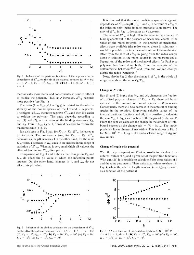
to oxidize the polymer. Thus, as J increases, E00ap becomes
more positive (see Fig. 1)
The ratio (1 � yN,Ox)/(1 � yN,R) is related to the relative
stability of the bound species on the Ox and R segments.
The bigger is yN,Ox the more negative E00ap and then it is easier
to oxidize the polymer. This ratio depends, according to
eqn (1) and (2), on the ratio of the binding constants KOx
and KR. Thus if KOx/KR 4 1, it would be easier to oxidize the
macromolecule (Fig. 2).
It is also seen in Fig. 2 that, for KOx 4 KR, E00ap increases as
pB increases. The converse is true, for KOx o KR, E00ap
decreases as the pB increases. On the other hand, for the same
KOx value, a decrease in KR leads to an increase in the range of
variation of E00ap. When aB is very small (high pB values), the
effect of binding on E00ap disappears.
Comparison of Fig. 1 and 2 shows that changes in KR and
KOx do affect the pB value at which the inflection points
appears. On the other hand, changes in jR and jOx do not
affect this pB value.
It is observed that the model predicts a symmetric sigmoid
dependence of E00ap on pB (Fig. 1 and 2). The value of E00
ap at
the inflexion point being its most probable value (mpv). The
mpv of E00ap in Fig. 1, decreases as J decreases.
The value of E00ap at high pB is the value in the absence of
binding effects but in the presence of mechanical effects. If the
value of the redox potential in the absence of mechanical
effects were available (the redox center alone in solution), it
would be possible to obtain the contribution of the mechanical
effect from the shift of E00ap in going from the redox couple
alone in solution to the redox couple in the macromolecule.
Separation of the redox and mechanical effects for Pani type
polymers has been done both, from the analysis of the
voltammetric behaviour25,27 and from the volume changes
during the redox switching.38
Note, also in Fig. 2, that the change in E00ap in the whole pB
range depends on the ratio KR/KOx.
Change in N with h
Eqn (1) and (2) imply that NOx and NR change as the fraction
of oxidized polymer changes. If KOx 4 KR, there will be an
increase in the amount of bound species as y increases.
Consequently there will be a decrease in the amount of binding
species in the solution. Employing suitable values of the
internal partition functions and M, it is possible to calculate
the sum NOx + NR, as a function of the degree of oxidation, y.From the sum we calculate the change in the amount of total
bound species as the change DN = Ny – Ny=0. The model
predicts a linear change of DN with y. This is shown in Fig. 3
for M = 105, P = 1, JR = 0.2 and a selected range of KR and
KOx values.
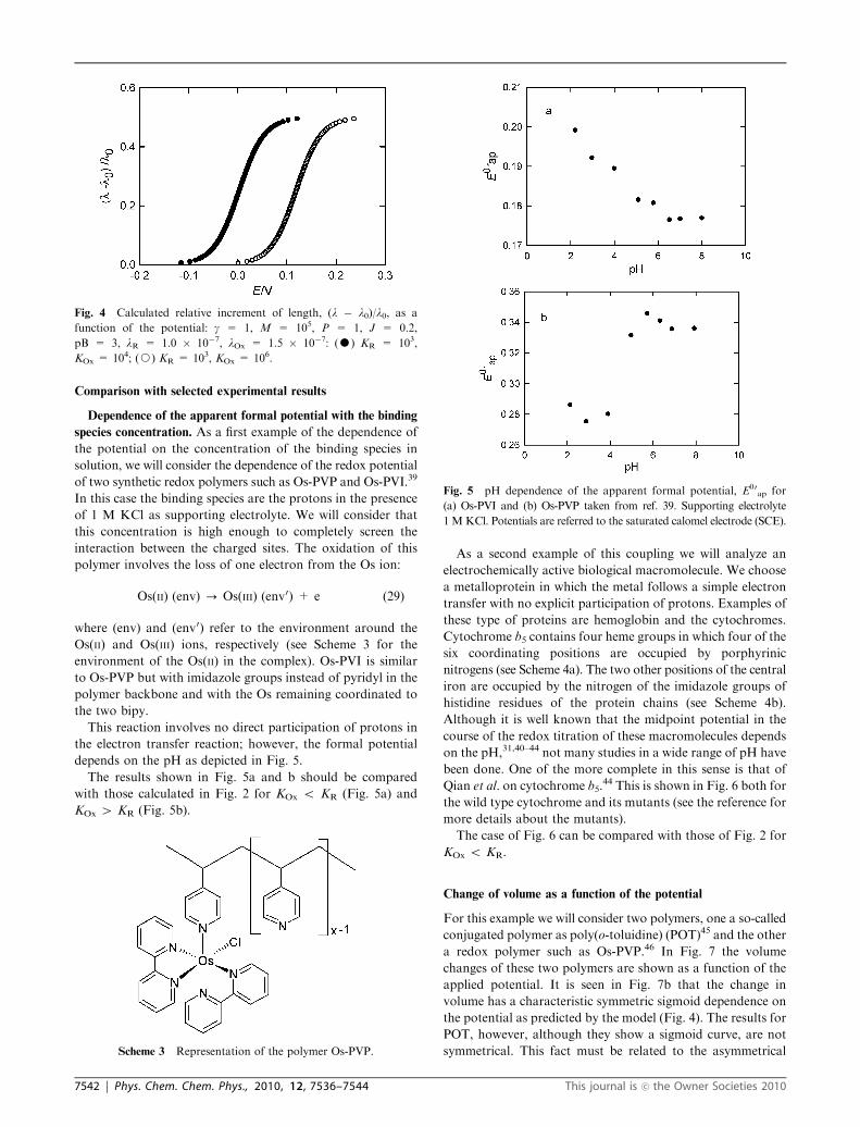
Change of length with potential
With the help of eqn (6) and (9) it is possible to calculate l for
different values of yn and a given set of the partition functions.
With eqn (26) it is possible to calculate E for these values of yand the same parameters. These calculated values are shown in
Fig. 4, where the relative length increase, (l � l0)/l0 is shownas a function of the potential.
Fig. 1 Influence of the partition functions of the segments on the
dependence of E00ap on the pB of the external solution for y = 0.5,
g = 1, P = 1, KR = 103, KOx = 101: (K) J = 0.2, (J) J = 1, (&)
J = 5.
Fig. 2 Influence of the binding constants on the dependence of E00ap
on the pB of the external solution for y = 0.5, g = 1, P = 1, J = 0.2:
(J) KR = 103, KOx = 101; (K) KR = 103, KOx = 104; (n) KR = 101,
KOx = 104; (&) KR = 103, KOx = 106.
Fig. 3 DN as a function of the oxidation fraction, y;M= 105, P= 1,
J = 0.2, g = 1, pB = 3: (K) KR = 101, KOx = 104; (J) KR = 104,
KOx = 101; (&) KR = 101, KOx = 101.
This journal is �c the Owner Societies 2010 Phys. Chem. Chem. Phys., 2010, 12, 7536–7544 | 7541
Comparison with selected experimental results
Dependence of the apparent formal potential with the binding
species concentration. As a first example of the dependence of
the potential on the concentration of the binding species in
solution, we will consider the dependence of the redox potential
of two synthetic redox polymers such as Os-PVP and Os-PVI.39
In this case the binding species are the protons in the presence
of 1 M KCl as supporting electrolyte. We will consider that
this concentration is high enough to completely screen the
interaction between the charged sites. The oxidation of this
polymer involves the loss of one electron from the Os ion:
Os(II) (env) - Os(III) (env0) + e (29)
where (env) and (env0) refer to the environment around the
Os(II) and Os(III) ions, respectively (see Scheme 3 for the
environment of the Os(II) in the complex). Os-PVI is similar
to Os-PVP but with imidazole groups instead of pyridyl in the
polymer backbone and with the Os remaining coordinated to
the two bipy.
This reaction involves no direct participation of protons in
the electron transfer reaction; however, the formal potential
depends on the pH as depicted in Fig. 5.
The results shown in Fig. 5a and b should be compared
with those calculated in Fig. 2 for KOx o KR (Fig. 5a) and
KOx 4 KR (Fig. 5b).
As a second example of this coupling we will analyze an
electrochemically active biological macromolecule. We choose
a metalloprotein in which the metal follows a simple electron
transfer with no explicit participation of protons. Examples of
these type of proteins are hemoglobin and the cytochromes.
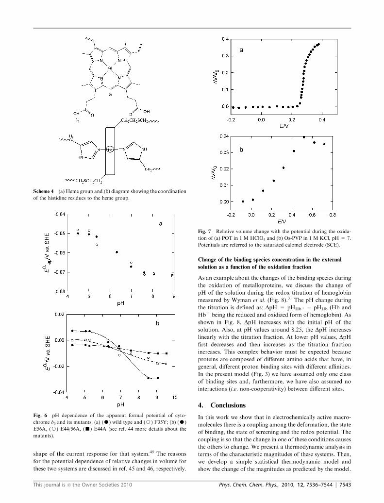
Cytochrome b5 contains four heme groups in which four of the
six coordinating positions are occupied by porphyrinic
nitrogens (see Scheme 4a). The two other positions of the central
iron are occupied by the nitrogen of the imidazole groups of
histidine residues of the protein chains (see Scheme 4b).
Although it is well known that the midpoint potential in the
course of the redox titration of these macromolecules depends
on the pH,31,40–44 not many studies in a wide range of pH have
been done. One of the more complete in this sense is that of
Qian et al. on cytochrome b5.44 This is shown in Fig. 6 both for
the wild type cytochrome and its mutants (see the reference for
more details about the mutants).
The case of Fig. 6 can be compared with those of Fig. 2 for
KOx o KR.
Change of volume as a function of the potential
For this example we will consider two polymers, one a so-called
conjugated polymer as poly(o-toluidine) (POT)45 and the other
a redox polymer such as Os-PVP.46 In Fig. 7 the volume
changes of these two polymers are shown as a function of the
applied potential. It is seen in Fig. 7b that the change in
volume has a characteristic symmetric sigmoid dependence on
the potential as predicted by the model (Fig. 4). The results for
POT, however, although they show a sigmoid curve, are not
symmetrical. This fact must be related to the asymmetrical
Fig. 4 Calculated relative increment of length, (l � l0)/l0, as a
function of the potential: g = 1, M = 105, P = 1, J = 0.2,
pB = 3, lR = 1.0 � 10�7, lOx = 1.5 � 10�7: (K) KR = 103,
KOx = 104; (J) KR = 103, KOx = 106.
Scheme 3 Representation of the polymer Os-PVP.
Fig. 5 pH dependence of the apparent formal potential, E00ap for
(a) Os-PVI and (b) Os-PVP taken from ref. 39. Supporting electrolyte
1 MKCl. Potentials are referred to the saturated calomel electrode (SCE).
7542 | Phys. Chem. Chem. Phys., 2010, 12, 7536–7544 This journal is �c the Owner Societies 2010
shape of the current response for that system.45 The reasons
for the potential dependence of relative changes in volume for
these two systems are discussed in ref. 45 and 46, respectively.
Change of the binding species concentration in the external
solution as a function of the oxidation fraction
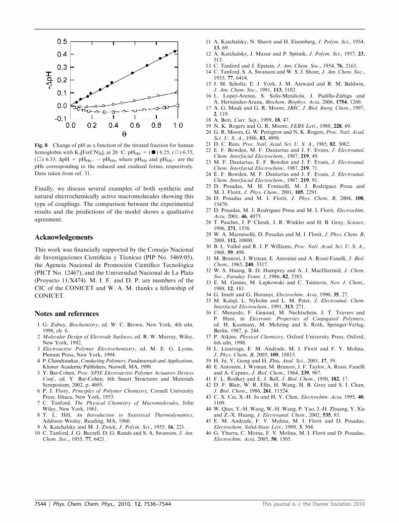
As an example about the changes of the binding species during
the oxidation of metalloproteins, we discuss the change of
pH of the solution during the redox titration of hemoglobin
measured by Wyman et al. (Fig. 8).31 The pH change during
the titration is defined as: DpH = pHHb+ � pHHb (Hb and
Hb+ being the reduced and oxidized form of hemoglobin). As
shown in Fig. 8, DpH increases with the initial pH of the
solution. Also, at pH values around 8.25, the DpH increases
linearly with the titration fraction. At lower pH values, DpHfirst decreases and then increases as the titration fraction
increases. This complex behavior must be expected because
proteins are composed of different amino acids that have, in
general, different proton binding sites with different affinities.
In the present model (Fig. 3) we have assumed only one class
of binding sites and, furthermore, we have also assumed no
interactions (i.e. non-cooperativity) between different sites.
4. Conclusions
In this work we show that in electrochemically active macro-
molecules there is a coupling among the deformation, the state
of binding, the state of screening and the redox potential. The
coupling is so that the change in one of these conditions causes
the others to change. We present a thermodynamic analysis in
terms of the characteristic magnitudes of these systems. Then,
we develop a simple statistical thermodynamic model and
show the change of the magnitudes as predicted by the model.
Scheme 4 (a) Heme group and (b) diagram showing the coordination
of the histidine residues to the heme group.
Fig. 6 pH dependence of the apparent formal potential of cyto-
chrome b5 and its mutants: (a) (K) wild type and (J) F35Y; (b) (K)
E56A, (J) E44/56A, (’) E44A (see ref. 44 more details about the
mutants).
Fig. 7 Relative volume change with the potential during the oxida-
tion of (a) POT in 1 M HClO4 and (b) Os-PVP in 1 M KCl, pH = 7.
Potentials are referred to the saturated calomel electrode (SCE).
This journal is �c the Owner Societies 2010 Phys. Chem. Chem. Phys., 2010, 12, 7536–7544 | 7543
Finally, we discuss several examples of both synthetic and
natural electrochemically active macromolecules showing this
type of couplings. The comparison between the experimental
results and the predictions of the model shows a qualitative
agreement.
Acknowledgements
This work was financially supported by the Consejo Nacional
de Investigaciones Cientıficas y Tecnicas (PIP No. 5469/05),
the Agencia Nacional de Promocion Cientıfico Tecnologica
(PICT No. 12467), and the Universidad Nacional de La Plata
(Proyecto 11/X474). M. I. F. and D. P. are members of the
CIC of the CONICET and W. A. M. thanks a fellowship of
CONICET.
Notes and references
1 G. Zubay, Biochemistry, ed. W. C. Brown, New York, 4th edn,1998, ch. 6.
2 Molecular Design of Electrode Surfaces, ed. R. W. Murray, Wiley,New York, 1992.
3 Electroactive Polymer Electrochemistry, ed. M. E. G. Lyons,Plenum Press, New York, 1994.
4 P. Chandrasekar,Conducting Polymers, Fundamentals and Applications,Kluwer Academic Publishers, Norwell, MA, 1999.
5 Y. Bar-Cohen, Proc. SPIE Electroactive Polymer Actuators DevicesConf., ed. Y. Bar-Cohen, 6th Smart Structures and MaterialsSymposium, 2002, p. 4695.
6 P. J. Flory, Principles of Polymer Chemistry, Cornell UniversityPress, Ithaca, New York, 1953.
7 C. Tanford, The Physical Chemistry of Macromolecules, JohnWiley, New York, 1961.
8 T. L. Hill, An Introduction to Statistical Thermodynamics,Addison–Wesley, Reading, MA, 1960.
9 A. Katchalsky and M. J. Zwick, J. Polym. Sci., 1955, 16, 221.10 C. Tanford, J. G. Buzzell, D. G. Rands and S. A. Swanson, J. Am.
Chem. Soc., 1955, 77, 6421.
11 A. Katchalsky, N. Shavit and H. Eisemberg, J. Polym. Sci., 1954,13, 69.
12 A. Katchalsky, J. Mazur and P. Spitnik, J. Polym. Sci., 1957, 23,513.
13 C. Tanford and J. Epstein, J. Am. Chem. Soc., 1954, 76, 2163.14 C. Tanford, S. A. Swanson and W. S. J. Shore, J. Am. Chem. Soc.,
1955, 77, 6414.15 J. M. Scholtz, E. J. York, J. M. Ateward and R. M. Baldwin,
J. Am. Chem. Soc., 1991, 113, 5102.16 L. Lopez-Arenas, S. Solıs-Mendiola, J. Padilla-Zuniga and
A. Hernandez-Arana, Biochem. Biophys. Acta, 2006, 1754, 1260.17 A. G. Mauk and G. R. Moore, JBIC, J. Biol. Inorg. Chem., 1997,
2, 119.18 A. Bott, Curr. Sep., 1999, 18, 47.19 N. K. Rogers and G. R. Moore, FEBS Lett., 1988, 228, 69.20 G. R. Moore, G. W. Pettigrew and N. K. Rogers, Proc. Natl. Acad.
Sci. U. S. A., 1986, 83, 4998.21 D. C. Rees, Proc. Natl. Acad. Sci. U. S. A., 1985, 82, 3082.22 E. F. Bowden, M. F. Dautartas and J. F. Evans, J. Electroanal.
Chem. Interfacial Electrochem., 1987, 219, 49.23 M. F. Dautartas, E. F. Bowden and J. F. Evans, J. Electroanal.
Chem. Interfacial Electrochem., 1987, 219, 71.24 E. F. Bowden, M. F. Dautartas and J. F. Evans, J. Electroanal.
Chem. Interfacial Electrochem., 1987, 219, 91.25 D. Posadas, M. H. Fonticelli, M. J. Rodriguez Presa and
M. I. Florit, J. Phys. Chem., 2001, 105, 2291.26 D. Posadas and M. I. Florit, J. Phys. Chem. B, 2004, 108,
15470.27 D. Posadas, M. J. Rodriguez Presa and M. I. Florit, Electrochim.
Acta, 2001, 46, 4075.28 T. Pascher, J. P. Chesik, J. R. Winkler and H. B. Gray, Science,
1996, 271, 1558.29 W. A. Marmisolle, D. Posadas and M. I. Florit, J. Phys. Chem. B,
2008, 112, 10800.30 B. L. Vallee and R. J. P. Williams, Proc. Natl. Acad. Sci. U. S. A.,
1968, 59, 498.31 M. Brunori, J. Wyman, E. Antonini and A. Rossi-Fanelli, J. Biol.
Chem., 1965, 240, 3317.32 W. S. Huang, B. D. Humprey and A. J. MacDiarmid, J. Chem.
Soc., Faraday Trans. 1, 1986, 82, 2385.33 E. M. Genies, M. Lapkowski and C. Tsintavis, New J. Chem.,
1988, 12, 181.34 G. Inzelt and G. Horanyi, Electrochim. Acta, 1990, 35, 27.35 M. Kalaji, L. Nyholm and L. M. Peter, J. Electroanal. Chem.
Interfacial Electrochem., 1991, 313, 271.36 C. Menardo, F. Genoud, M. Nechtschein, J. T. Travers and
P. Hani, in Electronic Properties of Conjugated Polymers,ed. H. Kuzmany, M. Mehring and S. Roth, Springer-Verlag,Berlin, 1987, p. 244.
37 P. Atkins, Physical Chemistry, Oxford University Press, Oxford,6th edn, 1998.
38 L. Lizarraga, E. M. Andrade, M. I. Florit and F. V. Molina,J. Phys. Chem. B, 2005, 109, 18815.
39 H. Ju, Y. Gong and H. Zhu, Anal. Sci., 2001, 17, 59.40 E. Antonini, J. Wyman, M. Brunori, J. F. Taylor, A. Rossi. Fanelli
and A. Caputo, J. Biol. Chem., 1964, 239, 907.41 F. L. Rodkey and E. J. Ball, J. Biol. Chem., 1950, 182, 17.42 D. F. Blair, W. R. Ellis, H. Wang, H. B. Gray and S. I. Chan,
J. Biol. Chem., 1986, 261, 11524.43 C. X. Cai, X.-H. Ju and H. Y. Chen, Electrochim. Acta, 1995, 40,
1109.44 W. Qian, Y.-H. Wang, W.-H. Wang, P. Yao, J.-H. Zhuang, Y. Xie
and Z.-X. Huang, J. Electroanal. Chem., 2002, 535, 85.45 E. M. Andrade, F. V. Molina, M. I. Florit and D. Posadas,
Electrochem. Solid-State Lett., 1999, 3, 504.46 G. Ybarra, C. Moina, F. V. Molina, M. I. Florit and D. Posadas,
Electrochim. Acta, 2005, 50, 1505.
Fig. 8 Change of pH as a function of the titrated fraction for human
hemoglobin with K3[Fe(CN)6], at 20 1C: pHHb = (K) 8.25, (J) 6.75,
(&) 6.33; DpH = pHHb+ � pHHb, where pHHb and pHHb+ are the
pHs corresponding to the reduced and oxidized forms, respectively.
Data taken from ref. 31.
7544 | Phys. Chem. Chem. Phys., 2010, 12, 7536–7544 This journal is �c the Owner Societies 2010