Sirt1 contributes critically to the redox-dependent fate of neural progenitors
Upload
independentCategory
view
1download
0
The Addicted Brain Craves New Neurons: Putative Role forAdult-Born Progenitors in Promoting Recovery
Chitra D. Mandyam and George F. KoobCommittee on the Neurobiology of Addictive Disorders, The Scripps Research Institute, La Jolla,CA, USA
AbstractAddiction is a chronic relapsing disorder associated with compulsive drug taking and drug seekingand a loss of control in limiting intake, reflected in three stages of a recurrent cycle: binge/intoxication, withdrawal/negative affect, and preoccupation/anticipation (“craving”). This reviewdiscusses the role of adult-born neural and glial progenitors in drug-seeking associated with thedifferent stages of the addiction cycle. A review of the current literature suggests that the loss ofnewly born progenitors, particularly in hippocampal and cortical regions, may play a role indetermining vulnerability to relapse in rodent models of drug addiction. The normalization ofdrug-impaired neurogenesis or gliogenesis may help reverse neuroplasticity during abstinence, andthus may help reduce the vulnerability to relapse and aid recovery.
Keywords
Prefrontal cortex; Gliogenesis; Hippocampus; neurogenesis; addiction
Introduction
Addiction to drugs of abuse has taken emotional and financial tolls on society, cutting acrossages, races, ethnicities, and genders with increases in mortality, morbidity, and economiccosts. Broadly defined, addiction is a chronic relapsing disorder characterized by acompulsion to seek and take drugs, a loss of control in limiting intake, and emergence of anegative emotional state during withdrawal [1]. The addiction cycle involves elements ofboth impulsivity and compulsivity and is composed of three stages: binge/intoxication,withdrawal/negative affect, and preoccupation/anticipation (craving) (see Glossary). Thestudy of the neurobiological bases of addiction and relapse has significantly progressed, butto date few treatments are known to reverse the drug-induced neuroplasticity changes thatconvey the vulnerability to relapse (see [2] for a review). Understanding the neuroplasticchanges that underlie the relapse stage of addiction can help generate better treatmentoptions for addiction.
© 2011 Elsevier Ltd. All rights reserved.
Correspondence: Chitra D. Mandyam, Ph.D., Committee on the Neurobiology of Addictive Disorders, The Scripps Research Institute,10550 North Torrey Pines Road, SP30-2400, La Jolla, CA 92037 USA; Tel: (858) 784-9039, Fax: (858) 784-2086,[email protected].
Publisher's Disclaimer: This is a PDF file of an unedited manuscript that has been accepted for publication. As a service to ourcustomers we are providing this early version of the manuscript. The manuscript will undergo copyediting, typesetting, and review ofthe resulting proof before it is published in its final citable form. Please note that during the production process errors may bediscovered which could affect the content, and all legal disclaimers that apply to the journal pertain.
NIH Public AccessAuthor ManuscriptTrends Neurosci. Author manuscript; available in PMC 2013 April 1.
Published in final edited form as:Trends Neurosci. 2012 April ; 35(4): 250–260. doi:10.1016/j.tins.2011.12.005.
NIH
-PA
Author M
anuscriptN
IH-P
A A
uthor Manuscript
NIH
-PA
Author M
anuscript

The ability of the brain to continuously generate new progenitors throughout adulthood mayhave important implications for addiction. Broadly defined, progenitors are the progeny ofstem cells characterized by limited self-renewal and can survive and mature intodifferentiating cells, such as neurons and glia, in the brain. There are two main neurogenicareas in the adult brain: the subventricular zone (SVZ) that lines the lateral ventricles in theforebrain, which contain progenitor cells that give rise to neurons in the olfactory bulb, andthe subgranular zone (SGZ) in the dentate gyrus (DG) of the hippocampus that gives rise togranule cell neurons (Figure 1). Neurogenesis has also been shown to occur in the neocortex(medial prefrontal cortex [mPFC]; Figure 2) [3–5]. Emerging evidence suggests thatalterations in the rate of adult neurogenesis and gliogenesis in these brain regions, maycontribute to the regulation of drug taking and drug seeking, particularly in the hippocampusand cortex. This review, therefore, focuses on altered plasticity in these regions that resultfrom drug self-administration and considers recent findings that implicate alterations inneural and glial progenitors in the phenomenology of drug abuse.
Hippocampus and mPFC: Roles in Drug Taking and Seeking
Animal models have been developed that parallel the three stages of the addiction cycle andinclude various paradigms of drug self-administration for the binge/intoxication stage [6],motivational elements of withdrawal for the withdrawal/negative affect stage, and drug-,cue-, context-, and stress-induced reinstatement for the preoccupation/anticipation stage [7].These models have been extensively used to uncover the key brain regions, brain circuitry,neurotransmitters, and neuromodulators associated with drug-taking and - seeking behavior[8, 9]. The ventral striatum (i.e., a brain region that includes the nucleus accumbens [NAc]core and shell and some nuclei of the olfactory tubercle), a terminal projection of the neuralconnections from the ventral tegmental area (VTA) and PFC, is considered a focal point forthe reward and reinstatement associated with drug-seeking behavior [1]. The release of theneurotransmitter dopamine in these regions is considered to be significantly modulated byvarious drugs of abuse, particularly psychostimulants, such as cocaine, methamphetamine,and nicotine, to produce their rewarding effects. Furthermore, evidence indicates thatneurotransmitters other than dopamine may also play a significant role in the rewardingeffects of drugs of abuse, including opioid peptides. The NAc is also tactically situated inthe brain such that it receives inputs from several other brain regions, including the mPFC,basolateral amygdala, insula, and hippocampal regions, and these inputs are hypothesized toplay a key role in cue- and context-specific associations with drugs [1]. Activation ofdopamine, glutamate, and corticotropin-releasing factor systems in these key brain regionsare associated with drug-, cue-, and stress-induced reinstatement, respectively [10, 11].
The hippocampus and mPFC are implicated in the modulation of the reinforcing actions ofdrugs of abuse and play a key role in the reinstatement of drug-seeking behavior [1, 8].Hippocampal integrity may be important for drug-context memories associated with drugreward [12] and is fundamental to the formation of context-specific memories associatedwith the reinstatement of drug seeking [13–16]. This hypothesis is supported by theobservation that activation and inactivation of the hippocampal-subicular and hippocampal-VTA pathways, respectively, enhances and blocks drug-seeking behavior in rodents [14,16]. Thus, it appears that the hippocampal neural plasticity that underlies learning andmemory function also contributes to the modulation of reward pathways in drug addiction[17].
The prelimbic and infralimbic cortices of the mPFC have widespread connections to thebasal forebrain, amygdala, and hypothalamus that mediate diverse functions, includingattentional processing, goal-directed behavior, and working memory [18]. These functionsthat depend on the mPFC play a prominent role in the fundamental pathway that underlies
Mandyam and Koob Page 2
Trends Neurosci. Author manuscript; available in PMC 2013 April 1.
NIH
-PA
Author M
anuscriptN
IH-P
A A
uthor Manuscript
NIH
-PA
Author M
anuscript

drug reinstatement triggered by drug priming, conditioned stimuli, and external stress [8].Pharmacologically, excitatory inputs from the hippocampus and mPFC increase dopaminerelease in the NAc and VTA, which have been hypothesized to contribute to drug seeking inresponse to cues and context following extinction [16, 19–21]. Altogether, the hippocampusand PFC appear to play critical roles in the modulation of the reinforcing effects of drugs,and the release of neurotransmitters from key brain regions associated with cues and contextare hypothesized to be essential components of the human condition of craving.
Additionally, it is well established that the neural connections from the CA1 and subiculumof the hippocampus that terminate in the prelimbic cortex of the mPFC function to facilitatethe acquisition, maintenance, and independent storage and consolidation of declarative,spatial, and associative long-term memories [22]. The mPFC and hippocampus interactunder a variety of functional demands, including drug taking. Such interactions may dependon both memory processes and content [1, 23] and may be critical for optimal performance[24]. Therefore, excessive drug use during abuse and addiction could compromise normallearning and memory systems and, in addition to activation of reward pathways associatedwith craving, may also disrupt executive control pathways, thus contributing to theimpulsivity and impairment of decision making characteristic of individuals with addiction[25].
Hippocampal Neurogenesis, Drug Taking, and Drug Seeking
One possible contributor to hippocampal neural plasticity is adult neurogenesis in the SGZof the DG. The process of neurogenesis involves stem-like precursor cells that proliferateinto preneuronal progenitors, which in turn differentiate into immature neurons andeventually mature into granule cell neurons [26] (Figure 1). It is widely acknowledged that alarge proportion (> 80%) of hippocampal progenitors migrate a short distance to becomegranule neurons in the DG [27]. Furthermore, there is sufficient evidence that supports thefunctional incorporation of the newly born neurons in the DG [28–30], although thehypothesis concerning the role of new DG neurons on sparse coding (facilitation of theformation of new hippocampus-dependent memories) awaits confirmation [31–33]. NewDG neurons are also involved in certain aspects of addiction. Reinforcing doses of drugsself-administered by rodents decrease DG neurogenesis [34–43] (Table 1). It has beenhypothesized that new DG neurons may block memories associated with the contextualreinstatement of drug seeking or enhance extinction learning [44, 45]. Thus, the reduction inspontaneous neurogenesis (i.e., a reduction in neuronal turnover) that is observed after self-administration of various drugs of abuse may result in a more robust and long-lastingmemory of drug taking and seeking or decrease extinction learning. However, additionalexperiments are needed before the precise function of DG neurogenesis in drug taking andseeking is determined [14, 46]
The developmental stages of DG progenitors and the lineage for adult-generated DGneurons have been determined by utilizing transgenic mice that express nestin (a type VIintermediate filament protein that is highly expressed in neural progenitor cells) under thecontrol of a green fluorescent protein (GFP) promoter (Figure 1B). Data from mouse studiesindicate that proliferating cells in the postnatal DG are not homogeneous, and the process ofpostnatal neurogenesis is an uncoordinated cluster of developmental stages that progress inparallel, including actively dividing cells that are radial glia-like (type1), preneuronal(type2a), intermediate (type2b), and early neuronal (type3) [47] (Figure 1B). Importantly,pools of slowly dividing type1 cells appear to be the precursors of adult-generated DGneurons. Furthermore, the distinct cell types have been individually labeled usingcombinations of exogenous (5-bromo-2’-deoxyuridine [BrdU]; Box 1 and Figure 2B) andendogenous (Ki-67, proliferating cell nuclear antigen, phosphorylated histone-H3, sex-
Mandyam and Koob Page 3
Trends Neurosci. Author manuscript; available in PMC 2013 April 1.
NIH
-PA
Author M
anuscriptN
IH-P
A A
uthor Manuscript
NIH
-PA
Author M
anuscript

determining region Y-box 2 [Sox2]) markers of cell proliferation and cell differentiation(doublecortin [DCX], neurogenic differentiation factor 1 [NeuroD1], and polysialic acid-neural cell adhesion molecule [PSA-NCAM]) [47, 48]. By incorporating these exogenousand endogenous markers of proliferation and differentiation, critical information has beenobtained in adult rat and nonhuman primate models to demonstrate a similar developmentalprofile of DG progenitors compared with mouse models.
Recent studies have focused on how drugs of abuse alter the process of DG neurogenesis bymodifying the developmental stages of newly born adult DG progenitors and their pathwayto attain a neuronal phenotype [47] (Table 1). For example, limited-access nicotine self-administration decreases the proliferation and differentiation of DG progenitors [36].Extended-access heroin self-administration, which results in compulsive drug seeking,decreases the proliferation of DG progenitors [38]. Limited- and extended-access cocaineself-administration decreases the proliferation of DG progenitors [39, 43]. Furthermore,extended access to cocaine increases differentiation without altering the survival ofprogenitors [39]. Limited- and extended-access methamphetamine self-administrationdecreases the proliferation, differentiation, and survival of DG progenitors [35].Intermittent-access methamphetamine self-administration, surprisingly, increases theproliferation and differentiation of DG progenitors, but such an increase in the immatureneuronal population was not associated with an increase in neurogenesis [35].Nondependent ethanol self-administration studied in both rodent and nonhuman primatemodels decreased the proliferation, differentiation, and survival of DG progenitors [37, 48,49]. Excessive drinking during alcohol dependence was also found to decrease all aspects ofDG neurogenesis [37, 50]. Altogether, certain neuromodulatory effects on neurogenesisappear to be particularly sensitive to the amount of daily drug intake and that a higheramount of drug intake produces more pronounced effects on DG neurogenesis.
Role of Dentate Gyrus Neurogenesis in Drug Taking and Seeking
Mechanistic approaches to address potential roles for adult-generated DG neurons in drugtaking and drug seeking are currently being undertaken. For example, procedures such aslow doses of irradiation have been used to ablate proliferating progenitors in the DG, andchanges in the animal’s environment (e.g., voluntary wheel running in the home cage) havebeen used to enhance the proliferation and maturation of hippocampal progenitors to providea functional link between hippocampal neurogenesis and hippocampal-dependent memory[51]. Similarly, irradiation and wheel running have been used to examine the relationshipbetween hippocampal neurogenesis and drug taking and seeking. Studies in rats that haveused irradiation to ablate hippocampal neurogenesis before any cocaine experience havedemonstrated that irradiated rats had enhanced cocaine-taking behavior, reflected byincreased self-administration on a fixed-ratio schedule of reinforcement in an extended-access model compared with non-irradiated rats [46]. In other studies, voluntary wheelrunning before and during cocaine and methamphetamine experience reduced themaintenance of drug-reinforced behavior [52–55] and positive-reinforcing effects of cocainecompared with sedentary animals [56]. Voluntary wheel running before and during ethanolexperience reduced ethanol self-administration [57] and diminished intoxicated behavioralresponses to heavy binge ethanol administration compared with sedentary controls [58].Such studies did not measure neurogenesis in the hippocampus, which would be needed tocorrelate DG neurogenesis with the drug-induced behavioral outcomes. However, some ofthe effects of wheel running on drug taking may be attributable to the neuromodulatoryeffects of running on DG neurogenesis [59]. Thus, increased hippocampal neurogenesis maybe beneficial in diminishing the motivational impact of drugs after chronic exposure to drugsof abuse.
Mandyam and Koob Page 4
Trends Neurosci. Author manuscript; available in PMC 2013 April 1.
NIH
-PA
Author M
anuscriptN
IH-P
A A
uthor Manuscript
NIH
-PA
Author M
anuscript

The effects on DG proliferation, differentiation, and neurogenesis after drug withdrawal orreinstatement to drug seeking after abstinence have been less well-studied. The studies thathave addressed this issue have found that withdrawal from cocaine self-administrationdecreased the proliferation and enhanced the differentiation and maturation of DGprogenitors compared with control animals [39, 60]. Although much more work is requiredto support the hypothesis of the enhanced survival of DG progenitors during withdrawalfrom drug exposure, one could propose that the abnormal survival of progenitors duringwithdrawal from the drug could be a part of the recovery process. Consistent with thishypothesis, other studies suggested that the degree of neurogenesis in the DG may affectbehavioral responses after drug withdrawal. Ablation of DG progenitors by irradiationduring withdrawal from cocaine self-administration delayed the extinction of cocaine-seeking behavior in rats [46]. Furthermore, voluntary wheel running before and duringextinction reduced drug-primed [61, 62] and cue-induced [62, 63] cocaine-seeking behaviorin rats after a period of forced abstinence. The levels of neurogenesis in the hippocampuswere not measured in these studies, but, as discussed above, such findings suggest thatenhanced neurogenesis after wheel running may be an underlying mechanism for such areduction in drug-induced responses after withdrawal. Other studies have also indicated abeneficial role for hippocampal neurogenesis in reducing the vulnerability to relapse. Forexample, rats genetically inbred for high novelty-seeking behavior and rats exposed to earlyenvironmental stress (e.g. prenatal stress) exhibited reduced DG neurogenesis [42, 64, 65]and were prone to developing addictive behaviors [66–68]. Although more studies areneeded, adult DG neurogenesis appears to be important for the maintenance of hippocampalneuroplasticity, such that reducing spontaneous DG neurogenesis during abstinence mayenhance the vulnerability to relapse, and enhancing DG neurogenesis during abstinence mayhelp reduce the vulnerability to relapse.
Role of Olfactory Bulb Neurogenesis in Addiction
Neurogenesis in the olfactory bulb contributes to olfactory function [69], and deficits inolfactory function and sensitivity are evident in drug and alcohol dependent humans [70, 71]and these behavioral deficits may predict the propensity to relapse [72]. Recent studies havedemonstrated that chronic cocaine self-administration and exposure to chronic ethanolvapors in rodents (models of drug dependence) reduce the proliferation of neural progenitorsin the SVZ, a source of adult-generated olfactory neurons [39, 50]. However, protractedwithdrawal from chronic drug exposure normalizes proliferation in the SVZ, albeitproducing permanent changes in the SVZ neurogenic niche [39, 50]. Therefore, futurestudies should address whether drug-induced decreases in SVZ proliferation andconsequential decreases in olfactory bulb neurogenesis produce drug-induced deficits inolfaction.
Role for Glia in Modulating Neuroplasticity Responses in the Brain
Glial fibrillary acidic protein (GFAP)- and non-GFAP-positive glia have received someattention in the past decade with respect to their possible roles in addiction. GFAP-glia inthe mPFC contain cystine/glutamate antiporters [73] that maintain extracellular nonsynapticglutamate levels [74] and provide physical support to neurons by regulating extracellularpotassium and the uptake of glutamate at synapses [75]. The glutamate released by GFAP-positive glia antiporters is known to modulate neuronal metabotropic glutamate receptorsand extracellular glutamate and dopamine levels in areas other than the mPFC [74]. Thus,this is one way that GFAP-positive glia can influence local synaptic activity [76]. Althoughneuroadaptations in mPFC GFAP-glia have not yet been clearly indicated in thereinstatement of drug seeking [74], it is a hypothesis that merits testing.
Mandyam and Koob Page 5
Trends Neurosci. Author manuscript; available in PMC 2013 April 1.
NIH
-PA
Author M
anuscriptN
IH-P
A A
uthor Manuscript
NIH
-PA
Author M
anuscript

NG2 chondroitin sulphate proteoglycan (NG2)-positive glia (non-GFAP-positive glia)express AMPA receptors [77], making them antigenically distinct from GFAP-glia [75, 78].These cells are known to play important roles in the nervous system and may in certainenriched environments support the neurogenesis of cortical progenitors [79–81]. NG2-gliaare involved in the induction and expression of long-term potentiation (LTP) at neuron-gliasynapses [77]. Moreover, NG2-positive glia contain voltage-gated ion channels [82] thatassist with maintaining the homeostatic function of surrounding neurons in the hippocampus[77]. Other important roles of NG2-glia include draining excess ions and neurotransmittersfrom the extracellular space, and this activity may be important for maintaining theproliferative environment and activity of surrounding neurons [83]. Therefore, NG2-glia arelikely to play a more prominent role in maintaining both synaptic neuromodulatoryresponses and neuroplastic responses in the adult brain after brain insults, such as addictionand relapse.
Role of Dentate Gyrus Gliogenesis in Addiction
In addition to the neuronal network that mediates most of the neuromodulatory effectsassociated with drug taking and drug seeking, glia-mediated nonsynaptic events, includingsupportive/maintenance roles, also appear to be important. In the DG, more than 10% ofadult-born progenitors mature into glia, mostly into astroglia that express GFAP [84].Notably, drugs that are reinforcing in vivo do not alter the number or proportion of DGprogenitors that mature into GFAP-positive glia [35–39]. However, newly born DGmicroglia may play a role in drug withdrawal [85, 86]. Recent studies have demonstratedthat withdrawal from ethanol produces immediate exaggerated microglial proliferation in theDG and several other hippocampal regions [85, 86]. These studies suggest that thehippocampus in alcohol-dependent animals is greatly susceptible to inflammation, and suchpro-inflammatory events (i.e., microglial proliferation) following withdrawal may, in turn,affect DG neurogenesis and the optimal function of the hippocampus [87].
Role of mPFC Gliogenesis in Addiction
It has been demonstrated, using combinatorial labeling techniques, that a large proportion ofprogenitors in the adult mammalian mPFC, including the human PFC, mature into a glialphenotype (Figure 2A, B; regulated intramembrane proteolysis-positive [RIP+]oligodendrocyte, NG2+ oligodendrocyte precursor, and/or GFAP+ astrocyte), and a smallproportion mature into interneurons [3–5]. The distribution of progenitors in the mPFC issuggested to be uniform, and most of the cells in the mPFC (> 50%) mature intooligodendrocyte precursors [4, 5, 88–90] (Fig. 2C). External factors have been shown toboth positively (e.g., antidepressants, wheel running) and negatively (e.g., stress, drugs ofabuse, ethanol) regulate cell birth and cell maturity in the mPFC [4, 37, 88, 90–94].
Intravenous self-administration of methamphetamine decreased (with limited and extendedaccess) and increased (with intermittent access) the birth and survival of mPFC progenitors[4] (Table 2), whereas intermittent, limited, and extended access to methamphetamineincreased cell death in the mPFC [4]. Such findings support altered mPFC plasticity aftermethamphetamine exposure. Moreover, they indicate that the effects of methamphetamineon cell death (which includes both neurons and glia) are distinct compared with the changesobserved with cell birth (i.e., neurogenesis is observed to a much lesser extent in the mPFCcompared with gliogenesis). The progenitors reduced by limited and extended access wereGFAP-positive and NG2-positive glia in the mPFC, whereas most of the progenitorsinduced by intermittent access were phenotyped as NG2-positive glia [4]. Althoughevidence suggests that increases in GFAP-positive glia occur in almost all injury typesrestricted to sites of neuronal loss [95], normal levels of GFAP-positive glia and enhanced
Mandyam and Koob Page 6
Trends Neurosci. Author manuscript; available in PMC 2013 April 1.
NIH
-PA
Author M
anuscriptN
IH-P
A A
uthor Manuscript
NIH
-PA
Author M
anuscript

NG2-positive glial levels after intermittent access to methamphetamine could indicateotherwise. For example, an enhanced NG2-glial response could indicate a protectivemechanism (i.e., characteristic central nervous system [CNS] gliosis) againstmethamphetamine-induced brain insult [96]. The rapid response of increased NG2-gliaproliferation after intermittent methamphetamine insult could be attributable to alteredglutamate release locally by glutamatergic neurons following methamphetamine exposure.This is because NG2-glial differentiation and the response to CNS insult are mediated byglutamate released by neurons and GFAP-positive glia [97]. An increase in the number ofNG2-positive glia that express AMPA receptors may further produce alterations inextracellular glutamate in the mPFC, which could contribute to the neuromodulatory effectsthat occur via the mPFC in the other brain regions (e.g., the NAc and VTA) associated withthe binge/intoxication stage of the addiction cycle. The decrease in GFAP-positive andNG2-positive glia after prolonged limited and extended access to methamphetamine couldbe indicative of the neurotoxic effects of methamphetamine.
Exposure to ethanol vapors after excessive drinking (i.e., operant ethanol self-administrationin rodents followed by intermittent exposure to ethanol vapors over several weeks) resultedin a decrease in the birth and survival of mPFC progenitors [37]. Nondependent ethanol-drinking rats did not exhibit significant changes in cell birth and cell survival in the mPFC.Cell death was differentially regulated in both nondependent ethanol-drinking and alcohol-dependent rats, and both groups showed significantly decreased apoptosis [44], indicating acompensatory state in the mPFC in response to excessive ethanol exposure.
What can be gleaned from these two studies is that psychostimulants and ethanol alter thelocal homeostasis of the proliferative environment in the mPFC by decreasing the birth ofnewly born cells and increasing the death of existing older cells. Therefore, the involvementof the PFC in several aspects of an addiction phenotype, including an association withclassical conditioning to drug exposure [98] (a phenomenon associated with relapse to drugseeking), and the response inhibition behavior maintained by the PFC may involve a keyrole of newly generated progenitors.
In addition to these correlative studies, a few studies have demonstrated that an altered localmicroenvironment in the PFC contributes to the neuroadaptations associated with thereinstatement of drug seeking. Recent studies have implicated reduced brain-derivedneurotrophic factor (BDNF) levels [99] and increased phosphorylated extracellular signal-regulated kinase (pERK) [100] in the mPFC in cocaine craving and cocaine-associatedmemories that activate the neuronal circuitry associated with relapse. Particularly notable,wheel running during forced abstinence prevented some of the mPFC neuroadaptations,such as increases in pERK in the mPFC, and reduced cocaine craving and the reinstatementof cocaine seeking [63]. An independent study demonstrated that wheel running increasedNG2- and GFAP-positive glia in the mPFC [4], although such regulation has yet to bedemonstrated during forced abstinence after drug taking. Altogether, adult corticalgliogenesis appears to be altered after exposure to drugs of abuse; therefore, normalizinggliogenesis in the mPFC during abstinence may help restore some of the maladaptiveneuroplastic alterations in response to drug addiction.
Therapeutic Implications: Can Enhancing Neurogenesis Promote
Functional Recovery in the Addicted Brain?
Voluntary exercise appears to decrease relapse to drug seeking in some cases. The impact ofvoluntary exercise on abstinence from drug use has been studied in humans with regard tonicotine [101–103] and alcohol [104] addiction, with some of these studies indicating abeneficial effect. With regard to animal models of addiction, only a few studies have
Mandyam and Koob Page 7
Trends Neurosci. Author manuscript; available in PMC 2013 April 1.
NIH
-PA
Author M
anuscriptN
IH-P
A A
uthor Manuscript
NIH
-PA
Author M
anuscript

assessed whether exercise after the cessation of drug use can reduce the reinstatement ofdrug-seeking behavior [61–63]. However, as discussed above, none of these studiesaddressed whether the decrease in the reinstatement of drug-seeking behavior after wheelrunning during forced abstinence was caused by a reversal of drug-induced neuroplasticevents, such as increased neurogenesis and gliogenesis. Although one could reasonablyhypothesize that this could be the mechanism, mechanistic studies that prove otherwise arecurrently lacking.
As discussed in the previous sections, despite the reduction of neurogenesis and gliogenesisin animal models of addiction that may contribute to the addiction process, the addictedbrain responds to environmental factors that stimulate the neurogenic/gliogenic niche toreduce drug seeking [63, 105–108]. These studies also suggest that the hippocampus andmPFC of the addicted brain retain microenvironmental elements that revive the normalproliferation and survival of neural and glial progenitors during withdrawal. Therefore,knowledge about the cellular and molecular mechanisms that maintain the neurogenic nichein vivo should be beneficial for the design of new therapeutic strategies to augmentendogenous neural proliferation during abstinence.
Recent work in this field has also demonstrated how the neurogenic niche can be the sourceof extrinsic factors that promote the maturation and survival of newly born DG progenitors(reviewed in [109]). Particularly interesting is the fact that monoamines (e.g., dopamine)play an important role in maintaining and enhancing the proliferation and maturation ofhippocampal progenitors [110, 111]. This suggests that the drug-induced consequences oflong-term decreases in dopamine function associated with addiction could play a significantrole in drug-induced alterations in hippocampal neurogenesis. Other mechanistic studies(reviewed in [109]) have shown that certain transcription factors intrinsic to progenitorscould undergo chromatin remodeling, including histone modifications. Such epigeneticmodifications could control certain aspects of adult neurogenesis, such as the transition fromproliferation to differentiation. Furthermore, radial glia-like stem cell-derived intrinsicfactors in the neurogenic niche have been demonstrated to have nourishing and instructiveeffects that promote certain stages of adult neurogenesis [112–114]. Functional studies, suchas the knockdown of such intrinsic factors, including wingless-type MMTV integration sitefamily3 protein (WNT3) in the DG, have shown decreased neurogenesis and impairedhippocampal-dependent memory [115], supporting a role for the neurogenic niche inhippocampal function.
Other intrinsic factors, such as Notch [116–118], disrupted-in-schizophrenia1 (DISC1) [119,120], and cyclin-dependent kinase (Cdk5) [121, 122], are also known to regulate the stemcell pool and affect the differentiation/maturation of hippocampal progenitors. However, itremains to be determined if the efficacy of neuronal development can be augmented bymanipulating the hippocampal microenvironment, such as by enhancing the expression ofthe molecular targets that underlie the maintenance of the neurogenic niche in the addictedbrain. Therefore, the goal of such a therapeutic approach would be to first determinewhether drugs of abuse produce epigenetic changes in mature neurons or induce molecularchanges in progenitor cells to inhibit neurogenesis [109]. Follow-up studies could thendetermine whether normalizing the neurogenic and gliogenic niche by enhancing/reducingintrinsic molecular signals during abstinence contributes to enhanced endogenousneurogenesis and reduced drug seeking. Recruitment of the endogenous progenitor cellpopulation during the protracted abstinence/withdrawal stage may then assist in reversingthe altered neuroplasticity that occurs not only in neurogenic regions, but also throughoutthe many other brain regions that are known to be affected after addiction to drugs of abuse.
Mandyam and Koob Page 8
Trends Neurosci. Author manuscript; available in PMC 2013 April 1.
NIH
-PA
Author M
anuscriptN
IH-P
A A
uthor Manuscript
NIH
-PA
Author M
anuscript
Summary and Future Directions
The identification of adult neurogenesis in the hippocampus, cortex. and olfactory bulb, inthe past two decades has shed new light on the neuroplasticity of these brain regions and,more recently, the plasticity events that are associated with drug and alcohol addiction.Future studies aimed at understanding the potential link between correlative decreases inneurogenesis and the function of these brain regions will allow us to determine whetherdecreases in neuro- and glio- genesis by drugs of abuse is behaviorally relevant to theprocess of addiction. Demonstrating correlative changes in hippocampal/prefrontal cellgenesis associated with the reward and relapse phases of addiction in humans will supportthe hypothesis that adult neurogenesis is a vulnerability factor for addiction. This may pavethe way for future therapeutic possibilities for treating drug addiction disorders that involveenhancing neural stem cell production and the functional incorporation of new neurons intoaffected neural circuits (Box 2).
Box 1. Detection of Adult Born Progenitors, Adult Neurogenesis, andGliogenesis
Actively dividing progenitors in the adult mammalian brain are usually labeled withexogenously administered mitotic markers, such as [3H]thymidine, 5-bromo-2’-deoxyuridine (BrdU), 5-iodo-2’-deoxyuridine (IdU), 5-chloro-2’-deoxyuridine, and 5-ethynyl-2’-deoxyuridine (EdU) [123–126]. Exogenous mitotic markers are incorporatedinto DNA during the synthesis-(S)-phase of the cell cycle of actively dividing progenitorsin the brain, thereby assisting with birth dating the cells. The time (hours to days tomonths) of euthanasia after a pulse of the mitotic marker (usually administeredintraperitoneally or intravenously) determines the age of the progenitor cell whenanalyzed in post mortem tissue. For example, a BrdU pulse minutes to hours beforeeuthanasia will label proliferating cells, and a BrdU pulse days to months beforeeuthanasia will label surviving cells, thereby allowing the characterization of the kinetics,dynamics, and phenotype acquisition of newly born progenitors (Figures 1B and 2B).More recently, endogenous markers of cell proliferation and cell differentiation, such asKi-67, proliferating cell nuclear antigen (PCNA), phosphorylated histone-H3, sex-determining region Y-box 2 (Sox2), doublecortin (DCX), neurogenic differentiationfactor 1 (NeuroD1), polysialic acid-neural cell adhesion molecule (PSA-NCAM), andseveral others, have been used to label progenitors at distinct stages of maturation [127–129]. Combinatorial labeling with exogenous and endogenous markers has providedcritical information about the morphological and functional development of progenitorsin the adult brain.
Box 2. Outstanding questions
• Do epigenetics play a role in mediating alterations in hippocampal neurogenesisinduced by drugs of abuse?
• Do drugs of abuse distinctly alter intrinsic signals that maintain neurogenesis vs.gliogenesis?
• Can imaging human neurogenesis become feasible with improved technology?
• Does reduced hippocampal neurogenesis by drugs of abuse lead to cognitiveimpairments in human addicts?
• Do current therapeutic strategies in humans that incorporate physical activityduring abstinence alter neurogenesis and gliogenesis to reduce craving?
Mandyam and Koob Page 9
Trends Neurosci. Author manuscript; available in PMC 2013 April 1.
NIH
-PA
Author M
anuscriptN
IH-P
A A
uthor Manuscript
NIH
-PA
Author M
anuscript

• Do current or novel therapeutic strategies in humans that alter neurogenesis andgliogenesis reduce craving?
AcknowledgmentsPreparation of this review was supported by funds from the National Institute on Drug Abuse (DA022473 to C. D.M. and DA004398, DA023597, and DA010072 to G.F.K.) and National Institute on Alcohol Abuse andAlcoholism (AA008459 and AA006420 to G.F.K.). We appreciate the editorial assistance of Michael Arends. Thisis publication number 21090 from The Scripps Research Institute.
Glossary
Addiction Chronic relapsing disorder characterized by a compulsion to seekand take drugs, loss of control in limiting intake, and emergence ofa negative emotional state during withdrawal.
Compulsivity Behaviorally defined as the persistent reinitiation of habitual acts inresponding in the face of incorrect responses in choice situations orpreservation of responding in the face of adverse consequences.
Drug Craving Memory of the pleasant aspects of a drug superimposed on anegative emotional state.
Drug Dependence In animal models, manifestation of a withdrawal syndrome.
Drug Relapse Reinstatement of drug seeking in previously detoxified individuals.This can occur in individuals after detoxification and long periodsof abstinence, despite sincere efforts to refrain; behavior can beprovoked by stress, cues, or contexts previously associated withdrug use.
Impulsivity Behaviorally defined as a predisposition toward rapid, unplannedreactions to internal and external stimuli without regard fornegative consequences.
NegativeReinforcement
A process by which removal of an aversive stimulus (e.g., negativeemotional state of drug withdrawal or a footshock) increases theprobability of a response.
Place Conditioning A procedure whereby the effects of drugs are paired with distinctenvironments, and the animal changes its subsequent preference forthat distinct environment.
PositiveReinforcement
A process by which presentation of a stimulus (usually pleasant;e.g., pleasurable effects of a drug) increases the probability of aresponse.
Reinstatement ofDrug Seeking
Animal model that investigates relapse to drug seeking. Learnedself-administration behavior is extinguished by explicit nonreward,and subjects are later tested for their ability to reinstate drug-seeking behavior (e.g., lever pressing in the operant chamber) inresponse to a priming stimulus (i.e., drug, cue, context, or stressor).
Self-Administration
Arbitrary instrumental action, such as lever pressing, to gain accessto positive reinforcers, such as food or drugs of abuse.
Mandyam and Koob Page 10
Trends Neurosci. Author manuscript; available in PMC 2013 April 1.
NIH
-PA
Author M
anuscriptN
IH-P
A A
uthor Manuscript
NIH
-PA
Author M
anuscript

References
1. Koob GF, Volkow ND. Neurocircuitry of addiction. Neuropsychopharmacology. 2010; 35:217–238.[PubMed: 19710631]
2. Kalivas PW, Volkow ND. New medications for drug addiction hiding in glutamatergicneuroplasticity. Mol Psychiatry. 2011; 16:974–986. [PubMed: 21519339]
3. Gould E, et al. Neurogenesis in the neocortex of adult primates. Science. 1999; 286:548–552.[PubMed: 10521353]
4. Mandyam CD, et al. Methamphetamine self-administration and voluntary exercise have opposingeffects on medial prefrontal cortex gliogenesis. J Neurosci. 2007; 27:11442–11450. [PubMed:17942739]
5. Dayer AG, et al. New GABAergic interneurons in the adult neocortex and striatum are generatedfrom different precursors. J Cell Biol. 2005; 168:415–427. [PubMed: 15684031]
6. Caine, SB., et al. Intravenous drug-self administration techniques in animals. Oxford UniversityPress; 1993.
7. Rossi NA, Reid LD. Affective states associated with morphine injections. PhysiologicalPsychology. 1976; 4:269–274.
8. Shaham Y, et al. The reinstatement model of drug relapse: history, methodology and major findings.Psychopharmacology (Berl). 2003; 168:3–20. [PubMed: 12402102]
9. McFarland K, Kalivas PW. The circuitry mediating cocaine-induced reinstatement of drug-seekingbehavior. J Neurosci. 2001; 21:8655–8663. [PubMed: 11606653]
10. Koob GF. Stress, corticotropin-releasing factor, and drug addiction. Ann N Y Acad Sci. 1999;897:27–45. [PubMed: 10676433]
11. Knackstedt LA, Kalivas PW. Glutamate and reinstatement. Curr Opin Pharmacol. 2009; 9:59–64.[PubMed: 19157986]
12. Black YD, et al. Hippocampal memory system function and the regulation of cocaine self-administration behavior in rats. Behav Brain Res. 2004; 151:225–238. [PubMed: 15084439]
13. Fuchs RA, et al. The role of the dorsomedial prefrontal cortex, basolateral amygdala, and dorsalhippocampus in contextual reinstatement of cocaine seeking in rats. Neuropsychopharmacology.2005; 30:296–309. [PubMed: 15483559]
14. Vorel SR, et al. Relapse to cocaine-seeking after hippocampal theta burst stimulation. Science.2001; 292:1175–1178. [PubMed: 11349151]
15. Hernandez-Rabaza V, et al. The hippocampal dentate gyrus is essential for generating contextualmemories of fear and drug-induced reward. Neurobiol Learn Mem. 2008; 90:553–559. [PubMed:18644245]
16. Luo AH, et al. Linking context with reward: a functional circuit from hippocampal CA3 to ventraltegmental area. Science. 2011; 333:353–357. [PubMed: 21764750]
17. Hyman SE, et al. Neural mechanisms of addiction: the role of reward-related learning and memory.Annu Rev Neurosci. 2006; 29:565–598. [PubMed: 16776597]
18. Gabbott PL, et al. Prefrontal cortex in the rat: projections to subcortical autonomic, motor, andlimbic centers. J Comp Neurol. 2005; 492:145–177. [PubMed: 16196030]
19. Floresco SB, et al. Glutamatergic afferents from the hippocampus to the nucleus accumbensregulate activity of ventral tegmental area dopamine neurons. J Neurosci. 2001; 21:4915–4922.[PubMed: 11425919]
20. Taepavarapruk P, et al. Hyperlocomotion and increased dopamine efflux in the rat nucleusaccumbens evoked by electrical stimulation of the ventral subiculum: role of ionotropic glutamateand dopamine D1 receptors. Psychopharmacology (Berl). 2000; 151:242–251. [PubMed:10972471]
21. Hiranita T, et al. Suppression of methamphetamine-seeking behavior by nicotinic agonists. ProcNatl Acad Sci U S A. 2006; 103:8523–8527. [PubMed: 16717181]
22. McDonald RJ, White NM. A triple dissociation of memory systems: hippocampus, amygdala, anddorsal striatum. Behav Neurosci. 1993; 107:3–22. [PubMed: 8447956]
Mandyam and Koob Page 11
Trends Neurosci. Author manuscript; available in PMC 2013 April 1.
NIH
-PA
Author M
anuscriptN
IH-P
A A
uthor Manuscript
NIH
-PA
Author M
anuscript

23. Simons JS, Spiers HJ. Prefrontal and medial temporal lobe interactions in long-term memory. NatRev Neurosci. 2003; 4:637–648. [PubMed: 12894239]
24. Churchwell JC, et al. Prefrontal and hippocampal contributions to encoding and retrieval of spatialmemory. Neurobiol Learn Mem. 2010; 93:415–421. [PubMed: 20074655]
25. Koob GF, Le Moal M. Drug abuse: hedonic homeostatic dysregulation. Science. 1997; 278:52–58.[PubMed: 9311926]
26. Abrous DN, et al. Adult neurogenesis: from precursors to network and physiology. Physiol Rev.2005; 85:523–569. [PubMed: 15788705]
27. Kaplan MS, Hinds JW. Neurogenesis in the adult rat: electron microscopic analysis of lightradioautographs. Science. 1977; 197:1092–1094. [PubMed: 887941]
28. Gould E, et al. Learning enhances adult neurogenesis in the hippocampal formation. Nat Neurosci.1999; 2:260–265. [PubMed: 10195219]
29. Shors TJ, et al. Neurogenesis may relate to some but not all types of hippocampal-dependentlearning. Hippocampus. 2002; 12:578–584. [PubMed: 12440573]
30. Aimone JB, et al. Potential role for adult neurogenesis in the encoding of time in new memories.Nat Neurosci. 2006; 9:723–727. [PubMed: 16732202]
31. Deng W, et al. New neurons and new memories: how does adult hippocampal neurogenesis affectlearning and memory? Nat Rev Neurosci. 2010; 11:339–350. [PubMed: 20354534]
32. Aimone JB, et al. Resolving new memories: a critical look at the dentate gyrus, adult neurogenesis,and pattern separation. Neuron. 2011; 70:589–596. [PubMed: 21609818]
33. Castilla-Ortega E, et al. When is adult hippocampal neurogenesis necessary for learning? Evidencefrom animal research. Rev Neurosci. 2011; 22:267–283. [PubMed: 21591906]
34. Eisch AJ, Harburg GC. Opiates, psychostimulants, and adult hippocampal neurogenesis: Insightsfor addiction and stem cell biology. Hippocampus. 2006; 16:271–286. [PubMed: 16411230]
35. Mandyam CD, et al. Varied access to intravenous methamphetamine self-administrationdifferentially alters adult hippocampal neurogenesis. Biol Psychiatry. 2008; 64:958–965.[PubMed: 18490002]
36. Abrous DN, et al. Nicotine self-administration impairs hippocampal plasticity. J Neurosci. 2002;22:3656–3662. [PubMed: 11978841]
37. Richardson HN, et al. Permanent impairment of birth and survival of cortical and hippocampalproliferating cells following excessive drinking during alcohol dependence. Neurobiol Dis. 2009;36:1–10. [PubMed: 19501165]
38. Eisch AJ, et al. Opiates inhibit neurogenesis in the adult rat hippocampus. Proceedings of theNational Academy of Sciences of the United States of America. 2000; 97:7579–7584. [PubMed:10840056]
39. Noonan MA, et al. Withdrawal from cocaine self-administration normalizes deficits inproliferation and enhances maturity of adult-generated hippocampal neurons. J Neurosci. 2008;28:2516–2526. [PubMed: 18322096]
40. Catlow BJ, et al. Effects of MDMA ("ecstasy") during adolescence on place conditioning andhippocampal neurogenesis. Eur J Pharmacol. 2010; 628:96–103. [PubMed: 19932093]
41. Brown TE, et al. Reducing hippocampal cell proliferation in the adult rat does not prevent theacquisition of cocaine-induced conditioned place preference. Neurosci Lett. 2010; 481:41–46.[PubMed: 20600607]
42. Garcia-Fuster MJ, et al. Impact of cocaine on adult hippocampal neurogenesis in an animal modelof differential propensity to drug abuse. Eur J Neurosci. 2010; 31:79–89. [PubMed: 20104651]
43. Sudai E, et al. High cocaine dosage decreases neurogenesis in the hippocampus and impairsworking memory. Addict Biol. 2011; 16:251–260. [PubMed: 20731634]
44. Canales JJ. Adult neurogenesis and the memories of drug addiction. Eur Arch Psychiatry ClinNeurosci. 2007; 257:261–270. [PubMed: 17401727]
45. Canales JJ. Comparative neuroscience of stimulant-induced memory dysfunction: role forneurogenesis in the adult hippocampus. Behav Pharmacol. 2010; 21:379–393. [PubMed:20700045]
Mandyam and Koob Page 12
Trends Neurosci. Author manuscript; available in PMC 2013 April 1.
NIH
-PA
Author M
anuscriptN
IH-P
A A
uthor Manuscript
NIH
-PA
Author M
anuscript

46. Noonan MA, et al. Reduction of adult hippocampal neurogenesis confers vulnerability in an animalmodel of cocaine addiction. J Neurosci. 2010; 30:304–315. [PubMed: 20053911]
47. Steiner B, et al. Type-2 cells as link between glial and neuronal lineage in adult hippocampalneurogenesis. Glia. 2006; 54:805–814. [PubMed: 16958090]
48. Taffe MA, et al. Long-lasting reduction in hippocampal neurogenesis by alcohol consumption inadolescent nonhuman primates. Proc Natl Acad Sci U S A. 2010; 107:11104–11109. [PubMed:20534463]
49. Nixon K. Alcohol and adult neurogenesis: roles in neurodegeneration and recovery in chronicalcoholism. Hippocampus. 2006; 16:287–295. [PubMed: 16421863]
50. Hansson AC, et al. Long-term suppression of forebrain neurogenesis and loss of neuronalprogenitor cells following prolonged alcohol dependence in rats. Int J Neuropsychopharmacol.2010; 13:583–593. [PubMed: 20334723]
51. Kim WR, et al. Time-dependent involvement of adult-born dentate granule cells in behavior.Behav Brain Res. 2011
52. Cosgrove KP, et al. Wheel-running attenuates intravenous cocaine self-administration in rats: sexdifferences. Pharmacol Biochem Behav. 2002; 73:663–671. [PubMed: 12151042]
53. Smith MA, Pitts EG. Access to a running wheel inhibits the acquisition of cocaine self-administration. Pharmacol Biochem Behav. 2011; 100:237–243. [PubMed: 21924284]
54. Smith MA, et al. The effects of aerobic exercise on cocaine self-administration in male and femalerats. Psychopharmacology (Berl). 2011
55. Miller ML, et al. Reciprocal inhibitory effects of intravenous d-methamphetamine self-administration and wheel activity in rats. Drug Alcohol Depend. 2011
56. Smith MA, et al. Aerobic exercise decreases the positive-reinforcing effects of cocaine. DrugAlcohol Depend. 2008; 98:129–135. [PubMed: 18585870]
57. McMillan DE, et al. Effects of access to a running wheel on food, water and ethanol intake in ratsbred to accept ethanol. Drug Alcohol Depend. 1995; 40:1–7. [PubMed: 8746918]
58. Leasure JL, Nixon K. Exercise neuroprotection in a rat model of binge alcohol consumption.Alcohol Clin Exp Res. 2010; 34:404–414. [PubMed: 20028365]
59. van Praag H. Neurogenesis and exercise: past and future directions. Neuromolecular Med. 2008;10:128–140. [PubMed: 18286389]
60. Garcia-Fuster MJ, et al. Decreased Proliferation of Adult Hippocampal Stem Cells During CocaineWithdrawal: Possible Role of the Cell Fate Regulator FADD. Neuropsychopharmacology. 2011;36:2303–2317. [PubMed: 21796105]
61. Zlebnik NE, et al. Reduction of extinction and reinstatement of cocaine seeking by wheel runningin female rats. Psychopharmacology (Berl). 2010; 209:113–125. [PubMed: 20112008]
62. Smith MA, et al. Access to a running wheel decreases cocaine-primed and cue-inducedreinstatement in male and female rats. Drug Alcohol Depend. 2011
63. Lynch WJ, et al. Aerobic exercise attenuates reinstatement of cocaine-seeking behavior andassociated neuroadaptations in the prefrontal cortex. Biol Psychiatry. 2010; 68:774–777. [PubMed:20692647]
64. Lemaire V, et al. Prenatal stress produces learning deficits associated with an inhibition ofneurogenesis in the hippocampus. Proc Natl Acad Sci U S A. 2000; 97:11032–11037. [PubMed:11005874]
65. Mandyam CD, et al. Stress experienced in utero reduces sexual dichotomies in neurogenesis,microenvironment, and cell death in the adult rat hippocampus. Dev Neurobiol. 2008; 68:575–589.[PubMed: 18264994]
66. Davis BA, et al. The effects of novelty-seeking phenotypes and sex differences on acquisition ofcocaine self-administration in selectively bred High-Responder and Low-Responder rats.Pharmacol Biochem Behav. 2008; 90:331–338. [PubMed: 18445506]
67. Kippin TE, et al. Prenatal stress enhances responsiveness to cocaine. Neuropsychopharmacology.2008; 33:769–782. [PubMed: 17487224]
Mandyam and Koob Page 13
Trends Neurosci. Author manuscript; available in PMC 2013 April 1.
NIH
-PA
Author M
anuscriptN
IH-P
A A
uthor Manuscript
NIH
-PA
Author M
anuscript

68. Flagel SB, et al. An animal model of genetic vulnerability to behavioral disinhibition andresponsiveness to reward-related cues: implications for addiction. Neuropsychopharmacology.2010; 35:388–400. [PubMed: 19794408]
69. Alvarez-Buylla A, et al. The subventricular zone: source of neuronal precursors for brain repair.Prog Brain Res. 2000; 127:1–11. [PubMed: 11142024]
70. Gordon AS, et al. The effect of chronic cocaine abuse on human olfaction. Arch Otolaryngol HeadNeck Surg. 1990; 116:1415–1418. [PubMed: 2248743]
71. Potter H, Butters N. Continuities in the olfactory deficits of chronic alcoholics and alcoholics withthe Korsakoff syndrome. Curr Alcohol. 1979; 7:261–271. [PubMed: 552324]
72. Rupp CI, et al. Executive function and memory in relation to olfactory deficits in alcohol-dependent patients. Alcohol Clin Exp Res. 2006; 30:1355–1362. [PubMed: 16899038]
73. Pow DV. Visualising the activity of the cystine-glutamate antiporter in glial cells using antibodiesto aminoadipic acid, a selectively transported substrate. Glia. 2001; 34:27–38. [PubMed:11284017]
74. Baker DA, et al. Neuroadaptations in cystine-glutamate exchange underlie cocaine relapse. NatNeurosci. 2003; 6:743–749. [PubMed: 12778052]
75. Wigley R, et al. Morphological and physiological interactions of NG2-glia with astrocytes andneurons. J Anat. 2007; 210:661–670. [PubMed: 17459143]
76. Chung WS, Barres BA. The role of glial cells in synapse elimination. Curr Opin Neurobiol. 2011
77. Ge WP, et al. Long-term potentiation of neuron-glia synapses mediated by Ca2+-permeableAMPA receptors. Science. 2006; 312:1533–1537. [PubMed: 16763153]
78. Nishiyama A, et al. Astrocytes and NG2-glia: what's in a name? J Anat. 2005; 207:687–693.[PubMed: 16367796]
79. Benarroch EE. Neuron-astrocyte interactions: partnership for normal function and disease in thecentral nervous system. Mayo Clin Proc. 2005; 80:1326–1338. [PubMed: 16212146]
80. Alvarez-Buylla A, Lim DA. For the long run: maintaining germinal niches in the adult brain.Neuron. 2004; 41:683–686. [PubMed: 15003168]
81. Sanai N, et al. Unique astrocyte ribbon in adult human brain contains neural stem cells but lackschain migration. Nature. 2004; 427:740–744. [PubMed: 14973487]
82. Sontheimer H, Waxman SG. Expression of voltage-activated ion channels by astrocytes andoligodendrocytes in the hippocampal slice. J Neurophysiol. 1993; 70:1863–1873. [PubMed:7507520]
83. D'Ambrosio R, et al. Functional specialization and topographic segregation of hippocampalastrocytes. J Neurosci. 1998; 18:4425–4438. [PubMed: 9614220]
84. Cameron HA, et al. Differentiation of newly born neurons and glia in the dentate gyrus of the adultrat. Neuroscience. 1993; 56:337–344. [PubMed: 8247264]
85. Nixon K, Crews FT. Temporally specific burst in cell proliferation increases hippocampalneurogenesis in protracted abstinence from alcohol. J Neurosci. 2004; 24:9714–9722. [PubMed:15509760]
86. Nixon K, et al. Distinct cell proliferation events during abstinence after alcohol dependence:microglia proliferation precedes neurogenesis. Neurobiol Dis. 2008; 31:218–229. [PubMed:18585922]
87. Crews FT, Nixon K. Mechanisms of neurodegeneration and regeneration in alcoholism. AlcoholAlcohol. 2009; 44:115–127. [PubMed: 18940959]
88. Czeh B, et al. Chronic social stress inhibits cell proliferation in the adult medial prefrontal cortex:hemispheric asymmetry and reversal by fluoxetine treatment. Neuropsychopharmacology. 2007;32:1490–1503. [PubMed: 17164819]
89. Banasr M, et al. Chronic unpredictable stress decreases cell proliferation in the cerebral cortex ofthe adult rat. Biol Psychiatry. 2007; 62:496–504. [PubMed: 17585885]
90. Ongur D, et al. Electroconvulsive seizures stimulate glial proliferation and reduce expression ofSprouty2 within the prefrontal cortex of rats. Biol Psychiatry. 2007; 62:505–512. [PubMed:17336937]
Mandyam and Koob Page 14
Trends Neurosci. Author manuscript; available in PMC 2013 April 1.
NIH
-PA
Author M
anuscriptN
IH-P
A A
uthor Manuscript
NIH
-PA
Author M
anuscript
91. Madsen TM, et al. Electroconvulsive seizure treatment increases cell proliferation in rat frontalcortex. Neuropsychopharmacology. 2005; 30:27–34. [PubMed: 15383831]
92. Kodama M, et al. Chronic olanzapine or fluoxetine administration increases cell proliferation inhippocampus and prefrontal cortex of adult rat. Biol Psychiatry. 2004; 56:570–580. [PubMed:15476686]
93. Magavi SS, et al. Induction of neurogenesis in the neocortex of adult mice. Nature. 2000; 405:951–955. [PubMed: 10879536]
94. Hoehn BD, et al. Neurogenesis in rats after focal cerebral ischemia is enhanced by indomethacin.Stroke. 2005; 36:2718–2724. [PubMed: 16282546]
95. Miyake T, et al. Reactive proliferation of astrocytes studied by immunohistochemistry forproliferating cell nuclear antigen. Brain Res. 1992; 590:300–302. [PubMed: 1358404]
96. Burns KA, et al. Developmental and post-injury cortical gliogenesis: a genetic fate-mapping studywith Nestin-CreER mice. Glia. 2009; 57:1115–1129. [PubMed: 19115384]
97. Yuan X, et al. A role for glutamate and its receptors in the regulation of oligodendrocytedevelopment in cerebellar tissue slices. Development. 1998; 125:2901–2914. [PubMed: 9655812]
98. Rhodes JS, et al. Patterns of brain activation associated with contextual conditioning tomethamphetamine in mice. Behav Neurosci. 2005; 119:759–771. [PubMed: 15998197]
99. Berglind WJ, et al. A BDNF infusion into the medial prefrontal cortex suppresses cocaine seekingin rats. Eur J Neurosci. 2007; 26:757–766. [PubMed: 17651427]
100. Koya E, et al. Role of ventral medial prefrontal cortex in incubation of cocaine craving.Neuropharmacology. 2009; 56 Suppl 1:177–185. [PubMed: 18565549]
101. Taylor AH, et al. The acute effects of exercise on cigarette cravings withdrawal symptoms, affectand smoking behaviour: a systematic review. Addiction. 2007; 102:534–543. [PubMed:17286639]
102. Taylor AH, et al. Acute effects of self-paced walking on urges to smoke during temporarysmoking abstinence. Psychopharmacology (Berl). 2005; 181:1–7. [PubMed: 15844010]
103. Ussher MH, et al. Exercise interventions for smoking cessation. Cochrane Database Syst Rev.2008:CD002295. [PubMed: 18843632]
104. Brown RA, et al. Aerobic exercise for alcohol recovery: rationale, program description, andpreliminary findings. Behav Modif. 2009; 33:220–249. [PubMed: 19091721]
105. Thiel KJ, et al. Anti-craving effects of environmental enrichment. Int J Neuropsychopharmacol.2009; 12:1151–1156. [PubMed: 19691875]
106. Burmeister JJ, et al. Effects of fluoxetine and d-fenfluramine on cocaine-seeking behavior in rats.Psychopharmacology (Berl). 2003; 168:146–154. [PubMed: 12529808]
107. Crews FT, et al. Exercise reverses ethanol inhibition of neural stem cell proliferation. Alcohol.2004; 33:63–71. [PubMed: 15353174]
108. Herrera DG, et al. Selective impairment of hippocampal neurogenesis by chronic alcoholism:protective effects of an antioxidant. Proc Natl Acad Sci U S A. 2003; 100:7919–7924. [PubMed:12792022]
109. Hsieh J, Eisch AJ. Epigenetics, hippocampal neurogenesis, and neuropsychiatric disorders:unraveling the genome to understand the mind. Neurobiol Dis. 2010; 39:73–84. [PubMed:20114075]
110. Hoglinger GU, et al. Dopamine depletion impairs precursor cell proliferation in Parkinsondisease. Nat Neurosci. 2004; 7:726–735. [PubMed: 15195095]
111. Mu Y, et al. Dopaminergic modulation of cortical inputs during maturation of adult-born dentategranule cells. J Neurosci. 2011; 31:4113–4123. [PubMed: 21411652]
112. Song H, et al. Astroglia induce neurogenesis from adult neural stem cells. Nature. 2002; 417:39–44. [PubMed: 11986659]
113. Barkho BZ, et al. Identification of astrocyte-expressed factors that modulate neural stem/progenitor cell differentiation. Stem Cells Dev. 2006; 15:407–421. [PubMed: 16846377]
114. Lie DC, et al. Wnt signalling regulates adult hippocampal neurogenesis. Nature. 2005; 437:1370–1375. [PubMed: 16251967]
Mandyam and Koob Page 15
Trends Neurosci. Author manuscript; available in PMC 2013 April 1.
NIH
-PA
Author M
anuscriptN
IH-P
A A
uthor Manuscript
NIH
-PA
Author M
anuscript

115. Jessberger S, et al. Dentate gyrus-specific knockdown of adult neurogenesis impairs spatial andobject recognition memory in adult rats. Learn Mem. 2009; 16:147–154. [PubMed: 19181621]
116. Ables JL, et al. Notch1 is required for maintenance of the reservoir of adult hippocampal stemcells. J Neurosci. 2010; 30:10484–10492. [PubMed: 20685991]
117. Alexson TO, et al. Notch signaling is required to maintain all neural stem cell populations--irrespective of spatial or temporal niche. Dev Neurosci. 2006; 28:34–48. [PubMed: 16508302]
118. Breunig JJ, et al. Notch regulates cell fate and dendrite morphology of newborn neurons in thepostnatal dentate gyrus. Proc Natl Acad Sci U S A. 2007; 104:20558–20563. [PubMed:18077357]
119. Duan X, et al. Disrupted-In-Schizophrenia 1 regulates integration of newly generated neurons inthe adult brain. Cell. 2007; 130:1146–1158. [PubMed: 17825401]
120. Enomoto A, et al. Roles of disrupted-in-schizophrenia 1-interacting protein girdin in postnataldevelopment of the dentate gyrus. Neuron. 2009; 63:774–787. [PubMed: 19778507]
121. Lagace DC, et al. Cdk5 is essential for adult hippocampal neurogenesis. Proc Natl Acad Sci U SA. 2008; 105:18567–18571. [PubMed: 19017796]
122. Jessberger S, et al. Cdk5 regulates accurate maturation of newborn granule cells in the adulthippocampus. PLoS Biol. 2008; 6:e272. [PubMed: 18998770]
123. Altman J. Autoradiographic and histological studies of postnatal neurogenesis. 3. Dating the timeof production and onset of differentiation of cerebellar microneurons in rats. J Comp Neurol.1969; 136:269–293. [PubMed: 5788129]
124. Cameron HA, Gould E. Distinct populations of cells in the adult dentate gyrus undergo mitosis orapoptosis in response to adrenalectomy. J Comp Neurol. 1996; 369:56–63. [PubMed: 8723702]
125. Maslov AY, et al. Neural stem cell detection, characterization, and age-related changes in thesubventricular zone of mice. J Neurosci. 2004; 24:1726–1733. [PubMed: 14973255]
126. Chehrehasa F, et al. EdU, a new thymidine analogue for labelling proliferating cells in thenervous system. J Neurosci Methods. 2009; 177:122–130. [PubMed: 18996411]
127. Eisch AJ, Mandyam CD. Adult neurogenesis: can analysis of cell cycle proteins move us"Beyond BrdU"? Curr Pharm Biotechnol. 2007; 8:147–165. [PubMed: 17584088]
128. Johnson MA, et al. Cell-intrinsic signals that regulate adult neurogenesis in vivo: insights frominducible approaches. BMB Rep. 2009; 42:245–259. [PubMed: 19470237]
129. Kempermann G, et al. Milestones of neuronal development in the adult hippocampus. TrendsNeurosci. 2004; 27:447–452. [PubMed: 15271491]
130. Stewart CV, Plenz D. Inverted-U profile of dopamine-NMDA-mediated spontaneous avalancherecurrence in superficial layers of rat prefrontal cortex. J Neurosci. 2006; 26:8148–8159.[PubMed: 16885228]
Mandyam and Koob Page 16
Trends Neurosci. Author manuscript; available in PMC 2013 April 1.
NIH
-PA
Author M
anuscriptN
IH-P
A A
uthor Manuscript
NIH
-PA
Author M
anuscript

Figure 1.(A) Coronal section through the adult rat brain at bregma −3.6 highlighting the hippocampaldentate gyrus (yellow with black and gray shaded regions). The hippocampal trisynapticpathway is indicated: perforant path (PP) connections in violet, mossy fiber (mf)connections in pink, and Schaffer collaterals (Sch) connections in green. The dentate gyrusis subdivided into the molecular layer (Mol), granule cell layer (GCL, gray), and hilus (H,yellow). The subgranular zone (SGZ) is indicated as a black hatched area between thegranule cell layer and the hilus of the dentate gyrus. A granule cell neuron is indicated in theGCL, which is magnified in (B) to depict the various developmental milestones ofhippocampal neural stem cells. (B) Schematic of the hippocampal GCL demonstrating the
Mandyam and Koob Page 17
Trends Neurosci. Author manuscript; available in PMC 2013 April 1.
NIH
-PA
Author M
anuscriptN
IH-P
A A
uthor Manuscript
NIH
-PA
Author M
anuscript

sequence of preneuronal, early neuronal, and postmitotic cell types during postnatalneurogenesis. Cells are born as type-1 radial glia-like stem cells and slowly divide toproduce type-2 cells. Rapidly dividing type-2 cells differentiate into immature neuron type-3cells and finally into a mature granule cell neuron. Various endogenous markers ofproliferation (Sox2, Ki-67) and differentiation (DCX, PSA-NCAM, NeuroD1) can be usedin combination with the astrocytic maker GFAP to determine the cell type of proliferatingcells.
Mandyam and Koob Page 18
Trends Neurosci. Author manuscript; available in PMC 2013 April 1.
NIH
-PA
Author M
anuscriptN
IH-P
A A
uthor Manuscript
NIH
-PA
Author M
anuscript

Figure 2.(A) The adult rat medial prefrontal cortex (mPFC), which is equivalent to the humandorsolateral PFC, spans a 3 mm3 area bilaterally along the rostral-caudal levels of the rodentbrain [18]. Anatomically, the mPFC is clearly distinguishable from other cortical areas inbregma regions 3.7-2.2 of the adult rat brain [92]. A coronal section through the adult ratbrain at bregma 2.7 highlights the mPFC in tan color. The mPFC is further divided into theanterior cingulate cortex (Cg1), prelimbic cortex (PrL), and infralimbic cortex (IL)subregions [130]. LV, lateral ventricle). (B) The mPFC contains proliferating cells (blackfilled circles; one cell in a rectangle box is enlarged and is a cycling cell in yellow) thatmostly mature into glia (oligodendrocytes in blue and astrocytes in green) and endothelial
Mandyam and Koob Page 19
Trends Neurosci. Author manuscript; available in PMC 2013 April 1.
NIH
-PA
Author M
anuscriptN
IH-P
A A
uthor Manuscript
NIH
-PA
Author M
anuscript

cells (blue cells in blood vessels) or neurons (red) to a lesser extent. The percentage ofmature phenotypes reflect data pooled from various reports (see below) of 20-day or olderBrdU-IR cells that were co-labeled with various markers for immature (NG2; Rip) or mature(PLP, proteolipid protein; O4, oligodendrocyte marker clone O4) oligodendrocytes, matureastrocytes (GFAP; S100β, S100 calcium binding protein B), endothelial cells (RECA, pan-endothelium marker), or mature neurons [neuronal nuclease (NeuN)]. (C) Twenty-day-oldor older BrdU-immunoreactive cells were colabeled with various markers for immature(NG2, Rip) or mature (PLP, O4) oligodendrocytes, mature astrocytes (GFAP, S100β),endothelial cells (RECA), immature neurons (DCX), or mature neurons (NeuN). The ratio oflabeled phenotypes (y-axis) from various reports [4, 5, 88–92, 94] using adult rat brain tissue(x-axis) is indicated. Such findings indicate that there is a large variability between studies,although most studies found that NG2+ cells constituted the majority of BrdU labeled cells,whereas NeuN+ cells were relatively rare.
Mandyam and Koob Page 20
Trends Neurosci. Author manuscript; available in PMC 2013 April 1.
NIH
-PA
Author M
anuscriptN
IH-P
A A
uthor Manuscript
NIH
-PA
Author M
anuscript
NIH-PA Author ManuscriptNIH-PA Author ManuscriptNIH-PA Author Manuscript
Mandyam
and Koob
Page 21
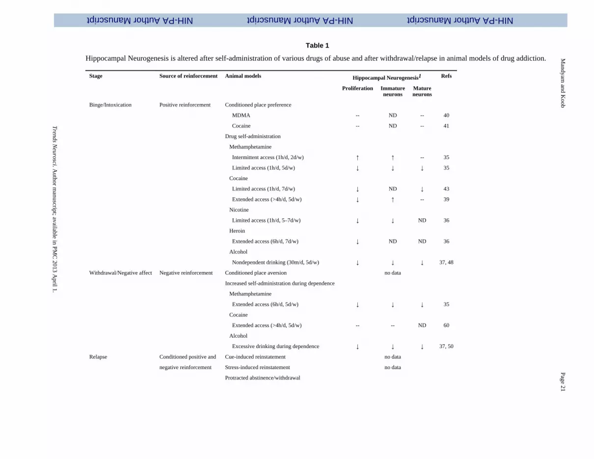
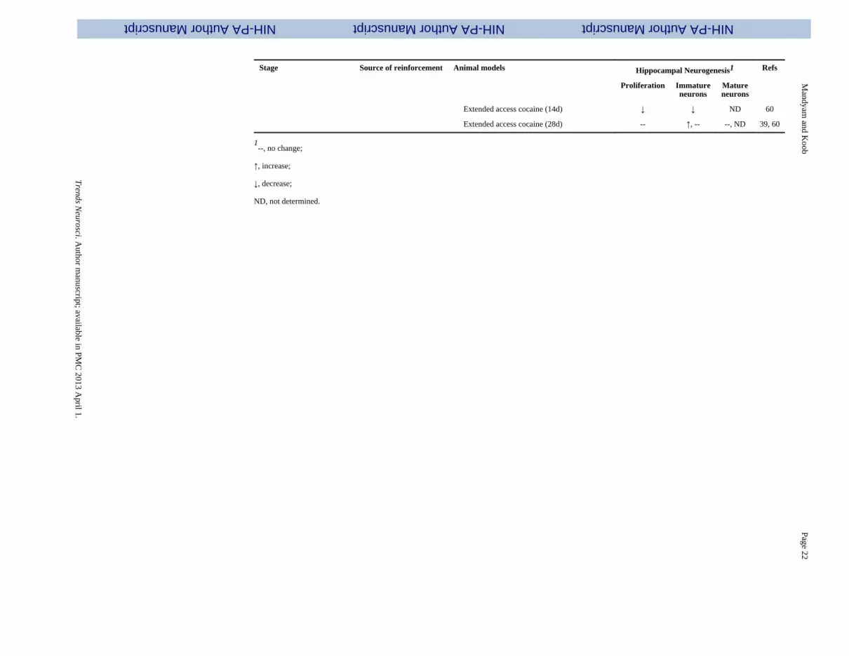
Table 1
Hippocampal Neurogenesis is altered after self-administration of various drugs of abuse and after withdrawal/relapse in animal models of drug addiction.
Stage Source of reinforcement Animal models Hippocampal Neurogenesis1 Refs
Proliferation Immatureneurons
Matureneurons
Binge/Intoxication Positive reinforcement Conditioned place preference
MDMA -- ND -- 40
Cocaine -- ND -- 41
Drug self-administration
Methamphetamine
Intermittent access (1h/d, 2d/w) ↑ ↑ -- 35
Limited access (1h/d, 5d/w) ↓ ↓ ↓ 35
Cocaine
Limited access (1h/d, 7d/w) ↓ ND ↓ 43
Extended access (>4h/d, 5d/w) ↓ ↑ -- 39
Nicotine
Limited access (1h/d, 5–7d/w) ↓ ↓ ND 36
Heroin
Extended access (6h/d, 7d/w) ↓ ND ND 36
Alcohol
Nondependent drinking (30m/d, 5d/w) ↓ ↓ ↓ 37, 48
Withdrawal/Negative affect Negative reinforcement Conditioned place aversion no data
Increased self-administration during dependence
Methamphetamine
Extended access (6h/d, 5d/w) ↓ ↓ ↓ 35
Cocaine
Extended access (>4h/d, 5d/w) -- -- ND 60
Alcohol
Excessive drinking during dependence ↓ ↓ ↓ 37, 50
Relapse Conditioned positive and Cue-induced reinstatement no data
negative reinforcement Stress-induced reinstatement no data
Protracted abstinence/withdrawal
Trends N
eurosci. Author m
anuscript; available in PM
C 2013 A
pril 1.
NIH-PA Author ManuscriptNIH-PA Author ManuscriptNIH-PA Author Manuscript
Mandyam
and Koob
Page 22
Stage Source of reinforcement Animal models Hippocampal Neurogenesis1 Refs
Proliferation Immatureneurons
Matureneurons
Extended access cocaine (14d) ↓ ↓ ND 60
Extended access cocaine (28d) -- ↑, -- --, ND 39, 60
1--, no change;
↑, increase;
↓, decrease;
ND, not determined.
Trends N
eurosci. Author m
anuscript; available in PM
C 2013 A
pril 1.

NIH-PA Author ManuscriptNIH-PA Author ManuscriptNIH-PA Author Manuscript
Mandyam
and Koob
Page 23
Table 2
Gliogenesis in the mPFC is altered after self-administration of various drugs of abuse and after withdrawal in animal models of drug addiction1
Stage Source of reinforcement Animal models mPFC Gliogenesis2 Refs
Proliferation Survival
GFAP-glia NG2-glia
Binge/Intoxication Positive reinforcement Conditioned place preference no data
Drug self-administration
Methamphetamine
Intermittent access (1h/d, 2d/w) ↑ ↑ -- ↑ 4
Limited access (1h/d, 5d/w) ↓ ↓ ↓ ↓ 4
Alcohol
Nondependent drinking (30m/d, 5d/w) -- -- ND 37
Withdrawal/Negative affect Negative reinforcement Conditioned place aversion no data
Increased self-administration during dependence
Methamphetamine
Extended access (6h/d, 5d/w) ↓ ↓ ↓ ↓ 4
Alcohol
Excessive drinking ↓ ↓ ND 37
during dependence
1The effects of relapse on gliogenesis in the mPFC have not yet been studied.
2--, no change;
↑increase;
↓decrease;
↓ND, not determined.
Trends N
eurosci. Author m
anuscript; available in PM
C 2013 A
pril 1.
Copyright © 2022 FDOKUMEN