Satellite tracking of sea turtles released after prolonged captivity periods
Putative neural consequences of captivity for elephants and ...
27
Bob Jacobs*, Heather Rally, Catherine Doyle, Lester O’Brien, Mackenzie Tennison and Lori Marino Putative neural consequences of captivity for elephants and cetaceans https://doi.org/10.1515/revneuro-2021-0100 Received July 26, 2021; accepted September 2, 2021; published online September 16, 2021 Abstract: The present review assesses the potential neural impact of impoverished, captive environments on large-brained mammals, with a focus on elephants and cetaceans. These species share several characteristics, including being large, wide-ranging, long-lived, cogni- tively sophisticated, highly social, and large-brained mammals. Although the impact of the captive environ- ment on physical and behavioral health has been well- documented, relatively little attention has been paid to the brain itself. Here, we explore the potential neural consequences of living in captive environments, with a focus on three levels: (1) The effects of environmental impoverishment/enrichment on the brain, emphasizing the negative neural consequences of the captive/impov- erished environment; (2) the neural consequences of stress on the brain, with an emphasis on corticolimbic structures; and (3) the neural underpinnings of stereo- typies, often observed in captive animals, underscoring dysregulation of the basal ganglia and associated cir- cuitry. To this end, we provide a substantive hypothesis about the negative impact of captivity on the brains of large mammals (e.g., cetaceans and elephants) and how these neural consequences are related to documented evidence for compromised physical and psychological well-being. Keywords: captivity; cerebral cortex; cetacea; chronic stress; elephants; impoverishment. Introduction Although some large mammals fare relatively well in captive environments (i.e., zoos and marine parks), those with extensive home ranges do not (Clubb and Mason 2003, 2007; Mason 2010). Large-brained animals with complex cognitive capacities such as elephants and ceta- ceans seem particularly prone to poor welfare in captive environments insofar as they do not have an adequately stimulating, natural environment. Globally, more than 3000 cetaceans and 17,000 elephants are held in captivity (Jackson et al. 2019; Riddle and Stemme 2011). In the pre- sent review, we begin by summarizing the shortcomings of captive environments and the concomitant, often stress- related clinical issues for elephants and cetaceans. Although one can directly observe the physical and behavioral manifestations of welfare in captivity, we cannot do the same for potential neural consequences. Thus, we must infer the effects on the brain from the ani- mal’s behavior, biomedical assays, and from inductive extrapolations of empirical neuroscience research. We propose several neural systems in elephants and cetaceans that are likely negatively affected by the chronic stress of captivity. Elephants and ∼75% of cetacean species, along with humans and three pinniped species, belong to a small subset of species with brain masses >700 g (Manger et al. 2013). The adult African elephant brain mass is ∼5000 g (Manger et al. 2009; Figure 1). Across the ∼86 species of odontocete and mysticete cetaceans, brain mass ranges from 164 g (Indus River dolphin, Platanista minor) to 8030 g (sperm whale, Physeter macrocephalus) (Manger 2006; Marino 2009). The African elephant (Loxodonta africana) brain contains ∼257 billion neurons, three times as many as the ∼86 billion neurons in the adult human brain, with ∼251 billion (or 97.5%) of these neurons in the cerebellum (compared to ∼69 billion in the human) and only 5.6 billion in the neocortex (compared to 16.3 billion in the human; Herculano-Housel 2009; Herculano-Housel et al. 2014). *Corresponding author: Bob Jacobs, Department of Psychology, Laboratory of Quantitative Neuromorphology, Neuroscience Program, Colorado College, 14 E. Cache La Poudre, Colorado Spring, CO, 80903, USA, E-mail: [email protected]. https://orcid.org/ 0000-0002-4662-3401 Heather Rally, Foundation to Support Animal Protection, Norfolk, VA, 23510, USA Catherine Doyle, Performing Animal Welfare Society, P.O. Box 849, Galt, CA, 95632, USA Lester O’Brien, Palladium Elephant Consulting Inc., 2408 Pinewood Dr. SE, Calgary, AB, T2B1S4, Canada Mackenzie Tennison, Department of Psychology, University of Washington, Seattle, WA, 98195, USA Lori Marino, Whale Sanctuary Project, Kanab, UT, 84741, USA Rev. Neurosci. 2022; 33(4): 439–465 Open Access. © 2021 Bob Jacobs et al., published by De Gruyter. This work is licensed under the Creative Commons Attribution 4.0 International License.
-
Upload
khangminh22 -
Category
Documents
-
view
1 -
download
0
Transcript of Putative neural consequences of captivity for elephants and ...
Bob Jacobs*, Heather Rally, Catherine Doyle, Lester O’Brien, Mackenzie Tennisonand Lori Marino
Putative neural consequences of captivity forelephants and cetaceans
https://doi.org/10.1515/revneuro-2021-0100Received July 26, 2021; accepted September 2, 2021;published online September 16, 2021
Abstract: The present review assesses the potentialneural impact of impoverished, captive environments onlarge-brained mammals, with a focus on elephants andcetaceans. These species share several characteristics,including being large, wide-ranging, long-lived, cogni-tively sophisticated, highly social, and large-brainedmammals. Although the impact of the captive environ-ment on physical and behavioral health has been well-documented, relatively little attention has been paid tothe brain itself. Here, we explore the potential neuralconsequences of living in captive environments, with afocus on three levels: (1) The effects of environmentalimpoverishment/enrichment on the brain, emphasizingthe negative neural consequences of the captive/impov-erished environment; (2) the neural consequences ofstress on the brain, with an emphasis on corticolimbicstructures; and (3) the neural underpinnings of stereo-typies, often observed in captive animals, underscoringdysregulation of the basal ganglia and associated cir-cuitry. To this end, we provide a substantive hypothesisabout the negative impact of captivity on the brains oflarge mammals (e.g., cetaceans and elephants) and howthese neural consequences are related to documentedevidence for compromised physical and psychologicalwell-being.
Keywords: captivity; cerebral cortex; cetacea; chronicstress; elephants; impoverishment.
Introduction
Although some large mammals fare relatively well incaptive environments (i.e., zoos and marine parks), thosewith extensive home ranges do not (Clubb and Mason2003, 2007; Mason 2010). Large-brained animals withcomplex cognitive capacities such as elephants and ceta-ceans seem particularly prone to poor welfare in captiveenvironments insofar as they do not have an adequatelystimulating, natural environment. Globally, more than3000 cetaceans and 17,000 elephants are held in captivity(Jackson et al. 2019; Riddle and Stemme 2011). In the pre-sent review, we begin by summarizing the shortcomings ofcaptive environments and the concomitant, often stress-related clinical issues for elephants and cetaceans.Although one can directly observe the physical andbehavioral manifestations of welfare in captivity, wecannot do the same for potential neural consequences.Thus, we must infer the effects on the brain from the ani-mal’s behavior, biomedical assays, and from inductiveextrapolations of empirical neuroscience research. Wepropose several neural systems in elephants and cetaceansthat are likely negatively affected by the chronic stress ofcaptivity.
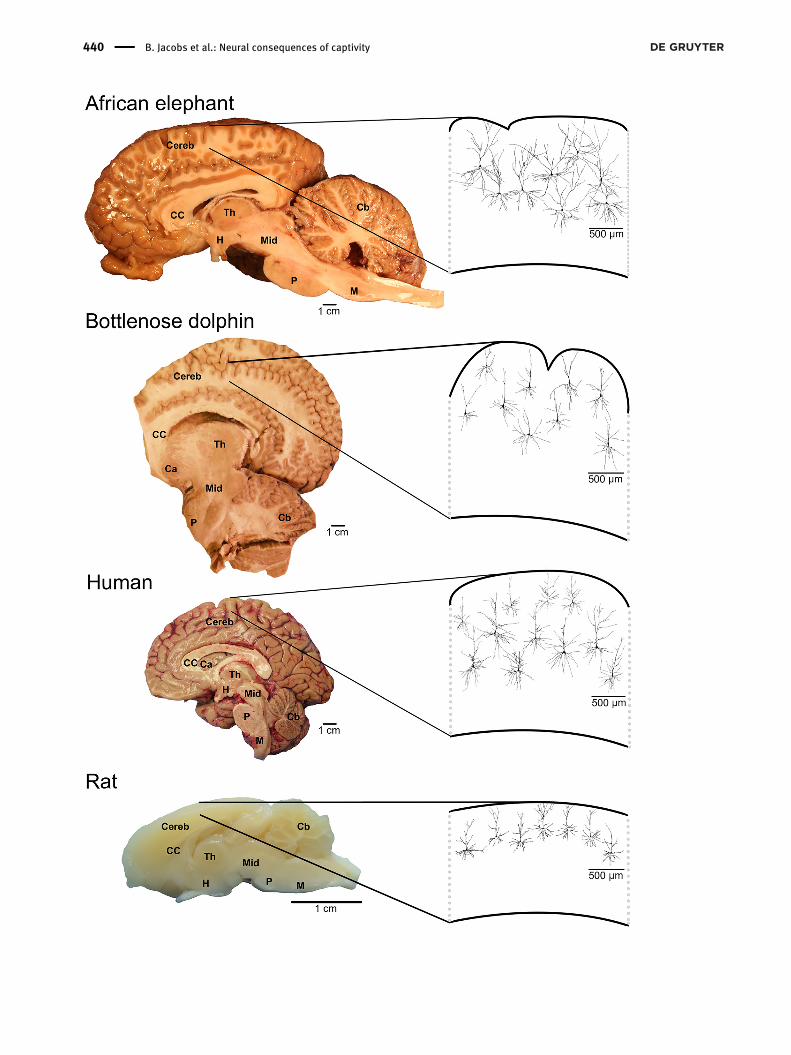
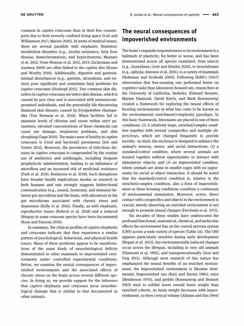
Elephants and ∼75% of cetacean species, along withhumans and three pinniped species, belong to a smallsubset of species with brain masses >700 g (Manger et al.2013). The adult African elephant brain mass is ∼5000 g(Manger et al. 2009; Figure 1). Across the ∼86 species ofodontocete and mysticete cetaceans, brain mass rangesfrom 164 g (Indus River dolphin,Platanistaminor) to 8030 g(sperm whale, Physeter macrocephalus) (Manger 2006;Marino 2009). The African elephant (Loxodonta africana)brain contains ∼257 billion neurons, three times asmany asthe∼86 billion neurons in the adult humanbrain, with∼251billion (or 97.5%) of these neurons in the cerebellum(compared to ∼69 billion in the human) and only 5.6 billionin the neocortex (compared to 16.3 billion in the human;Herculano-Housel 2009; Herculano-Housel et al. 2014).
*Corresponding author: Bob Jacobs, Department of Psychology,Laboratory of Quantitative Neuromorphology, Neuroscience Program,Colorado College, 14 E. Cache La Poudre, Colorado Spring, CO, 80903,USA, E-mail: [email protected]. https://orcid.org/0000-0002-4662-3401Heather Rally, Foundation to Support Animal Protection, Norfolk, VA,23510, USACatherine Doyle, Performing Animal Welfare Society, P.O. Box 849,Galt, CA, 95632, USALester O’Brien, Palladium Elephant Consulting Inc., 2408 PinewoodDr. SE, Calgary, AB, T2B1S4, CanadaMackenzie Tennison, Department of Psychology, University ofWashington, Seattle, WA, 98195, USALori Marino, Whale Sanctuary Project, Kanab, UT, 84741, USA
Rev. Neurosci. 2022; 33(4): 439–465
Open Access.©2021 Bob Jacobs et al., published by DeGruyter. This work is licensed under the Creative Commons Attribution 4.0 InternationalLicense.
440 B. Jacobs et al.: Neural consequences of captivity
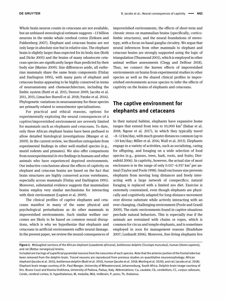
Whole brain neuron counts in cetaceans are not available,but an unbiased stereological estimate suggests ∼13 billionneurons in the minke whale cerebral cortex (Eriksen andPakkenberg 2007). Elephant and cetacean brains are notonly large in absolute size but in relative size. The elephantbrain is slightly larger than expected for its body size (Rothand Dicke 2005) and the brains of many odontocete ceta-cean species are significantly larger than predicted by theirbody size (Marino 2009). Size differences aside, all euthe-rian mammals share the same brain components (Finlayand Darlington 1995), with many parts of elephant andcetacean brains appearing to be highly conserved in termsof neuroanatomy and chemoarchitecture, including thelimbic system (Butti et al. 2015; Denver 2009; Jacobs et al.2011, 2015; Limacher-Burrell et al. 2018; Patzke et al. 2014).Phylogenetic variations in neuroanatomy for these speciesare primarily related to sensorimotor specializations.
For practical and ethical reasons, options forexperimentally exploring the neural consequences of acaptive/impoverished environment are severely limitedfor mammals such as elephants and cetaceans. To date,only three African elephant brains have been perfused toallow detailed histological investigations (Manger et al.2009). In the current review, we therefore extrapolate fromexperimental findings in other well-studied species (e.g.,murid rodents and primates). We also offer comparisonsfromnonexperimental in vivo findings in humans and otheranimals who have experienced deprived environments.Our inductive conclusions about the effects of captivity onelephant and cetacean brains are based on the fact thatbrain structures are highly conserved across vertebrates,especially across mammals (Finlay and Darlington 1995).Moreover, substantial evidence suggests that mammalianbrains employ very similar mechanisms for interactingwith their environment (Lupien et al. 2009).
The clinical profiles of captive elephants and ceta-ceans manifest in many of the same physical andpsychological perturbations as do other mammals inimpoverished environments. Such similar welfare out-comes are likely to be based on common neural disrup-tions, which is why we hypothesize that elephants andcetaceans in artificial environments suffer neural damage.In the present paper, we review the neural consequences of
impoverished environments, the effects of short-term andchronic stress on mammalian brains (specifically, cortico-limbic structures), and the neural foundations of stereo-typy, with a focus on basal ganglia circuitry. We argue thatneural inferences from other mammals to elephant andcetacean brains are strongly supported using the logic oftriangulation (Thurmond 2001),which is employed in otheranimal welfare assessments (Clegg and Delfour 2018).Thus, we connect the known effects of impoverishedenvironments on brains from experimental studies in otherspecies as well as the shared clinical profiles in impov-erished environments across species to infer the effects ofcaptivity on the brains of elephants and cetaceans.
The captive environment forelephants and cetaceans
In their natural habitat, elephants have expansive homeranges that extend from tens to 10,000 km2 (Bahar et al.2018; Ngene et al. 2017), in which they typically travel∼8–12 km/day,withmuch greater distances common (up to∼50 km/day; Miller et al. 2016; Wall et al. 2013). Elephantsengage in a variety of activities, such as socializing, caringfor offspring, and foraging on a wide selection of foodspecies (e.g., grasses, trees, bark, roots, and fruits; Dier-enfeld 2006). In captivity, however, the actual size of mostenclosures is in the range of only 0.017–6.937 km2 per an-imal (Taylor andPoole 1998). Small enclosure size preventselephants from moving long distances and freely inter-acting with a large network of conspecifics; naturalforaging is replaced with a limited zoo diet. Exercise isextremely constrained, even though elephants are physi-cally and cognitively adapted for long-distance movementover diverse substrate while actively interacting with anever-changing, challenging environment (Poole and Granli2009). The static environments found in captive situationspreclude natural behaviors. This is especially true if theanimals are restrained with chains or ropes, which iscommon for circus and temple elephants, and is sometimesemployed in zoos for management reasons (Bradshaw2007; Lenhardt 2006). Moreover, free-living elephants live
Figure 1: Midsagittal sections of the African elephant (Loxodonta africana), bottlenose dolphin (Tursiops truncatus), human (Homo sapiens),and rat (Rattus norvegicus) brains.Included are tracings of superficial pyramidal neurons from the neocortex of each species. Note that the anterior portion of the frontal lobe hasbeen removed from the dolphin brain. Traced neurons are reproduced from previous studies on quantitative neuromorphology: Africanelephant (Jacobs et al. 2011), bottlenose dolphin (Butti et al. 2015), human (Jacobs et al. 2018;Warling et al. 2020), and rat ( Jacobs et al. 2018).Elephant brain image courtesy of Dr. Paul Manger, University of Witwatersrand, Johannesburg, South Africa. Dolphin brain image courtesy ofDrs. Bruno Cozzi and Ksenia Orekhova, University of Padova, Padua, Italy. Abbreviations: Ca, caudate; Cb, cerebellum; CC, corpus callosum;Cereb, cerebral cortex; H, hypothalamus; M, medulla; Mid, midbrain; P, pons; Th, thalamus.
B. Jacobs et al.: Neural consequences of captivity 441

in matriarchal, multigenerational family groups of two to10 adult females and their immature offspring (de Silvaet al. 2011; Vance et al. 2009). Zoos, on the other hand, donot provide biologically appropriate social groups (Pooleand Granli 2009) insofar as conspecific interactions arelargely limited to small groups of mostly unrelated adultfemales and very few infants or juveniles (Clubb andMason2002), with some elephants even held in solitary confine-ment for decades (Lindsay 2017). Finally, most captiveelephants are forced to interact directly in some capacitywith humans, whether it is for entertainment, tourism, orreligious purposes, potentially creating further stress.
Similar issues obtain for captive cetaceans who,despite routinely swimming tens of kilometers a day in theocean (Matthews et al. 2011), are typically held in concretetanks that are too small and too shallow to allow for anynatural ranging or diving behaviors (McPhee and Carlstead2010). Even in the largest facilities, cetaceans are kept intanks that are ∼10,000 times smaller than their naturalhome range (https://www.cascadiaresearch.org/projects/killer-whales/using-dtags-study-acoustics-and-behavior-southern). Such tanks are characterized by reflective,barren, smooth surfaces as opposed to naturalistic texturesand substrates (Rose and Parsons 2019), creating an envi-ronment with constant, unnaturally high levels of ultravi-olet radiation. As with elephants, cetaceans naturally havelong juvenile periods and depend heavily on culturallearning as well as life-long support from a complex socialnetwork (O’Corry-Crowe et al. 2020). In contrast, captivecetaceans have little choice in terms of social associationsand partners. Most captive groupings are artificial andunstable because animals are moved among facilities forbreeding purposes (Clegg and Butterworth 2017). Aswith elephants, natural feeding behavior is absent incaptive cetaceans, who are fed a narrow selection of deadfish/invertebrates, which are delivered in an unnaturalmanner (i.e., above water, thrown directly into theirmouths) that requires none of the cognitive or behavioralengagement necessary in the wild. Finally, as withelephants, many captive cetaceans are trained to performartificial behaviors and are routinely forced to interactwith humans (e.g., swim with the dolphin programs;Frohoff 2018).
The clinical profiles of captiveelephants and cetaceans
Because elephants have complex physical and socialneeds that are difficult to meet even in professionally
accredited zoological institutions (Kagan et al. 2018), theysuffer from high rates of behavioral and physical pa-thology (Lahdenperä et al. 2018). In terms of behavior, aprevalent abnormality is stereotypic behavior (Masonand Rushen 2008), which consists of aberrant, repetitivemovements (e.g., limb swaying, and rocking) induced bythe frustration of natural impulses (Clegg et al. 2017). It isestimated that 47–85% of elephants in zoos and 100% ofthose in circuses exhibit stereotypies (Greco et al. 2016;Mason and Veasey 2010; Schmid 1995). Captive elephantsalso exhibit hyperaggression (Harvey et al. 2018), in partbecause there is no opportunity for physical distancingduring heighted intragroup stress (Archie et al. 2006).Medically, captive elephants suffer from both gastroin-testinal diseases (e.g., impaction and colic; Greene et al.2019) and nutritional/metabolic disorders because oftheir captive diet and lack of exercise (Khadpekar et al.2020), with obesity being a serious issue (Brown et al.2020). Across North American zoos, 74% of elephantswere found to be overweight with 34% believed to beclinically obese (Morfeld et al. 2016). Skin issues (e.g.,inflammation, lesions, and pressure sores) are common(Brown et al. 2020; Fowler 2006a) as are foot-relateddisorders (e.g., hyperkeratosis, cracked nails, and ab-scesses; Fowler 2001). Osteoarthritis in the feet, exacer-bated by locomotor stereotypies and obesity, occursprematurely in captive elephants and can lead to eutha-nasia (Issa and Griffin 2012). Finally, captive elephantsare particularly susceptible to several infectious diseases(e.g., Mycobacterium tuberculosis, TB, the endothelio-tropic herpesvirus, EEHV), which are highly contagious(Fuery et al. 2018; Mikota and Maslow 2011). TB is deadlyin elephants and treatment is often unsuccessful(Lyashchenko et al. 2006). EEHV is prevalent in captiveenvironments, particularly in young, chronicallystressed, immunocompromised Asian elephants (Elephasmaximus; Schaftenaar et al. 2010), and is now the leadingcause of death for captive elephant calves (Perrin et al.2021). These factors appear to be associated with repro-ductive issues (Clubb and Mason 2002; Perrin et al. 2021)and a reduced lifespan for both Asian and African ele-phants in captivity (Clubb et al. 2008, 2009).
Captive cetaceans also exhibit a variety of stereotypies(e.g., repetitive swimming patterns; regurgitation/rein-gestion of food), with the most common being oral ste-reotypies that result in severely worn teeth from gratingthem on hard surfaces (Jett et al. 2017; Ugaz et al. 2013).Some cetaceans also exhibit symptoms characteristic ofdepression (e.g., logging on the surface, lying motionlesson the bottom of the tank, and loss of appetite; Jett andVentre 2012). As with elephants, hyperaggression is more
442 B. Jacobs et al.: Neural consequences of captivity
common in captive cetaceans than in their free counter-parts due to their severely confined living space (Lott andWilliamson 2017; Marino 2020). In terms of medical issues,there are several parallels with elephants. Nutrition/metabolism disorders (e.g., insulin resistance, fatty liverdisease, hemochromatosis, and hypocitraturia; Mazzaroet al. 2012; Venn-Watson et al. 2012, 2013; Zuckerman andAssimos 2009) are often linked to the captive diet (Rosenand Worthy 2018). Additionally, digestive and gastroin-testinal disturbances (e.g., gastritis, ulcerations, and tor-sion) pose significant and sometimes fatal problems forcaptive cetaceans (Stoskopf 2015). Two common skin dis-orders in captive cetaceans are tattoo skin disease, which iscaused by pox virus and is associated with immunocom-promised individuals, and the potentially life-threateningdiamond skin disease, caused by Erysipelothrix rhusiopa-thia (Van Bressem et al. 2018). When facilities fail tomaintain levels of chlorine and ozone within strict pa-rameters, elevated concentrations of these chemicals cancause eye damage, respiratory problems, and skinsloughing (Gage 2010). Themain cause of fatality in captivecetaceans is (viral and bacterial) pneumonia (Jett andVentre 2012). Moreover, the prevalence of infectious dis-eases in captive cetaceans is compounded by the routineuse of antibiotics and antifungals, including frequentprophylactic administration, leading to an imbalance ofmicroflora and an increased risk of medicinal resistance(Park et al. 2020; Reidarson et al. 2018). Such disruptionshave broader health implications insofar as research inboth humans and rats strongly suggests bidirectionalcommunication (e.g., neural, hormonal, and immune) be-tween gut microbiota and the brain, with alterations in thegut microbiome associated with chronic stress anddepression (Kelly et al. 2016). Finally, as with elephants,reproductive issues (Robeck et al. 2018) and a reducedlifespan in some cetacean species have been documented(Rose and Parsons 2019).
In summary, the clinical profiles of captive elephantsand cetaceans indicate that they experience a similarpattern of psychological, behavioral, and physical healthissues. Many of these problems appear to be manifesta-tions of the same kinds of neurobiological deficitsdemonstrated in other mammals in impoverished envi-ronments under controlled experimental conditions.Below, we examine the neural consequences of impov-erished environments and the associated effects ofchronic stress on the brain across several different spe-cies. In doing so, we provide support for the inferencethat captive elephants and cetaceans incur neurobio-logical damage that is similar to that documented inother animals.
The neural consequences ofimpoverished environments
The brain’s exquisite responsiveness to its environment is ahallmark of plasticity, for better or worse, and has beendemonstrated across all species examined, from insects(e.g., honeybees; Groh and Rössler 2020), to invertebrates(e.g., aplysia; Antonov et al. 2001), to a variety of mammals(Holtmaat and Svoboda 2009). Following Hebb’s (1947)observation that free-roaming rats performed better oncognitive tasks than laboratory-housed rats, researchers atthe University of California, Berkeley (Edward Bennett,Marian Diamond, David Krech, and Mark Rosenzweig)created a framework for exploring the neural effects ofhousing environments in what has come to be known asthe environmental enrichment/complexity paradigm. Inthis basic framework, littermates are placed in one of threeconditions: (1) A relatively large, enriched/complex condi-tion together with several conspecifics and multiple ob-jects/toys, which are changed frequently to providenovelty—in short, the enclosure is designed to enhance theanimal’s sensory, motor, and social interactions; (2) astandard/control condition, where several animals arehoused together without opportunities to interact withstimulatory objects; and (3) an impoverished condition,where animals are alone in smaller cages with no oppor-tunity for social or object interaction. It should be notedthat the standard/control condition is, relative to theenriched/complex condition, also a form of impoverish-ment as these housing conditions constitute a continuumof environmental stimulation. Moreover, active, directcontact with conspecifics and objects in the environment iscrucial; merely observing an enriched environment is notenough to promote neural changes (Ferchmin et al. 1975).
Six decades of these studies have underscored theprofound functional, anatomical, chemical, andmoleculareffects the environment has on the central nervous system(CNS) across a wide variety of species (Table 1A). The CNSappears particularly sensitive during early development(Bogart et al. 2013), but environmentally induced changesoccur across the lifespan, including in very old animals(Diamond et al. 1985), and transgenerationally (Arai andFeig 2011). Although most research of this nature hasemphasized the neural benefits of an enriched environ-ment, the impoverished environment is likewise detri-mental. Impoverished rats (Katz and Davies 1984), mice(Henderson 1970), and gerbils (Rosenzweig and Bennett1969) tend to exhibit lower overall brain weight thanenriched cohorts. As brain weight decreases with impov-erishment, so does cortical volume (Altman and Das 1964)
B. Jacobs et al.: Neural consequences of captivity 443
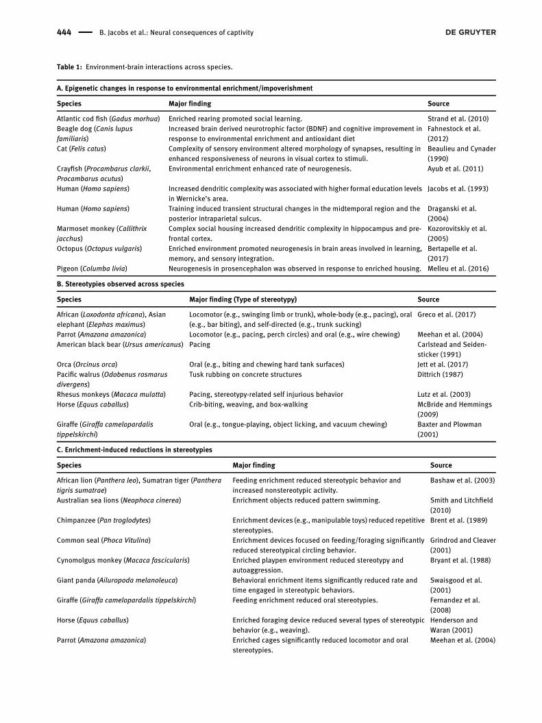
Table : Environment-brain interactions across species.
A. Epigenetic changes in response to environmental enrichment/impoverishment
Species Major finding Source
Atlantic cod fish (Gadus morhua) Enriched rearing promoted social learning. Strand et al. ()Beagle dog (Canis lupusfamiliaris)
Increased brain derived neurotrophic factor (BDNF) and cognitive improvement inresponse to environmental enrichment and antioxidant diet
Fahnestock et al.()
Cat (Felis catus) Complexity of sensory environment altered morphology of synapses, resulting inenhanced responsiveness of neurons in visual cortex to stimuli.
Beaulieu and Cynader()
Crayfish (Procambarus clarkii,Procambarus acutus)
Environmental enrichment enhanced rate of neurogenesis. Ayub et al. ()
Human (Homo sapiens) Increased dendritic complexity was associated with higher formal education levelsin Wernicke’s area.
Jacobs et al. ()
Human (Homo sapiens) Training induced transient structural changes in the midtemporal region and theposterior intraparietal sulcus.
Draganski et al.()
Marmoset monkey (Callithrixjacchus)
Complex social housing increased dendritic complexity in hippocampus and pre-frontal cortex.
Kozorovitskiy et al.()
Octopus (Octopus vulgaris) Enriched environment promoted neurogenesis in brain areas involved in learning,memory, and sensory integration.
Bertapelle et al.()
Pigeon (Columba livia) Neurogenesis in prosencephalon was observed in response to enriched housing. Melleu et al. ()
B. Stereotypies observed across species
Species Major finding (Type of stereotypy) Source
African (Loxodonta africana), Asianelephant (Elephas maximus)
Locomotor (e.g., swinging limb or trunk), whole-body (e.g., pacing), oral(e.g., bar biting), and self-directed (e.g., trunk sucking)
Greco et al. ()
Parrot (Amazona amazonica) Locomotor (e.g., pacing, perch circles) and oral (e.g., wire chewing) Meehan et al. ()American black bear (Ursus americanus) Pacing Carlstead and Seiden-
sticker ()Orca (Orcinus orca) Oral (e.g., biting and chewing hard tank surfaces) Jett et al. ()Pacific walrus (Odobenus rosmarusdivergens)
Tusk rubbing on concrete structures Dittrich ()
Rhesus monkeys (Macaca mulatta) Pacing, stereotypy-related self injurious behavior Lutz et al. ()Horse (Equus caballus) Crib-biting, weaving, and box-walking McBride and Hemmings
()Giraffe (Giraffa camelopardalistippelskirchi)
Oral (e.g., tongue-playing, object licking, and vacuum chewing) Baxter and Plowman()
C. Enrichment-induced reductions in stereotypies
Species Major finding Source
African lion (Panthera leo), Sumatran tiger (Pantheratigris sumatrae)
Feeding enrichment reduced stereotypic behavior andincreased nonstereotypic activity.
Bashaw et al. ()
Australian sea lions (Neophoca cinerea) Enrichment objects reduced pattern swimming. Smith and Litchfield()
Chimpanzee (Pan troglodytes) Enrichment devices (e.g., manipulable toys) reduced repetitivestereotypies.
Brent et al. ()
Common seal (Phoca Vitulina) Enrichment devices focused on feeding/foraging significantlyreduced stereotypical circling behavior.
Grindrod and Cleaver()
Cynomolgus monkey (Macaca fascicularis) Enriched playpen environment reduced stereotypy andautoaggression.
Bryant et al. ()
Giant panda (Ailuropoda melanoleuca) Behavioral enrichment items significantly reduced rate andtime engaged in stereotypic behaviors.
Swaisgood et al.()
Giraffe (Giraffa camelopardalis tippelskirchi) Feeding enrichment reduced oral stereotypies. Fernandez et al.()
Horse (Equus caballus) Enriched foraging device reduced several types of stereotypicbehavior (e.g., weaving).
Henderson andWaran ()
Parrot (Amazona amazonica) Enriched cages significantly reduced locomotor and oralstereotypies.
Meehan et al. ()
444 B. Jacobs et al.: Neural consequences of captivity
and section weight (Globus et al. 1973), even thoughimpoverished rats tend to have greater body weight thanenriched rats (Walsh 1981), a finding that parallels theobesity problem identified in many captive animals (Clubbet al. 2008).
Neocortical consequences ofimpoverishment
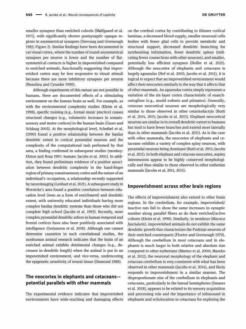
Consistent across environmental complexity studies is asignificant decrease in cortical thickness with impoverish-ment (Diamond et al. 1967), especially in occipital cortex(Katz and Davies 1984; Møllgaard et al. 1971; Figure 2),reflecting several changes in underlying cortical neuropil.Within the cortex, capillary volume, and hence corticalblood supply, tends to be lower in impoverished rats thanenriched rats (Diamond et al. 1964; Figure 2). Also, theimpoverished rat brain possesses fewer glial cells, espe-cially oligodendrocytes, than does the enriched rat brain(Altman and Das 1964; Katz and Davies 1984), which im-plies that cortical neurons in impoverished brains receiveless metabolic and structural support than neurons inenriched brains. Both nuclear and perikaryon diameters insupragranular neurons are smaller in impoverished rats(Diamond et al. 1967). Impoverished animals exhibit den-dritic systems for both pyramidal and stellate neurons thatare less complex in terms of number and length, especiallyfor more distal branches (Volkmar and Greenough 1972;Figure 2) in occipital (Sirevaag and Greenough 1985), pa-rietal (Leggio et al. 2005), and temporal (Greenough et al.1973) cortices for rats, and in motor cortex for deer mice(Turner et al. 2003). Marmoset monkeys (Callithrix jacchus)housed in standard cages for one month, compared to co-horts in complex environments, exhibited less basilardendritic complexity in pyramidal neurons of theprefrontalcortex (Kozorovitskiy et al. 2005), which is involved in ex-ecutive functions (e.g., cognitive flexibility and planning).
Several other neocortical deficits related to impover-ishment have also been documented. Pyramidal neuronsin both occipital (Globus et al. 1973) and parietal (Leggio
et al. 2005) cortex exhibit reduced spine density inimpoverished rats (Figure 2). Lower spine density along thebasilar dendrites of pyramidal neurons has also beenobserved in the prefrontal cortex of marmoset monkeys(Kozorovitskiy et al. 2005). At the synaptic level, impov-erished rats have been shown to have fewer synapses percortical neuron than their enriched counterparts, sug-gesting less overall synaptic activity (Sirevaag and Green-ough 1985). Moreover, impoverished rats tend to have
Table : (continued)
C. Enrichment-induced reductions in stereotypies
Species Major finding Source
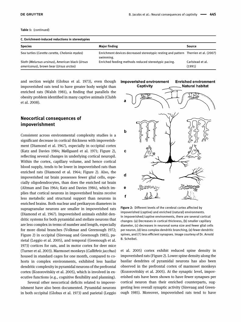
Sea turtles (Caretta caretta, Chelonia mydas) Enrichment devices decreased stereotypic resting and patternswimming.
Therrien et al. ()
Sloth (Melursus ursinus), American black (Ursusamericanus), brown bear (Ursus arctos)
Enriched feeding methods reduced stereotypic pacing. Carlstead et al.()
Figure 2: Different levels of the cerebral cortex affected byimpoverished (captive) and enriched (natural) environments.In impoverished/captive environments, there are several corticalchanges: (a) Decreases in cortical thickness, (b) smaller capillarydiameter, (c) decreases in neuronal soma size and fewer glial cellsper neuron, (d) less complex dendritic branching, (e) fewer dendriticspines, and ( f ) less efficient synapses. Image courtesy of Dr. ArnoldB. Scheibel.
B. Jacobs et al.: Neural consequences of captivity 445
smaller synapses than enriched cohorts (Møllgaard et al.1971), with significantly shorter postsynaptic opaque re-gions in asymmetrical synapses (Sirevaag and Greenough1985; Figure 2). Similar findings have been documented incat visual cortex, where the number of round-asymmetricalsynapses per neuron is lower and the number of flat-symmetrical contacts is higher in impoverished comparedto enriched animals, functionally suggesting that impov-erished cortex may be less responsive to visual stimulibecause there are more inhibitory synapses per neuron(Beaulieu and Cynader 1990).
Although experiments of this nature are not possible inhumans, there are documented effects of a stimulatingenvironment on the human brain as well. For example, aswith the environmental complexity studies (Kleim et al.1998), specific training (e.g., formal music practice) causesstructural changes (e.g., volumetric increases in somato-sensory and motor cortices) in the human brain (Gaser andSchlaug 2003). At the morphological level, Scheibel et al.(1990) found a positive relationship between the basilardendritic extent in cortical pyramidal neurons and thecomplexity of the computational task performed by thatarea, a finding confirmed in subsequent studies (monkey:Elston and Rosa 1997; human: Jacobs et al. 2001). In addi-tion, they found preliminary evidence of a positive associ-ation between dendritic complexity in the hand-fingerregion of primary somatosensory cortex and thenature of anindividual’s occupation, a relationship recently supportedbyneuroimaging (Lenhart et al. 2021). A subsequent study inWernicke’s area found a positive correlation between edu-cation level (seen as a form of enrichment) and dendriticextent, with university educated individuals having morecomplex basilar dendritic systems than those who did notcomplete high school (Jacobs et al. 1993). Recently, morecomplex pyramidal dendritic arbors in human temporal andfrontal cortices have also been positively associated withintelligence (Goriunova et al. 2018). Although one cannotdetermine causation in such correlational studies, thenonhuman animal research indicates that the brain of anenriched animal exhibits detrimental changes (e.g., de-creases in dendritic length) when the animal is put in animpoverished environment, and vice-versa, underscoringthe epigenetic sensitivity of neural tissue (Diamond 1988).
The neocortex in elephants and cetaceans—potential parallels with other mammals
The experimental evidence indicates that impoverishedenvironments have wide-reaching and damaging effects
on the cerebral cortex by contributing to thinner corticallaminae, a decreased blood supply, smaller neuronal cellsbodies with fewer glial cells to provide metabolic andstructural support, decreased dendritic branching forsynthesizing information, fewer dendritic spines (indi-cating fewer connectionswith other neurons), and smaller,potentially less efficient synapses (Holler et al. 2021).Although the neocortex of elephants and cetaceans islargely agranular (Hof et al. 2005; Jacobs et al. 2011), it islogical to expect that an impoverished environment wouldaffect their neocortex similarly to theway that it affects thatof othermammals. An agranular cortex simply represents avariation of the six-layer cortex characteristic of euarch-ontoglires (e.g., murid rodents and primates). Generally,cetacean neocortical neurons are morphologically verysimilar to those observed in other cetartiodactyls (Buttiet al. 2014, 2015; Jacobs et al. 2015). Elephant neocorticalneurons are similar in in overall dendritic extent to humansbut tend to have fewer branches and extend more laterallythan in other mammals (Jacobs et al. 2011). As is the casewith other mammals, the neocortex of elephants and ce-taceans exhibits a variety of complex spiny neurons, withpyramidal neurons being dominant (Butti et al. 2015; Jacobset al. 2011). In both elephant and cetaceanneocortex, aspinyinterneurons appear to be highly conserved morphologi-cally and thus similar to those observed in other eutherianmammals (Jacobs et al. 2011, 2015).
Impoverishment across other brain regions
The effects of impoverishment also extend to other brainregions. In the cerebellum, for example, impoverished/inactive rats fail to show the same increases in synapticnumber along parallel fibers as do their enriched/activecohorts (Kleim et al. 1998). Similarly, in monkeys (Macacafasciularis), impoverished animals do not exhibit the samedendritic growth that characterizes the Purkinje neurons oftheir enriched counterparts (Floeter and Greenough 1979).Although the cerebellum in most cetaceans and in ele-phants is much larger in both relative and absolute sizecompared to other eutherians (Marino et al. 2000; Masekoet al. 2012), the neuronal morphology of the elephant andcetacean cerebellum is very consistent with what has beenobserved in other mammals (Jacobs et al. 2014), and likelyresponds to impoverishment in a similar manner. Thedisproportionate size of the cerebellum in elephants andcetaceans, particularly in the lateral hemispheres (Smaerset al. 2018), appears to be related to its sensory acquisitionand processing role and the importance of infrasound inelephants and echolocation in cetaceans for exploring the
446 B. Jacobs et al.: Neural consequences of captivity

environment (Hanson et al. 2013; Jacobs et al. 2014). Itremains unclear to what extent the cerebellum also con-tributes to cognitive and emotional functions in elephantsand cetaceans, although such functions have beendemonstrated in other species (primates: Habas 2021; rats:Shipman and Green 2020).
Finally, two limbic structures are also negativelyaffected by impoverished environments. The hippocampal-dentate complex (or hippocampus), which is particularlysensitive to environmental influences, exhibits a lowervolume in impoverished animals compared to enrichedanimals largely because of decreased neurogenesis (mice:Kemperman et al. 1977; pigeons: Melleu et al. 2016; rats:Veena et al. 2009). In the amygdala, impoverished rats showgreater c-Fos (a gene involved in cell proliferation/differ-entiation following extracellular stimulation) expression inthe medial nucleus following aversive training thanenriched animals, suggesting they experience greater levelsof stress (Nikolaev et al. 2002). Comparatively, the elephantpossesses a typical mammalian hippocampus in terms ofboth size and architecture (Patzke et al. 2014). Although thehippocampus in cetaceans is smaller than onewould expect(Oelschläger and Oelschläger 2009; Patzke et al. 2015),perhaps because of a greatly reduced olfactory system(Kishida et al. 2015), it exhibits typical mammalian sub-regions (e.g., dentate gyrus, hippocampus proper, andsubiculum; Oelschläger et al. 2008). Moreover, despite thereduced hippocampal formation, the paralimbic regionin cetacean brains is enormously elaborated (primarily bythe well-developed entorhinal cortex and cortical limbiclobe) suggesting there may have been transfer and elabo-ration of non olfactory hippocampal functions (i.e., long-termmemory and learning) to the paralimbic cortex (Marino2015). Although this hypothesis has yet to be tested, itcomports with the behavioral evidence for sophisticatedcognitive functions in dolphins and many other cetaceans(Deecke 2018; Marino et al. 2008).
In terms of the amygdaloid complex, the Africanelephant has a well-developed amygdala similar to thatobserved in other mammals, although there are somespecializations (e.g., enlarged anterior cortical nucleus)thought to be related to the animal’s heavy reliance onolfaction (Ngwenya et al. 2011) and its affect-laden andsocial-empathic behaviors (Limacher-Burrell et al. 2018).The cetacean amygdaloid complex resembles that ofother mammals (Oelschläger et al. 2010) but is smaller inrelative size (Patzke et al. 2015). Moreover, the cetaceanlimbic lobe, which includes cingulate, insular, and par-ahippocampal cortices, is extensive with deep folds
(Oelschläger and Oelschläger 2009; Oelschläger et al.2010). It remains unclear how the relatively smaller hip-pocampus and amygdala affect susceptibility or resiliencyof cetaceans to environmental perturbations. Nevertheless,these relative size differences do not necessarily negate theargument that their psychological functions (and those ofotherwell-developed adjacent brain areas) are impacted byimpoverished environments, as the clinical profiles wouldsuggest.
Impoverishment at the molecular level
At the molecular level, epigenetic-related deficiencies inimpoverished brains are ubiquitous (Table 2). In thisregard, the chemoarchitecture of elephant and cetaceanbrains underscores considerable similarities across mam-mals. For example, the primary antibodies used in Africanelephant research (Maseko et al. 2013; Ngwenya et al. 2011;Patzke et al. 2014) were developed in the rabbit andwork inseveral species (e.g., bats, drosophila, felines, ferrets,humans, mice, mollusks, pigs, rats, and squid), suggestingsynapomorphic cytochemistry across a wide array of taxa.For example, quantitative distribution of gamma amino-butyric acid (GABA)-immunoreactive neurons in the BlackSea porpoise (Phocoena phocoena) visual cortex is similarto that observed in euarchontoglires (Garey et al. 1989). Inprimary visual cortex, the general typology of GABA-ergicneurons immunoreactive to calretinin (CR) is similar forbottlenose dolphins (Tursiops truncatus) and humans(Glezer et al. 1992). As with neuroanatomy, differences indistribution, density, and typology in neurochemical sys-tems typically reflect specialized sensorimotor andecological adaptations (Glezer et al. 1998; Manger et al.2021), but they do not diminish the fundamental similar-ities across all mammalian brains. Finally, many studieshave examined the impact of differential environments onneurotrophins, nerve growth factors (NGF), and brainderived neurotrophic factor (BDNF), all positively associ-ated with neurogenesis, neuroplasticity, emotional resil-ience, and improved cognitive performance (Table 2).Underlying these findings is the fundamental, lifelong ef-fect that the environment, including training or even asingle exposure to enrichment (Ali et al. 2009), has on theexpression of a large number of genes linked to neuronalstructure, synaptic plasticity, and neural transmission(Rampon et al. 2000) and, by extension, an animal’semotional and cognitive functioning (Neidl et al. 2016).
B. Jacobs et al.: Neural consequences of captivity 447
Impoverishment and lack of exercise in thecaptive environment
A crucial component to an enriched environment is exer-cise (Basso and Suzuki 2017), which is severely lacking forcaptive elephants and cetaceans (Clubb et al. 2008; Mor-feld et al. 2016). Exercise not only increases the supply ofoxygenated blood to a metabolically expensive brain, butalso increases serumneurotrophic factors andBDNF (Heiszet al. 2017; Liang et al. 2021) which, in turn, contribute topotential neurogenesis and enhanced cognitive abilitiesthrough a series of complex biochemical cascades (Hor-owitz et al. 2020). Moreover, exercise generally has a pos-itive influence on the immune system, leading to areduction in inflammatory biomarkers, and increases inantioxidant defenses (Gomes and Florida-James 2016).Finally, as reviewed by van Praag et al. (2000), exerciseappears to enhance the activity of several neurotransmittersystems in rats: (1) cholinergic functioning in the hippo-campus, which improves spatial learning (Fordyce and
Farrar 1991), (2) opioid activity, which modulates pain(Sforzo et al. 1986), and (3) monoamine functioning(noradrenaline and serotonin), which contributes tolearning and synaptic plasticity (Chaouloff 1989). One canlogically expect the same exercise-related neural changesin elephants and cetaceans.
Lack of exercise and other shortcomings of the captiveenvironment are apparent to those in the captive industry.However, zoos and aquariums cannot practically makewholesale changes to an animal’s environment as can bedone in laboratory settings, where general enrichment andexercise are known to reduce stress and anxiety (mice:Varman et al. 2012; rats: Veena et al. 2009), enhancememory (mice: van Praag et al. 2000), increase cognitivefunctions and neural plasticity (mice: Arai and Feig 2011;fish,Gadusmorhua: Strand et al. 2010), protect against leadtoxicity (mice: Schneider et al. 2001), treat developmentaldisorders (humans: Ball et al. 2019), and ameliorate severalpsychiatric and neurogenerative disorders in humans(Nithianantharajah and Hannan 2006). Instead, zoos and
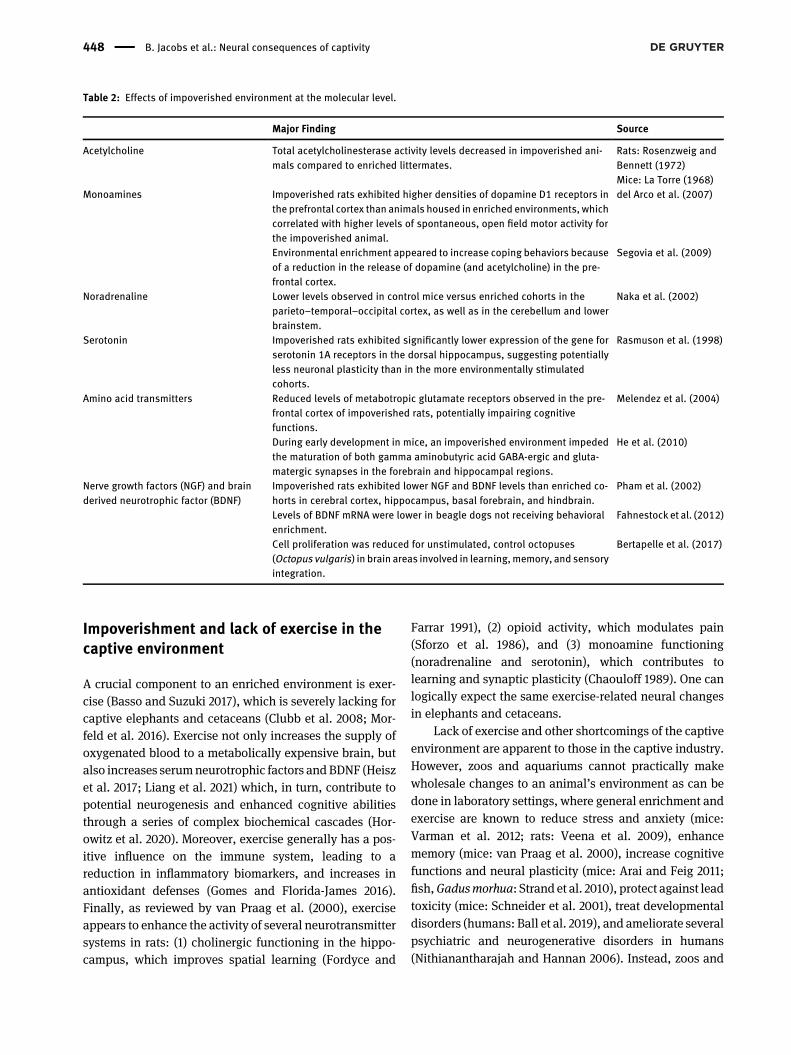
Table : Effects of impoverished environment at the molecular level.
Major Finding Source
Acetylcholine Total acetylcholinesterase activity levels decreased in impoverished ani-mals compared to enriched littermates.
Rats: Rosenzweig andBennett ()Mice: La Torre ()
Monoamines Impoverished rats exhibited higher densities of dopamine D receptors inthe prefrontal cortex than animals housed in enriched environments, whichcorrelated with higher levels of spontaneous, open field motor activity forthe impoverished animal.
del Arco et al. ()
Environmental enrichment appeared to increase coping behaviors becauseof a reduction in the release of dopamine (and acetylcholine) in the pre-frontal cortex.
Segovia et al. ()
Noradrenaline Lower levels observed in control mice versus enriched cohorts in theparieto–temporal–occipital cortex, as well as in the cerebellum and lowerbrainstem.
Naka et al. ()
Serotonin Impoverished rats exhibited significantly lower expression of the gene forserotonin A receptors in the dorsal hippocampus, suggesting potentiallyless neuronal plasticity than in the more environmentally stimulatedcohorts.
Rasmuson et al. ()
Amino acid transmitters Reduced levels of metabotropic glutamate receptors observed in the pre-frontal cortex of impoverished rats, potentially impairing cognitivefunctions.
Melendez et al. ()
During early development in mice, an impoverished environment impededthe maturation of both gamma aminobutyric acid GABA-ergic and gluta-matergic synapses in the forebrain and hippocampal regions.
He et al. ()
Nerve growth factors (NGF) and brainderived neurotrophic factor (BDNF)
Impoverished rats exhibited lower NGF and BDNF levels than enriched co-horts in cerebral cortex, hippocampus, basal forebrain, and hindbrain.
Pham et al. ()
Levels of BDNF mRNA were lower in beagle dogs not receiving behavioralenrichment.
Fahnestock et al. ()
Cell proliferation was reduced for unstimulated, control octopuses(Octopus vulgaris) in brain areas involved in learning,memory, and sensoryintegration.
Bertapelle et al. ()
448 B. Jacobs et al.: Neural consequences of captivity
aquariums engage animals in limited types of directedenrichment (Law and Kitchener 2017; Markowitz 1982) inan attempt to alleviate the specific psychological/behav-ioral problems arising from an impoverished environment.
Current evidence suggests that targeted, ad hoc zoo/aquarium enrichment remains insufficient for the overallneural health of mammals such as elephants and ceta-ceans as long as they remain constrained by standardcaptive conditions. Here it is worth noting a couple ofadditional points: natural environments appear to be bet-ter for the emotional health of rats (as measured by c-Fosactivation in the nucleus accumbens) than artificiallyenriched environments (Lambert et al. 2016), with similarfindings in humans (Lambert et al. 2015). Thus, not alltypes of enrichment are equally effective (Lyn et al. 2020).Moreover, transient, inconsistent enrichment can createmore stress and frustration for the animal than no enrich-ment at all (Latham and Mason 2010). Finally, insofar asthe developing brain is particularly susceptible to impov-erishment induced alterations (Narducci et al. 2018), thegreatest challenge is for those animals born into a captiveenvironment, which applies to most mammals in zoos(Hosey et al. 2020).
Corticolimbic structures and theneural consequences of stress
In response to environmental stressors, all animals attemptto maintain dynamic homeostasis (Schulkin 2011). Theexquisitely sensitive stress response system promotesquick activation of the body in the face of acute stress andthen a return to homeostasis once the threat has abated(Sapolsky et al. 2000). In captivity, however, stress canbecome chronic, leading to distress or “toxic stress”, whichadversely affects physiological mechanisms (McEwen2017). Three intricately interconnected systems areinvolved in the stress response: the nervous system, theendocrine system, and the immune system (Besedovskyand del Rey 1996). At the core of this tripartite schema is thehypothalamic–pituitary–adrenal (HPA) axis which,despite small heterospecific specializations (Atkinson et al.2015), is highly conserved across mammals (Denver 2009;Nikolova et al. 2018). Here, we provide a simplified over-view of the neural consequences when the HPA axis ischronically activated, resulting in an allostatic overloadthat is generally associated with poorer physical andmental health outcomes (Guidi et al. 2021).
Environmental stresses cause three, cascading linearevents: (1) Corticotrophin releasing hormones are released
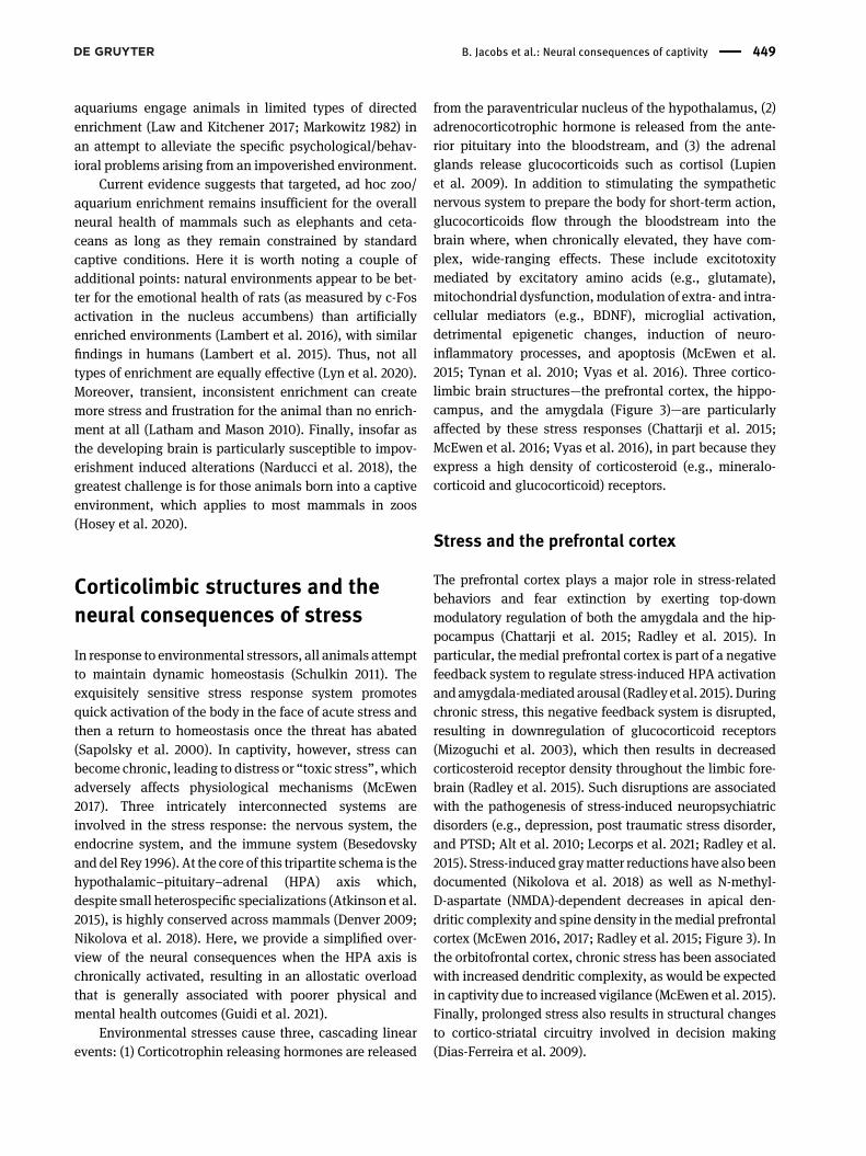
from the paraventricular nucleus of the hypothalamus, (2)adrenocorticotrophic hormone is released from the ante-rior pituitary into the bloodstream, and (3) the adrenalglands release glucocorticoids such as cortisol (Lupienet al. 2009). In addition to stimulating the sympatheticnervous system to prepare the body for short-term action,glucocorticoids flow through the bloodstream into thebrain where, when chronically elevated, they have com-plex, wide-ranging effects. These include excitotoxitymediated by excitatory amino acids (e.g., glutamate),mitochondrial dysfunction,modulation of extra- and intra-cellular mediators (e.g., BDNF), microglial activation,detrimental epigenetic changes, induction of neuro-inflammatory processes, and apoptosis (McEwen et al.2015; Tynan et al. 2010; Vyas et al. 2016). Three cortico-limbic brain structures—the prefrontal cortex, the hippo-campus, and the amygdala (Figure 3)—are particularlyaffected by these stress responses (Chattarji et al. 2015;McEwen et al. 2016; Vyas et al. 2016), in part because theyexpress a high density of corticosteroid (e.g., mineralo-corticoid and glucocorticoid) receptors.
Stress and the prefrontal cortex
The prefrontal cortex plays a major role in stress-relatedbehaviors and fear extinction by exerting top-downmodulatory regulation of both the amygdala and the hip-pocampus (Chattarji et al. 2015; Radley et al. 2015). Inparticular, the medial prefrontal cortex is part of a negativefeedback system to regulate stress-induced HPA activationandamygdala-mediated arousal (Radley et al. 2015). Duringchronic stress, this negative feedback system is disrupted,resulting in downregulation of glucocorticoid receptors(Mizoguchi et al. 2003), which then results in decreasedcorticosteroid receptor density throughout the limbic fore-brain (Radley et al. 2015). Such disruptions are associatedwith the pathogenesis of stress-induced neuropsychiatricdisorders (e.g., depression, post traumatic stress disorder,and PTSD; Alt et al. 2010; Lecorps et al. 2021; Radley et al.2015). Stress-induced graymatter reductions have also beendocumented (Nikolova et al. 2018) as well as N-methyl-D-aspartate (NMDA)-dependent decreases in apical den-dritic complexity and spine density in themedial prefrontalcortex (McEwen 2016, 2017; Radley et al. 2015; Figure 3). Inthe orbitofrontal cortex, chronic stress has been associatedwith increased dendritic complexity, as would be expectedin captivity due to increased vigilance (McEwen et al. 2015).Finally, prolonged stress also results in structural changesto cortico-striatal circuitry involved in decision making(Dias-Ferreira et al. 2009).
B. Jacobs et al.: Neural consequences of captivity 449
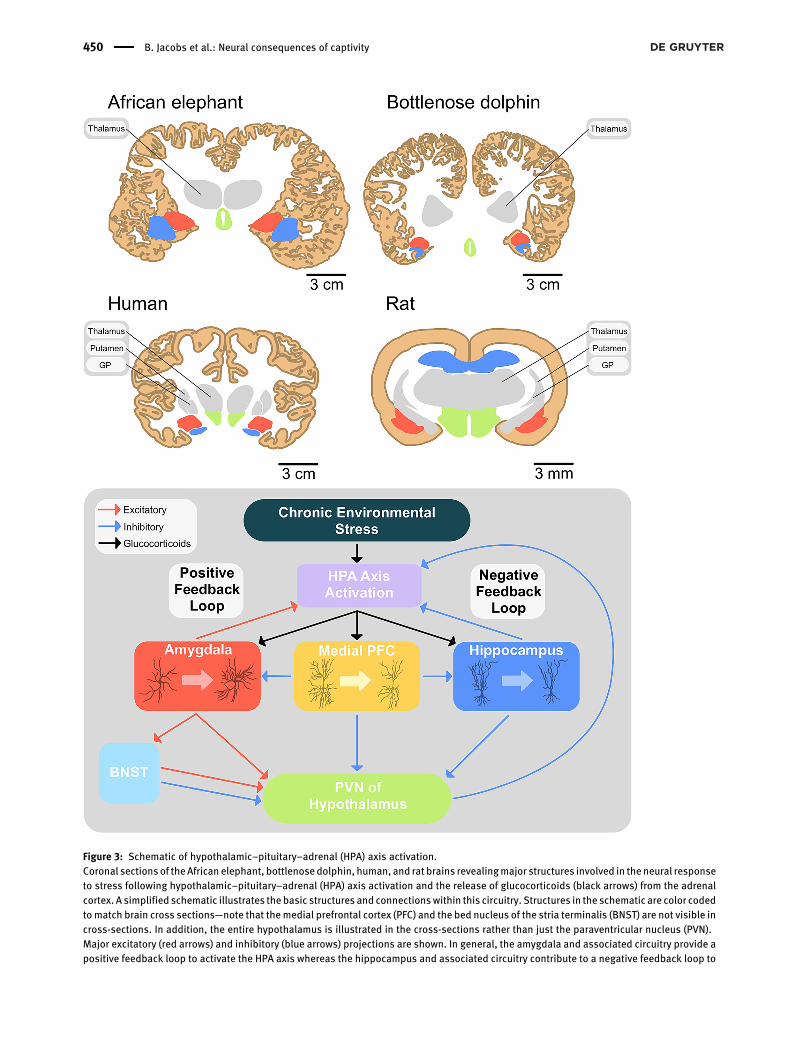
Figure 3: Schematic of hypothalamic–pituitary–adrenal (HPA) axis activation.Coronal sections of theAfrican elephant, bottlenosedolphin, human, and rat brains revealingmajor structures involved in the neural responseto stress following hypothalamic–pituitary–adrenal (HPA) axis activation and the release of glucocorticoids (black arrows) from the adrenalcortex. A simplified schematic illustrates the basic structures and connectionswithin this circuitry. Structures in the schematic are color codedtomatch brain cross sections—note that themedial prefrontal cortex (PFC) and the bed nucleus of the stria terminalis (BNST) are not visible incross-sections. In addition, the entire hypothalamus is illustrated in the cross-sections rather than just the paraventricular nucleus (PVN).Major excitatory (red arrows) and inhibitory (blue arrows) projections are shown. In general, the amygdala and associated circuitry provide apositive feedback loop to activate the HPA axis whereas the hippocampus and associated circuitry contribute to a negative feedback loop to
450 B. Jacobs et al.: Neural consequences of captivity

Stress and the hippocampus
The hippocampus is especially sensitive to sustainedexposure to glucocorticoids and mineralocorticoids (McE-wen 2016, 2017; McEwen et al. 2015). Because mineralo-corticoid receptors are more highly expressed in thehippocampus than in the prefrontal cortex (Patel et al.2000), their activation leads to excess release of glutamate(Olijslagers et al. 2008). In turn, it has been shown in bothrodents and primates that stress-elevated levels of extra-cellular glutamate activate NMDA receptors, which mobi-lizes free cytosolic calcium to toxic levels, subsequentlyresulting in hypoxia-ischemia, excitotoxic seizures, somashrinkage, nuclear pyknosis, and loss of dendritic spinesalong with apical dendritic atrophy (Figure 3), particularlyin CA3 pyramidal neurons (McEwen 2001; McEwen et al.2016). Similar structural-functional correlates (e.g.,reduction of hippocampal volume) have been documentedin humans and associatedwith clinical depression, bipolardisorders, PTSD, and other stress-related illnesses (McE-wen 2016; McEwen et al. 2016). Such stress-related damagecould be particularly detrimental in cetaceans insofar astheir hippocampus appears not to exhibit neurogenesisunder normal conditions (Parolisi et al. 2018).
Stress and the amygdala
The amygdala, in conjunction with interconnectedstructures involved in autonomic, neuroendocrine, andbehavioral arousal, is crucially involved in emotionalprocessing of sensory information and regulation ofemotional responsiveness, especially as related to fear(LeDoux 1994). Structurally, chronic stress contributes tolong-lasting morphological changes in the amygdala inboth human and nonhuman animals. Specifically, bothstellate and pyramidal neurons in the basolateral amyg-dala (a putative locus for the storage of fear memories)exhibit increased dendritic complexity and greater spinedensity in response to chronic stress (Mitra et al. 2005; rats:Vyas et al. 2002; Figure 3). In contrast, neurons in themedialamygdala show stress-induced decreases in dendritic extentand spine density, a change that appears associated withreduced social interaction (mice: McEwen 2017). Volumetricchanges in several corticolimbic regions (e.g., the nucleusaccumbens, Reynolds and Berridge 2008), including the
amygdala, also appear to positively correlate with chronicstress (Nikolova et al. 2018).
Under stressful conditions,whenglucocorticoid levels arehigh, the baseline GABA-ergic inhibitory control exerted bythe medial prefrontal cortex over the central nucleus (themajor output nucleus) of the amygdala is disrupted, resultingin amygdaloid hyperreactivity to perceived environmentalstressors (Skórzewska et al. 2015) with functional and struc-tural consequences (Christoffel et al. 2011). Functionally, thereisan increased fear response,prolongedHPAandsympatheticactivation, increased aggression and, because of the amyg-dala’s strong reciprocal connections with the bed nucleus ofthe stria terminalis (BNST), excessive anxiety (Avery et al.2016). In humans, such dysfunctions have been associatedwith a variety of anxiety disorders, including PTSD, general-ized anxiety disorder, phobias, panic disorders, andobsessive-compulsive disorders (Koenigs and Grafman 2009;Shin and Liberzon 2010). Amygdala hyperactivity may alsoincrease the risk for developing the stress-related symptomsofdepression (Nikolova et al. 2018). Moreover, under inescap-ably stressful conditions, the amygdala and the BNST,modulated by serotonergic input from the dorsal raphe nu-cleus, may mediate learned helplessness and conditioneddefeat (Maier and Seligman 2016).
Stress for elephants and cetaceans
Because of the complex social world of elephants and manycetaceans, an issue of special relevance to the present reviewis the effect that social isolation has on corticolimbic struc-tures (Mumtaz et al. 2018). In rats and nonhuman primates,chronic isolation appears to enhance HPA responsiveness tostressors (Serra et al. 2007) and increase basal cortisol levels(Hawkley et al. 2012). Stress from social isolation induces al-terations in several neurochemical systems, including (1)decreases in BDNF in the hippocampus (rats: Scaccianoceet al. 2006), which is associated with increased anxiety-likesymptoms in rats (Murínová et al. 2017) and several neuro-psychiatric disorders in humans (Autry and Monteggia 2012);(2) reduced levels of serotonin in both hippocampus andfrontal cortex, which is associated with increased aggressionand depression-like symptoms (rats: Miura et al. 2002); and(3) overproduction of nitric oxide, a retrograde messenger, inthe hippocampus, which is involved in excitotoxicity (mice:McLeod et al. 2001). Structural changes in response to social
reduce HPA activity. Although not shown in the schematic, the anterior BNST tends to increase HPA axis activity whereas the posterior divisiontends to inhibit it (Ch’ng et al. 2018). Also represented are three types of neurons and their response to chronic stress: (1) Stellate neurons inthe (basolateral) amygdala, which tend to increasedendritic extent; (2) cortical pyramidal neurons in themedial PFC,which show reductions inapical dendritic extent; and (3) CA3 pyramidal neurons in the hippocampus, which undergo degeneration of the apical dendrite.
B. Jacobs et al.: Neural consequences of captivity 451
isolation have also been observed, including selective loss ofprefrontal cortex volume (rats: Schubert et al. 2009). Thesefindings are not limited to rodents and nonhuman primates.For example, decreased dendritic complexity has beendocumented in pallial brain regions important for the devel-opment of social/sexual preferences in socially isolated zebrafinches (Taeniopygia guttata, Shukla and Sadananda 2021).Also, ants raised in isolation show impairment in the growthof the mushroom bodies, which are crucial for learning andmemory in social insects (Seid and Junge 2016), as well asweakened immune systems (Scharf et al. 2021). Humanssubjected to early socioemotional deprivation in Romanianorphanages exhibited several neural deficits, includingglucose hypometabolism and white matter abnormalities inlimbic and paralimbic structures (including prefrontal cortex,amygdala, and hippocampus; Chugani et al. 2001; Eluva-thingal et al. 2006). Such changes may underlie some of thecognitive, behavioral, and socioemotional deficits observedin these children. Human findings of this nature underscorethe importance of the early environment for shaping corti-colimbic systems and the potential long-term consequencesto chronic stress (Frodl and O’Keane 2013). Current humanresearch, in fact, suggests that childhood trauma may sub-sequently make the adult brain more vulnerable to mal-adaptive stress responses (Banihashemi et al. 2020), an issueparticularly relevant for long-lived, highly social animalssuch as elephants and cetaceans born into captivity.
Stress and neuroendocrine-immune systeminteractions
Neuroendocrine-immune interactions are dynamic (Wrona2006). Acute stress tends to enhance immune functionswhereas chronic stress tends to inhibit them (Schedlowskiand Schmidt 1996), with negative health and neural conse-quences (McEwen et al. 2015). Under chronic psychological orphysical stress, pro-inflammatory cytokines (e.g., interleukinsand tumor necrosis factors) are released by activated immunecells and can interact with multiple corticolimbic brainstructures, dysregulatingdifferent growth factors (e.g., BDNF)and neurogenesis (especially in the hippocampus), severalneurotransmitter systems (e.g., glutamate, serotonin, anddopamine), and neuroendocrine communication (Capuronand Miller 2011). One neural consequence under such con-ditions is microglia activation and a sustained release of in-flammatory mediators (Leszek et al. 2016). For example,chronic stress increases the number of activated microglia inseveral corticolimbic regions, which can lead to neuro-degeneration (mice:Nair andBonneau2006; rats: Tynanet al.2010) as well as neuroinflammation that contributes to
physiological, behavioral, affective, and cognitive disorders(de Pablos et al. 2014; McLeod et al. 2001).
Although there has beennodirect comparative researchon the neural consequences of stress in captive versus freecetaceans or elephants, existing data suggest that the im-mune system is negatively affected. For example, candidi-asis, which is often observed in immunocompromisedindividuals, is relatively common in captive cetaceans sec-ondary to stress (Ohno et al. 2019). In elephants, clinicaloutbreaks of salmonellosis tend to follow stress-relateddepression of the immune system (Fowler 2006b). Directcomparisons between captive animals and their free coun-terparts have also suggested weakened immune systems forsome captive animals (e.g., spotted hyenas: Flies et al. 2015;zebras: Seeber et al. 2020). Biomarkers such as cortisol havealso been examined to a limited degree, with acute mea-sures indicating expected elevations in cortisol levels asso-ciated with events such as beach strandings in dolphins(Kellar et al. 2015) or transportation/relocation in elephants(Laws et al. 2007) and cetaceans (Noda et al. 2007; Spoonand Romano 2012). Notably, captive bottlenose dolphinskept in sea pen facilities that allow for ocean water flow andentry of small fish had significantly lower salivary cortisollevels than their cohorts in tanks (Ugaz et al. 2013). Simi-larly, not only do Asian and African elephants in largerenclosures exhibit lower glucocorticoid metabolite concen-trations than their cohorts in smaller enclosures, but theyalso exhibit lower cortisol levels when they can accessdiverse enrichment options and allowed to be in compatiblesocial groups (Brown et al. 2019). In Asian elephants,cortisol levels negatively correlate with locomotion andpositively correlatewith stereotypies (Schmid et al. 2001). Tothe extent that captivity induces stress-related immuno-suppression, captive animals would thus be more suscep-tible not only to neuroinflammation but also toopportunistic infections and possible disruptions of fertility(Edwards et al. 2019).
Stress summary
From the neural perspective, both the PFC and hippo-campus attempt to inhibit HPA activity, thus enhancingcognitive functions. However, the amygdala tends tofacilitate HPA activity, potentially overriding the inhibitorymechanisms of the PFC and hippocampus, resulting inexcessive anxiety and fear reactivity and, when chronicallyactivated, inhibition of the immune system (Chattarji et al.2015). It has been suggested that these corticolimbicstructures are not only evolutionarily conserved in terms ofvolumetric measures, but also in terms on their functional
452 B. Jacobs et al.: Neural consequences of captivity
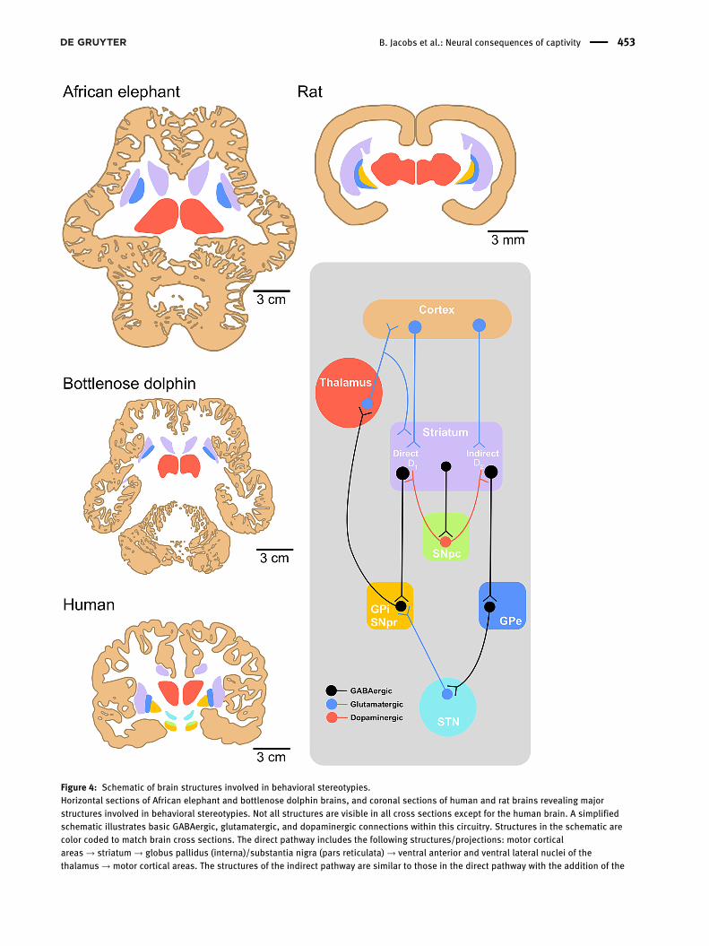
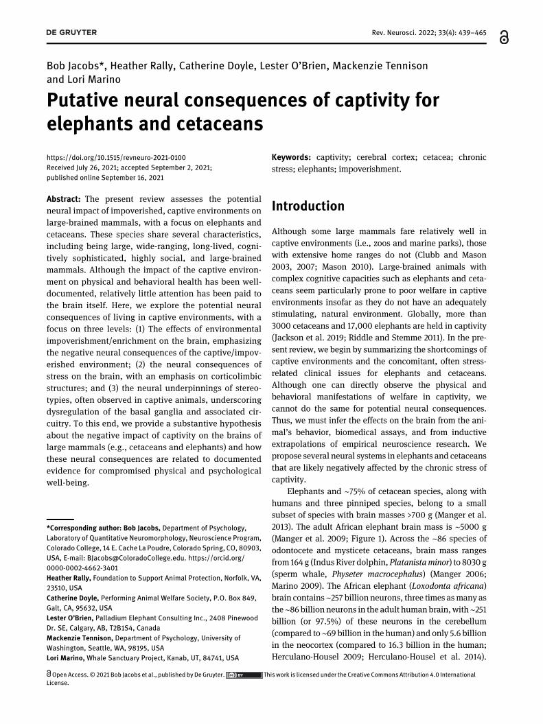
Figure 4: Schematic of brain structures involved in behavioral stereotypies.Horizontal sections of African elephant and bottlenose dolphin brains, and coronal sections of human and rat brains revealing majorstructures involved in behavioral stereotypies. Not all structures are visible in all cross sections except for the human brain. A simplifiedschematic illustrates basic GABAergic, glutamatergic, and dopaminergic connections within this circuitry. Structures in the schematic arecolor coded to match brain cross sections. The direct pathway includes the following structures/projections: motor corticalareas → striatum → globus pallidus (interna)/substantia nigra (pars reticulata) → ventral anterior and ventral lateral nuclei of thethalamus → motor cortical areas. The structures of the indirect pathway are similar to those in the direct pathway with the addition of the
B. Jacobs et al.: Neural consequences of captivity 453
interconnectivity (Nikolova et al. 2018). As such, we expectthat the large, complex brains of animals such as elephantsand cetaceans would react to a chronically stressful envi-ronment in a similar manner as do the brains of othermammals (including humans) that have been investigatedmore thoroughly (Marino et al. 2020). Indeed, much ofwhat we know about the neuropsychiatric consequences ofchronic stress in humans derives from nonhuman animalmodels (Chattarji et al. 2015; Lecorps et al. 2021).
Stereotypies and neuraldysregulation
Stereotypies are common human and nonhuman responsesto chronic stress. In humans, although clinical definitions ofstereotypy vary (Edwards et al. 2012), repetitive motor dys-functions (e.g., hand flapping and head nodding) have beendocumented in several conditions: Autism spectrum disor-der (Langenet al. 2014), primary complexmotor stereotypies(Singer 2013), frontotemporal dementia (Mendez et al. 2005),neurodevelopmental disorders (Wilkes and Lewis 2018),Rett syndrome (Temudo et al. 2007), and schizophrenia(Morrens et al. 2006). Children with a history of early insti-tutional care are more likely to exhibit stereotypies, under-scoring the influential role of the environment during earlydevelopment (Bos et al. 2010). In nonhuman animals, suchbehavioral stereotypies are seldom if ever observed in na-ture (Boorer 1972), but have been consistently documentedin many captive animals beyond murid rodents (Table 1B).
Imaging studies in humans implicate nucleus size,connectivity, and structural variation with restricted re-petitive behaviors (Wilkes and Lewis 2018), and haverevealed positive correlations between enlargement of thecaudate and putamen with the severity of stereotypic be-haviors (Langen et al. 2014). However, the fundamentalneural synapomorphy across eutherians allows for muchmore detailed (e.g., pharmacological, surgical, genetic)explorations in animal models (Langen et al. 2011b; Péteret al. 2017). The circuitry involved in motor control andstereotypies is complex. At the neural center of this cir-cuitry is the basal ganglia (or corpus striatum), one of thelargest subcortical structures in the cerebrum (Figure 4).Both elephants and cetaceans possess all components of
the basal ganglia found in other vertebrates (i.e., caudate,putamen, and globus pallidus) as this is a highly conservedsystem crucial for integrative functions (Oelschläger et al.2008). These structures also show the typical mammaliantopographic relationships to each other and to adjacentstructures (Cozzi et al. 2001; Oelschläger and Oelschläger2009). In cetaceans, the corpus striatum, involved inmotorand reward systems, is prominent in size (Oelschläger et al.2008) with a histological organization similar to thatobserved in other mammals (Oelschläger and Oelschläger2009). Through a series of reciprocal connections with thecerebral cortex, the basal ganglia select and orchestrateappropriate cortical activity for a given situation. To thisend, three parallel corticostriatal loops appear to beinvolved in this process: (1) Sensorimotor (involved withmotor output, including stereotypies), (2) associative(involved with cognitive processing and impulsivity/ri-gidity), and (3) limbic (involved with motivations and ob-sessions/compulsions; Langen et al. 2011a). A fourth,hyperdirect loop, has also been proposedwhich, in concertwith the subthalamic nucleus, acts to shut down basalganglia output (McBride and Parker 2015).
Direct and indirect motor control pathways
Within the sensorimotor loop,which ismost closely linked tostereotypic behavior, there are two parallel pathways, bothof which are modulated by dopaminergic input from thesubstantia nigra pars reticulata. The direct (striatonigral)pathway is a double inhibitory system (McBride and Parker2015) that ultimately activates motor programs (Figure 4).Functionally, dopamine from the substantia nigra (parscompacta) acts on D1 receptors in the striatum to enhanceexcitatory input from the cortex. This increases GABAergicinhibition of both the globus pallidus (interna) and sub-stantia nigra (pars reticulata) which, in turn, remove inhi-bition from the thalamocortical projections tomotor cortices,thereby activating motor programs. In contrast, the indirect(striatopallidal) pathway is a triple inhibitory system thatnormally inhibitsmotor programs (Figure 4). However, whendopamine from the substantia nigra (pars compacta) acts onD2 (instead of D1) receptors in the striatum, it reduces (ratherthan enhances) excitatory input from the cortex. This de-creases GABAergic inhibition of the globus pallidus
subthalamic nucleus: motor cortical areas → striatum → globus pallidus (externa) → subthalamic nucleus→ globus pallidus (interna)/substantia nigra (pars reticulata)→ ventral anterior and ventral lateral nuclei of the thalamus→ motor cortical areas (Calabresi et al. 2014;Langen et al. 2011b; Lewis et al. 2006). Abbreviations: D1 and D2, dopamine receptors; GPe, globus pallidus externa; GPi, globus pallidusinterna; SNpc, substantia nigra pars compacta; SNpr, substantia nigra pars reticulata; STN, subthalamic nucleus. Schematic is adapted fromGao and Singer (2013).
454 B. Jacobs et al.: Neural consequences of captivity
(externa) which, in turn, increases the amount of inhibitionon the subthalamic nucleus. Increased inhibition of thesubthalamic nucleus reduces its ability to excite the globuspallidus (interna) and substantia nigra (pars reticulata). Lessactivity in these two structures translates into greater disin-hibition (i.e., more excitation) of thalamocortical pro-jections, and more subsequent activity of motor programs.
The striatum and associated circuitry are thus taskedwith evaluating the processed information received fromdiverse cortical areas and determining the context appro-priate motor output for the given situation (Balleine et al.2007). Normal movement depends on a delicate balancebetween the direct and indirect pathways, which are inter-connected with other neural systems (e.g., mesolimbic).Several neurotransmitter systems influence these pathways(Gao and Singer 2013; Lewis et al. 2006), with dopamine andserotonin appearing to be themost crucial. Overactivation ofstriatal D2 receptors, for example, tends to suppress the in-direct pathway, allowing stereotypical behaviors to emerge(McBride and Hemmings 2005). Moreover, the dopaminergicsystem itself appears to be modulated by serotonin, espe-cially when stereotypies are stress induced (Langen et al.2011b). Chronic stress also creates heightened dopaminesensitivity in the nucleus accumbens, which is part of themesolimbic pathway associated with motivation (Cabib2006). Under such conditions, overactivation of the nucleusaccumbensmay enhance the selection of specific behavioralsequences, contributing to the emergence and maintenanceof spontaneous stereotypies (Poirier and Bateson 2017).
Environmental deprivation and social isolation haverepeatedly been shown to dysregulate these motor controlpathways in several species, resulting in stereotypies (rats:Hall et al. 1998; primates: Martin et al. 1991; horses: McBrideand Hemmings 2005; and pigs: Sharman et al. 1982). Byextension, environmental enrichment appears to rebalanceactivity in these pathways, thus at least partially amelioratingor even preventing the emergence of stereotypies (Table 1C).These effects have been documented in both human andnonhuman animals, underscoring common neural mecha-nisms (Garner et al. 2003). At the neural level, enrichment hasmultiple effects on these motor control systems. In the sub-thalamicnucleus andglobuspallidus, bothpart of the indirectpathway, significant enrichment-related increases inneuronalactivity and dendritic spine densities appear to attenuate ste-reotypies (mice: Bechard et al. 2016). Environmental enrich-ment appears to prevent stereotyped behaviors by increasingmetabolic activity (asmeasuredby cytochromeoxidase) in themotor cortex, the striatum, and the nucleus accumbens (mice:Turner et al. 2002). The prevention of stereotypies has alsobeen linked to increased BDNF in the striatum resulting fromenrichment (mice: Turner and Lewis 2003).
Stereotypies summary
Although the underlying neural mechanisms are notimmediately obvious, the presence of stereotypies incaptive animals, including elephants and cetaceans, re-flects the neural attempt to cope with an impoverishedenvironment and the resulting detrimental effects ofchronic psychosocial stress (Cabib 2006; Poirier andBateson 2017). What remains unclear is whether theobserved stereotypies are the result of temporary phar-macological dysregulation or permanent structural dam-age (Cabib 2006).
Conclusion
The evidence reviewed here supports the hypothesis thatcaptive elephants and cetaceans sustain impoverishment-related neural deficits and dysregulation similar to what hasbeendocumented in other species. Insofar as it is not possibleto conduct the same kinds of experimental and functionalneuroimaging studies in elephants and cetaceans as in othermammals, we have relied upon the method of triangulationto make inferences about the effects impoverished/captiveenvironments have on elephants and cetaceans. Two of thethree points in the triangle are known for captive elephantsand cetaceans. First, they exhibit behavioral patterns andphysical abnormalities similar to other mammals in impov-erished environments. Second, they possess very similar,highly conserved, neurobiological systems as do othermammals for responding to impoverishment and chronicstress. Therefore, we infer the third point, namely that ele-phants and cetaceans sustain neurobiological insults fromliving in confined, artificial environments. When elephantsand cetaceans are in impoverished environments, theirbrains likely are affected in a manner similar to all otherspecies that have been examined under similar conditions.The evolutionary continuity in neural structures that existsacross eutherians also strongly supports this conclusion.
To the extent that captive elephants and cetaceansexperience poor welfare and insofar as our hypothesis aboutneural damage is valid, there are a couple of options avail-able going forward. First, our hypothesis would be betteraddressed with neuroanatomical data on captive and free-ranging elephants and cetaceans. There are several brainbank collections for primate brains (e.g., The Primate BrainBank, The UCLA Brain Bank) and one that includes dolphinspecimens (e.g., Michigan State University’s Brain Biodiver-sity Bank). These kinds of efforts would be amplified andmade more scientifically substantive, and facilitate more
B. Jacobs et al.: Neural consequences of captivity 455
comparative analyses, if zoos and marine parks regularlycontributed well-preserved postmortem brains to these pro-jects. Unfortunately, there is currently little transparency orsharing of scientific information betweenmany zoos/marineparks and the scientific community.
Second, insofar as most captive elephants and ceta-ceans cannot be “rewilded” for scientific and ethicalreasons, the case can be made for transferring them toauthentic sanctuaries, where they may live in a morenatural environment. There are, for example, twoelephant sanctuaries in the U.S. (https://www.pawsweb.org/; https://www.elephants.com/) and others aroundthe world (e.g., https://globalelephants.org/overview/).Currently there is only one cetacean sanctuary in Icelandand it is housing only two beluga whales (https://belugasanctuary.sealifetrust.org/en/). Although moreresearch is clearly needed, authentic sanctuaries reportimproved physical and psychological health in elephantsafter their arrival, including decreased frequency orextinction of stereotypies, reduced aggression towardkeepers, muscle tone gain, and formation of social bondsbetween elephants with different social histories,including elephants who were abused, traumatized, orsolitary for decades (Buckley 2009; Derby 2009). In clos-ing, current evidence strongly suggests that zoos andmarine parks currently provide impoverished environ-ments that exact a neurobiological toll on elephants andcetaceans. Although systemic changes that address thesewelfare problems may be far off, continued scientificexploration of these issues appears warranted.
Author contributions: All the authors have acceptedresponsibility for the entire content of this submittedmanuscript and approved submission.Research funding: None declared.Conflict of interest statement: The authors declare noconflicts of interest regarding this article.
References
Ali, A.E., Wilson, Y.M., andMurphy, M. (2009). A single exposure to anenriched environment stimulates the activation of discreteneuronal populations in the brain of the fos-tau-lacZ mouse.Neurobiol. Learn. Mem. 92: 381–390.
Alt, S.R., Turner, J.D., Klok,M.D., Meijer, O.C., Lakke, E.A., Derijk, R.H.,and Muller, C.P. (2010). Differential expression of glucocorticoidreceptor transcripts in major depressive disorder is notepigenetically programmed. Psychoneuroendocrinology 35:544–556.
Altman, J. and Das, G.D. (1964). Autoradiographic examination of theeffects of enriched environment on the rate of glial multiplicationin the adult rat brain. Nature 204: 1161–1163.
Antonov, I., Antonova, I., Kandel, E.R., and Hawkins, R.D. (2001). Thecontribution of activity-dependent synaptic plasticity to classicalconditioning in Aplysia. J. Neurosci. 21: 6413–6422.
Arai, J.A. and Feig, L.A. (2011). Long-lasting and transgenerationaleffects of an environmental enrichment on memory formation.Brain Res. Bull. 85: 30–35.
Archie, E.A., Morrison, T.A., Foley, C.A., Moss, C.J., and Alberts, S.C.(2006). Dominance rank relationships amongwild female Africanelephants, Loxodonta africana. Anim. Behav. 71: 117–127.
Atkinson, S., Crocker, D., Houser, D., and Mashburn, K. (2015). Stressphysiology in marine mammals: how well do they fit theterrestrial model? J. Comp. Physiol. B. 185: 463–486.
Autry, A.E. and Monteggia, L.M. (2012). Brain-derived neurotrophicfactor and neuropsychiatric disorders. Pharmacol. Rev. 64:238–258.
Avery, S.N., Clauss, J.A., and Blackford, J.U. (2016). The human BNST:functional role in anxiety and addiction. Neuropsychopharm.Rev. 41: 126–141.
Ayub, N., Benton, J.L., Zhang, Y., and Beltz, B.S. (2011). Environmentalenrichment influences neuronal stem cells in the adult crayfishbrain. Devel. Neurobiol. 71: 351–361.
Bahar, A., Kasimi, N.H.A., and Hambali, K. (2018). Home range andmovement patterns of Asian elephant (Elephas maximus) in GuaMusang, Kelantan, Malaysia. Malay. Nat. J. 70: 221–232.
Ball, N.J., Mercado, E., III, and Orduña, I. (2019). Enrichedenvironments as a potential treatment for developmentaldisorders: a critical assessment. Front. Psychol. 10: 466.
Balleine, B.W., Balleine, M.R., and Hikosaka, O. (2007). The role of thedorsal striatum in reward and decision-making. J. Neurosci. 27:8161–8165.
Banihashemi, L., Wallace, M.L., Peng, C.W., Stinley, M.M., Germain,A., and Herringa, R.J. (2020). Interactions between childhoodmaltreatment and combat exposure trauma on stress-relatedactivity within the cingulate cortex: a pilot study. Mil. Psychol.32: 176–185.
Bashaw, M.J., Bloomsmith, M.A., Marr, M., and Maple, T.L. (2003). Tohunt or not to hunt? A feeding enrichment experiment withcaptive large felids. Zoo Biol. 22: 189–198.
Basso, J.C. and Suzuki, W.A. (2017). The effects of acute exercise onmood, cognition, neurophysiology, and neurochemicalpathways: a review. Brain Plast. 2: 127–152.
Baxter, E. and Plowman, A.B. (2001). The effect of increasing dietaryfibre on feeding, rumination and oral stereotypies in captivegiraffes (Giraffa camelopardalis). Anim. Welf. 10: 281–290.
Beaulieu, C. and Cynader, M. (1990). Effect of the richness of theenvironment on neurons in cat visual cortex. I. Receptive fieldproperties. Dev. Brain Res. 53: 71–81.
Bechard, A.R., Cacodcar, N., King, M.S., and Lewis, M.H. (2016). Howdoes environmental enrichment reduce repetitive motorbehaviors? Neuronal activation and dendritic morphology in theindirect basal ganglia pathway of a mouse model. Behav. BrainRes. 299: 122–131.
Bertapelle, C., Polese, G., and Di Cosmo, A. (2017). Enrichedenvironment increases PCNA and PARP1 levels in Octopusvulgaris central nervous system: first evidence of adultneurogenesis in Lophogtrochozoa. J. Exp. Zool. 328B: 347–359.
Besedovsky, H.O. and del Rey, A. (1996). Immune-neuro-endocrineinteractions: facts and hypotheses. Endocr. Rev. 17: 64–102.
Bogart, S.L., Bennett, A.J., Schapiro, S.J., Reamer, L.A., and Hopkins,W.D. (2013). Different early rearing experiences have long-term
456 B. Jacobs et al.: Neural consequences of captivity

effects on cortical organization in captive chimpanzees (Pantroglodytes). Dev. Sci. 17: 161–174.
Boorer, M.K. (1972). Some aspects of stereotyped patterns ofmovement exhibited by zoo animals. Int. Zoo Yearbk. 12:164–168.
Bos, K.J., Zeanah, C.H., Smyke, A.T., Fox, N.A., andNelson, C.A. (2010).Stereotypies in children with a history of early institutional care.Arch. Pediatr. Adolesc. Med. 164: 406–411.
Bradshaw, G.A. (2007). Elephants in circuses: analysis of practice,policy, and future. Animals and Society Institute, Ann Arbor, MI.
Brent, L., Lee, D.R., and Eichberg, J.W. (1989). Evaluation of twoenvironment enrichment devices for singly caged chimpanzees(Pan troglodytes). Am. J. Primatol. Suppl. 1: 65–70.
Brown, J.L., Bansiddhi, P., Khonmee, J., and Thitaram, C. (2020).Commonalities inmanagement and husbandry factors importantfor health and welfare of captive elephants in North America andThailand. Animals 10: 737.
Brown, J.L., Carlstead, K., Bray, J.D., Dickey, D., and Farin, C. (2019).Individual and environmental risk factors associated with fecalglucocorticoid metabolite concentrations in zoo-housed Asianand African elephants. PLoS One 14: e0217326.
Bryant, C., Rupniak, N., and Iversen, S. (1988). Effects of differentenvironmental enrichment devices on cage stereotypies andautoaggression in captive cynomolgus monkeys. J. Med.Primatol. 17: 257–269.
Buckley, C. (2009). Sanctuary: a fundamental requirement of wildlifemanagement. In: Forthman, D.L., Kane, L.F., and Waldau, P.(Eds.), An elephant in the room: the science and well being ofelephants in captivity. (Tufts University Cummings School ofVeterinary Medicine’s Center for Animals and Public Policy),Medford, MA, pp. 191–197.
Butti, C., Fordyce, E.R., Raghanti, M.A., Gu, X., Bonar, C.J., Wicinski,B.A.,Wong, E.W., Roman, J., Brake, A., Eaves, E., et al. (2014). Thecerebral cortex of the pygmy hippopotamus, Hexaprotodonliberiensis (Cetartiodactyla, Hippopotamidae): implications forbrain evolution in cetaceans. Anat. Rec. 297: 670–700.
Butti, C., Janeway, C.M., Townshend, C., Wicinski, B.A., Reidenberg,J.S., Ridgway, S.H., Manger, P.R., Sherwood, C.C., Hof, P.R., andJacobs, B. (2015). Neuronal morphology in cetartiodactyls. I. Acomparative Golgi analysis of neuronal morphology in thebottlenose dolphin (Tursiops truncatus), the minke whale(Balaenoptera acutorostrata), and the humpback whale(Megaptera novaeangliae). Brain Struct. Funct. 220: 3339–3368.
Cabib, S. (2006). The neurobiology of stereotypy II: the role of stress.Stereotypic animal behavior. In:Mason, G., and Rushen, J. (Eds.),Fundamentals and applications to welfare, 2nd ed. (CABI),Wallingford, CT, pp. 227–255.
Calabresi, P., Picconi, B., Tozzi, A., Ghiglieri, V., and Di Filippo, M.(2014). Direct and indirect pathways of basal ganglia: a criticalreappraisal. Nat. Neurosci. 17: 1022–1030.
Capuron, L. andMiller, A.H. (2011). Immune system to brain signaling:neuropsychopharmacological implications. Pharmacol. Ther.130: 226–238.
Carlstead, K. and Seidensticker, J. (1991). Seasonal variation instereotypic pacing in an American black bear Ursus americanus.Behav. Process. 25: 155–161.
Carlstead, K., Seidensticker, J., and Baldwin, R. (1991). Environmentalenrichment for zoo bears. Zoo Biol. 10: 3–16.
Ch’ng, S., Fu, J., Brown, R.M., McDougall, S.J., and Lawrence, A.J.(2018). The intersection of stress and reward: BNST modulation
of aversive and appetitive states. Prog. Neuropsychopharmacol.Biol. Psychiatr. 87A: 108–125.
Chaouloff, F. (1989). Physical exercise and brain monoamines: areview. Acta Physiol. Scand. 137: 1–13.
Chattarji, S., Tomar, A., Suvrathan, A., Ghosh, S., and Rahman, M.M.(2015). Neighborhood matters: divergent patterns of stress-induced plasticity across the brain. Nat. Neurosci. 18:1364–1375.
Christoffel, D.J., Golden, S.A., and Russo, S.J. (2011). Structural andsynaptic plasticity in stress-related disorders. Rev. Neurosci. 22:535–549.
Chugani, H.T., Behen, M.E., Muzik, O., Juhász, C., Nagy, F., andChugani, D.C. (2001). Local brain functional activity followingearly deprivation: a study of postinstitutionalized Romanianorphans. Neuroimage 14: 1290–1301.
Clegg, I.L. and Delfour, F. (2018). Can We assess marine mammalwelfare in captivity and in the wild? Considering the example ofbottlenose dolphins. Aquat. Mamm. 44: 181–200.
Clegg, I.L.K., and Butterworth, A. (2017). Assessing the welfare ofpinnipeds. In: Butterworth, A. (Ed.), Marine mammal welfare.Springer, Cham, Switzerland, pp. 273–295.
Clegg, I.L.K., van Elk, C.E., and Delfour, F. (2017). Applying welfarescience to bottlenose dolphins (Tursiops truncatus). Anim. Welf.26: 165–176.
Clubb, R. and Mason, G. (2002). A review of the welfare of zooelephants in Europe. RSPCA, Horsham, West Sussex, UK.
Clubb, R. and Mason, G. (2003). Captivity effects on wide-rangingcarnivores. Nature 425: 473–474.
Clubb, R. and Mason, G. (2007). Natural behavioural biology as a riskfactor in carnivore welfare: how analysing species differencescould help zoos improve enclosures. Appl. Anim. Behav. Sci. 102:303–328.
Clubb, R., Rowcliffe, M., Lee, P., Mar, K.U., Moss, C., and Mason, G.J.(2008). Compromised survivorship in zoo elephants. Science322: 1649.
Clubb, R., Rowcliffe, M., Lee, P., Mar, K.U., Moss, C., and Mason, G.J.(2009). Fecundity and population viability in female zooelephants: problems and possible solutions. Anim. Welf. 18:237–247.
Cozzi, B., Spagnoli, S., and Bruno, L. (2001). An overview of the centralnervous systemof the elephant through a critical appraisal of theliterature published in the XIX and XX centuries. Brain Res. Bull.(Arch. Am. Art) 54: 219–227.
Deecke, V.B. (2018). Dolphins and whales: taking the cognitiveresearch out of the tanks and into the wild. In:Comparative Guide, A., Bueno-Guerra, N.F., and Amici (Eds.),Field and laboratory methods in animal cognition. CambridgeUniversity Press, Cambridge, UK, pp. 146–176.
de Pablos, R.M., Herrera, A.J., Espinosa-Oliva, A.M., Sarmiento, M.,Muñoz,M.F., Machado, A., and Venero, J.L. (2014). Chronic stressenhances microglia activation and exacerbates death of nigraldopaminergic neurons under conditions of inflammation.J. Neuroinflammation 11: 34.
de Silva, S., Ranjeewa, S.D.G., and Kryazhimskiy, S. (2011). Thedynamics of social networks among female Asian elephants.BMC Ecol. 11: 17.
del Arco, A., Segovia, G., Canales, J.J., Garrido, P., de Blas, M., García-Verdugo, J.M., and Mora, F. (2007). Environmental enrichmentreduces the function of D1 dopamine receptors in the prefrontalcortex of the rat. J. Neural. Transm. 114: 43–48.
B. Jacobs et al.: Neural consequences of captivity 457
Denver, R.J. (2009). Structural and functional evolution of vertebrateneuroendocrine stress systems. Ann. N. Y. Acad. Sci. 1173: 1–16.
Derby, P. (2009). Changes in social and biophysical environment yieldimproved physical and psychological health for captiveelephants. An elephant in the room: the science and well-beingof elephants in captivity. In: Forthman, D.L., Kane, L.F., andWaldau, P. (Eds.), An elephant in the room: the science and wellbeing of elephants in captivity. Tufts University CummingsSchool of Veterinary Medicine’s Center for Animals and PublicPolicy, Medford, MA, pp. 198–207.
Diamond, M.C. (1988). Enriching heredity: the impact of theenvironment on the anatomy of the brain. Free Press, New York.
Diamond, M.C., Johnson, R.E., Protti, A.M., Ott, C., and Lajisa, L.(1985). Plasticity in the904-day-oldmale rat cerebral cortex. Exp.Neurol. 87: 309–317.
Diamond, M.C., Krech, D., and Rosenzweig, M.R. (1964). The effects ofan enriched environment on the histology of the rat cerebralcortex. J. Comp. Neurol. 123: 111–119.
Diamond, M.C., Lindner, B., and Raymond, A. (1967). Extensivecortical depth measurements and neuron size increases in thecortex of environmentally enriched rats. J. Comp. Neurol. 131:357–364.
Dias-Ferreira, E., Sousa, J.C., Melo, I., Morgado, P., Mesquita, A.R.,Cerqueira, J.J., Costa, R.M., and Sousa, N. (2009). Chronic stresscauses frontostriatal reorganization and affects decision-making. Science 325: 621–625.
Dierenfeld, E.S. (2006). Nutrition. In: Fowler, M.E., and Mikota, S.K.(Eds.), Biology, medicine, and surgery of elephants. BlackwellPublishing, Ames, Iowa, pp. 57–65.
Dittrich, L. (1987). Observations on keeping the Pacific walrusOdobenus rosmarus divergens at hanover zoo. Int. Zoo Yearbk.26: 163–170.
Draganski, B., Gaser, C., Busch, V., Schulerer, G., Bogdahn, U., andMay, A. (2004). Neuroplasticity: changes in grey matter inducedby training. Nature 427: 311–312.
Edwards, K.L., Edes, A.N., and Brown, J.L. (2019). Stress, well-beingand reproductive success. In: Holt, W.V., Brown, J.L., andComizzoli, P. (Eds.), Reproductive sciences in animalconservation, 2nd ed. Springer, Cham, Switzerland, pp. 91–162.
Edwards, M.J., Lang, A.E., and Bhatia, K.P. (2012). Stereotypies: acritical appraisal and suggestion of a clinically useful definition.Mov. Dis. 27: 179–185.
Elston, G.N. and Rosa, M.G.P. (1997). The occipitoparietal pathway ofthe macaque monkey: comparison of pyramidal cell morphologyin layer III of functionally related cortical visual areas. Cereb.Cortex. 7: 432–452.
Eluvathingal, T.J., Chugani, H.T., Behen, M.E., Juhász, C., Muzik, O.,Maqbool, M., Chugani, D.C., and Makki, M. (2006). Abnormalbrain connectivity in children after early severe socioemotionaldeprivation: a diffusion tensor imaging study. Pediatrics 117:2093–2100.
Eriksen,N. andPakkenberg, B. (2007). Total neocortical cell number inthe mysticete brain. Anat. Rec. 290: 83–95.
Fahnestock, M., Marchese, M., Head, E., Pop, V., Michalski, B.,Milgram, W.N., and Cotman, C.W. (2012). BDNF increases withbehavior enrichment and an antioxidant diet in the aged dog.Neurobiol. Aging 33: 546–554.
Ferchmin, P.A., Bennett, E.L., and Rosenzweig, M.R. (1975). Directcontact with enriched environment is required to alter cerebralweights in rats. J. Comp. Physiol. Psychol. 88: 360–367.
Fernandez, L.T., Bashaw, M.J., Sartor, R.L., Bouwens, N.R., and Maki,T.S. (2008). Tongue twisters: feeding enrichment to reduce oralstereotypy in giraffe. Zoo Biol. 27: 200–212.
Finlay, B.L. and Darlington, R.B. (1995). Linked regularities in thedevelopment and evolution of mammalian brains. Science 268:1578–1584.
Flies, A.S.,Mansfield, L.S., Grant, C.K.,Weldele,M.L., andHolekamp, K.E.(2015).Markedly elevatedantibody responses inwild versuscaptivespotted hyenas show that environmental and ecological factors areimportant modulators of immunity. PLoS One 10: e0137679.
Floeter, M.K. and Greenough, W.T. (1979). Cerebellar plasticity:modification of Purkinje cell structure by differential rearing inmonkeys. Science 206: 227–229.
Fordyce, D.E. and Farrar, R.P. (1991). Physical activity effects onhippocampal and parietal cortical cholinergic function and spatiallearning in F344 rats. Behav. Brain Res. 43: 115–123.
Fowler, M.E. (2001). An overview of foot conditions in Asian andAfrican elephants. In: Csuti, B., Sargent, E.L., and Bechert, U.S.(Eds.), The elephant’s foot: prevention and care of footconditions in captive Asian and African elephants. Iowa StateUniversity Press, Ames, Iowa, pp. 3–8.
Fowler, M.E. (2006a). Foot disorders. In: Fowler, M.E. andMikota, S.K.(Eds.). Biology, medicine, and surgery of elephants. BlackwellPublishing, Ames, Iowa, pp. 271–290.
Fowler, M.E. (2006b). Infectious diseases. In: Fowler, M.E. andMikota, S.K. (Eds.). Biology, medicine, and surgery of elephants.Blackwell Publishing, Ames, Iowa, pp. 131–158.
Frodl, T. and O’Keane, V. (2013). How does the brain deal withcumulative stress? A review with focus on developmental stress,HPA axis function and hippocampal structure in humans.Neurobiol. Dis. 52: 24–37.
Frohoff, T. (2018). Cetaceans and elephants: similar lives, treatment,and needs in captivity. In: Paper presented at the PAWSinternational captive wildlife conference. Los Angeles, CA, USA.
Fuery, A., Leen, A.M., Peng, R., Wong, M.C., Liu, H., and Ling, P.D.(2018). Asian elephant T cell responses to elephantendotheliotropic herpesvirus. J. Virol. 92: e01951–17.
Gage, L.J. (2010). Cetacean medicine. In: Paper presented at the wildwest veterinary conference. Reno, NV, USA.
Gao, S. and Singer, H.S. (2013). Complex motor stereotypies: anevolving neurobiological concept. Future Neurol. 8: 273–285.
Garey, L.J., Takács, J., Revishchin, A.V., and Hámori, J. (1989).Quantitative distribution of GABA-immunoreactive neurons incetacean visual cortex is similar to that in land mammals. BrainRes. 485: 278–284.
Garner, J.P., Meehan, C.L., and Mench, J.A. (2003). Stereotypies incaged parrots, schizophrenia and autism: evidence for acommon mechanism. Behav. Brain Res. 145: 125–134.
Gaser, C. and Schlaug, G. (2003). Brain structures differ betweenmusicians and non-musicians. J. Neurosci. 23: 9240–9245.
Glezer, I.I., Hof, P.R., and Morgane, P.J. (1992). Calretinin-immunoreactive neurons in the primary visual cortex of dolphinand human brains. Brain Res. 595: 181–188.
Glezer, I.I., Hof, P.R., and Morgane, P.J. (1998). Comparative analysisof calcium-binding protein-immunoreactive neuronalpopulations in the auditory and visual systems of the bottlenosedolphin (Tursiops truncatus) and the macaque monkey (Macacafascicularis). J. Chem. Neuroanat. 15: 203–237.
Globus, A., Rosenzweig, M.R., Bennett, E.L., and Diamond, M.C.(1973). Effects of differential experience on dendritic spine
458 B. Jacobs et al.: Neural consequences of captivity
counts in rat cerebral cortex. J. Comp. Physiol. Psychol. 82:175–181.
Gomes, E.C., and Florida-James, G. (2016). Exercise and the immunesystem. In: Esser, E. (Ed.), Environmental influences on theimmune system. Springer, Vienna, Austria, pp. 127–152.
Goriounova, N.A., Heyer, D.B., Wilbers, R., Verhoog, M.B., Giugliano,M., Verbist, C., Obermayer, J., Kerkhofs, A., and Smeding, H.(2018). Large and fast human pyramidal neurons associate withintelligence. eLife 7: e41714.
Greco, B.J., Meehan, C., Heinsius, J., and Mench, J. (2017). Why pace?The influence of social, housing, management, life history, anddemographic characteristics on locomotor stereotypy in zooelephants. Appl. Anim. Behav. Sci. 194: 104–111.
Greco, B.J., Meehan, C.L., Hogan, J.N., Leighty, K.A., Mellen, J., Mason,G.J., and Mench, J.A. (2016). The days and nights of zooelephants: using epidemiology to better understand stereotypicbehavior of African elephants (Loxodonta africana) and Asianelephants (Elephas maximus) in North American zoos. PloS One11: e0144276.
Greene, W., Dierenfeld, E.G., and Mikota, S. (2019). A review of Asianand African elephant gastrointestinal anatomy, physiology andpharmacology. J. Zoo Aquar. Res. 7: 1–14.
Greenough, W.T., Volkmar, F.R., and Juraska, J.M. (1973). Effects ofrearing complexity on dendritic branching in frontolateral andtemporal cortex of the rat. Exp. Neurol. 41: 371–378.
Grindrod, J.A.E. and Cleaver, J.A. (2001). Environmental enrichmentreduces the performance of stereotypic circling behaviour incaptive common seals (Phoca vitulina). Anim. Welf. 10: 53–63.
Groh, C. and Rössler, W. (2020). Analysis of synaptic microcircuits inthe mushroom bodies of the honeybee. Insects 11: 43.
Guidi, J., Lucente, M., Sonino, N., and Fava, G.A. (2021). Allostatic loadand its impact on health: a systematic review. Psychother.Psychosom. 90: 11–27.
Habas, C. (2021). Functional connectivity of the cognitive cerebellum.Front. Syst. Neurosci. 15: 642225.
Hall, F.S., Wilkinson, L.S., Humby, T., Inglis, W., Kendall, D.A.,Marsden, C.A., and Robbins, T.W. (1998). Isolation rearing inrats: pre- and postsynaptic changes in striatal dopaminergicsystems. Pharmacol, Biochem. Behav. 59: 859–872.
Hanson, A., Grisham,W., Sheh, C., Annese, J., and Ridgway, S. (2013).Quantitative examination of the bottlenose dolphin cerebellum.Anat. Rec. 296: 1215–1228.
Harvey, N., Daly, C., Clark, N., Ransford, E., Wallace, S., and Yon, L.(2018). Social interactions in two groups of zoo-housed adultfemale Asian Elephants (Elephas maximus) that differ inrelatedness. Animals 8: 132.
Hawkley, L.C., Cole, S.W., Capitanio, J.P., Norman, G.J., and Cacioppo,J.C. (2012). Effects of social isolation on glucocorticoid regulationin social mammals. Horm. Behav. 62: 314–323.
He, S., Ma, J., Liu, N., and Yu, X. (2010). Early enriched environmentpromotes neonatal GABAergic neurotransmission andaccelerates synapse maturation. J. Neurosci. 30: 7910–7916.
Hebb, D.O. (1947). The effects of early experience on problem- solvingat maturity. Am. Psychol. 2: 306–307.
Heisz, J.J., Clark, I.B., Bonin, K., and Paolucci, E.M. (2017). The effectsof physical exercise and cognitive training on memory andneurotrophic factors. J. Cog. Neurosci: 1895–1907, https://doi.org/10.1162/jocn_a_01164.
Henderson, J.V. andWaran, N.K. (2001). Reducing equine stereotypiesusing an equiball. Anim. Welf. 10: 73–80.
Henderson, N. (1970). Brain weight increases resulting fromenvironmental enrichment: a directional dominance in mice.Science 169: 776–778.
Herculano-Houzel, S. (2009). The human brain in numbers: a linearlyscaled-up primate brain. Front. Human Neurosci. 3: 31.
Herculano-Houzel, S., Avelino-de-Souza, K., Neves, K., Porfírio, J.,Messeder, D., Mattos Feijó, L., Maldonado, J., and Manger, P.R.(2014). The elephant brain in numbers. Front. Neuroanat.8: 46.
Hof, P.R., Chanis, R., and Marino, L. (2005). Cortical complexity incetacean brains. Anat. Rec. 287: 1142–1152.
Holler, S., Köstinger, G., Martin, K.A.C., Schulknecht, F.P., andStratford, K.J. (2021). Structure and function of a neocorticalsynapse. Nature 591: 111–116.
Holtmaat, A. and Svoboda, K. (2009). Experience-dependentstructural synaptic plasticity in the mammalian brain. Nat. Rev.Neurosci. 10: 647–658.
Horowitz, A., Fan, X., Bieri, G., Smith, L.K., Sanchez-Diaz, C.I., Schroer,A.B., Gontier, G., Casaletto, K.B., Kramer, J.H., Williams, K.E.,et al. (2020). Blood factors transfer beneficial effects of exerciseon neurogenesis and cognition to the aged brain. Science 369:167–173.
Hosey, G., Melfi, V., and Ward, S.J. (2020). Problematic animals in thezoo: the issue of Charismatic Megafauna. In: Angelici, F., andRossi, L. (Eds.), Problematic wildlife II. Springer, Cham,Switzerland, pp. 485–508.
Issa, R.I. and Griffin, T.M. (2012). Pathobiology of obesity andosteoarthritis: integrating biomechanics and inflammation.Pathobiol. Aging Age Relat. Dis. 2: 1.
Jackson, J., Childs, D.Z., Mar, K.U., Htut, W., and Lummaa, V. (2019).Long-term trends in wild-capture and population dynamics pointto an uncertain future for captive elephants. Proc. R. Soc. B 286:20182810.
Jacobs, B., Garcia, M.E., Shea-Shumsky, N.B., Tennison, M.E., Schall,M., Saviano, M.S., Tummino, T.A., Bull, A.J., Driscoll, L.L.,Raghanti, M.A., et al. (2018). Comparative morphology ofgigantopyramidal neurons in primary motor cortex acrossmammals. J. Comp. Neurol. 526: 496–536.
Jacobs, B., Harland, T., Kennedy, D., Schall, M., Wicinski, B., Butti, C.,Hof, P.R., Sherwood, C.C., and Manger, P.R. (2015). Theneocortex of cetartiodactyls. II. Neuronal morphology of thevisual and motor cortices in the giraffe (Giraffa camelopardalis).Brain Struct. Funct. 220: 2851–2872.
Jacobs, B., Johnson, N., Wahl, D., Schall, M., Maseko, B.C.,Lewandowski, A., Raghanti, M.A.,Wicinski, B., Butti, C., Hopkins,W.D., et al. (2014). Comparative neuronal morphology ofcerebellar cortex in afrotherians, primates, cetartiodactyls, andcarnivores. Front. Neuroanat. 8: 24.
Jacobs, B., Lubs, J., Hannan, M., Anderson, K., Butti, C., Sherwood,C.C., Hof, P.R., and Manger, P.R. (2011). Neuronal morphology inthe African elephant (Loxodonta africana) neocortex. BrainStruct. Funct. 215: 273–298.
Jacobs, B., Schall, M., Prather, M., Kapler, E., Driscoll, L., Baca, S.,Jacobs, J., Ford, K., Wainwright, M., and Treml, M. (2001).Regional dendritic and spine variation in human cerebral cortex:a quantitative Golgi study. Cerebr. Cortex 11: 558–571.
Jacobs, B., Schall, M., and Scheibel, A.B. (1993). A quantitativedendritic analysis of Wernicke’s area in humans. II. Gender,hemispheric, and environmental factors. J. Comp. Neurol. 327:97–111.
B. Jacobs et al.: Neural consequences of captivity 459

Jett, J. and Ventre, J. (2012). Orca (Orcinus orca) captivity andvulnerability to mosquito transmitted viruses. J. Mar. Anim. Ecol5: 9–16.
Jett, J., Visser, I.N., Ventre, J.,Waltz, J., and Loch, C. (2017). Toothdamagein captive orcas (Orcinus orca). Arch. Oral Biol. 84: 151–160.
Kagan, R., Allard, S., and Carter, S. (2018). What is the future for zoosand aquariums? J. Appl. Anim. Welfare Sci. 21: 59–70.
Katz, H.B. and Davies, C.A. (1984). Effects of differential environmentson the cerebral anatomy of rats as a function of previous andsubsequent housing conditions. Exp. Neurol. 83: 274–287.
Kellar, N.M., Catelani, K.N., Robbins, M.N., Trego, M.L., Allen, C.D.,Danil, K., and Chivers, S.J. (2015). Blubber cortisol: a potentialtool for assessing stress response in free-ranging dolphinswithout effects due to sampling. PloS One 10: e0115257.
Kelly, J.R., Borre, Y., O’Brien, C., Patterson, E., Aidy, S.E., Deane, J.,Kennedy, P.J., Beers, S., Scott, K., Moloney, G., et al. (2016).Transferring the blues: depression-associated gut microbiotainduces neurobehavioural changes in the rat. J. Psychiatr. Res.82: 109–118.
Kempermann, G., Georg Kuhn, H., and Gage, F.H. (1997). Morehippocampal neurons in adult mice living in an enrichedenvironment. Nature 386: 493–495.
Khadpekar, Y., Selvaraj, I., Sha, A., and Kamalanathan, M. (2020).Clinicalmanagement of intestinal impaction and colic in anAsianelephant. Gajah 51: 26–30.
Kishida, T., Thewissen, J., Hayakawa, T., Imai, H., and Agata, K. (2015).Aquatic adaptation and the evolution of smell and taste inwhales. Zool. Lett. 1: 9.
Kleim, J.A., Swain, R.A., Armstrong, K.A., Napper, R.M.A., Jones, T.A.,and Greenough, W.T. (1998). Selective synaptic plasticity withinthe cerebellar cortex following complex motor skill learning.Neurobiol. Learn. Mem. 69: 274–289.
Koenigs,M. andGrafman, J. (2009). Posttraumatic stress disorder: therole of medial prefrontal cortex and amygdala. Neuroscientist 15:540–548.
Kozorovitskiy, Y., Gross, C.G., Kopil, C., Battaglia, L., McBreen, M.,Stranahan, A.M., and Gould, E. (2005). Experience inducesstructural and biochemical changes in the adult primate brain.Proc. Nat. Acad. Sci. USA 102: 17478–17482.
La Torre, J.C. (1968). Effect of differential environmental enrichment onbrain weight and on acetylcholinesterase and cholinesteraseactivities in mice. Exp. Neurol. 22: 493–503.
Lahdenperä, M., Mar, K.U., Courtiol, A., and Lummaa, V. (2018).Differences in age-specific mortality between wild-caught andcaptive-born Asian elephants. Nat. Commun. 9: 3023.
Lambert, K., Hyer, M., Bardi, M., Rzucidlo, A., Scott, S., Terhune-Cotter, B., Hazelgrove, A., Silva, I., and Kinsley, C. (2016).Natural-enriched environments lead to enhanced environmentalengagement and altered neurobiological resilience.Neuroscience 330: 386–394.
Lambert, K.G., Nelson, R.J., Javanovic, T., and Cerdá, M. (2015). Brainsin the city: neurobiological effects of urbanization. Neurosci.Biobehav. Rev. 58: 107–122.
Langen, M., Dienke, B., Noordermeer, S.D., Nederveen, H.,van Engeland, H., and Durston, S. (2014). Changes in thedevelopment of striatum are involved in repetitive behavior inautism. Biol. Psychiatr. 76: 405–411.
Langen, M., Durston, S., Kas, M.J.H., van Engeland, H., Staal, W.G.,and Neurosci (2011a). The neurobiology of repetitive behavior: ofmen. BioBehav. Rev. 35: 356–365.
Langen, M., Kas, M.J.H., Staal, W.G., van Engeland, H., Durston, S.,and Neurosci (2011b). The neurobiology of repetitive behavior: ofmice. BioBehav. Rev. 35: 345–355.
Latham, N.R. and Mason, G. (2010). Frustration and perseveration instereotypic captive animals: is a taste of enrichment worse thannone at all? Behav. Brain Res. 211: 96–104.
Law,G. andKitchener, A.C. (2017). Environmental enrichment for Killerwhales Orcinus orca at zoological institutions: untried anduntested. Int. Zoo Yearbk. 51: 232–247.
Laws, N., Ganswindt, A., Heistermann, M., Harris, M., Harris, S., andChris Sherwin, C. (2007). A case study: fecal corticosteroid andbehavior as indicators of welfare during relocation of an Asianelephant. J. Appl. Anim. Welfare Sci. 10: 349–358.
Lecorps, B., Weary, D.M., and von Keyserlingk, M.A.G. (2021).Captivity-induced depression in animals. Trends Cognit. Sci. 25:539–541.
LeDoux, J.E. (1994). The amygdala: Contributions to fear and stress.Semin. Neurosci. 6: 231–237.
Leggio, M.G., Mandolesi, L., Federico, F., Spirito, F., Ricci, B., Gelfo, F.,and Petrosini, L. (2005). Environmental enrichment promotesimproved spatial abilities and enhanced dendritic growth in therat. Behav. Brain Res. 163: 78–90.
Lenhardt, J. (2006). Husbandry. In: Fowler, M.E., and Mikota, S.K.(Eds.), Biology, medicine, and surgery of elephants. BlackwellPublishing, Ames, Iowa, pp. 45–56.
Lenhart, L., Nagele, M., Steiger, R., Beliveau, B., Skalla, E., Zamarian,L., Gizewski, E.R., Benke, T., Delazer,M., and Scherfler, C. (2021).Occupation-related effects on motor cortex thickness amongolder, cognitive healthy individuals. Brain Struct. Funct. 226:1023–1030.
Leszek, J., Barreto, G.E., Gasiorowski, K., Koutsouraki, E., Avila-Rodrigues, M., and Aliev, G. (2016). Inflammatory mechanismsand oxidative stress as key factors responsible for progression ofneurodegeneration: role of brain innate immune system. CNSNeurol. Disord. Drug Targets 15: 329–336.
Lewis, M.H., Presti, M.F., Lewis, J.B., and Turner, C.A. (2006). Theneurobiology of stereotypy I: environmental complexity. In:Mason, G., and Rushen, J. (Eds.), Stereotypic animal behavior:fundamentals and applications to welfare, 2nd ed. CABI,Wallingford, UK, pp. 190–226.
Liang, J., Wang, H., Zeng, Y., Qu, Y., Liu, Q., Zhao, F., Duan, J., Jiang, Y.,Li, S., and Ying, J. (2021). Physical exercise promotes brainremodeling by regulating epigenetics, neuroplasticity andneurotrophins. Rev. Neurosci. 32: 615–629.
Limacher-Burrell, A., Bhagwandin, A., Maseko, B.C., and Manger, P.R.(2018). Nuclear organization of the African elephant (Loxodontaafricana) amygdaloid complex: an unusual mammalianamygdala. Brain Struct. Funct. 223: 1191–1216.
Lindsay, K. (2017). Solitary elephants in Japan, Available at: https://elephantsinjapan.com/wp-content/uploads/2017/08/EIJ_Final_report_ENG_web.pdf.
Lott, R., and Williamson, C. (2017). Cetaceans in captivity. In:Butterworth, A. (Ed.), Marine mammal welfare, 17. Springer,Cham, Switzerland, pp. 161–181.
Lupien, S.J., McEwen, B.S., Gunnar, M.R., and Heim, C. (2009). Effectsof stress throughout the lifespan on the brain, behaviour andcognition. Nat. Rev. Neurosci. 10: 434–445.
Lutz, C., Well, A., and Novak, M. (2003). Stereotypic and self-injuriousbehavior in rhesus macaques: a survey and retrospective analysisof environment and early experience. Am. J. Primatol. 60: 1–15.
460 B. Jacobs et al.: Neural consequences of captivity

Lyashchenko, K.P., Greenwald, R., Esfandiari, J., Olsen, J.H., Ball, R.,Dumonceaux, G., Dunker, F., Buckley, C., Richard, M., Murray, S.,et al. (2006). Tuberculosis in elephants: antibody responses todefined antigens of Mycobacterium tuberculosis, potential forearly diagnosis, and monitoring of treatment. Clin. VaccineImmunol. 13: 722–732.
Lyn, H., Bahe, H., Broadway, M.S., Samuelson, M.M., Shelley, J.K.,Hoffland, T., Jarvis, E., Pulis, K., Shannon, D., and Solangi, M.(2020). When is enrichment enriching? Effective enrichment andunintended consequences in bottlenose dolphins (Tursiopstruncatus). Int. J. Comp. Psychol. 33, https://doi.org/10.46867/ijcp.2020.33.04.01.
Maier, S.F. and Seligman,M.E.P. (2016). Learned helplessness at fifty:insights from neuroscience. Psychol. Rev. 123: 349–367.
Manger, P.R. (2006). An examination of cetacean brain structure witha novel hypothesis correlating thermogenesis to the evolution ofa big brain. Biol. Rev. Camb. Philos. Soc. 81: 293–338.
Manger, P.R., Patzke, N., Spocter, M.A., Bhagwandin, A., Karlsson,K.Æ., Bertelsen, M.F., Alagaili, A.N., Bennett, N.C., Mohammed,O.B., Herculano-Houzel, S., et al. (2021). Amplification ofpotential thermogenetic mechanisms in cetacean brainscompared to artiodactyl brains. Sci. Rep. 11: 5486.
Manger, P.R., Pillay, P., Madeko, B.C., Bhagwandin, A., Gravett, N.,Moon, D.-J., Jillani, N., and Hemingway, J. (2009). Acquisition ofbrains from the African elephant (Loxodonta africana): perfusion-fixation and dissection. J. Neurosci. Methods 179: 16–21.
Manger, P.R., Spocter, M.A., and Patzke, N. (2013). The evolutions oflarge brain size in mammals: the ‘over-700-gram club quartet’.Brain Behav. Evol. 82: 68–78.
Marino, L. (2009). Brain size evolution. In: Perrin,W.F.,Würsig, B., andThewissen, H. (Eds.), Encyclopedia of marine mammals, 2nd ed.Academic Press, London, UK, pp. 149–152.
Marino, L. (2015). The brain-structure and function. In: Johnson, C., andHerzing, D. (Eds.), Dolphin communication and cognition. MIT Press,Cambridge, MA, pp. 3–18.
Marino, L. (2020). Mental health issues in captive cetaceans. In:McMillan, F.D. (Ed.), Mental health and well-being in animals,2nd ed. CABI, Wallingford, UK, pp. 315–327.
Marino, L., Butti, C., Connor, R.C., Fordyce, R.E., Herman, L.M., Hof,P.R., Lefebvre, L., Lusseau, D., McCowan, B., Nimchinsky, E.A.,et al. (2008). A claim in search of evidence: reply to Manger’sthermogenesis hypothesis of cetacean brain structure. Biol. Rev.Biol. Proc. Camb. Phil. Soc. 83: 417–440.
Marino, L., Rilling, J.K., Lin, S.K., and Ridgway, S.H. (2000). Relativevolume of the cerebellum in the bottlenose dolphin andcomparison with anthropoid primates. Brain Behav. Evol. 56:204–211.
Marino, L., Rose, N.A., Visser, I.N., Rally, H., Ferdowsian, H., andSlootsky, V. (2020). The harmful effects of captivity and chronicstress on thewell-being of orcas (Orcinus orca). J. Vet. Behav. 35:69–82.
Markowitz, H. (1982).Behavioral enrichment in the zoo. VanNostrand-Reinhold, New York.
Martin, L.J., Spicer, D.M., Lewis, M.H., Gluck, J.P., and Cork, L.C.(1991). Social deprivation of infant rhesus monkeys alters thechemoarchitecture of the brain: I. Subcortical regions.J. Neurosci. 11: 3344–3358.
Maseko, B.C., Jacobs, B., Spocter,M.A., Sherwood, C.C., Hof, P.R., andManger, P.R. (2012). Qualitative and qualitative aspects of the
microanatomy of the African elephant cerebellar cortex. BrainBehav. Evol. 81: 40–55.
Maseko, B.C., Patzke, N., Fuxe, K., and Manger, P.R. (2013).Architectural organization of the African elephant diencephalonand brainstem. Brain Behav. Evol. 82: 83–128.
Mason, G.J. (2010). Species differences in responses to captivity:stress, welfare and the comparative method. Trends Ecol. Evol.25: 713–721.
Mason, G.J. and Rushen, J. (2008). Stereotypic animal behaviour:fundamentals and applications to welfare, 2nd ed. CABI,Oxfordshire, U.K.
Mason, G.J. and Veasey, J.S. (2010). What do population-level welfareindices suggest about the well-being of zoo elephants? Zoo Biol.29: 256–273.
Matthews, C.J.D., Luque, S.P., Petersen, S.D., Andrews, R.D., andFerguson, S.H. (2011). Satellite tracking of a killer whale (Orcinusorca) in the eastern Canadian Arctic documents ice avoidanceand rapid, long-distancemovement into the North Atlantic. PolarBiol. 34: 1091–1096.
Mazzaro, L.M., Johnson, S.P., Fair, P.A., Bossart, G., Carlin, K.P.,Jensen, E.D., Smith, C.R., Andrews, G.A., Chavey, P.S., andVenn-Watson, S. (2012). Iron indices in bottlenose dolphins(Tursiops truncatus). Comp. Med. 62: 508–515.
McBride, S.D. and Hemmings, A. (2005). Altered mesoaccumbens andnigro-striatal dopamine physiology is associatedwith stereotypydevelopment in a non-rodent species. Behav. Brain Res. 159:113–118.
McBride, S.D. and Hemmings, A. (2009). A neurologic perspective ofequine stereotypy. J. Equine Vet. Sci. 29: 10–16.
McBride, S.D. and Parker, M.O. (2015). The disrupted basal ganglia andbehavioural control: an integrative cross-domain perspective ofspontaneous stereotypy. Behav. Brain Res. 276: 45–58.
McEwen, B.S. (2001). Plasticity of thehippocampus: adaptation to chronicstress and allostatic load. Ann. N. Y. Acad. Sci. 933: 265–277.
McEwen, B.S. (2016). Stress-induced remodeling of hippocampal CA3pyramidal neurons. Brain Res. 1645: 50–54.
McEwen, B.S. (2017). Neurobiological and systemic effects of chronicstress. Chron. Stress: 1–11.
McEwen, B.S., Bowles, N.P., Gray, J.D., Hill, M.N., Hunter, R.G.,Karatsoreos, I.N., and Nasca, C. (2015). Mechanisms of stress inthe brain. Nat. Neurosci. 18: 1353–1363.
McEwen, B.S., Nasca, C., and Gray, J. (2016). Stress effects onneuronal structure: hippocampus, amygdala, and prefrontalcortex. Neuropsychopharmacology 41: 3–23.
McLeod, T.M., Lopez-Figueroa, A.L., and Lopez-Figueroa, M.O. (2001).Nitric oxide, stress, and depression. Psychopharmacol. Bull. 35:24–41.
McPhee, M.E., and Carlstead, K. (2010). The importance ofmaintaining natural behaviors in captive mammals. In:Kleiman, D.G., Thompson, K.V., and Baer, C.K. (Eds.), Wildmammals in captivity: principles and techniques for zoomanagement, 2nd ed. University of Chicago Press, Chicago, IL,pp. 303–313.
Meehan, C.L., Garner, J.P., and Mench, J.A. (2004). Environmentalenrichment and development of cage stereotypy in Orange-winged Amazon parrots (Amazona amazonica). Dev. Psychobiol.44: 209–218.
Melendez, R.I., Gregory, M.L., Bardo, M.T., and Kaliva, P.W. (2004).Impoverished rearing environment alters metabotropic
B. Jacobs et al.: Neural consequences of captivity 461
glutamate receptor expression and function in the prefrontalcortex. Neuropsychopharmacology 29: 1980–1987.
Melleu, F.F., Pinheiro, M.V., Lino-de-Oliveira, C., and Marino-Neto, J.(2016). Defensive behaviors and porencephalic neurogenesis inpigeons (Columba livia) are affected by environmentalenrichment in adulthood. Brain Struct. Funct. 221: 2287–2301.
Mendez, M.F., Shapira, J.S., and Miller, B.L. (2005). Stereotypicalmovements and frontotemporal dementia. Mov. Disord. 20:742–745.
Mikota, S.K. and Maslow, J.N. (2011). Tuberculosis at the human-animal interface: an emerging disease of elephants.Tuberculosis 91: 208–211.
Miller, L.J., Chase, M.J., and Hacker, C.E. (2016). A comparison ofwalking rates between wild and zoo African elephants. J. Appl.Anim. Welfare Sci. 19: 271–279.
Mitra, R., Jadhav, S., McEwen, B.S., Vyas, A., and Chattarji, S. (2005).Stress duration modulates the spatiotemporal patterns of spineformation in the basolateral amygdala. Proc. Nat. Acad. Sci. USA102: 9371–9376.
Miura, H., Qiao, H., and Ohta, T. (2002). Attenuating effects of theisolated rearing condition on increased brain serotonin anddopamine turnover elicited by novelty stress. Brain Res. 926:10–17.
Mizoguchi, K., Ishige, A., Aburada, M., and Tabira, T. (2003). Chronicstress attenuates glucocorticoid negative feedback: involvementof the prefrontal cortex and hippocampus. Neuroscience 119:887–897.
Møllgaard, K., Diamond, M.C., Bennett, E.L., Rosenzweig, M.R., andLindner, B. (1971). Quantitative synaptic changes withdifferential experience in rat brain. Int. J. Neurosci. 2: 113–128.
Morfeld, K.A., Meehan, C.L., Hogan, J.N., and Brown, J.L. (2016).Assessment of body condition in African (Loxodonta africana)and Asian (Elephas maximus) elephants in North American zoosand management practices associated with high body conditionscores. PloS One 11: e0155146.
Morrens, M., Hulstijn, W., Lewi, P.J., De Hert, M., and Sabbe, B.G.C.(2006). Stereotypy in schizophrenia. Schizophr. Res. 84:397–404.
Mumtaz, F., Khan, M.I., Zubair, M., and Dehpour, A.R. (2018).Neurobiology and consequences of social isolation stress inanimalmodel—a comprehensive review. Biomed. Pharmacother.105: 1205–1222.
Murínová, J., Nataša, H., Magdaléna, C., and Igor, R. (2017). Theevidence for altered BDNF expression in the brain of rats rearedor housed in social isolation: a systematic review. Front. Behav.Neurosci. 11: 101.
Nair, A. and Bonneau, R.H. (2006). Stress-induced elevation ofglucocorticoids increases microglia proliferation through NMDAreceptor activation. J. Neuroimmunol. 171: 72–85.
Naka, F., Shiga, T., Yaguchi, M., and Okado, N. (2002). An enrichedenvironment increases noradrenaline concentration in themouse brain. Brain Res. 924: 124–126.
Narducci, R., Baroncelli, L., Sansevero, G., Begenisic, T., Prontere, C.,Sale, A., Cenni, M.C., Berardi, N., and Maffei, L. (2018). Earlyimpoverished environment delays the maturation of cerebralcortex. Sci. Rep. 8: 1187.
Neidl, R., Schneider, A., Bousiges, O., Majchrzak, M., Barbelivien, A.,Barbelivien, A.P., Dorgans, K., Doussau, F., Loeffler, J.-P., Cassel,J.-C., et al. (2016). Late-life environmental enrichment inducesacetylation events and nuclear factor κB-dependent regulations
in thehippocampus of aged rats showing improvedplasticity andlearning. J. Neurosci. 36: 4351–4361.
Ngene, S., Okello, M.M., Mukeka, J., Muya, S., Njumbi, S., andIsiche, J. (2017). Home range sizes and space use of Africanelephants (Loxodonta africana) in the Southern Kenya andNorthern Tanzania borderland landscape. Int. J. Biodivers.Conserv. 9: 9–26.
Ngwenya, A., Patzke, N., Ihunwo, A.O., and Manger, P.R. (2011).Organisation and chemical neuroanatomy of the Africanelephant (Loxodonta africana) olfactory bulb. Brain Struct. Funct.216: 403–416.
Nikolaev, E., Kaczmarek, L., Zhu, S.W., Winblad, B., and Mohammed,A.H. (2002). Environmental manipulation differentially altersc-Fos expression in amygdaloid nuclei following aversiveconditioning. Brain Res. 957: 91–98.
Nikolova, Y.S., Misquitta, K.A., Rocco, B.R., Prevot, T.D., Knodt, A.R.,Ellegood, J., Voineskos, A.N., Lerch, J.P., Hariri, A.R., Sibille, E.,et al. (2018). Shifting priorities: highly conserved behavioral andbrain network adaptations to chronic stress across species.Transl. Psychiatry 8: 26.
Nithianantharajah, J. and Hannan, A.J. (2006). Enrichedenvironments, experience-dependent plasticity and disorders ofthe nervous system. Nat. Rev. Neurosci. 7: 697–709.
Noda, K., Akiyoshi, H., Aoki, M., Shimada, T., and Ohashi, F. (2007).Relationship between transportation stress andpolymorphonuclear cell functions of bottlenose dolphins,Tursiops truncates. J. Vet. Med. Sci. 69: 379–383.
O’Corry-Crowe, G., Suydam, R., Quakenbush, L., Smith, T.G.,Lydersen, C., Kovacs, K.M., Orr, J., Harwood, L., Litovka, D., andFerrer, T. (2020). Group structure and kinship in beluga whalesocieties. Sci. Rep. 10: 1–21.
Oelschläger, H.H.A., Haas-Rioth, M., Fung, C., Ridgway, S.H., andKnauth, M. (2008). Morphology and evolutionary biology of thedolphin (Delphinus sp.) brain-MR imaging and conventionalhistology. Brain Behav. Evol. 71: 68–86.
Oelschläger, H.H.A., and Oelschläger, J.S. (2009). Brains. In:Perrin, W.F., Würsig, B., and Thewissen, H. (Eds.), Encyclopediaof marine mammals, 2nd ed. Academic Press, London, UK, pp.134–149.
Oelschläger, H.H.A., Ridgway, S.H., and Knauth, M. (2010). Cetaceanbrain evolution: dwarf sperm whale (Kogia sima) and commondolphin (Delphinus delphis)–an investigation with high-resolution 3D MRI. Brain Behav. Evol. 75: 33–62.
Ohno, Y., Akune, Y., Inoshima, Y., and Kano, R. (2019). Firstisolation of voriconazole-resistant Candida albicans, C.tropicalis, and Aspergillus niger from the blowholes ofbottlenose dolphins (Tursiops truncatus). J. Vet. Med. Sci. 81:1628–1631.
Olijslagers, J.E., De Kloet, E.R., Elgersma, Y., VanWoerden,G.M., Joëls,M., and Karst, H. (2008). Rapid changes in hippocampal CA1pyramidal cell function via pre- as well as postsynapticmembrane mineralocorticoid receptors. Eur. J. Neurosci. 27:2542–2550.
Park, S.Y., Lee, K., Cho, Y., Lim, S.R., Kwon, H., Han, J.E., and Kim, J.H.(2020). Emergence of third-generation cephalosporin-resistantMorganella morganii in a captive breeding dolphin in SouthKorea. Animals 10: 2052.
Parolisi, R., Bruno, C., and Bonfanti, L. (2018). Humans anddolphins: decline and fall of adult neurogenesis. Front.Neurosci. 12: 497.
462 B. Jacobs et al.: Neural consequences of captivity
Patel, P.D., Lopez, J.F., Lyons, D.M., Burke, S., Wallace, M., andSchatzberg, A.F. (2000). Glucocorticoid and mineralocorticoidreceptor mRNA expression in squirrel monkey brain. J. Psychiatr.Res. 34: 383–392.
Patzke, N., Olaleye, O., Haagensen, M., Hof, P.R., Ihunwo, A.O., andManger, P.R. (2014). Organization and chemical neuroanatomyof the African elephant (Loxodonta africanus) hippocampus.Brain Struct. Funct. 216: 403–416.
Patzke, N., Spocter, M.A., Karlsson, K.Æ., Bertelsen,M.F., Haagensen,M., Chawana, R., Streicher, S., Kaswera, C., Gilissen, E., Alagaili,A.N., et al. (2015). In contrast tomany othermammals, cetaceanshave relatively small hippocampi that appear to lack adultneurogenesis. Brain Struct. Funct. 220: 361–383.
Perrin, K.L., Nielson, S.S., Martinussen, T., and Bertelsen, M.F. (2021).Quantification and risk factor analysis of elephantendotheliotropic herpesvirus-haemorrhagic disease fatalities inAsian elephants Elephas maximus in Europe (1985-2017). J. ZooAquar. Res. 9: 8–13.
Péter, Z., Oliphant, M.E., and Fernandez, T.V. (2017). Motorstereotypies: a pathophys, Available at: https://doi.org/10.3389/fnins.2017.00171.
Pham, T.M., Winblad, B., Granholm, A.-C., and Mohammed, A.H.(2002). Environmental influences on brain neurotrophins in rats.Pharmcol. Biochem. Behav. 73: 167–175.
Poirier, C. and Bateson, M. (2017). Pacing stereotypies in laboratoryrhesus macaques: implications for animal welfare and thevalidity of neuroscientific findings. Neurosci. Biobehav. Rev. 83:508–515.
Poole, J.H., and Granli, P. (2009). Mind and movement: meeting theinterests of elephants. In: Forthman, D.L., Kane, L.F., andWaldau, P. (Eds.), An elephant in the room: the science and wellbeing of elephants in captivity. Tufts University CummingsSchool of Veterinary Medicine’s Center for Animals and PublicPolicy, Medford, MA, pp. 2–21.
Radley, J.,Morilak, D., Viau, V., andCampeau,S. (2015). Chronic stressand brain plasticity: mechanisms underlying adaptive andmaladaptive changes and implications for stress-related CNSdisorders. Neurosci. Biobehav. Rev. 58: 79–91.
Rampon, C., Jiang, C.H., Dong, H., Tang, Y.-P., Lockhart, D.J., Schultz,P.G., Tsien, J.Z., and Hu, Y. (2000). Effects of environmentalenrichment on gene expression in the brain. Proc. Nat. Acad. Sci.USA 97: 12880–12884.
Rasmuson, S., Olsson, T., Henriksson, B.G., Kelly, P.A.T., Holmes,M.C., Seckl, J.R., and Mohammed, A.H. (1998). Environmentalenrichment selectively increases 5-HT1A receptor mRNAexpression and binding in the rat hippocampus. Brain Res. Mol.Brain Res. 53: 285–290.
Reidarson, T.H., García-Párraga, D., and Wiederhold, N.P. (2018).Marine mammal mycoses. In: Gulland, F.M.D., Dierauf, L.A., andWhitman, K.L. (Eds.), CRC handbook of marine mammalmedicine. CRC Press, Boca Raton, FL, pp. 389–423.
Reynolds, S. and Berridge, K. (2008). Emotional environments retunethe valence of appetitive versus fearful functions in nucleusaccumbens. Nat. Neurosci. 11: 423–425.
Riddle, H.S. and Stremme, C. (2011). Captive elephants: an overview.J. Threat. Taxa 3: 1826–1836.
Robeck, T.R., O’Brien, J.K., and Atkinson, S. (2018). Reproduction. In:Dierauf, L.A., andGulland, F.M.D. (Eds.), CRChandbookofmarinemammal medicine. CRC Press, Boca Raton, FL, pp. 169–207.
Rose, N.A. and Parsons, E.C.M. (2019). The case against marinemammals in captivity, 5th ed. AnimalWelfare Institute andWorldAnimal Protection, Washington, DC.
Rosen, D.A.S., and Worthy, G.A.J. (2018). Nutrition and energetics. In:Dierauf, L.A., andGulland, F.M.D. (Eds.), CRChandbookofmarinemammal medicine. CRC Press, Boca Raton, FL, pp. 309–335.
Rosenzweig, M.R. and Bennett, E.L. (1969). Effects of differentialenvironments on brain weights and enzyme activities in gerbils,rats, and mice. Dev. Psychobiol. 2: 87–95.
Rosenzweig, M.R. and Bennett, E.L. (1972). Cerebral changes in ratsexposed individually to an enriched environment. J. Comp.Physiol. Psychol. 80: 304–313.
Roth, G. and Dicke, U. (2005). Evolution of the brain and intelligence.Trends Cognit. Sci. 9: 250–257.
Sapolsky, R.M., Romero, L.M., and Munck, A.U. (2000). How doglucocorticoids influence stress responses? Integratingpermissive, suppressive, stimulatory, and preparative actions.Endocr. Rev. 21: 55–89.
Scaccianoce, S., Del Bianco, P., Paolone, G., Caprioli, D., Modafferi,A.M.E., Nencini, P., and Badiani, A. (2006). Social isolationselectively reduces hippocampal brain-derived neurotrophicfactor without altering plasma corticosterone. Behav. Brain Res.168: 323–325.
Schaftenaar, W., Reid, C., Martina, B., Fickel, J., and Osterhaus,A.D.M.E. (2010). Nonfatal clinical presentation of elephantendotheliotropic herpes virus discovered in a group of captiveAsian elephants (Elephas maximus). J. Zoo Wildl. Med. 41:626–632.
Scharf, I., Stoldt, M., Libbrecht, R., Höpfner, A.L., Jongepier, E., Kever,M., and Foitzik, S. (2021). Social isolation causesdownregulationof immune and stress response genes and behavioural changesin a social insect. Mol. Ecol 30: 2378–2389.
Schedlowski, M. and Schmidt, R.E. (1996). Streß und Immunsystem[Stress and the immune system]. Sci. Nat. 83: 214–220.
Scheibel, A.B., Conrad, T., Perdue, S., Tomiyasu, U., and Wechsler, A.(1990). A quantitative study of dendrite complexity in selectedareas of the human cerebral cortex. Brain Cognit. 12: 85–101.
Schmid, J. (1995). Behavioural effects of keeping Circus elephants inPaddocks. In: Spooner, N.G., and Whitear, J.A. (Eds.),Proceedings of the eighth UK elephant workshop. North ofEngland Zoological Society, Chester, UK, pp. 19–27.
Schmid, J., Heisterman, M., Gansloßer, U., and Hodges, J.K. (2001).Introduction of foreign female Asian elephants (Elephasmaximus) into an existing group: behavioral reactions andchanges in cortisol levels. Anim. Welf. 10: 357–372.
Schneider, J.S., Lee, M.H., Anderson, D.W., Zuck, L., and Lidsky, T.I.(2001). Enriched environment during development is protectiveagainst lead-induced neurotoxicity. Brain Res. 896: 48–55.
Schubert, M.I., Porkess, M.V., Dashdorj, N., Fone, K.C.F., and Auer,D.P. (2009). Effects of social isolation rearing on the limbic brain:a combined behavioral and magnetic resonance imagingvolumetry study in rats. Neuroscience 159: 21–30.
Schulkin, J. (2011). Evolutionary conservation of glucocorticoids andcorticotropin releasing hormone: behavioral and physiologicaladaptations. Brain Res. 1392: 27–46.
Seeber, P.A., Morrison, T., Ortega, A., East, M.L., Greenwood, A.D.,and Czirják, C.A. (2020). Immune differences in captive and free-ranging zebras (Equus zebra and E. quagga). Mamm. Biol. 100:155–164.
B. Jacobs et al.: Neural consequences of captivity 463
Segovia, G., del Arco, A., and Mora, F. (2009). Environmentalenrichment, prefrontal cortex, stress, and aging of the brain.J. Neural. Transm. 116: 1007–1016.
Seid, M.A. and Junge, E. (2016). Social isolation and braindevelopment in the antCamponotus floridanus. Sci. Nat. 103: 42.
Serra, M., Sanna, E., Mostallino, M.C., and Biggio, G. (2007). Socialisolation stress and neuroactive steroids. Eur.Neuropsychopharmacol 17: 1–11.
Sforzo, G.A., Seeger, T.F., Pert, C.B., Pert, A., and Dotson, C.O. (1986).In vivo opioid receptor occupation in the rat brain followingexercise. Med. Sci. Sports Exerc. 18: 380–384.
Sharman, D.F., Mann, S.P., Fry, J.P., Banns, H., and Stephens, D.B.(1982). Cerebral dopamine metabolism and stereotypedbehaviour in early-weaned piglets. Neuroscience 7:1937–1944.
Shin, L. and Liberzon, I. (2010). The neurocircuitry of fear, stress, andanxiety disorders. Neuropsychopharmacology 35: 169–191.
Shipman,M.L. and Green, J.T. (2020). Cerebellum and cognition: doesthe rodent cerebellum participate in cognitive functions?Neurobiol. Learn. Mem. 170: 106996.
Shukla, V., and Sadananda, M. (2021). Social isolation-inducedeffects on male-specific courtship behaviours and dendriticarchitecture in associative forebrain areas in Zebra finches(Taeniopygia guttata). Avian Biol. Res 14: 59–68.
Singer, H.S. (2013). Motor control, habits, complex motor stereotypies,and Tourette syndrome. Ann. N.Y. Acad. Sci. 1304: 22–31.
Sirevaag, A.M. andGreenough,W.T. (1985). Differential rearing effectson rat visual cortex synapses. II. Synaptic morphometry. Dev.Brain Res. 19: 215–226.
Skórzewska, A., Lehner, M., Wisłowska-Stanek, A., Turzyńskaa, D.,Sobolewskaa, A., Krząścik, P., and Płaźnik, A. (2015).GABAergic control of the activity of the central nucleus of theamygdala in low- and high-anxiety rats. Neuropharmacology99: 566–576.
Smaers, J.B., Turner, A.H., Gómez-Robles, A., and Sherwood, C.C.(2018). A cerebellar substrate for cognition evolved multipletimes independently in mammals. eLife 7: e35696.
Smith, B.P. and Litchfield, C.A. (2010). An empirical case studyexamining effectiveness of environmental enrichment in twocaptive Australian sea lions (Neophoca cinerea). J. Appl. Anim.Welfare Sci. 13: 103–122.
Spoon, T. and Romano, T. (2012). Neuroimmunological responseof beluga whales (Delphinapterus leucas) to translocationand a novel social environment. Brain Behav. Immun. 26:122–131.
Stoskopf, M.K. (2015). Environmental diseases of marine mammals.In: Line, S. and Moses, M.A. (Eds.). Merck veterinary manualonline. Kenilworth Merck and Co, New Jersey, Available at:https://www.merckvetmanual.com/exotic-and-laboratory-animals/marine-mammals/environmental-diseases-of-marine-mammals# (Accessed 31 March 2021).
Strand, D.A., Utne-Palm, A.C., Jakobsen, P.J., Braithwaite, V.A.,Jensen, K.H., and Salvanes, A.G.V. (2010). Enrichment promoteslearning in fish. Mar. Ecol. Prog. Ser. 412: 273–282.
Swaisgood, R.R., White, A.M., Zhou, X., Zhang, H., Zhang, G., Wei, R.,Hare, V.J., Tepper, E.M., and Lindburg, D.G. (2001). A quantitativeassessment of the efficacy of an environmental enrichmentprogramme for giant pandas. Anim. Behav. 61: 447.
Taylor, V.J. and Poole, T.B. (1998). Captive breeding and infantmortality in Asian elephants: a comparison between twenty
western zoos and three eastern elephant centers. Zoo Biol. 17:311–332.
Temudo, T., Oliveira, P., Santos, M., Dias, K., Vieira, J., Moreira, A.,Calado, E., Carrilho, I., Oliveira, G., Levy, A., et al. (2007).Stereotypies in Rett syndrome: analysis of 83 patients with andwithout detected MECP2 mutations. Neurology 68: 1183–1187.
Therrien, C.L., Gaster, L., Cunningham-Smith, P., and Manire, C.A.(2007). Experimental evaluation of environmental enrichment ofsea turtles. Zoo Biol. 26: 407–416.
Thurmond, V.A. (2001). The point of triangulation. J. Nurs. Scholarsh.33: 253–258.
Turner, C.A. and Lewis, M.H. (2003). Environmental enrichment:effects on stereotyped behavior and neurotrophin levels.Physiol. Behav. 80: 259–266.
Turner, C.A., Lewis, M.H., and King, M.A. (2003). Environmentalenrichment: effects on stereotyped behavior and dendriticmorphology. Dev. Psychobiol. 43: 20–27.
Turner, C.A., Yang, M.C., and Lewis, M.H. (2002). Environmentalenrichment: effects on stereotyped behavior and regionalneuronal metabolic activity. Brain Res. 938: 15–21.
Tynan, T.J., Naicker, S., Hinwood, M., Nalivaiko, E., Buller, K.M., Pow,D.V., Day, T.A., and Walker, F.R. (2010). Chronic stress alters thedensity and morphology of microglia in a subset of stress-responsive brain regions. Brain Behav. Immun. 24: 1058–1068.
Ugaz, R.C., Valdez, R.A., Romano, M.C., and Galindo, F. (2013).Behavior and salivary cortisol of captive dolphins (Tursiopstruncatus) kept in open and closed facilities. J. Vet. Behav. 8:285–290.
Van Bressem, M.-F., Van Waerebeek, K., and Duignan, P.J. (2018).Epidemiology of tattoo skin disease in captive commonbottlenose dolphins (Tursiops truncatus): are males morevulnerable than females? J. Appl. Anim.Welfare Sci. 21: 305–315.
van Praag, H., Kempermann, G., and Gage, F.H. (2000). Neuralconsequences of environmental enrichment. Nat. Rev. Neurosci.1: 191–198.
Vance, E.A., Archie, E.A., and Moss, C.J. (2009). Social networks inAfrican elephants. Comput. Math Organ. Theory 15: 273.
Varman, D.R., Marimuthu, G., and Rajan, K.E. (2012). Environmentalenrichment exerts anxiolytic effects in the Indian field mouse(Mus booduga). Appl. Anim. Behav. Sci. 136: 167–173.
Veena, J., Srikumar, B.N., Mahati, K., Bhagya, V., Raju, T.R., andShankaranarayana Rao, B.S. (2009). Enriched environmentrestores hippocampal cell proliferation and ameliorates cognitivedeficits in chronically stressed rats. J. Neurosci. Res. 87: 831–843.
Venn-Watson, S.K., Benham, C., Carlin, K., DeRienzo, D., andSt. Leger, J. (2012). Hemochromatosis and fatty liver disease:building evidence for insulin resistance in bottlenose dolphins(Tursiops truncatus). J. Zoo Wildl. Med. 43: S35–S47.
Venn-Watson, S.K., Smith, C.R., Stevenson, S., Parry, C., Daniels, R.,Jensen, E., Cendejas, V., Balmer, B., Janech,M., Neely, B.A., et al.(2013). Blood-based indicators of insulin resistance andmetabolic syndrome in bottlenose dolphins (Tursiops truncatus).Front. Endocrinol. 4: 136.
Volkmar, F.R. and Greenough, W.T. (1972). Rearing complexity affectsbranching of dendrites in the visual cortex of the rat. Science 176:1445–1447.
Vyas, A., Mitra, R., Shankaranarayana Rao, B.S., and Chattarji, S.(2002). Chronic stress induces contrasting patterns of dendriticremodeling in hippocampal and amygdaloid neurons.J. Neurosci. 22: 6810–6818.
464 B. Jacobs et al.: Neural consequences of captivity
Vyas, S., Rodrigues, A.J., Silva, J.M., Tronche, F., Almeida, O.F., Sousa,N., and Sotiropoulos, I. (2016). Chronic stress andglucocorticoids: from neuronal plasticity to neurodegeneration.Neural Plast. 2016: ID6391686.
Wall, J., Wittemyer, G., Klinkenberg, B., LeMay, V., and Douglas-Hamilton, I. (2013). Characterizing properties and drivers of longdistance movements by elephants (Loxodonta africana) in theGourma. Mali. Biol. Cons 157: 60–68.
Walsh, R.N. (1981). Effects of environmental complexity anddeprivation on brain anatomy and histology: a review. Int.J. Neurosci. 12: 33–51.
Warling, A., Uchida, R., Shin, H., Dodelson, C., Garcia, M.E., Shea-Shumsky, N.B., Svirsky, S., Pothast, M., Kelley, H., Schumann,
C.M., et al. (2020). Putative dendritic correlates of chronictraumatic encephalopathy: a preliminary quantitative Golgiexploration. J. Comp. Neurol. 529: 1308–1326.
Wilkes, B.J. and Lewis, M.H. (2018). The neural circuitry of restrictedrepetitive behavior: magnetic resonance imaging inneurodevelopmental disorders and animal models. Neurosci.BioBehav. Rev. 92: 152–171.
Wrona, D. (2006). Neural-immune interactions: an integrative view ofthe bidirectional relationship between the brain and immunesystems. J. Neuroimmunol. 172: 38–58.
Zuckerman, J.M. and Assimos, D.G. (2009). Hypocitraturia:pathophysiology and medical management. Rev. Urol. 11:134–144.
B. Jacobs et al.: Neural consequences of captivity 465