Targeting of Thyroid Hormone Receptor Variants to Aggresomes
63
W&M ScholarWorks W&M ScholarWorks Undergraduate Honors Theses Theses, Dissertations, & Master Projects 5-2008 Targeting of Thyroid Hormone Receptor Variants to Aggresomes Targeting of Thyroid Hormone Receptor Variants to Aggresomes Abigail Brunner College of William and Mary Follow this and additional works at: https://scholarworks.wm.edu/honorstheses Part of the Biology Commons Recommended Citation Recommended Citation Brunner, Abigail, "Targeting of Thyroid Hormone Receptor Variants to Aggresomes" (2008). Undergraduate Honors Theses. Paper 809. https://scholarworks.wm.edu/honorstheses/809 This Honors Thesis is brought to you for free and open access by the Theses, Dissertations, & Master Projects at W&M ScholarWorks. It has been accepted for inclusion in Undergraduate Honors Theses by an authorized administrator of W&M ScholarWorks. For more information, please contact [email protected].
-
Upload
khangminh22 -
Category
Documents
-
view
0 -
download
0
Transcript of Targeting of Thyroid Hormone Receptor Variants to Aggresomes
W&M ScholarWorks W&M ScholarWorks
Undergraduate Honors Theses Theses, Dissertations, & Master Projects
5-2008
Targeting of Thyroid Hormone Receptor Variants to Aggresomes Targeting of Thyroid Hormone Receptor Variants to Aggresomes
Abigail Brunner College of William and Mary
Follow this and additional works at: https://scholarworks.wm.edu/honorstheses
Part of the Biology Commons
Recommended Citation Recommended Citation Brunner, Abigail, "Targeting of Thyroid Hormone Receptor Variants to Aggresomes" (2008). Undergraduate Honors Theses. Paper 809. https://scholarworks.wm.edu/honorstheses/809
This Honors Thesis is brought to you for free and open access by the Theses, Dissertations, & Master Projects at W&M ScholarWorks. It has been accepted for inclusion in Undergraduate Honors Theses by an authorized administrator of W&M ScholarWorks. For more information, please contact [email protected].

Targeting of Thyroid Hormone Receptor Variants to Aggresomes
A thesis submitted in partial fulfillment of the requirement
for the degree of Bachelors of Science in Biology from
The College of William and Mary
by
Abigail Maria Brunner
Williamsburg, VA
May 2008
Table of Contents
Abstract………………………………………….………………………………….…1
Introduction…………………………………………………………………………...2
Protein folding and misfolding
Mechanism of aggresome formation
Degradation and clearance mechanisms induced by aggresome formation
Effects of the aggresome on cell components
Aggresome discovery
Other aggresome-forming mutant proteins
Aggresome markers
Alternate use of aggresomal pathway- viral replication and assembly
v-ErbA and the Avian Erythroblastosis Virus
Role of v-ErbA in oncogenesis/dominant negative activity
Specific aims of research
Materials and Methods………………………………………………………...…..…16
Plasmids
Cell culture, transfections and drug treatments
Fixation, immunofluorescence staining, and processing of cells for imaging
Fluorescence microscopy
Cell scoring and statistical analysis
Results…………………………………………………………………………………23
v-ErbA colocalizes with aggresomal markers
Formation of v-ErbA foci is microtubule-dependent
Proteasome inhibition enhances the size of v-ErbA foci
v-ErbA foci disrupt vimentin intermediate filaments
Discussion…………………………………………………………………………..…41
Dynamics and morphology of v-ErbA foci
Microtubule-dependent formation
Proteasome inhibition
Reorganization of vimentin
Significance of targeting of v-ErbA to aggresomes
General questions about the aggresome
Are aggresomes pathogenic or cytoprotective?
Future directions and conclusions
References……………………………………………………………………………52
Acknowledgements…………………………………………………………………..56
Appendix………………....…………………………………………………………...57
List of Figures
Figure 1: Aggresome formation 4
Figure 2: Alternate use of aggresomal pathway for viral
replication and assembly 11
Figure 3: Oncogenic conversion of TRα to v-ErbA 13
Figure 4: Transfection and cotransfection 18
Figure 5: Subcellular localization of the aggresomal markers
GFP-250 and GFP-170 24
Figure 6: Subcellular distribution of DsRed2 v-ErbA 26
Figure 7: Colocalization of the aggresomal marker GFP-250
and DsRed2-v-ErbA (48 hours post-transfection) 27
Figure 8: Colocalization statistics- GFP-250 and
DsRed2-v-ErbA 28
Figure 9: Colocalization of the aggresomal marker GFP-250
and DsRed2-v-ErbA (24 hours post-transfection) 30
Figure 10: Colocalization of the aggresomal marker GFP-170
and DsRed2-v-ErbA (48 hours post-transfection) 31
Figure 11: Formation of v-ErbA foci is microtubule-dependent 33
Figure 12: Effect of nocodazole on v-ErbA foci size 34
Figure 13: Effect of MG132 treatment of v-ErbA aggregate size 36
Figure 14: v-ErbA foci disrupt vimentin intermediate filaments 39
Figure 15: The cytoplasmic aggregates formed by GFP-170
disrupt vimentin intermediate filaments 40
1
Abstract
The thyroid hormone receptor (TR) alters gene transcription in response to thyroid
hormone (T3). Our prior studies demonstrated that dominant negative variants, including
the retroviral oncoprotein v-ErbA, mislocalize to the cytoplasm and sequester TR in foci
suggestive of aggresomes. Formation of the aggresome is a cellular response to the
accumulation of misfolded protein aggregates for turnover. Starting as mini-aggregates,
they are transported along microtubule tracks to the microtubule organizing center
(MTOC), where they disrupt vimentin intermediate filaments and recruit machinery for
protein degradation. Viral particles also follow the same aggresomal pathway to facilitate
replication and assembly. To test for association with aggresomes, HeLa cells were
cotransfected with the aggresomal markers GFP-250 and GFP-170 and DsRed2-tagged v-
ErbA. There was a strong colocalization between the aggresomal markers and v-ErbA,
suggesting that they are both targeted to the same subcellular location. v-ErbA foci
disrupt vimentin, further demonstrating their aggresome-like properties. Proteasome
inhibition is known to induce aggresome formation; thus, the effect of treatment of v-
ErbA-expressing cells with the proteasome inhibitor MG132 was assessed. Upon
treatment, there was a significant increase in foci size. Additionally, treatment with the
microtubule-disrupting drug nocodazole inhibited aggresome formation. Taken together,
these studies provide evidence for targeting of the oncoprotein v-ErbA to aggresomes,
which is most likely a mechanism for its turnover.
2
Introduction
Proper intracellular trafficking of proteins, especially between the nucleus and cytoplasm, is
necessary for cellular function. Accordingly, protein mislocalization can lead to a number of
diseases, especially cancer (Fabbro and Henderson, 2003). Protein mislocalization can be
caused by mutation and alteration of the original amino acid sequence, resulting in
misfolding and disruption of function. Thus, turnover of these proteins is necessary to
prevent further cell damage. v-ErbA, a retroviral oncogenic derivative of the thyroid
hormone receptor (TR), is an example of a mutant protein that has altered subcellular
localization and interferes with its wild type derivative. This thesis research investigated a
potential pathway for the turnover of the oncoprotein v-ErbA.
Protein folding and misfolding
Proper protein function requires correct folding and maintenance of a newly synthesized
amino acid chain into its correct three-dimensional conformation (Johnston et al., 1998;
Garcia-Mata et al., 1999; Wigley et al., 1999). However, this does not always occur.
Misfolding can result from particular mutations, errors in translation or environmental
stresses. Correctly folded proteins embed their hydrophobic side chains in the interior of the
folded protein. When proteins are not folded correctly, they expose their hydrophobic
domains and accumulate in the cytoplasm, leading to inappropriate associations with other
misfolded proteins and aggregation. This accumulation of cytoplasmic aggregates has been
3
correlated with the pathogenesis of a number of diseases, including Parkinson’s,
Huntington’s, and Alzheimer’s disease (Ross and Poirer, 2004). Though a strong correlation
exists between these diseases and the accumulation of cytoplasmic aggregates, the exact
mechanism of causation is not known. In response to these potential consequences, cells have
evolved mechanisms to deal with misfolded or unfolded proteins.
Mechanism of aggresome formation
Aggresome formation is a method of dealing with the accumulation of cytoplasmic
aggregates of misfolded proteins. The main purpose of the aggresome is to sequester these
aggregates in a localized area to facilitate their disposal. When small protein aggregates
accumulate in the cytoplasm due to misfolding, they are recognized by dynein motors and
transported in a minus-end direction to the microtubule organizing center (MTOC),
coalescing into a large aggregate of loosely associated particles (Garcia-Mata et al., 1999)
(Figure 1).
Once these aggregates have coalesced, the intermediate filament meshwork composed of
vimentin is disrupted and collapses around the aggregated proteins to immobilize and contain
them in this localized area (Johnston et al., 1998; Garcia-Mata et al., 1999). At this point,
chaperone proteins, proteasome machinery and ubiquitin are recruited to help degrade these
proteins. Ubiquitination is not a requirement for proteasome-mediated degradation;
ubiquitin-independent proteasome degradation can also occur in the aggresome (Garcia-Mata
et al., 1999).

4
(a)
(b)
Figure 1: Aggresome formation
(a) Sequestration and coalescence of aggregates of misfolded proteins at the microtubule
organizing center (MTOC), mediated by dynein motor proteins.
(b) Dynamics of aggresome formation. Dispersed aggregates fuse together and coalesce at
the MTOC to form the aggresome.

5
Despite the recruitment of chaperones and proteasomes, protein aggregates are only degraded
to a limited extent (Garcia-Mata et al., 2002). However, the aggresome also induces
autophagy, which engulfs these aggregates and directs them to the lysosome for degradation
(Fortun et al., 2003). This system is a lot more efficient in the disposal of protein aggregates
because these coalesced aggregates can be engulfed simultaneously due to their wholesale
sequestration in the cell.
Degradation and clearance mechanisms induced by aggresome formation
One mechanism of protein turnover is the ubiquitin-proteasome system. In this cellular
process, the protein of interest is linked to a number of ubiquitin molecules, which is a signal
for the protein to be directed to the proteasome (Wigley et al., 1999; Fabunmi et al., 2000;
Ross and Poirier, 2004). The most common proteasome complex in eukaryotic cells is the
26S proteasome, which consists of a 20S core and two 19S regulatory caps. The 19S caps
bind ubiquitinated proteins, remove ubiquitin, and unfold the substrate so that it can be
translocated into the 20S core, which is the site of proteolysis. The proteolytic subunits in the
20S core cleave peptide bonds between amino acids, resulting in short peptide fragments.
Proteolytic activity can be chymotrypsin-like, trypsin-like, and peptidyl-glutamyl peptide-
hydrolyzing (PHGH)-like, depending on the nature of the amino acid side-chains in the
substrate (Wojcik et al., 1996; Lee and Goldberg, 1998).
Autophagy is another mechanism of protein turnover activated by cellular stress, especially
the accumulation of misfolded proteins. The autophagic pathway allows isolation membranes
6
to engulf a portion of the cytoplasm containing the substrate of interest (Klionsky, 2005).
Isolation membranes mature into autophagosomes, which fuse with lysosomes and undergo
degradation via acid hydrolases (Mortimore et al., 1996).
Effects of the aggresome on cell components
Although the aggresome triggers a disruption of intermediate filaments (Johnston et al.,
1998; Garcia-Mata et al., 1999), it does not appear to disrupt any other components of the
cytoskeleton; the microfilaments and microtubules are not disturbed (Garcia-Mata et al.,
1999; Muchowski et al., 2000). Additionally, the structure and function of a number of
organelles, including the endoplasmic reticulum, Golgi, and lysosomes do not appear to be
altered. While the function of the mitochondria is not compromised, they have been shown to
migrate to the aggresome (Mittal et al., 2007). A possible explanation for mitochondrial
migration is to facilitate the supply of ATP to the chaperone proteins and proteasomes.
Aggresome discovery
The aggresome was first identified in a study that examined the cellular responses to the
accumulation of aggregates formed by a cystic fibrosis transmembrane regulator (CFTR)
mutant with the deletion of phenylalanine 508 (∆F508) (Johnston et al., 1999). The wild-type
transmembrane protein serves as a chloride channel. When CFTR does not fold correctly, it
cannot be transported properly to the cell membrane and cannot carry out its function,
resulting in cystic fibrosis. In the presence of proteasome inhibitors, misfolded CFTR
7
coalesced into a large aggregate at the MTOC, which was named the aggresome. This novel
structure contained ubiquitin, disrupted the intermediate filament meshwork composed of
vimentin, and required intact microtubules. The same results were obtained in response to an
accumulation of the misfolded integral membrane protein presenilin-1 (PS1), which is
associated with Alzheimer’s disease, suggesting that the aggresome is a general cellular
response to the aggregation of misfolded protein.
Other aggresome-forming mutant proteins
A number of additional mutant proteins have been shown to follow the aggresome formation
pathway and possess aggresomal characteristics. The vast majority of these aggregate-prone
proteins are associated with disease states, especially neurodegenerative diseases. For
example, aggresome-forming mutants of huntingtin (Lin et al., 2001; Muchowski et al.,
2002), α-synuclein (Masliah et al. 2000), and opsin (Saliba et al., 2002) are correlated with
Huntington’s disease, Parkinson’s disease, and Retinitis pigmentosa, respectively. Because of
the high degree of correlation between aggresome formation and pathogenesis, it has been
questioned whether the aggresome contributes to pathogenesis or protects cells from its
consequences. Because most aggresomal research has been conducted in cells grown in
culture, this question cannot be answered directly. However, studies in transgenic mice have
suggested that there is a strong link between aggresome formation and neurodegeneration
(Lin et al., 2001). Regardless, pathogenicity seems to be caused more by the lack of
functional protein than the aggresome itself.
8
Aggresome markers
In addition to studying aggresomes formed by naturally occurring proteins, synthetic protein
constructs have also been developed that mimic aggresome formation. These model systems
are useful for characterizing aggresome composition, mechanisms involved in aggresome
formation, and cellular responses to the aggresome. Additionally, these synthetic constructs
are used as aggresomal markers, serving as reference points of subcellular localization to
other potential aggresome-forming proteins. The aggresomal markers GFP-250 and GFP-170
were used extensively in this thesis research.
GFP-250
The aggresomal marker GFP-250 is composed of green fluorescent protein (GFP) fused at its
C-terminus to the first 250 amino acids of the protein p115 (Garcia-Mata et al., 1999). The
full length p115 is 959 amino acids long and is involved in the transport of cargo from the
ER to the Golgi, specifically involved in the docking and fusion of cargo to the Golgi
membrane (Nelson, et al. 1998). While the full length protein localizes to the Golgi complex,
the truncated GFP-250 fusion protein aggregates into a single coalesced structure at the
MTOC, displaying aggresomal characteristics (Garcia-Mata et al., 1999). Aggresome
formation does not require proteasome inhibition. This truncated protein construct is
aggregation-prone because the first 250 amino acids of p115 contain many hydrophobic side
chains and cannot fold properly.
9
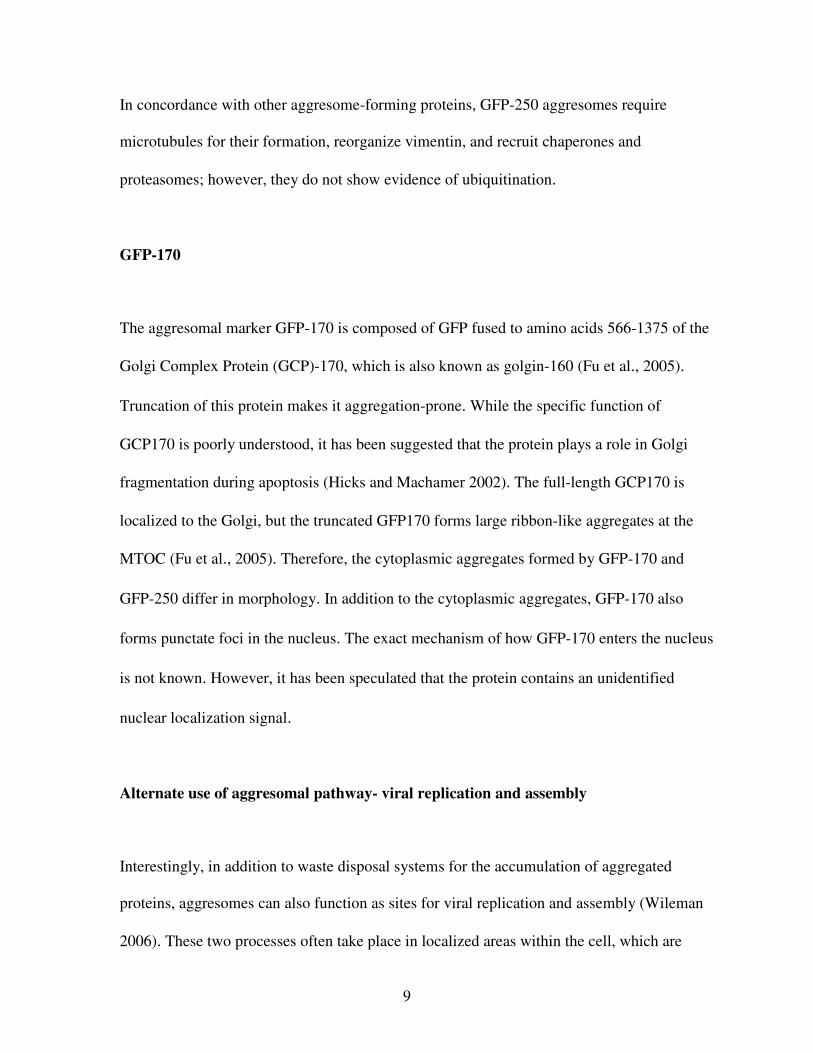
In concordance with other aggresome-forming proteins, GFP-250 aggresomes require
microtubules for their formation, reorganize vimentin, and recruit chaperones and
proteasomes; however, they do not show evidence of ubiquitination.
GFP-170
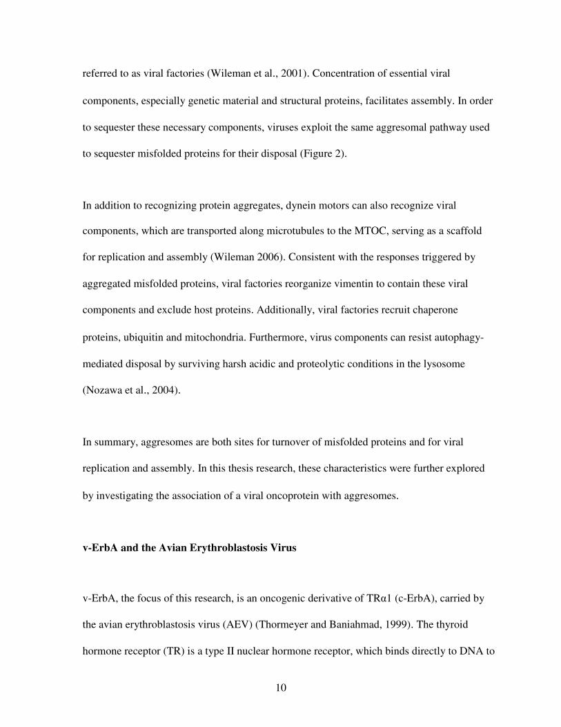
The aggresomal marker GFP-170 is composed of GFP fused to amino acids 566-1375 of the
Golgi Complex Protein (GCP)-170, which is also known as golgin-160 (Fu et al., 2005).
Truncation of this protein makes it aggregation-prone. While the specific function of
GCP170 is poorly understood, it has been suggested that the protein plays a role in Golgi
fragmentation during apoptosis (Hicks and Machamer 2002). The full-length GCP170 is
localized to the Golgi, but the truncated GFP170 forms large ribbon-like aggregates at the
MTOC (Fu et al., 2005). Therefore, the cytoplasmic aggregates formed by GFP-170 and
GFP-250 differ in morphology. In addition to the cytoplasmic aggregates, GFP-170 also
forms punctate foci in the nucleus. The exact mechanism of how GFP-170 enters the nucleus
is not known. However, it has been speculated that the protein contains an unidentified
nuclear localization signal.
Alternate use of aggresomal pathway- viral replication and assembly
Interestingly, in addition to waste disposal systems for the accumulation of aggregated
proteins, aggresomes can also function as sites for viral replication and assembly (Wileman
2006). These two processes often take place in localized areas within the cell, which are
10
referred to as viral factories (Wileman et al., 2001). Concentration of essential viral
components, especially genetic material and structural proteins, facilitates assembly. In order
to sequester these necessary components, viruses exploit the same aggresomal pathway used
to sequester misfolded proteins for their disposal (Figure 2).
In addition to recognizing protein aggregates, dynein motors can also recognize viral
components, which are transported along microtubules to the MTOC, serving as a scaffold
for replication and assembly (Wileman 2006). Consistent with the responses triggered by
aggregated misfolded proteins, viral factories reorganize vimentin to contain these viral
components and exclude host proteins. Additionally, viral factories recruit chaperone
proteins, ubiquitin and mitochondria. Furthermore, virus components can resist autophagy-
mediated disposal by surviving harsh acidic and proteolytic conditions in the lysosome
(Nozawa et al., 2004).
In summary, aggresomes are both sites for turnover of misfolded proteins and for viral
replication and assembly. In this thesis research, these characteristics were further explored
by investigating the association of a viral oncoprotein with aggresomes.
v-ErbA and the Avian Erythroblastosis Virus
v-ErbA, the focus of this research, is an oncogenic derivative of TRα1 (c-ErbA), carried by
the avian erythroblastosis virus (AEV) (Thormeyer and Baniahmad, 1999). The thyroid
hormone receptor (TR) is a type II nuclear hormone receptor, which binds directly to DNA to
11
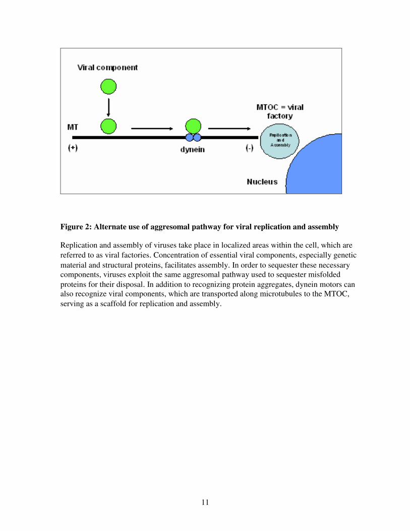
Figure 2: Alternate use of aggresomal pathway for viral replication and assembly
Replication and assembly of viruses take place in localized areas within the cell, which are
referred to as viral factories. Concentration of essential viral components, especially genetic
material and structural proteins, facilitates assembly. In order to sequester these necessary
components, viruses exploit the same aggresomal pathway used to sequester misfolded
proteins for their disposal. In addition to recognizing protein aggregates, dynein motors can
also recognize viral components, which are transported along microtubules to the MTOC,
serving as a scaffold for replication and assembly.
12
alter target gene transcription in response to the presence or absence of thyroid hormone (T3)
(Nagl et al., 1995). When bound to T3, it can activate transcription of genes involved in
mammalian and avian homeostasis, development, and metabolism. In the absence of T3, TR
acts a repressor of these genes. Three out of the four isoforms of TR, TRα1, TRβ1, and TRβ2
can activate transcription in response to T3 (Bunn et al., 2001).
AEV causes erythroleukemia and sarcomas in chickens (Braliou et al., 2001). v-ErbA acts as
a repressor of T3-responsive genes, contributing to the transformation of erythroblasts by
preventing cell differentiation and promoting sustained cell proliferation.
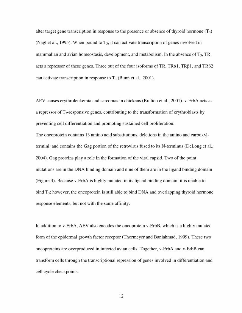
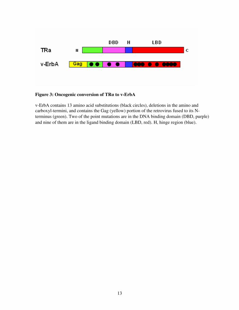
The oncoprotein contains 13 amino acid substitutions, deletions in the amino and carboxyl-
termini, and contains the Gag portion of the retrovirus fused to its N-terminus (DeLong et al.,
2004). Gag proteins play a role in the formation of the viral capsid. Two of the point
mutations are in the DNA binding domain and nine of them are in the ligand binding domain
(Figure 3). Because v-ErbA is highly mutated in its ligand binding domain, it is unable to
bind T3; however, the oncoprotein is still able to bind DNA and overlapping thyroid hormone
response elements, but not with the same affinity.
In addition to v-ErbA, AEV also encodes the oncoprotein v-ErbB, which is a highly mutated
form of the epidermal growth factor receptor (Thormeyer and Baniahmad, 1999). These two
oncoproteins are overproduced in infected avian cells. Together, v-ErbA and v-ErbB can
transform cells through the transcriptional repression of genes involved in differentiation and
cell cycle checkpoints.
13
Figure 3: Oncogenic conversion of TRα to v-ErbA
v-ErbA contains 13 amino acid substitutions (black circles), deletions in the amino and
carboxyl-termini, and contains the Gag (yellow) portion of the retrovirus fused to its N-
terminus (green). Two of the point mutations are in the DNA binding domain (DBD, purple)
and nine of them are in the ligand binding domain (LBD, red). H, hinge region (blue).
14
Role of v-ErbA in oncogenesis/dominant negative activity
v-ErbA can contribute to oncogenesis in a number of ways. First, it competes with TR for T3-
responsive DNA elements in the promoter region that aid in the transcription of T3-
responsive genes (Bonamy et al., 2005). Second, v-ErbA competes with TR for auxiliary
factors (such as retinoid X receptor) and cofactors that promote T3-mediated transcription.
Third, v-ErbA dimerizes with TR and sequesters it in cytoplasmic foci.
Though TR shuttles between the nucleus and cytoplasm, it localizes primarily to the nucleus
(Bunn et al., 2001). The physiological reason for the nucleocytoplasmic shuttling of TR is
poorly understood. v-ErbA also shuttles between the nucleus and cytoplasm, but it has a
significantly greater cytoplasmic localization than its homolog, TRα1 (DeLong et al., 2004).
While TRα1 exhibits a mostly diffuse distribution throughout the nucleus, v-ErbA
accumulates in cytoplasmic foci. Additionally, the oncoprotein exhibits a different mode of
nuclear export in comparison to TRα1 (DeLong et al., 2004). Prior research has shown that
v-ErbA exits the nucleus by a CRM1-dependent nuclear export pathway, primarily mediated
by the acquisition of Gag sequence on the protein, which contains a nuclear export sequence.
TRα1 is not solely dependent on CRM1 for nuclear export (Grespin et al., 2008). These
findings suggest that differential cellular localization plays an important role in oncogenesis.

15
Specific Aims of Research
The overall objective of this thesis research was to determine whether the cytoplasmic foci
formed by v-ErbA are a result of targeting to aggresomes. To this end, whether v-ErbA
colocalized with aggresomal markers and possessed defining aggresomal characteristics was
investigated. The following questions were addressed:
1. Does v-ErbA colocalize with aggresomal markers?
2. Is the formation of v-ErbA foci microtubule dependent?
3. Does proteasome inhibition enhance the size of v-ErbA foci?
4. Do v-ErbA foci disrupt vimentin intermediate filaments?
16
Materials and Methods
Plasmids
All plasmids used in this study encode fluorescently-tagged fusion proteins, in order to
analyze the subcellular distribution of the proteins of interest. The GFP-250 and GFP-170
plasmids were gifts from E. Sztul (University of Alabama). GFP-250 was constructed by
subcloning the PCR product of p115 cDNA into the enhanced green fluorescent protein
(EGFP) expression vector pEGFP-C2 (Garcia-Mata et al., 1999). The GFP-170 plasmid
expresses an EGFP- tagged portion of GCP-170 (Fu et al., 2005). The subcloning of v-ErbA
into the GFP and DsRed2 vectors was conducted by other researchers in Dr. Allison’s
laboratory (Bunn et al, 2001; Bonamy et al., 2005). These plasmids were amplified in E.coli
DH5α cells and purified using a Qiagen midi-prep kit according to the manufacturer’s
instructions. DNA yield was quantified using a Nanodrop ND-1000 full spectrum UV/Vis
Spectrophotometer (NanoDrop Technologies).
GFP and DsRed2 are both naturally fluorescent proteins. GFP is derived from the jellyfish
Aequorea victoria (Tsien, 1998). The protein has a major excitation peak at 395 nm (blue
light), resulting in emission of green light. DsRed2 is a variant of DsRed, which is a red
fluorescent protein derived from the coral Discosoma (Campbell et al., 2002). The DsRed2
protein has an excitation peak of 558 nm (green light), resulting in the emission of red light.
17
Cell culture, transfections, and drug treatments
Transfection experiments were performed in order to introduce plasmid DNA encoding
fluorescently-tagged aggresomal markers and v-ErbA into HeLa human cells (Figure 4).
After transfection, plasmid DNA was transcribed and translated within the cell into
fluorescently-tagged proteins and their subcellular localization was observed by fluorescence
microscopy. GFP-250, GFP-170, GFP-v-ErbA, and DsRed2-v-ErbA expression vectors were
transfected into HeLa cells. In single transfections, only one type of plasmid was introduced
into a cell (Figure 4a). In cotransfections, two plasmid DNA constructs were introduced into
a cell and expressed simultaneously in order to test for colocalization to the same region
within the cell (Figure 4b).
Cell culture was carried out in a Class II Biosafety Cabinet (Labconco). HeLa cells (human
cervix epithelioid carcinoma; ATCC CCL-2) were grown and maintained at 37ºC and 5%
CO2 in Minimum Essential Medium (Gibco) with 10% fetal bovine serum (FBS) (Invitrogen)
and penicillin-streptomycin antibiotics (100 U/mL penicillin and 100 µg/mL streptomycin) in
a sterile large (75cm3) filter-cap flask (NUNC Brand Products).
In preparation for transfection, cells were removed from the filter-cap flask by incubating
them in 2 mL of chilled 0.25% trypsin solution for approximately 2 minutes. The trypsin was
removed and the cells were incubated for another 5 minutes at 37ºC. After resuspension in
HeLa media, 2 mL of growth medium containing 1-3 X 10⁵
cells per well was added to each

18
(a) Single transfection (b) Cotransfection
Figure 4: Transfection and cotransfection
Transfection experiments were performed in order to introduce plasmid DNA encoding
fluorescently-tagged aggresomal markers and v-ErbA into HeLa cells. After transfection,
plasmid DNA was transcribed and translated into fluorescently-tagged proteins and their
subcellular localization was observed by fluorescence microscopy. GFP-250, GFP-170, GFP-
v-ErbA, and DsRed2-v-ErbA expression vectors were transfected into HeLa cells. In single
transfections, only one type of plasmid was introduced into a cell (a). In cotransfections, two
plasmid DNA constructs were introduced into a cell and expressed simultaneously in order to
test for colocalization to the same region within the cell (b).
19
well of a 6-well plate with glass coverslips (Fisher) and incubated at 37ºC for approximately
24 hours, reaching a confluency of about 50-60%.
Lipofectamine Reagent (Invitrogen) was used to perform transfections. For single
transfections, 2 µg of plasmid was diluted in Opti-MEM reduced serum media (Gibco) for a
total volume of 100µL. For cotransfections, 1 µg of each plasmid (2 µg total) was diluted in
Opti-MEM. A second 100µL solution was prepared with 10µL of Lipofectamine Reagent
and 90µL of Opti-MEM. The two solutions were combined (200 µL total) and incubated at
room temperature for 15 minutes. After incubation, 800 µL of Opti-MEM was added to the
200 µL solution of DNA-liposome complex. This 1 mL solution of diluted DNA-liposome
complex was added to each seeded coverslip in the 6-well plate. The transfected cells were
incubated at 37ºC for 16-20 hours. The Opti-MEM was removed from the cells and replaced
with 2 mL of complete media. Cells were fixed and stained 24-48 hours post-transfection to
be analyzed by fluorescence microscopy. The time period between transfection and fixation
depended on the experiment.
When testing the effect of treatment with the proteasome-inhibiting drug MG132 (Sigma)
and the microtubule-disrupting drug nocodazole (Sigma), 2 µL of MG132 solution,
nocodazole solution, or DMSO (vehicle control) were added to each well after media
replacement. Both MG132 and nocodazole were dissolved in DMSO at a concentration of 10
mg/mL. The treated cells were incubated for 16-20 hours to allow sufficient proteasome
inhibition and microtubule depolymerization.

20
Fixation, immunofluorescence staining, and processing of cells for imaging
24-48 hours after transfection, media was removed from cells. Cells were washed 3 times for
15 seconds in 2 mL per well with Dulbecco’s Phosphate-Buffered Saline (D-PBS: 0.10g
KCl, 0.10g KH2PO4, 4.00g NaCl, and 1.08g Na2HPO4·7H2O) for a solution of 500 mL). For
fixation, cells were incubated in a 3.7% formaldehyde solution with D-PBS for 10 minutes.
After fixation, the formaldehyde solution was removed and the cells were washed 3 times in
D-PBS for 5 minutes.
Cells being processed for immunofluorescence staining were permeabilized for 5 minutes in
a 0.2% Triton-X-100 solution diluted with D-PBS. Coverslips were inverted onto 30 µL of a
diluted antibody solution with 1.5% normal goat sera and D-PBS. A 1:200 dilution was used
for the monoclonal Cy3-conjugated anti-vimentin antibody (Sigma). The cells were
incubated in the antibody for 1 hour in the dark in a humidified chamber. The humidified
chamber consisted of a storage container lined with moist paper towels. The 30 µL of
antibody solution and coverslips were placed on parafilm, which was placed on top of the
moist paper towels.
Because the Cy3-conjugated vimentin antibody was labeled, incubation with a labeled
secondary antibody was not necessary. After incubation with the labeled primary antibody,
cells were washed 3 times in D-PBS for 5 minutes in the original 6-well plate. The washes
21
were performed on a rotating table, allowing antibody that was non-specifically bound to be
washed off the coverslip.
After the washes, coverslips were inverted and mounted onto slides with 8 µL of GelMount
with DAPI (0.5 mg/L). Cells that were not stained with antibody were mounted onto slides
and stained with DAPI immediately after the 3 post-fixation washes. All of the mounted
slides were viewed by fluorescence microscopy.
Fluorescence microscopy
Prepared slides were analyzed with an Olympus Fluorescence Microscope. Three separate
filters were used to visualize the DAPI-stained nucleus (UV for blue fluorescence), GFP-
tagged protein (blue light for green fluorescence), and both DsRed2-tagged protein and Cy3-
conjugated antibody (green light for red fluorescence). This allowed for the determination of
subcellular localization in relation to the nucleus. Cells were photographed with a Cooke
SensiCamQE digital camera. IP Lab software and Adobe Photoshop CS were used to
normalize, pseudocolor, and layer the captured images.
Cell scoring and statistical analysis
Three trials of colocalization assays between DsRed2-v-ErbA and GFP-250 were performed,
with at least 100 cells analyzed per trial. Transfected cells were categorized into 3 different
groups: total, partial, and no colocalization. Two trials of treatment of GFP-v-ErbA with
22
MG132 and nocodazole were conducted, with at least 100 cells analyzed per trial. Aggregate
size in the transfected and treated cells was categorized into 3 different groups: diffuse,
small, or large aggregates. Three trials of colocalization between GFP-v-ErbA and the anti-
vimentin antibody were performed, with greater than 100 cells analyzed per trial.
Once cells were counted in the drug treatment experiments, results were tested for statistical
significance using SPSS (v. 16). Data were analyzed for frequency distribution using a Chi-
Square test; a p-value of 0.001 or less was considered significant.
23
Results
v-ErbA colocalizes with aggresomal markers
Prior studies had shown that v-ErbA has a mainly cytoplasmic distribution, localized in
punctate foci (Bunn et al., 2001; Bonamy et al., 2005). To determine whether v-ErbA
associates with aggresomes, the distribution of v-ErbA was analyzed by transfection assays
with fluorescently-tagged fusion proteins.
Before testing for colocalization between v-ErbA and aggresomal markers, the cellular
localizations of DsRed2-v-ErbA, GFP-v-ErbA, GFP-170, and GFP-250 were observed
individually in HeLa cells. Consistent with prior studies (Garcia-Mata et al., 1999), GFP-250
formed entirely cytoplasmic aggregates (Figure 5a and b). After about 48 hours post-
transfection, some of the GFP-250-expressing cells displayed a single coalesced juxtanuclear
aggregate that was spherical in shape, while other cells displayed a number of smaller
aggregates throughout the cytoplasm. These smaller aggregates were not evenly dispersed or
uniform in size; aggregates were more heavily concentrated near one side of the nucleus. The
localization and variation in size of these aggregates suggests that smaller aggregates fused
together and were in the process of being transported to the microtubule organizing center
(MTOC). Also consistent with prior studies (Fu et al., 2005), GFP-170 formed both nuclear
and cytoplasmic aggregates after observing the protein’s distribution 48 hours after
transfection (Figure 5c and d). The protein localized to punctate foci in the nucleus and in
24
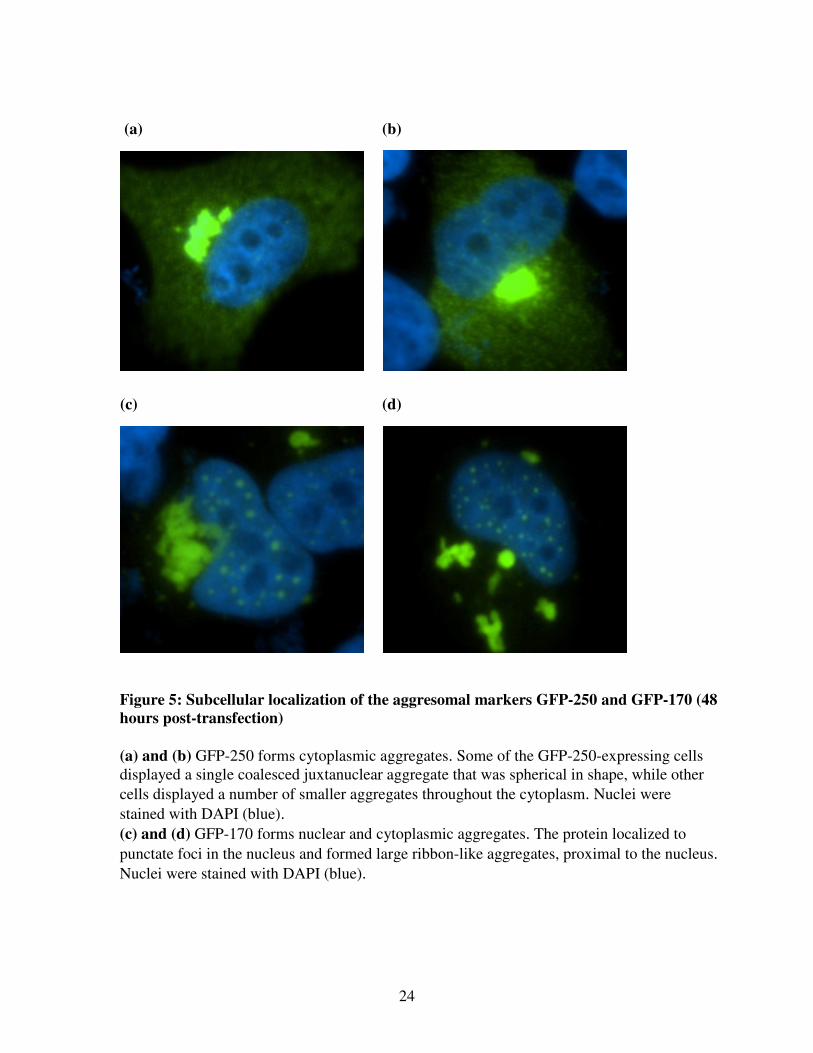
Figure 5: Subcellular localization of the aggresomal markers GFP-250 and GFP-170 (48
hours post-transfection)
(a) and (b) GFP-250 forms cytoplasmic aggregates. Some of the GFP-250-expressing cells
displayed a single coalesced juxtanuclear aggregate that was spherical in shape, while other
cells displayed a number of smaller aggregates throughout the cytoplasm. Nuclei were
stained with DAPI (blue).
(c) and (d) GFP-170 forms nuclear and cytoplasmic aggregates. The protein localized to
punctate foci in the nucleus and formed large ribbon-like aggregates, proximal to the nucleus.
Nuclei were stained with DAPI (blue).
(a) (b)
(c) (d)
25
contrast with GFP-250, formed large ribbon-like aggregates, instead of spherical aggregates,
proximal to the nucleus.
Forty-eight hours after transfection, GFP and DsRed2-tagged-v-ErbA formed almost entirely
cytoplasmic aggregates; there was a range of cytoplasmic distribution (not shown). Some v-
ErbA expressing cells contained a single coalesced juxtanuclear aggregate with a ribbon-like
and elongated morphology (Figure 6a), similar to the cytoplasmic aggregates formed by
GFP-170, while other cells contained smaller and more dispersed aggregates (Figure 6b), and
some cells had an entirely diffuse cytoplasmic distribution. As in the aggresomal markers,
the cytoplasmic foci formed by v-ErbA were not uniform in size or evenly dispersed,
suggesting that they were in the process of aggresome formation. The cellular distribution
pattern of v-ErbA was similar to the distribution patterns of the aggresomal marker GFP-250
and the cytoplasmic aggregates of GFP-170.
To investigate whether or not the foci formed by v-ErbA colocalized with aggresomal
markers, HeLa cells were cotransfected with GFP-tagged aggresomal markers (GFP-170 and
GFP-250) and DsRed2-tagged v-ErbA and their subcellular localizations were observed.
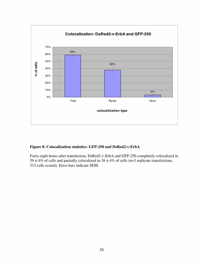
Forty-eight hours after transfection, DsRed2-v-ErbA and GFP-250 completely colocalized in
59 ± 4% of cells and partially colocalized in 38 ± 4% of cells (n=3 replicate transfections,
313 cells scored) (Figure 7, Figure 8). These results strongly suggest that DsRed2-v-ErbA
and GFP-250 are targeted to the same subcellular location, despite their slight differences in
morphology.
26
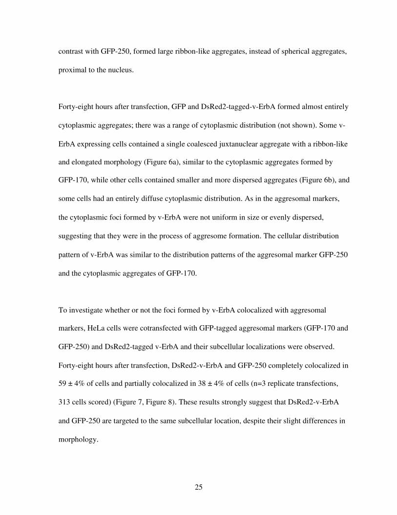
Figure 6: Subcellular distribution of DsRed2 v-ErbA (48 hours post-transfection)
(a) DsRed2-v-ErbA-expressing cell containing a single coalesced juxtanuclear aggregate
with a ribbon-like and elongated morphology, similar to the cytoplasmic aggregates formed
by GFP-170 (See Figure 5c). Nuclei were stained with DAPI (blue).
(b) DsRed2-v-ErbA-expressing cell containing smaller and more dispersed aggregates.
Nuclei were stained with DAPI (blue).
(a) (b)
27
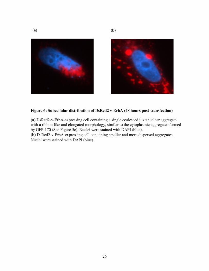
Figure 7: Colocalization of the aggresomal marker GFP-250 and DsRed2-v-ErbA (48
hours post-transfection)
(a) and (d) Subcellular distribution of GFP-250 (green)
(b) and (e) Subcellular distribution of DsRed2-v-ErbA (red)
(c) and (f) Merge of GFP-250 and DsRed2-v-ErbA. Yellow indicates colocalization between
GFP-250 and DsRed2-v-ErbA. Nuclei were stained with DAPI (blue).
(a) (b) (c)
(d) (e) (f)
28
Colocalization- DsRed2-v-ErbA and GFP-250
59%
3%
38%
0%
10%
20%
30%
40%
50%
60%
70%
Total Partial None
colocalization type
% o
f cell
s
Figure 8: Colocalization statistics- GFP-250 and DsRed2-v-ErbA
Forty-eight hours after transfection, DsRed2-v-ErbA and GFP-250 completely colocalized in
59 ± 4% of cells and partially colocalized in 38 ± 4% of cells (n=3 replicate transfections,
313 cells scored). Error bars indicate SEM.
29
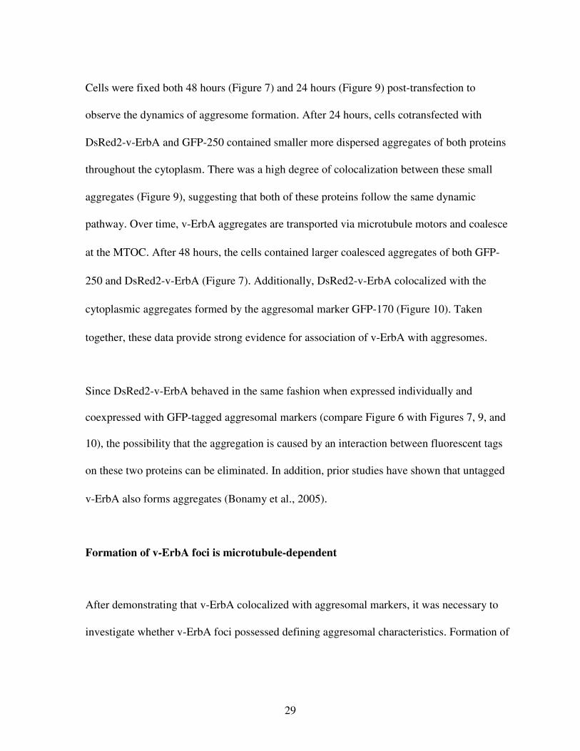
Cells were fixed both 48 hours (Figure 7) and 24 hours (Figure 9) post-transfection to
observe the dynamics of aggresome formation. After 24 hours, cells cotransfected with
DsRed2-v-ErbA and GFP-250 contained smaller more dispersed aggregates of both proteins
throughout the cytoplasm. There was a high degree of colocalization between these small
aggregates (Figure 9), suggesting that both of these proteins follow the same dynamic
pathway. Over time, v-ErbA aggregates are transported via microtubule motors and coalesce
at the MTOC. After 48 hours, the cells contained larger coalesced aggregates of both GFP-
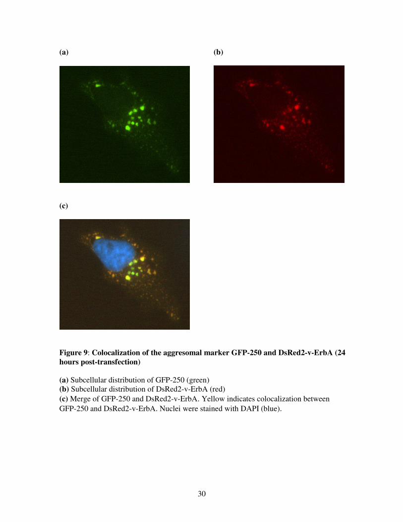
250 and DsRed2-v-ErbA (Figure 7). Additionally, DsRed2-v-ErbA colocalized with the
cytoplasmic aggregates formed by the aggresomal marker GFP-170 (Figure 10). Taken
together, these data provide strong evidence for association of v-ErbA with aggresomes.
Since DsRed2-v-ErbA behaved in the same fashion when expressed individually and
coexpressed with GFP-tagged aggresomal markers (compare Figure 6 with Figures 7, 9, and
10), the possibility that the aggregation is caused by an interaction between fluorescent tags
on these two proteins can be eliminated. In addition, prior studies have shown that untagged
v-ErbA also forms aggregates (Bonamy et al., 2005).
Formation of v-ErbA foci is microtubule-dependent
After demonstrating that v-ErbA colocalized with aggresomal markers, it was necessary to
investigate whether v-ErbA foci possessed defining aggresomal characteristics. Formation of
30
(a) (b)
(c)
Figure 9: Colocalization of the aggresomal marker GFP-250 and DsRed2-v-ErbA (24
hours post-transfection)
(a) Subcellular distribution of GFP-250 (green)
(b) Subcellular distribution of DsRed2-v-ErbA (red)
(c) Merge of GFP-250 and DsRed2-v-ErbA. Yellow indicates colocalization between
GFP-250 and DsRed2-v-ErbA. Nuclei were stained with DAPI (blue).
31
(a) (b)
(c)
Figure 10: Colocalization of the aggresomal marker GFP-170 and DsRed2-v-ErbA (48
hours post-transfection)
(a) Subcellular distribution of GFP-170 (green)
(b) Subcellular distribution of DsRed2-v-ErbA (red)
(c) Merge of GFP-170 and DsRed2-v-ErbA. Yellow indicates colocalization between GFP-
250 and DsRed2-v-ErbA. Nuclei were stained with DAPI (blue)
32
the aggresome requires transport of smaller aggregated proteins along microtubules. Thus,
microtubule disruption prevents aggresome formation.
If the cytoplasmic foci formed by v-ErbA are a result of targeting to aggresomes, then
microtubule inhibition would prevent the formation of these large, coalesced structures. To
determine whether the disruption of microtubules would prevent the formation of the
coalesced foci, HeLa cells expressing GFP-v-ErbA were treated with nocodazole for 20
hours (starting 16 hours post-transfection). Nocodazole disrupts microtubules by preventing
their polymerization. The drug binds to a sulfhydryl group on β-tubulin and prevents
disulfide linkages with additional tubulin heterodimers (Luduena and Roach, 1991; Vasquez
et al., 1997). Microtubule disruption inhibits transport of cargo and cell division (Luduena
and Roach, 1991).
If aggresome formation was inhibited, one would expect to see a diffuse expression pattern
or a dispersal of small aggregates throughout the cytoplasm. As predicted, treatment of GFP-
v-ErbA- expressing cells with nocodazole resulted in mostly cells with a diffuse pattern of v-
ErbA expression (Figure 11). Only 13 ± 3% of untreated cells (n=2 replicate transfections,
200 cells scored) contained diffuse aggregates (Figure 12). Upon nocodazole treatment, there
was a significant shift (p < 0.001) to 55 ± 3% of cells containing diffuse aggregates (Figure
12).While 46 ± 5% of untreated v-ErbA-expressing cells contained large aggregates, only 7 ±
3% of nocodazole-treated cells had large aggregates (Figure 12). Because nocodazole cannot
disrupt aggregates that are already intact prior to treatment, many of the aggregates observed
33
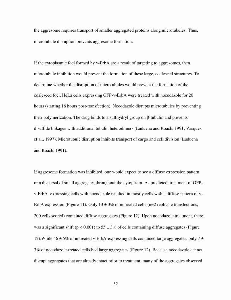
(a) (b)
(c)
Figure 11: Formation of v-ErbA foci is microtubule-dependent
To determine whether the disruption of microtubules would prevent the formation of the
coalesced foci, cells expressing GFP-v-ErbA were treated with nocodazole for 20 hours
(starting 16 hours post-transfection). Nuclei were stained with DAPI (blue).
(a) Untreated cell forming large juxtanuclear foci. (b) Nocodazole-treated cell forming diffuse aggregates
(c) Nocodazole-treated cell forming small aggregates, uniform in size.
34
Effect of Nocodazole on v-ErbA Foci Size
0%
10%
20%
30%
40%
50%
60%
70%
Diffuse Small Large
Aggregate size
% o
f cell
s
(-) nocodazole
(+) nocodazole
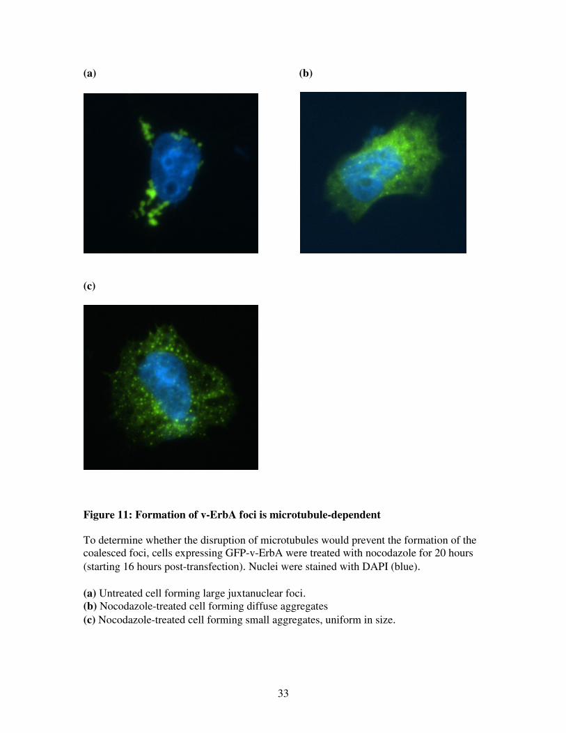
Figure 12: Effect of nocodazole on v-ErbA foci size
Upon nocodazole treatment, there was a significant shift (P < 0.001) in frequency distribution
from larger foci to smaller, more diffuse foci. Error bars indicate SEM.
Chi-Square
p > 0.001
200 cells scored

35
in treated cells could have been preexisting. Data thus suggest that over time, v-ErbA
aggregates are transported via microtubule motors and coalesce at the MTOC.
Proteasome inhibition enhances the size of v-ErbA foci
Proteasome inhibition leads to an increase in aggresome size (Johnston et al., 1998; Garcia-
Mata et al., 1999; Fu et al., 2005). The inhibition of proteasome activity leads to an increase
in protein that would normally be degraded, leading to an accumulation of aggregated protein
peripherally, which is transported to the MTOC. To determine whether the size of v-ErbA
foci would increase in response to proteasome inhibition, cells expressing GFP-v-ErbA were
treated with the proteasome inhibitor MG132 for 20 hours (starting 16 hours post-
transfection). MG132 inhibits proteasome degradation by inhibiting the chymotrypsin-like
activity of the proteasome, preventing the cleavage of peptide bonds within the protein of
interest (Lee and Goldberg, 1998).
As predicted, MG132 treatment increased the size v-ErbA foci. Only 41 ± 2% (n=2 replicate
transfections, 200 cells scored) of untreated cells contained large aggregates of GFP-v-ErbA,
but upon MG132 treatment, there was a significant shift (p > 0.001) to 78 ± 7% of v-ErbA-
expressing cells containing large aggregates (Figure 13). Interestingly, most of the large
aggregates were spherical in morphology and coalesced into a single aggregate (Figure 13b),
similar to the spherical aggregates formed by GFP-250, mutant huntingtin (Kuemmerle et al.,
1999) and cystic fibrosis transmembrane regulator (Johnston et al., 1998) after proteasome
36
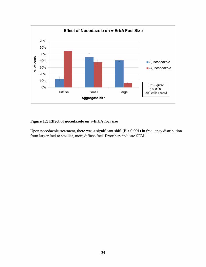
(a) (b)
(c)

37
(d)
Effect of MG132 Treatment on v-ErbA Aggregate SIze
0%
10%
20%
30%
40%
50%
60%
70%
80%
90%
Diffuse Small Large
Aggregate Size
% c
ell
s
(-) MG132
(+) MG132
Figure 13: Effect of MG132 treatment of v-ErbA aggregate size
To determine whether the size of v-ErbA foci would increase in response to proteasome
inhibition, cells expressing GFP-v-ErbA were treated with the proteasome inhibitor MG132
for 20 hours (16 hours post-transfection).
(a) Untreated v-ErbA expressing cell.
(b) MG132-treated v-ErbA expressing cell. Most of the large aggregates were spherical in
morphology and coalesced into a single aggregate.
(c) MG132-treated v-ErbA expressing cell. Some of the large aggregates had a more
undefined morphology (less common).
(d) Statistical analysis. Upon nocodazole treatment, there was a significant shift (p < 0.001)
in frequency distribution from smaller aggregates to large aggregates. Error bars indicate
SEM.
Chi-Square
p > 0.001
200 cells
scored
38
inhibition. This morphology is not observed in v-ErbA foci in the absence of proteasome
inhibition. In addition, a few of the large aggregates had a more undefined morphology
(Figure 13c). Taken together, these results suggest that the proteasome machinery normally
degrades some of the aggregated v-ErbA, thus preventing aggresome formation.
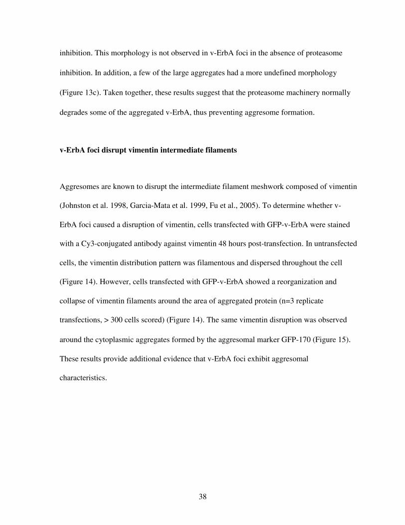
v-ErbA foci disrupt vimentin intermediate filaments
Aggresomes are known to disrupt the intermediate filament meshwork composed of vimentin
(Johnston et al. 1998, Garcia-Mata et al. 1999, Fu et al., 2005). To determine whether v-
ErbA foci caused a disruption of vimentin, cells transfected with GFP-v-ErbA were stained
with a Cy3-conjugated antibody against vimentin 48 hours post-transfection. In untransfected
cells, the vimentin distribution pattern was filamentous and dispersed throughout the cell
(Figure 14). However, cells transfected with GFP-v-ErbA showed a reorganization and
collapse of vimentin filaments around the area of aggregated protein (n=3 replicate
transfections, > 300 cells scored) (Figure 14). The same vimentin disruption was observed
around the cytoplasmic aggregates formed by the aggresomal marker GFP-170 (Figure 15).
These results provide additional evidence that v-ErbA foci exhibit aggresomal
characteristics.

39
(a) (b)
(c) (d)
Figure 14: v-ErbA foci disrupt vimentin intermediate filaments
To determine whether v-ErbA foci caused a disruption of vimentin filaments, cells
transfected with GFP-v-ErbA were stained with a Cy3-conjugated antibody against vimentin
48 hours post-transfection. (n=3, > 300 cells scored)
(a) Distribution of GFP-v-ErbA (green)
(b) Distribution of Cy3-conjugated antibody against vimentin (red)
(c) Merged image of GFP-v-ErbA and Cy3-vimentin. Nuclei stained with DAPI (blue). Cells
transfected with GFP-v-ErbA showed a reorganization and collapse of vimentin around the
area of aggregated protein. (d) In untransfected cells, the vimentin distribution pattern was filamentous and dispersed
throughout the cell. Nuclei stained with DAPI (blue).
40
(a) (b)
(c)
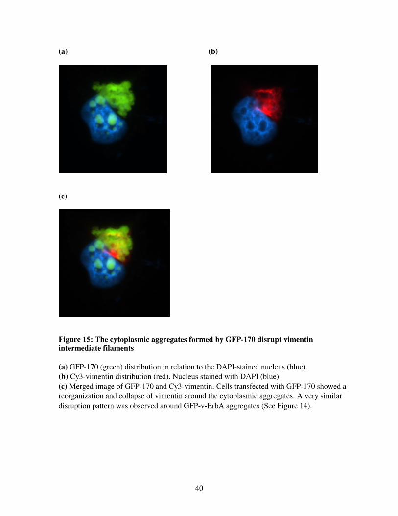
Figure 15: The cytoplasmic aggregates formed by GFP-170 disrupt vimentin
intermediate filaments
(a) GFP-170 (green) distribution in relation to the DAPI-stained nucleus (blue).
(b) Cy3-vimentin distribution (red). Nucleus stained with DAPI (blue)
(c) Merged image of GFP-170 and Cy3-vimentin. Cells transfected with GFP-170 showed a
reorganization and collapse of vimentin around the cytoplasmic aggregates. A very similar
disruption pattern was observed around GFP-v-ErbA aggregates (See Figure 14).
41
Discussion
v-ErbA, an oncoprotein derived from the avian erythroblastosis virus (AEV), mislocalizes to
cytoplasmic foci and sequesters the thyroid hormone receptor (TR) in these foci, contributing
to its oncogenic properties (Bonamy et al., 2005). This thesis research provides strong
evidence that cytoplasmic mislocalization of v-ErbA is a result of targeting to aggresomes,
either for turnover of misfolded protein or for viral replication and assembly. v-ErbA foci
colocalized with aggresomal markers, were dependent on microtubule transport for their
formation, were enhanced in size upon treatment with proteasome inhibitors, and disrupted
the intermediate filament meshwork composed of vimentin. These results indicate an
association between v-ErbA foci and aggresomes.
Dynamics and morphology of v-ErbA foci
While all aggresome-forming proteins follow similar dynamics, the final morphology of the
aggresome is variable. However, aggresome formation is generally categorized into two
broad categories: spherical or ribbon-like (Garcia-Mata et al., 2002). Spherical aggresomes
are formed by mutant huntingtin (Kuemmerle et al., 1999), mutant cystic fibrosis
transmembrane conductance regulator (CFTR) (Johnston et al., 1998), and the aggresomal
marker GFP-250 (Garcia-Mata et al., 1999). However, it is important to note that the
spherical aggresomes formed by CFTR are induced by proteasome inhibitors (Johnston et al.,
1998). Additionally, many viral particles that follow the aggresomal pathway have a
42
spherical morphology, evidenced by African Swine Fever Virus (ASFV) particles (Heath et
al., 2001). Ribbon-like aggresomes can be formed by UCH-L1 in Parkinson’s (Ardley et al.,
2004), glial fibrillary protein (GFAP) in Alexander’s Disease (Mignot et al., 2007), ATP7B
in Wilson’s disease (Harada et al., 2001), and the aggresomal marker GFP-170 (Fu et al.,
2005). The criteria for formation of either of these two distinct morphologies have yet to be
determined.
v-ErbA foci appear to form the ribbon-like shape seen in the cytoplasmic aggregates formed
by GFP-170, but are still very similar to the aggregates formed by GFP-250, which can be
more spherical in shape. Forty-eight hours post-transfection, the foci formed by DsRed2-
tagged-v-ErbA completely colocalized with GFP-250 in 59 ± 4% of cells and partially
colocalized with GFP-250 in 38 ± 4% of cells. The number of cells exhibiting complete
colocalization between v-ErbA and GFP-250 is somewhat higher than expected, since the
two proteins differ in final morphology. These results suggest that the final aggresomal
morphology is not always reached after 48 hours. Additionally, the results indicate that the
two proteins undergo the same dynamics, which is evidenced by the colocalization of smaller
aggregates formed by both v-ErbA and GFP-250 observed 24 hours post-transfection.
Microtubule-dependent formation
Disruption of microtubules with nocodazole yielded small, dispersed aggregates of v-ErbA
throughout the cytoplasm and prevented the coalescence of protein into a large aggregate at
the microtubule organizing center. Likewise, both misfolded proteins and viral particles are
43
dependent on microtubule transport for aggresome formation (Johnston et al., 1998; Garcia-
Mata et al., 1999; Heath et al., 2001). Thus, v-ErbA movement occurs by a microtubule
motor-driven process.
Proteasome inhibition
Treatment with the proteasome inhibitor MG132 enhanced the size of v-ErbA foci, which
was as predicted. Interestingly, the vast majority of the large aggregates coalesced into a
large, spherical structure. This shape was similar to the aggregates formed by GFP-250,
mutant huntingtin (Kuemmerle et al., 1999) and cystic fibrosis transmembrane regulator after
proteasome inhibition. This spherical morphology is not observed in v-ErbA foci in the
absence of proteasome inhibition. Because MG132 treatment speeds up the kinetics of
aggresome formation, the final shape of the aggresome can be reached more quickly, which
could explain why this spherical shape was not reached in untreated cells. Regardless, it is
evident from these results that protein degradation by the proteasome is critical for clearance
of v-ErbA from the cell.
Reorganization of vimentin
A number of aggresome-forming misfolded proteins and viral particles have been shown to
cause a collapse of vimentin filaments into a circular “cage” around the area of aggregated
protein for immobilization and containment (Johnston et al., 1998; Garcia-Mata et al., 1999;
Heath et al., 2001). This is only seen in aggresomes with a spherical morphology. Since v-

44
ErbA has a more ribbon-like morphology (in the absence of proteasome inhibition), the
formation of a circular “cage” around the area of aggregated protein was not expected. The
vimentin disruption pattern in v-ErbA-expressing cells was very similar to the disruption
pattern around the cytoplasmic aggregates formed by the aggresomal marker GFP-170.
However, it would be interesting to determine whether the vimentin collapsed in a cage-like
formation in v-ErbA-expressing cells that were treated with proteasome inhibitors, since
proteasome inhibition with MG132 caused v-ErbA to form a large and spherical aggregate.
Significance of targeting of v-ErbA to aggresomes- mechanism of turnover
The results presented here indicate an association between cytoplasmic v-ErbA foci and
aggresomes. One possible explanation for targeting of the oncoprotein v-ErbA to aggresomes
is to sequester the protein for turnover and disposal, preventing its dominant negative activity
on TR in the nucleus. If turnover of v-ErbA does in fact occur after the aggregates coalesce
at the microtubule-organizing center (MTOC), it is necessary to investigate the specific
mechanisms by which v-ErbA could be degraded.
First, the aggregates might by degraded by proteasome machinery. However, proteasome
degradation might not be very efficient at the aggresome (Garcia-Mata et al., 2002). Second,
the aggregates might undergo autophagic clearance. Proteasome degradation could be
dominant in the turnover of v-ErbA, autophagy could be dominant, or the two systems could
contribute equally. It is important to investigate the efficiency of v-ErbA degradation
promoted by both the proteasome and autophagy. It has been suggested that proteasome
45
degradation might not be very efficient at the aggresome (Garcia-Mata et al., 2002). Instead,
autophagy might be a more effective system for degradation because the process allows large
portions of cytoplasm containing aggregated proteins to be engulfed in an isolation
membrane and targeted to the lysosome for degradation. Regardless, either of these pathways
could become saturated after an accumulation of a certain amount of aggregated protein.
Therefore, there could be limits to how much protein degradation can be induced in response
to aggresome formation, regardless of the mechanisms. Cells might simply undergo
apoptosis after the accumulation of a specific amount of aggregated protein, evidenced in
cells containing glial fibrillary acidic protein (GFAP) aggregates (Mignot et al., 2007).
Significance of targeting of v-ErbA to aggresomes- remnant viral behavior for assembly
Another possible explanation for the association of cytoplasmic v-ErbA foci with
aggresomes could be a remnant viral behavior for assembly, exploiting the aggresomal
pathway for the turnover of misfolded protein. Aggresome-forming viral factories have been
shown to colocalize with the aggresomal marker GFP-250, disrupt vimentin intermediate
filaments, and depend on intact microtubules for their formation, consistent with the
properties of v-ErbA foci (Heath et al., 2001). The retroviral oncoprotein v-ErbA is a fusion
protein that contains a portion of the retroviral Gag sequence, which encodes structural
proteins involved in the formation of the viral capsid. It is not known whether or not the Gag
sequence itself is recognized by dynein motors, resulting in its transport to the MTOC.
Therefore, further studies could investigate the role of the Gag sequence in its aggresomal
behavior by investigating the effects of Gag deletion. Prior studies in our lab have shown that
46
the Gag portion of v-ErbA mediates CRM1-dependent nuclear export (DeLong et al., 2004).
Deletion of the v-ErbA Gag sequence resulted in a more nuclear localization. The functional
role of Gag-mediated nuclear export of v-ErbA is unknown, but the results in this thesis
support the hypothesis that it could be a remnant behavior of viral assembly by targeting of
v-ErbA to aggresomes.
General questions about the aggresome
There are a number of remaining questions about the aggresome. Although aggresome
formation by mutant proteins involved in neurodegenerative disorders and viral proteins is
well characterized, it is still not certain whether the aggresome is a general response to the
accumulation of aggregates caused by misfolded protein. To determine whether the
aggresome is a general response to misfolded protein, it is necessary to study aggresome
formation in more non-neurodegenerative disease systems.
Interestingly, aggresome formation is induced by a mutant of the androgen receptor (AR)
(Taylor et al., 2003), which is a member of the nuclear receptor subfamily along with the
thyroid hormone receptor (TR). However, it contains polyglutamine expansions and is
associated with spinobulbar muscular atrophy, a neurodegenerative disease. Therefore, the
AR mutant has very similar properties to the vast majority of aggresome-forming proteins
that have been studied and does not provide much insight into the nature of aggregation of
mutant proteins within the nuclear receptor family, specifically mutants of TR.
47
The association of aggresome formation with signaling pathways is also poorly understood.
For example, it is unclear what signaling pathways accompany the recruitment of proteasome
machinery, chaperone proteins, and mitochondria to the aggresome. Further insight into these
pathways could help find ways to induce and amplify degradation mechanisms, specifically
proteasome degradation and autophagy.
Are aggresomes pathogenic or cytoprotective?
Aggresome formation has been described to have pathogenic consequences. Studies in
transgenic mice have shown that the expression of a mutant huntingtin protein forms
aggresomes in a number of neurons, correlating with neurodegeneration and symptoms of
Huntington’s disease (Lin et al., 2001). Additionally, aggresomes formed by cytokeratin
proteins in alcoholic liver disease have been shown to correlate with cell death (Nakamichi et
al., 2002). However, this may have to do with the unavailability of functional protein and not
the aggresome itself.
The aggresome could contribute to pathogenesis for a number of reasons. First, the
aggresome recruits a great deal of the proteasome and chaperone machinery in the cell and
saturates the autophagic pathway. This would certainly have to cause detriment to the cell
eventually because other proteins in the cell would fail to be degraded; proto-oncogene
products would accumulate, disrupting cell cycle regulation. Second, the collapse of the
vimentin around the aggresome might make the rest of the cell vulnerable to mechanical
stress, since vimentin plays a structural role in the cell. Additionally, other organelles in the
48
cell might not be anchored appropriately due to the vimentin redistribution. Third, formation
of the aggresome might saturate dynein motors and lead to disrupted transport of organelles
and other cargo.
In contrast, a number of studies have described aggresome formation as cytoprotective.
Formation of this structure resulted in reduced levels of mutant Huntington and a decrease in
neurodegeneration (Arrasate et al., 2004; Iwata et al., 2005). Aggresome formation is also
associated with reduced cytotoxicity in cellular models containing mutants of peripheral
myelin protein 22 (PMP22), androgen receptor, and α-synuclein. (Fortun et al., 2003; Taylor
et al., 2003; Tanaka et al., 2004).
In conclusion, the aggresome seems to correlate with pathogenesis because of the
unavailability of functional protein, not because the aggresome itself is pathogenic.
Formation of the aggresome facilitates the turnover of aggregated protein, protecting cells
from consequences of the accumulation of dispersed cytoplasmic aggregates. The aggresome
might be an efficient temporary solution to the accumulation of aggregated protein, but it
might not be cytoprotective if proteasome machinery and autophagic pathways become
saturated.
If the association of v-ErbA and aggresomes is a result of sequestering v-ErbA for protein
turnover, formation of the aggresome appears to be cytoprotective in this situation.
49
Formation of the aggresome would protect cells from the pathogenic consequences of an
accumulation of v-ErbA aggregates dispersed throughout the cytoplasm. Additionally,
aggresome formation would protect cells from the dominant negative activity of v-ErbA on
TR in the nucleus. However, aggresome formation by v-ErbA could also have pathogenic
consequences because v-ErbA can sequester TR in its cytoplasmic foci, preventing TR from
regulating transcription of its target genes, adding to its dominant negative activity (Bonamy
et al., 2005; Bonamy and Allison, 2006).
Future directions
First, to confirm and extend the findings of this thesis research, it is necessary to provide
additional evidence that v-ErbA foci possess aggresomal characteristics. This can be
explored by testing the association of v-ErbA foci with proteasome subunits, chaperone
proteins, ubiquitin, the microtubule organizing center (gamma-tubulin), dynein and adaptor
proteins (dynactin and HDAC6), and markers of autophagy, primarily by using antibodies
against these components. These studies would not only strengthen the hypothesis of an
association between v-ErbA and aggresomes, but would also provide insight into the fate of
v-ErbA after its sequestration. For example, a strong association with ubiquitin and
proteasome subunits would suggest that v-ErbA foci undergo proteasome degradation, and a
strong association with autophagic markers would suggest that v-ErbA is targeted to the
lysosome for degradation. If there is an association between v-ErbA foci and proteasome
50
subunits or autophagic markers, it would be necessary to investigate the efficiency of these
turnover mechanisms.
The cellular fate of v-ErbA after localization to the aggresome can also be investigated by
tagging it with a photoactivatable fluorescent protein (PAFP). Future studies in our lab will
tag v-ErbA with Dendra, a PAFP derived from the octocoral Dendronephthya, which
undergoes laser-induced photoconversion from green to red fluorescence (Gurskaya et al.,
2006). Therefore, the cellular localization of v-ErbA can be determined both before and after
photoactivation, induced after v-ErbA has localized to the aggresome. These studies will
provide insight into whether specific, photoactivated v-ErbA foci enlarge, are degraded, or
migrate to another subcellular compartment, such as the nucleus, after aggresome formation.
Preliminary studies in our lab have demonstrated that the formation of v-ErbA foci; thus,
aggresome formation, is partially reversible. Because v-ErbA is a shuttling protein, it is
contained in the nucleus when export is blocked. When treated with the drug leptomycin B
(LMB), which inhibits CRM1-mediated nuclear export, smaller cytoplamic v-ErbA foci have
been shown to disappear and apparently accumulate in the nucleus. However, large
cytoplasmic foci formed by v-ErbA do not appear to undergo disassociation and remain
cytoplasmic.
In conclusion, the results presented in this thesis and future studies will all provide insight
into the viral oncoprotein v-ErbA and its associations with aggresomal pathways, either for
protein turnover or viral assembly. Additionally, more knowledge can be gained concerning
51
the fate of TR after its mislocalization to the cytoplasm by v-ErbA, and the role this plays in
oncogenesis. Finally, these studies contribute to a general understanding of the aggresome
and its role in pathogenesis and turnover of misfolded proteins.
52
References
Arnaud F, Murcia P, and Palmarini, M. Mechansims of late restriction induced by an
endogenous virus. J Virol. 2007; 81: 11441-51.
Arrasate M, Mitra S, Schweitzer ES, Segal MR, Finkbeiner S. Inclusion body formation
reduces levels of mutant huntingtin and risk of neuronal cell death. Nature 2004; 431: 805–
10.
Bonamy GM, Guiochon-Mantel A, Allison LA. Cancer promoted by the oncoprotein v-ErbA
may be due to subcellular mislocalization of nuclear receptors. Mol Endocrinol 2005; 19:
1213-1230.
Braliou GG, Ciana P, Klaassen W, Gandrillon O, Stunnenberg HG. The v-ErbA oncoprotein
quenches the activity of an erythroid-specific enhancer. Oncogene 2001; 20: 775–787.
Bunn CF, Neidig JA, Freidinger KE, Stankiewicz TA, Weaver BS, McGrew J, Allison LA.
Nucleocytoplasmic shuttling of the thyroid hormone receptor α. Mol Endocrinol 2001; 15:
512-33.
Campbell RE, Tour O, Palmer AE, Steinbach PA, Baird GS, Zacharias DA, Tsien RY. A
monomeric red fluorescent protein. Proc Natl Acad Sci USA 2002; 99: 7877-82.
DeLong LJ, Bonamy GM, Fink EN, Allison LA. Nuclear export of the oncoprotein v-ErbA is
mediated by acquisition of a viral nuclear export sequence. J Biol Chem. 2004; 279: 15356-
67.
Fabunmi RP, Wigley WC, Thomas PJ, DeMartino GN. Activity and regulation of the
centrosome-associated proteasome. J Biol Chem 2000; 275: 409–413.
Fabbro, M. and Henderson, B. Regulation of tumor suppressors by nuclear-cytoplasmic
shuttling. Exp. Cell Res. 1003; 282: 59–69.
Fortun J, Dunn W, Joy S, Li J, and Notterpek L. Emerging role for autophagy in the removal
of aggresomes in Schwann cells, J. Neurosci. 23; 2003:10672–10680.
Fu L, Gao YS, Tousson A, Shah A, Chen TL, Vertel BM, Sztul E. Nuclear Aggresomes
Form by Fusion of PML-associated Aggregates. Mol Biol Cell. 2005; 16:4905-4917.
Garcia-Mata R, Bebok Z, Sorscher EJ, Sztul ES. Characterization and dynamics of
aggresome formation by a cytosolic GFP-chimera. J Cell Biol 1999; 146:1239-1254.

53
Garcia-Mata R, Gao YS, Sztul E. Hassles with taking out the garbage: aggravating
aggresomes. Traffic 2002; 3: 388-96.
Grespin, M.E., Bonamy, G.M., Cameron, N.G., Adam, L.E., Atchison, A.P., Fratto, V.M.,
Allison, L.A. Thyroid hormone receptor α1 follows a cooperative CRM1/calreticulin-
mediated nuclear export pathway. Journal of Biological Chemistry 2008, under revision.
Gurskaya NG, Verkhusha VV, Shcheglov AS, Staroverov DB, Chepurnykh TV, Fradkov AF,
Lukyanov S, Lukyanov KA. Engineering of a monomeric green-to-red photoactivatable
fluorescent protein induced by blue light. Nat Biotechnol. 2006; 24: 461-5.
Harada M, Sakisaka S, Terada K, Kimura R, Kawaguchi T, Koga H, Kim M, Taniguchi E,
Hanada S, Suganuma T, Furuta K, Sugiyama T, Sata M. A mutation of the Wilson disease
protein, ATP7B, is degraded in the proteasomes and forms protein aggregates.
Gastroenterology 2001;120: 967–974.
Heath CM, Windsor M, Wileman T. Aggresomes resemble sites specialized for virus
assembly. J. Cell Biol. 2001; 153: 449–56.
Hicks, S. W., and Machamer, C. E. The NH2-terminal domain of Golgin-160 contains both
Golgi and nuclear targeting information. J. Biol. Chem. 2002; 277: 35833-35839.
Iwata A, Riley B, Johnston J and Kopito R. HDAC6 and microtubules are required for
autophagic degradation of aggregated huntingtin, J. Biol. Chem. 280; 2005:40282–40292.
Johnston JA, Ward CL, Kopito RR. Aggresomes: A cellular response to misfolded proteins. J
Cell Biol 1998; 143: 1883-1898.
Klionsky DL. The molecular machinery of autophagy: unanswered questions. J.
Cell Sci. 2005; 118: 7–18.
Kuemmerle S, Gutekunst CA, Klein AM, Li XJ, Li SH, Beal MF, Hersch SM, Ferrante RJ.
Huntington aggregates may not predict neuronal death in Huntington's disease. Ann Neurol
1999; 46: 842–849.
Lee, D.H., and Goldberg, A.L., Proteasome inhibitors: valuable new tools for cell biologists.
Trends Cell Biol. 1998; 8: 397-403.
Lin CH, Tallaksen-Greene S, Chien WM, Cearley JA, Jackson WS, Crouse AB, Ren S, Li
XJ, Albin RL, Detloff PJ. Neurological abnormalities in a knock-in mouse model of
Huntington's disease. Hum Mol Genet 2001; 10: 137-144.
Luduena, R.F., and Roach, M.C, Tubulin sulfhydryl groups as probes and targets for
antimitotic and antimicrotubule agents. Pharmacol. Ther. 1991; 49: 133-152.
54
Masliah E, Rockenstein E, Veinbergs I, Mallory M, Hashimoto M, Takeda A, Sagara Y, Sisk
A, Mucke L. Dopaminergic loss and inclusion body formation in alpha-synuclein mice:
Implications for neurodegenerative disorders. Science 2000; 287: 1265–1269
Mignot C, Delarasse C, Escaich S, Della Gaspera B, Noé E, Colucci-Guyon E, Babinet C,
Pekny M, Vicart P, Boespflug-Tanguy O, Dautigny A, Rodriguez D, Pham-Dinh D. Dynamics of mutated GFAP aggregates revealed by real-time imaging of an astrocyte model
of Alexander disease. Exp Cell Res. 2007; 313: 2766-79.
Mittal S, Dubey D, Yamakawa K, Ganesh S. Lafora disease proteins malin and laforin are
recruited to aggresomes in response to proteasomal impairment. Hum Mol Genet. 2007;
16:753-62.
Mortimore GE, Miotto G, Venerando R, Kadowaki M, Autophagy. Subcell Biochem 1996;
27: 93–135.
Muchowski PJ, Schaffar G, Sittler A, Wanker EE, Hayer-Hartl MK, Hartl FU. Hsp70 and
hsp40 chaperones can inhibit self-assembly of polyglutamine proteins into amyloid-like
fibrils. Proc Natl Acad Sci USA 2000; 97: 7841-7846.
Muchowski, P. J. , Ning, K. , D'Souza-Schorey, C. & Fields, S. Requirement of an intact
microtubule cytoskeleton for aggregation and inclusion body formation by a mutant
huntingtin fragment. Proc. Natl Acad. Sci. USA 2002; 99: 727–732.
Nagl SB, Nelson CC, Romaniuk PJ, Allison LA. Constitutive transactivation by the thyroid
hormone receptor and a novel pattern of activity of its oncogenic homolog v-ErbA in
Xenopus oocytes. Mol Endocrinol. 1995; 9: 1522-32.
Nakamichi I, Hatakeyama S, and Nakayama K. Formation of Mallory body-like inclusions
and cell death induced by deregulated expression of keratin 18, Mol. Biol. Cell 2002; 13:
3441–3451.
Nelson, D.S., Alvarez, C., Gao, Y.S., Garcia-Mata, R., Fialkowski, E., Sztul, E. The
membrane transport factor TAP/p115 cycles between the Golgi and earlier secretory
compartments and contains distinct domains required for its localization and function. J. Cell
Biol. 1998: 143: 319-331
Nozawa N, Yamauchi Y, Ohtsuka K, Kawaguchi Y, Nishiyama Y. Formation of aggresome-
like structures in herpes simplex virus type 2-infected cells and a potential role in virus
assembly. Exp Cell Res; 2004; 299: 486-97.
Ross CA and Poirier MA. Protein aggregation and neurodegenerative disease. Nat Rev Mol
Cell Biol. 2005; 6: 891-8.
55
Saliba RS, Munro PM, Luthert PJ, Cheetham ME. The cellular fate of mutant rhodopsin:
quality control, degradation and aggresome formation. J Cell Sci. 2002; 115: 2907-18.
Stommel J. M., Marchenko N. D., Jimenez G. S., Moll U. M., Hope T. J., Wahl G. M. A
leucine-rich nuclear export signal in the p53 tetramerization domain: regulation of
subcellular localization and p53 activity by NES masking. Embo J. 1999; 18:1660–72.
Tanaka M, Kim Y, Lee G, Junn E, Iwatsubo T, Mouradian M. Aggresomes formed by α-
synuclein and synphilin-1 are cytoprotective. J Biol Chem. 2004; 279: 4625-4631.
Taylor JP, Tanaka F, Robitschek J, Sandoval CM, Taye A, Markovic-Plese S, Fischbeck KH. Aggresomes protect cells by enhancing the degradation of toxic polyglutamine-containing
protein. Hum Mol Genet; 2003: 749-757.
Thormeyer D, Baniahmad A. The v-erbA oncogene. Int J Mol Med. 1999; 4: 351-8.
Tsien, RY. The green fluorescent protein. Annu. Rev. Biochem. 1998; 67: 509-44.
Vasquez, RJ, Howell B, Yvon AM, Wadsworth P, Cassumeris L. Nanomolar concentrations
of nocodazole alter microtubule dynamic instability in vivo and in vitro. Mol. Biol. Cell
1997; 8: 973-985.
Wigley WC, Fabunmi RP, Lee MG, Marino CR, Muallem S, DeMartino GN, Thomas PJ.
Dynamic association of proteasomal machinery with the centrosome. J Cell Biol 1999; 145:
481-490.
Wileman T. Aggresomes and autophagy generate sites of virus replication. Science 2006;
312: 875–78.
Wojcik C, Schroeter D, Wilk S, Lamprecht J, Paweletz N. Ubiquitin-mediated proteolysis
centers in HeLa cells: Indication from studies of an inhibitor of the chymotrypsin-like
activity of the proteasome. Eur J Cell Biol 1996; 71: 311–318.
Zatloukal K, Stumptner C, Lehner M, Denk H, Baribault H, Eshkind LG, Franke WW. Cytokeratin 8 protects from hepatotoxicity, and its ratio to cytokeratin 18 determines the
ability of hepatocytes to form Mallory bodies. Am J Pathol 2000; 156: 1263–1274.
56
Acknowledgements
A number of people contributed to the successful completion of this thesis research. First and
foremost, Dr. Lizabeth Allison provided endless support, advice, and time throughout the last
two years that she has supervised my research. Working in her laboratory has been an
incredibly rewarding and enjoyable experience. I would also like to thank Dr. Eric Engstrom,
Dr. Oliver Kerscher, and Dr. Lisa Landino for their advice, enthusiasm, and entertainment
while serving on my thesis committee. Vinny and Molly Roggero assisted with statistical
analyses of this thesis research. In addition, Vinny taught me a number of techniques when I
first started working in the laboratory. Other members of the Allison laboratory, past and
present, provided plasmids, assistance, and amusement throughout this thesis research. I
couldn’t have asked for a more delightful laboratory environment. Additionally, Elizabeth
Sztul contributed plasmids essential for this research.
This thesis research was supported by a number of Student Research Grants and a Student
Summer Research Fellowship Award, which were all funded by Howard Hughes Medical
Institute grants through the Undergraduate Biological Sciences Education Program to the
College of William and Mary. Additional funding was provided by a Mary E. Ferguson
Memorial Research Grant through the College of William and Mary Biology department.
Funding also came from grants to Dr. Allison from the National Science Foundation.
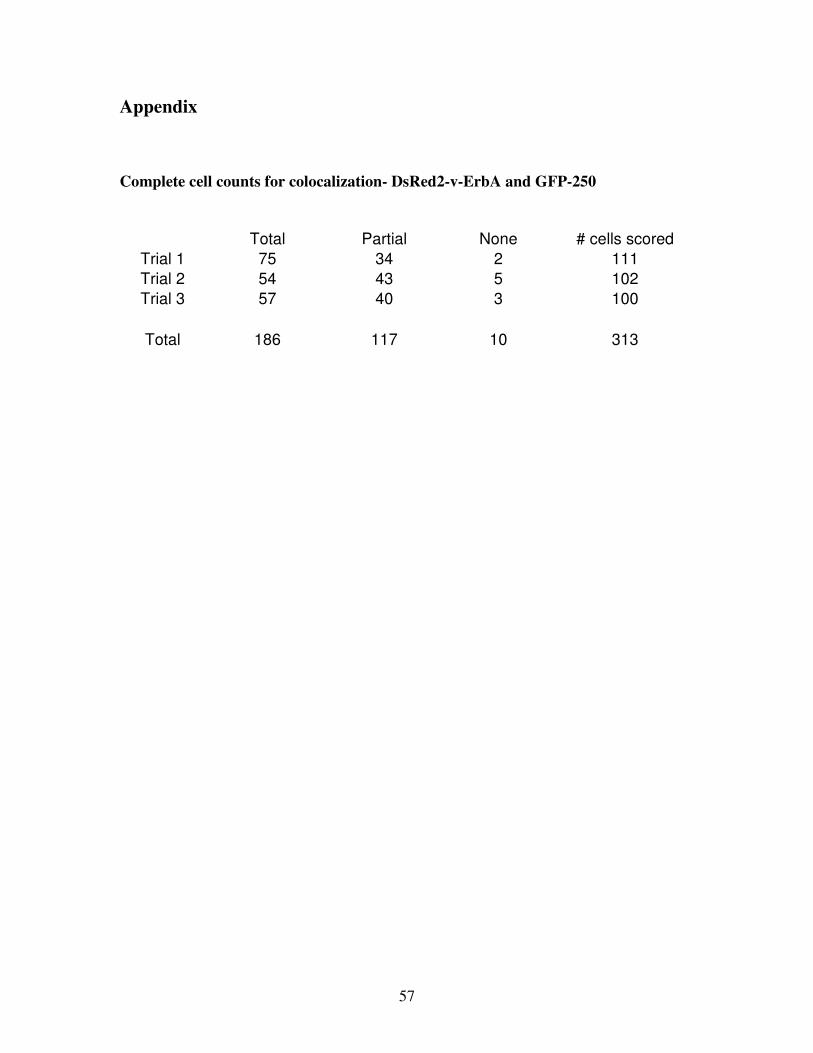
57
Appendix
Complete cell counts for colocalization- DsRed2-v-ErbA and GFP-250
Total Partial None # cells scored
Trial 1 75 34 2 111
Trial 2 54 43 5 102
Trial 3 57 40 3 100
Total 186 117 10 313
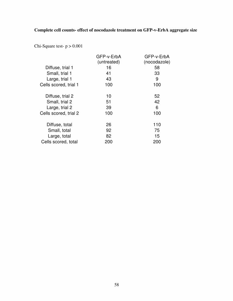
58
Complete cell counts- effect of nocodazole treatment on GFP-v-ErbA aggregate size
Chi-Square test- p > 0.001
GFP-v-ErbA (untreated)
GFP-v-ErbA (nocodazole)
Diffuse, trial 1 16 58
Small, trial 1 41 33
Large, trial 1 43 9
Cells scored, trial 1 100 100
Diffuse, trial 2 10 52
Small, trial 2 51 42
Large, trial 2 39 6
Cells scored, trial 2 100 100
Diffuse, total 26 110
Small, total 92 75
Large, total 82 15
Cells scored, total 200 200
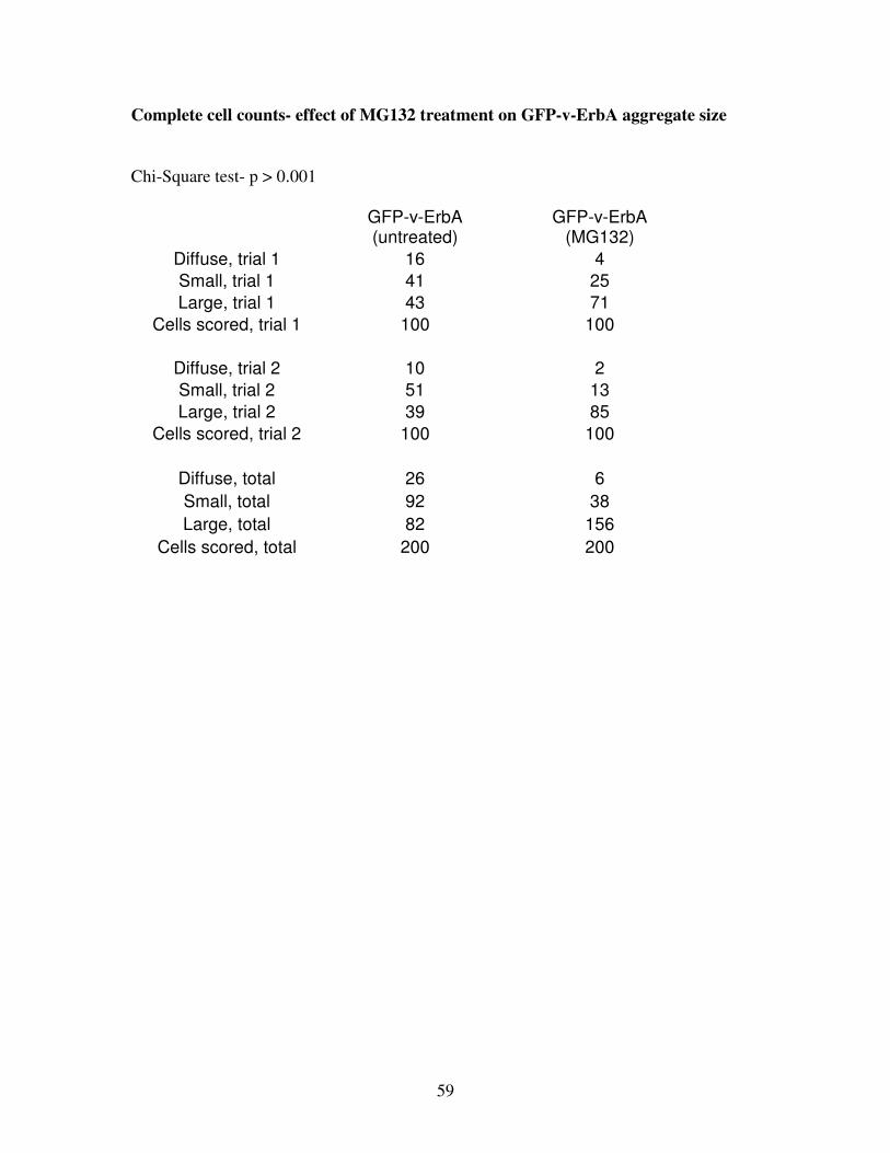
59
Complete cell counts- effect of MG132 treatment on GFP-v-ErbA aggregate size
Chi-Square test- p > 0.001
GFP-v-ErbA (untreated)
GFP-v-ErbA (MG132)
Diffuse, trial 1 16 4
Small, trial 1 41 25
Large, trial 1 43 71
Cells scored, trial 1 100 100
Diffuse, trial 2 10 2
Small, trial 2 51 13
Large, trial 2 39 85
Cells scored, trial 2 100 100
Diffuse, total 26 6
Small, total 92 38
Large, total 82 156
Cells scored, total 200 200