A temporary compendium of thyroid hormone target genes in brain
8
UNCORRECTED PROOF 1 Review 2 A temporary compendium of thyroid hormone target genes in brain ☆ F. Q1 Chatonnet a , F. Flamant a, ⁎, B. Morte b 4 a Institut de Génomique Fonctionnelle de Lyon, Université de Lyon, CNRS, INRA, École Normale Supérieure de Lyon, 46 allée d'Italie, 69364 Lyon Cedex 07, France b Instituto de Investigaciones Biomédicas, Center for Biomedical Research on Rare Diseases (Ciberer), Instituto de Salud Carlos III, Madrid, Spain abstract 6 article info 7 Article history: 8 Received 30 January 2014 9 Received in revised form 21 May 2014 10 Accepted 22 May 2014 11 Available online xxxx 12 Keywords: 13 Thyroid hormone 14 Nuclear receptors 15 Cistrome 16 Transcriptome 17 Q3 Thyroid hormone controls a number of developmental and physiological processes in the brain by directly acting 18 on gene expression. Transcriptome analyses in rodent identified a number of thyroid hormone regulated genes in 19 several brain areas at different stages. Genome wide analysis of chromatin occupancy in a neural cell line also 20 identified a subset of genes which transcription is likely to be directly regulated by thyroid hormone receptors 21 in neurons. However, the abundance of these data and apparent discrepancies between studies brought some 22 confusion. We present here a meta-analysis of available data to identify recurrent themes in thyroid hormone ac- 23 tion in brain cells. This provides a curated list of 734 regulated genes in rodent brain, and highlights a small num- 24 ber of likely direct target genes. Some of these genes are also regulated in amphibians during metamorphosis. 25 This article is part of a Special Issue entitled: Nuclear receptors in animal development. 26 © 2014 Published by Elsevier B.V. 30 31 1. Introduction 32 Thyroid hormones (TH including tri-iodo-thyronine or T3, and thy- 33 roxine, or T4, its less active precursor) play an important function in 34 fetal and adult brain. During neurodevelopment they are required for 35 proper neuronal and glial differentiation, neuronal migration, and mye- 36 lin formation. Thyroid hormone deficiency during human development 37 may cause irreversible mental retardation and variable degrees of 38 neurological impairment. In adults T3 is necessary for neural stem cell 39 proliferation and differentiation, and hypothyroidism is often associated 40 with mood disorders. T3 acts on gene expression mainly, if not only, 41 by binding to nuclear receptors (TRα1, TRβ1 and TRβ2, collectively 42 TR) encoded by the two Thra and Thrb genes and which act as ligand- 43 dependent transcription factors. TR forms heterodimers with RXR, 44 which bind to DNA in a T3 independent manner. In vitro experiments 45 suggest that binding could occur on several types of response ele- 46 ments, constituted by doublets of the half-site AGGCTA, or on DNA 47 elements bearing only limited similarity to this consensus. This binding 48 to regulatory elements of gene promoters induces changes of expres- 49 sion of neighboring genes, defined as “target genes”. As long distance 50 transactivation is possible, one could expect TR to regulate a very large 51 number of genes in many cell types. Identifying these genes is of crucial 52 importance to understand the functions of T3 in brain cells. This is how- 53 ever a challenging task, due to the extreme cellular heterogeneity of 54 most brain areas [1] and the fact that 80% of the known genes are 55 expressed in at least one of these cell types [2]. Transcriptome analyses 56 revealed that expression of thousands of genes changes after T3 stimu- 57 lation in several brain areas, and at different developmental stages. 58 Lists of putative TR target genes, which can be up-regulated or down- 59 regulated, are accumulating. A survey of this recent literature provides 60 the impression of a rather anarchical situation, with few consistencies 61 and little overlap between the various studies. This situation can be 62 explained in different ways: 63 1) TR might possess very different repertoires of target genes in dif- 64 ferent cell-types, different brain areas, and different developmental 65 stages. The response of several well characterized T3 responsive 66 genes actually varies in the brain. For example, the neuron-specific 67 RC3/neurogranin regulation by T3 displays regional and temporal 68 selectivity, which is not due to differential distribution of TR, 69 but most probably related to region-specific trans-acting elements 70 [3,4]. As discussed below, only two genome wide analyses of TR oc- 71 cupancy have been reported to date, but these already provide use- 72 ful indications on possible mechanisms for a cell-type dependent 73 regulation. 74 2) The second possibility is that many brain cell types share a common 75 set of TR target genes, but that the exact experimental conditions 76 deeply influence the outcome of transcriptome studies. For example, 77 T3 stimulation first turns on TR target genes. Among the TR target 78 genes are genes encoding transcription factors and cofactors, and 79 cells rapidly display a secondary response to T3 stimulation, due to 80 the increased expression of these T3-induced factors. Based on sen- 81 sitivity to cycloheximide, which inhibits mRNA translation, it has 82 been estimated that such secondary response represents 20% of 83 the T3-induced changes in gene expression within 3 h in cultured 84 cells [5]. Therefore, only a fraction of the T3 sensitive genes are Biochimica et Biophysica Acta xxx (2014) xxx–xxx ☆ This article is part of a Special Issue entitled: Nuclear receptors in animal development. ⁎ Corresponding author. Tel.: +33 426731332; fax: +33 4728080. E-mail address: Frederic.fl[email protected] (F. Flamant). BBAGRM-00745; No. of pages: 8; 4C: http://dx.doi.org/10.1016/j.bbagrm.2014.05.023 1874-9399/© 2014 Published by Elsevier B.V. Contents lists available at ScienceDirect Biochimica et Biophysica Acta journal homepage: www.elsevier.com/locate/bbagrm Please cite this article as: F. Chatonnet, et al., A temporary compendium of thyroid hormone target genes in brain, Biochim. Biophys. Acta (2014), http://dx.doi.org/10.1016/j.bbagrm.2014.05.023
-
Upload
independent -
Category
Documents
-
view
2 -
download
0
Transcript of A temporary compendium of thyroid hormone target genes in brain

1
2
3Q1
4
6
7891011
1213141516
30
31
32
33
34
35
36
37
38
39
40
41
42
43
44
45
46
47
48
49
50
51
52
53
54
55
Biochimica et Biophysica Acta xxx (2014) xxx–xxx
BBAGRM-00745; No. of pages: 8; 4C:
Contents lists available at ScienceDirect
Biochimica et Biophysica Acta
j ourna l homepage: www.e lsev ie r .com/ locate /bbagrm
Review
A temporary compendium of thyroid hormone target genes in brain☆
F
F. Chatonnet a, F. Flamant a,⁎, B. Morte b
a Institut de Génomique Fonctionnelle de Lyon, Université de Lyon, CNRS, INRA, École Normale Supérieure de Lyon, 46 allée d'Italie, 69364 Lyon Cedex 07, Franceb Instituto de Investigaciones Biomédicas, Center for Biomedical Research on Rare Diseases (Ciberer), Instituto de Salud Carlos III, Madrid, Spain
☆ This article is part of a Special Issue entitled:Nuclear re⁎ Corresponding author. Tel.: +33 426731332; fax: +3
E-mail address: [email protected] (F. Flam
http://dx.doi.org/10.1016/j.bbagrm.2014.05.0231874-9399/© 2014 Published by Elsevier B.V.
Please cite this article as: F. Chatonnet, et al.,http://dx.doi.org/10.1016/j.bbagrm.2014.05.
O
a b s t r a c t
a r t i c l e i n f o17 Q318
19
20
21
22
23
24
25
26
Article history:Received 30 January 2014Received in revised form 21 May 2014Accepted 22 May 2014Available online xxxx
Keywords:Thyroid hormoneNuclear receptorsCistromeTranscriptome
DPROThyroid hormone controls a number of developmental and physiological processes in the brain by directly acting
on gene expression. Transcriptome analyses in rodent identified a number of thyroid hormone regulated genes inseveral brain areas at different stages. Genome wide analysis of chromatin occupancy in a neural cell line alsoidentified a subset of genes which transcription is likely to be directly regulated by thyroid hormone receptorsin neurons. However, the abundance of these data and apparent discrepancies between studies brought someconfusion.Wepresent here ameta-analysis of available data to identify recurrent themes in thyroid hormone ac-tion in brain cells. This provides a curated list of 734 regulated genes in rodent brain, and highlights a small num-ber of likely direct target genes. Some of these genes are also regulated in amphibians during metamorphosis.This article is part of a Special Issue entitled: Nuclear receptors in animal development.
© 2014 Published by Elsevier B.V.
TE
56
57
58
59
60
61
62
63
64
65
66
67
68
69
70
71
72
73
74
75
76
77
78
79
80
UNCO
RREC
1. Introduction
Thyroid hormones (TH including tri-iodo-thyronine or T3, and thy-roxine, or T4, its less active precursor) play an important function infetal and adult brain. During neurodevelopment they are required forproper neuronal and glial differentiation, neuronal migration, andmye-lin formation. Thyroid hormone deficiency during human developmentmay cause irreversible mental retardation and variable degrees ofneurological impairment. In adults T3 is necessary for neural stem cellproliferation anddifferentiation, and hypothyroidism is often associatedwith mood disorders. T3 acts on gene expression mainly, if not only,by binding to nuclear receptors (TRα1, TRβ1 and TRβ2, collectivelyTR) encoded by the two Thra and Thrb genes and which act as ligand-dependent transcription factors. TR forms heterodimers with RXR,which bind to DNA in a T3 independent manner. In vitro experimentssuggest that binding could occur on several types of response ele-ments, constituted by doublets of the half-site AGGCTA, or on DNAelements bearing only limited similarity to this consensus. This bindingto regulatory elements of gene promoters induces changes of expres-sion of neighboring genes, defined as “target genes”. As long distancetransactivation is possible, one could expect TR to regulate a very largenumber of genes in many cell types. Identifying these genes is of crucialimportance to understand the functions of T3 in brain cells. This is how-ever a challenging task, due to the extreme cellular heterogeneity ofmost brain areas [1] and the fact that 80% of the known genes areexpressed in at least one of these cell types [2]. Transcriptome analyses
81
82
83
84
ceptors in animal development.3 4728080.
ant).
A temporary compendium of023
revealed that expression of thousands of genes changes after T3 stimu-lation in several brain areas, and at different developmental stages.Lists of putative TR target genes, which can be up-regulated or down-regulated, are accumulating. A survey of this recent literature providesthe impression of a rather anarchical situation, with few consistenciesand little overlap between the various studies. This situation can beexplained in different ways:
1) TR might possess very different repertoires of target genes in dif-ferent cell-types, different brain areas, and different developmentalstages. The response of several well characterized T3 responsivegenes actually varies in the brain. For example, the neuron-specificRC3/neurogranin regulation by T3 displays regional and temporalselectivity, which is not due to differential distribution of TR,but most probably related to region-specific trans-acting elements[3,4]. As discussed below, only two genome wide analyses of TR oc-cupancy have been reported to date, but these already provide use-ful indications on possible mechanisms for a cell-type dependentregulation.
2) The second possibility is that many brain cell types share a commonset of TR target genes, but that the exact experimental conditionsdeeply influence the outcome of transcriptome studies. For example,T3 stimulation first turns on TR target genes. Among the TR targetgenes are genes encoding transcription factors and cofactors, andcells rapidly display a secondary response to T3 stimulation, due tothe increased expression of these T3-induced factors. Based on sen-sitivity to cycloheximide, which inhibits mRNA translation, it hasbeen estimated that such secondary response represents 20% ofthe T3-induced changes in gene expression within 3 h in culturedcells [5]. Therefore, only a fraction of the T3 sensitive genes are
thyroid hormone target genes in brain, Biochim. Biophys. Acta (2014),

T
85
86
87
88
89
90
91
92
93
94
95
96
97
98
99
100
101
102
103
104
105
106
107
108
109
110
111
112
113
114
115
116
117
118
119
120
121
122
123
124
125
126
127
128
129
130
131
132
133
134
135
136
137
138
139
140
141
142
143
144
145
146
147
148
149
150Q4
151
152
153
154
155
156
157
158
159
160
161
162163
164
165
166
167
168
169
170
171
172
173
174
175
176
177
178
179
180
181
182
183
184
185
186Q5
187
188
189
190
191
192
193
194
195Q6
196
197
198
199
200
201
202Q7
203
204
205
206
207
208
209
210
211
2 F. Chatonnet et al. / Biochimica et Biophysica Acta xxx (2014) xxx–xxx
UNCO
RREC
genuine TR target genes, and the duration of T3 stimulation beforetranscriptome analysis profoundly modifies the outcome of theanalysis.
3) A third possibility would be that technical biases alter the analyses.Transcriptome analyses are notoriously difficult to reproduce indifferent laboratories. It is also now recognized that microarrayanalysis, used in most published studies, produces a much higherfalse discovery rate than the more recent RNAseq techniques.Among microarrays, different technical platforms also providedivergent results. Finally, statistical data analysis is not performedin a standardized manner, and reexamination of published datashows that statistical biases can occur. In many cases the numberof samples examined is too low to provide a sufficient statisticalpower to the analysis. Finally, defining a threshold to call a gene“T3 inducible” is a rather arbitrary decision, and has a major influ-ence on the result.
For all these reasons, the overall outcome of transcriptome analysesremains globally disappointing, failing to provide a comprehensivepicture of T3 cellular response in the brain, and suggesting that never-ending complications will be encountered. Here we performed a com-plete survey of available data for T3 influence on brain cells. In orderto filter for false positive discovery, we listed genes that were identifiedas T3 responsive in more than one study. We also intersected dataobtained in rodent central nervous system, with transcriptome data ob-tained in other organs and other species. This effort allowed us to out-line recurrent and important themes in the T3 response of brain cells.We first present a comparative analysis of TR chromatin occupancy,which outlines the difficulty to identify the genes directly transactivatedby TRs.
2. TR binding to proximal sequences is not the only determinant ofT3 transactivation
For other nuclear receptors, it has been demonstrated that chroma-tin access necessitates the previous intervention of cell-specific “pioneerfactors” that control the access to a subset of otherwise countless puta-tive binding sites of the genome [6]. To date, only two genome widestudies of TR occupancy by ChIP-Seq in mouse cells have been pub-lished. Both studies used tagged TRs and affinity purification instead ofchromatin immune-precipitation to circumvent the lack of high qualityanti-TR antibody. Both identified thousands of TR binding sites in thegenome either in a neural cell line [7] or in mouse hepatocytes [8].This is a much larger number than the one previously reported usingthe less sensitive and less precise ChIP-on-Chip analysis of TR chromatinoccupancy performed on mouse post-natal cerebellum [9,10]. Biotin-tagged TRβ1 was introduced into the liver by adenovirus-mediatedgene transfer, while DNA-transfection was used to restore a stable ex-pression of protein G tagged TRs in the C17.2 neural cell line (eitherTRα1 or TRβ1, providing the C17.2α and C17.2β cell lines). It shouldbe kept in mind that in both cases the expression level of the exogenousTR exceeds the level normally found in the cerebellum and liver respec-tively. Theoverlapbetween thedatasets of TR occupancy, or “cistromes”,is substantial, indicating that access to TR binding sites is not tightlyregulated for these nuclear receptors (Fig. 1). The two studies convergeto conclude that liganded TRs preferentially bind as heterodimers onDR4 elements, and that the number of geneswith a proximal TR bindinglargely exceeds the number of T3 responsive genes.
Although the cistromes largely intersect, the sets of T3-responsivegenes with a proximal TR binding site, which are likely to be directly re-sponsive to T3, do not overlap between the liver and neural cells. Inother words, a number of genes that are regulated by T3 only in neuralcells are bound by TRβ1 both in neural cells and liver. Most strikingly, inboth cases, the large majority of the TR binding sites are located next togenes that are not T3 responsive, suggesting that a majority of bindingevents do not result in transactivation of neighboring genes. Therefore,
Please cite this article as: F. Chatonnet, et al., A temporary compendium ofhttp://dx.doi.org/10.1016/j.bbagrm.2014.05.023
ED P
RO
OF
differential binding alone cannot explain why the repertoire of T3 re-sponsive genes differs between hepatocytes and neural cells. Similarly,the response mediated by TRα1 and TRβ1 in C17.2 is clearly different.Again, the differential response to T3 of a number of genes does not cor-relate with differential chromatin occupancy [7]. Other parameters, likethe expression of different transcription cofactors in different cell types,or the conformation of chromatin-bound TRs, must take part in cell-specific or receptor-selective response. Based only on these two reports,it is tempting to speculate that T3-liganded TR/RXR heterodimers,bound on DR4 elements, adopt a conformation that is not always suit-able for the recruitment of coactivators and transcription activation.This probably explains why bioinformatic tools have been of little helpto identify TR target genes [11].
3. A shortened list of T3 responsive genes in rodent brain
We reviewed all the publicationswewere aware of to identify T3 re-sponsive genes in brain cells. Comparisons between wild-type and TRknock-out mice proved difficult to integrate with studies analyzing ina more direct manner the response to TH [12]. It seems that in TRknock-out mice many changes in mRNA levels are not on TR targetgenes, but reflect modifications in the differentiation status of thecells, or changes in the composition of the cell population. We thusexcluded these knock-out analyses. This left us with 10 studies, usingseveral different experimental systems (Table 1 and SupplementaryTable S1): The table also contains data on cultured glial cells, thesewere considered separately, as this study does not analyze the actionof T3 alone, but analyze the differentiation of oligodendrocytes inducedby T3 addition and concomitant growth factor withdrawal [13]. Fromthe 750 genes differentially expressed during oligodendrocyte differen-tiation, 288 genes have also been reported to be regulated by T3 in thebrain. These genes could also be T3 target genes in oligodendrocytecells.
Study 1: Cerebellum RNA of post-natal day 4 (PND4) hypothyroidmice was compared to cerebellum RNA of hypothyroid mice re-ceiving T4-replacement for 1 h or 6 h [14]. Mice were renderedhypothyroid by administering 2-mercapto-1-methylimidazole indrinking water to their mothers from the 15th day of conception.Study 2: Cerebellum RNA at PND15 was analyzed in 3 groupsof mice. Two were made hypothyroid by administering propyl-thio-uracyl at two different doses [15].Study 3: Cerebellum RNA PND8 and PND15. HypothyroidmicewerePax8−/−micewith athyroidogenesis. Thesewere compared towild-type mice and Pax8−/− treated with T3/T4 for 6 h or 24 h. Primaryneuronal cell cultures were prepared from cerebellum of newborns.In vitro response to T3 (6 h) was addressed [11].Study 4: Primary neuronal cell cultureswere prepared from cerebel-lum of newborns and treated for 6, 16, 24 or 48 h with T3 [16].Study 5: Mouse cerebral cortex of PND21 mice made hypothyroidwith perchlorate and 2-mercapto-1-methylimidazole was com-pared with euthyroid mice [17].Study 6: Cerebral cortex in rat at embryonic day 21 (ED21). Fetal hy-pothyroidism was obtained by maternal thyroidectomy followed by2-mercapto-1-methylimidazole treatment [18].Study 7: Mouse cerebral cortex RNA was analyzed at embryonicE16. 4 groups of animals were included: 1) controls, 2) hypothyroid,3) hyperthyroid and 4) hypothyroid treated with thyroid hormone(T4 + T3). Mild fetal hypothyroidism was achieved in groups 2 and4 after a short treatment (from gestational days 13 to 16) of thedam with propyl-thio-uracyl and 2-mercapto-1-methylimidazole.Groups 3 and 4 dams were treated with thyroid hormones 12 h be-fore sacrifice [19].Study 8a and 8b: Cortex andhippocampus of PND21 rats. Graded hy-pothyroidism was obtained by prolonged propyl-thio-uracyl treat-ment [20].
thyroid hormone target genes in brain, Biochim. Biophys. Acta (2014),

CO
RRECTED P
RO
OF
212
213
214
215
216
217
218219
220
221
222
223
224
225
226
227
228
229
230
231
232
233Q8
234
235
236
237
238
239
240
241
Fig. 1. Cell specific response to T3 stimulation and possible role of cell specific TR chromatin binding. A) Comparison of TR occupancy in liver and in C17.2 cells, analyzed by ChIP-Seq in thepresence of T3. The proximal TR binding sites, defined as TR binding sites locatedwithin 30 kb of a T3 regulated gene are on the left. The other TR binding sites (distal binding sites) are onthe right side. The distance between TR binding sites and T3 regulated genes in liver cells is not considered for simplicity. The total number of TR binding sites, which is much smallerin C17.2 cells than in hepatocytes, has limited significance as it is mainly defined by technical parameters: expression level of the exogenous tagged receptor, purification efficiencyof TR-containing complexes, DNA sequencing depth and choice of statistical threshold for peak calling. 53% of the TR binding sites detected in C17.2 cells overlap with TR binding sitesreported in liver. Within C17.2 cells, the repertoires of TRα1 and TRβ1 binding sites, or cistromes, do not fully overlap. B) Transcriptome analysis identifies T3 responsive genes, whichare either up-regulated or down-regulated. Although some genes display a marked preference for one of the receptors, data obtained in C17.2α and C17.2β cells are pooled. Note thatthe number of T3 responsive genes is much lower that the number of TR binding sites in the genome. This implies that a large fraction of the TR binding sites does not confer T3 respon-siveness to the neighboring genes, even if these are actively transcribed. It also indicates that some genes can be bound by TRs in several cell types but transactivated by T3 in a cell specificmanner. C) T3 responsive genes with a proximal TR binding site (located between−10 kb and+5 kg of the transcription start site in liver Q2, or within 30 kb for C17.2 cells) are likely to bedirectly regulated by TRs upon T3 binding. These two studies did not identify a clear example of a TR target gene, which expression is regulated by T3 both in hepatocytes and C17.2 cells.D) Example of overlapping binding sites in the proximal sequences of the Nup62 gene. Tracks added to the UCSC genome browser are from the liver and the C17.2 cell data. The profile isfrom the C17.2α cells.
3F. Chatonnet et al. / Biochimica et Biophysica Acta xxx (2014) xxx–xxx
UNStudy 9: Striatum from adult rat. Thyroidectomized rats were com-
pared with thyroidectomized rats supplied with exogenous T3(1 or 5 days) [21].Study 10: Striatum of PND21 hypothyroid mice (2-mercapto-1-methylimidazole and perchlorate) were compared with euthyroidmice (B. M. Unpublished data).
Different genomic platforms were used for these studies. Agilentoligonucleotide microarrays were used in studies 1, 2, 5, 6 and 10.Affymetrix oligonucleotide microarrays were used in studies 4, 7, 8and 9. Study 9 additionally used Codelink arrays. Study 4 used non-commercial microarrays spotted with PCR products. Study 7 also useddedicated microarrays purchased from Agilent to analyze changes inmicroRNA expression, after 4 h of treatment. Although no T3 regulatedmicroRNA was identified in this way, later q-RT-PCR reveals that the
Please cite this article as: F. Chatonnet, et al., A temporary compendium ofhttp://dx.doi.org/10.1016/j.bbagrm.2014.05.023
expression level of some of them can be increased in hyperthyroidfetal brain.
The comparative analysis of these studies revealed that the expres-sionof 734 geneswas repeatedly reported as sensitive to the T3 signalinglevel (Fig. 2, Table S1). Gene nameswere from themouse genome. A fewrat genes, for which there were no mouse homologs, were excludedfrom the analysis. Both positive and negative regulations were observedin all experiments, and this orientation of regulation was consistentin studies for almost all the genes. This confirms that a large number ofgenes are T3 sensitive in the brain, and that this diversity is not due totechnological artifact and misconducted analyses. A set of 734 geneshave been reported repeatedly as T3 regulated, representing a subsetof the 4108 genes whose T3 regulation has only been reported once. Itcould be expected that without the overall difficulty to obtain reproduc-ible results with microarrays, and the high sensitivity of T3 response to
thyroid hormone target genes in brain, Biochim. Biophys. Acta (2014),

T
RO
OF
242
243
244
245
246
247
248Q9
249
250
251Q10
252
253
254
255
256
257
258
259
260
261
262
263
264
265
266
267
268
t1:1 Table 1t1:2 Studies used for comparison of T3/TR target genes in the central nervous system.
Study Reference Tissue Developmental stage Species Platform Treatmentt1:3
1 [14] Cerebellum PND 4 Mouse Agilent HypothyroidHypothyroid + T4 for 1 or 6 ht1:4
2 [15] Cerebellum PND 15 Mouse Agilent ControlHypothyroidt1:5
3 [11] Cerebellum PND 8–PND15 Mouse Non-commercial Wild-typePax8−/−t1:6
Primary neurons PCR products Pax8−/− + T3 for 6, 24 hWild-type + T3 for 6 ht1:7
4 [16] Cerebellum Primary neurons Mouse Affymetrix Control + T3 for 6, 16, 24, and 48 ht1:8
5 [17] Cerebral cortex PND 21 Mouse Agilent ControlHypothyroidt1:9
6 [18] Cerebral cortex ED 21 Rat Agilent ControlHypothyroidt1:10
7 [19] Cerebral cortex ED 16 Mouse Affymetrix ControlHypothyroidHypothyroid + T3/T4t1:11
8a [20] Cerebral cortex PND21 Rat Affymetrix ControlHypothyroidt1:12
8b [20] Hippocampus PND21 Rat Affymetrix ControlHypothyroidt1:13
9 [21] Striatum Adult Rat AffymetrixCodelink
HypothyroidHypothyroid + T3t1:14
10 Morte B. et al., unpublished Striatum PND 21 Mouse Agilent ControlHypothyroidt1:15
4 F. Chatonnet et al. / Biochimica et Biophysica Acta xxx (2014) xxx–xxx
REC
the experimental setting, the number of repeatedly reported geneswould be larger. Reciprocally, the list cannot be considered as complete.For example, the Reelin gene, which is expressed in several cell typesof the post-natal cortex, is known to be regulated by T3 only in theCajal–Retzius cells [22]. However, although probes were present on mi-croarrays, differential expressionwas detected in only onewhole-cortextranscriptome analysis. This example illustrates how the high cellularand molecular heterogeneity of brain tissue can mask changes in geneexpression restricted to a small fraction of the cell population.
4. Overlap with other neural response
The diversity of T3 response among brain areas and developmentalstages is still difficult to evaluate. However, it is noticeable that a fractionof genes can be regulated inmany situations. T3 sensitivity of genes thatwas reported in more than 4 experiments is listed in the Fig. 2 legend.Other genes display a response to T3 which seems to be restricted to
UNCO
R
Fig. 2. Distribution of genes among 10 transcriptome analyses performed in the centralnervous system. Gene expression has been reported more than once to be sensitive toT3 level for a total of 734 genes. The genes which sensitivity to T3 was reported in morethan 4 experiments are: Klf9, Dbp, Sema3c, Fos, Malat1, Hba-a1, Fxyd6, Itih3, Nefh,Homer1, Itpr1, Hr, Bcar3, Slc16a6, Sema7a, Cirbp, Scd1, Abcd2, Sorl1, Kirrel3, Taf15, Cxadr,Atp1a2, Tbc1d30,Mycn, Dnm3, Sfpq, Ryr2,Map2k6, Rbbp4, Pvr, Dysl3, Pvalb, Dusp1, and Dcx.
Please cite this article as: F. Chatonnet, et al., A temporary compendium ofhttp://dx.doi.org/10.1016/j.bbagrm.2014.05.023
ED Pspecific brain areas and developmental stages. It is noticeable that the
cerebellum contains a large fraction of granule cells, a specific type ofglutamatergic excitatory neurons, while the striatum contains mainlyGABAergic inhibitory neurons. Fig. 3 shows the intersection betweenthe 734 genes repeatedly reported as sensitive to T3 in the differentbrain areas. In the cerebral cortex, differences related to the develop-mental stage can be observed between the embryonic (studies 6and 7) and postnatal (studies 5 and 8) stages. Although some genesare regulated at both developmental stages, only 75 genes are sensitiveto T3 at the embryonic stages. Among these, for example, Pafah1b1 is in-volved in the positioning of neurons and the correct stratification of thecerebral cortex [23]. A prominent role of Camk4 and Creb1 pathways
Fig. 3. Response to T3 in different brain areas. Venn diagramof T3 sensitive genes reportedmore thanonce in various brain areas. Cerebellumcontains a large fraction of granule cells,a specific type of glutamatergic excitatory neurons, while striatum contains mainlyGABAergic inhibitory neurons. Studies using cultured cerebellum neurons are included.For clarity, 23 genes reported in the hippocampus are not displayed. They are equallydispatched in all categories.
thyroid hormone target genes in brain, Biochim. Biophys. Acta (2014),

TED P
RO
OF
269
270
271
272
273
274
275
276
277
278
279Q11
280
281
282
283
284
285
286
287
288Q12
289
290
291
292Q13
293
294
295
296
297
298
299
300
301
302
303
304
305
t2:1Table 2t2:2Most likely TR target genes in neural cells.
Gene name Nb studies ENS Liver Cb Cx Str t2:3
Acsl6 2 x x t2:4
Adamtsl4 3 x x t2:5
Arf1 2 x t2:6
Arhgap23 2 x x t2:7
Bcl3 2 x x t2:8
Btbd3 2 x x t2:9
Chl1 2 x x t2:10
Cml1 2 x t2:11
Col11a2 2 x x t2:12
Dbp 8 x x x x x t2:13
Dhrs7 2 x x t2:14
Elovl5 2 x x t2:15
Fos 5 x x t2:16
Glis1 2 x t2:17
Gpr146 2 x x x t2:18
Hr 6 x x x x t2:19
Htra1 2 x t2:20
Kcna1 3 x x x t2:21
Kdm6b 2 x t2:22
Klf9 10 x x x x t2:23
Layn 2 x x t2:24
Mapkapk3 2 x t2:25
Nr1h4 2 x x x t2:26
Nt5e 2 x x x t2:27
Pcsk4 2 x t2:28
Pde4b 2 x t2:29
Pfkfb3 2 x x t2:30
Scd1 5 x x x t2:31
Slc25a33 2 x x t2:32
Stat5a 3 x x x t2:33
Tgm2 2 x x t2:34
Tlcd1 2 x x x t2:35
Tmem2 2 x t2:36
Trp53inp2 3 x x x t2:37
Txnip 3 x t2:38
Vdr 2 x x t2:39
Wdr47 2 x x t2:40
t2:41Genes repeatedly regulated by T3 in brain and C17.2 cells with a TR binding site withint2:4230 kb of transcription start. Nb studies: refers to the number of publication reporting T3t2:43regulation in brain or cultured neurons. Bold characters are for genes which have beent2:44found in at least 3 independent studies. ENS: Enteric nervous system. Cb: Cerebellum.t2:45Cx: Cortex. Str: Striatum.
5F. Chatonnet et al. / Biochimica et Biophysica Acta xxx (2014) xxx–xxx
EC
was suggested to represent an important process in thyroid hormoneaction in the fetal cortex [18].
In order to better identify recurrent themes in neural response to T3,we compared this curated list of 734 genes responding to T3 in thebrain, with the list reported in two other in vitro studies performedon neural systems. These experiments addressed T3 response in twodifferent cell culture systems: microarrays were used to study T3 re-sponse (24 h) of enteric nervous system progenitor cells in primary cul-tures [24]. As this published analysis provides only a biased view of thedata, we reanalyzed the dataset deposited on the NCBI Gene ExpressionOmnibus (GEO). The neural C17.2 mouse cerebellum derived cell line[25] was transfected to restore the stable expression of either TRα1 orTRβ1 to restore T3 responsiveness [7]. T3 response was analyzed byRNAseq 6, 12 and 24 h after T3 addition to the cell culture medium. Tobroaden our comparison, we also introduced studies on liver or hepato-cyte cell lines [5,8,26,27]. The overlapwasmost obvious for C17.2 cells, acerebellum-derived cell line in which T3 regulates 78 of the genes iden-tified in the central nervous system. The overlap was also significantwith T3 regulated genes in enteric nervous system progenitors (32genes) and liver or hepatocytes (41 genes) (Fig. 4). However, it seemsthat the number of genes able to respond to T3 stimulation in all sys-tems is very small. The study of transfected C17.2 cells [7] also providesChIP-Seq data to define chromatin occupancy by TRα1 and TRβ1 ge-nome wide scale. This aids to recognize genes which transcription isdirectly regulated by TR from genes which regulation is only secondaryto early T3 cellular response. Table 2 highlights 28 genes which are re-peatedly regulated by T3 in the brain and C17.2 cells, with a TR bindingsitewithin 30kbof transcription start site in C17.2 cells, an arbitrary dis-tance used here to distinguish between proximal and distal TR bindingsites. These genes are thus very likely to be direct TR target genes notonly in C17.2 cells, but also in the brain.
5. Overlap with T3 responsive genes in amphibians
We also reasoned that important regulations tend to be conservedduring evolution and that we should be able to identify a subsetof genes regulated in the brain of all vertebrates. We thus crossedthe full list of genes which expression is sensitive to T3 in the rodentcentral nervous system to datasets obtained in transcriptome analysis
UNCO
RR
306
307
308
309
310
311
312
313
314
315
316
317
318
319
320
321
322
323
324
325
326
327
328
329
330
Fig. 4. Overlap between central nervous system results and dataset obtained in varioussystems. The curated list of genes regulated in brain by T3 was crossed with lists obtainedin C17.2 neural cells, enteric neural cells, and liver and hepatocyte cell lines.
Please cite this article as: F. Chatonnet, et al., A temporary compendium ofhttp://dx.doi.org/10.1016/j.bbagrm.2014.05.023
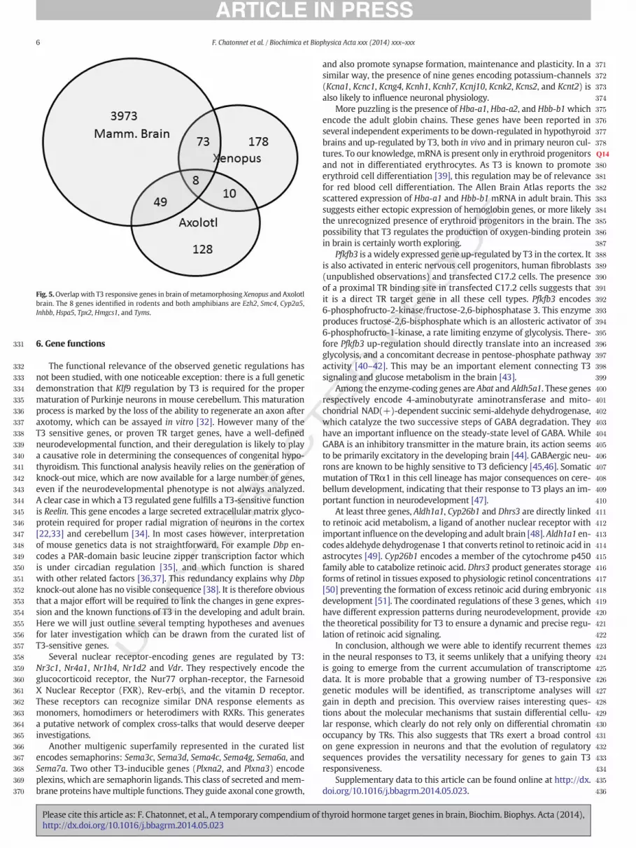
performed in the metamorphosing tadpole of Xenopus laevis [28]. Inthis amphibian species T3 triggers metamorphosis, which is markedby a major remodeling of the central nervous system. The comparisonidentified 81 genes. Among these, only 8 were also reported to be T3sensitive in axolotl brain (Ambystoma mexicanum), another classicalmodel to study T3-induced metamorphosis [29]. Only 12 out of the 81genes were among the genes that have been repeatedly reported to beT3 sensitive in rodent brain, representing a core of T3 sensitive genesprobably regulated in many vertebrate species (Table S2, Fig. 5). Foronly 2 of these genes (Klf9, and Dbp) do we have good evidence thatthey are direct TR target genes in rodent brain cells. This comparisonclearly suffers from the limited information gathered in amphibians,and difficulties in identifying orthologous genes. The presence ofmany cell-cycle regulated genes is however visible, and suggests thatT3 in both systems exerts an important influence on the proliferationof specific cell types. It has been proposed several times that rodentpost-natal development is a process analogous to metamorphosis, inwhich T3 also serves to synchronize independent events [30,31]. It ishowever noticeable that Thrb is inducible by T3 in amphibians but notin rodents. A similar positive feedback loop, based on Thra induction,also exists in some metamorphosing fishes. There is therefore an inter-esting correlation between amphibian metamorphosis and the exis-tence of a positive feedback loop in the T3 signaling pathway. Furthercomparative analyses could assess whether TR self-induction is a hall-mark of metamorphosis in non-mammalian vertebrates.
thyroid hormone target genes in brain, Biochim. Biophys. Acta (2014),
T331
332
333
334
335
336
337
338
339
340
341
342
343
344
345
346
347
348
349
350
351
352
353
354
355
356
357
358
359
360
361
362
363
364
365
366
367
368
369
370
371
372
373
374
375
376
377
378
379Q14
380
381
382
383
384
385
386
387
388
389
390
391
392
393
394
395
396
397
398
399
400
401
402
403
404
405
406
407
408
409
410
411
412
413
414
415
416
417
418
419
420
421
422
423
424
425
426
427
428
429
430
431
432
433
434
435
436
Fig. 5.Overlap with T3 responsive genes in brain of metamorphosing Xenopus and Axolotlbrain. The 8 genes identified in rodents and both amphibians are Ezh2, Smc4, Cyp2a5,Inhbb, Hspa5, Tpx2, Hmgcs1, and Tyms.
6 F. Chatonnet et al. / Biochimica et Biophysica Acta xxx (2014) xxx–xxx
UNCO
RREC
6. Gene functions
The functional relevance of the observed genetic regulations hasnot been studied, with one noticeable exception: there is a full geneticdemonstration that Klf9 regulation by T3 is required for the propermaturation of Purkinje neurons in mouse cerebellum. This maturationprocess is marked by the loss of the ability to regenerate an axon afteraxotomy, which can be assayed in vitro [32]. However many of theT3 sensitive genes, or proven TR target genes, have a well-definedneurodevelopmental function, and their deregulation is likely to playa causative role in determining the consequences of congenital hypo-thyroidism. This functional analysis heavily relies on the generation ofknock-out mice, which are now available for a large number of genes,even if the neurodevelopmental phenotype is not always analyzed.A clear case in which a T3 regulated gene fulfills a T3-sensitive functionis Reelin. This gene encodes a large secreted extracellular matrix glyco-protein required for proper radial migration of neurons in the cortex[22,33] and cerebellum [34]. In most cases however, interpretationof mouse genetics data is not straightforward. For example Dbp en-codes a PAR-domain basic leucine zipper transcription factor whichis under circadian regulation [35], and which function is sharedwith other related factors [36,37]. This redundancy explains why Dbpknock-out alone has no visible consequence [38]. It is therefore obviousthat a major effort will be required to link the changes in gene expres-sion and the known functions of T3 in the developing and adult brain.Here we will just outline several tempting hypotheses and avenuesfor later investigation which can be drawn from the curated list ofT3-sensitive genes.
Several nuclear receptor-encoding genes are regulated by T3:Nr3c1, Nr4a1, Nr1h4, Nr1d2 and Vdr. They respectively encode theglucocorticoid receptor, the Nur77 orphan-receptor, the FarnesoidX Nuclear Receptor (FXR), Rev-erbβ, and the vitamin D receptor.These receptors can recognize similar DNA response elements asmonomers, homodimers or heterodimers with RXRs. This generatesa putative network of complex cross-talks that would deserve deeperinvestigations.
Another multigenic superfamily represented in the curated listencodes semaphorins: Sema3c, Sema3d, Sema4c, Sema4g, Sema6a, andSema7a. Two other T3-inducible genes (Plxna2, and Plxna3) encodeplexins, which are semaphorin ligands. This class of secreted andmem-brane proteins havemultiple functions. They guide axonal cone growth,
Please cite this article as: F. Chatonnet, et al., A temporary compendium ofhttp://dx.doi.org/10.1016/j.bbagrm.2014.05.023
ED P
RO
OF
and also promote synapse formation, maintenance and plasticity. In asimilar way, the presence of nine genes encoding potassium-channels(Kcna1, Kcnc1, Kcng4, Kcnh1, Kcnh7, Kcnj10, Kcnk2, Kcns2, and Kcnt2) isalso likely to influence neuronal physiology.
More puzzling is the presence of Hba-a1, Hba-a2, and Hbb-b1whichencode the adult globin chains. These genes have been reported inseveral independent experiments to be down-regulated in hypothyroidbrains and up-regulated by T3, both in vivo and in primary neuron cul-tures. To our knowledge, mRNA is present only in erythroid progenitorsand not in differentiated erythrocytes. As T3 is known to promoteerythroid cell differentiation [39], this regulation may be of relevancefor red blood cell differentiation. The Allen Brain Atlas reports thescattered expression of Hba-a1 and Hbb-b1 mRNA in adult brain. Thissuggests either ectopic expression of hemoglobin genes, or more likelythe unrecognized presence of erythroid progenitors in the brain. Thepossibility that T3 regulates the production of oxygen-binding proteinin brain is certainly worth exploring.
Pfkfb3 is a widely expressed gene up-regulated by T3 in the cortex. Itis also activated in enteric nervous cell progenitors, human fibroblasts(unpublished observations) and transfected C17.2 cells. The presenceof a proximal TR binding site in transfected C17.2 cells suggests thatit is a direct TR target gene in all these cell types. Pfkfb3 encodes6-phosphofructo-2-kinase/fructose-2,6-biphosphatase 3. This enzymeproduces fructose-2,6-bisphosphate which is an allosteric activator of6-phosphofructo-1-kinase, a rate limiting enzyme of glycolysis. There-fore Pfkfb3 up-regulation should directly translate into an increasedglycolysis, and a concomitant decrease in pentose-phosphate pathwayactivity [40–42]. This may be an important element connecting T3signaling and glucose metabolism in the brain [43].
Among the enzyme-coding genes are Abat and Aldh5a1. These genesrespectively encode 4-aminobutyrate aminotransferase and mito-chondrial NAD(+)-dependent succinic semi-aldehyde dehydrogenase,which catalyze the two successive steps of GABA degradation. Theyhave an important influence on the steady-state level of GABA. WhileGABA is an inhibitory transmitter in the mature brain, its action seemsto be primarily excitatory in the developing brain [44]. GABAergic neu-rons are known to be highly sensitive to T3 deficiency [45,46]. Somaticmutation of TRα1 in this cell lineage has major consequences on cere-bellum development, indicating that their response to T3 plays an im-portant function in neurodevelopment [47].
At least three genes, Aldh1a1, Cyp26b1 and Dhrs3 are directly linkedto retinoic acid metabolism, a ligand of another nuclear receptor withimportant influence on the developing and adult brain [48]. Aldh1a1 en-codes aldehyde dehydrogenase 1 that converts retinol to retinoic acid inastrocytes [49]. Cyp26b1 encodes a member of the cytochrome p450family able to catabolize retinoic acid. Dhrs3 product generates storageforms of retinol in tissues exposed to physiologic retinol concentrations[50] preventing the formation of excess retinoic acid during embryonicdevelopment [51]. The coordinated regulations of these 3 genes, whichhave different expression patterns during neurodevelopment, providethe theoretical possibility for T3 to ensure a dynamic and precise regu-lation of retinoic acid signaling.
In conclusion, although we were able to identify recurrent themesin the neural responses to T3, it seems unlikely that a unifying theoryis going to emerge from the current accumulation of transcriptomedata. It is more probable that a growing number of T3-responsivegenetic modules will be identified, as transcriptome analyses willgain in depth and precision. This overview raises interesting ques-tions about the molecular mechanisms that sustain differential cellu-lar response, which clearly do not rely only on differential chromatinoccupancy by TRs. This also suggests that TRs exert a broad controlon gene expression in neurons and that the evolution of regulatorysequences provides the versatility necessary for genes to gain T3responsiveness.
Supplementary data to this article can be found online at http://dx.doi.org/10.1016/j.bbagrm.2014.05.023.
thyroid hormone target genes in brain, Biochim. Biophys. Acta (2014),

T
437
438439440441442443444445446447448449450451452453454455456457458459460461462463464465466467468469470471472473474Q17475476477478479480481482483484485486487488Q18489490491492493494495496497498499500501502503504505506507508509510511Q19512513514515516517518519520
521522523524525526527528529530531532533534535536537538539540Q20541542543544545546547548549550551552553554555556557558559560561562563Q21564565566567568569570571572573574575576577578579580581582583584585586587588589590591592593594595596597598599600601602603604605606
7F. Chatonnet et al. / Biochimica et Biophysica Acta xxx (2014) xxx–xxx
UNCO
RREC
References
[1] R.H. Masland, Neuronal cell types, Curr. Biol. 14 (2004) R497–R500.[2] E.S. Lein, M.J. Hawrylycz, N. Ao, M. Ayres, A. Bensinger, A. Bernard, A.F. Boe, M.S.
Boguski, K.S. Brockway, E.J. Byrnes, L. Chen, L. Chen, T.M. Chen, M.C. Chin, J. Chong,B.E. Crook, A. Czaplinska, C.N. Dang, S. Datta, N.R. Dee, A.L. Desaki, T. Desta, E. Diep,T.A. Dolbeare, M.J. Donelan, H.W. Dong, J.G. Dougherty, B.J. Duncan, A.J. Ebbert, G.Eichele, L.K. Estin, C. Faber, B.A. Facer, R. Fields, S.R. Fischer, T.P. Fliss, C. Frensley,S.N. Gates, K.J. Glattfelder, K.R. Halverson, M.R. Hart, J.G. Hohmann, M.P. Howell,D.P. Jeung, R.A. Johnson, P.T. Karr, R. Kawal, J.M. Kidney, R.H. Knapik, C.L. Kuan, J.H.Lake, A.R. Laramee, K.D. Larsen, C. Lau, T.A. Lemon, A.J. Liang, Y. Liu, L.T. Luong, J.Michaels, J.J. Morgan, R.J. Morgan, M.T. Mortrud, N.F. Mosqueda, L.L. Ng, R. Ng,G.J. Orta, C.C. Overly, T.H. Pak, S.E. Parry, S.D. Pathak, O.C. Pearson, R.B. Puchalski,Z.L. Riley, H.R. Rockett, S.A. Rowland, J.J. Royall, M.J. Ruiz, N.R. Sarno, K. Schaffnit, N.V.Shapovalova, T. Sivisay, C.R. Slaughterbeck, S.C. Smith, K.A. Smith, B.I. Smith, A.J. Sodt,N.N. Stewart, K.R. Stumpf, S.M. Sunkin,M. Sutram, A. Tam, C.D. Teemer, C. Thaller, C.L.Thompson, L.R. Varnam, A. Visel, R.M. Whitlock, P.E. Wohnoutka, C.K. Wolkey, V.Y.Wong, M. Wood, M.B. Yaylaoglu, R.C. Young, B.L. Youngstrom, X.F. Yuan, B. Zhang,T.A. Zwingman, A.R. Jones, Genome-wide atlas of gene expression in the adultmouse brain, Nature 445 (2007) 168–176.
[3] A.L. Dowling, R.T. Zoeller, Thyroid hormone of maternal origin regulates the expres-sion of RC3/neurogranin mRNA in the fetal rat brain, Brain Res. Mol. Brain Res. 82(2000) 126–132.
[4] A. Guadano-Ferraz, M.J. Escamez, B.Morte, P. Vargiu, J. Bernal, Transcriptional induc-tion of RC3/neurogranin by thyroid hormone: differential neuronal sensitivity is notcorrelated with thyroid hormone receptor distribution in the brain, Brain Res. Mol.Brain Res. 49 (1997) 37–44.
[5] J.Z. Lin, D.H. Sieglaff, C. Yuan, J. Su, A.S. Arumanayagam, S. Firouzbakht, J.J. CantuPompa, F.D. Reynolds, X. Zhou, A. Cvoro, P. Webb, Gene specific actions of thyroidhormone receptor subtypes, PLoS One 8 (2013) e52407.
[6] K.M. Jozwik, J.S. Carroll, Pioneer factors in hormone-dependent cancers, Nat. Rev.Cancer 12 (2012) 381–385.
[7] F. Chatonnet, R. Guyot, G. Benoit, F. Flamant, Genome-wide analysis of thyroidhormone receptors shared and specific functions in neural cells, Proc. Natl. Acad.Sci. U. S. A. 110 (2013) E766–E775.
[8] P. Ramadoss, B.J. Abraham, L. Tsai, Y. Zhou, E.S.R.H. Costa, F. Ye, M. Bilban, K. Zhao,A.N. Hollenberg, Novel mechanism of positive versus negative regulation by thyroidhormone receptor beta 1 (TRbeta1) identified by genome-wide profiling of bindingsites in mouse liver, J. Biol. Chem. (2013).
[9] H. Dong, C.L. Yauk, A. Rowan-Carroll, S.H. You, R.T. Zoeller, I. Lambert, M.G. Wade,Identification of thyroid hormone receptor binding sites and target genes usingChIP-on-chip in developing mouse cerebellum, PLoS ONE 4 (2009) e4610.
[10] R. Gagne, J.R. Green, H. Dong, M.G. Wade, C.L. Yauk, Identification of thyroid hor-mone receptor binding sites in developing mouse cerebellum, BMC Genomics 14(2013) 341.
[11] L. Quignodon, C. Grijota-Martinez, E. Compe, R. Guyot, N. Allioli, D. Laperriere, R.Walker, P. Meltzer, S. Mader, J. Samarut, F. Flamant, A combined approach identifiesa limited number of new thyroid hormone target genes in post-natal mouse cere-bellum, J. Mol. Endocrinol. 39 (2007) 17–28.
[12] L.D. Miller, P. McPhie, H. Suzuki, Y. Kato, E.T. Liu, S.Y. Cheng, Multi-tissue gene-expression analysis in a mouse model of thyroid hormone resistance, GenomeBiol. 5 (2004) R31.
[13] J. Dugas, T. YC, T.P. Speed, J. Ngai, B. Barres, Functional genomic analysis of oligoden-drocyte differentiation, J. Neurosci. 26 (2006) 10967–10983.
[14] M. Takahashi, T. Negishi, T. Tashiro, Identification of genesmediating thyroid hormoneaction in the developing mouse cerebellum, J. Neurochem. 104 (2008) 640–652.
[15] H. Dong, M. Wade, A. Williams, A. Lee, G.R. Douglas, C. Yauk, Molecular insight intothe effects of hypothyroidism on the developing cerebellum, Biochem. Biophys. Res.Commun. 330 (2005) 1182–1193.
[16] F. Chatonnet, R. Guyot, F. Picou, M. Bondesson, F. Flamant, Genome-wide search re-veals the existence of a limited number of thyroid hormone receptor alpha targetgenes in cerebellar neurons, PLoS One 7 (2012) e30703.
[17] B. Morte, A. Ceballos, D. Diez, C. Grijota-Martinez, A.M. Dumitrescu, C. Di Cosmo, V.A.Galton, S. Refetoff, J. Bernal, Thyroid hormone-regulated mouse cerebral cortexgenes are differentially dependent on the source of the hormone: a study in mono-carboxylate transporter-8- and deiodinase-2-deficient mice, Endocrinology 151(2010) 2381–2387.
[18] B. Morte, D. Diez, E. Auso, M.M. Belinchon, P. Gil-Ibanez, C. Grijota-Martinez, D.Navarro, G.M. de Escobar, P. Berbel, J. Bernal, Thyroid hormone regulation of geneexpression in the developing rat fetal cerebral cortex: prominent role of theCa2+/calmodulin-dependent protein kinase IV pathway, Endocrinology 151 (2010)810–820.
[19] H. Dong, S.H. You, A. Williams, M.G. Wade, C.L. Yauk, R. Thomas Zoeller, Transientmaternal hypothyroxinemia potentiates the transcriptional response to exogenousthyroid hormone in the fetal cerebral cortex before theonset of fetal thyroid function:a messenger and microRNA profiling study, Cereb. Cortex (2014).
[20] J.E. Royland, J.S. Parker, M.E. Gilbert, A genomic analysis of subclinical hypothyroid-ism in hippocampus and neocortex of the developing rat brain, J. Neuroendocrinol.20 (2008) 1319–1338.
[21] D. Diez, C. Grijota-Martinez, P. Agretti, G. DeMarco, M. Tonacchera, A. Pinchera, G.M.de Escobar, J. Bernal, B. Morte, Thyroid hormone action in the adult brain: geneexpression profiling of the effects of single andmultiple doses of triiodo-L-thyroninein the rat striatum, Endocrinology 149 (2008) 3989–4000.
[22] M. Alvarez-Dolado, M. Ruiz, J.A. Del Rio, S. Alcantara, F. Burgaya, M. Sheldon, K.Nakajima, J. Bernal, B.W. Howell, T. Curran, E. Soriano, A. Munoz, Thyroid hormone
Please cite this article as: F. Chatonnet, et al., A temporary compendium ofhttp://dx.doi.org/10.1016/j.bbagrm.2014.05.023
ED P
RO
OF
regulates reelin and dab1 expression during brain development, J. Neurosci. 19(1999) 6979–6993.
[23] A.H. Assadi, G. Zhang, U. Beffert, R.S. McNeil, A.L. Renfro, S. Niu, C.C. Quattrocchi, B.A.Antalffy,M. Sheldon, D.D. Armstrong, A.Wynshaw-Boris, J. Herz, G. D'Arcangelo, G.D.Clark, Interaction of reelin signaling and Lis1 in brain development, Nat. Genet. 35(2003) 270–276.
[24] R. Mohr, P. Neckel, Y. Zhang, S. Stachon, K. Nothelfer, K. Schaeferhoff, F. Obermayr,M. Bonin, L. Just, Molecular and cell biological effects of 3,5,3′-triiodothyronine onprogenitor cells of the enteric nervous system in vitro, Stem Cell Res. 11 (2013)1191–1205.
[25] E.Y. Snyder, D.L. Deitcher, C. Walsh, S. Arnold-Aldea, E.A. Hartwieg, C.L. Cepko,Multipotent neural cell lines can engraft and participate in development of mousecerebellum, Cell 68 (1992) 33–51.
[26] P.M. Yen, X. Feng, F. Flamant, Y. Chen, R.L. Walker, R.E. Weiss, O. Chassande, J.Samarut, S. Refetoff, P.S. Meltzer, Effects of ligand and thyroid hormone receptorisoforms on hepatic gene expression profiles of thyroid hormone receptor knockoutmice, EMBO Rep. 4 (2003) 581–587.
[27] H. Dong, C.L. Yauk, A. Williams, A. Lee, G.R. Douglas, M.G. Wade, Hepatic gene ex-pression changes in hypothyroid juvenile mice: characterization of a novel negativethyroid responsive element, Endocrinology (2007).
[28] A. Ishihara, Y. Makita, K. Yamauchi, Gene expression profiling to examine thethyroid hormone-disrupting activity of hydroxylated polychlorinated biphenylsin metamorphosing amphibian tadpole, J. Biochem. Mol. Toxicol. 25 (2011)303–311.
[29] P. Huggins, C.K. Johnson, A. Schoergendorfer, S. Putta, A.C. Bathke, A.J. Stromberg, S.R.Voss, Identification of differentially expressed thyroid hormone responsive genesfrom the brain of the Mexican Axolotl (Ambystoma mexicanum), Comp. Biochem.Physiol. Toxicol. Pharmacol. 155 (2012) 128–135.
[30] E. Kress, S. Skah, M. Sirakov, J. Nadjar, N. Gadot, J.Y. Scoazec, J. Samarut, M. Plateroti,Cooperation between the thyroid hormone receptor TRalpha1 and the WNT path-way in the induction of intestinal tumorigenesis, Gastroenterology 138 (2010)1863–1874.
[31] V. Laudet, The origins and evolution of vertebrate metamorphosis, Curr. Biol. 21(2011) R726–R737.
[32] H.X. Avci, C. Lebrun, R. Wehrle, M. Doulazmi, F. Chatonnet, M.P. Morel, M. Ema, G.Vodjdani, C. Sotelo, F. Flamant, I. Dusart, Thyroid hormone triggers the developmen-tal loss of axonal regenerative capacity via thyroid hormone receptor alpha1 andkruppel-like factor 9 in Purkinje cells, Proc. Natl. Acad. Sci. U. S. A. 109 (2012)14206–14211.
[33] Y. Jossin, J.A. Cooper, Reelin, Rap1 and N-cadherin orient the migration of multipolarneurons in the developing neocortex, Nat. Neurosci. 14 (2011) 697–703.
[34] C. Castagna, P. Aimar, S. Alasia, L. Lossi, Post-natal development of the Reeler mousecerebellum: an ultrastructural study, Ann. Anat. (2013).
[35] F. Gachon, E. Nagoshi, S.A. Brown, J. Ripperger, U. Schibler, Themammalian circadiantiming system: from gene expression to physiology, Chromosoma 113 (2004)103–112.
[36] Q. Wang, M. Maillard, U. Schibler, M. Burnier, F. Gachon, Cardiac hypertrophy, lowblood pressure, and low aldosterone levels in mice devoid of the three circadianPAR bZip transcription factors DBP, HLF, and TEF, Am. J. Physiol. Regul. Integr.Comp. Physiol. 299 (2010) R1013–R1019.
[37] F. Gachon, N. Leuenberger, T. Claudel, P. Gos, C. Jouffe, F. Fleury Olela, X. de Molleratdu Jeu,W.Wahli, U. Schibler, Proline- and acidic amino acid-rich basic leucine zipperproteins modulate peroxisome proliferator-activated receptor alpha (PPARalpha)activity, Proc. Natl. Acad. Sci. U. S. A. 108 (2011) 4794–4799.
[38] F. Gachon, P. Fonjallaz, F. Damiola, P. Gos, T. Kodama, J. Zakany, D. Duboule, B. Petit,M. Tafti, U. Schibler, The loss of circadian PAR bZip transcription factors results inepilepsy, Genes Dev. 18 (2004) 1397–1412.
[39] C. Angelin-Duclos, C. Domenget, A. Kolbus, H. Beug, P. Jurdic, J. Samarut, Thyroidhormone T3 acting through the thyroid hormone alpha receptor is necessary for im-plementation of erythropoiesis in the neonatal spleen environment in the mouse,Development 132 (2005) 925–934.
[40] A. Herrero-Mendez, A. Almeida, E. Fernandez, C. Maestre, S. Moncada, J.P. Bolanos,The bioenergetic and antioxidant status of neurons is controlled by continuousdegradation of a key glycolytic enzyme by APC/C-Cdh1, Nat. Cell Biol. 11 (2009)747–752.
[41] J.P. Bolanos, A. Almeida, S. Moncada, Glycolysis: a bioenergetic or a survivalpathway? Trends Biochem. Sci. 35 (2010) 145–149.
[42] P. Rodriguez-Rodriguez, A. Almeida, J.P. Bolanos, Brain energy metabolismin glutamate-receptor activation and excitotoxicity: role for APC/C-Cdh1 inthe balance glycolysis/pentose phosphate pathway, Neurochem. Int. 62 (2013)750–756.
[43] Y. Itoh, T. Esaki, M. Kaneshige, H. Suzuki, M. Cook, L. Sokoloff, S.Y. Cheng, J. Nunez,Brain glucose utilization in mice with a targeted mutation in the thyroid hormonealpha or beta receptor gene, Proc. Natl. Acad. Sci. U. S. A. 98 (2001) 9913–9918.
[44] Y. Ben-Ari, Excitatory actions of gaba during development: the nature of the nurture,Nat. Rev. Neurosci. 3 (2002) 728–739.
[45] K. Wallis, M. Sjogren, M. van Hogerlinden, G. Silberberg, A. Fisahn, K. Nordstrom, L.Larsson, H. Westerblad, G. Morreale de Escobar, O. Shupliakov, B. Vennstrom, Loco-motor deficiencies and aberrant development of subtype-specific GABAergic inter-neurons caused by an unliganded thyroid hormone receptor alpha1, J. Neurosci. 28(2008) 1904–1915.
[46] J. Manzano, M. Cuadrado, B. Morte, J. Bernal, Influence of thyroid hormone andthyroid hormone receptors in the generation of cerebellar gamma-aminobutyricacid-ergic interneurons from precursor cells, Endocrinology 148 (2007) 5746–5751.
[47] T. Fauquier, F. Chatonnet, F. Picou, S. Richard, N. Fossat, N. Aguilera, T. Lamonerie, F.Flamant, Purkinje cells andBergmannglia areprimary targets of the TRalpha1 thyroid
thyroid hormone target genes in brain, Biochim. Biophys. Acta (2014),

607608609610611612613
614615616617618619620
8 F. Chatonnet et al. / Biochimica et Biophysica Acta xxx (2014) xxx–xxx
hormone receptor during mouse cerebellum postnatal development, Development141 (2014) 166–175.
[48] K.D. Shearer, P.N. Stoney, P.J. Morgan, P.J. McCaffery, A vitamin for the brain, TrendsNeurosci. 35 (2012) 733–741.
[49] S.A. Adam, O. Schnell, J. Poschl, S. Eigenbrod, H.A. Kretzschmar, J.C. Tonn, U. Schuller,ALDH1A1 is a marker of astrocytic differentiation during brain development and cor-relateswith better survival in glioblastomapatients, Brain Pathol. 22 (2012) 788–797.
UNCO
RRECT
Please cite this article as: F. Chatonnet, et al., A temporary compendium ofhttp://dx.doi.org/10.1016/j.bbagrm.2014.05.023
[50] F. Cerignoli, X. Guo, B. Cardinali, C. Rinaldi, J. Casaletto, L. Frati, I. Screpanti, L.J. Gudas,A. Gulino, C.J. Thiele, G. Giannini, retSDR1, a short-chain retinol dehydrogenase/reductase, is retinoic acid-inducible and frequently deleted in human neuroblasto-ma cell lines, Cancer Res. 62 (2002) 1196–1204.
[51] L. Feng, R.E. Hernandez, J.S.Waxman, D. Yelon, C.B. Moens, Dhrs3a regulates retinoicacid biosynthesis through a feedback inhibition mechanism, Dev. Biol. 338 (2010)1–14.
ED P
RO
OF
thyroid hormone target genes in brain, Biochim. Biophys. Acta (2014),