Synthesis and Target Identification of Hymenialdisine Analogs
13
Chemistry & Biology, Vol. 11, 247–259, February, 2004, 2004 Elsevier Science Ltd. All rights reserved. DOI 10.1016/j.chembiol.2004.01.015 Synthesis and Target Identification of Hymenialdisine Analogs inhibitors of kinase function. While there are a few exam- ples of non-ATP competitive kinase inhibitors [14, 15], the majority of kinase inhibitors target the ATP binding Yongqin Wan, 1 Wooyoung Hur, 1 Charles Y. Cho, 2 Yi Liu, 1 Francisco J. Adrian, 1 Olivier Lozach, 3 Ste ´ phane Bach, 3 Thomas Mayer, 4 Doriano Fabbro, 4 Laurent Meijer, 3 pocket of the enzyme. These ATP site-directed inhibi- tors fall into a limited number of families including quin- and Nathanael S. Gray 1, * 1 Genomics Institute of the Novartis azolines [16], pyrimidines [17–20], indolinones [21, 22], purines [23–28], imidazoles [29–31], flavonoids [32, 33], Research Foundation Department of Chemistry paullones [34–36], and alkaloids (for example, stauro- sporine and its derivatives [37–39]). To date, approxi- 10675 John Jay Hopkins Drive San Diego, California 92121 mately 46 kinase inhibitors have entered clinical trials targeting kinases involved in cancer (EGFR, CDKs, Raf, 2 The Scripps Research Institute Department of Chemistry PKC, PKA, KDR, Mek-1), angiogenesis (KDR), leukemia (bcr-abl, FLT3), inflammation (p38), immune disorder 10550 N. Torrey Pines Road La Jolla, California 92037 (JNK), and neurodegenerative disorders (JNK). In this paper, we report the use of affinity chromatog- 3 CNRS, Cell Cycle Group Station Biologique, B.P. 74 raphy and in vitro enzymatic profiling against a panel of 60 kinases to identify additional potential targets of the 29682 Roscoff Cedex Bretagne natural product Hymenialdisine (HMD 1), as well as syn- thesis and biological characterization of a variety of ana- France 4 Oncology Research logs. HMD is one member of a family of tricyclic pyrrole compounds that is constructed from a brominated pyr- Novartis Pharma AG CH-4002 Basel rolo[2,3-c]azepine skeleton and a 5-membered glycocy- amidine ring system. It was isolated from marine sponges Switzerland including Hymeniacidon aldis, Axinella verrucosa, and Acanthella aurantiaca in the early 1980s based on its antiproliferative effects on cultured lymphocytic leuke- Summary mia cells [40–44]. It was later shown to exhibit strong inhibitory activity against several closely related CDKs, Hymenialdisine (HMD) is a sponge-derived natural such as CDK1/cyclin B (IC 50 22 nM), CDK2/cyclin A product kinase inhibitor with nanomolar activity (IC 50 70 nM), CDK2/cyclin E (IC 50 40 nM), and CDK5/ against CDKs, Mek1, GSK3, and CK1 and micromolar p25 (IC 50 28 nM), as well as against more distantly activity against Chk1. In order to explore the broader related kinases such as GSK3 (IC 50 10 nM) and CK1 application of the pyrrolo[2,3-c]azepine skeleton of (IC 50 35 nM) [45]. HMD was also isolated from the HMD as a general kinase inhibitory scaffold, we sponge Stylissa massa as the compound inhibiting the searched for additional protein targets using affinity Raf/Mek-1/MAPK cascade and shown to be a potent chromatography in conjunction with the synthesis of inhibitor of Mek1 (IC 50 6 nM) [46]. Kinetic analysis has diverse HMD analogs and profiled HMD against a shown that HMD acts in an ATP-competitive fashion panel of 60 recombinant enzymes. This effort has led to [45], and the cocrystal structure with CDK2 revealed nanomolar to micromolar inhibitors of 11 new targets that HMD occupies the ATP site and makes many of including p90RSK, KDR, c-Kit, Fes, MAPK1, PAK2, the hydrogen bonding interactions seen in other CDK- PDK1, PKC, PKD2, Rsk1, and SGK. The synthesis inhibitor complexes [45]. Despite potent activities of HMD analogs has resulted in the identification of against these kinases, HMD is relatively selective in vitro compounds with enhanced and/or dramatically al- and exhibits only moderate to weak activity against tered selectivities relative to HMD (28n) and in mole- many other kinases [45–50]. cules with antiproliferative activities 30-fold higher The activities of HMD as a potential antiproliferative, than HMD (28p). anti-inflammatory, and antineurodegenerative agent have been investigated in several cellular models. For Introduction example, HMD in the micromolar range was shown to have antiproliferative effects against human tumor cell Protein kinases play an essential role in regulating vari- lines, presumably as a result of CDK and Mek inhibitory ous cellular functions including gene expression, cellular activity [46]. Using U937 cells, HMD was shown to be proliferation, differentiation, membrane transport, and a micromolar inhibitor of NFB-mediated gene tran- apoptosis [1–7]. Aberrant kinase activities have been scription [51–53]. Furthermore, the hyperphosphoryla- associated with several diseases including cancer, dia- tion of tau, a known component of the neurofibrillary betes, and Alzheimer’s disease [8–13]. As a conse- plaques characteristic of patients with Alzheimer’s dis- quence, considerable effort has been directed toward ease, is inhibited in vitro by HMD at 50 M [45]. the development of specific and potent small molecule In order to identify additional potential intracellular targets of HMD, we performed affinity chromatography with immobilized HMD using brain extracts. Previously, *Correspondence: [email protected]
Transcript of Synthesis and Target Identification of Hymenialdisine Analogs

Chemistry & Biology, Vol. 11, 247–259, February, 2004, 2004 Elsevier Science Ltd. All rights reserved. DOI 10.1016/j .chembiol .2004.01.015
Synthesis and Target Identificationof Hymenialdisine Analogs
inhibitors of kinase function. While there are a few exam-ples of non-ATP competitive kinase inhibitors [14, 15],the majority of kinase inhibitors target the ATP binding
Yongqin Wan,1 Wooyoung Hur,1 Charles Y. Cho,2
Yi Liu,1 Francisco J. Adrian,1 Olivier Lozach,3
Stephane Bach,3 Thomas Mayer,4
Doriano Fabbro,4 Laurent Meijer,3 pocket of the enzyme. These ATP site-directed inhibi-tors fall into a limited number of families including quin-and Nathanael S. Gray1,*
1Genomics Institute of the Novartis azolines [16], pyrimidines [17–20], indolinones [21, 22],purines [23–28], imidazoles [29–31], flavonoids [32, 33],Research Foundation
Department of Chemistry paullones [34–36], and alkaloids (for example, stauro-sporine and its derivatives [37–39]). To date, approxi-10675 John Jay Hopkins Drive
San Diego, California 92121 mately 46 kinase inhibitors have entered clinical trialstargeting kinases involved in cancer (EGFR, CDKs, Raf,2 The Scripps Research Institute
Department of Chemistry PKC, PKA, KDR, Mek-1), angiogenesis (KDR), leukemia(bcr-abl, FLT3), inflammation (p38), immune disorder10550 N. Torrey Pines Road
La Jolla, California 92037 (JNK), and neurodegenerative disorders (JNK).In this paper, we report the use of affinity chromatog-3 CNRS, Cell Cycle Group
Station Biologique, B.P. 74 raphy and in vitro enzymatic profiling against a panel of60 kinases to identify additional potential targets of the29682 Roscoff Cedex
Bretagne natural product Hymenialdisine (HMD 1), as well as syn-thesis and biological characterization of a variety of ana-France
4 Oncology Research logs. HMD is one member of a family of tricyclic pyrrolecompounds that is constructed from a brominated pyr-Novartis Pharma AG
CH-4002 Basel rolo[2,3-c]azepine skeleton and a 5-membered glycocy-amidine ring system. It was isolated from marine spongesSwitzerlandincluding Hymeniacidon aldis, Axinella verrucosa, andAcanthella aurantiaca in the early 1980s based on itsantiproliferative effects on cultured lymphocytic leuke-Summarymia cells [40–44]. It was later shown to exhibit stronginhibitory activity against several closely related CDKs,Hymenialdisine (HMD) is a sponge-derived naturalsuch as CDK1/cyclin B (IC50 � 22 nM), CDK2/cyclin Aproduct kinase inhibitor with nanomolar activity(IC50 � 70 nM), CDK2/cyclin E (IC50 � 40 nM), and CDK5/against CDKs, Mek1, GSK3�, and CK1 and micromolarp25 (IC50 � 28 nM), as well as against more distantlyactivity against Chk1. In order to explore the broaderrelated kinases such as GSK3� (IC50 � 10 nM) and CK1application of the pyrrolo[2,3-c]azepine skeleton of(IC50 � 35 nM) [45]. HMD was also isolated from theHMD as a general kinase inhibitory scaffold, wesponge Stylissa massa as the compound inhibiting thesearched for additional protein targets using affinityRaf/Mek-1/MAPK cascade and shown to be a potentchromatography in conjunction with the synthesis ofinhibitor of Mek1 (IC50 � 6 nM) [46]. Kinetic analysis hasdiverse HMD analogs and profiled HMD against ashown that HMD acts in an ATP-competitive fashionpanel of 60 recombinant enzymes. This effort has led to[45], and the cocrystal structure with CDK2 revealednanomolar to micromolar inhibitors of 11 new targetsthat HMD occupies the ATP site and makes many ofincluding p90RSK, KDR, c-Kit, Fes, MAPK1, PAK2,the hydrogen bonding interactions seen in other CDK-PDK1, PKC�, PKD2, Rsk1, and SGK. The synthesisinhibitor complexes [45]. Despite potent activitiesof HMD analogs has resulted in the identification ofagainst these kinases, HMD is relatively selective in vitrocompounds with enhanced and/or dramatically al-and exhibits only moderate to weak activity againsttered selectivities relative to HMD (28n) and in mole-many other kinases [45–50].cules with antiproliferative activities 30-fold higher
The activities of HMD as a potential antiproliferative,than HMD (28p).anti-inflammatory, and antineurodegenerative agenthave been investigated in several cellular models. For
Introduction example, HMD in the micromolar range was shown tohave antiproliferative effects against human tumor cell
Protein kinases play an essential role in regulating vari- lines, presumably as a result of CDK and Mek inhibitoryous cellular functions including gene expression, cellular activity [46]. Using U937 cells, HMD was shown to beproliferation, differentiation, membrane transport, and a micromolar inhibitor of NF�B-mediated gene tran-apoptosis [1–7]. Aberrant kinase activities have been scription [51–53]. Furthermore, the hyperphosphoryla-associated with several diseases including cancer, dia- tion of tau, a known component of the neurofibrillarybetes, and Alzheimer’s disease [8–13]. As a conse- plaques characteristic of patients with Alzheimer’s dis-quence, considerable effort has been directed toward ease, is inhibited in vitro by HMD at 50 �M [45].the development of specific and potent small molecule In order to identify additional potential intracellular
targets of HMD, we performed affinity chromatographywith immobilized HMD using brain extracts. Previously,*Correspondence: [email protected]

Chemistry & Biology248
this method has yielded valuable insights into the cellu-lar targets of two other kinase inhibitors: purvalanol B[54] and paullone [55]. To complement this approach,we also tested HMD against a panel of recombinantkinases not previously examined.
Despite the description of the total synthesis of HMDby two research groups [56–58], there have been fewreported modifications to the core structure [59, 60].The second part of this paper describes the synthesisand the structure-activity relationship (SAR) of new HMDanalogs with altered kinase selectivity profiles. Of partic-ular interest are selected analogs that retain activityagainst CDKs or GSK3�, which are comparable to thatof the parent compound but have enhanced selectivity.The cellular activities of these compounds have alsobeen investigated, and selected analogs showed up to30-fold improvement over HMD.
Results and Discussion
Identifying Additional Substrates of HMDAffinity ChromatographyThe crystal structure of CDK2 complexed with HMDrevealed that HMD binds in the ATP binding pocket withits bromine pointing toward the outside of the pocket[45]. Based on this observation, we reasoned that addi-tion of a linker moiety at the 2-position of the pyrrole ringmight enable us to immobilize the compound withoutdestroying its kinase inhibitory activity. We first synthe-sized HMD 1 and SEM (2-(trimethylsilyl)ethoxymethyl)protected 2-bromoaldisine 5 [56]. 2-bromoaldisine, ananalog lacking the 5-membered glycocyamidine that iscrucial for potent kinase inhibitory activity, was usedas a negative control for the experiment. A palladium-catalyzed reaction using Pd(PPh3)2Cl2 [61] allowed thecoupling of 2-bromopyrrolo compounds to an amino-PEG linker containing a terminal alkyne. After removingthe SEM group, compounds 4 and 7 were immobilizedto an N-hydroxysuccinimide ester-activated agarosematrix through amide bond formation (Figure 1A).
In order to identify HMD-interacting proteins presentin neuronal cells, mouse brain extracts were incubatedwith the HMD or aldisine (AD) control matrix. After wash-ing the matrices to remove unbound/weakly bound pro-
(iv) 2-bromoaldisine 5, 3, PdCl2(PPh3)2, CuI, PPh3, Et2NH, DMF; (v) a.MeOH/10% aq. HCl (1:1), b. aq. NH3 in MeOH/H2O (1:1); (vi) 3 mMof 4 or 7 in DMSO, 1 ml of Affi-Gel 10 Gel (Bio-Rad Laboratory),24 hr.(B) HMD-affinity pull-down experiment data from mouse brain ex-tracts and intracellular target identification. Purv. B, purvalanol Bresin; AD, aldisine control resin; HMD, hymenialdisine resin. Brainextracts were loaded on Purv. B, AD, or HMD matrix. Bound proteinswere resolved by SDS-PAGE and visualized by silver staining andby Western blotting with anti-GSK3�/�, anti-Mek1/2, anti-CDK5,anti-p25, anti-p35, and anti-Erk2.(C) HMD-affinity pull-down experiment data from porcine brain ex-
Figure 1. Preparation of Hymenialdisine and Aldisine Control Affin- tracts and intracellular target identification. Brain extracts wereity Resins and Affinity Chromatography Pull-Down Experiment loaded on AD or HMD matrix. Bound proteins were resolved by
SDS-PAGE and visualized by silver staining and by Western blotting(A) Synthetic scheme for Hymenialdisine (HMD) and aldisine (AD)affinity resins. (i) 6-heptynoic acid, 2-[2-(2-azido-ethoxy)-ethoxy]- with anti-GSK3�/� and anti-CK. In order to purify Mek1 for micro-
sequencing, the extracts were also depleted of GSKs using axinethylamine, 1,3-diisopropylcarbodiimide, CH2Cl2; (ii) 2, PMe3, THF-1% H2O; (iii) HMD 1, 3, PdCl2(PPh3)2, CuI, PPh3, Et2NH, DMF; beads prior to chromatography with the HMD resin.

Synthesis and Targets of Hymenialdisine Analogs249
teins, bound proteins were eluted and separated bySDS-PAGE (Figure 1B). Prominent bands were excised,proteolytically digested, and then subjected to MALDImass spectrometry. This resulted in the identification ofsix proteins: three kinases known to be potently inhib-ited by HMD (GSK3�/� and Mek1), two kinases thathave not been previously known to be targets of HMD(p90RSK and an unknown kinase), and �-tubulin. Theunknown protein may be an isoform of BIKe (BMP2-inducible kinase) that was recently identified from a pre-chondroblastic cell line [62].
The ability of the affinity resin to allow identification ofsome known targets as well as to identify some unknownones demonstrates the utility of this approach. Sincep90RSK was identified as a new target of HMD, we thenmeasured the IC50s of HMD and linker-HMD against thisenzyme. The values were found to be 0.40 and 5.6 �M,respectively. Intriguingly, as observed with immobilizedpaullones [55], CDK5/p25 was absent from the affinityresin, despite its presence in the brain extract and itspurification on immobilized purvalanol [54]. This may bedue to some steric hindrance provided by the presenceof the linker, as suggested by the lower sensitivity ofCDK5 to linker-HMD. Although the linker-modified pur-valanol B derivative is equipotent against CDK5/p25when compared to purvalanol B (purvalanol B IC50 � 2nM versus linker-purvalanol B IC50 � 5 nM), the linkermodification of HMD led to 10-fold decrease in potencyagainst CDK/p25 (HMD IC50 � 40 nM versus linker-HMDIC50 � 400 nM). In addition, the other targets (GSK-3,MEK1, and CK1) may be much more abundant and di-rectly competing with low amounts of CDK5 for bindingto the ligand. Finally, it is possible that native CDK5, incontrast to recombinant CDK5, is associated with otherproteins that prohibit interaction with HMD.
To confirm the proteins identified by mass spectrome-try, Western blot analysis using antibodies specific toMek1, Mek2, GSK3�, GSK3�, and CDK5/p25 was per-formed after affinity chromatography on purvalanol B,HMD, and AD resin. The HMD resin specifically boundMek1, Mek2, and GSK3�/� relative to the control resin.In contrast, eluate from the purvalanol B resin stainedstrongly for GSK3� and CDK5/p25 but only weakly forGSK3� and Mek2.
To further validate the results obtained above, affinitychromatography with immobilized HMD was carried outusing porcine brain extracts (Figure 1C). GSK3�/� andCK1 were identified by Western blot and Mek1 by micro-sequencing. In order to purify Mek1 further for micro-sequencing, the extract was depleted of GSK3s usingaxin beads prior to chromatography with the HMDresin [63].Kinase Selectivity PanelIn order to identify additional potential in vitro targets,HMD was tested against a panel of 60 purified proteinkinases, most of which had not been previously exam-ined. Results expressed as a percentage of enzymeactivity at 10 �M inhibitor concentration are listed inTable 1. In addition to CDKs, Chk, Erk1, GSK3�, andMek1, known targets of HMD, we found that HMD inhib-its KDR, c-Kit, Fes, MAPK1, PAK2, PDK1, PKC�, PKD2,Rsk1, SGK, and c-Src as kinases inhibited in the micro-
Tab
le1.
Inhi
biti
on
of
Enz
ymes
by
HM
Dat
10�
MC
onc
entr
atio
n
%E
nzym
e%
Enz
yme
%E
nzym
e%
Enz
yme
%E
nzym
e%
Enz
yme
Kin
ase
Act
ivity
Kin
ase
Act
ivity
Kin
ase
Act
ivity
Kin
ase
Act
ivity
Kin
ase
Act
ivity
Kin
ase
Act
ivity
Ab
l(m)
25F
es(h
)7
IKK
�(h
)71
MA
PK
AP
-K2(
h)50
PD
K1(
h)4
Rsk
1(h)
2c-
Ab
l12
FG
FR
-118
IKK
�(h
)36
ME
K1
(h)
2P
KA
56S
AP
K2a
(h)
98A
uro
ra-A
(h)
58F
GF
R3
(h)
28In
s-R
68c-
Met
41P
KB
47S
AP
K2b
(h)
93A
xl(h
)30
Flt-
121
IR(h
)10
4M
KK
4(m
)23
PK
B�
(h)
67S
AP
K3
(h)
46B
mx(
h)76
Flt-
316
JNK
1�1(
h)48
MK
K6
(h)
38P
KC
�(h
)-H
is20
SG
K(h
)9
CaM
KIV
(h)
28F
lt-4
36JN
K2�
2(h)
29p
70S
6K(h
)23
PK
C�(h
)10
c-S
rc9
CD
K1/
cycl
inB
(h)
2G
SK
3ß(h
)1
KD
R7
PA
K2(
h)7
PK
D2(
h)4
Syk
(h)
51C
HK
2(h)
3H
ER
-160
c-K
it4
PD
GF
R�
(h)
89c-
Raf
(h)
82T
ek25
CK
2(h)
13H
ER
-252
Lck(
h)32
PD
GF
R-�
16c-
Raf
-184
Trk
B(h
)17
CS
K(h
)91
IGF
-1R
81M
AP
K1(
h)10
PD
K1
100
RO
CK
-II(h
)51
ZA
P-7
0(h)
90
The
resu
ltsar
ep
rese
nted
aski
nase
activ
ityas
ap
erce
ntag
eo
fth
atin
cont
roli
ncub
atio
ns.
AT
Pw
asp
rese
ntat
10�
Min
alla
ssay
s.A
ssay
sw
ere
cond
ucte
dac
cord
ing
toth
ep
roce
dur
esin
[64]
and
[65]
.
molar range. Synthetic analogs of HMD may be discov-

Chemistry & Biology250
ered with enhanced potency and selectivity toward Synthesis of Hydrazone and Amide DerivativesTo further probe SAR of HMD analogs, a variety of aro-these additional kinase targets.matic and aliphatic groups were introduced to replacethe glycocyamidine ring of HMD that can be divided
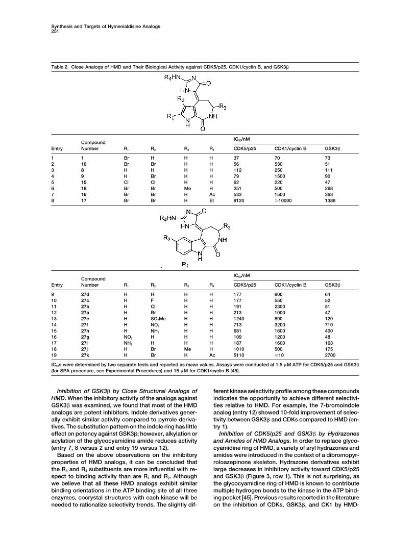
Synthesis of Analogs Based on HMD Scaffold into two categories: hydrazones and amides. HydrazoneThe crystal structure of CDK2 complexed with HMD has derivatives 28 of the previously synthesized pyrrolo- andrevealed multiple hydrogen bonds that are important to indolo-azepinediones were prepared by reacting ke-the interaction of HMD with the enzyme active site (Fig- tones with aromatic hydrazines (Figure 2C). Amides 31ure 4A). These include the pyrrole N1 with the carbonyl and 34 were prepared from carboxylic acids 30 andoxygen of Leu83, the carbonyl oxygen of azepine ring 33 (Figure 2D). Compound 30 was obtained from thewith the backbone amide of Leu83, the amide of the hydrolysis of t-Boc protected ester [60]. Double bondazepine ring N2 with the carbonyl oxygen of Glu81, the migration occurred during ester hydrolysis under basicguanidine amino group N5 with one of the side chain conditions as a result of the formation of a more stableoxygen atoms of Asp145 and the side chain carbonyl of carbanion intermediate [67, 68]. Amine building blocksAsn132, two water-mediated hydrogen bonds between chosen include various aliphatic and aromatic amines.the O2 of HMD with the main chain carbonyl of Asp145,and N5 of HMD with the main chain carbonyl of Gln131 Biological Results and SAR[45]. Since several of these hydrogen bonds are con- SAR on Kinase Assayserved in other inhibitor-CDK complexes, they are likely In this study, we focused on investigating the inhibitoryto contribute to the potency of HMD against CDKs and properties of HMD analogs against purified CDK5/p25,were therefore mostly retained in our synthetic efforts. CDK1/cyclin B, and GSK3�. The structure activity rela-The bromine substituent, in contrast, occupies a spa- tionships (SAR) of HMD analogs were examined by mea-cious hydrophobic pocket that is more completely filled suring the in vitro inhibitory activity of each HMD analogby other CDK inhibitors such as olomoucine and purva- against these three kinases. Table 2 is a summary oflanol [45]. In order to optimize the interactions in this the IC50s of close structural analogs of HMD containingregion, analogs were prepared where the pyrrole ring the pyrrolo- or indolo-azepine skeleton and the glycocy-was replaced with an indole. amidine appendage.Synthesis of Close Analogs of HMD Inhibition of CDK5/p25 by Close Structural AnalogsTo examine the effect of various substituents on the of HMD. As shown, non-, mono-, or dihalogenation atpyrrole ring, we synthesized a series of HMD analogs the 2- and 3-position on the pyrrole ring has little effectas shown in Table 2. HMD (1), debromohymenialdisine on the activity of these compounds against CDK5/p25.(8), 3-bromohymenialdisine (10), and 3-bromo-2-debro- Simple pyrrole derivatives (entry 1–5) are potent inhibi-mohymenialdisine (9) were prepared according to re- tors, whereas the simple indole analog (entry 9) exhibitsported procedures via hymenin (11b) [56–58]. As an al- reduced activity. Indole substituents such as a halogenternative procedure, alcohol 13b, obtained from the at the 7-position (entry 10–12) or a nitro/amino substitu-reduction of azepinedione 12b, reacts with 2-aminoimid- ent at the 9-position (entry 16 and 17) result in derivativesazole in methanesulfonic acid to give 11b through an in that are as potent as the nonsubstituted indole analogsitu generated intermediate alkene 14b (Figure 2A). (entry 9), whereas substitution of a nitro, amino, or meth-Since azepinediones can be used as common building anesulfonyl group at the 7-position on the indole resultsblocks for other structural modifications, we utilized this in reduced potency (entry 13–15).modified scheme to prepare other HMD analogs includ- We next investigated the effect of substitution on theing dichlorinated analog 15, acetamide 16, N-ethylated azepine ring (R3) and glycocyamidine ring (R4). When R3 isanalog 17, and 3-methyl analog 18. Cyclization of 3-[(4,5- a methyl group, the activity is reduced by approximatelydibromo-1H-pyrrole-2-carbonyl)-amino]-butyric acid in 80% for both pyrrole and indole derivatives (entry 6PPA gave the ketone in very poor yield, presumably due versus 2 and entry 18 versus 12). This phenomenonto electron deficiency of the dibromopyrrole ring and suggests that the hydrophobic pocket adjacent to meth-steric hindrance caused by the methyl group. We there- ylenes of the azepine ring is quite shallow. Alkylation orfore cyclized pyrrole-2-carboxylic acid and performed acylation of the glycocyamidine ring leads to a dramaticbromination after reduction of the ketone (see Sup- decrease in activity, presumably as a result of stericplemental Figure S1 at http://www.chembiol.com/cgi/ hindrance and loss of hydrogen bonds (entry 7, 8,content/full/11/2/247/DC1). and 19).
The synthesis of indole analogs is outlined in Figure Inhibition of CDK1/cyclin B by Close Structural Ana-2B. The attempt to synthesize the 6-chloro substituted logs of HMD. HMD appears to be the most potent inhibi-indole analog from acetal 20b [58] gave indole dimer as tor of CDK1/cyclin B among its close structural analogsthe exclusive product. Consequently, a modified scheme (entry 1 versus 2–5). The inhibitory activity of these ana-was attempted. Carboxylic acids 23a-g were cyclized logs against CDK1/cyclin B appears to be more suscep-in P2O5-methanesulfonic acid [66] to yield azepino tible to the halogen substitution pattern on the pyrrole[3,4-b]indole-1,5-diones 24a-g. These ketones were ring than that against CDK5/p25. In particular, introduc-then reduced to alcohols and reacted with aminoimidaz- ing a bromine at the 3-position on pyrrole (entry 4) hasole in methanesulfonic acid. Subsequent bromination of the most negative impact on the CDK1/cyclin B activity.26 afforded compound 27 in moderate to good yields. For indole analogs, fluorine, but not chlorine or bromine,Hydrogenation of 27a, 27f, and 27g furnished com- substitution at the 7-position increases the potency (en-
try 10, 11, 12 versus 9).pounds 27d, 27h, and 27i, respectively.
Synthesis and Targets of Hymenialdisine Analogs251
Table 2. Close Analogs of HMD and Their Biological Activity against CDK5/p25, CDK1/cyclin B, and GSK3�
IC50/nMCompound
Entry Number R1 R2 R3 R4 CDK5/p25 CDK1/cyclin B GSK3�
1 1 Br H H H 37 70 732 10 Br Br H H 56 530 513 8 H H H H 112 250 1114 9 H Br H H 79 1500 905 15 Cl Cl H H 62 220 476 18 Br Br Me H 251 500 2687 16 Br Br H Ac 533 1500 3638 17 Br Br H Et 9120 �10000 1388
IC50/nMCompound
Entry Number R1 R2 R3 R4 CDK5/p25 CDK1/cyclin B GSK3�
9 27d H H H H 177 800 6410 27c H F H H 177 550 5211 27b H Cl H H 191 2300 5112 27a H Br H H 213 1000 4713 27e H SO2Me H H 1240 890 12014 27f H NO2 H H 713 3200 71015 27h H NH2 H H 681 1600 40016 27g NO2 H H H 109 1200 4817 27i NH2 H H H 187 1600 16318 27j H Br Me H 1010 500 17519 27k H Br H Ac 5110 �10 2700
IC50s were determined by two separate tests and reported as mean values. Assays were conducted at 1.5 �M ATP for CDK5/p25 and GSK3�
(for SPA procedure, see Experimental Procedures) and 15 �M for CDK1/cyclin B [45].
Inhibition of GSK3� by Close Structural Analogs of ferent kinase selectivity profile among these compoundsindicates the opportunity to achieve different selectivi-HMD. When the inhibitory activity of the analogs against
GSK3� was examined, we found that most of the HMD ties relative to HMD. For example, the 7-bromoindoleanalog (entry 12) showed 10-fold improvement of selec-analogs are potent inhibitors. Indole derivatives gener-
ally exhibit similar activity compared to pyrrole deriva- tivity between GSK3� and CDKs compared to HMD (en-try 1).tives. The substitution pattern on the indole ring has little
effect on potency against GSK3�; however, alkylation or Inhibition of CDK5/p25 and GSK3� by Hydrazonesand Amides of HMD Analogs. In order to replace glyco-acylation of the glycocyamidine amide reduces activity
(entry 7, 8 versus 2 and entry 19 versus 12). cyamidine ring of HMD, a variety of aryl hydrazones andamides were introduced in the context of a dibromopyr-Based on the above observations on the inhibitory
properties of HMD analogs, it can be concluded that roloazepinone skeleton. Hydrazone derivatives exhibitlarge decreases in inhibitory activity toward CDK5/p25the R3 and R4 substituents are more influential with re-
spect to binding activity than are R1 and R2. Although and GSK3� (Figure 3, row 1). This is not surprising, asthe glycocyamidine ring of HMD is known to contributewe believe that all these HMD analogs exhibit similar
binding orientations in the ATP binding site of all three multiple hydrogen bonds to the kinase in the ATP bind-ing pocket [45]. Previous results reported in the literatureenzymes, cocrystal structures with each kinase will be
needed to rationalize selectivity trends. The slightly dif- on the inhibition of CDKs, GSK3�, and CK1 by HMD-

Chemistry & Biology252
Figure 2. Synthetic Schemes for Close Analogs of HMD and Analogs Bearing Replacements to the Glycocyamidine Ring of HMD
(A) Synthetic scheme for pyrrole analogs of HMD. (i) NaBH4, EtOH/DMF; (ii) 2-aminoimidazole or substituted 2-aminoimidazole, CH3SO3H;(iii) Br2, NaOAc/HOAc.(B) Synthetic scheme for indole analogs of HMD. (i) 2-(2-aminoethyl)-1,3-dioxolane, DIC, HOBt, DIEA, CH2Cl2; (ii) CH3SO3H; (iii) DIC, DIEA,HOBt, �-alanine ethyl ester; (iv) KOH; (v) P2O5, CH3SO3H; (vi) NaBH4, EtOH/DMF; (vii) 2-aminoimidazole, CH3SO3H; (viii) Br2, NaOAc/HOAc;(ix) H2, 10% Pd/C or SnCl2.(C) Hydrazone derivatives.(D) Amide derivatives. (i) a. LiOH, MeOH/H2O; b. H; (ii) HATU, NH2R, DMF.
related compounds isolated from marine sponges and pyrrolo- or indolo-azepinones (Figure 3, row 2). Indoleanalogs show at least a 10-fold improvement in activityfew synthetically modified HMD analogs have also
shown that slight structural modification of HMD has compared to their pyrrole counterparts. In addition, in-troducing a halogen at the 7-position of the indole signif-led to a dramatic drop in activity [45]. Nonetheless, hy-
drazones with 2-pyridyl, 3-pyridyl, or 4-sulfonamidophe- icantly enhances the potency of derivatives againstCDK5/p25 and GSK3� (compound 28t). We preparednyl groups exhibit CDK5/p25 IC50s in the 10 �M range
(compound 28c, 28l, 28m). We subsequently fixed the additional hydrazone derivatives in the context of a5-chloro or fluoro indoloazepinone skeleton (Figure 3,hydrazone moiety and explored the diversity of different
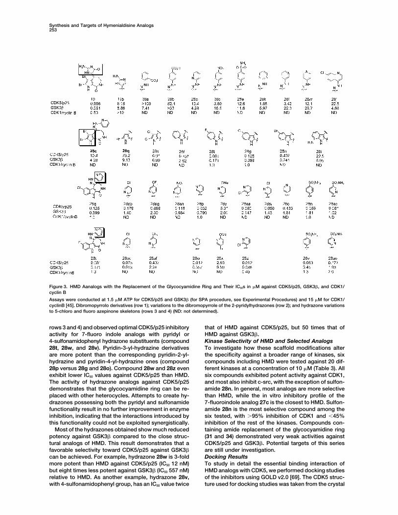
Synthesis and Targets of Hymenialdisine Analogs253
Figure 3. HMD Aanalogs with the Replacement of the Glycocyamidine Ring and Their IC50s in �M against CDK5/p25, GSK3�, and CDK1/cyclin B
Assays were conducted at 1.5 �M ATP for CDK5/p25 and GSK3� (for SPA procedure, see Experimental Procedures) and 15 �M for CDK1/cyclinB [45]. Dibromopyrrolo derivatives (row 1); variations to the dibromopyrrole of the 2-pyridylhydrazones (row 2); and hydrazone variationsto 5-chloro and fluoro azepinone skeletons (rows 3 and 4) (ND: not determined).
rows 3 and 4) and observed optimal CDK5/p25 inhibitory that of HMD against CDK5/p25, but 50 times that ofHMD against GSK3�.activity for 7-fluoro indole analogs with pyridyl or
4-sulfonamidophenyl hydrazone substituents (compound Kinase Selectivity of HMD and Selected AnalogsTo investigate how these scaffold modifications alter28t, 28w, and 28v). Pyridin-3-yl-hydrazine derivatives
are more potent than the corresponding pyridin-2-yl- the specificity against a broader range of kinases, sixcompounds including HMD were tested against 20 dif-hydrazine and pyridin-4-yl-hydrazine ones (compound
28p versus 28g and 28o). Compound 28w and 28z even ferent kinases at a concentration of 10 �M (Table 3). Allsix compounds exhibited potent activity against CDK1,exhibit lower IC50 values against CDK5/p25 than HMD.
The activity of hydrazone analogs against CDK5/p25 and most also inhibit c-src, with the exception of sulfon-amide 28n. In general, most analogs are more selectivedemonstrates that the glycocyamidine ring can be re-
placed with other heterocycles. Attempts to create hy- than HMD, while the in vitro inhibitory profile of the7-fluoroindole analog 27c is the closest to HMD. Sulfon-drazones possessing both the pyridyl and sulfonamide
functionality result in no further improvement in enzyme amide 28n is the most selective compound among thesix tested, with �95% inhibition of CDK1 and 45%inhibition, indicating that the interactions introduced by
this functionality could not be exploited synergistically. inhibition of the rest of the kinases. Compounds con-taining amide replacement of the glycocyamidine ringMost of the hydrazones obtained show much reduced
potency against GSK3� compared to the close struc- (31 and 34) demonstrated very weak activities againstCDK5/p25 and GSK3�. Potential targets of this seriestural analogs of HMD. This result demonstrates that a
favorable selectivity toward CDK5/p25 against GSK3� are still under investigation.Docking Resultscan be achieved. For example, hydrazone 28w is 3-fold
more potent than HMD against CDK5/p25 (IC50 12 nM) To study in detail the essential binding interaction ofHMD analogs with CDK5, we performed docking studiesbut eight times less potent against GSK3� (IC50 557 nM)
relative to HMD. As another example, hydrazone 28v, of the inhibitors using GOLD v2.0 [69]. The CDK5 struc-ture used for docking studies was taken from the crystalwith 4-sulfonamidophenyl group, has an IC50 value twice

Chemistry & Biology254
Table 3. 20 Enzyme Inhibition Pattern for HMD and Selected Analogs: Percent Remaining Enzyme Activity at 10 �M InhibitorConcentration
1 27c 28n 28p 28g 28t
c-Abl 12 36 62 40 9 14HER-1 60 81 96 64 60 69HER-2 52 53 76 44 57 74KDR 7 30 84 73 52 65Flt-3 16 37 57 41 28 25IGF-1R 81 81 70 44 18 24c-Raf-1 84 77 74 36 53 54PDGFR-� 16 35 79 3 32 42Flt-4 36 56 74 78 33 33c-Kit 4 14 67 53 21 31Flt-1 21 49 69 72 15 21Tek 25 61 73 30 27 19PKA 56 61 73 76 69 62c-src 9 11 56 22 8 10CDK1 1 1 5 3 12 2PKB 47 52 60 49 50 52PDK1 100 100 100 81 89 100Ins-R 68 81 73 52 38 46FGFR-1 18 11 74 70 71 82c-Met 41 26 93 43 49 88
The results are presented as kinase activity as a percentage of that in control incubations. ATP was present at 10 �M in all assays. Assayswere conducted according to the procedures in [64] and [65].
structure of CDK5 in complex with p25 (PDB code 1H4L) prevents the glycocyamidine-indole analogs from achiev-ing the perfect geometry for binding to CDK2 (see Sup-[70]. Water and p25 were extracted from the cocomplex
structure and hydrogen atoms were added to the CDK5 plemental Figure S2 at http://www.chembiol.com/cgi/content/full/11/2/247/DC1).protein. There are four residues missing in the glycine-
rich loop of CDK5 structure. Since these four residues In addition to the three commonly observed hydrogenbonds between the pyrrolo- or indolo-azepinones andare not important to the binding of HMD, they were not
included in the docking. All atom types and charges the backbone Glu81 and Cys83 (Leu83 in the case ofCDK2-HMD complex) of the enzyme, the modeling ofwere assigned in GOLD, and all docking parameters
were taken from the standard default settings. Although hydrazone analogs 28v, 28t, and 28w suggested possi-ble hydrogen bonds between the heterocyclic ring ap-the CDK2 in the CDK2-HMD complex is in an inactive
conformation while CDK5 in the CDK5-p25 complex is pendage and the enzyme (Figures 4C–4E). HMD mayform a hydrogen bond between the amino group of thein an active conformation, the binding pockets of HMD
in CDK2 and CDK5 are structurally very similar. After glycocyamidine and the side chain of Asp144, as wellas a water-mediated hydrogen bond between the aminosuperposition of 17 residues in the ATP binding pocket
within 5 A of HMD, the rmsd of C� atoms from CDK2 group of the glycocyamidine and the main chain amideof Gln130. The pyridyl groups of 28t and 28w may formand CDK5 is only 0.6 A. We compared the model of
HMD bound to active CDK5 with the crystal structure electrostatic interactions with the Asp144 carboxylate.In addition, Gln130 is within 5 A of the hydrazone proton,of inactive CDK2 in complex with HMD and found the
interaction of HMD with protein in both structures were making possible a water-mediated hydrogen bond. Inorder to fit into the ATP pocket, sulfonamide 28v mayvery similar. Experimentally, crystal structures of indiru-
bin bound to active and inactive CDK2 also showed that adopt a different geometry of the hydrazone doublebond from pyridyl compounds 28t and 28w. It may bea kinase inhibitor bound to active and inactive CDK2
with a similar binding mode [71]. capable of forming a hydrogen bond between the sulfon-amide and the Asp144 side chain, as well as a hydrogenWe noticed that for the close structural analogs of
HMD (Table 2), indole derivatives 27 exhibit reduced bond between the oxygen of the -SO2- and the Tyr11NH. The different hydrogen bonding mode could be re-CDK5 versus GSK3� activity relative to the correspond-
ing pyrrole analogs. One possible explanation for this sponsible for compound 28v’s different kinase selectiv-ity profile. Docking 28t or 28w with a different doublediffering kinase inhibitory profile of pyrrole and indole
compounds is the steric interaction between the 4-H of bond geometry leads to less favorable inhibitor-kinaseinteractions. As the orientation of heterocyclic append-the indole ring and the glycocyamidine amide. While the
tricyclic ring system of HMD appears to be coplanar age of the hydrazones is quite different from that ofHMD, we propose that the 10-fold improved potencywhen HMD is bound to CDK2 [45], the steric repulsion

Synthesis and Targets of Hymenialdisine Analogs255
Figure 4. Illustration of Possible Interactionsbetween HMD or Its Analogs with CDK2 orCDK5/p25
Hydrogen bonds and electrostatic interac-tions are indicated by broken lines.(A) Cartoon illustration of key hydrogen bondsin HMD and CDK2 complex, reproduced ac-cording to [45].(B) HMD and CDK5/p25.(C) Hydrazone 28v and CDK5/p25.(D) Hydrazone 28w and CDK5/p25.(E) Hydrazone 28t and CDK5/p25.
against CDK5/p25 and GSK3� of the hydrazone indoles adenocarcinoma cells with selected compounds andcharacterized cell proliferation and DNA content redistri-(e.g., compound 28w, Figure 3) versus the glycocyami-
dine indoles (e.g., 27c, Table 2) results from a more butions in the population using fluorescence micros-copy. The majority of unsynchronized 786-O cells grow-optimal orientation of the hydrazone heterocycle relative
to the indole core to relieve strain from the indole 4-H ing in serum are in G1 phase (53.1% � 1.6%), with16.1% � 0.9% and 30.8% � 1.6% in S and G2/M phases,position.
Cellular Effects and Cell Cycle Studies respectively (see Supplemental Figure S3 and Table S2at http://www.chembiol.com/cgi/content/full/11/2/247/To assess the efficacy of these compounds as CDK
inhibitors in a cellular context, we treated 786-O renal DC1). Inhibition of CDK1/cyclin activity results in G2/M
Chemistry & Biology256
1 mM NaF, 1 mM phenyl phosphate, 15 mM nitrophenyl phosphate,arrest [3]. Although HMD is an effective inhibitor of60 mM �-glycerophosphate, 100 mM benzamidine, 10 �g/ml leupep-CDK1/cyclin B activity in vitro (IC50 � 70 nM), its celltin, and 10 �g/ml aprotinin [pH 7.2]) using tissue grinders (Kontes).cycle progression inhibitory effect in 786-O cells is mod-The resulting homogenate was centrifuged for 30 min at 20,000 � g
est. The lowest concentration of HMD tested that results and 4 C. The supernatant was recovered and stored at �80 C untilin significant (�40%) G2/M arrest is 100 �M (Supple- used.mental Table S2). While this discrepancy between enzy-matic and cellular activity is large, it is generally consis-
Affinity Chromatographytent with an earlier study that shows HMD inhibition ofAffinity chromatography was performed as described in [54]. Affinity
GSK3 activity at 50 �M in cells [45]. matrices (30 �l) were washed with bead buffer (50 mM Tris HCl,HMD analogs that were most similar to HMD in struc- 5 mM EDTA, 5 mM EGTA, 5 mM NaF, 250 mM NaCl, 0.1% Nonidet
ture similarly showed limited cellular activity. Com- P-40, 100 mM benzamidine, 10 �g/ml leupeptin, and 10 �g/ml aproti-nin [pH 7.4]) twice before use and resuspended in 400 �l of beadpounds 10, 15, 27a, 27b, 27c, 27d, and 27g had no effectbuffer. Proteins (200 �g) in 400 �l of lysis buffer were added andon the percentage of cells in G2/M phase up to 30 �Mincubated with rotation for 1 hr at 4 C. The resins were then washedand no growth inhibition phenotype up to 15 �M. A4 times with bead buffer and bound proteins were eluted by heatingkey structural feature that these compounds share withat 95 C for 3 min in the presence of 2� Lammli sample buffer. The
HMD is the highly polar glycocyamidine ring, which may resulting supernatants were loaded onto SDS-PAGE gels, and thelimit the compounds’ cell permeability. Other com- separated proteins were visualized by silver staining or Westernpounds tested that were inactive included hydrazones blot analysis. To identify proteins by peptide mass fingerprinting,
proteins (1 mg) were loaded onto the same amount of resins and28c, 28e, 28l, 28n, and 28v. These compounds either hadeluted proteins were stained by Coomassie brilliant blue G (Bio-Radlow CDK inhibitory activity (28c and 28l) or sulfonamideLaboratories).groups that seemed to inhibit cellular activity (28e, 28n,
and 28v).The compounds with the highest inhibition of cell Silver Staining
Silver staining was performed as described in [70]. Gels were fixedgrowth were indole-hydrazone analogs. Compoundsin 50% methanol/5% acetic acid for 20 min then washed with water28p, 28w, and 28z (Figure 3, rows 3 and 4) arrestedfor 1 hr. They were sensitized by incubating in 0.02% (w/v) Na2S2O3cells in G2/M phase at concentrations as low as 3.8 �Mfor 1 min and then washed twice with water for 1 min before being(Supplemental Figure S3C and Table S2 at http://www. incubated in 0.1% (w/v) AgNO3 for 20 min. After being washed again
chembiol.com/cgi/content/full/11/2/247/DC1). Com- twice with water for 1 min, gels were developed in a solution con-pounds 28t, 28g, and 28ai did not alter the percentage taining 0.04% (v/v) formaldehyde and 2% (w/v) Na2CO3. The reaction
was terminated by discarding the reagent followed by addition ofof cells in G2/M phase but significantly inhibited cell5% acetic acid in water.proliferation. These compounds may have a more com-
plex kinase inhibitory profile (inhibiting targets that resultin G1 arrest in addition to CDK1/cyclin A) or act by other Protein In-Gel Digestionmechanisms that cause cell death. In-gel digestion was performed according to the published protocol
[72] with minor modifications. Protein bands in Coomassie-stainedgel were excised and rinsed with water for 15 min. They were thenSignificanceincubated in 100 mM NH4HCO3 (100 �l) for 15 min. The same volumeof CH3CN was added and incubated for 15 min. After the supernatantSeveral protein kinase inhibitors have advanced towas removed, protein bands were shrunk by dehydration in CH3CN.
clinical trials based on the indolocarbazole nucleus of CH3CN was removed by vacuum centrifuge and gel pieces wereStaurosporine (PKC412, Novartis; CEP-701, Cephalon; swollen in a digestion solution containing 50 mM NH4HCO3 and 12.5LY-333531, Eli Lilly). The pyrrole[2,3-c]azipine scaffold ng/�l of modified porcine trypsin (Promega) in an ice-cold bath. The
supernatant was removed 45 min later and replaced with 50 mMof HMD also exhibits great potential for developmentNH4HCO3 (15 �l) to keep gel pieces wet during the overnight enzy-as a therapeutically relevant kinase inhibitor scaffold.matic digestion at 37 C.The present work facilitates this goal by demonstra-
ting that analogs of HMD can be prepared by a varietyof synthetic routes and that these compounds posses Sample Preparation for MS Analysis and Peptide Mass
Fingerprinting Using MALDI-TOF MSaltered potencies and selectivities toward a variety ofTryptic digests were introduced to microscale purification [73],therapeutically relevant kinases.which allowed efficient concentration of peptides and cleanup ofanalytes. A micro purification column was prepared using a Gel-Experimental Proceduresoader tip (Eppendorf, Germany), in which Poros 50 R2 (PerSeptiveBiosystems) was packed and equilibrated. Samples were loadedPreparation and Use of Affinity Reagentsonto the column and briefly washed with formic acid (5%, 15 �l).Affi-Gel 10 Gel (Bio-Rad Laboratory) (1 ml bed volume) was rinsedPeptides bound to the resin were eluted directly onto a MALDI targetwith anhydrous DMSO (4 ml � 3) and transferred into a vial con-with 1 �l of saturated �-cyano-4-hydroxy-trans-cinnamic acid intaining a solution of linker tethered compound 4 or 7 in DMSO (3acetonitrile-formic acid-water (50:5:45) solution.�M, 1 ml). The mixture was agitated at room temperature for 1
The MALDI-TOF mass spectrometer (BIFLEX; Bruker Daltoniks,day in the presence of triethylamine (the progress of the couplingGermany) was operated in the reflector mode. Ion signals producedreaction was monitored by LC-MS). Upon the completion of theby trypsin autodigestion peptides were used as internal mass cali-coupling, excess amount of ethanolamine (8 �l) was added to blockbrants. Proteins were identified by searching against NCBI humanany unreacted groups that remain on the resin. The resulting beadsnonredundant (nr) and SwissProt databases with the list of mono-were washed with DMSO (4 ml � 3) and PBS (4 ml � 3) and storedisotopic tryptic peptide masses using 200 ppm mass tolerance.in PBS (1 ml) at 4 C until use.Database searching was performed using a web-based searchprogram, PROFOUND (http://prowl.rockefeller.edu/profound_bin/Extraction of ProteinswebProFound.exe) developed by Rockefeller University and Pro-Mouse brains were homogenized in lysis buffer (25 mM MOPS, 15
mM EGTA, 15 mM MgCl2, 2 mM DTT, 1 mM sodium orthovanadate, teometrix [74].
Synthesis and Targets of Hymenialdisine Analogs257
Radioenzymatic RSK1 Kinase Assay 4. Traxler, P., Bold, G., Buchdunger, E., Caravatti, G., Furet, P.,Manley, P., O’Reilly, T., Wood, J., and Zimmermann, J. (2001).The RSK1 (p90S6K) kinase inhibitory activity of HMD and HMD-
linker was evaluated following manufacturer’s (Upstate) recommen- Tyrosine kinase inhibitors: from rational design to clinical trials.Med. Res. Rev. 21, 499–512.dations using the peptide KKLNRTLSVA as a substrate.
5. Toogood, P.L. (2001). Cyclin-dependent kinase inhibitors fortreating cancer. Med. Res. Rev. 21, 487–498.In Vitro Kinase Assay Using SPA Assay
6. Bridges, A.J. (2001). Chemical inhibitors of protein kinases.SPA assay [75] was performed using SPA assay kit (AmershamChem. Rev. 101, 2541–2572.Pharmacia Biotech, UK). Inhibitors dissolved in DMSO were trans-
7. Scapin, G. (2001). Structural biology in drug design: selectiveferred to 5 �l of enzyme in assay buffer (50 mM MOPS [pH 7.2] andprotein kinase inhibitors. Drug Discov. Today 7, 601–611.5 mM MgCl2). Enzymatic reactions were initiated by adding 10 �l of
8. Garrett, M.D., and Fattaey, A. (1999). CDK inhibition and cancersolution containing 1.5 �M ATP, 1.5 �M biotinylated substrate, 0.01therapy. Curr. Opin. Gene Dev. 9, 104–111.�Ci [�-33P]ATP (NEN Life Science) and incubated at room tempera-
9. Malumbres, M., and Barbacid, M. (2001). Milestones in cell divi-ture for 1 hr in 384-well ProxiPlates (Perkin Elmer). Reactions weresion: to cycle or not to cycle: a critical decision in cancer. Nat.stopped by the addition of 10 �l PBS solution containing 50 �g ofRev. Cancer 1, 222–231.streptavidin-tagged polyvinyltoluene bead, 50 mM ATP, 5 mM EDTA,
10. Ali, A., Hoeflich, K.P., and Woodgett, J.R. (2001). Glycogen syn-and 0.1% Triton X-100. Before counting the signals with TopCountthase kinase-3: properties, functions, and regulation. Chem.(Packard Bioscience), the plates were spun for 3 min at 2000 rpm.Rev. 101, 2527–2540.For GSK3�, sequence of the peptide substrate was biotin-YRR
11. Imahori, K., and Uchida, T. (1997). Physiology and pathology ofAAVPPSPSLSRHSSPHQ(pS)EDEEE (Upstate). In CDK5/p25 assay,tau protein kinases in relation to Alzheimer’s disease. J. Bio-the peptide was biotin-aminohexanoic acid-AGAKKAVKTPKKAKKPchem. (Tokyo) 121, 179–188.derived from Histone H1.
12. Mandelkow, E.-M., and Mandelkow, E. (1998). Tau in Alzhei-mer’s disease. Trends Cell Biol. 8, 425–427.Cell Cycle Studies
13. Martinez, A., Castro, A., Dorronsoro, I., and Alonso, M. (2002).786-O renal adenocarcinoma cells were cultured in DMEM with 10%Glycogen synthase kinase 3 (GSK-3) inhibitors as new promisingfetal bovine serum supplemented with glutamine and antibiotics.drugs for diabetes, neurodegeneration, cancer, and inflamma-Cells were plated in black clear bottom 384-well microtiter platestion. Med. Res. Rev. 22, 373–384.(Greiner) and cultured for 8 hr. Compounds of interest were added
14. Profit, A.A., Lee, T.R., and Lawrence, D.S. (1999). Bivalent inhibi-to cells (1% DMSO final concentration) and incubated for 30 hrtors of protein tyrosine kinases. J. Am. Chem. Soc. 121, 280–283.at 37 C, with four replicate wells for each concentration. After wash-
15. Martinez, A., Alonso, M., Castro, A., Perez, C., and Moreno, F.J.ing with PBS, cells were fixed with 3.5% paraformaldehyde in PBS(2002). First non-ATP competitive glycogen synthase kinase 3for 20 min. Nuclei were stained with 4�,6-diamidino-2-phenylindole(GSK-3) inhibitors: thiadiazolidinones (TDZD) as potential drugsdihydrochloride (200 ng/ml) in 0.1% TritonX-100/PBS for 1 hr. Afterfor the treatment of Alzheimer’s disease. J. Med. Chem. 45,washing with PBS, cells were imaged on an EIDAQ100 (Q3DM) auto-1292–1299.mated inverted fluorescence microscope (Nikon TE300 with a Cohu
16. Fry, D.W., Kraker, A.J., McMichael, A., Ambroso, L.A., Nelson,video camera) with a 10�/0.5 objective (Nikon). Sixteen images wereJ.M., Leopold, W.R., Connors, R.W., and Bridges, A.J. (1994).collected per well. Image analysis was performed with CytoShopA specific inhibitor of the epidermal growth factor receptor tyro-software (Q3DM). After images were shade corrected and back-sine kinase. Science 265, 1093–1095.ground subtracted, objects were extracted and single cells defined
17. Rewcastle, G.W., Murray, D.K., Elliott, W.L., Fry, D.W., Howard,using geometric and total fluorescence parameters. HistogramsC.T., Nelson, J.M., Roberts, B.J., Vincent, P.W., Showalter, H.D.,plotting number of events versus total nuclear intensity for represen-Winters, R.T., et al. (1998). Tyrosine kinase inhibitors. 14. Struc-tative wells were used to define nuclear fluorescence boundariesture-activity relationships for methylamino-substituted deriva-for G1, S, and G2/M cells. Percentages of cells in each categorytives of 4-[(3-bromophenyl)amino]-6-(methylamino)-pyrido[3,4-were determined for all wells and replicate wells were averaged ford]pyrimidine (PD 158780), a potent and specific inhibitor of theeach condition.tyrosine kinase activity of receptors for the EGF family of growthfactors. J. Med. Chem. 41, 742–751.
Supplemental Data18. Traxler, P., Bold, G., Frei, J., Lang, M., Lydon, N., Mett, H.,
General synthetic methods and spectroscopic data for all com- Buchdunger, E., Meyer, T., Mueller, M., and Furet, P. (1997).pounds included in the main text, as well as cell proliferation and Use of a pharmacophore model for the design of EGF-R tyrosineDNA content redistributions in the population of 786-O renal adeno- kinase inhibitors: 4-(phenylamino)pyrazolo[3,4-d]pyrimidines. J.carcinoma cells after treatment with selected HMD analogs, are Med. Chem. 40, 3601–3616.included in the Supplemental Data at http://www.neuron.org/cgi/ 19. Traxler, P.M., Furet, P., Mett, H., Buchdunger, E., Meyer, T., andcontent/full/11/2/247/DC1. Lydon, N. (1996). 4-(Phenylamino)pyrrolopyrimidines: potent
and selective, ATP site directed inhibitors of the EGF-receptorAcknowledgments protein tyrosine kinase. J. Med. Chem. 39, 2285–2292.
20. Zimmermann, J., Buchdunger, E., Mett, H., Meyer, T., and Ly-We are grateful to Dr. Priscilla Yang and Dr. Tetsuo Uno for revising don, N.B. (1997). Potent and selective inhibitors of the ABL-this article and providing valuable suggestions. kinase: phenylaminopyrimidine (PAP) derivatives. Bioorg. Med.
Chem. Lett. 7, 187–192.Received: September 19, 2003 21. Fong, T.A.T., Shawver, L.K., Sun, L., Tang, C., App, H., Powell,Revised: November 18, 2003 T.J., Kim, Y.H., Schreck, R., Wang, X., Risau, W., et al. (1999).Accepted: November 24, 2003 SU5416 is a potent and selective inhibitor of the vascular endo-Published: February 20, 2004 thelial growth factor receptor (Flk-1/KDR) that inhibits tyrosine
kinase catalysis, tumor vascularization, and growth of multipleReferences tumor types. Cancer Res. 59, 99–106.
22. Sun, L., Tran, F., App, H., Hirth, P., McMahon, G., and Tang, C.1. Sridhar, R., Hanson-Painton, O., and Cooper, D.R. (2000). Pro- (1998). Synthesis and biological evaluations of 3-substituted
tein kinase as therapeutic targets. Pharm. Res. 17, 1345–1353. indolin-2-ones: a novel class of tyrosine kinase inhibitors that2. Garcıa-Echeverrıa, C., Traxler, P., and Evans, D.B. (2000). ATP exhibit selectivity toward particular receptor tyrosine kinases.
site-directed competitive and irreversible inhibitors of protein J. Med. Chem. 41, 2588–2603.kinases. Med. Res. Rev. 20, 28–57. 23. Vesely, J., Havlicek, L., Strnad, M., Blow, J.J., Donella-Deana,
3. Sielecki, T.M., Boylan, J.F., Benfield, P.A., and Trainor, G.L. A., Pinna, L., Letham, D.S., Kato, J.Y., Detivaud, L., Leclerc, S.,(2000). Cyclin-dependent kinase inhibitors: useful targets in cell et al. (1994). Inhibition of cyclin-dependent kinases by purine
analogs. Eur. J. Biochem. 224, 771–786.cycle regulation. J. Med. Chem. 43, 1–18.
Chemistry & Biology258
24. De Azevedo, W.F., Leclerc, S., Meijer, L., Havlicek, L., Strnad, 41. Kitagawa, I., Kobayashi, M., Kitanaka, K., Kido, M., and Kyo-goku, Y. (1983). Marine natural products. XII. On the chemicalM., and Kim, S.H. (1997). Inhibition of cyclin-dependent kinases
by purine analogs. Crystal structure of human cdk2 complexed constituents of the Okinawan marine sponge Hymeniacidonaldis. Chem. Pharm. Bull. 31, 2321–2328.with roscovitine. Eur. J. Biochem. 243, 518–526.
25. Gray, N.S., Wodicka, L., Thunnissen, A.-M.W.H., Normann, T.C., 42. De Nanteuil, G., Ahond, A., Guilhem, J., Poupat, C., Tran HuuDau, E., Potier, P., Pusset, M., Pusset, J., and Laboute, P. (1985).Kwon, S., Espinoza, F.H., Morgan, D.O., Barnes, G., LeClerc,
S., Meijer, L., et al. (1998). Exploiting chemical libraries, struc- Marine invertebrates from the New Caledonian lagoon. V. Isola-tion and identification of metabolites of a new species ofture, and genomics in the search for kinase inhibitors. Science
281, 533–538. sponge, Pseudaxinyssa cantharella. Tetrahedron Lett. 41, 6019–6033.26. Knockaert, M., Greengard, P., and Meijer, L. (2002). Pharmaco-
logical inhibitors of cyclin-dependent kinases. Trends Pharma- 43. Schmitz, F.J., Gunasekera, S.P., Lakshmi, V., and Tillekeratne,L.M.V. (1985). Marine natural products: pyrrololactams fromcol. Sci. 23, 417–425.
27. Chang, Y.T., Gray, N.S., Rosania, G.R., Sutherlin, D.P., Kwon, several sponges. J. Nat. Prod. 48, 47–53.44. Pettit, G.R., Herald, C.L., Leet, J.E., Gupta, R., Schaufelberger,S., Norman, T.C., Sarohia, R., Leost, M., Meijer, L., and Schultz,
P.G. (1996). Synthesis and application of functionally diverse D.E., Bates, R.B., Clewlow, P.J., Doubek, D.L., Manfredi, K.P.,2,6,9-trisubstituted purine libraries as CDK inhibitors. Chem. Rutzler, K., et al. (1990). Antineoplastic agents. 168. IsolationBiol. 5, 361–375. and structure of axinohydantoin. Can. J. Chem. 68, 1621–1624.
28. Legraverend, M., Tunnah, P., Noble, M., Ducrot, P., Ludwig, O., 45. Meijer, L., Thunnissen, A.-M.W.H., White, A.W., Garnier, M., Ni-Grierson, D.S., Leost, M., Meijer, L., and Endicott, J. (2000). kolic, M., Tsai, L.-H., Walter, J., Cleverley, K.E., Salinas, P.C.,Cyclin-dependent kinase inhibition by new C-2 alkynylated pu- Wu, Y.-Z., et al. (1999). Inhibition of cyclin-dependent kinases,rine derivatives and molecular structure of a CDK2-inhibitor GSK-3� and CK1 by hymenialdisine, a marine sponge constit-complex. J. Med. Chem. 43, 1282–1292. uent. Chem. Biol. 7, 51–63.
29. Lee, J.C., Badger, A.M., Griswold, D.E., Dunnington, D., Truneh, 46. Tasdemir, D., Mallon, R., Greenstein, M., Feldberg, L.R., Kim,A., Votta, B., White, J.R., Young, P.R., and Bender, P.E. (1993). S.C., Collins, K., Wojciechowicz, D., Mangalindan, G.C., Con-Bicyclic imidazoles as a novel class of cytokine biosynthesis cepcion, G.P., Harper, M.K., et al. (2002). Aldisine alkaloids frominhibitors. Ann. N Y Acad. Sci. 696, 149–170. the Philippine sponge Stylissa massa are potent inhibitors of
30. Adams, J.L., Boehm, J.C., Kassis, S., Gorycki, P.D., Webb, E.F., mitogen-activated protein kinase kinase-1 (MEK-1). J. Med.Hall, R., Sorenson, M., Lee, J.C., Ayrton, A., Grisword, D.E., et Chem. 45, 529–532.al. (1998). Pyrimidinylimidazole inhibitors of CSBP/p38 kinase 47. Curman, D., Cinel, B., Williams, D.E., Rundle, N., Block, W.D.,demonstrating decreased inhibition of hepatic cytochrome Goodarzi, A.A., Hutchins, J.R., Clarke, P.R., Zhou, B.-B., Lees-P450 enzymes. Bioorg. Med. Chem. Lett. 8, 3111–3116. Miller, S.P., et al. (2001). Inhibition of the G2 DNA damage check-
31. Laufer, S.A., Wagner, G.K., Kotschenreuther, D.A., and Albrecht, point and of protein kinases Chk1 and Chk2 by the marineW. (2003). Novel substituted pyridinyl imidazoles as potent sponge alkaloid debromohymenialdisine. J. Biol. Chem. 276,anticytokine agents with low activity against hepatic cyto- 17914–17919.chrome P450 enzymes. J. Med. Chem. 46, 3230–3244. 48. Nishizuka, Y. (1988). The molecular heterogeneity of protein
32. Carlson, B.A., Dubay, M.M., Sausville, E.A., Brizuela, L., and kinase C and its implications for cellular regulation. Nature 334,Worland, P.J. (1996). Flavopiridol induces G1 arrest with inhibi- 661–665.tion of cyclin-dependent kinase (CDK) 2 and CDK4 in human 49. Nishizuka, Y. (1989). The family of protein kinase C for signalbreast carcinoma cells. Cancer Res. 56, 2973–2978. transduction. JAMA 262, 1826–1833.
33. Losiewicz, M.D., Carlson, B.A., Kaur, G., Sausville, E.A., and 50. Nambi, P., and Patil, A.D. (September, 1993). Imidazolinylide-Worland, P.J. (1994). Potent inhibition of Cdc2 kinase activity nepyrroloazepine derivatives as protein kinase C inhibitor. Pa-by the flavonoid L86–8275. Biochem. Biophys. Res. Commun. tent WO-9316703.201, 589–595. 51. Breton, J.J., and Chabot-Fletcher, M.C. (1997). The natural
34. Zaharevitz, D.W., Gussio, R., Leost, M., Senderowicz, A.M., La- product hymenialdisine inhibits interleukin-8 production in U937husen, T., Kunick, C., Meijer, L., and Sausville, E.A. (1999). Dis- cells by inhibition of nuclear factor-�B. J. Pharmacol. Exp. Ther.covery and initial characterization of the paullones, a novel class 282, 459–466.of small-molecule inhibitors of cyclin-dependent kinases. Can- 52. Roshak, A., Jackson, J.R., Chabot-Fletcher, M., and Marshal,cer Res. 59, 2566–2569. L.A. (1997). Inhibition of NF�B-mediated interleukin-1�-stimu-
35. Schultz, C., Link, A., Leost, M., Zaharevitz, D.W., Gussio, R., lated prostaglandin E2 formation by the marine natural productSausville, E.A., Meijer, L., and Kunick, C. (1999). Paullones, a
hymenialdisine. J. Pharmacol. Exp. Ther. 283, 955–961.series of cyclin-dependent kinase inhibitors: synthesis, evalua-
53. Badger, A.M., Cook, M.N., Swift, B.A., Newman-Tarr, T.M., Go-tion of CDK1/cyclin B inhibition, and in vitro antitumor activity.
wen, M., and Lark, M. (1999). Inhibition of interleukin-1-inducedJ. Med. Chem. 42, 2909–2919.
proteoglycan degradation and nitric oxide production in bovine36. Leost, M., Schultz, C., Link, A., Wu, Y.-Z., Biernat, J., Mandel-articular cartilage/chondrocyte cultures by the natural product,kow, E.-M., Bibb, J.A., Snyder, G.L., Greengard, P., Zaharevitz,hymenialdisine. J. Pharmacol. Exp. Ther. 290, 587–593.D.W., et al. (2000). Paullones are potent inhibitors of glycogen
54. Knockaert, M., Gray, N., Damiens, E., Chang, Y.-T., Grellier, P.,synthase kinase-3b and cyclin-dependent kinase 5/p25. Eur. J.Grant, K., Fergusson, D., Mottram, J., Soete, M., Dubremetz,Biochem. 267, 5983–5994.J.-F., et al. (2000). Intracellular targets of cyclin-dependent ki-37. Furusaki, A., Hashiba, N., Matsumoto, T., Hirano, A., Iwai, Y.,nase inhibitors: identification by affinity chromatography usingand Omurs, S. (1982). The crystal and molecular structure ofimmobilised inhibitors. Chem. Biol. 7, 411–422.staurosporine, a new alkaloid from a Streptomyces strain. Bull.
55. Knockaert, M., Wieking, K., Schmitt, S., Leost, M., Grant, K.M.,Chem. Soc. Jpn. 55, 3681–3685.Mottram, J.C., Kunick, C., and Meijer, L. (2002). Intracellular38. Gadbois, D.M., Hamaguchi, J.R., Swank, R.A., and Bradbury,targets of paullones. Identification following affinity purificationE.M. (1992). Staurosporine is a potent inhibitor of p34cdc2 andon immobilized inhibitor. J. Biol. Chem. 277, 25493–25501.p34cdc2-like kinases. Biochem. Biophys. Res. Commun. 184,
56. Annoura, H., and Tatsuoka, T. (1995). Total syntheses of hymeni-80–85.aldisine and debromohymenialdisine: stereospecific construc-39. Hudkins, R.L., Iqbal, M., Park, C.-H., Goldstein, J., Herman, J.,tion of the 2-amino-4-oxo-2-imidazolin-5(Z)-disubstituted yli-Shek, E., Murakata, C., and Mallamo, J.P. (1998). Prodrug estersdene ring system. Tetrahedron Lett. 36, 413–416.of the indolocarbazole CEP-751 (KT-6587). Bioorg. Med. Chem.
57. Xu, Y.-Z., Yakushijin, K., and Horne, D.A. (1997). Synthesis ofLett. 8, 1873–1876.C11N5 marine sponge alkaloids: (�)-hymenin, stevensine, hy-40. Cimino, G., De Rosa, S., De Stefano, S., Mazzarella, L., Puliti,menialdisine, and debromohymenialdisine. J. Org. Chem. 62,R., and Sodano, G. (1982). Isolation and x-ray crystal structure456–464.of a novel bromo compound from two marine sponges. Tetrahe-
dron Lett. 23, 767–768. 58. Sosa, A.C.B., Yakushijin, K., and Horne, D.A. (2000). A practical
Synthesis and Targets of Hymenialdisine Analogs259
synthesis of (Z)-debromohymenialdisine. J. Org. Chem. 65,610–611.
59. Chacun-Lefevre, L., Joseph, B., and Merour, J.-Y. (2000). Syn-thesis and reactivity of azepino[3,4-b]indol-5-yl trifluorometh-anesulfonate. Tetrahedron 56, 4491–4499.
60. Chacun-Lefevre, L., Joseph, B., and Merour, J.-Y. (2001). Intra-molecular Heck coupling of alkenyl 3-iodoindole-2-carboxa-mide derivatives. Synlett. 6, 848–850.
61. Erdelyl, M., and Gogoll, A. (2001). Rapid homogeneous-phaseSonogashira coupling reactions using controlled microwaveheating. J. Org. Chem. 66, 4165–4169.
62. Kearns, A.E., Donohue, M.M., Sanyal, B., and Demay, M.B.(2001). Cloning and characterization of a novel protein kinasethat impairs osteoblast differentiation in vitro. J. Biol. Chem.276, 42213–42218.
63. Primot, A., Baratte, B., Gompel, M., Borgne, A., Liabeuf, S.,Romette, J.-L., Jho, E.-h., Costantini, F., and Meijer, L. (2000).Purification of GSK-3 by affinity chromatography on immobi-lized axin. Protein Expr. Purif. 20, 394–404.
64. Davis, S.P., Reddy, H., Caivano, M., and Cohen, P. (2000). Speci-ficity and mechanism of action of some commonly used proteinkinase inhibitors. Biochem. J. 251, 95–105.
65. Bain, J., McLauchan, H., Elliott, M., and Cohen, P. (2003). Thespecificities of protein kinase inhibitors: an update. Biochem.J. 371, 199–204.
66. Cho, H., and Matsuki, S. (1996). Ring construction of severalheterocycles with phosphorus pentoxide-methanesulfonic acid(PPMA). Heterocycles 43, 127–131.
67. Kano, S., Yokomatsu, T., Nemoto, H., and Shibuya, S. (1985).An efficient diastereoselective synthesis of 6-hydroxy-4a-[3,4-crowned(15-crown-5)phenyl]-trans-decahydroisoquinoline.Tetrahedron Lett. 26, 1531–1534.
68. Kano, S., Yokomatsu, T., Nemoto, H., and Shibuya, S. (1986).Effect of A-strain on a diastereoselective synthesis of 6-hydroxy-4a-aryldecahydroisoquinolines; revised structures of N-acyli-minium ion-polyene cyclization products. J. Org. Chem. 51,561–564.
69. Jones, G., Willett, P., Glen, R.C., Leach, A.R., and Taylor, R.(1997). Development and validation of a genetic algorithm forflexible docking. J. Mol. Biol. 267, 727–748.
70. Tarricone, C., Dhavan, R., Peng, J., Areces, L.B., Tsai, L.H., andMusacchio, A. (2001). Structure and regulation of the CDK5-p25 (nck5a) complex. Mol. Cell 8, 657–669.
71. Davies, T.G., Tunnah, P., Meijer, L., Marko, D., Eisenbrand, G.,Endicott, J.A., and Noble, M.E. (2001). Inhibitor binding to activeand inactive CDK2: the crystal structure of CDK2-cyclin A/indi-rubin-5-sulphonate. Structure 9, 389–397.
72. Shevchenko, A., Wilm, M., Vorm, O., and Mann, M. (1996). Massspectrometric sequencing of proteins silver-stained polyacryl-amide gels. Anal. Chem. 68, 850–858.
73. Kussmann, M., Nordhoff, E., Rahbek-Nielsen, H., Haebel, S.,Rossel-Larsen, M., Jakobsen, L., Gobom, J., Mirgorodskaya,E., Kroll-Kristensen, A., Palm, L., et al. (1997). Matrix-assistedlaser desorption/ionization mass spectrometry sample prepara-tion techniques designed for various peptide and protein ana-lytes. J. Mass Spectrom. 32, 593–601.
74. Zhang, W., and Chait, B.T. (2000). ProFound: an expert systemfor protein identification using mass spectrometric peptidemapping information. Anal. Chem. 72, 2482–2489.
75. Cook, N.D. (1996). Scintillation proximity assay—a versatile highthroughput screening technology. Drug Discov. Today 1,287–294.