Surgical Intervention and Accommodative Responses, I: Centripetal Ciliary Body, Capsule, and Lens...
11
Surgical Intervention and Accommodative Responses, I: Centripetal Ciliary Body, Capsule, and Lens Movements in Rhesus Monkeys of Various Ages Mary Ann Croft, 1 Jared P. McDonald, 1 Rebecca J. James, 1 Gregg A. Heatley, 1 Ting-Li Lin, 2 Elke Lu ¨tjen-Drecoll, 3 and Paul L. Kaufman 1,4 PURPOSE. To determine how surgically altering the normal re- lationship between the lens and the ciliary body in rhesus monkeys affects centripetal ciliary body and lens movement. METHODS. In 18 rhesus monkey eyes (aged 6 –27 years), accom- modation was induced before and after surgery by electrical stimulation of the Edinger-Westphal nucleus. Accommodative amplitude was measured by coincidence refractometry. Go- niovideography was performed before and after intra- and extracapsular lens extraction (ICLE, ECLE) and anterior re- gional zonulolysis (ARZ). Centripetal lens/capsule movements, centripetal ciliary process (CP) movements, and circumlental space were measured by computerized image analysis of the goniovideography images. RESULTS. Centripetal accommodative CP and capsule move- ment increased in velocity and amplitude after, compared with before, ECLE regardless of age (n 5). The presence of the lens substance retarded capsule movement by 21% in the young eyes and by 62% in the older eyes. Post-ICLE com- pared with pre-ICLE centripetal accommodative CP movement was dampened in all eyes in which the anterior vitreous was disrupted (n 7), but not in eyes in which the anterior vitreous was left intact (n 2). After anterior regional zonu- lolysis (n 4), lens position shifted toward the lysed quadrant during accommodation. CONCLUSIONS. The presence of the lens substance, capsule zonu- lar attachments, and Wieger’s ligament may play a role in centripetal CP movement. The capsule is still capable of cen- tripetal movement in the older eye (although at a reduced capacity) and may have the ability to produce 6 D of accom- modation in the presence of a normal, young crystalline lens or a similar surrogate. (Invest Ophthalmol Vis Sci. 2008;49: 5484 –5494) DOI:10.1167/iovs.08-1916 P resbyopia (the loss of accommodation with age) has been attributed to increased hardness of the lens, 1–7 lens growth, 6,8 –14 and loss of elasticity of the ciliary muscle’s pos- terior attachments. 15,16 No individual over the age of 45 years appears exempt, making presbyopia the most common ocular affliction in the world. Although certainly not a blinding con- dition and correctable by various optical means, presbyopia’s cost in devices and lost productivity is substantial. Since the accommodative apparatus of the rhesus monkey is similar to that of the human, 17–19 and both species develop presbyopia on a time scale similar to lifespan 19 –21 experimen- tal studies elucidating the role of the various structures for development of presbyopia can be performed in the rhesus. In addition, this study for the first time characterizes the function of the eye’s various accommodative components after lens extraction procedures, which are undertaken in millions of human patients every year (either unilaterally, bilaterally at separate time points, or in some cases bilaterally during the same surgery day). The information generated from these stud- ies is critical to the successful development of an accommo- dating intraocular lens (IOL). We determined how surgically altering the normal relation- ship between the lens and the ciliary body in live rhesus monkeys 6 to 27 years of age affects the normal centripetal movements of the ciliary body and lens/capsule. MATERIALS AND METHODS Details of all equipment and animal handling procedures for anesthe- sia, iridectomy, electrode implantation, central stimulation, measure- ment of accommodation, image calibration, goniovideography, veloc- ity, 22 and measurements have been described previously. 20,23–26 All procedures conformed to the ARVO Statement for the Use of Animals in Ophthalmic and Vision Research and were in accordance with institutionally approved animal protocols. Monkeys Twenty-five rhesus monkeys (Macaca mulatta) of either sex, aged 6 to 27 years and weighing 5.0 to 13.4 kg, were studied. The monkeys all had normal phakic eyes with no signs of ocular disease (other than age-related lenticular opacification), as assessed by slit lamp examina- tion. Animals were evaluated for corneal clarity and anterior chamber and anterior vitreous inflammatory reaction before and after surgery. Eight of the monkeys, aged 17 to 26 years and weighing between 7.2 and 9.5 kg, were designated solely for measurements used in the calculation of the normal velocity of the ciliary process (CP) and lens centripetal movements during accommodation and disaccommodation at supramaximum central stimulation. The remaining 17 monkeys underwent surgical procedures. Base- line measurements of the normal accommodative response in the eyes of each animal were taken before the surgical procedures were per- formed. Three lens surgery procedures (described later) were used in the study: intracapsular lens extraction (ICLE), extracapsular lens ex- traction (ECLE), and anterior regional zonulolysis (ARZ). From the Departments of 1 Ophthalmology and Visual Sciences and 2 Biostatistics and Medical Informatics, and the 4 Wisconsin National Primate Research Center, University of Wisconsin-Madison, Madison, Wisconsin; and the 3 Department of Anatomy II, University of Erlangen- Nu ¨rnberg, Erlangen, Germany. Supported in part by National Eye Institute Grant R01 EY10213 (PLK); the Ocular Physiology Research and Education Foundation; DFG DR 124/7 to (ELD); Base Grant 5P51 RR 000167 to the Wisconsin National Primate Research Center, University of Wisconsin-Madison; and Core Grant for Vision Research P30 EY016665. Submitted for publication February 21, 2008; revised May 19, 2008; accepted September 25, 2008. Disclosure: M.A. Croft, None; J.P. McDonald, None; R.J. James, None; G.A. Heatley, None; T.-L. Lin, None; E. Lu ¨tjen-Drecoll, None; P.L. Kaufman, None The publication costs of this article were defrayed in part by page charge payment. This article must therefore be marked “advertise- ment” in accordance with 18 U.S.C. §1734 solely to indicate this fact. Corresponding author: Mary Ann Croft, Department of Ophthal- mology and Visual Sciences, University of Wisconsin Clinical Sciences Center, 600 Highland Avenue, Madison, WI 53792-3220; [email protected]. Investigative Ophthalmology & Visual Science, December 2008, Vol. 49, No. 12 5484 Copyright © Association for Research in Vision and Ophthalmology
-
Upload
independent -
Category
Documents
-
view
3 -
download
0
Transcript of Surgical Intervention and Accommodative Responses, I: Centripetal Ciliary Body, Capsule, and Lens...
Surgical Intervention and Accommodative Responses, I:Centripetal Ciliary Body, Capsule, and Lens Movementsin Rhesus Monkeys of Various Ages
Mary Ann Croft,1 Jared P. McDonald,1 Rebecca J. James,1 Gregg A. Heatley,1 Ting-Li Lin,2
Elke Lutjen-Drecoll,3 and Paul L. Kaufman1,4
PURPOSE. To determine how surgically altering the normal re-lationship between the lens and the ciliary body in rhesusmonkeys affects centripetal ciliary body and lens movement.
METHODS. In 18 rhesus monkey eyes (aged 6–27 years), accom-modation was induced before and after surgery by electricalstimulation of the Edinger-Westphal nucleus. Accommodativeamplitude was measured by coincidence refractometry. Go-niovideography was performed before and after intra- andextracapsular lens extraction (ICLE, ECLE) and anterior re-gional zonulolysis (ARZ). Centripetal lens/capsule movements,centripetal ciliary process (CP) movements, and circumlentalspace were measured by computerized image analysis of thegoniovideography images.
RESULTS. Centripetal accommodative CP and capsule move-ment increased in velocity and amplitude after, compared withbefore, ECLE regardless of age (n � 5). The presence of thelens substance retarded capsule movement by �21% in theyoung eyes and by �62% in the older eyes. Post-ICLE com-pared with pre-ICLE centripetal accommodative CP movementwas dampened in all eyes in which the anterior vitreous wasdisrupted (n � 7), but not in eyes in which the anteriorvitreous was left intact (n � 2). After anterior regional zonu-lolysis (n � 4), lens position shifted toward the lysed quadrantduring accommodation.
CONCLUSIONS. The presence of the lens substance, capsule zonu-lar attachments, and Wieger’s ligament may play a role incentripetal CP movement. The capsule is still capable of cen-tripetal movement in the older eye (although at a reducedcapacity) and may have the ability to produce �6 D of accom-modation in the presence of a normal, young crystalline lens ora similar surrogate. (Invest Ophthalmol Vis Sci. 2008;49:5484–5494) DOI:10.1167/iovs.08-1916
Presbyopia (the loss of accommodation with age) has beenattributed to increased hardness of the lens,1–7 lens
growth,6,8–14 and loss of elasticity of the ciliary muscle’s pos-terior attachments.15,16 No individual over the age of 45 yearsappears exempt, making presbyopia the most common ocularaffliction in the world. Although certainly not a blinding con-dition and correctable by various optical means, presbyopia’scost in devices and lost productivity is substantial.
Since the accommodative apparatus of the rhesus monkey issimilar to that of the human,17–19 and both species developpresbyopia on a time scale similar to lifespan19–21 experimen-tal studies elucidating the role of the various structures fordevelopment of presbyopia can be performed in the rhesus. Inaddition, this study for the first time characterizes the functionof the eye’s various accommodative components after lensextraction procedures, which are undertaken in millions ofhuman patients every year (either unilaterally, bilaterally atseparate time points, or in some cases bilaterally during thesame surgery day). The information generated from these stud-ies is critical to the successful development of an accommo-dating intraocular lens (IOL).
We determined how surgically altering the normal relation-ship between the lens and the ciliary body in live rhesusmonkeys 6 to 27 years of age affects the normal centripetalmovements of the ciliary body and lens/capsule.
MATERIALS AND METHODS
Details of all equipment and animal handling procedures for anesthe-sia, iridectomy, electrode implantation, central stimulation, measure-ment of accommodation, image calibration, goniovideography, veloc-ity,22 and measurements have been described previously.20,23–26 Allprocedures conformed to the ARVO Statement for the Use of Animalsin Ophthalmic and Vision Research and were in accordance withinstitutionally approved animal protocols.
Monkeys
Twenty-five rhesus monkeys (Macaca mulatta) of either sex, aged 6 to27 years and weighing 5.0 to 13.4 kg, were studied. The monkeys allhad normal phakic eyes with no signs of ocular disease (other thanage-related lenticular opacification), as assessed by slit lamp examina-tion. Animals were evaluated for corneal clarity and anterior chamberand anterior vitreous inflammatory reaction before and after surgery.
Eight of the monkeys, aged 17 to 26 years and weighing between7.2 and 9.5 kg, were designated solely for measurements used in thecalculation of the normal velocity of the ciliary process (CP) and lenscentripetal movements during accommodation and disaccommodationat supramaximum central stimulation.
The remaining 17 monkeys underwent surgical procedures. Base-line measurements of the normal accommodative response in the eyesof each animal were taken before the surgical procedures were per-formed. Three lens surgery procedures (described later) were used inthe study: intracapsular lens extraction (ICLE), extracapsular lens ex-traction (ECLE), and anterior regional zonulolysis (ARZ).
From the Departments of 1Ophthalmology and Visual Sciencesand 2Biostatistics and Medical Informatics, and the 4Wisconsin NationalPrimate Research Center, University of Wisconsin-Madison, Madison,Wisconsin; and the 3Department of Anatomy II, University of Erlangen-Nurnberg, Erlangen, Germany.
Supported in part by National Eye Institute Grant R01 EY10213(PLK); the Ocular Physiology Research and Education Foundation; DFGDR 124/7 to (ELD); Base Grant 5P51 RR 000167 to the WisconsinNational Primate Research Center, University of Wisconsin-Madison;and Core Grant for Vision Research P30 EY016665.
Submitted for publication February 21, 2008; revised May 19,2008; accepted September 25, 2008.
Disclosure: M.A. Croft, None; J.P. McDonald, None; R.J. James,None; G.A. Heatley, None; T.-L. Lin, None; E. Lutjen-Drecoll, None;P.L. Kaufman, None
The publication costs of this article were defrayed in part by pagecharge payment. This article must therefore be marked “advertise-ment” in accordance with 18 U.S.C. §1734 solely to indicate this fact.
Corresponding author: Mary Ann Croft, Department of Ophthal-mology and Visual Sciences, University of Wisconsin Clinical SciencesCenter, 600 Highland Avenue, Madison, WI 53792-3220;[email protected].
Investigative Ophthalmology & Visual Science, December 2008, Vol. 49, No. 125484 Copyright © Association for Research in Vision and Ophthalmology
From the 17 monkeys designated for surgery, 18 eyes underwentsuccessful surgical procedures. These 18 eyes were divided into twogroups, according to age, to allow comparisons of young (6–13 years)versus older (17–27 years) eyes (Table 1). The two age groups werethen further subdivided into three groups, each comprising bothyoung and older monkeys, according to which surgical procedure theyunderwent (Table 1). No surgical group had two eyes from the samemonkey.
Ten monkeys (five young; five older) each provided one eye forsurgical intervention (ICLE, n � 4; ECLE, n � 2; ARZ, n � 4). Theopposite eye was iridectomized but was otherwise surgically un-touched and served as a contralateral control eye for morphologicexamination.
One additional young monkey also provided one eye for the ICLEprocedure (ICLE, n � 1), with its opposite eye iridectomized butotherwise surgically untouched; however, this monkey was not eu-thanatized and was retained for another study. Therefore, this eye didnot undergo morphologic examination. Another older monkey con-tributed one eye to the ICLE group and, in a subsequent surgery,contributed the opposite eye to the ECLE group. Surgery in thismonkey’s second eye was allowed by the veterinary staff of our insti-tution and the Institutional Animal Care and Use Committee, since thesurgery was performed at a separate time point from the first eye, andcognitive behavior was observed by laboratory personnel and veteri-nary staff, to ensure that the animal was functioning normally beforeand after surgery. If signs of visual or other distress had been observed,the animal would have been euthanatized. However, no overt signs ofdistress were noted in any animal. Both of these eyes underwentmorphologic examination.
In each of the five remaining monkeys (three young; two older),surgical procedures were performed initially in one eye (ICLE, n �3; ECLE, n � 2). However, the postsurgical clinical examination ofthese five eyes uncovered surgical or technical complications thatlikely would have affected the accommodative apparatus in waysnot intended by the surgery protocols, which were designed todisrupt specific parts of the accommodative apparatus. These com-plications were ciliary body degeneration (n � 2, ICLE), severing ofthe posterior zonular attachments (n � 1, ICLE), and perforation ofthe posterior capsule, capsular fibrosis, and lens cell regrowth withpronounced presence of pearls and Soemmering’s ring (n � 2,ECLE). Therefore, the decision was made, before postsurgical im-aging, not to include postsurgical imaging data for these five eyes inthe study. Subsequently, in each of these five monkeys, the con-tralateral eye also underwent surgery (ICLE, n � 3; ECLE, n � 2).These eyes were free of postsurgical complications, based on clin-ical examination, and the postsurgical imaging of these eyes wascompleted according to protocol. Again, cognitive behavior wasobserved for overt signs of distress after surgery in the second eye.However, the animals’ function in their cage environment appearednormal. For the older animals, the loss in accommodative ability(through either ECLE or ICLE) was not really a change since most,if not all, of their ability to accommodate had already been lost. Forthe younger animals, this meant an adjustment to the presbyopiccondition at an earlier age. These eyes (young and older), beingaphakic after surgery, lost distance acuity as well. However, be-
cause the monkeys were housed in a room within the animal carefacility, their visual space and need for far distance vision waslimited accordingly, and thus any aftereffects on the monkeys of thissurgically induced loss of distance vision were reduced. The secondeye for surgery did not appear to adversely affect the monkey’sability to function normally within the caged environment, as de-termined by daily observation by both laboratory and veterinarystaff. The strategy of using the second eye in these monkeys, underthe careful constraints indicated, avoided major intracranial surgeryin additional monkeys. All the eyes included in the study underwentmorphologic examination.
In summary, 26 eyes from 25 monkeys were used in the study:eight eyes were used solely to obtain measurements for calculatingnormal CP velocity and lens centripetal movements; 18 eyes from theremaining 17 monkeys underwent successful surgical procedures:ICLE (nine monkey eyes; five young, four older), ECLE (five monkeyeyes; three young, two older), and ARZ (four monkey eyes; one young,three older).
Surgical Procedures
Intracapsular Lens Extraction. ICLE, entailing removal ofthe entire lens and capsule, was performed by the standard clinicaltechnique in nine monkey eyes. After a fornix-based conjunctivalflap and a 150° corneoscleral limbal groove were formed, theanterior chamber was entered, the incision was extended withinthe groove for the full 150°, the zonule was lysed with �-chymo-trypsin (83 U/mL; Sigma-Aldrich, St. Louis, MO) injected into theanterior chamber, and the lens and capsule were removed intactwith a cryoprobe. In seven (three young, four older) of the monkeyeyes undergoing ICLE, the �-chymotrypsin was allowed to remain inthe anterior chamber for 1 to 2 minutes before rinsing and removalof the lens. Mechanical anterior vitrectomy (Ocutome) and toiletingof the wound were performed as needed, and the wound wasclosed with interrupted sutures of 9-0 or 10-0 nylon. In the remain-ing two monkey eyes (both young) in this group, the �-chymotryp-sin was allowed to remain in the eye for �30 seconds before fluidrinsing and removal of the lens with a cryoprobe. Wieger’s ligamentremained intact in these two young eyes post-ICLE, and mechanicalvitrectomy was not required.
Extracapsular Lens Extraction. ECLE, entailing removal ofthe lens substance from within the capsule, leaving an empty capsulebag still attached to the zonula, was performed in five monkey eyes(three young; two older). The standard clinical ECLE technique wasused, involving a fornix-based conjunctival flap; intracameral instilla-tion of viscoelastic; large anterior capsulotomy (�4 mm); irrigation/aspiration � phacoemulsification with phacoemulsification unit (series20000 Legacy model STTL; Alcon, Ltd., Fort Worth, TX); removal ofviscoelastic; and wound closure.
Regional Zonulolysis of the Anterior Zonule. In fourmonkey eyes (one young, three older), 2 �L of 40 U/mL of �– chy-motrypsin were dissolved in heavy sucrose medium (10% sucrosesolution) and injected into the anterior chamber near the anteriorzonular fibers, to regionally dissolve 1 to 2 clock hours of theanterior zonule27 (“anterior zonular fibers” in this article refers tothe fibers that course to the anterior, posterior, and equatorial lenssurfaces). The head of the monkey was oriented so that the heavysolution fell with gravity to the lens/anterior–zonule/muscle inter-face (see Movie S1, http://www.iovs.org/cgi/content/full/49/12/5484/DC1) in either the nasal (three eyes) or temporal (one eye)quadrant. The end of the needle was visualized so that when theinjection was made, the solution was seen falling onto the anteriorzonular fibers.
Accommodation, Stimulation, andResponse Measurements
Refractometry. A Hartinger coincidence refractometer (Jena,Germany) was used to measure resting refractive error and accommo-
TABLE 1. Division of Study Animal Eyes According to Age andSurgical Procedure
ICLE ECLE ARZ
Young eyes (n � 9; 6–13 y) 5* 3 1Older eyes (n � 9; 17–27 y) 4† 2 3Total (n � 18) 9 5 4
ARZ, anterior regional zonulolysis.* This group was further divided according to whether Wieger’s
ligament was intact (n � 2) or disturbed (n � 3).† ICLE eyes in which Wieger’s ligament was disrupted.
IOVS, December 2008, Vol. 49, No. 12 Determining Lens and Ciliary Muscle Function in Monkeys 5485
dation in response to electrical stimulation of the Edinger-Westphal(E-W) nucleus. Accommodation was stimulated centrally via the im-planted electrode. Supramaximum stimulus settings were chosen (asdefined later) that induced maximum accommodative responses (i.e.,maximum centripetal CP and lens/capsule movement, maximum for-ward ciliary body movement) and maximum accommodation, allowingcomparisons to be made between pre- and postsurgical accommodativeresponses. The forward ciliary body movements are beyond the scope ofthis article and are reported in the companion article in this issue.28
Maximum accommodative amplitude was induced, measured, andtabulated for each monkey eye during four to five separate experimen-tal sessions before surgery.
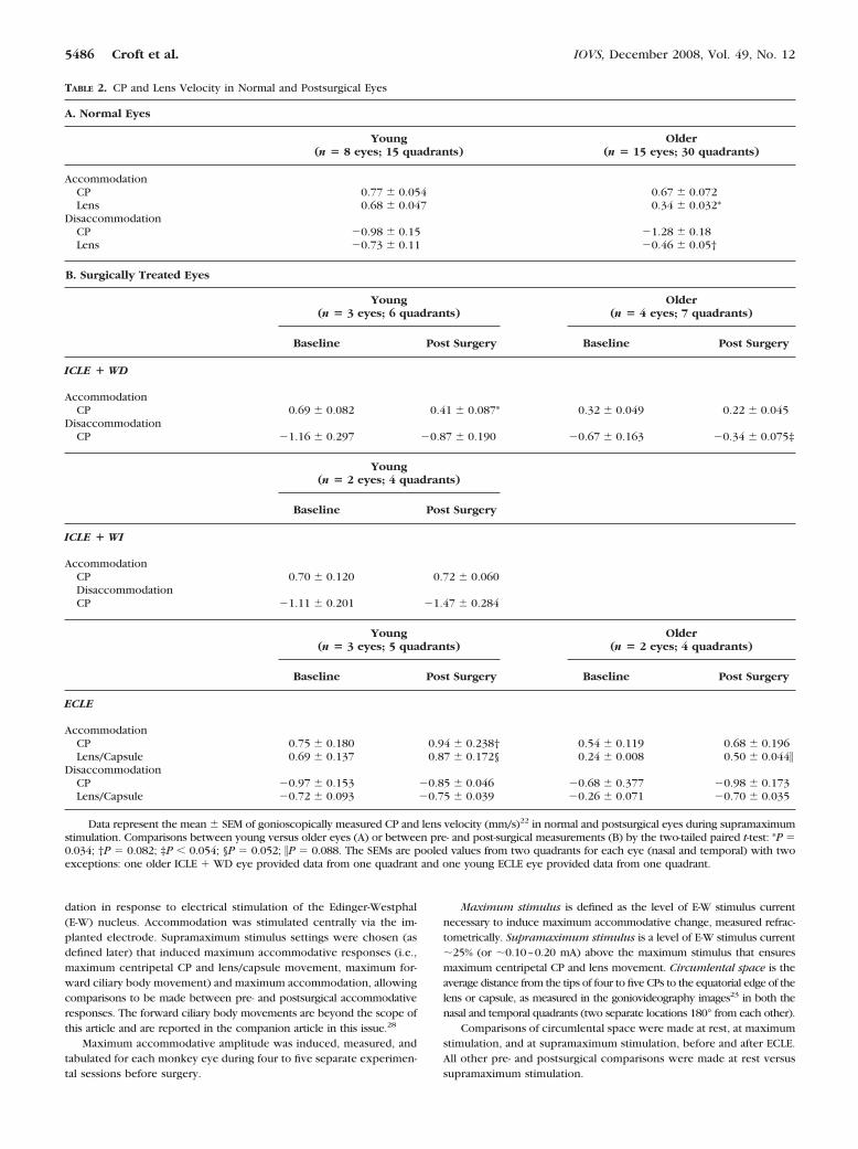
Maximum stimulus is defined as the level of E-W stimulus currentnecessary to induce maximum accommodative change, measured refrac-tometrically. Supramaximum stimulus is a level of E-W stimulus current�25% (or �0.10–0.20 mA) above the maximum stimulus that ensuresmaximum centripetal CP and lens movement. Circumlental space is theaverage distance from the tips of four to five CPs to the equatorial edge of thelens or capsule, as measured in the goniovideography images23 in both thenasal and temporal quadrants (two separate locations 180° from each other).
Comparisons of circumlental space were made at rest, at maximumstimulation, and at supramaximum stimulation, before and after ECLE.All other pre- and postsurgical comparisons were made at rest versussupramaximum stimulation.
TABLE 2. CP and Lens Velocity in Normal and Postsurgical Eyes
A. Normal Eyes
Young(n � 8 eyes; 15 quadrants)
Older(n � 15 eyes; 30 quadrants)
AccommodationCP 0.77 � 0.054 0.67 � 0.072Lens 0.68 � 0.047 0.34 � 0.032*
DisaccommodationCP �0.98 � 0.15 �1.28 � 0.18Lens �0.73 � 0.11 �0.46 � 0.05†
B. Surgically Treated Eyes
Young(n � 3 eyes; 6 quadrants)
Older(n � 4 eyes; 7 quadrants)
Baseline Post Surgery Baseline Post Surgery
ICLE � WD
AccommodationCP 0.69 � 0.082 0.41 � 0.087* 0.32 � 0.049 0.22 � 0.045
DisaccommodationCP �1.16 � 0.297 �0.87 � 0.190 �0.67 � 0.163 �0.34 � 0.075‡
Young(n � 2 eyes; 4 quadrants)
Baseline Post Surgery
ICLE � WI
AccommodationCP 0.70 � 0.120 0.72 � 0.060DisaccommodationCP �1.11 � 0.201 �1.47 � 0.284
Young(n � 3 eyes; 5 quadrants)
Older(n � 2 eyes; 4 quadrants)
Baseline Post Surgery Baseline Post Surgery
ECLE
AccommodationCP 0.75 � 0.180 0.94 � 0.238† 0.54 � 0.119 0.68 � 0.196Lens/Capsule 0.69 � 0.137 0.87 � 0.172§ 0.24 � 0.008 0.50 � 0.044�
DisaccommodationCP �0.97 � 0.153 �0.85 � 0.046 �0.68 � 0.377 �0.98 � 0.173Lens/Capsule �0.72 � 0.093 �0.75 � 0.039 �0.26 � 0.071 �0.70 � 0.035
Data represent the mean � SEM of gonioscopically measured CP and lens velocity (mm/s)22 in normal and postsurgical eyes during supramaximumstimulation. Comparisons between young versus older eyes (A) or between pre- and post-surgical measurements (B) by the two-tailed paired t-test: *P �0.034; †P � 0.082; ‡P � 0.054; §P � 0.052; �P � 0.088. The SEMs are pooled values from two quadrants for each eye (nasal and temporal) with twoexceptions: one older ICLE � WD eye provided data from one quadrant and one young ECLE eye provided data from one quadrant.
5486 Croft et al. IOVS, December 2008, Vol. 49, No. 12
Goniovideography Imaging. Dynamic goniovideography im-ages (using a Swan-Jacobs gonioscopy lens) were obtained duringstimulation of accommodation and then recorded to videotape (30frames per second).20 Goniovideography20 was performed before andafter each surgical procedure. The postsurgical imaging sessions wereperformed 2.5 weeks to 3 months after surgery. Measurements weretaken at the beginning of the study in all 25 monkeys, using go-niovideography, to enable calculation of the normal velocity22 of theCP and lens centripetal movements at supramaximum stimulation. Inthe 18 monkey eyes that underwent surgical procedures, goniovideo-graphy allowed measurement of the centripetal lens/capsule and CPmovement (i.e., velocity22 and amplitude20) and measurement of thecircumlental space width23 before and after surgical intervention.
Computerized analysis of the goniovideography images was used tomeasure the extent and dynamics of the centripetal lens and CPmovements during accommodation and disaccommodation in both thenasal and temporal quadrants (180° from each other) and an averagevalue calculated for each eye.20 The mean � SEM dynamic centripetalmovements of the capsule edge or lens equator (if present) and four tofive adjacent CPs during 2.2-second stimuli were plotted.
Histology
The animals were perfusion fixed through the heart with 4% parafor-maldehyde, after perfusion with 1 L of 0.1 M PBS (phosphate-bufferedsaline). After enucleation, slits were cut in the posterior sclera, and awindow was cut in the anterior cornea, to enhance fixative penetrationand preserve the architecture of the ciliary muscle and its posteriorattachment to Bruch’s membrane. The eyes were then immersed inIto’s fixative29 until they were sent to Erlangen, Germany, for morpho-logic investigation. Small sectors of the anterior globe, including theentire ciliary body and adjacent cornea and sclera, were embedded inEpon, and 1-�m semithin sections were cut and stained with Richard-son’s stain.30
Statistical Analysis
Average CP and lens movement amplitudes were calculated for twoquadrants (nasal and temporal) of each eye at each time point (onethirtieth of a second) during stimulation. The mean � SEM centripetalCP and lens movement amplitudes were calculated by using the move-ment amplitude from 20 consecutive frames, beginning 25 framesbefore termination of the stimulus (i.e., when the eye was in the stableaccommodated state). The initial velocity of CP and lens or capsulemovements was determined by the movement amplitude change from5 to 15 frames after the beginning of the stimulus. Disaccommodativevelocity was measured between 5 and 13 frames after the stimulus wasdiscontinued. The area under the entire stimulus response curve of CP
and lens or capsule movement was also calculated. The response curveincluded the 2.2-second stimulus duration plus the time during whichthe CPs or lens/capsule returned to baseline position. Comparisonswere made between pre- and postsurgery velocity, amplitude, and areaunder the curve for both lens and CP movement.
A two-tailed paired t-test was used to detect significant differences.P � 0.05 was considered significant; 0.05 � P � 0.10 was consideredto indicate a trend, given the small number of monkeys.
RESULTS
To give an indication of the range in accommodative ampli-tudes and variability within each eye for each group, themean � SEM accommodative amplitude was calculated for allpresurgical experimental sessions within each monkey eye.The resulting mean values for each of the young monkey eyesranged from 16.4 � 1.5 to 18.2 � 0.2 D, whereas the resultingmean values for each of the older monkey eyes ranged from0.9 � 0.1 to 6.9 � 0.5 D.
Centripetal CP Movement as Measuredby Goniovideography
During accommodation and disaccommodation, the velocity ofthe older lens was less than in the young eyes (accommoda-tion, P � 0.034; disaccommodation, P � 0.082; Table 2). Incontrast, the velocity of the CPs during the same period wassimilar in the young and older eyes.
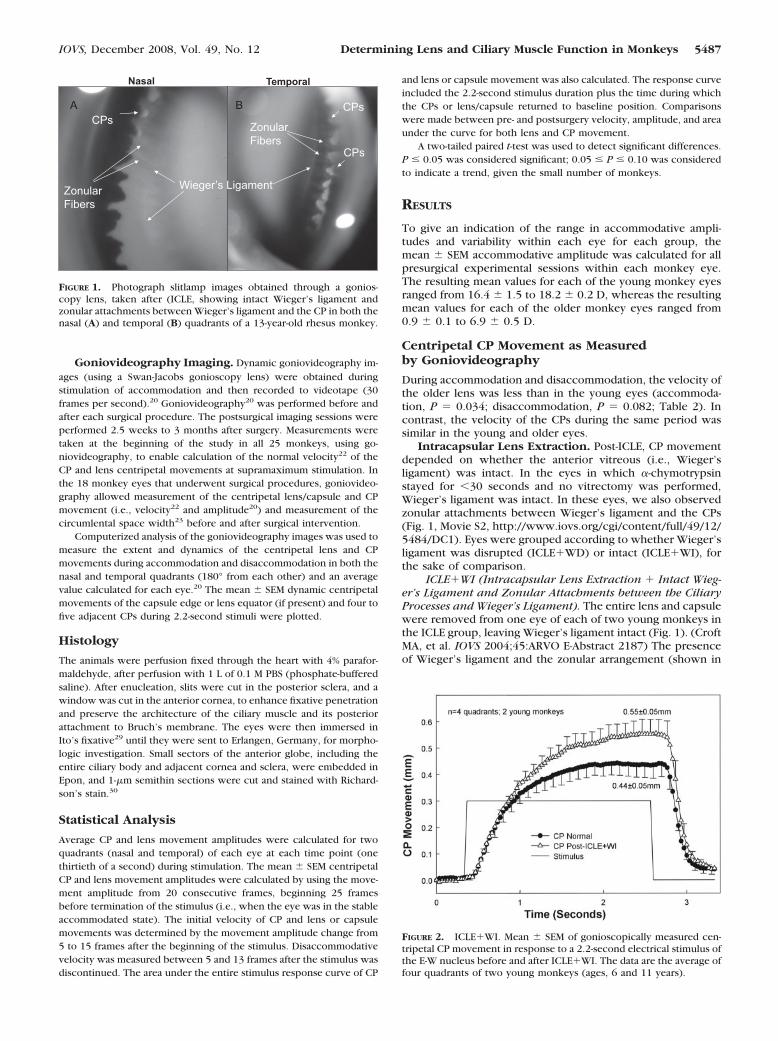
Intracapsular Lens Extraction. Post-ICLE, CP movementdepended on whether the anterior vitreous (i.e., Wieger’sligament) was intact. In the eyes in which �-chymotrypsinstayed for �30 seconds and no vitrectomy was performed,Wieger’s ligament was intact. In these eyes, we also observedzonular attachments between Wieger’s ligament and the CPs(Fig. 1, Movie S2, http://www.iovs.org/cgi/content/full/49/12/5484/DC1). Eyes were grouped according to whether Wieger’sligament was disrupted (ICLE�WD) or intact (ICLE�WI), forthe sake of comparison.
ICLE�WI (Intracapsular Lens Extraction � Intact Wieg-er’s Ligament and Zonular Attachments between the CiliaryProcesses and Wieger’s Ligament). The entire lens and capsulewere removed from one eye of each of two young monkeys inthe ICLE group, leaving Wieger’s ligament intact (Fig. 1). (CroftMA, et al. IOVS 2004;45:ARVO E-Abstract 2187) The presenceof Wieger’s ligament and the zonular arrangement (shown in
TemporalNasal
BACPs
Wieger’s Ligament
CPs
Zonular Fibers
CPs
Zonular Fibers
FIGURE 1. Photograph slitlamp images obtained through a gonios-copy lens, taken after (ICLE, showing intact Wieger’s ligament andzonular attachments between Wieger’s ligament and the CP in both thenasal (A) and temporal (B) quadrants of a 13-year-old rhesus monkey.
FIGURE 2. ICLE�WI. Mean � SEM of gonioscopically measured cen-tripetal CP movement in response to a 2.2-second electrical stimulus ofthe E-W nucleus before and after ICLE�WI. The data are the average offour quadrants of two young monkeys (ages, 6 and 11 years).
IOVS, December 2008, Vol. 49, No. 12 Determining Lens and Ciliary Muscle Function in Monkeys 5487
Movie S2, Fig. 1) was confirmed by a vitreoretinal specialist(Michael Nork, MD, University of Wisconsin-Madison) and acataract surgeon (author GH) by slit lamp examination. Fur-ther, the Wieger’s ligament structure seen in Figure 1 andMovie S2 has a smooth edge and cannot be the edge of a torncapsule remnant, as that would appear ragged rather thansmooth. Wieger’s ligament appeared to move centripetally inaccordance with the CP accommodative response, and thezonular fibers appeared to relax (see Movie S2). After ICLE�WI(Fig. 2), the initial CP velocity was similar to that at baseline(Table 2, Fig. 2), but the area under the CP movement curveand the amplitude of CP movement that were achieved nearthe end of the stimulus train were greater after ICLE�WI(Table 3, Fig. 2) than at baseline before lens removal, but thedifference was not statistically significant, given the small sam-ple size.
ICLE�WD (Intracapsular Lens Extraction � Disruptionof Wieger’s Ligament). In the three other young monkeys, andthe four older monkeys that underwent ICLE, the entire lensand capsule were removed from 1 eye and Wieger’s ligamentwas disrupted (Fig. 3). After ICLE�WD, CP velocity declinedsignificantly, whereas CP movement amplitude, and the areaunder the CP movement curve tended to decline (Table 3)compared with the same eyes presurgically, regardless of age(Fig. 3, Table 2).
Extracapsular Lens Extraction. In all five monkey eyes(three young, two older) that underwent ECLE, the lens sub-stance was removed from the capsule, leaving an empty cap-sule bag (CPs, zonules, and posterior capsule left intact; Fig. 4).
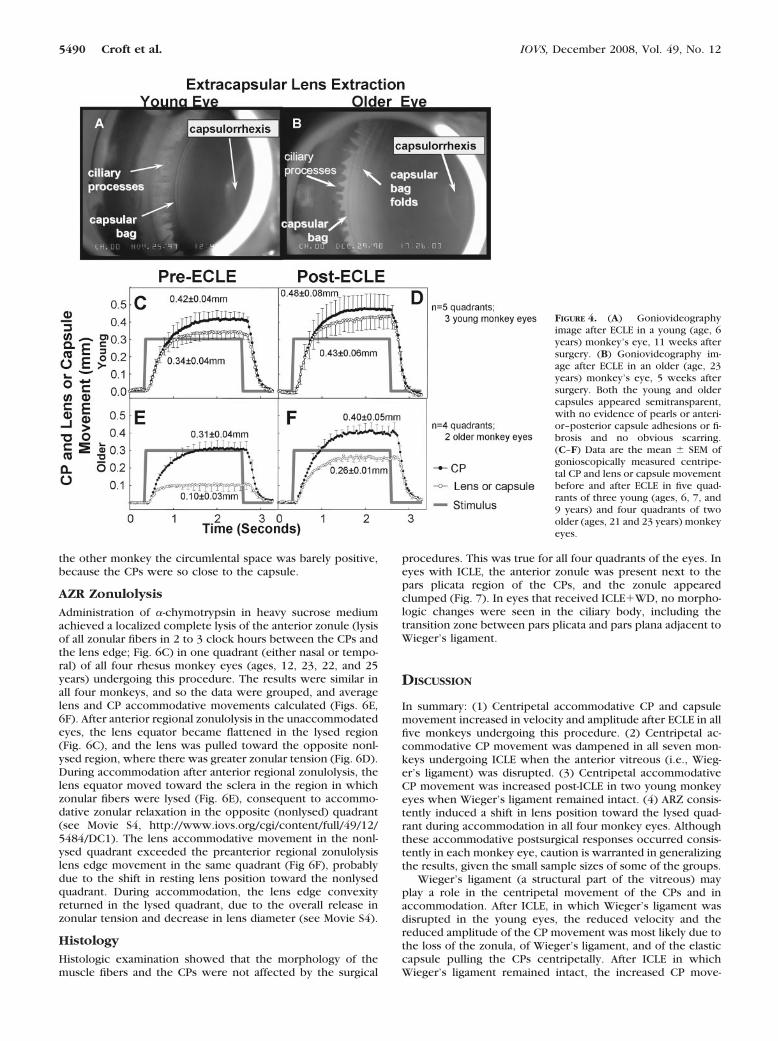
At the time of postsurgical imaging, the eyes were exam-ined for pearls, adhesions, and any age-related differences. Inthe young eyes, the empty capsular bags did not have folds andappeared smooth and semitransparent, with no evidence ofpearls or of anterior–posterior capsule adhesions (Fig. 4A com-pared to 4B; Movie S3, http://www.iovs.org/cgi/content/full/49/12/5484/DC1). In the older eyes, the empty capsule bagshad several folds, but also appeared semitransparent and with-out evidence of pearls or of anterior–posterior capsule adhe-sions (Fig. 4B). There was no evidence of fibrosis in or aroundthe capsulorrhexis or inside the capsular bag in either agegroup at the time of postsurgical imaging. Six months afterECLE the capsular bags (young and older) began to exhibit thepresence of pearls, fibrosis around the capsulorrhexis, andadhesions between the anterior and posterior capsular bagsurfaces.
Lens Capsule/CP Movement
After ECLE, CP accommodative velocity increased significantly(P � 0.02), while movement amplitude, and the area under the
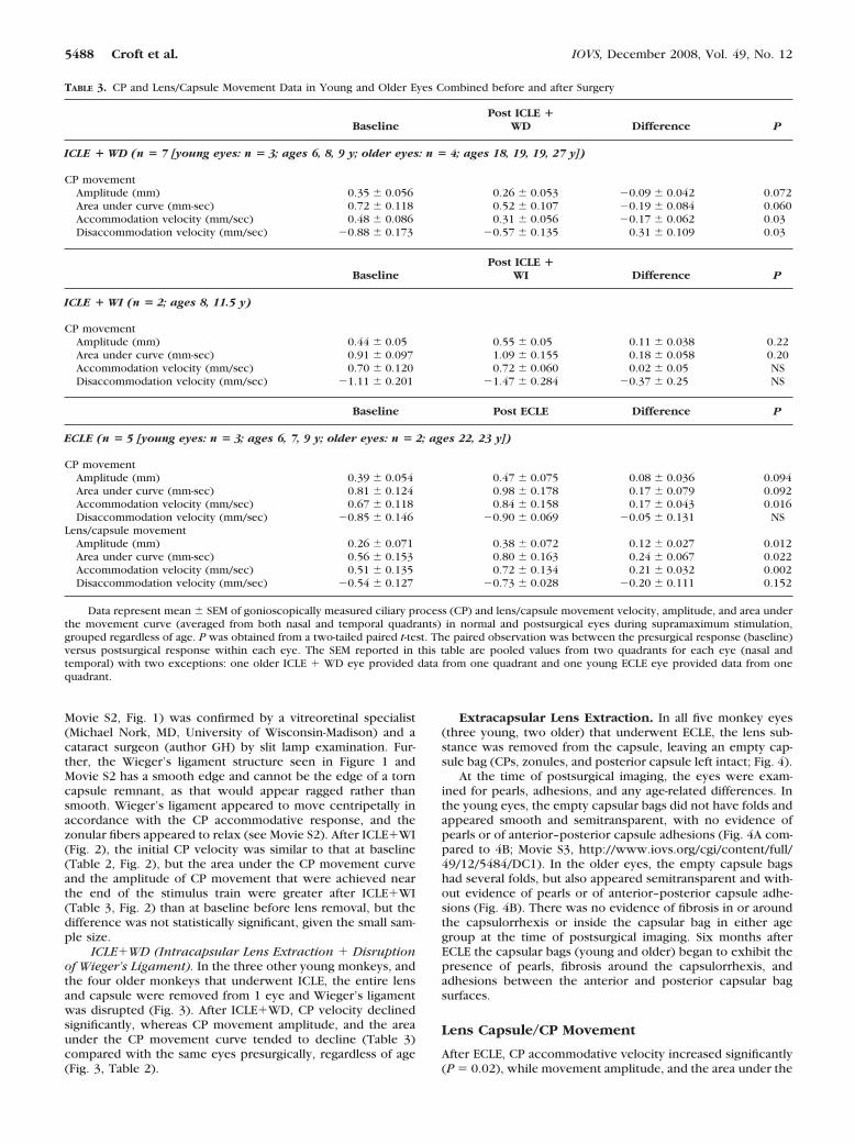
TABLE 3. CP and Lens/Capsule Movement Data in Young and Older Eyes Combined before and after Surgery
BaselinePost ICLE �
WD Difference P
ICLE � WD (n � 7 [young eyes: n � 3; ages 6, 8, 9 y; older eyes: n � 4; ages 18, 19, 19, 27 y])
CP movementAmplitude (mm) 0.35 � 0.056 0.26 � 0.053 �0.09 � 0.042 0.072Area under curve (mm-sec) 0.72 � 0.118 0.52 � 0.107 �0.19 � 0.084 0.060Accommodation velocity (mm/sec) 0.48 � 0.086 0.31 � 0.056 �0.17 � 0.062 0.03Disaccommodation velocity (mm/sec) �0.88 � 0.173 �0.57 � 0.135 0.31 � 0.109 0.03
BaselinePost ICLE �
WI Difference P
ICLE � WI (n � 2; ages 8, 11.5 y)
CP movementAmplitude (mm) 0.44 � 0.05 0.55 � 0.05 0.11 � 0.038 0.22Area under curve (mm-sec) 0.91 � 0.097 1.09 � 0.155 0.18 � 0.058 0.20Accommodation velocity (mm/sec) 0.70 � 0.120 0.72 � 0.060 0.02 � 0.05 NSDisaccommodation velocity (mm/sec) �1.11 � 0.201 �1.47 � 0.284 �0.37 � 0.25 NS
Baseline Post ECLE Difference P
ECLE (n � 5 [young eyes: n � 3; ages 6, 7, 9 y; older eyes: n � 2; ages 22, 23 y])
CP movementAmplitude (mm) 0.39 � 0.054 0.47 � 0.075 0.08 � 0.036 0.094Area under curve (mm-sec) 0.81 � 0.124 0.98 � 0.178 0.17 � 0.079 0.092Accommodation velocity (mm/sec) 0.67 � 0.118 0.84 � 0.158 0.17 � 0.043 0.016Disaccommodation velocity (mm/sec) �0.85 � 0.146 �0.90 � 0.069 �0.05 � 0.131 NS
Lens/capsule movementAmplitude (mm) 0.26 � 0.071 0.38 � 0.072 0.12 � 0.027 0.012Area under curve (mm-sec) 0.56 � 0.153 0.80 � 0.163 0.24 � 0.067 0.022Accommodation velocity (mm/sec) 0.51 � 0.135 0.72 � 0.134 0.21 � 0.032 0.002Disaccommodation velocity (mm/sec) �0.54 � 0.127 �0.73 � 0.028 �0.20 � 0.111 0.152
Data represent mean � SEM of gonioscopically measured ciliary process (CP) and lens/capsule movement velocity, amplitude, and area underthe movement curve (averaged from both nasal and temporal quadrants) in normal and postsurgical eyes during supramaximum stimulation,grouped regardless of age. P was obtained from a two-tailed paired t-test. The paired observation was between the presurgical response (baseline)versus postsurgical response within each eye. The SEM reported in this table are pooled values from two quadrants for each eye (nasal andtemporal) with two exceptions: one older ICLE � WD eye provided data from one quadrant and one young ECLE eye provided data from onequadrant.
5488 Croft et al. IOVS, December 2008, Vol. 49, No. 12
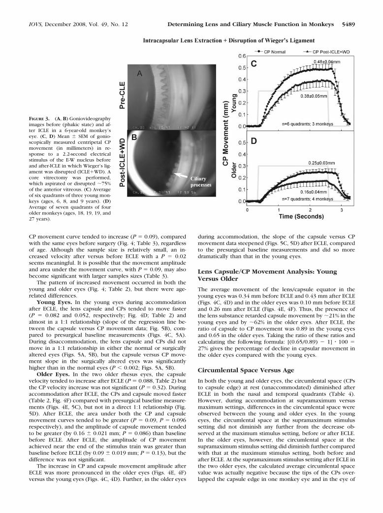
CP movement curve tended to increase (P � 0.09), comparedwith the same eyes before surgery (Fig. 4; Table 3), regardlessof age. Although the sample size is relatively small, an in-creased velocity after versus before ECLE with a P � 0.02seems meaningful. It is possible that the movement amplitudeand area under the movement curve, with P � 0.09, may alsobecome significant with larger samples sizes (Table 3).
The pattern of increased movement occurred in both theyoung and older eyes (Fig. 4; Table 2), but there were age-related differences.
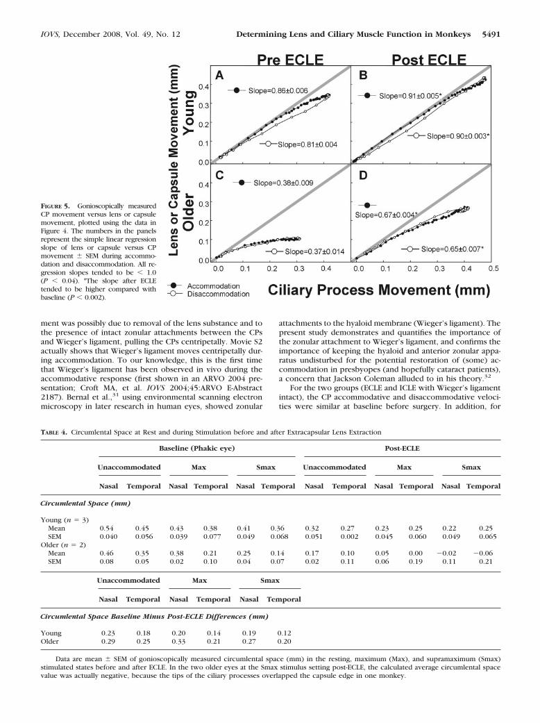
Young Eyes. In the young eyes during accommodationafter ECLE, the lens capsule and CPs tended to move faster(P � 0.082 and 0.052, respectively; Fig. 4D; Table 2) andalmost in a 1:1 relationship (slope of the regression line be-tween the capsule versus CP movement data; Fig. 5B), com-pared to presurgical baseline measurements (Figs. 4C, 5A).During disaccommodation, the lens capsule and CPs did notmove in a 1:1 relationship in either the normal or surgicallyaltered eyes (Figs. 5A, 5B), but the capsule versus CP move-ment slope in the surgically altered eyes was significantlyhigher than in the normal eyes (P � 0.002; Figs. 5A, 5B).
Older Eyes. In the two older rhesus eyes, the capsulevelocity tended to increase after ECLE (P � 0.088, Table 2) butthe CP velocity increase was not significant (P � 0.32). Duringaccommodation after ECLE, the CPs and capsule moved faster(Table 2, Fig. 4F) compared with presurgical baseline measure-ments (Figs. 4E, 5C), but not in a direct 1:1 relationship (Fig.5D). After ECLE, the area under both the CP and capsulemovement curves tended to be greater (P � 0.09, P � 0.096respectively), and the amplitude of capsule movement tendedto be greater (by 0.16 � 0.021 mm; P � 0.086) than baselinebefore ECLE. After ECLE, the amplitude of CP movementachieved near the end of the stimulus train was greater thanbaseline before ECLE (by 0.09 � 0.019 mm; P � 0.13), but thedifference was not significant.
The increase in CP and capsule movement amplitude afterECLE was more pronounced in the older eyes (Figs. 4E, 4F)versus the young eyes (Figs. 4C, 4D). Further, in the older eyes
during accommodation, the slope of the capsule versus CPmovement data steepened (Figs. 5C, 5D) after ECLE, comparedto the presurgical baseline measurements and did so moredramatically than that in the young eyes.
Lens Capsule/CP Movement Analysis: YoungVersus Older
The average movement of the lens/capsule equator in theyoung eyes was 0.34 mm before ECLE and 0.43 mm after ECLE(Figs. 4C, 4D) and in the older eyes was 0.10 mm before ECLEand 0.26 mm after ECLE (Figs. 4E, 4F). Thus, the presence ofthe lens substance retarded capsule movement by �21% in theyoung eyes and by �62% in the older eyes. After ECLE, theratio of capsule to CP movement was 0.89 in the young eyesand 0.65 in the older eyes. Taking the ratio of these ratios andcalculating the following formula: [(0.65/0.89) � 1] � 100 �27% gives the percentage of decline in capsular movement inthe older eyes compared with the young eyes.
Circumlental Space Versus Age
In both the young and older eyes, the circumlental space (CPsto capsule edge) at rest (unaccommodated) diminished afterECLE in both the nasal and temporal quadrants (Table 4).However, during accommodation at supramaximum versusmaximum settings, differences in the circumlental space wereobserved between the young and older eyes. In the youngeyes, the circumlental space at the supramaximum stimulussetting did not diminish any further from the decrease ob-served at the maximum stimulus setting, before or after ECLE.In the older eyes, however, the circumlental space at thesupramaximum stimulus setting did diminish further comparedwith that at the maximum stimulus setting, both before andafter ECLE. At the supramaximum stimulus setting after ECLE inthe two older eyes, the calculated average circumlental spacevalue was actually negative because the tips of the CPs over-lapped the capsule edge in one monkey eye and in the eye of
FIGURE 3. (A, B) Goniovideographyimages before (phakic state) and af-ter ICLE in a 6-year-old monkey’seye. (C, D) Mean � SEM of gonio-scopically measured centripetal CPmovement (in millimeters) in re-sponse to a 2.2-second electricalstimulus of the E-W nucleus beforeand after-ICLE in which Wieger’s lig-ament was disrupted (ICLE�WD). Acore vitrectomy was performed,which aspirated or disrupted �75%of the anterior vitreous. (C) Averageof six quadrants of three young mon-keys (ages, 6, 8, and 9 years). (D)Average of seven quadrants of fourolder monkeys (ages, 18, 19, 19, and27 years).
IOVS, December 2008, Vol. 49, No. 12 Determining Lens and Ciliary Muscle Function in Monkeys 5489
the other monkey the circumlental space was barely positive,because the CPs were so close to the capsule.
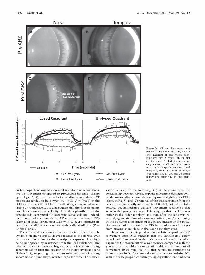
AZR ZonulolysisAdministration of �-chymotrypsin in heavy sucrose mediumachieved a localized complete lysis of the anterior zonule (lysisof all zonular fibers in 2 to 3 clock hours between the CPs andthe lens edge; Fig. 6C) in one quadrant (either nasal or tempo-ral) of all four rhesus monkey eyes (ages, 12, 23, 22, and 25years) undergoing this procedure. The results were similar inall four monkeys, and so the data were grouped, and averagelens and CP accommodative movements calculated (Figs. 6E,6F). After anterior regional zonulolysis in the unaccommodatedeyes, the lens equator became flattened in the lysed region(Fig. 6C), and the lens was pulled toward the opposite nonl-ysed region, where there was greater zonular tension (Fig. 6D).During accommodation after anterior regional zonulolysis, thelens equator moved toward the sclera in the region in whichzonular fibers were lysed (Fig. 6E), consequent to accommo-dative zonular relaxation in the opposite (nonlysed) quadrant(see Movie S4, http://www.iovs.org/cgi/content/full/49/12/5484/DC1). The lens accommodative movement in the nonl-ysed quadrant exceeded the preanterior regional zonulolysislens edge movement in the same quadrant (Fig 6F), probablydue to the shift in resting lens position toward the nonlysedquadrant. During accommodation, the lens edge convexityreturned in the lysed quadrant, due to the overall release inzonular tension and decrease in lens diameter (see Movie S4).
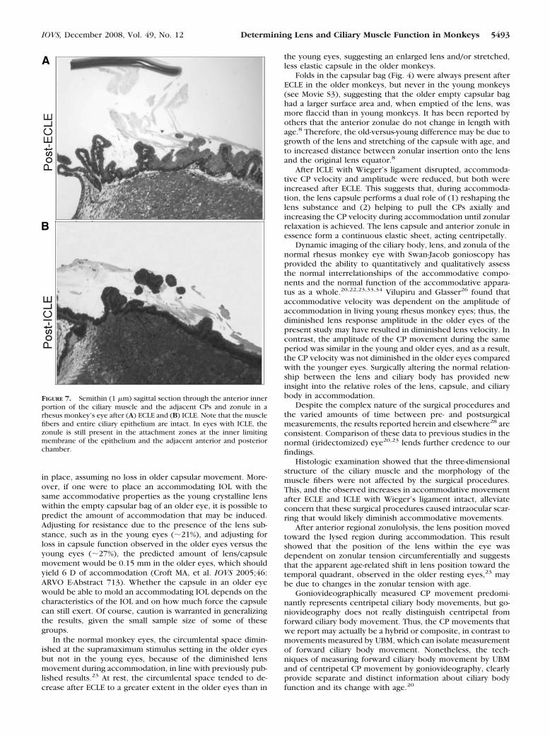
HistologyHistologic examination showed that the morphology of themuscle fibers and the CPs were not affected by the surgical
procedures. This was true for all four quadrants of the eyes. Ineyes with ICLE, the anterior zonule was present next to thepars plicata region of the CPs, and the zonule appearedclumped (Fig. 7). In eyes that received ICLE�WD, no morpho-logic changes were seen in the ciliary body, including thetransition zone between pars plicata and pars plana adjacent toWieger’s ligament.
DISCUSSION
In summary: (1) Centripetal accommodative CP and capsulemovement increased in velocity and amplitude after ECLE in allfive monkeys undergoing this procedure. (2) Centripetal ac-commodative CP movement was dampened in all seven mon-keys undergoing ICLE when the anterior vitreous (i.e., Wieg-er’s ligament) was disrupted. (3) Centripetal accommodativeCP movement was increased post-ICLE in two young monkeyeyes when Wieger’s ligament remained intact. (4) ARZ consis-tently induced a shift in lens position toward the lysed quad-rant during accommodation in all four monkey eyes. Althoughthese accommodative postsurgical responses occurred consis-tently in each monkey eye, caution is warranted in generalizingthe results, given the small sample sizes of some of the groups.
Wieger’s ligament (a structural part of the vitreous) mayplay a role in the centripetal movement of the CPs and inaccommodation. After ICLE, in which Wieger’s ligament wasdisrupted in the young eyes, the reduced velocity and thereduced amplitude of the CP movement was most likely due tothe loss of the zonula, of Wieger’s ligament, and of the elasticcapsule pulling the CPs centripetally. After ICLE in whichWieger’s ligament remained intact, the increased CP move-
FIGURE 4. (A) Goniovideographyimage after ECLE in a young (age, 6years) monkey’s eye, 11 weeks aftersurgery. (B) Goniovideography im-age after ECLE in an older (age, 23years) monkey’s eye, 5 weeks aftersurgery. Both the young and oldercapsules appeared semitransparent,with no evidence of pearls or anteri-or–posterior capsule adhesions or fi-brosis and no obvious scarring.(C–F) Data are the mean � SEM ofgonioscopically measured centripe-tal CP and lens or capsule movementbefore and after ECLE in five quad-rants of three young (ages, 6, 7, and9 years) and four quadrants of twoolder (ages, 21 and 23 years) monkeyeyes.
5490 Croft et al. IOVS, December 2008, Vol. 49, No. 12
ment was possibly due to removal of the lens substance and tothe presence of intact zonular attachments between the CPsand Wieger’s ligament, pulling the CPs centripetally. Movie S2actually shows that Wieger’s ligament moves centripetally dur-ing accommodation. To our knowledge, this is the first timethat Wieger’s ligament has been observed in vivo during theaccommodative response (first shown in an ARVO 2004 pre-sentation; Croft MA, et al. IOVS 2004;45:ARVO E-Abstract2187). Bernal et al.,31 using environmental scanning electronmicroscopy in later research in human eyes, showed zonular
attachments to the hyaloid membrane (Wieger’s ligament). Thepresent study demonstrates and quantifies the importance ofthe zonular attachment to Wieger’s ligament, and confirms theimportance of keeping the hyaloid and anterior zonular appa-ratus undisturbed for the potential restoration of (some) ac-commodation in presbyopes (and hopefully cataract patients),a concern that Jackson Coleman alluded to in his theory.32
For the two groups (ECLE and ICLE with Wieger’s ligamentintact), the CP accommodative and disaccommodative veloci-ties were similar at baseline before surgery. In addition, for
FIGURE 5. Gonioscopically measuredCP movement versus lens or capsulemovement, plotted using the data inFigure 4. The numbers in the panelsrepresent the simple linear regressionslope of lens or capsule versus CPmovement � SEM during accommo-dation and disaccommodation. All re-gression slopes tended to be � 1.0(P � 0.04). *The slope after ECLEtended to be higher compared withbaseline (P � 0.002).
TABLE 4. Circumlental Space at Rest and during Stimulation before and after Extracapsular Lens Extraction
Baseline (Phakic eye) Post-ECLE
Unaccommodated Max Smax Unaccommodated Max Smax
Nasal Temporal Nasal Temporal Nasal Temporal Nasal Temporal Nasal Temporal Nasal Temporal
Circumlental Space (mm)
Young (n � 3)Mean 0.54 0.45 0.43 0.38 0.41 0.36 0.32 0.27 0.23 0.25 0.22 0.25SEM 0.040 0.056 0.039 0.077 0.049 0.068 0.051 0.002 0.045 0.060 0.049 0.065
Older (n � 2)Mean 0.46 0.35 0.38 0.21 0.25 0.14 0.17 0.10 0.05 0.00 �0.02 �0.06SEM 0.08 0.05 0.02 0.10 0.04 0.07 0.02 0.11 0.06 0.19 0.11 0.21
Unaccommodated Max Smax
Nasal Temporal Nasal Temporal Nasal Temporal
Circumlental Space Baseline Minus Post-ECLE Differences (mm)
Young 0.23 0.18 0.20 0.14 0.19 0.12Older 0.29 0.25 0.33 0.21 0.27 0.20
Data are mean � SEM of gonioscopically measured circumlental space (mm) in the resting, maximum (Max), and supramaximum (Smax)stimulated states before and after ECLE. In the two older eyes at the Smax stimulus setting post-ECLE, the calculated average circumlental spacevalue was actually negative, because the tips of the ciliary processes overlapped the capsule edge in one monkey.
IOVS, December 2008, Vol. 49, No. 12 Determining Lens and Ciliary Muscle Function in Monkeys 5491
both groups there was an increased amplitude of accommoda-tive CP movement compared to presurgical baseline (phakiceyes; Figs. 2, 4), but the velocity of disaccommodative CPmovement tended to be slower (by �40%; P � 0.066) in theECLE eyes versus the ICLE eyes with Wieger’s ligament intact(Table 2). Collectively, the data suggest that the capsule damp-ens disaccommodative velocity. It is thus plausible that thecapsule aids centripetal CP accommodative velocity; indeed,the velocity of accommodative CP movement averaged 24%faster after ECLE versus post-ICLE with Wieger’s ligament in-tact, but the difference was not statistically significant (P �0.458) (Table 2).
The enhanced accommodative centripetal CP and capsuledynamics in the young ECLE eyes relative to the normal eyeswere most likely due to the centripetal capsular elasticity’sbeing unopposed by resistance from the lens substance. Theedge of the empty capsular bag moved at a faster rate duringaccommodation than the equator of the intact crystalline lens(Tables 2, 3), suggesting that the lens substance, even in youngaccommodating monkeys, resisted capsular force. This obser-
vation is based on the following: (1) In the young eyes, therelationship between CP and capsule movement during accom-modation and disaccommodation improved slightly after ECLE(slope in Fig. 5); and (2) removal of the lens substance from theolder eyes significantly improved (P � 0.002), but did not fullyrestore, accommodative capsule movement relative to thatseen in the young monkeys. This suggests that the lens wasstiffer in the older monkeys and that, after the lens was re-moved, age-related loss of capsular elasticity, and/or stiffeningof the posterior attachment of the ciliary muscle or the poste-rior zonule, still prevented the CPs in the older monkey eyesfrom moving as much as in the young monkey eyes.
The amount of centripetal accommodative capsule and CPmovement after ECLE suggests that the capsule and ciliarymuscle still functioned in the older eyes. Although the oldercapsule-to-CP-movement ratio was reduced compared with theyoung eyes, the older capsules still exhibited an amount ofmovement (0.26 mm, Fig. 4F) that would be sufficient toinduce up to 10 D of accommodation if an accommodating IOLwith the same properties as the young crystalline lens had been
Nasal Temporal
BA
DC
Pre
AR
ZP
ost A
RZ
Region of Lysed Zonule
Time (seconds)
CP
an
d L
ens
Mo
vem
ent
(mm
)
Lysed Quadrant
0 1 2 3
-0.3
-0.2
-0.1
0.0
0.1
0.2
0.3
0.4
0.5
Un-lysed Quadrant
0 1 2 3
CP Post Lysis
Lens Post Lysis
CP Pre Lysis
Lens Pre Lysis
Stimulus
FIGURE 6. CP and lens movementbefore (A, B) and after (C, D) ARZ inone quadrant of one rhesus mon-key’s eye (age, 23 years). (E, F) Dataare the mean � SEM of gonioscopi-cally measured CP and lens move-ment in both quadrants (nasal andtemporal) of four rhesus monkey’seyes (ages, 13, 23, 23, and 25 years)before and after ARZ in one quad-rant.
5492 Croft et al. IOVS, December 2008, Vol. 49, No. 12
in place, assuming no loss in older capsular movement. More-over, if one were to place an accommodating IOL with thesame accommodative properties as the young crystalline lenswithin the empty capsular bag of an older eye, it is possible topredict the amount of accommodation that may be induced.Adjusting for resistance due to the presence of the lens sub-stance, such as in the young eyes (�21%), and adjusting forloss in capsule function observed in the older eyes versus theyoung eyes (�27%), the predicted amount of lens/capsulemovement would be 0.15 mm in the older eyes, which shouldyield 6 D of accommodation (Croft MA, et al. IOVS 2005;46:ARVO E-Abstract 713). Whether the capsule in an older eyewould be able to mold an accommodating IOL depends on thecharacteristics of the IOL and on how much force the capsulecan still exert. Of course, caution is warranted in generalizingthe results, given the small sample size of some of thesegroups.
In the normal monkey eyes, the circumlental space dimin-ished at the supramaximum stimulus setting in the older eyesbut not in the young eyes, because of the diminished lensmovement during accommodation, in line with previously pub-lished results.23 At rest, the circumlental space tended to de-crease after ECLE to a greater extent in the older eyes than in
the young eyes, suggesting an enlarged lens and/or stretched,less elastic capsule in the older monkeys.
Folds in the capsular bag (Fig. 4) were always present afterECLE in the older monkeys, but never in the young monkeys(see Movie S3), suggesting that the older empty capsular baghad a larger surface area and, when emptied of the lens, wasmore flaccid than in young monkeys. It has been reported byothers that the anterior zonulae do not change in length withage.8 Therefore, the old-versus-young difference may be due togrowth of the lens and stretching of the capsule with age, andto increased distance between zonular insertion onto the lensand the original lens equator.8
After ICLE with Wieger’s ligament disrupted, accommoda-tive CP velocity and amplitude were reduced, but both wereincreased after ECLE. This suggests that, during accommoda-tion, the lens capsule performs a dual role of (1) reshaping thelens substance and (2) helping to pull the CPs axially andincreasing the CP velocity during accommodation until zonularrelaxation is achieved. The lens capsule and anterior zonule inessence form a continuous elastic sheet, acting centripetally.
Dynamic imaging of the ciliary body, lens, and zonula of thenormal rhesus monkey eye with Swan-Jacob gonioscopy hasprovided the ability to quantitatively and qualitatively assessthe normal interrelationships of the accommodative compo-nents and the normal function of the accommodative appara-tus as a whole.20,22,23,33,34 Vilupiru and Glasser26 found thataccommodative velocity was dependent on the amplitude ofaccommodation in living young rhesus monkey eyes; thus, thediminished lens response amplitude in the older eyes of thepresent study may have resulted in diminished lens velocity. Incontrast, the amplitude of the CP movement during the sameperiod was similar in the young and older eyes, and as a result,the CP velocity was not diminished in the older eyes comparedwith the younger eyes. Surgically altering the normal relation-ship between the lens and ciliary body has provided newinsight into the relative roles of the lens, capsule, and ciliarybody in accommodation.
Despite the complex nature of the surgical procedures andthe varied amounts of time between pre- and postsurgicalmeasurements, the results reported herein and elsewhere28 areconsistent. Comparison of these data to previous studies in thenormal (iridectomized) eye20,23 lends further credence to ourfindings.
Histologic examination showed that the three-dimensionalstructure of the ciliary muscle and the morphology of themuscle fibers were not affected by the surgical procedures.This, and the observed increases in accommodative movementafter ECLE and ICLE with Wieger’s ligament intact, alleviateconcern that these surgical procedures caused intraocular scar-ring that would likely diminish accommodative movements.
After anterior regional zonulolysis, the lens position movedtoward the lysed region during accommodation. This resultshowed that the position of the lens within the eye wasdependent on zonular tension circumferentially and suggeststhat the apparent age-related shift in lens position toward thetemporal quadrant, observed in the older resting eyes,23 maybe due to changes in the zonular tension with age.
Goniovideographically measured CP movement predomi-nantly represents centripetal ciliary body movements, but go-niovideography does not really distinguish centripetal fromforward ciliary body movement. Thus, the CP movements thatwe report may actually be a hybrid or composite, in contrast tomovements measured by UBM, which can isolate measurementof forward ciliary body movement. Nonetheless, the tech-niques of measuring forward ciliary body movement by UBMand of centripetal CP movement by goniovideography, clearlyprovide separate and distinct information about ciliary bodyfunction and its change with age.20
FIGURE 7. Semithin (1 �m) sagittal section through the anterior innerportion of the ciliary muscle and the adjacent CPs and zonule in arhesus monkey’s eye after (A) ECLE and (B) ICLE. Note that the musclefibers and entire ciliary epithelium are intact. In eyes with ICLE, thezonule is still present in the attachment zones at the inner limitingmembrane of the epithelium and the adjacent anterior and posteriorchamber.
IOVS, December 2008, Vol. 49, No. 12 Determining Lens and Ciliary Muscle Function in Monkeys 5493
Characterization of the performance of the accommodativeapparatus before and after surgical interventions may help tomodel the system for hypothesis testing. Information gleanedfrom such studies aids in understanding the accommodativemechanism itself and may also facilitate the design of accom-modating IOLs. Accommodating IOLs may be more effective inrestoring accommodation in the presbyopic eye if they rely oncentripetal ciliary body and thereby capsular edge movementrather than forward ciliary body movement.20 Previous studiesof accommodation in the normal iridectomized rhesus monkeyeye have shown a significant age-related loss of forward ciliarybody movement and lens centripetal movement, but the age-related loss in CP movement was far less pronounced.20,23
Accommodative centripetal movements of the ciliary body arealso still present in the presbyopic human eye,35 despite thereduced accommodative amplitudes. Forward muscle move-ment, as measured by muscle length, was still present inexcised, pharmacologically stimulated postmortem humaneyes.36 Whether the remaining movement is sufficient to pro-duce accommodation with an accommodating IOL will dependon the approach and the characteristics of the accommodatingIOL.
Acknowledgments
Thanks to James Reed for lending his technical expertise with theimage analysis systems; Mike Killips and Charles Roth for providinghelp in editing the video presentations; Joseph Sanchez for videocompilation at the Instructional Media Development Center (School ofEducation, University of Wisconsin-Madison); John Peterson for expertassistance in collecting the slit lamp photography images; and KateFahl for editorial contributions.
References
1. Pau H, Krantz J. The increasing sclerosis of the human lens withage and its relevance to accommodation and presbyopia. GraefesArch Clin Exp Ophthalmol. 1991;229:294–296.
2. Glasser A, Campbell MCW. Presbyopia and the optical changes inthe human crystalline lens with age. Vision Res. 1998;38(2):209–229.
3. Fisher RF. Elastic constants of the human lens capsule. J Physiol(Lond). 1969;201:1–19.
4. Fisher RF. The force of contraction of the human ciliary muscleduring accommodation. J Physiol (Lond). 1977;270:51–74.
5. Fisher RF. The elastic constants of the human lens. J Physiol(Lond). 1971;212:147–180.
6. Glasser A, Campbell MCW. Biometric, optical and physicalchanges in the isolated human crystalline lens with age in relationto presbyopia. Vision Res. 1999;39:1991–2015.
7. Heys KR, Cram SL, Truscott RJ. Massive increase in the stiffness ofthe human lens nucleus with age: the basis for presbyopia? MolVis. 2004;10:956–963.
8. Farnsworth PN, Shyne SE. Anterior zonular shifts with age. Exp EyeRes. 1979;28:291–297.
9. Scammon RE, Hesdorfer MB. Growth in mass and volume of thehuman lens in postnatal life. Arch Ophthalmol. 1937;17:104–112.
10. Weale RA. The lens. The Aging Eye. New York: Harper & Row;1963:68–102.
11. Willekens B, Kappelhof J, Vrensen G. Morphology of the aginghuman lens: I. Biomicroscopy and biometrics. Lens Res. 1987;4:207–230.
12. Schachar RA. Cause and treatment of presbyopia with a methodfor increasing the amplitude of accommodation. Ann Ophthalmol.1992;24:445–452.
13. Schachar RA, Black TD, Kash RL, Cudmore MS, Schanzlin DJ. Themechanism of accommodation and presbyopia in the primate.Ann Ophthalmol. 1995;27(2):59–67.
14. Schachar RA, Tello C, Cudmore DP, Liebmann JM, Black TD, RitchR. In vivo increase of the human lens equatorial diameter during
accommodation. Am J Physiol Regul Integr Comp Physiol. 1996;271:R670–R676.
15. Tamm S, Tamm E, Rohen JW. Age-related changes of the humanciliary muscle: a quantitative morphometric study. Mech AgeingDev. 1992;62:209–221.
16. Tamm E, Lutjen-Drecoll E, Jungkunz W, Rohen JW. Posterior at-tachment of ciliary muscle in young, accommodating old, presby-opic monkeys. Invest Ophthalmol Vis Sci. 1991;32(5):1678–1692.
17. Neider MW, Crawford K, Kaufman PL, Bito LZ. In vivo videographyof the rhesus monkey accommodative apparatus: age-related lossof ciliary muscle response to central stimulation. Arch Ophthal-mol. 1990;108:69–74.
18. Lutjen-Drecoll E, Tamm E, Kaufman PL. Age changes in rhesusmonkey ciliary muscle: light and electron microscopy. Exp EyeRes. 1988;47:885–899.
19. Bito LZ, DeRousseau CJ, Kaufman PL, Bito JW. Age-dependent lossof accommodative amplitude in rhesus monkeys: an animal modelfor presbyopia. Invest Ophthalmol Vis Sci. 1982;23:23–31.
20. Croft MA, Glasser A, Heatley G, et al. Accommodative ciliary bodyand lens function in rhesus monkeys: I. Normal lens, zonule andciliary process configuration in the iridectomized eye. Invest Oph-thalmol Vis Sci. 2006;47(3):1076–1086.
21. Duane A. Studies in monocular and binocular accommodationwith their clinical applications. Am J Ophthalmol. 1922;5:867–877.
22. Croft MA, Kaufman PL, Crawford KS, Neider MW, Glasser A, BitoLZ. Accommodation dynamics in aging rhesus monkeys. Am JPhysiol Regul Integr Comp Physiol. 1998;44:R1885–R1897.
23. Croft MA, Glasser A, Heatley G, et al. The zonula, lens, andcircumlental space in the normal iridectomized rhesus monkeyeye. Invest Ophthalmol Vis Sci. 2006;47(3):1087–1095.
24. Kaufman PL, Lutjen-Drecoll E. Total iridectomy in the primate invivo: surgical technique and postoperative anatomy. Invest Oph-thalmol. 1975;14:766–771.
25. Crawford K, Terasawa E, Kaufman PL. Reproducible stimulation ofciliary muscle contraction in the cynomolgus monkey via a per-manent indwelling midbrain electrode. Brain Res. 1989;503:265–272.
26. Vilupuru AS, Glasser A. Dynamic accommodation in rhesus mon-keys. Vision Res. 2002;42:125–141.
27. Barany EH, Rohen JW. Localized contraction and relaxation withinthe ciliary muscle of the vervet monkey (Cercopithecus ethiops).In: Rohen JW, ed. The Structure of the Eye: Second Symposium.Stuttgart: Schattauer; 1965;287–311.
28. Wasilewski R, McDonald JP, Heatley G, Lutjen-Drecoll E, KaufmanPL, Croft MA. Surgical intervention and accommodative responses,II: Forward ciliary body accommodative movement is facilitated byzonular attachments to the lens capsule. Invest Ophthalmol VisSci. 2008;49:5495–5502.
29. Ito S, Karnovsky MJ. Formaldehyde-glutaraldehyde fixatives con-taining trinitro compounds. J Cell Biol. 1968;39:168.
30. Richardson KC, Jarrel L, Finke H. Embedding in epoxy resins forultrathin sectioning in electron microscopy. Stain Technol. 1960;35:313–323.
31. Bernal A, Parel JM, Manns F. Evidence for posterior zonule fiberattachment on the anterior hyaloid membrane. Invest OphthalmolVis Sci. 2006;47(11):4708–4713.
32. Coleman DJ, Fish SK. Presbyopia, accommodation, and the maturecatenary. Ophthalmology. 2001;108(9):1544–1551.
33. Glasser A, Croft MA, Brumback L, Kaufman PL. Ultrasound biomi-croscopy of the aging rhesus monkey ciliary region. Optom Vis Sci.2001;78:417–424.
34. Glasser A, Kaufman PL. The mechanism of accommodation inprimates. Ophthalmology. 1999;106:863–872.
35. Strenk SA, Semmlow JL, Strenk IM, Munoz P, Gronlund-Jacob J,DeMarco JK. Age-related changes in human ciliary muscle andlens: a magnetic resonance imaging study. Invest Ophthalmol VisSci. 1999;40:1162–1169.
36. Pardue MT, Sivak JG. Age-related changes in human ciliary muscle.Optom Vis Sci. 2000;77:204–210.
5494 Croft et al. IOVS, December 2008, Vol. 49, No. 12