Stress-inducible phosphoprotein 1 has unique cochaperone activity during development and regulates...
14
The FASEB Journal • Research Communication Stress-inducible phosphoprotein 1 has unique cochaperone activity during development and regulates cellular response to ischemia via the prion protein Flavio H. Beraldo,* ,† Iaci N. Soares,* ,‡,# Daniela F. Goncalves,* , ** Jue Fan,* Anu A. Thomas,* ,† Tiago G. Santos, †† Amro H. Mohammad,* ,‡ Martin Roffé, †† Michele D. Calder, †,§,‡‡ Simona Nikolova,* Glaucia N. Hajj, †† Andre L. Guimaraes,* ,§§ Andre R. Massensini, ** Ian Welch, Dean H. Betts, †,‡‡ Robert Gros,* ,† Maria Drangova,* ,¶ Andrew J. Watson, †,§,‡‡ Robert Bartha,* ,¶ Vania F. Prado,* ,†,‡,1 Vilma R. Martins, ††,1,2 and Marco A. M. Prado* ,†,‡,1,2 *Robarts Research Institute, † Department of Physiology and Pharmacology, ‡ Department of Anatomy and Cell Biology, § Department of Obstetrics and Gynaecology, Animal Care and Veterinarian Services, and ¶ Department of Medical Biophysics, University of Western Ontario, London, Ontario, Canada; # Program in Molecular Medicine and **Department of Physiology and Biophysics, Federal University of Minas Gerais, Belo Horizonte, Minas Gerais, Brazil; †† Department of Molecular and Cell Biology, International Research Center, A. C. Camargo Cancer Center and National Institute for Translational Neuroscience, São Paulo, Brazil; ‡‡ Children’s Health Research Institute–Lawson Health Research Institute, London, Ontario, Canada; and §§ University of Montes Claros, Montes Claros, Minas Gerais, Brazil ABSTRACT Stress-inducible phosphoprotein 1 (STI1) is part of the chaperone machinery, but it also functions as an extracellular ligand for the prion protein. How- ever, the physiological relevance of these STI1 activities in vivo is unknown. Here, we show that in the absence of embryonic STI1, several Hsp90 client proteins are decreased by 50%, although Hsp90 levels are unaf- fected. Mutant STI1 mice showed increased caspase-3 activation and 50% impairment in cellular prolifera- tion. Moreover, placental disruption and lack of cellu- lar viability were linked to embryonic death by E10.5 in STI1-mutant mice. Rescue of embryonic lethality in these mutants, by transgenic expression of the STI1 gene, supported a unique role for STI1 during embry- onic development. The response of STI1 haploinsuffi- cient mice to cellular stress seemed compromised, and mutant mice showed increased vulnerability to ischemic insult. At the cellular level, ischemia increased the secretion of STI1 from wild-type astrocytes by 3-fold, whereas STI1 haploinsufficient mice secreted half as much STI1. Interesting, extracellular STI1 prevented ischemia-mediated neuronal death in a prion protein- dependent way. Our study reveals essential roles for intracellular and extracellular STI1 in cellular resil- ience.—Beraldo, F. H., Soares, I. N., Goncalves, D. F., Fan, J., Thomas, A. A., Santos, T. G., Mohammad, A. H., Roffe, M., Calder, M. D., Nikolova, S., Hajj, G. N., Guimaraes, A. N., Massensini, A. R., Welch, I., Betts, D. H., Gros, R., Drangova, M., Watson, A. J., Bartha, R., Prado, V. F., Martins, V. R., and Prado, M. A. M. Stress-inducible phosphoprotein 1 has unique cochaperone activity during development and regulates cellular response to ischemia via the prion protein. FASEB J. 27, 3594 –3607 (2013). www.fasebj.org Key Words: Hsp90 stroke maternal-effect gene Hsp70 prion protein The chaperones heat-shock protein 90 and 70 (Hsp90 and Hsp70) cooperate to assist the folding and 1 These authors contributed equally to this work. 2 Correspondence: M.A.M.P., Robarts Research Institute, P.O. Box 5015, 100 Perth Dr. London, ON, Canada, N6A 5K8. E-mail: [email protected]; V.R.M., A. C. Camargo Cancer Center, Rua Taguá, 440 –CEP: 01508-010, Liberdade–São Paulo, SP, Brazil. E-mail: [email protected] doi: 10.1096/fj.13-232280 This article includes supplemental data. Please visit http:// www.fasebj.org to obtain this information. Abbreviations: BAC, bacterial artificial chromosome; CM, conditioned medium; DIV, days in vitro; GRK2, G-protein- coupled receptor kinase 2; H&E, hematoxylin and eosin; HOP, heat-shock organizing protein; Hsp70, heat-shock protein 70; Hsp90, heat-shock protein 90; MCAO, middle cerebral artery occlusion; MEF, mouse embryonic fibroblast; micro-CT, micro- computed tomography; MRI, magnetic resonance imaging; OGD, oxygen and glucose deprivation; p53, protein 53; Piwi, P-element-induced wimpy testis; PrP C , prion protein; STAT3, signal transducer and activator of transcription 3; STI1, stress- inducible phosphoprotein I; Stip1, mouse stress-inducible phos- phoprotein I; TPR, tetratricopeptide repeat 3594 0892-6638/13/0027-3594 © FASEB
-
Upload
independent -
Category
Documents
-
view
0 -
download
0
Transcript of Stress-inducible phosphoprotein 1 has unique cochaperone activity during development and regulates...

The FASEB Journal • Research Communication
Stress-inducible phosphoprotein 1 has uniquecochaperone activity during development andregulates cellular response to ischemia via theprion protein
Flavio H. Beraldo,*,† Iaci N. Soares,*,‡,# Daniela F. Goncalves,*,** Jue Fan,*Anu A. Thomas,*,† Tiago G. Santos,†† Amro H. Mohammad,*,‡ Martin Roffé,††
Michele D. Calder,†,§,‡‡ Simona Nikolova,* Glaucia N. Hajj,†† Andre L. Guimaraes,*,§§
Andre R. Massensini,** Ian Welch,� Dean H. Betts,†,‡‡ Robert Gros,*,†
Maria Drangova,*,¶ Andrew J. Watson,†,§,‡‡ Robert Bartha,*,¶ Vania F. Prado,*,†,‡,1
Vilma R. Martins,††,1,2 and Marco A. M. Prado*,†,‡,1,2
*Robarts Research Institute, †Department of Physiology and Pharmacology, ‡Department of Anatomyand Cell Biology, §Department of Obstetrics and Gynaecology, �Animal Care and VeterinarianServices, and ¶Department of Medical Biophysics, University of Western Ontario, London, Ontario,Canada; #Program in Molecular Medicine and **Department of Physiology and Biophysics, FederalUniversity of Minas Gerais, Belo Horizonte, Minas Gerais, Brazil; ††Department of Molecular andCell Biology, International Research Center, A. C. Camargo Cancer Center and National Institute forTranslational Neuroscience, São Paulo, Brazil; ‡‡Children’s Health Research Institute–Lawson HealthResearch Institute, London, Ontario, Canada; and §§University of Montes Claros, Montes Claros,Minas Gerais, Brazil
ABSTRACT Stress-inducible phosphoprotein 1 (STI1) ispart of the chaperone machinery, but it also functionsas an extracellular ligand for the prion protein. How-ever, the physiological relevance of these STI1 activitiesin vivo is unknown. Here, we show that in the absence ofembryonic STI1, several Hsp90 client proteins aredecreased by 50%, although Hsp90 levels are unaf-fected. Mutant STI1 mice showed increased caspase-3activation and 50% impairment in cellular prolifera-tion. Moreover, placental disruption and lack of cellu-lar viability were linked to embryonic death by E10.5 inSTI1-mutant mice. Rescue of embryonic lethality inthese mutants, by transgenic expression of the STI1gene, supported a unique role for STI1 during embry-onic development. The response of STI1 haploinsuffi-cient mice to cellular stress seemed compromised, andmutant mice showed increased vulnerability to ischemicinsult. At the cellular level, ischemia increased thesecretion of STI1 from wild-type astrocytes by 3-fold,
whereas STI1 haploinsufficient mice secreted half asmuch STI1. Interesting, extracellular STI1 preventedischemia-mediated neuronal death in a prion protein-dependent way. Our study reveals essential roles forintracellular and extracellular STI1 in cellular resil-ience.—Beraldo, F. H., Soares, I. N., Goncalves, D. F.,Fan, J., Thomas, A. A., Santos, T. G., Mohammad,A. H., Roffe, M., Calder, M. D., Nikolova, S., Hajj,G. N., Guimaraes, A. N., Massensini, A. R., Welch, I.,Betts, D. H., Gros, R., Drangova, M., Watson, A. J.,Bartha, R., Prado, V. F., Martins, V. R., and Prado,M. A. M. Stress-inducible phosphoprotein 1 has uniquecochaperone activity during development and regulatescellular response to ischemia via the prion protein.FASEB J. 27, 3594–3607 (2013). www.fasebj.org
Key Words: Hsp90 � stroke � maternal-effect gene � Hsp70 �prion protein
The chaperones heat-shock protein 90 and 70(Hsp90 and Hsp70) cooperate to assist the folding and
1 These authors contributed equally to this work.2 Correspondence: M.A.M.P., Robarts Research Institute,
P.O. Box 5015, 100 Perth Dr. London, ON, Canada, N6A 5K8.E-mail: [email protected]; V.R.M., A. C. Camargo CancerCenter, Rua Taguá, 440–CEP: 01508-010, Liberdade–SãoPaulo, SP, Brazil. E-mail: [email protected]
doi: 10.1096/fj.13-232280This article includes supplemental data. Please visit http://
www.fasebj.org to obtain this information.
Abbreviations: BAC, bacterial artificial chromosome; CM,conditioned medium; DIV, days in vitro; GRK2, G-protein-coupled receptor kinase 2; H&E, hematoxylin and eosin;HOP, heat-shock organizing protein; Hsp70, heat-shock protein70; Hsp90, heat-shock protein 90; MCAO, middle cerebral arteryocclusion; MEF, mouse embryonic fibroblast; micro-CT, micro-computed tomography; MRI, magnetic resonance imaging;OGD, oxygen and glucose deprivation; p53, protein 53; Piwi,P-element-induced wimpy testis; PrPC, prion protein; STAT3,signal transducer and activator of transcription 3; STI1, stress-inducible phosphoprotein I; Stip1, mouse stress-inducible phos-phoprotein I; TPR, tetratricopeptide repeat
3594 0892-6638/13/0027-3594 © FASEB

stability of many client proteins that are critical forcellular homeostasis (1, 2). Cochaperones are thoughtto play important roles in assisting Hsp70 and Hsp90(3). The cochaperone stress-inducible phosphoprotein1 [STI1; or heat-shock organizing protein (Hop)] isthought to participate in different aspects of cellularfunction (4). The protein contains 3 tetratricopeptiderepeat (TPR)-containing domains (TPR1, TPR2A, andTPR2B), which allows simultaneous binding to Hsp70and Hsp90 (5). These observations led to the hypoth-esis that STI1 acts as an adaptor protein to transferclient substrates between Hsp70 and Hsp90. Indeed,STI1 can regulate the ATPase activity of Hsp90 to helpdrive the sequential steps of the chaperone machinery(6). Recent structural work suggests that STI1 canmaintain Hsp90 in an open conformation to receiveclient proteins recruited by Hsp70 (7). STI1 may alsofunction as a scaffolding protein, for example, bylinking Hsp90 to P-element-induced wimpy testis (Piwi),a process that regulates phenotype canalization inDrosophila (8).
STI1, similar to heat-shock proteins (9, 10), can alsobe secreted by cells. The protein is secreted by astro-cytes via microvesicles exerting extracellular effects onneurons and astrocytes (11–13). Extracellular STI1 canform a signaling complex with the prion protein (PrPC)in hippocampal neurons to activate cellular signalingby increasing intracellular calcium via �7 nicotinic AChreceptors (�7nAChRs; ref. 14). In other types of neu-rons, STI1 can also increase intracellular Ca2� in amanner that is independent of �7nAChRs (15). STI1/PrPC engagement can protect hippocampal neuronsagainst staurosporine-mediated cell death and also in-crease their differentiation (4, 16, 17).
Elimination of STI1 does not affect growth in yeast,unless in the presence of Hsp90 mutants (18). InCaenorhabditis elegans, lack of STI1 is not lethal, but itdecreases life span and increases sensitivity to stress(19). A number of cochaperones containing TPR do-mains, similar to those in STI1, interact with theMEEVD motif of Hsp90 (5); hence, it is possible thatother cochaperones could substitute for STI1. In mam-mals, the in vivo roles of STI1 are not understood.Here, we targeted STI1 in mice and found that thisprotein is expressed early during development. Embry-onic STI1 deficiency impairs survival of mice, and weshow that several Hsp90 client proteins are reduced inSTI1 mutant mice, suggesting that multiple Hsp90clients may be affected in the absence of STI1. Earlyembryonic death of STI1-mutant mice could be rescuedby transgenic expression of STI1, confirming that STI1has unique cochaperone activity in mice. In adult mice,reduced levels of STI1 affected the response to cellularstress, demonstrated by the increased sensitivity of STI1haploinsufficient mice to ischemic insult. We provideevidence that extracellular STI1 supports neuronalsurvival following ischemia and that this neuroprotec-tive effect is lost in PrPC-null neurons. These datasuggest that STI1 is a multifunctional protein required
during development, and in the absence of STI1, cellshave decreased resilience to stress.
MATERIALS AND METHODS
Mouse line generation
Genetically modified mice were generated using standardhomologous recombination techniques (20), using C57BL/6J EScells. Mice were generated by Ozgene (Perth, Australia).Construct design is shown in Supplemental Fig. S1A. Chime-ric mice were bred to C57BL/6J mice, and germline trans-mission of the mutant STI1 allele was identified by Southernblot (not shown). F1 mice were then crossed to constitutiveCre mice to remove loxP-flanked regions. Cre-recombinedmice were then crossed to C57BL/6J mice, and progenybearing the recombined STI1 allele, but lacking the Cretransgene, was identified by Southern blot analysis (notshown). These mice were then used to expand the colony.STI1�/� mice were then intercrossed to generate STI1�/�
mice. We have attempted to generate mice with a conditionalSTI1-floxed allele; however, we found that after removal ofthe neocassete, this particular floxed allele was null and alsocaused embryonic lethality (results not shown).
The BMQ-41A8 bacterial artificial chromosome (BAC), a56,549-bp DNA fragment from mouse chromosome 19 con-taining the STI1 gene, was obtained from Source BioScienceLife Sciences (Nottingham, UK). The BAC was digested andanalyzed using restriction enzymes. This BAC was then usedto generate transgenic mice using standard techniques at theJackson Laboratories (Bar Harbor, ME, USA) transgenicfacility in a C57BL/6J background. In all experiments thatinvolved quantification between genotypes, the experiment-ers were blinded to the genotypes.
Ethics statement
Animals were maintained and handled by the University ofWestern Ontario Animal Care and Veterinarian Service, or inthe A. C. Camargo Hospital vivarium. Procedures were con-ducted in accordance with approved animal use protocols atthe University of Western Ontario (2008/127) and the A. C.Camargo Hospital (037/09), and they were in accordancewith the Canadian Council of Animal Care (CCAC) and U.S.National Institutes of Health (NIH) guidelines.
Isolation of blastocysts and embryos
Heterozygous STI1 females, aged 4 to 5 wk, were superovu-lated (5 IU of pregnant mare serum gonadotropin followed48 h later by 5 IU human chorionic gonadotropin) and matedwith heterozygous STI1 males. Females were euthanized 3.5 dlater, and the blastocysts were collected by uterine flushingusing M2 medium (Sigma, Oakville, ON, Canada). Embryoswere collected from timed pregnant females and were geno-typed.
RT-PCR and qPCR
Samples [brain tissue, mouse embryos (E10) and astrocytecultures] were homogenized in TRIzol, and total RNA wasextracted using the Aurum Total RNA for fatty and fibroustissue kit from Bio-Rad (Hercules, CA, USA). qRT-PCR andqPCR were performed as described previously (21, 22). Anontemplate reaction was used as a negative control for eachexperiment, and �-actin mRNA levels were used to normalize
3595STI1 AND CELLULAR STRESS
the data, as described previously. Sequences of primers usedare shown in Supplemental Table S1.
Astrocyte primary culture
Astrocyte primary cultures were prepared as described previ-ously (11). Briefly, cortical tissue from each individual em-bryo was dissociated in 5 ml DMEM supplemented with 1%(v/v) penicillin/streptomycin and 10% FBS and plated in a100-mm cell culture dish. Cultures were maintained in anincubator at 37°C, 5% CO2 for 10–12 d, and the medium wasreplaced 1�/wk.
Neuronal culture
Primary cultures of hippocampal neurons from E17 pregnantfemales were obtained as described previously (17). Culturesfrom individual embryos were maintained separately. Neuro-nal cultures from control and PrPC-null mice were preparedas described previously (14).
Mouse embryonic fibroblast (MEF) culture
MEFs were prepared from E10.5 embryos as described previ-ously (23). The cultures were obtained from each embryo,and the heads were used for genotyping. For survival curves,2 � 104 cells were plated in duplicate into 24-well plates; toone of them, we added DMEM supplemented with 20% FBS,and to the other, we added half of the volume of the samemedium with 40% of FBS and half of 2� concentratedconditioned medium (CM) from STI1�/� MEFs. The me-dium was supplemented with recombinant STI1 (2 �M).
Western blot analysis
Samples were homogenized in ice-cold RIPA buffer (50 mMTris-HCl, pH 8; 150 mM NaCl; 1% Nonidet P-40; 1% sodiumdeoxycholate; and 1% SDS), and the protein concentrationwas evaluated using protein assay reagent (Bio-Rad, Hercules,CA, USA). Protein (5–20 �g) was loaded on each gel lane andtransferred to membrane for Western blot analysis, as de-scribed previously (21). All blots were quantified using Flu-oChem (Alpha Innotech; GE Healthcare, London, ON, Can-ada) or ImageJ software (NIH, Bethesda, MD, USA).
Oxygen and glucose deprivation (OGD)
Neurons were plated in P35 (5�105 cells) dishes, and thecultures were kept under the conditions described aboveuntil the medium was changed after 7 days in vitro (DIV).Cells were submitted to OGD by using a chamber to controlthe levels of oxygen (0.5%) and CO2 (5%) for 1 h, and theneurons were kept in glucose-deficient neurobasal mediumjust prior to the hypoxia treatment. After OGD, the glucose-free medium was replaced with regular medium with orwithout recombinant STI1 (1 �M), and the cells were re-turned to normal oxygen conditions for 24 h.
Astrocytes were plated in P35 dishes. After 70% of conflu-ence, the cells were deprived of FBS for 48 h, after which amedium change was performed. Astrocytes were submitted toOGD in glucose-deficient DMEM. For the secretion experi-ments, medium was collected after 9 h of OGD. In controlcells, medium was collected after 9 h of incubation, asdescribed below, and used in Western blots to detect secretedSTI1. For the cell death assay, glucose-free medium wasreplaced with regular medium, and cells were returned tonormal oxygen conditions for 48 h.
Secretion of STI1 in CM
Astrocytes or fibroblasts were plated in P60 dishes as de-scribed above. After 90% of confluence, the cells weredeprived of FBS for 48 h. Culture medium was then collectedand centrifuged at 10,000 g for 20 min at 4°C, and thesupernatant was used as CM.
Micro-computed tomography (micro-CT)
For micro-CT, embryos were harvested at E10.5 and fixed in10% formalin in PBS (48 h at 4°C), then immersed in Lugolsolution (10 g KI plus 5 g I in 400 ml dH2O) for 48 h (24).The Lugol solution acted as a micro-CT contrast agent.Scanning was performed using a GE Locus SP scanner (GEHealthcare) using the following scan parameters: 80 kVp with0.508-ml Al filtration, 80 �A, 900 views over 360°; 4 framesaveraged, with images reconstructed to yield 13-�m isotropicvoxels (25).
Hematoxylin and eosin (H&E) staining
Embryos (E10.5) were harvested, formalin fixed (10% forma-lin for 48 h at 4°C), and paraffin embedded. Sections (3–5�m) were deparaffinized and stained with H&E. Images wereacquired with an Olympus DP72 camera using cellSens Di-mension software (Olympus, Tokyo, Japan).
Immunofluorescence
Astrocytes (1�105 cells) and fibroblasts (1�105 cells) weregrown on coverslips. Cells were fixed in 4% paraformalde-hyde/PBS for 20 min and washed in PBS Triton X-100(0.05%) solution followed by blocking in PBS with 0.05%TritonX-100 plus 10% of normal goat serum. Cells wereincubated with anti-STI1 (1:400), anti-Hop (1:100; Enzo LifeSciences, Farmingdale, NY, USA), anti-STI1 (1:100; Sigma),anti-�-tubulin (1:100; Sigma), or anti-�-H2AX (1:100; CellSignaling, Boston, MA, USA) overnight at 4°C.
The deciduae containing E6.5 embryos were dissected,fixed overnight at 4°C in 4% paraformaldehyde, and cryopro-tected with 0.1 M phosphate buffer (pH 7.4) containing 30%sucrose. Whole deciduae were frozen with dry ice, andcryostat sections (10 �m) were obtained and mounted onsilanized slides. E10.5 embryos were harvested, formalin fixed(10% formalin for 48 h at 4°C), and paraffin embedded.Sections (3–5 �m) were deparaffinized. For immunofluores-cence, tissues were incubated with blocking solution (0.1 Mphosphate buffer, pH 7.4, containing 0.2% Triton X-100 plus20% goat serum) for 1 h at room temperature, followed byovernight incubation with anti-rabbit STI1 antibody (1:200)or anti-rabbit caspase 3 activated (1:200). Tissues were washed3� with PBS and anti-rabbit Alexa Fluor 488 plus TO-PRO3iodide (1:1000) or Hoechst (1:1000) for nuclei staining for 1h at room temperature. Slides were mounted on coverslipsusing Fluorsave (Calbiochem, La Jolla, CA, USA) and imagedusing a Leica TCS SP5 II laser scanning confocal system orZeiss LSM-510 confocal microscope (Carl Zeiss, Oberkochen,Germany).
Cell death assay
Neuronal and astrocyte cell death assays were performedusing the Live/Dead viability/cytotoxicity kit for mammaliancells (Invitrogen, Oakville, ON, Canada) as described in themanual. Counting was done using ImageJ software and cal-
3596 Vol. 27 September 2013 BERALDO ET AL.The FASEB Journal � www.fasebj.org
culated as percentage of dead cells [number of dead cells/(number of dead cells � viable cells) � 100].
Middle cerebral artery occlusion (MCAO)
Anesthesia was induced by inhalation of 4% isoflurane (inO2), and maintenance was done by inhalation of 1.5%isoflurane. A modification of a previously described method(26) for transient intraluminal MCAO was used. Under anoperating microscope, a monofilament nylon suture (diame-ter 0.06-0.09 mm; Doccol Corp., Redlands, CA, USA) wasinserted through the left common carotid artery into theinternal carotid artery and then into the circle of Willis,effectively occluding the MCA for 60 min. Sham-treatedanimals underwent the same procedure, with the exceptionof the advancement of the filament to occlude the cerebralartery. After filament insertion, the mice were removed fromanesthesia and kept at 37°C under a heat lamp freely movingduring 1 h of occlusion. Mice were then anesthetized again,and the filament was quickly removed. At 1 h after surgery,mice were injected i.p. with saline to maintain hydration. Thisprocedure was repeated again, if necessary, on the basis ofweight recovery of the animal and clinical assessment score.Food pellets were wetted and kept on the floor of the cage forrecovering mice. Neurological assessment was performed 1,24, and 48 h after surgery following scores modified from the5-point Bederson scale (27): 0, no deficit; 1, mild forelimbweakness; 2, severe forelimb weakness, consistent turns to thedeficit side when lift by the tail; 3, compulsory circling; 4,unconscious; 5, dead. Only mice that scored 2 or 3 were usedin the experiments. Heart rate data were obtained underbaseline and during the MCAO period (30 min postocclu-sion) in conscious mice using the CODA computerizednoninvasive system (Kent Scientific, Torrington, CT, USA).Rectal temperature was monitored by a homeothermic blan-ket control unit (Harvard Apparatus, Holliston, MA, USA).Arterial blood samples (obtained via cardiac puncture underisoflurane anesthesia) were taken at baseline or at the end ofthe MCAO period and analyzed for pH and glucose using ablood analyzer (ABL-725; Radiometer, Copenhagen, Den-mark).
Magnetic resonance imaging (MRI)
An Agilent (Palo Alto, CA, USA) 9.4-T small-animal horizon-tal-bore MRI system was used to acquire images of the mousebrain 24 h after MCAO. Two imaging sequences were used tovisualize tissue damage due to ischemia: a T2-weighted 2-di-mensional fast spin echo (FSE) sequence (TE�45 ms, TR�3000 ms, FOV 19.2�19.2 mm, 31 slices, slice thickness�500�m, acquisition matrix 128�128) and a 3-dimensional bal-
anced steady-state free precession (bSSFP) sequence (TE�3.7ms, TR�7.4 ms, FOV 19�16�13 mm, acquisition matrix154�132�102). Infarct volume was measured by manualtracing of the hyperintense tissue in each slice of the T2images using ImageJ by a single investigator blinded to thegenotypes (D.G.).
Adhesive removal
Postischemic and sham-treated animals were subjected tobehavioral tests 7 d after stroke. The adhesive removal test issensitive to unilateral somatosensory dysfunction and wasused to determine functional recovery (28). Three trials wereconducted and averaged per day. Individual trials were sepa-rated by at least 15 min.
RESULTS
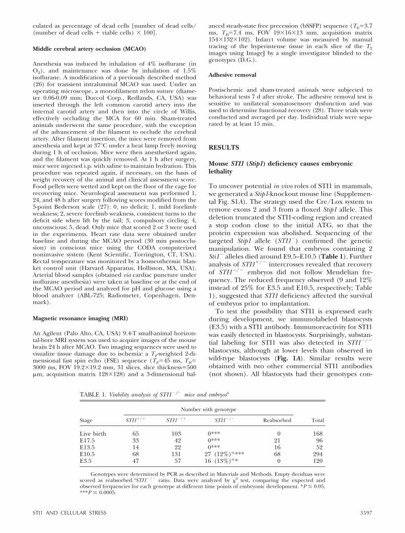
Mouse STI1 (Stip1) deficiency causes embryoniclethality
To uncover potential in vivo roles of STI1 in mammals,we generated a Stip1-knockout mouse line (Supplemen-tal Fig. S1A). The strategy used the Cre/Lox system toremove exons 2 and 3 from a floxed Stip1 allele. Thisdeletion truncated the STI1-coding region and createda stop codon close to the initial ATG, so that theprotein expression was abolished. Sequencing of thetargeted Stip1 allele (STI1�) confirmed the geneticmanipulation. We found that embryos containing 2Sti1� alleles died around E9.5–E10.5 (Table 1). Furtheranalysis of STI1�/� intercrosses revealed that recoveryof STI1�/� embryos did not follow Mendelian fre-quency. The reduced frequency observed (9 and 12%instead of 25% for E3.5 and E10.5, respectively; Table1), suggested that STI1 deficiency affected the survivalof embryos prior to implantation.
To test the possibility that STI1 is expressed earlyduring development, we immunolabeled blastocysts(E3.5) with a STI1 antibody. Immunoreactivity for STI1was easily detected in blastocysts. Surprisingly, substan-tial labeling for STI1 was also detected in STI1�/�
blastocysts, although at lower levels than observed inwild-type blastocysts (Fig. 1A). Similar results wereobtained with two other commercial STI1 antibodies(not shown). All blastocysts had their genotypes con-
TABLE 1. Viability analysis of STI1�/� mice and embryosa
Stage
Number with genotype
TotalSTI1�/� STI1�/� STI1�/� Reabsorbed
Live birth 65 103 0*** 0 168E17.5 33 42 0*** 21 96E13.5 14 22 0*** 16 52E10.5 68 131 27 (12%)a,*** 68 294E3.5 47 57 16 (13%)a,* 0 120
Genotypes were determined by PCR as described in Materials and Methods. Empty deciduas werescored as reabsorbed.aSTI1�/� ratio. Data were analyzed by 2 test, comparing the expected andobserved frequencies for each genotype at different time points of embryonic development. *P � 0.05,***P � 0.0005.
3597STI1 AND CELLULAR STRESS

firmed by PCR after immunostaining (Fig. 1B). Controlexperiments showed that immunolabeling with theSTI1 antibody was completely abolished by absorptionwith excess recombinant STI1 (Fig. 1C), indicating thatSTI1 labeling in blastocysts was specific. Since there areno other STI1 homologs, these results suggest thatmaternal STI1, deposited either as protein or mRNA, ispresent in blastocysts and may play a role in earlyembryonic development.
Given the early expression of STI1 in blastocysts andlethality of STI1�/� embryos, we tested whether genescritical for embryonic development might be altered inblastocysts. We did not observe a difference in theexpression of the pluripotency marker Oct4 betweenwild-type and STI1�/� blastocysts (Fig. 1D; all blasto-cysts were genotyped for STI1 alleles after immuno-staining). Moreover, CDX2 labeling, used to probe fortrophoblast-committed cells, was similarly expressed incontrol and STI1-deletion mutants (Fig. 1E).
Immunoblot analysis of individual embryos at the lastdevelopmental stage in which we could find STI1�/�
mice (E10.5) demonstrated that protein extracts (5 �gof protein) from STI1�/� embryos showed 50% reduc-tion in STI1 levels, while no STI1 immunoreactivity wasobserved in STI1�/� extracts (Fig. 2A). However, whenwe loaded the gel with a higher amount of protein (20�g), we were able to detect small quantities of STI1 inSTI1�/� extracts (20%; Fig. 2B). Moreover, by immu-nofluorescence, we observed weak immunolabeling forSTI1 in sections of STI1�/� embryos (E10.5; Fig. 2C).
We quantified STI1 mRNA in STI1�/� E10.5 embryosby RT-PCR and qPCR. Our results shown that no STI1mRNA is present in STI1�/� E10.5 embryos (Fig. 2D),suggesting that the immunoreactivity that we detectedin embryos might originate from extraembryonic sites.
Interestingly, we found high levels of STI1 present atextraembryonic sites in wild-type embryos, specificallyat the trophoblast layer (Fig. 2E). Whether this extra-embryonic STI1 may contribute for survival of mutantembryos during the early developmental stage is cur-rently unknown. In addition, we observed the presenceof acute inflammation in and around the labyrinth inthe placenta from STI1�/� embryos (Fig. 2F), suggest-ing that their placenta has had a vascular disruption, asthere was no evidence of infection. Hence, placentaldisruption might facilitate the transfer of maternalSTI1 to embryos but could also contribute to embry-onic dysfunction. STI1 has been shown to be secretedby distinct cell types (11, 29, 30), including ovarian cells(31, 32) and has also been found in plasma (31).
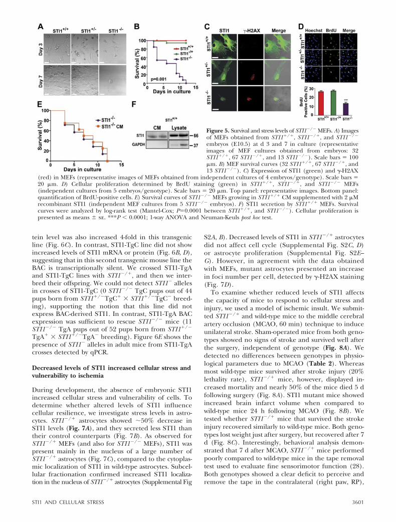
To understand the characteristics of embryonicdevelopment in STI1-mutant mice, we performedmicro-CT analysis. This experiment demonstratedthat neural tube and limb buds were poorly formedin STI1�/� embryos (Fig. 3A). In addition, histo-chemistry analysis showed that STI1�/� embryospresented a great number of cells that were under-going apoptotic cell death, characterized by in-creased levels of activated caspase-3 (Fig. 3B), as well
Figure 1. Expression of STI1, Oct4, and CDX2 in mouse blastocysts. A) STI1labeling (green) in STI1�/�, STI1�/�, and STI1�/� blastocysts (representativeimages of 24 STI1�/�, 22 STI1�/�, and 8 STI1�/� blastocysts). Nucleus is shownin blue. Scale bars � 20 �m. B) Agarose gel showing an example of blastocystgenotyping. C) Control experiments for STI1 antibody specificity. Antibody wasincubated (bottom panels) or not (upper panels) with recombinant STI1 andthen used to label STI1�/� blastocysts. D) Labeling for the pluripotency markerOct4 (green) in STI1�/�, STI1�/�, and STI1�/� blastocysts (representativeimages of 13 STI1�/�, 23 STI1�/�, and 4 STI1�/�). E) Labeling for marker oftrophoblast-committed cells, CDX2 (green), in STI1�/�, STI1�/�, and STI1�/�
blastocysts (representative images of 13 STI1�/�, 25 STI1�/�, and 5 STI1�/�).Scale bars � 20 �m.
3598 Vol. 27 September 2013 BERALDO ET AL.The FASEB Journal � www.fasebj.org
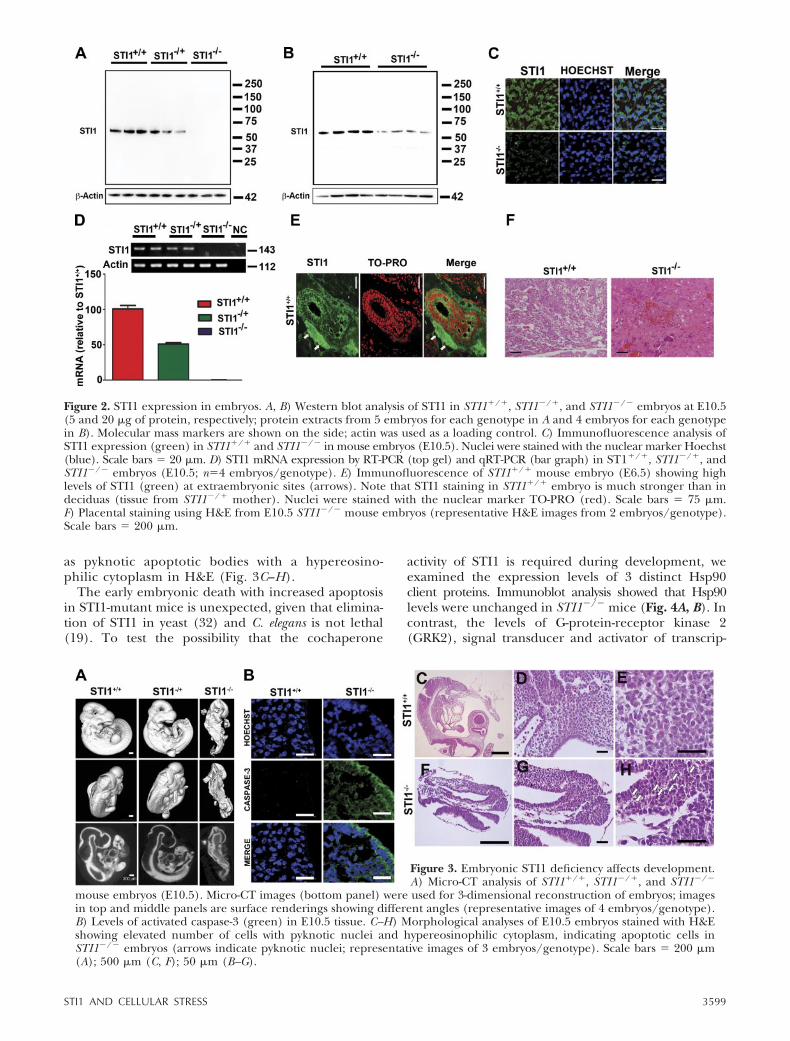
as pyknotic apoptotic bodies with a hypereosino-philic cytoplasm in H&E (Fig. 3C–H).
The early embryonic death with increased apoptosisin STI1-mutant mice is unexpected, given that elimina-tion of STI1 in yeast (32) and C. elegans is not lethal(19). To test the possibility that the cochaperone
activity of STI1 is required during development, weexamined the expression levels of 3 distinct Hsp90client proteins. Immunoblot analysis showed that Hsp90levels were unchanged in STI1�/� mice (Fig. 4A, B). Incontrast, the levels of G-protein-receptor kinase 2(GRK2), signal transducer and activator of transcrip-
Figure 3. Embryonic STI1 deficiency affects development.A) Micro-CT analysis of STI1�/�, STI1�/�, and STI1�/�
mouse embryos (E10.5). Micro-CT images (bottom panel) were used for 3-dimensional reconstruction of embryos; imagesin top and middle panels are surface renderings showing different angles (representative images of 4 embryos/genotype).B) Levels of activated caspase-3 (green) in E10.5 tissue. C–H) Morphological analyses of E10.5 embryos stained with H&Eshowing elevated number of cells with pyknotic nuclei and hypereosinophilic cytoplasm, indicating apoptotic cells inSTI1�/� embryos (arrows indicate pyknotic nuclei; representative images of 3 embryos/genotype). Scale bars � 200 �m(A); 500 �m (C, F); 50 �m (B–G).
Figure 2. STI1 expression in embryos. A, B) Western blot analysis of STI1 in STI1�/�, STI1�/�, and STI1�/� embryos at E10.5(5 and 20 �g of protein, respectively; protein extracts from 5 embryos for each genotype in A and 4 embryos for each genotypein B). Molecular mass markers are shown on the side; actin was used as a loading control. C) Immunofluorescence analysis ofSTI1 expression (green) in STI1�/� and STI1�/� in mouse embryos (E10.5). Nuclei were stained with the nuclear marker Hoechst(blue). Scale bars � 20 �m. D) STI1 mRNA expression by RT-PCR (top gel) and qRT-PCR (bar graph) in ST1�/�, STI1�/�, andSTI1�/� embryos (E10.5; n�4 embryos/genotype). E) Immunofluorescence of STI1�/� mouse embryo (E6.5) showing highlevels of STI1 (green) at extraembryonic sites (arrows). Note that STI1 staining in STI1�/� embryo is much stronger than indeciduas (tissue from STI1�/� mother). Nuclei were stained with the nuclear marker TO-PRO (red). Scale bars � 75 �m.F) Placental staining using H&E from E10.5 STI1�/� mouse embryos (representative H&E images from 2 embryos/genotype).Scale bars � 200 �m.
3599STI1 AND CELLULAR STRESS

tion 3 (STAT3), and protein 53 (p53), all Hsp90 clientproteins (33–35) were significantly reduced to almost50% (Fig. 4A, B).
To further understand the consequences of inter-ference with Stip1 for cellular function, we culturedMEFs from E10.5 embryos. During the initial days inculture, we could not distinguish wild-type fromSTI1�/� cells using morphological criteria. However,after a few days, we noticed that STI1�/� MEFs weredying, whereas wild-type or STI1�/� MEFs thrivedwell in culture (Fig. 5A, B). Interestingly, most STI1�/�
MEFs had died off following 8–10 d in culture (Fig. 5A,B). To understand potential causes of this increasedlethality in STI1�/� MEFs, we labeled these cells duringthe first 2 d of culture with an antibody against �-H2AX,a phosphorylated histone recruited to sites of DNAdouble-strand breaks in response to distinct cellularstresses (36). Wild-type MEFs showed no labeling for�-H2AX (Fig. 5C); however, STI1�/� MEFs presentedwidespread nuclear foci labeling (Fig. 5C), suggestive ofincreased cellular stress levels. Interestingly, STI1�/�
MEFs also presented �-H2AX labeling (Fig. 5C). Sinceenhanced cellular stress levels increase DNA damage,which may affect cell cycle (37), we examined prolifer-ation of STI1�/� MEFs and found decreased prolifera-tion, as compared to STI1�/� or STI1�/� MEFs (Fig.5D). Hence, STI1�/� MEFs were impaired in cellularproliferation and showed increased levels of cellularstress, which likely affected cellular viability.
We examined STI1 expression in MEFs and foundthat STI1�/� MEFs presented weak nuclear immunola-beling for STI1 (Fig. 5C). Immunolabeling in STI1�/�
MEFs was specific because the same pattern of STI1immunolabeling was observed with distinct antibodies(Supplemental Fig. S1B); STI1 immunolabeling inMEFs and other cells could be absorbed with excessrecombinant STI1 (Supplemental Fig. S1C); and themain antibody that we used in these experimentsrecognizes only one band in immunoblots with thecorrect molecular mass in embryos or in brain extractsof adult STI1�/� and STI1�/� mice (Fig. 2A, B andSupplemental Fig. S1D). Hence, it is likely that MEFs
containing some maternal STI1 may have been selectedto survive longer in these cultures.
The weak STI1 labeling was concentrated mostly inthe nucleus in STI1�/� MEFs (Fig. 5C), as opposedto the predominantly cytoplasmic labeling observedin wild-type MEFs and in blastocysts (see Fig. 1A).Fibroblasts obtained from STI1�/� MEFs, which pre-sented overall weaker labeling than wild-type MEFs,also showed increased nuclear labeling for STI1 (Fig.5C). This increased level of nuclear STI1 is likelyrelated to increased levels of cellular stress, as stress hasbeen shown to increase nuclear accumulation of STI1(38). STI1 has been previously shown to have a nuclearlocalization signal (NLS; refs. 38, 39). Given that wild-type MEFs secrete STI1 (Fig. 5F), we examined whetherincreasing levels of extracellular STI1 could perhapsattenuate the lethality observed in STI1�/� MEFs. Weincubated STI1�/� MEFs with CM obtained from wild-type MEFs supplemented with 2 �M recombinant STI1(Fig. 5E). Lethality of STI1�/� MEFs could not berescued by increasing extracellular levels of STI1 or byother factors secreted by wild-type MEFs in this condi-tion. This result suggests that extracellular STI1 is notsufficient to maintain mutant MEFs viability.
Rescue of lethality in STI1�/� mice
Given the presence of small amounts of extraembry-onic STI1 in STI1-mutant mice, we decided to testfurther the requirement for embryonic STI1 duringdevelopment. For this experiment, we initially gener-ated STI1 transgenic mice, which were crossed withSTI1�/�, and tested for rescue of lethality of STI1�/�
mice. Since STI1 appears to be required prior toimplantation for survival of blastocysts, we used BACcontaining the STI1 gene to generate transgenic mouselines and reproduce any required early expression ofSTI1 during embryogenesis. We generated two distinctlines of STI1 BAC transgenic mice (STI1-TgA andSTI1-TgC) presenting 10 and 2 extra alleles of STI1,respectively, as determined by qPCR (Fig. 6A). STI1mRNA levels were increased 5-fold in STI1-TgA linecompared to wild-type littermates (Fig. 6B), and pro-
Figure 4. Analysis of protein expression in STI1�/� E10.5 embryos. A) Western blot analysis of Hsp90, GRK2, p53, and STAT3in STI1�/� and STI1�/� embryos at E10.5 (20 �g protein). B) Quantification of protein expression from Western blots (proteinextracts from 4 embryos/genotype for GRK2 and p53; 7 embryos/genotype for Hsp90 and STAT3). Results are presented asmeans se. *P � 0.01, **P � 0.001; Student’s t test.
3600 Vol. 27 September 2013 BERALDO ET AL.The FASEB Journal � www.fasebj.org
tein level was also increased 4-fold in this transgenicline (Fig. 6C). In contrast, STI1-TgC line did not showincreased levels of STI1 mRNA or protein (Fig. 6B, D),suggesting that in this second transgenic mouse line theBAC is transcriptionally silent. We crossed STI1-TgAand STI1-TgC lines with STI1�/�, and then we inter-bred their offspring. We could not detect STI1� allelesin crosses of STI1-TgC (0 STI1�/� TgC pups out of 44pups born from STI1�/�TgC� � STI1�/�TgC� breed-ing), supporting the notion that this line did notexpress BAC-derived STI1. In contrast, STI1-TgA BACexpression was sufficient to rescue STI1�/� mice (11STI1�/� TgA pups out of 52 pups born from STI1�/�
TgA� � STI1�/�TgA� breeding). Figure 6E shows thepresence of STI1� alleles in adult mice from STI1-TgAcrosses detected by qPCR.
Decreased levels of STI1 increased cellular stress andvulnerability to ischemia
During development, the absence of embryonic STI1increased cellular stress and vulnerability of cells. Todetermine whether altered levels of STI1 influencecellular resilience, we investigate stress levels in astro-cytes. STI1�/� astrocytes showed �50% decrease inSTI1 levels (Fig. 7A), and they secreted less STI1 thantheir control counterparts (Fig. 7B). As observed forSTI1�/� MEFs (and also for STI1�/� MEFs), STI1 waspresent mainly in the nucleus of a large number ofSTI1�/� astrocytes (Fig. 7C), compared to the cytoplas-mic localization of STI1 in wild-type astrocytes. Subcel-lular fractionation confirmed increased STI1 localiza-tion in the nucleus of STI1�/� astrocytes (Supplemental Fig
S2A, B). Decreased levels of STI1 in STI1�/� astrocytesdid not affect cell cycle (Supplemental Fig. S2C, D)or astrocyte proliferation (Supplemental Fig. S2E–G). However, in agreement with the data obtainedwith MEFs, mutant astrocytes presented an increasein foci number per cell, detected by �-H2AX staining(Fig. 7D).
To examine whether reduced levels of STI1 affectsthe capacity of mice to respond to cellular stress andinjury, we used a model of ischemic insult. We submit-ted STI1�/� and wild-type mice to the middle cerebralartery occlusion (MCAO, 60 min) technique to induceunilateral stroke. Sham-operated mice from both geno-types showed no signs of stroke and survived well afterthe surgery, independent of genotype (Fig. 8A). Wedetected no differences between genotypes in physio-logical parameters due to MCAO (Table 2). Whereasmost wild-type mice survived after stroke injury (20%lethality rate), STI1�/� mice, however, displayed in-creased mortality and nearly 50% of the mice died 5 dfollowing surgery (Fig. 8A). STI1 mutant mice showedincreased brain infarct volume when compared towild-type mice 24 h following MCAO (Fig. 8B). Wetested whether STI1�/� mice that survived the strokeinjury recovered similarly to wild-type mice. Both geno-types lost weight just after surgery, but recovered after 7d (Fig. 8C). Interestingly, behavioral analysis demon-strated that 7 d after MCAO, STI1�/� mice performedpoorly compared to wild-type mice in the tape removaltest used to evaluate fine sensorimotor function (28).Both genotypes showed a clear deficit to perceive andremove the tape in the contralateral (right paw, RP),
Figure 5. Survival and stress levels of STI1�/� MEFs. A) Imagesof MEFs obtained from STI1�/�, STI1�/�, and STI1�/�
embryos (E10.5) at d 3 and 7 in culture (representativeimages of MEF cultures obtained from embryos: 32STI1�/�, 67 STI1�/�, and 13 STI1�/�). Scale bars � 100�m. B) MEF survival curves (32 STI1�/�, 67 STI1�/�, and13 STI1�/�). C) Expression of STI1 (green) and �-H2AX
(red) in MEFs (representative images of MEFs obtained from independent cultures of 4 embryos/genotype). Scale bars �20 �m. D) Cellular proliferation determined by BrdU staining (green) in STI1�/�, STI1�/�, and STI1�/� MEFs(independent cultures from 5 embryos/genotype). Scale bars � 20 �m. Top panel: representative images. Bottom panel:quantification of BrdU-positive cells. E) Survival curves of STI1�/� MEFs growing in STI1�/� CM supplemented with 2 �Mrecombinant STI1 (independent MEF cultures from 5 STI1�/� embryos). F) STI1 secretion by STI1�/� MEFs. Survivalcurves were analyzed by log-rank test (Mantel-Cox; P�0.0001 between STI1�/�, and STI1�/�). Cellular proliferation ispresented as means se. ***P � 0.0001; 1-way ANOVA and Neuman-Keuls post hoc test.
3601STI1 AND CELLULAR STRESS

but not in the ipsilateral (left paw, LP) ischemic side(Fig. 8D), indicating a functional deficit due to isch-emia. However, STI1�/� mice showed decreased dex-terity in their RP compared to wild-type controls toremove the tape (Fig. 8E). These experiments indicatethat decreased STI1 levels affect brain injury afterischemic stroke, survival of animals, and their func-tional recovery.
To establish potential mechanisms by which STI1may affect neuronal function after stroke, we examinedthe response of astrocytes and neuronal cultures toOGD. We found that intracellular STI1 was reduced inSTI1�/� astrocytes after OGD (Fig. 9A). In STI1�/�
astrocytes, the intracellular levels of STI1 were furtherreduced by OGD. Experiments using proteasome in-
hibitors indicated that reduced levels of STI1 were notdue to increased protein degradation (not shown),suggesting the possibility of changes in secretion. In-deed, wild-type astrocytes increased STI1 secretion byclose to 3-fold after OGD (Fig. 9B). Although astrocytesderived from STI1�/� mice also showed increasedsecretion of STI1 following OGD, the levels of extracel-lular STI1 were, as expected, �2-fold lower than thoseof wild-type astrocytes (Fig. 9B). Therefore, as a conse-quence of ischemia, astrocytes secrete significantamounts of STI1, (Fig. 9A, B); however, since STI1�/�
astrocytes have 50% less protein, the amount of STI1secreted by these mutated cells is lower. OGD did notincrease cell death of astrocytes from either genotype(Fig. 9C).
Figure 6. Rescue of embryonic lethality of STI1�/� mice by transgenic expression ofSTI1. A) Number of copies of the STI1 gene in genomic DNA from STI1�/� (n�8) andSTI1�/� mice (n�4), as well as transgenic mouse lines STI1-TgA (n�4) and STI1-TgC(n�5). B) STI1 mRNA expression in the brain of STI1�/� mice (n�9) and transgeniclines STI1-TgA (n�4) and STI1-TgC (n�4). C) Representative immunoblotting andSTI1 protein expression quantification in the brain of ST1�/� (n�4) and STI1-TgAmice (n�4). D) Representative immunoblotting and STI1 protein expression quanti-fication in the brain of ST1�/� (n�4) and STI1-TgC mice (n�4 mice). E) qPCR todetermine the number of copies of the STI1� allele (which is absent in wild-type mice)
in tissue from adult mice, showing that expression of BAC-STI1 allowed survival of STI1�/� mice (4 STI1�/�, 11 STI1�/�,and 5 TgASTI1�/�). Results are presented as means se; data were analyzed and compared by 1-way ANOVA andNewman-Keuls post hoc test (A, B), Student’s t test (C, D), and 2 test (P�0.05 from the expected distribution; E). **P �0.001, ***P � 0.0001.
Figure 7. STI1 secretion, expression, and cellular localization in STI1�/� and STI1�/� astrocyte cultures. A) STI1 expressionin STI1�/� and STI1�/� astrocytes (cultures obtained from 4 embryos/genotype). B) Detection of STI1 in CM from astrocytes(cultures obtained from 5 embryos/genotype). C) Immunofluorescence showing a reduction of STI1 labeling and localizationof STI1 in STI1�/� and STI1�/� astrocytes (top panel) and quantification of cells with nuclear STI1 (bottom panel). Scale bars �15 �m. D) Top panel: immunofluorescence for STI1 expression (green) and labeling of �-H2AX (red) in STI1�/� and STI1�/�
astrocytes. Scale bars � 30 �m. Bottom panel: quantification of number of foci per cell (cultures obtained from 7embryos/genotype). Results are presented as means se. *P � 0.01; Student’s t test.
3602 Vol. 27 September 2013 BERALDO ET AL.The FASEB Journal � www.fasebj.org

In cultured primary neurons, 1 h OGD caused in-creased cell death (Fig. 9D), an effect that was similar inboth control and STI1�/� neurons. Hence, changes inintracellular levels of STI1 did not seem to affect thesurvival of neurons. To mimic the secretion of STI1from astrocytes, we treated neurons with recombinantSTI1, which reproduces the effects of astrocyte-secretedprotein (40). Treatment of neurons from both geno-types with extracellular recombinant STI1 attenuatedneuronal death in response to OGD (Fig. 9D), suggest-
ing that rather than intracellular STI1, it is the extra-cellular protein that protects neurons from ischemicinjury.
PrPC, an STI1-interacting protein, plays a role inischemic injury and increased levels of PrPC seem toprotect, whereas lack of PrPC exacerbates neuronalinjury in response to ischemic insults in vivo (41–43).To investigate the possibility that extracellular STI1protects neurons via a PrPC-dependent pathway inneurons, we repeated these experiments using neurons
Figure 8. Regulation of functional recovery in ischemia by STI1. A) Survival curve of STI1�/� and STI1�/� mice submitted to60 min of unilateral ischemia (MCAO). B) Representative MR images and group analyses of infarct volume in brains of STI1�/�
(n�6) and STI1�/� mice (n�5). C) Weight of mice submitted to MCAO (n�6 Sham STI1�/�, n�5 Sham STI1�/�, n�9STI1�/�, n�7 STI1�/�). D) Functional recovery of STI1�/� and STI1�/� mice submitted to MCAO, determined at d 7 afterstroke using the tape removal test. Time to perceive the tape in the right paw (RP) or left paw (LP) (n�9 STI1�/�, n�7STI1�/�). E) Identical to D, but showing the time to remove the tape. Results are presented as means se; data were analyzedand compared by 1-way ANOVA and Newman-Keuls post hoc test, Student’s t test, and Mantel-Cox log-rank test (P�0.0002; A).*P � 0.01 vs. control mice.
TABLE 2. Physiological measurements before and during MCAO
Measurement STI1�/� STI1�/�
Before MCAOHeart rate (beats/min) 697.4 43.8 (n�9) 669.2 34.8 (n�9)Arterial blood pH 7.193 0.003 (n�3) 7.233 0.007 (n�3)Oxygen saturation (%) 99.37 3.74 (n�3) 97.80 5.8 (n�3)Temperature (°C) 35.90 0.30 (n�3) 36.20 0.36 (n�3)Glucose (mM) 8.067 0.633 (n�3) 8.833 0.033 (n�3)
During MCAOHeart rate (beats/min) 693.1 42.9 (n�6) 693.1 55.9 (n�4)Arterial blood pH 7.107 0.0201 (n�6) 7.100 0.044 (n�4)Oxygen saturation (%) 99.32 3.47 (n�6) 73.55 10.31 (n�4)Temperature (°C) 34.35 0.43 (n�6) 34.65 0.52 (n�4)Glucose (mM) 10.03 1.73 (n�6) 9.650 1.707 (n�4)
Values are expressed as means SE. MCAO, middle cerebral artery occlusion.
3603STI1 AND CELLULAR STRESS

of PrPC-null mice (Prnp0/0). The neuroprotective effectof STI1 was prevented in neurons from PrPC-null mice(Fig. 9E), suggesting that activation of PrPC by STI1plays a role in protecting neurons against ischemicinsult.
DISCUSSION
The present experiments demonstrate that Stip1 showscharacteristics of a maternal (oogenetic)-effect gene,playing critical roles during development in mammals.STI1�/� blastocysts obtained from STI1�/� inter-crosses were observed at 50% of the expected mende-lian frequency, suggesting that blastocysts originatingfrom zygotes lacking sufficient maternally inheritedSTI1 may not survive. Interestingly, surviving STI1�/�
blastocysts showed immunostaining for STI1, suggest-ing the possibility that part of the blastocyts may havereceived sufficient maternally inherited STI1 to allowdevelopment of embryos. It would be of interest todefine the precise mechanism by which maternallyderived STI1 is transferred to blastocysts. Embryonicallyexpressed STI1 is critical in later stages of development,as mutant embryos could not survive past E10.5. Weshow that maternal STI1 protein might be transferredto embryos in later stages of development, as we coulddetect small amounts of STI1 immunoreactivity inE10.5 embryos, likely due to placental disruption. No-tably, we did not detect STI1 mRNA in STI1�/� E10.5
embryos, indicating lack of embryonic STI1 proteinsynthesis. The rescue of lethality by transgenic BACexpression supports the notion that STI1 has uniqueroles during development and that maternally trans-ferred STI1 cannot support embryonic development.Embryonic lethality is commonly associated with pla-cental dysfunction in the E9–E12 stage. Interestingly,Hsp90�-knockout mice (44), but not Hsp90�-knockoutmice (45), present disrupted placenta. Hence, placen-tal disruption may also contribute to embryonic lethal-ity in STI1 mutant embryos.
Despite recent insights from structural models ofSTI1 interaction with Hsp70 and Hsp90 demonstratingSTI1 regulation of client recruitment (7, 46–48), it isunknown whether STI1 has unique or overlappingroles in mammals in vivo. Our data suggest that loss ofembryonic STI1 is not tolerated during development,and cells present decreased resilience to stress, showingincreased DNA damage and cell death. Hence, othercochaperones do not seem to be able to compensatefor the loss of STI1. These results contrast with those ofmice deficient for p23, another Hsp90 chaperone,which present perinatal lethality specifically related toskin and lung development (49), suggesting distinctrequirements for different cochaperones during devel-opment.
The early effects of STI1 in embryogenesis are com-patible with its regulation of stem cell self-renewal (50).Interestingly, Hsp90 and, more recently, STI1 have
Figure 9. STI1 rescues neurons fromOGD-induced cell death. A) IntracellularSTI1 in astrocytes obtained from proteinextracts after 0 or 9 h of OGD (proteinextract from independent cultures from6 embryos/genotype). **P � 0.001 vs. 0 hof OGD in wild-type mice. B) STI1 secre-tion from astrocytes in response to 9 hOGD (CM from independent culturesfrom 6 embryos/genotype). ***P �0.0001 vs. all other conditions. C) Celldeath in astrocytes exposed to OGD for 3,6, and 9 h (independent cultures ob-
tained from 5 embryos/genotype). D) Cell death induced by 1 h OGD in STI1�/� and STI1�/� neurons treated or not withrecombinant STI1 (1 �M) 1 h prior to OGD (independent cultures obtained from 6 embryos/genotype). **P � 0.001, ***P �0.0001 vs. STI1 treatment or neurons without OGD. E) Cell death induced by 1 h OGD in wild-type and Prnp0/0 neuronstreated or not with recombinant STI1 (1 �M) 1 h prior to OGD (independent cultures obtained from 4 embryos/genotype). Results are presented as means se; data were analyzed and compared by 1-way ANOVA and Newman-Keulspost hoc test. ***P � 0.0001 vs. neurons without OGD treatment or wild-type neurons treated with STI1 after OGD.
3604 Vol. 27 September 2013 BERALDO ET AL.The FASEB Journal � www.fasebj.org

been shown to regulate epigenetic programs and trans-poson silencing via piRNAs in Drosophila (8, 51). Re-markably, maternal STI1 appears to influence canaliza-tion in Drosophila via the Piwi pathway (8). Futurestudies aimed at further defining these mechanismsmay provide novel insight in mammalian embryonicdevelopment.
We show that STI1 has a critical role in cellularsurvival. This conclusion is supported by the increasedcell death in embryos and the inability to maintainSTI1�/� MEFs in culture. Because of the early deaththat we observed in blastocysts, as well as the inability ofmutant cells to survive, we favor the possibility that theplacental defect that we identified contributes to, but itis not the only cause of, cellular death in these mutantembryos. These effects of Stip1 seem to depend, at leastin part, on intracellular STI1, as we were unable torescue STI1�/� MEFs using recombinant extracellularSTI1, but could rescue STI1�/� mice by transgenicexpression of a BAC containing Stip1. The increase infoci number in STI1�/� MEFs indicates that lack ofchaperone activity may affect DNA damage response.Interestingly, Hsp90, which is regulated by STI1, hasbeen previously implicated in the DNA damage re-sponse (52–55). By targeting STI1, we likely affectedthe functions of a wide range of Hsp90/Hsp70 clientproteins (56–58). Indeed, this seems to be the case,given that three known Hsp90 clients (59–61) showreduced levels in STI1-mutant mice, even thoughHsp90 levels were unaffected. In contrast with Hsp90and Hsp70 that have different isoforms, there are noknown homologs for Stip1, which may explain why weobserved such striking phenotype during development.Given the very large number of Hsp90 client proteins(5, 56, 57), it is unlikely that the phenotypes that weuncovered are related to one specific client. It is alsounclear whether the remaining amounts of Hsp90client proteins in STI1 mutant embryos are functional.Hence, inhibitors of STI1 may have more widespreadeffects in mammals than inhibitors of Hsp90. Indeed, arecent report demonstrates that inhibition of STI1interaction with Hsp90 by drugs is effective to killcancer cells (62).
Our experiment using a stroke model supports animportant role for endogenous STI1 in the recoveryfrom ischemic injury in vivo. PrPC expression has beenshown to protect the brain following ischemic insults,and lack of PrPC leads to an increase in neuronal injuryin stroke models (41, 42, 63). However, the exactmechanisms by which PrPC influences outcomes instroke are unknown. We now show that OGD, used asan in vitro model of ischemic insult, can increasesecretion of STI1 from astrocytes and that secretionfrom STI1�/� astrocytes is decreased in this condition.Our experiments using cultured neurons suggest thatthe increased sensitivity observed in vivo in STI1�/�
mice may not be related to decreased intracellularlevels of STI1 in neurons, as both control and STI1�/�
neurons responded similarly to OGD. In these neuro-nal cultures, extracellular STI1 was able to provide
neuroprotection for OGD-induced neuronal death.Moreover, this effect of STI1 was strictly dependent onthe presence of PrPC in neurons. These results expandprevious observations in which STI1 was shown toprevent neuronal death induced by staurosporine (14,17) to demonstrate a role for this secreted cochaperonein a pathological relevant insult, ischemic injury. Our invitro data with recombinant STI1 and in vivo withSTI1�/� mice suggest that lack of STI1-mediated sig-naling may underlie the increased sensitivity of PrPC-null mice to ischemic injury.
Our experiments provide novel evidence that STI1plays a unique and nonoverlapping role as a cochaper-one during embryonic development and in cellularsurvival, suggesting that loss of STI1-regulated chaper-one activity is not tolerated. Our data also support arole for STI1 in neuroprotection against ischemic in-sult, by a mechanism involving increased secretionfrom astrocytes and activation of PrPC. Hence, STI1 is astress-response protein that presents multiple intracel-lular and extracellular roles with unique properties forprotection of cells against stress.
The authors thank Joy Dunmore-Buyze for micro-CT sam-ple preparation and image acquisition and Sanda Raulic andWeiyan Wen for help with mouse colonies. The authors alsothank Dr. Gerald Kidder (University of Western Ontario) forhelp with data analysis and interpretation. This work wassupported by grants from the Canadian Institute of HealthResearch (MOP 93651 and MOP 126000, M.A.M.P., R.B., andV.F.P), PrioNet-Canada (M.A.M.P., R.B. and V.F.P.), Cana-dian Foundation for Innovation (M.A.M.P., V.F.P., and R.G.),Ontario Research Fund (M.A.M.P., V.F.P., and R.G.), theAlzheimer’s Association (M.A.M.P., V.R.M., and V.F.P.), Con-selho Nacional de Desenvolvimento Científico e Tecnológico(CNPq; Brazil; V.R.M.), and Fundação de Amparo a Pesquisado Estado de São Paulo (FAPESP; São Paulo, Brazil; V.R.M.).F.H.B. and I.N.S. received fellowships from the Departmentof Foreign Affairs and International Trade (Canada). I.N.S.and D.F.G. received a fellowship from Coordenação de Aper-feiçoamento de Pessoal de Nível Superior (Brazil). A.H.M.received a fellowship from the Ontario Graduate ScholarshipProgram; T.G.S. and M.R. received a fellowship from FAPESP;A.L.G. received a fellowship from CNPq. R.G. and M.D. aresupported by awards from the Heart and Stroke Foundationof Canada.
REFERENCES
1. Picard, D. (2002) Heat-shock protein 90, a chaperone forfolding and regulation. Cell. Mol. Life Sci. 59, 1640–1648
2. Young, J. C., Agashe, V. R., Siegers, K., and Hartl, F. U. (2004)Pathways of chaperone-mediated protein folding in the cytosol.Nat. Rev. Mol. Cell Biol. 5, 781–791
3. Pearl, L. H., and Prodromou, C. (2006) Structure and mecha-nism of the Hsp90 molecular chaperone machinery. Annu. Rev.Biochem. 75, 271–294
4. Linden, R., Martins, V. R., Prado, M. A., Cammarota, M.,Izquierdo, I., and Brentani, R. R. (2008) Physiology of the prionprotein. Physiol. Rev. 88, 673–728
5. Taipale, M., Jarosz, D. F., and Lindquist, S. (2010) HSP90 at thehub of protein homeostasis: emerging mechanistic insights. Nat.Rev. Mol. Cell Biol. 11, 515–528
6. Richter, K., Muschler, P., Hainzl, O., Reinstein, J., and Buchner,J. (2003) Sti1 is a non-competitive inhibitor of the Hsp90
3605STI1 AND CELLULAR STRESS
ATPase. Binding prevents the N-terminal dimerization reactionduring the atpase cycle. J. Biol. Chem. 278, 10328–10333
7. Southworth, D. R., and Agard, D. A. (2011) Client-loadingconformation of the Hsp90 molecular chaperone revealed inthe cryo-EM structure of the human Hsp90: Hop complex. Mol.Cell 42, 771–781
8. Gangaraju, V. K., Yin, H., Weiner, M. M., Wang, J., Huang, X. A.,and Lin, H. (2011) Drosophila Piwi functions in Hsp90-mediatedsuppression of phenotypic variation. Nat. Genet. 43, 153–158
9. Eustace, B. K., Sakurai, T., Stewart, J. K., Yimlamai, D., Unger,C., Zehetmeier, C., Lain, B., Torella, C., Henning, S. W., Beste,G., Scroggins, B. T., Neckers, L., Ilag, L. L., and Jay, D. G. (2004)Functional proteomic screens reveal an essential extracellularrole for Hsp90 alpha in cancer cell invasiveness. Nat. Cell Biol. 6,507–514
10. De Maio, A. (2011) Extracellular heat shock proteins, cellularexport vesicles, and the stress observation system: a form ofcommunication during injury, infection, and cell damage. CellStress Chaperones 16, 235–249
11. Lima, F. R., Arantes, C. P., Muras, A. G., Nomizo, R., Brentani,R. R., and Martins, V. R. (2007) Cellular prion protein expres-sion in astrocytes modulates neuronal survival and differentia-tion. J. Neurochem. 103, 2164–2176
12. Arantes, C., Nomizo, R., Lopes, M. H., Hajj, G. N., Lima, F. R.,and Martins, V. R. (2009) Prion protein and its ligand stressinducible protein 1 regulate astrocyte development. Glia 57,1439–1449
13. Hajj, G. N., Arantes, C. P., Dias, M. V., Roffe, M., Costa-Silva, B.,Lopes, M. H., Porto-Carreiro, I., Rabachini, T., Lima, F. R.,Beraldo, F. H., Prado, M. A. M., Linden, R., and Martins, V. R.(2013) The unconventional secretion of stress-inducible protein1 by a heterogeneous population of extracellular vesicles. [E-pub ahead of print] Cell. Mol. Life Sci. 10.1007/s00018-013–1328-y
14. Beraldo, F. H., Arantes, C. P., Santos, T. G., Queiroz, N. G.,Young, K., Rylett, R. J., Markus, R. P., Prado, M. A., and Martins,V. R. (2010) Role of alpha7 nicotinic acetylcholine receptor incalcium signaling induced by prion protein interaction withstress-inducible protein 1. J. Biol. Chem. 285, 36542–36550
15. Santos, T. G., Beraldo, F. H., Hajj, G. N., Lopes, M. H., Roffe,M., Lupinacci, F. C., Ostapchenko, V. G., Prado, V. F., Prado,M. A., and Martins, V. R. (2013) Laminin-gamma1 chain andstress inducible protein 1 synergistically mediate PrPC-depen-dent axonal growth via Ca2� mobilization in dorsal root ganglianeurons. J. Neurochem. 124, 210–223
16. Beraldo, F. H., Arantes, C. P., Santos, T. G., Queiroz, N. G.,Young, K., Rylett, R. J., Markus, R. P., Prado, M. A., and Martins,V. R. (2010) Role of �7 nicotinic acetylcholine receptor incalcium signaling induced by prion protein interaction withstress-inducible protein 1. J. Biol. Chem. 19, 36542–36550
17. Lopes, M. H., Hajj, G. N., Muras, A. G., Mancini, G. L., Castro,R. M., Ribeiro, K. C., Brentani, R. R., Linden, R., and Martins,V. R. (2005) Interaction of cellular prion and stress-inducibleprotein 1 promotes neuritogenesis and neuroprotection bydistinct signaling pathways. J. Neurosci. 7, 11330–11339
18. Chang, H. C., Nathan, D. F., and Lindquist, S. (1997) In vivoanalysis of the Hsp90 cochaperone Sti1 (p60). Mol. Cell. Biol. 17,318–325
19. Song, H. O., Lee, W., An, K., Lee, H. S., Cho, J. H., Park, Z. Y.,and Ahnn, J. (2009) C. elegans STI-1, the homolog of Sti1/Hop,is involved in aging and stress response. J. Mol. Biol. 390,604–617
20. Prado, V. F., Martins-Silva, C., de Castro, B. M., Lima, R. F.,Barros, D. M., Amaral, E., Ramsey, A. J., Sotnikova, T. D.,Ramirez, M. R., Kim, H. G., Rossato, J. I., Koenen, J., Quan, H.,Cota, V. R., Moraes, M. F., Gomez, M. V., Guatimosim, C.,Wetsel, W. C., Kushmerick, C., Pereira, G. S., Gainetdinov, R. R.,Izquierdo, I., Caron, M. G., and Prado, M. A. (2006) Micedeficient for the vesicular acetylcholine transporter are myas-thenic and have deficits in object and social recognition. Neuron51, 601–612
21. Martins-Silva, C., De Jaeger, X, Guzman, M. S., Lima, R. D.,Santos, M. S., Kushmerick, C., Gomez, M. V., Caron, M. G.,Prado, M. A., and Prado, V. F. (2011) Novel strains of micedeficient for the vesicular acetylcholine transporter: insights ontranscriptional regulation and control of locomotor behavior.PLoS ONE 6, e17611
22. Guzman, M. S., De, J., X, Raulic, S., Souza, I. A., Li, A. X.,Schmid, S., Menon, R. S., Gainetdinov, R. R., Caron, M. G.,Bartha, R., Prado, V. F., and Prado, M. A. (2011) Elimination ofthe vesicular acetylcholine transporter in the striatum revealsregulation of behaviour by cholinergic-glutamatergic co-trans-mission. PLoS Biol. 9, e1001194
23. Migliorini, D., Lazzerini, D. E., Danovi, D., Jochemsen, A.,Capillo, M., Gobbi, A., Helin, K., Pelicci, P. G., and Marine, J. C.(2002) Mdm4 (Mdmx) regulates p53-induced growth arrest andneuronal cell death during early embryonic mouse develop-ment. Mol. Cell. Biol. 22, 5527–5538
24. Degenhardt, K., Wright, A. C., Horng, D., Padmanabhan, A.,and Epstein, J. A. (2010) Rapid 3D phenotyping of cardiovas-cular development in mouse embryos by micro-CT with iodinestaining. Circ. Cardiovasc. Imaging 3, 314–322
25. Badea, C. T., Drangova, M., Holdsworth, D. W., and Johnson,G. A. (2008) In vivo small-animal imaging using micro-CT anddigital subtraction angiography. Phys. Med. Biol. 53, R319–R350
26. Longa, E. Z., Weinstein, P. R., Carlson, S., and Cummins, R.(1989) Reversible middle cerebral artery occlusion withoutcraniectomy in rats. Stroke 20, 84–91
27. Bederson, J. B., Pitts, L. H., Tsuji, M., Nishimura, M. C., Davis,R. L., and Bartkowski, H. (1986) Rat middle cerebral arteryocclusion: evaluation of the model and development of aneurologic examination. Stroke 17, 472–476
28. Bouet, V., Boulouard, M., Toutain, J., Divoux, D., Bernaudin,M., Schumann-Bard, P., and Freret, T. (2009) The adhesiveremoval test: a sensitive method to assess sensorimotor deficitsin mice. Nat. Protoc. 4, 1560–1564
29. Erlich, R. B., Kahn, S. A., Lima, F. R., Muras, A. G., Martins,R. A., Linden, R., Chiarini, L. B., Martins, V. R., and Moura, N.,V (2007) STI1 promotes glioma proliferation through MAPKand PI3K pathways. Glia 55, 1690–1698
30. Tsai, C. L., Tsai, C. N., Lin, C. Y., Chen, H. W., Lee, Y. S., Chao,A., Wang, T. H., Wang, H. S., and Lai, C. H. (2012) Secretedstress-induced phosphoprotein 1 activates the ALK2-SMAD sig-naling pathways and promotes cell proliferation of ovariancancer cells. Cell Rep. 2, 283–293
31. Wang, T. H., Chao, A., Tsai, C. L., Chang, C. L., Chen, S. H.,Lee, Y. S., Chen, J. K., Lin, Y. J., Chang, P. Y., Wang, C. J., Chao,A. S., Chang, S. D., Chang, T. C., Lai, C. H., and Wang, H. S.(2010) Stress-induced phosphoprotein 1 as a secreted bio-marker for human ovarian cancer promotes cancer cell prolif-eration. Mol. Cell. Proteomics 9, 1873–1884
32. Nicolet, C. M., and Craig, E. A. (1989) Isolation and character-ization of STI1, a stress-inducible gene from Saccharomycescerevisiae. Mol. Cell. Biol. 9, 3638–3646
33. Matkovich, S. J., Diwan, A., Klanke, J. L., Hammer, D. J.,Marreez, Y., Odley, A. M., Brunskill, E. W., Koch, W. J., Schwartz,R. J., and Dorn, G. W. (2006) Cardiac-specific ablation ofG-protein receptor kinase 2 redefines its roles in heart develop-ment and beta-adrenergic signaling. Circ. Res. 99, 996–1003
34. Takeda, K., Noguchi, K., Shi, W., Tanaka, T., Matsumoto, M.,Yoshida, N., Kishimoto, T., and Akira, S. (1997) Targeteddisruption of the mouse Stat3 gene leads to early embryoniclethality. Proc. Natl. Acad. Sci. U. S. A. 94, 3801–3804
35. Donehower, L. A., Harvey, M., Slagle, B. L., McArthur, M. J.,Montgomery, C. A., Jr., Butel, J. S., and Bradley, A. (1992) Micedeficient for p53 are developmentally normal but susceptible tospontaneous tumours. Nature 356, 215–221
36. Lukas, J., Lukas, C., and Bartek, J. (2011) More than just a focus:The chromatin response to DNA damage and its role in genomeintegrity maintenance. Nat. Cell Biol. 13, 1161–1169
37. Bartek, J., Bartkova, J., and Lukas, J. (2007) DNA damagesignalling guards against activated oncogenes and tumour pro-gression. Oncogene 26, 7773–7779
38. Daniel, S., Bradley, G., Longshaw, V. M., Söti, C., Csermely, P.,and Blatch, G. L. (2008) Nuclear translocation of the phospho-protein Hop (Hsp70/Hsp90 organizing protein) occurs underheat shock, and its proposed nuclear localization signal isinvolved in Hsp90 binding. Biochim. Biophys. Acta 1783, 1003–1014
39. Longshaw, V. M., Chapple, J. P., Balda, M. S., Cheetham, M. E.,and Blatch, G. L. (2004) Nuclear translocation of the Hsp70/Hsp90 organizing protein mSTI1 is regulated by cell cyclekinases. J. Cell Sci. 15, 701–710
3606 Vol. 27 September 2013 BERALDO ET AL.The FASEB Journal � www.fasebj.org

40. Caetano, F. A., Lopes, M. H., Hajj, G. N., Machado, C. F., Pinto,A. C., Magalhaes, A. C., Vieira, M. P., Americo, T. A., Massensini,A. R., Priola, S. A., Vorberg, I., Gomez, M. V., Linden, R., Prado,V. F., Martins, V. R., and Prado, M. A. (2008) Endocytosis ofprion protein is required for ERK1/2 signaling induced bystress-inducible protein 1. J. Neurosci. 28, 6691–6702
41. McLennan, N. F., Brennan, P. M., McNeill, A., Davies, I.,Fotheringham, A., Rennison, K. A., Ritchie, D., Brannan, F.,Head, M. W., Ironside, J. W., Williams, A., and Bell, J. E. (2004)Prion protein accumulation and neuroprotection in hypoxicbrain damage. Am. J. Pathol. 165, 227–235
42. Shyu, W. C., Lin, S. Z., Chiang, M. F., Ding, D. C., Li, K. W.,Chen, S. F., Yang, H. I., and Li, H. (2005) Overexpression ofPrPC by adenovirus-mediated gene targeting reduces ischemicinjury in a stroke rat model. J. Neurosci. 25, 8967–8977
43. Guillot-Sestier, M. V., Sunyach, C., Druon, C., Scarzello, S., andChecler, F. (2009) The alpha-secretase-derived N-terminal prod-uct of cellular prion, N1 displays neuroprotective function, invitro and in vivo. J. Biol. Chem. 284, 35973–35986
44. Voss, A. K., Thomas, T., and Gruss, P. (2000) Mice lackingHSP90� fail to develop a placental labyrinth. Development 127,1–11
45. Imai, T., Kato, Y., Kajiwara, C., Mizukami, S., Ishige, I., Ichiyan-agi, T., Hikida, M., Wang, J. Y., and Udono, H. (2011) Heatshock protein 90 (HSP90) contributes to cytosolic translocationof extracellular antigen for cross-presentation by dendritic cells.Proc. Natl. Acad. Sci. U. S. A. 108, 16363–16368
46. Lee, C. T., Graf, C., Mayer, F. J., Richter, S. M., and Mayer, M. P.(2012) Dynamics of the regulation of Hsp90 by the co-chaper-one Sti1. EMBO. J. 31, 1518–1528
47. Schmid, A. B., Lagleder, S., Grawert, M. A., Rohl, A., Hagn, F.,Wandinger, S. K., Cox, M. B., Demmer, O., Richter, K., Groll,M., Kessler, H., and Buchner, J. (2012) The architecture offunctional modules in the Hsp90 co-chaperone Sti1/Hop.EMBO J. 31, 1506–1517
48. Scheufler, C., Brinker, A., Bourenkov, G., Pegoraro, S., Mo-roder, L., Bartunik, H., Hartl, F. U., and Moarefi, I. (2000)Structure of TPR domain-peptide complexes: critical elementsin the assembly of the Hsp70-Hsp90 multichaperone machine.Cell 101, 199–210
49. Grad, I., McKee, T. A., Ludwig, S. M., Hoyle, G. W., Ruiz, P.,Wurst, W., Floss, T., Miller, C. A., III, and Picard, D. (2006) TheHsp90 cochaperone p23 is essential for perinatal survival. Mol.Cell. Biol. 26, 8976–8983
50. Santos, T. G., Silva, I. R., Costa-Silva, B., Lepique, A. P., Martins,V. R., and Lopes, M. H. (2011) Enhanced neural progenitor/stem cells self-renewal via the interaction of stress-inducibleprotein 1 with the prion protein. Stem Cells 29, 1126–1136
51. Specchia, V., Piacentini, L., Tritto, P., Fanti, L., D’Alessandro,R., Palumbo, G., Pimpinelli, S., and Bozzetti, M. P. (2010)Hsp90 prevents phenotypic variation by suppressing the muta-genic activity of transposons. Nature 463, 662–665
52. Arlander, S. J., Eapen, A. K., Vroman, B. T., McDonald, R. J.,Toft, D. O., and Karnitz, L. M. (2003) Hsp90 inhibition depletesChk1 and sensitizes tumor cells to replication stress. J. Biol.Chem. 278, 52572–52577
53. Oda, T., Hayano, T., Miyaso, H., Takahashi, N., and Yamashita,T. (2007) Hsp90 regulates the Fanconi anemia DNA damageresponse pathway. Blood 109, 5016–5026
54. Ha, K., Fiskus, W., Rao, R., Balusu, R., Venkannagari, S.,Nalabothula, N. R., and Bhalla, K. N. (2011) Hsp90 inhibitor-mediated disruption of chaperone association of ATR withhsp90 sensitizes cancer cells to DNA damage. Mol. Cancer Ther.10, 1194–1206
55. Quanz, M., Herbette, A., Sayarath, M., de, K. L., Dubois, T., Sun,J. S., and Dutreix, M. (2012) Heat shock protein 90alpha(Hsp90alpha) is phosphorylated in response to DNA damageand accumulates in repair foci. J. Biol. Chem. 287, 8803–8815
56. Sharma, K., Vabulas, R. M., Macek, B., Pinkert, S., Cox, J., Mann,M., and Hartl, F. U. (2012) Quantitative proteomics reveals thatHsp90 inhibition preferentially targets kinases and the DNAdamage response. Mol. Cell. Proteomics 11, M111
57. Zhao, R., Davey, M., Hsu, Y. C., Kaplanek, P., Tong, A., Parsons,A. B., Krogan, N., Cagney, G., Mai, D., Greenblatt, J., Boone, C.,Emili, A., and Houry, W. A. (2005) Navigating the chaperonenetwork: an integrative map of physical and genetic interactionsmediated by the hsp90 chaperone. Cell 120, 715–727
58. Zhao, R., and Houry, W. A. (2007) Molecular interactionnetwork of the Hsp90 chaperone system. Adv. Exp. Med. Biol.594, 27–36
59. Shah, M., Patel, K., Fried, V. A., and Sehgal, P. B. (2002)Interactions of STAT3 with caveolin-1 and heat shock protein 90in plasma membrane raft and cytosolic complexes. Preservationof cytokine signaling during fever. J. Biol. Chem. 277, 45662–45669
60. Muller, L., Schaupp, A., Walerych, D., Wegele, H., and Buchner,J. (2004) Hsp90 regulates the activity of wild type p53 underphysiological and elevated temperatures. J. Biol. Chem. 279,48846–48854
61. Luo, J., and Benovic, J. L. (2003) G protein-coupled receptorkinase interaction with Hsp90 mediates kinase maturation. J.Biol. Chem. 278, 50908–50914
62. Pimienta, G., Herbert, K. M., and Regan, L. (2011) A compoundthat inhibits the HOP-Hsp90 complex formation and hasunique killing effects in breast cancer cell lines. Mol. Pharm. 8,2252–2261
63. Mitsios, N., Saka, M., Krupinski, J., Pennucci, R., Sanfeliu, C.,Miguel, T. M., Gaffney, J., Kumar, P., Kumar, S., Sullivan, M.,and Slevin, M. (2007) Cellular prion protein is increased in theplasma and peri-infarcted brain tissue after acute stroke. J.Neurosci. Res. 85, 602–611
Received for publication April 11, 2013.Accepted for publication May 14, 2013.
3607STI1 AND CELLULAR STRESS