Spatial and temporal expression of vasodilator-stimulated phosphoprotein (VASP) in fetal and adult...
6
Int. J. Devl Neuroscience 29 (2011) 131–136 Contents lists available at ScienceDirect International Journal of Developmental Neuroscience journal homepage: www.elsevier.com/locate/ijdevneu Spatial and temporal expression of vasodilator-stimulated phosphoprotein (VASP) in fetal and adult human cerebral cortex Aylin Yaba a , Umit Kayisli b , Gamze Tanriover a , Necdet Demir a,∗ a Akdeniz University School of Medicine, Department of Histology and Embryology, Umlupinar Street, 07070 Campus, Antalya, Turkey b Yale University School of Medicine, Department of Obstetrics and Gynecology and Reproductive Science, 06520 New Haven, CT, USA article info Article history: Received 1 June 2010 Received in revised form 3 December 2010 Accepted 7 December 2010 Keywords: VASP Human Cerebral cortex Modeling abstract Vasodilator-stimulated phosphoprotein (VASP) is a member of the Ena/VASP protein family and was for the first time identified in human platelets. VASP plays a role for cell adhesion and cell migration because of E/DFPPPPXD/E amino acid sequence in its structure which gives reaction with focal adhesion proteins. In this study we suggest that VASP expression may have important role for neural cell migration, differentiation, axonal growth and angiogenesis during prenatal cerebral cortical development. Our aim is to detect VASP expression by means of immunohistochemistry and Western blot analysis in developing human neocortex and adult brain cortex (n = 12 samples from first, second, and third trimesters and n = 3 adult normal cerebral cortex). Our results suggest that VASP showed different immunostaining patterns between cerebral cortical plates in prenatal and adult human brain samples. We observed that the expression patterns of the VASP protein are clearly identified in fibers, cytoplasm of neural cells and endothelial cells of vessels. We detected that VASP indicates progressive expression from the adult brain to second trimester neocortexes. Therefore, we suggest that VASP may play a crucial role in the regulation of human neonatal cerebral cortical development. © 2010 ISDN. Published by Elsevier Ltd. All rights reserved. 1. Introduction The cerebral cortex is a striking example of an ingenious and eco- nomic design of nature. Although the surface area of the neocortex is vastly different in the wide variety of mammals, the consistent feature of the cortex organization forms anatomically and phys- iologically distinct laminar and columnar compartments [1,2]. All cortical neurons in primates, including humans, are generated dur- ing the first half of gestation within the proliferative zone lining the surface of the cerebral ventricle [3]. After their final mitotic divi- sion, cortical neurons migrate away from the place of their origin, toward the pial surface, where each successive generation passes one another and settles in an inside-out pattern within the cortical plate [4]. In all mammals, the cerebral cortex is generated from the proliferative ventricular zone (VZ) and subventricular zone (SVZ) which line the lateral ventricles. Neuronal cells born in the VZ and SVZ then migrate outward to form the forebrain cortical plate [3]. The size of the cerebral cortex is determined by the rate of produc- tion of neuron and glia in the VZ and SVZ. Cell migration is a critical event in the development of the ner- vous system. Neurons and precursor cells migrate long distances along the dorsal–ventral and anterior–posterior axes of the ner- ∗ Corresponding author. Tel.: +90 242 249 68 84; fax: +90 242 227 44 86. E-mail address: [email protected] (N. Demir). vous system during the early embryonic period [5]. Development of the multilayered cerebral cortex involves extensive regulated migration of neurons arising from the deeper germinative layers of the mammalian brain. The anatomy and formation of the cortical layers has been well characterized; however, the underlying molec- ular mechanisms that control the migration and the final position of neurons within the cortex remain poorly understood. However, we also know that cortical development involves neuronal migra- tion and neuritogenesis in the mammalian that latter process forms the structural precursors to axons and dendrites. Vasodilator-stimulated phosphoprotein (VASP) is a member of the Ena/VASP protein family and was for the first time identi- fied in human platelets [6]. VASP is associated with filamentous actin formation and likely plays a widespread role in cell adhesion and motility [7]. VASP may also be involved in the intracellular signal pathways that regulate integrin–extracellular matrix inter- actions and formation of the cytoskeleton [8]. Elucidating the pathways that regulate the cytoskeleton to drive these processes is fundamental to our understanding of cortical development in this protein alters intra-layer positioning the cortical plate, dur- ing corticogenesis [9]. It localizes on focal adhesions, actin stress fibers, the lamellipodial and filopodial leading edges [10–13]. VASP has an important role for neural cell migration in mouse cere- bral cortex [14] and it positively regulates axonal growth [15]. Otherwise Ena/VASP family proteins have been identified as phys- iological key regulators of neuronal polarity [16]. The study with 0736-5748/$36.00 © 2010 ISDN. Published by Elsevier Ltd. All rights reserved. doi:10.1016/j.ijdevneu.2010.12.004
-
Upload
independent -
Category
Documents
-
view
7 -
download
0
Transcript of Spatial and temporal expression of vasodilator-stimulated phosphoprotein (VASP) in fetal and adult...
Journal Identification = DN Article Identification = 1435 Date: February 11, 2011 Time: 12:2 am
S(
Aa
b
a
ARRA
KVHCM
1
nificisstoppwSTt
va
0d
Int. J. Devl Neuroscience 29 (2011) 131–136
Contents lists available at ScienceDirect
International Journal of Developmental Neuroscience
journa l homepage: www.e lsev ier .com/ locate / i jdevneu
patial and temporal expression of vasodilator-stimulated phosphoproteinVASP) in fetal and adult human cerebral cortex
ylin Yabaa, Umit Kayisli b, Gamze Tanriovera, Necdet Demira,∗
Akdeniz University School of Medicine, Department of Histology and Embryology, Umlupinar Street, 07070 Campus, Antalya, TurkeyYale University School of Medicine, Department of Obstetrics and Gynecology and Reproductive Science, 06520 New Haven, CT, USA
r t i c l e i n f o
rticle history:eceived 1 June 2010eceived in revised form 3 December 2010ccepted 7 December 2010
eywords:ASP
a b s t r a c t
Vasodilator-stimulated phosphoprotein (VASP) is a member of the Ena/VASP protein family and wasfor the first time identified in human platelets. VASP plays a role for cell adhesion and cell migrationbecause of E/DFPPPPXD/E amino acid sequence in its structure which gives reaction with focal adhesionproteins. In this study we suggest that VASP expression may have important role for neural cell migration,differentiation, axonal growth and angiogenesis during prenatal cerebral cortical development. Our aim isto detect VASP expression by means of immunohistochemistry and Western blot analysis in developing
umanerebral cortexodeling
human neocortex and adult brain cortex (n = 12 samples from first, second, and third trimesters andn = 3 adult normal cerebral cortex). Our results suggest that VASP showed different immunostainingpatterns between cerebral cortical plates in prenatal and adult human brain samples. We observed thatthe expression patterns of the VASP protein are clearly identified in fibers, cytoplasm of neural cells andendothelial cells of vessels. We detected that VASP indicates progressive expression from the adult brainto second trimester neocortexes. Therefore, we suggest that VASP may play a crucial role in the regulation
ral co
of human neonatal cereb. Introduction
The cerebral cortex is a striking example of an ingenious and eco-omic design of nature. Although the surface area of the neocortex
s vastly different in the wide variety of mammals, the consistenteature of the cortex organization forms anatomically and phys-ologically distinct laminar and columnar compartments [1,2]. Allortical neurons in primates, including humans, are generated dur-ng the first half of gestation within the proliferative zone lining theurface of the cerebral ventricle [3]. After their final mitotic divi-ion, cortical neurons migrate away from the place of their origin,oward the pial surface, where each successive generation passesne another and settles in an inside-out pattern within the corticallate [4]. In all mammals, the cerebral cortex is generated from theroliferative ventricular zone (VZ) and subventricular zone (SVZ)hich line the lateral ventricles. Neuronal cells born in the VZ and
VZ then migrate outward to form the forebrain cortical plate [3].he size of the cerebral cortex is determined by the rate of produc-
ion of neuron and glia in the VZ and SVZ.Cell migration is a critical event in the development of the ner-ous system. Neurons and precursor cells migrate long distanceslong the dorsal–ventral and anterior–posterior axes of the ner-
∗ Corresponding author. Tel.: +90 242 249 68 84; fax: +90 242 227 44 86.E-mail address: [email protected] (N. Demir).
736-5748/$36.00 © 2010 ISDN. Published by Elsevier Ltd. All rights reserved.oi:10.1016/j.ijdevneu.2010.12.004
rtical development.© 2010 ISDN. Published by Elsevier Ltd. All rights reserved.
vous system during the early embryonic period [5]. Developmentof the multilayered cerebral cortex involves extensive regulatedmigration of neurons arising from the deeper germinative layers ofthe mammalian brain. The anatomy and formation of the corticallayers has been well characterized; however, the underlying molec-ular mechanisms that control the migration and the final positionof neurons within the cortex remain poorly understood. However,we also know that cortical development involves neuronal migra-tion and neuritogenesis in the mammalian that latter process formsthe structural precursors to axons and dendrites.
Vasodilator-stimulated phosphoprotein (VASP) is a member ofthe Ena/VASP protein family and was for the first time identi-fied in human platelets [6]. VASP is associated with filamentousactin formation and likely plays a widespread role in cell adhesionand motility [7]. VASP may also be involved in the intracellularsignal pathways that regulate integrin–extracellular matrix inter-actions and formation of the cytoskeleton [8]. Elucidating thepathways that regulate the cytoskeleton to drive these processesis fundamental to our understanding of cortical development inthis protein alters intra-layer positioning the cortical plate, dur-ing corticogenesis [9]. It localizes on focal adhesions, actin stress
fibers, the lamellipodial and filopodial leading edges [10–13]. VASPhas an important role for neural cell migration in mouse cere-bral cortex [14] and it positively regulates axonal growth [15].Otherwise Ena/VASP family proteins have been identified as phys-iological key regulators of neuronal polarity [16]. The study with
Journal Identification = DN Article Identification = 1435 Date: February 11, 2011 Time: 12:2 am
1 euros
Ed
ncSlmfsontiu
2
2
nsaCbcmatAtibrwdnu
2
ertaUtobtaarDit(
2
tpaprebbswafat
32 A. Yaba et al. / Int. J. Devl N
na mutant Drosophila showed developmental nervous systemefects [17].
Therefore, we suggested that VASP participates in regulation ofeuronal migration and axonal growth during human fetal cerebralortical development, and may have crucial role cortical modeling.ubsequently we hypothesized that Ena/VASP protein might regu-ate the geometry of the actin cytoskeleton, thereby influencing cell
orphology and motility in human cortical development. This is soar a few studies which have attempted to determine VASP expres-ion in human neocortex during prenatal development. This studypens the possibility that VASP could be important for prenataleocortical modeling in human. Thus, our aim was to investigatehe spatial and temporal expression of VASP protein in develop-ng human neocortex during prenatal life and adult cerebral cortexsing immunohistochemistry and Western blot analysis.
. Materials and methods
.1. Tissue collection
Twelve developing human brains from spontaneous abortus (n = 3, n = 5, n = 4,= 3 from first, second, third trimesters and adult, respectively.) were used in the
tudy. Written informed consent was obtained from each woman before the oper-tion, and consent forms and protocols were approved by the Human Investigationommittee of Akdeniz University. Motor cortical area was collected from each brainy paying attention to pregnancy age. None of the specimens used in the study haderebral malformation or cerebral ischemic/hemorrhagic abnormalities. All speci-ens used were in normal neurological morphological and histological condition
s analyzed by the pathologist [18]. The samples had no pathological defects athe macroscopic and microscopic levels, as evaluated by Department of Pathology,kdeniz University Medical Faculty. Following the opening of the fetal cranium on
he line of fissura longitudinalis, liquid nitrogen was used to preserve the cortexn the dorsocentral region of both hemispheres, which was then excised from therain. Portions of tissue were stored in liquid nitrogen for Western blot analysis. Theemaining tissue samples were fixed in 10% formalin for 24 h and washed with tapater for approximately 6 h. Thereafter, the tissues were dehydrated and embed-ed in paraffin. Serial sections were cut at 5 �m parallel to the coronal plane of theeocortex. The slides were then processed for immunohistochemistry and analyzednder a microscope.
.2. Immunohistochemistry
After deparaffinized in xylene and rehydration in a decreasing graded series ofthanol, the slides were boiled in citrate buffer (10 mM; pH 6.0) for 8 min for antigenetrieval. Sections were immersed in 3% hydrogen peroxide in methanol for 10 mino quench the endogenous peroxidase activity. The slides were then incubated inhumidified chamber with blocking Normal Goat Serum (Vector, Burlingame, CA,SA) for 1 h at room temperature. After removing excess blocking solution, the sec-
ions were incubated with anti-VASP rabbit antiserum (ImmunoGlobe, Germany)vernight in a humidified chamber at 4 ◦C. For negative controls the primary anti-odies were replaced by normal rabbit IgG serum. The sections were washed threeimes for 5 min each with PBS, and then incubated with biotinylated goat anti-rabbitntibody (InnoGenex, CA, USA) for 20 min at room temperature. The antigen-ntibody complex was detected by incubating the slides in streptavidin-peroxidaseeagent (InnoGenex, CA, USA) for 20 min at room temperature and followed byAB (InnoGenex, CA, USA) incubation as chromogenic reagent to reveal the VASP
mmunoreactivity for 2 min. Sections were counterstained with Mayer’s hema-oxylin (Dako, CA, USA), dehydrated, mounted and examined by a Zeiss-AxioplanOberkochen, Germany) microscope.
.3. Western blot analysis
Total protein from the fetal and adult brain tissues was extracted using T-PERissue protein extraction reagent (Pierce, Rockford, IL, USA), supplemented withrotease inhibitor cocktail (1 mM Na3VO4, 10 �g/ml leupeptin, 10 �g/ml aprotininnd 1 mM phenylmethylsulphonylfluoride (Calbiochem, San Diego, CA, USA). Therotein concentration was determined by Bio-Rad Protein Assay (Bio-Rad Laborato-ies, Hercules, CA, USA). For Western blot analysis, 20-�g of protein was loaded intoach lane, separated electrophoretically by SDS-PAGE using 10% gels, and electro-lotted onto nitrocellulose membrane (Bio-Rad Laboratories). The membrane waslocked with 5% non-fat dry milk in TBS-T buffer (0.1% Tween-20 in Tris-buffered
aline) for 1 h to reduce the non-specific binding. The membrane was then incubatedith rabbit anti-VASP antibody (1:2000 dilution, ImmunoGlobe) overnight at 4 ◦C,nd washed three times with TBS-T for 20 min. Then, the membrane was incubatedor 1 h with peroxidase-labeled goat anti-rabbit IgG (dilution 1:10,000, Pierce, USA)nd subsequently washed with TBS-T three times for 20 min. VASP immunoreac-ivity was detected using chemiluminescent detecting reagents (Perkin Elmer Life
cience 29 (2011) 131–136
Sciences, Boston, MA, USA) and exposure of the membrane to BioMax film (Kodak,Rochester, NY, USA). Membrane were stripped with stripping solution and thenincubated with mouse monoclonal �-actin (Santa Cruz Biotechnology Inc., SantaCruz, CA, USA) for 2 h at room temperature.
3. Results
Twelve human neocortexes and 3 adult brain cortexes wereevaluated using immunohistochemistry and Western blotting. Thedistribution according to the weeks of pregnancy was as follows:first trimester (n = 3, from weeks 8, 9, 12), second trimester (n = 5,from weeks 16, 18, 20, 22, 23), third trimester (n = 4, from weeks28, 32), and adult brain (n = 3). Tissue samples from individual neo-cortexes and adult brain were analyzed by immunohistochemistryand Western blotting.
Our immunohistochemistry results suggested that VASPshowed different immunostaining patterns between cerebral cor-tical plates in prenatal and adult human brain samples. VASPimmunoreactivity was evaluated throughout the entire cerebralcortex. The staining was observed in around of the cells in ven-tricular system, i.e. the ventricular zone layer, which give rise tofuture neuron and glia in the central nervous system during 1st,2nd and 3rd trimester brain samples.
The immunoreactivity of VASP in VZ was higher than that ofSVZ neuronal cells during first trimester. There was a weak VASPimmunostaining in SVZ cells and a strong immunostaining was alsoobserved in VZ during first trimesters (Fig. 1b–d). Moreover, VASPimmunoreactivity was mainly observed in the vascular endothelialcells of neocortical vessels (Fig. 1c, e and f). On the other hand,some of the neocortical cells were revealed that very intense VASPimmunolabeling during first trimesters (Fig. 1d, insert, arrow).
During second trimester, VASP showed cytoplasmic stainingsimilar to earlier stage in the cerebral cortex in VZ, SVZ but almostno staining in intermediate (IZ). On the other hand, the cortical plate(CP) zone showed a different expression patterns than the IZ for theVASP protein and also the immunoreactions was clearly detectedin this zone (Fig. 2b–e). It was more distinct on neuronal and glialprocesses in cortical plate compare than the first trimester (Fig. 2c).Moreover, vascular endothelium was revealed that an intense VASPimmunoreactivity through VZ to molecular zone (MZ) (Fig. 2d). Inaddition, VASP immunoreactivity was clearly observed in devel-oping cellular processes, apical dendrites, perikaryons (Fig. 2c andd) and differentiating cortical neurons during second trimester(Fig. 2e, insert, arrow). VASP immunoreactivity was predominantlylocalized in the cell bodies.
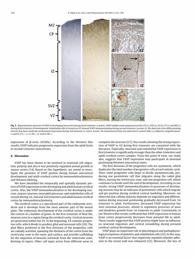
VASP immunoreactivity was a weak staining detected in VZ,SVZ, IZ, CP and MZ during third trimester (Fig. 3b–e). Immunos-taining was considerably distinctive on neuronal and glial fibersall cortical levels (Fig. 3c–e). Interestingly, vascular endothelialcells showed a weak immunoreactivity for VASP compare than thefirst and second trimesters (Fig. 3d). When we evaluated neuronalVASP immunoreactivity, we detected a few differentiating neuronthat have moderate perikarional expression during 3rd trimester(Fig. 3e, insert, arrow).
Cerebral cortex derived from two developmental cell popula-tions: the primordial plexiform layer (PPL) and the cortical plate(CP). The CP split the PPL into two layers (layer 1 at the top andthe subplate at the bottom). CP derived cortical layers are pro-gressively elaborated in mammals and layer I at the top and thesubplate (SP) at the bottom [19]. Almost no VASP immunore-activity was observed in the Layer I. However, the layers II, III,
and IV exhibited a weak VASP immunoreactivity in neuronalcells but the fibers exhibited no immunoreactivity. There was amoderate VASP immunoreaction localized in layer V (Fig. 4). More-over, VASP immunoreactivity was densely observed in vascularendothelial cells in cortical layers of adult brain (Fig. 4). Nega-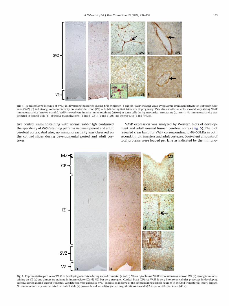
Journal Identification = DN Article Identification = 1435 Date: February 11, 2011 Time: 12:2 am
A. Yaba et al. / Int. J. Devl Neuroscience 29 (2011) 131–136 133
F esterz ring fii rrow)d ; (d, i
ttctt
FtcN
ig. 1. Representative pictures of VASP in developing neocortex during first trimone (SVZ) (c) and strong immunoreactivity on ventricular zone (VZ) cells (d) dummunoreactivity (arrows, e and f). VASP showed very intense immunostaining (aetected in control slide (a) (objective magnifications: (a and b) 2.5×; (c and d) 20×
ive control immunostaining with normal rabbit IgG confirmed
he specificity of VASP staining patterns in development and adulterebral cortex. And also, no immunoreactivity was observed onhe control slides during developmental period and adult cor-exes.ig. 2. Representative pictures of VASP in developing neocortex during second trimester (aining on VZ (e) and almost no staining in intermediate (IZ) (d) MZ, but very strong oerebral cortex during second trimester. We detected very extensive VASP expression ino immunoreactivity was detected in control slide (a) (arrow: blood vessel) (objective m
(a and b). VASP showed weak cytoplasmic immunoreactivity on subventricularrst trimester of pregnancy. Vascular endothelial cells showed very strong VASPin some cells during neocortical structuring (d, insert). No immunoreactivity was
nsert) 40×; (e and f) 40×).
VASP expression was analyzed by Western blots of develop-
ment and adult normal human cerebral cortex (Fig. 5). The blotrevealed clear band for VASP corresponding to 46–50 kDa in bothsecond, third trimesters and adult cortexes. Equivalent amounts oftotal proteins were loaded per lane as indicated by the immuno-a and b). Weak cytoplasmic VASP expression was seen on SVZ (e), strong immunos-n Cortical Plate (CP) (c). VASP is very intense on cellular processes in developingsome of the differentiating cortical neurons in the 2nd trimester (e, insert, arrow).agnifications: (a and b) 2.5×; (c–e) 20×; (e, insert) 40×).
Journal Identification = DN Article Identification = 1435 Date: February 11, 2011 Time: 12:2 am
134 A. Yaba et al. / Int. J. Devl Neuroscience 29 (2011) 131–136
F ter (ad P immn arrow(
ert
4
tmtda
tcrvc
dptnaigavff
ig. 3. Representative pictures of VASP in developing neocortex during third trimesuring third trimester of development. Endothelial cells of vessels on VZ showed VASeuron that have moderate perikarional expression during 3rd trimester (e, insert,a and b) 2.5×; (c–e) 20×; (e, insert) 40×).
xpression of �-actin (43 kDa). According to the Western blotesults, VASP indicates progressive expression from the adult braino second trimester neocortexes.
. Discussion
VASP has been shown to be involved in neuronal cell migra-ion, polarity and also it was positively regulated axonal growth in
ouse cortex [14]. Based on this hypothesis, we aimed to inves-igate the presence of VASP protein during human neocorticalevelopment and adult cerebral cortex by immunohistochemistrynd Western blotting.
We have identified the temporally and spatially dynamic pat-ern of VASP expression in the developing and adult human cerebralortex. Also, the VASP immunolocalization in the developing neu-ons, mature neurons, neurophil processes and endothelial cells ofessels during 1st, 2nd and 3rd trimesters and adult human cerebralortex by immunohistochemistry.
The cerebral cortex is a specialized part of the embryonic ecto-erm and it develops from the most anterior part of the neurallate. The embryonic development of the cerebral cortex is underhe control of a number of genes. In the first trimester of fetal life,eurons arise in a region lining the cerebral cavity. Cortical neuronsre generated within the VZ. In the beginning, VZ contains progen-tor cells, which divide to produce glial and neuronal cells [20]. The
lial fibers produced in the first divisions of the progenitor cellsre radially oriented, spanning the thickness of the cortex from theentricular zone to the outer, pial surface, and provide scaffoldingor the migration of neurons outwards from the ventricular zone,orming its layers. Other cell types arrive from different areas toand b). VASP exhibit weak immunoreactivity VZ (e), SVZ (e), IZ (d), CP (c) and MZ (c)unostaining during second trimester (arrows, d). We detected a few differentiating
). No immunoreactivity was detected in control slide (a) (objective magnifications:
complete the structure [21]. Our results showing the strong expres-sion of VASP in VZ during first trimester are consistent with theliterature. Especially, neuronal and endothelial VASP expression infirst trimester is significantly stronger than the other trimesters andadult cerebral cortex samples. From this point of view, our studyalso, suggests that VASP expression may participate in neuronalpositioning between neocortical zones.
The first divisions of the progenitor cells are symmetric, whichduplicates the total number of progenitor cells at each mitotic cycle.Then, some progenitor cells begin to divide asymmetrically, pro-ducing one postmitotic cell that migrates along the radial glialfibers, leaving the ventricular zone, and one progenitor cell, whichcontinues to divide until the end of development. According to ourresults, strong VASP immunolocalization in processes of develop-ing neurons may be an indicator of postmitotic cells which migrateand get position during cerebral cortical modeling. Moreover, weobserved that cellular polarity depends on migration and differen-tiation during neuronal positioning gradually decreased from 1sttrimester to adult. Furthermore, decreased VASP expression hasseen neuronal processes, attend to regulation processes of axonand dendrites growth from 1st trimester to adult. Consistently, inour Western blot results confirmed that VASP expression in humanbrain cortex progressively decreases from prenatal life to adult.These results suggested that VASP protein might have an influenceon neuronal migration and cortical remodeling during prenatal
cerebral cortical development.VASP plays an important role in physiological and pathophysio-logical responses of platelets and endothelial cells [22]. In this way,VASP is an essential factor that negatively regulates platelet adhe-sion to the vessel wall was enhanced [22]. Moreover, the loss of

Journal Identification = DN Article Identification = 1435 Date: February 11, 2011 Time: 12:2 am
A. Yaba et al. / Int. J. Devl Neuros
Fe(
Vldgih
Ftec
ig. 4. Representative pictures of VASP in adult cortical plate (CP) (a). Vascularndothelial cells represent VASP immunoreactivity in cortical plate of adult brainobjective magnification: (a) 5×; (b–d) 40×).
ASP increases platelet adhesion to the atherosclerotic endothe-ium and subendothelial matrix [22]. Its expression is elevated
uring the endothelial reorganization phase of capillary morpho-enesis in vitro [23]. Our data showing the expression of VASPn the endothelial cells during prenatal life and adult support theypothesis that VASP may have an important role during neurovas-ig. 5. Western blot analysis of the human neocortex obtained from second (II)rimester, third (III) trimester and adult brain. VASP indicates gradually increasexpression from the adult brain to second trimester neocortex. �-Actin provided aontrol ensuring comparable expression.
cience 29 (2011) 131–136 135
cular angiogenesis. However, we also found that endothelial VASPexpression decreases 1st trimester to adult cerebral cortex. Ourresults are partly consistent with data from expression studies ofVASP might have a crucial role in different signal pathways and mayhave interaction with signal molecules for corticogenesis.
Although most interest has focused on understanding VASPexpression in human cerebral cortex, an important but unansweredquestion is whether or not phosphorylation of VASP is essential forthe neocortical development. For answer this question, phospho-rylated form of VASP antibody may demonstrate on same samples.Because it is possible that phosphorylation of VASP on specificsites may be impressive its activity in cell migration and vesselformation. Its proposed involvement in processes of neuronal dif-ferentiation and migration during cerebral cortical modeling. Xieet al. showed that Ang II markedly stimulated VASP phosphoryla-tion in wild-type vascular smooth muscle cells and Ang II-inducedVASP and Akt phosphorylation in a time-dependent manner [24].
Finally, the exclusive VASP staining of endothelial cells in mostparts of the cerebral cortex strongly suggest that VASP also hasadditional functions in the fetal cerebral cortical vasculogenesis. Inour study, the cytoplasmic localization in endothelial cells withinblood vessels and their importance in vascular function are consis-tent with previous report by Markert et al. where they have shownthe high expression levels of VASP in endothelial cells in vivo [25].VASP might have actions in cortical neuronal positioning. Menzieset al. suggested that Mena−/−VASP−/− double mutants die peri-natally and display defects in neurulation, craniofacial structures,and the formation of several fiber tracts in the CNS and periph-eral nervous system [26]. This study also supported that VASP mayhave a crucial role neocortical remodeling and future studies willbe required to elucidate the role of VASP in these processes to indi-cate whether VASP might contribute to the pathology of neonatalbrain development.
In conclusion, VASP is intimately involved in neuronal posi-tioning and vascular formation during corticogenesis. Still, weknow relatively little about how effectors of the cytoskeleton reg-ulate neuronal polarization. Therefore, here we intended to conveywhich actin and microtubule modulators have the potential to reg-ulate polarization and how they could converge on the level ofcytoskeletal dynamics. The present study demonstrates that VASPmay increase neuronal cell migration depend on pregnancy age,participate cerebral cortical remodeling, and may have a role neu-rovascular angiogenesis with stimulation of endothelial migration.As a result, it is strongly possible that there is a relationship betweencerebral cortical remodeling and VASP expression.
Acknowledgments
The authors would like to thank Elif PESTERELI for her excellentscientific collaboration from the Department of Pathology, AkdenizUniversity School of Medicine. This study was partially supportedby the Akdeniz University Research Foundation, Antalya, Turkey.
References
Eccles, 1984. Cerebral Cortex. Plenum, NY.Mountcastle, 1979. The Neurosciences: Fourth Study Program. MIT Press, Cam-
bridge, MA.Sidman, R., 1973. Neuronal migration, with special reference to developing human
brain: a review. Brain Res. 62, 1–35.Rakic, 1974. Neurons in rhesus monkey visual cortex: systematic relation between
time of origin and eventual disposition. Science 183, 425–427.
Rakic, 1972. Mode of cell migration to the superficial layers of fetal monkey neocor-tex. J. Comp. Neurol. 145, 61–83.Halbrugge, Friedrich, Eigenthaler, Schanzenbacher, Walter, 1990. Stoichiomet-
ric and reversible phosphorylation of a 46-kDa protein in human plateletsin response to cGMP- and cAMP-elevating vasodilators. J. Biol. Chem. 265,3088–3093.

Journal Identification = DN Article Identification = 1435 Date: February 11, 2011 Time: 12:2 am
1 euros
K
H
K
G
L
R
R
G
R
W
B
36 A. Yaba et al. / Int. J. Devl N
rause, Dent, Bear, Loureiro, Gertler, 2003. Ena/VASP proteins: regulators of theactin cytoskeleton and cell migration. Annu. Rev. Cell Dev. Biol. 19, 541–564.
olt, Critchley, Brindle, 1998. The focal adhesion phosphoprotein, VASP. Int. J.Biochem. Cell Biol. 30, 307–311.
wiatkowski, Rubinson, Dent, Edward van Veen, Leslie, Zhang, Mebane, Philippar,Pinheiro, Burds, Bronson, Mori, Fassler, Gertler, 2007. Ena/VASP is required forneuritogenesis in the developing cortex. Neuron 56, 441–455.
ertler, Niebuhr, Reinhard, Wehland, Soriano, 1996. Mena, a relative of VASP andDrosophila enabled, is implicated in the control of microfilament dynamics. Cell87, 227–239.
anier, Gertler, 2000. From Abl to actin: Abl tyrosine kinase and associated proteinsin growth cone motility. Curr. Opin. Neurobiol. 10, 80–87.
einhard, Halbrugge, Scheer, Wiegand, Jockusch, Walter, 1992. The 46/50 kDa phos-phoprotein VASP purified from human platelets is a novel protein associatedwith actin filaments and focal contacts. EMBO J. 11, 2063–2070.
ottner, Behrendt, Small, Wehland, 1999. VASP dynamics during lamellipodia pro-trusion. Nat. Cell Biol. 1, 321–322.
oh, Cai, Cepko, Gertler, 2002. Ena/VASP proteins regulate cortical neuronal posi-tioning. Curr. Biol. 12, 565–569.
einhard, Jarchau, Walter, 2001. Actin-based motility: stop and go with Ena/VASPproteins. Trends Biochem. Sci. 26, 243–249.
itte, Bradke, 2008. The role of the cytoskeleton during neuronal polarization. Curr.Opin. Neurobiol. 18, 479–487.
oukhelifa, Parast, Bear, Gertler, Otey, 2004. Palladin is a novel binding partner forEna/VASP family members. Cell Motil. Cytoskel. 58, 17–29.
cience 29 (2011) 131–136
Tanriover, Kayisli, Demir, Pestereli, Karaveli, Demir, 2004. Distribution of N-cadherinin human cerebral cortex during prenatal development. Histochem. Cell Biol.122, 191–200.
Hill, Walsh, 2005. Molecular insights into human brain evolution. Nature 437, 64–67.
Noctor, Flint, Weissman, Dammerman, Kriegstein, 2001. Neurons derived fromradial glial cells establish radial units in neocortex. Nature 409, 714–720.
Rakic, 1988. Specification of cerebral cortical areas. Science 241, 170–176.Massberg, Gruner, Konrad, Garcia Arguinzonis, Eigenthaler, Hemler, Kersting,
Schulz, Muller, Besta, Nieswandt, Heinzmann, Walter, Gawaz, 2004. Enhancedin vivo platelet adhesion in vasodilator-stimulated phosphoprotein (VASP)-deficient mice. Blood 103, 136–142.
Salazar, Bell, Davis, 1999. Coordinate induction of the actin cytoskeletal regulatoryproteins gelsolin, vasodilator-stimulated phosphoprotein, and profilin duringcapillary morphogenesis in vitro. Exp. Cell Res. 249, 22–32.
Xie, Gong, Su, Turk, Guo., 2007. Group VIA phospholipase A2 (iPLA2beta) participatesin angiotensin II-induced transcriptional up-regulation of regulator of g-proteinsignaling-2 in vascular smooth muscle cells. J. Biol. Chem. 282, 25278–25289.
Markert, Krenn, Leebmann, Walter, 1996. High expression of the focal adhesion- and
microfilament-associated protein VASP in vascular smooth muscle and endothe-lial cells of the intact human vessel wall. Basic Res. Cardiol. 91, 337–343.Menzies, Aszodi, Williams, Pfeifer, Wehman, Goh, Mason, Fassler, Gertler, 2004.Mena and vasodilator-stimulated phosphoprotein are required for multipleactin-dependent processes that shape the vertebrate nervous system. J. Neu-rosci. 24, 8029–8038.