Introduction to DFT: VASP - Center for Autonomous Materials ...
Upload
independentCategory
view
3download
0
Developmental Cell, Vol. 7, 571–583, October, 2004, Copyright 2004 by Cell Press
Lamellipodin, an Ena/VASP Ligand, Is Implicatedin the Regulation of Lamellipodial Dynamics
account for only 0.25% of all inositol-containing lipids,making them prime candidates for regulatory signalingmolecules (Rameh and Cantley, 1999). Once polarity is
Matthias Krause,1 Jonathan D. Leslie,1
Mary Stewart,1 Esther M. Lafuente,2
Ferran Valderrama,3 Radhika Jagannathan,1
established, rearrangement of the actin cytoskeletonGeraldine A. Strasser,1 Douglas A. Rubinson,1 Hui Liu,1
provides the driving force for movement of the cell orMichael Way,3 Michael B. Yaffe,1
growth cone up the chemotactic gradient. However, aVassiliki A. Boussiotis,2 and Frank B. Gertler1,*direct connection between polarized phosphoinositides1Department of Biology andand proteins that remodel the actin cytoskeleton re-Center for Cancer Researchmains elusive.MIT
In response to membrane-based signaling events,Cambridge, Massachusetts 02139changes in actin dynamics are induced by a variety of2 Department of Medical Oncologyactin-associated proteins including the Ena/VASP pro-Dana-Farber Cancer Institutetein family. The Ena/VASP family includes three verte-Harvard Medical Schoolbrate members, Mena, VASP, and EVL (see review byBoston, Massachusetts 02115Krause et al., 2003). They share a conserved domain3 Cell Motility Groupstructure: an N-terminal Ena/VASP homology 1 (EVH1)Cancer Research UKdomain, a central proline-rich region, and a C-terminalLincoln’s Inn Fields LaboratoriesEna/VASP homology 2 (EVH2) domain. The EVH2 do-London WC2A 3PXmain mediates tetramerization of Ena/VASP proteinsUnited Kingdomthrough a coiled-coil motif and binds both G- andF-actin. The central proline-rich region harbors bindingsites for SH3 and WW domain-containing proteins and
Summary for the G-actin binding protein Profilin. The EVH1 domainconfers binding to proteins containing a specific proline-
Lamellipodial protrusion is regulated by Ena/VASP rich motif (D/E)(F/L/W/Y)PPPPX(D/E)(D/E) (abbreviatedproteins. We identified Lamellipodin (Lpd) as an Ena/ as FPPPP) (Niebuhr et al., 1997). This interaction is im-VASP binding protein. Both proteins colocalize at the portant for intracellular targeting of Ena/VASP proteinstips of lamellipodia and filopodia. Lpd is recruited to and for their recruitment to receptor/signaling com-EPEC and Vaccinia, pathogens that exploit the actin plexes. For example, Ena/VASP proteins are targeted tocytoskeleton for their own motility. Lpd contains a PH focal adhesions through interaction with FPPPP motifsdomain that binds specifically to PI(3,4)P2, an asym- found in Zyxin and Vinculin (Drees et al., 2000; Gertlermetrically localized signal in chemotactic cells. Lpd’s et al., 1996). FPPPP motifs are also found in the hemato-PH domain can localize to ruffles in PDGF-treated fi- poietic protein Fyb/SLAP/ADAP, the axon guidance re-broblasts. Lpd overexpression increases lamellipodial ceptor sax-3/Robo, and the ActA protein of the intracel-protrusion velocity, an effect observed when Ena/ lular pathogen Listeria monocytogenes (Bashaw et al.,VASP proteins are overexpressed or artificially tar- 2000; Chakraborty et al., 1995; Krause et al., 2000).geted to the plasma membrane. Conversely, knockdown In addition to their focal adhesion localization, Ena/of Lpd expression impairs lamellipodia formation, re- VASP proteins localize to the tips of filopodia and lamelli-duces velocity of residual lamellipodial protrusion, and podia (Lanier et al., 1999; Reinhard et al., 1992; Rottnerdecreases F-actin content. These phenotypes are et al., 1999). A GFP-EVH2 domain fusion localizes tomore severe than loss of Ena/VASP, suggesting that lamellipodia in a broad pattern when expressed in fibro-Lpd regulates other effectors of the actin cytoskeleton blasts (Loureiro et al., 2002). In contrast, a GFP-EVH1in addition to Ena/VASP. domain construct localizes to the tip of lamellipodia
(Bear et al., 2000). This led us to hypothesize that Ena/VASP proteins localize to filopodia and lamellipodilaIntroductionthrough combined function of the EVH1 and EVH2 do-mains and that the EVH1 domain restricts Ena/VASPDirected cell motility is essential for animal developmentlocalization to the tip. However, there were no known
and physiology. Processes such as lymphocyte chemo-proteins containing EVH1 binding motifs that localize to
taxis and axon guidance require changes in motility inthe leading edge.
response to environmental signals. Establishment of cell Leading edge localization is essential for Ena/VASPpolarity is a prerequisite for these processes. Since che- function in regulating random fibroblast motility. Ena/motactic receptors are uniformly distributed, an intracel- VASP proteins regulate the length and branching densitylular event must initiate polarization (Servant et al., 1999). of actin filaments. Overexpression or artificial targetingThe first polarized molecules downstream of many che- of Ena/VASP proteins to the leading edge leads tomotaxis receptors are phosphatidylinositol lipid prod- longer, less branched actin filaments. This results inucts of PI-3 kinase, PI(3,4,5)P3 and its metabolite PI(3,4)P2 faster protruding, but less stable, lamellipodia, which(Servant et al., 2000). These two 3�-phosphoinositides frequently form nonproductive ruffles. In contrast, lamel-
lipodia that lack Ena/VASP protrude slowly but continueto extend for longer times. These steadier protrusions*Correspondence: [email protected]

Developmental Cell572
Figure 1. Lpd Is an Ena/VASP Binding Partner
(A) Amino acid sequence of Lpd. The RA domain (underlined), the PH domain (bold), eight SH3 binding sites (bold and italicized), three Profilinbinding sites (boxed), and six EVH1 binding sites (bold and underlined) are shown.(B) Lpd Northern blot. Arrows show Lpd mRNA at 2, 2.2, 5, and 9 kb.(C) Immunodetection of Lpd in extracts of mouse brain, MVD7 fibroblasts, and CAD cells. The polyclonal Lpd antibody reacts specifically witha single band at approximately 200 kDa.(D and E) Far Western blot analysis with purified, recombinant VASP on GST fusion proteins of Lpd. The location of GST-, Mito- (mitochondrialtargeting anchor-), and EGFP-fusion proteins in Lpd is schematically indicated in (D). A GST fusion protein of the Ena/VASP binding sites inActA serves as positive (GST-ActA-FP4) and GST as negative control. Bound VASP was detected with antibodies against VASP. Note thatbands representing GST-fusion protein degradation products are visible.(F) Peptide spot analysis of full-length Lpd. Peptides were synthesized on a cellulose membrane (SPOTS blot) and incubated with purified,recombinant EVL. Positive spots were detected with antibodies to EVL. Blots were scanned and intensity of positive spots quantified relativeto the negative (ActA APPPP) and positive (ActA FPPPP) control.(G) Coimmunoprecipitation of Lpd and Mena from primary cortical neurons. Extracts of primary cortical neurons were incubated with polyclonalantibodies to Lpd or preimmune serum as control. Immune complexes were collected using protein-A agarose, separated by SDS-PAGE,blotted, and probed with mAbs against Mena. Note that Mena is expressed in three isoforms of 80, 88, and 140 kDa in neurons.

Lamellipodin Regulates Leading Edge Dynamics573
result in faster net cell translocation rates of randomly 2001). Therefore, we propose to rename ALS2CR9 asLamellipodin-S (for “short;” Lpd-S). Northern blot analy-migrating fibroblasts (Bear et al., 2002).sis with a probe encoding part of the N terminus identi-Here we report identification of Lamellipodin (Lpd), anfied three broadly expressed Lpd isoforms of approxi-EVH1 ligand of Ena/VASP proteins. Lpd colocalizes withmately 9, 5, and 2 kb. Lpd is most highly expressed inEna/VASP proteins exclusively at the tips of filopodiabrain, heart, ovary, and developing embryo. An addi-and lamellipodia. In addition, Lpd is recruited to EPECtional 2.2 kb isoform is expressed in embryo, ovary, andand Vaccinia virus, pathogens that use the cellular sig-liver (Figure 1B). EST data and gene prediction programsnaling machinery to exploit the host actin cytoskeletonsuggest that the 5 kb and 9 kb bands code for Lpdfor their motility. Lpd’s PH domain binds specifically tocDNAs with 3� UTRs of varying length. The 2 and 2.2 kbPI(3,4)P2 and is sufficient for plasma membrane recruit-species may represent shorter splice variants, includingment upon treatment with PDGF. Lpd overexpressionLpd-S.produces faster lamellipodial protrusions similar to
We raised and affinity purified rabbit polyclonal anti-those observed when Ena/VASP is overexpressed orbodies against the Lpd C terminus. These antibodiesartificially targeted to the plasma membrane. In contrast,recognize a single 200 kDa band in Western blots of cellknockdown of Lpd expression results in impairment ofand mouse brain extracts (Figure 1C). This suggestslamellipodia formation, a phenotype more severe thanthat the shorter splice variants lack the C terminus andthat caused by loss of all Ena/VASP proteins. We pro-are not recognized by the antibody. In vitro translationpose a model in which Lpd cooperates with Ena/VASPof the Lpd cDNA also produced a 200 kDa band, indicat-proteins and other regulators of actin cytoskeleton toing that this cDNA represents full-length protein (datatransduce signals from chemotaxis receptors into cy-not shown).toskeletal dynamics.
Lpd Binds Ena/VASP Proteins In Vitro and In VivoResultsSince Lpd harbors four clusters of putative EVH1 bindingsites, we tested whether Lpd binds VASP directly. WeIdentification of Lpd as an EVH1 Ligandpurified four GST-fusion proteins, each containing oneTo identify EVH1 ligands, we screened public databasesof the clusters of potential EVH1 binding sites (Figurefor proteins harboring FPPPP motifs. Several candidate1D). The fusion proteins were blotted and overlaid withproteins were identified. One human cDNA, KIAA1681,purified recombinant VASP in a far Western assay. Awas chosen for further characterization based on theGST-fusion protein containing the known EVH1 bindingpresence of a pleckstrin homology (PH) domain, raisingsites from Listeria ActA served as the positive controlthe possibility that it may localize to the plasma mem-and GST alone as the negative control. VASP bound tobrane. Furthermore, KIAA1681 is a mammalian orthologthe second, third, and fourth Lpd GST-fusion proteinsof C. elegans mig-10, a gene identified in a screen forbut not to the first, providing evidence that VASP canneuronal cell migration defects (Manser et al., 1997). Webind Lpd directly (Figure 1E).named KIAA1681 Lamellipodin (Lpd), since it localized
To clarify which putative EVH1 binding sites withinto the tips of lamellipodia and filopodia (see below).Lpd bind Ena/VASP, we used EVL to probe a SPOTSblot of individual EVH1 binding sites. A SPOTS blot con-
Domain Structure and Isoforms of Lpdtains a series of peptides synthesized directly on a mem-
The N terminus of Lpd (aa 1–50) is highly charged and is brane as individual spots. All six potential EVH1 bindingfollowed by a putative coiled-coil motif, Ras-association sites in Lpd showed medium to strong binding to EVL(RA), and PH domains. The C terminus is proline-rich, when compared to the EVH1 binding site from Listeriaharboring eight potential SH3 binding sites, three poten- ActA (Figure 1F). This indicates that all six sites can bindtial Profilin binding sites, and four clusters containing a the EVH1 domain of Ena/VASP proteins. In light of thistotal of six putative EVH1 binding sites (Figure 1A). data, the failure of the first site to interact in the far
During our analysis of Lpd, a related mammalian pro- Western assay could represent misfolding of this GST-tein called RIAM (Rap1 interacting adaptor molecule) fusion protein.appeared in the database, revealing that Lpd belongs To determine whether Ena/VASP and Lpd associate into a family of proteins (see accompanying article by cells, we used anti-Lpd antibodies to immunoprecipitateLafuente et al. [2004]). Both proteins share conserved Lpd from lysates of 293 cells overexpressing both GFP-RA and PH domains followed by a more divergent pro- Lpd and mRFP-EVL. We found that mRFP-EVL coimmu-line-rich C terminus (23.2% amino acid identity). Both C noprecipitated with GFP-Lpd from these lysates (datatermini contain putative binding sites for EVH1 domains, not shown). Since Lpd and Mena are most highly ex-SH3 domains, and Profilin. However, the C terminus of pressed in the nervous system, we repeated the immu-Lpd is 500 amino acids longer than RIAM’s. The two noprecipitation with endogenous protein using extractsproteins also differ at their N termini (29.1% amino acid of cultured primary cortical neurons. We were able toidentity), with RIAM harboring two additional putative coimmunoprecipitate Mena with Lpd (Figure 1G). There-EVH1 binding sites and one additional potential coiled- fore, endogenous Lpd and Ena/VASP proteins are pres-coil domain. ent in protein complexes in vivo.
Database analysis also revealed that Lpd has at leastone splice isoform, an uncharacterized cDNA termed Lpd Colocalizes with Ena/VASP ProteinsALS2CR9. This cDNA was originally cloned as one of at the Tips of Lamellipodia and Filopodia42 genes in the genomic region responsible for amyotro- We used affinity-purified polyclonal antibodies to ana-phic lateral sclerosis 2 (ALS2). However, mutations in lyze Lpd’s localization in cultured cells. In Rat2 fibro-
blasts, WI-38 human lung fibroblasts, and B16-F1ALS2CR9 are not responsible for ALS2 (Hadano et al.,

Developmental Cell574
Figure 2. Lpd Colocalizes with Ena/VASPProteins at the Tips of Filopodia and Lamelli-podia
(A and C) Rat2 cells (A–A″) and CAD cells(C–C″) were cultured on glass coverslips,fixed, and double stained for Lpd (A and C)and Mena (A� and C�).(D) Primary hippocampal neurons (D–D″)were double stained for Lpd (D) and F-actin(D�). Lpd is localized at the tips of filopodia(arrowhead in C and D).(B–B″) Rat2 cells, stably expressing GFP-Lpd(B) were stained for Mena (B�).Colocalization between Lpd and Mena is ob-served in the merged images at the tips oflamellipodia (arrow in A″, B″, and C″) and filo-podia (arrowheads in C–C″). Scale bars in(A)–(D) (for first two panels of each row) equal15 �m.
mouse melanoma cells, Lpd was concentrated at the cytoskeletal proteins associate with these pathogensand have been implicated in F-actin dynamics (Frisch-tips of lamellipodia and filopodia and colocalized with
Ena/VASP proteins (arrow in Figures 2A–2A″ and 4C–4C″ knecht and Way, 2001; Gruenheid and Finlay, 2003). Wesought to determine if such pathogens could recruit Lpdand not shown). Unlike two other known FPPPP-con-by staining infected cells with the anti-Lpd antibody.taining proteins, Zyxin and Vinculin, Lpd was not de-
Both Listeria monocytogenes and Shigella flexneri in-tected in focal adhesions (arrowhead in Figures 2A andvade mammalian cells and use the actin cytoskeleton2A�). Consistent with the immunolocalization, EGFP-to propel them through the cytoplasm on top of F-actintagged full-length Lpd colocalized with endogenoustails (Cossart, 2000). Lpd is not recruited to the interfaceMena at the tips of filopodia and lamellipodia in Rat2between Listeria or Shigella and their F-actin tails (Fig-fibroblasts (arrow in Figures 2B–2B″). In the growthures 3C and 3D).cone-like structures of differentiated CAD cells, a murine
Vaccinia infects cells, multiplies, and is then trans-neuronal cell line, Lpd also colocalized with Ena/VASPported on microtubules to the plasma membrane as anproteins at the distal tips of lamellipodia and filopodiaenveloped virus (Smith et al., 2002). After membrane(arrow and arrowhead, respectively, in Figures 2C–2C″).fusion, the extracellular virus induces an intracellularSimilarly, in primary growth cones, Lpd was found atactin tail that propels it along the surface of the plasmathe tips of filopodia (arrowhead in Figures 2D–2D″) andmembrane (Rietdorf et al., 2001). Interestingly, Lpd local-in spots along the axon shaft.ized to the interface between Vaccinia and its actin tail(arrow in Figure 3A).
Enteropathogenic E. coli (EPEC) does not invade theLpd Colocalizes with Vaccinia Virus and EPECbut Not Shigella and Listeria host cell, but instead docks on the outer membrane
surface and injects bacterial proteins into its host. ThisA variety of pathogens exploit the host actin cytoskele-ton in different ways for their own motility. Several host results in the formation of “actin pedestals” underneath

Lamellipodin Regulates Leading Edge Dynamics575
Figure 3. Lpd Localizes to Vaccinia and EPECbut Not Listeria and Shigella
HeLa cells were infected with (A) Vaccinia vi-rus, (B) Enteropathogenic E. coli (EPEC), (C)Listeria monocytogenes, and (D) Shigellaflexneri. After fixation, cells were stained withanti-Lpd antibodies and specific, dye-labeledprobes for F-actin (phalloidin) and DNA (DAPIstain). High-magnification insets show local-ization of Lpd (arrows in [A] and [B]) at theinterface between Vaccinia or EPEC and therespective F-actin-rich structure. Scale bar in(A) for (A)–(D) equals 20 �m.
the attached bacteria (Celli et al., 2000). Lpd was re- not contain EVH1 binding sites (Lpd-Pro-Mito) did notcause Mena relocalization (Figures 4E–4E″). Therefore,cruited to the interface between EPEC and their actin
pedestals (arrow in Figure 3B). the EVH1 binding motifs within Lpd can recruit Ena/VASP proteins to specific sites within living cells.
Lpd Localization Is Independentof Ena/VASP Proteins The Lpd PH Domain Is Specific for PI(3,4)P2
and Sufficient for Leading Edge TargetingMVD7 cells lack all three Ena/VASP proteins (Bear et al.,2000). Lpd localization in this cell line was indistinguish- upon PDGF Stimulation
To delineate the region of Lpd required for its leadingable from cells expressing Ena/VASP proteins (Figure4A). Ena/VASP localization at the leading edge requires edge localization, we expressed the N terminus of Lpd
encompassing the RA and PH domains as an EGFPboth the EVH1 domain and association of the EVH2domain with free barbed ends of actin filaments. Expo- fusion (N-RA-PH-EGFP, see Figure 1D). N-RA-PH-EGFP
localized to the leading edge in a pattern identical tosure of cells to low doses (150 nM) of cytochalasin D(CD) blocks free barbed ends and displaces Ena/VASP full-length Lpd (Figures 5A–5A″). A smaller fragment
containing just the RA and PH domains also localizedproteins from the leading edge of cells (Bear et al., 2002).Lpd localization was unaffected by the same CD treat- to the leading edge (data not shown). Therefore, the
RA and PH domains together are sufficient for leadingment (Figures 4B–4B″). Control cells treated with DMSO,the solvent for CD, showed normal colocalization of edge targeting.
PH domains are phospholipid binding modules thatMena and Lpd (Figures 4C–4C″). Therefore, leading edgelocalization of Lpd is independent of Ena/VASP proteins can target proteins to the inner leaflet of the plasma
membrane (Fruman et al., 1999). PH domains often pos-and free F-actin barbed ends.Since Lpd localizes independently of Ena/VASP pro- sess specificity for certain phosphoinositides, and vari-
ous signaling pathways can lead to enrichment or deple-teins and can bind to them in vivo, we wondered whetherLpd could direct Ena/VASP localization. To test this hy- tion of specific phosphoinositides in different membrane
compartments. We tested the specificity of the Lpd PHpothesis, we targeted parts of Lpd to mitochondria byfusing them to a mitochondrial membrane anchor (Bear domain in a lipid binding assay in which individual phos-
pholipids were spotted on a membrane and then over-et al., 2000). Expression of the second cluster of Ena/VASP binding sites (Lpd-FP4-2-Mito, see Figure 1D) laid with GST-fusion proteins (Dowler et al., 2002; Kanai
et al., 2001). A GST-Lpd PH domain fusion protein boundcaused Mena to relocalize to the mitochondrial surface(Figures 4D–4D″). A proline-rich region of Lpd that does specifically to PI(3,4)P2. The PX domain of P40 phox, a

Developmental Cell576
(lipid-vesicle) binding. Proteins were tested for their abil-ity to interact with liposomes composed of phosphatidylcholine (PC), phosphatidyl ethanolamine (PE), with orwithout PI(3,4)P2. The PX domain of P47phox, known tobind specifically to PI(3,4)P2 (Kanai et al., 2001), boundto PC/PE/PI(3,4)P2 liposomes as expected. The PH do-main of Lpd bound to PC/PE liposomes containingPI(3,4)P2 (two independent experiments: 15% and 23%binding) but did not bind to control PC/PE liposomes(0% binding).
Quiescent fibroblasts stimulated with PDGF react withimmediate PI(3,4)P2 production and ruffle formation (Ra-meh and Cantley, 1999). To test the hypothesis thatPI(3,4)P2 recruits the PH domain of Lpd to the plasmamembrane, we expressed the PH domain of Lpd as anEGFP fusion (EGFP-PH) in Rat2 fibroblasts. Cells wereserum starved and then treated with PDGF to induceruffling. The EGFP-PH construct was recruited to PDGF-induced membrane ruffles, where it colocalized withendogenous Mena (Figures 5C–5C″).
Lpd Overexpression in Fibroblast IncreasesLamellipodial Protrusion VelocityTo evaluate Lpd function during lamellipodia formation,we analyzed the behavior of lamellipodia over time bykymography (Hinz et al., 1999). Within the kymograph,the slope of a protrusion represents lamellipodial exten-sion velocity.
We first examined the effect of Lpd overexpressionon nascent lamellipodia. To synchronize the activationstate of cells, we analyzed fibroblasts recovering fromazide treatment (Svitkina et al., 1986). Treatment of cellswith sodium azide in the absence of glucose causesrapid depletion of intracellular ATP, causing actin fila-ment depolymerization and cell retraction. Upon azidewashout and restoration of glucose, cells respond withFigure 4. Lpd Localizes Independently of Ena/VASP Proteins to thesynchronized protrusions of circumferential lamellipodia.Leading Edge and Can Recruit Ena/VASP Proteins to Specific Sites
Under these conditions, wild-type Rat2 fibroblasts ex-within the Cell
tend slow wave-like protrusions (Figures 6A and 6B). In(A) The Ena/VASP-deficient cell line MVD7 was fixed and stained withanti-Lpd antibodies. Lpd localizes to the tips of filopodia (arrowhead) contrast, Rat2 fibroblasts stably overexpressing EGFP-and lamellipodia (arrow). Lpd to roughly double the normal expression level (Fig-(B and C) WI-38 lung fibroblasts treated with either 150 nM Cytocha- ure 2B and not shown) formed faster protrusions thatlasin D (CD) in DMSO (B–B″) or with DMSO only (C–C″) were fixed
frequently turned into ruffles (Figures 6C and 6D). Quan-and costained with antibodies against Lpd (B and C) and Mena (B�tification revealed that lamellipodia of Lpd-overexpress-and C�). Merged images of (B) and (B�) or (C) and (C�) are shown ining cells protruded with significantly faster velocity than(B″) and (C″), respectively. Note that Mena but not Lpd is delocalized
by treatment with CD (arrows). wild-type Rat2 fibroblasts (Figure 6E). A similar pheno-(D and E) Lpd can recruit Ena/VASP proteins to specific sites within type was also observed when cells were analyzed undercells. The second cluster of the EVH1 binding sites in Lpd (Lpd- steady-state conditions without azide treatment (FigureFp4-2Mito) (D–D″) or the proline-rich region behind the PH domain
6F). Importantly, the Lpd overexpression phenotype re-of Lpd (Lpd-Pro-Mito) (E–E″) was fused to DSred2 as a marker andsembles the effects of Ena/VASP overexpression or arti-to a mitochondrial membrane anchor (see Figure 1D for location official targeting of Ena/VASP to the plasma membranefragments in Lpd). Stably transfected Rat2 fibroblasts were fixed
and stained with anti-Mena antibodies (D� and E�). Mena is only (Bear et al., 2002).corecruited to mitochondria by the Lpd-FP4-2 fragment (yellow To test whether the Lpd overexpression phenotypecolor in D″). is dependent on Ena/VASP proteins, we neutralized Ena/Scale bars in (A), (C) (for all [B] and [C] panels), and (D�) (for all [D]
VASP function in the Lpd-overexpressing Rat2 fibro-and [E] panels) equal 15 �m.blasts and control, wild-type Rat2 cells. We used a strat-egy to deplete all Ena/VASP proteins from their normalintracellular locations by expressing the EVH1 bindingphosphoinositide binding domain specific for PI(3)P,sites from ActA fused to a mitochondrial membrane an-was used as a positive control and GST alone as thechor (FPPPP-Mito), thereby artificially recruiting Ena/negative control (Figure 5B).VASP proteins to mitochondria (Bear et al., 2000). Rat2The association of the Lpd PH domain with PI(3,4)P2
was verified in an ultracentrifugation assay for liposome fibroblasts in which Ena/VASP function had been blocked

Lamellipodin Regulates Leading Edge Dynamics577
Figure 5. Lpd’s PH Domain Binds to PI(3,4)P2
and Is Sufficient for Leading Edge Targetingupon PDGF Treatment of Serum-Starved Fi-broblasts
(A) An EGFP-fusion protein with the N termi-nus of Lpd including the RA and PH domainwas stably expressed in Rat2 fibroblasts.After fixation, cells were stained with anti-Mena antibodies (arrow in A�). Note thatEGFP-N-term-RA-PH localizes to the leadingedge (arrow in A). (A″) Colocalization betweenthe EGFP fusion protein and Mena is ob-served in the merged images at the tips oflamellipodia (arrow in A″).(B) A protein-lipid binding assay was usedto analyze the ability of purified GST-fusionproteins to bind a variety of phosphoinosi-tides. Serial dilutions of the indicated phos-phoinositides (200, 100, 50, 25, 12.5, and 6.25pmol) were spotted on to HybondC-mem-branes and incubated with GST-fusion pro-teins. After washing the membranes, boundproteins were detected with anti-GST anti-bodies. The PX domain of p40phox servedas the positive control (specific for PI(3)P)and GST alone as negative control. The PHdomain of Lpd binds specifically only toPI(3,4)P2.(C) The localization of an EGFP-fusion proteinwith the PH domain of Lpd (EGFP-Lpd-PH)was tested in PDGF-stimulated Rat2 fibro-blasts. Rat2 cells, stably expressing EGFP-Lpd-PH, were serum starved, treated with
PDGF, fixed, and stained with anti-Mena antibodies. EGFP-Lpd-PH is recruited to PDGF-induced ruffles were it colocalizes with Mena (arrows).Scale bar in (C) (for A and C) equals 15 �m.
extended lamellipodia with a significantly reduced ve- by a decrease in total area of the Lpd-KD cells (notshown). Analysis of the dynamics of the residual lamelli-locity compared to wild-type Rat2 cells or Rat2 cells
expressing a control construct (APPPP-mito) that does podia in the Lpd-KD cell line by kymography showedthat these lamellipodia protruded with a significantlynot bind to Ena/VASP proteins (Bear et al., 2000). Ex-
pression of FPPPP-mito in Lpd-overexpressing Rat2 lower velocity than control (Figures 7B–7D).Actin polymerization directly underneath the plasmareduced the velocity of lamellipodial protrusions signifi-
cantly (Figure 6F). Therefore, the effect of Lpd overex- membrane provides the driving force for lamellipodialprotrusion. We fixed and stained the Lpd-KD and controlpression on lamellipodial protrusion velocity can be sup-
pressed by blocking Ena/VASP function. cell lines with fluorescently labeled phalloidin to analyzethe integrity of the actin cytoskeleton. Interestingly, theF-actin content of the Lpd-KD cell line appeared highly
Knockdown of Lpd Expression Impairs reduced (Figures 7F and 7G). Quantification by spectro-Lamellipodia Formation fluorometry revealed that total F-actin content in Lpd-We used lentiviral-mediated expression of a Lpd-spe- KD cells was indeed reduced compared to control (twocific shRNA to stably knockdown Lpd expression. We independent assays in triplicate: reduced by 26.8% �analyzed B16-F1 mouse melanoma cells, as these cells 14.6% compared to control). However, total actin con-do not express the related RIAM protein (data not tent appeared to be comparable in knockdown andshown). As a negative control, we used a shRNA directed control cell lines as judged by Western blot analysisagainst the corresponding rat Lpd sequence, which dif- (Figure 7E). This suggests that the ratio of F- to G-actin isfers in several base pairs. The Lpd knockdown cell line reduced in the Lpd knockdown cells. We used platinum(Lpd-KD) showed highly reduced expression of Lpd replica electron microscopy to analyze the underlyingcompared to a control cell line expressing the rat Lpd actin ultrastructure in more detail. A thin rim of normalshRNA (control) (Figure 7E). When plated on laminin, dense meshwork of actin filaments was visible directlyB16-F1 cells form large lamellipodia that can be labeled underneath the plasma membrane of Lpd-KD cells; how-with an antibody against N-WASP. Strikingly, Lpd-KD ever, large areas deeper inside the cell periphery con-cells formed very few lamellipodia (Figures 7A and 7A�). tained only thick bundles of F-actin and were devoid ofQuantification of the ratio between the perimeter of la- a dense F-actin meshwork. In contrast, the control cellmellipodium to total perimeter of the cell revealed that line showed a dense actin filament meshwork through-reduction in Lpd expression reduced lamellipodia for- out the cell periphery (Figures 7H and 7I and enlarged
areas in 7H� and 7I�).mation significantly (Figure 7A″). This was accompanied

Developmental Cell578
Figure 6. Kymograph Analysis of Rat2 CellsOverexpressing EGFP-Lpd
(A–D) Time-lapse movies of Rat2 cells (A andB) and Rat2 cells overexpressing EGFP-Lpd(C and D) recovering from ATP starvationwere analyzed by drawing a one pixel wideline at a 90� angle across a lamellipodium inthe direction of protrusion (line in individualframe of movie in [A] and [C]). The image fromthis line was copied from each frame of amovie and pasted along the x axis to generatea composite kymograhic image (B and D).The black line in (B) and (D) indicates thelocation of frames in kymographs. Within thekymograph, a steeper slope of a protrusioncorresponds to higher velocity rates for la-mellipodia protrusion. Scale bar for (A) and(C) and scale vectors for (B) and (D) are shownin top images.(E) Box and whisker plots for velocity of indi-vidual nacsent protrusion events in wild-typeand EGFP-Lpd overexpressing Rat2 cells re-covering from ATP starvation. Top and bot-tom of box represent 75th and 25th quartile,and whiskers 10th and 90th percentiles, re-spectively. Dot indicates mean and middleline of box median. Data for wild-type cellsis from 570 and for EGFP-Lpd overexpressingRat2 cells from 934 protrusion events. Aster-isk indicates significant difference in Stu-dent’s t test (p � 0.001).(F) Box and whisker plots for velocity of individ-ual protrusions from cells under steady-stateconditions. Rat2 cells expressing APPPP-Mito(control) or FPPPP-Mito (to block Ena/VASPfunction) alone or along with Lpd overexpres-sion are shown. Brackets with asterisks indi-cate statistically significant differences be-tween data sets using ANOVA (p � 0.005).
Discussion a phenotype not observed in cells devoid of all Ena/VASP proteins. This suggests that Lpd signals to other
Based on our results, we propose that Lpd acts as a regulators of the actin cytoskeleton in addition to Ena/convergence point between upstream signaling path- VASP proteins.ways and cytoskeletal remodeling at the plasma mem-brane. This hypothesis is supported by the recruitment Lpd Contains High-Affinity Binding Sitesof Lpd to two pathogens, EPEC and Vaccinia, that ex- for the EVH1 Domain of Ena/VASPploit cellular signaling processes at the plasma mem- The EVH1 binding sites within Lpd match the canonicalbrane to reorganize the actin cytoskeleton for their own motif for EVH1 binding, found in the Listeria monocyto-motility. Furthermore, Lpd is concentrated at the tip of genes protein ActA. Two of the motifs in Lpd containgrowth cone filopodia, a structure that plays a key role a phenylalanine followed by four consecutive prolines,in guidance signal detection and growth cone motility. flanking acidic amino acids, and a leucine in position 13Lpd is likely to receive signals directly from both phos- (D1S2D3F4P5P6P7P8P9E10T11D12L13). This combination hasphoinositides through its PH domain and Ras superfam- been shown to have a high binding affinity for EVH1ily proteins through its RA domain. The Lpd PH domain domains and has not been found in any other mamma-selectively binds PI(3,4)P2, a phosphoinositide associ- lian EVH1 ligand (Ball et al., 2000; Niebuhr et al., 1997).ated with polarization signals in a variety of cell types.
Furthermore, Lpd and RIAM, with a total of 6 EVH1One way that Lpd is linked to cytoskeletal dynamics
binding sites each, contain the largest number of EVH1is through Ena/VASP proteins. Lpd associates with Ena/
binding sites of any known protein.VASP by direct binding and can be detected in proteincomplexes from primary neurons. Ena/VASP and Lpd
Lpd Localizes to the Tips of Lamellipodiacolocalize in filopodia and lamellipodial tips, and bothand Filopodiatypes of proteins play critical roles in regulating lamelli-Lpd and Ena/VASP proteins colocalize in migrating cellspodial dynamics. Lpd overexpression induces an in-at the tips of lamellipodia and filopodia. Lpd’s localiza-crease in lamellipodial ruffling, similar to phenotypestion is independent of Ena/VASP proteins and freethat are observed when Ena/VASP activity is elevatedbarbed ends of actin filaments. Furthermore, redistribu-in mammalian cells, and the Lpd overexpression pheno-tion of the Lpd EVH1 binding sites to the surface oftype requires Ena/VASP function. Interestingly, knock-
down of Lpd led to impairment of lamellipodia formation, mitochondria resulted in substantial corecruitment of

Lamellipodin Regulates Leading Edge Dynamics579
Figure 7. Knockdown of Lpd Expression Results in Impairment of Lamellipodia Formation
(A) B16-F1 cell lines stably expressing a control shRNA (A) or an shRNA against mouse Lpd (A�) were plated on laminin and fixed and stainedwith an N-WASP antibody (arrow in [A] and arrowhead indicating residual lamellipodia in [A�]). (A″) Quantification of the ratio of the length oflamellipodia to length of perimeter of cell depicted as box and whiskers plot (see Figure 6). Data is from 100 cells each. Nonoverlapping 95%convidence intervals are indicated by asterisks.(B–D) Time-lapse movies of B16-F1 Lpd KD or control cells were analyzed by kymography (see Figure 6 for description of method). (D) Boxand whisker plots for velocity of lamellipodial protrusion (t test, p � 0.0001, Lpd knockdown 172 and control 200 protrusion events). Nonoverlap-ping 95% convidence intervals are indicated by asterisks.(E) Western blot analysis of B16-F1 Lpd knockdown and control cell line. The upper part of the blot was probed with an antibody to Lpd andthe lower part with an antibody against Actin.(F–I) Analysis of F-actin in B16-F1 control and Lpd knockdown cell lines. Cells were plated on laminin and fixed, and F-actin was stained withAlexa 647 phalloidin (F and G) or processed for platinum replica electron microscopy (H and I). Scale bar in (A) (for [A], [A�], [F], and [G]) equals15 �m; scale bars in (B) (for [B] and [C] panels), (H) (for [H] and [I]), and (H�) (for [H�] and [I�]) represent 10 �m.
Developmental Cell580
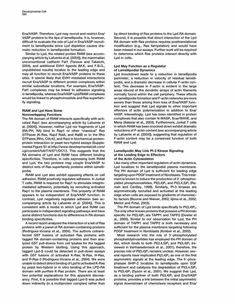
Ena/VASP. Therefore, Lpd may recruit and restrict Ena/ by direct binding of Ras proteins to the Lpd RA domain.Second, it is possible that direct interaction of the LpdVASP proteins to the tips of lamellipodia. It is, however,
difficult to evaluate the role of Lpd in Ena/VASP recruit- RA domain with Ras proteins requires posttranslationalmodification (e.g., Ras farnyslation) and would havement to lamellipodia since Lpd depletion causes dra-
matic reduction in lamellipodial formation. been missed in our assays. Further work will be requiredto determine which Ras proteins interact directly withSimilar to Lpd, the related protein RIAM (see accom-
panying article by Lafuente et al. [2004]), the mammalian Lpd in cells.unconventional cadherin Fat1 (Tanoue and Takeichi,2004), and additional EVH1 ligands (M.K. and F.B.G., Lpd May Function as a Regulatorunpublished results) localize to the leading edge and of Lamellipodial Dynamicsmay all function to recruit Ena/VASP proteins to these Lpd knockdown leads to a reduction in lamellipodiasites. It seems likely that EVH1-mediated interactions perimeter, a reduction in velocity of residual lamelli-recruit Ena/VASP to different protein complexes within podia, and a dramatic decrease in cellular F-actin con-similar subcellular locations. For example, Ena/VASP- tent. This decrease in F-actin is evident in the largeFat1 complexes may be linked to adhesion signaling areas devoid of the dendritic arrays of actin filamentsin lamellipodia, whereas Ena/VASP-Lpd/RIAM complexes normally found within the cell periphery. These effectswould be linked to phosphoinositide and Ras superfam- on lamellipodia formation and F-actin networks are moreily signaling. severe than those arising from loss of Ena/VASP func-
tion and suggest that Lpd signals to other importanteffectors of actin polymerization in addition to Ena/RIAM and Lpd Have SomeVASP. Interestingly, Lpd has been identified in proteinNonoverlapping Functionscomplexes that also contain N-WASP, Scar/WAVE, andThe RA domain of RIAM interacts specifically with acti-Mena (Salazar et al., 2003). Furthermore, Jurkat T cellsvated Rap1 (see accompanying article by Lafuente etin which RIAM has been knocked down exhibit dramatical. [2004]). However, neither Lpd nor fragments of Lpdreductions of F-actin content (see accompanying article(RA-PH, RA) bind to Rap1 or other “classical” Rasby Lafuente et al. [2004]), suggesting that regulation ofGTPases (K-Ras, Rap2 RalA, and RalB) or to the RhoF-actin content may be a conserved function of bothGTPases (Rho, Cdc42, and Rac) in biochemical protein-RIAM and Lpd.protein interaction or yeast two-hybrid assays (Supple-
mental Figure S1 at http://www.developmentalcell.com/cgi/content/full/7/4/571/DC1/). This suggests that the Lamellipodin May Link PI-3 Kinase Signaling
at the Leading Edge to EffectorsRA domains of Lpd and RIAM have different bindingspecificities. Therefore, in cells expressing both RIAM of the Actin Cytoskeleton
Like many other important regulators of actin dynamics,and Lpd, the two proteins may couple Ena/VASP todistinct sets of Ras superfamily proteins within lamelli- Lpd localizes to the lamellipodial plasma membrane.
The PH domain of Lpd is sufficient for leading edgepodia.RIAM and Lpd also exhibit opposing effects on cell targeting upon PDGF treatment of fibroblasts. This treat-
ment is known to induce the production of 3�-phosphor-adhesion. RIAM positively regulates adhesion. In JurkatT cells, RIAM is required for Rap1 to stimulate integrin- ylated phosphoinositides, PI(3,4)P2 and PI(3,4,5)P3 (Ra-
meh and Cantley, 1999). Similarly, PI-3 kinases aremediated adhesion, potentially by recruiting activatedRap1 to the plasma membrane. This property of RIAM asymmetrically recruited and activated at the leading
edge when cells are exposed to gradients of chemotac-appears to be independent of Ena/VASP function. Incontrast, Lpd negatively regulates adhesion (see ac- tic factors (Bourne and Weiner, 2002; Iijima et al., 2002;
Merlot and Firtel, 2003).companying article by Lafuente et al. [2004]). This isconsistent with a model in which Lpd and RIAM can The PH domain of Lpd binds specifically to PI(3,4)P2.
The only other known proteins that possess a PH domainparticipate in independent signaling pathways and havesome distinct functions due to differences in RA domain specific for PI(3,4)P2 are TAPP1 and TAPP2 (Dowler et
al., 2000). Similar to our observation for Lpd, the PHbinding specificities.A recent report analyzed the interaction of a set of Ras domain of TAPP1 and TAPP2 is both necessary and
sufficient for the plasma membrane targeting followingproteins with a panel of RA domain-containing proteins(Rodriguez-Viciana et al., 2004). The authors cotrans- PDGF treatment in fibroblasts (Kimber et al., 2002).
Most research into the role of 3�-phosphorylatedfected GST fusions of activated Ras proteins withtagged RA domain-containing proteins and then ana- phosphatidylinositides has employed the PH domain of
Akt, which binds to both PI(3,4,5)P3 and PI(3,4)P2 (re-lyzed GST pull-downs from cell lysates for the taggedprotein by Western blotting. Using this approach, viewed in Vanhaesebroeck et al., 2001); therefore, the
precise role of PI(3,4)P2 remains unclear. However, sev-tagged Lpd-S could be detected in pull-down assayswith GST fusions of activated K-Ras, N-Ras, H-Ras, eral reports have implicated PI(3,4)P2 as one of the first
asymmetric signals at the leading edge. The 5�-phos-and R-Ras-3 (Rodriguez-Viciana et al., 2004). We wereunable to detect direct binding of Lpd to activated K-Ras phatase SHIP-2 localizes to lamellipodia upon EGF
treatment and catalyzes the degradation of PI(3,4,5)P3by yeast two-hybrid or in vitro binding of the LPD-RAdomain with purified K-Ras protein. There are at least to PI(3,4)P2 (Dyson et al., 2001). We suggest that Lpd,
as a binding partner of both PI(3,4)P2 and Ena/VASPtwo potential explanations for this apparent discrep-ancy. First, it is possible that tagged Lpd-S was pulled proteins, provides a link between the initial asymmetric
signal downstream of chemotaxis receptors and Ena/down indirectly (in a multiprotein complex) rather than

Lamellipodin Regulates Leading Edge Dynamics581
and maintained for 24–48 hr in glial-conditioned media (Neurobasal,VASP proteins as well as other regulators of actin poly-2% (v/v) B27 supplement, 1% (v/v) Glutamine (GIBCO-BRL).merization.
Cell were fixed either with methanol for 10 min at �20�C or with4% (w/v) paraformaldehyde in PHEM (60 mM PIPES, 25 mM HEPES,Experimental Procedures10 mM EGTA, 2 mM MgCl2, 120 mM sucrose, 4% (w/v) paraformalde-hyde [pH 7.3]) for 20 min at room temperature and permeabilizedMolecular Cloning and Northern Blot Analysiswith 0.1% (v/v) Triton-x-100 in TBS. The following antibodies wereSubcloning and PCR were performed using standard methods. ESTused: mAb anti Mena A351F7D9 (Lebrand et al., 2004), pAb anti-clones were purchased from Genome Systems/Incyte (Beverly, MA),Lpd #3917, anti-N-WASP pAb (Rohatgi et al., 1999). Images wereand the cDNA of KIAA1681 was a gift of T. Nagase (Kazusa DNAcollected on either a Deltavision system (Applied Precision) or aResearch Institute, Kisarazu, Chiba, Japan). Full-length human LpdNikon TE2000 microscope equipped with a Yokagawa spinning diskcDNA (AY494951) was obtained by amplifying the 5� part of ESTconfocal head (McBain Instruments) and an Orca-ER digital cameraclone (IMAGE 2814736) and cloning it into the unique Bgl II site at(Hamamatsu, Japan) and processed using Photoshop 7.0 (Adobe).the N terminus of the KIAA1681 cDNA. EGFP expression constructsCD treatment and kymography were performed as described (Bearwere cloned by amplifying full-length Lpd, N-term-RAPH (aa 1–529),et al., 2002).RAPH (aa 254–529), and PH (aa 383–547) fragments and cloning
them into pML2(EGFP-N1) and/or pEGFP-N1/C1. pML2(EGFP-N1)Platinum Replica Electron Microscopywas derived by subcloning EGFP and multiple cloning sites fromCells were plated on laminin-coated coverslips and processed forpEGFP-N1 into pMSCVneo (Clontech). The Mito targeting con-electron microscopy as described (Svitkina and Borisy, 1998).structs were made by cloning Lpd fragments aa 544–726 (Lpd-
pro-Mito) and aa 888–1061 (Lpd-FP4-2-Mito) into a retroviral vectorVaccinia InfectionpBLT-2. The Lpd knockdown constructs were made by cloning Lpd-HeLa cells were infected with the Vaccinia virus Western Reservespecific oligonucleotides (mouse Lpd: forward, 5�-tgcgtcaagtcacagstrain at a MOI of 1 pfu per cell in serum-free MEM for 1 hr at 37�C.aaccattcaagagatggttctgtgacttgacgctttttggaaagaattcg-3�; reverse,Cells were incubated with MEM � 10% FCS for 8 hr and then fixed.5�-tcgacgaattctttccaaaaagcgtcaagtcacagaaccatctcttgaatggttctgt
gacttgacgca-3�; rat Lpd: forward, 5�-TgcgccaagtcacagaacccTTCAAEPEC, Listeria, and Shigella InfectionsGAGAgggttctgtgacttggcgcTTTTTGGAAAGAATTCG-3�; reverse,HeLa cells in MEM � 10% FCS were infected with 20 �l of an5�-tcgaCGAATTCTTTCCAAAAAgcgccaagtcacagaacccTCTCTTGAAovernight bacterial culture of Entheropatogenic Escherichia coligggttctgtgacttggcgcA-3�) into pLL3.7 and used to generate lentivirus(EPEC) (strain E2348/69), Shigella flexneri (strain SC301), and Liste-as described (Rubinson et al., 2003). All constructs were verified byria monocytogenes (strain 10403S). After 4 hr the cells were fixed.DNA sequencing.
The murine multiple tissue Northern blot (Ambion, Austin, TX) wasprobed with a Not I-Sac I, 520 bp, N-terminal fragment of the EST Protein Interaction Assays
SPOTS membranes (Sigma Genosys) were overlayed as describedclone AW490888.(Niebuhr et al., 1997) with purified recombinant EVL. Bound EVLwas detected using monoclonal antibodies to EVL (#84H1) and theProteins and Antibody ProductionECL kit (Amersham). Film was scanned and analyzed with Image-Fragments encoding amino acids aa 383–547 (Lpd-GST-PH), aaQuant (Molecular Dynamics).726–888 (Lpd-GST-Fp4-1), aa 888–1060 (Lpd-GST-FP4-2), aa 1026–
Protein-lipid overlay and liposome binding assays were done as1126 (Lpd-GST-FP4-3), and aa 1126–1250 (Lpd-GST-FP4-4) weredescribed (Kanai et al., 2001; Kavran et al., 1998). Ras and Rhogenerated by PCR and cloned into the pGEX-6P1 vector (AmershamGTPase in vitro binding studies were done as described (Vojtek etPharmacia Biotech). Full-length cDNAs from Ras and Rho GTPasesal., 1993).were amplified from EST clones and cloned into pGEX-6P1. GST-
fusion proteins were purified on glutathione agarose (Pierce). Immo-bilized Lpd-GST-FP4-2 was digested with Prescission protease F-Actin Measurement
F-actin was quantified as described (Diakonova et al., 2002). Briefly,(Amersham Pharmacia Biotech) and used to raise polyclonal rabbitantiserum #3917 (Covance), which was affinity purified using cross- 7 � 105 cells were plated per well on laminin-coated 6-well dishes.
After 5 hr, cells were fixed and stained for 30 min in the dark withlinked Lpd-GST-FP4-2 agarose.Maltose binding protein fusion proteins were generated by ampli- 250 nM rhodamine-phalloidin, 0.1% TritonX-100, 4% PFA in PHEM.
After 3 washes with 5 ml PBS, cells were scraped off with 700fying RA domains of Lpd (aa 245–394) or c-Raf-1 (aa 26–175 RIP3)(Vojtek et al., 1993) and cloning into pMAL-c2G (NEB) and were �l Methanol and extracted for 24 hr at �20�C. DNA content was
measured from each sample by using PicoGreen (Molecular Probes)purified on Amylose resin following the instructions of the vendor(NEB). Purified, recombinant VASP was a gift of Melanie Barzik (MIT, and used to normalize for variations in cell numbers. Extracted rhoda-
mine-phalloidin was measured using a spectrofluometer (FluoroMax-3,Cambridge, MA).Jobin Yvon, Inc.) (excitation 540 nm and emission 565 nm).
Cell Culture and Fluorescence MicroscopyB16-F1 (ATCC CRL-6323), WI-38 (ATCC CCL-75), and Rat2 (ATCC Immunoprecipitation and Western Blot Analysis
Neocortex was dissected from E16 mouse embryos and plated atCRL-1764) cells were cultured as recommended by ATCC. MVD7
cells were cultured as described (Bear et al., 2000). CAD cells were 11 � 106 cells/dish on poly-D-lysine-coated 60 mm dishes. Neuronswere cultured in Neurobasal, 2% (v/v) B27 supplement, 1% (v/v)cultured as described (Qi et al., 1997). Retroviral packaging, infec-
tion, and fluorescence-activated cell sorting (FACS) was done as Glutamine (GIBCO-BRL) for 48 hr and lysed with ice-cold GST-buffer(50 mM Tris-Cl, 200 mM NaCl, 1% (v/v) IGEPAL CA-630, 2 mM MgCl2,described (Loureiro et al., 2002). Lpd knockdown cell lines were
obtained by infection with lentivirus expressing Lpd-specific shRNA 10% (v/v) glycerol [pH 7.4]), phosphatase inhibitors (1 mM Na3VO4,10 mM NaF), and a protease inhibitor cocktail (Roche completeand CMV promotor driven EGFP and sorted by FACS for high EGFP
expression. Since cell population drifted extensively, cells were protease inhibitor tablets without EDTA). Lysates were incubatedfor 30 min on ice and centrifuged for 15 min at 18,000 � g atused for only two passages after the last FACS sort. Fugene (Roche)
or standard calcium phosphate were used for transfection of CAD 4�C. Supernatants (700 �g/reaction) were precleared with protein-Aagarose and immunoprecipitated with polyclonal antiserum #3917cells and 293, respectively. For immunofluorescence analysis, cells
were plated on acid-washed coverslips coated with either 10 �g/ml against Lpd or preserum as control, and protein-A agarose (Pierce).After washing with GST-buffer, immunoprecipitated proteins werefibronectin or 25 �g/ml laminin.
Primary hippocampal neurons were prepared from mouse em- separated on 7.5% SDS-PAGE gels and transferred to Immobilon-Pmembranes (Millipore). Blots were probed with the anti-Mena mAbbryos (day E16) as described for rat embryos (day E18) (Goslin and
Banker, 1989). Glial cultures were prepared from P1 mouse cortex A351F7D9 and an HRP-coupled donkey anti-mouse secondary anti-body (Jackson Immunologicals) and developed using the ECL kitand hippocampus as described (Voutsinos-Porche et al., 2003). Hip-
pocampal neurons were plated on poly-D-lysine-coated coverslips (Amersham-Pharmacia Biotech).

Developmental Cell582
Yeast Two-Hybrid Analysis (2001). The SH2-containing inositol polyphosphate 5-phosphatase,SHIP-2, binds filamin and regulates submembraneous actin. J. CellFull-length Ras and Rho GTPases were cloned into the “bait” vector
pLexA (Clontech) and either full-length RIAM and Lpd or a fragment Biol. 155, 1065–1079.covering the RA-PH domains (aa 150–430 for RIAM, aa 245–529 for Frischknecht, F., and Way, M. (2001). Surfing pathogens and theLpd) were cloned into the “prey” vector pB42AD (Clontech). Plas- lessons learned for actin polymerization. Trends Cell Biol. 11, 30–38.mids were transformed into yeast strains EGY48 and EGY191 (In-
Fruman, D.A., Rameh, L.E., and Cantley, L.C. (1999). Phosphoinosi-vitrogen). Transformants were plated on plates with selection media
tide binding domains: embracing 3-phosphate. Cell 97, 817–820.(SD/Gal/Raf/-His/-Trp/-Ura/-Leu) containing X-gal (20 �g/ml) for
Gertler, F.B., Niebuhr, K., Reinhard, M., Wehland, J., and Soriano,blue and white screening.P. (1996). Mena, a relative of VASP and Drosophila Enabled, is impli-cated in the control of microfilament dynamics. Cell 87, 227–239.AcknowledgmentsGoslin, K., and Banker, G. (1989). Experimental observations on the
We thank T. Nagase (Kazusa DNA Research Institute, Kisarazu, development of polarity by hippocampal neurons in culture. J. CellChiba, Japan) for cDNA of KIAA1681, R. Rohatgi and M. Kirschner Biol. 108, 1507–1516.(Harvard Medical School) for anti-N-WASP antibody, M. Barzik for Gruenheid, S., and Finlay, B.B. (2003). Microbial pathogenesis andpurified VASP, Angelique Dousis for technical assistance, and Adam cytoskeletal function. Nature 422, 775–781.Kwiatkowski and Joe Loureiro for comments on the manuscript.
Hadano, S., Hand, C.K., Osuga, H., Yanagisawa, Y., Otomo, A.,Funding was provided by a Keck Distinguished Scholar award and
Devon, R.S., Miyamoto, N., Showguchi-Miyata, J., Okada, Y., Singa-NIH GM68678 to F.B.G. D.A.R. was supported by a Ludwig Fel-
raja, R., et al. (2001). A gene encoding a putative GTPase regulatorlowship.
is mutated in familial amyotrophic lateral sclerosis 2. Nat. Genet.29, 166–173.
Received: December 11, 2003Hinz, B., Alt, W., Johnen, C., Herzog, V., and Kaiser, H.W. (1999).Revised: May 25, 2004Quantifying lamella dynamics of cultured cells by SACED, a newAccepted: July 19, 2004computer-assisted motion analysis. Exp. Cell Res. 251, 234–243.Published: October 11, 2004Iijima, M., Huang, Y.E., and Devreotes, P. (2002). Temporal and
References spatial regulation of chemotaxis. Dev. Cell 3, 469–478.
Kanai, F., Liu, H., Field, S.J., Akbary, H., Matsuo, T., Brown, G.E.,Ball, L.J., Kuhne, R., Hoffmann, B., Hafner, A., Schmieder, P., Volk- Cantley, L.C., and Yaffe, M.B. (2001). The PX domains of p47phoxmer-Engert, R., Hof, M., Wahl, M., Schneider-Mergener, J., Walter, and p40phox bind to lipid products of PI(3)K. Nat. Cell Biol. 3,U., et al. (2000). Dual epitope recognition by the VASP EVH1 domain 675–678.modulates polyproline ligand specificity and binding affinity. EMBO
Kavran, J.M., Klein, D.E., Lee, A., Falasca, M., Isakoff, S.J., Skolnik,J. 19, 4903–4914.E.Y., and Lemmon, M.A. (1998). Specificity and promiscuity in phos-
Bashaw, G.J., Kidd, T., Murray, D., Pawson, T., and Goodman, C.S. phoinositide binding by pleckstrin homology domains. J. Biol. Chem.(2000). Repulsive axon guidance: Abelson and Enabled play oppos- 273, 30497–30508.ing roles downstream of the roundabout receptor. Cell 101, 703–715.
Kimber, W.A., Trinkle-Mulcahy, L., Cheung, P.C., Deak, M., Marsden,Bear, J.E., Loureiro, J.J., Libova, I., Fassler, R., Wehland, J., and L.J., Kieloch, A., Watt, S., Javier, R.T., Gray, A., Downes, C.P., etGertler, F.B. (2000). Negative regulation of fibroblast motility by Ena/ al. (2002). Evidence that the tandem-pleckstrin-homology-domain-VASP proteins. Cell 101, 717–728. containing protein TAPP1 interacts with Ptd(3,4)P2 and the multi-Bear, J.E., Svitkina, T.M., Krause, M., Schafer, D.A., Loureiro, J.J., PDZ-domain-containing protein MUPP1 in vivo. Biochem. J. 361,Strasser, G.A., Maly, I.V., Chaga, O.Y., Cooper, J.A., Borisy, G.G., 525–536.and Gertler, F.B. (2002). Antagonism between Ena/VASP proteins Krause, M., Sechi, A.S., Konradt, M., Monner, D., Gertler, F.B., andand actin filament capping regulates fibroblast motility. Cell 109, Wehland, J. (2000). Fyn-binding protein (Fyb)/SLP-76-associated509–521. protein (SLAP), Ena/vasodilator-stimulated phosphoprotein (VASP)Bourne, H.R., and Weiner, O. (2002). A chemical compass. Nature proteins and the Arp2/3 complex link T cell receptor (TCR) signaling419, 21. to the actin cytoskeleton. J. Cell Biol. 149, 181–194.Celli, J., Deng, W., and Finlay, B.B. (2000). Enteropathogenic Esche- Krause, M., Dent, E.W., Bear, J.E., Loureiro, J.J., and Gertler, F.B.richia coli (EPEC) attachment to epithelial cells: exploiting the host (2003). ENA/VASP proteins: regulators of the actin cytoskeleton andcell cytoskeleton from the outside. Cell. Microbiol. 2, 1–9. cell migration. Annu. Rev. Cell Dev. Biol. 19, 541–564.Chakraborty, T., Ebel, F., Domann, E., Niebuhr, K., Gerstel, B., Pistor, Lafuente, E.M., van Puijenbroek, A.A.F.L., Krause, M., Carman, C.V.,S., Temm-Grove, C.J., Jockusch, B.M., Reinhard, M., Walter, U., et Freeman, G.J., Berezovskaya, A., Constantine, E., Springer, T.A.,al. (1995). A focal adhesion factor directly linking intracellularly mo- Gertler, F.B., and Boussiotis, V.A. (2004). RIAM, an Ena/VASP andtile Listeria monocytogenes and Listeria ivanovii to the actin-based profilin ligand, interacts with Rap1-GTP and mediates Rap1-inducedcytoskeleton of mammalian cells. EMBO J. 14, 1314–1321. adhesion. Dev. Cell 7, this issue, 585–595.Cossart, P. (2000). Actin-based motility of pathogens: the Arp2/3 Lanier, L.M., Gates, M.A., Witke, W., Menzies, A.S., Wehman, A.M.,complex is a central player. Cell. Microbiol. 2, 195–205. Macklis, J.D., Kwiatkowski, D., Soriano, P., and Gertler, F.B. (1999).Diakonova, M., Bokoch, G., and Swanson, J.A. (2002). Dynamics of Mena is required for neurulation and commissure formation. Neuroncytoskeletal proteins during Fcgamma receptor-mediated phagocy- 22, 313–325.tosis in macrophages. Mol. Biol. Cell 13, 402–411. Lebrand, C., Dent, E.W., Strasser, G.A., Lanier, L.M., Krause, M.,Dowler, S., Currie, R.A., Campbell, D.G., Deak, M., Kular, G., Downes, Svitkina, T.M., Borisy, G.G., and Gertler, F.B. (2004). Critical role ofC.P., and Alessi, D.R. (2000). Identification of pleckstrin-homology- Ena/VASP proteins for filopodia formation in neurons and in functiondomain-containing proteins with novel phosphoinositide-binding downstream of Netrin-1. Neuron 42, 37–49.specificities. Biochem. J. 351, 19–31. Loureiro, J.J., Rubinson, D.A., Bear, J.E., Baltus, G.A., Kwiatkowski,Dowler, S., Kular, G., and Alessi, D.R. (2002). Protein lipid overlay A.V., and Gertler, F.B. (2002). Critical roles of phosphorylation andassay. Sci STKE 2002, PL6. actin binding motifs, but not the central proline-rich region, for Ena/
vasodilator-stimulated phosphoprotein (VASP) function during cellDrees, B., Friederich, E., Fradelizi, J., Louvard, D., Beckerle, M.C.,migration. Mol. Biol. Cell 13, 2533–2546.and Golsteyn, R.M. (2000). Characterization of the interaction be-
tween zyxin and members of the Ena/vasodilator-stimulated phos- Manser, J., Roonprapunt, C., and Margolis, B. (1997). C. elegansphoprotein family of proteins. J. Biol. Chem. 275, 22503–22511. cell migration gene mig-10 shares similarities with a family of SH2
domain proteins and acts cell nonautonomously in excretory canalDyson, J.M., O’Malley, C.J., Becanovic, J., Munday, A.D., Berndt,M.C., Coghill, I.D., Nandurkar, H.H., Ooms, L.M., and Mitchell, C.A. development. Dev. Biol. 184, 150–164.

Lamellipodin Regulates Leading Edge Dynamics583
Merlot, S., and Firtel, R.A. (2003). Leading the way: directional sens- neurons and astrocytes in the mouse developing cortex. Neuron37, 275–286.ing through phosphatidylinositol 3-kinase and other signaling path-
ways. J. Cell Sci. 116, 3471–3478.
Niebuhr, K., Ebel, F., Frank, R., Reinhard, M., Domann, E., Carl, U.D.,Walter, U., Gertler, F.B., Wehland, J., and Chakraborty, T. (1997). Anovel proline-rich motif present in ActA of Listeria monocytogenesand cytoskeletal proteins is the ligand for the EVH1 domain, a proteinmodule present in the Ena/VASP family. EMBO J. 16, 5433–5444.
Qi, Y., Wang, J.K., McMillian, M., and Chikaraishi, D.M. (1997). Char-acterization of a CNS cell line, CAD, in which morphological differen-tiation is initiated by serum deprivation. J. Neurosci. 17, 1217–1225.
Rameh, L.E., and Cantley, L.C. (1999). The role of phosphoinositide3-kinase lipid products in cell function. J. Biol. Chem. 274, 8347–8350.
Reinhard, M., Halbrugge, M., Scheer, U., Wiegand, C., Jockusch,B.M., and Walter, U. (1992). The 46/50 kDa phosphoprotein VASPpurified from human platelets is a novel protein associated withactin filaments and focal contacts. EMBO J. 11, 2063–2070.
Rietdorf, J., Ploubidou, A., Reckmann, I., Holmstrom, A., Frisch-knecht, F., Zettl, M., Zimmermann, T., and Way, M. (2001). Kinesin-dependent movement on microtubules precedes actin-based motil-ity of vaccinia virus. Nat. Cell Biol. 3, 992–1000.
Rodriguez-Viciana, P., Sabatier, C., and McCormick, F. (2004). Sig-naling specificity by ras family GTPases is determined by the fullspectrum of effectors they regulate. Mol. Cell. Biol. 24, 4943–4954.
Rohatgi, R., Ma, L., Miki, H., Lopez, M., Kirchhausen, T., Takenawa,T., and Kirschner, M.W. (1999). The interaction between N-WASPand the Arp2/3 complex links Cdc42-dependent signals to actinassembly. Cell 97, 221–231.
Rottner, K., Behrendt, B., Small, J.V., and Wehland, J. (1999). VASPdynamics during lamellipodia protrusion. Nat. Cell Biol. 1, 321–322.
Rubinson, D.A., Dillon, C.P., Kwiatkowski, A.V., Sievers, C., Yang,L., Kopinja, J., Rooney, D.L., Ihrig, M.M., McManus, M.T., Gertler,F.B., et al. (2003). A lentivirus-based system to functionally silencegenes in primary mammalian cells, stem cells and transgenic miceby RNA interference. Nat. Genet. 33, 401–406.
Salazar, M.A., Kwiatkowski, A.V., Pellegrini, L., Cestra, G., Butler,M.H., Rossman, K.L., Serna, D.M., Sondek, J., Gertler, F.B., and DeCamilli, P. (2003). Tuba: a novel protein containing Bin/Amphiphysin/Rvs (BAR) and Dbl homology domains links dynamin to regulationof the actin cytoskeleton. J. Biol. Chem. 278, 49031–49043.
Servant, G., Weiner, O.D., Neptune, E.R., Sedat, J.W., and Bourne,H.R. (1999). Dynamics of a chemoattractant receptor in living neutro-phils during chemotaxis. Mol. Biol. Cell 10, 1163–1178.
Servant, G., Weiner, O.D., Herzmark, P., Balla, T., Sedat, J.W., andBourne, H.R. (2000). Polarization of chemoattractant receptor sig-naling during neutrophil chemotaxis. Science 287, 1037–1040.
Smith, G.L., Vanderplasschen, A., and Law, M. (2002). The formationand function of extracellular enveloped vaccinia virus. J. Gen. Virol.83, 2915–2931.
Svitkina, T.M., and Borisy, G.G. (1998). Correlative light and electronmicroscopy of the cytoskeleton of cultured cells. Methods Enzymol.298, 570–592.
Svitkina, T.M., Neyfakh, A.A., Jr., and Bershadsky, A.D. (1986). Actincytoskeleton of spread fibroblasts appears to assemble at the celledges. J. Cell Sci. 82, 235–248.
Tanoue, T., and Takeichi, M. (2004). Mammalian Fat1 cadherin regu-lates actin dynamics and cell-cell contact. J. Cell Biol. 165, 517–528.
Vanhaesebroeck, B., Leevers, S.J., Ahmadi, K., Timms, J., Katso,R., Driscoll, P.C., Woscholski, R., Parker, P.J., and Waterfield, M.D.(2001). Synthesis and function of 3-phosphorylated inositol lipids.Annu. Rev. Biochem. 70, 535–602.
Vojtek, A.B., Hollenberg, S.M., and Cooper, J.A. (1993). MammalianRas interacts directly with the serine/threonine kinase Raf. Cell74, 205–214.
Voutsinos-Porche, B., Bonvento, G., Tanaka, K., Steiner, P., Welker,E., Chatton, J.Y., Magistretti, P.J., and Pellerin, L. (2003). Glial gluta-mate transporters mediate a functional metabolic crosstalk between
Copyright © 2022 FDOKUMEN