Characterization of a Hank’s Type Serine/Threonine Kinase and Serine/Threonine Phosphoprotein...
8
JOURNAL OF BACTERIOLOGY, 0021-9193/99/$04.0010 Nov. 1999, p. 6615–6622 Vol. 181, No. 21 Copyright © 1999, American Society for Microbiology. All Rights Reserved. Characterization of a Hank’s Type Serine/Threonine Kinase and Serine/Threonine Phosphoprotein Phosphatase in Pseudomonas aeruginosa SUBHENDU MUKHOPADHYAY, VINAYAK KAPATRAL, WENBIN XU, AND A. M. CHAKRABARTY* Department of Microbiology and Immunology, University of Illinois College of Medicine, Chicago, Illinois 60612 Received 19 May 1999/Accepted 18 August 1999 Pseudomonas aeruginosa is an opportunistic pathogen that causes infections in eye, urinary tract, burn, and immunocompromised patients. We have cloned and characterized a serine/threonine (Ser/Thr) kinase and its cognate phosphoprotein phosphatase. By using oligonucleotides from the conserved regions of Ser/Thr kinases of mycobacteria, an 800-bp probe was used to screen P. aeruginosa PAO1 genomic library. A 20-kb cosmid clone was isolated, from which a 4.5-kb DNA with two open reading frames (ORFs) were subcloned. ORF1 was shown to encode Ser/Thr phosphatase (Stp1), which belongs to the PP2C family of phosphatases. Overlapping with the stp1 ORF, an ORF encoding Hank’s type Ser/Thr kinase was identified. Both ORFs were cloned in pGEX- 4T1 and expressed in Escherichia coli. The overexpressed proteins were purified by glutathione-Sepharose 4B affinity chromatography and were biochemically characterized. The Stk1 kinase is 39 kDa and undergoes autophosphorylation and can phosphorylate eukaryotic histone H1. A site-directed Stk1 (K86A) mutant was shown to be incapable of autophosphorylation. A two-dimensional phosphoamino acid analysis of Stk1 re- vealed strong phosphorylation at a threonine residue and weak phosphorylation at a serine residue. The Stp1 phosphatase is 27 kDa and is an Mn 21 -, but not a Ca 21 - or a Mg 21 -, dependent Ser/Thr phosphatase. Its activity is inhibited by EDTA and NaF, but not by okadaic acid, and is similar to that of PP2C phosphatase. Phosphorylation and dephosphorylation of many proteins is a well-known mechanism for the regulation of their cellular activities. Such protein kinases (and protein phosphate phos- phatases) have been classified into a number of groups based primarily on the target amino acids that are phosphorylated (or dephosphorylated after phosphorylation), i.e., histidine ki- nases, tyrosine kinases, serine/threonine (Ser/Thr) kinases, etc. (7, 8, 15, 20, 26, 30). Even though early classifications indicated histidine kinases to be the predominant kinases in prokaryotes and tyrosine or Ser/Thr kinases to be the predominant kinases in eukaryotes, subsequent investigations demonstrated the presence of eukaryote-like kinases and phosphatases in pro- karyotes as well (24, 31). Such eukaryote-like protein-serine/ threonine/tyrosine kinases and phosphoprotein-serine/threo- nine/tyrosine phosphatases have been implicated in regulating bacterial growth, development, and virulence characteristics (24, 31). The advent of genomics in recent years has provided inter- esting insights into the presence of multiple genes encoding tyrosine or Ser/Thr kinases and phosphatases in prokaryotes. For example, DNA sequences and in vitro kinase assays dem- onstrated the presence of at least seven eukaryote-like protein kinases in Mycobacterium tuberculosis, several of which were biochemically identified (7). A complete genome analysis showed the presence of 11 such genes in M. tuberculosis (4), although very little is known about their cellular functions. Another example is the presence of at least four putative Ser/Thr kinases in Pseudomonas aeruginosa, one of which has been implicated in its virulence (29). It is interesting to note that both a protein-tyrosine phosphatase (YopH) and a Ser/ Thr kinase (YpkA) have been shown in Yersinia pseudotuber- culosis to be encoded by the virulence plasmid and to be se- creted into the host cell by a type III secretion mechanism (5, 8, 9). While YopH modulates host function by dephosphorylation of p130 cas and FAK and the disruption of peripheral focal complexes, the YpkA protein is targeted to the inner surface of the plasma membrane and causes morphological alterations (5, 8). However, no target protein of YpkA has yet been identi- fied. Since P. aeruginosa uses a type III secretion mechanism similar to that of the Yersinia Yop system, delivering ExoS and other enzymes directly into the host cell (27, 28), it is possible that, like YpkA or YopH, the P. aeruginosa Ser/Thr kinase or phosphatase types of eukaryote-like proteins may also be tar- geted to the host cells by a type III mechanism. The detection of such secreted enzymes depends upon their presence in the eukaryotic cells, as determined by Western blotting, as well as the fusion of genes encoding enzymes such as adenylate cyclase to the kinase/phosphatase gene. This requires the availability of the genes and purified enzymes of the Ser/Thr kinase/phos- phatase. As a first step to such detailed studies, we have cloned and sequenced a gene cluster encoding a Ser/Thr kinase and its cognate phosphoprotein phosphatase. We purified the gene products, and we describe here some of the characteristics of these proteins and demonstrate that these genes are a part of large operon and are cotranscribed. MATERIALS AND METHODS Strains and plasmids. The bacterial strains, plasmids, and oligonucleotides used in this study are given in Table 1. P. aeruginosa PAO and Escherichia coli were grown in Luria-Bertani medium at 37°C. Kanamycin (40 mg/ml), ampicillin (100 mg/ml), or tetracycline (12 mg/ml) were used whenever required. The oli- gonucleotides, described in Table 1, were synthesized by Gibco-BRL Laborato- ries. Inhibitors were purchased from Sigma Chemicals (Sigma, St. Louis, Mo.). * Corresponding author. Mailing address: Department of Microbi- ology and Immunology (M/C 790), University of Illinois at Chicago College of Medicine, 835 S. Wolcott Ave., Chicago, IL 60612. Phone: (312) 996-4586. Fax: (312) 996-6415. E-mail: Ananda.Chakrabarty @uic.edu. 6615
-
Upload
independent -
Category
Documents
-
view
0 -
download
0
Transcript of Characterization of a Hank’s Type Serine/Threonine Kinase and Serine/Threonine Phosphoprotein...

JOURNAL OF BACTERIOLOGY,0021-9193/99/$04.0010
Nov. 1999, p. 6615–6622 Vol. 181, No. 21
Copyright © 1999, American Society for Microbiology. All Rights Reserved.
Characterization of a Hank’s Type Serine/Threonine Kinase andSerine/Threonine Phosphoprotein Phosphatase in
Pseudomonas aeruginosaSUBHENDU MUKHOPADHYAY, VINAYAK KAPATRAL, WENBIN XU,
AND A. M. CHAKRABARTY*
Department of Microbiology and Immunology, University of IllinoisCollege of Medicine, Chicago, Illinois 60612
Received 19 May 1999/Accepted 18 August 1999
Pseudomonas aeruginosa is an opportunistic pathogen that causes infections in eye, urinary tract, burn, andimmunocompromised patients. We have cloned and characterized a serine/threonine (Ser/Thr) kinase and itscognate phosphoprotein phosphatase. By using oligonucleotides from the conserved regions of Ser/Thr kinasesof mycobacteria, an 800-bp probe was used to screen P. aeruginosa PAO1 genomic library. A 20-kb cosmid clonewas isolated, from which a 4.5-kb DNA with two open reading frames (ORFs) were subcloned. ORF1 was shownto encode Ser/Thr phosphatase (Stp1), which belongs to the PP2C family of phosphatases. Overlapping withthe stp1 ORF, an ORF encoding Hank’s type Ser/Thr kinase was identified. Both ORFs were cloned in pGEX-4T1 and expressed in Escherichia coli. The overexpressed proteins were purified by glutathione-Sepharose 4Baffinity chromatography and were biochemically characterized. The Stk1 kinase is 39 kDa and undergoesautophosphorylation and can phosphorylate eukaryotic histone H1. A site-directed Stk1 (K86A) mutant wasshown to be incapable of autophosphorylation. A two-dimensional phosphoamino acid analysis of Stk1 re-vealed strong phosphorylation at a threonine residue and weak phosphorylation at a serine residue. The Stp1phosphatase is 27 kDa and is an Mn21-, but not a Ca21- or a Mg21-, dependent Ser/Thr phosphatase. Itsactivity is inhibited by EDTA and NaF, but not by okadaic acid, and is similar to that of PP2C phosphatase.
Phosphorylation and dephosphorylation of many proteins isa well-known mechanism for the regulation of their cellularactivities. Such protein kinases (and protein phosphate phos-phatases) have been classified into a number of groups basedprimarily on the target amino acids that are phosphorylated(or dephosphorylated after phosphorylation), i.e., histidine ki-nases, tyrosine kinases, serine/threonine (Ser/Thr) kinases, etc.(7, 8, 15, 20, 26, 30). Even though early classifications indicatedhistidine kinases to be the predominant kinases in prokaryotesand tyrosine or Ser/Thr kinases to be the predominant kinasesin eukaryotes, subsequent investigations demonstrated thepresence of eukaryote-like kinases and phosphatases in pro-karyotes as well (24, 31). Such eukaryote-like protein-serine/threonine/tyrosine kinases and phosphoprotein-serine/threo-nine/tyrosine phosphatases have been implicated in regulatingbacterial growth, development, and virulence characteristics(24, 31).
The advent of genomics in recent years has provided inter-esting insights into the presence of multiple genes encodingtyrosine or Ser/Thr kinases and phosphatases in prokaryotes.For example, DNA sequences and in vitro kinase assays dem-onstrated the presence of at least seven eukaryote-like proteinkinases in Mycobacterium tuberculosis, several of which werebiochemically identified (7). A complete genome analysisshowed the presence of 11 such genes in M. tuberculosis (4),although very little is known about their cellular functions.Another example is the presence of at least four putativeSer/Thr kinases in Pseudomonas aeruginosa, one of which has
been implicated in its virulence (29). It is interesting to notethat both a protein-tyrosine phosphatase (YopH) and a Ser/Thr kinase (YpkA) have been shown in Yersinia pseudotuber-culosis to be encoded by the virulence plasmid and to be se-creted into the host cell by a type III secretion mechanism (5, 8,9). While YopH modulates host function by dephosphorylationof p130cas and FAK and the disruption of peripheral focalcomplexes, the YpkA protein is targeted to the inner surface ofthe plasma membrane and causes morphological alterations (5,8). However, no target protein of YpkA has yet been identi-fied. Since P. aeruginosa uses a type III secretion mechanismsimilar to that of the Yersinia Yop system, delivering ExoS andother enzymes directly into the host cell (27, 28), it is possiblethat, like YpkA or YopH, the P. aeruginosa Ser/Thr kinase orphosphatase types of eukaryote-like proteins may also be tar-geted to the host cells by a type III mechanism. The detectionof such secreted enzymes depends upon their presence in theeukaryotic cells, as determined by Western blotting, as well asthe fusion of genes encoding enzymes such as adenylate cyclaseto the kinase/phosphatase gene. This requires the availabilityof the genes and purified enzymes of the Ser/Thr kinase/phos-phatase. As a first step to such detailed studies, we have clonedand sequenced a gene cluster encoding a Ser/Thr kinase and itscognate phosphoprotein phosphatase. We purified the geneproducts, and we describe here some of the characteristics ofthese proteins and demonstrate that these genes are a part oflarge operon and are cotranscribed.
MATERIALS AND METHODS
Strains and plasmids. The bacterial strains, plasmids, and oligonucleotidesused in this study are given in Table 1. P. aeruginosa PAO and Escherichia coliwere grown in Luria-Bertani medium at 37°C. Kanamycin (40 mg/ml), ampicillin(100 mg/ml), or tetracycline (12 mg/ml) were used whenever required. The oli-gonucleotides, described in Table 1, were synthesized by Gibco-BRL Laborato-ries. Inhibitors were purchased from Sigma Chemicals (Sigma, St. Louis, Mo.).
* Corresponding author. Mailing address: Department of Microbi-ology and Immunology (M/C 790), University of Illinois at ChicagoCollege of Medicine, 835 S. Wolcott Ave., Chicago, IL 60612. Phone:(312) 996-4586. Fax: (312) 996-6415. E-mail: [email protected].
6615

Cloning of the stk1 and stp1 genes from P. aeruginosa PAO1. By using oligo-nucleotides 9 and 10 derived from the conserved sequence of serine-threoninekinases from M. tuberculosis (7, 11), an 800-bp fragment from P. aeruginosa wasPCR amplified. The PCR reaction was carried out in a 50 ml of reaction volumecontaining 13 PFU buffer, 2.5 mM deoxynucleoside triphosphates (dNTPs), 2 ngof P. aeruginosa chromosomal DNA, and 2.5 U of Pfu polymerase (Stratagene,La Jolla, Calif.) under the following conditions: 95°C for 2 min, 53°C for 2 min,and 72°C for 2 min per cycle for 30 cycles. The amplified DNA was purified andwas cloned into pGEM-T Easy vector to generate pSM100. The insert wassequenced, and the translated sequence showed 50% sequence identity to theSer/Thr kinase gene from S. coelicolor. In order to clone the complete gene fromP. aeruginosa, the 800-bp DNA was labeled with [a-32P]dCTP by random priming(Amersham Life Sciences) and used as a probe to screen the P. aeruginosa PAO1genomic library. A cosmid clone of ;20 kb (pSM101) was isolated. Subsequently,a 4.5-kb EcoRI fragment from pSM101 was subcloned into pKS1 to generatepSM102, and a 1.8-kb EcoRV-EcoRI fragment was further subcloned frompSM102 into pKS1 to generate pSM103. The plasmid pSM102 was sequenced onboth strands at the Cancer Research Center of the University of Chicago. Thesequence was translated by using Expasy translate tool and was aligned by usingthe Align program.
Purification of the Stp1-GST fusion protein. The upstream region of thepSM103 was sequenced and an open reading frame (ORF) was identified. Byusing two oligonucleotides with EcoRI (oligonucleotide 1) and HindIII (oligo-nucleotide 2) restriction sites (Table 1), the 726-bp ORF was PCR amplified andcloned into pET24a as pWB101. A BamHI/NotI fragment was excised and clonedinto pGEX-4T1 to generate pSM202. The glutathione S-transferase (GST)-phosphatase fusion protein was purified by using a glutathione-Sepharose 4Baffinity column. The Stp1 fusion protein was visualized for the expressed proteinby sodium dodecyl sulfate-polyacrylamide gel electrophoresis (SDS-PAGE) on a12.5% gel and also by Western blot with an anti-GST monoclonal antibody(Pierce Company, Rockford, Ill.).
Characterization of the Stp1 phosphatase. With p-nitrophenyl phosphate(pNPP) as a substrate, the Stp1 phosphatase activity was determined by adding2 mg of the purified phosphatase in 20 ml of buffer (50 mM Tris-HCl [pH 8] with
0.1% dithiothreitol [DTT]). The samples were incubated in the presence of5 mM Ca21, 5 mM Mn21, or 5 mM Mg21 ions for various time points, and theabsorbance was measured at 410 nm with a Shimadzu Biospec 1601 spectropho-tometer (Shimadzu Instruments, Inc., Wood Dale, Ill.). The optimal concentra-tion of Mn21 was also determined by the above method with different concen-trations of MnCl2. The effect of various inhibitors such as EDTA (10 and 100mM), okadaic acid (0.1, 10, and 100 mM), NaF (5, 50, and 500 mM), and sodiumvanadate (2, 20, and 200 mM) were also tested according to a similar procedure.
In order to show that the purified Stp1 phosphatase exhibited a Ser/Thrphosphoprotein phosphatase activity, casein was labeled at the Ser/Thr residueswith [g-32P]ATP by using cyclic AMP-dependent casein kinase II (CKII) in 20 mlof TMD buffer and was incubated at 37°C for 15 min. The unincorporated[g-32P]ATP was removed by a G50 Sephadex Quick Spin column. The 32P-labeled casein was incubated with the purified Stp1 (;2 mg) in a buffer contain-ing 50 mM Tris-HCl (pH8.0), 5 mM MnCl2, and 0.1% DTT at 30°C. The totalreaction volume was 400 ml. Four 100-ml aliquots were taken at 0, 30, 60, and 90min. The reaction was terminated by adding 5 ml of 23 SDS-gel loading buffer,and 30 ml of the sample was analyzed by SDS-PAGE on a 12.5% gel. The amountof 32P-labeled casein was estimated by exposing the gel to a phosphorimagercassette by using a STORM 860 phosphorimager.
Purification of the Stk1-GST fusion protein. In order to express the Ser/Thrkinase as a GST fusion protein, the 1.1-kb BamHI-NotI fragment from pSM105was excised and ligated into the compatible sites of pGEX-4T1 (PharmaciaBiotech, Piscataway, N.J.) to generate pSM200. pSM200 was transformed intoE. coli BL21 for protein induction. The fusion enzyme was induced with 1 mMIPTG (isopropyl-b-D-thiogalactopyranoside) and was purified by glutathione-Sepharose 4B affinity chromatography according to the manufacturer’s instruc-tions. The purified protein was tested for purity by SDS–12.5% PAGE and alsoby Western blotting with the anti-GST monoclonal antibody.
Purification of Stk1-T7 tag fusion protein. In order to overexpress the stk1gene, PCR with oligonucleotides 3 and 8 (Table 1) and pSM103 as the templatewas performed. The PCR reaction was carried out in a 50-ml reaction volumewith Pfu buffer, 2 mM dNTP concentrations, and Pfu polymerase at 95°C for 2min, 55°C for 2 min, and 72°C for 2 min for 25 cycles. The 1.1-kb PCR product
TABLE 1. Strains, plasmids, and oligonucleotides used in this study
Strain, plasmid, oroligonucleotide Genotype or sequence Source or
reference
StrainsE. coli DH5a endA1 hsdR17 supE44 thi-1 recA1 gyrA96 relA1 D(lacZYA-argF)U169 l-f80 dlacZDM15 F2 ompT hsdSB
(rB2 mB
2) gal dcmGibco-BRL
E. coli BL21(DE3) NovagenP. aeruginosa PAO1 Wild type 25
PlasmidspLAFR1 Vector cosmid, Tetr
pGEM-T Easy TA cloning vector PromegapKS1 Bluescript, Ampr StratagenepET24a T7-tagged expression vector, Kanr NovagenpGEX-4T1 GST fusion vector PharmaciapSM100 800-bp PCR product in pGEM-T Easy This studypSM101 20-kb P. aeruginosa cosmid in pLAFR1 This studypSM102 4.5-kb EcoRI fragment from pSM101 in KS1 This studypSM103 1.8-kb EcoRI-EcoRV fragment from pSM102 in KS1 This studypSM104 1.1-kb EcoRI-HindIII clone in pGEM-T Easy This studypSM105 1.1-kb EcoRI-HindIII clone in pET24a This studypSM106 K86A mutation in stk1 gene in pGEM-T Easy This studypSM107 K86A mutation in stk1 gene in pET24a This studypSM200 BamHI-NotI fragment from pSM105 in pGEX-4T1 This studypWB101 726-bp EcoRI-HindIII fragment of stp1 in pET24a This studypSM202 BamHI-NotI fragment from pWB101 in pGEX-4T1 This study
Oligonucleotides1 GGAATTCATGCAACGCACCGAGCCCTG2 AAGCTTTCAGGGACAGCGAAGGCA3 GGAATTCATGAACGAACCGC4 AAGCTTTCAACGGGCAAGAACGCC5 GGTCGGGGCGGCGAAGGCGAA6 GCAGCGCTAGCGCTACCAGCGGCGCCGGGT7 GGTAGCGCTAGCGCTCAACGAGTCGGT8 GGAAGCTTTCAGGGACAGCGAAGGCA9 AGTTCGTAGAGGACGCAGGC10 GACGGCCGTGGTGATCCGCC
6616 MUKHOPADHYAY ET AL. J. BACTERIOL.

obtained was digested with EcoRI and HindIII and was cloned in frame with theN-terminal sequence of the gene 10 leader peptide of phage T7 (T7 tag) ofpET24a to generate pSM105. The plasmid pSM105 was transformed into E. coliBL21(DE3) for induction of the Stk1 protein. The induced protein was purifiedby using T7-conjugated affinity chromatography (Novagen, Madison, Wis.).
Construction of K86A mutation in Stk1. A K86A mutation in stk1 was con-structed by two-step overlapping PCR according to the method of Ho et al. (12).In the first step, two independent PCR reactions were performed. By using anexternal oligonucleotide at the 59 end of stk1 (oligonucleotide 3) and an internalaltered oligonucleotide (oligonucleotide 6), a PCR was performed with pSM102as the template. Similarly, by using a combination of the altered oligonucleotide7 and the 39-end oligonucleotide 8, a second PCR reaction was performed. ThePCR products were purified and mixed in appropriate proportions, and thesecond-step PCR reaction to obtain the entire 1.1-kb gene was performed withthe flanking oligonucleotides 3 and 8. The PCR reactions were carried out in a50-ml volume with Pfu polymerase. The PCR product was cloned in pGEM-TEasy and was designated pSM106. The K86A mutation in the mutant stk1created an NheI restriction site and was confirmed by restriction analysis.
Immunoprecipitation of Stk1 and kinase assays. E. coli BL21(DE3) strain wastransformed with pSM105 (wild type) or pSM107 (K86A). pSM105 carries thestk1 gene cloned in frame with the T7 tag. Similarly, pSM107, which carries aK86A substitution in the stk1 gene that was generated by site-directed mutagen-esis, was also cloned in frame with the T7 tag. E. coli cells harboring theseconstructs were induced with 1 mM IPTG. Both the wild-type and K86A mutantproteins were purified by immunoprecipitation.
Approximately 200 mg of cellular protein was added to 40 ml of 50% slurrycontaining T7-monoclonal antibody Sepharose conjugate (Novagen, Madison,Wis.). The Sepharose conjugate was rinsed and washed with TNN buffer (Tris-HCl, 50 mM, pH 7.5; NaCl, 10 mM; 0.1% NP-40) prior to mixing and was set toslow rocking for 2 h at 4°C. The immunoprecipitate was washed once with TNNbuffer and thrice in TMD buffer by low-speed centrifugation. Finally, the immu-noprecipitated protein was assayed for kinase reaction in a buffer containing 10mM MgCl2, 50 mM Tris-HCl (pH 7.5), 1 mM DTT, 2.5 mM ATP, 10 mCi of[g-32P]ATP, and histone H1 (2 mg) as the substrate. The reaction mixture wasincubated at room temperature for 10 min, after which the reaction was termi-nated by the addition of 2 ml of SDS sample buffer, and the proteins wereseparated by SDS–12.5% PAGE and autoradiographed.
Phosphoamino acid analysis. The phosphorylated Stk1 protein correspondingto the expected 39 kDa was excised from the gel, eluted with deionized distilledwater, and hydrolyzed with 6 N HCl at 110°C for 1 h as described by Boyle et al.(3). The hydrolyzate was lyophilized and resuspended in a solution containingnonradioactive phospho-Ser, phosho-Thr, and phospho-Tyr (Sigma). The samplewas resolved on a thin-layer chromatography (TLC) plate (CBS Scientific) byelectrophoresis in a buffer containing formic acid-glacial acetic acid-water (25:78:897) in the first dimension at pH 1.9 for 20 min at 1.5 kV. The TLC plate wasdried in an oven and electrophoresed in the second dimension in a buffercontaining glacial acetic acid-pyridine-water (50:5:945) at pH 3.5 for 16 min at1.3 kV. The TLC plate was dried in the oven for 10 min at 65°C. The positionsof the three standard phosphoamino acids were detected by staining with nin-hydrin. The TLC plate was exposed to an X-ray film for 7 days and was subse-quently developed.
RT-PCR and Northern hybridization. Total mRNA from a mid-log-phaseP. aeruginosa PAO1 culture was isolated by using the hot-phenol extraction meth-od (22). The isolated RNA was treated with RNase-free DNase (PharmaciaBiotech, Piscataway, N.J.) and was purified by using the RNAeasy Mini Kit (Qia-gen, San Diego, Calif.). Reverse transcriptase PCR (RT-PCR) was performed byusing the Superscript One-Step RT-PCR system (Gibco-BRL) according to themanufacturer’s protocol. About 2.5 mg of total RNA was used for each reactionin a total 50-ml reaction volume. The cDNA synthesis was performed at 50°C for30 min, followed by denaturation at 94°C for 2 min. By using various oligonu-cleotide combinations (see Fig. 8 and Table 1), amplification was carried out at94°C for 15 s, 54°C for 90 s, and 72°C for 90 s per cycle for 30 cycles, with a finalextension at 72°C for 6 min. The amplified products were analyzed by agarose gelelectrophoresis. For Northern analysis, ca. 25 mg of the RNA isolated from themid-log-phase culture was analyzed on a 1.8% agarose gel containing formalde-hyde and was transferred onto a nylon membrane (Amersham). The membranewas UV cross-linked and then hybridized at 65°C either with the stk1 or stp1 gene
FIG. 1. Genetic organization of the Ser/Thr phosphatase (stp1) and Ser/Thr kinase (stk1) of P. aeruginosa PAO1. A 4.5-kb EcoRI fragment encodes two overlappingORFs, stp1 and stk1. The stp1 ORF is 726 bp and encodes a Ser/Thr phosphatase belonging to the PP2C family. The stk1 ORF is 987 bp and encodes a Hank’s type Ser/Thr kinase. Upstream of stp1 is an ORF with sequence similarity to the L. pneumophila icmF gene. A search of the P. aeruginosa genome database indicated the presenceof yet another ORF with very little intervening sequence, suggesting that stp1 and stk1 genes might be a part of a regulon.
FIG. 2. (A) Amino acid sequence alignment of P. aeruginosa (P.a) Stk1 withM. tuberculosis (M.t), S. coelicolor (S.c), and M. xanthus (M.x) kinases. The con-served residues are indicated by the asterisk, and various motifs described in thetext are marked. (B) Amino acid sequence alignment of P. aeruginosa (P.a) Stp1with M. tuberculosis (M.t) and B. subtilis (B.s) phosphatases. The conserved residuesare indicated by the asterisk, and various motifs described in the text are marked.
VOL. 181, 1999 Ser/Thr KINASE AND PHOSPHATASE IN P. AERUGINOSA 6617

labeled with [a-32P]dCTP by random priming. The blots were washed twice with23 SSC (13 SSC is 0.15 M NaCl plus 0.015 M sodium citrate) with 0.1% SDSfor 30 min and were then autoradiographed.
RESULTS
Characterization of stk1 and stp1 genes from P. aeruginosa.An 800-bp PCR-amplified DNA from P. aeruginosa (pSM100)with sequence similarity to conserved M. tuberculosis Ser/Thrkinase genes (7) was used to screen a PAO1 genomic library. A20-kb cosmid clone that hybridized to this probe was identified(pSM101). A 4.5-kb EcoRI fragment, which showed positivehybridization with the probe, was then subcloned into pKS1 togenerate pSM102. DNA sequence analysis of pSM102 revealedtwo ORFs with one nucleotide overlap (Fig. 1). The ORF1 is726 bp (termed stp1) and encodes a 242-amino-acid protein (27kDa), which showed 34% identity at the amino acid level tophosphatases from B. subtilis and M. tuberculosis (Fig. 2B). Theserine/threonine phosphatases vary in size from 28 kDa (B. sub-tilis) to 57 kDa (M. tuberculosis). There are 11 conserved motifsin the PPM family (which includes PP2C) of phosphatases, andthe Stp1 has most of the 11 conserved motifs (Fig. 2B), like thePP2C subfamily of phosphatases (2, 24).
The second ORF (with a one-nucleotide overlap withORF1) encodes a 329-amino-acid protein with sequence sim-ilarity to Ser/Thr kinases (termed stk1). The translated geneproduct has an estimated molecular mass of 39 kDa. Theamino acid sequence of the kinase contains essentially all ofthe consensus subdomains (Fig. 2A). The translated proteinsequence showed an overall 35% identity with several bacterialSer/Thr kinases. The alignment of the Stk1 sequence with thatof the Ser/Thr kinase of S. coelicolor and of M. tuberculosis isshown in Fig. 2A. The Stk1 sequence differs significantly fromthe S. coelicolor kinase PkaA in the domains I, VI, VIII, IX,and X. For example, in domain I, Stk1 has the sequence ofGAGGMGTV, whereas PkaA has GRGATGTV. Domain VIof Stk1 has the sequence HGDLKPSNVML, while PKaA hasHRDLKPANVLL. Domain VIII of Stk1 has GYAAPE, whilePkaA has AYVAPE. Domain IX of both has the conservedaspartic acid, but the flanking sequences are different. Theprokaryotic family of Ser/Thr kinases vary in size from 47 to110 kDa. It is interesting to note that all of the 11 conserveddomains are located toward the amino and central regions ofthe protein, whereas the C-terminal regions show considerablevariation and, therefore, only the homologous regions areshown in Fig. 2A (10). The Stk1 is the smallest functionalkinase observed thus far compared to the M. tuberculosis (47kDa), M. xanthus (87 kDa), and S. coelicolor (58 kDa) Ser/Thrkinases.
Characterization of the Stk1 enzyme. To demonstrate thatthe stk1 gene encodes a functional protein kinase, E. coli car-rying pSM105 was induced with IPTG to allow expression ofthe stk1 gene, and the protein was purified by immunoprecipi-tation by using T7-monoclonal antibody.
The stk1 gene was also cloned as a GST fusion by excisingthe 1.1-kb BamHI-NotI fragment from pSM105 and ligating itinto the pGEX-4T1 vector. The purified protein was analyzedby SDS–12.5% PAGE as shown in Fig. 3A, lane 2. The purifiedprotein was also tested by Western blot analysis with the anti-GST monoclonal antibody and showed positive cross-reaction(data not shown). The estimated size of the fusion product(GST-Stk1) is 67 kDa. The T7-tagged immunoprecipitated pu-rified Stk1 was used for autophosphorylation in a kinase reac-tion buffer by using [g-32P]ATP. As shown in Fig. 4A (lane 1),a 39-kDa phosphorylated protein was detected, whereas theStk1 mutant (K86A) did not undergo autophosphorylation(Fig. 4A, lane 2). Thus, K86 is critical for catalyzing the phos-phorylation reaction. The lysine residue is an invariant residuethat is involved in the autophosphorylation reaction among allof the Ser/Thr kinases studied (6, 13). In order to see if theoverexpressed Stk1 is active, we tested its ability to phosphor-ylate histone H1, a commonly used substrate for such kinases(17). As shown in Fig. 4B, the purified autophosphorylatedStk1 phosphorylated the histone H1 (lane 3). In presence ofheat-inactivated Stk1 and [g-32P]ATP (lane 1) or in presenceof [g-32P]ATP alone (lane 2), no phosphorylation of histone
FIG. 3. (A) Purification of the P. aeruginosa Stk1 as a GST fusion in E. coli. Lanes 1, molecular mass standards; 2, Stk1-GST fusion protein (67 kDa). (B) Purificationof the P. aeruginosa Stp1 as a GST fusion in E. coli. Lanes 1, molecular mass standards; 2, Stp1-GST fusion protein (53 kDa).
FIG. 4. (A) Autophosphorylation of the purified Stk1 (wild-type) and Stk1(K86A) mutant proteins by using [g-32P]ATP. First, 2 mg of the purified kinaseand the K86A mutant protein were incubated with [g-32P]ATP in TMD bufferfor 10 min before being loaded onto an SDS–12.5% PAGE gel. A 39-kDaphosphorylated band was observed in lane 1 (wild-type Stk1), whereas no suchphosphorylated band was observed with the mutant protein (lane 2). (B) Phos-phorylation of the histone H1 protein by Stk1. A total of 2 mg of histone H1 wasincubated with 2 mg of the GST-Stk1 for 10 min. The reaction mixture wasanalyzed on an SDS–12% PAGE gel and autoradiographed. Lane 1, boiled Stk1plus histone H1 plus [g-32P]ATP; lane 2, histone H1 plus [g-32P]ATP; lane 3,Stk1 plus histone H1 plus [g-32P]ATP.
6618 MUKHOPADHYAY ET AL. J. BACTERIOL.

H1 was observed, a result suggesting that phosphorylation wascatalyzed by Stk1.
Phosphoamino acid analysis of the Stk1 kinase. To identifythe amino acid residue(s) that underwent phosphorylation, weperformed a two-dimensional-phosphoamino acid analysis ofthe 32P-labeled T7-tagged Stk1 protein. It is clear from thephosphoamino acid analysis that Stk1 was labeled predomi-nantly at the threonine residue and weakly at the serine residue(Fig. 5A). The relative positions of the standard P-serine, P-threonine, and P-tyrosine as stained by ninhydrin overlappedwith the 32P-phosphorylated threonine and serine residues ofthe purified Stk1 (Fig. 5B).
Characterization of the Stp1 phosphatase. The gene dem-onstrating sequence homology to known phosphatases wascloned from pWB101 as a 726-bp BamHI-NotI fragment intopGEX-4TI as a gst gene fusion to generate pSM202. The plas-mid pSM202 was introduced into E. coli BL21 and induced forprotein expression, and the purified fusion protein was isolatedas described for Stk1. SDS-PAGE analysis demonstrated thepresence of a 53-kDa fusion protein (Fig. 3B), which showed apositive cross-reaction to the anti-GST monoclonal antibody(data not shown). The phosphatase activity of the purified Stp1was tested by pNPP hydrolysis monitored at 410 nm. Thephosphatase activity was observed only in the presence ofMn21 but not in the presence of other divalent cations, such asCa21 or Mg21 (Fig. 6A). The optimal Mn21 concentration wasdetermined by varying the concentrations of MnCl2 and wasfound to be 5 to 6 mM (Fig. 6B). The Ser/Thr phosphataseactivity of the Stp1 was demonstrated by its ability to dephos-phorylate labeled casein as shown in Fig. 7. Lanes 3, 4, and 5show a significant decrease in signal compared to lane 1 (con-trol), which was incubated in the absence of the enzyme for 90min. The casein was phosphorylated at Ser and Thr residues byCKII kinase. A boiled control (Fig. 7, lane 2) was included toshow that boiling did not appreciably lower the radioactivity,since boiling was part of the processing of the samples in lanes3, 4, and 5. Inhibitors such as NaF or EDTA inhibited the
FIG. 5. (A) Phosphoamino acid analysis of the purified Ser/Thr kinase, Stk1, from P. aeruginosa. Stk1 was expressed in E. coli and purified as a T7-tag fusion protein.The kinase was autophosphorylated and two-dimensional phosphoamino acid analysis was performed as described in Materials and Methods. Threonine is stronglyphosphorylated, and serine is weakly phosphorylated, whereas tyrosine is not phosphorylated at all. (B) Ninhydrin staining of the TLC plate showing the relativepositions of the phosphoamino acids.
FIG. 6. (A) Effect of divalent cations on the Stp1 activity as determined bypNPP hydrolysis. Symbols: Mn11 (h), Ca11 (■), Mg11 (}). (B) Effect of man-ganese ion concentration on the Stp1 activity as determined by pNPP hydrolysis.
VOL. 181, 1999 Ser/Thr KINASE AND PHOSPHATASE IN P. AERUGINOSA 6619

activity of Stp1 at high concentrations (Table 2). Okadaic acid,a potent inhibitor of PP2A and PP2B family of phosphatases(16), did not inhibit the Stp1 phosphatase, which is one of theunique characteristics of the PP2C family of phosphatases.Similarly, sodium vanadate, a tyrosine phosphatase inhibitor,did not affect the Stp1 phosphatase activity at normal inhibi-tory concentrations (Table 2). Thus, Stp1 is a PP2C phospha-tase.
stk1 and stp1 are cotranscribed. The sequence data in Fig. 1showed that there is one nucleotide overlap between the end ofstp1 and the beginning of stk1, suggesting that these two genes
might be cotranscribed. Indeed, the sequence data furtherupstream of stp1 showed that both stp1 and stk1 might be partof a larger regulon encompassing the icmF-like gene and per-haps other genes as well. Since coregulation of a Ser/Thr ki-nase with its cognate phosphatase has never been reported, wewere interested in knowing whether stp1 and stk1 might becotranscribed, perhaps with icmF. We performed RT-PCR byusing a single step RT-PCR kit from Gibco-BRL with variouscombinations of oligonucleotides. As shown in Fig. 8, a 750-bpamplification corresponding to the size of the stp1 gene wasobtained when a set of oligonucleotides 1 and 2 was used (Fig.8, lane B). Similarly, when the pair of oligonucleotides 3 and 4was used, a 1-kb amplification corresponding to the stk1 genewas obtained (Fig. 8, lane C). Primer combinations 7 and 4yielded, as expected, a shorter PCR product (Fig. 8, lane D)than primer combinations 3 and 4 (Fig. 8, lane C). By usingcombinations of oligonucleotides 1 and 5 and oligonucleotides1 and 6, 835- and 950-bp products were obtained, respectively(Fig. 8, lanes E and F). When the combination of oligonucle-otides 3 and 8 was used, no cDNA amplification was observed(Fig. 8, lane G), suggesting that the transcriptional unit doesnot extend beyond the stk1 gene. Based on the results of RT-PCR analysis, we concluded that the stp1 and the stk1 genesare in tandem and are cotranscribed.
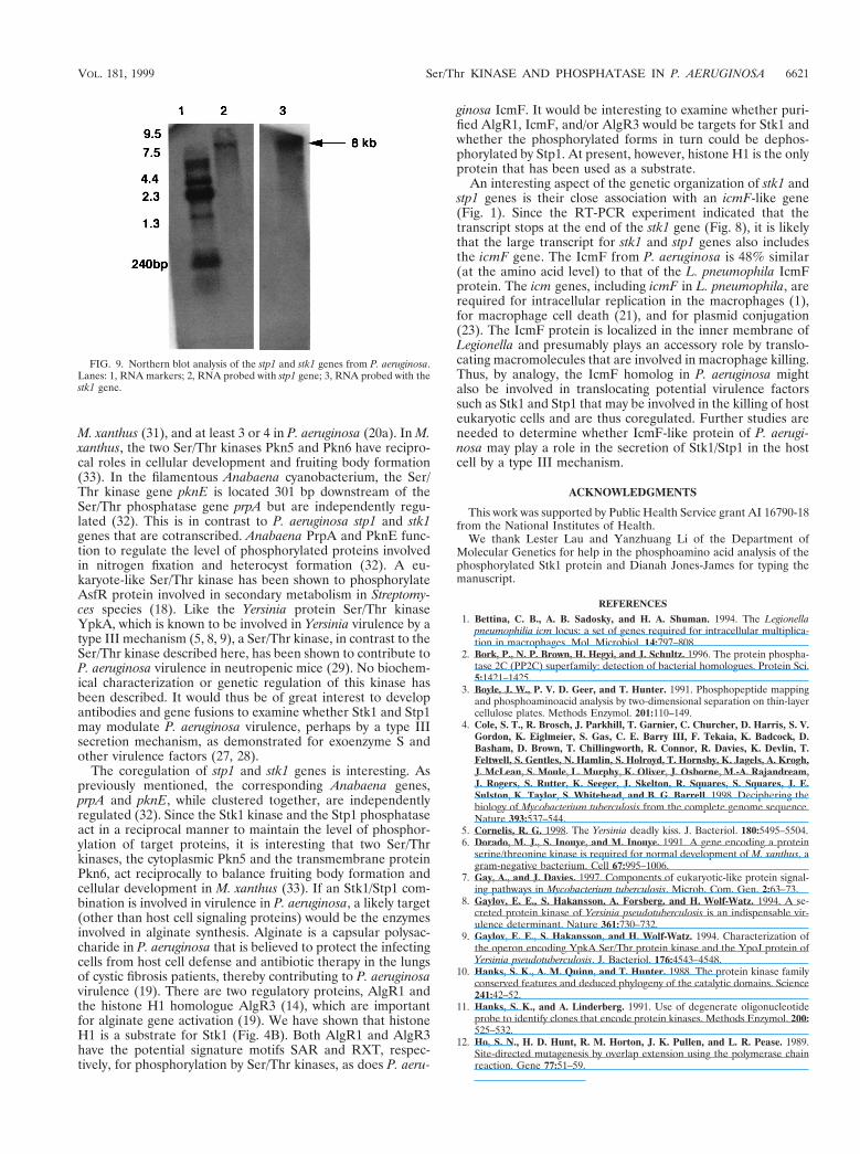
In order to determine the size of the transcript formedduring transcription of the stp1 and stk1 genes, we performedNorthern hybridizations by using RNA isolated from P. aerugi-nosa PAO1. With the stp1 gene used as a probe, an approxi-mately 8-kb transcript was detected (Fig. 9, lane 2). When thestk1 gene was used as a probe, a similar-sized transcript wasagain detected (Fig. 9, lane 3), implying that both stp1 and stk1transcripts are present in a single fragment of 8 kb, presumablyrepresenting transcription of a larger operon with an icmFhomologue and other genes upstream.
DISCUSSION
One of the unique features of eukaryote-like Ser/Thr or Tyrkinases and their cognate phosphoprotein phosphatases is thepresence of multiple genes for these enzymes both in eu-karyotes and in prokaryotes (24). In prokaryotes, such enzymeshave been detected in E. coli, M. tuberculosis, Y. pseudotuber-culosis, S. coelicolor, M. xanthus, and other strains. In M. tu-berculosis, there are 11 copies of Ser/Thr kinase genes (4), 5 in
FIG. 7. Activity of the purified P. aeruginosa Stp1 phosphatase overexpressedin E. coli. Phosphorylated casein was treated with the purified Stp1, and thedephosphorylated casein protein was analyzed by SDS–12.5% PAGE. Lane 1,phosphorylated casein (unboiled); lane 2, phosphorylated casein (boiled); lanes3, 4, and 5, phosphorylated casein incubated with the purified Stp1 phosphatasefor 30, 60, and 90 min, respectively. The radioactivities were measured by aphosphorimager, and the percent bound radioactivities are depicted below.
TABLE 2. Inhibitors of Stp1 activity
No. Inhibitors (concn) % Relative activitya
1 Control 100
2 EDTA (10 mM) 14EDTA (100 mM) 0
3 Sodium fluoride (5 mM) 50Sodium fluoride (50 mM) 10Sodium fluoride (500 mM) 0
4 Okadaic acid (100 nM) 92Okadaic acid (10 nM) 95Okadaic acid (100 mM) 95
5 Sodium vanadate (2 mM) 95Sodium vanadate (20 mM) 95Sodium vanadate (200 mM) 95
a Activity was measured by pNPP hydrolysis at 410 nm in the absence or thepresence of various inhibitors.
FIG. 8. RT-PCR analysis showing the contranscription of stp1 and stk1 genesfrom P. aeruginosa PAO1. Lanes: A, DNA markers (kilobases); B, primer com-bination 1 and 2; C, primer combination 3 and 4; D, primer combination 7 and4; E, primer combination 1 and 5; F, primer combination 1 and 6; G, primercombination 3 and 8. All of the PCR products were of the expected sizes, andthere was no amplification of the cDNA when primers 3 and 8 were used(control).
6620 MUKHOPADHYAY ET AL. J. BACTERIOL.
M. xanthus (31), and at least 3 or 4 in P. aeruginosa (20a). In M.xanthus, the two Ser/Thr kinases Pkn5 and Pkn6 have recipro-cal roles in cellular development and fruiting body formation(33). In the filamentous Anabaena cyanobacterium, the Ser/Thr kinase gene pknE is located 301 bp downstream of theSer/Thr phosphatase gene prpA but are independently regu-lated (32). This is in contrast to P. aeruginosa stp1 and stk1genes that are cotranscribed. Anabaena PrpA and PknE func-tion to regulate the level of phosphorylated proteins involvedin nitrogen fixation and heterocyst formation (32). A eu-karyote-like Ser/Thr kinase has been shown to phosphorylateAsfR protein involved in secondary metabolism in Streptomy-ces species (18). Like the Yersinia protein Ser/Thr kinaseYpkA, which is known to be involved in Yersinia virulence by atype III mechanism (5, 8, 9), a Ser/Thr kinase, in contrast to theSer/Thr kinase described here, has been shown to contribute toP. aeruginosa virulence in neutropenic mice (29). No biochem-ical characterization or genetic regulation of this kinase hasbeen described. It would thus be of great interest to developantibodies and gene fusions to examine whether Stk1 and Stp1may modulate P. aeruginosa virulence, perhaps by a type IIIsecretion mechanism, as demonstrated for exoenzyme S andother virulence factors (27, 28).
The coregulation of stp1 and stk1 genes is interesting. Aspreviously mentioned, the corresponding Anabaena genes,prpA and pknE, while clustered together, are independentlyregulated (32). Since the Stk1 kinase and the Stp1 phosphataseact in a reciprocal manner to maintain the level of phosphor-ylation of target proteins, it is interesting that two Ser/Thrkinases, the cytoplasmic Pkn5 and the transmembrane proteinPkn6, act reciprocally to balance fruiting body formation andcellular development in M. xanthus (33). If an Stk1/Stp1 com-bination is involved in virulence in P. aeruginosa, a likely target(other than host cell signaling proteins) would be the enzymesinvolved in alginate synthesis. Alginate is a capsular polysac-charide in P. aeruginosa that is believed to protect the infectingcells from host cell defense and antibiotic therapy in the lungsof cystic fibrosis patients, thereby contributing to P. aeruginosavirulence (19). There are two regulatory proteins, AlgR1 andthe histone H1 homologue AlgR3 (14), which are importantfor alginate gene activation (19). We have shown that histoneH1 is a substrate for Stk1 (Fig. 4B). Both AlgR1 and AlgR3have the potential signature motifs SAR and RXT, respec-tively, for phosphorylation by Ser/Thr kinases, as does P. aeru-
ginosa IcmF. It would be interesting to examine whether puri-fied AlgR1, IcmF, and/or AlgR3 would be targets for Stk1 andwhether the phosphorylated forms in turn could be dephos-phorylated by Stp1. At present, however, histone H1 is the onlyprotein that has been used as a substrate.
An interesting aspect of the genetic organization of stk1 andstp1 genes is their close association with an icmF-like gene(Fig. 1). Since the RT-PCR experiment indicated that thetranscript stops at the end of the stk1 gene (Fig. 8), it is likelythat the large transcript for stk1 and stp1 genes also includesthe icmF gene. The IcmF from P. aeruginosa is 48% similar(at the amino acid level) to that of the L. pneumophila IcmFprotein. The icm genes, including icmF in L. pneumophila, arerequired for intracellular replication in the macrophages (1),for macrophage cell death (21), and for plasmid conjugation(23). The IcmF protein is localized in the inner membrane ofLegionella and presumably plays an accessory role by translo-cating macromolecules that are involved in macrophage killing.Thus, by analogy, the IcmF homolog in P. aeruginosa mightalso be involved in translocating potential virulence factorssuch as Stk1 and Stp1 that may be involved in the killing of hosteukaryotic cells and are thus coregulated. Further studies areneeded to determine whether IcmF-like protein of P. aerugi-nosa may play a role in the secretion of Stk1/Stp1 in the hostcell by a type III mechanism.
ACKNOWLEDGMENTS
This work was supported by Public Health Service grant AI 16790-18from the National Institutes of Health.
We thank Lester Lau and Yanzhuang Li of the Department ofMolecular Genetics for help in the phosphoamino acid analysis of thephosphorylated Stk1 protein and Dianah Jones-James for typing themanuscript.
REFERENCES
1. Bettina, C. B., A. B. Sadosky, and H. A. Shuman. 1994. The Legionellapneumophilia icm locus: a set of genes required for intracellular multiplica-tion in macrophages. Mol. Microbiol. 14:797–808.
2. Bork, P., N. P. Brown, H. Hegyi, and J. Schultz. 1996. The protein phospha-tase 2C (PP2C) superfamily: detection of bacterial homologues. Protein Sci.5:1421–1425.
3. Boyle, J. W., P. V. D. Geer, and T. Hunter. 1991. Phosphopeptide mappingand phosphoaminoacid analysis by two-dimensional separation on thin-layercellulose plates. Methods Enzymol. 201:110–149.
4. Cole, S. T., R. Brosch, J. Parkhill, T. Garnier, C. Churcher, D. Harris, S. V.Gordon, K. Eiglmeier, S. Gas, C. E. Barry III, F. Tekaia, K. Badcock, D.Basham, D. Brown, T. Chillingworth, R. Connor, R. Davies, K. Devlin, T.Feltwell, S. Gentles, N. Hamlin, S. Holroyd, T. Hornsby, K. Jagels, A. Krogh,J. McLean, S. Moule, L. Murphy, K. Oliver, J. Osborne, M.-A. Rajandream,J. Rogers, S. Rutter, K. Seeger, J. Skelton, R. Squares, S. Squares, J. E.Sulston, K. Taylor, S. Whitehead, and B. G. Barrell. 1998. Deciphering thebiology of Mycobacterium tuberculosis from the complete genome sequence.Nature 393:537–544.
5. Cornelis, R. G. 1998. The Yersinia deadly kiss. J. Bacteriol. 180:5495–5504.6. Dorado, M. J., S. Inouye, and M. Inouye. 1991. A gene encoding a protein
serine/threonine kinase is required for normal development of M. xanthus, agram-negative bacterium. Cell 67:995–1006.
7. Gay, A., and J. Davies. 1997. Components of eukaryotic-like protein signal-ing pathways in Mycobacterium tuberculosis. Microb. Com. Gen. 2:63–73.
8. Gaylov, E. E., S. Hakansson, A. Forsberg, and H. Wolf-Watz. 1994. A se-creted protein kinase of Yersinia pseudotuberculosis is an indispensable vir-ulence determinant. Nature 361:730–732.
9. Gaylov, E. E., S. Hakansson, and H. Wolf-Watz. 1994. Characterization ofthe operon encoding YpkA Ser/Thr protein kinase and the YpoJ protein ofYersinia pseudotuberculosis. J. Bacteriol. 176:4543–4548.
10. Hanks, S. K., A. M. Quinn, and T. Hunter. 1988. The protein kinase familyconserved features and deduced phylogeny of the catalytic domains. Science241:42–52.
11. Hanks, S. K., and A. Linderberg. 1991. Use of degenerate oligonucleotideprobe to identify clones that encode protein kinases. Methods Enzymol. 200:525–532.
12. Ho, S. N., H. D. Hunt, R. M. Horton, J. K. Pullen, and L. R. Pease. 1989.Site-directed mutagenesis by overlap extension using the polymerase chainreaction. Gene 77:51–59.
FIG. 9. Northern blot analysis of the stp1 and stk1 genes from P. aeruginosa.Lanes: 1, RNA markers; 2, RNA probed with stp1 gene; 3, RNA probed with thestk1 gene.
VOL. 181, 1999 Ser/Thr KINASE AND PHOSPHATASE IN P. AERUGINOSA 6621

13. Kamps, M. P., and B. M. Sefton. 1986. Neither arginine nor histidine cancarry out the function of lysine-295 in the ATP binding site of p60src. Mol.Cell. Biol. 6:751–757.
14. Kato, J., T. K. Misra, and A. M. Chakrabarty. 1990. AlgR3, a proteinresembling eukaryotic histone H1, regulates alginate synthesis in Pseudomo-nas aeruginosa. Proc. Natl. Acad. Sci. USA 87:2887–2891.
15. Kennelly, P. J., and M. Potts. 1996. Fancy meeting you here! A fresh look at‘prokaryotic’ protein phosphorylation. J. Bacteriol. 178:4759–4764.
16. Mackintosh, C., and R. W. Mackintosh. 1994. Inhibitors of protein kinasesand phosphatases. Trends Biochem. Sci. 19:444–447.
17. Maizels, E. T., C. A. Peters, M. Kline, R. E. Cutler, M. Shanmugam, and M.Hunzicker-Dunn. 1998. Heat-shock protein-25/27 phosphorylation by the sisoform of protein kinase C. Biochem. J. 332:703–712.
18. Matsumoto, A., S. K. Hong, H. Ishizuka, S. Horinouchi, and T. Beppu. 1994.Phosphorylation of the AfsR protein involved in secondary metabolism inStreptomyces species by a eukaryotic-type protein kinase. Gene 146:47–55.
19. May, T. B., and A. M. Chakrabarty. 1994. Pseudomonas aeruginosa: genesand enzymes of alginate biosynthesis. Trends Microbiol. 2:151–157.
20. Parkinson, J. S., and E. Kofoid. 1992. Communication modules in bacterialsignaling proteins. Annu. Rev. Genet. 26:71–112.
20a.Pseudomonas aeruginosa Genome Sequence. 15 March 1999, copyright date.[Online.] http://www.pseudomonas.com. [10 April 1999, last date accessed.]
21. Purcell, M., and H. A. Shuman. 1998. The Legionella pneumophila icmGCDJBF genes are required for killing of human macrophages. Infect. Im-mun. 66:2245–2255.
22. Sambrook, J., E. F. Fritsch, and T. Maniatis. 1989. Molecular cloning: alaboratory manual, 2nd ed. Cold Spring Harbor Laboratory, Cold SpringHarbor, N.Y.
23. Segal, G., M. Purcell, and H. A. Shuman. 1998. Host killing and bacterialconjugation requires overlapping sets of genes within a 22-kb region of theLegionella pneumophila genome. Proc. Natl. Acad. Sci. USA 95:1669–1674.
24. Shi, L., M. Potts, and P. J. Kennelly. 1998. The serine, threonine and/ortyrosine specific protein kinases and protein phosphatases of prokaryoticorganisms: a family portrait. FEMS. Microbiol. Rev. 22:229–253.
25. Shortridge, V. D., A. Lazdunski, and M. V. Vasil. 1992. Osmoprotectants andphosphate regulate expression of phospholipase C in Pseudomonas aerugi-nosa. Mol. Microbiol. 6:863–871.
26. Urabe, H., and H. Ogawara. 1995. Cloning, sequencing and expression ofserine/threonine-encoding genes from Streptomyces coelicolor A3(2). Gene153:99–104.
27. Vallis, A. J., V. Finck-Barbancon, T. L. Yahr, and D. W. Frank. 1999.Biological effects of Pseudomonas aeruginosa type III secreted proteins onCHO cells. Infect. Immun. 67:2040–2044.
28. Vallis, A. J., T. L. Yahr, J. T. Barbieri, and D. W. Frank. 1999. Regulationof ExoS production and secretion by Pseudomonas aeruginosa in response totissue culture conditions. Infect. Immun. 67:914–920.
29. Wang, J., C. Li, H. Yang, A. Mushegian, and S. Jin. 1998. A novel serine/threonine protein kinase homologue of Pseudomonas aeruginosa is specifi-cally inducible within the host infection site and is required for full virulencein neutropenic mice. J. Bacteriol. 180:6764–6768.
30. Zhang, C. C. 1993. A gene encoding a protein related to eukaryotic proteinkinases from the filamentous heterocystous cyanobacterium Anabaena PCC7120. Proc. Natl. Acad. Sci. USA 90:11840–11844.
31. Zhang, C. C. 1996. Bacterial signalling involving eukaryotic-type proteinkinases. Mol. Microbiol. 20:9–15.
32. Zhang, C. C., A. Friry, and L. Peng. 1998. Molecular and genetic analysis oftwo closely linked genes that encode, respectively, a protein phosphatase1/2A/2B homolog and a protein kinase homolog in the cyanobacteriumAnabaena sp. strain PCC 7120. J. Bacteriol. 180:2616–2622.
33. Zhang, W., M. Inouye, and S. Inouye. 1996. Recirprocal regulation of thedifferentiation of Myxococcus xanthus by Pkn5 and Pkn6, eukaryotic likeSer/Thr protein kinases. Mol. Microbiol. 20:435–447.
6622 MUKHOPADHYAY ET AL. J. BACTERIOL.