Somatic mosaic IDH1 and IDH2 mutations are associated with enchondroma and spindle cell hemangioma...
8
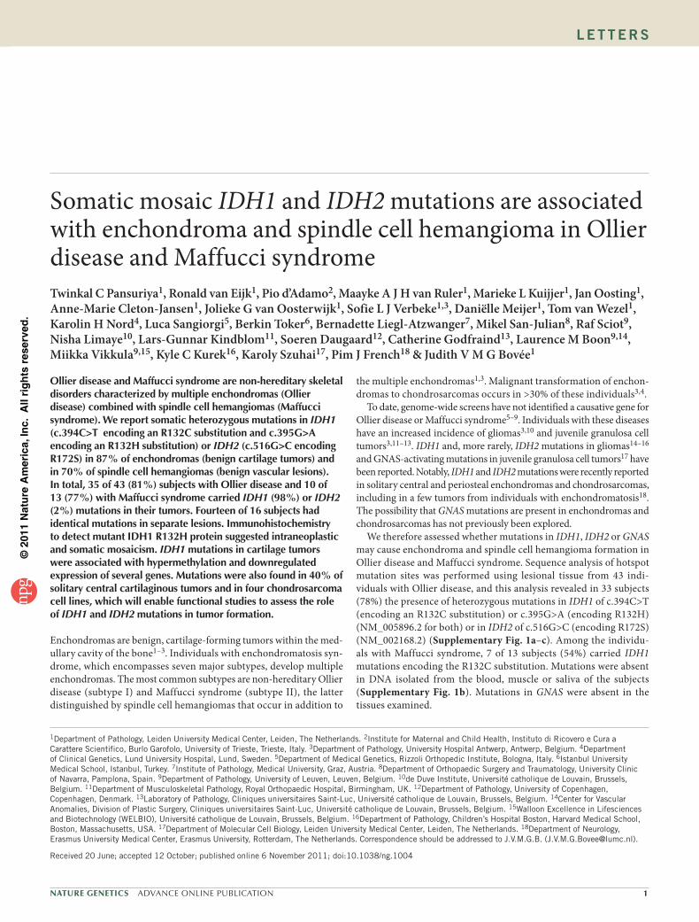
© 2011 Nature America, Inc. All rights reserved. NATURE GENETICS ADVANCE ONLINE PUBLICATION LETTERS Ollier disease and Maffucci syndrome are non-hereditary skeletal disorders characterized by multiple enchondromas (Ollier disease) combined with spindle cell hemangiomas (Maffucci syndrome). We report somatic heterozygous mutations in IDH1 (c.394C>T encoding an R32C substitution and c.395G>A encoding an R32H substitution) or IDH2 (c.56G>C encoding R72S) in 87% of enchondromas (benign cartilage tumors) and in 70% of spindle cell hemangiomas (benign vascular lesions). In total, 35 of 43 (8%) subjects with Ollier disease and 0 of 3 (77%) with Maffucci syndrome carried IDH1 (98%) or IDH2 (2%) mutations in their tumors. Fourteen of 6 subjects had identical mutations in separate lesions. Immunohistochemistry to detect mutant IDH R32H protein suggested intraneoplastic and somatic mosaicism. IDH1 mutations in cartilage tumors were associated with hypermethylation and downregulated expression of several genes. Mutations were also found in 40% of solitary central cartilaginous tumors and in four chondrosarcoma cell lines, which will enable functional studies to assess the role of IDH1 and IDH2 mutations in tumor formation. Enchondromas are benign, cartilage-forming tumors within the med- ullary cavity of the bone 1–3 . Individuals with enchondromatosis syn- drome, which encompasses seven major subtypes, develop multiple enchondromas. The most common subtypes are non-hereditary Ollier disease (subtype I) and Maffucci syndrome (subtype II), the latter distinguished by spindle cell hemangiomas that occur in addition to the multiple enchondromas 1,3 . Malignant transformation of enchon- dromas to chondrosarcomas occurs in >30% of these individuals 3,4 . To date, genome-wide screens have not identified a causative gene for Ollier disease or Maffucci syndrome 5–9 . Individuals with these diseases have an increased incidence of gliomas 3,10 and juvenile granulosa cell tumors 3,11–13 . IDH1 and, more rarely, IDH2 mutations in gliomas 14–16 and GNAS-activating mutations in juvenile granulosa cell tumors 17 have been reported. Notably, IDH1 and IDH2 mutations were recently reported in solitary central and periosteal enchondromas and chondrosarcomas, including in a few tumors from individuals with enchondromatosis 18 . The possibility that GNAS mutations are present in enchondromas and chondrosarcomas has not previously been explored. We therefore assessed whether mutations in IDH1, IDH2 or GNAS may cause enchondroma and spindle cell hemangioma formation in Ollier disease and Maffucci syndrome. Sequence analysis of hotspot mutation sites was performed using lesional tissue from 43 indi- viduals with Ollier disease, and this analysis revealed in 33 subjects (78%) the presence of heterozygous mutations in IDH1 of c.394C>T (encoding an R132C substitution) or c.395G>A (encoding R132H) (NM_005896.2 for both) or in IDH2 of c.516G>C (encoding R172S) (NM_002168.2) (Supplementary Fig. 1a–c). Among the individu- als with Maffucci syndrome, 7 of 13 subjects (54%) carried IDH1 mutations encoding the R132C substitution. Mutations were absent in DNA isolated from the blood, muscle or saliva of the subjects (Supplementary Fig. 1b). Mutations in GNAS were absent in the tissues examined. Somatic mosaic IDH1 and IDH2 mutations are associated with enchondroma and spindle cell hemangioma in Ollier disease and Maffucci syndrome Twinkal C Pansuriya 1 , Ronald van Eijk 1 , Pio d’Adamo 2 , Maayke A J H van Ruler 1 , Marieke L Kuijjer 1 , Jan Oosting 1 , Anne-Marie Cleton-Jansen 1 , Jolieke G van Oosterwijk 1 , Sofie L J Verbeke 1,3 , Daniëlle Meijer 1 , Tom van Wezel 1 , Karolin H Nord 4 , Luca Sangiorgi 5 , Berkin Toker 6 , Bernadette Liegl-Atzwanger 7 , Mikel San-Julian 8 , Raf Sciot 9 , Nisha Limaye 10 , Lars-Gunnar Kindblom 11 , Soeren Daugaard 12 , Catherine Godfraind 13 , Laurence M Boon 9,14 , Miikka Vikkula 9,15 , Kyle C Kurek 16 , Karoly Szuhai 17 , Pim J French 18 & Judith V M G Bovée 1 1 Department of Pathology, Leiden University Medical Center, Leiden, The Netherlands. 2 Institute for Maternal and Child Health, Instituto di Ricovero e Cura a Carattere Scientifico, Burlo Garofolo, University of Trieste, Trieste, Italy. 3 Department of Pathology, University Hospital Antwerp, Antwerp, Belgium. 4 Department of Clinical Genetics, Lund University Hospital, Lund, Sweden. 5 Department of Medical Genetics, Rizzoli Orthopedic Institute, Bologna, Italy. 6 Istanbul University Medical School, Istanbul, Turkey. 7 Institute of Pathology, Medical University, Graz, Austria. 8 Department of Orthopaedic Surgery and Traumatology, University Clinic of Navarra, Pamplona, Spain. 9 Department of Pathology, University of Leuven, Leuven, Belgium. 10 de Duve Institute, Université catholique de Louvain, Brussels, Belgium. 11 Department of Musculoskeletal Pathology, Royal Orthopaedic Hospital, Birmingham, UK. 12 Department of Pathology, University of Copenhagen, Copenhagen, Denmark. 13 Laboratory of Pathology, Cliniques universitaires Saint-Luc, Université catholique de Louvain, Brussels, Belgium. 14 Center for Vascular Anomalies, Division of Plastic Surgery, Cliniques universitaires Saint-Luc, Université catholique de Louvain, Brussels, Belgium. 15 Walloon Excellence in Lifesciences and Biotechnology (WELBIO), Université catholique de Louvain, Brussels, Belgium. 16 Department of Pathology, Children’s Hospital Boston, Harvard Medical School, Boston, Massachusetts, USA. 17 Department of Molecular Cell Biology, Leiden University Medical Center, Leiden, The Netherlands. 18 Department of Neurology, Erasmus University Medical Center, Erasmus University, Rotterdam, The Netherlands. Correspondence should be addressed to J.V.M.G.B. ([email protected]). Received 20 June; accepted 12 October; published online 6 November 2011; doi:10.1038/ng.1004
-
Upload
independent -
Category
Documents
-
view
7 -
download
0
Transcript of Somatic mosaic IDH1 and IDH2 mutations are associated with enchondroma and spindle cell hemangioma...
©20
11 N
atu
re A
mer
ica,
Inc.
All
rig
hts
res
erve
d.
Nature GeNetics ADVANCE ONLINE PUBLICATION �
l e t t e r s
Ollier disease and Maffucci syndrome are non-hereditary skeletal disorders characterized by multiple enchondromas (Ollier disease) combined with spindle cell hemangiomas (Maffucci syndrome). We report somatic heterozygous mutations in IDH1 (c.394C>T encoding an R�32C substitution and c.395G>A encoding an R�32H substitution) or IDH2 (c.5�6G>C encoding R�72S) in 87% of enchondromas (benign cartilage tumors) and in 70% of spindle cell hemangiomas (benign vascular lesions). In total, 35 of 43 (8�%) subjects with Ollier disease and �0 of �3 (77%) with Maffucci syndrome carried IDH1 (98%) or IDH2 (2%) mutations in their tumors. Fourteen of �6 subjects had identical mutations in separate lesions. Immunohistochemistry to detect mutant IDH� R�32H protein suggested intraneoplastic and somatic mosaicism. IDH1 mutations in cartilage tumors were associated with hypermethylation and downregulated expression of several genes. Mutations were also found in 40% of solitary central cartilaginous tumors and in four chondrosarcoma cell lines, which will enable functional studies to assess the role of IDH1 and IDH2 mutations in tumor formation.
Enchondromas are benign, cartilage-forming tumors within the med-ullary cavity of the bone1–3. Individuals with enchondromatosis syn-drome, which encompasses seven major subtypes, develop multiple enchondromas. The most common subtypes are non-hereditary Ollier disease (subtype I) and Maffucci syndrome (subtype II), the latter distinguished by spindle cell hemangiomas that occur in addition to
the multiple enchondromas1,3. Malignant transformation of enchon-dromas to chondrosarcomas occurs in >30% of these individuals3,4.
To date, genome-wide screens have not identified a causative gene for Ollier disease or Maffucci syndrome5–9. Individuals with these diseases have an increased incidence of gliomas3,10 and juvenile granulosa cell tumors3,11–13. IDH1 and, more rarely, IDH2 mutations in gliomas14–16 and GNAS-activating mutations in juvenile granulosa cell tumors17 have been reported. Notably, IDH1 and IDH2 mutations were recently reported in solitary central and periosteal enchondromas and chondrosarcomas, including in a few tumors from individuals with enchondromatosis18. The possibility that GNAS mutations are present in enchondromas and chondrosarcomas has not previously been explored.
We therefore assessed whether mutations in IDH1, IDH2 or GNAS may cause enchondroma and spindle cell hemangioma formation in Ollier disease and Maffucci syndrome. Sequence analysis of hotspot mutation sites was performed using lesional tissue from 43 indi-viduals with Ollier disease, and this analysis revealed in 33 subjects (78%) the presence of heterozygous mutations in IDH1 of c.394C>T (encoding an R132C substitution) or c.395G>A (encoding R132H) (NM_005896.2 for both) or in IDH2 of c.516G>C (encoding R172S) (NM_002168.2) (Supplementary Fig. 1a–c). Among the individu-als with Maffucci syndrome, 7 of 13 subjects (54%) carried IDH1 mutations encoding the R132C substitution. Mutations were absent in DNA isolated from the blood, muscle or saliva of the subjects (Supplementary Fig. 1b). Mutations in GNAS were absent in the tissues examined.
Somatic mosaic IDH1 and IDH2 mutations are associated with enchondroma and spindle cell hemangioma in Ollier disease and Maffucci syndromeTwinkal C Pansuriya1, Ronald van Eijk1, Pio d’Adamo2, Maayke A J H van Ruler1, Marieke L Kuijjer1, Jan Oosting1, Anne-Marie Cleton-Jansen1, Jolieke G van Oosterwijk1, Sofie L J Verbeke1,3, Daniëlle Meijer1, Tom van Wezel1, Karolin H Nord4, Luca Sangiorgi5, Berkin Toker6, Bernadette Liegl-Atzwanger7, Mikel San-Julian8, Raf Sciot9, Nisha Limaye10, Lars-Gunnar Kindblom11, Soeren Daugaard12, Catherine Godfraind13, Laurence M Boon9,14, Miikka Vikkula9,15, Kyle C Kurek16, Karoly Szuhai17, Pim J French18 & Judith V M G Bovée1
1Department of Pathology, Leiden University Medical Center, Leiden, The Netherlands. 2Institute for Maternal and Child Health, Instituto di Ricovero e Cura a Carattere Scientifico, Burlo Garofolo, University of Trieste, Trieste, Italy. 3Department of Pathology, University Hospital Antwerp, Antwerp, Belgium. 4Department of Clinical Genetics, Lund University Hospital, Lund, Sweden. 5Department of Medical Genetics, Rizzoli Orthopedic Institute, Bologna, Italy. 6Istanbul University Medical School, Istanbul, Turkey. 7Institute of Pathology, Medical University, Graz, Austria. 8Department of Orthopaedic Surgery and Traumatology, University Clinic of Navarra, Pamplona, Spain. 9Department of Pathology, University of Leuven, Leuven, Belgium. 10de Duve Institute, Université catholique de Louvain, Brussels, Belgium. 11Department of Musculoskeletal Pathology, Royal Orthopaedic Hospital, Birmingham, UK. 12Department of Pathology, University of Copenhagen, Copenhagen, Denmark. 13Laboratory of Pathology, Cliniques universitaires Saint-Luc, Université catholique de Louvain, Brussels, Belgium. 14Center for Vascular Anomalies, Division of Plastic Surgery, Cliniques universitaires Saint-Luc, Université catholique de Louvain, Brussels, Belgium. 15Walloon Excellence in Lifesciences and Biotechnology (WELBIO), Université catholique de Louvain, Brussels, Belgium. 16Department of Pathology, Children’s Hospital Boston, Harvard Medical School, Boston, Massachusetts, USA. 17Department of Molecular Cell Biology, Leiden University Medical Center, Leiden, The Netherlands. 18Department of Neurology, Erasmus University Medical Center, Erasmus University, Rotterdam, The Netherlands. Correspondence should be addressed to J.V.M.G.B. ([email protected]).
Received 20 June; accepted 12 October; published online 6 November 2011; doi:10.1038/ng.1004
©20
11 N
atu
re A
mer
ica,
Inc.
All
rig
hts
res
erve
d.
2 ADVANCE ONLINE PUBLICATION Nature GeNetics
l e t t e r s
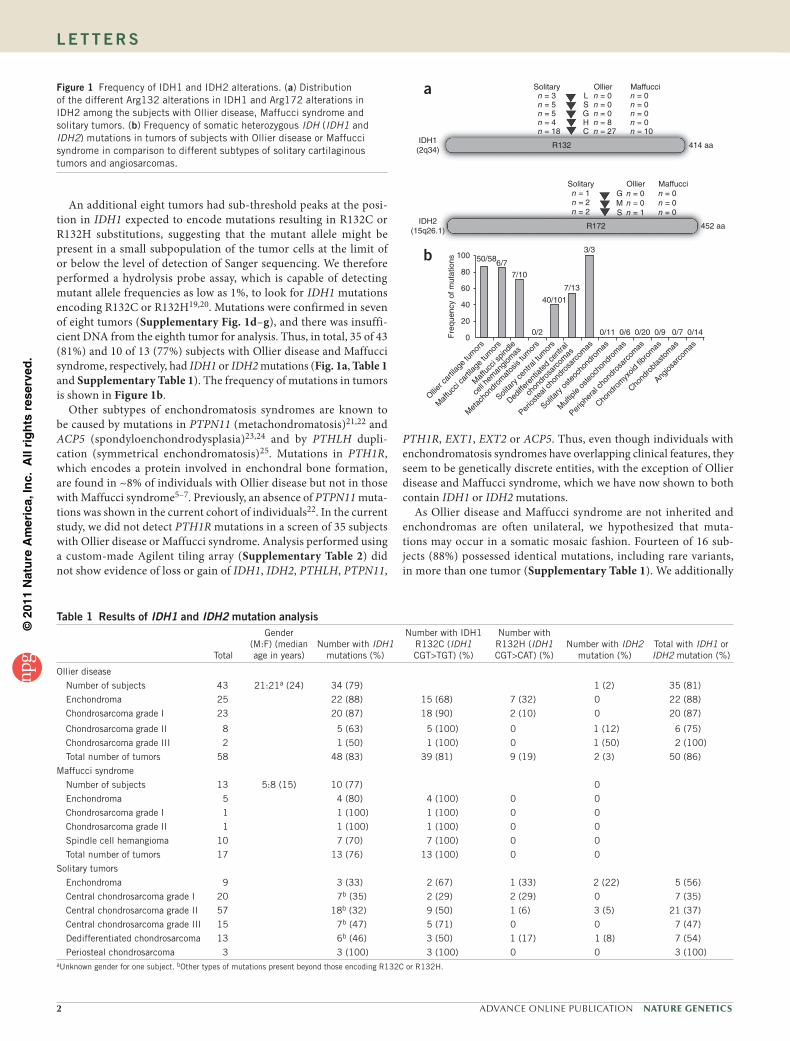
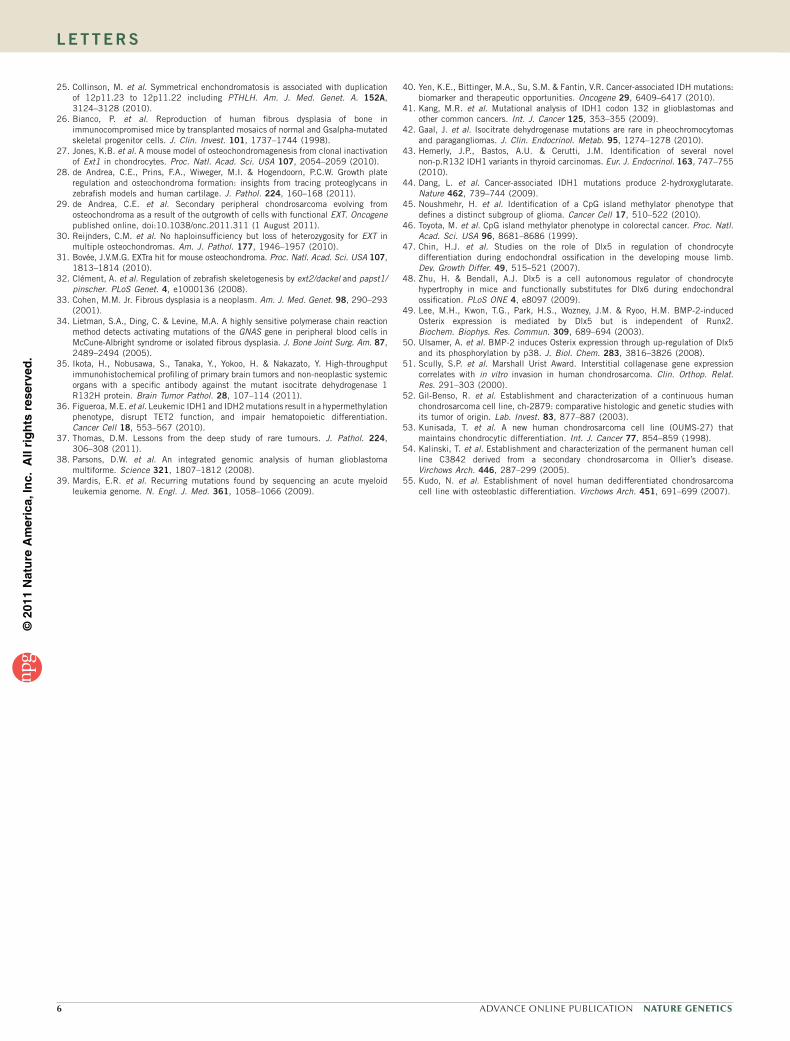
An additional eight tumors had sub-threshold peaks at the posi-tion in IDH1 expected to encode mutations resulting in R132C or R132H substitutions, suggesting that the mutant allele might be present in a small subpopulation of the tumor cells at the limit of or below the level of detection of Sanger sequencing. We therefore performed a hydrolysis probe assay, which is capable of detecting mutant allele frequencies as low as 1%, to look for IDH1 mutations encoding R132C or R132H19,20. Mutations were confirmed in seven of eight tumors (Supplementary Fig. 1d–g), and there was insuffi-cient DNA from the eighth tumor for analysis. Thus, in total, 35 of 43 (81%) and 10 of 13 (77%) subjects with Ollier disease and Maffucci syndrome, respectively, had IDH1 or IDH2 mutations (Fig. 1a, Table 1 and Supplementary Table 1). The frequency of mutations in tumors is shown in Figure 1b.
Other subtypes of enchondromatosis syndromes are known to be caused by mutations in PTPN11 (metachondromatosis)21,22 and ACP5 (spondyloenchondrodysplasia)23,24 and by PTHLH dupli-cation (symmetrical enchondromatosis)25. Mutations in PTH1R, which encodes a protein involved in enchondral bone formation, are found in ~8% of individuals with Ollier disease but not in those with Maffucci syndrome5–7. Previously, an absence of PTPN11 muta-tions was shown in the current cohort of individuals22. In the current study, we did not detect PTH1R mutations in a screen of 35 subjects with Ollier disease or Maffucci syndrome. Analysis performed using a custom-made Agilent tiling array (Supplementary Table 2) did not show evidence of loss or gain of IDH1, IDH2, PTHLH, PTPN11,
PTH1R, EXT1, EXT2 or ACP5. Thus, even though individuals with enchondromatosis syndromes have overlapping clinical features, they seem to be genetically discrete entities, with the exception of Ollier disease and Maffucci syndrome, which we have now shown to both contain IDH1 or IDH2 mutations.
As Ollier disease and Maffucci syndrome are not inherited and enchondromas are often unilateral, we hypothesized that muta-tions may occur in a somatic mosaic fashion. Fourteen of 16 sub-jects (88%) possessed identical mutations, including rare variants, in more than one tumor (Supplementary Table 1). We additionally
IDH1(2q34)
Solitarya
b
Solitary
R132
R172
100 50/586/7
7/10
40/101
0/2
7/13
3/3
0/11 0/6 0/20 0/9 0/7 0/14
80
60
40
20
Fre
quen
cy o
f mut
atio
ns
0
Ollier c
artila
ge tu
mor
s
Maf
fucc
i car
tilage
tum
ors
Maf
fucc
i spin
dle
cell h
eman
giom
as
Met
acho
ndro
mat
osis
tum
ors
Solita
ry ce
ntra
l tum
ors
Dediffe
rent
iated
cent
ral
chon
dros
arco
mas
Perios
teal
chon
dros
arco
mas
Solita
ry o
steoc
hond
rom
as
Mult
iple
oste
ocho
ndro
mas
Periph
eral
chon
dros
arco
mas
Chond
rom
yxoid
fibro
mas
Chond
robla
stom
as
Angios
arco
mas
GMS
414 aa
452 aa
LSGHC
Ollier
Ollier
Maffucci
Maffucci
n = 3
n = 1 n = 0 n = 0n = 0n = 0
n = 0n = 1
n = 2n = 2
n = 0n = 0n = 0n = 8n = 27
n = 0n = 0n = 0n = 0n = 10
n = 5n = 5n = 4n = 18
IDH2(15q26.1)
Figure 1 Frequency of IDH1 and IDH2 alterations. (a) Distribution of the different Arg132 alterations in IDH1 and Arg172 alterations in IDH2 among the subjects with Ollier disease, Maffucci syndrome and solitary tumors. (b) Frequency of somatic heterozygous IDH (IDH1 and IDH2) mutations in tumors of subjects with Ollier disease or Maffucci syndrome in comparison to different subtypes of solitary cartilaginous tumors and angiosarcomas.
table 1 results of IDH1 and IDH2 mutation analysis
Total
Gender (M:F) (median age in years)
Number with IDH1 mutations (%)
Number with IDH1 R132C (IDH1 CGT>TGT) (%)
Number with R132H (IDH1 CGT>CAT) (%)
Number with IDH2 mutation (%)
Total with IDH1 or IDH2 mutation (%)
Ollier disease
Number of subjects 43 21:21a (24) 34 (79) 1 (2) 35 (81)
Enchondroma 25 22 (88) 15 (68) 7 (32) 0 22 (88)
Chondrosarcoma grade I 23 20 (87) 18 (90) 2 (10) 0 20 (87)
Chondrosarcoma grade II 8 5 (63) 5 (100) 0 1 (12) 6 (75)
Chondrosarcoma grade III 2 1 (50) 1 (100) 0 1 (50) 2 (100)
Total number of tumors 58 48 (83) 39 (81) 9 (19) 2 (3) 50 (86)
Maffucci syndrome
Number of subjects 13 5:8 (15) 10 (77) 0
Enchondroma 5 4 (80) 4 (100) 0 0
Chondrosarcoma grade I 1 1 (100) 1 (100) 0 0
Chondrosarcoma grade II 1 1 (100) 1 (100) 0 0
Spindle cell hemangioma 10 7 (70) 7 (100) 0 0
Total number of tumors 17 13 (76) 13 (100) 0 0
Solitary tumors
Enchondroma 9 3 (33) 2 (67) 1 (33) 2 (22) 5 (56)
Central chondrosarcoma grade I 20 7b (35) 2 (29) 2 (29) 0 7 (35)
Central chondrosarcoma grade II 57 18b (32) 9 (50) 1 (6) 3 (5) 21 (37)
Central chondrosarcoma grade III 15 7b (47) 5 (71) 0 0 7 (47)
Dedifferentiated chondrosarcoma 13 6b (46) 3 (50) 1 (17) 1 (8) 7 (54)
Periosteal chondrosarcoma 3 3 (100) 3 (100) 0 0 3 (100)aUnknown gender for one subject. bOther types of mutations present beyond those encoding R132C or R132H.
©20
11 N
atu
re A
mer
ica,
Inc.
All
rig
hts
res
erve
d.
Nature GeNetics ADVANCE ONLINE PUBLICATION 3
l e t t e r s
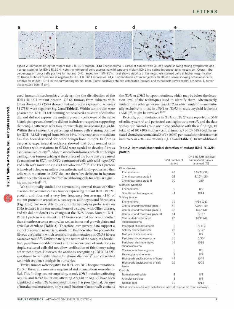
used immunohistochemistry to determine the distribution of the IDH1 R132H mutant protein. Of 68 tumors from subjects with Ollier disease, 17 (25%) showed mutant protein expression, whereas 51 (75%) were negative (Fig. 2 and Table 2). Within tumors that were positive for IDH1 R132H staining, we observed a mixture of cells that did and did not express the mutant protein (cells were of the same histologic type and therefore did not include entrapped or supporting elements), a pattern we refer to as intraneoplastic mosaicism (Fig. 2a,b). Within these tumors, the percentage of tumor cells staining positive for IDH1 R132H ranged from 50% to 95%. Intraneoplastic mosaicism has also been described for other benign bone tumors. In fibrous dysplasia, experimental evidence showed that both normal cells and those with mutations in GNAS were needed to develop fibrous dysplasia–like lesions26. Also, in osteochondromas, which are benign cartilaginous tumors arising at the surface of the bone that are caused by mutations in EXT1 or EXT2, a mixture of cells with wild-type EXT and cells with mutations in EXT was observed27–30. The EXT protein is involved in heparan sulfate biosynthesis, and it is hypothesized that cells with mutations in EXT that are therefore deficient in heparan sulfate need heparan sulfate from neighboring cells for cellular signal-ing and survival31,32.
We additionally studied the surrounding normal tissue of Ollier disease–derived and solitary tumors expressing mutant IDH1 R132H protein and observed a very low frequency (on average <1%) of mutant protein in osteoblasts, osteocytes, adipocytes and fibroblasts (Fig. 2d,e). We were able to perform the hydrolysis probe assay on DNA isolated from one normal bone of a subject with Ollier disease, and we did not detect any changes at the IDH1 locus. Mutant IDH1 R132H protein was absent in 12 bones resected for reasons other than chondrosarcoma removal as well as in normal growth plates and articular cartilage (Table 2). Therefore, our current data support a model of somatic mosaicism, similar to that described for polyostotic fibrous dysplasia in which somatic mosaic mutations in GNAS have a causative role33,34. Unfortunately, the nature of the samples (decalci-fied, paraffin-embedded bone) and the occurrence of mutations in single, scattered cells did not allow verification of this theory using other techniques. However, the antibody recognizing IDH1 R132H was shown to be highly reliable for glioma diagnosis35 and correlated well with sequence analysis in our series.
Twelve tumors were negative for IDH1 or IDH2 hotspot mutations. For 5 of these, all exons were sequenced and no mutations were identi-fied. This finding was not surprising, as only IDH1 mutations affecting Arg132 and IDH2 mutations affecting Arg140 or Arg172 have been identified in other IDH-associated tumors. It is possible that, because of intralesional mosaicism, only a small fraction of tumor cells contains
the IDH1 or IDH2 hotspot mutations, which may be below the detec-tion level of the techniques used to identify them. Alternatively, mutations in other genes such as TET2, in which mutations are mutu-ally exclusive to those in IDH1 or IDH2 in acute myeloid leukemia (AML)36, might be involved18,37.
Recently, point mutations in IDH1 or IDH2 were reported in 56% of solitary central and periosteal cartilaginous tumors18, and the data within our control group are in concordance with these findings. In total, 40 of 101 (40%) solitary central tumors, 7 of 13 (54%) dedifferen-tiated chondrosarcomas and 3 of 3 (100%) periosteal chondrosarcomas had IDH1 or IDH2 mutations (Fig. 1b and Table 1). In six additional
a b c
Bone
T
dT
e
Bone
Figure 2 Immunostaining for mutant IDH1 R132H protein. (a,b) Enchondroma (L1490) of subject with Ollier disease showing strong cytoplasmic and nuclear staining for IDH1 R132H. Note the mixture of cells expressing wild-type and mutant IDH1 indicating intraneoplastic mosaicism. Overall, the percentage of tumor cells positive for mutant IDH1 ranged from 50–95%. Inset shows viability of the negatively stained cells at higher magnification. (c) Grade II chondrosarcoma is negative for IDH1 R132H expression. (d,e) Enchondromas from subjects with Ollier disease showing occasional cells positive for mutant IDH1 in the surrounding normal bone. Some positively stained osteocytes (arrows) and osteoblasts (arrowheads) are seen. T, tumor tissue (scale bars, 5 µm).
table 2 Immunohistochemical detection of mutant IDH1 r132H protein
Total number of tumors
IDH1 R132H–positive tumors/total tumors
analyzed (%)
Ollier disease
Enchondroma 46 14/43a (32)
Chondrosarcoma grade I 22 3/17a (18)
Chondrosarcoma grade II 10 0/8a
Maffucci syndrome
Enchondroma 9 0/9
Spindle cell hemangioma 14 0/14
Solitary tumors
Enchondroma 19 4/19 (21)
Central chondrosarcoma grade I 42 4/38a (10)
Central chondrosarcoma grade II 36 1/32a (3)
Central chondrosarcoma grade III 14 0/11a
Central dedifferentiated chondrosarcoma
26 1/24a (4)
Periosteal chondrosarcoma 6 1/6 (17)
Solitary osteochondroma 20 0/17a
Multiple osteochondroma 7 0/7
Peripheral chondrosarcoma 45 0/35a
Peripheral dedifferentiated chondrosarcoma
16 0/16
Conventional hemangioma 3 0/3
Hemangioendothelioma 2 0/2
High grade angiosarcoma of bone 44 0/44
High grade angiosarcoma of soft tissue
22 0/22
Controls
Normal growth plate 3 0/3
Articular cartilage 3 0/3
Normal bone 12 0/12aNot all tumors included were evaluated due to loss of tissue on the tissue microarrays.
©20
11 N
atu
re A
mer
ica,
Inc.
All
rig
hts
res
erve
d.
4 ADVANCE ONLINE PUBLICATION Nature GeNetics
l e t t e r s
tumors, the mutant allele seemed to be present below the detection level of Sanger sequencing. IDH1 or IDH2 mutations were absent in other subtypes of cartilaginous tumors, in angiosarcomas (Fig. 1b) and in DNA isolated from subjects’ blood. Immunostaining for IDH1 R132H protein on tissue microarrays (TMAs) containing cartilagi-nous and vascular tumor samples confirmed that the expression of mutant IDH1 was restricted to central, dedifferentiated and periosteal cartilage tumors, whereas all other tumors lacked mutant expression (Table 2). Of note, four of eight solitary chondrosarcoma cell lines carried different types of mutations in IDH1 or IDH2 (Table 3). To the best of our knowledge, no representative cell lines with IDH1 or IDH2 mutations were previously available. IDH1 or IDH2 mutations were more frequently found in solitary central tumors located in the hands and feet (11 of 14 tumors) compared to those located in long and flat bones (28 of 84 tumors) (P = 0.006, Pearson’s χ2 test), which was also reported previously18. This correlation was absent in Ollier disease (20 of 22 tumors from the hands and feet compared to 28 of 34 tumors from long or flat bones, P = 0.5, Pearson’s χ2 test). Whereas in gliomas, mutations in IDH1 or IDH2 predict a favorable outcome38, we found no significant prognostic value of these mutations in solitary central cartilaginous tumors using multivariate analysis (Cox regres-sion, P value = 0.3).
IDH1 and IDH2 mutations have also been reported at lower fre-quencies in various other cancers, such as AML (8%)39,40, prostate cancer (2.7%)40,41, paragangliomas (0.7%)40,42 and thyroid carci-noma (16%)43. The high mutation frequency in enchondromas and the fact that these mutations occur early suggest a causal rather than a bystander role for IDH1 and IDH2 mutations in tumorigenesis in Ollier disease and Maffucci syndrome. In gliomas, mutations in IDH1 and IDH2 lead to a gain of function, causing the production of 2-hydroxyglutarate (2HG), a structural analog of α-ketoglutarate (α-KG), which thereby reduces α-KG production44. In AML, it was demonstrated that mutant IDH expression results in DNA hyper-methylation and impairment of hematopoietic differentiation36,
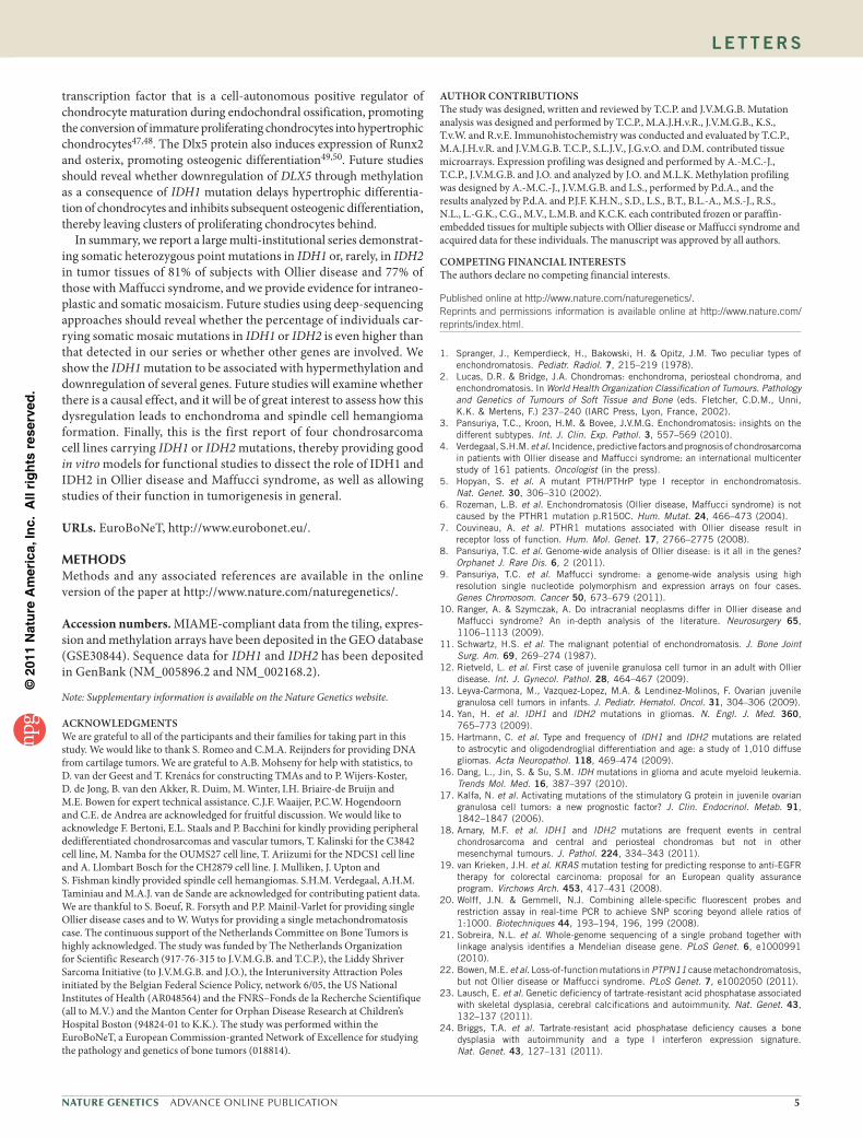
and in gliomas, the presence of an IDH1 mutation was strongly associated with hypermethylation45. Therefore, we used Illumina HumanMethylation27 BeadChips to examine possible differences in methylation between enchondromas with (N = 8) and without (N = 4) IDH1 mutations, as determined by Sanger sequencing. Unsupervised clustering of the 2,000 most variable CpG methylation sites gave two subgroups (Fig. 3). One of these subgroups showed an overall higher methylation at the examined CpG sites, a phenotype that is similar to the CpG island methylator phenotype (CIMP) described in colon carcinoma and glioblastoma45,46. All but one enchondromas with an IDH1 mutation were positive for this CIMP. Supervised clustering analysis indicated that 797 CpG sites were differentially methylated by more than 20% (with P < 0.05) between enchondromas with and without IDH1 mutations. Of note, 710 of these differentially methyl-ated CpG sites (89.1%) were methylated in the enchondromas with IDH1 mutations (Supplementary Table 3). These results are in line with the hypothesis that IDH1 mutations induce methylation and thus contribute to CIMP occurence36.
To assess the effect of IDH1 or IDH2 mutation on mRNA expres-sion levels in cartilaginous tumors, we performed whole-genome gene expression analysis using Illumina Human-6 v3 arrays. High-quality mRNA was available for only three tumors in which no mutation was found (N = 1) or in which mutations occurred at a frequency below the threshold of detection with Sanger sequencing (thus possibly carrying a low percentage of cells with mutations) (N = 2). Comparison of mRNA expression in these tumors with that from 18 tumors with clearly detectable IDH1 or IDH2 mutation using LIMMA analysis revealed 36 differentially expressed probes encoding 33 genes (Supplementary Table 4). Of these 33 genes, 32 were downregulated in the tumor sam-ples with an IDH1 or IDH2 mutation. There was no overlap between the affected genes identified by methylation or expression analysis.
One of the most differentially methylated genes was DLX5, for which there was a trend of downregulation in the expression data compar-ing enchondromas from subjects with Ollier disease and controls. However, this difference was not significant (adjusted P value = 0.3, Supplementary Fig. 2). The controls consisted of two growth plates and four articular or rib cartilage samples. DLX5 encodes a homeodomain
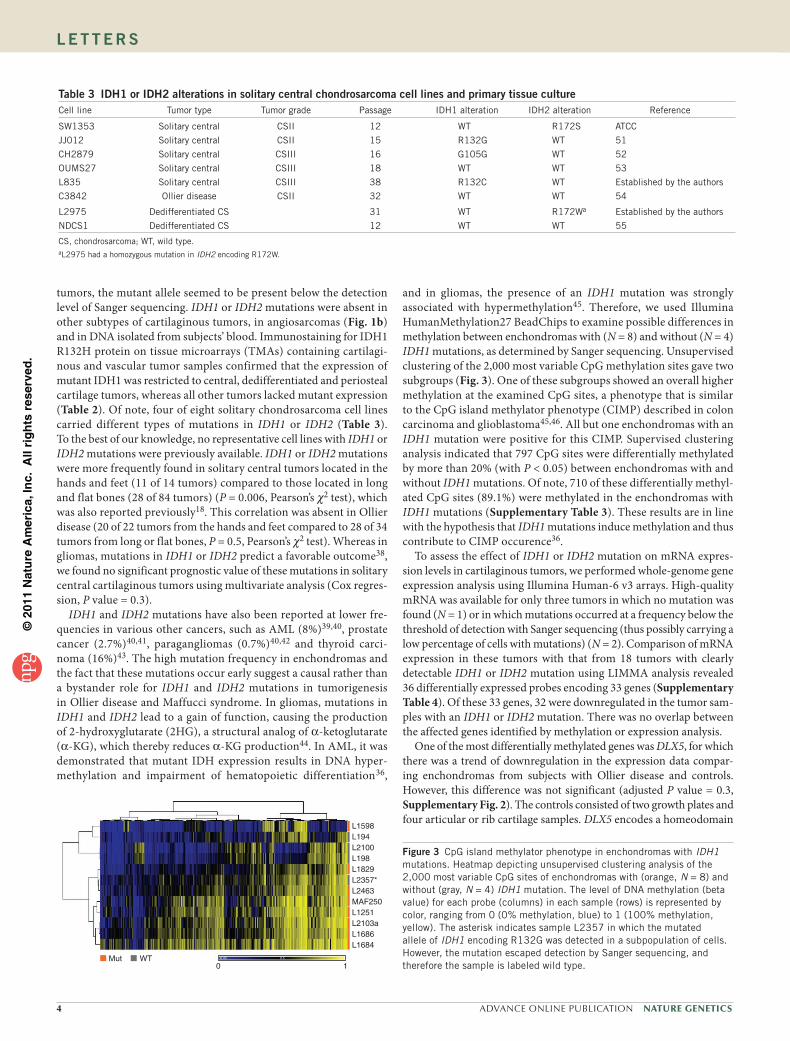
table 3 IDH1 or IDH2 alterations in solitary central chondrosarcoma cell lines and primary tissue cultureCell line Tumor type Tumor grade Passage IDH1 alteration IDH2 alteration Reference
SW1353 Solitary central CSII 12 WT R172S ATCC
JJ012 Solitary central CSII 15 R132G WT 51
CH2879 Solitary central CSIII 16 G105G WT 52
OUMS27 Solitary central CSIII 18 WT WT 53
L835 Solitary central CSIII 38 R132C WT Established by the authors
C3842 Ollier disease CSII 32 WT WT 54
L2975 Dedifferentiated CS 31 WT R172Wa Established by the authors
NDCS1 Dedifferentiated CS 12 WT WT 55
CS, chondrosarcoma; WT, wild type.aL2975 had a homozygous mutation in IDH2 encoding R172W.
Mut WT 0.05 0.5 0.95
L1598L194L2100L198L1829L2357*L2463MAF250L1251L2103aL1686L1684
0 1
Figure 3 CpG island methylator phenotype in enchondromas with IDH1 mutations. Heatmap depicting unsupervised clustering analysis of the 2,000 most variable CpG sites of enchondromas with (orange, N = 8) and without (gray, N = 4) IDH1 mutation. The level of DNA methylation (beta value) for each probe (columns) in each sample (rows) is represented by color, ranging from 0 (0% methylation, blue) to 1 (100% methylation, yellow). The asterisk indicates sample L2357 in which the mutated allele of IDH1 encoding R132G was detected in a subpopulation of cells. However, the mutation escaped detection by Sanger sequencing, and therefore the sample is labeled wild type.
©20
11 N
atu
re A
mer
ica,
Inc.
All
rig
hts
res
erve
d.
Nature GeNetics ADVANCE ONLINE PUBLICATION 5
l e t t e r s
transcription factor that is a cell-autonomous positive regulator of chondrocyte maturation during endochondral ossification, promoting the conversion of immature proliferating chondrocytes into hypertrophic chondrocytes47,48. The Dlx5 protein also induces expression of Runx2 and osterix, promoting osteogenic differentiation49,50. Future studies should reveal whether downregulation of DLX5 through methylation as a consequence of IDH1 mutation delays hypertrophic differentia-tion of chondrocytes and inhibits subsequent osteogenic differentiation, thereby leaving clusters of proliferating chondrocytes behind.
In summary, we report a large multi-institutional series demonstrat-ing somatic heterozygous point mutations in IDH1 or, rarely, in IDH2 in tumor tissues of 81% of subjects with Ollier disease and 77% of those with Maffucci syndrome, and we provide evidence for intraneo-plastic and somatic mosaicism. Future studies using deep-sequencing approaches should reveal whether the percentage of individuals car-rying somatic mosaic mutations in IDH1 or IDH2 is even higher than that detected in our series or whether other genes are involved. We show the IDH1 mutation to be associated with hypermethylation and downregulation of several genes. Future studies will examine whether there is a causal effect, and it will be of great interest to assess how this dysregulation leads to enchondroma and spindle cell hemangioma formation. Finally, this is the first report of four chondrosarcoma cell lines carrying IDH1 or IDH2 mutations, thereby providing good in vitro models for functional studies to dissect the role of IDH1 and IDH2 in Ollier disease and Maffucci syndrome, as well as allowing studies of their function in tumorigenesis in general.
URLs. EuroBoNeT, http://www.eurobonet.eu/.
MeTHODSMethods and any associated references are available in the online version of the paper at http://www.nature.com/naturegenetics/.
Accession numbers. MIAME-compliant data from the tiling, expres-sion and methylation arrays have been deposited in the GEO database (GSE30844). Sequence data for IDH1 and IDH2 has been deposited in GenBank (NM_005896.2 and NM_002168.2).
Note: Supplementary information is available on the Nature Genetics website.
ACKNOWLEDGMENTSWe are grateful to all of the participants and their families for taking part in this study. We would like to thank S. Romeo and C.M.A. Reijnders for providing DNA from cartilage tumors. We are grateful to A.B. Mohseny for help with statistics, to D. van der Geest and T. Krenács for constructing TMAs and to P. Wijers-Koster, D. de Jong, B. van den Akker, R. Duim, M. Winter, I.H. Briaire-de Bruijn and M.E. Bowen for expert technical assistance. C.J.F. Waaijer, P.C.W. Hogendoorn and C.E. de Andrea are acknowledged for fruitful discussion. We would like to acknowledge F. Bertoni, E.L. Staals and P. Bacchini for kindly providing peripheral dedifferentiated chondrosarcomas and vascular tumors, T. Kalinski for the C3842 cell line, M. Namba for the OUMS27 cell line, T. Ariizumi for the NDCS1 cell line and A. Llombart Bosch for the CH2879 cell line. J. Mulliken, J. Upton and S. Fishman kindly provided spindle cell hemangiomas. S.H.M. Verdegaal, A.H.M. Taminiau and M.A.J. van de Sande are acknowledged for contributing patient data. We are thankful to S. Boeuf, R. Forsyth and P.P. Mainil-Varlet for providing single Ollier disease cases and to W. Wutys for providing a single metachondromatosis case. The continuous support of the Netherlands Committee on Bone Tumors is highly acknowledged. The study was funded by The Netherlands Organization for Scientific Research (917-76-315 to J.V.M.G.B. and T.C.P.), the Liddy Shriver Sarcoma Initiative (to J.V.M.G.B. and J.O.), the Interuniversity Attraction Poles initiated by the Belgian Federal Science Policy, network 6/05, the US National Institutes of Health (AR048564) and the FNRS–Fonds de la Recherche Scientifique (all to M.V.) and the Manton Center for Orphan Disease Research at Children’s Hospital Boston (94824-01 to K.K.). The study was performed within the EuroBoNeT, a European Commission-granted Network of Excellence for studying the pathology and genetics of bone tumors (018814).
AUTHOR CONTRIBUTIONSThe study was designed, written and reviewed by T.C.P. and J.V.M.G.B. Mutation analysis was designed and performed by T.C.P., M.A.J.H.v.R., J.V.M.G.B., K.S., T.v.W. and R.v.E. Immunohistochemistry was conducted and evaluated by T.C.P., M.A.J.H.v.R. and J.V.M.G.B. T.C.P., S.L.J.V., J.G.v.O. and D.M. contributed tissue microarrays. Expression profiling was designed and performed by A.-M.C.-J., T.C.P., J.V.M.G.B. and J.O. and analyzed by J.O. and M.L.K. Methylation profiling was designed by A.-M.C.-J., J.V.M.G.B. and L.S., performed by P.d.A., and the results analyzed by P.d.A. and P.J.F. K.H.N., S.D., L.S., B.T., B.L.-A., M.S.-J., R.S., N.L., L.-G.K., C.G., M.V., L.M.B. and K.C.K. each contributed frozen or paraffin-embedded tissues for multiple subjects with Ollier disease or Maffucci syndrome and acquired data for these individuals. The manuscript was approved by all authors.
COMPETING FINANCIAL INTERESTSThe authors declare no competing financial interests.
Published online at http://www.nature.com/naturegenetics/. Reprints and permissions information is available online at http://www.nature.com/reprints/index.html.
1. Spranger, J., Kemperdieck, H., Bakowski, H. & Opitz, J.M. Two peculiar types of enchondromatosis. Pediatr. Radiol. 7, 215–219 (1978).
2. Lucas, D.R. & Bridge, J.A. Chondromas: enchondroma, periosteal chondroma, and enchondromatosis. In World Health Organization Classification of Tumours. Pathology and Genetics of Tumours of Soft Tissue and Bone (eds. Fletcher, C.D.M., Unni, K.K. & Mertens, F.) 237–240 (IARC Press, Lyon, France, 2002).
3. Pansuriya, T.C., Kroon, H.M. & Bovee, J.V.M.G. Enchondromatosis: insights on the different subtypes. Int. J. Clin. Exp. Pathol. 3, 557–569 (2010).
4. Verdegaal, S.H.M. et al. Incidence, predictive factors and prognosis of chondrosarcoma in patients with Ollier disease and Maffucci syndrome: an international multicenter study of 161 patients. Oncologist (in the press).
5. Hopyan, S. et al. A mutant PTH/PTHrP type I receptor in enchondromatosis. Nat. Genet. 30, 306–310 (2002).
6. Rozeman, L.B. et al. Enchondromatosis (Ollier disease, Maffucci syndrome) is not caused by the PTHR1 mutation p.R150C. Hum. Mutat. 24, 466–473 (2004).
7. Couvineau, A. et al. PTHR1 mutations associated with Ollier disease result in receptor loss of function. Hum. Mol. Genet. 17, 2766–2775 (2008).
8. Pansuriya, T.C. et al. Genome-wide analysis of Ollier disease: is it all in the genes? Orphanet J. Rare Dis. 6, 2 (2011).
9. Pansuriya, T.C. et al. Maffucci syndrome: a genome-wide analysis using high resolution single nucleotide polymorphism and expression arrays on four cases. Genes Chromosom. Cancer 50, 673–679 (2011).
10. Ranger, A. & Szymczak, A. Do intracranial neoplasms differ in Ollier disease and Maffucci syndrome? An in-depth analysis of the literature. Neurosurgery 65, 1106–1113 (2009).
11. Schwartz, H.S. et al. The malignant potential of enchondromatosis. J. Bone Joint Surg. Am. 69, 269–274 (1987).
12. Rietveld, L. et al. First case of juvenile granulosa cell tumor in an adult with Ollier disease. Int. J. Gynecol. Pathol. 28, 464–467 (2009).
13. Leyva-Carmona, M., Vazquez-Lopez, M.A. & Lendinez-Molinos, F. Ovarian juvenile granulosa cell tumors in infants. J. Pediatr. Hematol. Oncol. 31, 304–306 (2009).
14. Yan, H. et al. IDH1 and IDH2 mutations in gliomas. N. Engl. J. Med. 360, 765–773 (2009).
15. Hartmann, C. et al. Type and frequency of IDH1 and IDH2 mutations are related to astrocytic and oligodendroglial differentiation and age: a study of 1,010 diffuse gliomas. Acta Neuropathol. 118, 469–474 (2009).
16. Dang, L., Jin, S. & Su, S.M. IDH mutations in glioma and acute myeloid leukemia. Trends Mol. Med. 16, 387–397 (2010).
17. Kalfa, N. et al. Activating mutations of the stimulatory G protein in juvenile ovarian granulosa cell tumors: a new prognostic factor? J. Clin. Endocrinol. Metab. 91, 1842–1847 (2006).
18. Amary, M.F. et al. IDH1 and IDH2 mutations are frequent events in central chondrosarcoma and central and periosteal chondromas but not in other mesenchymal tumours. J. Pathol. 224, 334–343 (2011).
19. van Krieken, J.H. et al. KRAS mutation testing for predicting response to anti-EGFR therapy for colorectal carcinoma: proposal for an European quality assurance program. Virchows Arch. 453, 417–431 (2008).
20. Wolff, J.N. & Gemmell, N.J. Combining allele-specific fluorescent probes and restriction assay in real-time PCR to achieve SNP scoring beyond allele ratios of 1:1000. Biotechniques 44, 193–194, 196, 199 (2008).
21. Sobreira, N.L. et al. Whole-genome sequencing of a single proband together with linkage analysis identifies a Mendelian disease gene. PLoS Genet. 6, e1000991 (2010).
22. Bowen, M.E. et al. Loss-of-function mutations in PTPN11 cause metachondromatosis, but not Ollier disease or Maffucci syndrome. PLoS Genet. 7, e1002050 (2011).
23. Lausch, E. et al. Genetic deficiency of tartrate-resistant acid phosphatase associated with skeletal dysplasia, cerebral calcifications and autoimmunity. Nat. Genet. 43, 132–137 (2011).
24. Briggs, T.A. et al. Tartrate-resistant acid phosphatase deficiency causes a bone dysplasia with autoimmunity and a type I interferon expression signature. Nat. Genet. 43, 127–131 (2011).
©20
11 N
atu
re A
mer
ica,
Inc.
All
rig
hts
res
erve
d.
6 ADVANCE ONLINE PUBLICATION Nature GeNetics
l e t t e r s
25. Collinson, M. et al. Symmetrical enchondromatosis is associated with duplication of 12p11.23 to 12p11.22 including PTHLH. Am. J. Med. Genet. A. 152A, 3124–3128 (2010).
26. Bianco, P. et al. Reproduction of human fibrous dysplasia of bone in immunocompromised mice by transplanted mosaics of normal and Gsalpha-mutated skeletal progenitor cells. J. Clin. Invest. 101, 1737–1744 (1998).
27. Jones, K.B. et al. A mouse model of osteochondromagenesis from clonal inactivation of Ext1 in chondrocytes. Proc. Natl. Acad. Sci. USA 107, 2054–2059 (2010).
28. de Andrea, C.E., Prins, F.A., Wiweger, M.I. & Hogendoorn, P.C.W. Growth plate regulation and osteochondroma formation: insights from tracing proteoglycans in zebrafish models and human cartilage. J. Pathol. 224, 160–168 (2011).
29. de Andrea, C.E. et al. Secondary peripheral chondrosarcoma evolving from osteochondroma as a result of the outgrowth of cells with functional EXT. Oncogene published online, doi:10.1038/onc.2011.311 (1 August 2011).
30. Reijnders, C.M. et al. No haploinsufficiency but loss of heterozygosity for EXT in multiple osteochondromas. Am. J. Pathol. 177, 1946–1957 (2010).
31. Bovée, J.V.M.G. EXTra hit for mouse osteochondroma. Proc. Natl. Acad. Sci. USA 107, 1813–1814 (2010).
32. Clément, A. et al. Regulation of zebrafish skeletogenesis by ext2/dackel and papst1/pinscher. PLoS Genet. 4, e1000136 (2008).
33. Cohen, M.M. Jr. Fibrous dysplasia is a neoplasm. Am. J. Med. Genet. 98, 290–293 (2001).
34. Lietman, S.A., Ding, C. & Levine, M.A. A highly sensitive polymerase chain reaction method detects activating mutations of the GNAS gene in peripheral blood cells in McCune-Albright syndrome or isolated fibrous dysplasia. J. Bone Joint Surg. Am. 87, 2489–2494 (2005).
35. Ikota, H., Nobusawa, S., Tanaka, Y., Yokoo, H. & Nakazato, Y. High-throughput immunohistochemical profiling of primary brain tumors and non-neoplastic systemic organs with a specific antibody against the mutant isocitrate dehydrogenase 1 R132H protein. Brain Tumor Pathol. 28, 107–114 (2011).
36. Figueroa, M.E. et al. Leukemic IDH1 and IDH2 mutations result in a hypermethylation phenotype, disrupt TET2 function, and impair hematopoietic differentiation. Cancer Cell 18, 553–567 (2010).
37. Thomas, D.M. Lessons from the deep study of rare tumours. J. Pathol. 224, 306–308 (2011).
38. Parsons, D.W. et al. An integrated genomic analysis of human glioblastoma multiforme. Science 321, 1807–1812 (2008).
39. Mardis, E.R. et al. Recurring mutations found by sequencing an acute myeloid leukemia genome. N. Engl. J. Med. 361, 1058–1066 (2009).
40. Yen, K.E., Bittinger, M.A., Su, S.M. & Fantin, V.R. Cancer-associated IDH mutations: biomarker and therapeutic opportunities. Oncogene 29, 6409–6417 (2010).
41. Kang, M.R. et al. Mutational analysis of IDH1 codon 132 in glioblastomas and other common cancers. Int. J. Cancer 125, 353–355 (2009).
42. Gaal, J. et al. Isocitrate dehydrogenase mutations are rare in pheochromocytomas and paragangliomas. J. Clin. Endocrinol. Metab. 95, 1274–1278 (2010).
43. Hemerly, J.P., Bastos, A.U. & Cerutti, J.M. Identification of several novel non-p.R132 IDH1 variants in thyroid carcinomas. Eur. J. Endocrinol. 163, 747–755 (2010).
44. Dang, L. et al. Cancer-associated IDH1 mutations produce 2-hydroxyglutarate. Nature 462, 739–744 (2009).
45. Noushmehr, H. et al. Identification of a CpG island methylator phenotype that defines a distinct subgroup of glioma. Cancer Cell 17, 510–522 (2010).
46. Toyota, M. et al. CpG island methylator phenotype in colorectal cancer. Proc. Natl. Acad. Sci. USA 96, 8681–8686 (1999).
47. Chin, H.J. et al. Studies on the role of Dlx5 in regulation of chondrocyte differentiation during endochondral ossification in the developing mouse limb. Dev. Growth Differ. 49, 515–521 (2007).
48. Zhu, H. & Bendall, A.J. Dlx5 is a cell autonomous regulator of chondrocyte hypertrophy in mice and functionally substitutes for Dlx6 during endochondral ossification. PLoS ONE 4, e8097 (2009).
49. Lee, M.H., Kwon, T.G., Park, H.S., Wozney, J.M. & Ryoo, H.M. BMP-2-induced Osterix expression is mediated by Dlx5 but is independent of Runx2. Biochem. Biophys. Res. Commun. 309, 689–694 (2003).
50. Ulsamer, A. et al. BMP-2 induces Osterix expression through up-regulation of Dlx5 and its phosphorylation by p38. J. Biol. Chem. 283, 3816–3826 (2008).
51. Scully, S.P. et al. Marshall Urist Award. Interstitial collagenase gene expression correlates with in vitro invasion in human chondrosarcoma. Clin. Orthop. Relat. Res. 291–303 (2000).
52. Gil-Benso, R. et al. Establishment and characterization of a continuous human chondrosarcoma cell line, ch-2879: comparative histologic and genetic studies with its tumor of origin. Lab. Invest. 83, 877–887 (2003).
53. Kunisada, T. et al. A new human chondrosarcoma cell line (OUMS-27) that maintains chondrocytic differentiation. Int. J. Cancer 77, 854–859 (1998).
54. Kalinski, T. et al. Establishment and characterization of the permanent human cell line C3842 derived from a secondary chondrosarcoma in Ollier’s disease. Virchows Arch. 446, 287–299 (2005).
55. Kudo, N. et al. Establishment of novel human dedifferentiated chondrosarcoma cell line with osteoblastic differentiation. Virchows Arch. 451, 691–699 (2007).
©20
11 N
atu
re A
mer
ica,
Inc.
All
rig
hts
res
erve
d.
Nature GeNeticsdoi:10.1038/ng.1004
ONLINe MeTHODSSubjects and clinical specimens. Fresh-frozen tumor tissues (N = 60) of 44 subjects with multiple cartilage tumors (36 individuals with Ollier disease and 8 with Maffucci syndrome) (Table 1 and Supplementary Table 1) were collected from the EuroBoNeT consortium8 and the Laboratory of Human Molecular Genetics at the de Duve Institute, Université catholique de Lovain. In addition, paraffin-embedded tumor tissues (N = 15) from 12 subjects were obtained from the archives of the Children’s Hospital Boston. Samples were handled according to the ethical guidelines of the host institutions. All sam-ples were coded and the ethical guidelines described in the “Code for Proper Secondary Use of Human Tissue in The Netherlands” (Dutch Federation of Medical Scientific Societies) were followed in all procedures. Chondrosarcoma samples were graded as described56. DNA derived from normal saliva, blood or muscle was available from 12 individuals with Ollier disease. The ages of the subjects were documented at the time of operation. Demographic and sur-vival data were obtained from patient records at the host institutions. Written informed consent was obtained for all study participants from whom normal DNA was included. For the use of tumor tissue, the Code for Proper Secondary Use of Human Tissue in The Netherlands guidelines were followed.
For comparison with other cartilage tumors, we included DNA from solitary enchondromas (N = 9), solitary central chondrosarcomas (N = 92), central dedif-ferentiated chondrosarcomas (N = 13), periosteal chondrosarcomas (N = 3) and 37 peripheral cartilaginous tumors (solitary osteochondroma (N = 11), peripheral chondrosarcomas (N = 20) and multiple osteochondromas (N = 6)), as well as from chondromyxoid fibromas (N = 9), chondroblastomas (N = 7) and osteochondroma-like lesions of metachondromatosis (N = 2). Matching blood-derived DNA was also available from 24 subjects as controls. Additionally, we included DNA from angiosarcomas (N = 14) because individuals with Maffucci syndrome have central cartilage tumors combined with vascular tumors. The angiosarcomas, chondro-myxoid fibromas and chondroblastomas were analyzed for IDH1 mutations only. Thus, in total, we analyzed 261 tumors from 242 subjects.
DNA extraction and mutation analysis. Genomic DNA from frozen tumors containing at least 80% tumor cells, as estimated on haematoxylin and eosin–stained frozen sections, and from blood and saliva was isolated as described previously8. DNA from paraffin-embedded tissue was isolated after micro-dissection as described8. For cell lines and primary tissue culture, DNA was isolated from cell pellets using the Wizard Genomic DNA Purification kit (Promega), according to the manufacturer’s instructions.
PCR amplification was performed on exon 4 of IDH1 for all the samples. Exon 4 of IDH2 was amplified in samples without IDH1 mutation, and exon 8 of GNAS was studied in samples without IDH1 or IDH2 mutation. To correlate with possible PTH1R mutations, we also amplified exon 4 of PTH1R for muta-tions encoding G121E and A122T substitutions, exon 5 for mutations encoding R150C and exon 9 for mutations encoding R255H using DNA from 35 subjects with Ollier disease or Maffucci syndrome. PCR was carried out in a reaction volume of 25 µl, with 10 ng of DNA, 12.5 µl of iQ SYBR green Supermix (Bio-Rad) and 10 pmol M13-tailed primers (sequences provided in Supplementary Table 5). PCR was performed in a CFX 96 Real-Time PCR detection system (Bio-Rad), with an initial denaturation step of 5 min at 95 °C followed by 40 cycles of 10 s at 95 °C, 10 s at 60 °C and 10 s at 72 °C. After a final elongation step of 10 min at 72 °C, a melt curve was obtained to evaluate the quality of the PCR products. PCR products were purified using the Qiagen MinElute 96 UF PCR Purification system and eluted in 25 µl of sterile water. PCR amplicons were sequenced by a commercial entity using standard forward and reverse M13 primers (Macrogen). The sequence trace files were analyzed with Mutation Surveyor DNA Variant Analysis software (version 3.97, SoftGenetics).
To validate the mutations in IDH1 encoding R132C and R132H, we designed hydrolysis probe assays (probe sequences provided in Supplementary Table 6), using the Custom Taqman Assay Design Tool (Applied Biosystems). Assays were performed on 144 samples, including tumors derived from subjects with Ollier disease and Maffucci syndrome, as well as solitary cartilaginous tumors, chondrosarcoma cell lines and blood from subjects with Ollier dis-ease. Assays were also performed on negative controls (healthy donor DNA) and no template controls. qPCR was carried out in a reaction volume of 10 µl as described previously57 in a CFX38 Real-Time PCR Detection System (Bio-Rad), with an initial denaturation step of 10 min at 95 °C followed by 40 cycles of
10 s at 92 °C and 30 s at 60 °C. The quantification cycle (Cq) was used for quality assessment and samples with Cq > 35 for the wild-type allele were considered as DNA negative. The threshold for the mutant alleles of IDHI encoding R132C (c.394C>T) or R132H (c.395G>A) was set after subtracting the highest background signal from the negative controls.
There was sufficient DNA left from 5 of 12 tumors without mutation to perform sequence analysis for all exons of IDH1 and IDH2. One tumor with an IDH1 mutation was also sequenced. PCR was performed as described above for exon 4, and primer sequences are listed in Supplementary Table 5.
Tiling resolution targeted oligonucleotide arrays. Custom-designed Agilent tiling oligonucleotide array–comparative genomic hybridization analysis con-sisting of 15,000 probes with a tiling coverage of genes involved in the different types of enchondromatosis syndromes (IDH1, IDH2, ACP5, PTH1R, PTPN11, EXT1, EXT2 and PTHLH) (Supplementary Table 2) was performed to detect possible small intragenic losses and gains in these genes. In total, 16 enchon-dromas and chondrosarcomas from subjects with Ollier disease or Maffucci syndrome, with (N = 14) and without (N = 2) IDH1 or IDH2 mutations were selected. Labeling and hybridization of genomic DNA from freshly frozen tumors and data processing were performed as described58.
Immunohistochemistry. To examine the protein expression of the IDH1 R132H mutant, immunohistochemistry was performed as described8 using antibody rec-ognizing IDH1 R132H from Dianova (1:200 dilution in 5% non-fat milk, citrate antigen retrieval and blocking for 30 min with 5% non-fat milk). We used 403 tumors (Table 2) on 19 TMAs, for which details were previously published8,59–61. Additional samples from subjects with Ollier disease or Maffucci syndrome were collected through the European Musculo-Skeletal Oncology Society (EMSOS), and clinical details for these individuals are described separately5. Glioma tis-sue with a known IDH1 mutation was used as a positive control, and primary antibody was omitted as a negative control. Only strong cytoplasmic staining combined with nuclear staining was considered a positive result35. To study pos-sible mosaicism in the tumors and in surrounding normal tissues, we selected resection specimens from tumors expressing the mutant IDH1 R132H protein (N = 7) and stained multiple tissue blocks from different areas. All except nine tumors from subjects with Ollier disease that were used for mutation analysis were also included in the TMAs, and results were confirmed.
Statistical analysis for clinical correlation. Follow-up data were available from 83 subjects with solitary tumors (range 2–335 months, mean 115.23). To inves-tigate the relation of IDH1 or IDH2 mutation with the clinical features of the subjects, multivariate survival analysis (Cox regression) and cross-tabulations (Pearson’s χ2 test) were performed using SPSS version 16.0. Statistical analysis was not performed for subjects with Ollier disease because nearly all subjects with available follow-up data had IDH1 or IDH2 mutations. All the P values reported are two-sided, and P < 0.05 was considered to indicate statistical significance.
DNA methylation profiling. A total of 12 samples, which included 8 enchon-dromas with IDH1 mutation (4 Ollier enchondromas, 2 Maffucci enchondromas and 2 solitary enchondromas) and 4 enchondromas (1 Ollier enchondroma and 3 solitary enchondromas) without IDH1 or IDH2 mutations, were analyzed. Of the 4 enchondromas without IDH1 mutation, one had cells with mutated IDH1 encoding the R132G alteration present in a subpopulation, which was below the threshold of detection by Sanger sequencing. Bisulfite treatment was performed using the EZ DNA Methylation kit (Zymo Research). Bisulfite-converted DNA was then hybridized to Illumina HumanMethylation27 BeadChips by following the manufacturer’s instructions. Infinium unsupervised clustering analysis was performed using the Ward’s clustering algorithm based on Euclidian distance. The 2,000 most variable CpG sites (excluding those on the X and Y chromo-somes) were used in the clustering analysis.
Genome-wide gene expression analysis. A total of 21 tumors, including 6 enchondromas and 10 chondrosarcomas (6 grade I and 4 grade II) from subjects with Ollier disease and Maffucci syndrome, as well as 1 solitary enchondroma, 4 solitary chondrosarcomas, grade II, and 6 controls (2 growth plates and 4 normal cartilage), were used. We determined differential expression between tumors with IDH1 or IDH2 mutation (N = 18) compared to tumors without
©20
11 N
atu
re A
mer
ica,
Inc.
All
rig
hts
res
erve
d.
Nature GeNetics doi:10.1038/ng.1004
detectable IDH1or IDH2 mutation (N = 3) using Sanger sequencing. Two of these samples showed sub-threshold peaks for mutations in IDH1 encoding R132G and R132C, suggesting that the mutation was present in a minor sub-population of tumor cells. Expression analysis using Illumina Human-6 v3.0 Expression BeadChips was performed as described previously8,62,63. LIMMA analysis64 was used to determine differential expression between the groups. Probes with Benjamini and Hochberg false discovery rate–adjusted P values <0.05 and a log fold change >0.1 were considered to be significantly differ-entially expressed.
56. Evans, H.L., Ayala, A.G. & Romsdahl, M.M. Prognostic factors in chondrosarcoma of bone. A clinicopathologic analysis with emphasis on histologic grading. Cancer 40, 818–831 (1977).
57. van Eijk, R. et al. Rapid KRAS, EGFR, BRAF and PIK3CA mutation analysis of fine needle aspirates from non-small-cell lung cancer using allele-specific qPCR. PLoS ONE 6, e17791 (2011).
58. Szuhai, K. et al. Tiling resolution array-CGH shows that somatic mosaic deletion of the EXT gene is causative in EXT gene mutation negative multiple osteochondromas patients. Hum. Mutat. 32, E2036–E2049 (2011).
59. Verbeke, S.L. et al. Distinct histological features characterize primary angiosarcoma of bone. Histopathology 58, 254–264 (2011).
60. Meijer, D. et al. Expression of aromatase and estrogen receptor alpha in chondrosarcoma, but no beneficial effect of inhibiting estrogen signaling both in vitro and. in vivo. Clin. Sarcoma Res. 1, 5 (2011).
61. Rozeman, L.B. et al. Dedifferentiated peripheral chondrosarcomas: regulation of EXT-downstream molecules and differentiation-related genes. Mod. Pathol. 22, 1489–1498 (2009).
62. Hallor, K.H. et al. Genomic profiling of chondrosarcoma: chromosomal patterns in central and peripheral tumors. Clin. Cancer Res. 15, 2685–2694 (2009).
63. Buddingh, E.P. et al. Tumor-infiltrating macrophages are associated with metastasis suppression in high-grade osteosarcoma: a rationale for treatment with macrophage-activating agents. Clin. Cancer Res. 17, 2110–2119 (2011).
64. Smyth, G.K. Linear models and empirical bayes methods for assessing differential expression in microarray experiments. Stat. Appl. Genet. Mol. Biol. 3, Article3 (2004).