XRHAMM Functions in Ran-Dependent Microtubule Nucleation and Pole Formation during Anastral Spindle...
11
Current Biology, Vol. 14, 1801–1811, October 26, 2004, 2004 Elsevier Ltd. All rights reserved. DOI 10.1016/j.cub.2004.10.002 XRHAMM Functions in Ran-Dependent Microtubule Nucleation and Pole Formation during Anastral Spindle Assembly Although microtubule nucleation from centrosomes is well understood, nucleation without centrosomes re- mains ill defined [5]. Anastral microtubule nucleation is driven by chromatin and is thought to involve the small GTP binding protein Ran [6]. When Ran is bound to GTP, Aaron C. Groen, 1,2, * Lisa A. Cameron, 2,3 Margaret Coughlin, 1 David T. Miyamoto, 1 Timothy J. Mitchison, 1,2 and Ryoma Ohi 1 1 Department of Systems Biology Harvard Medical School Boston, Massachusetts 02115 it induces the assembly of microtubules by releasing and activating various spindle components, such as nuclear 2 Cell Division Group Marine Biological Laboratory mitotic apparatus (NuMA) and TPX2, from importin / complexes [7]. Because Ran guanine nucleotide ex- Woods Hole, Massachusetts 02543 3 Department of Biology change factor (RCC1) localizes to chromatin, whereas the cytosol contains Ran GTPase activating protein University of North Carolina, Chapel Hill Chapel Hill, North Carolina 27599 (GAP), GTP bound Ran is restricted to a region near chromatin, thereby concentrating microtubule nucle- ation near chromatin [8]. The nucleation factor(s) trig- gered by Ran is not well defined. Previous studies show Summary that the RanGTP-driven assembly of microtubules is dependent on the -tubulin ring complex (-TuRC), a Background: The regulated assembly of microtubules known microtubule nucleator [6], and TPX2, which may is essential for bipolar spindle formation. Depending on also nucleate microtubules [9]. How TPX2 and -TuRC cell type, microtubules nucleate through two different precisely function to induce microtubule nucleation is pathways: centrosome-driven or chromatin-driven. The not well understood [6]. chromatin-driven pathway dominates in cells lacking Once microtubules assemble near chromatin, a com- centrosomes. bination of plus-end-directed and minus-end-directed Results: Human RHAMM (receptor for hyaluronic-acid- microtubule-based motor proteins are thought to ar- mediated motility) was originally implicated in hyal- range the microtubules into a bipolar spindle with two uronic-acid-induced motility but has since been shown focused poles [10]. These anastral poles accumulate to associate with centrosomes and play a role in astral NuMA and TPX2, both of which are thought to partici- spindle pole integrity in mitotic systems. We have identi- pate in assembly of protein complexes that stabilize fied the Xenopus ortholog of human RHAMM as a micro- spindle poles [11]. It is not known whether other factors tubule-associated protein that plays a role in focusing are involved in focusing spindle poles. Through analysis spindle poles and is essential for efficient microtubule of the spindle pole Xenopus microtubule associated pro- nucleation during spindle assembly without centro- tein (MAP), XRHAMM, we present evidence that a new somes. XRHAMM associates both with -TuRC, a com- factor contributes to chromatin-dependent microtubule plex required for microtubule nucleation and with TPX2, nucleation and the focusing of anastral spindle poles. a protein required for microtubule nucleation and spin- dle pole organization. Conclusions: XRHAMM facilitates Ran-dependent, chro- Results and Discussion matin-driven nucleation in a process that may require coordinate activation of TPX2 and -TuRC. Identification of XRHAMM from Xenopus Egg Extracts Introduction Taxol-stabilized GMPCPP microtubules were incubated with clarified metaphase-arrested Xenopus egg extract Eukaryotic chromosome segregation during both mito- at 0C to identify MAPs that function during spindle sis and meiosis relies on the capacity of cells to organize assembly. The proteins were isolated by two rounds of polymeric tubulin into bipolar spindles. The mechanistic centrifugation through sucrose cushions and eluted with details of microtubule nucleation during spindle assem- high salt [12]. A 150 kDa protein, designated XMAP150, bly varies among cell types; it depends primarily on was identified by ion trap mass spectrometry (Figure whether preassembled microtubule organizing centers 1A; courtesy of W. Lane). The full-length polypeptide is (MTOCs) are present. In most somatic cells, spindle as- composed of 1175 amino acids that have a high proba- sembly occurs via an MTOC-directed pathway where bility of assembling into coiled coils (accession number microtubules nucleate from a pair of separating centro- AY729653) [13]. somes [1, 2]. In many germ line cells, spindle assembly Database searches revealed that XMAP150 is homol- occurs without centrosomes (anastral spindles) through ogous to murine and human RHAMM/IHABP (receptor a pathway driven by chromatin nucleation of microtu- for hyaluronic-acid-mediated motility/intracellular hy- bules [3, 4]. Thus, germ line cells have mechanisms to aluronic acid binding protein); therefore, we will refer nucleate microtubules without the presence of a preas- to it as XRHAMM. The Xenopus, mouse, and human sembled MTOC. proteins are particularly homologous at their N and C termini, where XRHAMM is 45% and 65% identical to human and mouse RHAMM/IHABP, respectively (Figure *Correspondence: [email protected]
Transcript of XRHAMM Functions in Ran-Dependent Microtubule Nucleation and Pole Formation during Anastral Spindle...

Current Biology, Vol. 14, 1801–1811, October 26, 2004, 2004 Elsevier Ltd. All rights reserved. DOI 10.1016/j .cub.2004.10.002
XRHAMM Functions in Ran-DependentMicrotubule Nucleation and Pole Formationduring Anastral Spindle Assembly
Although microtubule nucleation from centrosomesis well understood, nucleation without centrosomes re-mains ill defined [5]. Anastral microtubule nucleation isdriven by chromatin and is thought to involve the smallGTP binding protein Ran [6]. When Ran is bound to GTP,
Aaron C. Groen,1,2,* Lisa A. Cameron,2,3
Margaret Coughlin,1 David T. Miyamoto,1
Timothy J. Mitchison,1,2 and Ryoma Ohi11Department of Systems BiologyHarvard Medical SchoolBoston, Massachusetts 02115 it induces the assembly of microtubules by releasing and
activating various spindle components, such as nuclear2 Cell Division GroupMarine Biological Laboratory mitotic apparatus (NuMA) and TPX2, from importin �/�
complexes [7]. Because Ran guanine nucleotide ex-Woods Hole, Massachusetts 025433 Department of Biology change factor (RCC1) localizes to chromatin, whereas
the cytosol contains Ran GTPase activating proteinUniversity of North Carolina, Chapel HillChapel Hill, North Carolina 27599 (GAP), GTP bound Ran is restricted to a region near
chromatin, thereby concentrating microtubule nucle-ation near chromatin [8]. The nucleation factor(s) trig-gered by Ran is not well defined. Previous studies showSummarythat the RanGTP-driven assembly of microtubules isdependent on the �-tubulin ring complex (�-TuRC), aBackground: The regulated assembly of microtubulesknown microtubule nucleator [6], and TPX2, which mayis essential for bipolar spindle formation. Depending onalso nucleate microtubules [9]. How TPX2 and �-TuRCcell type, microtubules nucleate through two differentprecisely function to induce microtubule nucleation ispathways: centrosome-driven or chromatin-driven. Thenot well understood [6].chromatin-driven pathway dominates in cells lacking
Once microtubules assemble near chromatin, a com-centrosomes.bination of plus-end-directed and minus-end-directedResults: Human RHAMM (receptor for hyaluronic-acid-microtubule-based motor proteins are thought to ar-mediated motility) was originally implicated in hyal-range the microtubules into a bipolar spindle with twouronic-acid-induced motility but has since been shownfocused poles [10]. These anastral poles accumulateto associate with centrosomes and play a role in astralNuMA and TPX2, both of which are thought to partici-spindle pole integrity in mitotic systems. We have identi-pate in assembly of protein complexes that stabilizefied the Xenopus ortholog of human RHAMM as a micro-spindle poles [11]. It is not known whether other factorstubule-associated protein that plays a role in focusingare involved in focusing spindle poles. Through analysisspindle poles and is essential for efficient microtubuleof the spindle pole Xenopus microtubule associated pro-nucleation during spindle assembly without centro-tein (MAP), XRHAMM, we present evidence that a newsomes. XRHAMM associates both with �-TuRC, a com-factor contributes to chromatin-dependent microtubuleplex required for microtubule nucleation and with TPX2,nucleation and the focusing of anastral spindle poles.a protein required for microtubule nucleation and spin-
dle pole organization.Conclusions: XRHAMM facilitates Ran-dependent, chro-
Results and Discussionmatin-driven nucleation in a process that may requirecoordinate activation of TPX2 and �-TuRC.
Identification of XRHAMM from XenopusEgg Extracts
Introduction Taxol-stabilized GMPCPP microtubules were incubatedwith clarified metaphase-arrested Xenopus egg extract
Eukaryotic chromosome segregation during both mito- at 0�C to identify MAPs that function during spindlesis and meiosis relies on the capacity of cells to organize assembly. The proteins were isolated by two rounds ofpolymeric tubulin into bipolar spindles. The mechanistic centrifugation through sucrose cushions and eluted withdetails of microtubule nucleation during spindle assem- high salt [12]. A �150 kDa protein, designated XMAP150,bly varies among cell types; it depends primarily on was identified by ion trap mass spectrometry (Figurewhether preassembled microtubule organizing centers 1A; courtesy of W. Lane). The full-length polypeptide is(MTOCs) are present. In most somatic cells, spindle as- composed of 1175 amino acids that have a high proba-sembly occurs via an MTOC-directed pathway where bility of assembling into coiled coils (accession numbermicrotubules nucleate from a pair of separating centro- AY729653) [13].somes [1, 2]. In many germ line cells, spindle assembly Database searches revealed that XMAP150 is homol-occurs without centrosomes (anastral spindles) through ogous to murine and human RHAMM/IHABP (receptora pathway driven by chromatin nucleation of microtu- for hyaluronic-acid-mediated motility/intracellular hy-bules [3, 4]. Thus, germ line cells have mechanisms to aluronic acid binding protein); therefore, we will refernucleate microtubules without the presence of a preas- to it as XRHAMM. The Xenopus, mouse, and humansembled MTOC. proteins are particularly homologous at their N and C
termini, where XRHAMM is �45% and �65% identical tohuman and mouse RHAMM/IHABP, respectively (Figure*Correspondence: [email protected]

Current Biology1802
Figure 1. Identification of XRHAMM
(A) The Coomasie-stained SDS-PAGE gel of proteins binding to GMPCPP-polymerized microtubules from meiotic Xenopus egg extracts.(B) Protein sequence alignment of Xenopus (XRHAMM), human, and murine RHAMM; the most homologous regions contained at the N andC termini are highlighted. Domains I–IV for mouse and human and I–V for Xenopus indicate predicted coiled coil regions.(C and D) Specificity of XRHAMM antibodies. In (C), XRHAMM immunoblots of meiotic Xenopus extract (Ex) and the proteins binding topolymerized microtubules (Mt) from Xenopus extract are shown. In (D), An XRHAMM immunoblot of the purified recombinant protein expressedin SF9 insect cells is shown.(E) XRHAMM-microtubule copelleting assays. Supernatant (S) and pellet (P) of XRHAMM and polymerized microtubules (MTs), XRHAMMalone, or microtubules alone are shown.
1B; [14, 15]). As suggested by its name, RHAMM/IHABP formed with Xenopus egg extracts revealed a similardistribution to that observed in intact cells. XRHAMMwas originally implicated in hyaluronic-acid-induced
motility but has since been shown to associate with was enriched at the spindle poles and showed diffuselocalization on all spindle microtubules (Figure 2B).centrosomes and play a role in astral spindle pole integ-
rity in mitotic systems [16, 17]. The microtubule minus-end-directed motor complex,dynein/dynactin, is thought to have a role in focusing thePolyclonal antibodies were raised against XRHAMM’s
C-terminal 137 amino acids and affinity purified to study minus ends of microtubules into a pole by transportingvarious structural factors, such as TPX2 and NuMA, tothe function of XRHAMM. Immunoblotting of Xenopus
egg extracts (meiotic and interphase), our MAP prepara- the minus ends [15, 18]. We therefore tested whetherXRHAMM spindle pole localization requires the functiontion, and recombinant XRHAMM with affinity purified
anti-XRHAMM antibodies detects a single comigrating of the dynein/dynactin complex. We blocked dynein/dynactin activity during spindle assembly in Xenopuspolypeptide of the molecular weight predicted for
XRHAMM, demonstrating antibody specificity (Figures extracts with 1 mg/ml of the dynactin complex compo-nent p50, a dominant-negative treatment that produces1C and 1D; data not shown). Recombinant XRHAMM
copellets with taxol-stabilized GMPCPP microtubules, splayed spindle poles [18], and then analyzed XRHAMMlocalization by immunofluorescence and linescans (Fig-demonstrating that XRHAMM is a microtubule-associ-
ated protein (Figure 1E). ures 3A and 3B). Under such conditions, XRHAMM failedto localize to the ends of the unfocused spindle polesand instead showed a uniform distribution on spindleXRHAMM Localizes to Spindle Polesmicrotubules (Figure 3). We also found that XRHAMMin a Dynein-Dependent Mannerimmunoprecipitates contain the dynactin componentTo help understand the function of XRHAMM, we exam-p50 but not dynein intermediate chain 70.1, showingined its localization by indirect immunofluorescence ofthat a physical interaction exists between XRHAMM andXenopus tissue culture (XTC) cells (Figure 2A). XRHAMMthe dynactin complex (data not shown; [19, 20]). Thus,localizes to spindle microtubules with appreciable con-XRHAMM most likely requires the activity of the dynein/centration at the two spindle poles. XRHAMM immuno-dynactin complex for localization or maintenance of lo-staining was not observed on spindle midzone microtu-calization on the microtubule minus ends of the spindlebules during anaphase A and became undetectablepole.during telophase. During interphase, XRHAMM could
also be detected on centrosomes; this was determinedby costaining with the centriolar protein centrin (Figure XRHAMM Facilitates Anastral Spindle Assembly
To probe the function of XRHAMM in spindle assembly,2A, bottom row).Examination of XRHAMM localization on spindles we performed immunodepletion experiments. These

XRHAMM Functions in Anastral Spindle Assembly1803
Figure 2. XRHAMM Concentrates at SpindlePoles
(A) The localizations of XRHAMM (green), tu-bulin (red), and DNA (blue) throughout the cellcycle in Xenopus XTC tissue cells. XRHAMMassociates with microtubules during pro-phase and metaphase, with higher concen-trations near the spindle pole. During ana-phase, XRHAMM is undetectable in thespindle midzone, but during telophase, it isnot on the mitotic spindle. During interphase,XRHAMM concentrates as puncta that colo-calize with the centriole marker centrin, indi-cating that it localizes to the centrosome.(B) The localization of XRHAMM in Xenopusegg extract spindles. XRHAMM localizes tothe microtubules of the spindle and focusesat the spindle pole. The scale bar represents3 �m.
were problematic with demembranated sperm nuclei (or being completely functional or codepleting factors alsobeing responsible for the defects. Thus, XRHAMM and,sperm spindles) for triggering spindle assembly because
sperm centrosomes contain significant amounts of possibly, codepleting factors are essential for efficientanastral spindle assembly in Xenopus egg extract.XRHAMM (data not shown). Thus, we turned to anastral
spindles assembled around DNA beads [21]. As in the Given that depletion of XRHAMM blocks microtubuleassembly in anastral spindles, we assessed the possibil-case of sperm spindles, XRHAMM localized to the poles
of anastral spindles (Figure 4A). Therefore, we can use ity that XRHAMM functions downstream of Ran in chro-matin-driven spindle assembly. Microtubule assemblyspindles assembled on DNA beads as an alternative to
test XRHAMM function by immunodepletion. can be induced by adding a constitutively active mutantof Ran, Ran(Q69L)-GTP (which is unable to hydrolyzeXRHAMM-depleted extracts did not assemble spin-
dles efficiently (Figures 4A and 4B). Strikingly, no micro- GTP) to extract [6, 22]. The assembled microtubulesform anastral poles. Extracts depleted of XRHAMM formtubule polymerization was detected around approxi-
mately 60% of the DNA beads. In contrast, mock- approximately 80% fewer anastral poles than mock de-pleted extracts do, suggesting that XRHAMM plays adepleted extracts efficiently assembled bipolar spindles
around 60% of the DNA beads (Figures 4A and 4B). role in efficient Ran-dependent microtubule assembly(Figure 4D). The quantification of pelleted Ran-inducedFurthermore, addback of purified recombinant XRHAMM
to endogenous levels (�70 nM) rescued spindle assem- microtubules isolated from extracts with and withoutXRHAMM confirmed that counting asters accurately re-bly defects to about 70% of mock-depleted extracts,
suggesting that XRHAMM is at least partially responsi- flected the associated microtubule assembly defects(data not shown). The addition of recombinant XRHAMMble for the defects (Figures 4A–4C). The incomplete res-
cue may result from either the recombinant protein not to depleted extracts rescued aster assembly defects to
Current Biology1804
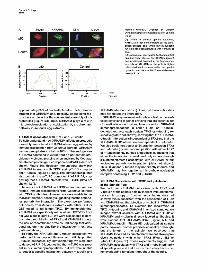
Figure 3. XRHAMM Depends on Dynein/Dynactin Complex to Concentrate on SpindlePoles
(A) Unlike in control spindle reactions,XRHAMM is not concentrated on the unfo-cused spindle pole when dynein/dynactinfunction has been perturbed with 1 mg/ml ofp50.(B) Linescans of p50 treated (left) and controlspindles (right) stained for XRHAMM (green)and tubulin (red). Notice that the fluorescenceintensity of XRHAMM at the pole is higherrelative to the midzone only when the dynein/dynactin complex is active. The scale bar rep-resents 2 �m.
approximately 60% of mock-depleted extracts, demon- XRHAMM (data not shown). Thus, �-tubulin antibodiesmay not detect the interaction.strating that XRHAMM and, possibly, codepleting fac-
XRHAMM may make microtubule nucleation more ef-tors have a role in the Ran-dependent assembly of mi-ficient by linking together proteins that are essential forcrotubules (Figure 4D). Thus, XRHAMM plays a role inchromatin-dependent microtubule nucleation. XRHAMMmicrotubule nucleation or stabilization by the chromatinimmunoprecipitations in either TPX2- or �-tubulin-pathway in Xenopus egg extracts.depleted extracts each contain TPX2 or �-tubulin, re-spectively (data not shown), showing that the XRHAMM-
XRHAMM Associates with TPX2 and �-Tubulin�-tubulin interaction is independent of TPX2 and that the
To help understand how XRHAMM affects microtubule XRHAMM-TPX2 interaction is independent of �-tubulin.assembly, we isolated XRHAMM interacting proteins by We also could not detect an interaction between TPX2immunoprecipitation from Xenopus extracts. XRHAMM and �-tubulin (by immunoprecipitation with either TPX2immunoprecipitates contain �90% of the endogenous or �-tubulin affinity purified antibodies), suggesting thatXRHAMM contained in extract but do not contain stoi- either the interaction is weak and only occurs throughchiometric binding proteins when analyzed by Coomas- a substoichiometric association with XRHAMM or oursie-stained protein gel electrophoresis (PAGE) (data not antibodies perturb the interaction (data not shown).shown; Figure 5A). However, immunoblots show that Thus, TPX2 and �-tubulin may not directly interact, andXRHAMM interacts with TPX2 and �-TuRC compon- XRHAMM may link together a microtubule nucleationent �-tubulin (Figure 5B; [23]). The immunoprecipitates complex containing TPX2 and �-TuRC.also contain the �-TuRC component XGRIP109, sug-gesting that XRHAMM interacts with �-TuRC (data not XRHAMM Colocalizes with TPX2 and �-Tubulinshown; [24]). at the Spindle Pole
To verify the XRHAMM and TPX2 interaction, we per- We find that XRHAMM colocalizes with TPX2 andformed immunoprecipitations from Xenopus extracts �-tubulin at the spindle pole by indirect immunofluores-with TPX2 antibodies. However, we were unable to de- cence microscopy of fixed extract spindles (data nottect an interaction, possibly suggesting that the antibod- shown); this is consistent with the association of TPX2ies perturb the interaction. Therefore, we performed and XRHAMM and the detection of �-tubulin in XRHAMMpull-downs from Xenopus extracts with either GST or immunoprecipitates. To examine the localization ofGST fused to full-length TPX2. By immunoblotting, TPX2, �-tubulin, and XRHAMM in unfixed samples, weXRHAMM was specifically retrieved with GST-TPX2 but imaged extract spindles with XRHAMM and TPX2 ornot GST alone (Figure 5C). We were also unable to dem- XRHAMM and �-tubulin directly labeled antibodies. Itonstrate direct binding of TPX2 and XRHAMM through was evident that XRHAMM/TPX2 (Figure 5D) andthe use of recombinant proteins, indicating that addi- XRHAMM/�-tubulin (Figure 5E) colocalized at spindletional factors may stabilize the interaction in extracts poles; however, neither precisely colocalized through-(data not shown). out the length of the spindle. We observed that
To verify the XRHAMM and �-tubulin interaction, we XRHAMM localized as puncta that were not always pre-performed immunoprecipitations with affinity purified cisely coincident with either TPX2 (Figure 5D) or�-tubulin antibodies. By immunoblotting, we were able �-tubulin (Figure 5E). These experiments suggest thatto detect XGRIP109, suggesting that �-TuRC was pres- XRHAMM associates with TPX2 and �-tubulin primarilyent in our immunoprecipitations, but we were unable at spindle poles and that these proteins may have other
nonoverlapping functions throughout the spindle.to detect a specific interaction between �-tubulin and
XRHAMM Functions in Anastral Spindle Assembly1805
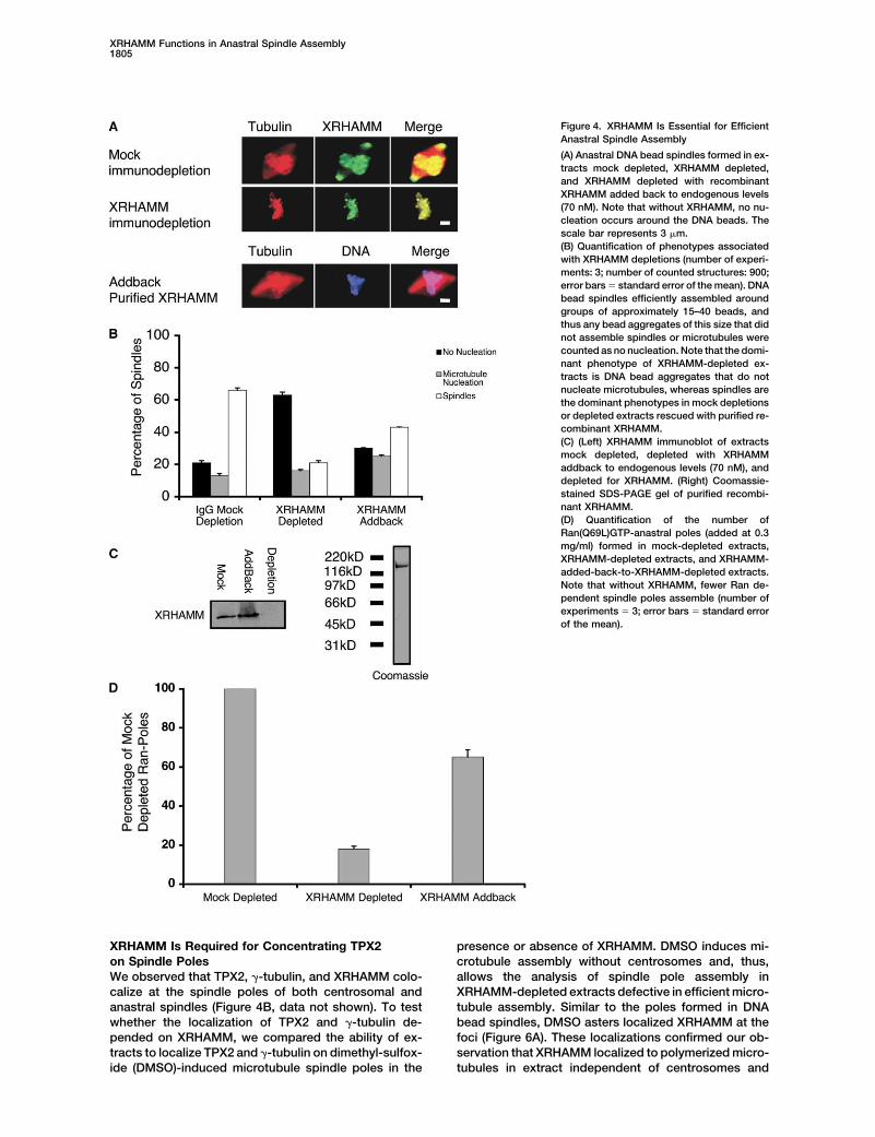
Figure 4. XRHAMM Is Essential for EfficientAnastral Spindle Assembly
(A) Anastral DNA bead spindles formed in ex-tracts mock depleted, XRHAMM depleted,and XRHAMM depleted with recombinantXRHAMM added back to endogenous levels(70 nM). Note that without XRHAMM, no nu-cleation occurs around the DNA beads. Thescale bar represents 3 �m.(B) Quantification of phenotypes associatedwith XRHAMM depletions (number of experi-ments: 3; number of counted structures: 900;error bars � standard error of the mean). DNAbead spindles efficiently assembled aroundgroups of approximately 15–40 beads, andthus any bead aggregates of this size that didnot assemble spindles or microtubules werecounted as no nucleation. Note that the domi-nant phenotype of XRHAMM-depleted ex-tracts is DNA bead aggregates that do notnucleate microtubules, whereas spindles arethe dominant phenotypes in mock depletionsor depleted extracts rescued with purified re-combinant XRHAMM.(C) (Left) XRHAMM immunoblot of extractsmock depleted, depleted with XRHAMMaddback to endogenous levels (70 nM), anddepleted for XRHAMM. (Right) Coomassie-stained SDS-PAGE gel of purified recombi-nant XRHAMM.(D) Quantification of the number ofRan(Q69L)GTP-anastral poles (added at 0.3mg/ml) formed in mock-depleted extracts,XRHAMM-depleted extracts, and XRHAMM-added-back-to-XRHAMM-depleted extracts.Note that without XRHAMM, fewer Ran de-pendent spindle poles assemble (number ofexperiments � 3; error bars � standard errorof the mean).
XRHAMM Is Required for Concentrating TPX2 presence or absence of XRHAMM. DMSO induces mi-crotubule assembly without centrosomes and, thus,on Spindle Poles
We observed that TPX2, �-tubulin, and XRHAMM colo- allows the analysis of spindle pole assembly inXRHAMM-depleted extracts defective in efficient micro-calize at the spindle poles of both centrosomal and
anastral spindles (Figure 4B, data not shown). To test tubule assembly. Similar to the poles formed in DNAbead spindles, DMSO asters localized XRHAMM at thewhether the localization of TPX2 and �-tubulin de-
pended on XRHAMM, we compared the ability of ex- foci (Figure 6A). These localizations confirmed our ob-servation that XRHAMM localized to polymerized micro-tracts to localize TPX2 and �-tubulin on dimethyl-sulfox-
ide (DMSO)-induced microtubule spindle poles in the tubules in extract independent of centrosomes and

Current Biology1806
Figure 5. XRHAMM Interacts with TPX2 and�-tubulin
(A) Coomassie-stained SDS-PAGE of lysatesof XRHAMM and IgG immunoprecipitations.Note the lack of specific stoichiometricXRHAMM-interacting proteins.(B) Immunoblots showing XRHAMM immuno-precipitates with TPX2 and �-tubulin frommeiotic Xenopus extracts.(C) Immunoblots showing that GST-TPX2specifically interacts with XRHAMM.(D) The localizations of XRHAMM and TPX2on unfixed Xenopus egg extract spindles.Note that XRHAMM and TPX2 colocalize pre-dominantly at the spindle pole.(E) The localizations of XRHAMM and �-tubu-lin on unfixed Xenopus egg extract spindles.Note that XRHAMM and �-tubulin only colo-calize at the spindle pole. The scale bar repre-sents 2 �m.
showed that DMSO asters are a comparable model for spindle poles was more diffuse than the localization onmock-depleted controls (Figure 6C). Similar to �-tubulin,our experiment.
The anastral DMSO-induced spindle poles assembled NuMA and the dynein/dynactin complex were diffuselylocalized near the centers of the XRHAMM-deficientin XRHAMM-depleted extracts were unfocused com-
pared to controls (Figure 6B, top and middle rows), yet aster structures (Figure 6C; data not shown). However,we cannot determine whether these changes in localiza-TPX2 and �-tubulin remain localized to the XRHAMM-
deficient poles (Figure 6B, middle row, and Figure 6C, tion result from, or contribute to, the unfocused spindlepoles assembled in XRHAMM depleted extracts. Thesebottom row). Thus, XRHAMM is not essential for TPX2
and �-tubulin to bind to microtubules. However, in con- localizations do indicate that XRHAMM is not requiredfor the dynein/dynactin-dependent arrangement of mi-trast to the case of mock-depleted extracts (Figure 6B,
top row), TPX2 did not concentrate on the centers of crotubules in a spindle pole structure [25] but ratherplays a role in focusing poles, possibly through the activ-the poles assembled in XRHAMM-depleted extracts but
instead localized all over the microtubule structures (Fig- ity of TPX2.ure 6B, middle row). Interestingly, unlike TPX2, �-tubulinlocalized to the centers of the XRHAMM-deficient spin- XRHAMM-Associated Proteins Nucleate
Microtubules in Pure Tubulin Solutionsdle poles (Figure 6C, bottom row). The addition of puri-fied recombinant XRHAMM rescued not only the unfo- We found that XRHAMM plays a role in chroma-
tin-induced microtubule nucleation in Xenopus egg ex-cused poles of the XRHAMM-deficient structures, butalso the mislocalization of TPX2, demonstrating that tracts. To test whether XRHAMM directly induces mi-
crotubule assembly in vitro, we mixed recombinantthese defects are due entirely to the removal ofXRHAMM (Figure 6B, bottom row). XRHAMM with pure tubulin. We found that XRHAMM
did not detectably nucleate microtubules in vitro butThe localization of �-tubulin on XRHAMM-deficient

XRHAMM Functions in Anastral Spindle Assembly1807
Figure 6. XRHAMM Is Essential for Concen-trating TPX2 on Spindle Poles
(A) DMSO-induced microtubule spindle polesconcentrate XRHAMM at the pole. The scalebar represents 3 �m.(B and C) DMSO-induced microtubule polesassembled in XRHAMM-depleted extractsare not focused and do not concentrate TPX2at the pole, whereas NuMA and �-tubulin re-main faintly localized to the unfocused centerof the pole. NuMA and �-tubulin were some-times difficult to image on the unfocusedspindle poles because the distribution wasdispersed and faint. The images shown in thisfigure are representative for the bright distri-butions that were observed in many of thepoles examined (�60%). Addition of purifiedrecombinant XRHAMM rescues the unfo-cused pole defect and the mislocalization ofTPX2. The scale bar represents 3 �m.
could strongly associate and bundle polymerized micro- ure 7C) contain XRHAMM and �-tubulin but were unableto nucleate microtubules in pure tubulin solutions undertubules (data not shown). Thus, XRHAMM indirectly con-
tributes to microtubule nucleation. any condition (Figure 7Bii; data not shown). Addition ofrecombinant TPX2 to the extract could rescue this de-We next tested whether XRHAMM plus associated
proteins could nucleate microtubules by adding mag- fect. Addition of TPX2 to beads after removal from TPX2-depleted extract, however, could not rescue this defectnetic beads coated with anti-XRHAMM to Xenopus ex-
tract. As expected, XRHAMM beads associate with (data not shown). Thus, TPX2 is essential for microtubulenucleation from XRHAMM beads, both in extracts andTPX2 and �-TuRC (data not shown). However, we did not
observe microtubule assembly (data not shown; Figure with pure tubulin, but the TPX2 must be present in ex-tract perhaps to promote assembly of nucleation com-7Ai). To test whether the protein complexes recruited to
these beads were able to nucleate microtubules in vitro plexes.We also tested if nucleation from XRHAMM beadswithout the presence of various catastrophe factors
present in extract, we washed the beads and challenged was dependent on �-TuRC, an XRHAMM-associatedprotein complex that has an established role in microtu-them with pure tubulin below the critical concentration
of self-assembly (1 mg/ml). Once again, we did not ob- bule nucleation [26]. XRHAMM beads isolated fromRan(Q69L)-GTP and nocodazole-treated extracts immu-serve microtubule assembly (Figure 7Aii).
We next tested whether Ran(Q69L)-GTP could induce nodepleted of �-tubulin (approximately 80%–90% de-pleted), an essential component of the �-TuRC [25], con-microtubule assembly from XRHAMM beads. When
XRHAMM beads were placed in extract treated with tain XRHAMM and TPX2 but were unable to nucleatemicrotubules in pure tubulin solutions (Figures 7Biii andRan(Q69L)-GTP, microtubules assembled around the
beads (Figure 7Aiii). Experiments in extract cannot distin- 7C; data not shown). We have so far been unable topurify enough functional �-tubulin to complement theguish microtubule nucleation versus stabilization; there-
fore, we washed and incubated the beads in pure tubulin depleted extract. Thus, similar to microtubule assemblyaround chromatin, microtubule nucleation by X-RHAMMsolutions (1 mg/ml). XRHAMM beads were added to
Ran(Q69L)GTP-treated extract plus nocodazole to sup- beads is dependent on RanGTP, TPX2, and �-tubulin.press microtubule polymerization and were incubatedfor 20–30 min (Figure 7Aiv). The beads were then re- Conclusions
In this study, we identify XRHAMM, the Xenopus or-trieved, washed at 4�C (to ensure complete microtubuledepolymerization), and challenged with pure tubulin tholog of human RHAMM/IHABP. Previously, RHAMM/
IHABP was thought to have intracellular and extracel-(1 mg/ml). Remarkably, these beads nucleated abundantmicrotubules in vitro (Figure 7Av). Interestingly, microtu- lular functions involving the glycosaminoglycan hyal-
uronan, but recent studies have demonstrated thatbule assembly was not detected when TPX2 or �-tubulinbound antibody beads were incubated in Ran(Q69L)- RHAMM/IHABP colocalizes with microtubules and is im-
portant for maintaining spindle pole stability, suggestingGTP-treated extract (data not shown), possibly sug-gesting that the TPX2 or �-tubulin antibodies may per- that RHAMM/IHABP function instead involves the micro-
tubule cytoskeleton [15]. Consistent with this idea wasturb the function of the nucleation complex.Because TPX2 associates with XRHAMM and has our identification of XRHAMM as one of the predominant
MAPs in meiotic Xenopus egg extracts; we showed thatbeen implicated in microtubule nucleation, we testedwhether microtubule nucleation from XRHAMM beads it not only associates with proteins established in micro-
tubule nucleation but is essential for efficient anastralwas TPX2-dependent [9]. XRHAMM beads isolated fromRan(Q69L)GTP and nocodazole-treated extracts immu- spindle assembly. Our data show that XRHAMM has
two functions: to help to focus the anastral pole andnodepleted for TPX2 (approximately 80% depleted; Fig-
Current Biology1808
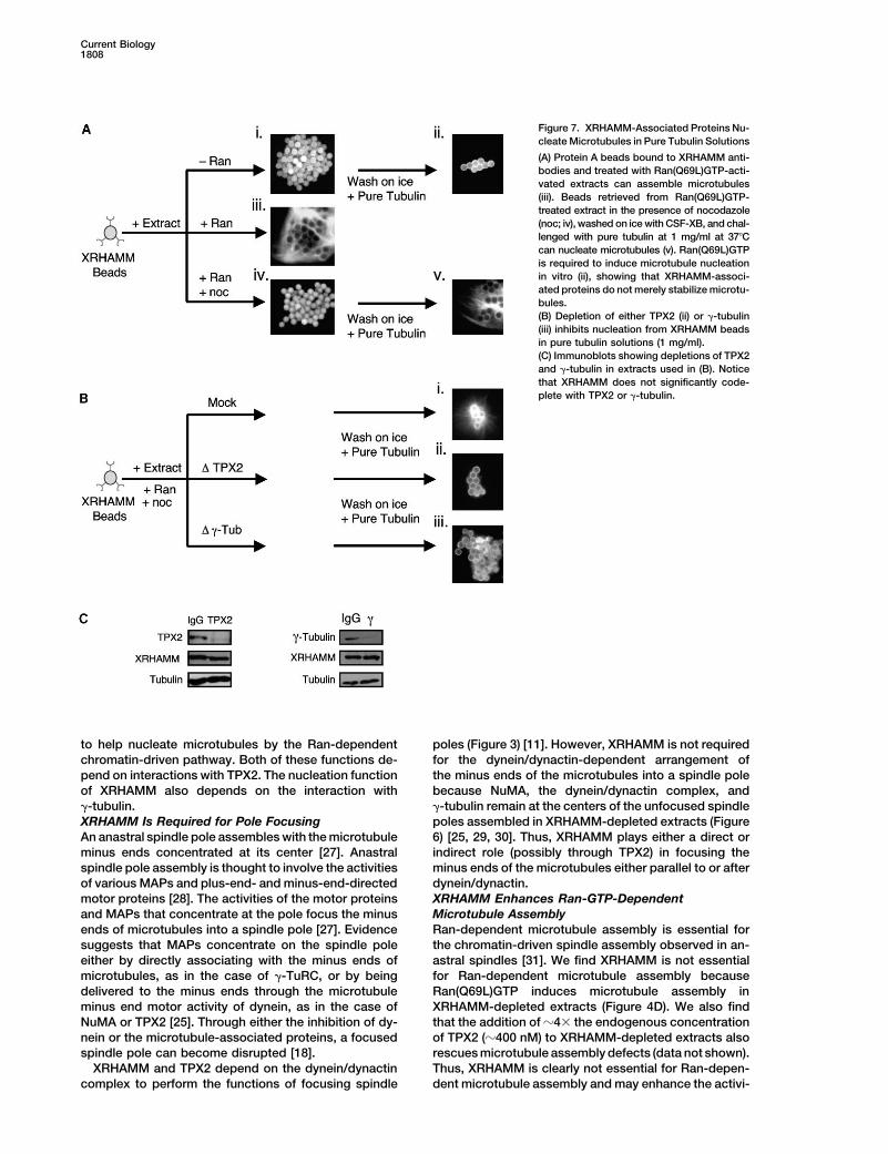
Figure 7. XRHAMM-Associated Proteins Nu-cleate Microtubules in Pure Tubulin Solutions
(A) Protein A beads bound to XRHAMM anti-bodies and treated with Ran(Q69L)GTP-acti-vated extracts can assemble microtubules(iii). Beads retrieved from Ran(Q69L)GTP-treated extract in the presence of nocodazole(noc; iv), washed on ice with CSF-XB, and chal-lenged with pure tubulin at 1 mg/ml at 37�Ccan nucleate microtubules (v). Ran(Q69L)GTPis required to induce microtubule nucleationin vitro (ii), showing that XRHAMM-associ-ated proteins do not merely stabilize microtu-bules.(B) Depletion of either TPX2 (ii) or �-tubulin(iii) inhibits nucleation from XRHAMM beadsin pure tubulin solutions (1 mg/ml).(C) Immunoblots showing depletions of TPX2and �-tubulin in extracts used in (B). Noticethat XRHAMM does not significantly code-plete with TPX2 or �-tubulin.
to help nucleate microtubules by the Ran-dependent poles (Figure 3) [11]. However, XRHAMM is not requiredfor the dynein/dynactin-dependent arrangement ofchromatin-driven pathway. Both of these functions de-
pend on interactions with TPX2. The nucleation function the minus ends of the microtubules into a spindle polebecause NuMA, the dynein/dynactin complex, andof XRHAMM also depends on the interaction with
�-tubulin. �-tubulin remain at the centers of the unfocused spindlepoles assembled in XRHAMM-depleted extracts (FigureXRHAMM Is Required for Pole Focusing
An anastral spindle pole assembles with the microtubule 6) [25, 29, 30]. Thus, XRHAMM plays either a direct orindirect role (possibly through TPX2) in focusing theminus ends concentrated at its center [27]. Anastral
spindle pole assembly is thought to involve the activities minus ends of the microtubules either parallel to or afterdynein/dynactin.of various MAPs and plus-end- and minus-end-directed
motor proteins [28]. The activities of the motor proteins XRHAMM Enhances Ran-GTP-DependentMicrotubule Assemblyand MAPs that concentrate at the pole focus the minus
ends of microtubules into a spindle pole [27]. Evidence Ran-dependent microtubule assembly is essential forthe chromatin-driven spindle assembly observed in an-suggests that MAPs concentrate on the spindle pole
either by directly associating with the minus ends of astral spindles [31]. We find XRHAMM is not essentialfor Ran-dependent microtubule assembly becausemicrotubules, as in the case of �-TuRC, or by being
delivered to the minus ends through the microtubule Ran(Q69L)GTP induces microtubule assembly inXRHAMM-depleted extracts (Figure 4D). We also findminus end motor activity of dynein, as in the case of
NuMA or TPX2 [25]. Through either the inhibition of dy- that the addition of �4� the endogenous concentrationof TPX2 (�400 nM) to XRHAMM-depleted extracts alsonein or the microtubule-associated proteins, a focused
spindle pole can become disrupted [18]. rescues microtubule assembly defects (data not shown).Thus, XRHAMM is clearly not essential for Ran-depen-XRHAMM and TPX2 depend on the dynein/dynactin
complex to perform the functions of focusing spindle dent microtubule assembly and may enhance the activi-

XRHAMM Functions in Anastral Spindle Assembly1809
previously [12]. Proteins were identified by liquid chromatogra-ties of XRHAMM-interacting proteins such as TPX2,phy-mass spectrometry/mass spectrometry (LC-MS/MS) as de-making microtubule nucleation more efficient in Xeno-scribed [12].pus egg extracts.
How Does Chromatin-Driven MicrotubuleXRHAMM-Microtubule Copelleting Assay
Nucleation Occur? Approximately 1 �M of taxol-stabilized GMPCPP-polymerized mi-The pathway of chromatin-induced microtubule nucle- crotubules (5 �g of XRHAMM, bound on ice for 5 min) was spunation during anastral spindle assembly is not well de- through BRB80/30% glycerol cushion at 227,000 � g. Samples were
diluted in sample buffer and analyzed by immunoblots.fined. One problem is that experiments in extracts orin vivo cannot clearly distinguish microtubule nucleation
Xenopus Extractfrom microtubule stabilization. The two are typicallyMetaphase-arrested Xenopus extract (CSF), X-rhodamine-labeled tu-scored together as “microtubule assembly.” Becausebulin, demembranated sperm nuclei, and extract spindles were pre-
cells and extracts contain multiple microtubule stabiliz- pared as described previously [34–36]. Cycled meiotic spindles assem-ing and destabilizing factors, this distinction can only bled from sperm nuclei were formed as previously described [34].be made with pure tubulin. Our assay with XRHAMM Anastral DNA bead spindles were formed as detailed previously [21].
XRHAMM-, TPX2-, and �-tubulin-immunodepleted CSF was madebeads is the first assay in which the Ran-dependentby three rounds of incubating 50 �l of ProteinA dynabeads (Dynal)microtubule nucleation pathway has been observed inbound to 20 �g of affinity purified XRHAMM polyclonal antibodiespure tubulin solutions (Figure 7).with 150 �l of extract for 1 hr at 4�C. Immunoblots were used toWe find that Ran-dependent microtubule nucleationconfirm depletions.
in pure tubulin solutions depends on both TPX2 and�-tubulin. Although the �-TuRC, which contains �-tubu- Microtubule Pole Formation in Xenopus Extractlin, has an established role in microtubule nucleation, it DMSO microtubule poles were formed with the addition of 5%
DMSO and were imaged 20 min later with directly labeled tubulinis not clear how TPX2 precisely functions in this process.(70 �g/ml), directly labeled TPX2, �-tubulin, and NuMA antibodiesTPX2 is essential for the Ran-dependent activation of(10–50 �g/ml). RanGTP poles were induced by the addition ofAurora A kinase, yet it is not known whether this activeRan(Q69L)GTP to 0.3 mg/ml and were imaged similarly [22, 37].kinase has a role in microtubule assembly [32]. We foundThe number of Ran-induced poles was counted from each sample
that Aurora kinase inhibitors [33] did not inhibit Ran- (mock-depleted, XRHAMM-depleted, and XRHAMM addback todependent microtubule nucleation (from XRHAMM XRHAMM-depleted extracts) per 2 �l of extract and was expressedbeads in pure tubulin solutions or in extract), suggesting as a percentage of mock-depleted poles for each experiment.that Aurora A kinase plays no role in Ran-dependent
Immunofluorescencemicrotubule assembly. Recombinant TPX2 can nucleateTPX2 and XRHAMM colocalization on spindles was imaged in realmicrotubules in vitro, showing that TPX2 may play atime with spinning disk confocal microscopy. For details, see thecentral role in the mechanism of microtubule nucleationSupplemental Data available with this article online. Images were[9], though the additional requirement of �-tubulin foracquired with a 100�/1.4 NA Plan Apochromat objective (Nikon) on
microtubule nucleation in extract suggests that the a Nikon TE-300 inverted microscope fitted with a Yokogawa spin-mechanism is more complex. ning disk confocal head (Perkin Elmer) and Orca ER cooled CCD
Although the XRHAMM bead assay reported here is camera set to 2 � 2 binning (Hamamatsu) with MetaMorph software(Universal Imaging) to capture the 488 and 568 wavelengths almosta step forward, the precise roles of Ran, TPX2, �-tubulin,simultaneously, as previously described [38].and XRHAMM in nucleation are not yet understood. Mi-
The localization of �-tubulin, XRHAMM, NuMA, and the dynein/crotubule nucleation depends on the presence of Ran-dynactin complex on spindles and DMSO-induced spindle polesGTP in the extract, but so far we have detected nowere imaged on an upright Nikon E800 with a cooled CCD (Princeton
difference in the protein composition of XRHAMM beads Instruments) and MetaMorph software to control shutters, wave-retrieved from Ran-GTP and control extracts. The two length selection, and image acquisition with directly labeledproteins we know are essential for nucleation, �-tubulin �-tubulin, TPX2, and NuMA antibodies (10–50 �g/ml) and directly
labeled p50 (32 �g/ml).and TPX2, are recruited to similar levels in both cases.XTC cells were fixed (and permeabilized at the same time) withWe also find that the addition of importin �/�, a complex
0.1% gluteraldehyde and 2% PFA in 200 mM KPIPES, 0.4% Triton,that binds to and sequesters the activities of TPX2, in-20 mM KEGTA, and 2mM MgCl2 and stained for immunofluores-hibits in vitro microtubule nucleation from XRHAMM cence. Directly labeled antibodies were used at 1 �g/ml.
beads (data not shown), suggesting that the importinshave a role in regulating the nucleation complex. How- Construction of XRHAMM, TPX2, and Ran(Q69L) Plasmidsever, because the importins did not coimmunoprecipi- Xenopus TPX2 was amplified by PCR from a Xenopus ovary cDNA
library (a kind gift from Aaron Straight, Stanford University) with thetate with XRHAMM (as analyzed by immunoblots), wefollowing primers: TPX_GEX_F (5-CGCCCGGGGATGGAAGATACAsuspect that either our antibodies perturb the interactionCAGGACACC-3) and TPX_GEX_R (5-CGGCGGCCGCTCAACACTTor the importins may regulate an unidentified factor (dataAAACCTGTCCGAGAAG-3). The amplified DNA was digested withnot shown). We suppose that undetermined proteins areSma1 and Not1 and inserted into the Sma1/Not1 sites of pGEX4T-1
required in addition to TPX2 and �-tubulin and/or that (Amersham Biosciences). The construct pGST-TPX2 was created.one or more must be posttranslationally modified. In Oligos XRHAMM_5, 5-AGTCATTGCCCAGTCAGC-3) andfuture experiments, we will use pure proteins to reconsti- XRHAMM_6 (5-CAAACAAGTGGCGCACCC-3, corresponding to
nucleotides 3113–3130 of the XRHAMM coding sequence and atute microtubule nucleation testing models in whichportion of the 3-UTR, respectively, were used to PCR amplify a 430TPX2 may activate �-TuRC for nucleation.base pair fragment of XRHAMM from the Xenopus ovary cDNAlibrary and were used to screen the same library. The clone con-Experimental Procedurestaining the largest insert was sequenced completely, and an SpeIsite was introduced immediately upstream of the XRHAMM codingIdentification of XRHAMMregion by PCR to produce pSK(�)XRHAMM_SpeI. The XRHAMMIdentification of binding proteins to taxol-stabilized GMPCPP-poly-
merized microtubules in Xenopus meiotic extracts was described coding region was excised from this plasmid with SpeI and li-
Current Biology1810
gated into the SpeI site of pFASTBAC HTc (Invitrogen), creating AcknowledgmentspFASTBAC_XRHAMM.
pMAL-Ran(Q69L)GTP (a kind gift from Adrian Salic) was sub- We thank William S. Lane for mass spectrometry and members ofthe Mitchison lab and M. Shirasu-Hiza, K. Kozminski, B. Brieher,cloned into the BamH1 and Sal1 sites of pGEX4T-1.and E.D. Salmon for comments on the manuscript. We thank R.Heald, T. Maresca, Y. Zheng, and J. Walter for antibodies. We thankPurification of Recombinant Ran(Q69L)GTPA. Salic for Ran(Q69L) constructs and A. Straight for the ovary cDNAGST-Ran(Q69L) was purified from BL21(DE3) (Stratagene) bacteriaXenopus library. This work was supported by National Institutes ofwith conventional purification methods for GST agarose beadsHealth grants to T.J.M. (GM39565) and R.O. (GM20309) and a Na-(Sigma). The purified protein was dialyzed in the following buffer:tional Science Foundation Graduate Research fellowship to A.C.G.10 mM HEPES, 200 mM KCl, and 1 mM GTP (pH 7.7). GST-TPX2L.A.C. is supported by postdoctoral fellowship PF-02-095-01-CCGwas purified according to the same protocol, except that GTP andfrom the American Cancer Society.EDTA were not included in the buffers.
Purification of Recombinant XRHAMM Received: June 26, 2004pFASTBAC_XRHAMM was used to produce a baculovirus, as de- Revised: August 25, 2004tailed in the Bac-to-Bac system (Invitrogen). His-XRHAMM was puri- Accepted: September 9, 2004fied from SF9 cells (Invitrogen) with conventional purification meth- Published: October 26, 2004ods. The purified protein was gel filtrated on a Superdex-200 column(Amersham Biosciences) equilibrated with 10 mM HEPES and 200
ReferencesmM KCl (pH 7.7).
1. Inoue, S., and Ritter, H., Jr. (1975). Dynamics of mitotic spindlePurification of p50organization and function. Soc. Gen. Physiol. Ser. 30, 3–30.p50 was purified as described previously [18].
2. Inoue, S. (1981). Cell division and the mitotic spindle. J. CellBiol. 91, 131s–147s.Preparation of XRHAMM, Centrin, and �-Tubulin Antibodies
3. Gaglio, T., Dionne, M.A., and Compton, D.A. (1997). Mitotic spin-The C-terminal 137 amino acids of XRHAMM were fused to the Cdle poles are organized by structural and motor proteins interminus of GST, and the resulting fusion protein (GST-XRHAMM.C1)addition to centrosomes. J. Cell Biol. 138, 1055–1066.was purified from E. coli BL21DE3/pLysS cells with glutathione aga-
4. Cullen, C.F., and Ohkura, H. (2001). Msps protein is localizedrose (Sigma). Xenopus centrin, PCR amplified from the ovary cDNAto acentrosomal poles to ensure bipolarity of Drosophila meioticlibrary described above, was also expressed and purified fromspindles. Nat. Cell Biol. 3, 637–642.BL21DE3/pLysS as a GST fusion protein. GST-XRHAMM.C1 and
5. Mitchison, T., and Kirschner, M. (1984). Microtubule assemblyGST-Xcentrin were used to immunize rabbits (Cocalico) and werenucleated by isolated centrosomes. Nature 312, 232–237.affinity purified over Affi-Gel 10 (BioRad) columns covalently
6. Wilde, A., and Zheng, Y. (1999). Stimulation of microtubule asterattached to the recombinant immunogens after depletion of �-GSTformation and spindle assembly by the small GTPase Ran. Sci-antibodies from the sera.ence 284, 1359–1362.
�-tubulin (Xen-C) C-terminal peptide antibodies were created as7. Nachury, M.V., Maresca, T.J., Salmon, W.C., Waterman-Storer,described previously [26, 39]. NuMA peptide antibodies were pre-
C.M., Heald, R., and Weis, K. (2001). Importin beta is a mitoticpared as described previously [39].target of the small GTPase Ran in spindle assembly. Cell 104,95–106.
Immunoprecipitations and Pull-Down Assays 8. Kalab, P., Weis, K., and Heald, R. (2002). Visualization of a Ran-Approximately 10 �g of XRHAMM antibodies (or rabbit IgG) were GTP gradient in interphase and mitotic Xenopus egg extracts.bound to 10 �l AffiPrep protein A beads (BioRad) and incubated Science 295, 2452–2456.with 150 �l of CSF for 1 hr at 4�C. Beads were washed six times 9. Schatz, C.A., Santarella, R., Hoenger, A., Karsenti, E., Mattaj,with TBS and mixed with 15 �l of 2� sample buffer. Approximately I.W., Gruss, O.J., and Carazo-Salas, R.E. (2003). Importin alpha-1 �l of each sample was run on SDS-PAGE, transferred to nitrocellu- regulated nucleation of microtubules by TPX2. EMBO J. 22,lose, and probed for by antibodies for TPX2, XHRAMM, and �-tubulin 2060–2070.(Sigma). 10. Gaglio, T., Saredi, A., Bingham, J.B., Hasbani, M.J., Gill, S.R.,
Approximately 20 �l of glutathione beads (Sigma) were prebound Schroer, T.A., and Compton, D.A. (1996). Opposing motor activi-with 125 �g of GST-TPX2 (or GST) in the presence of 5% bovine ties are required for the organization of the mammalian mitoticserum albumin (BSA) in CSF-XB (10 mM K-HEPES [pH 7.7], 100 mM spindle pole. J. Cell Biol. 135, 399–414.KCl, 1 mM MgCl2, 0.1 mM CaCl2, 50 mM sucrose, 1 mM MgCl2, 11. Wittmann, T., Wilm, M., Karsenti, E., and Vernos, I. (2000). TPX2,and 5 mM EGTA). Beads were washed four times in CSF-XB and A novel Xenopus MAP involved in spindle pole organization. J.incubated in 150 �l of CSF. The beads were washed five times with Cell Biol. 149, 1405–1418.TBS and mixed with 25 �l of 2� sample buffer. Approximately 2 �l 12. Ohi, R., Coughlin, M.L., Lane, W.S., and Mitchison, T.J. (2003).of each sample was run on SDS-PAGE, transferred to nitrocellulose, An inner centromere protein that stimulates the microtubuleand immunoblotted with antibodies for TPX2 and XRHAMM. depolymerizing activity of a KinI kinesin. Dev. Cell 5, 309–321.
13. Lupas, A., Van Dyke, M., and Stock, J. (1991). Predicting CoiledXRHAMM Bead Nucleation Assays Coils from Protein Sequences. Science 252, 1162–1164.XRHAMM magnetic beads isolated from immunodepletions (as de- 14. Lynn, B.D., Li, X., Cattini, P.A., Turley, E.A., and Nagy, J.I. (2001).scribed above) were washed three times with CSF-XB and were Identification of sequence, protein isoforms, and distribution ofdiluted in 50 �l of CSF-XB. About 10–15 �l of this bead mixture was the hyaluronan-binding protein RHAMM in adult and developingadded to 200 �l of fresh meiotic Xenopus egg extract and incubated rat brain. J. Comp. Neurol. 439, 315–330.for 5 min at 20�C. Then, 10 �m nocodazole and 1 mg/ml 15. Assmann, V., Jenkinson, D., Marshall, J.F., and Hart, I.R. (1999).RanQ69L(GTP) were added to the extract and incubated for 20 min The intracellular hyaluronan receptor RHAMM/IHABP interactsat 20�C. The beads were then retrieved, washed three times with 4� with microtubules and actin filaments. J. Cell Sci. 112, 3943–BRB80, incubated at 37�C in an X-rhodamine-labeled tubulin mix 3954.(1 mg/ml), and viewed by fluorescent microscopy for microtubule 16. Hardwick, C., Hoare, K., Owens, R., Hohn, H.P., Hook, M.,assembly. Moore, D., Cripps, V., Austen, L., Nance, D.M., and Turley, E.A.
(1992). Molecular cloning of a novel hyaluronan receptor thatmediates tumor cell motility. J. Cell Biol. 117, 1343–1350.Supplemental Data
Additional Experimental Procedures for this article are available at 17. Maxwell, C.A., Keats, J.J., Crainie, M., Sun, X., Yen, T., Shibuya,E., Hendzel, M., Chan, G., and Pilarski, L.M. (2003). RHAMM iswww.current-biology.com/cgi/content/full/14/20/1801/DC1/.

XRHAMM Functions in Anastral Spindle Assembly1811
a centrosomal protein that interacts with dynein and maintains 38. Maddox, P., Desai, A., Oegema, K., Mitchison, T.J., and Salmon,E.D. (2002). Poleward microtubule flux is a major component ofspindle pole stability. Mol. Biol. Cell 14, 2262–2276.
18. Wittmann, T., and Hyman, T. (1999). Recombinant p50/dynami- spindle dynamics and anaphase a in mitotic Drosophila em-bryos. Curr. Biol. 12, 1670–1674.tin as a tool to examine the role of dynactin in intracellular
processes. Methods Cell Biol. 61, 137–143. 39. Field, C.M., Oegema, K., Zheng, Y., Mitchison, T.J., and Walc-zak, C.E. (1998). Purification of cytoskeletal proteins using pep-19. Paschal, B.M., Holzbaur, E.L., Pfister, K.K., Clark, S., Meyer,
D.I., and Vallee, R.B. (1993). Characterization of a 50-kDa poly- tide antibodies. Methods Enzymol. 298, 525–541.peptide in cytoplasmic dynein preparations reveals a complexwith p150GLUED and a novel actin. J. Biol. Chem. 268, 15318– Accession Numbers15323.
20. Echeverri, C.J., Paschal, B.M., Vaughan, K.T., and Vallee, R.B. The GenBank accession number for the XMAP150 sequence re-ported in this article is AY729653.(1996). Molecular characterization of the 50-kD subunit of dy-
nactin reveals function for the complex in chromosome align-ment and spindle organization during mitosis. J. Cell Biol. 132,617–633.
21. Heald, R., Tournebize, R., Blank, T., Sandaltzopoulos, R.,Becker, P., Hyman, A., and Karsenti, E. (1996). Self-organizationof microtubules into bipolar spindles around artificial chromo-somes in Xenopus egg extracts. Nature 382, 420–425.
22. Bischoff, F.R., Klebe, C., Kretschmer, J., Wittinghofer, A., andPonstingl, H. (1994). RanGAP1 induces GTPase activity of nu-clear Ras-related Ran. Proc. Natl. Acad. Sci. USA 91, 2587–2591.
23. Zheng, Y., Wong, M.L., Alberts, B., and Mitchison, T. (1995).Nucleation of microtubule assembly by a gamma-tubulin-con-taining ring complex. Nature 378, 578–583.
24. Martin, O.C., Gunawardane, R.N., Iwamatsu, A., and Zheng, Y.(1998). Xgrip109: A gamma tubulin-associated protein with anessential role in gamma tubulin ring complex (gammaTuRC)assembly and centrosome function. J. Cell Biol. 141, 675–687.
25. Moritz, M., Braunfeld, M.B., Guenebaut, V., Heuser, J., andAgard, D.A. (2000). Structure of the gamma-tubulin ring com-plex: A template for microtubule nucleation. Nat. Cell Biol. 2,365–370.
26. Zheng, Y., Wong, M.L., Alberts, B., and Mitchison, T. (1998).Purification and assay of gamma tubulin ring complex. MethodsEnzymol. 298, 218–228.
27. Heald, R., Tournebize, R., Habermann, A., Karsenti, E., and Hy-man, A. (1997). Spindle assembly in Xenopus egg extracts: Re-spective roles of centrosomes and microtubule self-organiza-tion. J. Cell Biol. 138, 615–628.
28. Walczak, C.E., Vernos, I., Mitchison, T.J., Karsenti, E., andHeald, R. (1998). A model for the proposed roles of differentmicrotubule-based motor proteins in establishing spindle bipo-larity. Curr. Biol. 8, 903–913.
29. Merdes, A., Ramyar, K., Vechio, J.D., and Cleveland, D.W.(1996). A complex of NuMA and cytoplasmic dynein is essentialfor mitotic spindle assembly. Cell 87, 447–458.
30. Merdes, A., Heald, R., Samejima, K., Earnshaw, W.C., and Cleve-land, D.W. (2000). Formation of spindle poles by dynein/dynac-tin-dependent transport of NuMA. J. Cell Biol. 149, 851–862.
31. Carazo-Salas, R.E., Guarguaglini, G., Gruss, O.J., Segref, A.,Karsenti, E., and Mattaj, I.W. (1999). Generation of GTP-boundRan by RCC1 is required for chromatin-induced mitotic spindleformation. Nature 400, 178–181.
32. Eyers, P.A., Erikson, E., Chen, L.G., and Maller, J.L. (2003). Anovel mechanism for activation of the protein kinase aurora a.Curr. Biol. 13, 691–697.
33. Henri, J.F., George, B.A., John, K.N., and Austen, M.A. (2001).World Intellectual Property Organization (WPO) patentWO0121596.
34. Desai, A., Murray, A., Mitchison, T.J., and Walczak, C.E. (1999).The use of Xenopus egg extracts to study mitotic spindle as-sembly and function in vitro. Methods Cell Biol. 61, 385–412.
35. Hyman, A., Drechsel, D., Kellogg, D., Salser, S., Sawin, K., Stef-fen, P., Wordeman, L., and Mitchison, T. (1991). Preparation ofmodified tubulins. Methods Enzymol. 196, 478–485.
36. Lohka, M.J., and Masui, Y. (1984). Roles of cytosol and cyto-plasmic particles in nuclear envelope assembly and sperm pro-nuclear formation in cell-free preparations from amphibianeggs. J. Cell Biol. 98, 1222–1230.
37. Stewart, M., Kent, H.M., and McCoy, A.J. (1998). The structureof the Q69L mutant of GDP-Ran shows a major conformationalchange in the switch II loop that accounts for its failure to bindnuclear transport factor 2 (NTF2). J. Mol. Biol. 284, 1517–1527.