Cortical dendritic activity correlates with spindle-rich ... - Nature
Upload
independentCategory
view
4download
0
TBCD Links Centriologenesis, Spindle MicrotubuleDynamics, and Midbody Abscission in Human CellsMonica Lopez Fanarraga1, Javier Bellido1, Cristina Jaen1, Juan Carlos Villegas2, Juan Carlos Zabala1*
1 Departamentos de Biologıa Molecular, Instituto de Formacion e Investigacion Marques de Valdecilla Facultad de Medicina, Universidad de Cantabria, Santander, Spain,
2 Anatomıa y Biologıa Celular, Instituto de Formacion e Investigacion Marques de Valdecilla Facultad de Medicina, Universidad de Cantabria, Santander, Spain
Abstract
Microtubule-organizing centers recruit a- and b-tubulin polypeptides for microtubule nucleation. Tubulin synthesis iscomplex, requiring five specific cofactors, designated tubulin cofactors (TBCs) A–E, which contribute to various aspects ofmicrotubule dynamics in vivo. Here, we show that tubulin cofactor D (TBCD) is concentrated at the centrosome andmidbody, where it participates in centriologenesis, spindle organization, and cell abscission. TBCD exhibits a cell-cycle-specific pattern, localizing on the daughter centriole at G1 and on procentrioles by S, and disappearing from older centriolesat telophase as the protein is recruited to the midbody. Our data show that TBCD overexpression results in microtubulerelease from the centrosome and G1 arrest, whereas its depletion produces mitotic aberrations and incomplete microtubuleretraction at the midbody during cytokinesis. TBCD is recruited to the centriole replication site at the onset of thecentrosome duplication cycle. A role in centriologenesis is further supported in differentiating ciliated cells, where TBCD isorganized into ‘‘centriolar rosettes’’. These data suggest that TBCD participates in both canonical and de novo centriolarassembly pathways.
Citation: Fanarraga ML, Bellido J, Jaen C, Villegas JC, Zabala JC (2010) TBCD Links Centriologenesis, Spindle Microtubule Dynamics, and Midbody Abscission inHuman Cells. PLoS ONE 5(1): e8846. doi:10.1371/journal.pone.0008846
Editor: Daniel Lew, Duke University Medical Centre, United States of America
Received October 12, 2009; Accepted January 4, 2010; Published January 22, 2010
Copyright: � 2010 Lopez Fanarraga et al. This is an open-access article distributed under the terms of the Creative Commons Attribution License, which permitsunrestricted use, distribution, and reproduction in any medium, provided the original author and source are credited.
Funding: This study was funded by BFU2007-64882 and Consolider-Ingenio Spanish Ministry of Education and Science Centrosome-3D (http://ciencia.micinn.fecyt.es/ciencia/jsp/plantilla.jsp?area = consolider&id = 51) and the Instituto de Formacion e Investigacion Marques de Valdecilla (http://www.fmdv.org/index2.htm). The funders had no role in study design, data collection and analysis, decision to publish, or preparation of the manuscript.
Competing Interests: The authors have declared that no competing interests exist.
* E-mail: [email protected]
Introduction
Understanding how the centrosomal components function and
are organized during the cell cycle could shed light on many
human diseases, from ciliary syndromes to cancer. The centro-
some is the major microtubule-organizing center (MTOC) in
animal cells, consisting of two centrioles surrounded by protein-
aceous pericentriolar material (PCM), from which microtubules
nucleate and are released. Microtubules are built of ab-tubulin
heterodimers organized in a head-to-tail fashion. This confers
polarity on these polymers, with two different microtubule ends:
the plus end, which is more dynamic and oriented towards the
periphery of the cells, and the minus end, generally embedded in
the centrosome. At the centrosomal core, a centriole pair
organizes the surrounding PCM. Centrioles, which are required
to assemble the axonemes of cilia and flagella, are structurally very
complex. In mammals, they are 500 nm long cylinders with a
200 nm diameter consisting of nine blades arranged in a circle,
each containing three highly specialized microtubule segments.
Both the exact composition and the assembly of these peculiar
microtubules are still unknown. There is also imprecise informa-
tion regarding the composition and structure of the PCM, in
which recent studies have identified hundreds of proteins, most of
which play as yet unknown roles [1]. However, one of the most
widely accepted concepts is that MTOCs, and in particular the
centrosome, accumulate ab-tubulin polypeptides as part of the
PCM for microtubule nucleation. This suggests that there is some
tubulin supply at the MTOCs. Yet, the assembly of ab-tubulin
heterodimers is not a trivial matter. The process of association of
one a-tubulin with one b-tubulin molecule requires the co-
ordinated interaction of a series of tubulin-specific partners,
designated tubulin cofactors (TBCs) A–E [2–5]. TBCs also play
roles in tubulin dissociation [5–8], transitory tubulin storage
[5,9–12], and tubulin degradation processes [13–15], all of which
suggest that these proteins, in addition to their original role in
tubulin biogenesis, participate in microtubule dynamics by
controlling the amount of tubulin available for polymerization.
There is considerable evidence in the literature supporting a role
for TBCs in this centrosome. Mutations in TBCD, in particular,
have been shown to produce aberrations in chromosome numbers
in Saccharomyces cerevisiae [16], Schizosaccharomyces pombe
[17], Arabidopsis thaliana [18,19], and Caenorhabditis elegans
[20,21]. TBCD mutations also induce a G1/S blockage and
spindle pole body separation in S. pombe [22,23] and abnormal
cytokinesis [19]. In C. elegans, TBCD silencing results in a
reduced rate of microtubule nucleation and produces abnormal
spindle lengths [21]. Recently, human TBCD (HsTBCD) has been
shown to play a role in the organization of the mitotic spindle, and
has been hypothesized to recruit from among cytosolic centroso-
mal proteins, such as pericentrin or c-tubulin [24].
Our data show that TBCD accumulates in immature centrioles
and at the midbody ring during cytokinesis. We demonstrate
TBCD recruitment into ‘‘centriolar rosettes’’ during basal-body
assembly in a novel primary cell culture system of differentiating
ciliated cells, which we developed for this study. More interest-
ingly, we demonstrate that the manipulation of TBCD levels
PLoS ONE | www.plosone.org 1 January 2010 | Volume 5 | Issue 1 | e8846

produces several abnormalities at the centrosome and midbody,
resulting in spindle and anaphase defects, G1 blockage, and cell
abscission failure. These findings indicate that TBCD is a
fundamental protein in cell division.
Results
TBCD Is Concentrated in Centrioles and MidbodiesTBCD has been reported in the centrosome and has been
shown to co-sediment with c-tubulin when overexpressed [24], but
the exact location of the protein has not been documented. To
investigate more precisely its centrosomal distribution, we first
produced and affinity purified several anti-TBCD antibodies
(Figure S1A), which we used to identify the sub-cellular locations
of this cofactor in HeLa cells, among other systems. We observed
that despite all the biochemical evidence suggesting that TBCD is
predominantly a cytoplasmic protein, TBCD accumulated prom-
inently at the centrosome (Figure 1A), as determined by double
immunostaining for standard markers, such as c-tubulin,
NEDD1/GCP-WD (Figure S1B), and others. Microtubule
depolymerization by nocodazole and cold treatment showed that
TBCD localization at the centrosome is not dependent on
microtubules, thus demonstrating that TBCD behaves as a
genuine centrosomal protein.
Using high-resolution confocal microscopy, we next investigated
the relationship of these TBCD spots with the centrioles and
primary cilium. For this purpose, we doubly/triply immunostained
cells with our antibodies and combinations of various markers,
including GT335, which is a broadly characterized antibody that
recognizes glutamylated tubulin and is typically used to label
centrioles and the primary cilium. We observed one TBCD spot
accompanying the primary cilium in cells at G1, which we
identified as occurring at the daughter centriole by double
immunostaining with an antibody that recognizes e-tubulin, which
did not co-localize with TBCD (Figure 1B–D). As mitosis
progressed, two stronger TBCD signals devoid of GT335 staining
arose immediately adjacent to the existing centrioles. This suggests
that TBCD is localized on developing procentrioles before tubulin
glutamylation.
During late G2, although often four GT335 spots were visible
(Figure 1B, C), only three TBCD spots of different intensities were
observed. Two intense spots were localized on developing
centrioles before tubulin glutamylation, and a weaker signal on
the former daughter centriole. Sometime after cilia disassembly,
the TBCD signal on the original daughter centriole weakened and
by the beginning of mitosis, in prophase, most dividing cells
already showed only two strong TBCD spots localized on the
newest centriole of each pair. Occasionally, a TBCD halo
surrounding older centrioles was also observed (Figure 1B, arrows),
suggesting that this protein connects both the old and the new
centrioles at this point. Partial co-localization of the TBCD signal
with c- and d-tubulin was observed during the cell cycle. TBCD
also appeared to co-localize with e-tubulin in cells at late S/G2
phase (Figure 1D), coinciding with the reported redistribution of
this tubulin [25]. By telophase, after centriole separation, only the
new centriole of each pair was labelled for TBCD. This
phenomenon coincided chronologically with the accumulation of
TBCD in a ring-like structure localized at the midbody at the
point of cell abscission (Figure 1A), where we also identified c- and
e-tubulin (Figure 1E).
Immuno-gold localization of TBCD revealed proximal centrio-
lar and procentriolar labelling, which was also conspicuous at the
proximal end of the basal bodies of the cilia (Figure 1F), a pattern
almost identical to that described for c-tubulin [26]. TBCD
labelling was also observed on ciliary microtubules. We further
confirmed TBCD localization at the proximal end of centrioles
immunostaining GFP-centrin1 transfected HeLa cells (Figure 1G).
The observed cell-cycle-specific pattern and the association of
TBCD with procentrioles implicate this tubulin cofactor in
centriologenesis. To explore this hypothesis further, we performed
additional experiments in different systems. First, we analysed
TBCD expression using immunofluorescence in Chinese hamster
ovary (CHO) cells arrested at G1/S phase by thymidine
treatment. This induces repeated cycles of centriole/centrosome
synthesis, thus providing a convenient assay with which to identify
the molecular components involved in centriologenesis. Our
results confirmed the TBCD immunostaining on developing
centrioles after G1 blockage (Figure S2A). An alternative method
used to trigger massive centriologenesis is the overexpression of
proteins such as PLK4 or SAS-6, which are among the first
proteins reported in the recruitment of centriolar components
[27,28]. Therefore, we overexpressed PLK4 on HeLa cells and
looked for TBCD clustering at the developing centrioles. This
system also confirmed TBCD accumulation at the centrosome
(Figure S2B).
TBCD Overexpression Releases Microtubules from theCentrosome
Human TBCD (HsTBCD) has been reported to behave
differently when overexpressed to the bovine protein (BtTBCD)
[24], which was the first TBCD isolated and has therefore been
more often studied in vitro and in vivo [6,8]. Consequently, we
decided to investigate changes in the microtubule cytoskeleton
organization in cells overexpressing these two orthologues. We
observed that, compared with BtTBCD, HsTBCD was more
condensed at the centrosome and the microtubule cytoskeleton
took longer to depolymerise. Quantification of microtubule
depolymerisation in over-expressing cells at 15 and 30 hours
post-transfection revealed that although both proteins resulted in a
massive microtubule destruction at 30 hours post-transfection
(71% Hs Vs 83% Bt), HsTBCD overexpressing cells exhibited no
obvious microtubule depolymerisation signs at 15 hours (100%
cells contained microtubules) while 82% of cells overexpressing
BtTBCD displayed a complete microtubule destruction. More
interestingly, we found that HsTBCD accumulation appeared to
detach the microtubule minus ends from the centrosome
(Figure 2A). This finding can be understood in view of previous
observations that overexpressed HsTBCD removes c-tubulin from
the centrosome [24] and suggests that this tubulin cofactor could
play a role in microtubule release in c-tubulin-dependent
MTOCs.
We also observed that TBCD overexpression produced
aberrant mitotic figures, where supernumerary MTOCs contain-
ing c-tubulin, but no detectable centrioles, developed (Figure 2B).
These observations suggest that there is a delay at mitosis, which
we next investigated by flow cytometry in cells transfected with
constructs encoding HsTBCD–yellow fluorescent protein (YFP) or
H2B–YFP as the control. Flow cytometry analysis 15 h post-
transfection revealed that 65% of TBCD positive cells were found
at G1 compared to 45% of controls. Hence, we conclude that
TBCD overexpression results in G1 delay (Figure 2C). This
finding is consistent with those described in S. pombe, where
mutations in the TBCD orthologue, Alp1p, produced a G1/S
block [22–23]. In view of recent reports demonstrating that
centrosomal integrity is required for G1/S progression [29], these
findings support the notion that TBCD is a key centrosomal
protein.
TBCD Role in Centriologenesis
PLoS ONE | www.plosone.org 2 January 2010 | Volume 5 | Issue 1 | e8846

Figure 1. TBCD is concentrated in centrioles and midbodies. (A) Confocal-microscopic images of a HeLa cell (ATCCR number: CCL-2TM)immunostained for tubulin, TBCD, and GT335 (which labels glutamylated tubulin at the centrioles and primary cilium), where a single spot of TBCD isobserved close to the basal body of the cilium. HeLa cells in anaphase and undergoing cytokinesis display clear centrosomal labelling andrecruitment of TBCD to the midbody ring, respectively. (B) High-resolution confocal-microscopic section images of doubly immunostainedcentrosomes of HeLa cells. The cells were photographed at different stages of the cell cycle, labelled with GT335 and anti-TBCD antibody. This tubulincofactor is observed at one of the centrioles during G1. Two stronger additional TBCD signals immediately adjacent to the existing centrioles, devoidof GT335 staining, were stained at G1. By late S three TBCD spots were detected. By prophase, most dividing cells contained two strong TBCD spots,one in each centrosome. Partial TBCD halos surrounding older centrioles were also observed (arrows). At the end of telophase, only one of thecentrioles in each daughter cell was labelled for TBCD. (C) Diagram of the distribution of TBCD during the centriolar cycle; centrioles, procentrioles,and primary cilium are shown in red and TBCD is shown in green. (D) Partial co-localization of the TBCD signal with c- and d-tubulins was observedthroughout the cell cycle. The e-tubulin signal concentrated at the mother centriole during G1 did not co-localize with TBCD. Later in the cell cycle,both proteins appeared to partially co-localize. (E) TBCD accumulated at the midbody, where c- and e-tubulins were also detected. Lateral and frontalviews of the structure correspond to different cells. (F) Immuno-electron-microscopic analysis of TBCD on murine epithelial cells. (Left) TBCD localiseson the proximal region of the basal body (the original mother centriole) during procentriole assembly (arrow) S stage. The former daughter centrioleis also observed in the section (gold particles are outlined with circles). (Right top) TBCD labelling was also detected at the proximal ends of basalbodies and (Right bottom) at the outer microtubule doublets of motile tracheal cilia. (G) Immunostaining of HeLa cells transfected with GFP-centrin1(red, pseudocolor) confirm TBCD localization at the proximal end of the daughter centriole.doi:10.1371/journal.pone.0008846.g001
TBCD Role in Centriologenesis
PLoS ONE | www.plosone.org 3 January 2010 | Volume 5 | Issue 1 | e8846

TBCD Contains a Microtubule-Binding Region and TwoCentriolar-Targeting Regions
We next undertook to localize the centriolar-targeting region in
the TBCD polypeptide. Bio-informatic analysis of different TBCD
sequences revealed two segments in the polypeptide, extending
approximately from amino acids (aas) 350 to 550 and from aa
1000 to the C-terminus, with a significant degree of evolutionary
conservation (over 30% and 21% identity between mammals and
yeast, respectively, for the first region, and over 10% for the
second region in both organisms). We next generated constructs
encoding these TBCD segments (aas 377–545 and aas 1006–1200)
by PCR and cloned them for transitory transfection into HeLa
cells for in vivo study. This study was complemented with other
polypeptide segments, including aas 1–324, 325–1200, 324–887,
377–630, and 888–1200, of the BtTBCD sequence (Figure 3A).
The study revealed that the two conserved TBCD protein
segments were directed to the centrosome, co-localizing with
various centrosomal markers (data not shown). Whereas none of
these constructs caused microtubule depolymerization, microtu-
bule pattern changes at the centrosome were frequently observed
(Figure 3B). Weakly TBCD-expressing cells showed two individual
green fluorescent protein (GFP) spots, which we localized to
regions on the centrioles at which c-tubulin was also more
abundant, presumably the proximal region [26] (Figure 3C).
Interestingly, we also noted that both the GFP–TBCD888–1200
and the longer GFP–TBCD324–1200 segments decorated the
microtubules 48 h after transfection (Figure 3D). This finding
suggests that these TBCD regions contain both a centriolar-
targeting sequence and a microtubule/tubulin-binding domain.
TBCD binding to microtubules has also been reported for Alp1p,
the S. pombe orthologue [17]. However, there is so far no
biochemical evidence that mammalian TBCD also binds poly-
merized tubulin. To investigate this point, we performed classical
biochemical microtubule polymerization binding assays with in-
vitro-synthesized radioactive HsTBCD and BtTBCD, but none of
these studies confirmed that TBCD binds to microtubules. We also
investigated this subject in vivo, using the transitory transfection of
TBCD-encoding constructs in taxol-treated HeLa cells. However,
we again failed to confirm that TBCD binds to microtubules. In
conclusion, these results suggest that although TBCD contains a
microtubule/tubulin-binding domain, it does not bind microtu-
bules. CPAP, a protein required for centriole duplication [30], has
also been reported to contain a microtubule-binding domain, but
as observed with TBCD, it does not bind microtubules [31].
Therefore, we hypothesize that this microtubule/tubulin-binding
region is necessary to drive TBCD and tubulins to the
procentriolar structures.
Finally, we also observed that the TBCD fragments discussed
above localized at the Fleming bodies (Figure 3D) during
cytokinesis, and aberrant midbodies with increased lengths and
decreased thicknesses were often observed (Figure 3E, open
arrow). Another frequent finding in weakly expressing cells was an
increased distance between centrioles (.2 mm; Figure 3E).
TBCD Is Required for Mitotic-Spindle Organization andSpindle-Pole Cohesion
There is considerable evidence in the literature suggesting a role
for TBCD in mitosis at metaphase, but no mechanism has yet
Figure 2. TBCD overexpression resulted in microtubule detachment from the centrosome and spindle abnormalities. (A) Confocal-microscopic projected images of a HeLa cell overexpressing HsTBCD doubly immunostained for tubulin and TBCD. (Top) Microtubule release fromthe centrosome was clear in most cells 15 h after transfection (arrow). (Bottom) Microtubule network destruction occurred approximately 30 h aftertransfection. Right panel shows a closer view of the microtubule pattern of the cell labelled with an arrow.(B) TBCD overexpression also producedaberrant mitotic figures, where acentriolar supernumerary MTOCs containing c-tubulin were observed (arrow). (C) Flow-cytometric profiles of cells(.10,000 cells per profile) labelled with Hoechst and transfected with HsTBCD–YFP or a control–YFP construct. Clear G1 arrest, typical of the loss ofcentrosomal integrity, was observed in the cells analysed at both 15 and 30 h after transfection.doi:10.1371/journal.pone.0008846.g002
TBCD Role in Centriologenesis
PLoS ONE | www.plosone.org 4 January 2010 | Volume 5 | Issue 1 | e8846

been established. To understand the role of this cofactor in mitosis,
we silenced TBCD gene expression in HeLa cells with interference
RNA. We used a pool of four synthetic RNAs designed for the
knockdown of the HsTBCD gene, with guaranteed high specificity
and a reduced off-target effect. We investigated TBCD expression
by western blotting at 24, 48, and 72 h after interference. We
observed that by 72 h, there was almost complete depletion of this
protein in HeLa cell extracts, whereas both a- and b-tubulin levels
were apparently unaffected (Figure 4A).
We observed 25% of aberrant mitotic figures and several kinds
of spindle defects were common, such as unipolar, multipolar,
disorganized, and abnormally short spindles (Figure 4B). Acen-
triolar spindle poles and mislocalized centrioles were also often
observed (Figure 4C). We also noted that existing primary cilia
looked more developed in the TBCD-silenced cultures. We
quantified the pole-to-pole lengths of the mitotic spindles and
primary cilia on confocal-microscopic projected images. Statistical
analysis of the data confirmed a highly significant reduction in the
spindle size (degrees of freedom [DF] = 63; P = 561024) and a
significant increase in the lengths of cilia (DF = 147; P = 361023;
Figure 4E). Longer primary cilia might reflect a G1 mitotic block,
reported when changes resulted in centrosomal damage [29].
However, this finding could also indicate that TBCD plays a role
in cilia disassembly, for instance by microtubule depolymerization.
TBCD Participates in Microtubule Retraction at theMidbody and Cell Abscission
TBCD silencing also resulted in anaphase defects and aberrant
microtubule arrays at the midzone (Figure 4B). However, more
importantly, we observed that at cytokinesis, the midbodies were
more persistent in the TBCD-silenced cells than in the controls.
We observed cells, apparently at interphase, that were still
connected to sister cells by long, narrow, and continuous
cytoplasmic bridges containing acetylated microtubules
(Figure 4D). We measured the sizes of the midbodies on
confocal-microscopic projected images and confirmed by statistical
analysis that there was a significant difference in the average
lengths of these structures in cultures where TBCD had been
silenced (DF = 174; P = 0.1). This finding demonstrates that the
localization of TBCD at the midbody is genuine. However, more
interestingly, persistent microtubules at the midbody suggest that
TBCD plays a role in microtubule retraction during cell
abscission, probably by involvement in tubulin heterodimer
dissociation.
TBCD Is Recruited into ‘‘Centriolar Rosettes’’ duringCiliogenesis
The biogenesis of nascent centrioles is tightly coupled to the cell
cycle and is precisely co-ordinated with DNA replication [32].
However, in differentiating ciliated cells, this finely tuned
mechanism controlling the one-mother-one-daughter centriole
ratio is overcome to allow the assembly of the masses of centrioles
that become basal bodies. During early ciliated-cell differentiation,
up to nine daughter centrioles assemble simultaneously, either
around one maternal template or in association with a matrix of
electron-dense fibrous granules, called a ‘‘deuterosome’’ [33,34].
These pre-centriolar structures have been called ‘‘centriolar
rosettes’’ because of their peculiar flower-petal configuration
[32,35,36]. After assembly, the centrioles migrate towards the
apical pole of the epithelial cell and become basal bodies. In
mammals, ciliated epithelium is found in the airways, the oviduct,
and in the ependymal layer of the brain. It is widely accepted that
the centrioles formed in ciliated cells and the centrosomes in
dividing cells have similar protein constituents [37]. Therefore, we
investigated the function of TBCD during ciliogenesis, using the
ependymal epithelium of the brain as a model system.
To confirm the presence of detectable amounts of TBCD and to
study the pattern of protein expression during neurogenesis, we
Figure 3. TBCD contains a microtubule-binding region and twocentriolar-targeting regions. (A) Diagram of the full-length TBCDpolypeptide and the truncation mutants produced for this study. Thedegree of evolutionary conservation of the polypeptide is shown inrelative shades of grey. (B) Confocal images of HeLa cells transfectedwith constructs encoding GFP-fusion TBCD truncation mutants andimmunostained for tubulin. The constructs shown correspond to thoselabelled with an asterisk in A. The GFP labelling at the centrosomalregion is shown in the inset. (C) High-resolution confocal images oftriply labelled centrioles in weakly expressing cells show the co-localization of these two TBCD truncation mutants with c-tubulin in theproximal region of both centrioles. (D) TBCD contains a microtubule-binding region. GFP–TBCD888–1200-decorated microtubules 48 h aftertransfection. (Left) TBCD centrosomal-binding truncation mutants werealso recruited to the Fleming bodies of cells at the end of mitosis. (E)Phenotypes observed for TBCD centrosomal-binding truncation mu-tants. Aberrant midbodies with increased lengths and reducedthicknesses were common (right, open arrow). Centriolar separationwas increased (.2 mm) in many of the transfected cells (centrosomes 1,2 versus 3, 4).doi:10.1371/journal.pone.0008846.g003
TBCD Role in Centriologenesis
PLoS ONE | www.plosone.org 5 January 2010 | Volume 5 | Issue 1 | e8846

first investigated TBCD expression by western blotting in total
murine brain extracts obtained at different ages (Figure 5A). The
region of the brain in which TBCD was most expressed was
localized by immunohistochemistry. As shown in Figure 5B,
TBCD immunostaining was identified at the ependymal epithelia.
In neonates, the signal was localized in large aggregates within the
cytoplasm of differentiating ependymal cells, whereas in adults,
small TBCD spots were concentrated at the apical borders of these
cells, apparently co-localizing with the basal bodies. TBCD
immunostaining was also observed at the centrosomes of
surrounding parenchymal cells (Figure 5B, solid arrows).
These results led us to develop a primary cell culture method
that would allow the detailed study of the full differentiation
process of ependymal cells in vitro (Materials and Methods;
Figure 5C, Figure 6). A similar culture model developed by Vladar
and Stearns [37] describes four stages (I–IV) of tracheal epithelial
cell differentiation. In stage I, centrosomal proteins accumulate at
the centrosome; in stage II, primary cilia are removed and
centrosomal proteins accumulate in clusters; finally, in stages III
and IV, the centrioles move towards the plasma membrane for the
assembly of ciliary axonemes. Although the cells in our culture
system were not synchronized, we observed a perfect correlation
between these stages and the differentiation processes of individual
ependymal cells.
The examination of immunostained ependymal cell cultures
(stages III–IV) revealed that TBCD expression was more
abundant in undifferentiated and differentiating ependymal cells
(Figure 5C, solid arrow). In differentiated multiciliated cells,
TBCD was restricted to small dots of approximately 0.3 mm in
diameter that were localized at the base of each cilium (Figure 5D,
Figure 4. TBCD depletion resulted in microtubule spindle abnormalities and failure of cell abscission. (A) Western blot confirmation ofTBCD silencing 72 h after siRNA treatment. Whole cell lysates (50 mg/lane) were loaded and analysed by immunoblotting with antibodies directedagainst CoD and the a- and b-tubulins. TBCD depletion did not affect a- or b-tubulin levels. (B) Confocal-microscopic projected images of differentmitotic spindle defects observed after TBCD interference in HeLa cell cultures. Mitotic aberrations included abnormally short spindles, abnormalanaphase figures, and multipolar spindle defects, among others. (C) Acentriolar spindle poles were also observed (arrow). (D) TBCD silencing resultedin cell abscission failure. The maintenance of cytoplasmic bridges containing microtubules is shown (1, 2). These abnormally long midbodies arecharacterized by their content of acetylated microtubules (2). (E) Statistical analysis of the length of the primary cilia (DF = 147; P = 361023) andmidbodies (DF = 174; P = 0.1) confirmed significant increases in the lengths of these two structures after TBCD depletion. A highly significantreduction in the spindle pole-to-pole distance (DF = 63; P = 561024) was also detected (*).doi:10.1371/journal.pone.0008846.g004
TBCD Role in Centriologenesis
PLoS ONE | www.plosone.org 6 January 2010 | Volume 5 | Issue 1 | e8846
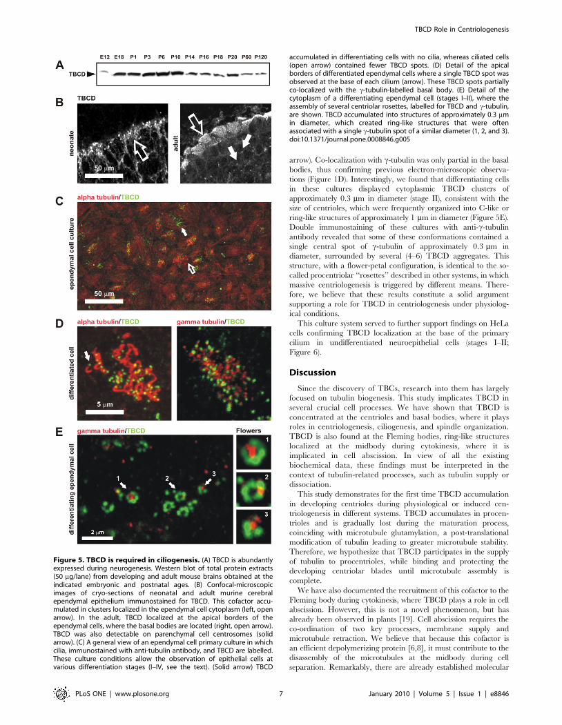
arrow). Co-localization with c-tubulin was only partial in the basal
bodies, thus confirming previous electron-microscopic observa-
tions (Figure 1D). Interestingly, we found that differentiating cells
in these cultures displayed cytoplasmic TBCD clusters of
approximately 0.3 mm in diameter (stage II), consistent with the
size of centrioles, which were frequently organized into C-like or
ring-like structures of approximately 1 mm in diameter (Figure 5E).
Double immunostaining of these cultures with anti-c-tubulin
antibody revealed that some of these conformations contained a
single central spot of c-tubulin of approximately 0.3 mm in
diameter, surrounded by several (4–6) TBCD aggregates. This
structure, with a flower-petal configuration, is identical to the so-
called procentriolar ‘‘rosettes’’ described in other systems, in which
massive centriologenesis is triggered by different means. There-
fore, we believe that these results constitute a solid argument
supporting a role for TBCD in centriologenesis under physiolog-
ical conditions.
This culture system served to further support findings on HeLa
cells confirming TBCD localization at the base of the primary
cilium in undifferentiated neuroepithelial cells (stages I–II;
Figure 6).
Discussion
Since the discovery of TBCs, research into them has largely
focused on tubulin biogenesis. This study implicates TBCD in
several crucial cell processes. We have shown that TBCD is
concentrated at the centrioles and basal bodies, where it plays
roles in centriologenesis, ciliogenesis, and spindle organization.
TBCD is also found at the Fleming bodies, ring-like structures
localized at the midbody during cytokinesis, where it is
implicated in cell abscission. In view of all the existing
biochemical data, these findings must be interpreted in the
context of tubulin-related processes, such as tubulin supply or
dissociation.
This study demonstrates for the first time TBCD accumulation
in developing centrioles during physiological or induced cen-
triologenesis in different systems. TBCD accumulates in procen-
trioles and is gradually lost during the maturation process,
coinciding with microtubule glutamylation, a post-translational
modification of tubulin leading to greater microtubule stability.
Therefore, we hypothesize that TBCD participates in the supply
of tubulin to procentrioles, while binding and protecting the
developing centriolar blades until microtubule assembly is
complete.
We have also documented the recruitment of this cofactor to the
Fleming body during cytokinesis, where TBCD plays a role in cell
abscission. However, this is not a novel phenomenon, but has
already been observed in plants [19]. Cell abscission requires the
co-ordination of two key processes, membrane supply and
microtubule retraction. We believe that because this cofactor is
an efficient depolymerizing protein [6,8], it must contribute to the
disassembly of the microtubules at the midbody during cell
separation. Remarkably, there are already established molecular
Figure 5. TBCD is required in ciliogenesis. (A) TBCD is abundantlyexpressed during neurogenesis. Western blot of total protein extracts(50 mg/lane) from developing and adult mouse brains obtained at theindicated embryonic and postnatal ages. (B) Confocal-microscopicimages of cryo-sections of neonatal and adult murine cerebralependymal epithelium immunostained for TBCD. This cofactor accu-mulated in clusters localized in the ependymal cell cytoplasm (left, openarrow). In the adult, TBCD localized at the apical borders of theependymal cells, where the basal bodies are located (right, open arrow).TBCD was also detectable on parenchymal cell centrosomes (solidarrow). (C) A general view of an ependymal cell primary culture in whichcilia, immunostained with anti-tubulin antibody, and TBCD are labelled.These culture conditions allow the observation of epithelial cells atvarious differentiation stages (I–IV, see the text). (Solid arrow) TBCD
accumulated in differentiating cells with no cilia, whereas ciliated cells(open arrow) contained fewer TBCD spots. (D) Detail of the apicalborders of differentiated ependymal cells where a single TBCD spot wasobserved at the base of each cilium (arrow). These TBCD spots partiallyco-localized with the c-tubulin-labelled basal body. (E) Detail of thecytoplasm of a differentiating ependymal cell (stages I–II), where theassembly of several centriolar rosettes, labelled for TBCD and c-tubulin,are shown. TBCD accumulated into structures of approximately 0.3 mmin diameter, which created ring-like structures that were oftenassociated with a single c-tubulin spot of a similar diameter (1, 2, and 3).doi:10.1371/journal.pone.0008846.g005
TBCD Role in Centriologenesis
PLoS ONE | www.plosone.org 7 January 2010 | Volume 5 | Issue 1 | e8846
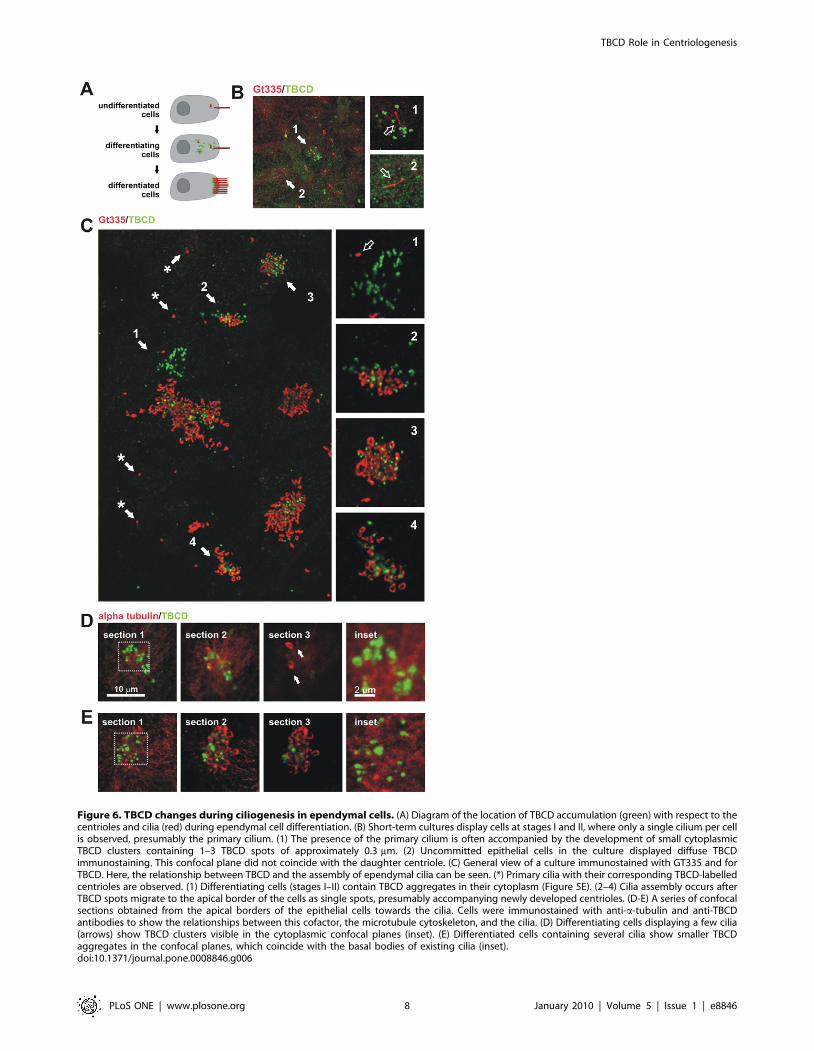
Figure 6. TBCD changes during ciliogenesis in ependymal cells. (A) Diagram of the location of TBCD accumulation (green) with respect to thecentrioles and cilia (red) during ependymal cell differentiation. (B) Short-term cultures display cells at stages I and II, where only a single cilium per cellis observed, presumably the primary cilium. (1) The presence of the primary cilium is often accompanied by the development of small cytoplasmicTBCD clusters containing 1–3 TBCD spots of approximately 0.3 mm. (2) Uncommitted epithelial cells in the culture displayed diffuse TBCDimmunostaining. This confocal plane did not coincide with the daughter centriole. (C) General view of a culture immunostained with GT335 and forTBCD. Here, the relationship between TBCD and the assembly of ependymal cilia can be seen. (*) Primary cilia with their corresponding TBCD-labelledcentrioles are observed. (1) Differentiating cells (stages I–II) contain TBCD aggregates in their cytoplasm (Figure 5E). (2–4) Cilia assembly occurs afterTBCD spots migrate to the apical border of the cells as single spots, presumably accompanying newly developed centrioles. (D-E) A series of confocalsections obtained from the apical borders of the epithelial cells towards the cilia. Cells were immunostained with anti-a-tubulin and anti-TBCDantibodies to show the relationships between this cofactor, the microtubule cytoskeleton, and the cilia. (D) Differentiating cells displaying a few cilia(arrows) show TBCD clusters visible in the cytoplasmic confocal planes (inset). (E) Differentiated cells containing several cilia show smaller TBCDaggregates in the confocal planes, which coincide with the basal bodies of existing cilia (inset).doi:10.1371/journal.pone.0008846.g006
TBCD Role in Centriologenesis
PLoS ONE | www.plosone.org 8 January 2010 | Volume 5 | Issue 1 | e8846

links between the centrosome and midbody for other proteins,
which are predominantly involved in the process of membrane
supply [38]. Here, we have shown that not only TBCD, but also
the c- and e-tubulins occur at the Fleming body. Although we
cannot explain the role of e-tubulin in this structure, we believe
that TBCD and c-tubulin play a shared role in this secondary
MTOC during cytokinesis. Significantly, a failure of cell abscission
has also been reported in cultures in which c-tubulin had been
silenced [39].
Our data also demonstrate a significant reduction in the pole-to-
pole spindle length after TBCD depletion, suggesting that this
cofactor participates in microtubule spindle dynamics. Indeed,
F16D3.4, the C. elegans TBCD orthologue, has been shown to be
a regulator of the microtubule growth rate [21]. Classical
experiments performed by Mitchinson [40] and later reviewed
by Rogers et al. [41] demonstrated that spindle microtubules are
extremely dynamic, insofar as tubulin subunits are continuously
incorporated into the microtubule plus ends while they are
removed at their minus ends, embedded at the spindle poles. This
model implies that there should be identical tubulin polymerizing–
depolymerizing rates at both microtubule extremes to maintain
the spindle symmetry and length. At the minus ends, proteins such
as Kin 1 or katanin could be pulling and severing the
microtubules, whereas at the plus ends, there must be a sufficient
tubulin supply to maintain a constant microtubule length. Both
mechanisms require an exquisite co-ordination of tubulin
processing and trafficking events between microtubule extremes,
where we hypothesize that TBCD is implicated. Our data show
how excess TBCD releases microtubules from the centrosome and
produces c-tubulin-containing supernumerary MTOCs at meta-
phase, findings suggesting that TBCD contributes to microtubule
release from the centrosome during mitosis, as well as in
interphase. Although the molecular mechanism of microtubule
release from the centrosome is still unknown, it is broadly accepted
that the anchoring of the microtubule to this organelle depends on
the c-tubulin ring complex (cTuRC), and that any mechanism
leading to cTuRC detachment releases microtubules into the
cytosol. TBCD is one of several candidate proteins that may
anchor the cTuRC to the centrosome [24]. The contribution of
TBCD to this process would explain why in mammalian
centrioles, microtubules are nucleated through their entire length
(where there is c-tubulin), whereas microtubules are only anchored
in the distal part of the mother centriole [42], a region devoid of
TBCD.
Centrioles are not just required at the centrosome but also for
the assembly of the basal bodies of cilia. Unfortunately, the
mechanisms involved in massive centriologenesis and centriolar
differentiation into basal bodies are still unclear. Recent studies
have revealed that more than 1,000 different proteins are
implicated in ciliary function, but almost no information regarding
their roles in centriolar differentiation or ciliary axoneme assembly
is available. Here, we have shown that TBCD is also crucial in
centriologenesis via the acentriolar pathway, where it is organized
into ‘‘centriolar rosettes’’. We propose a model in which TBCD
molecules associate to form a scaffold for the assembly of other
centriolar proteins, in both the canonical and the acentriolar
assembly pathways. Further studies of TBCD at the biochemical
level will shed light on the mechanisms underlying the way this
tubulin cofactor is recruited into these procentriolar structures
during the differentiation of multi-ciliated cell. An increased
understanding of the process of ciliogenesis will assist the better
diagnosis and treatment of cilia-associated human diseases, such as
primary ciliary dyskinesia and Bardet–Biedl, Alstrom, and
Meckel–Gruber syndromes.
Materials and Methods
Ethics Statement25 Swiss-Webster mice were used in this study. They were killed
according to PHS policy and the U.S. National Institutes of Health
guidelines.
Antisera, Immunocytochemistry, Flow Cytometry andCultures
Affinity purified primary antibodies were produced against
TBCD recombinant protein fragments of BtTBCD (obtained
from Dr. Cowan, New York University, NY, USA), HsTBCD
isoform 1 and M. musculus TBCD (RPZD, Berlin, Germany).
Rabbit sera were affinity purified against HsTBCD baculovirus-
infected Sf9 cell extracts, as described previously [43]. CHO cell
(ATCCR number: CCL-61TM) S-phase arrest was induced by
treatment with 2 mM thymidine (Sigma-Aldrich, MO, USA) for
16 h. Immuno-electron-microscopic analysis was performed on
80 nm sections of 4% paraformaldehyde/0.1% glutaraldehyde-
fixed tissue embedded in Unicryl (BBInternational, Cardiff, UK).
Antibodies used were anti-a- and anti-b-tubulin (B512 and
Tub2.1, respectively), anti-acetylated tubulin, anti-NEDD1/
GCP-WD, and anti-e- and anti-d-tubulin antibodies (all from
Sigma Aldrich). The anti-glutamylated tubulin antibody
(GT335) was a gift from Dr Janke (CNRS, Montpellier, France)
and the anti-c-tubulin antibody (TU30) from Dr Draber
(Institute of Molecular Genetics, Prague, Czech Republic). The
GFP-centrin1 construct was kindly provided by Dr M. Bornens
(Institut Curie, Paris). Secondary antibodies were Alexa-Fluor-
488-conjugated goat anti-rabbit IgG and goat-anti-mouse IgG,
Alexa-Fluor-647-conjugated goat anti-mouse IgG (Molecular
Probes, Invitrogen), Cy3-conjugated goat anti-mouse IgG and
goat anti-mouse IgG1, and Cy5-conjugated goat anti-rabbit IgG
(Jackson ImmunoResearch Laboratories, Inc.) and a 10 nm anti-
rabbit IgG gold conjugate (Sigma, St. Louis, MO). RNA
interference was performed with a ‘‘Smart Pool’’ mixture
targeting human TBCD (Dharmacon, CO, USA). TBCD
silencing was confirmed 24, 48, and 72 h after RNAi treatment
by western blotting. TBCD cDNA fragments were produced by
PCR. The TBCD mutants were sequenced and cloned into the
pEGFP-C2 and/or pEGFP-N3 vectors from Invitrogen (Life
technologies, California USA). P2 cerebellar mixed primary cell
cultures were grown on poly-L-lysine and laminin-coated
(Sigma-Aldrich, MO, USA) coverslips and were grown in 10%
foetal bovine serum in IMDM medium (all from Gibco,
Invitrogen). The cultures were fixed at different time points
after plating (after 5–20 days in culture). Beating cilia were
observed approximately 10 days after dissociation. Cell lines and
cultures used for immunocytochemistry were fixed in ice-cold
(220uC) methanol or 4% paraformaldehyde, and further
permeabilized in phosphate-buffered saline (PBS)–0.1% Triton
X-100 in PBS (PBS-T). For some experiments microtubules were
depolymerized with 2 mM nocodazole and cold (4uC) treatments
for 30 min. CHO cell centrosome overduplication was induced
by treatment with 2 mM thymidine (Sigma-Aldrich) for 16 h.
Flow cytometry was performed on cells with Hoechst-stained
DNA 15 and 30 h after transfection with HsTBCD–YFP or
H2B–YFP kindly supplied by Dr DiCroce (ICREA, CRG,
Barcelona, Spain) as the control. A Becton Dickinson FACS
CantoII equipment equipped with a 405 nm and 488 nm laser
diodes was used. Ten thousand cells were collected for each
sample. The data were analysed using the FACS Diva software
(Becton Dickinson, NJ, USA).
TBCD Role in Centriologenesis
PLoS ONE | www.plosone.org 9 January 2010 | Volume 5 | Issue 1 | e8846

Confocal Microscopy, Measurements and StatisticalAnalysis
Cell measurements were made on confocal-microscopic pro-
jected images obtained with a Zeiss 636 lens. Microscopy was
performed on a Zeiss LSM-510 confocal microscope equipped
with an argon (488 nm) laser and two HeNe (543 and 633 nm)
lasers. Images in the same confocal planes were scanned
sequentially to avoid fluorescent channel emission cross-talk. The
maximum confocality was according to the manufacturer’s
instructions. A t test was performed on the data obtained for
confocal microscopy measurements on two different coverslips,
resulting in three different experiments. Statistical analysis of data
and graphing were performed using the SigmaPlot 8.0 software
(Systat Software, Richmond, CA). The data in Figure 4E represent
mean values and standard error bars.
Microtubule Binding TestsBovine brain tubulin was purified as described by [44]. Full-
length cDNAs encoding HsTBCD and BtTBCD were used as the
templates for coupled transcription and translation in rabbit
reticulocyte cell-free lysates (Promega Corporation) in the presence
of 35S-methionine (.1000 Ci/mmol; Amersham Pharmacia
Biotech), as described elsewhere [45]. The products of in vitro
TBCD synthesis were incubated with taxol-stabilized microtu-
bules, as previously described [8].
Supporting Information
Figure S1 Specificity of TBCD antibodies and confocal images
at metaphase. (A) Specificity of the antibodies directed against
TBCD produced for the study. Western blot of total HeLa cell
extracts (50 mg/lane) immunostained with an antibody recogniz-
ing full-length HsTBCD (antibody 1) and a second antiserum
recognizing a fragment of mouse TBCD (MmTBCD; antibody 2).
Both antibodies demonstrated great specificity after affinity
purification. (B) Confocal-microscopic images of triply/doubly
labelled HeLa cells at metaphase. TBCD partially co-localized
with NEDD1/GCP-WD but did not co-localize with e-tubulin.
Found at: doi:10.1371/journal.pone.0008846.s001 (0.88 MB TIF)
Figure S2 TBCD is associated with centriologenesis in different
systems. (A) TBCD immunostaining in thymidine-treated S-
arrested CHO cells. (top) CHO cells generally contained aberrant
centriolar numbers and when blocked at S-phase by thymidine
treatment, underwent repeated cycles of centriole synthesis. After
16 h blockage, TBCD accumulation at the developing centrioles
was specifically observed. (B) TBCD immunostaining of a HeLa
cell transfected with a construct encoding PLK4. This protein is
known to trigger centriologenesis when overexpressed. TBCD
labelled the developing centrioles 24 h after transfection.
Found at: doi:10.1371/journal.pone.0008846.s002 (1.31 MB TIF)
Acknowledgments
We thank Laura Alvarez and Begona Ubilla for technical assistance.
Author Contributions
Conceived and designed the experiments: MLF JCZ. Performed the
experiments: MLF JB CJ JCV. Contributed reagents/materials/analysis
tools: JB JCV. Wrote the paper: MLF JCZ.
References
1. Andersen JS, Wilkinson CJ, Mayor T, Mortensen P, Nigg EA, et al. (2003)Proteomic characterization of the human centrosome by protein correlation
profiling. Nature 426: 570–574.
2. Lewis SA, Tian G, Cowan NJ (1997) The alpha- and beta-tubulin folding
pathways. Trends Cell Biol 12: 479–484.
3. Lopez-Fanarraga M, Avila J, Guasch A, Coll M, Zabala JC (2001) Review:postchaperonin tubulin folding cofactors and their role in microtubule dynamics.
J Struct Biol 135: 219–229.
4. Szymanski D (2002) Tubulin folding cofactors: half a dozen for a dimer. Curr
Biol 12: 767–769.
5. Tian G, Lewis SA, Feierbach B, Stearns T, Rommelaere H, et al. (1997) Tubulinsubunits exist in an activated conformational state generated and maintained by
protein cofactors. J Cell Biol 138: 821–832.
6. Bhamidipati A, Lewis SA, Cowan NJ (2000) ADP ribosylation factor-like protein
2 (Arl2) regulates the interaction of tubulin-folding cofactor D with native
tubulin. J Cell Biol 149: 1087–1096.
7. Kortazar D, Carranza G, Bellido J, Villegas JC, Fanarraga ML, et al. (2006)
Native tubulin-folding cofactor E purified from baculovirus-infected Sf9 cellsdissociates tubulin dimers. Protein Expr Purif 49: 196–202.
8. Martın L, Fanarraga ML, Aloria K, Zabala JC (2000) Tubulin folding cofactor
D is a microtubule destabilizing protein. FEBS Lett 470: 93–95.
9. Abruzzi KC, Smith A, Chen W, Solomon F (2002) Protection from free beta-
tubulin by the beta-tubulin binding protein Rbl2p. Mol Cell Biol 22: 138–147.
10. Archer JE, Vega LR, Solomon F (1995) Rbl2p, a yeast protein that binds to
beta-tubulin and participates in microtubule function in vivo. Cell 82: 425–434.
11. Archer JE, Magendantz M, Vega LR, Solomon F (1998) Formation andfunction of the Rbl2p-b-tubulin complex. Mol Cell Biol 18: 1757–1761.
12. Fanarraga ML, Parraga M, Aloria K, del Mazo J, Avila J, et al. (1999) Regulatedexpression of p14 cofactor A during spermatogenesis. Cell Motil Cytoskel 43:
243–254.
13. Bartolini F, Tian G, Piehl M, Cassimeris L, Lewis SA, et al. (2005) Identificationof a novel tubulin-destabilizing protein related to the chaperone cofactor E. J Cell
Sci 118: 1197–1207.
14. Keller CE, Lauring BP (2005) Possible regulation of microtubules through
destabilization of tubulin. Trends Cell Biol 15: 571–573.
15. Kortazar D, Fanarraga ML, Carranza G, Bellido J, Villegas JC, et al. (2007)Role of cofactors B (TBCB) and E (TBCE) in tubulin heterodimer dissociation.
Exp Cell Res 313: 425–436.
16. Hoyt MA, Stearns T, Botstein D (1990) Chromosome instability mutants of
Saccharomyces cerevisiae that are defective in microtubule-mediated processes.
Mol Cell Biol 1: 223–34.
17. Hirata D, Masuda H, Eddison M, Toda T (1998) Essential role of tubulin-
folding cofactor D in microtubule assembly and its association with microtubules
in fission yeast. EMBO J 17: 658–666.
18. Liu CM, Meinke DW (1998) The titan mutants of Arabidopsis are disrupted in
mitosis and cell cycle control during seed development. Plant J 16: 21–31.
19. Steinborn K, Maulbetsch C, Priester B, Trautmann S, Pacher T, et al. (2002)
The Arabidopsis PILZ group genes encode tubulin-folding cofactor orthologs
required for cell division but not cell growth. Genes Dev 16: 959–971.
20. Sonnichsen B, Koski LB, Walsh A, Marschall P, Neumann B, et al. (2005) Full-
genome RNAi profiling of early embryogenesis in Caenorhabditis elegans.
Nature 434: 462–469.
21. Srayko M, Kaya A, Stamford J, Hyman A (2005) A. Identification and
characterization of factors required for microtubule growth and nucleation in
the early C. elegans embryo. Dev Cell 9: 223–236.
22. Fedyanina OS, Mardanov PV, Tokareva EM, McIntosh JR, Grishchuk EL
(2006) Chromosome segregation in fission yeast with mutations in the tubulin
folding cofactor D. Curr Genet 50: 281–294.
23. Fedyanina OS, Book AJ, Grishchuk EL (2009) Tubulin heterodimers remain
functional for one cell cycle after the inactivation of tubulin-folding cofactor D in
fission yeast cells. Yeast 26: 235–247.
24. Cunningham LA, Kahn RA (2008) Cofactor D functions as a centrosomal
protein and is required for the recruitment of the gamma-tubulin ring complex
at centrosomes and organization of the mitotic spindle. J Biol Chem 283:
7155–7165.
25. Chang P, Stearns T (2000) Delta-tubulin and epsilon-tubulin: two new human
centrosomal tubulins reveal new aspects of centrosome structure and function.
Nat Cell Biol 2: 30–35.
26. Bornens M (2002) Centrosome composition and microtubule anchoring
mechanisms. Curr Opin Cell Biol 14: 25–34.
27. Kleylein-Sohn J, Westendorf J, Le Clech M, Habedanck R, Stierhof YD,
et al. (2007) Plk4-induced centriole biogenesis in human cells. Dev Cell 13:
190–202.
28. Rodrigues-Martins A, Bettencourt-Dias M, Riparbelli M, Ferreira C, Ferreira I,
et al. (2007) DSAS-6 organizes a tube-like centriole precursor, and its absence
suggests modularity in centriole assembly. Curr Biol 17: 1465–1472.
29. Mikule K, Delaval B, Kaldis P, Jurcyzk A, Hergert P, et al. (2007) Loss of
centrosome integrity induces p38-p53-p21-dependent G1-S arrest. Nat Cell Biol
9: 160–170.
30. Tang CJ, Fu RH, Wu KS, Hsu WB, Tang TK (2009) CPAP is a cell-cycle
regulated protein that controls centriole length. Nat Cell Biol 11: 825–831.
TBCD Role in Centriologenesis
PLoS ONE | www.plosone.org 10 January 2010 | Volume 5 | Issue 1 | e8846

31. Hsu WB, Hung LY, Tang CJ, Su CL, Chang Y, et al. (2008) Functional
characterization of the microtubule-binding and -destabilizing domains of CPAP
& d-SAS-4. Exp Cell Res 314: 2591–602.
32. Tsou MF, Stearns T (2006) Mechanism limiting centrosome duplication to once
per cell cycle. Nature 442: 947–951.
33. Dirksen ER (1991) Centriole and basal body formation during ciliogenesis
revisited. Biol Cell 72: 31–38.
34. Hagiwara H, Ohwada N, Takata K (2004) Cell biology of normal & abnormal
ciliogenesis in the ciliated epithelium. Int Rev Cytol 234: 101–141.
35. Dawe HR, Farr H, Gull K (2007) Centriole/basal body morphogenesis and
migration during ciliogenesis in animal cells. J Cell Sci 120: 7–15.
36. Strnad P, Gonczy P (2008) Mechanisms of procentriole formation. Trends Cell
Biol 18: 389–396.
37. Vladar EK, Stearns T (2007) Molecular characterization of centriole assembly in
ciliated epithelial cells. J Cell Biol 178: 31–42.
38. Doxsey SJ (2005) Molecular links between centrosome & midbody. Mol Cell 20:
170–172.
39. Shu HB, Li Z, Palacios MJ, Li Q, Joshi HC (1995) A transient association of
gamma-tubulin at the midbody is required for the completion of cytokinesisduring the mammalian cell division. J Cell Sci 108: 2955–2962.
40. Mitchison TJ (1989) Polewards microtubule flux in the mitotic spindle: evidence
from photoactivation of fluorescence. J Cell Biol 109: 637–652.41. Rogers GC, Rogers SL, Sharp DJ (2005) Spindle microtubules in flux. J Cell Sci
118: 1105–1116.42. Piel M, Meyer P, Khodjakov A, Rieder CL, Bornens M (2000) The respective
contributions of the mother and daughter centrioles to centrosome activity and
behavior in vertebrate cells. J Cell Biol 149: 317–330.43. Lajoie-Mazenc I, Tollon Y, Detraves C, Julian M, Moisand A, et al. (1994)
Recruitment of antigenic gamma-tubulin during mitosis in animal cells: presenceof gamma-tubulin in the mitotic spindle. J Cell Sci 107: 2825–2837.
44. Avila J, Soares H, Fanarraga ML, Zabala JC (2008) Isolation of microtubulesand microtubule proteins. In Current Protocols in Cell Biology, John Wiley &
Sons Inc, ed. Malden, MA, USA. pp 3.29.1–3.29.28.
45. Zabala JC, Cowan NJ (1992) Tubulin dimer formation via the release of alpha-and beta-tubulin monomers from multimolecular complexes. Cell Motil.
Cytoskel 23: 222–230.
TBCD Role in Centriologenesis
PLoS ONE | www.plosone.org 11 January 2010 | Volume 5 | Issue 1 | e8846
Copyright © 2022 FDOKUMEN