A Novel In Vitro Human Model of Hemangioma - Nature
8
METHODS IN PATHOLOGY A Novel In Vitro Human Model of Hemangioma Swee T. Tan, M.B.B.S., F.R.A.C.S., Qurratulain Hasan, Ph.D., Marija Velickovic, B.Sc., Beate M. Ru ¨ ger, M.M.L.Sc., Richard P.F. Davis, Paul F. Davis, Ph.D. Swee Tan Plastic Surgery Trust (STT) and Department of Medicine, Wellington School of Medicine (QH, MV, BR, RPFD, PFD), Wellington, New Zealand. Hemangioma, the most common tumor of infancy, is characterized by a proliferation of capillary endo- thelial cells with multilamination of the basement membrane and accumulation of cellular elements, including mast cells. The initial rapid growth is fol- lowed by an inevitable but slow involution. The cur- rently available therapies are empirical and unsat- isfactory because what is known of the cellular and molecular basis of hemangioma development is ru- dimentary. Advances in the understanding of its programmed biologic behavior has been hampered by the lack of a valid human model. We report here a novel in vitro culture system that is a useful human model of hemangioma. A small fragment of hemangioma biopsy is embedded in fibrin gel in a well of culture plates and incubated in a serum-free, buffered-salt, minimal medium. A complex network of microvessels grows out from the tissue fragments. Biopsies taken from all three phases of hemangioma development were cultured successfully; proliferative phase samples developed microvessels in 1 to 4 days, involuting phase in 5 to 7 days, and involuted phase in 7 to 12 days. The relative growth rates of the microvessels in the cul- ture of biopsies taken from different stages of hem- angioma development reflect the growth patterns seen clinically. This model has been validated using histo- chemistry, immunohistochemistry, and reverse transcriptase-polymerase chain reaction. Compari- son of the number, localization, and phenotype of endothelial and mast cells and the distribution of basement membrane constituents (type IV collagen, perlecan, and laminins) and growth factors (basic fibroblast growth factor, vascular endothelial growth factor, transforming growth factor-bs) in the biopsy and the tissue after culture shows that many of the characteristics of the original tissues were retained in culture. This in vitro human model of hemangioma over- comes some of the deficiencies associated with ear- lier models. It offers an opportunity for studying the precise cellular, biochemical, and molecular basis of hemangioma. It may also help to elucidate the mechanisms of action of existing therapies and may lead to the identification of novel treatments for hemangioma. KEY WORDS: Angiogenesis, Culture, Hemangioma, Immunohistochemistry, Reverse transcriptase- polymerase chain reaction. Mod Pathol 2000;13(1):92–99 Hemangioma, a primary tumor of microvascula- ture, is characterized by a proliferation of capillary endothelial cells with multilamination of the base- ment membrane and accumulation of cellular ele- ments, including mast cells, macrophages, plasma cells, and pericytes (1– 4). Thirty to 40% of heman- giomas are first noticed at birth, usually as a pink spot, blanched area, or telangiectasia (4), and ex- hibit a rapid postnatal growth for 8 to 12 months (proliferative phase), followed by a slow regression for the next 1 to 5 years (involuting phase), with continued improvement in the remaining children until 10 to 12 years (involuted phase) (5). Normal skin is restored in 50% of patients when involution is complete (4, 6). The trigger that initiates heman- gioma seems to be a local event, not a hereditary predisposition (7). Currently available experimental models include the induction of hemangioma in mice by transgenic endothelial cells (8), mouse endothelial cells that have been transformed by polyoma middle T on- cogene (9, 10), and endothelial cells obtained from murine and human hemangioma (11). Each of these models has deficiencies; they use animal rather than human cells and tissue, or they involve viral transformation of endothelial cells, which is not known to occur in human hemangioma, or they use a cell culture system that is not influenced by the metabolism of neighboring cells as occurs in Copyright © 2000 by The United States and Canadian Academy of Pathology, Inc. VOL. 13, NO. 1, P. 92, 2000 Printed in the U.S.A. Date of acceptance: August 5, 1999. This work was presented, in part, at the International Society for Study of Vascular Anomalies Workshop, Berlin, Germany, June 27–28, 1998. Address reprint requests to: Swee T. Tan, M.B.B.S., F.R.A.C.S., Swee Tan Plastic Surgery Trust, Bowen Hospital, Churchill Drive, Wellington, New Zealand; e-mail: [email protected]; fax: 64 – 4-479 –2660. 92
-
Upload
khangminh22 -
Category
Documents
-
view
1 -
download
0
Transcript of A Novel In Vitro Human Model of Hemangioma - Nature
METHODS IN PATHOLOGY
A Novel In Vitro Human Model of HemangiomaSwee T. Tan, M.B.B.S., F.R.A.C.S., Qurratulain Hasan, Ph.D., Marija Velickovic, B.Sc.,Beate M. Ruger, M.M.L.Sc., Richard P.F. Davis, Paul F. Davis, Ph.D.
Swee Tan Plastic Surgery Trust (STT) and Department of Medicine, Wellington School of Medicine (QH,MV, BR, RPFD, PFD), Wellington, New Zealand.
Hemangioma, the most common tumor of infancy,is characterized by a proliferation of capillary endo-thelial cells with multilamination of the basementmembrane and accumulation of cellular elements,including mast cells. The initial rapid growth is fol-lowed by an inevitable but slow involution. The cur-rently available therapies are empirical and unsat-isfactory because what is known of the cellular andmolecular basis of hemangioma development is ru-dimentary. Advances in the understanding of itsprogrammed biologic behavior has been hamperedby the lack of a valid human model.
We report here a novel in vitro culture systemthat is a useful human model of hemangioma. Asmall fragment of hemangioma biopsy is embeddedin fibrin gel in a well of culture plates and incubatedin a serum-free, buffered-salt, minimal medium. Acomplex network of microvessels grows out fromthe tissue fragments. Biopsies taken from all threephases of hemangioma development were culturedsuccessfully; proliferative phase samples developedmicrovessels in 1 to 4 days, involuting phase in 5 to7 days, and involuted phase in 7 to 12 days. Therelative growth rates of the microvessels in the cul-ture of biopsies taken from different stages of hem-angioma development reflect the growth patternsseen clinically.
This model has been validated using histo-chemistry, immunohistochemistry, and reversetranscriptase-polymerase chain reaction. Compari-son of the number, localization, and phenotype ofendothelial and mast cells and the distribution ofbasement membrane constituents (type IV collagen,perlecan, and laminins) and growth factors (basicfibroblast growth factor, vascular endothelialgrowth factor, transforming growth factor-bs) inthe biopsy and the tissue after culture shows that
many of the characteristics of the original tissueswere retained in culture.
This in vitro human model of hemangioma over-comes some of the deficiencies associated with ear-lier models. It offers an opportunity for studying theprecise cellular, biochemical, and molecular basisof hemangioma. It may also help to elucidate themechanisms of action of existing therapies and maylead to the identification of novel treatments forhemangioma.
KEY WORDS: Angiogenesis, Culture, Hemangioma,Immunohistochemistry, Reverse transcriptase-polymerase chain reaction.
Mod Pathol 2000;13(1):92–99
Hemangioma, a primary tumor of microvascula-ture, is characterized by a proliferation of capillaryendothelial cells with multilamination of the base-ment membrane and accumulation of cellular ele-ments, including mast cells, macrophages, plasmacells, and pericytes (1– 4). Thirty to 40% of heman-giomas are first noticed at birth, usually as a pinkspot, blanched area, or telangiectasia (4), and ex-hibit a rapid postnatal growth for 8 to 12 months(proliferative phase), followed by a slow regressionfor the next 1 to 5 years (involuting phase), withcontinued improvement in the remaining childrenuntil 10 to 12 years (involuted phase) (5). Normalskin is restored in 50% of patients when involutionis complete (4, 6). The trigger that initiates heman-gioma seems to be a local event, not a hereditarypredisposition (7).
Currently available experimental models includethe induction of hemangioma in mice by transgenicendothelial cells (8), mouse endothelial cells thathave been transformed by polyoma middle T on-cogene (9, 10), and endothelial cells obtained frommurine and human hemangioma (11). Each ofthese models has deficiencies; they use animalrather than human cells and tissue, or they involveviral transformation of endothelial cells, which isnot known to occur in human hemangioma, or theyuse a cell culture system that is not influenced bythe metabolism of neighboring cells as occurs in
Copyright © 2000 by The United States and Canadian Academy ofPathology, Inc.VOL. 13, NO. 1, P. 92, 2000 Printed in the U.S.A.Date of acceptance: August 5, 1999.This work was presented, in part, at the International Society for Study ofVascular Anomalies Workshop, Berlin, Germany, June 27–28, 1998.Address reprint requests to: Swee T. Tan, M.B.B.S., F.R.A.C.S., Swee TanPlastic Surgery Trust, Bowen Hospital, Churchill Drive, Wellington, NewZealand; e-mail: [email protected]; fax: 64 – 4-479 –2660.
92
vivo. Mulliken et al. (12) cultured endothelial cellsfrom human hemangioma on plasma clots. How-ever, the absence of other cellular elements, as wellas the necessity of plasma, makes this system lessthan ideal, especially for investigating the role ofmodulating molecules. Therefore, none of thesesystems simulates the cellular and extracellular ma-trix characteristics of human hemangioma.
We describe here an in vitro tissue culture systemthat uses human hemangioma specimens that areangiogenically active in a serum-free medium. Us-ing histochemistry, immunohistochemistry, and re-verse transcriptase-polymerase chain reaction (RT-PCR), we have established that a number ofcharacteristics of hemangioma were retained in thecultured samples.
MATERIALS AND METHODS
Tissue SamplesTen hemangioma biopsies were obtained from
nine patients (Table 1) according to a protocol ap-proved by the Wellington Ethics Committee. Thespecimens were kept moist with normal saline andcleansed of all blood clots with phosphate-bufferedsaline. Samples from them were snap-frozen in liq-uid nitrogen for RNA isolation, fixed in formalin forhistochemistry and immunohistochemistry, andcultured as described next.
Tissue CulturePieces of fresh operative hemangioma tissue
were submerged in serum-free MCDB131 medium(GIBCO BRL, Gaithersburg, MD). Under an operat-ing microscope, the tissue was cut at room temper-ature into 1-mm explants and embedded in fibringel. For the gel, sheep fibrinogen fraction I (Sigma,St. Louis, MO) (3 mg/mL) was dissolved inMCDB131 medium with streptomycin (0.1 mg/mL)and penicillin (100 U/mL), supplemented withe-amino-caproic acid (0.3 mg/mL) (Sigma) to pre-vent fibrinolysis. Human plasma thrombin (final
concentration 0.5 U/mL) (Serva Feinbiochemica,Heidelberg, Germany) was added to the fibrinogen,and 0.7 mL of the mixture was immediately placedin each well of a culture plate (Nunclon; Nunc InterMedia, Roskilde, Denmark). After gel formation, thetissue explants were placed on top of it. A secondaliquot of fibrinogen-thrombin-medium mixture(0.8 mL) was added so that the tissue fragment wassandwiched between two layers of fibrin gel. Aftergel formation, 1.5 mL of the same supplementedMCDB131 medium was added to each well. Foreach biopsy, 24 well culture plates were used so thatmultiple analyses with replicates could be carriedout. The plates were incubated at 37° C in 3% CO2/97% air in a humidified environment.
The medium was changed every 5 days. After thefirst passage, some of the cultured tissue sampleswere placed in Trizol (GIBCO, Life-Technologies)for RNA isolation, and the remainder were fixed informalin and embedded in paraffin for histochem-ical and immunohistochemical analyses.
Quantitative Assay of AngiogenesisImages of the cultures were captured using a
Pixera digital image camera professional system(Digitech, Miami, FL) attached to an inverted mi-croscope (Olympus CK2; Olympus, Tokyo, Japan).The images were taken approximately every 3 daysto assess the rate of growth of the neovessels. Theneovascular area in each image was outlined andcalculated using the program Image 1.62b7 soft-ware (National Institutes of Health, Bethesda, MD)as described earlier (13). This area is expressed rel-ative to the area of the explant.
HistochemistryThree-mm-thick sections from the paraffin blocks
were attached to poly-L-lysine– coated glass slides(Sigma). Deparaffinized sections were stained withhematoxylin and eosin. Mast cells were detectedusing Csaba stain as described (14).
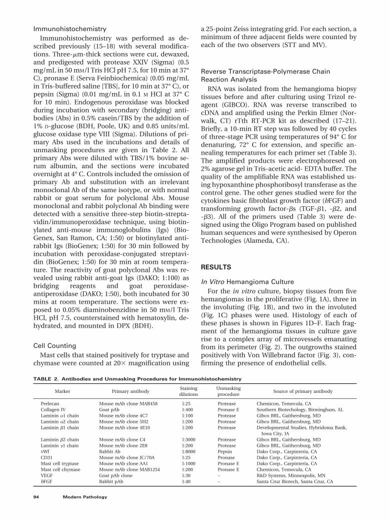
TABLE 1. Details of Primary Cultures of Hemangiomas Including Age of Patient at Biopsy, Sex, Site of Lesion,
Developmental Phase of Hemangioma, and Time in Culture Before New Vessels Were Detected
Patient Age Sex Site Phase First sprouts (days)
1 18 days F Perianal Proliferative 2–42 3 mo F Axilla Proliferativea 1–43 5 mo F Axilla Proliferativeb 2–44 5 mo M Forehead Proliferative 3–45 8 mo F Chest Proliferative 3–46 13 mo M Scalp Involuting 5–77 13 mo F Elbow Involuting 6–78 5 yr F Upper lip Involuting 5–79 6 yr M Glabella Involuted 7–10
10 8 yr F Submentum Involuted 9–12
a Tissue obtained before intralesional triamcinolone injection.b Tissue obtained 5 weeks after intralesional triamcinolone injection.
Culture of Human Hemangioma (S.T. Tan et al.) 93
ImmunohistochemistryImmunohistochemistry was performed as de-
scribed previously (15–18) with several modifica-tions. Three-mm-thick sections were cut, dewaxed,and predigested with protease XXIV (Sigma) (0.5mg/mL in 50 mM/l Tris HCl pH 7.5, for 10 min at 37°C), pronase E (Serva Feinbiochemica) (0.05 mg/mLin Tris-buffered saline [TBS], for 10 min at 37° C), orpepsin (Sigma) (0.01 mg/mL in 0.1 M HCl at 37° Cfor 10 min). Endogenous peroxidase was blockedduring incubation with secondary (bridging) anti-bodies (Abs) in 0.5% casein/TBS by the addition of1% D-glucose (BDH, Poole, UK) and 0.85 units/mLglucose oxidase type VIII (Sigma). Dilutions of pri-mary Abs used in the incubations and details ofunmasking procedures are given in Table 2. Allprimary Abs were diluted with TBS/1% bovine se-rum albumin, and the sections were incubatedovernight at 4° C. Controls included the omission ofprimary Ab and substitution with an irrelevantmonoclonal Ab of the same isotype, or with normalrabbit or goat serum for polyclonal Abs. Mousemonoclonal and rabbit polyclonal Ab binding weredetected with a sensitive three-step biotin-strepta-vidin/immunoperoxidase technique, using biotin-ylated anti-mouse immunoglobulins (Igs) (Bio-Genex, San Ramon, CA; 1:50) or biotinylated anti-rabbit Igs (BioGenex; 1:50) for 30 min followed byincubation with peroxidase-conjugated streptavi-din (BioGenex; 1:50) for 30 min at room tempera-ture. The reactivity of goat polyclonal Abs was re-vealed using rabbit anti-goat Igs (DAKO; 1:100) asbridging reagents and goat peroxidase-antiperoxidase (DAKO; 1:50), both incubated for 30mins at room temperature. The sections were ex-posed to 0.05% diaminobenzidine in 50 mM/l TrisHCl, pH 7.5, counterstained with hematoxylin, de-hydrated, and mounted in DPX (BDH).
Cell CountingMast cells that stained positively for tryptase and
chymase were counted at 203 magnification using
a 25-point Zeiss integrating grid. For each section, aminimum of three adjacent fields were counted byeach of the two observers (STT and MV).
Reverse Transcriptase-Polymerase ChainReaction Analysis
RNA was isolated from the hemangioma biopsytissues before and after culturing using Trizol re-agent (GIBCO). RNA was reverse transcribed tocDNA and amplified using the Perkin Elmer (Nor-walk, CT) rTth RT-PCR kit as described (17–21).Briefly, a 10-min RT step was followed by 40 cyclesof three-stage PCR using temperatures of 94° C fordenaturing, 72° C for extension, and specific an-nealing temperatures for each primer set (Table 3).The amplified products were electrophoresed on2% agarose gel in Tris-acetic acid- EDTA buffer. Thequality of the amplifiable RNA was established us-ing hypoxanthine phosphoribosyl transferase as thecontrol gene. The other genes studied were for thecytokines basic fibroblast growth factor (bFGF) andtransforming growth factor-bs (TGF-b1, -b2, and-b3). All of the primers used (Table 3) were de-signed using the Oligo Program based on publishedhuman sequences and were synthesised by OperonTechnologies (Alameda, CA).
RESULTS
In Vitro Hemangioma CultureFor the in vitro culture, biopsy tissues from five
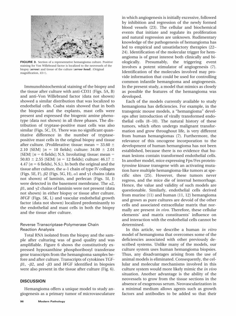
hemangiomas in the proliferative (Fig. 1A), three inthe involuting (Fig. 1B), and two in the involuted(Fig. 1C) phases were used. Histology of each ofthese phases is shown in Figures 1D–F. Each frag-ment of the hemangioma tissues in culture gaverise to a complex array of microvessels emanatingfrom its perimeter (Fig. 2). The outgrowths stainedpositively with Von Willebrand factor (Fig. 3), con-firming the presence of endothelial cells.
TABLE 2. Antibodies and Unmasking Procedures for Immunohistochemistry
Marker Primary antibodyStainingdilutions
Unmaskingprocedure
Source of primary antibody
Perlecan Mouse mAb clone MAB458 1:25 Protease Chemicon, Temecula, CACollagen IV Goat pAb 1:400 Pronase E Southern Biotechology, Birmingham, ALLaminin a1 chain Mouse mAb clone 4C7 1:100 Protease Gibco BRL, Gaithersburg, MDLaminin a2 chain Mouse mAb clone 5H2 1:200 Protease Gibco BRL, Gaithersburg, MDLaminin b1 chain Mouse mAb clone 4E10 1:200 Protease Developmental Studies, Hybridoma Bank,
Iowa City, IALaminin b2 chain Mouse mAb clone C4 1:3000 Protease Gibco BRL, Gaithersburg, MDLaminin g1 chain Mouse mAb clone 2E8 1:200 Protease Gibco BRL, Gaithersburg, MDvWf Rabbit Ab 1:8000 Pepsin Dako Corp., Carpinteria, CACD31 Mouse mAb clone JC/70A 1:25 Pronase Dako Corp., Carpinteria, CAMast cell tryptase Mouse mAb clone AA1 1:1000 Pronase E Dako Corp., Carpinteria, CAMast cell chymase Mouse mAb clone MAB1254 1:200 Pronase E Chemicon, Temecula, CAVEGF Goat pAb clone 1:30 – R&D Systems, Minneapolis, MNbFGF Rabbit pAb 1:40 – Santa Cruz Biotech, Santa Cruz, CA
94 Modern Pathology

Quantitation of the In Vitro HemangiomaAngiogenesis
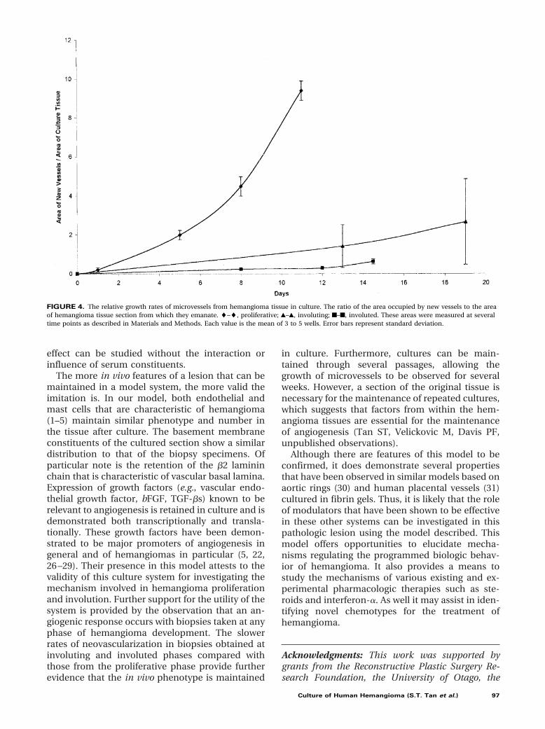
The extent of the vessel outgrowths was quanti-fied by digital image analysis. Tissue samples cul-tured from proliferative phase lesions were found togive rise to microvessels within 1 to 4 days in cul-ture. The first sprouts of microvessels were seenbetween 5 and 7 days when tissue was taken duringinvoluting phase, whereas involuted biopsy tissuestook 7 to 12 days to grow new microvessels. Relativegrowth rates are shown in Figure 4.
Phenotypic Features of Hemangioma Tissueafter Culture
Examination by histochemistry and immunohis-tochemistry of the expression of a number of cel-lular components, matrix proteins, and growth fac-tors previously described for hemangioma (5, 22)showed that these phenotypic characteristics wereretained in culture. RT-PCR also demonstrated thatthe transcripts for the growth factors studied werepresent in both the biopsies and the tissues afterculture.
TABLE 3. The Genes Analysed and the Primer Sequences Used for Reverse Transcriptase-Polymerase Chain Reaction
Gene Primers (sense and antisense) Annealing temperature Reference no.
HPRT 59CCTTTGGGCGGATTGTTGT39 52°C 17, 2059TTTTTTTTTTTTTTAAATTTTTGGGAAT39
TGF-b1 59GGAAACCCACAACGAAATCTATGAC39 56°C 2159TTCCCCTCCACGGCTCAAC39
TGF-b2 59GCGAGAGGAGCGACGAAGAG39 56°C 2159AGCCTGGGTTGGAGATGTTA39
TGF-b3 59CTGGCGGAGCACAACGAACT39 56°C 2159GGACTCTCTTCTCAACAGCCACTCAC39
bFGF 59AGCGACCCTCACATCAAGC39 60°C 1959AAAAGAAACACTCATCCGTAACACA39
HPRT, hypoxanthine phosphoribosyl transferase; TGF, transforming growth factor; bFGF, basic fibroblast growth factor.
FIGURE 1. Hemangioma at different phases of development. A, an ulcerated proliferative hemangioma in the perianal region of an 18-day-oldinfant. B, an involuting hemangioma on the upper lip of a 5-year-old girl. C, an involuted hemangioma in the submental region of an 8-year-old girl.D–F, hematoxylin and eosin staining of sections from biopsies from A, B, and C, respectively (original magnification, 3573).
FIGURE 2. Human hemangioma in culture. Tissue taken from a proliferative hemangioma embedded in fibrin gel and cultured, as described inMaterials and Methods, at 2 days (A), at 4 days (B), and at 6 days in culture (C) (original magnification, 22.53).
Culture of Human Hemangioma (S.T. Tan et al.) 95
Immunohistochemical staining of the biopsy andthe tissue after culture with anti-CD31 (Figs. 5A, B)and anti–Von Willebrand factor (data not shown)showed a similar distribution that was localized toendothelial cells. Csaba stain showed that in boththe biopsies and the explants, mast cells werepresent and expressed the biogenic amine pheno-type (data not shown) in all three phases. The dis-tribution of tryptase-positive mast cells was alsosimilar (Figs. 5C, D). There was no significant quan-titative difference in the number of tryptase-positive mast cells in the original biopsy and tissueafter culture. (Proliferative: tissue: mean 5 33.60 62.10 [SEM] [n 5 10 fields]; culture: 34.00 6 2.01[SEM] [n 5 6 fields]; N.S. Involuting: tissue: mean 550.83 6 2.55 [SEM] [n 5 12 fields]; culture: 46.17 64.47 [n 5 6 fields], N.S.). In both the original and thetissue after culture, the a-1 chain of type IV collagen(Figs. 5E, F), b2 (Figs. 5G, H), a1 and g1 chains (datanot shown) of laminin, and perlecan (Figs. 5I, J)were detected in the basement membrane. The a2,b1, and g2 chains of laminin were not present (datanot shown) in either biopsy or tissue after culture.bFGF (Figs. 5K, L) and vascular endothelial growthfactor (data not shown) localized predominantly tothe endothelial and mast cells in both the biopsyand the tissue after culture.
Reverse Transcriptase-Polymerase ChainReaction Analysis
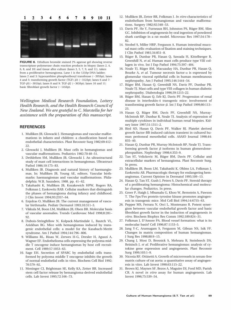
Total RNA isolated from the biopsy and the sam-ple after culturing was of good quality and wasamplifiable. Figure 6 shows the constitutively ex-pressed hypoxanthine phosphoribosyl transferasegene transcripts from the hemangioma samples be-fore and after culture. Transcripts of cytokines TGF-b1, -b2, and -b3 and bFGF identified in biopsieswere also present in the tissue after culture (Fig. 6).
DISCUSSION
Hemangioma offers a unique model to study an-giogenesis as a primary tumor of microvasculature
in which angiogenesis is initially excessive, followedby inhibition and regression of the newly formedblood vessels (23). The cellular and biochemicalevents that initiate and regulate its proliferationand natural regression are unknown. Rudimentaryknowledge of the pathogenesis of hemangioma hasled to empirical and unsatisfactory therapies (22–24). Identification of the molecular trigger for hem-angioma is of great interest both clinically and bi-ologically. Presumably, the triggering eventinvolves a potent stimulator of angiogenesis (7).Identification of the molecules involved may pro-vide information that could be used for controllingcommon infantile hemangioma and angiogenesis.In the present study, a model that mimics as closelyas possible the features of the hemangioma wasdeveloped.
Each of the models currently available to studyhemangioma has deficiencies. For example, in thetransgenic mouse models, a “hemangioma” devel-ops after introduction of virally transformed endo-thelial cells (8 –10). The natural history of thesetumors, which often undergo malignant transfor-mation and grow throughout life, is very differentfrom human hemangiomas (7). Furthermore, therelevance of this oncogene incorporation to thedevelopment of human hemangioma has not beenestablished, because there is no evidence that hu-man lesions contain transformed endothelial cells.In another model, mice expressing Fps/Fes protein-tyrosine kinase transgene with an activating muta-tion have multiple hemangioma-like tumors at spe-cific sites (25). However, these tumors neverregress, and the mice die of internal hemorrhage.Hence, the value and validity of such models arequestionable. Similarly, endothelial cells derivedfrom murine (11) and human (11, 12) hemangiomaand grown as pure cultures are devoid of the othercells and associated extracellular matrix that nor-mally are present in vivo. Therefore, these cellularelements’ and matrix constituents’ influence onand interaction with the endothelial cells cannot bedetermined.
In this article, we describe a human in vitromodel of hemangioma that overcomes some of thedeficiencies associated with other previously de-scribed systems. Unlike many of the models, ourculture system uses human hemangioma biopsies.Thus, any disadvantages arising from the use ofanimal models is eliminated. Consequently, the cel-lular and molecular mechanisms involved in thisculture system would more likely mimic the in vivosituation. Another advantage is the ability of theneovessels to grow from the tissue sections in theabsence of exogenous serum. Neovascularization ina minimal medium allows agents such as growthfactors and antibodies to be added so that their
FIGURE 3. Section of a representative hemangioma culture. Positivestaining for Von Willebrand factor is localized to the neovessels of thebiopsy (arrow) and tissue of the culture (arrow head). (Originalmagnification, 433).
96 Modern Pathology
effect can be studied without the interaction orinfluence of serum constituents.
The more in vivo features of a lesion that can bemaintained in a model system, the more valid theimitation is. In our model, both endothelial andmast cells that are characteristic of hemangioma(1–5) maintain similar phenotype and number inthe tissue after culture. The basement membraneconstituents of the cultured section show a similardistribution to that of the biopsy specimens. Ofparticular note is the retention of the b2 lamininchain that is characteristic of vascular basal lamina.Expression of growth factors (e.g., vascular endo-thelial growth factor, bFGF, TGF-bs) known to berelevant to angiogenesis is retained in culture and isdemonstrated both transcriptionally and transla-tionally. These growth factors have been demon-strated to be major promoters of angiogenesis ingeneral and of hemangiomas in particular (5, 22,26 –29). Their presence in this model attests to thevalidity of this culture system for investigating themechanism involved in hemangioma proliferationand involution. Further support for the utility of thesystem is provided by the observation that an an-giogenic response occurs with biopsies taken at anyphase of hemangioma development. The slowerrates of neovascularization in biopsies obtained atinvoluting and involuted phases compared withthose from the proliferative phase provide furtherevidence that the in vivo phenotype is maintained
in culture. Furthermore, cultures can be main-tained through several passages, allowing thegrowth of microvessels to be observed for severalweeks. However, a section of the original tissue isnecessary for the maintenance of repeated cultures,which suggests that factors from within the hem-angioma tissues are essential for the maintenanceof angiogenesis (Tan ST, Velickovic M, Davis PF,unpublished observations).
Although there are features of this model to beconfirmed, it does demonstrate several propertiesthat have been observed in similar models based onaortic rings (30) and human placental vessels (31)cultured in fibrin gels. Thus, it is likely that the roleof modulators that have been shown to be effectivein these other systems can be investigated in thispathologic lesion using the model described. Thismodel offers opportunities to elucidate mecha-nisms regulating the programmed biologic behav-ior of hemangioma. It also provides a means tostudy the mechanisms of various existing and ex-perimental pharmacologic therapies such as ste-roids and interferon-a. As well it may assist in iden-tifying novel chemotypes for the treatment ofhemangioma.
Acknowledgments: This work was supported bygrants from the Reconstructive Plastic Surgery Re-search Foundation, the University of Otago, the
FIGURE 4. The relative growth rates of microvessels from hemangioma tissue in culture. The ratio of the area occupied by new vessels to the areaof hemangioma tissue section from which they emanate. l–l, proliferative; Œ–Œ, involuting; f–f, involuted. These areas were measured at severaltime points as described in Materials and Methods. Each value is the mean of 3 to 5 wells. Error bars represent standard deviation.
Culture of Human Hemangioma (S.T. Tan et al.) 97
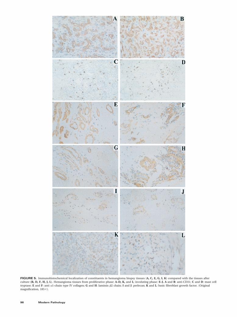
FIGURE 5. Immunohistochemical localization of constituents in hemangioma biopsy tissues (A, C, E, G, I, K) compared with the tissues afterculture (B, D, F, H, J, L). Hemangioma tissues from proliferative phase: A–D, K, and L; involuting phase: E–J. A and B: anti-CD31; C and D: mast celltryptase; E and F: anti a1-chain type IV collagen; G and H: laminin b2 chain; I and J: perlecan; K and L: basic fibroblast growth factor. (Originalmagnification, 1853).
98 Modern Pathology
Wellington Medical Research Foundation, LotteryHealth Research, and the Health Research Council ofNew Zealand. We are grateful to C. Marstella for herassistance with the preparation of this manuscript.
REFERENCES
1. Mulliken JB, Glowacki J. Hemangiomas and vascular malfor-mations in infants and children: a classification based onendothelial characteristics. Plast Reconstr Surg 1982;69:412–22.
2. Glowacki J, Mulliken JB. Mast cells in hemangiomas andvascular malformations. Pediatrics 1982;70:48 –51.
3. Dethlefsen SM, Mulliken JB, Glowacki J. An ultrastructuralstudy of mast cell interactions in hemangiomas. UltrastructPathol 1986;10:175– 83.
4. Mulliken JB. Diagnosis and natural history of haemangio-mas. In: Mulliken JB, Young AE, editors. Vascular birth-marks: haemangiomas and vascular malformations. Phila-delphia: W.B. Saunders; 1988. pp. 41– 62.
5. Takahashi K, Mulliken JB, Kozakewich HPW, Rogers RA,Folkman J, Ezekowitz RAB. Cellular markers that distinguishthe phases of hemangioma during infancy and childhood.J Clin Invest 1994;93:2357– 64.
6. Enjolras O, Mulliken JB. The current management of vascu-lar birthmarks. Pediatr Dermatol 1993;10:311–3.
7. Vikkula M, Boon LM, Mulliken JB, Olsen BR. Molecular basisof vascular anomalies. Trends Cardiovasc Med 1998;8:281–92.
8. Dubois-Stringfellow N, Kolpack-Martindale L, Bautch VL,Azizkhan RG. Mice with hemangiomas induced by trans-genic endothelial cells: a model for the Kasabach-Merittsyndrome. Am J Pathol 1994;144:796 – 806.
9. Williams RL, Risau W, Zerwes H-G, Drexler H, Aguzzi A,Wagner EF. Endothelioma cells expressing the polyoma mid-dle T oncogene induce hemangiomas by host cell recruit-ment. Cell 1989;57:1053– 63.
10. Sage EH. Secretion of SPARC by endothelial cells trans-formed by polyoma middle T oncogene inhibits the growthof normal endothelial cells in vitro. Biochem Cell Biol 1992;70:579 –92.
11. Meninger CJ, Brightman SE, Kelly KA, Zetter BR. Increasedstem cell factor release by hemangioma-derived endothelialcells. Lab Invest 1995;72:166 –73.
12. Mulliken JB, Zetter BR, Folkman J. In vitro characteristics ofendothelium from hemangiomas and vascular malforma-tions. Surgery 1982;92:348 –53.
13. Davis PF, He Y, Furneaux RH, Johnston PS, Ruger BM, SlimGC. Inhibition of angiogenesis by oral ingestion of powderedshark cartilage in a rat model. Microvasc Res 1997;54:178 –82.
14. Strobel S, Miller HRP, Ferguson A. Human intestinal muco-sal mast cells: evaluation of fixation and staining techniques.J Clin Pathol 1981;34:851– 8.
15. Ruger B, Dunbar PR, Hasan Q, Sawada H, Kittelberger R,Greenhill N, et al. Human mast cells produce type VIII col-lagen in vivo. Int J Exp Pathol 1994;75:397– 404.
16. Neale TJ, Ruger BM, Macuaulay HA, Dunbar PR, Hasan Q,Bourke A, et al. Tumour necrosis factor-a is expressed byglomerular visceral epithelial cells in human membranousnephropathy. Am J Pathol 1995;146:1444 –54.
17. Ruger BM, Hasan Q, Greenhill NS, Davis PF, Dunbar PR,Neale TJ. Mast cells and type VIII collagen in human diabeticnephropathy. Diabetologia 1996;39:1215–22.
18. Rüger BM, Hasan Q, Erb KJ, Davis PF. Progression of renaldisease in interleukin-4 transgenic mice: involvement oftransforming growth factor-b. Int J Exp Pathol 1999;80:113–23.
19. Hasan Q, Rüger BM, Davis PF, Greenhill NS, Murray-McIntosh RP, Dunbar R, Neale TJ. Analysis of expression ofmultiple cytokines in individual human renal biopsies. Kid-ney Inter 1997;51:1311–2.
20. Bird SD, Hasan Q, Davis PF, Walker RJ. Platelet derivedgrowth factor-BB induced calcium transient in cultured hu-man peritoneal mesothelial cells. ASAIO Journal 1998;44:835– 40.
21. Hasan Q, Dunbar PR, Murray-McIntosh RP, Neale TJ. Trans-forming growth factor b isoforms in human glomerulone-phropathies. Nephrology 1998;4:353–9.
22. Tan ST, Velickovic M, Rüger BM, Davis PF. Cellular andextracellular markers of hemangioma. Plast Reconstr Surg.In press.
23. Mulliken JB, Boon LM, Takahashi K, Ohlms LA, Folkman J,Ezekowitz AB. Pharmacologic therapy for endangering hem-angiomas. Current Opinion in Dermatol 1995;109 –13.
24. Hasan Q, Tan ST, Gush J, Peters S, Davis PF. Steroid therapyof a proliferating hemangioma: Histochemical and molecu-lar changes. Pediatrics. In press.
25. Greer P, Haigh J, Mbamalu G, Khoo W, Bernstein A, PawsonT. The Fps/Fes protein-tyrosine kinase promotes angiogen-esis in transgenic mice. Mol Cell Biol 1994;14:6755– 63.
26. Pepper MS, Ferrara N, Orci L, Montesano R. Potent syner-gism between vascular endothelial growth factor and basicfibroblast growth factor in the induction of angiogenesis invitro. Biochem Biophys Res Comm 1992;189:824 –31.
27. Folkman J, D’Amore PA. Blood vessel formation: what is itsmolecular basis? Cell 1996;87:1153–5.
28. Jang Y-C, Arumugam S, Ferguson M, Gibran NS, Isik FF.Changes in matrix composition of human hemangiomas.J Surg Res 1998;80:9 –15.
29. Chang J, Most D, Bresnick S, Mehrara B, Steinbrech DS,Reinisch J, et al. Proliferative hemangiomas: analysis of cy-tokine gene expression and angiogenesis. Plast ReconstrSurg 1999;103:1–9.
30. Nicosia RF, Ottinetti A. Growth of microvessels in serum-freematrix culture of rat aorta: a quantitative assay of angiogen-esis in vitro. Lab Invest 1990;63:115–22.
31. Brown KJ, Maynes SF, Bezos A, Maguire DJ, Ford MD, ParishCR. A novel in vitro assay for human angiogenesis. LabInvest 1996;75:539 –54.
FIGURE 6. Ethidium bromide stained 2% agarose gel showing reversetranscriptase-polymerase chain reaction products in biopsy (lanes 2, 4,6, 8, and 10) and tissue after culture (lanes 3, 5, 7, 9, and 11), takenfrom a proliferative hemangioma. Lane 1 is the 123bp DNA ladder;lanes 2 and 3: hypoxanthine phosphoribosyl transferase (;395bp); lanes4 and 5: transforming growth factor (TGF)-b3 (;312bp); lanes 6 and 7:TGF-b1(;301bp); lanes 8 and 9: TGF-b2 (;363bp); lanes 10 and 11:basic fibroblast growth factor (;145bp).
Culture of Human Hemangioma (S.T. Tan et al.) 99