Hematopathology - Nature
36
232A ANNUAL MEETING ABSTRACTS Design: The 3 cases were identified by a database search of our files for all nasopharyngeal tumors. The clinicopathologic features were analyzed. Representative paraffin blocks were immunohistochemically stained with antibodies against cytokeratins (AE1/AE3, CAM5.2, OSCAR, CK5/6, CK7, CK8, CK19, CK20), thyroglobulin, TTF-1, p63, calponin, carcinoembryonic antigen (CEA), chromogranin and synaptophysin. Results: Of our 3 cases, 2 occurred in women ages 13 and 18, and 1 in a man age 64. Patients complained of nasal obstruction and examination revealed nasopharyngeal- based exophytic lesions. The light microscopic features were those typically seen in LGNPPA. All three tumors showed diffuse (nuclear) immunoreactivity for TTF-1, as well as cytokeratin immunoreactivity (AE1/AE3, OSCAR, CAM5.2, CK7, CK8, CK19). No immunoreactivity was seen for thyroglobulin, CEA, p63, calponin, chromogranin and synaptophysin. Conclusions: TTF-1, although considered a dedicated marker of thyroid and lung carcinomas, may be found in neuroendocrine carcinomas of non-pulmonary origin. In addition, we report herein the presence of TTF-1 immunoreactivity in LGNPPA, thereby extending the list of tumors that may stain with TTF-1. In conjunction with light microscopic features overlapping with those of TPC, the presence of TTF-1 may result in an erroneous diagnosis of metastatic TPC. The presence of surface epithelial derivation and absence of thyroglobulin staining should allow for a diagnosis of LGNPPA differentiating it from TPC even in the presence of TTF-1 staining. The origin for the “aberrant” TTF-1 expression remains uncertain but the embryologic development of the thyroid gland from the primitive pharynx may provide a mechanism to explain this phenomenon. Hematopathology 1062 Different Chemokine Receptor Expression Profile in Extragastric and Gastric MALT-Lymphomas A Aigelsreiter, AJA Deutsch, E Stelzl, C Beham-Schmid, A Beham, W Linkesch, H Schaider, HH Kessler, P Neumeister. MUG, Graz, Austria. Background: Chemokine receptors mediate migration and activation of lymphocytes through binding of their ligands and contribute to the development of hematopoietic neoplasms. Strong expression of CXCR5 was detected in transformed B cells in Helicobacter (H.) pylori positive gastric MALT lymphomas. Recently, Chlamydia (C.) psittaci was identified as the causative infectious agent of ocular adnexal MALT lymphomas. The aim of this study was to identify the expression pattern of all 19 currently known chemokine receptors in extragastric and gastric MALT-lymphomas. Furthermore, extragastric MALT lymphomas were investigated for the presence of C. psittaci DNA and their chemokine profile compared to their uninfected controls. Design: 16 extragastric and 5 H. pylori positive gastric formalin-fixed, paraffin- embedded MALT lymphoma samples were processed for RNA and DNA isolation. Extragastric sites of lymphoma origin were salivary gland, thyroid gland, skin and ocular adnexa. Semiquantitative Real Time PCR was performed on a GeneAmp® 5700 Sequence Detector. Expression of 19 chemokine receptors was determined in triplicate and the number of cycles was compared to the reference gene HPRT and peripheral blood, based on calculations of 2-deltadeltaCT. The samples were further analyzed for presence of C. psittaci DNA via Real-time PCR performed on the LightCycler instrument. Results: Comparing the mean values of chemokine receptor expression of extragastric to H. pylori positive gastric MALT lymphomas, CXCR1 (23 times down regulated, p=0.002), CXCR2 (170 times down regulated, p=0.029 ), CX3CR1 (22 times up regulated, p=0.002) and XCR1 (de novo expressed, p=0.002) were significantly differently expressed. 10 of 16 (63%) extragastric and none of 5 H. pylori positive gastric MALT lymphomas tested positive for C. psittaci DNA. No difference in the chemokine receptor expression profile between C. psittaci infected vs. uninfected extragastric MALT lymphoma samples could be observed. Conclusions: C. psittaci infection is associated with a significant proportion of extragastric MALT lymphomas other than ocular adnexa. Further, differently expressed chemokine receptors might be responsible for the various sites of origin of MALT lymphomas. Thereby, the expression profiles of CXCR1, CXCR2, CX3CR1 and XCR1 seem to be the key determinant in the homing of malignant B-cells either into the stomach or into extragastric sites. 1063 DRAQ-5 Based No-Lyse, No-Wash Bone Marrow Aspirate Evaluation by Flow Cytometry RW Allan, MA Ansari-Lari, S Jordan. University of Florida, Gainesville, FL; Memorial Regional Hospital, Hollywood, FL. Background: Flow cytometry (FC) is a powerful tool for objective phenotyping of hematolymphoid neoplasia. Analysis of bone marrow aspirates and peripheral blood specimens by flow cytometry typically requires an erythrocyte lysis or gradient separation method to remove erythrocytes prior to analysis which may result in the loss of certain populations, in particular nucleated erythroid cells. This results in FC analysis not reflecting the true composition of nucleated cells. DRAQ-5 is a novel, far-red fluorescing DNA specific dye that penetrates live cells and is excitable using a 488-nm laser. We sought to develop a method where we could evaluate bone marrow aspirates (BMAs) by FC without specimen manipulation/ lysis by exploiting DRAQ5 fluorescence as a gating parameter to analyze nucleated events. Design: We analyzed a total of 30 normal and abnormal BMAs (15 males, 15 females) from patients with a variety of diagnoses on an FC500 flow cytometer utilizing a DRAQ5 based no-lyse, no-wash FC protocol (DRAQ 5 protocol) in combination with CD71 FITC and CD45 PE antibodies to determine the percent of different major cell populations present in the BMA (nucleated RBCs, blasts, myeloid, monocytic and lymphs). These were compared to blinded morphologic differential counts performed on Wright stained slides prepared from the same specimen and to differential counts obtained by conventional erythrocyte lysis FC analysis. Results: Light scatter and fluorescence staining of the cells in the DRAQ5 protocol were comparable to conventional FC analysis and allowed discrimination of the major bone marrow cell populations in normal and abnormal specimens. The correlation coefficient between DRAQ5 protocol differential counts and morphologic counts were: 0.97 nRBCS; 0.97 myeloid; 0.94 blasts; 0.42 monocytes; 0.58 lymphs. Correlation coefficients between conventional FC and morphologic differentials were: 0.84 nRBCS; 0.89 myeloid; 0.98 blasts; 0.40 monocytes; 0.52 lymphs. NRBC counts were significantly different between the conventional FC and morphology (p<0.001); no significant difference was observed for the DRAQ5 protocol. Conclusions: The DRAQ5 protocol is a simple method to quantify the major cell populations in the BMA. The method more accurately quantifies nucleated erythroid cells compared to conventional FC and thus allows for quantitation of blasts/ abnormal cells that reflect the morphologic nucleated cell differential. 1064 CXCR4 Expression in Follicular Lymphomas MS Almiski, N Razumilava, M Kurrer, E Levi. Wayne State University, Detroit, MI; University Hospital, Zurich, Switzerland; John D. Dingell VA Medical Center, Detroit, MI. Background: CXCR4 is a chemokine receptor that is involved in lymphocyte trafficking, stem cell mobilization and is associated with tumor cell invasiveness. We previously have shown that in diffuse large B cell lymphomas CXCR4 is a poor prognostic factor. It has also been shown that CXCR4 is one of several preferentially overexpressed peptides in follicular lymphomas and also it has been demonstrated that CXCR4 is a chemotactic factor for follicular lymphoma cells. We investigated the expression of CXCR4 in follicular lymphomas and correlated its expression with prognosis. Design: Patients with follicular lymphoma (n=218) followed up at University Hospital, Zurich were selected to generate a tissue array. Patient mean follow up was 60 months. Immunohistochemical studies were done utilizing CXCR4 (fusin h-118; sc-9046 Santa Cruz). In addition, CXCR4 expression was correlated with Fak, MLK3 and CARP-1. The cases were scored according to staining intensity and percentage tumor cell staining. We scored the staining by multiplying staining intensity with percent cells staining. A cutoff value of 100 was utilized to classify the cases as expressors vs. non-expressors. Results: In normal lymphoid tissues, CXCR4 had a weak diffuse staining in the germinal centers which was both cytoplasmic and nuclear. In the interfollicular areas there were scattered staining lymphocytes. CXCR4 staining was present in 143/218 cases of follicular lymphoma. The staining was predominantly nuclear but also cytoplasmic. CXCR4 expression did not have any prognostic value in the follicular lymphomas tested. We also analyzed a subgroup of diffuse large cell lymphomas of follicular origin and found that expression of CXCR4 was a poor prognostic factor with marginal significance. CXCR4 expression had a strong correlation with Fak and CARP-1 expression. Conclusions: CXCR4 is frequently expressed in follicular lymphomas. Its expression correlates with expression of Fak. However, unlike that of transformed follicular lymphomas, or diffuse large B cell lymphomas in general, its expression is not a poor prognostic factor. 1065 Loss of Carp-1 Expression Is a Poor Prognostic Factor in Follicular Lymphomas MS Almiski, N Razumilava, M Kurrer, A Rishi, E Levi. Wayne State University, Detroit, MI; University Hospital, Zurich, Switzerland; John D. Dingell VAMC, Detroit, MI. Background: Carp-1 is a recently described apoptosis and cell cycle arrest inducing peptide that presumably acts as a tumor suppressor. We have previously shown that its expression strongly correlates with apoptotic activity in diffuse large B cell lymphomas and is inversely associated with pAkt expression. We have also demonstrated its loss of expression in a variety of solid tumors that are high grade. Since the follicular lymphomas are associated with resistance to apoptosis acquired by the t(14;18) translocation, we hypothesized that CARP-1 expression would have a biological and prognostic value. Design: A tissue array was generated utilizing 220 cases of follicular lymphoma followed up at University Hospital Zurich, with a mean follow up of 60 months. CARP-1 antibody was generated by immunizing rabbits to synthetic CARP-1 peptide. The cases were represented in duplicate in the array. The cases that demonstrated at least 25% staining were considered positive for expression. We also correlated CARP-1 expression with Fak, CXCR4 and MLK3 expression, all of which were prognostic markers in diffuse large cell lymphomas. Results: CARP-1 expression was present in 172 of 220 cases. Its expression correlated with Fak expression. we have previously demonstrated Fak to be a good prognostic factor in diffuse large cell lymphomas, and associated with a germinal center cell phenotype. In addition, CARP-1 expression showed a strong correlation with CXCR4 expression. The patients expressing CARP-1 had a better overall survival compared to non-expressors (106 vs. 57 months mean survival; p=0.0312). We also tested a follicular lymphoma cell line (WSU-FSCCL1) to observe the in vitro effects of CARP-1. In vitro transduction of CARP-1 and its shorter fragments induced apoptosis which was associated with translocation of Nur77 to the cytoplasm. Immunoprecipitation studies demonstrated binding and colocalization of Nur77 and CARP-1 in several lymphoma cell lines, suggesting Nur77 is involved in apoptosis induced via CARP-1. Conclusions: Loss of CARP-1 expression is associated with a poor prognosis in follicular lymphomas. CARP-1 is a candidate as a prognostic marker and a potential target for therapy in follicular lymphomas.
-
Upload
khangminh22 -
Category
Documents
-
view
1 -
download
0
Transcript of Hematopathology - Nature

232A ANNUAL MEETING ABSTRACTSDesign: The 3 cases were identified by a database search of our files for all nasopharyngeal tumors. The clinicopathologic features were analyzed. Representative paraffin blocks were immunohistochemically stained with antibodies against cytokeratins (AE1/AE3, CAM5.2, OSCAR, CK5/6, CK7, CK8, CK19, CK20), thyroglobulin, TTF-1, p63, calponin, carcinoembryonic antigen (CEA), chromogranin and synaptophysin.Results: Of our 3 cases, 2 occurred in women ages 13 and 18, and 1 in a man age 64. Patients complained of nasal obstruction and examination revealed nasopharyngeal-based exophytic lesions. The light microscopic features were those typically seen in LGNPPA. All three tumors showed diffuse (nuclear) immunoreactivity for TTF-1, as well as cytokeratin immunoreactivity (AE1/AE3, OSCAR, CAM5.2, CK7, CK8, CK19). No immunoreactivity was seen for thyroglobulin, CEA, p63, calponin, chromogranin and synaptophysin.Conclusions: TTF-1, although considered a dedicated marker of thyroid and lung carcinomas, may be found in neuroendocrine carcinomas of non-pulmonary origin. In addition, we report herein the presence of TTF-1 immunoreactivity in LGNPPA, thereby extending the list of tumors that may stain with TTF-1. In conjunction with light microscopic features overlapping with those of TPC, the presence of TTF-1 may result in an erroneous diagnosis of metastatic TPC. The presence of surface epithelial derivation and absence of thyroglobulin staining should allow for a diagnosis of LGNPPA differentiating it from TPC even in the presence of TTF-1 staining. The origin for the “aberrant” TTF-1 expression remains uncertain but the embryologic development of the thyroid gland from the primitive pharynx may provide a mechanism to explain this phenomenon.
Hematopathology1062 DifferentChemokineReceptorExpressionProfileinExtragastricandGastricMALT-LymphomasA Aigelsreiter, AJA Deutsch, E Stelzl, C Beham-Schmid, A Beham, W Linkesch, H Schaider, HH Kessler, P Neumeister. MUG, Graz, Austria.Background: Chemokine receptors mediate migration and activation of lymphocytes through binding of their ligands and contribute to the development of hematopoietic neoplasms. Strong expression of CXCR5 was detected in transformed B cells in Helicobacter (H.) pylori positive gastric MALT lymphomas. Recently, Chlamydia (C.) psittaci was identified as the causative infectious agent of ocular adnexal MALT lymphomas. The aim of this study was to identify the expression pattern of all 19 currently known chemokine receptors in extragastric and gastric MALT-lymphomas. Furthermore, extragastric MALT lymphomas were investigated for the presence of C. psittaci DNA and their chemokine profile compared to their uninfected controls.Design: 16 extragastric and 5 H. pylori positive gastric formalin-fixed, paraffin-embedded MALT lymphoma samples were processed for RNA and DNA isolation. Extragastric sites of lymphoma origin were salivary gland, thyroid gland, skin and ocular adnexa. Semiquantitative Real Time PCR was performed on a GeneAmp® 5700 Sequence Detector. Expression of 19 chemokine receptors was determined in triplicate and the number of cycles was compared to the reference gene HPRT and peripheral blood, based on calculations of 2-deltadeltaCT. The samples were further analyzed for presence of C. psittaci DNA via Real-time PCR performed on the LightCycler instrument.Results: Comparing the mean values of chemokine receptor expression of extragastric to H. pylori positive gastric MALT lymphomas, CXCR1 (23 times down regulated, p=0.002), CXCR2 (170 times down regulated, p=0.029 ), CX3CR1 (22 times up regulated, p=0.002) and XCR1 (de novo expressed, p=0.002) were significantly differently expressed. 10 of 16 (63%) extragastric and none of 5 H. pylori positive gastric MALT lymphomas tested positive for C. psittaci DNA. No difference in the chemokine receptor expression profile between C. psittaci infected vs. uninfected extragastric MALT lymphoma samples could be observed.Conclusions: C. psittaci infection is associated with a significant proportion of extragastric MALT lymphomas other than ocular adnexa. Further, differently expressed chemokine receptors might be responsible for the various sites of origin of MALT lymphomas. Thereby, the expression profiles of CXCR1, CXCR2, CX3CR1 and XCR1 seem to be the key determinant in the homing of malignant B-cells either into the stomach or into extragastric sites.
1063 DRAQ-5 Based No-Lyse, No-Wash Bone Marrow AspirateEvaluationbyFlowCytometryRW Allan, MA Ansari-Lari, S Jordan. University of Florida, Gainesville, FL; Memorial Regional Hospital, Hollywood, FL.Background: Flow cytometry (FC) is a powerful tool for objective phenotyping of hematolymphoid neoplasia. Analysis of bone marrow aspirates and peripheral blood specimens by flow cytometry typically requires an erythrocyte lysis or gradient separation method to remove erythrocytes prior to analysis which may result in the loss of certain populations, in particular nucleated erythroid cells. This results in FC analysis not reflecting the true composition of nucleated cells. DRAQ-5 is a novel, far-red fluorescing DNA specific dye that penetrates live cells and is excitable using a 488-nm laser. We sought to develop a method where we could evaluate bone marrow aspirates (BMAs) by FC without specimen manipulation/ lysis by exploiting DRAQ5 fluorescence as a gating parameter to analyze nucleated events.Design: We analyzed a total of 30 normal and abnormal BMAs (15 males, 15 females) from patients with a variety of diagnoses on an FC500 flow cytometer utilizing a DRAQ5 based no-lyse, no-wash FC protocol (DRAQ 5 protocol) in combination with
CD71 FITC and CD45 PE antibodies to determine the percent of different major cell populations present in the BMA (nucleated RBCs, blasts, myeloid, monocytic and lymphs). These were compared to blinded morphologic differential counts performed on Wright stained slides prepared from the same specimen and to differential counts obtained by conventional erythrocyte lysis FC analysis.Results: Light scatter and fluorescence staining of the cells in the DRAQ5 protocol were comparable to conventional FC analysis and allowed discrimination of the major bone marrow cell populations in normal and abnormal specimens. The correlation coefficient between DRAQ5 protocol differential counts and morphologic counts were: 0.97 nRBCS; 0.97 myeloid; 0.94 blasts; 0.42 monocytes; 0.58 lymphs. Correlation coefficients between conventional FC and morphologic differentials were: 0.84 nRBCS; 0.89 myeloid; 0.98 blasts; 0.40 monocytes; 0.52 lymphs. NRBC counts were significantly different between the conventional FC and morphology (p<0.001); no significant difference was observed for the DRAQ5 protocol.Conclusions: The DRAQ5 protocol is a simple method to quantify the major cell populations in the BMA. The method more accurately quantifies nucleated erythroid cells compared to conventional FC and thus allows for quantitation of blasts/ abnormal cells that reflect the morphologic nucleated cell differential.
1064 CXCR4ExpressioninFollicularLymphomasMS Almiski, N Razumilava, M Kurrer, E Levi. Wayne State University, Detroit, MI; University Hospital, Zurich, Switzerland; John D. Dingell VA Medical Center, Detroit, MI.Background: CXCR4 is a chemokine receptor that is involved in lymphocyte trafficking, stem cell mobilization and is associated with tumor cell invasiveness. We previously have shown that in diffuse large B cell lymphomas CXCR4 is a poor prognostic factor. It has also been shown that CXCR4 is one of several preferentially overexpressed peptides in follicular lymphomas and also it has been demonstrated that CXCR4 is a chemotactic factor for follicular lymphoma cells. We investigated the expression of CXCR4 in follicular lymphomas and correlated its expression with prognosis.Design: Patients with follicular lymphoma (n=218) followed up at University Hospital, Zurich were selected to generate a tissue array. Patient mean follow up was 60 months. Immunohistochemical studies were done utilizing CXCR4 (fusin h-118; sc-9046 Santa Cruz). In addition, CXCR4 expression was correlated with Fak, MLK3 and CARP-1. The cases were scored according to staining intensity and percentage tumor cell staining. We scored the staining by multiplying staining intensity with percent cells staining. A cutoff value of 100 was utilized to classify the cases as expressors vs. non-expressors.Results: In normal lymphoid tissues, CXCR4 had a weak diffuse staining in the germinal centers which was both cytoplasmic and nuclear. In the interfollicular areas there were scattered staining lymphocytes. CXCR4 staining was present in 143/218 cases of follicular lymphoma. The staining was predominantly nuclear but also cytoplasmic. CXCR4 expression did not have any prognostic value in the follicular lymphomas tested. We also analyzed a subgroup of diffuse large cell lymphomas of follicular origin and found that expression of CXCR4 was a poor prognostic factor with marginal significance. CXCR4 expression had a strong correlation with Fak and CARP-1 expression.Conclusions: CXCR4 is frequently expressed in follicular lymphomas. Its expression correlates with expression of Fak. However, unlike that of transformed follicular lymphomas, or diffuse large B cell lymphomas in general, its expression is not a poor prognostic factor.
1065 LossofCarp-1ExpressionIsaPoorPrognosticFactorinFollicularLymphomasMS Almiski, N Razumilava, M Kurrer, A Rishi, E Levi. Wayne State University, Detroit, MI; University Hospital, Zurich, Switzerland; John D. Dingell VAMC, Detroit, MI.Background: Carp-1 is a recently described apoptosis and cell cycle arrest inducing peptide that presumably acts as a tumor suppressor. We have previously shown that its expression strongly correlates with apoptotic activity in diffuse large B cell lymphomas and is inversely associated with pAkt expression. We have also demonstrated its loss of expression in a variety of solid tumors that are high grade. Since the follicular lymphomas are associated with resistance to apoptosis acquired by the t(14;18) translocation, we hypothesized that CARP-1 expression would have a biological and prognostic value.Design: A tissue array was generated utilizing 220 cases of follicular lymphoma followed up at University Hospital Zurich, with a mean follow up of 60 months. CARP-1 antibody was generated by immunizing rabbits to synthetic CARP-1 peptide. The cases were represented in duplicate in the array. The cases that demonstrated at least 25% staining were considered positive for expression. We also correlated CARP-1 expression with Fak, CXCR4 and MLK3 expression, all of which were prognostic markers in diffuse large cell lymphomas.Results: CARP-1 expression was present in 172 of 220 cases. Its expression correlated with Fak expression. we have previously demonstrated Fak to be a good prognostic factor in diffuse large cell lymphomas, and associated with a germinal center cell phenotype. In addition, CARP-1 expression showed a strong correlation with CXCR4 expression. The patients expressing CARP-1 had a better overall survival compared to non-expressors (106 vs. 57 months mean survival; p=0.0312). We also tested a follicular lymphoma cell line (WSU-FSCCL1) to observe the in vitro effects of CARP-1. In vitro transduction of CARP-1 and its shorter fragments induced apoptosis which was associated with translocation of Nur77 to the cytoplasm. Immunoprecipitation studies demonstrated binding and colocalization of Nur77 and CARP-1 in several lymphoma cell lines, suggesting Nur77 is involved in apoptosis induced via CARP-1.Conclusions: Loss of CARP-1 expression is associated with a poor prognosis in follicular lymphomas. CARP-1 is a candidate as a prognostic marker and a potential target for therapy in follicular lymphomas.

ANNUAL MEETING ABSTRACTS 233A
1066 IntrasinusoidalInfiltrationIsMoreFrequentinSplenicMarginalZoneLymphoma,butLymphoidFollicleFormationIsCommoninAllTypesofMarginalZoneLymphomasHM Amin, KV Inamdar, LJ Medeiros, EJ Schlette. The University of Texas M.D. Anderson Cancer Center, Houston, TX.Background: Marginal zone lymphomas (MZL) include extranodal marginal zone lymphoma of mucosa associated lymphoid tissue (MALT lymphoma), nodal marginal zone lymphoma (NMZL) and splenic marginal zone lymphoma (SMZL). Clinically, MALT lymphoma involves extranodal sites and often remains localized, but can disseminate. NMZL primarily involves the lymph nodes, but can also involve bone marrow (BM). In contrast, SMZL presents with splenomegaly and is frequently associated with BM involvement. Intrasinusoidal infiltration pattern as well as lymphoid follicle formation in BM has been described as a fairly specific and frequent finding in SMZL [Franco V et al. Cancer, 91: 294, 2001]. We assessed the frequency and extent of intrasinusoidal infiltration as well as lymphoid follicle formation in various MZL types involving BM.Design: The study group included 30 cases of MZL including 19 SMZL, 6 MALT lymphomas and 5 NMZL involving the BM. Hematoxylin and eosin stained tissue sections of staging BM biopsies were reviewed for extent and pattern of involvement. Immunohistochemistry was performed on BM biopsy sections of all 30 cases using antibodies specific for CD20 and/or PAX5, CD21 and CD23. Intrasinusoidal infiltration was defined as 5 or more CD20 or PAX5 positive lymphoid cells forming linear groups. The presence of follicular dendritic networks identified by CD21 and/or CD23 defined lymphoid follicle formation.Results: The median percentage of lymphomatous BM involvement was 15% of the medullary space (range, <5%-60%). Multiple patterns of lymphomatous infiltration were seen in 22/30 (73%) of MZL cases whereas the remaining 8 cases showed a single pattern of BM infiltration. The most frequent pattern of infiltration was nodular (17/19; 89% in SMZL, 9/11; 82% in other MZL types) followed by interstitial (13/19; 68% in SMZL, 6/11; 54.5% in other MZL types). Intrasinusoidal infiltration was more frequent in SMZL (6/19; 31.5%) than other MZL types (1/11; 9%), but was always associated with other patterns. CD21 and /or CD23 highlighted the follicular dendritic networks in the infiltrates of 16/17 (94%) SMZL compared with 7/10 (70%) cases of other MZL types.Conclusions: Intrasinusoidal infiltration of BM was relatively infrequent in patients with MZL, but was associated with SMZL. It was not seen as the sole pattern in any type of MZL. Lymphoid follicle formation was identified in all types of MZL.
1067 CorrelationofD-TypeCyclinExpressionwithImmunophenotypeinDiffuseLargeB-CellLymphomasSR Backus, S Chickkamuniyappa, YH Diep, RS Robetorye. The University of Texas Health Science Center at San Antonio, San Antonio, TX.Background: Diffuse large B-cell lymphoma (DLBCL) is a clinically heterogeneous lymphoma that can be divided into prognostically important subgroups using gene expression profiling and immunohistochemical markers, including germinal center B-cell (GCB), activated GCB (AGCB), activated non-GCB (ABC), and unclassified lymphoma groups. The GCB subgroup has been reported to have significantly better survival than the activated and unclassified subgroups. D-type cyclins are involved in regulation of progression of cells from G1 to S phase of the cell cycle, and expression of these genes has also been associated with prognostic significance in DLBCL. The purpose of this study is to determine whether DLBCL immunophenotype correlates with the expression of specific D-type cyclins.Design: 67 cases of nodal DLBCL were analyzed for expression of cyclins D1, D2, and D3 using quantitative real-time RT-PCR. 25 DLBCL cases that exhibited relatively high expression of at least one of the D-type cyclins were also subjected to immunophenotyping using specific antibodies for CD3, CD10, CD20, CD138, BCL-2, BCL-6, FOXP1, Ki-67, MUM1, and PKC-beta. Cases were classified as GCB if the germinal center markers CD10 and/or BCL-6 were expressed without activation markers (MUM1 and CD138), AGCB if at least one GCB marker and one of the activation markers were expressed, and ABC if at least one activation marker was expressed without GCB markers.Results: DLBCLs that exhibited over-expression of at least one of the D-type cyclins had an immunophenotype consistent with the GCB subgroup in 36% of cases, the AGCB subgroup in 40% of cases, and the ABC subgroup in 24% of cases. Specific D-type cyclin expression did not correlate with DLBCL subgroup except for the ABC subgroup which exhibited over-expression of only cyclins D2 or D3, but not D1. DLBCL cases that exhibited high levels of D-type cyclin expression also showed a high proliferative rate (82% of cases with >50% Ki-67 expression) and high-level expression of the prognostic markers BCL-2 (76%), PKC-beta (79%), and FOXP1 (63%).Conclusions: Over-expression of at least one of the D-type cyclins correlated with an activated DLBCL immunophenotype in approximately two-thirds of cases. These cases also exhibited high-level expression of Ki-67, BCL-2, PKC-beta, and FOXP1. These results suggest that over-expression of D-type cyclins may contribute to the adverse prognosis associated with DLBCLs that exhibit an activated immunophenotype.
1068 Clinicopathologic Features of B-Cell Post-TransplantLymphoproliferative Disorders Occurring in LymphoidTissue of theWaldeyer’sRingD Baiyee, R Gupta, B Alobeid, G Bhagat. Columbia University, New York, NY.Background: B-cell post-transplant lymphoproliferative disorders (PTLD) more commonly occur at extranodal sites but can also arise in lymphoid tissue of the Waldeyer’s ring (WR). However, data regarding the spectrum of PTLD involving the WR and their clinical outcomes are limited. We thus investigated the frequency and clinico-pathologic features of B-cell PTLD occurring at this location.
Design: Clinical data of all B-cell PTLD diagnosed at our institute, over 16 yrs, were reviewed to identify PTLD arising in the WR. H&E sections were reviewed and immunohistochemical stains, in situ hybridization (ISH) for EBER, flow cytometry, and PCR for IgH gene rearrangement were performed. PTLD were classified using current WHO criteria and divided into 2 groups according to age at occurrence (<18 and >18 yrs of age).Results: Of 91 B-cell PTLD, including 16 early lesions (EL) (8 plasmacytic hyperplasia (PH) and 8 Infectious Mononucleosis-like lesions (IMLL), 28 polymorphic PTLD (P-PTLD), and 47 monomorphic PTLD (M-PTLD), 19 (20.8 %) PTLD occurred in the WR (6M, 13F, age 1-52 yrs, median 7 yrs). These comprised 12/19 (63.2%) EL (4PH, 8 IMLL), 3/19(15.8%) P-PTLD, and 4/19 (21.1%) M-PTLD (2 plasmacytoma-like lesions [PLL], 2 diffuse large B-cell lymphomas). All P-PTLD and M-PTLD and all except 1 EL (PH) occurred in patients <18 yrs of age. Mean duration from transplantation to diagnosis was 39.5 months for EL, 35 months for P-PTLD, and 33.5 months for M-PTLD. Four PTLD (1 IMLL, 1 P-PTLD, 2 M-PTLD) were clonal. ISH for EBER was positive in 12 cases (7 EL, 3 P-PTLD, and 2 M-PTLD), both PLL were EBER-. One patient with PH subsequently developed PLL at the same site after 9 yrs and 3 patients (1 PH, 1 IMLL, 1 P-PTLD) developed PTLD (1 PH, 2 P-PTLD) at other sites, either 1 month previously (gastrointestinal tract) or 17-26 months later (gingiva, lymph node). Sixteen of 19 (84%) patients are currently alive 7-181 months (median 50 months) post diagnosis; none of the deaths were attributed to PTLD.Conclusions: B-cell PTLD involving the WR almost exclusively occur in patients < 18 yrs of age. They have an indolent clinical course and only a minority (5%) develop metachronous PTLD at the same or different sites. EL represent the majority but P-PTLD and M-PTLD are not infrequent. EBV+ PTLD predominate, however, EBV- PLL also occur at this location. Since PLL have overlapping morphologic features with PH and P-PTLD, phenotypic and molecular analyses are essential for diagnosing such lesions.
1069 ZAP70ExpressionDeterminedbyImmunohistochemistry(IHC)ofCellClotsCorrelateswithIgHMutationStatusandCytogeneticsinChronicLymphocyticLeukemia/SmallLymphocyticLymphoma(CLL/SLL)I Bansal, E Hyjek, W Tam, A Chiu, R Furman, M Coleman, S Ely, E Fusco, S Mathew, DM Knowles, A Chadburn. Weill Cornell Medical College, NY, NY.Background: The presence or absence of somatic hypermutations in the immunoglobulin heavy chain gene (IgH) in CLL/SLL is prognostic (unmutated (U)-poor; mutated (M)-good). However, DNA sequence analysis is impractical in clinical practice. ZAP70 expression by flow cytometry (FC) correlates with IgH status and prognosis in CLL/SLL. However, the optimal method of ZAP70 evaluation by FC (antibody clone, fluorochrome, cut-offs) has not been determined. We previously showed IHC of CLL/SLL cells in a thrombin-fibrin matrix (cell clots; CCs) is an efficient method of determining ZAP70 expression. We now show ZAP70 expression by IHC correlates with IgH status and other prognostic parameters.Design: IHC for ZAP70 (2F3.2, Upstate), PAX5 and CD3 (DAKO) was performed on CCs of peripheral blood (20), bone marrow aspirate (4) and lymph node (1) lymphocytes from 25 CLL/SLL patients with known IgH status (12 M, 13 U). A case was considered ZAP70+ if the positive cells were 20% >/= the percentage of CD3+ cells. ZAP70 expression was correlated with IgH status, CD38 expression (FC), cytogenetics (poor prognosis-17p/11q del; good prognosis-13q del only) and WBC (>40K).Results: ZAP70 IHC was available 24-72 hours after specimen submission. 13 cases were ZAP70+. ZAP70 expression correlated with IgH status in 21/25 (84%; p=0.001; Pearson Chi-Square test) of cases (10/12 M and 11/13 U). ZAP70 IHC also correlated with cytogenetic abnormalities: ZAP70 was negative in 7/8 cases with only 13q del, but was positive in all 6 cases with 17p/11q del (p=0.002). ZAP70 expression correlated with CD38 expression (ZAP70+/CD38+; ZAP70-/CD38-) in 62% of cases, but did not correlate with WBC.Conclusions: IHC of CCs for ZAP70 expression in CLL/SLL correlates with IgH status (84%) and is comparable to reported results obtained by FC. ZAP70 expression by IHC also correlates with cytogenetics, being expressed in all cases with 17p/11q del and in only 1/8 cases with only 13q del. ZAP70 determination by IHC is relatively fast, easily comparable to CD3 expression and is not associated with the complicating issues of FC. Thus, determination of ZAP-70 expression by IHC is a reliable surrogate marker for IgH gene status in CLL/SLL and is easily applicable in the clinical diagnostic setting.
1070 CD33AntigenDetectionwithaNewMonoclonalAntibodyPWS44ReactiveinParaffinTissueSections:PatternofReactivityandPotentialDiagnosticUtilityI Bansal, EL Flintoft, NH Piggott, M Rees, E Udale, V Reid, J Doherty, PW Scorer, GG McIntosh, A Chadburn, DM Knowles, E Hyjek. Weill Cornell Medical College, New York, NY; Newcastle upon Tyne, United Kingdom.Background: CD33, a siglec family member, is only expressed by hematopoietic cells. It is expressed by the earliest myeloid progenitors, but not hematopoietic stem cells, is present during myelomonocytic differentiation, and is expressed at low levels on granulocytes and resident histiocytes (HC). It is retained on monocytes and expressed on dendritic cells (DC). Anti-CD33 antibodies are used for phenotyping acute myelogenous leukemia (AML) and therapeutic purging of CD33+ AML. So far CD33 can only be detected in viable cell suspensions (flow cytometry; FC) and/or frozen sections (immunohistochemistry; FS IHC) due to a lack of reagents reactive in fixed tissues. We have developed a paraffin reactive monoclonal antibody (Mab), PWS44, recognizing the extracellular domain of CD33 and have characterized its pattern of reactivity.Design: IHC with PWS44 was done on paraffin sections from 5 normal bone marrows (nl BM), 6 reactive lymph nodes (RLN)/spleen, 2 Kikuchi lymphadenitis (KL), 3 non-caseating granulomas (NCG), 5 MPDs (3 spleens, 2 BMs), 2 cell blocks of CD33+ cell lines (K562 and HL60), 12 BM with AML (6 CD33+, 6 CD33- by FC using MY9

234A ANNUAL MEETING ABSTRACTSor P67.6 clones), 4 B ALL (3 CD33+, 1 CD33-), 6 granulocytic sarcoma (GS) and 3 NK/T cell NHL mimicking GS on Bond-Max autostainer using Define Polymer HRP Detection System (Vision-BioSystems).Results: PWS44 was reactive with nl MPD and MDS granulocytic and monocytic lineage cells but not erythroid precursors or megakaryocytes. In RLN/spleen PWS44 was reactive with sinus, germinal center, epitheliod and splenic sinus HC; DC; and plasmacytoid monocytes but not nl B or T cells. PWS44 was reactive with CD33+ cell lines, 6/6 CD33+ AML, 4/6 CD33- AML, 6/6 GS, 3/3 CD33+ B ALL but was negative in 1/1 CD33- B ALL and 3/3 T/NK NHL.Conclusions: PWS44 identifies normal/neoplastic granulocytic and monocytic lineage cells in fixed tissues; the pattern of PWS44 reactivity parallels that of anti-CD33 Mabs used in FC/FS. It may identify CD33 expression in some AMLs not detected by other Mabs and in conjunction with other markers can be helpful in the differential diagnosis of myeloid tumors from their morphologic lymphoid mimics. Thus, PWS44 is diagnostically useful and, with the development of monoclonal anti-CD33 therapy, may have therapeutic implications.
1071 RegulatoryandCytotoxicTCellSubsetsinHodgkinLymphomaEA Barnhart, SD Hudnall. UTMB, Galveston, TX.Background: There is growing interest in the role of tumor-infiltrating lymphocytes (TIL) in the host immune response to tumors. Hodgkin lymphoma (HL) is characterized by malignant Reed-Sternberg (RS) cells within a T cell-rich inflammatory infiltrate. Our goal is to identify the number and pattern of CD25/FoxP3+ regulatory T cells and perforin+ cytotoxic T cells within HL to better understand the role of TIL in HL pathogenesis.Design: 50 pre-treatment HL lymph nodes and 10 reactive lymph nodes were examined. Immunoperoxidase stains available for review on HL cases included CD3 and EBV. Additional stains for CD25, FoxP3, and perforin were obtained to determine number and distribution of CD25+ FoxP3+ regulatory T cells and perforin+ cytotoxic T cells. Appropriate statistical tests were performed to identify significant findings.Results: Numerous bright CD25+ T cells were noted in all HL cases, often forming rosettes around RS cells. Numerous FoxP3+ T cells were also noted (fig 1), but RS cell rosettes were seldom seen (fig 2). Uninvolved regions of most HL nodes contained far fewer CD25+ and FoxP3+ T cells than lesional areas. Mnay FoxP3+ T cells were found in interfollicular regions of reactive nodes, while virtually no bright CD25+ T cells were seen. While all reactive nodes contained many perforin+ T cells, a significant number of HL cases (34%) contained markedly reduced numbers. An inverse correlation between the number of FoxP3+ regulatory T cells and the number of perforin+ cytotoxic T cells in HL was noted. No associations between T cell subsets and EBV status or histologic subtype was noted.Conclusions: HL tumor-derived T cells have been shown to be poorly immunoreactive when tested in vitro. We have detected an inverse correlation between the number of CD25+ FoxP3+ regulatory T cells and perforin+ cytotoxic T cells in HL. This result suggests that regulatory T cells may promote HL tumor growth by inhibiting development of cytotoxic immune responses to RS tumor cells.
1072 SphingosineKinase1ProteinandmRNAIsHighlyExpressedinB-CellNHLandIsaPotentialTargetforPharmacologicalInhibitionMG Bayerl, A Abou-Elella, RD Bruggeman, E Conroy, JA Hengst, M Jiminez, JK Yun. Penn State College of Medicine, Hershey, PA.Background: Sphingosine kinase 1 (SphK1) is a critical first and second messenger molecule regulating cell fate through shifting the intracellular balance from the ceramide (growth inhibiting, pro-apoptotic) to sphnigosine-1-phosphate (favoring proliferation, anti-apoptotic). SK1 has been shown to have oncogenic properties in vitro. In various human cancers, SK1 levels have been shown to be elevated and associated with poor survival, whereas blockade of SK1 restores chemosensitivity in MDR-associated myeloid leukemia cells. We sought to investigate the expression of SphK1 in B-cell NHL as a potential target for pharmacological inhibition.
Design: Total protein and RNA were extracted from fresh frozen tissue samples comprising 44 B-cell NHL and 25 reactive hyperplasias (RH). SphK1 protein expression was determined by Western blot. mRNA expression was determined by real-time quantitative reverse transcriptase PCR. Statistical comparisons were performed with F-test.Results: SphK1 mRNA and protein levels were higher in the B-cell NHL group compared to RH (p= 0.035 and 0.008 respectively). Between subtypes of B-cell NHL, grade 3 follicular lymphoma siginficantly expressed more SphK1 protein levels compared to follicular lymphoma, grade 1&2 and compared to extranodal marginal zone lymphoma (p=0.007 & 0.025 respectively), while diffuse large B cell lymphoma expressed less protein compared to grade 3 follicular lymphoma. mRNA levels showed similar trends.Conclusions: 1) SphK1 protein and mRNA is highly expressed in B-cell NHL. 2) There is a statistically significant difference in SphK1 protein and mRNA levels between B-cell NHL and RH. 3) There is a statistically significant difference in SphK1 protein levels between the some subtypes of B-cell NHL. 4) SK1 is a potential target for pharmaceutical inhibition as a treatment for B-cell NHL.
1073 B-Cell Lymphomas with MarkedTissue Eosinophilia andPlasmacytoidDifferentiation,aStudyof25CasesS Bian, L Jiang, A Vivero, S Pittaluga, M Raffeld, ES Jaffe. NIH, Bethesda, MD; Singapore General Hospital, Singapore City, Singapore, Singapore.Background: Tissue eosinophilia is well-recognized in T-cell lymphomas, but is generally thought to be rare in B-cell lymphomas. We identified 25 cases of B-cell lymphoma showing marked tissue eosinophilia. All cases displayed plasmacytoid differentiation, and the majority had morphology and phenotype of nodal marginal zone B-cell lymphoma (MZBCL).Design: 25 cases of B-cell lymphomas contains increased tissue eosinophils were identified during the period from 1992-2006. Cases were examined by H&E stain and a immunohistochemical panel including, CD20, PAX-5, CD79a, MUM-1, CD38, CD138, bcl-2, bcl-6, CD10, CD23, kappa and lambda light chains, Ig heavy chains, and T cell-associated markers (CD3, CD5, CD2, CD4, CD8, and CD7).Results: Patient (pt) age ranged from 11 to 92 years old (mean 76). M:F ratio was 16:9. The most common site was cervical lymph node (LN) (9), followed by axillary (6) and inguinal (5) LNs. For 20 pts with available clinical information and according to Ann Arbor stage, 8 were stage I, 3 were stage II, 4 were stage III, and 5 were stage IV. B-cell lineage was identified by expression of CD20, CD79a, or PAX5. The majority of cases demonstrated morphologic evidence of plasmacytic differentiation and were classified as nodal MZBCL with low grade (7) or high grade features (5), extranodal MZBCL (1), small lymphocytic lymphoma (3), lymphoplasmacytic lymphoma (3), diffuse large B-cell lymphoma (3), and B-cell lymphoma, not otherwise specified (3). Plasmacytic differentiation was confirmed by restricted and excess expression of kappa or lambda light chain in 21/25 cases.Conclusions: Tissue eosinophilia, when see in B-cell lymphomas, is usually an indication of accompanying plasmacytic differentiation. The most common histological subtype was nodal MZBCL (12/25). The pathogenesis of eosinophilia with plasmacytoid differentiation is unknown, although secretion of an eosinophilotactic factor by the neoplastic cells is a probably cause.
1074 Lownm23-H1ExpressionCorrelateswithClinicalAggressivenessofB-CellNon-HodgkinLymphomasS Bircan, KV Inamdar, GZ Rassidakis, LJ Medeiros. The University of Texas M.D. Anderson Cancer Center, Houston, TX.Background: nm23-H1, a nucleotide diphosphate kinase, is important in regulating tumor metastasis. Previous studies have assessed serum levels of nm23-H1 in patients with lymphoma and have shown that high serum nm23-H1 levels correlate with aggressive histology and poor prognosis [Niitsu et al. Blood 97:1202, 2001]. Few studies have assessed the intracellular expression of nm23-H1 in lymphomas. Thus, we performed this study to assess nm23-H1 expression in a variety of lymphoma types.Design: We analyzed 10 non-Hodgkin lymphoma (NHL) (Karpas 299, SU-DHL-1, SUP-M2, MAC2A, Mino, Jeko, Z138, Raji, Ramos, Jurkat) and 4 Hodgkin lymphoma (HL) (HDLM-2, L-1236, MDA-E and MDA-V) cell lines for nm23-H1 expression by Western blot (WB) analysis. We also assessed 176 B-cell and 21 T-cell NHL and 10 HL cases for nm23-H1 expression by immunohistochemistry. A mouse monoclonal nm23-H1 antibody (Novocastra, UK) was used for both WB and immunohistochemistry. Any cytoplasmic and/or nuclear staining was considered positive. Each case was semi-quantitatively graded for percentage of nm23-H1 positive cells as 0; <25%; 25-75%; >75% and for staining intensity as 1-3+. Mann Whitney U test was utilized for statistical analysis.Results: nm23-H1 was expressed in all (100%) NHL and HL cell lines. nm23-H1 was also detected in 162 (92%) B-cell NHL, 20 (95.2%) T-cell NHL, and 10 (100%) HL. T-cell (90.5%) NHL and HL (90%) more frequently had > 75% cells expressing nm23-H1 than B-cell NHL (43.8%) (T- versus B-NHL, p=0.0000; HL versus B-NHL, p=0005). 3+ nm23-H1 staining intensity was more frequently observed in T-cell NHL (85.7%) and HL (70%) compared with B-cell NHL (39.2%) (T-versus B-NHL, p=0.000; HL versus B-NHL, p=0.025). In B-cell NHL, clinically indolent chronic lymphocytic leukemia/small lymphocytic lymphoma (CLL/SLL) (72.2%) and follicular lymphoma (FL) (73.9%) cases more often had 3+ staining intensity than clinically aggressive difuse large B-cell (DLBCL) (43.2%) and Burkitt Lymphoma (BL) (35.3%) (CLL/SLL versus DLBCL, p=0.026; CLL versus BL, p=0.041); (FL versus DLBCL, p=0.011; FL versus BL, p= 0.025).Conclusions: nm23-H1 is commonly expressed in B-and T-cell NHL and HL, with a greater number of positive cells in T-cell NHL and HL. In B-cell NHL, nm23-H1 staining intensity is greater in clinically indolent than clinically aggressive tumors, suggesting that downregulation of nm23-H1 is related to aggressive biological behavior. nm23-H1 might be a molecular target for the treatment of lymphoma patients.

ANNUAL MEETING ABSTRACTS 235A
1075 FlowCytometricDetectionoftheIL-6Receptor(SubunitsCD126andCD130)inPlasmaCellDyscrasiasJ Black, A Ewton, Y Zu, A Ponce de Leon, C Chang. Weill Medical College of Cornell University, Houston, TX.Background: Interleukin-6 (IL-6) is an important cytokine in the survival of neoplastic plasma cells and in disease progression of multiple myeloma (MM). The functional transmembrane protein subunits of the interleukin-6 receptor (IL6-R), CD 126 (IL-6Rα, gp80) and CD130 (IL6Rβ, gp130), are consequently important mediators of the effects of IL-6. Limited studies have suggested that surface expression of CD126 and CD130 on myeloma cells may be increased, and that such expression may play a role in disease behavior.Design: Forty-four samples were analyzed using four-color flow cytometry for surface expression of CD38, CD56, CD138, CD117, CD45, CD20, CD19, CD10, CD126 and CD130. These samples included 27 samples of MM (4 Durie-Salmon Stage I, 5 Stage II, and 15 Stage III, others unknown stage), 1 case of smoldering myeloma (SM), 4 cases of monoclonal gammopathy of undetermined significance (MGUS), 3 cases of amyloidosis (AM)and 9 cases of controls. Positive or negative plasma cell expression of CD markers was determined by comparison with the CD38+ cells labeled with isotype controls.Results: Among the MM cases, 26% showed CD126 expression and 46% expressed CD130, with 23% showing coexpression of these markers and 54% showing no expression of either marker. Expression of IL6-R in the other cases was as follows (%CD126 and %CD130, respectively): MGUS 25% and 25%, SM 0% and 0%, AM 33% and 33%, and controls 11% and 11%. CD130 expression was seen more frequently in MM cases than in the controls (12/26 vs. 1/9, p=0.0608, Chi-square); however, the difference in CD126 expression between MM and controls was not statistically significant. Additionally, CD126 expression was more frequently observed in stage III myeloma than in stage I and II myeloma (5/15 vs. 0/9, p=0.05; Chi-square). The expression of CD126 and CD130 did not correlate with expression of CD117, CD19, CD45, CD56 or CD20.Conclusions: CD130 expression appears increased in cases of MM compared to the controls suggesting that IL-6R plays a role in myeloma genesis. The discordance of CD126 and CD130 expression is seen only in MM and this discordance may serve as a diagnostic marker for MM. Furthermore, up-regulation of CD126 expression appears to be significantly increased in advanced cases of MM (Durie-Salmon Stage III), intimating that this subunit of IL6-R may be important in the prognosis of MM.
1076 GrowthFactorReceptorExpressioninAcuteMyeloidLeukemia:PrognosticImplicationsofCD114andCD116M Bodnar, CO Kelly, A Beri, R Sutherland, H Chang. University Health Network, Toronto, Canada; University of Toronto, Toronto, Canada.Background: The receptors for granulocyte-colony stimulating factor (G-CSF-receptor, CD114) and granulocyte-macrophage colony stimulating factor (GM-CSF-receptor, CD116) are important for myeloid cell growth and differentiation. There is increasing clinical interest in the therapeutic use of these growth factors in acute myeloid leukemia (AML). The purpose of the current study is to evaluate the expression profile of these receptors in AML and to determine if they have any prognostic significance.Design: The bone marrows from 73 patients with newly diagnosed AML between January 2002 and February 2005 were subjected to multi-parameter flow cytometric immunophenotype analysis that included a standard leukemic panel plus monoclonal antibodies directed against CD114 and CD116. The cutoff for a positive value of a given antigen was 20%. Correlation with clinical outcomes was assessed using data from the Acute Leukemia Database at UHN.Results: Of the 73 patients, 52 (71.2%) were positive for CD114 and only 7 (9.6%) were positive for CD116. All cases positive for CD116 also co-expressed CD114. CD116 positivity tended to associate with AML-M4 and M5 subtypes. There was no correlation between the expression of CD114 or CD116 and any cytogenetic risk groups. The median duration of follow-up was 43 weeks (range 1-165 weeks). At the end of follow-up, 21 patients had died and 32 were still alive. The majority underwent standard induction chemotherapy with daunorubicin and Ara-C. The complete remission rate was similar in pateints with and without CD114 expression (51% vs 44%, p=0.61). The median overall survivals in patients with and without CD114 expression were not statistically significant different (p=0.81). Moreover, CD114 expression status did not predict for differences in relapse free survival or event free survival. The data for CD116 precluded meaningful prognostic analysis as only 4 patients had follow-up information available for study.Conclusions: We conclude that although CD114 is frequently expressed in AML, the expression of CD114 does not predict cytogenetic risk group and does not affect patient clinical outcome. In contrast, CD116 was expressed in only a minority of patients and typically these patients were of the M4 or M5 subtype, its prognostic relevance remains to be determined.
1077 TheFrequencyofSpecificChromosomalAbnormalitiesinPrimaryCentralNervousSystemLymphoma(PCNSL)DiffersfromSystemicDiffuseLargeB-CellLymphoma(DLBL),SuggestingaDistinctPathogenesisFM Cady, ME Law, AB Porter-Umphrey, BP O’Neill, C Giannini, ED Remstein, A Dogan. Mayo Clinic, Rochester, MN.Background: The molecular pathogenesis of PCNSL is largely unknown. Small preliminary studies have suggested immunoglobulin heavy chain (IGH) gene translocations may play a role, particularly those involving BCL6. The aim of this study is to determine the incidence of IGH, BCL6, and MYC gene rearrangements in PCNSL affecting immunocompetent patients.Design: Forty-four cases of PCNSL affecting patients without clinical evidence of immunosuppression (including HIV infection) who were diagnosed and treated at Mayo Clinic between 1992 and 2006 were studied. All cases were classified as DLBL
according to the World Health Organization classification and were confined to the CNS. Interphase fluorescence in-situ hybridization (FISH) was performed using a two-color IGH-BCL6 dual-fusion probe and a two-color MYC breakapart (BAP) probe on thin sections of paraffin-embedded tumor samples. Two-color BCL6 or IGH BAP FISH probes were also used in cases showing extra FISH signals without fusion using the IGH-BCL6 probe.Results: Sufficient tumor cells for FISH analysis using the IGH-BCL6 and MYC probes were available in 43 and 39 cases, respectively. IGH-BCL6 fusion was present in 6 (14%) cases, one of which also had a separated MYC signal. Three (7%) cases showed three intact BCL6 signals using both the IGH-BCL6 and BCL6 BAP probes, indicating trisomy 3. One (2%) showed three intact IGH signals using both the IGH-BCL6 and IGH BAP probes, indicating trisomy 14. The remaining 33 (77%) cases lacked abnormalities involving IGH, BCL6, or MYC.Conclusions: In this study, IGH-BCL6 fusion was present in 14% of cases, one of which also had a translocation involving MYC and an unknown partner gene. All other cases lacked translocations involving IGH, BCL6, and MYC. The frequency of IGH-BCL6 translocations in PCNSL is less than that seen in systemic DLBCL. However, other translocations involving IGH that are described in systemic DLBCL, such as IGH-BCL2 and IGH-MYC, are rare to absent in PCNSL. These data suggest that PCNSL has a distinct pathogenesis from systemic DLBCL.
1078 Follicular Lymphoma: Protein-Based Survival (PBS) PredictorUsingTissue-Microarrays(TMA)FI Camacho, R Arranz-Saez, C Corbacho, J Cannata, L Cereceda, L Gonzalez, ME Perez-Martin, T Alvaro, L Sanchez-Verde, C Montalban, JF Garcia, C Bellas, MA Piris. Hospital Virgen de la Salud, Toledo, Spain; Hospital La Princesa, Madrid, Spain; Hospital Puerta Hierro, Madrid, Spain; Spanish National Cancer Centre, Madrid, Spain; Hospital Ramon y Cajal, Madrid, Spain; Hospital 12 de Octubre, Madrid, Spain; Hospital Virgen de la Cinta, Tortosa, Spain.Background: Follicular lymphoma (FL) is the most common type of low-grade non-Hodgkin’s lymphoma. Clinical course in FL patients is highly heterogeneous. Survival predictors for FL patients are mainly based on clinical data or markers from the non-tumoral cells. The aim of this study is to identify new biological prognostic markers, and to explore their contribution to clinical prediction.Design: We have retrospectively analyzed the expression of a group of relevant proteins in a series of 268 FLs using TMA. The association of these molecules with overall survival (OS), and their usefulness to discriminate among FLIPI and IPI groups was evaluated.Results: Univariate analysis showed several clinical parameters with capacity to predict OS. Statistically significant differences in OS were found using the FLIPI and IPI score. No significant differences in OS were observed between FL grades 1-3, or using Ki67 expression. Skp2, mantle IgD and p53 showed statistically significant differences in OS probability in univariate anlysis (p<0,05), and multivariate analysis showed that Skp2 and p53 were markers with predictive capability (Table 1). A protein-based survival (PBS) predictor, including these markers, predicts survival independently from clinical parameters (FLIPI) (Table 2).
table1table 1 Univariate MultivariateVariables n High Low pvalue Exp(B) pvalue Exp(B)p53 223 5 218 0,0009 7,8 0,0018 7Skp2 216 113 103 0,03 1,9 0,05 1,8MantleIgD 250 112 138 0,02 0,51 - -Mdm2 225 110 115 0,08 1,6 - -Table2 Multivariate 95%ClforExp(B)Variables n pvalue Exp(B) Lower UpperPBS 191 0,007 2,62 1,3 5,2FLIPI 229 0,01 3,97 1,3 11,4
Conclusions: A new set of markers in FL is proposed, including the presence of preserved mantle zone cells and the expression of p53/Mdm2 and SKP2, a protein involved in p27 degradation by ubiquitination.
1079 JAK2MutationDetection:ComparisonoftheIVSJAK2ActivatingMutationAssaywithaMeltingCurveAssayM Cankovic, L Whiteley, R Hawley, RJ Zarbo. Henry Ford Health System, Detroit, MI.Background: Janus kinase 2 (JAK2) is a cytoplasmic tyrosine kinase that mediates growth factor receptor signaling. Recently, an acquired point mutation in the JH2 domain of JAK2 resulting in valine to phenylalanine substitution (V617F) has been identified in myeloid cells of patients with chronic myeloproliferative disorders (CMPD). This mutation appears to be largely restricted to CMPD, and has been found in most patients with polycythemia vera (PV) and many patients with either essential thrombocythemia (ET) or chronic idiopathic myelofibrosis (CIMF). Simple and reliable clinical assays to detect JAK2 V617F could be of great utility in the diagnosis of CMPD, and several protocols have recently become available.Design: The objective of this study was to validate two different methods for use in our molecular pathology laboratory: 1) JAK2 Activating Mutation Assay (InVivo Scribe, San Diego, CA) using PCR, BsaXI endonuclease digestion and capillary electrophoresis, and 2) Melting curve analysis assay using FRET probes and real-time PCR (Am J Clin Pathol 2006;125:625-633). Twenty-two bone marrow and 4 peripheral blood specimens from patients undergoing evaluation for CMPD were analyzed by the two methods.Results: The JAK2 V617F mutation was detected by both methods in 11 of 26 cases: 5 PV, 4 ET, 1 CIMF, and 1 CMPD unclassifiable. The mutation was not detected by either method in 14 cases: 3 ET, 1 PV, 1 CMPD unclassifiable, 3 secondary erythrocytoses, 3 secondary thrombocytoses, 1 reactive granulocytosis, 1 multiple myeloma, and 1 marrow without pathology. The Activating Mutation Assay, but not the melting curve assay, detected the mutation in 1 case of ET. Overall concordance between the two

236A ANNUAL MEETING ABSTRACTSassays was 96%. The Activating Mutation Assay identified the mutation in 12 of 16 cases of CMPD (75%), and the melting curve assay did so in 11 of 16 cases (69%). Activating Mutation Assay results were compatible with JAK2 V617F homozygosity in 1 of 12 positive cases and with heterozygosity in 11 of 12 positive cases. Melting curve assay results were in keeping with heterozygosity in all 11 positive cases. Neither assay detected the mutation in conditions other than CMPD.Conclusions: Both of the methods reliably detect the JAK2 V617F mutation in the majority of CMPD. In comparison to the melting curve assay, the Activating Mutation Assay is slightly more sensitive and may more reliably assess homozygosity vs. heterozygosity, but requires a digestion step and is more time consuming and technically demanding.
1080 LackofStabilityofImmunophenotypeinPlasmaCellMyelomaW Cao, BP Nelson, CL Goolsby, S Singhal, LC Peterson. Feinberg School of Med., Northwestern Univ, Chicago, IL.Background: Multiparametric immunophenotyping of plasma cell myeloma (PCM) by flow cytometry (FC) has been incorporated into practice in many laboratories, not only for diagnosis and monitoring residual disease, but, also, for evaluating antigens such as CD20 and CD52 that are potential targets of immunotherapy. However, little information is available about the stability of the immunophenotype of myeloma cells in individual patients.Design: 320 FC analyses of bone marrow aspirates from PCM patients followed at Northwestern Memorial Hospital from January, 2003 through August, 2006 were reviewed. The expression of CD56, CD20 and CD52 were analyzed on gated cells containing >99% monoclonal PCs (bright CD38+ and/or CD138+, dim CD45+ to CD45-, restricted cytoplasmic kappa or lambda). 56 patients that had at least 2 FC results with monotypic PCs were identified. Changes of positive to negative or vice versa were re-reviewed. Therapy, PC morphology, and extent of marrow involvement were correlated with immunophenotypic changes.Results: Immunophenotypic change was found in 22 (39%) of 56 patients based on FC results at intervals ranging from 2 to 38 months. Baseline immunophenotypes were kappa 73%, lambda 27%, CD56+ 73%, CD20+ 33%, and CD52+ 55%. The extent of marrow involvement ranged from 1 to 95%. The % of PC’s analyzed by FC ranged from 0.1 to 56% of total events. Changes included: CD56 in 6 patients, 2 CD56+ to CD56- and 4 CD56- to CD56+; CD20 in 7 patients, 5 CD20 + to CD20-, 1 CD20- to CD20+ and 1 with multiple changes; CD52 in 17 patients, 9 CD52 + to CD52-, 5 CD52- to CD52+ and 3 with multiple changes. Combined CD20 and CD52 changes were found in 4 and combined CD56 and CD52 changes were found in 3 patients. No correlation was found between immunophenotypic change and PC morphology, extent of marrow involvement, type of therapy (chemotherapy or auto stem cell transplant) or response to therapy. None of the patients were treated with either rituximab (anti-CD20) or alemtuzumab (anti-CD52).Conclusions: Our study demonstrates a change in the immunophenotype in over 1/3 of patients with PCM. The changes did not correlate with PC morphology, extent of marrow involvement, type of therapy or degree of response to therapy. We can not rule out that subpopulations of PCs with differing phenotypes were selected by therapy or that FC was not representative of the entire PC population. Nevertheless, recognition of lack of stability in immunophenotype may be important, especially if treatment decisions (such as rituximab or alemtuzumab) are based on phenotype.
1081 AngioimmunoblasticT-Cell Lymphoma: ATissue MicroarrayAnalysisP Chandra, X Zhang, L Chiriboga, S Ibrahim. New York University School of Medicine, New York, NY.Background: Pathologically, angioimmunoblastic T-cell lymphoma (AITL) is characterized by a pleomorphic population of small to medium sized predominantly CD4+ T-lymphocytes with a smaller population of larger cells with clear cytoplasm concentrated around expanded follicular dendritic cell (FDC) meshworks and extensive amount of high endothelial venules. Clinically, AITL is characterized by rash, hypergammaglobulinemia, and/or lymphadenopathy. While studies have supported the diagnostic utility of CD10 and BCL-6 in AITL, they have also recently linked the origin of AITL to a germinal center T-helper cell (GC-Th). This study endeavored to evaluate the diagnostic utility of immunohistochemical staining in the evaluation of AITL, and to investigate the origin of the neoplastic cell in AITL utilizing a panel of antibodies including those that stain subtypes of GC-Th cells.Design: Using 4mm punches on archived paraffin blocks, a tissue microarray was constructed comprising 38 cases including 14 cases of AITL, 11 cases of peripheral T-cell lymphoma, unspecified (PTCL-u) and 13 cases of reactive lymphadenopathy (RL). Cases were classified according to current WHO criteria. At a minimum, a panel of immunostains for CD3, CD4, CD8, CD10, Bcl-6, CD20, CD21, CD57, and CD25 was performed on the tissue microarray. In cases of AITL and PTCL-u, a positive result was interpreted when at least ten percent of the neoplastic cells labeled with the immunohistochemical antibody.Results: All cases of AITL and PTCL-u showed positive staining for CD3. CD4 was positive in 88% (14/16) of AITL, 27% (3/11) cases of PTCL-u and 69% (9/13) cases of RL. CD10 was positive in 36% (5/14) of AITL, 0% of PTCL-u, and in the reactive germinal centers in 85% (10/13) of RL cases. CD21 highlighted disorganized proliferation of FDC meshworks and/or perivascular clear cells in 63% (10/16) of AITL, and 9% (1/11) PTCL-u cases. Bcl-6 was positive in 7% (1/14) of AITL, and 0% of PTCL-u cases. CD25 was positive in 64% (9/14) of AITL, 55% (6/11) of PTCL-u ,and 38% (5/13) of RL. CD57 was positive in 71% (10/14) of AITL, 18% (2/11) of PTCL-u, and 7% (1/13) of RL cases. CD25 and CD57 did not correlate with CD4 staining.
Conclusions: The diagnosis of AITL should primarily rely on histological findings in the appropriate clinical setting. While CD21 staining of disorganized FDC meshworks has good diagnostic utility in the distinction of AITL from PTCL-u, the diagnostic utility of Bcl-6 and CD10 seems to be limited, contrary to published reports. Our findings also support the need for further studies to elucidate the precise cell of origin of the neoplastic cells of AITL.
1082 Aberrant Expression of Epstein-BarrVirus Latent MembraneProtein 1 and Cyclin A in Hodgkin Lymphoma: Implications for theMorphogenesisofHodgkin,Reed-Sternberg,andMummifiedCellsKC Chang, PCH Chen, D Jones, IJ Su. National Cheng Cheng Kung University Hospital, Tainan, Taiwan; Veterans General Hospital-Taipei, Taipei, Taiwan; University of Texas M.D. Anderson Cancer Center, Houston, TX.Background: The morphogenesis of Hodgkin lymphoma (HL) is characterized by the presence of a spectrum of mononuclear Hodgkin (H) cells, multinucleated Reed-Sternberg (RS) cells, and mummified cells against a background rich in inflammatory cells. Little, however, is known about the molecular mechanism underlying these morphologic variation of HL. Recent studies revealed that the accumulation of viral proteins may cause cytoplasmic expression of cyclin A and result in multinucleation of virus-infected cells.Design: To study the potential role of Epstein-Barr virus (EBV) latent membrane protein 1 (LMP1) in the morphogenesis of HL, 156 cases of HL were tested for EBV association by EBER (EBV encoded early RNA) in situ hybridization and 61% (92/151) turned out to be EBER-positive. Specifically, a total of 2709 H cells, 795 RS cells, and 90 mummified cells were evaluated individually for the expression of LMP1 and cyclin A by immunohistochemistry in these EBV-associated HL cases.Results: Contrary to previous belief of the membranous expression of LMP1, we demonstrated that LMP1 showed variable membranous, cytoplasmic, and endoplasmic reticulum (ER)-Golgi expression in individual HL case. The membrane expression of LMP1 was more frequent in RS cells (37%) than in H cells (5%, p < 0.001), while cytoplasmic expression was more frequent in H cells (68%) than RS cells (31%, p < 0.001). ER-Golgi expression of LMP-1 was more frequent in RS cells (19%) than H cells (12%, p = 0.16). The expression of cyclin A was predominantly (54%) nuclear in H cells, while aberrant expression of cyclin A in cytoplasm and ER-Golgi zone was more frequent (37% and 50%) in RS cells (p < 0.001). The mummified cells only rarely expressed LMP1 or cyclin A, consistent with an apoptotic process. The expression of ER stress markers (GRP78 and XBP1 in 49% and 34% of cases, respectively) was a universal phenomenon in HL independent of EBV status.Conclusions: Our observation of the aberrant expression of LMP1 and cyclin A in HL may therefore shed light on understanding the morphogenesis and cell biology of HL.
1083 CorrelationofZAP-70,CD38andPrognosticFISHStudiesinCLL/SLLS Chen, H Vall, T Ha, A Hi, KJ Bloom. CLARiENT, Aliso Viejo, CA.Background: CLL/SLL is the most common leukemia in western society. Many efforts have been made to predict the clinical course, including levels of ZAP-70 and CD38 and more recently several FISH markers. We have noted that these individual prognostic markers do not always correlate so we undertook this study to better understand the relationship between these prognostic markers.Design: We retrieved FISH and flow cytometry results on 50 cases of CLL/SLL in which FISH data was available. All flow cases were run on a Beckman Coulter FC500, and analyzed with FCS Express software (De Novo Software). A ZAP-70 level of less than 20% was considered as a good prognosis as was a CD38 level of less than 30%. FISH probes for 13q14.3 (D13S319), 11q22.3 (ATM), 17p13 (p53) and Centromere 12 (Abbott Labs) were performed. Deletion of 13q14.3 only was considered as a good prognosis marker while deletion of 11q22.3 or deletion of 17p13 was considered as a poor prognostic marker.Results: Sixteen (32%) of 50 cases were categorized as a having a good prognosis by ZAP-70, 38 (76%) by CD38 and 15 (30%) by FISH. Among the 16 cases categorized as having a good prognosis with ZAP-70, 7 were categorized as having a good prognosis by FISH and 15 were categorized as having a good prognosis by CD38. Among the 17 cases categorized as having a poor prognosis by ZAP-70, 6 were categorized as having a poor prognosis by FISH and 3 were categorized as having a poor prognosis by CD38.Conclusions: In conclusion, good correlation was found only between ZAP-70 and CD38 for those cases categorized as having a good prognosis by ZAP-70. The correlation between ZAP-70 and FISH markers is poor in all categories and is poor between ZAP-70 and CD38 for those cases assessed as having a poor prognosis by ZAP-70. Further studies are necessary to clarify the use of these markers as predictors of clinical outcome in CLL/SLL.
1084 Expression of Cyclins D1, D2, and D3 in Chronic LymphocyticLeukemia/SmallLymphocyticLymphoma(CLL/SLL)W Chen, GZ Rassidakis, V Leventaki, LV Abruzzo, CE Bueso-Ramos, RW McKenna, LJ Medeiros. UT Southwestern Medical Center at Dallas, Dallas; UT MD Anderson Cancer Center, Houston.Background: Using immunohistochemical (IHC) methods and the rabbit monoclonal antibody SP4, low-level cyclin D1 expression can be detected in a subset of CLL/SLL cases. In this study, we assessed the pattern of expression of cyclins D1, D2, and D3 in CLL/SLL.Design: Fixed, paraffin-embedded tissue sections were prepared from lymph node biopsy specimens and assessed by IHC for cyclin D1 (Lab Vision, Fremont, CA, USA), cyclin D2 (Cell Signaling Technology, Beverly, MA)), cyclin D3 (Novocastra,

ANNUAL MEETING ABSTRACTS 237ANewcastle upon Tyne, UK) and Ki-67 (MIB-1) (Westbrook, ME, USA). The results are reported as the number of positive cells per high power field (HPF, x400). Double IHC for cyclins and B-cell markers, and fluorescence in situ hybridization (FISH) were performed on a subset of cases.Results: Two histologic groups were recognized. In group 1 (15 cases), lymph nodes were replaced by typical CLL/SLL with multiple proliferation centers (PCs). Cyclin D1 was variably over-expressed (5-184/HPF, median 36), predominantly in prolymphocytes/paraimmunoblasts within PCs. This was confirmed by showing co-expression of cyclin D1 and CD79a by double IHC. In PCs, FISH was negative for t(11;14) and showed no evidence of cyclin D1 amplification. The patterns of cyclin D2 and Ki-67 were similar to that of cyclin D1, accentuated in the PCs (81-202 for Ki-67, median 151). Cyclin D2 over-expression in B cells was confirmed by double IHC. Cyclin D3 expression was detected in a small subset of cells (median 24/HPF). In group 2 (6 cases), there was diffuse effacement of architecture by CLL/SLL with increased large cells, and in 3 cases large B-cell lymphoma was present (Richter syndrome). Cyclin D1 over-expression was detected in only rare cells (2-9/HPF, median 5), significantly lower than that in group 1 (p<0.05), in spite of higher Ki-67 expression (151-279/HPF, median 245) (p<0.05). Cyclin D2 was diffusely over-expressed in group 2. Cyclin D3 expression was similar to group 1 (median 15/HPF, p>0.05).Conclusions: Variable cyclin D1 over-expression, not related to t(11;14) or cyclin D1 amplification, is present in CLL/SLL and predominantly concentrated in PCs. Cyclin D2 is also over-expressed in the PCs of CLL/SLL. When the CLL/SLL evolves into a higher-grade process, cyclin D1 over-expression is markedly suppressed, whereas cyclin D2 over-expression persists, suggesting that dynamic changes in cyclin D1 expression may contribute to disease progression.
1085 Hairy Cell Leukemia B Cells Express Innate CarbohydrateReceptorsoftheMannoseC-TypeLectinFamilyA Chiu, W Xu, B He, P Santini, SR Dillon, A Chadburn, DM Knowles, A Cerutti. Weill Medical College of Cornell University, New York, NY; ZymoGenetics, Inc, Seattle, WA.Background: Hairy cell leukemia (HCL) is a rare chronic B cell lymphoproliferative disorder characterized by massive infiltration of the spleen, liver, and bone marrow by cells displaying a unique “hairy” morphology. Hairy cells (HCs) are thought to originate from a post-germinal center (GC) B cell precursor. Despite their B cell origin, HCLs form fine cytoplasmic projections and express surface CD11c and CD123, a dendritic cell (DC)-like phenotype similar to that of marginal zone (MZ) B cells. The goal of this study was to further characterize the phenotype and ontogeny of HCs.Design: Non-malignant naïve, GC, memory, plasmacytoid cells and MZ B cells from healthy donors as well as malignant B cells from 7 HCL, 3 chronic lymphocytic leukemia (CLL), 3 follicular lymphoma (FL), and 3 splenic marginal zone lymphoma (SMZL) patients were analyzed by flow cytometry for expression of DC markers (CD11c, CD83, CD123, CD205, CD206, CD207, CD209) and MZ B cell markers (CD1c, CD27).Results: Malignant B cells from HCLs expressed CD1c, CD11c, CD27, CD83 and CD123. In addition, HCs were equipped with variable levels of CD205 (DEC-205), CD206 (mannose receptor), CD207 (langerin), and CD209 (DC-SIGN, DC-specific ICAM-3-grabbing nonintegrin). These molecules are innate carbohydrate receptors of the mannose C-type lectin family (MCLR) and mediate immune recognition of viral glycoproteins and bacterial polysaccharides. Of non-malignant B cells, only non-malignant MZ B cells expressed a phenotype similar to that of HCs. Although originating from a putative MZ B cell precursor, malignant B cells from SMZLs lacked CD83, CD123, CD206, CD207, and CD209, but expressed CD1c, CD11c, CD27, and CD205. Finally, malignant B cells from CLLs and FLs lacked CD1c, CD11c, CD83, CD123, CD206, CD207, and CD209, but expressed CD27 and CD205.Conclusions: Our findings suggest that HCL originates from a non-malignant MZ B cell precursor different from that giving rise to SMZL and involved in the recognition of microbial carbohydrates through MCLRs. Of note, non-malignant MZ B cells respond to microbial carbohydrates without requiring T cell help. Thus, we propose that HCs undergo clonal expansion as a result of chronic stimulation of MCLR-positive MZ B cells by microbial carbohydrates through a T-cell independent pathway.
1086 FollicularLymphomaswithBCL6Rearrangementsbutwithoutat(14;18):FrequentDeceptiveHistologicalFeaturesLeadingtoDiagnosticPitfallsWWL Choi, K Fu, BJ Dave, WG Sanger, WC Chan, CP Hans, DD Weisenburger, TC Greiner. University of Nebraska Medical Center, Omaha, NE.Background: About 85% of follicular lymphomas (FL) harbor a t(14;18), while 15% contain a BCL6 rearrangement (RR). Grade 3 FL have BCL6 RR more frequently than lower grade FL, and these cases are often negative for BCL2 by immunohistochemistry (IHC). Therefore, the lack of a t(14;18) and BCL2 expression in the presence of some benign histological features may lead to an erroneous diagnosis of reactive hyperplasia. We examined cases of FL with BCL6 RR to determine the incidence of benign histological features, and compared Grade 3 subgroups with each translocation.Design: We reviewed 16 cases of FL with BCL6 RR but lacking a t(14;18) (15/16 confirmed by FISH) for the following: tingible body macrophages (TBM), serpentine follicles (SF), polarity, thick mantle zone (TMZ) and hyalinized follicular vessels (HFV). The presence of any feature was given a benign histology score (BHS) of 1, with a possible cumulative BHS from 0-5. Nineteen Grade 3 FL that were positive for a t(14;18) but negative for BCL6 RR by FISH, were selected for comparison. Chi-square and Student t tests were used to compare each feature and mean BHS respectively between translocation subgroups.Results: There were 5 men and 11 women, with a mean age of 64.9 years. The cases were classified as Grade 1 (2 cases), Grade 2 (4 cases) and Grade 3 (10 cases) by WHO criteria. Five of 6 cases studied were negative for BCL2 expression. Since the majority of cases were Grade 3, we only compared Grade 3 FL translocation subgroups.
All BCL6 RR+, t(14;18)- cases (n=16)
Grade 3 BCL6 RR+, t(14;18)- group (n=10)
Grade 3 BCL6 RR-, t(14;18)+ group (n=19)
Statistical significance
TBM+ 13/16=81% 9/10=90% 7/19=37% p<0.01SF+ 1/16=6% 1/10=10% 1/19=5% NSPolarity+ 0/16=0% 0/10=0% 0/19=0% NSTMZ+ 4/16=25% 2/10=20% 1/19=5% NSHFV+ 7/16=44% 2/10=20% 3/19=16% NSMean BHS 1.56 1.40 0.58 p=0.02NS: Not significantConclusions: Follicular lymphomas with BCL6 RR but without a t(14;18), when compared to FL with a t(14;18), have a higher frequency of TBM and a higher BHS. This adds to the risk of misdiagnosing this group of FL as benign hyperplasia, if negative IHC for BCL2 and/or negative FISH for a t(14;18) are obtained. In recurrent lymphadenopathy, a high degree of suspicion is needed, and FISH for BCL6 RR is indicated to identify this morphologically deceptive group of FL.
1087 Lymphomas in Pat ients wi th Rheumatoid Ar thr i t is :AClinicopathologicInvestigationof28CasesWWL Choi, X Shi, JA Stoner, K Fu, JO Endo, JO Armitage, TR Mikuls, P Aoun. University of Nebraska Medical Center, Omaha, NE; University of Wisconsin, Madison, WI.Background: Recent studies have shown that the majority (88%) of lymphomas in RA patients are EBV-negative, and that among the diffuse large B-cell lymphomas (DLBCL) most show an activated B-cell phenotype. We have previously compared outcomes of RA patients with lymphomas to non-RA controls (J Clin Oncol 2006;24:1597-1602). In this study we further examined the pathologic features of 28 of these cases and correlated these with clinical features and prognosis.Design: Lymphoma subtypes were assigned using WHO criteria. Immunostains for CD3, CD10, CD20, bcl-2, bcl-6, and MUM1, and in situ hybridization for EBER were performed on either tissue microarrays or routine sections. Clinical features and prognosis were analyzed.Results: There were 21 women and 7 men. The mean age at lymphoma diagnosis was 65.6 yrs (range, 42-85 yrs). The median duration of RA prior to lymphoma diagnosis was 13.7 yrs (range, 0.9-34.9 yrs). Use of disease modifying anti-rheumatic drugs (DMARD) in the 12 months preceding development of lymphoma was documented in 21/28 cases. The most common lymphoma subtypes were DLBCL (10/28 cases) and follicular (FL) (10/28 cases). Of the DLBCL, 60% demonstrated a germinal center B-cell (GCB) phenotype. The FL included 6 cases of grade 3, and 4 cases of grade 1 or 2. One case showed composite FL grade 3 and DLBCL. Although MUM1 is generally not associated with follicle center cells, 3 cases of FL were MUM1 positive. The remaining cases included 1 case each of small lymphocytic lymphoma, splenic marginal zone lymphoma, peripheral T-cell lymphoma unspecified, and nodular sclerosis Hodgkin lymphoma (NSHL). Three cases were unclassifiable B-cell lymphoma. EBV expression was present only in the case of NSHL. Excluding the HL, cases were categorized into aggressive (18), indolent (6) and unclassifiable (3) groups for clinical analysis. No significant differences in age, duration of RA, gender, IPI, or use of DMARD were identified. The 5-year OS and EFS were 61% and 38%, respectively. Comparison to non-RA control subjects is in progress.Conclusions: Our data support that the majority of lymphomas in RA patients are not EBV-associated. In contrast to a recent series, the majority of DLBCL in our series have a GCB phenotype.
1088 How To Dist inguish Precursor T- Cell Lymphoblast icLymphomafromLymphocyte-RichSubtypesofThymomas:ARoleforImmunohistochemistry?B Coleman, M Czader, EE Torlakovic, J Henley, A Orazi. Indiana University, Indianapolis, IN; Royal University Hospital, Saskatoon, SK, Canada.Background: The differentiation between precursor T lymphoblastic lymphoma (T-LBL) and lymphocyte-rich (WHO B1 and B2) thymoma may be difficult, particularly, when only a small size biopsy is available for evaluation. In this study, we attempted to determine whether immunohistochemistry could be helpful in differentiating between these two entities.Design: Nine cases of routinely processed precursor T-lymphoblastic lymphoma and 10 cases of B1 and B2 thymoma were studied by immunohistochemistry. The antibodies used included: CD3, CD5, CD4, CD8, CD1a, CD10, TdT, CD34, CD20, CD79a, CD117 and cytokeratin cocktail (AE1/AE3 and CAM5.2).Results: As expected, B1 and B2 thymomas revealed a consistent diffuse network of cytokeratin positive epithelial cells. The epithelial cells were identified in each high power field as compared to only single scattered positive cells identified in cases of T-LBL. CD3, CD4 and TdT were found in all cases of T-LBL. CD3 and TdT were uniformly and strongly positive in almost all lymphoma cells. In thymomas, CD3 showed a less consistent degree of positivity and TdT showed a spectrum of positivity from strong expression in lymphoid cells to weak staining and negative cells. CD5 and CD10 were positive in 6/7 and 6/8 LBL cases. Rare cases of T-LBL were positive for CD34 and CD1a (1/8 and 2/8, respectively). Also in contrast with LBL, CD1a positivity was more commonly seen in thymomas. CD20 and CD79a highlighted scattered aggregates of B-cells in thymomas while they were largely absent in our T-LBL cases. However, this may be due to sampling variability since our LBL cases included a larger number of small biopsies. Other analyzed markers showed considerable overlap between T-LBL and thymoma.Conclusions: The identification of a diffuse network of cytokeratin positive epithelial cells in thymoma, remains the single best discriminator between this entity and mediastinal T-LBL, at least in routinely processed histologic material. Although the expression profile of T-cell associated antigens is not entirely specific for either diagnosis and may be subject to interlaboratory technical variability, selected lymphoid markers may be helpful, particularly in cases in which limited biopsy material is submitted for histological examination and/or flow cytometry is not available.

238A ANNUAL MEETING ABSTRACTS1089 Array-Based Comparative Genomic Hybridization of OcularAdnexaMALTLymphomaJR Cook, D Gaile, N Nowak, J Conroy, SH Swerdlow, R Tubbs. Cleveland Clinic, Cleveland, OH; Roswell Park Cancer Institute, Buffalo, NY; University of Pittsburgh School of Medicine, Pittsburgh, PA.Background: Recurring numerical chromosomal abnormalities, such as +3/+3q, have been described in marginal zone lymphomas, including MALT lymphomas. Prior cytogenetic studies of ocular adnexa MALT lymphomas have employed classical cytogenetics, interphase FISH for specific MALT-lymphoma associated abnormalities, and metaphase comparative genomic hybridization (CGH). To identify additional numerical chromosomal abnormalities that may contribute to lymphomagenesis, we performed array based CGH on 8 cases of ocular adnexa MALT lymphoma using a high density BAC array.Design: The BAC array contains ∼19,000 RPCI-11 clones in duplicate covering the entire genome with an average resolution of 175 kb. Log2 Cy5/Cy3 ratios were loess corrected to account for non-linear dependency of the signal ratio to the signal intensity. A 10:1 cross validated two dimensional Gaussian smoother was applied to account for residual array-specific spatial effects. Median adjusted median log2 test/control (ALR, adjusted log ratio) values were calculated for each BAC clone by obtaining the mean of the replicate (which passed quality control) processed log2 ratios for each BAC and subtracting the median log2 ratio calculated across all autosomal BACs. Copy number aberration calls (relative loss or gain) were made using a cut-off of ± 0.225.Results: Individual BACs displaying gains in at least 3 cases were identified at: 1p21.1, 1p12, 3q13.31, 3q26.31, 6q25.3, 8q21.2, 19q13.2, 19q13.31, and 22q11.21. Similarly, BACs with losses in at least 3 cases were identified at: 2p16.2, 2q34, 2q37.3, 4q12, 4q21.23, 4q28.1, 4q31.1, 7p21.1, 7p14.3, 10q11.21, 11q13.1, 11q24.2, 16q12.1, 17p13.3, and 19p13.2. Of the 6 cases displaying gains on chromosome 3q, 5 displayed +CEP3 by FISH in 12-56% of interphase nuclei, while the remaining case could not be successfully analyzed by FISH.Conclusions: High density array based CGH identifies several recurrent numerical chromosomal abnormalities in ocular adnexa MALT lymphoma, including regions previously reported using other techniques as well as novel regions. Additional studies are ongoing to confirm selected gains and losses and to further investigate their potential biologic significance.
1090 DifferentialExpressionoftheID2Helix-Loop-HelixProteininT-CellLymphomasCV Cotta, V Leventaki, A Vidaki, LJ Medeiros, GZ Rassidakis. UT MDACC, Houston, TX.Background: ID2 belongs to the inhibitor domain (ID) family of helix-loop-helix proteins. The ID proteins are important in development, mainly due to their interaction with E-family transcription factors. Animal models show that ID2 is an important regulator of hematopoiesis, and a target for retinoblastoma protein, suggesting that it plays a role in tumorigenesis. In Hodgkin lymphomas (HL) ID2 is responsible for downregulation of B-specific genes. In B-cell non-Hodgkin lymphomas (NHL) it is expressed in high-grade lesions. The pattern of expression of ID2 in T-cell NHL is unknown.Design: ID2 expression was investigated in 4 cell lines: 2 ALK+ anaplastic large cell lymphoma (ALCL) (Karpas 299, SUDHL1), 1 ALK- ALCL (Mac2A) and 1 precursor T-lymphoblastic leukemia/lymphoma (T-ALL) (Jurkat) by reverse-transcriptase polymerase chain reaction (RT-PCR) and by Western blot analysis. Two HL cell lines served as positive controls (L428, MDA-E). By immunohistochemistry, ID2 expression was assessed in reactive lymph nodes and in 115 T-and NK-cell NHL. Weakly positive cases had nuclear staining with an intensity comparable to that of centroblasts, while strongly positive cases had greater staining intensity.Results: All cell lines expressed ID2 at the mRNA and protein level. ALK+ and ALK- ALCL cells expressed ID2 at higher levels than T-ALL cells. Cell cycle arrest after serum starvation did not lead to changes in ID2 levels when compared to cycling cells, suggesting that ID2 was constantly expressed during cell cycle. In normal lymphoid tissue ID2 was expressed mainly in centroblasts (nuclear) and in histiocytes (cytoplasmic). Immunoblasts were positive, but most cells in mantle, marginal and T-cell zones were negative. Table#1 summarizes the results for ID2 stain in various types of T-cell NHL.Conclusions: ID2 is expressed mainly in nasal-type extranodal NK/T cell lymphoma and ALCL, irrespective of ALK status, suggesting a role for ID2 in cell differentiation or pathogenesis of these tumors.Lymphoma type No. positive/Total Weak Positive Strong PositiveExtranodal NK/T Nasal Type 5/5 0 5 (100%)ALK- ALCL 15/19 0 15 (79%)ALK+ ALCL 6/8 0 6 (75%)Lymphomatoid Papulosis 25/37 16 (43%) 9 (24%)Cutaneous ALCL 2/4 1 (25%) 1 (25%)Enteropathy type 4/9 2 (22%) 2 (22%)T PLL 3/8 2 (22%) 1 (13%)PTCL NOS 1/5 0 1 (20%)Mycosis Fungoides 2/10 2 (20%) 0T-ALL 1/5 1 (20%) 0AITL 1/6 1 (17%) 0
1091 C-RELExpressionCombinedwithGerminalCenterandActivatedB-CellMarkersImprovesPatientRiskStratificationinDiffuseLargeB-CellLymphomaCV Curry, RJ Olsen, YC Liu, SL Perkins, CC Chang. Methodist Hosp, Houston, TX; Case Western Reserve Univ, Cleveland, OH; Univ of Utah, Salt Lake City, UT; Weill Cornell Med Coll, New York, NY.Background: Classification of diffuse large B cell lymphomas (DLBCLs) into germinal center (GC) and activated B-cell (ABC) phenotypes is known to correlate with prognosis. The c-REL subunit of NF-κB plays a crucial role in tumorigenesis and c-REL amplification was reported to be associated with GC subtype of DLBCL. However, c-REL expression and its correlation with clinical outcome in DLBCLs as well as association with GC and ABC phenotypes has not been studied.Design: Tissue microarray (TMA) blocks were constructed from 56 archived cases of de novo DLBCLs, each consisting of three 1.2 mm cores. Immunohistochemical (IHC) staining with c-REL, BCL6, CD10, MUM1/IRF4, and CD138 was performed on TMAs. Cases were classified into GC and ABC phenotypes based on GC markers (BCL6 and CD10) and ABC markers (MUM1/IRF4 and CD138) as previously described (Am J Surg Pathol 2004;28:464-70).Results: Thirty-three of 56 cases (59%) showed positive c-REL expression (> 20% of neoplastic large lymphocytes with moderate to strong nuclear and cytoplasmic staining), and twenty-three cases (41%) showed negative or weak cytoplasmic expression. Seventeen of 51 cases (33%) were classified as GC phenotype and thirty-four cases (67%) as ABC. There was no correlation between c-REL expression and GC or ABC phenotype.The median follow-up was 40 months (range 0-142 months). GC subgroup showed better overall survival (OS) than ABC subgroup [50% cumulative survival (50%CS) 81-96 months for GC vs 12-18 months for ABC, Kaplan-Meier survival (KMS) analysis, p = 0.033, log rank test]. Using only c-REL expression, no correlation with OS was observed. However, it appeared to be a predictive parameter in GC subgroup [50%CS 96-142 months for c-REL negative vs 23-44 months for c-REL positive, KMS analysis, p = 0.119, log rank test], whereas in ABC subgroup, there was no correlation. Furthermore, the GC phenotype cases with negative c-REL had significantly better OS than the remaining cases [50%CS 96-142 months vs 20 months, KMS analysis, p = 0.025, log rank test].Conclusions: By IHC, c-REL expression does not correlate with GC or ABC phenotype of DLBCL. However, our data indicate that evaluation of c-REL expression combined with GC and ABC phenotypes improves patient risk stratification in DLBCL.
1092 DetectionofLossofHeterozygosityinBoneMarrowsofPatientswithIron-DeficiencyAnemiaM Czader, M Wang, LD Cripe, L Cheng, A Orazi. Indiana Unviersity, Indianapolis, IN; Indiana University, Indianapolis, IN.Background: The diagnosis of myelodysplastic syndrome is based on clinical data, morphologic features of the bone marrow and conventional cytogenetic analysis. However, the majority of patients with LR-MDS do not demonstrate abnormal marrow karyotypes and morphologic changes similar to those seen in LR-MDS occur in a variety of systemic illnesses. Thus, there is a need for more definitive diagnostic methods. The loss of heterozygosity (LOH) can be supportive of the diagnosis of MDS. Indeed, previous studies showed high incidence of allelic imbalance in MDS. However, there is no data on the baseline LOH in bone marrows of individuals without hematologic disorder. To explore the utility of LOH analysis in refining the diagnosis of LR-MDS, we investigated the incidence of allelic imbalance in bone marrows of patients with iron-deficiency anemia.Design: Nineteen patients were included in the study (median age 71 years; 8 males; median Hb 9.4 g/dL). All patients showed depleted iron stores on bone marrow aspirate smears. Review of bone marrow morphology and follow-up showed no evidence of primary bone marrow disorder. Karyotypes, available in 7 patients, were normal, with the exception of one case showing loss of chromosome Y. The LOH analysis was performed using formalin-fixed, paraffin-embedded bone marrow clot sections and non-neoplastic control tissues. The oligonucleotide primers included D1S450, D11S1363, IRF1, D11S1338 and WT1.Results: Ninety two percent of the samples were informative. The overall frequency of LOH for all loci was 16% (12-21%, Tab. 1). LOH was seen in 10 cases (1 locus involved in 6 cases, 2 loci affected in 4 patients). LOH was not seen at any loci in the control samples from 19 non-bone marrow tissues from the same patients.
Tab. 1 Overall frequency of LOH (%) at the studied loci in bone marrows with iron-deficiency.Samples IRF1 D11S1363 D1S450 D11S1338 WT1Iron-deficiency anemia 12 18 21 16 12MDS* 31 26 20 19 40*Data from 16 MDS cases (Modern Pathology 2004;17 suppl 1:255A) Conclusions: We demonstrated a significant rate of LOH in non-neoplastic bone marrow samples. For selected loci, the frequency of LOH approximated the rate seen in MDS samples. However, in the contrary to MDS group, no more than two markers were involved in any one patient with iron-deficiency anemia, thus, the detection of LOH at numerous loci (>2), may still serve as a valuable ancillary diagnostic tool.
1093 Reduced Expression of Foll icular Lymphoma VariantTranslocation-1(FVT-1)inDiffuseLargeB-CellLymphomaofGerminalCenterTypeDR Czuchlewski, B Csernus, D Bubman, A Chadburn, DM Knowles, E Cesarman. Weill Medical College of Cornell University, New York, NY.Background: The follicular lymphoma variant translocation gene (FVT-1), which was first identified through its involvement in variant t(2;18) translocation in follicular lymphoma, codes for a key enzyme in the synthetic pathway of ceramide. We have previously found FVT-1 to be differentially expressed in EBV-positive and EBV-negative

ANNUAL MEETING ABSTRACTS 239Aprimary effusion lymphoma (PEL). Expression of FVT-1 is also upregulated in stimulated lymphocytes in vitro. Given that bioactive sphingolipids such as ceramide play a role in the regulation of apoptosis, we hypothesized that alteration in expression levels of FVT-1 could contribute to lymphomagenesis.Design: RNA was isolated from 46 total samples, including snap frozen tissue from 17 diffuse large B-cell lymphomas (DLBCL), 6 chronic lymphocytic leukemias (CLL), 8 follicular lymphomas (FL), 2 Burkitt lymphomas, 2 splenic marginal zone lymphomas, 3 normal tonsils, and 1 normal peripheral blood; in addition, RNA was isolated from 4 PEL cell lines (2 KSHV+/EBV+ and 2 KSHV+/EBV-) and 3 DLBCL cell lines of known immunophenotype. Immunophenotyping of the DLBCL cases was performed on corresponding formalin-fixed paraffin embedded sections with antibodies to CD10, bcl-6 and MUM-1, using the method of Hans et al., yielding a total of 8 cases of germinal center type (GC-DLBCL) and 12 cases of non-GC type (non-GC-DLBCL). Expression of FVT-1 in relation to the housekeeping gene GAPDH was quantified by real-time RT-PCR.Results: FVT-1 is underexpressed by GC-DLBCL when compared to non-GC-DLBCL (3.25 fold change, p=0.0006), FL (4.5 fold change, p=0.0003) and normal tonsil (4.3 fold change, p=0.01). In addition, FVT-1 is underexpressed, although not as significantly, by GC-DLBCL when compared to CLL (2.6 fold change, p=0.06). EBV(-)PEL showed a 9 fold increase in expression compared to EBV(+)PEL, consistent with our previous observations. Expression amongst the remaining groups did not differ significantly.Conclusions: The differential expression of FVT-1 by GC-DLBCL and non-GC-DLBCL underscores the distinction between these entities. In addition, the relative underexpression of FVT-1 by GC-DLBCL in comparison to normal controls, FL and CLL suggests that changes in the concentrations of bioactive sphingolipids may be unusually important in the pathogenesis of GC-DLBCL. Further investigation of the role of sphingolipid synthesis in lymphomagenesis may be a promising avenue of inquiry.
1094 LackofAberrantCD19ExpressioninAMLwitht(8;21)AssociatedwithTrisomy4andKITD816MutationJ De, R Zanjani, BH Davis. University of Texas & Houston Medical School, Houston, TX; US Labs, Irvine, CA.Background: AML1-ETO fusion transcript formed due to t(8;21) affects expression of B-lymphoid markers, such as CD19, present on myeloid blasts in 66-82% of AML with t(8;21). The presence of KIT tyrosine kinase domain (TKD) mutation at codon 816 (TKD816) occurs in 12% of AML with t(8;21) and confers a worse prognosis in an otherwise prognostically favorable group of AML. Trisomy 4 also defines a minor subset with poor outcome. This study was aimed at determining the distinct immunophenotype, in particular CD19, associated with these two unfavorable genetic subgroups of AML with t(8;21).Design: Twenty one patients with AML with t(8;21) diagnosed at US Labs between Sep 2003 and June 2006 were included in this series. Direct sequencing of exon 17 of c-kit was performed on DNA extracted from fixed cytopellets to detect the KIT TKD816 mutations. A Hinf-1 restriction enzyme digestion assay was performed to specifically detect the D816V (Asp->Val) mutation. The mutation analysis was correlated with concurrent cytogenetics and flow cytometry studies. P-values were derived using Fisher’s exact test.Results: KIT TKD816 mutations were detected in 4/21 (19.0%, D816Hx3, D816Yx1) cases of AML with t(8;21) in this series. The Hinf-1 restriction enzyme assay for D816V mutation did not yield any positive results. CD19 expression on myeloid blasts was seen in 9/17 (52.9%) TKD816 unmutated cases, but in 0/4 TKD816 mutated cases (p = 0.083). There were no significant differences in the expression of CD11b, CD13, CD33, CD34, CD64, CD117, HLA-DR, CD4 and CD7 in TKD816 mutated and unmutated cases. Two patients in our series had trisomy 4, both of which had a TKD816 mutation (p=0.017) and also lacked CD19.Conclusions: Aberrant CD19 expression, commonly seen in AML with t(8;21), was absent in 4/4 cases with an associated KIT TKD816 mutation and 2/2 cases with trisomy 4. The association of trisomy 4 with KIT TKD816 mutation in AML is consistent with the amplification of the mutated c-kit on chromosome 4 and suggests a possible role in leukemogenesis. Thus, CD19(-) blast phenotype in AML with t(8;21) could identify unfavorable genetic subgroups that may benefit from novel treatment strategies, such as KIT-selective tyrosine kinase inhibitors.
1095 Serine 194-Phoshorylated FADD (pFADD) Is DifferentiallyExpressedandCorrelateswithCellProliferationinB-CellNon-HodgkinLymphomas(B-NHL)E Drakos, E Schlette, P Lin, LJ Medeiros, GZ Rassidakis. The University of Texas M.D. Anderson Cancer Center, Houston, TX.Background: Fas-associated death domain (FADD) protein plays an important role in activation of the extrinsic apoptotic pathway. However, animal models have shown that FADD is also important for the proliferation of lymphocytes. Recently, it was found that phosphorylation at serine residue194 is critical for the FADD-mediated effect on cell proliferation, and the response of tumor cells to chemotherapeutic agents. The expression patterns and biologic significance of FADD phosphorylation (pFADD) in lymphomas is unknown.Design: pFADD expression was assessed by immunohistochemisrty in reactive lymph nodes (LN) and 156 B-NHL tumors using an antibody specific for serine194-phosphorylated FADD (Cell Signaling Technology). The study group included chronic lymphocytic leukemia/small lymphocytic lymphoma (CLL/SLL, n=31), follicular lymphoma (FL, n=30), marginal zone lymphoma of MALT (MALT-lymphoma, n=5), diffuse large B-cell lymphoma (DLBCL, n=41), Burkitt lymphoma (BL, n=27) and plasma cell myeloma (PCM, n=22). Proliferation index was designated as the percentage of Ki-67 (MIB-1)-positive tumor cells. In each tumor, the percentage of pFADD-positive cells was calculated by counting at least 500 tumor cells. The Spearman R correlation coefficient and Kruskal-Wallis tests were used for statistical analysis.
Results: In reactive LN, pFADD was expressed predominantly in the germinal center cells with a nuclear pattern. In B-NHL tumors, the mean percentage of pFADD-positive tumor cells varied significantly among different lymphoma types and was 81% (+/-14) in BL, 41% (+/-18) in DLBCL, 18% (+/-8) in FL, 18% (+/- 18) in PCM, 12% (+/-7) in MALT-lymphoma, and 2% (+/-4) in CLL/SLL (p<0.0001, Kruskal-Wallis test). The percentage of pFADD-positive cells also significantly correlated with the proliferation index in the entire study group (Spearman R=0.90, p<0.0001).Conclusions: FADD is highly phosphorylated at serine 194 in proliferating normal and neoplastic lymphoid cells suggesting a potential role in cell cycle progression of B-NHL tumors.
1096 JAK2Mutationin153PatientswithKnownorSuspectedMPDHL Drum, S Ibrahim, C Liu. New York University Medical Center, New York, NY.Background: JAK2 V617F mutation is often found in patients with BCR-ABL negative myeloproliferative disorders (MPD). A PCR analysis is used in this study to examine the value of JAK2 mutation status in the diagnosis and differential diagnosis of patients with established or suspected MPD.Design: We investigated a total of 153 consecutive patients with known or suspected diagnosis of MPD. Bone marrow aspirate or peripheral blood sample was submitted to the Hematopathology Laboratory of the New York University School of Medicine for JAK2 V617F mutation analysis using Amplification Refractory Mutation System (ARMS). Patients were divided into six groups based on their clinical manifestations (Table 1). Also included in this study was a 29 year-old man with Budd-Chiari syndrome (BCS), who underwent liver transplant three times due to thrombosis developed following each liver transplant.Results: JAK2 V617F mutation was detected in 81% of patients with known PV and 53% of patients with known ET. Approximately 42% of patients with suspected PV and 59% of patients with suspected ET were positive for JAK2 V617F mutation. In addition, JAK2 V617F mutation was detected in approximately 15% of patients with leukocytosis who had been clinically and cytogenetically ruled out for chronic myelogenous leukemia, as well as in 33% of patients with more than two elevated cell lineages (table 1). JAK V617F mutation was detected in that patient with Budd-Chiari syndrome (BCS).Conclusions: The positive rates for JAK2 mutation in patients with PV (81%) and patients with ET (53%) are compatible with the current literature. The patients with JAK2 mutation (15%) in group 5 (leukocytosis as the only finding) may represent those with early MPD. The patients with positive JAK2 mutation in group 6 most likely represent MPD, unclassifiable. The JAK2 mutation seen in the patient with BCS in this study confirmed the diagnosis of MPD/PV which was suspected but not confirmed even with bone marrow examination due to lack of morphologic features. Overall detection of JAK2 V617F mutation is a fast, non-invasive, reliable method for the diagnosis and differential diagnosis of MPD, especially early MPD.
Table 1Group Number Clinical diagnosis Total number
of cases JAK2 + JAK2 + (%)
1 PV, known 22 18 812 Clinical Suspicion for PV 21 9 423 ET, known 15 8 534 Clinical suspicion for ET (increased plt) 41 24 595 Increased WBC or neutrophils, clinical rule out CML 27 4 156 Increased HB/RBC, WBC, and/ or plateltes 27 9 33
Total 153 72
1097 ComparisonofMicroRNA(MiRNA)ExpressioninNormalMyeloidCellsandAcuteMyelogenousLeukemia(AML)Y Dzurik, S Liang, MA Thompson. Vanderbilt University Medical Center, Nashville, TN.Background: MicroRNAs are small RNA molecules, about 22 nucleotides long, which negatively regulate the expression of mRNAs. MiRNAs regulate the expression of genes by binding to complementary sequences in specific mRNAs, leading to post-transcriptional repression. MiRNAs regulate genes involved in development, cell differentiation, proliferation, and apoptosis. As regulators of many cell cycle genes, protooncogenes, and tumor suppressor genes, miRNAs are thought to be involved in oncogenesis. We seek to determine whether altered miRNA expression plays a role in the oncogenesis of AML.Design: We compared expression patterns of miRNAs in myeloid subsets from 2 normal bone marrow samples and bone marrow or blood from 12 patients with AML. AML samples of at least 50% blasts were collected from pediatric and adult patients with a range of cytogenetic alterations and FAB categories, and both de-novo and MDS related leukemia. A panel of 8 miRNAs was chosen based on published reports of miRNAs present in hematopoietic cells. Flow cytometry was performed to sort myeloid cells from normal bone marrow samples into CD11b-CD16- promyelocytes, CD11b+ CD16- metamyelocytes, CD11b+ CD16+ neutrophils, and CD33+ total myeloid cells. Expression patterns of miRNAs in the sorted myeloid populations and AML samples were obtained using multiplex primer-specific reverse transcription followed by Taqman quantitative RT-PCR (Applied Biosystems). RT-PCR results were normalized to expression of the ubiquitous SNRP RNU-24, and compared to the value for CD33+ myeloids.

240A ANNUAL MEETING ABSTRACTSResults: The sorted myeloid populations showed no change in expression of the miRNAs with maturation. All myeloids exhibited strong expression of miR16, miR142, and miR223, low expression of miR181, miR155, and miR27a, and no expression of neuron specific miR134. AML samples showed strong expression of miR16 and miR223 and no expression of miR134. AML samples differed from normal myeloids in that they had increased expression of miR155 and decreased expression of miR142. MiR155 expression increased a mean of 32.7 fold with a range from 2.0 to 81.5 fold. The fold increase in miR155 expression does not correlate with a specific translocation. MiR142 expression decreased a mean of 2.2 fold with a range from 1.2 to 3.1 fold.Conclusions: Our results indicate a significant upregulation of miR155 expression in AML samples as compared to normal myeloids. Overexpression may be down-regulating a tumor suppressor gene; several candidates are identified by computational programs that predict miRNA targets.
1098 ClonalRelationshipbetweenFollicularLymphomaandHistiocytic/DendriticCellNeoplasmsOccurringintheSamePatientAL Feldman, DA Arber, M Raffeld, JS Burke, S Pittaluga, R Warnke, ES Jaffe. National Cancer Institute, Bethesda, MD; Stanford University, Stanford, CA; Alta Bates Summit Medical Center, Berkeley, CA.Background: Rare cases of histiocytic/dendritic cell (H/DC) neoplasms have been reported in patients with follicular lymphoma (FL), but the biologic relationship between the two neoplasms is unclear. Previous reports by us and others have demonstrated a clonal relationship between lymphoid neoplasms and H/DC tumors occurring in the same patient. These might derive from a common precursor cell or might represent transdifferentiation from a lymphoid to an H/DC phenotype. We explored this relationship in patients with FL and H/DC tumors.Design: Six patients with both FL and H/DC neoplasms were identified from Stanford University, Alta Bates Summit Medical Center and the National Cancer Institute. Frozen and paraffin-embedded tissue blocks were analyzed using morphologic assessment, immunohistochemistry, fluorescent in situ hybridization (FISH) for Bcl2/IgH, and PCR/sequencing of Bcl2 and IgH rearrangements. DNA was isolated from macrodissected regions of H/DC tumors with absence of infiltrating FL cells documented immunohistochemically.Results: There were 3 men and 3 women (age range, 30-62 y). All cases of FL were positive for t(14;18). The phenotype of the H/DC tumors was primarily histiocytic in 4 patients and primarily dendritic in 2 based on expression of CD68, CD163, S100, and lysozyme. Four H/DC tumors were metachronous, following FL by <1 to 12 y. Two patients had synchronous FL and H/DC tumors. In 3 metachronous cases, the H/DC tumors demonstrated genetic aberrations identical to the FL by FISH (12-54% of nuclei showing Bcl2/IgH fusion) and PCR/sequencing of Bcl2 and IgH gene rearrangements. In two cases there was evidence of common clonality by either FISH or PCR but the other method could not be performed. In one synchronous case the H/DC showed no evidence of either t(14;18) or clonal IgH rearrangement.Conclusions: This is the largest series of FL patients with H/DC tumors to date. In at least half of cases, the H/DC neoplasm had genetic aberrations identical to the FL, including clonal IgH gene rearrangements and t(14;18). These findings establish a biologic relationship between the two phenotypically distinct tumors in some patients. The finding of FL-associated genetic events in H/DC tumors suggests this relationship may represent true transdifferentiation of FL to an H/DC phenotype.
1099 CytogeneticCharacterizationoft(14;18)-NegativeNon-CutaneousFollicularLymphomaK Fu, J Iqbal, HM Geng, F d’Amore, E Chan, XL Shi, S Jain, BJ Dave, WC Chan. University of Nebraska Medical Center, Omaha, NE; Århus University Hospital, Århus, Denmark.Background: A t(14;18)(q32;q21) and its variants involving the BCL-2 gene on chromosome 18 is the most common cytogenetic alteration in follicular lymphoma (FL). However, in up to 15% of the FL cases, the t(14;18) is absent. The pattern of cytogenetic alterations in these t(14;18)-negative FL is not as well characterized as in the t(14;18)-positive counterpart.Design: FL cases with abnormal karyotypes were retrieved from the Nebraska Lymphoma Study Group Registry. Cases of primary cutaneous FL were excluded. t(14;18)-negative FL cases were confirmed by fluorescence in situ hybridization (FISH) for BCL-2 rearrangement using a break-apart probe. The karyotype data were then analyzed for the frequency and temporal sequence of occurrence of cytogenetic abnormalities.Results: Among 438 FL cases with abnormal karyotypes, 78 cases lacked a t(14;18). FISH study confirmed that 59 cases were t(14;18)-negative FLs. Karyotype analysis in these cases revealed that the most frequent genomic imbalances were: +7, +3, and +12, followed in order of decreasing frequency by +2, +8, +21, and +18. Analysis of the number of imbalances per tumor (NIPT) showed a monotonically decreasing distribution, with 24%, 15% and 15% of the cases showing a one, two, and three genomic imbalances, respectively. This group was regarded as the one harboring early cytogenetic events. The most frequent early imbalances included +2, +X, +12, +16, +3, add(14)(q32), and dup(1)(q21). In contrast, the most frequent early imbalances in t(14;18)-positive FL, such as +7, +18, and del(6q), were not seen in t(14;18)-negative FLs with less than 4 genomic imbalances. An analysis of clones with 4-6 or >6 genomic imbalances revealed +8, +18, and +21 as frequent intermediate events, and +7, -X, +10, +9, -13, and -5 as frequent late events, respectively. The analysis of the association of different abnormalities using K-meaning clustering showed association of +7,+8,+6 and –X in one cluster, and association of +3 and +12 in another.Conclusions: The t(14;18) negative FL represents an unique subgroup of FL with a characteristic pattern of cytogenetic alterations. The difference in frequency and temporal sequence of karyotype abnormalities between t(14;18)-positive and t(14;18)-negative FLs indicate different genetic pathways for tumor development and progression.
1100 Myelodysplastic Syndrome with Erythroid Hypoplasia/AplasiaShows Distinct Clinicopathological Features and ClonalT CellExpansionY Gang, L Hutchinson, R Hasserjian, N Galili, S Hao, A Raza, B Woda, SA Wang. University of Massachusetts Medical Center, Worcester, MA; Massachusetts General Hospital, Boston, MA.Background: Background: Myelodysplastic syndrome (MDS) with erythroid hypoplasia/aplasia is a rare form of MDS with less than 20 cases reported in literature. The relationship of this entity to immune-mediated pure red cell aplasia is uncertain. The current study is conducted to characterize the clinicopathological and biological features of this rare entity.Design: The pathology files from 550 consecutive adult MDS patients seen at two large institutions between 2000 and 2006 were retrieved. MDS with erythroid hypoplasia/aplasia is defined as absolute erythroid hypoplasia (<10%) with markedly elevated M:E ratio (>10:1) and morphological dysplasia in at least one lineage of the hematopoietic cells. T cell gamma chain gene rearrangement by PCR was performed on frozen bone marrow samples when available.Results: 16 (2.9%) cases met the criteria were identified and reviewed, including 2 refractory anemia(RA); 9 refractory cytopenia with multilineage dysplasia (RCMD); 1 RCMD with ringed-sideroblasts, 3 RA with excess blasts-1 (RAEB-1), and 1 myelodysplastic/myeloproliferative-unclassifiable. The patients had a mean age of 70 yr (50-89 yr) and showed a strong male predominance (14M:2F). All patients presented with anemia (hemoglobin 9.5±1.0 g/dl, MCV 91.3±2.6 fl), and were transfusion dependent. They had elevated serum erythropoietin (mean 1368 milliunits/ml, range 85-3544 ) and decreased reticulocytes (mean 0.9%, range 0-2.6%). Cytogenetic abnormalities were detected in 3/15(20%) cases. The bone marrow showed lymphoid aggregates in 7/16 (43.7%) patients, and a markedly reversed CD4:CD8 T cell ratio (<0.5) in 6/13 (46%) cases. TCR gene rearrangement study revealed a small monoclonal band (approximately 1% or less) in 4/9 (44.4%) cases, oligoclonal bands in 2 (22.2%) and polyclonality in 3(33%) patients. With a median follow-up of 22 months (1-77 months), the median survival was 19 months. No patient had clinical evidence of a lymphoproliferative disorder.Conclusions: MDS with erythroid hypoplasia/aplasia exhibits some distinct clinicopathological characteristics. The detection of monoclonal/oligoclonal T cell population suggests that immune-mediated suppression of hematopoiesis may be involved in the pathogenesis of this disorder.
1101 PrimaryCutaneousSmall/MediumPleomorphicT-CellLymphoma(PCSM-PTL)ofNonCytotoxicPhenotypeA Garcia, L Colomo, J Carreras, O Balague, A Martinez, T Estrach, E Campo. Hospital Clinic, Barcelona, Spain.Background: Primary cutaneous T-cell lymphomas (CTCLs) other than mycosis fungoides (MF) are heterogeneous and infrequent tumors. The recent WHO/EORTC classification proposes CD4+ PCSM-PTL as a provisional entity with favourable prognosis.Design: To examine the clinicopathological features and the outcome of small cell CTCLs we have studied a series of 24 non-cytotoxic T-cell lymphomas constituted by small cells (less than 30% of large cells) without MF criteria or CD8 expression. B and T-cell markers and CD30 were used to characterize the tumors. TCRγ was studied in 17 cases.Results: Morphologically, all tumors were composed by a population of small to medium-sized atypical cells with variable background of reactive cells. All cases expressed beta-F1 and were CD30-. Most cases were CD4 positive, but partial or complete loss of the marker was observed (5/24 and 3/24, respectively). TCRγ was clonal in 10 cases. The clinicopathological features of these patients identified three different groups of tumors. Group 1 included 16 cases characterized by a high infiltration of CD8+ lymphocytes and abundant B-cells organized in primary follicles or clusters of plasma cells. Most lesions were solitary (15/16). In 12 patients with available follow-up (mean follow up 20 months) skin relapses occurred only in two and no lymphoma-related deaths were observed. Group 2 included 5 cases with virtual absence of CD8+ accompanying T-cells. B-cells were present but scarce. CD4 was expressed in 3 cases and one coexpressed CD20. Three patients had single lesions and 2 multiple. All these patients died of disease, 3 with CNS involvement (mean follow up 24 months). Group 3 included 3 cases characterized by a prominent reactive background composed by eosinophils and histiocytes. CD4 was positive in all cases, and CD8+ and B-cells were present. Clinically, the three patients had recurrent multifocal skin lesions but no systemic dissemination or lymphoma-related deaths were observed even after a long follow-up (mean follow up 70 months).Conclusions: PCSM-PTL is mainly derived from mature, CD3+/beta-F1+, non-cytotoxic T cells with variable expression of CD4. Lack of CD8+ infiltrating lymphocytes and low number of B-cells may identify a subgroup of PCSM-PTL with an aggressive clinical behaviour. The peculiar clinicopathological features of group 3 suggest that these lesions may correspond to a distinct primary cutaneous lymphoproliferative entity.
1102 DetectionofJAK2KinaseMutationsinFormalin-FixedSpleen,aComparisonofTwoMolecularAssaysTI George, K Seo, S Blais, DA Arber. Stanford University, Stanford, CA.Background: The detection of JAK2 kinase mutations in patients with the chronic myeloproliferative disorders (CMPD) has primarily relied on fresh blood and/or bone marrow samples. The ability to detect JAK2 V617F mutation in formalin-fixed bone marrow clot sections has been recently described (Murugesan 2006). We aimed to detect JAK2 kinase mutations in formalin-fixed spleen and to compare two PCR-based assays, the Invivo Scribe JAK2 activating mutation assay with the ARMS (amplification refractory mutation system)-allele specific amplification assay.

ANNUAL MEETING ABSTRACTS 241ADesign: Archival samples of formalin-fixed spleen was obtained from 39 patients with splenectomies performed for either CMPD or non-CMPD. Two samples were unable to be amplified. The remaining 37 samples included 21 CMPD and16 non-CMPD (see Table 1). The samples were tested with the Invivo Scribe JAK2 activating mutation assay (San Diego, CA) and an allele-specific amplification ARMS assay. Cutoff values were established for Invivo Scribe using 6 wild type controls with mean ratio 0.159, max ratio 0.223, min ratio 0.101, SD 0.049, 95% CI 0.039 with scoring of positives based on the maximum value of controls.Results:
JAK2 mutation resultsDisease InvivoScribe ARMS
1 MF + +2 MF + +3 MF + +4 MF + +5 MF + +6 MF + +7 MF + +8 MF + -9 MF - +10 MF - X11 MF - X12 MF - -13 MF - +14 MF + +15 ET + +16 PV + +17 PV + +18 MPD-NOS + +19 MPD-NOS + +20 JMML + +21 Mastocytosis - -22 ITP + -23 ITP - -24 ITP - -25 ITP - -26 ITP - -27 ITP - -28 ITP - -29 Chronic congestion - -30 Chronic congestion - -31 History DLBCL + X32 History DLBCL - -33 T-LGL - +34 SLE - X35 Idiopathic hepatitis - X36 EMH - X37 Hemolytic anemia - -+=pos,- = neg,X = no amp, MF=myelofibrosis, ET=essential thrombocythemia, PV= polycythemia vera, ITP=idiopathic thrombocytopenic purpura, DLBCL=diffuse large B-cell lymphoma, T-LGL=large granulocytic leukemia, T-cell type, SLE=systemic lupus erythematosus, EMH=extramedullary hematopoiesis, no history of a CMPD. Conclusions: 1. Using the InvivoScribe method, 15 of 21 (71%) JAK2 kinase mutations in non-chronic myelogenous leukemia CMPD patients were detected versus 17 of 19 (89%) using the ARMS assay, suggesting a greater sensitivity with the ARMS assay. 2. The InvivoScribe method detected 2 of 26 (8%) JAK2 kinase mutations in non-CMPD patients versus 1 of 22 (5%) using the ARMS assay, suggesting a greater specificity of the ARMS assay. 3. Detection of JAK2 mutation analysis can readily be performed on formalin-fixed tissues, including spleen.
1103 Precursor Lymphoblastic Leukemia: ImmunophenotypeComparisonbetweenChildrenandAdultsG Gheorghe, T Sun, CH Dunphy, P Lin, A Gupta, X Liang. University of Colorado Health Science Center, Denver, CO; VA Medical Center, Denver, CO; University of North Carolina at Chapel Hill, Chapel Hill, NC; University of Texas, M.D. Anderson Cancer Center, Huston, TX; The Children’s Hospital, Denver, CO.Background: Differentiation antigens (DA) are acquired on normal hemopoietic cells in a lineage- and developmental stage-specific fashion. Precursor lymphoblastic leukemia (PLL) results from the clonal expansion of lymphoid progenitor cells and frequently expresses DA physiologically restricted to other stages of differentiation or different lineages (aberrant phenotypes, AP). Adult (Ad) PLLs have more unfavorable prognosis than pediatric (Ped) PLLs. We examined AP in Ad- and Ped- PLLs to investigate if the pattern of AP and genotype is associated with these 2 age groups.Design: The immunophenotype of 197 cases of newly diagnosed PLLs (95 Ped B-PLLs, 35 Ad B-PLLs, 46 Ped T-PLLs, and 21 Ad T-PLLs) was examined by flow cytometry and cytogenetic/RT-PCR analysis. Infant PLL was excluded. Expression of an antigen in <20% of blasts was considered negative.Results:
Ped B-PLL Ad B-PLL Ped T-PLL Ad T-PLL P-value (B/T)CD2/7+ 7/95 (7%) 6/35 (17%) 0.097CD13/33+ 26/95 (27%) 16/35 (46%) 6/34 (18%) 5/20 (25%) 0.039/0.377BCR/ABL 0/26 (0%) 6/11 (55%) 0.0002TdT- 2/91 (2%) 2/17 (29%) 11/46 (24%) 1/21 (5%) 0.112/0.053CD10- 5/91 (5%) 2/36 (6%) 28/45 (62%) 10/20 (50%) 0.641/0.536CD34- 23/95 (24%) 8/35 (23%) 28/44 (64%) 17/20 (85%) 0.536/0.072
Conclusions: More frequent aberrant expression of CD13/33 and its association with BCR-ABL in Ad B-PLLs than in Ped B-PLL suggest CD13/33 may be associated with more aggressive biological behavior of adult B-PLLs, resulting from different genetic mechanisms between Ad B-PLL and Ped B-PLL.
1104 VariableLevelsofAcetylatedHistonesinBoneMarrowBiopsiesofAcuteMyeloidLeukemiaasAssessedbyImmunohistochemistrySE Gibson, ED Hsi. Cleveland Clinic, Cleveland, OH.Background: Alterations in histone acetylation have been shown to be directly related to the induction of acute myeloid leukemia (AML), particularly in AMLs with the recurrent genetic abnormalities t(8;21)(q22;q22), t(15;17)(q22;q12), and inv(16)(p13q22). The fusion proteins active in these AML subtypes interact closely with histone deacetylases (HDACs) to deacetylate histones and repress gene transcription. This finding has led to the use of several HDAC inhibitors in recent clinical trials for these leukemias. The purpose of this study was to evaluate patterns of histone acetylation in AML by immunohistochemistry (IHC), and to determine if levels of global histone acetylation vary by AML subtype.Design: Fifty-four cases of AML, including 15 cases with t(8;21)(q22;q22), 6 cases with t(15;17)(q22;q12), and 1 case with inv(16)(p13q22), as well as 5 normal bone marrow samples were analyzed. Levels of histone acetylation were studied by IHC in B5-fixed core biopsies with an anti-acetyl histone H4 antibody. Five hundred blasts were counted in each case and only strong nuclear staining was classified as positive.Results: All normal bone marrow samples were positive for acetyl-H4, predominantly in immature hematopoietic cells and occasional megakaryocytes. Only rare mature granulocytes, erythroid precursors, lymphocytes, and plasma cells were positive. Based on analysis of the cell counts, cases of AML were classified as positive (strong nuclear staining in 40% or more blasts) or negative. Twenty-eight (52%) cases of AML demonstrated positive staining for acetyl-H4 and 26 cases were negative. Interestingly, 14/22 (64%) cases of AML with recurrent genetic abnormalities were positive for acetyl-H4 (P=0.09, Fisher exact test). Although the mean percentage of acetyl-H4 positive blasts was higher in cases with recurrent genetic abnormalities compared to other AML subtypes, this was not statistically significant (P=0.09, Student’s t-test).Conclusions: In contrast to what might be expected, high levels of histone acetylation may be present in AML with t(8;21)(q22;q22), t(15;17)(q22;q12), and inv(16)(p13q22) by acetyl-H4 immunostaining, suggesting that HDAC inhibitors might not be effective in many of these cases. Using this global assessment of histone acetylation in bone marrow, there was no difference in positivity compared to AMLs without these recurrent translocations. It would be of interest to determine whether clinical response to HDAC inhibitors in AML could be predicted using this technique.
1105 PatternsofSpectrinExpression inB-CellLymphomas:LossofSpectrin Isoforms Is Associated with Nodule-Forming and GerminalCenter-RelatedLymphomasE Gorman, L Chen, J Albanese, H Ratech. Montefiore Med Ctr/Albert Einstein Coll Med, Bronx, NY.Background: Spectrins are a family of cytoskeletal proteins that organize and link membranes to subcellular motors and filaments. Although they have been traditionally divided into erythroid and non-erythroid forms, the discovery of new spectrins in various tissues indicates that their distribution is not yet fully characterized. To our knowledge, there is no comprehensive analysis of spectrins in lymphoid malignancies.Design: Using tumor arrays of paraffin blocks from Montefiore Medical Center, Bronx, NY, we immunohistochemically studied 96 B-cell malignancies, including follicular lympoma (FL) grades I (10), II (7), and III (7); Burkitt lymphoma (BL, 5); diffuse large B-cell lymphoma (DLBCL, 10); small lymphocytic lymphoma (SLL, 15); mantle cell lymphoma (MCL, 6); marginal zone B-cell lymphoma (MZL, 11); nodular sclerosis Hodgkin lymphoma (NSHL, 10); mixed cellularity Hodgkin lymphoma (MCHL, 8); nodular lymphocyte predominance Hodgkin lymphoma (NLPHL, 8). Expression of spectrins αI, αII, βI, βII, and βIII was scored using a 20% cut-off for positive immunoperoxidase staining.Results: Based on the absence of staining for 1 or more spectrin isoforms in at least 50% of cases, we identified 3 patterns of spectrin loss. 1. Loss of αII and βII in FL, grades II and III; NLPHL; NSHL. 2. Loss of βI only in BL. 3. Loss of αII and βI in MCHL. In contrast, αII, βI, and βII were expressed in 70-90% of FL, grade I and in 80-100% of DLBCL. The other lymphoma subtypes studied retained αII, βI, βII, βIII in greater than 50% of cases. As expected, no case expressed the erythroid-restricted αI spectrin.Conclusions: We identified the loss of particular spectrin isoforms in B-cell lymphomas that have a nodular growth pattern and/or are believed to arise from germinal center B-cells, i.e., FL grades 2 and 3, NLPHL, NSHL, MCHL, and BL. The loss of αII and βII in higher grade FL and the loss of βI in BL suggest that the absence of particular spectrin isoforms may correlate with transformation or aggressive biologic behavior for some lymphoma subtypes.
1106 FollicularLymphomawithPlasmacyticDifferentiation:FluorescenceImmunophenotypicandInterphaseCytogeneticCharacterizationJF Gradowski, SH Swerdlow, U Surti, ES Jaffe, S Pittaluga, S Prakash. University of Pittsburgh School of Medicine, Pittsburgh, PA; National Cancer Institute, Bethesda, MD; Magee-Womens Hospital, Pittsburgh, PA.Background: Although plasmacytic differentiation is well recognized in about 4% of follicular lymphomas (FL), full documentation that these FL are not marginal zone lymphomas with follicular colonization and that the plasma cells are clonally related to the FL is often absent.Design: We therefore performed paraffin section fluorescence immunophenotyping using a CD138 antibody to identify plasma cells and interphase cytogenetics (FICTION) using a BCL2 dual color break-apart probe on 8 FL with a plasma cell population that had clonal light chain restriction. 148 to 205 CD138+ and CD138- cells were counted. Three reactive tonsils were used to establish the threshold for positivity for the FICTION studies (>5%).Results: The FL occurred in adults (M:F 3:5, 41-82 years old). Six of the cases involved lymph nodes, 1 involved the parotid and 1 the kidney. All FL tested were CD10+ (8/8), bcl-6+ (7/7), and bcl-2+ (8/8) and were of grade 1 (4 cases), grade 2 (1 case) and grade

242A ANNUAL MEETING ABSTRACTS3 (3 cases). Dutcher bodies were seen in the light chain class restricted plasma cells in 4 of 8 cases. In cases where light chain expression on lymphoid cells was evaluable, the plasma cells and lymphoid cells demonstrated the same light chain restriction. Both the CD138- cells that would include the FL cells and the CD138+ plasma cells demonstrated BCL2 rearrangement in 5/8 cases (CD138- cells 42-62% positive, CD138+ cells 16-78% positive). The CD138+ cells were CD10+ in at least 2 of these cases. Of the 3 remaining cases, FICTION was technically suboptimal in 1 case (grade 2) and did not demonstrate BCL2 rearrangement in either population in the other 2 cases (1 grade 1 and 1 grade 3).Conclusions: This study demonstrates that bona fide FL with BCL2 rearrangements can show plasmacytic differentiation. FICTION showed identical abnormalities, when present, in cells with and without plasmacytic differentiation. In at least 2 of these cases the neoplastic plasma cells were also CD10+. Additional studies are required to further evaluate the cases without a BCL2 rearrangement and to look for additional chromosomal abnormalities that may be characteristic of the subset of FL with plasmacytic differentiation, similar to what has been reported with marginal zone differentiation in FL.
1107 PrimaryCutaneousCD4-PositiveSmall/MediumSizedPleomorphicT-Cell Lymphoma: A ClonalT Cell Lymphoproliferative Disorder withIndolentBehaviorKL Grogg, S Jung, LA Erickson, A Dogan. Mayo Clinic, Rochester, MN.Background: Primary cutaneous CD4-positive small/medium sized pleomorphic T-cell lymphoma (CSMTCL) was included as a provisional entity in the WHO-EORTC classification for cutaneous lymphomas. Due to its rarity, clinicopathologic features are not well established.Design: Consultation files were searched for cases that met criteria for CSMTCL. Patients with a history of plaques or patches characteristic of mycosis fungoides were excluded. The work-up included immunostains for CD2, CD3, CD4, CD5, CD7, CD8, CD20, betaF1, and kappa/lambda light chains. PCR and/or Southern blot (SB) analysis of Ig and TCR genes was performed. Clinical follow-up was obtained.Results: 12 cases were identified: 5 males and 7 females, ranging in age from 14 to 74 yrs (avg 56). 11 cases represented isolated cutaneous lesions on the head and neck (n=7), trunk (n=1), or upper extremity (n=3), ranging 1-2.5 cm in size. A single case presented with multiple lesions on the trunk and extremities; the largest grew to 14 cm over 1 year. Histologically, the infiltrate lacked epidermotropism, involved the entire dermal thickness with nodular extension into the subcutis, and showed a polymorphous composition with a predominance of small to medium sized CD4-positive T cells with moderate cytologic atypia. Most cases showed normal expression of pan T cell antigens; diminished/absent expression of CD7 was seen in 3 cases and CD2 expression was absent in one case. All cases showed a notable reactive infiltrate including frequent B cells, plasma cells and histiocytes. Clonal TCR gene rearrangments were detected in each case (8 by PCR, 4 by PCR and SB). No clonal Ig gene rearrangements were detected. Follow-up was available in 9 patients (range 6-26 months, avg 13). No patient showed systemic disease by clinical staging. In the majority the lesion resolved without relapse, 1 without treatment, 3 with excision, and 4 with radiation therapy. The patient with multiple lesions had disease progression despite chemotherapy and autologous stem cell transplant.Conclusions: These cases highlight the polymorphic histology and prominent reactive B cell component present in CSMTCL. Diagnosis requires molecular genetic analysis, as cytologic atypia and immunophenotypic aberrancy are rare. Differential diagnosis includes reactive lymphoid hyperplasia, mycosis fungoides and cutaneous B cell lymphomas. In patients with single lesions, the indolent behavior of this rare T cell neoplasm should be recognized to avoid unnecessary treatment.
1108 Absence of Expression of CD34 andTdT Is More Common inPrecursorT-LymphoblasticLeukemiaThaninPrecursorB-LymphoblasticLeukemiaA Gupta, P Lin, G Gheorghe, CH Dunphy, T Sun, MA Lovell, N Dirks, X Liang. The Children’s Hospital, Denver, CO; University of Texas, M.D. Anderson Cancer Center, Houston, TX; University of Colorado Health Sciences Center, Denver, CO; University of North Carolina at Chapel Hill, Chapel Hill, NC; VA Medical Center, Denver, CO.Background: CD34 and terminal deoxynucleotidyl transferase (TdT) characteristically are expressed on lymphoid progenitor cells and their malignant counterparts, precursor lymphoblastic leukemias (PLL). These antigens serve as useful markers for distinguishing PLLs from mature lymphoid neoplasms. The frequencies of CD34-
and TdT- and their association with blast lineages in PLLs have not been examined extensively. We studied a large series of PLLs to determine if there is any correlation between CD34-, TdT-, expression of aberrant lineage antigens and B- or T-cell lineage, in order to assess the developmental status of lymphoblasts in PLLs.Design: The immunophenotype of 197 cases of newly diagnosed PLLs (130 B-PLLs and 67 T-PLLs) was examined by flow cytometry. Expression of an antigen in <20% of blasts was considered negative.Results:
B-PLL T-PLL P valueTdT- 4/108 (4%) 12/67 (18%) 0.002CD34- 31/130 (24%) 45/66 (68%) <0.001CD13/33+ 42/130 (32%) 11/54 (20%) 0.112CD2/7+ 13/129 (10%) 0.306*CD19/20+ 2/37 (9%) 0.306** Combined P value for CD2/7+ & CD19/20+. Conclusions: Absence of expression of CD34 and TdT shows a statistically significant association with T-PLLs as opposed to B-PLLs, suggesting that lymphoblasts in T-PLLs likely represent a more mature stage of development than lymphoblasts in B-PLLs. In contrast, there appears to be no correlation between expression of aberrant lineage antigens and blast lineages in PLLs.
1109 MultipleMyeloma1(MUM1)ExpressioninBurkittLymphomaIsMoreFrequentinCaseswithComplexKaryotypesandCorrelatesInverselywithEpstein-BarrVirus(EBV)InfectionR Gupta, D Baiyee, S Alexander, VV Murty, S Subramaniyam, B Alobeid, G Bhagat. Columbia University Medical Center, New York, NY.Background: Burkitt lymphoma (BL) and Burkitt-like lymphoma (BLL) are thought to derive from germinal center (GC) B-cells, since they express GC markers: CD10 and BCL6. Deregulated expression of MUM1, a marker of late GC and post-GC B-cells, is often observed in diffuse large B-cell lymphomas (DLBCL), where this protein is co-expressed with other GC antigens. Since data regarding MUM1 expression in BL/BLL are limited, we investigated the frequency and pathogenetic correlations of MUM1 expression in BL/BLL.Design: H&E stained sections of BL (n=14) and BLL (n=2) cases (12/M, 4/F, age range 3-74 yrs, mean 29 yrs, median 25 yrs) were evaluated. After review of clinical data, BL/BLL were subdivided into 3 categories: sporadic (n=8), HIV-associated (n=6), and post-transplant (n=2). Immunohistochemical staining for CD20, CD10, BCL6, BCL2, CD43, MUM1 and in situ hybridization for EBER was performed. Percentage of cells expressing MUM-1 was scored using a 3-tiered scale (1+ =10-20%, 2+= 20-50%, 3+greater than 50%). G-banded karyotypes of all cases and FISH using IGH/MYC and/or MYC break-apart probes on 11 cases were reviewed. Statistical analysis was performed using Fischer’s exact test.Results: 8 BL and 1 BLL occurred in lymph nodes while 6 BL and 1 BLL occurred at extranodal sites. All BL/BLL expressed CD10 and BCL6, 14/16 expressed CD43, and 5/16 cases expressed BCL2. Nine of 14 (64%) BL (4 sporadic, 3 HIV-related, 2 post-transplant) and 1/2 (50%) BLL (sporadic) expressed MUM1 (1+ =3, 2+=7, and 3+=1). 15/16 cases had informative karyotypes (8 simple, 7 complex). MUM1 was expressed by 3/8 (37%) BL/BLL with simple translocations and 6/7 (85%) with complex translocations (p= 0.07), the BL with a failed karyotype showed 1+ MUM1 staining. MUM1 expression correlated inversely with EBV infection, (5/6 MUM1- cases vs. 2/10 MUM1+ cases were EBER+, p=0.02). No correlation between MUM1 expression and site of disease (6/9 nodal vs. 4/7 extranodal, p=0.5) or immunosuppressed status (5/8 sporadic vs.5/8 HIV/post transplant BL/BLL, p=0.6) was observed.Conclusions: BL/BLL show frequent (62%), albeit variable levels of MUM1 expression, which appears similar to that reported for DLBCL. MUM1 expression is more often observed in BL/BLL with complex abnormalities and inversely correlates with EBV infection. In contrast to previous reports, HIV-associated BL/BLL also express MUM1 with a frequency comparable to sporadic cases.
1110 Therapy-Related Acute Myeloid Leukemia Associated witht(8;21)(q22;q22).AStudyof8PatientsSA Gustafson, P Lin, L Chen, LV Abruzzo, LJ Medeiros. The University of Texas M.D. Anderson Cancer Center, Houston, TX.Background: Acute myeloid leukemia (AML) with t(8;21)(q22;q22) is considered a prognostically favorable type of AML. In contrast, therapy-related AML usually has a poor prognosis. The prognostic significance of detecting the t(8;21)(q22;q22) in therapy-related AML is unknown.Design: We searched the files for cases of therapy-related AML in which the t(8;21)(q22;q22) was detected. Wright-Giemsa stained bone marrow aspirate smears and hematoxylin-eosin stained bone marrow clot and biopsy specimens were reviewed. Conventional cytogenetics was performed on all cases and fluorescence in situ hybridization (FISH) was performed on 3 cases. Clinical follow-up was obtained by review of the medical records.Results: There were 8 patients, 6 women and 2 men, with a median age of 49.5 years (range, 36-84). The initial neoplasms were: breast adenocarcinoma (n=3), classical Hodgkin lymphoma (n=2), anaplastic large cell lymphoma (n=1), unspecified low-grade B-cell lymphoma (n=1), and squamous cell carcinoma of the lung (n=1). Seven patients received chemotherapy (3 also had radiation therapy), and 1 patient received radiation therapy alone. The median interval between initial neoplasm and diagnosis of AML was 26.5 months (range, 16 to 84 months). In 1 patient, AML was preceded by a myelodysplastic syndrome, refractory anemia with excess blasts, 14 months earlier. At the time of AML diagnosis, the median bone marrow cellularity was 47.5% (range, 10% to 100%). Aspirate smears showed dysplastic changes including: dysgranulopoiesis in 4 cases and dyserythropoiesis in 4 cases. Ringed sideroblasts were present in 1 case. There was no evidence of dysmegakaryopoiesis. Cytogenetic analysis showed the t(8;21)(q22;q22) as a sole abnormality in 3 cases, and as part of a complex karyotype in 5 cases. FISH showed the AML1/ETO fusion gene in all 3 cases assesed. All patients responded to chemotherapy for AML, but at last follow-up 5 patients have died, with a median survival of 12 months (range, 17 to 26 months). Three patients remain alive at last follow up, 2 with relapsed AML.Conclusions: The poor outcome of patients in this study suggests that therapy-related AML associated with the t(8;21)(q22;q22) has a poor prognosis similar to other patients with therapy-related AML.
1111 IdentificationofChromosomalCopyNumberChangesUsingHigh-ResolutionArray-BasedComparativeGenomicHybridizationinFollicularLymphomaAM Halldorsdottir, NL Bartlett, WR Burack. Washington University School of Medicine, St. Louis, MO.Background: Most previous studies analyzing chromosomal copy number changes in FL have used classic karyotyping or low-resolution comparative genomic hybridisation (CGH) unable to identify changes smaller than several megabases (mb). High resolution CGH arrays using oligonucleotide probes have the potential to increase the resolution to several kilobases (kb).Design: Six FL cases were characterized with an array containing 385,000 probes with a median probe spacing of 6,000 bp (Nimblegen, Madison, WI). To minimize background

ANNUAL MEETING ABSTRACTS 243Adue to copy number variations (CNV) within the population, each patient’s germline DNA (from peripheral blood) was used as the control. 2 FL cases were grade 1, 2 were grade 3 and 2 were fine-needle aspirates from patients with grade 1 and grade 2 FL respectively. The CGH data were analysed with SignalMap software using SignalMap GFF files at 60,000 bp resolution.Results: The average copy number variants (CNV) in the six FL patients were 22 (median 13, range 0 to 45). Low-grade FL contained fewer CNVs (5-7) compared to high-grade FL (21-59). Deletions were more common (median 11 per case, range 3 – 24) than amplifications (median 4, range 0 – 45). The size of involved chromosomal areas varied widely: deletions varied from 60 to 37,000 kb (median=670kb) and amplifications varied from 120 to 66,000 kb (median=950kb). All cases had identifiable IGH and/or IGK deletions likely representing immunoglobulin rearrangements in the neoplastic B-cell clone. Three cases had deletions in the T-cell receptor alpha locus. Two cases had small deletions (420 kb and 520 kb) in a chromosome 9 area containing the CDKN2A and CDKN2B genes that encode for cyclin-dependent kinase inhibitors 2A (p16) and 2B (p15). Amplified areas contained several interesting genes, including CD79B (immunoglobulin-associated beta), MAP3K3 (mitogen-activated protein kinase kinase 3) and PML (promyelocytic leukaemia) but each was involved in only one case.Conclusions: The CGH array detected heterogenous changes in the FL specimens analysed, but small deletions/amplifications were present in all specimens. Two cases had deletions involving the p15 and p16 genes that would not have been detected using older lower-resolution methods. In a larger series of cases his technique may identify frequently amplified/deleted chromosomal areas in malignancies, including FL.
1112 Down-RegulationofHDM4IsAssociatedwithUp-Regulationofp21andGrowthArrestinLeukemiaCellsX Han, G Garcia-Manero, GZ Rassidakis, LM Nguyen, M Fernandez, CE Bueso-Ramos. The University of Texas MD Anderson Cancer Center, Houston, TX.Background: HDM4 is a recently isolated p53-binding protein. Both HDM4 and human homologue of murine double minute 2 (MDM2), HDM2, function as negative regulators of p53. Reactivation of p53 by Nutlin-3, an antagonist of p53-HDM2 interaction, has been proven to be an effective strategy to improve response of tumor cells to chemotherapy. Nutlin-3 does not effectively disrupt the HDM4-p53 interaction, and over-expression of HDM4 has recently been shown to be associated with refractoriness of tumor cells to Nutlin-3. We previously reported that HDM4 is widely expressed in precursor-B lymphoblastic leukemia. Here, we studied the expression of HDM4 in normal hematopoietic cells, in leukemia cell lines, and the effect of this protein on the function of p53.Design: We examined the expression of HDM4 and splicing variant HDM4-S in normal hematopoietic cells and leukemia cell lines using immunohistochemical staining and reverse transcriptase-polymerase chain reaction methods. The effects of down-regulating of HDM4 RNA by small interfering RNA (siRNA) were also studied in the precursor B-cell lymphoblastic lymphoma, Nalm6 cell line. Expression levels of p21, caspase 3, annexin V, and Ki-67 (MIB-1) were evaluated.Results: Normal CD34+ hematopoietic progenitor cells, CD71+ normoblasts, and CD33+ granulocytic precursors expressed HDM4 by RT-PCR. Both HDM4 and HDM4-S were amplified in normal circulating CD19+ B-cells. Nuclear staining for HDM4 was seen in normoblasts, granulocytic precursors, and megakaryocytes, but not in neutrophils. The preliminary data indicated that down-regulation of HDM4 protein by siRNA is associated with an increased level of p21 and with decreased nuclear staining of MIB-1 in Nalm6 cells. No notable difference was found in caspase 3 activities or the expression of annexin V in Nalm6 cells.Conclusions: HDM4, but not HDM4-S, is expressed in normal CD34+ hematopoietic progenitor cells, normoblasts, granulocytic precursors, and megakaryocytes. Down-regulation of HDM4 is associated with up-regulation of p21 and growth arrest of leukemia cells.
1113 SomaticHypermutationStatusinCLLCorrelateswithPatternsofGenotypicAberrations:ResultsofaLargeSingleInstitutionDataSetC Hannah, L Abruzzo, JL Jorgensen, N Reeves, R Luthra, D Jones. MD Anderson Cancer Center, Houston, TX.Background: CLL can be divided into pre- and post-germinal center (GC) subsets based on base pair changes in the immunoglobulin heavy chain variable region (VH) arising through somatic hypermutation (SHM). The unmutated/pre-germinal center subset is regarded to have a more aggressive clinical course requiring earlier treatment. CD38 and particularly ZAP70 expression are surrogate markers of the unmutated subset. Previous studies of CLL patients have shown evidence of biased VH gene usage possibly related to the role of antigen-driven proliferation. Large scale studies are required to further assess this hypothesis. We examined VH in 896 patients with CLL to correlate VH gene locus usage with SHM, genetic aberrations, antigen selection and markers of pre- and post-GC subsets.Design: SHM status and VH usage was determined for 896 PB or bone marrow CLL samples using multiplex PCR of and DNA sequencing of the expressed VH segment. The percent mutation of VH determined using VBASE. Mutated cases were those of 2% or greater VH changes compared to germline. ZAP70 determined on bone marrow biopsy by paraffin section immunohistochemistry and CD38 expression determined by flow cytometry on parallel marrow aspirate or PB samples. In situ hybridization (ish) performed on Vysis multi-color probe panel providing detection of ATM, TP53, D13S319 and LAMP loci and chromosome 12 copy with 200 interphases analyzed. The number of discrete cytogenetic abnormalities on G-banded karyotype as a marker of clonal evolution was recorded.Results: Of 896 CLL tumors, 475 (53%) had unmutated VH and this subset correlated with expression of ZAP70 (56% of cases) and ≥30%CD38+ tumor cells (29%). The
most commonly expressed VH segments were 4-34 (10%), 3-23 (7.8%), 3-30 (7.8%), 2-05 (5.3%), 3-21 (5%), 3-07 (5%) and 5-51 (5%), with some segments preferentially expressed in CLL compared to the reported normal distribution in non-neoplastic B-cells. ATM locus deletion (n = 123) and TP53 deletion (n = 112) were correlated with the unmutated subset (R = .29, .18, respectively).Conclusions: We present the largest single institution analysis of SHM and VH usage in CLL. Biased usage of VH segments is found equally commonly in both unmutated and mutated groups and thus may arise from mechanisms other than antigen selection. There is a strong correlation of certain genetic aberrations with SHM status further supporting a model of divergent pathways of transformation in these two functional subsets of CLL.
1114 Trisomy15:AClonalChromosomalAbnormalitywithNoApparentHematologicSignificanceCA Hanson, JM Hodnefield, PL Nguyen, JD Hoyer, DS Viswanatha, Y Zou, DL Van Dyke, RP Ketterling. Mayo Clinic, Rochester, MN.Background: Cytogenetic karyotyping is considered a gold standard for providing objective evidence of clonality in myelodysplasia (MDS) and in some cases may offer the only definitive diagnostic finding for MDS. In this study, we have identified a recurring clonal chromosomal abnormality, trisomy 15, that does not appear to be associated with any morphologic features of bone marrow dysplasia and has not shown any clinical features of MDS.Design: Review of the Mayo Clinic Rochester patient cytogenetic database identified 18 cases of clonal +15, either in isolation or in combination with -Y. Interphase FISH testing for +15 and -Y was performed on 14 samples. Morphologic review of blood smears and marrow aspirates/biopsies was done in all cases. Clinical reviews of the patients’ medical records were also performed.Results: All but one patient was older than 65 years (med=74); the M:F = 13:5. 14/18 patients had underlying non-hematologic, chronic disorders, including cardiac, renal, hepatic, neurologic, and autoimmune diseases. 1 patient each had histories of AML, large cell lymphoma, CLL, and myeloma. Bone marrow studies were done for evaluation of unexplained cytopenias in 17 patients. The number of +15 metaphases ranged from 2 to 20 (med=4); 3 cases had >80% +15 metaphases. 11/18 specimens also had a -Y. Interphase FISH for +15 verified similar low or high level +15 clones in each case studied. Extensive morphologic review of the blood/marrow specimens showed no MDS features. No ringed sideroblasts or increases in monocytes were present in any of the cases. All but one case had a marrow cellularity of <50% (med=30%). Clinical follow-up was available in all patients and ranged from 2 to 163 mos. (med=21); 8 had >3 yrs. follow-up. No clinical disease progression to MDS was identified in any of the patients. 4 patients are known to have died, all from their underlying disease process and none from bone marrow failure causes. Two patients received chemotherapy (myeloma and breast cancer), both prior to identification of +15.Conclusions: +15 in isolation or in combination with -Y does not appear to be associated with any definitive morphologic or clinical features of MDS. No predisposing chemotherapy or correlative pathologic entity appears to accompany an isolated +15 clone. Pathologists and clinicians should not equate this clonal abnormality as diagnostic evidence of myelodysplasia and must look for other causes of unexplained and persistent cytopenias in these patients.
1115 FollicularImmunoarchitectureofMarginalZoneB-CellLymphomaandIt’sUsefulnessinDiagnosisS Hao, JPS Yang, S Wang, K Dresser, B Woda. University of Massachusetts Medical School, Worcester, MA; Fallon Clinic, Worcester, MA.Background: The follicular immunoarchitecture defined by Ki67 and CD23 expression has been shown to be highly sensitive for distinguishing follicular lymphomas (FL) from reactive hyperplasia (RH). Marginal zone B-cell lymphomas (MZL) often exhibit a follicular growth pattern, with or without recognizable follicle centers. In this study, we examined the Ki67/CD23 expression of follicular pattern MZL, in comparison to FL and RH.Design: Twenty-eight MZL with a follicular growth pattern (20 nodal, 7 MALT, and 1 splenic) were retrieved from the pathology files between 2000 and 2006. Immunostaining was performed using Ki67 for proliferating follicle center cells (FCC), and CD23 for both follicular dendritic cells (FDC) and mantle cells (MC). RH and FL (15 cases each), from our previous Ki67/CD23 study, were retrieved from the departmental research files. Immunostaining evaluation included: Ki67+ FCC density (1-3+), distribution and circumscription; CD23+ FDC meshwork preservation and distribution; CD23+ MC crescent.Results: The follicular Ki67/CD23 expression in RF, MZL and FL are as follows.Ki67+FCC. RH: 3+, usually polarized, well circumscribed and sharply delineated from MC (15/15); MZL: Mostly1-2+, randomly distributed and merged into para- and interfollicular zones (28/28); FL: Mostly1-2+, non-polarized, poorly circumscribed and delineated from para- and interfollicular zones (14/15). CD23+FDCMeshwork. RH: Intact, occupying the entire light zone (15/15); MZL: Absent (13/28), atrophic and disrupted (15/28); FL: Absent (6/15), diminished and peripherally displaced (7/15). CD23+MCCrescent. RH: Present (13/15); MZL: Absent (27/28); FL: Absent (8/15), with irregular peripheral rimming (7/15). Of note, MZL and FL showed distinctive Ki67/CD23 expression with rare exception; in 1/15 FL, the FCC distribution resembled MZL.Conclusions: MZL follicular Ki67/CD23 expression, as we have observed with FL previously, differed markedly from RH, Our studies demonstrated that follicular architecture, highlighted by Ki67 and CD23, is a simple and reliable diagnostic adjunct in differentiating various reactive and neoplastic follicular B-cell lesions, including RH, FL and MZL. Further, the follicular immunoarchitecture in MZL and FL are largely distinguishable, and may be helpful in the differential diagnosis of these disorders.

244A ANNUAL MEETING ABSTRACTS1116 SmallLymphocyte-LikeMyelomaDisplaysaHomogeneousGeneExpressionProfilingSignatureDominatedbyCyclinDActivationA Heerema-McKenney, F Zhan, Y Huang, B Barlogie, SM Hughes, JA Waldron, JD Shaughnessy, Jr. University of Arkansas for Medical Sciences, Little Rock, AR.Background: Results of gene expression profiling (GEP) of patients with plasma cell myeloma treated at the University of Arkansas for Medical Sciences on Total Therapy 2 (TT2) have been recently published, identifying 7 unique subgroups correlating with genetic and clinical features (Zhan et al, Blood. 2006).We now seek to identify possible morphologic/genotypic correlations, and here report GEP of cases demonstrating the unusual small lymphocyte-like morphology.Design: Review of 351 bone marrow aspirates and biopsies from plasma cell multiple myeloma (MM) patients treated on TT2 identified 12 cases with a predominantly small lymphocyte-like morphology (defined as greater than 50% plasma cells demonstrating a nuclear to cytoplasmic volume ratio of 0.6 or more, and mature lymphocyte-like chromatin). GEP of CD138 selected plasma cells was performed with the Affymetrix U133Plus2.0 microarray platform. Unsupervised hierarchical clustering analysis identified seven disease subgroups. The GEP signature of the 12 morphologically defined cases was queried.Results: Nine of the 12 cases of small lymphocyte like MM shared the “CD2” GEP signature, characterized by hyper-expression of cyclin D1 and cyclin D3 and over-expression of CD20, MS4A1, PAX5 and the early B-cell marker VPREB3. Of the three remaining cases, one had the “CD1” signature, also with hyper-expression of cyclin D, one had an “MF” signature, with hyper-expression of MAF and MAFB proto-oncogenes, and the third had a predominantly myeloid pattern on gene expression array, likely reflecting contamination by myeloid cells due to a low plasma cell yield. Both the “MF” case and the myeloid pattern case also had strong cyclin D2 expression detected. The “MF” signature case was morphologically unique in that a greater degree of polymorphism was seen in a subpopulation of plasma cells. A t(11;14) translocation was detected in 5 of the 12 cases. The homogeneity of the gene expression profiling data suggests the majority of these cases likely share the translocation.Conclusions: Morphologically defined small lymphocyte-like MM share a homogeneous gene expression profile dominated by hyper-expression of cyclin D genes. Consistent with the morphologic resemblance to B-lymphocytes, the common GEP signature also shows increased expression of the B-cell genes CD20 and VPREB3.
1117 CaseswithTypicalMantleCellLymphomaImmunophenotypesbyFlowCytometryOftenIncludeChronicLymphocyticLeukemiasAK Ho, S Hill, SN Preobrazhensky, Z Chen, DW Bahler. University of Utah Health Sciences Center, Salt Lake City, UT; Salt Lake City, UT.Background: Mantle cell lymphoma (MCL) and chronic lymphocytic leukemia (CLL) are distinct small B-cell neoplasms that typically express CD5. Flow cytometry immunophenotyping is often used to help distinguish CLL from MCL, and a characteristic CLL phenotype is thought to be essentially diagnostic. However, it is unknown how precisely a typical flow cytometry determined MCL immunophenotype can distinguish MCL from CLL.Design: Our clinical flow cytometry database was searched for cases with typical MCL immunophenotypes (CD5+, bright CD20+, CD23-, FMC7+, bright monoclonal light chains) and cryopreserved cells. Initial smears and cytospin preps were morphologically evaluated from 28 identified cases. Cryopreserved cells from each case were also analyzed by FISH for t(11;14) and a panel of CLL markers [ATM (11q22.3-q23.1), chromosome 12 centromere, RB-1(13q14), D13S25 (13q14.3), and p53(17p13.1)].Results: The majority of the specimens were peripheral bloods, all of which showed lymphocytosis. 57% of cases (16/28) demonstrated a t(11;14) translocation consistent with MCL, while 32% (9/28) lacked t(11;14) but harbored various cytogenetic abnormalities commonly found in CLL. 11% (3/28) were negative for the cytogenetic abnormalities evaluated. The mean fluorescent intensities for CD19, CD20, FMC-7, and kappa and lambda light chains showed no significant differences between the MCL and CLL cases. MCL cases had higher levels of lymphocytoses than the CLL cases and both displayed similar cytologic features with few, if any, smudge cells.Conclusions: Many peripheral blood and bone marrow based mature small B-cell neoplasms with typical MCL immunophenotypes represent cases of phenotypically atypical CLL. In addition, morphologic analysis of peripheral blood specimens often does not help in differentiating MCL from phenotypically atypical CLL. Standard flow cytometry immunophenotyping can not reliably distinguish MCL from CLL and should not be used as the only ancillary study when making an initial diagnosis of MCL.
1118 High Affinity IL-2 Receptor (CD25) Expression among ChronicLymphocyticLeukemia(CLL)PatientsIsIndependentofMutationalStatusandZAP-70PositivityJM Howell, J Luider, H Wong, M Perrizolo, D Demetrick, I Auer, A Mansoor. University of Calgary/Calgary Laboratory Services (CLS), Calgary, AB, Canada.Background: Recent studies have suggested promising roles for immunotherapy through CD40 ligand /IL-2 signaling pathway in CLL patients (Cancer 2006; Clin Cancer Res. 2005). Differential expression of the IL-2 receptor (IL-2 R) has been reported in CLL and other B-cell malignancies. Because CLL is divided into two distinct prognostic groups (mutated vs. unmutated, or ZAP-70- vs. ZAP-70+), it is important to determine the correlation of IL-2 R expression with mutational status and/or ZAP-70 positivity in CLL for better patient group stratification for these clinical trials.Design: CD19+ /CD5+ populations from diagnostic peripheral blood samples, among CLL patients were gated and analyzed for CD25 and ZAP-70 (clone1E 7.2, Caltag) expression using 5 colour analysis on an FC500 (Beckman Coulter) analyzer. CD25
expression was divided into negative; dim and positive groupings. ZAP-70+ was considered with >65% expression and T-cell population was used as control. IgVH mutational staus was determined on same sample by sequencing clonal PCR amplicons obtained by PCR using VH consensus leader sequence primers. The sequences were compared with sequences in V base and GenBank databases. Sequences with <97% homology to the corresponding germ-line IgVH sequence were considered mutated (SHM). Fisher’s exact test was used for data analysis. Confidence intervals > 95% and p- values =<0.05 were considered significant.Results: 132 patients (82 M / 50F; M: F 1.6) with median age of 66 yrs ( range of 33-91 yrs) were included. 67 / 132 (51%) patients were CD25+, 44 /132 (33%) CD25- and 16% were CD25 dim (not further analyzed). 19 /132 (14%) patients were ZAP-70+ and 113 /132 (86%) were ZAP-70 negative. Mutational data (n=93; 70%) revealed 60/ 93 (65%) with SHM and 33/ 93 (35%) unmutated. Correlation between ZAP-70+ and absence of SHM was highly significant (p< 0.0001), however, no statistically significant correlation was noted between CD25 expression and SHM (p= 0.140) or ZAP positivity (p= 0.785).Conclusions: This data suggests that IL-2 R expression in CLL is noted among a significant proportion and is independent of IgVH somatic hypermutation and /or ZAP-70 expression. These observations will be of value in designing future clinical trails for immunotherapy among CLL patients.
1119 DiagnosticUtilityofFlowCytometricMeasurementofLossofCD26andCD7byNeoplasticT-LymphocytesinMycosisFungoidesandSézarySyndrome(MF/SS)AC Hristov, EC Vonderheid, MJ Borowitz. The Johns Hopkins Hospital, Baltimore, MD.Background: Loss of CD26 and CD7 have been implicated as immunophenotypic markers of the neoplastic T-cells of MF/SS. By detecting loss of these antigens, flow cytometry provides a useful means to identify and quantify neoplastic T-cells in the blood of these patients.Design: Peripheral blood samples were collected from 116 patients: 73 with MF/SS, 35 normals without skin disorders, and 8 non-MF patients with a skin disorder clinically similar to MF. Four-color flow cytometry was performed to identify CD3/CD4+ T-cells with loss of CD26, CD7, or both. Qualitatively abnormal populations were identified as clusters of T-cell events showing loss of CD7 and/or CD26 with abnormal intensity of expression of CD3 and/or CD4, or abnormally large size. Quantitatively abnormal populations were defined as percentages of CD7- and/or CD26-neg T-cells three standard deviations above the mean of normals.Results: Abnormal T-cell populations with loss of CD26/CD7 were detected in the blood of 45/71 (62%) MF/SS patients and 5/8 (63%) non-MF patients. Though the frequency of quantitative abnormalities was the same in MF and non-MF patients, only one non-MF patient had qualitative abnormalities. Among patients with known MF, the proportion of patients with abnormalities and the percentage of abnormal cells increased with advancing clinical and T-stage. Abnormalities were seen in all patients classified as SS and in 25-75% of patients with other forms of MF. The 12 patients with large CD26/CD7-neg T-cells had aggressive disease; these included 5/8 patients with SS and 4/8 patients who died of disease. Loss of CD26 more closely related to advanced disease than CD7. CD26-, but not CD7-neg cells, correlated with Sézary cell count (r: 0.84, p: <0.0001), and more closely correlated to B-rating than Sézary count (r: 0.92 vs. r: 0.78). Follow-up is limited, but worsening of T-cell abnormalities was associated with progressive disease, while patients who responded to therapy showed a decrease in abnormal T-cells.Conclusions: Flow cytometry is a useful tool in the evaluation of patients with suspected MF/SS. Levels of CD26-, and to a lesser extent CD7-CD4+ T-cells are associated with advancing clinical stage and adverse prognosis. Increased numbers of these cells in some patients with non-MF/SS skin disorder limits their role in the diagnosis of early stage MF.
1120 Small Lymphocyte-Like Plasma Cell Myeloma: Morphology,Immunophenotype,CytogeneticsandClinicalCharacteristicsSM Hughes, A Heerema-McKenney, JA Waldron. University of Arkansas for Medical Sciences, Little Rock, AR.Background: Plasma cell multiple myeloma (MM) rarely demonstrates a predominantly small lymphocyte-like morphology resembling mature B-cell lymphoma. Reportedly, cases with the t(11:14) frequently display a lymphoplasmacytoid appearance, however, the clinical presentation, pattern of bone marrow involvement, immunophenotype and cytogenetics of MM cases with a predominantly small lymphocyte-like morphology have not been fully described.Design: We reviewed bone marrow aspirates and biopsies from 351 newly diagnosed MM patients enrolled on Total Therapy 2 at the University of Arkansas for Medical Sciences. The small lymphocyte-like designation was assigned to cases with a nucleus:cytoplasmic volume ratio of 0.6 or greater in at least 50% of the plasma cells, with mature lymphocyte-like chromatin. Cases were correlated with cytogenetics, immunophenotype, and clinical follow up data.Results: Twelve of the 351 cases had a predominantly small lymphocyte-like appearance (3.4%), including 5 cases with an exclusively small lymphocyte-like appearance (>90% of plasma cells). All cases exhibited a diffuse interstitial pattern of bone marrow involvement, with paratrabecular accentuation in one case. Seven of 12 cases showed extensive (>80%) marrow involvement, four showed involvement of 50-70% of the marrow and the one paratrabecular case involved only 10% of the marrow. Flow cytometry was available on 11/12 cases. The plasma cells were CD45 negative/CD38 and CD138 bright in all cases. CD20 expression was present in 5, including 2 of the

ANNUAL MEETING ABSTRACTS 245Acases with an exclusively small-lymphocyte like appearance. No case had an increase in bone marrow B-lymphocytes by flow cytometry. Two of 11 patients with available peripheral blood smears had plasma cell leukemia with a similar morphology in the peripheral blood. The majority (8/12) had secretory disease (IgA and IgG). The M component was trace in 4/12 with free light chain excretion. Cytogenetics or FISH identified a t(11;14) in 5/12 cases. All patients had at least focal lytic lesions, with extensive skeletal involvement in 7/12. At 3 years minimum follow-up, six patients relapsed, and four have died.Conclusions: Small lymphocyte-like MM is rare, but poses significant potential diagnostic difficulty. The morphology is similar to that of mature B-cell lymphomas, and CD20 expression is frequent. These cases are distinguished from B-cell lymphoma by the lack of CD45 and presence of CD138, as well as the clinical presentation of secretory disease and lytic lesions.
1121 MYC GeneTranslocationinChronicLymphocyticLeukemiaYO Huh, F Vega, E Schlette, LJ Medeiros, LV Abruzzo. University of Texas M. D. Anderson Cancer Center, Houston, TX.Background: Chromosomal translocations involving the MYC (c-MYC) gene are characteristic of Burkitt lymphoma, but are also reported in small subsets of diffuse large B-cell lymphoma, pre-B lymphoblastic lymphoma/leukemia, blastoid variant of mantle cell lymphoma, and transformed follicular lymphoma. We report 7 cases of chronic lymphocytic leukemia (CLL) with MYC gene rearrangements.Design: Seven cases of CLL with MYC gene rearrangements were identified from the files of the Clinical Cytogenetics Laboratory. The clinical, morphologic, immunophenotypic, and cytogenetic features were reviewed.Results: There were 4 men and 3 women with a median age of 71 years (range, 49-74). All had bone marrow involvement and absolute peripheral blood lymphocytosis. Four had lymphadenopathy and 5 had splenomegaly. The cases were classified as CLL with increased prolymphocytes (CLL/PL) (5 cases; prolymphocytes 15-46%), and CLL in prolymphocytic transformation (CLL-PT) (2 cases, prolymphocytes >55%). Immunophenotypic analysis by flow cytometry demonstrated that all cases expressed CD5, CD20 and CD23. The CLL score was 4 or 5 in 6 of 7 cases. Immunohistochemical staining for cyclin D1 was negative in all 6 cases tested. ZAP-70 was positive in 2 of 5 cases tested. Cytogenetic studies demonstrated t(8;14)(q24;q32) in 4 cases, t(8;22)(q24;q11) in 2 cases, and t(2;8)(p12;q24) in 1 case. The t(8;14) was the sole abnormality in 2 cases. In 5 cases, the t(8;14), t(8;22) or t(2;8) was part of a complex karyotype. FISH analysis for MYC gene rearrangement was positive in all 4 cases tested. All patients received treatment with combination chemotherapy; 2 are alive and 5 have died of disease.Conclusions: The presence of MYC gene rearrangements in a subset of CLL with increased prolymphocytes suggests that acquisition of MYC gene rearrangements is associated with histologic progression and poor prognosis.Age/ Gender PBL* LN/
Spleen**CLL Score*** Prolymph (%) BM Diagnosis Cytogenetics Outcome
74/F 9.5K -/+ 3 58 CLL-PT t(2;8), complex Died74/M 80K +/+ 5 20 CLL/PL t(8;14) Alive, CR72/F 7.5K -/+ 5 46 CLL/PL t(8;14) Alive, CR49/M 108K +/+ 5 15 CLL/PL t(8;14), complex Died67/M 111K +/- 4 30 CLL/PL t(8;14), complex Died71/M 19K -/- 4 79 CLL-PT t(8;22), complex Died66/F 339K +/+ 5 18 CLL/PL t(8;22), complex Died*PBL, peripheral blood lymphocytes; **Lymphadenopathy/Splenomegaly; ***CLL Score based on immunophenotype (Matutes et al. Leukemia, 1994); CR, complete remission
1122 T-Cell Large Granular Lymphocytic Leukemia Associated withMyelodysplasticSyndromeYO Huh, F Ravandi, A Aribi, L Chen, S Konoplev, J Jorgensen, D Jones, LV Abruzzo, LJ Medeiros. University of Texas M. D. Anderson Cancer Center, Houston, TX.Background: T-cell large granular lymphocytic (T-LGL) leukemia is characterized by cytopenias and clonal proliferation of large granular lymphocytes. T-LGL leukemia may be found in patients with a myelodysplastic syndrome (MDS).Design: We identified 9 patients with T-LGL leukemia and MDS in the same bone marrow specimen that met WHO critera for both diseases (T-LGL/MDS). These cases were selected from 28 paients with T-LGL leukemia seen at our institution over a period of 8 years. We reviewed CBC data, peripheral blood and bone marrow morphology, and we compared the T-LGL/MDS group to patients with T-LGL alone. Flow cytometric immunophenotyping was performed. All T-LGL/MDS and T-LGL cases had monoclonal T-cell receptor (gamma and/or beta) gene rearrangements.Results: The data for 9 cases of T-LGL/MDS are summarized in the table. There were 6 men and 3 women with a median age of 60 years (range, 25-74). All had anemia and neutropenia. The median hemoglobin was 8.5 g/dL (6.7-11) vs 10.3 (7.5-16) in the T-LGL group. Five of 9 (56%) had thrombocytopenia compared to 16% (3/19) in the T-LGL group. The median absolute lymphocyte count was 1.3 x 10⁹/L (0.7-3.6) vs 2.9 (0.6-24) in the T-LGL group. In T-LGL/MDS, the MDS was classified as CMML (1), RA (2), RCMD (5) and RCMD-RS (1). Atypical T-cells were CD3+CD8+CD56-CD57+ by flow cytometric analysis in 6 cases. Cytogenetic analysis showed del(6) in 2 cases.
Conclusions: T-LGL leukemia may occur in patients with MDS (T-LGL/MDS). Patients with T-LGL/MDS tend to be more anemic and thrombocytopenic, and their peripheral blood absolute lymphocyte count is lower than patients with T-LGL alone.
Age/ Sex
Absolute lymphocyte count
Absolute neutrophil count
Hemoglobin (g/dL)
Platelets (x109/L) Immunophenotype BM
Diagnosis Cytogenetics
68/F 1.3 0.74 8.1 3 CD3+CD8+ CD56-CD57+
T-LGL, CMML Diploid
41/M 2.9 1.1 11 277 CD3+CD8+ CD56-CD57-
T-LGL, RA Diploid
54/F 3.2 1.5 10.5 25 CD3+CD8+ CD57+
T-LGL, RCMD del(6)
60/F 3.6 0.76 7.5 396 CD3-CD5+ CD7+CD8-
T-LGL, RCMD Diploid
62/M 0.7 2.7 10 438 CD3+CD8+ TIA-1+
T-LGL, RA Diploid
25/M 1.2 0.02 11 134 CD3+CD8+ CD57+
T-LGL, RCMD-RS Diploid
51/M 1.1 1.3 7.4 18 CD3+CD8+ CD56-CD57+
T-LGL, RCMD
del(6), complex
74/M 2.86 1.9 6.7 180 CD3+CD8+ CD56-CD57-
T-LGL, RCMD Diploid
71/M 0.9 1.2 8.5 138 CD3+CD8+ CD56-CD57+
T-LGL, RCMD Diploid
1123 TelomeraseOverexpressionbyFlowCytometryinMyelodysplasticSyndromeKA Iczkowski, D Coco, JA Iturraspe, CG Pantazis. U FL, Gainesville, FL; VAMC, Gainesville; Hematopathology Assoc, Gainesville; Munroe Reg Med Ctr, Ocala.Background: Telomerase is a ribonucleoprotein complex comprising an RNA component and a catalytic reverse transcriptase component (hTERT). hTERT is expressed by almost all human neoplasms, but has not been studied in myelodysplastic syndromes (MDS). CD16 among other markers is proposed for diagnostic and prognostic value in MDS (Maynadie M et al. Blood 2002;100:2349-56).Design: Myeloid marrow cells from 19 patients with hematologic disorders (11 MDS, 4 myeloproliferative disorder (MPD), 4 acute myelogenous leukemia, 7 chronic myelogenous leukemia) and 47 with reactive change, were assessed. MDS criteria included cytogenetics, cytopenias, dysplasia, and myeloblast count; exclusion criteria were infection or growth factor treatment. Monoclonal antibody to the hTERT component of telomerase (Geron, S. San Francisco) was conjugated to fluorescein isothiocyanate (Sigma, St. Louis). hTERT and CD16% reactivity in granulocytes were measured by multidimensional flow cytometry. In all cases, hTERT fluorescence was expressed as a ratio to that of an IgG isotype control. As a positive control for hTERT overexpresion, the promyelocytic leukemia cell line HL60/MX2 was tested with each run. Correlations (SAS Software) were performed for granulocyte hTERT expression between neoplastic and non-neoplastic, “reactive” conditions; the same was done for CD16; and hTERT was compared to CD16.Results: Neoplastic groups expressed higher hTERT than reactive groups (p<0.0001). By pairwise comparison, mean hTERT/IgG ratio for AML (11.7±8.7) exceeded reactive (2.7±0.8) (p=0.0003), as did MDS, with mean of 12.3±6.0 (p<0.0001) but not MPD at 4.64. A negative correlation between hTERT and CD16 expression (-0.45827, p=0.0095) was found, but only in the reactive group. No significant correlation of hTERT with CD16 was disclosed in MDS, AML, or CML groups. CD16 expression differed significantly across all five groups (p=0.024) but unlike hTERT, not between reactive and neoplastic groups. Discriminant analysis of the neoplastic and reactive groups showed that hTERT alone had the lowest error rate (18%) and CD16 expression had a higher error rate (40%). Combining CD16 and hTERT expression did not lower the error rate.Conclusions: hTERT expression is significantly altered in granulocytes of patients with myeloid disorders. hTERT expression was 82% accurate in discriminating MDS from reactive, non-neoplastic bone marrow. Its discriminant value for MDS vs. CML, AML, or MPD fell below significance, but a larger study is needed.
1124 ExpressionofeIF4EPredictsClinicalOutcomeinPatientswithMantleCellLymphomaTreatedwithH-CVAD+RRegimenKV Inamdar, JE Romaguera, RJ Knoblock, E Drakos, LJ Medeiros, GZ Rassidakis. The University of Texas M.D. Anderson Cancer Center, Houston, TX.Background: Mantle cell lymphoma (MCL) is associated with an aggressive clinical course. Previously we have shown that mammalian target of rapamycin (mTOR) signaling is activated and contributes to tumor cell growth and survival in MCL through its downstream target, the elongation initiation factor 4E (eIF4E). In this study we investigated the clinical significance of eIF4E expression in MCL patients uniformly treated with fractionated cyclophosphamide, vincristine, doxorubicin and dexamethasone alternating with rituximab plus high dose methotrexate-cytarabine (H-CVAD+R) regimen.Design: The study group included 62 patients (49 men and 13 women) with a median age of 62 years. Fifty-three tumors had typical histology and 9 were blastoid variant. eIF4E expression was assessed by immunohistochemistry using tissue biopsy specimens obtained prior to treatment and an antibody specific for eIF4E (Cell Signaling Technology). Each case was graded as negative, weakly positive or strongly positive. Failure-free survival (FFS), was defined as the time from diagnosis to first relapse or disease progression. Kaplan-Meier method and Cox-proportional hazards model were used for univariate and multivariate survival analyses, respectively.Results: eIF4E was expressed in 44 (71%) MCL tumors with a cytoplasmic staining pattern; 24 (39%) of these tumors were strongly positive. Strong eIF4E expression was more commonly found in blastoid variant than typical MCL (67% vs. 34%,

246A ANNUAL MEETING ABSTRACTSp=0.077). After a median follow-up of 46 months for survivors, the 5-year FFS was 20% for patients with eIF4E strongly positive tumors compared with 57% for patients with eIF4E weakly positive or negative tumors (p=0.009, logrank, figure 1). eIF4E expression was an independent prognostic factor along with age older than 60 years and high LDH levels.Conclusions: We conclude that eIF4E, which seems to recapitulate most of the biologic effects of mTOR signaling pathway in MCL, is an independent predictor of clinical outcome in MCL patients treated with the H-CVAD+R regimen.
1125 ChronicLymphocyticLeukemia:AssociationbetweenFunctionalAurora-A Kinase Expression, Chromosomal Instability and ActiveChromatinConfigurationKV Inamdar, S O’Brien, S Sen, LV Abruzzo, MH Nguyen, LJ Medeiros, C Bueso-Ramos. The University of Texas M.D. Anderson Cancer Center, Houston, TX.Background: Proliferating cells in chronic lymphocytic leukemia (CLL) are associated with accelerated clinical course and occurrence of cytogenetic abnormalities. Aurora-A kinase (AA) is a cell cycle-regulating Ser/Thr kinase required for proper chromosomal segregation. AA overexpression has been shown to correlate with tumor proliferation and chromosomal instability [Zhou H et al. Nat Genet. 20:189, 1998]. We hypothesized that overexpression of functional AA induces abnormal chromosomal segregation, leading to genetic instability and karyotype changes in CLL.Design: AA expression was analyzed in peripheral blood B-cell lysates of 47 CLL and 13 normal subjects by Western blot (WB) analysis, and in bone marrow (BM) biopsy specimens of 38 CLL and 7 cases with benign lymphoid infiltrates by immunohistochemistry (IHC) using a rabbit polyclonal anti-AA antibody. Conventional cytogenetics or FISH were performed on all CLL cases. Expression of acetylated (active) histone H3 (acHH3), a downstream target of AA associated with active chromatin configuration, was also examined by IHC in all CLL cases stained for AA. MCF-7 breast cancer cell line lysate and blastoid mantle cell lymphoma tissue served as positive controls for WB and IHC, respectively.Results: AA was detected by WB in all (100%) CLL patients and normal subjects. AA expression was significantly higher in CLL than normal in 43/47 (92%) patients. In the 43 CLL patients with elevated AA expression, 28/43 (66%) had 1 or more chromosome abnormalities including trisomy 12 or ATM deletion in 19 (44%). AA was also detected in 38/38 (100%) CLL cases by IHC, with a nuclear staining pattern. The larger prolymphocytes and paraimmunoblasts showed stronger AA expression than did the small lymphocytes. In contrast, in normal BM histiocytes and immature myeloid cells, but not reactive lymphoid cells stained for AA. Immunostaining for acHH3 showed a nuclear speckled pattern in all 38 CLL cases. There was a complete concordance between AA and acHH3 nuclear protein expression.Conclusions: AA is overexpressed in CLL cells and may be functional as suggested by acHH3 expression. AA overexpression in CLL may lead to chromosomal abnormalities and may be a target for therapeutic intervention.
1126 Activationofp70S6Kinase(p70S6K)IsIndependentlyAssociatedwithaPoorerPrognosisinPatientswithMantleCellLymphomaTreatedwithH-CVAD+RKV Inamdar, RJ Knoblock, JE Romaguera, E Drakos, LJ Medeiros, GZ Rassidakis. The University of Texas M.D. Anderson Cancer Center, Houston, TX.Background: p70S6K is a serine/threonine kinase, which phosphorylates and activates the ribosomal protein S6 (rpS6) that is involved in ribosome biogenesis. p70S6K is a known downstream target of PI3K/AKT/mTOR and RAS/MEK/ERK oncogenic pathways. Phosphorylation at Thr389 is critical for p70S6 kinase activity in vitro and in vivo. The biologic and clinical significance of p70S6K activation (Thr389 phosphorylation, P-p70S6K) in mantle cell lymphoma (MCL) is unknown.Design: The study group included 50 patients with MCL uniformly treated with fractionated cyclophosphamide, vincristine, doxorubicin and dexamethasone alternating with rituximab plus high dose methotrexate-cytarabine (H-CVAD+R) regimen. There were 41 males and 9 females with a median age of 61 years. Forty tumors had typical and 10 blastoid morphology. Pre-treatment tissue specimens were assessed by immunohistochemistry using an antibody specific for Thr389-phosphorylated p70S6K (Cell Signaling Technology). Cases were graded as negative, weakly and strongly positive. Failure-free survival (FFS) was defined as the time from diagnosis to first relapse or disease progression. Kaplan-Meier method and Cox-proportional hazards model were used for survival analysis.Results: P-p70S6K was expressed in 41/50 (82%) tumors. Strong expression of P-p70S6K was found in 21 (42%) cases. p70S6K phosphorylation was not associated with presenting clinical or laboratory features including age, sex, elevated serum levels of LDH or β2-microglobulin. After a median follow-up of 53.5 months for survivors, the 5-year FFS was 22% for patients with high P-p70S6K expression compared with 58%
for patients with low levels or no expression of P-p70S6K (p= 0.016, logrank, figure 1). In multivariate analysis, high P-p70S6K expression was an independent prognostic factor along with the international prognostic index (IPI) score.Conclusions: We conclude that p70S6K activation (phosphorylation) is an independent adverse prognosticator in patients with MCL treated with H-CVAD+R.
1127 ExpressionofmTORPathwayProteinsinMalignantLymphomaTV Isaacson, LB Smith, S Tripp, SL Perkins, R Mehra, A Chinnaiyan, KSJ Elenitoba-Johnson, MS Lim. University of Michigan Health System, Ann Arbor, MI; University of Utah, Salt Lake City, UT.Background: The mTOR pathway plays a key role in the regulation of cell growth and proliferation. Our previous quantitative mass spectrometry-based proteomic studies revealed the overexpression of proteins in the mTOR pathway in ALK+ anaplastic large cell lymphoma. Expression of the mTOR protein and its downstream targets has not been well characterized in malignant lymphoma. To investigate the role of the mTOR pathway in lymphomas, we determined the prevalence of phospho-mTOR (p-mTOR), phospho-70S6 kinase (p-70), and phospho-S6 ribosomal protein (p-S6) in diffuse large B-cell lymphoma (DLBCL), Hodgkin lymphoma (HL), follicular lymphoma (FL), and anaplastic large cell lymphoma (ALCL).Design: Immunohistochemical analysis was performed using antibodies against p-mTOR, p-70, and p-S6 on tissue microarrays consisting of DLBCL (n=75), HL (n-20), FL (n=15), reactive lymphoid hyperplasia (n=8), and tissue sections of ALK-positive ALCL (n=18). For all three proteins, cytoplasmic and nuclear staining was appraised and intensity was evaluated and scored relative to reactive lymphocytes (positive=staining greater than reactive lymphocytes). Endothelial cells served as controls for internal reactivity.Results: In hyperplastic lymphoid tissue, centroblasts expressed all three proteins while small reactive lymphocytes demonstrated negligible expression. Results from lymphoma cases are summarized below.
Percent Positive CasesType of Lymphoma # Cases m-TOR p-70 p-S6DLBCL 75 28 73 70HL 20 45 70 75FL 15 12 53 54ALCL 18 88 44 72
m-TOR = phospho-mTOR, p-70 = phospho-70S6 kinase, p-S6 = phospho-S6 ribosomal protein
88% of ALCLs showed upregulation of nuclear expression of p-mTOR. The prevalence of p-mTOR expression was less in B-cell lymphomas (DLBCL 28%, FL 12%), and HL (12%). The prevalence of p-70 was highest in DLBCL (73%) and HL (70%), but reactivity was also moderately elevated in FL and ALCL (53% and 44% respectively). All cases stained with p-70 demonstrated upregulation in both the cytoplasm and nucleus. The expression of p-S6 was similar in all cases, with upregulated cytoplasmic expression observed in greater than 50% of each type.Conclusions: Our findings demonstrate overexpression of many proteins in the mTOR pathway in ALK-positive ALCLs, HLs as well as in a subset of B-cell lymphoma and support further studies into the evaluation of this pathway as a target for novel therapeutic approaches.
1128 Monitoring of the Graft vs Leukemia (GVL) Effect in ChronicLymphocytic Leukemia/Small Lymphocytic Lymphoma (CLL/SLL) afterAllogeneicHematopoieticStemCellTransplant(AlloHSCT)TV Isaacson, OI Krijanovski, WG Finn. University of Michigan, Ann Arbor, MI.Background: A potent GVL effect is a major therapeutic component in CLL/SLL after alloHSCT. Determining the significance of residual disease is therefore a potential pitfall for diagnostic pathologists. We evaluated the clinical pace of the GVL effect to determine the significance of persistent disease following alloHSCT.Design: 29 patients (pts) who underwent alloHSCT for CLL/SLL were identified from our clinical HSCT service. We evaluated serial flow cytometry and molecular engraftment (VNTR) data from bone marrow samples.Results: There were 6 women and 23 men, with median age at diagnosis of 45 (range 22-59). Median time from diagnosis to transplant was 1737 days (range 256-4342). 19 had related and 10 had unrelated donors. 17 deaths occurred over a follow-up interval ranging from 8 to 3492 days (median 304). The cohort overall had median survival of 405 days. 4 deaths occurred prior to flow cytometry or engraftment analysis, all within 45 days of transplant. Of the remaining 25 pts, 8 had no detectable disease or donor cells on first post-transplant evaluation, with median followup of 45 days (range 31-111). 10 pts had persistent disease on post transplant evaluation, but with eventual disease eradication after a median interval of 181 days. 7 pts had persistent disease at most recent analysis, but 3 had intervals of less than 45 days post transplant and died prior to additional analysis; the remaining 4 had intervals ranging from 106 to 450 days post transplant and each died of disease. Overall median time from HSCT to disease eradication was 118 days, with no difference in this interval between pts with low grade (0-2) vs high grade (3-4) graft-vs-host disease by skin, oral mucosal, or gastrointestinal

ANNUAL MEETING ABSTRACTS 247Abiopsy. There was a nonsignificant trend toward longer overall survival in patients with a longer interval to disease eradication (although this is likely subject to selection bias), and, in pts whose disease was eventually eradicated, there was no difference in number of deaths in the groups with and without persistent disease at first follow-up.Conclusions: Persistent disease is common after AlloHSCT for CLL/SLL. Prolonged sustained persistence of CLL/SLL was associated with death from disease, but eventual eradication of disease through the GVL effect was the norm. Practicing pathologists should be aware of the fact that initial detection of persistent disease following alloHSCT is usually not an indicator of therapeutic failure.
1129 High Grade B-Cell Lymphomas, Not Otherwise Specified: AMultiparameterStudyEK Jacob, SH Swerdlow, U Surti, FE Craig, S Prakash. University of Pittsburgh School of Medicine, Pittsburgh, PA.Background: Diffuse large B-cell lymphomas with “high grade” features (DLBCL-HG) that do not fulfill the criteria for classic Burkitt lymphoma (BL) or atypical BL pose both a diagnostic and therapeutic challenge. Reproducible criteria for recognizing these cases are not well established.Design: Morphologic, phenotypic, and where available, cytogenetic and clinical features of 41 cases diagnosed as DLBCL-HG because they had at least some BL-like features were evaluated. Based on the morphologic and phenotypic findings, the cases were classified into 3 groups: cases most closely resembling BL (group I), cases most closely resembling diffuse large B-cell lymphoma (group III) and all other cases (group II).Results: The characteristics of the 3 groups are summarized in table 1. When the cases were compared based on bcl2 positivity, there were no major differences between the bcl2+ and bcl2- groups. All but 2 cases, both of which were bcl2+, had at least 2 features unlike BL.Conclusions: The DLBCL-HG demonstrate a morphologic and phenotypic spectrum with some cases resembling BL more closely than others. Bcl2 expression and c-MYC rearrangement do not clearly distinguish these groups. The full clinical implications of these cases and whether a subset of these are better considered BL remains to be determined.
Table 1. Characteristics of DLBCL-HGAllcases(n=41)
GroupI(n=11)
GroupII(n=27)
GroupIII(n=3)
Age(mean,years) 63.07 66.9 62.03 58.3SexRatio(M:F) 18:23 4:7 13:14 1:2Site Extranodal 66% 45.5% 70% 100%Singlenucleoli >10% 32% 27% 33% 33%Pleomorphism Moderate-Marked 66% 18% 85% 67%Admixedsmalllymphocytes >5% 85% 82% 85% 100%Starrysky Absent 32% 36% 30% 33%
<80% area 54% 46% 55% 67%>80% area 14% 18% 15% 0%
Apoptosis Present 100% 100% 100% 100%Cytoplasm Non-amphophilic 56% 36% 59% 100%Coagulativenecrosis Present 54% 64% 48% 67%Mitoses/10hpf 36.8 50.2 32.9 22.6CD10 Negative 17% 0% 26% 0%Bcl-2 Positive 51% 54% 55% 0%Bcl-6 Negative 9% 11% 9% 0%Ki-67 >95% 22% 45% 11% 33%
80-95% 63% 45% 70% 67%<80% 15% 10% 19% 0%
Follow-up(months) Median 6.5 7 6 7Follow-upoutcome No evidence of disease 5/20 1/5 2/12 2/3C-MYCrearrangement Present 4/14 1/3 3/11 0/0BCL2rearrangement Present 2/10 0/2 2/8 0/0% refers to the proportion of cases within each group showing the designated feature
1130 Coexistenceofp190and“Aberrant”BCR-ABL1FusionTranscriptsin p210+ Chronic Myelogenous Leukemia (CML) AreTransient andCorrelatewithComplexKaryoytpeXY Jiang, P Zhao, M Wellings, S Low, M Engel, Z Shavghnessy, JM Howell, B Roland, GD Sinclair, A Mansoor. University of Calgary and Calgary Laboratory Services (CLS), Calgary, AB, Canada.Background: Reciprocal translocation t (9; 22) (q34; q11) resulting in BCR/ABL1 gene fusion is the hallmark of CML. Depending on BCR breakpoint, fusion genes are generated that encodes 190-, 210- or 230- kDa forms of Bcr-Abl tyrosine kinase. Most t (9; 22) among CML patients (pts) occurs in major breakpoint cluster region (M-bcr, distal to BCR exons14 or 15, p210); however in a minority, translocation at minor breakpoint cluster region (m-bcr, distal to BCR exon 1; p190) is noted. Coexistance of p190 with p210 in a handful of CML pts has been reported. Among rare cases, multiple aberrant BCR-ABL fusion transcripts are also detected by PCR, however, clinical significance of coexistence of p190 and aberrant amplicons in p210+ CML pts has not been well-studied.Design: Diagnostic peripheral blood or bone marrow samples from t (9; 22) positive CML pts were utilised to extract RNA . Reverse transcriptase-directed, nested, polymerase chain reaction (RT-PCR) was performed to amplify M-bcr (491 bp and 416 bp) and m-bcr (321 bp) amplicons under standard protocol. Ethedium bromide stained PCR products were fractionated using 5% pre-cast polyacrelamide gel. Sequential follow-up samples were analysed under similar conditions. PCR results were correlated with conventional cytogenetic findings. Fisher’s exact test was used for data analysis.Results: 1186 samples on 184 pts were analyzed. The ages ranged from 2-87yrs ( 114 M & 70 F ; M: F 1.6:1). 134 / 184 (73%) pts had only M-bcr. 27% (50/184) patients had both M-bcr (primary PCR) and m-bcr (nested PCR) transcripts at diagnosis. 22 of these 50 pts (44%) also had additional, aberrant sized amplicons (upto 750 bp). 11 of 22 (50%) pts with additional amplicons had complex chromosome abnormalities, whereas only 7% (2/28) of pts without aberrant bands showed complex karyoytpe (p≤0.001).
Sequential samples (n=40) showed absence of m-bcr within 6 months in 68% and within 1 yr in 95% of pts. Majority (94%) of pts had persistence of M-bcr transcript, despite complete cytogenetic remission.Conclusions: Significant proportion of CML pts had coexistence of M-bcr with m-bcr and aberrant transcripts; which are frequently associated with complex karyotype. However, m-bcr and aberrant transcripts disappear within one year of treatment.
1131 CD81 Is Aberrantly Underexpressed by Most Precursor B-CellAcuteLymphoblasticLeukemias:ImplicationsforFlowCytometricMinimalResidualDiseaseAssaysJL Jorgensen, A Brahmandam, DA Thomas. UT MD Anderson Cancer Center, Houston, TX; UTMDACC.Background: CD81 is a broadly expressed tetraspanin membrane protein, involved in cell signaling, adhesion and migration. Reduced expression of CD81 was described in a small series of precursor B-cell acute lymphoblastic leukemia (B-ALL) cases, compared to benign marrow precursor B-cells (hematogones) (Barrena et al., Leukemia 19:1376, 2005). This finding suggested a possible role for CD81 in testing for minimal residual disease (MRD) by flow cytometry (FC) in B-ALL.Design: Bone marrow aspirate samples from patients with known or suspected B-ALL were submitted for FC, most for MRD studies. The MRD samples were stained for CD81 PE in combination with CD19, CD34 and CD10. The full B-ALL MRD panel also included CD9, CD13, CD15, CD20, CD22, CD33, CD45, CD45RA, CD52, CD58, CD66c, CD79a, and TdT. All antibodies were from BD Biosciences (San Diego, CA), with the exception of CD10 and CD66c (Beckman Coulter, Fullerton, CA), CD52 (Caltag, Burlingame, CA), and TdT (Supertechs, Bethesda, MD). Cells were acquired on FACSCalibur cytometers (BD Biosciences) and analyzed using FlowJo (Treestar, Ashland, OR). Cases were categorized as positive or negative for B-ALL based on comparison to the known phenotype of immature CD19+CD34+ hematogones. For the 40 MRD studies positive for B-ALL, all markers including CD81 were scored as aberrant or unremarkable. A representative set of 20 cases negative for MRD was also scored for CD81 expression.Results: In all cases negative for MRD, the CD19+CD34+ hematogones showed bright CD81 expression, with only rare cells in the dim to moderate range. 53 cases were positive for B-ALL, 29 cases with overt involvement (>5%) and 24 with MRD (0.007% to 2.8%, median 0.18%). CD81 expression was decreased in at least a subset of the CD19+CD34+ cells in most of the positive cases (46/53, 87%), with the majority of cases (37/53, 70%) showing only partial or no overlap with the normal range of bright CD81 expression. In comparison, the rate of aberrant expression patterns for CD9 and CD38 in the examined cases was 90% (36/40) and 80% (32/40), respectively, and all other markers showed a lower rate of aberrancy.Conclusions: CD81 is a sensitive and specific marker for aberrant precursor B-lymphoblasts, with decreased expression in comparison to benign B-cell precursors in 87% of cases. As such, it is a useful addition to a panel used for flow cytometric detection of minimal residual disease in precursor B-cell ALL.
1132 Plasmacytoid Dendritic Cells Expressing CD123 in HistiocyticNecrotizingLymphadenitis(Kikuchi-FujimotoDisease)E Kalandarova, B Kim, A Auerbach, NS Aguilera. Armed Forces Institute of Pathology, Washington, DC.Background: CD123+ plasmacytoid dendritic cells (pDC) are one of two principal subsets of dendritic cells that play a role in regulating immune response. Recent studies suggest that pDC may have a significant role in the pathogenesis of autoimmune diseases such as rheumatoid arthritis and systemic lupus erythematosus (SLE). Of interest, it has been suggested that Kikuchi-Fujimoto (K-F) may be a “forme fruste” of SLE. Although plasmacytoid monocytes (pM) are described in K-F, to date expression of CD123 in pM indicating a pDC origin has not been documented.Design: 15 lymph nodes from 15 patients met the criteria for K-F. Clinical data (age, sex, site of involvement) were reviewed. All cases were primary diagnoses and had paraffin tissue blocks. Histologic evaluation and immunohistochemical studies were performed using the following antibodies: CD163, CD1a, CD68, CD123, CD20 and CD3. Infectious disease was excluded in the study cases. Five documented cases of cat scratch disease were used for controls.Results: The patients (M=4; F=11) ranged in age from 15 to 42 years (median age 34 years). Clinical history was available in 12 of 15 patients. 12/12 presented with lymphadenopathy. Symptoms included fever only in 3/12; multiple symptoms in 1/12 (migraine headache, sore throat, fever, local pain); no symptoms in 8/12. Histologically, CD123 highlighted often, well-defined clusters of pDC surrounding eosinophilic zones of karyorrhectic, apoptotic debris and tingible body macrophages. Variable numbers of CD163+ and/or CD68+ histocytic cells were present. CD123+ pDC were not clearly immunoreactive for CD68 or CD163. Immunoblasts and T-lymphocytes were also present. pDC were negative for CD20, CD3 and CD1a.Conclusions: “Plasmacytoid monocytes” in K-F express CD123 and would best be considered plasmacytoid dendritic cells. Whether pDC play a role in the non-inflammatory necrosis in K-F remains uncertain. Plasmacytoid dendritic cells may provide a link between K-F and autoimmune processes. More study is necessary.
1133 ClinicopathologicCharacteristicsofNodalFollicularLymphomasLackingIgH/BCL2TranslocationsC Keller, S Subramaniyam, V Murty, B Alobeid, G Bhagat. Columbia University, New York, NY.Background: The majority of follicular lymphomas (FL) have underlying IgH/BCL2 translocations; however, FL lacking IgH/BCL2 have been described. The latter more often occur at extranodal sites, have lower frequencies of BCL2 and CD10 expression, and are thought to have a better prognosis. We undertook this study to discern the morphologic, phenotypic, and cytogenetic attributes of nodal FL lacking IgH/BCL2, and their impact on the clinical course.

248A ANNUAL MEETING ABSTRACTSDesign: G-banded karyotypes of FL submitted for chromosomal analysis at our institute over 9 years were reviewed to identify non-IgH/BCL2 FL. FISH using the IgH/BCL2 break-apart probe was performed in all cases. H&E sections were used for morphologic analysis. Immunohistochemistry and flow cytometry for CD20, CD10, BCL6, and BCL2 were performed. Student’s t-test and Fisher’s exact test were used for statistical analysis.Results: Forty-nine FL (M:F=1.1:1, age 27-87 years, mean 60.1 years) had abnormal karyotypes. Thirteen FL (26.5%) lacked IgH/BCL2 (M:F=1.6:1, age 34-84 yrs, mean 62.8 yrs), confirmed by FISH. Three of the latter had BCL6 translocations and other non-recurrent aberrations included: +18, +7, and del(6q). One case in each group had a simple karyotype. Of the non-IgH/BCL2 FL, 3/13 (23.1%) were FL1, 6/13 (46.2%) FL2, 3/13 (23.1%) FL3a, and 1/13 (4%) FL3b. Two of 12 (17%) non-IgH/BCL2 FL did not express BCL2, while all FL with IgH/BCL2 were BCL2+ (p=0.06). Six of 13 non-IgH/BCL2 FL (46.2%) were CD10-, compared to 3/36 FL (8.3%) with IgH/BCL2 (p=0.006). All FL expressed BCL6. Eight of 13 (61.5%) non-IgH/BCL2 FL showed variant morphology, including fused follicles, floral pattern, and plasmacytoid or differentiation, compared to 1/35 (2.8%) of FL with IgH/BCL2 (p<0.001). There was no significant difference in the mean number of karyotypic abnormalities between FL with (7.0) and without (5.15) IgH/BCL2. Seven of 36 (19.4%) FL with IgH/BCL2 recurred (0.3-4.9 years, mean 2.5 years) compared to 4/13 (30.8%) non-IgH/BCL2 FL (0.7-4.9 years, mean 2.3 years) (p=0.45). One non-IgH/BCL2 FL and two FL with IgH/BCL2 transformed to diffuse large B-cell lymphomas.Conclusions: Our findings confirm the karyotypic heterogeneity of FL, suggesting different pathways of disease evolution. Variant morphology and lack of CD10 expression are good predictors of IgH/BCL2 absence. Contrary to previous reports, only a minority of non-IgH/BCL2 FL lacked BCL2 expression. The clinical course of non-IgH/BCL2 FL did not appear to differ from FL with IgH/BCL2 translocations.
1134 TheIntratumoralRatioofFOXP3+TCellstoGranzymeB+TCellsIsaPowerfulPredictorofoutcomeinClassicalHodgkinLymphomaandIllustratestheImportanceoftheImmuneResponseTW Kelley, B Pohlman, P Elson, ED Hsi. Cleveland Clinic, Cleveland, OH.Background: T cells are a dominant component of the microenvironment in classical Hodgkin lymphoma (cHL) and are represented by subsets with differing biologic functions including regulatory T cells (Tregs; CD4+, FOXP3+) and cytotoxic T cells (CD8+, Granzyme B+). We studied the prognostic importance of Treg numbers, cytotoxic T cell numbers and the ratio of Tregs to cytotoxic T cells in tumors from patients with cHL.Design: Biopsies from 98 newly diagnosed cHL patients were studied. Immunohistochemistry was performed for FOXP3 to identify Tregs and granzyme B (GzB) to identify activated cytotoxic T cells. The mean number of FOXP3+ and GzB+ cells per 1000x high power field (HPF) was obtained by counting 5 HPFs in the tumor. The medical records were reviewed for clinical characteristics, lymphoma progression, and survival. Analyses were performed using failure-free survival (FFS) and overall survival (OS) as clinical endpoints.Results: 20% of patients have died and 29% have progressed and/or died. 70 patients (71%) remain in remission at a median of 8.6 years. Cases with fewer FOXP3+ Tregs were associated with age≥45 (P=.02) and stage III/IV disease (P=.01). Cases with fewer GzB+ cells were associated with female gender (P=.006), age<45 (P=.02) and nodular sclerosis histology (P=.001). Patients with <25 FOXP3+ cells/HPF had a 5-yr FFS of 64%±7% vs 85%±5% for patients with ≥25 FOXP3+ cells/HPF (P=.05). There were no significant associations between the number of GzB+ cells and either FFS (P=.21) or OS (P=.18) and there was no correlation between the number of FOXP3+ cells and the number of GzB+ cells. However, a ratio of FOXP3+:GzB+ cells of <0.85 was highly associated with poor FFS (43%±11% vs 86%±4%; P<.001) and OS (67%±10% vs 92%±3%;P<.001). Furthermore, multivariate analysis, which included all clinical and pathologic features, showed that a low FOXP3:GzB ratio was the most significant independent predictor of poor FFS and OS (P<.001).Conclusions: The FOXP3:GzB ratio is a powerful predictor of FFS and OS in cHL and may be useful in risk assessment. It also reflects the importance of the relative composition of the T cell infiltrate in the tumor microenvironment and provides rationale for immunotherapies that modulate this balance.
1135 Immunophenotypic (IP) Analysis of the Kaposi’s SarcomaHerpesvirus (KSHV; HHV-8) Infected B Cells in HIV+ MulticentricCastleman’sDisease(MCD)F Khanani, E Hyjek, W Tam, YF Liu, T Rengifo, E Cesarman, DM Knowles, A Chadburn. Weill Cornell Medical College, New York, NY.Background: KSHV is etiologically related to Kaposi’s sarcoma (KS), primary effusion lymphoma (PEL) and MCD. We and others have demonstrated that the neoplastic PEL B cells lack PAX5, PU.1, CD10 and BCL-6; usually lack B cell antigens, immunoglobulin (Ig) and OCT2; but are CD30, MUM1, HLA-DR and CD138+ and contain a small number of viral interleukin-6 (vIL6)+ cells. The majority of HIV+ PELs are also infected with Epstein Barr Virus (EBV). In contrast, only limited IP studies have been performed in HIV+ MCD. In this study we use double immunohistochemistry (IHC) to characterize the KSHV+ cells in MCD including for the expression of the key regulator of terminal B cell differentiation, PRMD1/BLIMP1.Design: 16 MCDs (HIV+; 15 M/1 F; 26-46 yrs; CD4 7-228; 75% with KS) were examined by double IHC (polymer HRP/DAB with LSAB-AP/permanent red) for expression of KSHV LANA with CD27, CD138, κ or λ; PAX5, OCT2, BCL6, MUM1 or BLIMP1 with vIL6; and ISH for EBV (EBER) with vIL6. All IHC/ISH was done on BondMax Autostainer (Vision Biosystems). Cases were scored (0-4+; <5, 5-20, 20-50, 50-90, >90%) based on the percent of LANA/vIL6+ cells co-expressing each antigen. The findings were compared with those of previously characterized HIV+ PELs.
Results: The IP profile of KSHV+ cells in MCD was BLIMP1, MUM1 and λ positive (4+); PAX5, BCL-6 and CD138 negative. A few KSHV+ cells were CD27+ (0-2+; most faint), while there was a range of OCT2+/vIL6+ cells (1-3+). All cases had few to many EBER+ cells that were KSHV negative (vIL6 negative). A large number of vIL6+ cells (2-3+) were present.Entity PAX5/BCL6 OCT2 MUM1 BLIMP1 CD138 s/cIg EBV+KSHV+MCD %+cases 0 100 100 100 0 100 0PEL %+ cases 0 13 100 100 45 17 80
Conclusions: KSHV+ cells in both HIV+ MCD and PEL express BLIMP1 and MUM1, but lack BCL-6 and PAX5, indicative of a pre-terminal stage of B cell differentiation. However, in contrast with the Ig/OCT2 negative, CD138+, EBV+ PELs, the KSHV+ cells in MCD are cIg+ and they variably express OCT2, lack CD138 and are uniformly EBV negative. In conjunction with previous studies showing PELs have hypermutated Ig genes, while KSHV+ cells of MCD do not, our data suggest that the KSHV+ cells in HIV+ MCD may have arisen from extrafollicular B cells in contrast to the HIV+ PELs that have traversed through the germinal center.
1136 FlowCytometryEvaluationofPeripheralBloodLymphocytosisinDifferentiatingMatureB-CellNeoplasmsBenefitsfromRoutineT(11;14)StudiesBH Kim, R Linfesty, RW Sharpe, KJ Bethel. Scripps Clinic, La Jolla, CA.Background: Lymphocytosis can be the presenting finding in a number of B-cell lymphoproliferative disorders. Among these disorders, mantle cell lymphoma (MCL) mandates a significantly different management from other more common processes, particularly B-cell chronic lymphocytic leukemia (B-CLL). Since MCL has cytologic features that overlap with other disorders, emphasis has been placed on the utility of the phenotypic profile. Specifically, CD5+/CD23- monoclonal B-cells can represent MCL as well as a small but significant percentage of B-CLL. Hence, we investigate in a retrospective study (in preparation for a more rigorous prospective analysis) the specificity of the CD5+/CD23- phenotype for MCL in the context of patients undergoing evaluation for lymphocytosis. As well, we attempt to evaluate the utility of the expression of FMC-7 to enhance the specificity of the CD5+/CD23- profile.Design: We investigated 50 peripheral blood specimens containing monoclonal B-cell populations (30:20 male:female, 39-82 years age range) collected from 2001 to 2006. The morphologic and phenotypic features were characterized with emphasis on markers CD5, CD20, CD22, CD23, FMC-7 and surface immunoglobulin light chain expression. In a subset of the cases, results of molecular testing were available including testing for either CCND-1/IgH fusion by fluorescent in-situ hybridization (FISH), bcl-1 heavy chain rearrangement by polymerase chain reaction (PCR), and karyotypic analysis. Data were then compiled to correlate clinical, cytologic, phenotypic, and molecular findings of the CD5+/CD23- and CD5+/CD23-/FMC-7+ phenotypic profiles for MCL.Results: 45/50 cases were CD5+. 23/50 cases were CD23- and 19/50 cases demonstrated a CD5+/CD23- profile. 15/50 cases were FMC-7+ and 9/50 showed a CD5+/FMC7+/CD23- profile . Among the CD5+/CD23- cases, eight had molecular studies for evidence of t(11;14) or karyotypic analysis. Among these eight cases only one showed evidence of t(11;14). Among the CD5+/CD23-/FMC-7+ cases, molecular or karyotypic analysis was performed in two, where one case was positive for t(11;14).Conclusions: When lymphocytosis is the initial presentation, a CD5+/CD23- monoclonal B-cell population appears to lack specificity for and has a low predictive value for MCL. Hence, this study suggests that molecular assessment for t(11;14) should be a routine component in the evaluation of CD5+/CD23- processes and that a significant percentage of cases are expected to be low grade lymphoproliferative disorders other than MCL.
1137 Nuclear Factor Kappa B Is Overexpressed in Acute MyeloidLeukemia with inv(16)(p13q22) Compared with Acute MyelomonocyticLeukemia(M4)withoutinv16S Konoplev, X Sun, HM Kantarjian, EH Estey, LJ Medeiros, CE Bueso-Ramos. MD Anderson Cancer Center, Houston, TX.Background: Nuclear factor kappa B (NF-kB) is a transcription factor that plays a crucial role by its ability to regulate the expression of genes modulating proliferation and cell survival. Intrinsic activation of NF-kB has been reported in lymphoid and myeloid malignancies and was suggested to be a part of malignant transformation. Acute myeloid leukemia (AML) with inv(16)(p13q22) (FAB M4Eo) is a distinct type of leukemia characterized by a novel chimeric protein CBFbeta-MYH11 and favorable prognosis. We analyzed the expression of NF-kB and one of its downstream targets, CCND2, in cases of newly diagnosed AML with inv16 by immunohistochemistry and compared it to cases of AML M4 without inv16.Design: Institutional database was searched for pts with AML M4 with and without inv16 from 1998 till 2004. Available paraffin blocks of bone marrow biopsy specimens at the time of diagnosis were retrieved. 4 micron thick sections were assessed by immunohistochemistry using antibodies specific for NF-kB p65 and CCND2. Statistical analysis was performed using Statistica6 software (StatSoft, Tulsa, OK).Results: There were 43 men and 41 women with a median age of 58 y (range, 20-80). 49 patients had M4Eo, and 35 pts had AML M4 without inv16. All pts received intensive induction and consolidation chemotherapy according to one of institutional protocols. Median follow-up was 98 wks (range, 1-342 wks). The overal 3 year survival was significantly better in pts with M4Eo (69%) compared to pts without inv16 (22%, p<0.001). Nuclear immunoreactivity of NF-kB p65 was significantly higher in cases with M4Eo compared to cases of AML M4 without inv16. NF-kB was functional as confirmed by increased expression of a downstream gene, CCND2.
AML M4Eo AML M4Protein Median (%) Number Median (%) Number P valueNF-kB p65 (%) 37 49 11 35 <0.001CCND2 (%) 35 63 11.5 30 <0.001

ANNUAL MEETING ABSTRACTS 249AConclusions: The expression of NF-kB protein and its downstream target CCND2 in AML cases with inv16 (FAB M4Eo) demonstrate a functional, deregulated NF-kB pathway. Significantly higher expression of NF-kB in pts with M4Eo compared to pts with AML without inv16 may be responsible for favorable prognosis of the former group. NF-kB inhibitory molecules may be clinically useful in the therapy of pts with AML with inv16.
1138 CXCR4 Upregulation by Imatinib Mesylate Induces CML CellMigrationtoBoneMarrowStromaandPromotesSurvivalofChemoresistantQuiescentCMLCellsS Konoplev, Y Tabe, L Jin, CE Leysath, H Kantarjian, LJ Medeiros, M Andreeff, M Konopleva. MD Anderson Cancer Center, Houston, TX; Juntendo University School of Medicine, Tokyo, Japan.Background: Chronic myelogeneous leukemia (CML) is driven by constitutively activated BCR-ABL tyrosine kinase. The overexpression of p210BCR-ABL was reported to downregulate CXCR4 membrane expression or inhibit CXCR4 function resulting into cell migration defects. We proposed that Bcr/Abl inhibitor imatinib mesylate may restore CXCR4 expression, hence causing the restoration of cell migration to bone marrow microenvironment niches which in turn results in stroma-mediated chemoresistance of CML progenitor cells.Design: CXCR4 expression was analyzed by 2-color flow cytometry after gating on CD34+ cells. Effects on cell viability were assessed by cell counts with Trypan blue exclusion and by annexin V flow cytometry. Migration was analyzed in co-cultures with bone marrow-derived stromal cells (MSC). Cell cycle analysis was conducted using DNA flow cytometry with propidium iodide.Results: CXCR4 expression was significantly lower in untreated (n=12, 10.1+2.8%) or hydroxyurea-treated CML pts (n=16, 11.2+2.4%) compared with normal BM donors (n=12, 46.9+6.2%, p<0.001). CXCR4 expression was increased in pts following INF-α therapy (32.4+5.2%, n=15, p=0.001). Exposure of KBM-5 CML cells in co-culture with MSC to imatinib or INF-α increased CXCR4 expression and restored the migration ability of CML cells, which was inhibited by CXCR4 antagonist AMD3465 (AnorMED). In KBM-5 cells, 0.5mM imatinib induced a G0/1 cell cycle block which further increased in co-culture with MSC (% cells in G1/G0: KBM-5 control 38.8+5.2, KBM-5 co-cultured with MSC 53.7+4.1, imatinib 59.8+2.5, imatinib with MSC 77.9+5.5, p=0.01). Imatinib/MSC-mediated cell cycle arrest resulted in inhibition of Ara-C induced apoptosis, which was reversed by AMD3465.Conclusions: p210BCR-ABL deregulates CXCR4 expression in CML cells which can be restored by Abl tyrosine kinase inhibitors or INF-α. Upregulation of CXCR4 by imatinib within the BM microenvironment promotes the G0/1 cell-cycle arrest of CML cells, hence ensuring the survival of quiescent CML progenitor cells. Interfering with the protective effects of BM stroma through CXCR4 inhibition could be beneficial for eradication of quiescent chemoresistant CML cells.
1139 AbstractMovedto#1494.5
1140 Primary Lung Lymphomas: A Clinicopathologic Analysis of 61Cases, Focusing on Immunophenotype, Cytogenetics and MolecularGeneticsP Kotsali, SK Barta, PA Hamlin, J Teruya-Feldstein, DA Filippa. Memorial Sloan Kettering Cancer Center, New York, NY.Background: Primary pulmonary lymphomas are a rare (1%) form of extranodal lymphomas.The peak incidence is in the sixth decade of life with a slight male predominance. Due to frequent multifocal presentation they may be confused with inflammatory diseases of the lung. The purpose of this study is to define the histology, immunophenotype, molecular biology and prognostic features of this disease and identify diagnostic approaches that may minimize invasive procedures.Design: We reviewed all lung lymphomas between November 1986 and May 2005 from the pathology files of MSKCC. Of the158 patients recorded, 55 were excluded for insufficient clinical information to determine if the lung lesion was a primary or secondary lymphoma. Of the remaining 103 cases, additional 42 were excluded for extrapulmonary, or previous lymphoma. The remaining 61 are the subject of this study, with full clinical follow up in 44 cases . Histologic examination and the following immunohistochemical panel were performed: CD3, CD20, CD5, CD10, CD21, BCL2, BCL6, BCL10, MUM-1, CD79a, CD30,CD138 and MIB-1. Additional studies, including cytogenetics, flow cytometry and molecular genetics were performed when possible.Results: Of the 61 patients, 45 were diagnosed as marginal zone lymphoma (MZL), 14 as diffuse large B cell lymphoma, (DLBCL), and 2 with other lymphomas. The median age was 57.5 years, with a small female preponderance (57%). The predominant immunophenotype for the MZL group was: CD20, BCL2, BCL10, CD79a positive.The proliferation marker MIB-1 was low. In the DLBCL group, the predominant immunophenotype was:CD20,BCL2, BCL6, MUM-1, and CD79a positive.The proliferation marker MIB-1 was high. The median follow-up for surviving patients was 35 months. At 35 months of follow-up, the survival in the MZL group was 97% and 50% in the DLBCL group. Cytogenetics was available in 44 cases. Out of 21 MZL cases, 5 were normal, 3 were failures, and the majority 13/21, showed several abnormalities, including t(1;14), t(4;18), t(2;2), trisomy 3, 7, 12, 18, and deletion 6 and (7q). Out of 13 DLBCL cases, 9 had normal karyotype, 2 were failures, and 2 were abnormal with t(3; 14), t(1;7), and trisomy 7,12.Conclusions: The most common forms of primary pulmonary lymphoma are MZL(73%) and DLBCL (23%). These two forms of the disease have different immunophenotype, clinical evolution and characteristic features when involving the lung.
1141 TheJAK2V617FMutationIsRareinReactiveLymphoidInfiltratesofChronicMyeloproliferativeDisorders(cMPD)andAbsentinConcomitantNon-HodgkinLymphomas(NHL)M Kremer, T Horn, C Peschel, S Gattenloehner, L Quintanilla-Martinez, F Fend. Technical University of Munich, Munich, Germany; University of Wuerzburg, Wuerzburg, Germany; GSF-Research Center, Neuherberg, Germany.Background: Recently, a somatic point mutation in the JAK2 gene was reported in a high percentage of patients with Ph- cMPD. This mutation is present in myeloid lineages, but data concerning lymphoid cells are conflicting. This prompted us to investigate the presence of JAK2 V617F mutation in reactive lymphoid nodules (LN) and in concomitant B-cell NHL, respectively, in patients with cMPD.Design: Paraffin-embedded bone marrow (BM) biopsies from patients with cMPD containing lymphoid nodules (LN) were identified by a retrospective search. LN were isolated from serial sections after diagnostic immunostainings by laser capture microdissection. Contamination of myeloid cells was carefully excluded by extensive prior laser depletion. IgH and TCRg rearrangements were amplified by PCR. The JAK2 V617F mutation was detected using both, a previously published allele-specific PCR, as well as an allele-specific quantitative TaqMan PCR, using DNA from whole BM sections as well as microdissected LN as template. Nineteen BM trephines with reactive changes served as controls.Results: The presence of LN was documented in 52/203 (25%) patients with cMPD. In 15/52 cases with LN, sufficient tissue was available for detailed analyses. The V617F mutation was detected in 12/15 of the cMPD cases (80%). Six of the LN cases (40%) showed a clonal IgH rearrangement. Phenotypically, all these cases could be classified as concomitant B-NHL. Only in a single case of a JAK2+ Polycythemia vera, the V617F mutation was reproducibly detected in the reactive LN in a heterozygous state. All other reactive LN as well as the neoplastic B-NHL infiltrates failed to show the JAK2 mutation.Conclusions: Concomitant LN in patients with cMPD represent polyclonal benign aggregates in the majority of cases, but a simultaneous infiltration by a B-NHL may occur. The JAK2 V617F mutation is rare in reactive LN in cMPD. The absence of the mutation in concomitant B-NHL in patients with JAK2+ cMPD points to an independent origin of the two neoplasms. Further analyses are required to ascertain the T or B-lineage of the mutated lymphocytes carrying the mutation.
1142 CharacterizationoftheBCR-ABLKinaseDomainMutationsinCMLPatientsFailingImanitibTherapyFC Kuo, C Kuo, L Sholl, JA Longtine. Brigham and Women’s Hospital, Boston, MA; Newton South High School, Newton, MA.Background: Imanitib has emerged as the standard treatment for patients diagnosed with chronic myelogenous leukemia (CML). A small but distinct percentage of patients fails to achieve three log reduction in BCR-ABL fusion transcript levels while on imanitib therapy. BCR-ABL kinase domain mutations are found in a significant portion of these patients. Certain mutations have been shown to affect imanitib binding to BCR-ABL structurally and biochemically. Published reports on the prognostic significance of mutations in this patient population have discrepant findings.Design: RNA isolated from 110 peripheral blood or bone marrow samples previously submitted for quantitative measurement of BCR-ABL transcript levels were used to amplify by nested RT-PCR a 800 bp segment containing the kinase domain. The specimens are from 30 patients who did not achieve complete molecular remission on imanitib therapy from December 2004 to June 2006. The PCR products were directly sequenced to identify mutations. The clinical information for these patients was reviewed and correlate with their mutation status.Results: Titration study showed that mutations can be detected when mutant transcripts were present at more than 25% of the total transcripts. In total, 10 different mutations (2 patient has two simultaneous mutations and 3 had different mutations at different times) in the BCR-ABL kinase domain are identified in 12 of the 30 (40%) patients. Residues known to affect imanitib binding (T315I and F317V) are the most common, followed by P-loop mutations. Two mutations (G333A, F441S) have not been reported before in the literature. Some patients have mutations detectable early in the course of treatment failure while the mutations in others become detectable months after treatment failure. Patients who have progressed to accelerated phase and/or blast crisis are more likely to have mutations.Conclusions: RT-PCR/direct sequencing method can be adapted to RNA samples previously isolated for quantitative purposes. Since the mutant transcripts need to exceed 25% of the total transcripts to be detectable and mutation study may be negative early in the course of treatment failure, testing on serial samples is more reliable in determining the mutational status. Although log increases in BCR-ABL transcript levels prompt clinical decision for alternative therapies, mutational status may impact the timing and choice of treatment options.
1143 FibroblastGrowthFactorReceptor3(FGFR3)ExpressioninNon-HodgkinLymphomaA Larson, JR Cook. Cleveland Clinic, Cleveland, OH.Background: Small molecule inhibitors of fibroblast growth factor receptor 3 (FGFR3) and a cytotoxic anti-FGFR3 antibody have been developed as therapeutic agents targeting FGFR3 expression in plasma cell myeloma. FGFR3 is expressed in 10-15% of myeloma, usually in association with t(4;14)(p16;q32). FGFR3 expression has also been reported in classical Hodgkin lymphoma. However, the incidence of FGFR3 expression in non-Hodgkin lymphoma (NHL) has not been previously reported.Design: FGFR3 expression was evaluated by immunohistochemistry (sc-13121, Santa Cruz Biotechnology, Santa Cruz, CA) in 20 bone marrow core biopsies involved by myeloma, in 10 cases of classical Hodgkin lymphoma, and in a 1.5 mm core tissue

250A ANNUAL MEETING ABSTRACTSmicroarray containing 46 cases of NHL. The presence or absence of t(4;14) was documented in myeloma using interphase FISH on paraffin clot sections. Cases were classified as positive for FGFR3 when >20% of neoplastic cells displayed positive staining.Results: Moderate to strong membrane and/or cytoplasmic staining for FGFR3 was identified in 2/20 (10%) cases of plasma cell myeloma, including 2 of the 3 cases with t(4;14) by interphase FISH. Weak to moderate cytoplasmic FGFR3 staining was observed in 1/10 cases (10%) of classical Hodgkin lymphoma and 5/46 cases (11%) of NHL, including 1/5 (20%) marginal zone lymphomas, 1/10 (10%) follicular lymphomas, 1/5 (20%) lymphoplasmacytic lymphomas, 0/5 (0%) diffuse large B-cell lymphomas, 0/5 (0%) Burkitt lymphomas, 1/5 (20%) small lymphocytic lymphomas, 1/6 (17%) mantle cell lymphomas, 0/3 (0%) precursor T-cell lymphoblastic lymphomas, and 0/2 (0%) precursor B-cell lymphoblastic lymphomas.Conclusions: This study demonstrates that FGFR3 expression occurs infrequently in several types of NHL. Additional studies are required to determine if this subset of NHL patients will benefit from anti-FGFR3 therapies.
1144 Comparative Study for Expression and PrognosticValue ofDifferentiationMarkersbetweenNodalandExtranodalDiffuseLargeBCellLymphomasDK Lee, YJ Kim, S Park, KH Kim, WS Kim, YH Ko. Samsung Medical Center, Sungkyunkwan University, Seoul, Korea; Seoul Veterans Hospital, Seoul, Korea.Background: Diffuse large B cell lymphoma(DLBCL) is a heterogeneous group of disease and divided into important prognostic subgroups by immunophenotypic profile based on B-cell differentiation markers such as CD10, Bcl-6 and MUM-1/IRF-4. This study aims to compare the distribution and the prognostic value of subgroups defined by immunophenotypic profile between nodal and extranodal DLBCLs.Design: One-hundred nodal DLBCLs and 235 extranodal DLBCLs were enrolled for the study. The immunohistochemical study for CD10, MUM-1/IRF-4, and Bcl-6 was performed as well as stain for Bcl-2. EBV in situ hybridization using EBER-1 probe was carried out. Clinical data including age, sex, site, international prognostic index, and survival were correlated with immunophenotypic profile. Statistical analysis was undertaken using SPSS software for Windows, version 11.0 (SPSS Inc., Chicago, IL, USA), and P < .05 was considered statistically significant.Results: 1. Clinically, nodal DLBCLs showed higher association with poor prognostic clinical parameters and poorer survival comparing to extranodal DLBCLs. Among extranodal DLBCLs, the prognosis of CNS lymphoma was the worst and similar to that of nodal DLBCLs. 2. In contrast to nodal lymphoma, a majority of DLBCLs of CNS and GI tract consisted of MUM-1/IRF4 –positive non-germinal center B-cell (GCB) type. In these sites, CD10+ GCB-like DLBCLs frequently expressed MUM-1/IRF4. 3. Waldeyer’s ring(W-R) was different from G-I tract and CNS in its higher frequency of GCB-like DLBCLs. GCB-like DLBCLs in W-R was different from GCB-like DLBCLs of the lymph node in their common co-expression of CD10 and MUM-1. 4. There was no difference of survival according to immunoprofile in nodal lymphoma. In extranodal sites, MUM-1 negative non-GCB –like DLBCL was the worst prognostic subgroup and highly associated with EBV. Bcl2 was an adverse prognostic marker in GCB-like DLBCLs irrespective of primary site.Conclusions: In extranodal sites, DLBCLs more commonly express MUM-1/IRF4 and MUM-1/IRF-negative non-GCB like DLBCL is a distinct subtype with the worst prognosis.
1145 Genome-WideArrayBasedComparativeGenomicHybridizationofOcularMarginalZoneBCellLymphoma:ComparisonwithPulmonaryandNodalMarginalZoneBCellLymphomaDK Lee, WS Kim, S Karnan, H Tagawa, YD Kim, M Seto, YH Ko. Samsung Medical Center, Sungkyunkwan University, Seoul, Korea; Aichi Cancer Center Research Institute, Nagoya, Japan.Background: Ocular marginal zone B cell lymphoma(MZBCL) is the 2nd most common MZBCL. This study aims to investigate genetic changes of ocular MZBCL with comparison with MZBCLs arising in other anatomical sites.Design: Twenty-four cases of primary ocular MZBCL, 11 pulmonary MZBCLs, and 10 cases from 7 patients with nodal MZBCL were enrolled for the study. FISH analysis for MALT1 gene alteration was performed on 4-um thick paraffin section using MALT1 (18q21) Dual Color, Break Apart Rearrangement Probe (Vysis, Downers Grove, IL, USA). To detect API2-MALT1 fusion transcript, RT-PCR was carried out in 11 cases of pulmonary MZBCL. For array CGH, fresh tumor tissues were analyzed with a genome-wide scanning array with 2304 BAC and PAC clones covering the whole human genome at a resolution of roughly 1.3 Mb.Results: The recurrent genomic alterations of ocular MZBCL were loss of chromosome 6q25.3(2/24, 8%), 6q23.3(9/24, 38%), 7q36.3(2/24, 8%), and 13q34(2/24, 8%), and gain of 3(9/24, 38%), 15(4/24, 16%), 18q(4/24, 16%), and 6p(2/24, 8%). T(11;18) was not detected. FISH analysis confirmed extracopy of MALT1 gene in three cases with gain of chromosome 18q. Loss of chromosome 6q23.3 and gain of chromosome 18q were mutually exclusive. The genomic alteration of pulmonary MZBCL included recurrent loss of 13q34(2/11, 19%) and gain of 18q21(2/11, 19%). T(11;18) was detected in 5 out of 8 cases(63%). Nodal marginal zone B cell lymphoma showed recurrent chromosomal loss of 13q34(2/7, 30%) and gain of 8p23.2(2/7, 30%). No MALT1 alteration was detected.Conclusions: Array CGH profile of ocular MZBCL is distinct from those of pulmonary and nodal MZBCL. Deletion of chromosome 6q23.3 in ocular MZBCL is a novel finding and may be a primary change related to lymphomagenesis of ocular MZBCL.
1146 AnalysisoftheVHMutationalStatusinMantleCellLymphoma–AComparativeStudyEvaluatingMethodsDifferingintheChoiceofPCRTemplatesandtheUseofSubcloningS Lefresne, E Strachan, A Battochio, R Lai. Cross Cancer Institute, Edmonton, AB, Canada.Background: The somatic hypermutation (SH) status of the VH in mantle cell lymphoma (MCL) was recently reported to be prognostically useful, although this conclusion is not shared by other groups. We hypothesize that some of these discrepancies are related to the use of different PCR templates (DNA versus RNA) and whether subcloning is used.Design: We performed a comparative study of 11 cases of MCL using 3 similar methods, differing only in the choice of the PCR templates (DNA extracted from paraffin-embedded tumors versus RNA extracted from frozen tumors) and whether subcloning was performed: 1) RNA/direct sequencing, 2) RNA/subcloning, and 3) DNA/subcloning. When subcloning was performed, the predominant VH species present for each case was recorded. Cases with a <98% homology compared to the germline were regarded as mutated.Results: The use of RNA/direct sequencing allowed clear identification of a VH species in 10 of 11 cases. The use of DNA/subcloning or RNA/subcloning led us to identify a predominant VH species in all except one case. In the remaining 9 cases, we compared results from RNA/direct sequencing with those from RNA/subcloning. Different conclusions regarding the SH status (mutated versus un-mutated) were identified in 3 cases, all of which showed a lower degree of homology if sub-cloning was performed. In the other 6 cases, the VH species identified using direct sequencing matched those from RNA/subcloning. The SH status derived from RNA/direct sequencing was identical to that of DNA/subcloning in 7 of 9 cases. However, the VH species identified using RNA/direct sequencing matched the predominant VH species from DNA/subcloning in only 4 of 9 cases. Similar findings were observed when the results from RNA/subcloning and DNA/subcloning were compared. Interestingly, 4 of these 9 cases had no discrepancies in both the SH status and the VH species identified by these 3 methods. Compared to the other 5 cases, these ‘concordance’ cases had relatively lower degree of VH heterogeneity detectable by the 3 methods, with an average of 2.5 VH species (range, 1-3) detected, as compared to an average of 5.4 VH species (range, 5-6) detectable in the discordance group.Conclusions: To conclude, the choice of the PCR templates and whether subcloning is used can substantially influence the conclusion regarding the SH status and the VH species used in MCL, especially in those cases in which the clones are highly heterogeneous.
1147 NPM-ALKActivatesc-JunN-TerminalKinase(JNK)inAnaplasticLargeCellLymphomaV Leventaki, E Drakos, M Lim, A Vidaki, KSJ Elenitoba-Johnson, FX Claret, LJ Medeiros, GZ Rassidakis. The University of Texas M.D. Anderson Cancer Center, Houston, TX; University of Michigan, Ann Arbor, MI.Background: Anaplastic large cell lymphoma (ALCL) frequently carries the t(2;5)(p23;q35) resulting in aberrant expression of nucleophosmin-anaplastic lymphoma kinase (NPM-ALK) chimeric protein. NPM-ALK mediates its oncogenic effects through phosphorylation of a number of proteins involved in known signal transduction pathways including PLC, PI3K-AKT and JAK-STAT. In this study, we hypothesized that NPM-ALK activates JNK, which, then phosphorylates and activates c-Jun, thus contributing to cell cycle progression in ALCL.Design: Expression of Thr183/Tyr185-phosphorylated JNK (pJNK) was assessed in two ALK+ ALCL cell lines (Karpas 299 and SU-DHL-1), and 21 ALCL tumors (6 ALK+, 15 ALK-) by Western blot analysis and immunohistochemistry, respectively, using a specific antibody (Cell Signaling Technology). pJNK expression was evaluated as negative, weak or strong. To study whether NPM-ALK oncoprotein induces JNK activation, 293T (embryonic kidney cells) and Jurkat (T-cell ALL) cells were transfected with a vector expressing NPM-ALK with active kinase domain or a construct lacking NPM-ALK kinase activity or empty vector. Conversely, SU-DHL-1 cells were treated with an ALK (WHI-P154) inhibitor. Physical interaction between JNK and ALK was assessed by co-immunoprecipitation studies. Electrophoretic mobility shift assay (EMSA) was used to assess AP-1 activity.Results: Both ALK+ ALCL cell lines tested, and all 6 (100%) ALK+ ALCL tumors expressed pJNK; 4 (67%) of these tumors expressed pJNK strongly. By contract, 7 (47%) of ALK- ALCL expressed pJNK. However, none (0%) of the ALK- ALCL tumors showed strong pJNK expression (2X3 table, p=0.0012, χ2 test). Transfection of 293T and Jurkat cells with a vector expressing active NPM-ALK resulted in increased levels of JNK and c-Jun phosphorylation in immunoblots. Conversely, pharmacologic inhibition of ALK activity in SU-DHL1 resulted in a concentration-dependent decrease of JNK and c-Jun phosphorylation levels, which was associated with decreased AP-1 activity assessed by EMSA. In addition, co-immunoprecipitation studies revealed that NPM-ALK physically binds to JNK in ALK+ ALCL cells.Conclusions: We conclude that NPM-ALK contributes to JNK activation resulting in cJun phosphorylation and up-regulation and increased AP-1 activity in ALK+ ALCL.
1148 Novel, Alternatively Spliced BCRABLTranscripts in ChronicMyeloidLeukemiaJ Lin, HC Weigelin, JK Howard, JA Thorson. University of Michigan, Ann Arbor, MI.Background: Chronic myeloid leukemia (CML) is characterized by the t(9;22) and the production of BCRABL fusion transcripts. The BCRABL p210 protein results from a transcript in which BCR exon 13 or exon 14 is fused to ABL exon 2 (e13a2, e14a2); however, many p210+ patients also harbor a low level of p190, or e1a2, transcripts. In the majority of cases, the co-expression of p210 and p190 transcripts is believed to result from alternative RNA splicing events. Because clinical assays for BCRABL

ANNUAL MEETING ABSTRACTS 251Atypically detect and/or quantify only p210 and p190 transcripts, we investigated the possibility that other alternatively spliced forms of BCRABL may be present but undetected in CML patients.Design: RNA from 50 samples received in the clinical laboratory for BCRABL assessment (39 BCRABL p210+, 11 negative/normal) and 4 normal volunteers were assayed. RT-PCR/capillary electrophoresis using forward primers in BCR exons 1, 2, 5, 8, 11, 15, 18, and 21 combined with a labeled reverse primer in ABL exon 2 was used to identify BCRABL transcripts. Novel BCRABL exon fusions were inferred based upon amplicon size, and subsequently sequenced to confirm their identity. Quantitation of BCR, ABL, and newly identified BCRABL transcripts was performed by real time PCR using cloned examples of each transcript as standards.Results: RT-PCR/capillary electrophoresis performed on 12 BCRABL p210+ samples identified amplicons corresponding to 1) an e1a2 fusion (p190) in 11/12 cases, 2) a BCRABL e8a2 fusion in 11/12 cases, and 3) an atypical e14a2 transcript in which BCR exons 9-11 are deleted, with BCR exon 8 being fused to BCR exon 12 (e8-12a2), in 5/12 specimens. No fusion transcripts were identified in 15/15 BCRABL- samples. Direct sequencing of RT-PCR amplicons confirmed the exon fusion structures inferred from amplicon sizes. By quantitative RT-PCR, the e8a2/ABL ratios of 27 additional p210+ specimens ranged from 0% - 0.3%, while the e8-12a2/ABL ratios ranged from 0% - 2.7%.Conclusions: Two novel, alternatively spliced BCRABL fusion transcripts were identified in a majority of CML patients studied. The e8a2 fusion is out of frame; however, the e8-12a2 fusion transcript is in-frame and presumably productive. This transcript would either be transparent to, or not uniquely identified by, clinical assays currently in use. The functional significance of an in-frame deletion of BCR exons on BCRABL protein activity awaits discovery; however, our data suggest that the relative amounts of these atypical transcripts may be variable and thus could result in altered clinical behavior.
1149 Myelodyplasia,RefractoryAnemiawithRingedSideroblastsunderErythropoietinTherapyMayMimicAcuteErythroidLeukemiaZ Liu, H Dong, J Weisbergr. Bio-Reference Laboratories, Elmwood Park, NJ; Genzyme, New York, NJ.Background: WHO classification recognizes two subtypes of acute erythroid leukemia. One of them is defined as erythroid cells >80% of marrow cells without a significant myeloblastic component (pure erythroid leukemia, AML M6b). One type of low grade myelodyspalsia (MDS), refractory anemia with ringed sideroblasts (RARS) typically presents with erythroid hyperplasia and dysplasia. Erythropoietin is frequently used growth factor for treatment of anemia in myelodysplastic syndromes. This may create potential diagnostic pitfall due to secondary erythroid proliferation, which can mimic AML M6b.Design: 6 cases of bone marrow morphology with a diagnosis of MDS, RARS, and suspicion for acute erythroid leukemia were collected during 2002-2006. The bone marrow morphology was re- reviewed and additional clinical information was taken.Results: Patients were 5 male and 1 female with average age of 74.7 (66-86). All 6 cases presented with severe anemia (average Hgb 9.28dl/ul). Bone marrow aspirate smears showed marked erythroid hyperplasia (approximately 80%), and showed megaloblastoid changes with left-ward shifted maturation and increased pronomoblasts. Binucleation and nuclear lobulation were frequently seen. The bone marrow core biopsies showed a markedly hypercellular marrow with predominant early erythroid precursors. Iron stain showed an increase in ringed of sideroblasts (more than 15%). Myeloid blasts ranged from 1% to 4%. Due to the predominant, immature erythroid population (more than 80%) and marked dysplastic features, evolution to acute erythroid leukemia was suspected initially. Further investigation revealed that all of the 6 cases were on erythropoietin. Follow-up study showed none of these cases developed acute leukemia, but remained as MDS in 3-6 months follow-up.Conclusions: Myelodysplasia, RARS usually shows erythroid hyperplasia with megaloblastoid changes, which may be even presented with more than 80 % erythroid precursors with prominent left-ward shifted maturation due to erythropoietin therapy and mimic pure erythroid leukemia. Exclusion of erythropoietin therapy is essential to avoid overdiagnosis of pure erythroid leukemia (AML, M6b).
1150 Pediatric CD117 Posit ive Precursor T LymphoblasticLeukemia/Lymphoblastic Lymphoma Is Not Exclusively of ProT-CellImmunophenotypeS Lott, A Orazi, M Czader. Ball Memorial Hospital, Muncie, IN; Indiana University, Indianapolis, IN.Background: The c-kit (CD117) is a tyrosine kinase transmembrane receptor that plays an essential role in myelopoiesis and early T-cell development. More than 60% of acute myeloid leukemia cases express CD117, however precursor T lymphoblastic leukemia/lymphoma (T-LL) is rarely positive for c-kit antigen. In adults, these rare cases display the most immature T-cell phenotype corresponding to early thymic precursors. Detailed immunophenotypic data on pediatric CD117+ T-LL is sparse. We studied a series of pediatric CD117+ T-LL cases to determine whether this rare entity display an early T-cell (pro-T-cell) immunophenotype. For comparison, a cohort of CD117- T-LL is presented.Design: Thirty cases of pediatric T-LL were studied (8 cases positive and 22 cases negative for CD117 antigen). Material consisted of 24 bone marrows, 4 peripheral blood and 2 lymph node specimens. Three or four-color flow cytometry was performed using the following antibodies: CD117 (clone 104D2, Dako, Carpinteria, CA), CD34, CD38, HLA-DR, CD2, CD7, CD3, CD5, CD1a, CD4, CD8, CD56, TdT, CD33, CD13, CD15, CD11b, CD61, CD64, CD14, MPO, CD19, CD20, kappa, lambda, CD71. Statistical analysis was performed using t-test and Fisher exact test.Results: The CD117+ and CD117- T-LL cases showed similar age and gender distribution, and comparable frequency of mediastinal involvement and peripheral lymphadenopathy. Three patterns of CD117 expression were seen: 1) brightly positive in >90% of leukemic
blasts (n=4), 2) heterogeneous distribution with both positive and negative cells (n=2), 3) partial expression with distinct positive and negative leukemic populations (n=2). With the exception of one case, all cases of CD117+ T-LL were negative for surface CD3 antigen. Interestingly, CD2 antigen was less frequently seen in CD117+ T-LL (50% cases) in comparison to CD117- cases (90%, p<0.05). The frequency of expression of other T-cell associated antigens, CD10, CD34 and TdT was similar in CD117+ and CD117- groups. Only rare cases from both groups were positive for myeloid antigens CD33 and CD13. MPO was negative in all cases.Conclusions: In pediatric T-LL, the expression of CD117 is not limited to the most immature leukemia subset. Contrasting with data for adult cases, pediatric CD117+ T-LL shows immunophenotypes spanning the differentiation spectrum from pro-T-cell to cortical thymocyte. The analysis of clinical outcome data for this series is in progress.
1151 OverExpressionofRegulatorofG-ProteinSignalling-1 (RGS1)inMyelomaCellsResultinDecreasedAdhesion&EnhancedMigrationthroughMAPKPathwayJV MacLaren, NJ Bhalis, C Yeun, L Robinson, F Sheikh, K Gratton, O Bathe, DA Stewart, A Mansoor. University of Calgary/Calgary Laboratory Services, Calgary, AB, Canada; University of Calgary, Calgary, AB, Canada.Background: Selective localization of Multiple myeloma (MM) cells in bone marrow micro-environment is characterized by interaction of CXCR4 on MM cells with its natural ligand, stromal derived factor (SDF-1) by activating heterotrimeric G-proteins. RGS1 is a member of a family of proteins (regulator of G-protein signaling or RGS), which is known to regulate G-protein by enhancing its GTPase activity. In this study, we have shown, that over expression of RGS1 in MM cell line, effects adhesion and migration by regulating chemokine receptor signaling, most likely through MAPK pathway.Design: RGS1 cDNA cloned into pDEST47 vector (Invitrogen) was transfected into 8226S myeloma cell line. Stable clones were screened for RGS1 expression by western blot. Effects of RGS1 over expression on apoptosis (Annexin V assay), cell cycle (propidium Iodide assay) and CXCR4 expression ( CD184 antibody-FASC) was determined. RGS1+ clones alongside vector only controls were exposed to predetermined concentration of SDF1 (100nM) & and IGF1 (100ng/ml). Latter, these cells were studied for migration (transwell migration assay) and adhesion (fibronectin adhesion assay) functions. Downstream signaling protein pERK (P-p44/p42) expression was determined by western blot.Results: RGS1 showed no effect on apoptosis, cell cycle, or expression of CXCR4 in transfected cell line. 8226S+RGS1 showed 2.8-folds (SDF-1) and 3.2 folds (IGF-1) decrease in adhesion compared to controls under similar conditions (p<0.04). 15 folds reduction in transwell migration towards SDF-1 gradient was noted in 8226S+ RGS1 as apposed to control. No difference in migration towards IGF-1 gradient was present. Marked reduction in pERK (P-p44/p42) expression noted in 8226S+ RGS1 cells exposed to SDF-1. Controls showed upregulation of pERK under similar conditions.Conclusions: We have shown that decreased adhesion and enhanced migration due to over expression of RGS1 in MM cells can result in stromal independence and may play a key role in egression of MM cells from bone marrow mirco-enviornment. These effects may be produced by blocking CXCR4 signalling through MAPK pathway. Further studies are underway to fully understand this function of RGS1 protien.
1152 Lack of Coexistant t(4;14) or FGFR3 Expression among∆ 13MyelomaIsAssociatedwithPlasmablasticMorphologyA Mansoor, F Sheik, XY Jiang, N Bhalis, DJ Easton, DA Stewart, A Magliocco, B Roland. University of Calgary and Calgary Laboratory Services, Calgary, AB, Canada; University of Calgary, Calgary, AB, Canada.Background: A strong correlation between chromosome 13 abnormalities (∆13) and aberrant expression of FGFR3 / t(4;14) has been reported in MGUS and myeloma patients (pts). Plasmablastic morphology is not a feature of MGUS but is seen in 10% of myeloma pts, and is associated with poor prognosis. Gene expression profiling studies have revealed a distinct signature for plasmablasts compared to mature plasma cells which may carry several additional genetic abnormalities. Association of plasmablastic morphology with specific common chromosomal lesions such as ∆13 or t(4;14) has not been studied previously. Therefore, we hypothesized that co-existence of t(4;14) with ∆13 in myeloma may be associated with mature plasma cell morphology.Design: Diagnostic bone marrow (aspirate & biopsy) samples from myeloma patients between 2002-2005 were examined. Plasmablastic myeloma was considered if the material contained >25% plasmablasts (large cells; size >2 x mature plasma cell, with open chromatin, single, central, prominent, eosinophilic nucleolus). 200 plasma cells on different aspirate slides were counted. FGFR3 monoclonal antibody (1;100, Santa Cruz) was used by immunoperoxidase method. FISH analysis was carried out using dual color probes (13q) and dual fusion translocation probes for t(4;14) (Vysis). Data was analysed by Fisher exact test and two tailed, unpaired student t-test.Results: 117 patients, 65 men & 52 women (M: F= 1.2:1) between 40-91 yrs of age (median 65 yrs) were included. 20 of 27 (74%) samples with t(4;14) showed positive FGFR3 stain compared to 0 of 20 (100%) without t(4;14). In 27 (23%) pts with plasmablastic myeloma 10 (37%) had only ∆ 13, none had only t(4;14), 4 (15%) had ∆13 & t (4;14), while FISH was normal in 13 (28%) . In 90 (77%) pts with plasmacytic myeloma; ∆13 only was seen in 17 (19%); t (4;14) only in 11 (12%) , ∆13 and t(4;14) in 12 (13%) , while FISH was normal in 50/90 (56%). Plasmablastic morphology was seen frequently among patients with ∆13 only (p<0.019). Coexistence of ∆13 and t (4;14) showed higher correlation with plasmacytic myeloma (p<0.02) and insignificant association with plasmablastic morphology (p =0.73).Conclusions: These data suggest that lack of t(4;14) in ∆13 myelomas is associated with plasmablastic morphology, and coexistence of t (4; 14) with del 13q is seen in plasmacytic myeloma.

252A ANNUAL MEETING ABSTRACTS1153 UtilityofBoneMarrowExaminationinMonoclonalGammopathyinOlderVeteranPatientsSC Mathur, MH Hashmi, O Ulusarac, C Stasik, S Kambhampati, P VanVeldhuizen, C Huang. VA Medical Center, Kansas City, MO; VA Medical Center, Topeka, KS; University of Kansas Medical Center, Kansas City, KS.Background: The utility of bone marrow examination (BME) as a routine procedure in all patients found to have a monoclonal serum or urine paraprotein is unclear due to its invasive nature, cost, and the fact that most cases represent monoclonal gammopathy of undetermined significance (MGUS) which has a high prevalence with increasing age.Design: We retrospectively investigated the value of laboratory and radiologic findings typically associated with plasma cell myeloma (PCM) (paraprotein >30 g/l, calcium >2.75 mmol/l, renal insufficiency with creatinine >106 µmol/l, anemia with hemoglobin <100 g/l, bone lesions) (PCM-type findings) in determining the utility of BME. Demographic, clinical, radiologic, and laboratory data were abstracted from the medical records of 117 consecutive veteran patients with serum or urine paraprotein who had BME performed.Results: The patients were elderly (66.9 ± 1.1 y, mean ± sem) and predominantly male (98.3%). Serum paraprotein confirmed by immunofixation electrophoresis was present in 114 (97.4%) patients, and was IgG in 69 (59.0%), IgA in 15 (12.8%), IgM in 16 (13.7%), light chain only in 7 (6.0%), and biclonal in 7 (6.0%). 3 patients (2.6%) had urine paraprotein only. Monoclonal free light chain was identified in 41.8% of patients that had urine immunofixation performed. PCM-type findings were present in 70 (59.8%) of patients and BME showed PCM in 20 (28.6%), new diagnosis of other B-cell lymphoproliferative disorders (B-LPD) in 2 (2.9%), and MGUS in 48 (68.6%). In the remaining 47 patients, BME showed PCM in 5 (10.6%), new diagnosis of B-LPD in 2 (4.3%), and MGUS in 40 (85.1%). In addition, of the 40 MGUS patients without PCM-type findings, 4 (10.0%) had a diagnosis of myelodysplastic syndrome (refractory anemia with excess blasts or refractory cytopenia with multilineage dysplasia).Conclusions: PCM-type laboratory and radiologic findings are often seen nonspecifically in older veterans. Use of PCM-type findings to determine the need for bone marrow examination would miss 6.0% of PCM and B-LPD diagnoses (sensitivity and negative predictive value of only 75.9% and 85.1%, respectively). Additionally, in the older veteran population, other significant diagnoses may be established by BME.
1154 Evaluation of Incidentally Discovered Lymphoid Aggregates inBoneMarrowBiopsiesJH McDermott, LE Roach, BJ George, MD Brissette, JA Vos. Brooke Army Medical Center, Ft Sam Houston, TX; Walter Reed Army Medical Center, Washington, DC.Background: The diagnostic evaluation of lymphoid aggregates (LA) in bone marrow biopsies is problematic and inconsistent, especially when discovered in patients without a history of lymphoma. The aim of this study was to identify histopathologic features in incidentally discovered LA which correlate with progression to lymphoma.Design: Review of 1223 consecutive bone marrow biopsies revealed 110 cases which met inclusion criteria for this study: (1) LA on core biopsy or clot section, (2) tissue blocks available for immunohistochemical (IHC) staining, (3) no prior history of lymphoma, and (4) available clinical follow-up data. LA were evaluated for cell composition by IHC, location (paratrabecular vs. non-paratrabecular), circumscription, size and number. Chart review was carried out independently for follow-up data. Chi-squared analysis was used to compare histologic variables against clinical outcome.Results: Fifteen of 110 patients were eventually diagnosed with lymphoma based on chart review (average follow-up 3.85 years). Of the 12 cases with B-cell (CD20) predominant LA, eight (67%) eventually developed lymphoma. Cases with either T-cell (CD3) predominant or mixed T and B-cell LA (7/89, 7.9%) less often developed lymphoma (p <0.001). The remaining nine cases had insufficient tissue for IHC, however none developed lymphoma. Of cases with paratrabecular LA, 7/16 (44%) developed lymphoma, versus 4/37 (11%) with non-paratrabecular LA (p≤0.01). Cases with poorly demarcated LA more often developed lymphoma (9/29, 31%) than cases with moderately (5/51, 9.8%) or well demarcated LA (3.3%, 1/30) (p≤0.01). Cases with LA present on the core biopsy more frequently developed lymphoma (12/53, 23%) compared to cases with LA only present in the clot section (3/57, 5.3%) (p≤0.025). LA measuring ≥0.24 mm (14/72, 19%) developed lymphoma more often than those <0.24 mm (1/38, 2.6%) (p≤0.025). Cases that were either B-cell predominant, poorly circumscribed or paratrabecular more often developed lymphoma (11/39, 28%) than those lacking all three features (4/71, 5.6%, p≤0.001).Conclusions: Incidentally discovered LA on bone marrow biopsy are often clinically significant. Determination of cell composition by IHC and evaluation of morphology for circumscription, relation to trabeculae and size are useful in deciding which cases need further pathologic evaluation. This diagnostic approach may also aid in the clinical management of these cases.
1155 FrequencyoftheJAK2V617FMutationinPatientswithVisceralThrombosesVariesbySiteC McMahon, L Robinson, FA Bontempo, K Abu-Elmagd, AJ Demetris, SH Swerdlow, JA Kant. University of Pittsburgh, Pittsburgh, PA.Background: Chronic myeloproliferative diseases (CMPD) are a well-known risk factor for hepatic vein thrombosis (Budd-Chiari Syndrome, or BCS) and other visceral thromboses. The clinical diagnosis of CMPD in the setting of intra-abdominal thrombosis can be obscured by the fact that the hematocrit and peripheral blood cell counts may not be elevated. A recently described somatic mutation in the gene encoding a growth factor receptor-associated tyrosine kinase, JAK2, is present in myeloid cells of the majority of patients with polycythemia vera (PV) and many patients with essential thrombocythemia (ET). This mutation results in the substitution of phenylalanine for valine (V617F) in
the auto-inhibitory domain of JAK2 and renders the kinase constitutively active. Given the difficulty that is sometimes encountered in recognizing CMPD in patients with intra-abdominal thromboses, molecular testing for the JAK2 V617F mutation may be useful in this setting.Design: Patients who had suffered catastrophic thromboses involving the visceral vasculature were identified based on their having received intestine or liver transplants at the University of Pittsburgh. Residual DNA samples available from 50 of these patients were tested for the JAK2 V617F mutation by allele-specific PCR. Available clinical information was correlated with the JAK2 V617F mutation status in a blinded fashion.Results: The JAK2 V617F mutation was present in 10 of 50 patients who had suffered catastrophic intra-abdominal thromboses (20%). The prevalence of the JAK2 V617F mutation was highest among patients with BCS (50% of 10 patients). Among the 40 patients with intra-abdominal thromboses other than BCS, the V617F mutation was detected in 5 patients (12.5%), including patients with thromboses involving the superior mesenteric and/or portal veins (3) and the superior mesenteric artery (2). Of the JAK2 V617F-positive patients with intra-abdominal thromboses other than BCS, 4 patients were not suspected clinically of having a CMPD and their peripheral blood cell counts at presentation were not elevated.Conclusions: The CMPD-associated JAK2 V617F mutation is present in a substantial number of patients with visceral thromboses, including patients with who do not have a recognizable CMPD. Among the intra-abdominal vessels, the hepatic veins appear to be particularly vulnerable to thrombosis in patients with this mutation.
1156 Grb2 and Paxillin Demonstrate Distinct Expression Patterns inBenignLymphoidandMalignantLymphomaTissuesRR Miles, S Tripp, GZ Rassidakis, LJ Medeiros, KSJ Elenitoba-Johnson, MS Lim. University of Michigan, Ann Arbor, MI; University of Utah and ARUP, Salt Lake City, UT; MD Anderson Cancer Center, Houston, TX.Background: Our previous mass spectrometry-based proteomic studies revealed the over expression of growth factor receptor-bound protein-2 (Grb2) and paxillin (PXN) in ALK+ anaplastic large cell lymphoma (ALCL). Grb2 is an adaptor protein involved in growth factor signal transduction. PXN is associated with focal adhesion complexes and participates in integrin signaling and cell migration. The expression of these proteins in lymphomas has not been described.Design: We studied expression of these proteins using immunohistochemistry (IHC) on reactive lymphoid tissue and lymphoma tissue microarrays (TMA) representing a variety of B-cell non-Hodgkin lymphomas (NHL), Hodgkin lymphomas (HL), and ALCLs (n=109). In addition, we studied Grb2 expression in human hematopoietic cell lines using a cell line array.Results: Expression of PXN was restricted to T-cells and endothelial cells while Grb2 was expressed in B-cells and histiocytes but not in T-cells. Grb2 was uniformly expressed in B-cell NHL tissues including diffuse large B-cell (DLBCL, 46/49), follicular (FL, 14/14), small lymphocytic (SLL) (4/4), and splenic lymphomas (2/2). Interestingly, the expression of Grb2 was rarely seen in the Reed-Sternberg (RS) cells of HL (1/16). PXN expression was rare in B-cell NHLs, including DLBCL (1/52), FL (0/12), SLL (0/2), splenic lymphoma (0/2), and mantle cell lymphoma (0/1). PXN was not expressed in RS cells of HL (0/15). PXN expression was more frequent in ALK+ ALCL than ALK- ALCL (13/16 vs. 5/24, p<0.0002, χ2). Grb2 was expressed in 17/42 ALCL with no difference between ALK+ and ALK- cases (p=0.66). Grb2 was expressed in 27/40 cell lines including 4/4 myeloid, 2/11 T-cell, 0/2 HL, and 20/22 of B-cell origin.Conclusions: The expression of PXN and Grb2 in lymphoid tissue is lineage specific; PXN is limited to T-cells while B-cells express Grb2. Grb2 was not expressed in benign T-cells but was expressed in a subset of ALK+ and ALK- ALCL suggesting that deregulated Grb2 expression may be associated with T-cell lymphomagenesis. Grb2 was expressed in the majority of B-cell NHLs irrespective of subtype, but was not expressed in RS cells of HL and therefore may be useful in the distinction between HL and DLBCLs. Further studies are warranted to characterize the significance of the correlation between expression of PXN and ALK in ALCL.
1157 Flow Cytometric Identification of CD5-Negative ChronicLymphocyticLeukemiaME Miller, S Hill, JH Greenwood, SN Preobrazhenshy, Z Chen, DW Bahler. University of Utah Health Sciences Center, Salt Lake City, UT; ARUP Institute for Clinical and Experimental Pathology, Salt Lake City, UT.Background: Expression of the CD5 antigen is usually considered essential for the diagnosis of chronic lymphocytic leukemia (CLL). However, most CLL cases also show characteristic aberrant expression of several other surface antigens relative to normal B-cells. We hypothesized that cases of CD5 negative CLL are occasionally encountered and can still be identified by flow cytometry provided they display the other characteristic immunophenotypic features.Design: Our flow cytometry database was searched for small B-cell neoplasms with cryopreserved cells that lacked CD5, CD10, and CD103, but had all other phenotypic features characteristic of CLL (dim monoclonal light chains, dim CD20, CD23, lack of FMC7). Smears and any clinical data were reviewed from the 8 identified cases. Cryopreserved cells from each case were analyzed by FISH for CLL associated abnormalities using probes for chromosome 12 centromere, RB-1 (13q14), D13S25 (13q14.3), ATM (11q22.3-q23.1), and p53 (17p13.1) loci. Various CD5 antibodies were also evaluated by flow cytometry on CD5 positive CLL and the cryopreserved cases.Results: All specimens (7 PB, 1 BM) showed moderate to marked lymphocytoses but had variable cytologic features and smudge cell numbers. Expression intensities of CD20, surface light chains, and other markers were similar to CD5 positive control

ANNUAL MEETING ABSTRACTS 253ACLL cases. The use of different CD5 antibodies was found to significantly affect flow cytometric detection of CD5 in control CLL cases. 50% (4/8) of the study cases demonstrated a CLL associated cytogenetic abnormality (2 with ATM deletions, 1 with D13S25 deletion, and 1 with ATM, RB1, and D13S25 deletions).Conclusions: Peripheral blood based small B-cell neoplasms that lack detectable CD5 but have other phenotypic features characteristic of CLL often represent CLL. Therefore, the lack of CD5 should not, by itself, exclude a diagnosis of CLL. Using antibodies conjugated to bright fluorochromes is sometimes necessary for flow cytometric detection of CD5 on CLL cases that may show weak expression. Flow cytometry is well suited to identify occasional cases of CLL that may lack CD5 because the other characteristic phenotypic features can be easily assessed.
1158 GCET1(Centerin)aNewHighlyRestrictedMarker forGerminalCenterBCells.ImmunohistochemicalInvestigationofItsExpressioninLymphoidNeoplasmsS Montes-Moreno, G Roncador, L Maestre, L Sanchez-Verde, C Murillo-Lazaro, P Conesa-Zamora, JF Garcia, FI Camacho, Y Shen, WC Chan, MA Piris. Spanish National Cancer Centre, Madrid, Spain; University of Nebraska Medical Centre, Omaha, NE.Background: Gcet1(centerin, serpin A9), a gene that belongs to the serpin family of proteins, is located on chromosome 14q32. It is induced when B cells are stimulated with CD40L and it may play an important role in GC-B cell physiology and survival. Previous studies have shown that the expression of GCET1 is primarily restricted to GC B-cells and also to lymphoid malignancies with GC B-cell maturation.Design: The aim of this study was to characterize GCET1 expression at the protein level in human tissues, cell lines and a large series of human B and T cell neoplasms. For that purpose we have performed immunohistochemical and western blot analysis using a newly generated monoclonal antibody reactive in paraffin embedded tissues.Results: GCET1 was expressed in normal GC B cells (centroblasts and large centrocytes but not small centrocytes) . Other non-lymphoid tissues were negative for GCET-1. GC-type DLBCL cell lines showed a strong staining by Western Blot. The expression in different lymphoid neoplasms is summarized in the table below.
table1DIAGNOSIS Numberofcases GCET1+(%) CommentsCLL 20 0SMZL 8 0MCL 19 0BL 19 7(37%)FL 89 82(92%)DLBCL 72 34(47%) GC:21/31+ ; NON GC:3/21+T/HRBCL 13 7(54%)NLPHL 40 38(95%)LRCHL 15 2(13%)CHL (other) 56 0PTCL NOS 18 0
Conclusions: GCET1 expression is highly restricted to a subpopulation of germinal B center B-cells. - Neoplasms derived from GC B-cells exhibit a heterogeneous staining, with subgroups of FL and BL negative for GCET1. - DLBCL, GC-type (Hans et al. Blood .2004; 103:275-282) shows increased expression for GCET1, but the matching is not perfect. - In HL, GCET1 recognizes near all the NLPHL cases, a subset of LRCHL and none of the NS and MC cases. Other features of interest that are being evaluated are the prognostic value of GCET1 expression in DLBCL, as well as its specificity in the differential diagnosis of FL with MZL.
1159 Infant Acute Lymphoblastic Leukemia: A 20Year Children’sHospitalExperienceRAF Murray, G Thom, R Gardner, RD Craver. Louisiana State University Health Science Center, New Orleans, LA.Background: Acute lymphoblastic leukemia (ALL) in infants < 1 year, comprising 2.6% of all ALL, has a poorer prognosis (40-50% mortality) when compared to older children, especially in those presenting < 6 months of age. Although most infants with ALL achieve complete remission, many relapse within the first year. This has prompted more aggressive initial therapeutic regimens.Design: We retrospectively reviewed our 20 year experience with infant ALL to determine the frequency of infant ALL, comparing the presentation, laboratory findings, and overall survival to that in the literature, and to determine if there has been any improvement in survival.Results: Of 214 children diagnosed with ALL, 9 were infants (4.2%), all diagnosed before 6 months of age. Of 8 infants with information available about presentation, 7 (87.5%) had WBC from 42,000 to 1.6 million / uL ( 5 were >50,000/uL), 6 (75%) had hepatosplenomegaly, and 1 had subcutaneous lesions. Six of 9 (66.6%) had CNS disease. Of 7 with cytogenetic information, 6 (85.7%) were diploid, the remaining child had 47 XY,+8,del(21)(q22). Four of 7 (57 %) had the MLL 11q23 abnormality, 2 of which died (GVHD following relapse, short gut syndrome). Of 8 children with treatment information available, all received chemotherapy, 6 were enrolled in collaborative research protocols, although 1 child underwent induction therapy only, dying 5 years post diagnosis. Four (50%) had stem cell transplants, with the only death secondary to GVHD after relapse. Overall survival was 67% (6 of 9), range 10 months-18 years post diagnosis. Deaths ranged from 9 months to 5 years after diagnosis. Two of 3 who died had MLL abnormality. Two infants relapsed, both died.Two of the 3 that died had CNS disease.Conclusions: Despite more extensive disease at presentation than reported in the literature(WBC> 50,000/uL 62.5% vs 47%, CNS disease 66.7% vs 20 %), and all of our patients < 6 months old at diagnosis which is supposed to be an even worse prognosis, the 67% survival represents a slight improvement. Improved outcomes may be due to recognition that more aggressive therapy is warranted, including early use of stem cell transplant, and advances in supportive care techniques.
1160 NAT-105, a New Marker for FollicularT-Cells, Useful for theDiagnosisofNodularLymphocytePredominantHodgkinLymphomaSH Nam-Cha, G Roncador, S Montes-Moreno, L Sanchez-Verde, MA Piris. Spanish National Cancer Centre, Madrid, Spain.Background: Follicular T-cell are known to be important for Germinal Centre formation and the generation of antibody-secreting plasma as well as memory B cells. The presence and histological distribution of follicular T-cell is a potential tool in lymphoma diagnosis that needs to be fully explored.Design: Here we describe the application of a new Follicular T-cell marker (NAT105) to the differential diagnosis of NLPHL with Lymphocyte-rich CHL and TCRBCL. All the cases were immunostained with NAT-105 and other follicular T-cell markers: CD10, bcl-6, and CD57.Results: The findings are summarised in the Table 1:
Table 1Typeoflymphoma Cases CD57rosettes NAT-105rosettesNSHL 43 0 0MCHL 14 0 0LRCHL 13 7 10NLPHL 58 44 57NLPHL with diffuse areas 7 Nodules 7 / Diffuse 0 Nodules 5 / Diffuse 0NLPHL vs T/HRBCL 5 3 4T/HRBCL 12 0 0
Conclusions: NAT-105 seems to be a more specific marker for follicular T-cells than the currently used. The presence of Follicular T-cell rosettes are not only a characteristic feature of NLPHL, but also of a subset of LRCHL, presumably reflecting the Germinal centre origin of the tumoral cells in both cases. Presence of NAT105 rosettes are a useful feature in the differential between NLPHL and T/HRBCL. Cases borderline between both entities seem to fall more on the NLPHL group, following this criteria.
1161 Fluorine-18Fluoro-DeoxyglucosePositronEmissionTomography(FDG-PET) Scan Correlates with Histologic Grade in FollicularLymphomaT Nazeer, I Ahmad, RA Patel, A Hayner-Buchan, CE Sheehan, G Wilner, L Novak, J Cyriac, M Karam. Albany Medical College, Albany, NY.Background: Follicular lymphoma (FL) is one of the most common lymphomas in developed countries. Because of the great variability in the clinical course and the wide variety of therapeutic options in the management of follicular lymphoma prognostic stratification at initial presentation would be very helpful. Histologic grade has been shown to provide prognostic information with grade 3 benefiting from adriamycin containing regimens. However, discordant histology may be observed at various sites. The avidity for FDG as measured by the standard uptake value (SUV) has been shown to have a moderate correlation with clinical course of lymphomas overall. But few studies have looked at its utility in low grade lymphomas only, in particular in identification of high grade FL.Design: 22 previously untreated patients with biopsy proven FL who had FDG-PET were included in the study. Hematoxylin-Eosin stained sections were evaluated and graded according to WHO criteria. Maximum SUVs of all abnormal foci were measured.Results: All 22 patients (Grade 1 = 7, Grade 2 = 11, Grade 3 = 4) had abnormal foci (n=88) by FDG-PET. Grade I patients had 2 to 7 foci with mean SUV of 5.25 (range = 1.4-11.7). Grade II patients also had 2-7 foci with mean SUV of 7.58 (range = 2.5-3.1). Grade III had 3-7 foci with mean SUV of 10.71 (range = 6.4-16.4). Statistical analysis showed strong correlation between histologic grade and mean SUV (p = 0.013 Grade I vs Grade II, p = 0.00001 Grade I vs. Grade III, p = 0.009 Grade II vs. Grade III). When Grade I and Grade II were combined and compared with Grade III, the difference in SUV remained statistically significant (p = 0.0004).Conclusions: SUV max shows strong correlation with histologic grade in FL. Given the presence of discordant histology in FL, FDG-PET may prove useful in directing targeted biopsies at foci with the highest uptake and identifying high grade FL who require a more aggressive therapeutic regimen.
1162 MicroRNA-MediatedTranslation Repression of theTumorSuppressorGenePRDM1/Blimp-1inDiffuseLargeB-CellLymphomasK Nie, HT Allawi, V Lyamichev, MF Gomez, YF Liu, A Chadburn, DM Knowles, W Tam. Weill Cornell Medical College, New York, NY; Third Wave Technologies, Madison, WI.Background: Mutational studies have implicated PRDM1 as a tumor suppressor gene in diffuse large B-cell lymphomas (DLBCL). Besides deleterious gene mutations, it has been suggested that PRDM1 may also be inactivated in DLBCL by an epigenetic mechanism. In this study, we examined the hypothesis of microRNA (miRNA)-mediated down-regulation of PRDM1 in DLBCL.Design: 25 clinical DLBCL samples, 5 of activated B cell type (ABC-DLBCL) with PRDM1 gene deletion and inactivating mutation (Group I), 12 ABC-DLBCLs without PRDM1 mutation (II) and 8 of germinal center B-cell type (GCB-DLBCL; III) were analyzed for PRDM1 RNA and protein expression by real-time RT-PCR and immunohistochemistry. The myeloma cell line U266, which expresses relatively abundant PRDM1α (functional isoform) mRNA and protein, was used as a reference standard (set as 1). MiRNA(s) with a potential regulatory role in PRDM1 expression was identified through sequence analysis, expression studies by a modified Invader assay, and gene reporter assays.Results: The mean levels of PRDM1α mRNA in groups I, II and III were: 2.21+0.53 (p<0.05 vs. II & III), 0.84+0.19, and 0.43+0.24, respectively. Immunohistochemistry demonstrated in all three DLBCL groups, that an average of only <5% (range 0 to 10%) of the neoplastic B cells weakly expressed PRDM1. Evidence supports a role for miRNA let-7a in mediating translation repression of PRDM1 in DLBCLs with significant up-regulation of PRDM1 mRNA but lacking inactivating PRDM1 mutation, including most cases in group II and a minority of cases in group III. In groups II and

254A ANNUAL MEETING ABSTRACTSIII, the mean levels of let-7a are 10 to 13 fold that of U266, and 1.5 to 2 fold that of group I (p<0.05), which has the lowest let-7a/PRDM1α mRNA ratio among the 3 groups (p<0.05). Enforced expression of let-7a caused specific binding site-dependent reduction in reporter luciferase activites of ∼50%.Conclusions: Inactivation of PRDM1 by genetic mutation is associated with ABC-DLBCLs with the highest levels of induction of PRDM1 transcripts and a relatively low let-7a/PRDM1a mRNA ratio, which would normally favor translational up-regulation of PRDM1 during post-follicular differentiation. In other ABC-DLBCLs and some cases of GBC-DLBCL, PRDM1 inactivation is likely mediated at least in part via translation repression by a possibly dysregulated let-7a. Let-7a may be considered an oncogenic miRNA in DLBCL.
1163 DownregulationoftheTumorSuppressorGenePRDM1/Blimp-1byPutativeOncogenicMicroRNAsinHodgkin/Reed-SternbergCellsK Nie, MF Gomez, P Landgraf, JF Garcia, A Chadburn, YF Liu, LHC Tan, T Tuschl, DM Knowles, W Tam. Weill Cornell Medical College, New York, NY; The Rockefeller University, New York, NY; Spanish National Cancer Center, Madrid, Spain; Singapore General Hospital, Singapore.Background: The PR (PRDI-BF1-RIZ) domain zinc finger protein 1 (PRDM1), a master regulator of plasma cell differentiation, has been implicated as a tumor suppressor in diffuse large B-cell lymphomas. In this study, we extend our previous preliminary analysis to provide additional evidence of microRNA (miRNA)-mediated inactivation of PRDM1 in Hodgkin/Reed-Sternberg (HRS) cells in Hodgkin lymphomas (HL).Design: Regulation of PRDM1 by miRNAs in HRS cells was tested by a combination of approaches, including (1) sequence analysis of 3’ UTR; (2) reporter gene assays; (3) correlative expression studies of PRDM1 mRNA, protein and miRNAs in HL cell lines, clinical HL cases, and the myeloma cell line U266; and (4) regulation of endogenous PRDM1 expression by manipulating miRNA levels in the HL cell line L428.Results: Sequence analysis predicted binding sites for at least three miRNAs in the 3’ untranslated region of PRDM1 mRNA: three for miR-9, and one each for let-7a and the EBV-encoded miR-BHRF1. All three miRNAs, either singly or in combination, repressed luciferase activities in reporter assays by at least 50% via translation inhibition mediated by binding to specific binding sites. The levels of these miRNAs in HL cell lines and U266 inversely correlate with PRDM1 protein expression, which is 4-10 fold lower in HL lines despite similar PRDM1 mRNA expression levels as U266. Like their in vitro couterpart, HRS cells in clinical HL samples frequently lacked or expressed low levels of PRDM1. Western blotting showed that L428 transfected with anti-sense miR-9 or let-7a RNA oligonucleotides resulted in PRDM1 induction of ∼1.5 fold. A higher level of induction (∼3 fold) was seen when both miR-9 and let-7a were inhibited compared with inhibition of either miRNA alone, suggesting co-operativity between miR-9 and let-7a in regulation of PRDM1 in HRS cells at physiological levels.Conclusions: MiR-9, let-7a and miR-BHRF1 can down-regulate expression of the tumor suppressor gene PRDM1 in HRS cells and therefore may function as oncogenic miRNAs in HL. These findings also suggest inhibition of terminal differentiation as an important pathogenetic event in HL.
1164 IgG4-Related Lymphadenopathy: A Type of ReactiveLymphadenopathyThat May Be Etiologically Related to AutoimmunePancreatitisK Notohara, M Fujisawa, Y Wani, C Tsukayama. Kurashiki Central Hospital, Kurashiki, Japan; Himeji Red Cross Hospital, Himeji, Japan.Background: Autoimmune pancreatitis and its related disorders (AIPrDs), such as sclerosing cholangitis and sclerosing sialadenitis (SS), share IgG4-related abnormalities including elevated IgG4 level in the patients’ serum and numerous IgG4-positive plasma cells (IG4PCs) in the affected tissues. Because patients with AIPrDs may also show lymphadenopathy with florid follicular and/or plasmacytic hyperplasia, we hypothesized that some reactive lymph nodes with the comparable histological features in our routine practice may in fact belong to the IgG4-related disorders.Design: The pathology file of Kurashiki Central Hospital from 1996 was searched for lymph node biopsies for which the final diagnosis was reactive or inflammatory. Excluded were patients with a history of AIPrDs at the point of biopsy. All the H&E-stained slides were reviewed, and 44 cases with predominantly follicular and/or plasmacytic hyperplasia were selected to perform immunostaining for IgG1 and IgG4. Cases were determined to be IgG4-related lymphadenopathy (IG4LA) if 1) IG4PCs were numerous (more than 10 per HPF in average), and 2) the number of IG4PCs was comparable to or more than that of IgG1-positive plasma cells. Additional 3 outside cases with IG4LA were also included in the study. Clinical information was obtained by chart review.Results: A total of 8 cases fulfilled the criteria of IG4LA. The average age of the patients was 58 years (37-70); 6 were male. The patients had longstanding rheumatoid arthritis (1 patient), bronchial asthma (2), pneumonia of unknown etiology (1), and SS in the follow-up (1). Hypergammaglobulinemia (HG) was seen in 5 out of 6. Two patients was treated with steroid and responded well. Lymphadenopathy in six patients was multifocal in a localized region (neck in 5 patients and axilla in one), and was involved by florid follicular hyperplasia with intrafollicular plasmacytosis. Numerous eosinophils were seen in 2. Lymphadenopathy in 2 cases was systemic, and consisted predominantly of plasmacytic hyperplasia. One of them had been diagnosed to have idiopathic plasmacytic lymphadenopathy with HG (IPL).Conclusions: Patients with IG4LA were typically elderly male with associated conditions, such as bronchial asthma and SS. This is similar to AIPrDs, suggesting that IG4LA may belong to the same category. Some cases with IPL or florid follicular hyperplasia of unknown causes may represent IG4LA. IG4LA should be recognized properly because steroid therapy may be effective.
1165 CD33 Expression by Immunoperoxidase in Paraffin EmbeddedBoneMarrowGD Novak, JD Gamez, A Dogan, JD Hoyer. Mayo Clinic, Rochester, MN.Background: CD33 is a member of the immunoglobulin superfamily expressed by myelo/monocytic precursor cells. CD33 is absent in hematopoietic stem cells and has been extensively used for diagnosis of hematopoietic neoplasms, and classification of acute leukemias. Treatment targeting CD33 is now available; therefore detection is critical for both diagnosis and therapy. However CD33 analysis is currently limited to fresh tissues by frozen immunohistochemistry or flow cytometry (FC). This study describes an anti-CD33 that recognizes CD33 epitope in paraffin embedded material by immunohistochemistry(IHC).Design: The presence of CD33 was analyzed in bone marrow by FC on fresh cells and by IHC in paraffin embedded core biopsies. FC was performed using a phycoerythrin labeled anti-CD33. IHC was performed using an anti-CD33 antibody that recognizes an epitope preserved in fixed paraffin-embedded tissues (Novocastra laboratories, UK). The study group included normal marrows(5),AML M0(12) M1-M7(3 of each), precursor B-ALL(9), precursor T-ALL(7), Myleodysplastic syndrome/Chronic myeloproliferative disease(MDS/CMPD)(10), mastocytosis(3), non-Hodgkin lymphoma (14), Hodgkin lymphoma(5), metastatic carcinoma(3) IHC was performed in all 89 cases, FC in 43 cases.Results: IHC for CD33 labeled precursor myelo/monocytic cell lineages weakly and mast cells strongly, including 3 cases of mastocytosis. The mast cell staining was confirmed by tryptase and c-kit staining. The results for acute leukemias are summarized in Table 1. CD33 was positive in 33 cases and absent in 10 cases by both IHC and FC showing 100% correlation. Expression of CD33 was increased in left-shifted maturing myelo/monocytic precursors in MDS/CMPD and negative in all other lesions tested.Conclusions: CD33 expression can be detected reliably by IHC on fixed-paraffin embedded bone marrow biopsies using an antibody recognizing a fixation resistant epitope. CD33 expression by IHC shows 100% correlation with FC. When fresh tissue is not available, IHC for CD33 will be an important tool for diagnosis of myeloid neoplasms and targeted therapies. IHC will facilitate retrospective analysis on original diagnostic material possibly giving relapsing patients an additional therapeutic option.
CD33 Expression in acute leukemia Immunohistochemical verse Flow cytometryAcute leukemia IHC Positive FC PositiveM0 11/12 11/12M1 3/3 3/3M2 3/3 3/3M3 3/3 3/3M4 3/3 3/3M5 3/3 3/3M6 3/3 3/3M7 3/3 2/2Pre T-ALL 3/7 1/5Pre B-ALL 4/9 4/9
1166 The JAK2V617F Mutation Is Not Identified in HematologicalMalignanciesAssociatedwithBoneMarrowFibrosisOtherThanChronicMyeloproliferativeDisordersRJ Olsen, CV Curry, AL Vanguri, CH Dunphy, CC Chang. The Methodist Hospital, Houston, TX; University of Texas, Austin, TX; University of North Carolina, Chapel Hill, NC.Background: Chronic myeloprolifertive disorders (CMPDs) such as polycythemia vera (PV), essential thrombocythemia (ET) and chronic idiopathic myelofibrosis (CIMF) are clonal hematopoietic stem cell dyscrasias characterized by abnormal myeloid proliferation and variably progressive fibrosis. A specific mutation in the Janus Kinase 2 gene (JAK2 V617F) was recently demonstrated to occur in the majority of CMPD cases. Although extensively studied in CMPDs, JAK2 V617F has not been previously investigated in other hematological malignancies associated with bone marrow fibrosis.Design: Forty bone marrow specimens showing mild to marked fibrosis were identified by retrospective review of hematopathology files. Each case was reevaluated to confirm the initial diagnosis, and medical record review was performed for subsequent clinical-pathological correlation. DNA was extracted from archived peripheral blood or bone marrow specimens and assayed for the JAK2 wildtype/mutant allele with our previously described real-time PCR methodology.Results: JAK2 V617F was identified in specimens diagnosed as prefibrotic stage of CIMF (2 cases), fibrotic stage of CIMF (4 cases), AML transformed from CIMF (2 cases), PV/post-polycythemic myelofibrosis (3 cases) and ET with mild reticulin fibrosis (1 case). In addition, the V617F allele was identified in four cases initially diagnosed as fibrotic myelodysplastic syndrome not otherwise specified (fibrotic MDS NOS) that were subsequently reclassified on follow-up as prefibrotic CIMF. The mutation was not identified in specimens confirmed as fibrotic MDS NOS (13 cases) or leukemia with primary or secondary fibrosis (3 cases of B-cell ALL, 4 cases of AML, 2 cases of CMML and 2 cases of CLL).Conclusions: Consistent with previous reports, the JAK2 V617F mutant allele was identified in each case of CMPD or leukemia transformed from a preexisting CMPD, whereas it was absent in fibrotic marrow specimens associated with other hematologic malignancies. The high frequency of JAK2 V617F in prefibrotic CIMF supports its classification as a subset of CMPDs. Furthermore, JAK2 V617F may be a useful diagnostic marker for distinguishing morphologically similar entities such as prefibrotic CIMF and fibrotic MDS or AML de novo and AML transformed from CMPD.

ANNUAL MEETING ABSTRACTS 255A
1167 Immunophenotypic Identification of High-Risk MonoclonalGammopathyofUndeterminedSignificanceH Olteanu, H Wang, W Chen, RW McKenna, NJ Karandikar. University of Texas Southwestern Medical Center, Dallas, TX.Background: Monoclonal gammopathy of undetermined significance (MGUS) is a common plasma cell dyscrasia, comprising the most indolent form of monoclonal gammopathy. However, approximately 25% of MGUS cases ultimately progress to plasma cell myeloma (PCM) or related diseases. It is difficult to predict which subset of patients will transform. In this study, we examined the immunophenotypic differences of plasma cells in MGUS and PCM and evaluated their utility in predicting disease progression.Design: Bone marrow specimens from 32 consecutive MGUS patients and 32 consecutive PCM patients were analyzed by 4-color flow cytometry, using cluster analysis of ungated data, for the expression of several markers, including CD10, CD19, CD20, CD38, CD45, CD56 and surface and intracellular immunoglobulin light chains.Results: All MGUS patients had two subpopulations of plasma cells, one with a “normal” phenotype [CD19(+), CD56(-), CD38(bright +)] and one with an aberrant phenotype [either CD19(-)/CD56(+) or CD19(-)/CD56(-)]. The normal subpopulation ranged from 4.4 to 86% (mean 27%) of total plasma cells. Only 20 of 32 PCM cases showed an identifiable normal subpopulation at significantly lower frequency [range 0-32%, mean 3.3%, p<<0.001]. The plasma cells in PCM were significantly less likely to express CD19 [1/32 (3.1%) vs. 13/29 (45%), p<<0.001] and more likely to express surface immunoglobulin [21/32 (66%) vs. 3/28 (11%), p<<0.001], compared to MGUS. Those expressing CD19 did so at a significantly lower level than in MGUS, with no overlap in mean fluorescence intensities [174±25 vs. 430±34, p<<0.001]. There were no significant differences in CD56 expression [23/32 (72%) vs. 18/29 (62%), p=0.29], CD45 expression [15/32 (47%) vs. 20/30 (67%), p=0.10] or CD38 mean fluorescence intensities [6552±451 vs. 6365±420, p=0.38]. Two of the six MGUS cases (33%) with >90% CD19(-) plasma cells showed progression of disease, whereas none of the cases with >10% CD19(+) plasma cells evolved to PCM.Conclusions: MGUS cases at high risk for progression lack CD19 expression on >90% of their plasma cells, displaying an immunophenotypic profile similar to PCM plasma cells. A higher relative proportion of CD19(+) plasma cells in MGUS is associated with a lower risk of disease progression.
1168 Atypical Chronic Myeloid Leukemia (aCML): A JAK2 V617FNegativeAtypicalMyeloproliferativeDiseaseA Orazi, T Horn, J Kock, F Fend. Indiana University, Indianapolis, IN; Technical University of Munich, Munich, Germany.Background: The WHO criteria for diagnosing aCML require the presence of granulocytic leukocytosis with dysgranulopoiesis, lack of basophilia, and usually anemia and/or thrombocytopenia. The bone marrow (BM) of aCML is usually hypercellular, predominantly granulocytic, with dysmegakaryopoiesis. Blasts are <20%. In contrast to chronic myelogenous leukemia and most of the Philadelphia chromosome negative chronic myeloproliferative diseases (Ph’ neg.CMPDs), the molecular pathogenesis of aCML is still unknown. In the Ph’ neg.CMPDs, the JAK2 V617F mutation is commonly found and is thought to contribute to the myeloproliferation by its induction of growth factor independence. In this study, we looked for JAK2 mutation in aCML by a retrospective analysis of BM specimens from two large medical institutions.Design: We analyzed formalin fixed, nitric acid or EDTA decalcified, paraffin embedded BM biopsies and (undecalcified) clot sections of 11 cases of aCML characterized according to the WHO 2001 rules, and compared the results with those observed in similarly processed 55 cases of Ph’ neg. CMPDs and normal controls. Both a standard allele-specific PCR and an allele-specific TaqMan(R) PCR with small amplicon size approach were used to detect JAK2 V617F mutation, confirmed by sequencing in selected cases.Results: 9 of 11 cases of aCML were successfully amplified. The TaqMan(R) PCR was more successful in amplifying the inferior quality DNA of acid decalcified BM material, whereas both methods rendered identical results with EDTA-decalcified biopsies and with clot sections. None of the cases of aCML showed a JAK2 mutation with either method. Of the Ph’ neg. CMPD cases, 43/55 (78%) contained the JAK2 mutation. In addition to detecting the presence of the mutation, TaqMan(R) PCR allowed to assess the allele frequencies and thus determine zygosity status in JAK2 positive cases.Conclusions: Our molecular approach was capable of identifying JAK2 V617F mutations in paraffin material, including BM acid-decalcified biopsies, as confirmed by our cases of Ph’ neg. CMPDs which were positive at roughly the expected frequency. Furthermore, in contrast to the CMPDs group, we demonstrated the absence of the JAK2 mutations in aCML which should therefore be considered as a JAK2 negative atypical myeloproliferative disease, properly categorized, alongside chronic myelomonocytic leukemia and juvenile myelomonocytic leukemia, in the WHO group of myelodysplastic/myeloproliferative diseases.
1169 Chronic Myelogenous Leukemia with Deletions of der(9) IsMorphologicallySimilartoCMLwithoutDeletionsA Orazi, M Czader, GH Vance. Indiana University School of Medicine, Indianapolis, IN.Background: Chronic myelogenous leukemia (CML) is characterized by a BCR/ABL fusion gene, usually as a consequence of the Philadelphia (Ph) translocation between chromosomes 9 and 22. Recently the development of new fluorescence in-situ hybridization (FISH) techniques has identificated unexpected deletions of the reciprocal translocation product, the ABL/BCR fusion product, on the derivative chromosome 9, in a minority of patients with CML. These deletions, which occur at the same time
as the Ph translocation, give rise to molecular heterogeneity in CML. Several studies have shown that CML patients with del der(9) exhibit a more rapid progression to blast crisis and a shorter survival. It is unclear whether or not these cases are morphologically different from other cases of CML without the deletion.Design: We analyzed bone marrows from 4 cases of del der(9) CML and compared the results with those observed in 10 cases of non-deleted CML. None of the cases fulfilled the WHO criteria for accelerated or blastic phase. Morphologic findings which were analyzed included: granulocytic versus granulocytic/megakaryocytic proliferative pattern, blast count, presence of dysplasia, and evaluation of fibrosis by reticulin staining. Additionally, CD34 was used to compare the blast counts between the two groups. Unpaired t test was used to compare the results observed in the two groups. FISH analysis was performed with a dual-color dual fusion probe strategy.Results: No difference was observed in the proliferative pattern, blast count [mean 3% on the aspirate in del der(9) cases], dysplasia, or fibrosis between the two groups. The mean values of CD34 positivity was 5.1% (SD+2.5) in the del der(9) CML group and 2.5% (SD+1.0) in the control CML group (two tailed p value: 0.0484).Conclusions: Our study describes in detail the morphologic and immunohistologic characteristics seen in cases of CML with a del der(9). In spite of the prognostic meaning associated with a del der(9), our cases lacked morphologic features of aggressiveness. The only exception was the CD34 result which showed a slight higher frequency of positive cells in the del der(9) group as opposed to the CML controls. This may potentially indicate a propensity for early progression in cases which otherwise do not fulfill the criteria for accelerated phase CML. Until the precise biologic and/or prognostic meaning of this anomaly is better defined, del der(9) CML should remain classified within the main group of CML as outlined by the WHO system, noting the presence of the genetic abnormality.
1170 CXCL13 Expression Pat terns in B - Cell Non-HodgkinLymphomasLL Pate, Y Natkunam, TI George. Stanford University, Stanford, CA.Background: CXCL13 is a novel chemokine highly expressed in germinal center T-helper cells in angioimmunoblastic T-cell lymphoma (AITL), but not in paracortical lymphoid hyperplasia (Grogg 2006). AITL’s differential diagnosis includes B-cell non-Hodgkin lymphoma, such as marginal zone lymphoma and T-cell rich diffuse large B-cell lymphoma. This study analyzes CXCL13 expression as a marker of germinal center T-helper cells in tumor-infiltrating lymphocytes accompanying non-Hodgkin B-cell lymphoma.Design: A tissue microarray (TMA) containing 497 hematolymphoid malignancies and 28 controls was stained with CXCL13 (R&D Systems, Minneapolis, MN) using standard techniques (mild heat retrieval, Ventana Benchmark, Tuscon, AZ). The TMA contained B-cell and T-cell non-Hodgkin lymphomas, including 174 follicular (FL), 139 diffuse large B-cell (DLBCL), 38 chronic lymphocytic leukemia/small lymphocytic (CLL/SLL), 37 marginal zone (MZL), 19 mantle cell (MCL), and 5 lymphoplasmacytic lymphomas. Lesional and non-lesional cells were scored separately on a 0 to 3 scale (0 = negative, 1 = uninterpretable, 2 = weak positive, and 3 = strong positive) using two blinded observers. All uninterpretable samples were excluded from analysis, as were B-cell NHL types with less than 6 interpretable samples. Of the DLBCL specimens, 34 had been classified as either germinal center (GC) or activated B-cell (ABC) type using Hans’ model (Hans 2003). Statistical significance was calculated using the Chi-square test.Results:
CXCL13 POSITIVITY BY LYMPHOMA TYPE
Type of Lymphoma # of Tissue Specimens Scored
# with CXCL13+ NonLesional Cellsa
% of CXCL13+ Non-Lesional Cells
MZL 29 11 38%FL--All Grades 151 56 37%FL Grade 1 40 14 35%b
FL Grade 2 50 21 42%b
FL Grade 3 61 21 34%b
DLBCL 104 31 30%CLL/SLL 35 8 23%MCL 17 2 12%
CXCL13 POSITIVITY IN DLBCL: GC VS. ABC
DLCBL TYPE # of Tissue Specimens Classified
# with CXCL13+ Non-Lesional Cellsa
% of CXCL13+ Non-Lesional Cells
GC 17 7 41%b
ABC 17 9 53%b
GC + ABC 34 16 47%a Positive CXCL13 staining included both weak and strong positives. No definite staining was observed in lesional cells. b p > 0.5.Conclusions: Non-Hodgkin B-cell lymphomas contain CXCL13-positive germinal center T-cells in varying percentages, ranging from low in MCL (12%) and CLL/SLL (23%) to higher levels of in DLBCL (30%), FL (37%) and MZL (38%). Follicular lymphomas showed no significant difference between grades with respect to numbers of CXCL13-positive T cells. There was no significant difference in CXCL13-positive germinal center T-helper cells accompanying GC (41%) versus ABC (53%) DLBCL.
1171 CD26ExpressioninNeoplasticandReactiveT-CellProcessesinTissueandBodyFluidSpecimensDM Pierson, D Jones, LJ Medeiros, JL Jorgensen. MD Anderson Cancer Center, Houston, TX.Background: CD26 is considered an activation marker and is expressed predominantly by CD4+ T-cells. Absence of CD26 expression in the tumor cells of mycosis fungoides/Sézary syndrome (MF/SS) in blood samples can be useful diagnostically. Expression of CD26 in other T-cell lymphoproliferative disorders and reactive proliferations has not been well-characterized.

256A ANNUAL MEETING ABSTRACTSDesign: Tissues and body fluid specimens (excluding peripheral blood and bone marrow) were stained with CD26 PE, along with CD3, CD4 and CD45 (all antibodies from BD Biosciences, San Diego, CA). Data was acquired on FACSCalibur cytometers and analyzed using CellQuest Pro. The proportion of CD4+ T-cells positive for CD26 was assessed (denoted “CD26%” below). Normal peripheral blood specimens were stained for comparison (n=25). A mean of 65% of peripheral blood CD4+ T-cells were positive for CD26 (range of 41-85%). Final diagnoses were based on combined morphologic, flow cytometric, and molecular studies, and correlation with clinical history.Results: Many T-cell lymphomas in tissue/fluids showed a CD26% of less than 10 (14/35), and most had CD26% less than 20 (20/35).
CD26 expression in neoplastic T-cells (LN/fluid samples)DISORDER n RANGE % (MEAN)Angioimmunoblastic T-cell lymphoma 3 8-62 (38)Anaplastic large cell lymphoma 5 7-90 (42)Cutaneous T-cell lymphoma/Mycosis fungoides 14 2-62 (15)Peripheral T-cell lymphoma, not otherwise specified 13 2-51 (26)n = number of cases
CD26 expression in reactive T-cells (LN/fluid samples)TISSUE n RANGE (MEAN)Lymph node 10 13-67 (43)Bronchiolar alveolar lavage 7 36-94 (67)Pleural/pericardial fluid 3 36-88 (63)Lung 2 18-49 (33)Waldeyer ring in viral infection 3 4-13 (8)B-cell lymphoma 5 1-19 (13)NK/T cell lymphoma 2 66-78 (72)Metastatic seminoma 1 13n = number of casesAll cases outside of Waldeyer’s ring showed a CD26% of at least 10, and only 2/24 of these cases had a CD26% less than 20.Conclusions: Most cases of CTCL/MF and PTCL showed reduced levels of CD26 expression, while AITL and ALCL showed more variable expression. However, the normal range of CD26 expression on CD4+ T-cells in tissue specimens was broad, with many benign cases (e.g., viral infection) also showing reduced expression levels compared with peripheral blood. Therefore, a low level of CD26 expression, in the absence of other phenotypic aberrancies, should be interpreted with caution in tissues and body fluids.
1172 An Evaluation of Mast Cells Subsets Using Computed ImageAnalysisintheMyeloproliferativeDisorders(MPD)andMyelodysplasticSyndromes(MDS)andaCorrelationwithMarrowFibrosisMP Powers, A Ahmed, CH Dunphy, KA Youker, Y Zu, CC Chang. The Methodist Hospital and TMHRI, Houston, TX; Roswell Park, Buffalo, NY; University of North Carolina, Chapel Hill, NC; TMHRI, Houston, TX; Weill Cornell College of Medicine, Houston, TX.Background: Systemic mast cell disease is associated with MPD and MDS and the mast cell contributes directly and indirectly to normal and abnormal fibrotic processes. At least two different mast cell subtypes (expressing only tryptase or both chymase and tryptase) are known, and each are differentially represented in different pathophysiological conditions. We examined both the quantity and subtypes of mast cells in MPS and MDS and correlated these results with disease subtype and degree of marrow fibrosis.Design: Fixed bone marrow core biopsies from MPD (31), MDS (13) and controls (12), were stained for tryptase and chymase using a double staining method developed by us, and the level of fibrosis was determined with a reticulin stain and graded by the European consensus criteria. Computed image analysis (Image Pro PluS) was used to determine the precentage of mast cells over total cellularity(Mast cell %). Non-parametric statistics (Kruskal-Wallace and Mann-Whitney) and Fisher’s exact test were used where appropriate.Results: In all samples analyzed, tryptase-only mast cells were predominantly seen while tryptase and chymase postive cells were virtually absent. CIMF had the most mast cells(0.34%; 0.01-1.6%), MDS(0.13%; 0.001-0.9%) and ET(0.11%; 0.003-0.25%) had the next most, and PV(0.012%; 0.003-0.03%) and chronic phase CML(0.019%; 0-0.1%) had the fewest mast cells (p=0.01). The amounts of mast cells in chronic phase CML and PCV were similar to the control. Also, the number of mast cells correlated significantly with the fibrosis grade in all the MPD cases (grade 0=0.003%, 1=0.04%, 2=0.29%) (p=0.04).Conclusions: Our data show an increased number of mast cells (the tryptase-only subset) in both MDS and MPD, that the number of mast cells differs between different MPD subtypes, and that the number of mast cells correlates with the degree of fibrosis in MPD. The findings suggest that the abnormal mast cells seen in MPD may contribute to the fibrosis and progression of MPD. Furthermore, our results indicate that in the setting of marrow fibrosis in MPD and MDS, the tryptase and chymase positive subset of mast cells does not play a major role, contrasting with their known roles in cardiac fibrosis.
1173 Deletion of 1p21 Is a Poor Prognostic Factor in MultipleMyelomaC Qi, W Xu, Y Ning, H Chang. University Health Network and Dept of Lab Medicine & Pathobiology, University of Toronto, Toronto, ON, Canada; University of Maryland, MD.Background: Amplifications and deletions involving both short and long arms of chromosome 1 are commonly found in multiple myeloma (MM). However, the pathogenesis and clinical significance of the genetic changes on chromosome 1 in MM are largely unknown. We recently showed that CKS1B amplifications at 1q21 in 30-40% of MM patients were associated with disease progression. Considering that deletions in the short arm of chromosome 1, especially the 1p21 locus, are also frequently found in MM by conventional cytogenetic analysis, we investigated the frequency and prognostic significance of 1p21 deletion in a cohort of MM patients treated at our institution.
Design: Using interphase fluorescence in situ hybridization combined with cytoplasmic fluorescence immunoglobulin light chain detection (cIg-FISH), we examined 1p21 locus in clonal plasma cells from 73 patients with MM. Bone marrow aspirate samples were obtained at the time of diagnosis. All of the patients were subsequently treated with melphalan based high-dose therapy and stem cell transplantation. The FISH results were correlated with their clinical outcomes.Results: Of the 73 patients, cIg-FISH detected hemizygous 1p21 deletions in 13 cases (18%). 1p21 deletion was correlated with translocation t(4;14) (p= 0.02) but not with deletions of 13q or p53. There was no association between 1p21 deletion and CKS1B amplification at 1q21 in this cohort. 1p21 deletion was not associated with other biological factors such as age, gender, Hb, albumin, c-reactive protein, beta-2 mcroglobulin levels, isotype, or bone marrow plasmcytosis. Patients with 1p21 deletions had a significantly shorter progression free survival (median 10.3 vs. 25.7 months, p=0.008) and shorter overall survival (median 34.2 months vs. not reached, p=0.015) than those without such deletions. On multi-variate analysis adjusting for other genetic risk factors, 1p21 deletion remains as an independent risk factor for progression free survival in MM (p=0.04).Conclusions: Our results indicate for the first time that the 1p21 deletion is associated with poor prognosis in MM. If confirmed in larger series, deletion of 1p21 may be regarded as a novel genetic risk factor for the risk stratification of MM patients. Further studies are required to delineate the minimal deleted region and to identify the candidate tumor suppressor gene(s) at the1p21 locus.
1174 Analysisof6qDeletioninWaldenstromMacroglobulinemiaC Qi, W Xu, JC Reader, Y Ning, H Chang. University Health Network and Dept of Lab Medicine & Pathobiology, University of Toronto, Toronto, ON, Canada; University of Maryland, MD.Background: Waldenstrom macroglobulinemia (WM) is a clinical syndrome characterized by a clonal lymphoplasmacytic bone marrow infiltrate and a serum IgM paraprotein. The molecular pathogenesis and specific genetic abnormalities associated with this disorder are largely undiscovered. We and others have shown that unlike multiple myeloma, WM rarely harbors IgH translocations or 13q deletions. Recent studies suggested that chromosome 6q deletion was a frequent event in WM, but the clinical and pathological significance of 6q deletion in WM has not been established. Here, we examined frequency and prognostic significance of 6q deletion in a cohort of WM patients treated at a single institution.Design: A total of 34 cases diagnosed with WM at the University Health Network between 2000-2005 were entered into the study. Lymphoplasmacytic cells from bone marrow aspirates of the patients were first identified by the positive staining of IgM immunofluorescence and further assessed for 6q deletions by interphase fluorescence in situ hybridization (FISH) with 6q21 and 6q25 probes.Results: Among the 34 WM patients with a median age of 61 (range, 38-75) at diagnosis, the median IgM level was 15.5 g/L (range, 8.1-93.5). Overall, interphase FISH with either 6q21 or 6q25 probes detected hemizygous 6q deletions in 13 of 34 (38%) WM cases. The 6q21 locus was deleted in 10 of 34 cases (29%), whereas 6q25 deletion was found in 11 of 34 (32%) cases. Both 6q21 and 6q25 were deleted in 8 of the 13 deletion cases (62%), which is 24% of the total WM cases. 6q deletion was not associated with biological factors such as age, gender, viscosity, C-reactive protein, beta-2 microglobulin, albumin, or IgM levels. There was no statistically significant difference in the progression free survivals between the patients with and without 6q deletions (p=0.13). Furthermore, overall survivals were not statistically different in these two groups (p=0.14).Conclusions: We demonstrate that the range of the deleted region on 6q in WM is beyond 6q21 and involves at least 6q25 locus. Although the 6q deletion is frequently detected in WM, it does not appear to influence the disease outcome in our cohort and therefore may not be considered as a prognostic marker for WM. Further studies in larger series are required to confirm its clinical relevance and to identify the minimal deleted region involving potential tumor suppressor gene(s) in the pathogenesis of WM.
1175 CD20+T-CellLymphoma:ClinicopathologicFeaturesof5CasesA Rahemtullah, NL Harris, JA Ferry. Massachusetts General Hospital, Boston, MA.Background: The CD20 antigen is expressed by most B-cell lymphomas, but only rare cases of CD20+ T-cell lymphoma (TCL) have been reported. Such neoplasms may arise from a normal T-cell subset that coexpresses CD20. We report our experience with CD20+ TCL to better define the clinical and morphologic spectrum of this unusual entity.Design: Five cases of CD20+ TCL were identified in a computer-assisted search of pathology files at our institution (1991-2006). Pathology reports, histologic sections, immunophenotyping and molecular studies were reviewed. Clinical data were obtained from medical records.Results: The 5 cases (4 M, 1 F, median age: 74 yrs, range: 39-77) included 4 peripheral TCL unspecified (PTCL) and 1 mycosis fungoides (MF). All 4 PTCL involved lymph nodes, and in 1, tonsil in addition to nodes. 2 were composed of small to medium cells with scattered large cells and epithelioid histiocytes, and 2 contained medium to large cells with mitoses and apoptosis, and bizarre multinucleated cells in 1. The MF case was composed of medium-sized convoluted cells with scattered large cells. 1/3 cases had marrow involvement. Immunohistochemistry and/or flow cytometry revealed expression of CD20 (5/5, dim or variable in 4), CD2 (3/3), CD3 (5/5), CD5 (4/5), CD7 (3/4, dim in 2), CD4 (4/5), CD8 (1/5). There was no staining for CD1a, TdT, CD10, Pax5 or CD79a. In the initial MF biopsy, few malignant T cells were CD20+, increasing to 80% of cells in subsequent biopsies. In 2 other cases, CD20 expression was lost in subsequent biopsies. 2/5 cases, including the MF case, showed scattered large CD30+ cells. The CD8+ case coexpressed dim CD56. 1/3 cases had scattered EBER+ cells, but polyclonal episomal EBV. The EBER+ case had a clonal TCR gene rearrangement; 1 other case with molecular studies (EBER not done) had clonal EBV without a clonal

ANNUAL MEETING ABSTRACTS 257ATCR. Neither case had IgH gene rearrangement. Follow-up was available in 4. 3 PTCL cases were treated with chemotherapy (including adriamycin), radiation (2/3), and rituximab (2/3). 1 is alive at 4 m, on treatment; 1 died of disease at 16 m after multiple relapses and auto-BMT; 1 with clonal EBV+ TCL relapsed at 50 m with EBV+ B-cell lymphoma not further classifiable, and died at 66 m. The MF patient experienced multiple cutaneous relapses, was treated with PUVA, chemotherapy and radiation, and died of disease at 35 m.Conclusions: CD20+ TCL is clinically and pathologically heterogeneous. CD20 expression in TCL is rare, and when present is usually dimmer than that of normal B cells. The proportion of CD20+ cells in CD20+ TCL may change over time.
1176 AberrantExpressionofCD7inMyeloblastsIsHighlyAssociatedwithDeNovoAcuteMyeloidLeukemiaswithFLT3-ITDMutationV Rausei-Mills, KL Chang, KK Gaal, LM Weiss, Q Huang. City of Hope National Medical Center, Duarte, CA.Background: In acute myeloid leukemia (AML), prognostic determinants in addition to cytogenetics are required to predict which patients have increased risk of relapse or resistance to therapy. An internal tandem duplication of the FLT3 gene (FLT3/ITD) leading to constitutive activation of receptor tyrosine kinase activities has been reported in de novo AML patients with normal cytogenetics and may be associated with a poor prognosis. However, the clinical and pathologic features of AML with FLT/ITD mutation remain largely unknown.Design: Patient information was obtained from a search of case files from 2002 to 2006 at City of Hope National Medical Center. 15 cases of de novo AML with FLT3/ITD mutation and normal cytogenetics were identified. Clinical, pathologic and immunophenotypic data were analyzed and compared to 16 cases of de novo AML without FLT3/ITD mutation from the same time period.Results: Fifteen cases of de novo AML with FLT3/ITD mutation were studied, including 2 (FAB-M0), 6 (M1), 3 (M2), 3 (M4) and 1(M5) cases. There were 5 male and 10 female patients with a median age of 49 years (range 30-81 years). All patients presented with leukocytosis with average WBC of 119.0 K/µl (range 10.7- 325.0 K/µL) and a high percentage of circulating blasts. In comparison to cases of AML without FLT3/ITD, the FLT3/ITD+ cases more often showed minimally differentiated morphology or monocytic differentiation. Immunophenotype demonstrated that FLT3/ITD+ cases were positive for CD33 and CD13 with variable expression of CD34 or CD117. Notably, 11 of 15 (73%) FLT3/ITD+ cases displayed aberrant expression of CD7, while only 1 of 16 (6%) cases of AML without FLT3/ITD expressed CD7 (p<0.001). Clinically, more than half of FLT3/ITD+ AML cases showed persistent disease or relapsed shortly after remission. In 12 patients with available follow up data, 8 patients died within one year after the initial diagnosis.Conclusions: We demonstrate patients with de novo AML with FLT3/ITD mutation, when compared to similar cases but without the FLT3/ITD mutation, are relatively younger and more often present with marked leukocyt, more often show minimal differentiation morphology, more frequently show aberrant CD7 expression, and have poorer prognosis. Close association of aberrant CD7 expression and FLT3/ITD mutation suggests FLT3/ITD-mediated leukemic transformation occurs in very early stage of myeloid progenitor cells and aberrant CD7 expression may serve as a surrogate marker for predicating FLT3/ITD mutation.
1177 Refractory Anemia with Ringed Sideroblasts and MarkedThrombocytosis(RARS-T):AClinico-PathologicReviewof9CasesA Rawal, D Kodali, P Gupta, H Mesa. Minneapolis VA Medical Center, Minneapolis, MN.Background: RARS-T is recognized as a provisional MDS/MPD overlap syndrome in the current WHO classification system. This study was designed to characterize the clinico-pathologic features of this entity.Design: Cases were selected from the pathology files at the Minneapolis VAMC between 1980 and 2006. Pathologic features were evaluated in peripheral blood (PB), bone marrow (BM) aspirates and core biopsies. Clinical information was obtained from the medical records.Results: Nine cases (2.1%) were identified from 432 cases of MDS and MDS/MPD. All patients (pts) were male (VA population bias); mean age was 77 years (range 65-87). All pts were anemic (mean Hgb-9.1 g/dl; range 6.6-12.1) with macrocytosis in 5 pts (mean MCV-99 fl, range 81.6-119) and 5 pts showed dimorphic RBCs. Mean platelet count (PC) was 798 x 109/l (range 444-952). PC was < 600 x 109/l at diagnosis in 4 cases (1 pt on hydroxyurea; 3 pts developed higher PC on follow-up). WBC was mildly increased in 2 pts (mean: 9.3, range: 4.9-15.2 x 10⁹/l). Mean BM cellularity was 65%, range: 50-95. Erythroid hyperplasia was seen in 4 cases (mean 34%; range 14-58). Ringed sideroblasts ranged from 17-90% (mean 40%). Megakaryocytic (Mgk) hyperplasia was seen in 8/9 cases, with focal loose clustering in 5 cases. Five cases exhibited large, hyperlobulated Mgk resembling those seen in essential thrombocythemia (ET). Moderate diffuse reticulin fibrosis in was present in 1/5 cases. Karyotype was normal in 5/7 cases; trisomy 8 and –Y were present in one case each. Jak-2 mutation was positive in 2/3 pts. Four pts received hydroxyurea, 4 required RBC transfusions (tx) and 1 required plt tx for post-BM biopsy bleeding. Median follow-up was 15 months (range:1-142); 6 pts were alive after 1-24 months, one pt died of lung cancer at 36 months and two died of unknown causes at 14 and 142 months.Conclusions: RARS-T is a rare but distinct entity bridging MDS and MPD. The MDS component is always RARS, while the MPD component is more heterogeneous with occasional JAK-2 mutation, ET-like Mgk in some cases, and nonspecific Mgk changes in others. Cytogenetic analysis is pivotal to exclude other MDS subtypes with thrombocytosis e.g. 5q- syndrome and 3q21-26 abnormalities. The importance of subdividing RARS-T on basis of Mgk changes and JAK-2 status is not yet established. Prolonged survival was seen in some pts; however, longer follow-up and larger studies are needed to determine the prognosis of this entity.
1178 CytogeneticAbnormalitiesinReactiveLymphNodesR Rayes, AE Cocco, H Meyerson. University Hospitals Case Medical Center, Cleveland, OH.Background: Cytogenetic abnormalities in normal or reactive lymph node specimens are uncommon findings. The frequency and significance of structural cytogenetic defects in benign lymphadenopathy is unclear.Design: We reviewed all lymph nodes specimens sent for cytogenetic analysis over a six year period (from 1/1/2000 to 2/28/06) that lacked t(8;14), t(14;18) or t(8;22) translocations and correlated the findings with the surgical pathology diagnoses to identify reactive specimens with abnormal cytogenetics.Results: A total of 158 lymph nodes fit the criteria, 91 of which had no cytogenetic abnormalities. 62 of the 67 lymph nodes with chromosomal abnormalities had malignant diagnoses, ranging from lymphoma to metastatic carcinoma. Five specimens (3%) were from reactive lymph node specimens with follicular hyperplasia. Four of these five patients had an underlying immune deficiency (three with HIV and one with common variable immune deficiency). All specimens had single structural abnormalities (t(3;22), t(12;14), trisomy 6, and inversion 12) except one patient with both t(1;6) and t(6;15) translocations. None of these patients have developed lymphoma although a repeat biopsy on one patient demonstrated HHV-8 related Castleman’s disease six months later.Conclusions: Cytogenetic abnormalities may occur in reactive lymph node specimens demonstrating follicular hyperplasia and may be associated with immune deficiency.
1179 Comparison of Flow Cytometry and ImmunohistochemicalDetectionofTerminalDeoxynucleotidylTransferaseinAcuteLeukemiasP Reddy, D Dwyre, K Siami, K Janatpour. University of California, Davis, Sacramento, CA.Background: Terminal deoxynucleotidyl transferase (TdT) is an enzyme found in the nucleus of immature lymphocytes. High TdT levels are reported in leukemic cells from acute lymphoblastic leukemia (ALL), lymphoblastic lymphoma, lymphoblastic crisis of chronic myelogenous leukemia (CML), and in a small subset of acute myeloid leukemia (AML). Immunophenotyping of bone marrow specimens for TdT is essential for classification of acute leukemia and may be performed by flow cytometry or immunoperoxidse (IP) staining. The objective of this study is to compare TdT staining in acute leukemias (AL) by flow cytometry and IP methods.Design: Using flow cytometric results as the gold standard, positive, negative and overall concordance were determined in acute leukemia cases having both IP and flow cytometric analysis of TdT identified in our electronic database since 2002.Results: Table 1. There was good concordance in 42 cases of acute leukemia having both flow cytometry and IP TdT results (39 ALL, 1 biphenotypic leukemia, 1 ambiguous lineage, 4 AML, 7 other hematopoietic disorders). Two cases with positive flow results were negative by IP staining (AL with ambiguous lineage, and B-cell ALL). There was a single discrepant result with negative flow cytometry but positive IP TdT staining (T- cell ALL).
Table 1. Comparison of TdT IP and flow cytometry resultsIPResult Concordance(%)
FlowResult Positive Negative Positive Negative OverallTdT + (n=13) 11 2 87 -TdT- (n=29) 1 28 - 97 93
Conclusions: Flow cytometric analysis of intranuclear TdT versus IP methods has not been extensively studied. Achieved results indicate good concordance between flow cytometry and IP methods of detection. However, a small number of leukemias that are TdT positive may be missed when only one modality is used.
1180 CPGOligonucleotidesInduceGrowthofMaltLymphomaCellsIn Vitro AndRevealNovelCytogeneticAbnormalitiesED Remstein, ME Law, GW Dewald, PJ Kurtin, A Dogan. Mayo Clinic, Rochester, MN.Background: Karyotyping studies on extranodal marginal zone B-cell lymphoma of mucosa-associated lymphoid tissue (MALT lymphoma) have been limited due to difficulties in in vitro growth of tumor cells. To overcome this, we developed a cell culture strategy that mimics physiological mechanisms that maintain the memory B-cell pool based on Toll-like receptor (TLR) stimulation. Here, we report novel cytogenetic abnormalities identified in MALT lymphoma using this new method.Design: Tumor cell suspensions from 25 fresh (n=6) or previously frozen (n=19) cases of MALT lymphoma were grown in culture in the presence of CPG oligonucleotides (TLR9 ligand) and IL2 and IL15 up to five days. A subset of the cultures were monitored for maintenance of tumor cell growth by flow cytometry. At the end of the culture period conventional kayotyping was performed. Three splenic marginal zone lymphomas (SMZL) and 5 normal tonsils were also studied as positive and negative controls.Results: The culture system was successful in stimulating cell growth in 22/25 cases (88%). Flow cytometric phenotyping in 3 representative cases showed maintenance of the tumor cells at the end of culture period. Abnormal karyotypes were obtained in 13/22 cases (59%). These included t(11;18)(q21;q21) (n=3) and multiple cytogenetic abnormalities not previously described in MALT lymphoma, such as novel translocations and other karyotypic abnormalities involving the 6p region (n=4), and several other novel translocations such as t(3;4)(q21;p16), t(2;19)(p15;q13.4), t(5;17)(p11;p11), t(1;22)(q11;p11), t(1;3)(q32;p14.2) and t(X;6)(q22;q13). The cytogenetic abnormalities were validated by FISH in the original paraffin-embedded specimens in 7 cases. No karyotypic abnormalities were identified in 5 normal tonsils cultured under identical conditions. Three SMZL cases cultured by both conventional and CPG-stimulated methods produced identical clonal abnormal karyotypes.Conclusions: MALT lymphoma cells can be selectively grown in vitro using a culture system based on TLR9 stimulation mimicking the physiological mechanism of memory

258A ANNUAL MEETING ABSTRACTSB-cell pool maintenance. Novel cytogenetic abnormalities identified indicate that genetic events in MALT lymphoma are more complex than currently recognized. Identification of the genes involved in these translocations will be critical in understanding the molecular pathogenesis of B-cell lymphoma.
1181 TheUtilizationofSTR-PCRTestingToDetermineDonorVersusHostOriginofPostTransplantLymphoproliferativeDisordersArisinginSolidOrganTransplantsB Richendollar, J Pettay, J Starbuck, D Cook, RR Tubbs, ED Hsi. Cleveland Clinic, Cleveland, OH.Background: Post transplant lymphoproliferative disorders (PTLD) develop as a result of immunosuppresion in recipients of solid organ or allogenic bone marrow transplants. Prior studies demonstrate that most (over 90%) PTLDs are of host origin. In a previous study, we noted a propensity for PTLDs from lung transplantation patients to arise in the allograft. Given the presence of passenger lymphocytes in these allografts, we hypothesized that some PTLDs arising in the organ of transplantation might be of donor origin. We studied a series of PTLDs arising either in or outside the allograft and performed short tandem report (STR) analysis of tumor, donor, and recipient DNA to determine the origin of the PTLD.Design: A total of fourteen cases were identified with adequate amounts of PTLD tissue, uninvolved donor tissue, and uninvolved host tissue for analysis. These consisted of 5 PTLDs arising within the donor organ (4 lung, 1 kidney) and 9 PTLDs arising outside of the donor organ (3 heart, 2 liver, 1 lung, 2 kidney, 1 kidney-pancreas). PTLD tissue was microdissected from formalin-fixed paraffin embedded tissue (FFPET) and DNA was extracted for PCR analysis. Donor and host DNA was extracted for PCR analysis from either FFPET or from frozen samples of whole blood. STR-PCR testing was performed using the Power Plex 16 system (Promega). Assignment of origin of PTLD was determined by gender mismatch (two cases) or a minimum of four informative STR loci (twelve cases, 4 – 15 informative STR loci per case, median of 10 informative loci).Results: All cases studied produced informative data. All but one case demonstrated concordant results at all informative loci. This case demonstrated six of seven informative loci to be of recipient origin, and recipient origin was assigned to the case. One of five cases arising within the organ of transplantation was found to be of donor origin (kidney). The remaining four cases arising within the organ of transplantation and the nine cases arising outside of the organ of transplantation were all of host origin.Conclusions: Our incidence of PTLDs arising from the donor is similar to prior published series. Firm conclusions can not be made due to the small numbers of cases available for analysis; however, the only case found to be of donor origin arose in the transplanted organ. Analysis of additional cases is warranted.
1182 Anaplastic Large Cell Lymphoma inVicinity to Breast Implant:Histologic, Immunophenotypic,MolecularGeneticStudiesandClinicalFollow-UpofThreeCasesAC Roden, WR Macon, GL Keeney, JL Myers, A Dogan. Mayo Clinic, Rochester, MN; University of Michigan Health System, Ann Arbor, MI.Background: Non-Hodgkin lymphomas of the breast are infrequent and mostly of B cell phenotype. Recently, rare cases have been reported adjacent to a silicone or saline breast implant. Interestingly, the majority were anaplastic large cell lymphomas (ALCL) of T-cell phenotype. Although the number of these patients is very small in comparison to the overall number of women receiving a breast implant, it is of concern for patients as well as clinicians. The purpose of this study is to characterize the pathology and clinical features of primary ALCL of the breast presenting adjacent to breast implants.Design: Three cases of primary ALCL of breast in vicinity to breast implants were identified in Mayo Clinic archives. All cases showed morphologic and phenotypic characteristics of ALCL as described in WHO classification. All samples were immunostained for antibodies listed in Table 1. TCRγ chain rearrangement studies were performed by PCR on paraffin blocks. Clinical data was obtained from treating clinicians.Results: All patients were female and 34, 45, 59 years-old. Two patients had a saline and one had a silicone implant. All presented with seroma and Stage 1E disease. The pathological findings are summarized in Table 1. One patient received postsurgical radiation. Another patient received chemotherapy/radiation 5 months after diagnosis. One patient underwent capsulectomy 5 months after diagnosis, however rejected any adjuvant therapy. All patients are alive without recurrence or systemic disease at the last follow-up (9-20 months after diagnosis).Conclusions: Our study and limited literature suggest that an unusual Alk-1 negative ALCL can develop adjacent to breast implants. Unlike systemic Alk-1 negative ALCLs which are often characterized by clinically aggressive disease, primary ALCL of breast associated with breast implants appears to be an indolent disease which remains localized. Conservative, locally targeted treatment approaches may be more appropriate for such lesions.
Pathological features of ALCLs associated with breast implants (n=3)Case 1 2 3ALK-1 - - -CD2 focal + + +CD3 weak + + weak +CD4 + focal + weak +CD5 + - -CD7 focal + + -CD8 focal + + -CD20 N/A - -CD30 + + +CD43 + + N/ACD45 + + +TIA-1 + + +Granzyme B + + +Beta F1 - - -TCR rearrangement clonal polyclonal clonal‘+’ expressed, ‘-’ not expressed, N/A not available
1183 6-ColorFlowCytometry(FC)RevealsAbnormalNK-CellReceptor(NKR)ExpressioninNK-&T-LGLAC Roden, WG Morice. Mayo Clinic, Rochester, MN.Background: Expression patterns of NKR can identify abnormal T & NK-cell expansions. Particular analysis of killer cell immunoglobulin like receptors (KIRs) by FC can reveal aberrant patterns of expression which provide indirect evidence of clonality in both NK-cell and cytotoxic T-cell disorders. The use of 6-color FC to evaluate NKR expression in potential NK-LGL or T-LGL cases was investigated. Results were compared to those from the current 3-color FC NKR assay used in our laboratory.Design: Bone marrow aspirate (BM, n=3) and peripheral blood (PB, n=6) specimens in which 3-color FC raised the possibility of NK-LGL or T-LGL were selected for study. The 3- and 6-color FC assays both included antibodies (ABYs) to CD3 & CD16 to allow for gating on either CD3-/CD16+ NK-cells or CD3+/CD16+ T-cells. For 3-color FC, FITC conjugated NKR ABYs were added to these ABYs in separate tubes. 6-color FC ABY conjugates are listed in Table 1.
6-color FC ABYsFluorochrome FITC PE PECy7 APC APCCy PerCPTube 1 158b 158a 16 161 3 452 NKAT2 158e 16 56 3 453 94 NKG2a 16 NKG2c 3 45
3-color FC was performed on a FACSCaliber; the data was analyzed with CellQuest Pro software. 6-color FC was performed on a FACSCanto; the data was analyzed using FACSDiva software.Results: Results of the 6-color FC studies are summarized in Table 2.
Results of 6-color NKR FC
Patient Phenotype KIRs CD56 CD161 CD94/NKG2A CD94/NKG2c Indicates clonality
1 NK-LGL CD158a(br1) p2/dim3 -4 dim - Yes
2 NK-LGL CD158a(br); CD158b(p) p/dim exp5 dim - Yes
3 NK-LGL - br - br - Yes4 NK-LGL - - - dim - Yes
5 NK-LGL CD158b(exp); CD158e(br) p/dim dim dim - Yes
6 NK-LGL CD158b(br) - exp br - Yes7 NK-LGL - br - br - Yes8 T-LGL - - - - - No
9 T-LGL CD158b(br); CD158e(br) - - - - Yes
1bright; 2partial; 3diminished; 4negative; 5expressedThese results were similar to those obtained by 3-color FC. 6-color FC allowed for analysis of antigens such as the CD94 pairing partner in the CD94/NKG2 receptor complex not included in the 3-color method so these results could not be compared. Analysis of PB from 2 control specimens did not reveal any abnormalities of NKR expression by the 6-color FC.Conclusions: The use of 6-color FC of NKR-expression allowed for detailed characterization of abnormal T & NK-cell populations superior to that obtained by 3-color FC. 6-color FC allowed for assessment of more NKRs in fewer tubes than 3-color FC (3 vs. 8). These advantages allow for rapid analysis of potential disorders of T- & NK-cells and may allow for more accurate distinction of these cases from reactive cell expansions.
1184 The Detection ofTCL-1 and CD38 Facilitates the Distinction ofBurkittLymphomafromDiffuseLargeB-CellLymphomaSJ Rodig, A Shahsafaei, DM Dorfman. Brigham & Women’s Hospital, Boston, MA.Background: The distinction of Burkitt lymphoma (BL) from diffuse large B-cell lymphoma (DLBCL) can be difficult using standard morphologic and immunohistochemical criteria alone. Recently, unique gene expression profiles differentiating these tumors were reported and found to include higher transcript levels of TCL-1 and CD38 in BL relative to DLBCL. We examined the protein expression of TCL-1 and CD38 in a small cohort of BL and DLBCL cases to determine whether the detection of these proteins might serve as useful ancillary markers in distinguishing these tumors.Design: A total of fourteen cases of BL and seventeen cases of DLBCL were identified from the case files at our institution and stained for TCL-1 and CD38 using commerically available antibodies and standard immunohistochemical techniques. Positive and negative staining was independently determined by two hematopathologists.Results: We found that 14/14 cases (100%) of BL but only 4/16 cases (25%) of DLBCL expressed detectable TCL-1. In addition, 11/13 cases (85%) of BL and 4/17 cases (24%) of DLBCL expressed detectable CD38. Together, 14/14 cases (100%) of BL, but only 5/17 cases (29%) of DLBCL expressed TCL-1 and/or CD38.Conclusions: We find that the detection of TCL-1 and/or CD38 in tumor cells is highly sensitive (sensitivity= 100%), and moderately specific (specificity= 71%) for the diagnosis of BL, when the differential diagnosis is with DLBCL (p value< 0.0001, Fisher’s exact test). Together, the positive predictive value of these markers for the diagnosis of BL is 74% and the negative predictive value is 100%. Although these findings need to be confirmed in a larger series, they suggest that the expression of TCL-1 and/or CD38 identifies the critical subset of aggressive B-cell lymphomas in which additional molecular cytogenetic studies should be performed to establish the diagnosis of Burkitt lymphoma.

ANNUAL MEETING ABSTRACTS 259A
1185 BurkittLymphomaandKi67MeasuredProliferationIndex:TheirClinicalManifestationsandCommonImmunohistochemicalFeaturesK Ru, J Choi, M Paessler. Children Hospital of Philadelphia, Philadelphia, PA; Hospital of University of Pennsylvania, Philadelphia, PA.Background: The WHO classification requires a high proliferation index (usually >95%) as a prerequisite to make the diagnosis of Burkitt lymphoma and its morphologic variants. However, current available literature gives variable growth rates on Burkitt lymphoma depending on the different subtypes. We undertook this study to determine whether all the Burkitt lymphomas have a proliferative fraction more than 95%.Design: Archives from 1990 to 2005 at Children Hospital of Philadelphia, Department of Pathology were searched, and 23 typical Burkitt lymphomas with stainable tissue blocks were identified and reviewed. Of the 23 cases, 17 cases from primary lesion were reviewed, and 6 cases from both primary and recurrent tumor were reviewed. Formalin-fixed, paraffin-embedded tissue sections of each Burkitt lymphoma were immunostained for ki67 as well as other markers including CD20, CD79a, CD10, BCL2, BCL6, TdT, kappa, lambda, etc. All cases were reviewed by two hematopathologists independently, and analyzed with extensive immunohistochemical stains.Results: Patients ranged in age from 2 to 17 years. 15 were male, and 8 were female. The primary involved area includes lymph node, bone marrow, nasopharyngeal area, tonsil, salivary gland, retroperitonium, and GI tract. All cases had the typical morphology of Burkitt lymphoma, and are positive for CD20, CD79a, CD10, and negative for BCL2. When analyzed by the WHO criteria, 8/23 (35%) of the patients have a proliferation index of less than 95%, while the rest (65%) of the patients have a consistently high growth rate (>95%). The proliferation index in the former ranges from 20% to 90%, with 1 case at 20% (4%), 1 case at 40% (4%), 5 cases at 70% to 80% (22%), and 1 case at 90% (4%). Cytogenetic studies are available on 5 cases, and all of them are positive for MYC gene rearrangement. FISH studies for MYC translocations are being investigated on rest of the cases.Conclusions: Our results suggest that proliferative index in Burkitt lymphoma could be less than 95%. The high growth rate should be considered as a helpful criterion for the diagnosis of Burkitt lymphoma, but not a necessary feature of this neoplasm. The relatively low growth rate in the neoplastic lymphocytes should not prevent pathologists from considering Burkitt lymphoma in the differential diagnosis.
1186 Morphologic and Immunophenotypic Findings in ExtranodalMarginalZoneB-CellLymphomaof theOcularAdnexa:Analysisof34CasesA Ruiz, SH Swerdlow, JR Cook. Cleveland Clinic, Cleveland, OH; University of Pittsburgh School of Medicine, Pittsburgh, PA.Background: Extranodal marginal zone B-cell lymphoma of MALT type is the most frequent type of malignant lymphoma in the ocular adnexa. However, there is limited data in the literature regarding the morphologic and phenotypic findings in MALT lymphomas in this region.Design: 34 cases of MALT lymphoma involved lacrimal gland (n=6), conjunctiva (n=8), or orbit (n=20). The clinical data, morphologic findings, and results of previously performed immunophenotypic studies for CD5, CD10, and surface immunoglobulin light chains were reviewed. Immunohistochemistry for CD20, CD3, CD43, MUM1 and cytoplasmic kappa and lambda light chains were performed in all cases with sufficient tissue.Results: The median age at diagnosis was 66 years, with a 2.4:1 male:female ratio. 16 cases had complete staging information: 9 stage IE, 1 stage II, and 6 stage IV. A typical morphologic pattern, consisting of a diffuse infiltrate of small lymphocytes admixed with patchy monocytoid areas and variable numbers of plasma cells, was noted in 27/34 cases (79%). Predominantly monocytoid or plasmacytic morphology was uncommon (12% and 9%, respectively). Scattered polykaryocytes were identified in 18 cases (53%). Germinal centers, frequently appearing colonized, were found in 14 cases (41%). Dutcher bodies were observed in 6 cases (18%). Lymphoepithelial lesions were identified in only 1 case (3%). Immunophenotypic studies demonstrated CD20 staining in all 34 cases, with aberrant expression of CD43 in 4 cases (12%) and CD5 in 1 case (3%). MUM1 staining was positive in plasma cells, but was negative in the lymphoid cells in all cases. Clonal surface immunoglobulin light chain was shown by flow cytometry in 23 of 25 cases (92%). Immunohistochemistry demonstrated monotypic plasma cells in 14 of 34 cases (41%). Plasmacytic differentiation was associated with stage IV disease (p=0.036).Conclusions: This study describes the common morphologic and immunophenotypic findings in ocular adenxa MALT lymphoma that may facilitate rendering a definitive diagnosis. Cases with plasmacytic differentiation are associated with disseminated disease and may represent a distinct subset. Several of the characteristic features observed (lack of monocytoid morphology, rare lymphoepithelial lesions, infrequent CD43 expression) differ from MALT lymphomas arising at other sites, emphasizing the site-dependent variability of extranodal MALT lymphomas.
1187 Fluorescence Immunophenotypic and Interphase CytogeneticCharacterization(FICTION)ofNodal/ExtramedullaryLymphoplasmacyticLymphoma(LPL)RL Sargent, NI Aguilera, JR Cook, U Surti, SL Abbondanzo, SM Gollin, SH Swerdlow. U Pitt Sch of Med, Pittsburgh, PA; AFIP, Washington, DC; Cleveland Clinic, Cleveland, OH; UPMC Magee Women’s Hosp, Pittsburgh, PA; U Pitt Grad Sch of Pub Health and U Pitt Cancer Inst Cytogenetic Facility, Pittsburgh, PA.Background: LPL is a small B-cell lymphoma (SBCL) with plasmacytic differentiation that does not fulfill criteria for any other SBCL. Our prior studies suggest that tissue-based LPL differs from marrow-based LPL/Waldenstrom’s macroglobulinemia (WM); however, neither characteristic cytogenetic abnormalities nor definitive criteria for its distinction from non-monocytoid marginal zone lymphomas (MZL) with plasmacytic differentiation have been described.
Design: 18 cases of tissue-based LPL were studied using FICTION on paraffin-embedded tissue sections with a CD79a antibody and CEP probes for chromosomes 3, 12, and 18, breakapart probes for IgH and BCL6, MALT1 breakapart and/or MALT1/API2 probes, and a BAC contig for 6q21 (FISH only). Cases with IgH rearrangements (R) were further studied with BCL2 and BCL6 breakapart probes. Tonsils were used to set positivity thresholds. Cases with low CEP3 positivity (1-7% above the positivity threshold) but without +3q27 were not considered to show definite +3. The histopathology, immunophenotype, and available clinical and serum protein studies, were also reviewed.Results: Patients were 44-82 years old (M:F 9:9) and at least 6 had a paraprotein (4 IgM and 2 IgG). 6/18 cases had cytogenetic abnormalities with +3/3q27 in 0/17, +12 in 2/16, +18 in 2/18, +MALT1 without +18 in 1/17, del 6q in 0/17, MALT1-R in 0/17, BCL6-R in 0/7, and PAX5-R in 0/10 (previously reported). An IgH/BCL2-R with +18 was seen in 1/18 cases and IgH-R without BCL2/BCL6-R in 1/18. 1 case had +12 and loss of the centromeric IgH signal but not IgH/BCL2. Cytogenetic abnormalities were found in 1/5 cases with the most classic LPL/WM histology and in 5/13 other cases.Conclusions: LPL, as broadly defined above, is a heterogeneous entity. While at least 1/3 of cases studied have cytogenetic abnormalities, none show either the 6q deletion previously reported in marrow-based LPL or definitive +3 that is often associated with MZL. Although cytogenetic abnormalities that characterize some MZL/SBCL were found in LPL cases, the findings are non-specific and require further clarification to define the relationship between these entities.
1188 MPLW515LIsUncommoninMyeloproliferativeDiseasesbutIsAssociatedwithAbnormalSTAT5ActivationSimilar toThatSeenwithJAK2V617FAE Schade, H Szpurka, B Bak, JP Maciejewski, ED Hsi. Cleveland Clinic, Cleveland, OH.Background: The JAK2 V617F mutation occurs in over 90% of polycythemia vera (PV) cases and approximately 40% of essential thrombocythemia (ET) and chronic idiopathic myelofibrosis (CIMF) cases. It is also present in a small percentage of “atypical” or overlap myelodysplastic/myeloproliferative diseases (MDS/MPD). In prior work, we demonstrated an abnormal nuclear megkaryoctyic (nMEG) pSTAT5 immunohistochemistry (IHC) pattern in all cases with JAK2 V617F and in a minority of ET and CIMF bone marrow biopsies with wildtype JAK2. Recently, an alternate activating mutation, MPL W515L, has been reported in approximately 5% of ET and CIMF, regardless of the JAK2 mutation status. We investigated the frequency of this mutation in ET, CIMF, and MDS/MPD and whether the pSTAT5 IHC pattern was abnormal.Design: Using allele specific PCR, we evaluated bone marrow or blood samples from ET and CIMF patients for JAK2 V617F and MPL W515L. IHC for pSTAT5 was performed in the bone marrow biopsy of MPL mutated cases.Results: We studied 29 cases of CIMF, 27 cases of ET, and 15 cases of MDS/MPD. JAK2 V617F was found in 11/29 (38%) of CIMF, 11/27 (41%) of ET, and 1/15 (7%) of MDS/MPD. MPL W515L was found in only one case of MDS/MPD that was JAK2 WT. This case demonstrated a nMEG pSTAT5 pattern by IHC.Conclusions: The MPL W515L activating mutation is rare in ET, CIMF, and MDS/MPD. However, the pattern of STAT5 activation by IHC in the case with MPL W515L was identical to JAK2 V617F cases, suggesting a similar biologic mechanism. IHC for pSTAT5 in bone marrow is capable of detecting genetic alterations other than JAK2 V617F. Thus, wildtype JAK2 cases with nMEG pSTAT5 are likely the result of cooperative signaling pathways associated with STAT5 activation and should be examined for alternate tyrosine kinase mutations.
1189 PreventionofAcuteGraft-Versus-HostDiseasebyBlockingTCellEntrytoSecondaryLymphoidOrgansS Schulz, A Beilhack, J Baker, G Beilhack, R Nishimura, G Landan, E Baker, EI Herman, EC Butcher, CH Contag, RS Negrin. Stanford University, Stanford, CA; Technical University Munich, Munich, Bavaria, Germany.Background: Acute graft-versus-host disease (aGVHD) results from alloreactive donor derived T cells attacking targets in the gastrointestinal tract, liver and skin. We demonstrated that the aGVHD initiation phase is confined to secondary lymphoid organs until day+3 after allogeneic hematopoietic cell transplantation. Alloreactive effector T cells upregulate important intestinal and skin homing receptors in gut associated lymphatic tissues or skin draining lymph nodes, respectively before aGVHD target entry. Therefore, we asked whether the lack of specific lymphoid priming sites would lead to decreased alloreactive T cell infiltration in the gut compared to the liver and skin.Design: Either myeloablative conditioned BALB/c (H-2d, 800cGy) or C57Bl/6 (H-2b, 900cGy) recipient mice were transplanted with allogeneic luciferase expressing (luc+) donor T cells (FVB, H-2q). To study the role of aGVHD initiation sites we transplanted recipient mice lacking Peyers patches (PP), PP and lymph nodes (LN. lymphotoxin alpha-/-) or spleens (splenectomy) or prevented access to selected lymphoid organs using blocking antibodies (anti-CD62L and anti-MADCAM-1). T cell migration was analyzed by in vivo bioluminescence imaging (BLI), flowcytometry and immunofluorescence.Results: We demonstrated that 1.) mesenteric LN and spleen are sufficient to induce aGVHD in PP deficient mice 2.) the spleen alone was sufficient to induce aGVHD in PP and LN deficient mice 3.) PP and LN were sufficient to induce aGVHD in splenectomized mice 4.) preventing the access to all secondary lymphoid organs (splenectomized lymphotoxin alpha-/- recipients) or splenectomized Balb/c WT recipients plus anti-CD62L and anti-MADCAM-1 antibodies prevented aGVHD target manifestation (100% survival, >120d, p<0.0001).Conclusions: Multiple priming sites are involved in aGVHD initiation and target organ manifestation. The spleen compensates for the lack of PP and mesenteric LN and vice versa, whereas splenectomy and antibody blocking completely prevents aGVHD.

260A ANNUAL MEETING ABSTRACTS1190 Eleven-Parameter Flow Cytometry of Lymphoma-InfiltratingT Lymphocytes: Expansion of Memory and Suppressor/RegulatoryTCellsAC Seegmiller, RW McKenna, NJ Karandikar. UT Southwestern Medical Center, Dallas, TX.Background: Multiple studies have suggested that specific subsets of tumor-infiltrating T lymphocytes (TITLs) play an important role in the pathogenesis of B-cell non-Hodgkin lymphoma (NHL) either by mediating or suppressing lymphoma-specific immune responses. However, the phenotype and functional role of TITLs is only partly understood due, in part, to limited resolution afforded by 3- or 4-color flow cytometry. This study analyzed the immunophenotypes of TITLs in NHL by 9-color (11-parameter) flow cytometry in an attempt to understand the differences in T cells that infiltrate different forms of NHL versus those in benign, reactive lymph nodes (LNs).Design: T cells from prospectively collected LN specimens (11 NHL and 10 reactive LN) were evaluated by polychromatic flow cytometry using two antibody panels (7- and 9-color). Data were acquired using FACSDiva software on the BD LSR II cytometer and analyzed using FlowJo software. Antibody-specific cut-offs were determined using either event-clustering or fluorescence-minus-one (FMO) analysis.Results: The most striking difference between TITLs in NHL vs. benign LNs was a significant expansion of CD45RO(+) cells in both CD4(+) and CD8(+) T cells (p<0.001). Among the CD4(+)/CD45RO(+) cells, there was an independent increase in CD25(hi)/CD127(dim)/Foxp3(+) suppressor/regulatory T cells (p<0.001). Both CD4(+)/CD45RO(+) and CD8(+)/CD45RO(+) cells were predominantly CD27(+/-)/CD28(+)/CCR7(-), consistent with an effector/memory phenotype. The CD57(+) subset from both of these subpopulations was significantly increased in NHL, compared to benign LNs (p=0.01 and p=0.04, respectively), indicating terminal differentiation/pre-senescence among TITLs. Preliminary analysis indicated significant differences in TITLs infiltrating specific subtypes of B-cell NHL (particularly follicular lymphoma vs. small lymphocytic lymphoma).Conclusions: TITLs in NHL show a striking shift towards effector/memory T-cells, compared with T cells in benign reactive lymph nodes. Within this population there are independent expansions of suppressor/regulatory T cells and terminally differentiated T cells. This pattern is consistent with chronic (but “frustrated”) activation of lymphoma-infiltrating T cells. Furthermore, the increase in suppressor/regulatory T cells in certain lymphomas may explain the lack of an effective anti-lymphoma immune response.
1191 MyeloproliferativeDisordersandMyelodysplasticSyndromesHaveDifferentDegreesofp38MAPKActivationM Shahjahan, CH Dunphy, L Garza, L Novoa-Takara, Y Zu, C Chang. The Methodist Hospital, Houston, TX; University of North Carolina, Chapel Hill, NC; Medical College of Wisconsin, Milwaukee, WI.Background: p38 mitogen-activated protein kinase (MAPK) signaling pathway plays a critical role in regulating hematopoiesis mediated by inhibitory cytokines and growth factors. Its role in the differentiation and proliferation of leukemic cell lines has been reported. Although recent in vitro studies have also implicated p38 MAPK in the pathophysiology of myelodysplastic syndromes (MDS), the role of p38 MAPK pathway in the pathophysiology of myeloproliferative disorders and myelodysplastic syndromes has not been well established.Design: Specific antibody to phosphorylated (activated) p38 MAPK (Thr180/Tyr182, Cell Signaling Technology) was utilized to assess the immunoreactivity of formalin-fixed decalcified bone marrow core biopsies from 32 MPD, 37 MDS, and 11 control cases with no known hematologic malignancies. Degrees of activation of p38 MAPK were scored as weak (<20% cells), moderate (21-50% cells) or strong (> 50% cells) for erythroid, myeloid and megakaryocytic cell lineages.Results: In erythroid lineage, moderate immunoreactivity for p38 MAPK was commonly seen (28/37) in MDS while weak immunoreactivity was seen in all MPD cases and in all but one controls. In myeloid and megakaryocytic lineages, strong p38 activation was more commonly observed in MDS than in MPD or in controls (myeloid: 25/37 vs. 2/32, p<0.0001; 25/37 vs. 2/11, p=0.006; megakaryocytic: 19/25 vs. 5/32, p<0.0001; 19/25 vs. 3/9, p=0.04, Fisher’s exact test). In contrast, weak p38 activation was more commonly observed in myeloid and megakaryocytic lineage in MPDs compared to the controls (myeloid: 15/32 vs. 1/11, p=0.03; megakaryocytic: 16/32 vs. 0/9, p=0.007, Fisher’s exact test).Conclusions: The majority of MDS cases show increased p38 activation in erythroid, myeloid and megakaryocytic lineage compared to MPD or controls, while the majority cases of MPD revealed decreased p38 activation compared to controls. Our results suggest that increased p38 MAPK activation may play a role in inhibiting hematopoiesis leading to cytopenias in MDSs, whereas, the relatively decreased p38 activation in MPDs might promote hematopoiesis resulting in cytosis.
1192 SecondaryAcuteLymphoblasticLeukemia.ASingle InstitutionExperienceandReviewoftheLiteratureFA Shakil, RS Stein, A Yenamandra, TL McCurley. Vanderbilt University School of Medicine, Nashville, TN; Genetics Associates, Nashville, TN.Background: Acute lymphoblastic leukemia secondary to treatment of a primary malignancy (t-ALL) is rare with only a small number of cases previously reported in the literature. The majority of these t-ALLs are pre B-and show cytogenetic abnormalities involving 11q23, but t(9;22) has also been reported. The purpose of this report is to review the experience with t-ALLs at a single referral institution and to describe the clinical, morphologic, cytogenetic, immunophenotypic features and outcomes of the disease.
Design: All Cases of therapy related precursor T and B acute lymphoblastic leukemia between December 1998 and December 2005 at Vanderbilt University Medical Center were reviewed. Of these, seven cases of precursor B and T acute lymphoblastic leukemia were identified. All clinical data, pathology, immunophenotypic and cytogenetic studies were reviewed for each case.Results: Seven patients with t-ALLs were identified at Vanderbilt University Medical Center from 1998 to 2005. The t-ALLs followed the treatment of carcinoma of the breast (4 cases), chronic lymphocytic leukemia (one case), Wilm’s tumor (one case) and carcinoma of the larynx (one case). The treatment of their primary malignancies includes topo-2 inhibitors (5 of 7), alkylating agents (5 of 7), with or without radiation (two received both chemotherapy and radiotherapy, and one received only radiotherapy). Six out of seven t-ALLs were Pre-B ALL (6 of 6 were CD10 positive) and one was Pre-T ALL. Two out of seven cases showed t(9;22), one showed t(1;14), one showed 46,XX with isochromosome 9 and two had normal karyotypes. No cytogenetic results were obtained for one case. The latency period between the primary diagnosis and secondary ALLs ranged from 25-192 months (median 46 months). Only one patient with T-ALL has had a documented (long term) disease free remission (2 years).Conclusions: Our studies confirm recent reports from other institutions that ALL is a rare complication following chemotherapy, is most commonly seen following adjuvant therapy for breast carcinoma and has a poor prognosis. Our patient group differs from the literature in the absence of 11q23 translocations, frequent CD10 positivity of precursor B lymphoblasts on immunophenotyping and somewhat longer median time interval from the end of therapy to the diagnosis of leukemia.
1193 T(14;18)-Negative Non-Cutaneous Follicular Lymphoma (FL): AClinicopathologicalStudyof59CasesXL Shi, BJ Dave, F d’Amore, E Chan, S Jain, W Sanger, WWL Choi, TC Greiner, P Aoun, DD Weisenburger, WC Chan, K Fu. University of Nebraska Medical Center, Omaha, NE; Århus University Hospital, Århus, Denmark.Background: A t(14;18)(q32;q21) and its variants involving the BCL2 gene is the most common cytogenetic alteration in FL. However, in up to 15% of the FL cases, the t(14;18) is absent. The clinicopathologic features of these t(14;18)-negative FL are not as well characterized as in the t(14;18)-positive counterpart.Design: FLs with abnormal karyotypes were retrieved and cases of primary cutaneous FL were excluded. FLs without a t(14;18) were then analyzed by fluorescence in situ hybridization (FISH) for BCL2 and BCL6 rearrangements.Results: Among 438 FL cases with abnormal karyotypes, 78 cases lacked a t(14;18). FISH confirmed that 59 of 78 cases were negative for a BCL2 rearrangement. There were 24 males and 35 females with a median age of 61 years (range 7-85 years). Forty-eight cases (81.4%) involved the lymph nodes whereas 11 cases (18.6%) occurred in extranodal sites. Most cases had a predominantly follicular pattern. Thirteen cases were classified as grade 1 (22%), 13 grade 2 (22%) and 33 grade 3 (56%). Sixteen grade 3 cases showed a component of diffuse large B cell lymphoma (DLBCL). Immunohistochemistry (IHC) showed that the tumor cells were positive for CD10 in 66% of 59 cases, 54% for BCL2 and 71% for BCL6. Whereas 80% of low grade (grade 1 or 2) FLs were positive for CD10, only 55% of high grade (grade 3) FL were positive (p<0.05). Low grade FLs were more often positive for BCL2 than high grade cases (76% vs 39%; p<0.025). In contrast, high grade FLs were more likely to be positive for BCL6 than low-grade cases (81% vs 58%; p<0.05). Among all 59 cases, only seven (12%) were positive for BCL6 by FISH, including 3 high-grade cases. Clinical follow-up was available in 29 of the 59 patients. By the time of last follow-up, 11 patients were alive with median follow-up of 10.7 years (range 1.9 to 16.5 years). The ten-year overall survival for low- and high-grade cases were 71% (95% CI: 26-92%) and 32% (95% CI: 13-52%), respectively.Conclusions: The t(14;18)-negative FL represents an unique clinicopathologic subgroup of FL, in which more than 50% of the cases have a grade 3 morphology and many have a component of DLBCL. When compared to the t(14;18)-positive cases, the t(14;18)-negative FLs, especially high grade ones, are more often negative for BCL2 by IHC.
1194 ProteomicAnalysisofPKC-InhibitorMediatedChangesinNPM/ALK-PositiveALCLCM Sjostrom, DK Crockett, CE Seiler, KSJ Elenitoba-Johnson, MS Lim. University of Utah School of Medicine, Salt Lake City, UT; ARUP Institute for Clinical and Experimental Pathology, Salt Lake City, UT; University of Michigan, Ann Arbor, MI.Background: Constitutive tyrosine kinase activity resulting from t(2;5)(p23;q35) chromosomal aberration is a key oncogenic event in NPM/ALK-positive ALCL. Our previous proteomic studies identified protein kinase C (PKC) within the ALK interactome (Crockett D et al., Oncogene 2004). Western blot analysis illustrated overexpression of PKC in NPM/ALK-positive ALCL cell lines relative to reactive CD3 T-cells. PKC is a serine/threonine kinase involved in mediating growth and survival signaling in many cancer types, however its role in ALCL is largely unknown.Design: To determine the efficacy of targeting PKC-mediated signaling in NPM/ALK-positive ALCL we analyzed the effect of a PKC inhibitor on SUDHL-1 cell viability, caspase-3 activity, cell cycle, and growth regulatory proteins. Quantitative proteomic changes were determined using trypsin catalyzed 16O/18O isotope labeling and liquid chromatography-tandem mass spectrometry (LC-MS/MS). Differential expression was determined using the ZoomQuant algorithm. Proteins were identified using SEQUEST, evaluated by Ingenuity Pathway Analysis, and the expression of selected proteins was confirmed by immunoblotting.Results: Inhibition of PKC resulted in a dose and time-dependent decrease in cell viability (48 hour IC50 = 10 µM). Flow cytometry, caspase-3 assays, and immunoblotting demonstrated that PKC-inhibition arrested SUDHL-1 cells in G2/M, but did not affect G1/S or promote caspase-3 mediated apoptosis. LC-MS/MS identified a total of 511 proteins with ≤5% false positive rate. The differentially expressed proteins identified by MS represented diverse functional groups, suggesting a complex multifactorial effect

ANNUAL MEETING ABSTRACTS 261Aof PKC-inhibition. Western blotting and in silico analysis of MS identified proteins showed many changes, downstream from PKC, including inhibition of anti-apoptotic pathways through decreased expression of BCL-2, G2/M cell cycle arrest through overexpression of ATM (ultimately inhibiting CDC25), and diminished cell stimulation through decreased growth factor receptor expression.Conclusions: This study indicates the role of PKC in mediating the survival of NPM/ALK-positive ALCL and supports its use as a therapeutic agent. Furthermore, our quantitative MS data improves our understanding of the diverse PKC-mediated signaling pathways that are relevant to the pathogenesis of ALCLs.
1195 B-Cell Heavy Chain Gene Rearrangement in Classical HodgkinLymphomaLB Smith, B Schnitzer, HC Weigelin, JA Thorson. University of Michigan, Ann Arbor, MI.Background: Assessment of the immunoglobulin heavy chain gene (IGH) for a clonal rearrangement is a molecular test frequently used in confirming a diagnosis of B-cell lymphoma. With the advent of polymerase chain reaction (PCR)-based techniques, this assay is widely available. Occasionally, lymphomas arise in which the differential diagnosis includes large B-cell lymphoma and classical Hodgkin lymphoma (HL). Although the detection of a clonal IGH rearrangement is believed to be helpful in making a definitive diagnosis, cases have been reported in which HL is positive for IGH by PCR.Design: We studied the incidence of clonal IGH rearrangement in a series of known HL patients. DNA was extracted from 41 previously diagnosed, formalin fixed, paraffin embedded tissue specimens retrieved from the files of the University of Michigan Department of Pathology. The cases were diagnosed as classical Hodgkin lymphoma and included nodular sclerosis (NSHD), nodular sclerosis syncytial variant, mixed cellularity, and lymphocyte rich classical HL. A PCR/capillary electrophoresis assay employing forward primers specific for IGH framework regions 1, 2, and 3 in combination with a labeled reverse JH primer was used to assess IGH rearrangements. Results were correlated with the subtype of Hodgkin lymphoma and immunohistochemical profiles.Results: A clonal IGH rearrangement was detected in 8/41 samples analyzed to date. No amplifiable DNA was obtained from 5 cases. Positive results occurred in all subtypes of classical Hodgkin lymphoma and in cases with and without syncytial areas. CD20 expression in the Reed-Sternberg cells was found in only 2/8 of the IGH positive cases.Conclusions: In our study of 41 classical HL cases, 20% (8/41) were positive for a clonal IGH rearrangement as assessed by PCR. Although the current sample size is limited, these findings suggest that caution is necessary when using the IGH rearrangement in the differentiation of large B-cell lymphoma from HL.
1196 MALTLymphoma:NovelTranslocationsandThreeDistinctGeneExpressionProfilesB Streubel, M Bilban, M Raderer, A Chott. Medical University of Vienna, Vienna, Austria.Background: The common translocations identified in MALT lymphomas include the t(11;18)/API2-MALT1, t(1;14)/IGH-BCL10, and the t(14;18)/IGH-MALT1. Molecular investigations have suggested that these three disparate translocations affect a common pathway, resulting in the constitutive activation of NF-κB. However, the vast majority of MALT lymphomas is negative for any of the above mentioned translocations and the underlying pathogenesis unclear.Design: Fresh tissue of 29 gastric and extragastric MALT lymphomas were studied for genetic aberrations by conventional karyotyping, LDI-PCR, FISH, and QRT-PCR. To search for distinct gene expression profiles, 24 MALT lymphomas were analyzed using whole genome U133 2.0 plus GeneChips.Results: We identified 6 further translocations, including the hitherto unknown t(5;14)/IGH-ODZ2, t(9;14)/IGH-JMJD2C , and t(1;14)/IGH-CNN3. The dissimilarity matrix corresponding to the final level of the hierarchical tree revealed three distinct subgroups which were independent of the underlying translocations. The subgroups showed significant enrichment of different biological processes: (A) ‘Cell adhesion’, (B) ‘Immune/inflammatory and defense response’, and (C) ‘Intracellular signaling cascade’.Conclusions: Our study expands the knowledge on the cytogenetic and biological heterogeneity of MALT lymphomas.
1197 Novel Monoclonal Antibodies to Human Kappa and LambdaImmunoglobulinLightChainsSuitableforImmunohistochemicalStainingandDirectImmunofluorescenceMicroscopyonFormalin-Fixed,Paraffin-Embedded(FFPE)TissuesSI Syrbu, OW Rokhlin, MB Cohen. University of Iowa, Iowa City, IA.Background: The hallmark of B-cell neoplasia is proliferation of B-cells and clonal rearrangement of the immunoglobulin gene, which are lambda or kappa light chain restricted. The assessment of light chain restriction on viable cells is readily performed by flow cytometry immunophenotyping or direct immunofluorescence microscopy (DIFM) on frozen tissues. Since fresh tissue is not always available immunohistochemical (IHC) staining for kappa and lambda light chains is often performed on FFPE tissue. Commercially available polyclonal antibodies have a high background and false negative rates, which makes it difficult to prove clonality.Design: Mice were immunized with purified and unmodified kappa and lambda immunoglobulin light chains (provided by Dr. A. Solomon, University of Tennessee, Knoxville). Screening of the hybridoma populations and clone selection was performed in two steps. Primary screening was done by ELISA in plates coated with purified human kappa and lambda light chains fixed with 10% formalin. The positive populations were then selected by IHC staining of FFPE containing B-cell neoplasms with known immunophenotype.
Results: We selected eight hybridomas that produced highly specific monoclonal antibodies (mAbs) directed to the constant regions of kappa (4 clones) and lambda light chains (4 clones). The antibodies were used for IHC staining and DIFM on FFPE tissues. Plasma cell neoplasms could be stained without antigen retrieval. For uniform and reproducible staining of diffuse large B-cell lymphomas and some marginal zone B-cell lymphomas with plasmacytoid differentiation a heat-based antigen retrieval procedure was necessary. Interestingly, 4 unequivocal (due to high background) and 5 negative cases of plasma cell myeloma with commercial antibodies showed lambda light chain restriction by IHC staining with our mAbs.Conclusions: We have developed a series of new mAbs to human kappa and lambda light chains that may be used for IHC staining and DIFM of FFPE tissue. These antibodies are superior to commercially available antibodies with a low false negative rate and low background staining.
1198 ClinicopathologicAnalysisof94CasesofBreastLymphomasSS Talwalkar, JR Valbuena, GZ Rassidakis, LJ Medeiros, AW Martin. University of Louisville, Louisville, KY; UT MD Anderson Cancer Center, Houston, TX.Background: Lymphomas involving the breast account for approximately 2% of all extranodal and <1% of all non-Hodgkin lymphomas. They may be either localized, presumably arising in the breast, or a part of disseminated disease. In this study we assessed the relative frequencies of lymphomas involving the breast, correlated histologic findings with clinical data and further characterized a subset of localized diffuse large B-cell lymphomas (DLBCL) of the breast into germinal center (GC) and non-GC types using immunohistochemical (IHC) methods.Design: The study included 98 patients with breast lymphomas. Patients were divided into 3 groups: group 1, localized (stage I) breast lymphoma; group 2, breast involvement as part of disseminated lymphoma; and group 3, patients with history of lymphoma with relapse involving the breast. To simplify analysis, groups 2 and 3 were combined. The follow-up period ranged from 3 to 252 months (median=48). Immunophenotyping to assess B- or T-cell lineage was performed by IHC or flow cytometry. GC versus non-GC type was determined using antibodies specific for CD10, Bcl-6, and MUM1.Results: There were 93 women and 1 man. Group 1 (n=45) had a median age 61 years (range, 27-88). Groups 2 (n=45) and 3 (n=4) combined had a median age 63 years (range, 8-80). In group 1, DLBCL was the most frequent followed by MALT lymphoma [see Table]. A subset of 15 DLBCL cases was studied by IHC for CD10, Bcl-6, and MUM1. 10 cases were non-GC type, 4 were GC type, and 1 was unclassified. Follicular lymphoma (FL) involved the breast exclusively as part of disseminated disease (p=0.0004). Group 1 patients appeared to have better overall survival than group 2, but this was not statistically significant (p=0.10).Conclusions: Histologic classification of breast lymphoma correlates, in part, with the clinical stage. DLBCL and MALT lymphoma are the most common localized breast lymphomas. Most localized DLBCL are of non-GC type. FL usually involves the breast as part of disseminated disease.
Table: Histology and Frequencies of Breast lymphomasHistology Localized DisseminatedDLBCL 29/45 (64%) 9/49 (18%)MALT 13/45 (29%) 10/49 (20%)FL 0/45 (0%) 12/49 (25%)Mantle cell 0/45 (0%) 3/49 (6%)B-ALL 1/45 (2%) 6/49 (12%)Burkitt 1/45 (2%) 0/49 (0%)Peripheral T-cell, Unspecified 1/45 (2%) 0/49 (0%)Anaplastic large cell 0/45 (0%) 6/49 (12%)T-ALL 0/45 (0%) 1/49 (2%)Classical Hodgkin 0/45 (0%) 2/49 (4%)
1199 Histological and Immunophenotypical Characterization ofAngioimmunoblasticT-Cell Lymphoma (AILT) Involving the BoneMarrowMAL Tan, W Chen, D Jones, V Leventaki, LJ Medeiros, F Vega. UT MD Anderson Cancer Center, Houston, TX.Background: AILT is a distinctive type of T-cell lymphoma that has been proposed to derive from intrafollicular T-cells. In support of this, AILT in lymph node is associated with follicular dendritic cell expansion, CD4 expression in most cases, and variable expression of CD10 and CXCL13. Although, AILT frequently involves the bone marrow, these follicle-associated features have not been systematically assessed at this site.Design: 24 bone marrow specimens (from 18 patients) involved by AILT were analyzed. The patients were 14 men and 4 women, 32 to 76 years of age. H&E stained bone marrow biopsy sections were reviewed. The immunophenotype of the neoplasm and the bone marrow stroma was studied using antibodies for CD3, CD4, CD8, CD10, CD20, CD21, CD35, CXCL13, and BCL-6. The topographic relationship between AILT and the stromal network was assessed using reticulin silver staining.Results: Pattern of bone marrow involvement was nodular in 20 (mostly intertrabecular) and diffuse in 4 specimens. Multifocal involvement was seen in 19. Granulomas were present in 3. The infiltrate was predominantly lymphohistiocytic; eosinophils and/or plasma cells were seen in 18. Vascular proliferation, stromal edema and patent sinusoids were seen in cases with extensive involvement. In all cases, the infiltrate was identified within the stromal corridors without disruption of the stromal microarchitecture. The percentage of CD4+ lymphocytes within the nodules was highly variable ranging from <5% to approximately 60% (median, 20%). In 4 specimens, large cells with dendritic cell morphology (but not lymphocytes) were positive for CXCL13. In 11 of 24 specimens CD10 expression was absent in the stroma within the aggregates compared with the non-involved bone marrow. CD10 was also focally expressed by lymphocytes in 4 specimens. Stroma cells were negative for CD21 and CD35.Conclusions: Bone marrow involvement by AILT is characterized by multinodular lymphohistyocytic aggregates that commonly lack the follicle-associated features of AILT in lymph node. Differences with AILT involving lymph nodes include: 1)

262A ANNUAL MEETING ABSTRACTSCXCL13 and CD10 expression tend to be absent in the tumor cells and 2) little evidence of dendritic cell differentiation is identified in the stroma. These data suggest that the expression of CD10 and CXCL13 by AILT may be a function of their colonization of preexisting lymphoid follicles.
1200 DiagnosticUtilityofFlowCytometricEnumerationofHematogonesinMDSBoneMarrowsL Tsao, KK Reichard, K Foucar. Univ of New Mexico, Albuquerque, NM.Background: MDS are characterized by ineffective hematopoiesis and peripheral cytopenias. Studies of CD34+ immature hematopoietic cells in MDS have shown reduced expression of B-cell associated antigens and genes. These studies have focused on the most immature subset of hematogones (HG). There has been no formal study of HG number in diagnostic bone marrows (BM) of MDS patients. Using 4-color flow cytometry (FCA), we analyze the percent HG (%HG) in the BM of MDS patients and age-matched controls.Design: Retrospectively, 34 BM each of MDS patients and age-matched (+/- 2 years) controls (negative staging BM) were identified. MDS was established by cytogenetic (CG)/FISH studies and/or morphology. HG were identified by CD19/CD10 coexpression and absence of surface immunoglobulin, and enumerated (% of total events) using a 4-color CD19/CD10/surface kappa/surface lambda tube. Standard G-banded karyotyping/FISH was performed in 33 of 34 MDS and 16 of 34 controls. Two-tailed Wilcoxon ranked-sum and McNemar’s tests were used.Results: The M:F ratio and median age (yrs) are similar for MDS and controls: 1.8 and 1.3, 74 (range 28-85) and 74 (range 28-86), respectively. The %HG in the MDS cases is significantly decreased vs. controls (p=0.0001). The %HG in the MDS cases with and without CG abnormalities is also significantly decreased vs. controls (p=0.001 and p<0.05, respectively).
Mean%HG(median,range) #Casesw/oHGControl MDS p Control MDS
MDS/Control 0.40 (0.28,0-1.8) 0.08 (0,0-1.4) 0.0001 7/34 22/34MDSCG+/Control 0.42 (0.20,0-1.8) 0.10 (0,0-1.4) 0.001 5/25 15/25MDSCG-/Control 0.34 (0.50,0-0.64) 0.002 (0,0-0.02) <0.05 2/8 7/8
The absence of detectable HG is significantly more common in MDS vs. controls (p=0.002), and has a sensitivity and specificity of 65% and 79%, respectively, for MDS.
MDSHGAbsent Present
Control Absent 4 3HG Present 18 9
Conclusions: We show a statistically significant decrease in %HG in BM of MDS vs. controls (p=0.0001). The absence of detectable HG by 4-color FCA is significantly more common in BM of MDS (p=0.002) with moderate specificity. MDS cases with/without CG abnormalities both show significantly less %HG vs. controls suggesting that the defect in HG is seen prior to CG abnormalities, correlating with previous gene expression studies. Finally, a larger scale study may establish a %HG threshold with optimal sensitivity and specificity for MDS cases.
1201 Array-Based Comparative Genomic Hybridization (aCGH) ofSplenicandNodalMarginalZoneLymphomas(MZLs)F Vega, PA Lennon, M Luthra, D Jones, M Breeden, JH Cho, LJ Medeiros, R Luthra. The University of Texas MD Anderson Cancer Center, Houston, TX.Background: The diagnosis of nodal and splenic MZL is not completely reproducible, in part because of the absence of specific immunophenotypic or genotypic features. To further characterize these lymphomas and to identify potential new diagnostic markers, we have compared splenic and nodal MZLs by aCGH.Design: 6 cases of splenic MZL and 5 of nodal MZL were classified according to the predominant clinical pattern of disease. Genomic DNA was isolated from fresh tissue (spleen or lymph node) using the DNeasy tissue kit (Qiagen, Germantown, MD). Lymphoma involvement of these samples was confirmed by the detection of monoclonal IgH gene rearrangements. We used an oligonucleotide aCGH that contains 40,000 probes that span the human genome with a spatial resolution of approximately 75 kb (Agilent Technologies, Palo Alto, CA). It includes three probes for each of 1,200 known cancer genes of importance. A subset of genomic imbalances detected were selected for further validation by fluorescence in situ hybridization and immunohistochemistry. Criteria used for this selection included: reproducibility on the dye swaps; presence of imbalance in at least 2 cases and in 2 different probes; and absence of known genetic polymorphisms for that gene.Results: A total of 86 single genetic loci imbalances were detected in splenic MZL: 28 (32.6%) gains and 58 (67.4%) losses. 51 of these were detected in 2 or more samples of splenic MZL and were not found in nodal MZL. A total of 143 single imbalances were detected in nodal MZL, 71 (49.6%) gains and 72 (50.4%) losses. Different abnormalities between nodal MZL and splenic MZL include gains or losses of the following genes: Pax-7, sonic hedgehog homolog (Shh), Ras associated protein (RAB6C), TMTC2 (DKFZp762A217), cyclin M2, FADD Fas (TNFRSF6), and RNF3. 25 single genetic aberrations were seen in samples of both splenic and nodal MZLs. They included abnormalities in ERCC4 and BCL-7A genes. Gains or losses of chromosomal regions that spanned at least 2 genetic loci were frequently seen in splenic MZL but rarely in nodal MZL. Deletions involving loci at 7q22-36 were seen in 3 splenic MZLs but not in nodal MZL.Conclusions: Nodal and splenic MZL are cytogenetically different. Losses involving 7q are frequently seen in splenic MZL. Loss of the Shh gene, located at 7q36, is a frequent event in splenic MZL, either as part of the del(7q) or as a single genetic imbalance per se.
1202 TheMorphologicalSubcategoriesofAcuteMonocyticLeukemia(M5aandM5b)ShareSimilarImmunophenotypicandCytogeneticFeaturesandClinicalOutcomesP Villeneuve, J Brandwein, H Chang. University Health Network and Dept of Lab Medicine & Pathobiology, University of Toronto, Toronto, ON, Canada.Background: Acute monocytic leukemia (M5) is a subtype of acute myeloid leukemia (AML) with two distinct morphologic subcategories, M5a and M5b. We investigated the immunophenotype, cytogenetic features and outcome of patients with M5 relative to other AMLs as well as differences between the M5a and M5b subtypes.Design: We studied 112 patients with M5 (76 M5a and 36 M5b), and 801 non-M5 AML patients diagnosed and treated at a single institution between 1993 and 2004. All cases were evaluated by morphology, cytochemistry, flow cytometric immunophenotyping and karyotype analysis and treated by institutional protocol.Results: The 3 year overall survival (OS) was 38% for M5 patients and 33% for non-M5 AML patients (P= 0.93). The median survivals were 15.5 and 14.2 months, respectively. The three year disease-free survivals (DFS) were 41% for both M5 and non-M5 patients. Translocation 11q23 was the sole cytogenetic aberration detected in 18.6% of M5 patients but only 3.2% of non-M5 (p<0.0001). Trisomy 8 was also more prevalent in M5 (17%) than non-M5 8.7%; p=0.05). Complex cytogenetic abnormalities and monosomy 7 were detected equally in M5 and non-M5 AML. The OS was 55% and 60% for M5a and M5b subtypes, with median survivals of 15.1 and 21 months (p=0.54) and a one year DFS of 60% and 65% (p=0.28). M5a and M5b did not have statistically significant distinct immunophenotypes. The most highly expressed antigens were CD11b and CD33 detected in 96.1% and 96.5% of M5 patients (100% of M5a, 87.5% of M5b for CD11b; 97.4% of M5a and 94.4% of M5b for CD33). There was no statistical difference in the prevalence of chromosomal abnormalities between M5a and M5b.Conclusions: In this large cohort treated with modern chemotherapeutic protocols, AML M5 has a prognosis similar to non-M5 AML patients. The M5a and M5b subtypes do not differ significantly in immunophenotype, cytogenetic profile or clinical outcome.
1203 QuantitativeAnalysisofFlowCytometryImmunophenotypicDataintheDiagnosisofMDSH Vu-Nishino, A Ewton, Y Zu, L Rice, K Baker, G Carrum, CC Chang. Baylor College of Medicine, Houston, TX; The Methodist Hospital, Houston, TX.Background: Myelodysplastic syndromes (MDS) are clonal stem cell diseases characterized by ineffective hematopoiesis, multilineage dysplasia, and peripheral cytopenias with normo/hypercellular marrow. Recent studies using qualitative analysis of flow cytometry data have demonstrated various immunophenotypic abnormalities associated with MDS. However, there are limited reports assessing the ability of quantitative immunophenotypic analysis to discriminate MDS from other cytopenic conditions.Design: Using flow cytometry, we studied 46 bone marrow specimens from 23 patients with MDS as defined by morphologic, clinical, and/or cytogenetic findings (7 RA, 4 RARS, 4 RCMD, 3 RAEB-I, 3 RAEB-II, 1 5q-, and 1 MDS-NOS), 8 patients with AML transformed from MDS (t-AML/MDS), and 14 cytopenic patients with non-clonal hematologic disorders receiving marrow evaluation (age-matched with MDS patients).Results: Samples were analyzed qualitatively and quantitatively for percentages of T-cells (CD3+), B-cells (CD20+), NK cells (CD3-CD56+), granulocytes (moderate CD45/high side scatter), monocytes (CD14+CD11c+), blasts (dim CD45/low side scatter, CD34+ and/or CD117+), erythroid precursors (CD71+CD45-), and plasma cells (bright CD38); CD4:CD8 ratio; % granulocyte subsets (based on the expression of CD10, CD36, CD64, CD11b and CD16); % CD56+ monocytes; and % erythroid precursor subsets (glycophorin A+ or A- erythroid precursors per total). Quantitative analysis of immunophenotypic data in MDS patients compared to controls showed decreases in total granulocytes (p=0.037) and more mature subsets of CD11b+CD16bright granulocytes (p=0.0046) and CD10+ granulocytes (p=0.022). MDS patients also showed a trending increase in subset percentage of CD56+monocytes (p=0.056). Furthermore, t-AML/MDS patients showed decreased total granulocytes (p=0.0001) and subset percentages of CD16-CD11b+ granulocytes (p=0.029) and CD64+CD36- granulocytes (p=0.008).Conclusions: These findings suggest that quantitative analysis of immunophenotypic markers may be useful for distinguishing MDS from non-clonal cytopenic disorders. Additionally, patients with t-AML/MDS have distinct immunophenotypes that reveal its association with MDS. Thus, flow cytometric immunophenotyping may serve as a valuable resource for the diagnosis and management of these clonal hematopoietic disorders.
1204 MacrophagesandRegulatoryTCellsinFollicularLymphoma(FL):ATissueMicroarray(TMA)Studyon181PatientsS Waelti, D Korol, E Levi, C Renner, N Probst-Hensch, H Moch, M Kurrer. University Hospital, Zurich, Switzerland; John D. Dingell VAMC and Wayne State University, Detroit, MI.Background: Gene array studies on FL have associated characteristic intratumoral macrophage and/or T cell signatures with both increased and decreased survival. Other studies have associated very high macrophage numbers with decreased survival in patients younger than 60 years and high regulatory T cells numbers with increased survival.Design: A TMA was constructed with duplicate cores from 252 archival follicular lymphoma samples from the Dept. of Pathology, University Hospital Zurich. Immunohistochemistry was performed for CD68 (clone PGM-1) and FOXP3 (ab10563) on a Ventana Discovery module and quantified by conventional counting of stained cells and determination of stained area fraction after grey scale thresholding at three different levels. In addition, large epithelioid macrophages were counted. Survival information (median follow up 9.4 years) could be obtained from 181 patients. Kaplan-Meier survival analysis was performed after grouping patients into quartiles according to cell numbers
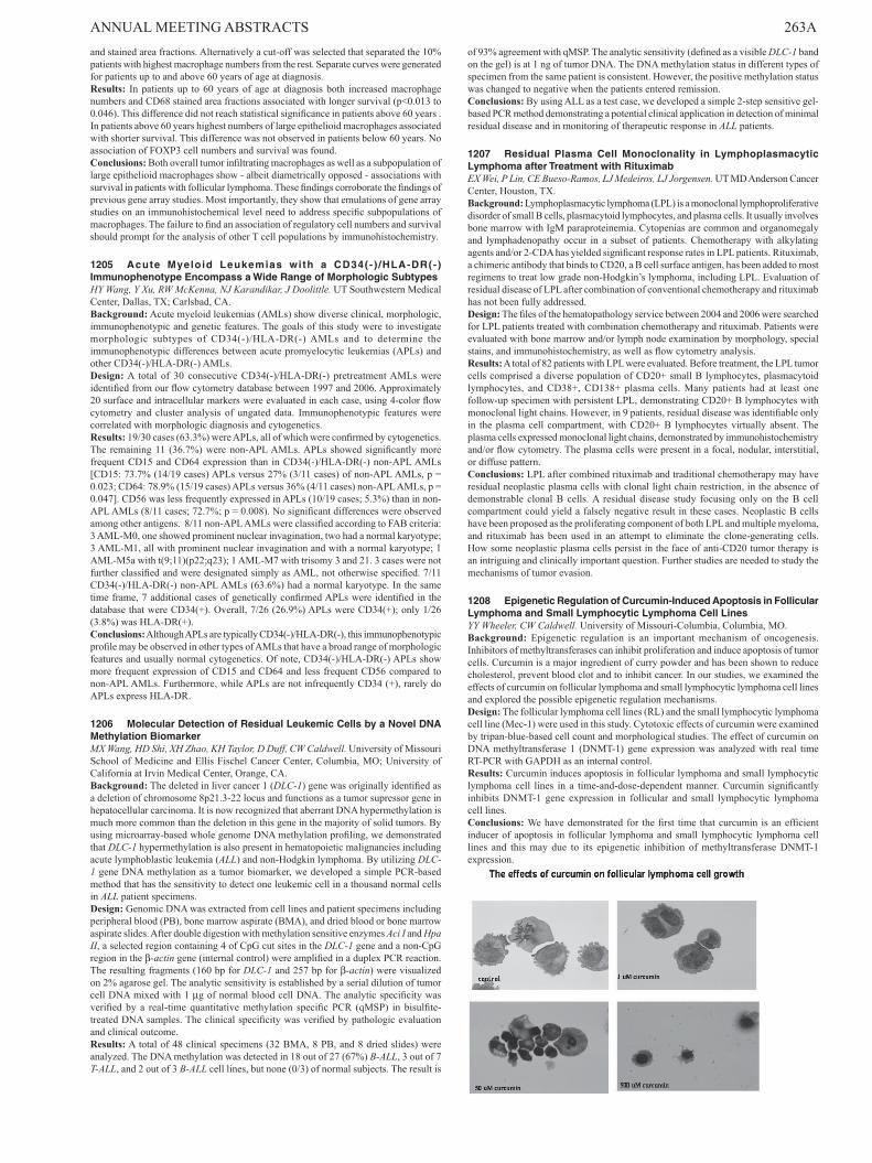
ANNUAL MEETING ABSTRACTS 263Aand stained area fractions. Alternatively a cut-off was selected that separated the 10% patients with highest macrophage numbers from the rest. Separate curves were generated for patients up to and above 60 years of age at diagnosis.Results: In patients up to 60 years of age at diagnosis both increased macrophage numbers and CD68 stained area fractions associated with longer survival (p<0.013 to 0.046). This difference did not reach statistical significance in patients above 60 years . In patients above 60 years highest numbers of large epithelioid macrophages associated with shorter survival. This difference was not observed in patients below 60 years. No association of FOXP3 cell numbers and survival was found.Conclusions: Both overall tumor infiltrating macrophages as well as a subpopulation of large epithelioid macrophages show - albeit diametrically opposed - associations with survival in patients with follicular lymphoma. These findings corroborate the findings of previous gene array studies. Most importantly, they show that emulations of gene array studies on an immunohistochemical level need to address specific subpopulations of macrophages. The failure to find an association of regulatory cell numbers and survival should prompt for the analysis of other T cell populations by immunohistochemistry.
1205 Acute Myeloid Leukemias with a CD34(- ) /HLA-DR(-)ImmunophenotypeEncompassaWideRangeofMorphologicSubtypesHY Wang, Y Xu, RW McKenna, NJ Karandikar, J Doolittle. UT Southwestern Medical Center, Dallas, TX; Carlsbad, CA.Background: Acute myeloid leukemias (AMLs) show diverse clinical, morphologic, immunophenotypic and genetic features. The goals of this study were to investigate morphologic subtypes of CD34(-)/HLA-DR(-) AMLs and to determine the immunophenotypic differences between acute promyelocytic leukemias (APLs) and other CD34(-)/HLA-DR(-) AMLs.Design: A total of 30 consecutive CD34(-)/HLA-DR(-) pretreatment AMLs were identified from our flow cytometry database between 1997 and 2006. Approximately 20 surface and intracellular markers were evaluated in each case, using 4-color flow cytometry and cluster analysis of ungated data. Immunophenotypic features were correlated with morphologic diagnosis and cytogenetics.Results: 19/30 cases (63.3%) were APLs, all of which were confirmed by cytogenetics. The remaining 11 (36.7%) were non-APL AMLs. APLs showed significantly more frequent CD15 and CD64 expression than in CD34(-)/HLA-DR(-) non-APL AMLs [CD15: 73.7% (14/19 cases) APLs versus 27% (3/11 cases) of non-APL AMLs, p = 0.023; CD64: 78.9% (15/19 cases) APLs versus 36% (4/11 cases) non-APL AMLs, p = 0.047]. CD56 was less frequently expressed in APLs (10/19 cases; 5.3%) than in non-APL AMLs (8/11 cases; 72.7%; p = 0.008). No significant differences were observed among other antigens. 8/11 non-APL AMLs were classified according to FAB criteria: 3 AML-M0, one showed prominent nuclear invagination, two had a normal karyotype; 3 AML-M1, all with prominent nuclear invagination and with a normal karyotype; 1 AML-M5a with t(9;11)(p22;q23); 1 AML-M7 with trisomy 3 and 21. 3 cases were not further classified and were designated simply as AML, not otherwise specified. 7/11 CD34(-)/HLA-DR(-) non-APL AMLs (63.6%) had a normal karyotype. In the same time frame, 7 additional cases of genetically confirmed APLs were identified in the database that were CD34(+). Overall, 7/26 (26.9%) APLs were CD34(+); only 1/26 (3.8%) was HLA-DR(+).Conclusions: Although APLs are typically CD34(-)/HLA-DR(-), this immunophenotypic profile may be observed in other types of AMLs that have a broad range of morphologic features and usually normal cytogenetics. Of note, CD34(-)/HLA-DR(-) APLs show more frequent expression of CD15 and CD64 and less frequent CD56 compared to non-APL AMLs. Furthermore, while APLs are not infrequently CD34 (+), rarely do APLs express HLA-DR.
1206 MolecularDetectionofResidualLeukemicCellsbyaNovelDNAMethylationBiomarkerMX Wang, HD Shi, XH Zhao, KH Taylor, D Duff, CW Caldwell. University of Missouri School of Medicine and Ellis Fischel Cancer Center, Columbia, MO; University of California at Irvin Medical Center, Orange, CA.Background: The deleted in liver cancer 1 (DLC-1) gene was originally identified as a deletion of chromosome 8p21.3-22 locus and functions as a tumor supressor gene in hepatocellular carcinoma. It is now recognized that aberrant DNA hypermethylation is much more common than the deletion in this gene in the majority of solid tumors. By using microarray-based whole genome DNA methylation profiling, we demonstrated that DLC-1 hypermethylation is also present in hematopoietic malignancies including acute lymphoblastic leukemia (ALL) and non-Hodgkin lymphoma. By utilizing DLC-1 gene DNA methylation as a tumor biomarker, we developed a simple PCR-based method that has the sensitivity to detect one leukemic cell in a thousand normal cells in ALL patient specimens.Design: Genomic DNA was extracted from cell lines and patient specimens including peripheral blood (PB), bone marrow aspirate (BMA), and dried blood or bone marrow aspirate slides. After double digestion with methylation sensitive enzymes Aci I and Hpa II, a selected region containing 4 of CpG cut sites in the DLC-1 gene and a non-CpG region in the β-actin gene (internal control) were amplified in a duplex PCR reaction. The resulting fragments (160 bp for DLC-1 and 257 bp for β-actin) were visualized on 2% agarose gel. The analytic sensitivity is established by a serial dilution of tumor cell DNA mixed with 1 µg of normal blood cell DNA. The analytic specificity was verified by a real-time quantitative methylation specific PCR (qMSP) in bisulfite-treated DNA samples. The clinical specificity was verified by pathologic evaluation and clinical outcome.Results: A total of 48 clinical specimens (32 BMA, 8 PB, and 8 dried slides) were analyzed. The DNA methylation was detected in 18 out of 27 (67%) B-ALL, 3 out of 7 T-ALL, and 2 out of 3 B-ALL cell lines, but none (0/3) of normal subjects. The result is
of 93% agreement with qMSP. The analytic sensitivity (defined as a visible DLC-1 band on the gel) is at 1 ng of tumor DNA. The DNA methylation status in different types of specimen from the same patient is consistent. However, the positive methylation status was changed to negative when the patients entered remission.Conclusions: By using ALL as a test case, we developed a simple 2-step sensitive gel-based PCR method demonstrating a potential clinical application in detection of minimal residual disease and in monitoring of therapeutic response in ALL patients.
1207 Residual Plasma Cell Monoclonality in LymphoplasmacyticLymphomaafterTreatmentwithRituximabEX Wei, P Lin, CE Bueso-Ramos, LJ Medeiros, LJ Jorgensen. UT MD Anderson Cancer Center, Houston, TX.Background: Lymphoplasmacytic lymphoma (LPL) is a monoclonal lymphoproliferative disorder of small B cells, plasmacytoid lymphocytes, and plasma cells. It usually involves bone marrow with IgM paraproteinemia. Cytopenias are common and organomegaly and lymphadenopathy occur in a subset of patients. Chemotherapy with alkylating agents and/or 2-CDA has yielded significant response rates in LPL patients. Rituximab, a chimeric antibody that binds to CD20, a B cell surface antigen, has been added to most regimens to treat low grade non-Hodgkin’s lymphoma, including LPL. Evaluation of residual disease of LPL after combination of conventional chemotherapy and rituximab has not been fully addressed.Design: The files of the hematopathology service between 2004 and 2006 were searched for LPL patients treated with combination chemotherapy and rituximab. Patients were evaluated with bone marrow and/or lymph node examination by morphology, special stains, and immunohistochemistry, as well as flow cytometry analysis.Results: A total of 82 patients with LPL were evaluated. Before treatment, the LPL tumor cells comprised a diverse population of CD20+ small B lymphocytes, plasmacytoid lymphocytes, and CD38+, CD138+ plasma cells. Many patients had at least one follow-up specimen with persistent LPL, demonstrating CD20+ B lymphocytes with monoclonal light chains. However, in 9 patients, residual disease was identifiable only in the plasma cell compartment, with CD20+ B lymphocytes virtually absent. The plasma cells expressed monoclonal light chains, demonstrated by immunohistochemistry and/or flow cytometry. The plasma cells were present in a focal, nodular, interstitial, or diffuse pattern.Conclusions: LPL after combined rituximab and traditional chemotherapy may have residual neoplastic plasma cells with clonal light chain restriction, in the absence of demonstrable clonal B cells. A residual disease study focusing only on the B cell compartment could yield a falsely negative result in these cases. Neoplastic B cells have been proposed as the proliferating component of both LPL and multiple myeloma, and rituximab has been used in an attempt to eliminate the clone-generating cells. How some neoplastic plasma cells persist in the face of anti-CD20 tumor therapy is an intriguing and clinically important question. Further studies are needed to study the mechanisms of tumor evasion.
1208 EpigeneticRegulationofCurcumin-InducedApoptosisinFollicularLymphomaandSmallLymphocyticLymphomaCellLinesYY Wheeler, CW Caldwell. University of Missouri-Columbia, Columbia, MO.Background: Epigenetic regulation is an important mechanism of oncogenesis. Inhibitors of methyltransferases can inhibit proliferation and induce apoptosis of tumor cells. Curcumin is a major ingredient of curry powder and has been shown to reduce cholesterol, prevent blood clot and to inhibit cancer. In our studies, we examined the effects of curcumin on follicular lymphoma and small lymphocytic lymphoma cell lines and explored the possible epigenetic regulation mechanisms.Design: The follicular lymphoma cell lines (RL) and the small lymphocytic lymphoma cell line (Mec-1) were used in this study. Cytotoxic effects of curcumin were examined by tripan-blue-based cell count and morphological studies. The effect of curcumin on DNA methyltransferase 1 (DNMT-1) gene expression was analyzed with real time RT-PCR with GAPDH as an internal control.Results: Curcumin induces apoptosis in follicular lymphoma and small lymphocytic lymphoma cell lines in a time-and-dose-dependent manner. Curcumin significantly inhibits DNMT-1 gene expression in follicular and small lymphocytic lymphoma cell lines.Conclusions: We have demonstrated for the first time that curcumin is an efficient inducer of apoptosis in follicular lymphoma and small lymphocytic lymphoma cell lines and this may due to its epigenetic inhibition of methyltransferase DNMT-1 expression.
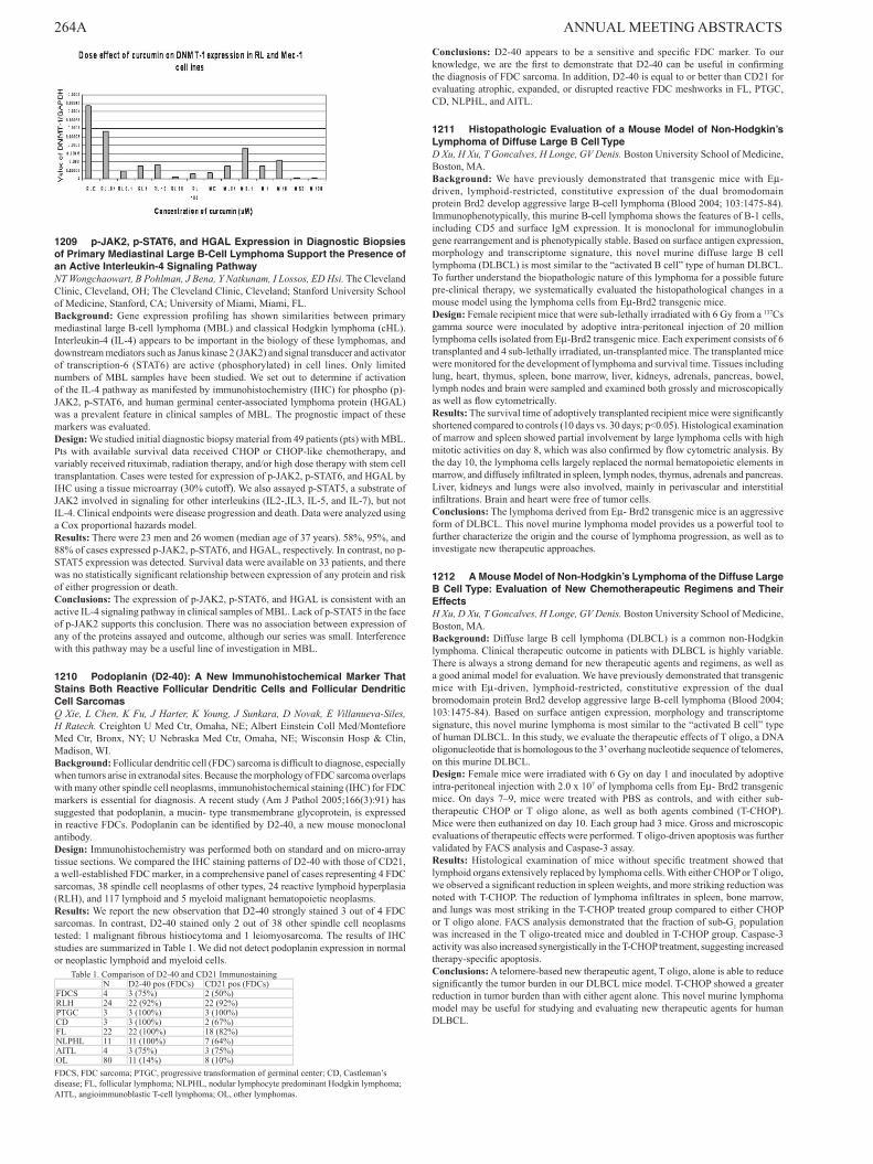
264A ANNUAL MEETING ABSTRACTS
1209 p-JAK2,p-STAT6,andHGALExpressioninDiagnosticBiopsiesofPrimaryMediastinalLargeB-CellLymphomaSupportthePresenceofanActiveInterleukin-4SignalingPathwayNT Wongchaowart, B Pohlman, J Bena, Y Natkunam, I Lossos, ED Hsi. The Cleveland Clinic, Cleveland, OH; The Cleveland Clinic, Cleveland; Stanford University School of Medicine, Stanford, CA; University of Miami, Miami, FL.Background: Gene expression profiling has shown similarities between primary mediastinal large B-cell lymphoma (MBL) and classical Hodgkin lymphoma (cHL). Interleukin-4 (IL-4) appears to be important in the biology of these lymphomas, and downstream mediators such as Janus kinase 2 (JAK2) and signal transducer and activator of transcription-6 (STAT6) are active (phosphorylated) in cell lines. Only limited numbers of MBL samples have been studied. We set out to determine if activation of the IL-4 pathway as manifested by immunohistochemistry (IHC) for phospho (p)-JAK2, p-STAT6, and human germinal center-associated lymphoma protein (HGAL) was a prevalent feature in clinical samples of MBL. The prognostic impact of these markers was evaluated.Design: We studied initial diagnostic biopsy material from 49 patients (pts) with MBL. Pts with available survival data received CHOP or CHOP-like chemotherapy, and variably received rituximab, radiation therapy, and/or high dose therapy with stem cell transplantation. Cases were tested for expression of p-JAK2, p-STAT6, and HGAL by IHC using a tissue microarray (30% cutoff). We also assayed p-STAT5, a substrate of JAK2 involved in signaling for other interleukins (IL2-,IL3, IL-5, and IL-7), but not IL-4. Clinical endpoints were disease progression and death. Data were analyzed using a Cox proportional hazards model.Results: There were 23 men and 26 women (median age of 37 years). 58%, 95%, and 88% of cases expressed p-JAK2, p-STAT6, and HGAL, respectively. In contrast, no p-STAT5 expression was detected. Survival data were available on 33 patients, and there was no statistically significant relationship between expression of any protein and risk of either progression or death.Conclusions: The expression of p-JAK2, p-STAT6, and HGAL is consistent with an active IL-4 signaling pathway in clinical samples of MBL. Lack of p-STAT5 in the face of p-JAK2 supports this conclusion. There was no association between expression of any of the proteins assayed and outcome, although our series was small. Interference with this pathway may be a useful line of investigation in MBL.
1210 Podoplanin (D2-40): A New Immunohistochemical MarkerThatStainsBothReactiveFollicularDendriticCellsandFollicularDendriticCellSarcomasQ Xie, L Chen, K Fu, J Harter, K Young, J Sunkara, D Novak, E Villanueva-Siles, H Ratech. Creighton U Med Ctr, Omaha, NE; Albert Einstein Coll Med/Montefiore Med Ctr, Bronx, NY; U Nebraska Med Ctr, Omaha, NE; Wisconsin Hosp & Clin, Madison, WI.Background: Follicular dendritic cell (FDC) sarcoma is difficult to diagnose, especially when tumors arise in extranodal sites. Because the morphology of FDC sarcoma overlaps with many other spindle cell neoplasms, immunohistochemical staining (IHC) for FDC markers is essential for diagnosis. A recent study (Am J Pathol 2005;166(3):91) has suggested that podoplanin, a mucin- type transmembrane glycoprotein, is expressed in reactive FDCs. Podoplanin can be identified by D2-40, a new mouse monoclonal antibody.Design: Immunohistochemistry was performed both on standard and on micro-array tissue sections. We compared the IHC staining patterns of D2-40 with those of CD21, a well-established FDC marker, in a comprehensive panel of cases representing 4 FDC sarcomas, 38 spindle cell neoplasms of other types, 24 reactive lymphoid hyperplasia (RLH), and 117 lymphoid and 5 myeloid malignant hematopoietic neoplasms.Results: We report the new observation that D2-40 strongly stained 3 out of 4 FDC sarcomas. In contrast, D2-40 stained only 2 out of 38 other spindle cell neoplasms tested: 1 malignant fibrous histiocytoma and 1 leiomyosarcoma. The results of IHC studies are summarized in Table 1. We did not detect podoplanin expression in normal or neoplastic lymphoid and myeloid cells.
Table 1. Comparison of D2-40 and CD21 ImmunostainingN D2-40 pos (FDCs) CD21 pos (FDCs)
FDCS 4 3 (75%) 2 (50%)RLH 24 22 (92%) 22 (92%)PTGC 3 3 (100%) 3 (100%)CD 3 3 (100%) 2 (67%)FL 22 22 (100%) 18 (82%)NLPHL 11 11 (100%) 7 (64%)AITL 4 3 (75%) 3 (75%)OL 80 11 (14%) 8 (10%)FDCS, FDC sarcoma; PTGC, progressive transformation of germinal center; CD, Castleman’s disease; FL, follicular lymphoma; NLPHL, nodular lymphocyte predominant Hodgkin lymphoma; AITL, angioimmunoblastic T-cell lymphoma; OL, other lymphomas.
Conclusions: D2-40 appears to be a sensitive and specific FDC marker. To our knowledge, we are the first to demonstrate that D2-40 can be useful in confirming the diagnosis of FDC sarcoma. In addition, D2-40 is equal to or better than CD21 for evaluating atrophic, expanded, or disrupted reactive FDC meshworks in FL, PTGC, CD, NLPHL, and AITL.
1211 HistopathologicEvaluationofaMouseModelofNon-Hodgkin’sLymphomaofDiffuseLargeBCellTypeD Xu, H Xu, T Goncalves, H Longe, GV Denis. Boston University School of Medicine, Boston, MA.Background: We have previously demonstrated that transgenic mice with Eµ-driven, lymphoid-restricted, constitutive expression of the dual bromodomain protein Brd2 develop aggressive large B-cell lymphoma (Blood 2004; 103:1475-84). Immunophenotypically, this murine B-cell lymphoma shows the features of B-1 cells, including CD5 and surface IgM expression. It is monoclonal for immunoglobulin gene rearrangement and is phenotypically stable. Based on surface antigen expression, morphology and transcriptome signature, this novel murine diffuse large B cell lymphoma (DLBCL) is most similar to the “activated B cell” type of human DLBCL. To further understand the biopathologic nature of this lymphoma for a possible future pre-clinical therapy, we systematically evaluated the histopathological changes in a mouse model using the lymphoma cells from Eµ-Brd2 transgenic mice.Design: Female recipient mice that were sub-lethally irradiated with 6 Gy from a 137Cs gamma source were inoculated by adoptive intra-peritoneal injection of 20 million lymphoma cells isolated from Eµ-Brd2 transgenic mice. Each experiment consists of 6 transplanted and 4 sub-lethally irradiated, un-transplanted mice. The transplanted mice were monitored for the development of lymphoma and survival time. Tissues including lung, heart, thymus, spleen, bone marrow, liver, kidneys, adrenals, pancreas, bowel, lymph nodes and brain were sampled and examined both grossly and microscopically as well as flow cytometrically.Results: The survival time of adoptively transplanted recipient mice were significantly shortened compared to controls (10 days vs. 30 days; p<0.05). Histological examination of marrow and spleen showed partial involvement by large lymphoma cells with high mitotic activities on day 8, which was also confirmed by flow cytometric analysis. By the day 10, the lymphoma cells largely replaced the normal hematopoietic elements in marrow, and diffusely infiltrated in spleen, lymph nodes, thymus, adrenals and pancreas. Liver, kidneys and lungs were also involved, mainly in perivascular and interstitial infiltrations. Brain and heart were free of tumor cells.Conclusions: The lymphoma derived from Eµ- Brd2 transgenic mice is an aggressive form of DLBCL. This novel murine lymphoma model provides us a powerful tool to further characterize the origin and the course of lymphoma progression, as well as to investigate new therapeutic approaches.
1212 AMouseModelofNon-Hodgkin’sLymphomaoftheDiffuseLargeBCellType:EvaluationofNewChemotherapeuticRegimensandTheirEffectsH Xu, D Xu, T Goncalves, H Longe, GV Denis. Boston University School of Medicine, Boston, MA.Background: Diffuse large B cell lymphoma (DLBCL) is a common non-Hodgkin lymphoma. Clinical therapeutic outcome in patients with DLBCL is highly variable. There is always a strong demand for new therapeutic agents and regimens, as well as a good animal model for evaluation. We have previously demonstrated that transgenic mice with Eµ-driven, lymphoid-restricted, constitutive expression of the dual bromodomain protein Brd2 develop aggressive large B-cell lymphoma (Blood 2004; 103:1475-84). Based on surface antigen expression, morphology and transcriptome signature, this novel murine lymphoma is most similar to the “activated B cell” type of human DLBCL. In this study, we evaluate the therapeutic effects of T oligo, a DNA oligonucleotide that is homologous to the 3’ overhang nucleotide sequence of telomeres, on this murine DLBCL.Design: Female mice were irradiated with 6 Gy on day 1 and inoculated by adoptive intra-peritoneal injection with 2.0 x 10⁷ of lymphoma cells from Eµ- Brd2 transgenic mice. On days 7–9, mice were treated with PBS as controls, and with either sub-therapeutic CHOP or T oligo alone, as well as both agents combined (T-CHOP). Mice were then euthanized on day 10. Each group had 3 mice. Gross and microscopic evaluations of therapeutic effects were performed. T oligo-driven apoptosis was further validated by FACS analysis and Caspase-3 assay.Results: Histological examination of mice without specific treatment showed that lymphoid organs extensively replaced by lymphoma cells. With either CHOP or T oligo, we observed a significant reduction in spleen weights, and more striking reduction was noted with T-CHOP. The reduction of lymphoma infiltrates in spleen, bone marrow, and lungs was most striking in the T-CHOP treated group compared to either CHOP or T oligo alone. FACS analysis demonstrated that the fraction of sub-G1 population was increased in the T oligo-treated mice and doubled in T-CHOP group. Caspase-3 activity was also increased synergistically in the T-CHOP treatment, suggesting increased therapy-specific apoptosis.Conclusions: A telomere-based new therapeutic agent, T oligo, alone is able to reduce significantly the tumor burden in our DLBCL mice model. T-CHOP showed a greater reduction in tumor burden than with either agent alone. This novel murine lymphoma model may be useful for studying and evaluating new therapeutic agents for human DLBCL.

ANNUAL MEETING ABSTRACTS 265A
1213 ChronicLymphocyticLeukemiawithDualSurfaceImmunoglobulinLightChainExpressionH Xu, S Yang, X Zhong, D Xu. Boston University School of Medicine, Boston, MA.Background: Normally, a mature B cell expresses an immunoglobulin heavy chain (IgH) paired with either a Kappa (κ) or a Lambda (λ) light chain, which is known as isotopic exclusion. However, double light chain gene rearrangement or dual light chain expression on a single mature B cell can occur, which is apparently in violation of allelic or isotopic exclusion (Arch Pathol Lab Med. 2006; 130:853-6). Here, we reported 3 chronic lymphocytic leukemia (CLL) cases that showed unusual dual surface light chain expression. Diagnostic and clinical implication of dual light chain expression is discussed.Design: We retrospectively analyzed flow cytometric data of 110 peripheral blood specimens from patients with CLL from 2001 to 2006 at Boston Medical Center. All cases were diagnosed based on clinical presentation, peripheral lymphocyte morphology and immunophenotyic features determined by 4-color flow cytometer. For some cases, polymerase chain reaction (PCR) analysis for IgH gene rearrangement was also performed.Results: Three cases (2.7%) with dual surface κ and λ expression were identified. 2 males (57 and 59 year old, respectively) and 1 female (57year old) had absolute lymphocyte counts of 37,500/µL, 15,400/µL and 22,900/µL, respectively. Among them, 2 cases had a typical CLL immunophenotype with CD19, CD5 and CD23 expression, and 1 case was CD19+, CD5-, and CD23-. All three cases showed a predominant κ expressing B cell population with λ light chain co-expression. Based on a CD45+ lymphocyte gate, the percentages of κ and κ/λ in 3 cases were 86.5% and 52.6%, 87.5% and 19.3%, and 83.6% and 81.3%, respectively. Despite of their polytypic surface κ/λ expression pattern, all 3 cases, however, exhibited a monoclonal IgH gene rearrangement by PCR analysis.Conclusions: Dual κ/λ light chain expression on mature B cells do occur. Awareness of this unusual phenomenon has diagnostic implications in leukemia/lymphoma immunophenotyping since the most common flow cytometric analysis for determining B-cell monotype is the percent of κ and λ ratio. It is recommended that if the sum of κ+/CD19+ plus λ+/CD19+ expressing B cells is 10% greater than the total percent CD19+ B-cells or if the dual light chain expressing B-cell populations comprise more than 10% of the total number of CD19+ B cells, an IgH PCR studies to look for clonality may be justified.
1214 Evaluation of Plasma CellVolume (PCV) and Clonality inAL (Primary) Amyloidosis by Analysis of Pre and Post Stem CellTransplantationBoneMarrowCoreBiopsies(BMCB)H Xu, P Soo Hoo, G Doros, M Skinner, C O’Hara. Boston University School of Medicine, Boston, MA.Background: AL amyloidosis, a monoclonal plasma cell dyscrasia, is the most common form of systemic amyloidosis in the US. The fibrillar amyloid protein, derived from circulating monoclonal immunoglobulin light chains, deposits in tissue and leads to progressive organ failure and death. Amyloidosis can now be successfully treated with high-dose melphalan chemotherapy followed by autologous peripheral blood stem cell transplantation. The aim of this study was to examine the effect of treatment on bone marrow plasma cell (PC) volume and immunoglobulin light chain (LC) clonality in 99 patients(pts) with AL amyloidosis (67λ, 21κ, 11 no detectable clonality). In 35 pts (25λ, 6κ, 4 no detectable clonality), correlation between PC data and FLC levels and ratios was assessed.Design: Sequential Zenker’s fixed BMCB on 99 pts (total 316, 1996-2005) were assessed using immunostain for CD138 and light chain immunoglobulins. Percentage of PC was estimated as follows: <5, 5, 5-10, 10, 10-15 etc., and clonality on the basis of a κ to λ ratio >3 or <1. All pts had pre-treatment BMCB and an average 3-4 post-treatment BMCB. Serum free light-chain (FLC) concentrations were measured by a sensitive nephelometric immunoassay in 35 of 99 pts.Results: Of 99 pts examined, 95 had an initial PCV of 5% or greater and 88 had monoclonal LC staining. Post-treatment at one year there was a decrease in PCV of at least 5% in 65/92 (70.7%) and 50/86 (58.1%) exhibited a conversion from monoclonal to polyclonal PC staining. Subsequent follow-ups revealed sustained polyclonality in these pts up to 5 years post-treatment. In the 35 pts in which FLC data was available, 32/35 (91.4%) showed a reduction in PCV and/or conversion to polyclonality which corresponded to a reduction in FLC levels and improvement in the FLC ratio in 27/35 (77.1%) and complete normalization of these parameters in 15/35 (42.9%) of pts.Conclusions: The effect of high dose intravenous melphalan and autologous stem-cell transplantation in AL amyloidosis is reflected in a reduction in PCV and sustained polyclonality in the majority of pts. The normalization of the bone marrow plasma cells is reflected in improvement in FLC parameters; incongruous results may reflect other factors that influence FLC levels.
1215 Aberrantp53ExpressionCorrelateswithHemizygousp53GeneDeletionandPredictsaPoorOverallSurvival inPatientswithMultipleMyelomaJ Yeung, C Qi, W Xu, H Chang. University Health Network and Dept of Lab Medicine & Pathobiology, University of Toronto, Toronto, ON, Canada.Background: Hemizygous deletion of the p53 tumour suppressor gene detected by fluorescence in situ hybridization (FISH) has been established as an adverse prognostic factor for patients with multiple myeloma (MM) even after treatment with high-dose chemotherapy and stem cell transplantation. However, whether p53 protein expression is related to p53 gene abnormalities and whether p53 expression affects the clinical outcome in MM are not clear. We set to explore the relationship between a hemizygous p53 deletion and p53 protein expression as well as its prognostic significance in a cohort of MM patients homogenously treated at our institution.
Design: The p53 expression was evaluated by immunohistochemistry (IHC) in decalcified, paraffin-embedded bone marrow biopsies from 87 MM patients. Clonal plasma cells of the bone marrow aspirates from the same cohort were examined for p53 gene status by cytoplasmic interphase FISH.Results: Of 87 patients, FISH detected a hemizygous p53 deletion in 13 (15%) while IHC detected aberrant p53 expression in 12 (14%) of the cases. Of the 13 p53-deleted cases, 9 (69.2%) had aberrant p53 nuclear immunostaining, whereas 9 (75%) of 12 IHC p53 positive cases had p53 deletion on FISH. Hemizygous p53 deletions and aberrant p53 expression were strongly correlated (P < 0.001). Furthermore, patients with aberrant p53 expression had a significantly shorter overall survival than those without expressing p53 (median 24.7 vs. 48.1 moths, P < 0.001).Conclusions: Our results indicate that in MM, hemizygous p53 deletion is associated with aberrant p53 protein expression. p53 IHC my therefore be used to screen cases for p53 deletion and to predict a poor prognosis in MM. Further studies are required to address the mechanism(s) resulting in aberrant p53 expression and its functional consequences in MM.
1216 Chronic Lymphocytic Leukemia/Small Lymphocytic Lymphomawitht(2;14)(p13;q32)IsAssociatedwithAtypicalMorphology,UnmutatedIgVHGenes,andanAggressiveClinicalCourseCC Yin, LJ Medeiros, LL Barron, LV Abruzzo. UT MD Anderson Cancer Center, Houston, TX.Background: The t(2;14)(p13;q32) is a rare recurrent chromosomal abnormality in B-cell lymphomas and leukemias. This translocation has been reported previously in 6 cases of chronic lymphocytic leukemia/small lymphocytic lymphoma (CLL/SLL), but the clinicopathologic and immunophenotypic features of these cases have not been described.Design: We identified 6 cases of CLL/SLL with t(2;14)(p13;q32) from the files of the cytogenetics laboratory. We reviewed bone marrow aspirate and biopsy specimens (n=6) and lymph node biopsy specimens (n=4). Immunophenotyping was performed by flow cytometry and immunohistochemistry. Karyotypic analysis was performed on bone marrow (n=5) or peripheral blood (n=1). Fluorescence in-situ hybridization (FISH) using probes specific for the IgH gene at 14q32 was performed (n=2). The somatic hypermutation status of the IgH variable region (IgVH) genes was determined from DNA extracted from fixed, paraffin-embedded tissue (n=2) or peripheral blood (n=1).Results: There were 4 men and 2 women with a median age of 51 years (range 39-65). All had bone marrow involvement and 5 had absolute peripheral blood lymphocytosis. Five had lymphadenopathy and 4 had hepatosplenomegaly. All cases showed atypical morphology with plasmacytoid differentiation and nuclear indentation in a subset of the cells. Four cases had increased prolymphocytes (range 10-47%). Flow cytometric analysis demonstrated typical immunophenotypes in 5 cases; 1 case showed an atypical immunophenotype, with moderate expression of CD20 and IgM/D, as well as FMC-7. Immunohistochemistry demonstrated that all cases were ZAP-70 positive and cyclin D1 negative. Karyotypic studies identified t(2;14)(p13;q32) as the sole abnormality in 1 case, and as part of complex karyotypes in 5 cases. FISH analysis in 2 cases showed that the IgH gene at 14q32 was involved. All 3 cases assessed showed unmutated IgVH genes. All patients received multi-agent chemotherapy. With a median follow-up of 35 months, 2 have died of disease, and 4 are alive with disease (one with Richter’s transformation).Conclusions: Cases of CLL/SLL with t(2;14)(p13;q32) are associated with atypical morphology, unmutated IgVH genes, and an aggressive clinical course. We have confirmed involvement of IgH at 14q32 by FISH. The gene at 2p13 in these cases is unknown at this time, but the literature suggests that BCL11A may be involved.
1217 The Diversity of p53/p21Transactivation Phenotype of p53Signaling Pathway andTheir Relationship toTP53 Gene Mutation andClinicalOutcomeinDiffuseLargeB-CellLymphoma(DLBCL)KH Young, MB Møller, GWB Colleoni, M Sánchez-Beato, FR Kerbauy, K Leroy, MA Piris, JC Eickhoff, AH Young, K Kanehira, S Cook, EA Ranheim, BS Kahl, M Olivier, E Campo, J Delabie, RD Gascoyne, A Rosenwald, RM Braziel, ES Jaffe, WH Wilson, LM Staudt, WC Chan, DD Weisenburger, TC Greiner. University of Wisconsin School of Medicine and Public Health, Madison, WI; Odense University Hospital, Odense, Denmark; Federal University of São Paulo, Brazil; Spanish National Cancer Center, Madrid, Spain; Hospital Henri Mondor, Paris, France; World Health Organization, Lyon, France; Lymphoma Leukemia Molecular Profiling Project.Background: The TP53 gene is often inactivated by mutations resulting in p53 protein over-expression and down regulation of p21, an important TP53 downstream target gene. The purpose of this study was to evaluate the prognostic value of the p53/p21 transactivation phenotype, and to correlate it with the TP53 mutations in DLBCL.Design: Mutations on the TP53 gene were analyzed and immunostains for p53 and p21 were performed in 191 cases from 12 medical centers. A positive immunostain was defined as nuclear staining in 10% or more of the tumor cells. The Kaplan-Meier method was used for survival analysis.Results: Fifty cases with TP53 mutations and 141 cases with wild type-TP53 (WT) were found (see Table). Seventy-eight of 82 p53-/p21- cases had WT-TP53 (95%), whereas 42 of 66 p53+/p21- cases had TP53 mutations (65%). In contrast, 18 of 20 p53-/p21+ and 21 of 23 p53+/p21+ phenotype cases had WT-TP53 (90% and 91.3%, respectively). Forty-six of 50 cases with mutations showed no p21 expression (92%). However, the p53+/p21- phenotype was found in 24 patients with WT-TP53. The p53+/p21- phenotype was correlated with a poor overall survival (OS), and multivariate analysis showed it was an independent predictor of OS with a hazard ratio of 1.8 (P=0.003).

266A ANNUAL MEETING ABSTRACTSConclusions: This study demonstrates the value of the p53/p21 transactivation phenotype for predicting clinical outcome. However, because of a relatively high frequency in patients with WT-TP53 in this group, the p53+/p21- phenotype should not be used to predict TP53 mutation status.TransactivationPhenotype
TotalCases(%) WT-TP53 TP53-Mutation 5-yrOSRate MedianSurvival
(Yr)P53-/p21- 82 (42%) 78 4 42% 6.8P53+/p21- 66 (35%) 24 42 22% 1.0P53-/p21+ 20 (10%) 18 2 40% 4.5P53+/p21+ 23 (13%) 21 2 42% 5.4Summary 191 (100%) 141 50 40% 4.4
1218 GerminalCenterTHelperCellMarkersPD-1andCXCL13AreBothExpressedbyNeoplasticCellsinAngioimmunoblasticLymphomaH Yu, A Shahsafaei, DM Dorfman. Brigham and Women’s Hospital, Boston, MA.Background: The CD3+CD4+CD10+ neoplastic cells of angioimmunoblastic lymphoma (AIL) may arise from germinal center T-helper cells (GCThC). Gene expression profiling identified genes uniquely expressed by human GCThC compared with other T-cell subsets, including Programmed Death (PD-1), a member of the CD28 receptor family which regulates the cellular immune response, and CXCL13, a critical chemokine for B-cell entry to lymphoid follicles. Recently we demonstrated that PD-1 is a marker of GCThC and AIL, but not other peripheral T cell lymphomas (PTL). The goal of this study was to investigate the expression pattern of CXCL13 in comparison to PD-1 in AIL and other PTL.Design: 40 cases of peripheral T cell lymphoma were studied, including 21 AILs, 5 anaplastic T-cell lymphomas (ALCL), 9 PTL-NOS, 4 NK/T cell lymphomas (NK/T), and 1 precursor T lymphoblastic lymphoma/leukemia (T-ALL). Immunohistochemical studies were performed following heat-induced epitope retrieval in 10mM citrate buffer, pH 6.0, for PD-1 monoclonal antibody EH12, or 1 mM EDTA buffer, pH 8.0, for CXCL13 monoclonal antibody 53610, using standard indirect avidin-biotin horseradish peroxidase method and diaminobenzidine color development. The pattern and intensity of immunoreactivity were evaluated for both markers.Results: In cases of AIL and in reactive lymphoid tissue, the pattern of expression of PD-1 and CXCL13 are similar, with positive cells seen at foci of expanded CD21+ follicular dendritic cell networks in AIL and in germinal center T cells in reactive lymphoid tissue. However, the intensity of the staining varies among AILs, with 3 cases showing strong staining for both markers, 2 cases showing weak staining for both markers, 9 cases showing stronger staining with CXCL13 (perinuclear dot pattern), and 7 cases showing stronger PD-1 staining (cell surface and cytoplasmic). CXCL13 expression is limited in other PTLs: only 1 ALCL, 1 NK/T, and 2 PTL-NOS showed focal reactivity, possibly at foci of residual reactive lymphoid tissue, and all other cases were negative. Previously we showed that PD-1 is not expressed in other PTL.Conclusions: Both PD-1 and CXCL13 identify germinal center T-helper cells and show a similar pattern of expression in AIL, without significant staining in other peripheral T cell lymphomas. Both PD-1 and CXCL13 should serve as useful new markers for AIL. The similar pattern of expression of CXCL13 and PD-1 in AIL provide further evidence that AIL is a neoplasm derived from germinal center T-cells.
1219 TCellMarkerChanges inPeriphearlTCellLymphmoabyFlowCytometryAnalysisC Yue, A Anthony, H Bolton. US Labs, Irvine, CA.Background: Peripheral T cell lymphoma is relatively rare but can be diagnosed by flow cytometry via analysis of T cell markers. The common clue for a T cell lymphoma is loss of T cell CD markers or co-expression of CD4 with CD8. The purpose of this study is to analyze the frequency of loss of different T cell markers in peripheral T cell lymphoma.Design: We have done 46420 flow cytometry cases in 2004 and 2005 at US Labs, of which 29095 cases are with global interpretations. 40 cases of peripheral T cell lymphoma have been diagnosed in these two years by flow cytometry and confirmed by histology or molecular studies (incidence 0.137% (40/29095)). We analyzed the 40 cases and using 10% as cutoff point for marker loss to calculate the frequencies of each T cell marker changes.Results: The frequency for loss of each T cell marker is as follows in peripheral T cell lymphoma: CD7: 70% (28/40), CD3: 47.5% (19/40); CD5: 35% (14/40); CD2 22.5% (9/40), Loss of both CD4 and CD8: 15% (6/40). Coexpression of CD4 with CD8: 5% (2/40).Conclusions: CD7 loss is the most common phenomena in peripheral T cell lymphoma (but not quite specific as reactive conditions also cause CD7 loss up to 35-40% particularly after chemotherapy, personal observation). CD4/CD8 coexpression is the least common event ( it is commonly seen with precursor T cell ALL rather than PTL). Loss of CD3 and CD5 is less common than CD7 but more diagnostic of T cell malignancy.
1220 HypocellularityinMyelodysplasticSyndromeIsanIndependentFactorWhichPredictsaFavorableOutcomeG Yue, S Baker, S Hao, N Galili, M Mehdi, A Raza, B Woda, SA Wang. University of Massachusetts Medical Center, Worcester, MA; University of Massachusetts School of Medicine, Worcester, MA.Background: Despite peripheral cytopenias, majority of the myelodysplastic syndromes (MDS) shows a normal/hypercellular bone marrow. However, some patients exhibit a hypocellular marrow. Several studies have investigated the clinicopathological features of hypocellular MDS, but no consensus on its clinical significance has been reached. Hypocellular MDS is not recognized as a separate entity in the WHO classification.
Design: Clinical and laboratory data were retrieved from all consecutive adult MDS patients seen at Rush University Medical Center and the University of Massachusetts Medical Center over an 11-year period (1995–2006). The diagnosis of MDS was based initially on FAB classification and later by the WHO criteria based on cytogenetics, blast count and hematological parameters. Hypocellularity in patients ≤ 70 years of age was defined as ≤30% and in patients >70 yr as ≤20%.Results: A total 1053 patients with a diagnosis of MDS were identified, of which 163 (15.5%) were hypocellular and 890 were normal/hypercellular. In contrast to the normal/hypercellular MDS, the hypocellular MDS patients were younger (62 yr vs 67 yr, p=0.02), less anemic (hemoglobin 10.6 vs 9.8 g/dl, p=0.02), but more neutropenic (absolute neutrophil count 1.5 vs 2.3 x10⁹/L, p<0.001); and thrombocytopenic (140 vs 157 x109/L) with more favorable cytogenetics (p=0.04). There was no difference in sex, blast count, IPSS, or marrow fibrosis between these two groups. With a median follow-up of 52 months, hypocellular-MDS showed a similar rate of transformation to AML (14% vs 15%). The hypocellular MDS showed a favorable overall survival (OS) over normal/hypocellular MDS (67 vs 37 months, p<0.001). The survival preference in hypocelluar MDS was also statistically significant when other risk factors were co-analyzed (multivariate analysis: Cox regression test, p=0.01).Conclusions: Our results showed that patients with hypocellular MDS had some characteristic clinicopathological features, and showed favorable survival than those with normal/hypocellular MDS. Bone marrow hypocellularity at the time of MDS presentation is an independent risk factor which predicts better overall survival.
1221 Loss of theY Chromosome withTrisomy 15 in Elderly NotAssociatedwithMyelodysplasticSyndromeL Zhang, A Wong, R Schreck, S Lee. Cedars Sinai Medical Center, Los Angeles, CA; Cytogenetic Institute, Cedars Sinai Medical Center, Los Angeles, CA.Background: Loss of the Y chromosome is not an unusual finding in the bone marrow of elderly males1 although it has also been associated with acute non lymphocytic leukemia2. Trisomy 15 is found in some hematological malignancies including MDS3. Y loss in conjunction with trisomy 15 is a rare genetic combination. There has been controversy as to whether the specific chromosomal alteration (loss of Y and trisomy 15) is an effect of aging, as opposed to being related to the development of MDS or AML3, 4 .Our study tries to identify if the loss of Y chromosone and trisomy 15 in our group of patients is a result of MDS or not.Design: A retrospective study was designed. Archival cytogenetic data was retrieved for patients who had both a loss of the Y chromosome and trisomy 15 from 03/1990 to 05/2006. Seven male patients with age ranging form 71 to 91(average 83.4) year old were available for analysis. Medical charts were reviewed for clinical history, laboratory data and bone marrow biopsy reports coinciding with the date of karyotype analysis.Results: All 7 patients with the loss of Y chromosome and trisomy 15 showed no clinical history of MDS or morphological findings suggesting MDS in their peripheral blood or bone marrow examination. See table 1.Conclusions: Loss of the Y chromosome and trisomy 15 is a rare karyotypic combination. In our study all 7 patients with loss of Y and trisomy 15 were elderly (>75 year old) but had no evidence of morphologic MDS. We assumed that this karyotype is related to aging effect but not MDS. References: 1. Kigdon Cancer Cytogentics Grouop: Genes Chromosone Cancer 1992. 2. Wiktor A et al: Genes Chromosomes Cancer 2000. 3. Smith A et al: Cancer Genet Cytogenet 1999. 4. Sinclair EJ et al: Cancer Genet Cytogenet 1998.
Table 1. Summary of Clinical, Pathologic and Cytogenetic ResultsID Age Clinical Diagnosis Specimen Pathologic Diagnosis Karyotyping
RF 77 Anemia, thrombocytopenia BM no MDS 45 X,-Y[2]/46 X,-Y, +15[5]/46;XY[18]
HK 86 Iron deficiency anemia, diverticulosis BM no MDS 46 X,-Y,+15[2]/
46 XY[33]MW 90 Anemia, GI polyps BM no MDS 46 X,-Y,+15[5]/
46 XY[15]IH 91 Anemia BM no MDS 45 X,-Y[4]/46 X,-Y,
+15[5]/46 XY[11]MB 82 Anemia, thrombocytopenia,
chronic renal failure BM no MDS 46 X,-Y,+15[20] 46 XY[1]
DP 83 Anemia, demensia, multiple stroke attacks BM no MDS 46 X,-Y,+15[20]
WD 75 Anemia, diabetic, chronic renal failure BM no MDS 45 X,-Y[1]/46 X,-Y,+
15[1]/46 XY[23]
1222 AFlowPanelinDistinguishingHypoplasticMDSfromHypoplasticMarrowL Zhang, JR Lopategui, SE Kelly, T Neer, S Lee. Cedars Sinai Medical Center, Los Angeles, CA.Background: Hypoplastic MDS (hMDS) appears to be a distinct clinicopathologic entity, accounting for 15% of all MDS. It can be difficult to diagnose based on morphologic bone marrow examination or cytogentic analysis due to paucicellularity. Flow cytometry (FCM) approaches have not been well described. We devised an 8-parameter FCM panel, mainly including myeloid and erythroid maturation markers, to differentiate hMDS marrows from normal marrows.Design: 65 patients with cytopenia(s) or anemia were included (10/2005-07/2006). Cases were divided into 3 groups: A) normorcellular with normomorphology, 41 cases, B) hypocellualr with normal or unequivocal morphology, 14 cases. C) Morphologically diagnostic of MDS, 10 cases. An 8 FCM was performed and scored: 1) hypogranularity, 2) aberrant expression of CD56, 3) lack of CD10 expression, 4) decreased CD64 expression, 5) and 6) lack of CD13 or CD33 expression, 7) CD 34 expression on all cells excluding erythroid, 8) decreased expression of CD71/Glycophorin gating on erythroid precursors. Karyotypings were analyzed.Results: Our FCM panel can discriminate hypocellualr marrow from hMDS (Table 1 & 2). Normal karyotypes wer found in both group A (39) and B (10), but abnormal in 3 of 9 in group C.

ANNUAL MEETING ABSTRACTS 267AConclusions: In the 8-parameter FCM panel, score > 3 more suggested hMDS. Hypogranularity, increased CD34 might be useful in discriminating hMDS from hypoplastic marrow. Decreased CD71 was frequently found in hMDS with erythoid dysplasia.
Comparison of 8 Flow Parameters in Patients with/without hMDSParameters Normal (A) Hypocellular (B) hMDS (C) *P Value (B vs C)Hypogranularity 4/41 (9.8%) 1/14 (7.1%) 7/10 (70%) 0.001CD56 0/41 (0%) 1/14 (7.1%) 3/10 (30%) 0.14CD10 5/41 (12.1%) 4/14 (28.5%) 6/10 (60%) 0.12CD64 5/41 (12.1%) 4/14 (28.5%) 3/10 (30%) 0.94CD13 0/41 (0%) 0/14 ( 0%) 0/10 (0%) 1CD33 0/41 (0%) 1/14 (7.1%) 1/10 (10%) 0.80CD34 1/41 (2.4%) 1/14 (7.1%) 4/10 (40%) 0.05CD71 2/41 (4.9%) 2/14 (14.3%) 6/10 (60%) 0.02*Chi-square Test
Table 2. Flow Scoring in Patients with/without hMDSScores Normal (A) Hypocellular (B) hMDS (C)Score <3 41/41 (100%) 12/14 (85.7%) 1/10 (10%)Score >=3 0/41 (0%) 2/14 (14.3%) 9/10 (90%)X+/-SD 0.66 +/- 0.69 1.29 +/- 1.14 3.30 +/- 1.06Student t-test: P Value: A vs B=0.07, B vs C<0.001, A vs C <0.0001
1223 CorrelationofATMProteinExpressionwithATMMutationStatusandOverallSurvivalinMantleCellLymphomaMI Zulfiqar, JG Hacia, WC Chan, DD Weisenburger, TC Greiner. University of Nebraska Medical Center, Omaha, NE; University of Southern California, Los Angeles, CA.Background: Mantle cell lymphoma (MCL) is an aggressive subtype of B-cell non-Hodgkin’s lymphoma that has a median survival of 3 to 4 years. One of the frequent abnormalities found in MCL is ATM gene inactivation via genomic deletions in 35-40% of cases; and deleterious point mutations, including missense mutations and protein truncating nonsense mutations that occur in 33-40% of cases. The aim of this study is to determine how protein expression correlates with ATM mutation status and whether protein expression predicts for overall survival in MCL.Design: A total of 39 cyclin D1 positive MCL cases with a median age of 60.6 years were selected for the study. Immunohistochemical (IHC) staining for ATM was performed on the cases using a tissue microarray (TMA) with three cores for each case. Five micron sections were cut from each TMA and stained with CyclinD1 to evaluate involvement of MCL in the cores. After citrate antigen retrieval, the cores were stained with an antibody to ATM (ATX08, NeoMarkers, CA), which stains normal mantle cells. The percentage of tumor cells stained with each antibody was recorded in 5% increments and the core with the highest percentage of tumor cells stained was used for analysis. Cases were considered positive if more than 20% of the tumor cells showed positive staining for ATM. 30 of the 39 cases had previously collected ATM mutation data. Kaplan Meier analysis was used to estimate median overall survival.Results: Positive expression of ATM protein was detected in 28 of 39 (71.7%) cases of MCL and in 22 of 30 (73%) cases of MCL with known ATM mutation status. The proportion of positive cells ranged from 40-100% with a mean of 88%. In the latter subset of 22 ATM protein positive MCL cases, 15 (68%) had wild type ATM and 7 (31%) cases had a missense mutation. Of the 8 ATM protein negative MCL cases, 2 (25%) cases had a missense mutation, 5 (62.5%) cases had a nonsense mutation and only one case had wild type ATM. Nonsense mutations were highly predictive of negative ATM protein expression (p=0.001; chi square test). The median overall survival in ATM positive cases and negative cases was 3.25 years and 4.42 years, respectively (p=0.37).Conclusions: Most of the ATM negative cases are secondary to nonsense mutations which truncate the protein. However, ATM protein expression is not predictive of overall survival in mantle cell lymphoma.
1224 Mast Cell Nuclear Phospho-STAT5 Expression in SystemicMastocytosis: A Diagnostically Useful Phenotypic AbnormalityThatReflectsUnderlyingBiologicAlterationsT Zuluaga Toro, F Hsieh, H Dong, ED Hsi. Cleveland Clinic, Cleveland, OH; Genzyme Genetics, New York, NY.Background: Systemic mastocytosis (SM) is characterized by the abnormal proliferation and accumulation of mast cells (MCs). Constitutive activation of KIT, a receptor tyrosine kinase (TK), has been associated with all classifications of SM and >80% of cases harbor a D816V KIT mutation. However, other KIT mutations may occur, and other TKs (FIP1L1-PDGFRa) can be abnormally activated in SM. Signal transducers and activators of transcription (STATs), such as STAT5, are activated (phosphorylated) by these TKs. Phospho-STAT5 (pSTAT5) enters the nucleus and regulates gene transcription. We hypothesized that detection of nuclear pSTAT5 in MCs might reflect TK activation and would be a marker of abnormal MCs in SM.Design: Archival cases of SM and cutaneous mastocytosis (CM) were obtained. Control tissues for non-neoplastic mast cells included solid tumors (4), bone marrow (BM) containing lymphoplasmacytic lymphoma (1), and normal BM from lymphoma staging procedures (3). Clinical information was reviewed when available and SM patients were categorized by WHO criteria. Expression of tryptase, CD25, and pSTAT5 was evaluated by immunohistochemistry (IHC) on paraffin-embedded tissue. Dual color IHC was performed for tryptase and pSTAT5 to confirm pSTAT5 status when MCs were difficult to identify. KIT exon 17 sequencing was perfomed on peripheral blood, BM, and lesional tissue samples when available.
Results: 31 patients were included in the study (23 SM, 8 CM). Cases of SM included: 5 indolent SM, 6 aggressive SM, 6 SM with associated hematologic disorder, and 1 SM with eosinophilic leukemia (FIP1L1-PDGFRa+). The remaining 5 cases of SM were not subclassified due to lack of clinical information. Sequence analysis on 11 evaluable SM patients showed D816V (4 patients), D816Y (1 patient), and wild type codon 816 (6 patients). 21/23 SM had CD25+ MCs. 1/8 CM expressed nuclear pSTAT5 in MCs. Interestingly, 21/23 of SM expressed pSTAT5 in MC nuclei while all control tissue MCs were negative for pSTAT5 (P<.0001, Fisher Exact).Conclusions: Nuclear pSTAT5 in MCs from SM likely reflects abnormal TK activation. Evaluation of pSTAT5 by IHC may serve as a surrogate marker of KIT activation and as a marker of SM (regardless of KIT mutation status). We propose nuclear pSTAT5 positivity in MCs as an additional minor phenotypic criterion for diagnosis of SM.
Infections1225 StrongyloidesColitisMimicsUlcerativeColitis:AMorphologicalDifferentialDiagnosisRA Abadeer, UR Kundu, A Wanger, Z Qu. The University of Texas Health Science Center at Houston, Houston, TX.Background: Strongyloides colitis (SC) is a severe form of Strongyloidiasis that carries a mortality rate of 50%. The low index of suspicion due to its relative rarity and morphologic resemblance to Ulcerative colitis (UC) has been a source for histological misdiagnosis at a rate of 44-47%, based on a review of cases from the literature and our institution. We attempt to characterize histological features of SC, contrast it to those of UC, and propose helpful clues to the correct diagnosis of this curable, frequently misdiagnosed and potentially fatal infectious disease.Design: Twenty four cases including those from the literature and our institution were reviewed. We characterize the gender, mean age, location, and unique clinical and histological findings of this infectious entity. We contrast these findings to that of UC and propose helpful diagnostic criteria for SC.Results: Patients with SC ranged from 21 to 80 years of age with a median age of 64 years. Male to female ratio was 2.5:1. The most commonly and severely affected site of the colon was the cecum and proximal colon, although pancolitis was a common finding. At least 60% of the cases had prior or concurrent steroid therapy. The main reported histological changes in the order of frequency were mucosal ulcers, acute and chronic inflammation with abscess formation, eosinophilic granulomatous inflammation, inflammatory polyps and ischemia. All cases had larvae present in the tissue or in the stool.Conclusions: Histological features of SC seem to significantly overlap with those of UC. A significant number of reported cases are initially diagnosed as UC. Differences between SC and UC albeit subtle are, however, significant enough to lend clues to the correct diagnosis. The most helpful distinguishing features are eosinophil-rich inflammation with eosinophilic abscess formation, microabscess not associated with crypts in the lamina propria, extension to submucosa, and normal intervening mucosa (skip lesion). Presence of larvae in the colonic wall or in the stool is diagnostic. The constellation of clinical history such as steroid therapy, refractory diarrhea, skin rash, pulmonary infiltration, peripheral esinophilia, and endoscopic findings of right-sided colitis can aid in clinico-pathologic diagnosis. In difficult cases, it is prudent to suggest stool and serologic studies for strongyloides, especially in patients who fail to respond to immune suppression therapy for inflammatory bowel diseases.
1226 UseoftheMeanNeutrophilVolumeGreatlyImprovestheLaboratoryDiagnosisofBacteremiaS Arya, K Quillen, S Daniels, F Chaves. Boston Univ Medical Center, Boston, MA.Background: In previous studies (USCAP 2005, Abstract 1205), we have demonstrated that the cell volume of neutrophils (Mean Neutrophil Volume, MNV) increases during acute bacterial infection, and that the sensitivity of this new parameter is superior to that of other commonly used tests, such as the WBC and the neutrophil percentage (%Neu). However, these case-control, proof of concept studies were not meant to fully evaluate the diagnostic potential of the MNV. Since the positive and negative predictive values (PPV / NPV) depend on disease prevalence they could not be calculated. This follow up study was designed to better evaluate the diagnostic value of the MNV, both alone and in combination with other markers for infection, in cases with a high clinical suspicion for this condition.Design: The study population included all emergency room patients who had a blood culture (BC) ordered on 10 random non-consecutive days (n=150). Data collected included WBC, Absolute Neutrophil Count (Abs Neut), % Neu, and the MNV, tested on blood samples obtained up to 2 days prior to the BC collection. The sensitivity, specificity, PPV and NPV for predicting a positive BC were calculated for each of these parameters. BCs that yielded bacteria likely to be contaminants, such as coagulase negative Staphylococci, were considered negative.Results: Twelve BCs were reported positive. The value of each of the parameters studied in predicting a positive BC is shown in Fig 1a. Although the MNV showed a lower sensitivity then in previous studies, it still had the best PPV. All parameters had sub-optimal PPVs, reflecting the difficulty of confirming a diagnosis of bacteremia. When we analyzed the MNV and %Neu in combination, we obtained a significant improvement in the PPV, while maintaining the same NPV and Specificity (Fig1b).Conclusions: The MNV could greatly improve the value of laboratory testing in the diagnosis of bacteremia. It has a better diagnostic performance than other tests traditionally used for this purpose. It is a morphological parameter obtained automatically, at no extra cost, during a regular CBC with differential, and could be most useful if combined with quantitative parameters such as the %Neu.