Sofie Buurgaard Lionett.pdf - NTNU Open
187
Doctoral theses at NTNU, 2021:207 Sofie Buurgaard Lionett The effects of high-intensity interval training on adipose tissue pathophysiology, reproductive and metabolic health in women with polycystic ovary syndrome Doctoral thesis NTNU Norwegian University of Science and Technology Thesis for the Degree of Philosophiae Doctor Faculty of Medicine and Health Sciences Department of Circulation and Medical Imaging
-
Upload
khangminh22 -
Category
Documents
-
view
3 -
download
0
Transcript of Sofie Buurgaard Lionett.pdf - NTNU Open
ISBN 978-82-326-6263-0 (printed ver.)ISBN 978-82-326-5600-4 (electronic ver.)
ISSN 1503-8181 (printed ver.)ISSN 2703-8084 (online ver.)
Doctoral theses at NTNU, 2021:207
Sofie Buurgaard Lionett
The effects of high-intensityinterval training on adiposetissue pathophysiology,reproductive and metabolichealth in women with polycysticovary syndrome
Doc
tora
l the
sis
Doctoral theses at N
TNU
, 2021:207Sofie Buurgaard Lionett
NTN
UN
orw
egia
n U
nive
rsity
of S
cien
ce a
nd T
echn
olog
yTh
esis
for t
he D
egre
e of
Philo
soph
iae
Doc
tor
Facu
lty o
f Med
icin
e an
d H
ealth
Sci
ence
s
Dep
artm
ent o
f Circ
ulat
ion
and
Med
ical
Imag
ing

Thesis for the Degree of Philosophiae Doctor
Trondheim, June 2021
Norwegian University of Science and TechnologyFaculty of Medicine and Health SciencesDepartment of Circulation and Medical Imaging
Sofie Buurgaard Lionett
The effects of high-intensity interval training on adipose tissue pathophysiology, reproductive and metabolic health in women with polycystic ovary syndrome
NTNUNorwegian University of Science and Technology
Thesis for the Degree of Philosophiae Doctor
Faculty of Medicine and Health SciencesDepartment of Circulation and Medical Imaging
© Sofie Buurgaard Lionett
ISBN 978-82-326-6263-0 (printed ver.)ISBN 978-82-326-5600-4 (electronic ver.)ISSN 1503-8181 (printed ver.)ISSN 2703-8084 (online ver.)
Doctoral theses at NTNU, 2021:207
Printed by NTNU Grafisk senter
III
Sammendrag Polycystisk ovariesyndrom (PCOS) er den vanligste endokrine forstyrrelsen hos kvinner
i fertil alder med en prevalens på opptil 13% globalt. PCOS diagnostiseres på bakgrunn
av uregelmessig/manglende menstruasjon, hyperandrogenisme og/eller polycystiske
ovarier, og syndromet er assosiert med en rekke reproduktive, kardiometabolske, og
psykologiske risikofaktorer. Til tross for dette er den bakenforliggende etiologien og den
optimale behandlingen for PCOS fortsatt usikker. Selv om livsstilsendringer (som
inkluderer fysisk aktivitet) anbefales som førstelinjebehandling for å forbedre symptomer
og risikofaktorer hos kvinner med PCOS, finnes det ennå ingen spesifikk
treningsanbefaling for denne gruppen. Hovedmålet med studien «IMproving
Reproductive function in women with Polycystic OVary syndrome with high-intensity
Interval Training (IMPROV-IT)» (Artikkel I og II) var å undersøke om 16 ukers delvis
veiledet høy-intensiv intervalltrening (HIT), enten med lavt eller høyt treningsvolum,
etterfulgt av 36 ukers hjemmebasert HIT kunne øke menstruasjonsfrekvensen hos kvinner
med PCOS. Sekundære utfallsmål var om treningen påvirket kondisjon, antallet
graviditeter, helkropps-insulinfølsomhet og livskvalitet.
IMPROV-IT var en randomisert kontrollert klinisk studie med to senter som
inkluderte 64 kvinner med PCOS. Deltakerne ble tilfeldig fordelt (1:1:1) til enten: lav-
volum HIT (LV-HIT, 3 dager/uke; 10 x 1 min drag med maksimal intensitet); høy-volum
HIT (HV-HIT, 3 dager/uke; 4 x 4 min drag med 90-95% av maksimal hjertefrekvens);
eller en kontrollgruppe. Deltakerne gjennomgikk omfattende testing ved inklusjon, etter
16 uker og 12 måneder etter inklusjon. Hovedfunnene i IMPROV-IT var at det ikke var
noen forskjell mellom gruppene i menstruasjonsfrekvens ved 12 måneder, men at der var
en signifikant økning i menstruasjonsfrekvens fra inklusjon til 12 måneder i alle tre
grupper (inklusiv i kontrollgruppen). Videre ble flere kvinner gravide i LV-HIT-gruppen
sammenlignet med kontrollgruppen. Få kvinner i treningsgruppene fulgte
treningsprotokollene gjennom hele studien og dette kan være en av årsakene til at vi ikke
observerte en forskjell imellom gruppene i menstruasjonsfrekvens og at vi kun fant
begrenset forbedringer i sekundære utfallsmål som kroppssammensetning,
insulinsensitivitet og kondisjon. En annen årsak kan være at kontrollgruppen viste
forbedring i flere av utfallsmålene etter både 16 uker og 12 måneder. Livskvaliteten var
IV
forbedret i to ut av fem PCOS-relaterte domener (problemer med infertilitet og
kroppsbehåring) i HV-HIT-gruppen sammenlignet med kontrollgruppen etter 12
måneder.
Det kompliserte samspillet mellom flere biologiske systemer kan delvis forklare
den begrensede kunnskapen om PCOS-etiologien. Flere har foreslått at dysfunksjonelt
fettvev kan spille en viktig rolle i de metabolske problemene hos kvinner med PCOS.
Formålene med artikkel III og IV var å sammenligne uttrykket av forskjellige microRNA
i fettvev og i blod, fettcellestørrelse, helkropps-fettoksidasjon under submaksimalt arbeid
og mitokondrierespirasjon i fettvev hos kvinner med og uten PCOS. Vi undersøkte også
respons på 16 ukers HIT på disse utfallsmålene i begge grupper. Hovedfunnene fra
artikkel III var at uttrykket av microRNA-27b målt i blodet var høyere hos kvinner med
PCOS og at uttrykket av sirkulerende microRNA-27b var redusert etter 16 ukers LV-HIT.
Vi fant ingen forskjeller i de åtte utvalgte microRNAene vi målte i fettvev hos kvinner
med eller uten PCOS, eller etter 16 ukers HIT. Hovedfunnene i artikkel IV var at
fettoksidasjon under submaksimalt arbeid kun var forbedret hos kvinner uten, men ikke
med PCOS, til tross for lik økning i kondisjon. Dette tyder på en metabolsk infleksibilitet
hos kvinner med PCOS. Vi fant også at kvinner med PCOS hadde lavere
mitokondrierespirasjon i subkutant abdominalt fettvev sammenlignet med kvinner uten
PCOS. Dette underbygger hypotesen om at dysfunksjonelt fettvev muligvis spiller en
viktig rolle i patofysiologien til PCOS.
Sofie Buurgaard Lionett
Institutt for sirkulasjon og bildediagnostikk, Det Medisinske Fakultet, Norges teknisk-
naturvitenskapelige universitet (NTNU).
Hovedveileder: Trine Moholdt, PhD, Forsker
Biveileder: John A. Hawley, PhD, Professor
Finansiering: Samarbeidsorganet Helse Midt-Norge.
V
Summary Polycystic ovary syndrome (PCOS) is the most common endocrine disorder in
reproductive-aged women, affecting up to 13% of women worldwide and has wide-
ranging reproductive, metabolic and psychological health implications. Still, the etiology
and optimal therapies for PCOS remain unclear. Even if lifestyle modification, including
exercise training, is the first-line therapy for improving reproductive, metabolic and
psychological features in PCOS, there is currently no specific exercise prescription for
the management of PCOS. Therefore, the primary aim of the IMproving Reproductive
function in women with Polycystic OVary syndrome with high-intensity Interval
Training (IMPROV-IT) trial (Paper I and II) was to investigate whether 16 weeks of either
low-volume or high-volume semi-supervised high-intensity interval training (HIT),
followed by subsequent 36 weeks of home-based HIT could increase menstrual frequency
in women with PCOS during a 1-year period. Secondary aims were to examine the effects
of HIT on cardiorespiratory fitness, rates of pregnancies, whole-body insulin sensitivity,
and quality of life.
IMPROV-IT was a two-center randomized controlled trial (RCT) that included
64 women with PCOS. The participants were randomly allocated (1:1:1) to either: low-
volume HIT (LV-HIT, 3 days/week of 10 x 1 min work bouts at maximal, sustainable
intensity); high-volume HIT (HV-HIT, 3 days/week of 4 x 4 min work bouts at 90-95%
of maximal heart rate); or non-exercise control (Non-Ex). Outcome measures were
assessed at baseline, after 16 weeks and 12 months after baseline. The main findings in
IMPROV-IT were that there were no between-group differences in menstrual frequency
at 12 months but a significant increase in menstrual frequency from baseline to 12 months
in all three groups including in the Non-Ex (control) group. Furthermore, pregnancy rates
increased significantly after LV-HIT. Adherence to the exercise protocols was poor
which may explain the lack of difference between groups in menstrual frequency as well
as limited findings in secondary outcome measures (including insulin sensitivity and
cardiorespiratory fitness). Quality of life improved in two out of five PCOS-related
domains (infertility problems and body hair) in the HV-HIT group compared to the
control group at 12 months follow-up.
VI
The complex relationship between multiple biological systems may partly explain
the limited understanding of the etiology of PCOS. Adipose tissue dysfunction has been
suggested to play a central role in the metabolic abnormalities observed in PCOS. The
aims of Paper III and IV were to compare the expression of selected microRNAs in
adipose tissue (and the circulation), fat cell size, rates of whole-body fat oxidation during
submaximal exercise and rates of mitochondrial respiration in adipose tissue taken from
women with and without PCOS. We also investigated the response to 16 weeks of HIT
in these outcome measures in both groups of women. The main findings of Paper III were
that the expression of circulating microRNA-27b was increased in PCOS, and that the
expression of this microRNA was reduced after 16 weeks of LV-HIT. No differences
were observed in the eight selected microRNAs in adipose tissue taken from women with
and without PCOS, or following 16 weeks of LV- or HV-HIT. The main finding of Paper
IV was that rates of whole-body fat oxidation during submaximal exercise only improved
in women without, but not with PCOS, despite similar increases in cardiorespiratory
fitness, indicating metabolic inflexibility in women with PCOS. Furthermore, we found
that at baseline, women with PCOS had lower mitochondrial respiration through complex
I + II in subcutaneous abdominal adipose tissue compared to women without PCOS,
which supports the notion that abnormal adipose tissue may play a central role in PCOS
pathophysiology.

VII
Acknowledgements I started the journey as a PhD student in March 2017 at the Department of Circulation
and Medical Imaging, Faculty of Medicine, Norwegian University of Science and
Technology (NTNU) in Trondheim, Norway. In January 2018, I went to the Mary
MacKillop Institute for Health Research at the Australian Catholic University in
Melbourne, Australia. Due to the covid-19 pandemic, I moved back to Denmark, my
home country, in April 2020 where I completed my thesis. The research was funded by
the Liaison Committee for education, research and innovation in Central Norway.
Firstly, thank you for opening my thesis. My thesis is a product of four years of
hard work that are reduced to these pages. I know that many of you will not make it past
the acknowledgements (you will be bored), but I am still grateful and proud. I have so
many special and dear people I need to thank for supporting me during these last four
years. I ended up completing my PhD in three different countries and labs, and my
acknowledgements will follow the chronological order of the countries; Norway,
Australia and Denmark. I am so grateful to all the people I met during the years.
Ida Almenning Kiel, we have truly shown that teamwork makes the dreamwork.
Although we have only been located in the same country for less than 12 months, you
have always been helpful and supportive, and I could not have done this without you.
Thank you to Kirsti Krohn Garnæs for giving mental support, and for repeating to me that
one day I would complete the thesis writing; Øyvind Salvsesen and Stian Lydersen for
your patience and amazing ability to explain statistics; and Eszter Vanky for sharing your
extensive knowledge about women with PCOS. A special thanks to my main supervisor,
Trine Moholdt, for providing guidance along the way. You have inspired me with your
extraordinary drive and passion for research. Also, a huge thank you for the care you
showed when I was far away from home and the pandemic struck, something I will never
forget.
Thank you to my colleagues in Australia for teaching me an invaluable life-lesson:
how to drink coffee! My colleagues in Australia were not only my colleagues, they were
my family far away from home and I am so grateful that you have become a part of my
life. Thank you to Kelcey Bland for being my English dictionary, for our daily coffee
walks to Market Lane discussing science and life in general, and for our weekly Facetime
VIII
chats after I left Melbourne. You have motivated and encouraged me throughout most of
my PhD period; Eva Zopf and Mark Trevaskis for being my Australian mum and dad and
for making me practice my German during our unforgettable adventures; Ashley Bigaran
for forcing me to stop adding sugar in my coffee and for making me laugh even on a rainy
day; Natalie Janzen for quickly including me in the group and for having the same
extreme passion to watch Australian Open as me; Bridget Radford for being the most
positive soul in the world; Donny Camera for constant encouragement and positivity
(which is tough when you are an Arsenal supporter); Evelyn Parr for picking me up in
the airport when I arrived to Melbourne (it meant the world to me) and for always taking
care of me professionally as well as privately; Imre Kouw for always having time for my
many questions and for being my female idol in science; and Rhiannon Patten for constant
motivation and invaluable discussions during my writing process, I am so grateful we
ended up collaborating as we did. I also want to express great gratitude to my co-
supervisor, Professor John A. Hawley (or as he prefers to be called: Mr. 24/7). I cherish
that we have managed to have an excellent and respectful balance between being
boss/student but also friends. You have amazed me with your extensive knowledge, and
although it was tough getting my manuscripts/thesis back from you as they were mostly
red and with caps lock comments, you have truly taught me to be a better researcher and
writer.
A special thanks goes to my ‘mentor’, Professor Jørn Wulff Helge, not only did
you encourage me to apply for this PhD but you have supported me throughout the
process and always shown great interest in my development as a scientist and human. I
cannot thank you enough for offering me to have an office at your lab in Copenhagen,
Denmark, when I was forced to leave Melbourne. I would not have been done by now
without your support (thanks for being caring during the many tears you have witnessed).
Sofie Vestergaard, thanks for being my office buddy during the final stages of the thesis
writing and for encouraging me on a daily basis. Eva Frederikke Høy Helms, you deserve
so much credit for drawing the illustration for my front page (and figure 6), you are so
talented. Steen Larsen, thanks for your great support and our learning full discussions.
Karina Husted, my old lab buddy, if covid-19 brought something positive along, it was
that we were reunited and could work ‘together’ professionally again. Thank you for
being supportive and always having my back.
IX
Thank you to my friends outside of research for always being supportive and
interested in my work, and for all the smiles and laughs. Last but not least, I owe a huge
thank you to my family for constant support, although you did not always understand the
world of science and sometimes was mad at it. Thank you for visiting me in Trondheim
and Melbourne, and for all the memories we have created. Thank you for teaching me the
value of working hard and striving for greatness while still having a good work-life-
balance. I am so grateful for not only having you in my life, but for calling you family.
Copenhagen, March 2021
Sofie Buurgaard Lionett
X
Table of Contents
List of Papers ................................................................................................................ 1
Abbreviations ............................................................................................................... 2
1.0 Introduction............................................................................................................ 3
1.1 Exercise training ............................................................................................................ 3
1.1.1 Exercise therapy ................................................................................................................... 4
Traditional moderate-intensity, continuous exercise training .................................................... 4
High-intensity Interval training .................................................................................................. 5
MICT versus HIT ........................................................................................................................ 8
1.1.2 Exercise therapy in PCOS ...................................................................................................... 9
HIT versus MICT in PCOS ......................................................................................................... 15
Low-volume HIT versus high-volume HIT ................................................................................. 17
1.2 Polycystic ovary syndrome (PCOS)............................................................................... 18
1.2.1 Definitions of PCOS ............................................................................................................. 18
1.2.2 Diagnostic features of PCOS ................................................................................................ 19
Hyperandrogenism ................................................................................................................. 20
Oligo-/anovulation.................................................................................................................. 20
Polycystic ovaries.................................................................................................................... 20
1.2.3 Prevalence of PCOS............................................................................................................. 21
1.2.4 Phenotypes of PCOS ........................................................................................................... 21
1.2.5 Etiology and Pathophysiology ............................................................................................. 22
1.2.6 PCOS, obesity and insulin resistance ................................................................................... 25
1.2.7 Metabolic inflexibility in PCOS............................................................................................. 25
1.2.8 The potential role of adipose tissue in PCOS pathophysiology ............................................. 27
Adipose tissue morphology ..................................................................................................... 29
Mitochondrial respiration in adipose tissue ............................................................................. 30
MicroRNAs ............................................................................................................................. 33
1.2.9 Treatment of PCOS ............................................................................................................. 36
Lifestyle modifications ............................................................................................................ 36
1.2.10 PCOS, exercise and reproductive function ......................................................................... 37
2.0 Gaps in the literature ........................................................................................... 39
3.0 Aim of the thesis ................................................................................................... 40
XI
3.1 Aims of the studies: ..................................................................................................... 40
3.1.1 Paper I: ............................................................................................................................... 40
3.1.2 Paper II: .............................................................................................................................. 40
3.1.3 Paper III: ............................................................................................................................. 40
3.1.4 Paper IV: ............................................................................................................................. 40
4.0 Methods and methodological considerations ...................................................... 42
4.1 Study design ................................................................................................................ 42
4.2 Study population ......................................................................................................... 45
4.3 Interventions ............................................................................................................... 46
4.4 Assessments ................................................................................................................ 49
4.4.1 Reproductive outcomes ...................................................................................................... 49
Menstrual frequency .............................................................................................................. 49
Ovarian morphology ............................................................................................................... 50
Fertility and pregnancies ......................................................................................................... 50
4.4.2 Physiological outcomes ....................................................................................................... 51
Anthropometric measures and blood pressure........................................................................ 51
Cardiorespiratory fitness......................................................................................................... 51
Rates of whole-body fat oxidation during submaximal exercise ............................................... 52
Blood biochemistry and insulin sensitivity ............................................................................... 53
Physical activity monitoring, diet recording and physical activity enjoyment scale ................... 54
Adipose tissue biopsies ........................................................................................................... 54
Mitochondrial respiration ....................................................................................................... 56
Fat cell size ............................................................................................................................. 57
miRNA .................................................................................................................................... 57
4.4.3 Quality of life ...................................................................................................................... 58
4.5 Ethics ........................................................................................................................... 59
4.6 Randomization and blinding ........................................................................................ 60
4.7 Power calculation and sample size .............................................................................. 60
4.8 Statistical analyses ...................................................................................................... 61
5.0 Summary of Results .............................................................................................. 64
5.1 Flow diagrams of the trials .......................................................................................... 64
XII
5.1.1 The IMPROV-IT trial ............................................................................................................ 64
5.1.2 The HITFAT trial .................................................................................................................. 65
5.2 Paper II: Effect of HIT on menstrual frequency during 1-year follow up ...................... 66
5.2.1 Main results........................................................................................................................ 66
5.2.2 Adherence to the exercise protocols ................................................................................... 67
5.3 Paper III: Circulating and adipose tissue miRNA expression and effect of HIT ............. 68
5.4 Paper IV: Rates of whole-body fat oxidation during submaximal exercise + adipose
tissue mitochondrial respiration and cell size and response to HIT ................................... 68
5.5 Additional analyses from the IMPROV-IT study ........................................................... 69
5.5.1 Norwegian versus Australian women .................................................................................. 69
5.5.2 BMI categories.................................................................................................................... 71
5.6 New knowledge ........................................................................................................... 72
6.0 Discussion ............................................................................................................. 73
6.1 Menstrual frequency ................................................................................................... 73
6.2 Exercise adherence ...................................................................................................... 75
6.3 Cardiorespiratory fitness ............................................................................................. 77
6.4 Fat oxidation and metabolic flexibility ........................................................................ 77
6.5 Adipose tissue in PCOS pathophysiology ..................................................................... 79
6.5.1 Mitochondrial respiration ................................................................................................... 79
6.5.2 Fat cell size ......................................................................................................................... 81
6.5.3 microRNAs.......................................................................................................................... 81
6.6 Strengths ..................................................................................................................... 83
6.7 Limitations ................................................................................................................... 83
6.8 Clinical relevance and future perspectives .................................................................. 85
7.0 Conclusion ............................................................................................................ 87
References .................................................................................................................. 88
1
List of Papers Paper I:
Ida Almenning Kiel, Sofie Lionett, Evelyn Bridget Parr, Helen Jones, Maria Aurora
Hernandez Røset, Øyvind Salvesen, Eszter Vanky & Trine Moholdt.
Improving reproductive function in women with polycystic ovary syndrome with high-
intensity interval training (IMPROV-IT): study protocol for a two-centre, three-armed
randomised controlled trial.
BMJ Open 2020;10:e034733.
Paper II:
Ida Almenning Kiel*, Sofie Lionett*, Evelyn Bridget Parr, Helen Jones, Maria Aurora
Hernandez Røset, Øyvind Salvesen, John Alan Hawley, Eszter Vanky & Trine Moholdt.
Improving reproductive function in women with polycystic ovary syndrome with high-
intensity interval training (IMPROV-IT): a two-centre, three-armed randomised
controlled trial.
In review.
Paper III:
Sofie Lionett, Ida Almenning Kiel, Donny Michael Camera, Eszter Vanky, Evelyn
Bridget Parr, Stian Lydersen, John Alan Hawley & Trine Moholdt.
Circulating and Adipose Tissue miRNAs in Women With Polycystic Ovary Syndrome
and Responses to High-Intensity Interval Training.
Front Physiol. 2020 Jul 30;11:904
Paper IV:
Sofie Lionett, Ida Almenning Kiel, Ragnhild Røsbjørgen, Stian Lydersen, Steen Larsen
& Trine Moholdt.
Absent exercise-induced improvements in fat oxidation in women with polycystic ovary
syndrome after high-intensity interval training.
Accepted in Front Physiol (26.02.2021).
* shared first authorship.
2
Abbreviations ACU: the Australian Catholic University
ADP: Adenosine diphosphate
AE-PCOS: The Androgen Excess and
PCOS Society
AMH: Anti-Müllerian hormone
ATP: Adenosine triphosphate
AUC: Area under the curve
BMI: Body mass index
DXA: Dual X-ray Absorptiometry
FAI: Free androgen index
FSH: Follicle stimulating hormone
GLUT-4: Glucose transporter type 4
GnRH: Gonadotropin releasing hormone
HDL: High density lipoproteins
HIT/HIIT: High-intensity interval training
HOMA-IR: Homeostatic Model
Assessment for Insulin Resistance
HRmax: Maximal heart rate
HV-HIT: High-volume high-intensity
interval training
HA: Hyperandrogenism
iAUC: Incremental area under the curve
IMPROV-IT: IMproving Reproductive
function in women with Polycystic OVary
syndrome with high-intensity Interval
Training
LDL: Low density lipoproteins
LH: Luteinizing hormone
LV-HIT: Low-volume high-intensity
interval training
MICT: Moderate-intensity continuous
exercise training
NIH: The National Institutes of Health
OA: Oligo-/anovulation
OGTT: Oral glucose tolerance test
PCO: Polycystic ovaries
PCOS: Polycystic ovary syndrome
RCT: Randomized controlled trial
RER: Respiratory exchange ratio
RT-PCR: Real-time polymerase chain
reaction
SHBG: Sex hormone binding globulin
NTNU: the Norwegian University of
Science and Technology
T2DM: Type 2 diabetes mellitus
U: Ubiquinone
VO2max: Maximal oxygen uptake
VO2peak: Peak oxygen uptake
WHO: World Health Organization
3
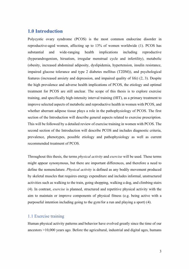
1.0 Introduction Polycystic ovary syndrome (PCOS) is the most common endocrine disorder in
reproductive-aged women, affecting up to 13% of women worldwide (1). PCOS has
substantial and wide-ranging health implications including reproductive
(hyperandrogenism, hirsutism, irregular menstrual cycle and infertility), metabolic
(obesity, increased abdominal adiposity, dyslipidemia, hypertension, insulin resistance,
impaired glucose tolerance and type 2 diabetes mellitus (T2DM)), and psychological
features (increased anxiety and depression, and impaired quality of life) (2, 3). Despite
the high prevalence and adverse health implications of PCOS, the etiology and optimal
treatment for PCOS are still unclear. The scope of this thesis is to explore exercise
training, and specifically high-intensity interval training (HIT), as a primary treatment to
improve selected aspects of metabolic and reproductive health in women with PCOS, and
whether aberrant adipose tissue plays a role in the pathophysiology of PCOS. The first
section of the Introduction will describe general aspects related to exercise prescription.
This will be followed by a detailed review of exercise training in women with PCOS. The
second section of the Introduction will describe PCOS and includes diagnostic criteria,
prevalence, phenotypes, possible etiology and pathophysiology as well as current
recommended treatment of PCOS.
Throughout this thesis, the terms physical activity and exercise will be used. These terms
might appear synonymous, but there are important differences, and therefore a need to
define the nomenclature. Physical activity is defined as any bodily movement produced
by skeletal muscles that requires energy expenditure and includes informal, unstructured
activities such as walking to the train, going shopping, walking a dog, and climbing stairs
(4). In contrast, exercise is planned, structured and repetitive physical activity with the
aim to maintain or improve components of physical fitness (e.g. being active with a
purposeful intention including going to the gym/for a run and playing a sport) (4).
1.1 Exercise training Human physical activity patterns and behavior have evolved greatly since the time of our
ancestors >10,000 years ago. Before the agricultural, industrial and digital ages, humans
4
expended large amounts of energy on a daily basis through activities required to sustain
life, including hunting and gathering, and escaping predators (5). Such a lifestyle
markedly contrasts current activity patterns, in which sedentary behavior, supported by
mechanized forms of transport and labor-saving devices at home and at work, are a
common part of our everyday life. Physical activity for the vast majority of people in the
modern era is an optional activity, separated from daily tasks of living, and what we
define as exercise (5).
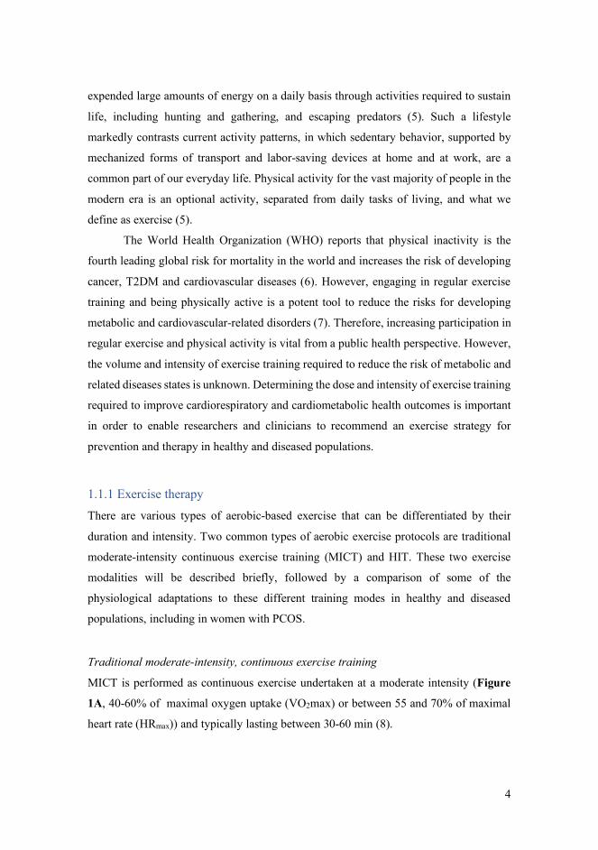
The World Health Organization (WHO) reports that physical inactivity is the
fourth leading global risk for mortality in the world and increases the risk of developing
cancer, T2DM and cardiovascular diseases (6). However, engaging in regular exercise
training and being physically active is a potent tool to reduce the risks for developing
metabolic and cardiovascular-related disorders (7). Therefore, increasing participation in
regular exercise and physical activity is vital from a public health perspective. However,
the volume and intensity of exercise training required to reduce the risk of metabolic and
related diseases states is unknown. Determining the dose and intensity of exercise training
required to improve cardiorespiratory and cardiometabolic health outcomes is important
in order to enable researchers and clinicians to recommend an exercise strategy for
prevention and therapy in healthy and diseased populations.
1.1.1 Exercise therapy
There are various types of aerobic-based exercise that can be differentiated by their
duration and intensity. Two common types of aerobic exercise protocols are traditional
moderate-intensity continuous exercise training (MICT) and HIT. These two exercise
modalities will be described briefly, followed by a comparison of some of the
physiological adaptations to these different training modes in healthy and diseased
populations, including in women with PCOS.
Traditional moderate-intensity, continuous exercise training
MICT is performed as continuous exercise undertaken at a moderate intensity (Figure
1A, 40-60% of maximal oxygen uptake (VO2max) or between 55 and 70% of maximal
heart rate (HRmax)) and typically lasting between 30-60 min (8).
5
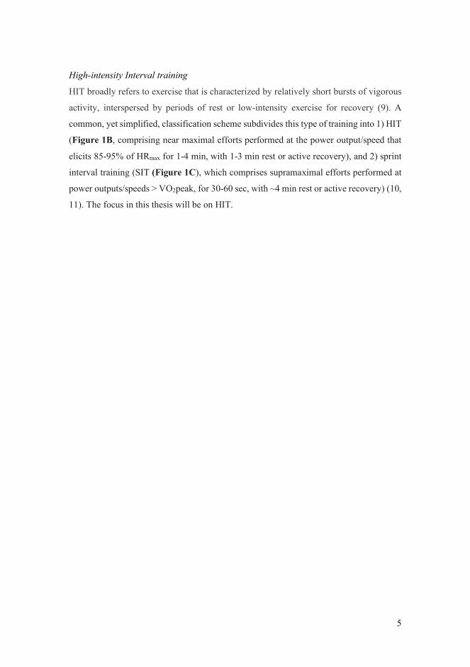
High-intensity Interval training
HIT broadly refers to exercise that is characterized by relatively short bursts of vigorous
activity, interspersed by periods of rest or low-intensity exercise for recovery (9). A
common, yet simplified, classification scheme subdivides this type of training into 1) HIT
(Figure 1B, comprising near maximal efforts performed at the power output/speed that
elicits 85-95% of HRmax for 1-4 min, with 1-3 min rest or active recovery), and 2) sprint
interval training (SIT (Figure 1C), which comprises supramaximal efforts performed at
power outputs/speeds > VO2peak, for 30-60 sec, with ~4 min rest or active recovery) (10,
11). The focus in this thesis will be on HIT.

6
Figure 1. A graphical depiction of the main types of aerobic exercise. Reproduced from MacInnis
and Gibala (2017) (10) with permission from J Physiol. (A-C) representative examples of
moderate-intensity continuous training (MICT), high-intensity interval training (HIIT), and low
and high volumes of sprint interval training (SIT). The intensity is depicted as a percentage of the
peak power output (PPO) obtained during a standard ramp VO2peak test. (D) the training volume
associated with each protocol based on the durations and training frequencies provided. The
MICT and HIIT protocols shown in A and B are work-matched when performed for the same
7
duration and at the same frequency. The low-volume SIT protocol requires less total work to
complete relative to HIIT and MICT, whereas performing three sessions of the high-volume SIT
protocol matches the training volume in the MICT and HIIT protocols.
Several HIT protocols exist and they can broadly be divided into low-volume HIT (LV-
HIT) and high-volume HIT (HV-HIT). LV-HIT normally has at least a 1:1 ratio in work
bout and recovery time whereas HV-HIT has a shorter work:recovery ratio.
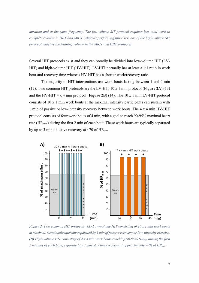
The majority of HIT interventions use work bouts lasting between 1 and 4 min
(12). Two common HIT protocols are the LV-HIT 10 x 1 min protocol (Figure 2A) (13)
and the HV-HIT 4 x 4 min protocol (Figure 2B) (14). The 10 x 1 min LV-HIT protocol
consists of 10 x 1 min work bouts at the maximal intensity participants can sustain with
1 min of passive or low-intensity recovery between work bouts. The 4 x 4 min HV-HIT
protocol consists of four work bouts of 4 min, with a goal to reach 90-95% maximal heart
rate (HRmax) during the first 2 min of each bout. These work bouts are typically separated
by up to 3 min of active recovery at ~70 of HRmax.
Figure 2. Two common HIT protocols: (A) Low-volume HIT consisting of 10 x 1 min work bouts
at maximal, sustainable intensity separated by 1 min of passive recovery or low-intensity exercise,
(B) High-volume HIT consisting of 4 x 4 min work bouts reaching 90-95% HRmax during the first
2 minutes of each bout, separated by 3 min of active recovery at approximately 70% of HRmax.
100 -
10 -
90 -
80 -
70 -
60 -
50 -
40 -
30 -
20 -
% o
f max
imum
effo
rt
10 x 1 min HIT work bouts
Warm-up
Cool
down
A)
% o
f HR m
ax
4 x 4 min HIT work bouts
Cool
down
100 -
10 -
90 -
80 -
70 -
60 -
50 -
40 -
30 -
20 -
B)
Warm-up
Time (min)
Time (min)1010 20 30 20 30 40
8
MICT versus HIT
A large body of evidence demonstrates that in both healthy individuals and diseased
populations, HIT can provide similar or greater benefits to a number of cardiorespiratory
measures and health markers compared to MICT (12, 14-18). Helgerud and colleagues
reported greater improvement in VO2max after eight weeks of three weekly sessions of
HIT (4 x 4 min at 90-95% of HRmax) compared to MICT (continuous running at 70%
HRmax for 45 min) in healthy male university students (14). The training protocols
employed in that study were matched for total work and frequency, suggesting that
training intensity is an important consideration with regard to improving
cardiorespiratory fitness. The superior improvements in VO2max following HIT are
important since poor cardiorespiratory fitness is strongly associated with increased risk
of developing the metabolic syndrome and T2DM (19). Furthermore, Farrell and
colleagues showed that poor cardiorespiratory fitness is an independent predictor of all-
cause mortality in women (20).
HIT confers greater health benefits compared to MICT in several clinical
populations (12, 15, 16). Tjønna and colleagues demonstrated superior improvements in
cardiovascular fitness, fasting glucose concentrations and insulin sensitivity (based on
Homeostatic Model Assessment for Insulin Resistance (HOMA-IR)) in patients with the
metabolic syndrome after 16 weeks of three weekly HIT sessions (4 x 4 min at 90% of
HRmax) compared to work-matched MICT (47 min at 70% of HRmax) (17). Improvements
in glucose control and insulin sensitivity have also been reported following HIT in
patients with T2DM (18, 21). Little and colleagues demonstrated that LV-HIT (6 sessions
of 10 x 1 min at ~90% of HRmax) reduced average 24 h blood glucose concentration, area
under the 24 h blood glucose curve and the sum of three-hour postprandial area under the
glucose curve for breakfast, lunch and dinner in patients with T2DM. The latter study
(21) was an uncontrolled trial and did not directly compare the effects of HIT with MICT.
However, a randomized controlled trial (RCT) undertaken by Karstoft and colleagues
demonstrated superior effects of four months of interval walking compared to energy-
expenditure matched continuous walking on VO2max, body composition and glycemic
control (decreased fasting glucose levels and mean continuous glucose monitoring
concentration) in patients with T2DM (18).
9
To include HIT as a type of training can result in greater health benefits gained in
less time, which makes HIT (and especially LV-HIT with a total exercise time of ~30 min
including warm-up and cool-down) a time-efficient and attractive option to include for
individuals who are time-poor. From a public health perspective, these findings are
important as lack of time is an often-cited barrier to regular exercise participations (22).
Training volumes usually differ when the physiological effects of LV-HIT versus MICT
have been compared (23), whereas training volumes are usually matched in studies that
have compared the physiological effects of HV-HIT versus MICT (14, 17, 18). Although
HV-HIT (such as 4 x 4 min intervals with a total exercise time of 38 min including a 10
min warm-up and 3 min cool-down) is a more time-efficient approach compared to
volume matched MICT (total exercise time of ~45-50 min), HV-HIT is still time-
demanding. Apart from being time-efficient, evidence suggests that HIT is also perceived
to be more enjoyable than MICT despite higher perceived exertion during HIT (24).
Therefore, HIT may prove as a feasible strategy to improve long-term exercise adherence
and participation.
1.1.2 Exercise therapy in PCOS
Lifestyle interventions, including exercise training, are recommended as first-line therapy
for women with PCOS (3). However, there are few well-designed RCTs that have
investigated the effect of exercise on reproductive and metabolic outcomes in women
with PCOS. Accordingly, clinicians’ face many challenges and lack a solid evidence-base
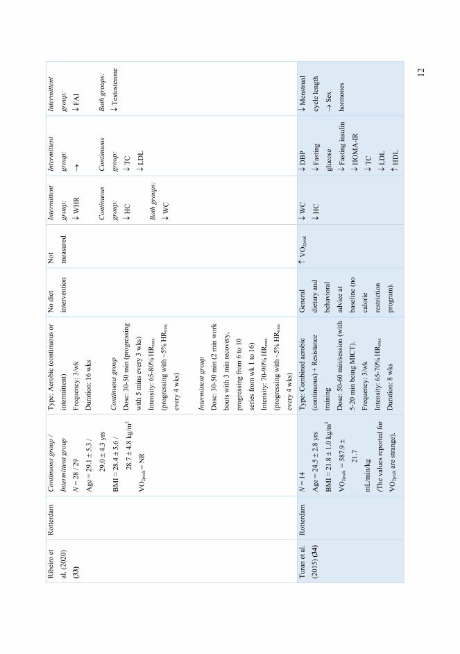
when attempting to prescribe specific modes of exercise to women with PCOS. Table 1
summarizes the results from RCTs including different types of aerobic exercise
interventions in women with PCOS. RCTs that included the use of pharmacotherapy were
excluded.

10
Tabl
e 1.
Sum
mar
y of
RC
Ts w
ith a
erob
ic e
xerc
ise
inte
rven
tion(
s) in
wom
en w
ith P
CO
S St
udy
Def
initi
on
of P
CO
S
Exer
cise
gro
up
char
acte
rist
ics a
t
base
line
Exer
cise
pro
toco
l D
iet
inte
rven
tion
Car
dio-
resp
irat
ory
outc
omes
Body
com
posit
ion
outc
omes
Car
diom
etab
olic
outc
omes
Hor
mon
al a
nd
repr
oduc
tive
outc
omes
Alm
enni
ng
et a
l.
(201
5)
(25)
Rot
terd
am
N =
8
Age
= 2
7.2 ±
5.5
yrs
BM
I = 2
6.1 ±
6.5
kg/m
2
VO
2pea
k =
35.6
± 5
.6
m
L/m
in/k
g
Type
: Aer
obic
(HIT
)
Dos
e: 1
9 (1
0 x
1 m
in w
ith 1
min
reco
very
) and
25
min
/ses
sion
(4 x
4 m
in w
ith 3
min
reco
very
)
Freq
uenc
y: 3
/wk
Inte
nsity
: 4 x
4 m
in a
t 90-
95%
HR
max
(2/w
k) a
nd 1
0 x
1 m
in
with
max
imal
inte
nsity
(1/w
k)
Dur
atio
n: 1
0 w
ks
No
diet
inte
rven
tion
V
O2p
eak
¯ FM
¯ B
F%
¯ H
OM
A-IR
¯ Fa
stin
g in
sulin
H
DL
¯ D
HEA
S
Bro
wn
et
al. (
2009
)
(26)
NIH
N
= 8
Age
= 3
6.5
± 5.
0 yr
s
BM
I = 3
7.9
± 9.
4 kg
/m2
VO
2pea
k =
22.5
± 6
.0
m
L/m
in/k
g
Type
: Aer
obic
(con
tinuo
us)
Dos
e: ~
228
min
/wk
Freq
uenc
y: N
R
Inte
nsity
: 40-
60%
VO
2pea
k
Dur
atio
n: 2
0-24
wks
No
diet
inte
rven
tion
V
O2p
eak
®
®
Not
mea
sure
d
Bru
ner e
t
al. (
2006
)
(27)
Rot
terd
am
N =
7
Age
= 3
2.3 ±
1.0
yrs
BM
I = 3
6.2 ±
2.0
kg/m
2
VO
2pea
k =
22.6
± 2
.1
m
L/m
in/k
g
Type
: Com
bine
d ae
robi
c
(con
tinuo
us) +
Res
istan
ce
train
ing
Dos
e: 3
0 m
in/se
ssio
n (M
ICT)
and
~60
min
/sess
ion
(RT)
Freq
uenc
y: 3
/wk
Inte
nsity
: 70-
85%
HR
max
Dur
atio
n: 1
2 w
ks
Nut
ritio
nal
coun
selin
g
1hr/w
k (in
the
exer
cise
& c
ontro
l
grou
p)
V
O2p
eak
¯ W
C
¯ Fa
stin
g in
sulin
®

11
Cos
ta e
t al.
(201
8)
(28)
Rot
terd
am
N =
14
Age
= 2
7.6 ±
4.5
yrs
BM
I = 3
2.0 ±
4.2
kg/m
2
VO
2pea
k =
27.9
± 3
.3
m
L/m
in/k
g
Type
: Aer
obic
(con
tinuo
us)
Dos
e: 4
0 m
in/se
ssio
n
Freq
uenc
y: 3
/wk
Inte
nsity
: 60-
85%
HR
max
(pro
gres
sing
with
~5%
HR
max
ever
y 4
wks
)
Dur
atio
n: 1
6 w
ks
No
diet
inte
rven
tion
V
O2p
eak
¯ BM
I
¯ W
C
¯ SB
P
¯ D
BP
¯ TC
Not
mea
sure
d
Jede
l et a
l.
(201
1) (2
9)
and
Sten
er-
Vic
torin
et
al. (
2012
)
(30)
Rot
terd
am
N =
30
Age
= 3
0.2 ±
4.7
yrs
BM
I = 2
7.7 ±
6.4
kg/m
2
VO
2pea
k =
33.9
± 8
.5
m
L/m
in/k
g
Type
: Aer
obic
(con
tinuo
us)
Dos
e: 3
0 m
in/se
ssio
n
Freq
uenc
y: 3
/wk
Inte
nsity
: fas
ter t
han
norm
al
wal
king
pac
e
Dur
atio
n: 1
6 w
ks
No
diet
inte
rven
tion
V
O2p
eak
®
¯ D
BP
M
enst
rual
frequ
ency
¯ Te
stos
tero
ne
¯ D
HEA
S
Nag
elbe
rg
et a
l.
(201
6)
(31)
NIH
N
= 1
0
Age
= N
R
BM
I =
≥25-
29.9
kg/
m2 (n
=3)
≥30-
39.9
kg/
m2 (n
=7)
VO
2pea
k =
NR
Type
: Aer
obic
(con
tinuo
us)
Dos
e: N
R
Freq
uenc
y: 7
/wk
Inte
nsity
: NR
Dur
atio
n: 4
wks
Parti
cipa
nts
cont
acte
d
wee
kly
to
disc
uss
nutri
tiona
l
goal
s
Not
mea
sure
d
Not
mea
sure
d
Not
mea
sure
d ®
Orio
et a
l.
(201
6)
(32)
Rot
terd
am
N =
39
Age
= 2
5.9 ±
2.7
yrs
BM
I = 2
6.7 ±
2.8
kg/m
2
VO
2pea
k =
19.0
± 2
.1
m
L/m
in/k
g
Type
: Aer
obic
(con
tinuo
us)
Dos
e: 4
5 m
in/se
ssio
n
Freq
uenc
y: 3
/wk
Inte
nsity
: 60-
70%
VO
2pea
k
Dur
atio
n: 2
4 w
ks
800
kcal
/day
defic
it
V
O2p
eak
¯ BM
I
¯ W
HR
¯ TC
¯ LD
L
¯ Fa
stin
g in
sulin
¯ A
UC
insu
lin
¯ H
OM
A-IR
H
DL
M
enst
rual
frequ
ency
® S
ex
horm
ones
12
Rib
eiro
et
al. (
2020
)
(33)
Rot
terd
am
Con
tinuo
us g
roup
/
Inte
rmitt
ent g
roup
N =
28
/ 29
Age
= 2
9.1 ±
5.3
/
2
9.0 ±
4.3
yrs
BM
I = 2
8.4 ±
5.6
/
28.7
± 4
.8 k
g/m
2
VO
2pea
k =
NR
Type
: Aer
obic
(con
tinuo
us o
r
inte
rmitt
ent)
Freq
uenc
y: 3
/wk
Dur
atio
n: 1
6 w
ks
Con
tinuo
us g
roup
Dos
e: 3
0-50
min
(pro
gres
sing
with
5 m
ins e
very
3 w
ks)
Inte
nsity
: 65-
80%
HR
max
(pro
gres
sing
with
~5%
HR
max
ever
y 4
wks
)
Inte
rmitt
ent g
roup
Dos
e: 3
0-50
min
(2 m
in w
ork
bout
s with
3 m
in re
cove
ry,
prog
ress
ing
from
6 to
10
serie
s fro
m w
k 1
to 1
6)
Inte
nsity
: 70-
90%
HR
max
(pro
gres
sing
with
~5%
HR
max
ever
y 4
wks
)
No
diet
inte
rven
tion
Not
mea
sure
d
Inte
rmitt
ent
grou
p:
¯ W
HR
Con
tinuo
us
grou
p:
¯ H
C
Both
gro
ups:
¯ W
C
Inte
rmitt
ent
grou
p:
®
Con
tinuo
us
grou
p:
¯ TC
¯ LD
L
Inte
rmitt
ent
grou
p:
¯ FA
I
Both
gro
ups:
¯ Te
stos
tero
ne
Tura
n et
al.
(201
5) (3
4)
Rot
terd
am
N =
14
Age
= 2
4.5 ±
2.8
yrs
BM
I = 2
1.8 ±
1.0
kg/m
2
VO
2pea
k =
587
.9 ±
2
1.7
mL/
min
/kg
(The
val
ues r
epor
ted
for
VO
2pea
k are
stra
nge)
.
Type
: Com
bine
d ae
robi
c
(con
tinuo
us) +
Res
istan
ce
train
ing
Dos
e: 5
0-60
min
/sess
ion
(with
5-20
min
bei
ng M
ICT)
.
Freq
uenc
y: 3
/wk
Inte
nsity
: 65-
70%
HR
max
Dur
atio
n: 8
wks
Gen
eral
diet
ary
and
beha
vior
al
advi
ce a
t
base
line
(no
calo
rie
rest
rictio
n
prog
ram
).
V
O2p
eak
¯ W
C
¯ H
C
¯ D
BP
¯ Fa
stin
g
gluc
ose
¯ Fa
stin
g in
sulin
¯ H
OM
A-IR
¯ TC
¯ LD
L
H
DL
¯ M
enst
rual
cycl
e le
ngth
® S
ex
horm
ones

13
Vig
orito
et
al. (
2007
)
(35)
Rot
terd
am
N =
45
Age
= 2
1.7 ±
2.3
yrs
BM
I = 2
9.3 ±
2.9
kg/m
2
VO
2pea
k =
17.6
± 2
.5
m
L/m
in/k
g
Type
: Aer
obic
(con
tinuo
us)
Dos
e: 3
0 m
in/se
ssio
n
Freq
uenc
y: 3
/wk
Inte
nsity
: 60-
70%
VO
2pea
k
Dur
atio
n: 1
2 w
ks
Gen
eral
diet
ary
and
beha
vior
al
advi
ce a
t
base
line
(no
calo
rie
rest
rictio
n
prog
ram
).
V
O2p
eak
¯ BM
I
¯ W
C
¯ W
HR
¯ Fa
stin
g in
sulin
¯ A
UC
insu
lin
A
UC
gluc
ose/
AU
Cin
sulin
ratio
M
enst
rual
cycl
icity
(in
60%
of
parti
cipa
nts)
® S
ex
horm
ones
AM
H,
Ant
i M
ülle
rian
horm
one;
AU
C, A
rea
unde
r cu
rve;
BF%
, B
ody
fat
perc
enta
ge;
BM
I, B
ody
mas
s in
dex;
BP,
Blo
od p
ress
ure;
BW
, B
ody
wei
ght;
DH
EAS,
Deh
ydro
epia
ndro
stero
ne su
lfate
; DB
P, D
iasto
lic b
lood
pre
ssur
e; F
AI,
Free
and
roge
n in
dex;
FFM
, Fat
-fre
e m
ass;
FM
, Fat
mas
s; F
SH, F
ollic
le st
imul
atin
g ho
rmon
e; H
C,
Hip
circ
umfe
renc
e; H
DL,
Hig
h de
nsity
lipo
prot
ein
chol
este
rol;
HIT
, Hig
h-in
tens
ity in
terv
al tr
aini
ng; H
OM
A-I
R, H
omeo
stat
ic m
odel
ass
essm
ent o
f ins
ulin
resis
tanc
e;
HR
max
, Max
imal
hea
rt ra
te; L
DL,
Low
den
sity
lipop
rote
in c
hole
stero
l; LH
, Lut
eini
zing
hor
mon
e; N
IH, N
atio
nal I
nstit
utes
of H
ealth
; NR,
Not
repo
rted;
PC
OS,
Pol
ycys
tic
ovar
y sy
ndro
me;
SB
P, S
ysto
lic b
lood
pre
ssur
e; S
HB
G, S
ex h
orm
one
bind
ing
glob
ulin
; TC,
Tot
al c
hole
stero
l; TG
, Trig
lyce
rides
; VO
2pea
k, Pe
ak o
xyge
n up
take
; WC,
Wai
st c
ircum
fere
nce,
WH
R, W
aist
-to-h
ip ra
tio.
ill
ustra
tes a
n in
crea
se, ¯
a d
ecre
ase
and ®
no
chan
ge. E
xerc
ise
dose
doe
s not
incl
ude
war
m-u
p an
d co
ol-d
own
but d
oes i
nclu
de re
cove
ry ti
me
in b
etw
een
wor
k bo
uts
in H
IT se
ssio
ns. N
refle
cts t
he n
umbe
r of p
artic
ipan
ts in
clud
ed in
the
anal
ysis.
Cha
nges
are
bas
ed o
n w
ithin
- and
bet
wee
n-gr
oup
diffe
renc
es.
14
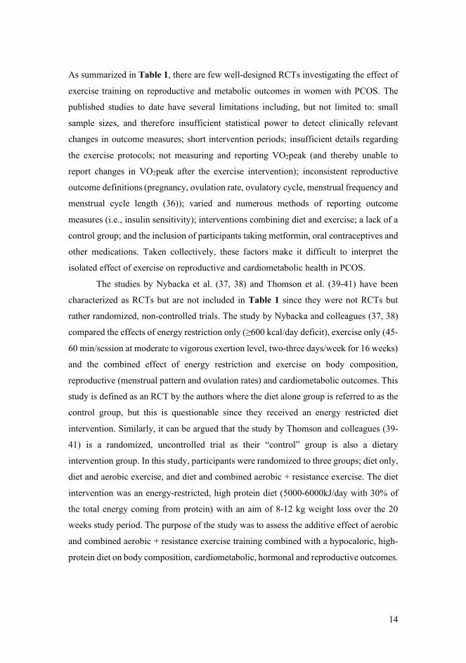
As summarized in Table 1, there are few well-designed RCTs investigating the effect of
exercise training on reproductive and metabolic outcomes in women with PCOS. The
published studies to date have several limitations including, but not limited to: small
sample sizes, and therefore insufficient statistical power to detect clinically relevant
changes in outcome measures; short intervention periods; insufficient details regarding
the exercise protocols; not measuring and reporting VO2peak (and thereby unable to
report changes in VO2peak after the exercise intervention); inconsistent reproductive
outcome definitions (pregnancy, ovulation rate, ovulatory cycle, menstrual frequency and
menstrual cycle length (36)); varied and numerous methods of reporting outcome
measures (i.e., insulin sensitivity); interventions combining diet and exercise; a lack of a
control group; and the inclusion of participants taking metformin, oral contraceptives and
other medications. Taken collectively, these factors make it difficult to interpret the
isolated effect of exercise on reproductive and cardiometabolic health in PCOS.
The studies by Nybacka et al. (37, 38) and Thomson et al. (39-41) have been
characterized as RCTs but are not included in Table 1 since they were not RCTs but
rather randomized, non-controlled trials. The study by Nybacka and colleagues (37, 38)
compared the effects of energy restriction only (≥600 kcal/day deficit), exercise only (45-
60 min/session at moderate to vigorous exertion level, two-three days/week for 16 weeks)
and the combined effect of energy restriction and exercise on body composition,
reproductive (menstrual pattern and ovulation rates) and cardiometabolic outcomes. This
study is defined as an RCT by the authors where the diet alone group is referred to as the
control group, but this is questionable since they received an energy restricted diet
intervention. Similarly, it can be argued that the study by Thomson and colleagues (39-
41) is a randomized, uncontrolled trial as their “control” group is also a dietary
intervention group. In this study, participants were randomized to three groups; diet only,
diet and aerobic exercise, and diet and combined aerobic + resistance exercise. The diet
intervention was an energy-restricted, high protein diet (5000-6000kJ/day with 30% of
the total energy coming from protein) with an aim of 8-12 kg weight loss over the 20
weeks study period. The purpose of the study was to assess the additive effect of aerobic
and combined aerobic + resistance exercise training combined with a hypocaloric, high-
protein diet on body composition, cardiometabolic, hormonal and reproductive outcomes.
15
Even if they were able to report results based on their aim, this was not an RCT as they
did not include a no-intervention/usual care control group.
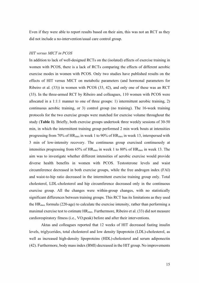
HIT versus MICT in PCOS
In addition to lack of well-designed RCTs on the (isolated) effects of exercise training in
women with PCOS, there is a lack of RCTs comparing the effects of different aerobic
exercise modes in women with PCOS. Only two studies have published results on the
effects of HIT versus MICT on metabolic parameters (and hormonal parameters for
Ribeiro et al. (33)) in women with PCOS (33, 42), and only one of these was an RCT
(33). In the three-armed RCT by Ribeiro and colleagues, 110 women with PCOS were
allocated in a 1:1:1 manner to one of three groups: 1) intermittent aerobic training, 2)
continuous aerobic training, or 3) control group (no training). The 16-week training
protocols for the two exercise groups were matched for exercise volume throughout the
study (Table 1). Briefly, both exercise groups undertook three weekly sessions of 30-50
min, in which the intermittent training group performed 2 min work bouts at intensities
progressing from 70% of HRmax in week 1 to 90% of HRmax in week 13, interspersed with
3 min of low-intensity recovery. The continuous group exercised continuously at
intensities progressing from 65% of HRmax in week 1 to 80% of HRmax in week 13. The
aim was to investigate whether different intensities of aerobic exercise would provide
diverse health benefits in women with PCOS. Testosterone levels and waist
circumference decreased in both exercise groups, while the free androgen index (FAI)
and waist-to-hip ratio decreased in the intermittent exercise training group only. Total
cholesterol, LDL-cholesterol and hip circumference decreased only in the continuous
exercise group. All the changes were within-group changes, with no statistically
significant differences between training groups. This RCT has its limitations as they used
the HRmax formula (220-age) to calculate the exercise intensity, rather than performing a
maximal exercise test to estimate HRmax. Furthermore, Ribeiro et al. (33) did not measure
cardiorespiratory fitness (i.e., VO2peak) before and after their interventions.
Aktas and colleagues reported that 12 weeks of HIT decreased fasting insulin
levels, triglycerides, total cholesterol and low density lipoprotein (LDL)-cholesterol, as
well as increased high-density lipoproteins (HDL)-cholesterol and serum adiponectin
(42). Furthermore, body mass index (BMI) decreased in the HIT group. No improvements
16
were observed following 12 weeks of MICT, however, there were no significant between-
group differences. There are several limitations to this study. Insufficient details were
provided regarding the exercise intensities of the two interventions. The authors described
that both groups exercised for 30 min three days a week for 12 weeks and that the HIT
group ran for 2 minutes, then walked for 2 minutes, while the MICT group ran for 30
minutes at a moderate tempo and at a constant speed. Furthermore, this was a combined
exercise and diet intervention, in which both exercise groups followed a diet program that
induced a 2-4 kg weight loss per month. There was no control group in this study, which
makes it difficult to determine whether the observed effects were a result of the diet-
and/or exercise intervention.
One RCT determined the effects of HIT as the sole intervention on metabolic and
reproductive outcomes in women with PCOS (25). In a pilot study examining the effects
of HIT, ten weeks of two weekly sessions of 4 x 4 min at 90-95% of HRmax and one
weekly session of 10 x 1 min at maximal intensity improved VO2max, HOMA-IR, fasting
insulin concentrations, HDL-cholesterol, and body fat percentage, compared to a non-
exercising control group. Of note, these improvements occured in the absence of any
change in body mass (25).
The results of a recent meta-analysis conclude that vigorous intensity exercise is
superior to moderate intensity exercise for improvements in cardiometabolic outcomes
including VO2peak, BMI and waist circumference in women with PCOS (43). Similarly,
data from a cross-sectional study in women with PCOS showed that vigorous exercisers
had lower HOMA-IR, higher HDL and sex hormone binding globulin (SHBG), and
reduced prevalence of the metabolic syndrome, compared with those who exercised at
moderate intensity or were inactive (44). In summary, the majority of evidence to date
suggests that HIT is superior to MICT for inducing improvements in cardiorespiratory
fitness, body composition and hormonal outcomes in women with PCOS. However,
further research is required to determine whether exercise protocols utilized as part of
clinical trials (8-12 weeks of supervised exercise) is realistic in a real-world scenario, and
whether unsupervised training should be considered in order to promote long-term
sustainability of exercise behaviors. Furthermore, research on the impact of various HIT
protocols is necessary in order to determine the most successful interventions for retaining
engagement and improving health.
17
Low-volume HIT versus high-volume HIT
Superior changes in a range of physiological and health-related markers have been
reported after HIT compared to MICT in both healthy and clinical populations. However,
far less is known regarding which type of HIT protocol is most effective in free-living,
unsupervised conditions in women with PCOS. Baekkerud and colleagues compared the
effects of three popular exercise modalities on VO2max in men and women with
overweight/obesity (23). Participants were randomly allocated to six weeks of either 4 x
4 min HIT at 85-95% of HRmax (HV-HIT), 10 x 1 min at VO2max load (LV-HIT), or 45
min MICT at 70% of HRmax. All groups completed three supervised, sessions per week
(18 sessions in total). Only the 4 x 4 min HIT group increased their VO2max, whereas
plasma volume and hemoglobin mass increased only in the 10 x 1 min HIT group.
Skeletal muscle citrate synthase activity, a surrogate marker of mitochondrial content,
increased in all three groups. These findings illustrated divergent adaptations following
LV- and HV-HIT, and support previous findings suggesting that greater volumes of high-
intensity work result in greater improvements in VO2max (45, 46). In addition to a high
volume of high-intensity exercise (≥15 min/session), a recent meta-analysis on the effects
of different HIT protocols on improvements in VO2max showed that HIT with longer
work bouts (≥ 2 min) and longer training periods (4-12 weeks) improved VO2max the
greatest in healthy individuals and those with overweight/obesity (46). No previous
studies have compared the effects of LV-HIT and HV-HIT on reproductive and
cardiometabolic outcomes in women with PCOS.
18
1.2 Polycystic ovary syndrome (PCOS)
1.2.1 Definitions of PCOS
Almost ninety years ago, Stein and Leventhal described the disorder we now know as
PCOS (47). They described a reproductive disorder in which they observed an association
between polycystic ovaries and amenorrhea. Over time, the definition of PCOS has
evolved, and hyperandrogenism has since been acknowledged as an important component
of the condition. Today, PCOS is considered a ‘syndrome’ with no single diagnostic test,
but rather a condition based on a collection of clinical signs and features (2). Over the
past three decades, three sets of criteria have been developed to standardize the diagnosis
of PCOS: 1) the National Institutes of Health (NIH) criteria, 2) the Rotterdam criteria,
and 3) the Androgen Excess and PCOS Society (AE-PCOS) criteria (Table 2). Currently,
diagnosis of PCOS is most often based on the Rotterdam criteria, and this is the criteria
used in the work presented in this thesis. The NIH and the AE-PCOS criteria are still used
in the literature, therefore, all three sets of criteria are outlined below.
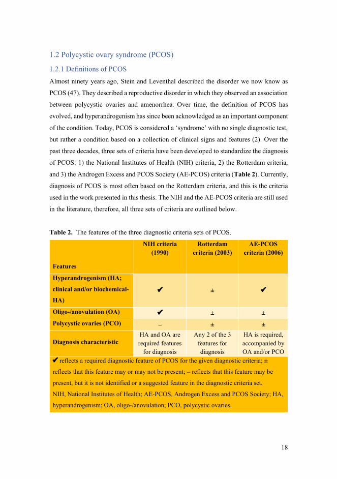
Table 2. The features of the three diagnostic criteria sets of PCOS.
Features
NIH criteria (1990)
Rotterdam criteria (2003)
AE-PCOS criteria (2006)
Hyperandrogenism (HA;
clinical and/or biochemical-
HA) ü ± ü
Oligo-/anovulation (OA) ü ± ± Polycystic ovaries (PCO) – ± ±
Diagnosis characteristic HA and OA are required features
for diagnosis
Any 2 of the 3 features for diagnosis
HA is required, accompanied by OA and/or PCO
ü reflects a required diagnostic feature of PCOS for the given diagnostic criteria; ±
reflects that this feature may or may not be present; – reflects that this feature may be
present, but it is not identified or a suggested feature in the diagnostic criteria set.
NIH, National Institutes of Health; AE-PCOS, Androgen Excess and PCOS Society; HA,
hyperandrogenism; OA, oligo-/anovulation; PCO, polycystic ovaries.
19
The NIH criteria were the first described diagnostic criteria set and were developed at the
National Institutes of Health consensus conference on PCOS in 1990 (48). According to
these criteria, a PCOS diagnosis requires clinical and/or biochemical signs of
hyperandrogenism and oligo-/anovulation. Following the NIH criteria, it was recognized
that the syndrome also encompasses signs and symptoms of ovarian dysfunction
(polycystic ovaries). This lead to a new set of criteria developed at the 2003 consensus
workshop in Rotterdam, also known as the Rotterdam criteria (49). The Rotterdam criteria
require at least two out of the following three features: 1) clinical and/or biochemical
signs of hyperandrogenism, 2) oligo- or anovulation and 3) polycystic ovary morphology.
In 2006, after reviewing the available data, the Androgen Excess and PCOS (AE-PCOS)
Society proposed a new definition for clinically diagnosing PCOS (2). According to the
AE-PCOS criteria, clinical and/or biochemical signs of hyperandrogenism were proposed
as the indispensable feature of PCOS and should be accompanied by oligo-/anovulation
and/or polycystic ovaries. Thereby, the AE-PCOS criteria were a compromise between
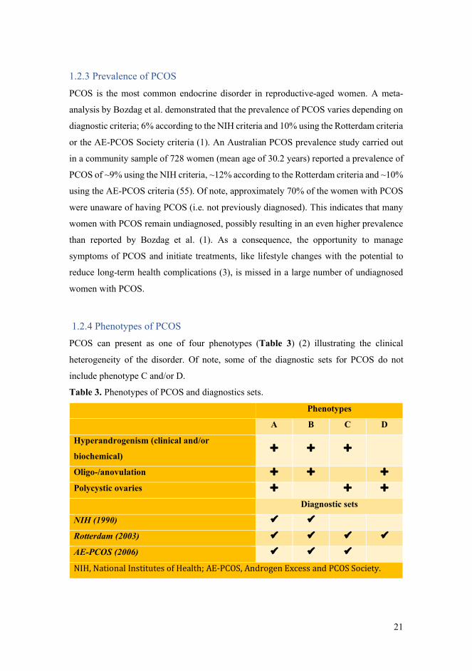
the NIH and Rotterdam criteria and excluded the non-hyperandrogenic phenotype
proposed by the Rotterdam criteria (phenotype D in Table 3). All three sets of criteria are
based on the assumption that other etiologies (congenital adrenal hyperplasia, androgen-
secreting tumors and Cushing’s syndrome) are excluded before the diagnosis of PCOS.
Although the Rotterdam criteria are most commonly used to diagnose PCOS
today, the NIH and AE-PCOS criteria are still used in the scientific and clinical literature.
Using three different sets of diagnostic criteria for diagnosing PCOS complicates PCOS
research by decreasing the comparability of studies and their research findings, and in
addition causes confusion in clinical practice.
1.2.2 Diagnostic features of PCOS
Although the NIH, Rotterdam and AE-PCOS criteria are different, they all require
combinations of these three features; clinical and/or biochemical hyperandrogenism,
menstrual irregularities, and polycystic ovaries. These diagnostic features will be
described in the following paragraphs.
20
Hyperandrogenism
Hyperandrogenism is a feature of PCOS that can manifest clinically or biochemically.
Clinical manifestations of hyperandrogenism are hirsutism, acne and androgenic
alopecia, with hirsutism being the most predominant feature of clinical hyperandrogenism
(50). Hirsutism is assessed by using the modified Ferriman-Gallway scoring system
where seven areas of the body (the upper lip, chin, chest, chest, upper and lower back,
and upper and lower abdomen) are scored from 0 to 4, where 0 is no excess hair growth
and 4 is severe hair growth (51).
Biochemical hyperandrogenism can be assessed when clinical signs of
hyperandrogenism are unclear or absent. To assess biochemical hyperandrogenism,
women need to have withdrawn from the use of hormonal contraceptives for a minimum
of three months before the measurements are performed (52). Biochemical
hyperandrogenism is determined by elevated serum levels of androgens including total
testosterone, calculated free testosterone or by FAI (52). FAI is calculated as 100 x (total
testosterone/SHBG).
Oligo-/anovulation
Ovulatory dysfunction usually manifests as irregular menstrual cycles
(oligo/amenorrhea) resulting from chronic oligo- or anovulation (53). Oligomenorrhea is
defined as an intermenstrual interval >35 days and <8 menstrual cycles during a year.
Amenorrhea is defined as absent menstruations in the past 90 days. Approximately 75%
of women with PCOS have irregular menstrual cycles (54).
Polycystic ovaries
According to the 2003 consensus workshop in Rotterdam, polycystic ovaries are defined
as 12 or more 2-9 mm follicles and/or increased ovarian volume (>10 mL) in at least one
ovary measured by ultrasound scans (49). In 2018, the criteria for polycystic ovaries were
revised in line with advances in technology allowing for increased sensitivity. The revised
criteria recommend that the diagnostic threshold for polycystic ovaries should be based
on the presence of ≥20 follicles (2-9 mm) per ovary and/or an ovarian volume ≥10 mL
when using ultrasound transducers with a frequency bandwidth of ≥8MHz (52).
21
1.2.3 Prevalence of PCOS
PCOS is the most common endocrine disorder in reproductive-aged women. A meta-
analysis by Bozdag et al. demonstrated that the prevalence of PCOS varies depending on
diagnostic criteria; 6% according to the NIH criteria and 10% using the Rotterdam criteria
or the AE-PCOS Society criteria (1). An Australian PCOS prevalence study carried out
in a community sample of 728 women (mean age of 30.2 years) reported a prevalence of
PCOS of ~9% using the NIH criteria, ~12% according to the Rotterdam criteria and ~10%
using the AE-PCOS criteria (55). Of note, approximately 70% of the women with PCOS
were unaware of having PCOS (i.e. not previously diagnosed). This indicates that many
women with PCOS remain undiagnosed, possibly resulting in an even higher prevalence
than reported by Bozdag et al. (1). As a consequence, the opportunity to manage
symptoms of PCOS and initiate treatments, like lifestyle changes with the potential to
reduce long-term health complications (3), is missed in a large number of undiagnosed
women with PCOS.
1.2.4 Phenotypes of PCOS
PCOS can present as one of four phenotypes (Table 3) (2) illustrating the clinical
heterogeneity of the disorder. Of note, some of the diagnostic sets for PCOS do not
include phenotype C and/or D.
Table 3. Phenotypes of PCOS and diagnostics sets.
Phenotypes
A B C D
Hyperandrogenism (clinical and/or
biochemical) È È È
Oligo-/anovulation È È È
Polycystic ovaries È È È
Diagnostic sets
NIH (1990) ü ü
Rotterdam (2003) ü ü ü ü
AE-PCOS (2006) ü ü ü
NIH,NationalInstitutesofHealth;AE-PCOS,AndrogenExcessandPCOSSociety.
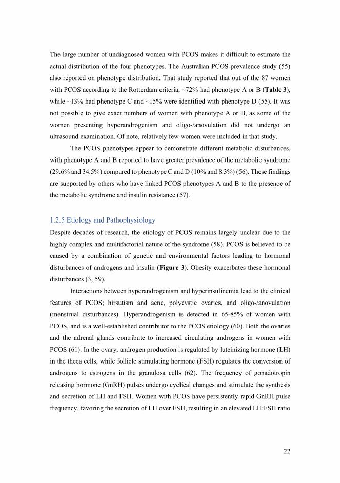
22
The large number of undiagnosed women with PCOS makes it difficult to estimate the
actual distribution of the four phenotypes. The Australian PCOS prevalence study (55)
also reported on phenotype distribution. That study reported that out of the 87 women
with PCOS according to the Rotterdam criteria, ~72% had phenotype A or B (Table 3),
while ~13% had phenotype C and ~15% were identified with phenotype D (55). It was
not possible to give exact numbers of women with phenotype A or B, as some of the
women presenting hyperandrogenism and oligo-/anovulation did not undergo an
ultrasound examination. Of note, relatively few women were included in that study.
The PCOS phenotypes appear to demonstrate different metabolic disturbances,
with phenotype A and B reported to have greater prevalence of the metabolic syndrome
(29.6% and 34.5%) compared to phenotype C and D (10% and 8.3%) (56). These findings
are supported by others who have linked PCOS phenotypes A and B to the presence of
the metabolic syndrome and insulin resistance (57).
1.2.5 Etiology and Pathophysiology
Despite decades of research, the etiology of PCOS remains largely unclear due to the
highly complex and multifactorial nature of the syndrome (58). PCOS is believed to be
caused by a combination of genetic and environmental factors leading to hormonal
disturbances of androgens and insulin (Figure 3). Obesity exacerbates these hormonal
disturbances (3, 59).
Interactions between hyperandrogenism and hyperinsulinemia lead to the clinical
features of PCOS; hirsutism and acne, polycystic ovaries, and oligo-/anovulation
(menstrual disturbances). Hyperandrogenism is detected in 65-85% of women with
PCOS, and is a well-established contributor to the PCOS etiology (60). Both the ovaries
and the adrenal glands contribute to increased circulating androgens in women with
PCOS (61). In the ovary, androgen production is regulated by luteinizing hormone (LH)
in the theca cells, while follicle stimulating hormone (FSH) regulates the conversion of
androgens to estrogens in the granulosa cells (62). The frequency of gonadotropin
releasing hormone (GnRH) pulses undergo cyclical changes and stimulate the synthesis
and secretion of LH and FSH. Women with PCOS have persistently rapid GnRH pulse
frequency, favoring the secretion of LH over FSH, resulting in an elevated LH:FSH ratio
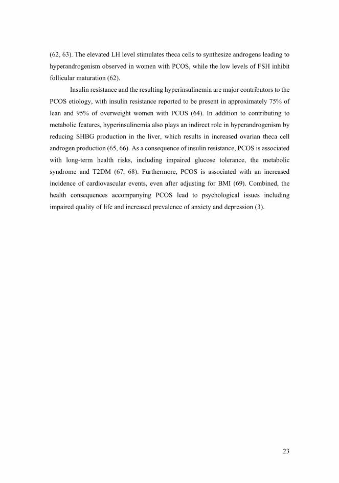
23
(62, 63). The elevated LH level stimulates theca cells to synthesize androgens leading to
hyperandrogenism observed in women with PCOS, while the low levels of FSH inhibit
follicular maturation (62).
Insulin resistance and the resulting hyperinsulinemia are major contributors to the
PCOS etiology, with insulin resistance reported to be present in approximately 75% of
lean and 95% of overweight women with PCOS (64). In addition to contributing to
metabolic features, hyperinsulinemia also plays an indirect role in hyperandrogenism by
reducing SHBG production in the liver, which results in increased ovarian theca cell
androgen production (65, 66). As a consequence of insulin resistance, PCOS is associated
with long-term health risks, including impaired glucose tolerance, the metabolic
syndrome and T2DM (67, 68). Furthermore, PCOS is associated with an increased
incidence of cardiovascular events, even after adjusting for BMI (69). Combined, the
health consequences accompanying PCOS lead to psychological issues including
impaired quality of life and increased prevalence of anxiety and depression (3).
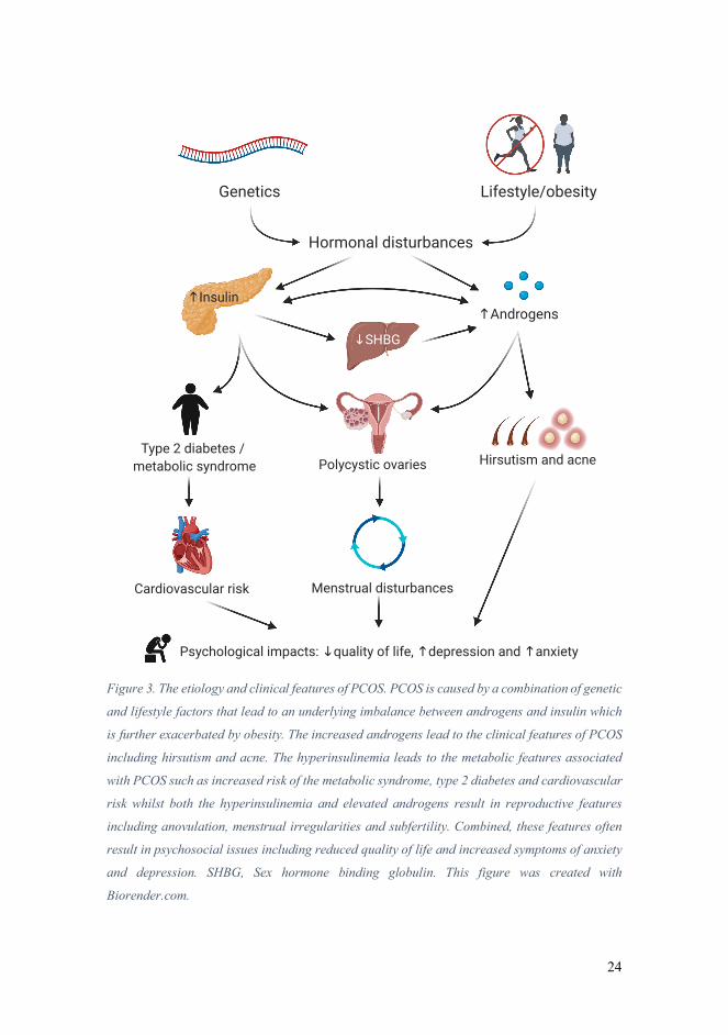
24
Figure 3. The etiology and clinical features of PCOS. PCOS is caused by a combination of genetic
and lifestyle factors that lead to an underlying imbalance between androgens and insulin which
is further exacerbated by obesity. The increased androgens lead to the clinical features of PCOS
including hirsutism and acne. The hyperinsulinemia leads to the metabolic features associated
with PCOS such as increased risk of the metabolic syndrome, type 2 diabetes and cardiovascular
risk whilst both the hyperinsulinemia and elevated androgens result in reproductive features
including anovulation, menstrual irregularities and subfertility. Combined, these features often
result in psychosocial issues including reduced quality of life and increased symptoms of anxiety
and depression. SHBG, Sex hormone binding globulin. This figure was created with
Biorender.com.
Lifestyle/obesity Genetics
Hormonal disturbances
↑ Androgens
Polycystic ovaries
Psychological impacts: ↓ quality of life, ↑ depression and ↑ anxiety
↓ SHBG
↑ Insulin
Menstrual disturbances
Hirsutism and acne Type 2 diabetes /
metabolic syndrome
Cardiovascular risk
25
1.2.6 PCOS, obesity and insulin resistance
The number of people with overweight and obesity (i.e. BMI ≥ 25 kg/m2) has increased
dramatically on a global scale over the past decades. In 2016, the World Health
Organization estimated that 39% of the adult population had overweight or obesity (70).
In parallel with the rise in overweight and obesity observed in the general population, the
prevalence of overweight and obesity is also increasing in women with PCOS. The
percentage of women with PCOS and obesity (i.e. BMI ≥ 30 kg/m2) increased from 51%
to 74% in just 10-15 years (71). Obesity per se does not cause PCOS, but exacerbates
some of the metabolic and reproductive dysfunctions linked to PCOS including
hyperandrogenism, hirsutism and infertility (72). Obesity also worsens insulin resistance
in women with PCOS (64, 73). A meta-analysis showed that women with PCOS have a
27% reduction in insulin sensitivity as measured by the gold standard euglycemic-
hyperinsulinemic clamp compared to women without PCOS, independent of BMI and
age (73). Furthermore, this meta-analysis found that a higher BMI exacerbated the
reduction in insulin sensitivity in women with PCOS. Although the prevalence of insulin
resistance is exacerbated by obesity, insulin resistance is still more prevalent in lean
women with PCOS compared to BMI-matched women without PCOS (64, 74, 75). The
underlying mechanisms for the observed insulin resistance in women with PCOS remain
elusive.
Insulin resistance and the compensatory hyperinsulinemia is believed to play a
central role in the etiology of PCOS, and to contribute to the metabolic and reproductive
features of the condition (66). Furthermore, adipose tissue dysfunction has been
suggested to play a central role in the metabolic abnormalities observed in PCOS (63,
76). The potential role of adipose tissue in PCOS pathophysiology is described in
paragraph 1.2.8.
1.2.7 Metabolic inflexibility in PCOS
Metabolic inflexibility, characterized by distorted nutrient sensing and blunted substrate
switching in response to physiological stimuli including the transition from fasting to fed
states/insulin stimulation or exercise, is linked to insulin resistance (77). A recent
systematic review reported metabolic inflexibility in the rested, insulin-stimulated state
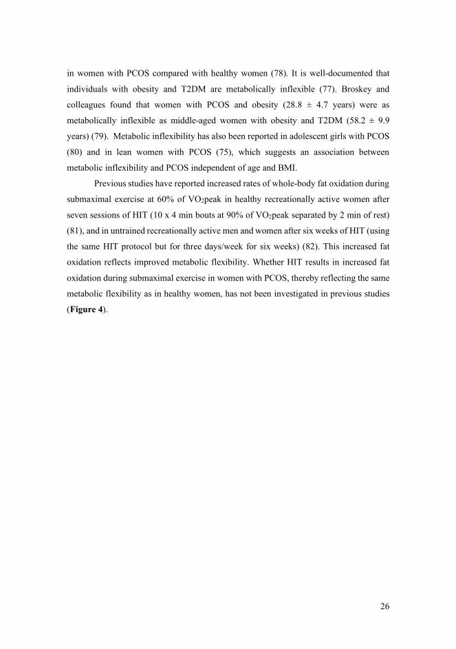
26
in women with PCOS compared with healthy women (78). It is well-documented that
individuals with obesity and T2DM are metabolically inflexible (77). Broskey and
colleagues found that women with PCOS and obesity (28.8 ± 4.7 years) were as
metabolically inflexible as middle-aged women with obesity and T2DM (58.2 ± 9.9
years) (79). Metabolic inflexibility has also been reported in adolescent girls with PCOS
(80) and in lean women with PCOS (75), which suggests an association between
metabolic inflexibility and PCOS independent of age and BMI.
Previous studies have reported increased rates of whole-body fat oxidation during
submaximal exercise at 60% of VO2peak in healthy recreationally active women after
seven sessions of HIT (10 x 4 min bouts at 90% of VO2peak separated by 2 min of rest)
(81), and in untrained recreationally active men and women after six weeks of HIT (using
the same HIT protocol but for three days/week for six weeks) (82). This increased fat
oxidation reflects improved metabolic flexibility. Whether HIT results in increased fat
oxidation during submaximal exercise in women with PCOS, thereby reflecting the same
metabolic flexibility as in healthy women, has not been investigated in previous studies
(Figure 4).
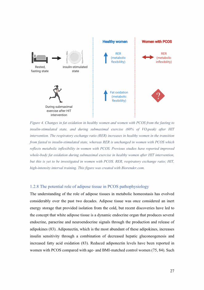
27
Figure 4. Changes in fat oxidation in healthy women and women with PCOS from the fasting to
insulin-stimulated state, and during submaximal exercise (60% of VO2peak) after HIT
intervention. The respiratory exchange ratio (RER) increases in healthy women in the transition
from fasted to insulin-stimulated state, whereas RER is unchanged in women with PCOS which
reflects metabolic inflexibility in women with PCOS. Previous studies have reported improved
whole-body fat oxidation during submaximal exercise in healthy women after HIT intervention,
but this is yet to be investigated in women with PCOS. RER, respiratory exchange ratio; HIT,
high-intensity interval training. This figure was created with Biorender.com.
1.2.8 The potential role of adipose tissue in PCOS pathophysiology
The understanding of the role of adipose tissues in metabolic homeostasis has evolved
considerably over the past two decades. Adipose tissue was once considered an inert
energy storage that provided isolation from the cold, but recent discoveries have led to
the concept that white adipose tissue is a dynamic endocrine organ that produces several
endocrine, paracrine and neuroendocrine signals through the production and release of
adipokines (83). Adiponectin, which is the most abundant of these adipokines, increases
insulin sensitivity through a combination of decreased hepatic gluconeogenesis and
increased fatty acid oxidation (83). Reduced adiponectin levels have been reported in
women with PCOS compared with age- and BMI-matched control women (75, 84). Such
Rested, fasting state
insulin-stimulated state
Healthy women Women with PCOS
RER (metabolic flexibililty)
RER (metabolic inflexibility)
During submaximal exercise after HIT
intervention
Fat oxidation (metabolic flexibililty)
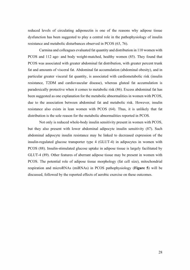
28
reduced levels of circulating adiponectin is one of the reasons why adipose tissue
dysfunction has been suggested to play a central role in the pathophysiology of insulin
resistance and metabolic disturbances observed in PCOS (63, 76).
Carmina and colleagues evaluated fat quantity and distribution in 110 women with
PCOS and 112 age- and body weight-matched, healthy women (85). They found that
PCOS was associated with greater abdominal fat distribution, with greater percent trunk
fat and amounts of visceral fat. Abdominal fat accumulation (abdominal obesity), and in
particular greater visceral fat quantity, is associated with cardiometabolic risk (insulin
resistance, T2DM and cardiovascular disease), whereas gluteal fat accumulation is
paradoxically protective when it comes to metabolic risk (86). Excess abdominal fat has
been suggested as one explanation for the metabolic abnormalities in women with PCOS,
due to the association between abdominal fat and metabolic risk. However, insulin
resistance also exists in lean women with PCOS (64). Thus, it is unlikely that fat
distribution is the sole reason for the metabolic abnormalities reported in PCOS.
Not only is reduced whole-body insulin sensitivity present in women with PCOS,
but they also present with lower abdominal adipocyte insulin sensitivity (87). Such
abdominal adipocyte insulin resistance may be linked to decreased expression of the
insulin-regulated glucose transporter type 4 (GLUT-4) in adipocytes in women with
PCOS (88). Insulin-stimulated glucose uptake in adipose tissue is largely facilitated by
GLUT-4 (89). Other features of aberrant adipose tissue may be present in women with
PCOS. The potential role of adipose tissue morphology (fat cell size), mitochondrial
respiration and microRNAs (miRNAs) in PCOS pathophysiology (Figure 5) will be
discussed, followed by the reported effects of aerobic exercise on these outcomes.
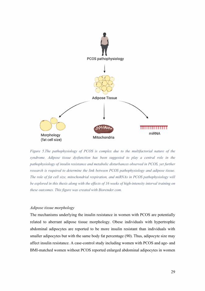
29
Figure 5.The pathophysiology of PCOS is complex due to the multifactorial nature of the
syndrome. Adipose tissue dysfunction has been suggested to play a central role in the
pathophysiology of insulin resistance and metabolic disturbances observed in PCOS, yet further
research is required to determine the link between PCOS pathophysiology and adipose tissue.
The role of fat cell size, mitochondrial respiration, and miRNAs in PCOS pathophysiology will
be explored in this thesis along with the effects of 16 weeks of high-intensity interval training on
these outcomes. This figure was created with Biorender.com.
Adipose tissue morphology
The mechanisms underlying the insulin resistance in women with PCOS are potentially
related to aberrant adipose tissue morphology. Obese individuals with hypertrophic
abdominal adipocytes are reported to be more insulin resistant than individuals with
smaller adipocytes but with the same body fat percentage (90). Thus, adipocyte size may
affect insulin resistance. A case-control study including women with PCOS and age- and
BMI-matched women without PCOS reported enlarged abdominal adipocytes in women
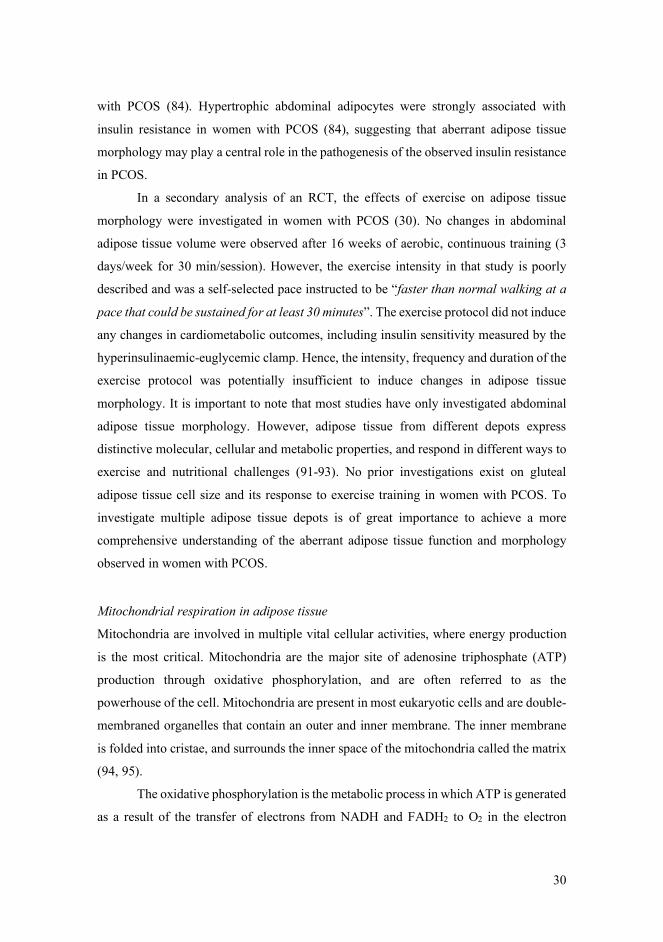
30
with PCOS (84). Hypertrophic abdominal adipocytes were strongly associated with
insulin resistance in women with PCOS (84), suggesting that aberrant adipose tissue
morphology may play a central role in the pathogenesis of the observed insulin resistance
in PCOS.
In a secondary analysis of an RCT, the effects of exercise on adipose tissue
morphology were investigated in women with PCOS (30). No changes in abdominal
adipose tissue volume were observed after 16 weeks of aerobic, continuous training (3
days/week for 30 min/session). However, the exercise intensity in that study is poorly
described and was a self-selected pace instructed to be “faster than normal walking at a
pace that could be sustained for at least 30 minutes”. The exercise protocol did not induce
any changes in cardiometabolic outcomes, including insulin sensitivity measured by the
hyperinsulinaemic-euglycemic clamp. Hence, the intensity, frequency and duration of the
exercise protocol was potentially insufficient to induce changes in adipose tissue
morphology. It is important to note that most studies have only investigated abdominal
adipose tissue morphology. However, adipose tissue from different depots express
distinctive molecular, cellular and metabolic properties, and respond in different ways to
exercise and nutritional challenges (91-93). No prior investigations exist on gluteal
adipose tissue cell size and its response to exercise training in women with PCOS. To
investigate multiple adipose tissue depots is of great importance to achieve a more
comprehensive understanding of the aberrant adipose tissue function and morphology
observed in women with PCOS.
Mitochondrial respiration in adipose tissue
Mitochondria are involved in multiple vital cellular activities, where energy production
is the most critical. Mitochondria are the major site of adenosine triphosphate (ATP)
production through oxidative phosphorylation, and are often referred to as the
powerhouse of the cell. Mitochondria are present in most eukaryotic cells and are double-
membraned organelles that contain an outer and inner membrane. The inner membrane
is folded into cristae, and surrounds the inner space of the mitochondria called the matrix
(94, 95).
The oxidative phosphorylation is the metabolic process in which ATP is generated
as a result of the transfer of electrons from NADH and FADH2 to O2 in the electron

31
transfer chain (Figure 6) (96). NADH and FADH2 are generated by the oxidation of
macronutrients in food through the glycolysis, fatty acid oxidation, and the citric acid
cycle. NADH and FADH2 are then transported to the inner membrane of the mitochondria
where the oxidative phosphorylation takes place. The electron transfer chain is series of
enzymes (Complex I to IV) that are embedded into the inner mitochondrial membrane.
Briefly, NADH donates electrons to complex I which powers the active transport of
protons from the matrix to the intermembrane space. The pumping of protons across the
membrane, driven by energy of electron transfer, produces an electrochemical gradient
referred to as the proton-motive force. From complex I the electrons are transported via
ubiquinone (Q) to complex III. FADH2 donates electrons to complex II that are similarly
transported via Q to complex III. Complex III transfers the electrons from Q to
cytochrome c with the transport of protons from the matrix to the intermembrane space,
and cytochrome c then transfers the electrons to complex IV. Complex IV carries the
electrons to O2 and reduces it to H2O while using protons. Furthermore, complex IV also
pumps one proton from the matrix to the intermembrane space for every electron that
passes, which adds to the proton-motive force produced by complexes I and III. The
protons then flow down the electrochemical gradient from the intermembrane space to
the matrix via. ATP synthase which creates kinetic energy to phosphorylate adenosine
diphosphate (ADP) to ATP (95).
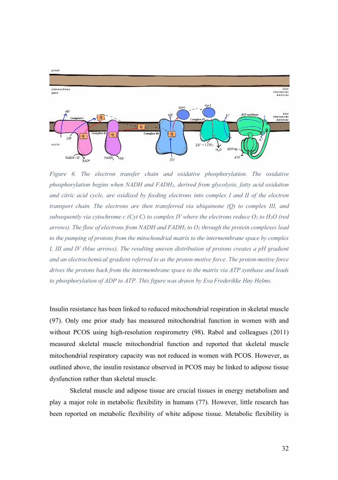
32
Figure 6. The electron transfer chain and oxidative phosphorylation. The oxidative
phosphorylation begins when NADH and FADH2, derived from glycolysis, fatty acid oxidation
and citric acid cycle, are oxidized by feeding electrons into complex I and II of the electron
transport chain. The electrons are then transferred via ubiquinone (Q) to complex III, and
subsequently via cytochrome c (Cyt C) to complex IV where the electrons reduce O2 to H2O (red
arrows). The flow of electrons from NADH and FADH2 to O2 through the protein complexes lead
to the pumping of protons from the mitochondrial matrix to the intermembrane space by complex
I, III and IV (blue arrows). The resulting uneven distribution of protons creates a pH gradient
and an electrochemical gradient referred to as the proton-motive force. The proton-motive force
drives the protons back from the intermembrane space to the matrix via ATP synthase and leads
to phosphorylation of ADP to ATP. This figure was drawn by Eva Frederikke Høy Helms.
Insulin resistance has been linked to reduced mitochondrial respiration in skeletal muscle
(97). Only one prior study has measured mitochondrial function in women with and
without PCOS using high-resolution respirometry (98). Rabol and colleagues (2011)
measured skeletal muscle mitochondrial function and reported that skeletal muscle
mitochondrial respiratory capacity was not reduced in women with PCOS. However, as
outlined above, the insulin resistance observed in PCOS may be linked to adipose tissue
dysfunction rather than skeletal muscle.
Skeletal muscle and adipose tissue are crucial tissues in energy metabolism and
play a major role in metabolic flexibility in humans (77). However, little research has
been reported on metabolic flexibility of white adipose tissue. Metabolic flexibility is
33
driven by cellular processes that may be linked to mitochondrial function (77). Research
on mitochondrial respiration in adipose tissue are sparse, and the first protocol to quantify
mitochondrial respiration in adipose tissue using high-resolution respirometry was
developed only a decade ago in 2010 by Kraunsoe and colleagues (99).
In addition to their central role in energy metabolism, mitochondria in adipose
tissue play an important role in adipocyte differentiation, lipogenesis and adipokine
secretion (96). Furthermore, obesity and insulin resistance has been linked to
mitochondrial dysfunction in adipose tissue (96).
Conflicting findings have been reported on exercise-induced changes in adipose
tissue mitochondrial respiration. Larsen and colleagues investigated the effects of 6
weeks of LV-HIT on mitochondrial respiration in subcutaneous abdominal adipose tissue
and skeletal muscle in overweight but otherwise healthy men and women (100). The
participants exercised 15 min three times weekly, and each session included five 1 min
maximal effort work-bouts. Skeletal muscle mitochondrial respiratory capacity increased,
but with no change in mitochondrial respiration in abdominal adipose tissue. In contrast,
Dohlmann and colleagues (2018) reported decreased mitochondrial respiration in
subcutaneous abdominal adipose tissue in healthy men and women after 6 weeks of LV-
HIT (18 HIT sessions with 7 x 1 min exercise bouts at ~100% of VO2peak) (101).
Mendham and colleagues (2020) reported increased mitochondrial respiration in
subcutaneous abdominal adipose tissue in obese, black South-African women after 12
weeks of combined aerobic and resistance exercise training (four days/week of 40-60 min
with aerobic exercise at 75-80% HRpeak and resistance training that included upper- and
lower-body exercises at 60-70% HRpeak) (102). There are no prior publications on
mitochondrial respiration and the effects of HIT in this metabolically active endocrine
organ in women with PCOS.
MicroRNAs
Micro-RNAs (miRNAs) are small non-coding RNAs that act as post-transcriptional
regulators through two divergent mechanisms depending on the complementarity
between the miRNA and its target mRNA (Figure 7). Perfect base-pairing between the
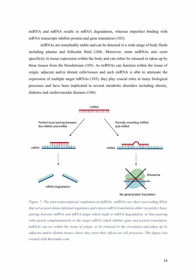
34
miRNA and mRNA results in mRNA degradation, whereas imperfect binding with
mRNA transcripts inhibits protein and gene translation (103).
miRNAs are remarkably stable and can be detected in a wide range of body fluids
including plasma and follicular fluid (104). Moreover, some miRNAs also exert
specificity in tissue expression within the body and can either be released or taken up by
these tissues from the bloodstream (105). As miRNAs can function within the tissue of
origin, adjacent and/or distant cells/tissues and each miRNA is able to attenuate the
expression of multiple target mRNAs (103), they play crucial roles in many biological
processes and have been implicated in several metabolic disorders including obesity,
diabetes and cardiovascular diseases (106).
Figure 7. The post-transcriptional regulation of miRNAs. miRNAs are short non-coding RNAs
that act as post-transcriptional regulators and repress mRNA translation either via perfect base-
pairing between miRNA and mRNA target which leads to mRNA degradation, or base-pairing
with partial complementarity to the target mRNA which inhibits gene and protein translation.
miRNAs can act within the tissue of origin, or be released to the circulation and taken up by
adjacent and/or distant tissues where they exert their effects on cell processes. This figure was
created with Biorender.com.
35
Several studies have reported altered expression of miRNAs in distinct tissues in
women with PCOS including in the circulation (107-116), adipose tissue (114, 117-119),
follicular fluid and granulosa cells (114, 120). The abnormal expression of miRNAs
observed in women with PCOS compared to women without PCOS suggest a potential
role for miRNAs in the pathophysiology of PCOS. Specifically, abnormally expressed
miRNAs with putative roles in regulating glucose metabolism in adipocytes have been
reported in subcutaneous abdominal adipose tissue in women with PCOS and in insulin
resistant women regardless of PCOS status (117, 119, 121). As such, it is likely that
miRNAs are implicated in the adipose tissue abnormalities and adipocyte insulin
resistance in women with PCOS.
An overlooked and understudied area of adipose tissue miRNA research is the
anatomical location of adipose tissue sampling. As outlined previously, adipose tissue
from different depots exhibit different molecular, cellular and metabolic properties,
therefore, research on miRNAs in adipose tissue should include tissue from more depots
than subcutaneous abdominal adipose tissue. There are no publications on miRNA
expression in gluteal adipose tissue in women with PCOS or any that have compared the
miRNA expression between abdominal and gluteal adipose tissue in this population.
Exercise training can modulate the expression of circulating miRNAs in healthy
individuals and in persons with overweight and obesity (122-125). However, no prior
studies have investigated the effect of exercise training on the expression of circulating
or adipose tissue miRNAs in women with PCOS. This is of interest, as exercise training
could potentially regulate and normalize the miRNAs that are abnormally expressed in
women with PCOS. The effect of aerobic exercise training on abdominal and gluteal
adipose tissue miRNA expression has also only been sparsely investigated in other
populations (93). Tsiloulis and colleagues reported no effect of 6 weeks of aerobic
exercise training (four supervised session/week; three moderate intensity sessions and
one HV-HIT session) on the expression of 526 miRNAs in abdominal or gluteal adipose
tissue in overweight males. However, in that study they had a very low sample size (only
five complete data sets were included) which likely reduced the statistical power to detect
small differences and increased the risk of type 2 errors.
36
1.2.9 Treatment of PCOS
Despite the high prevalence of PCOS in reproductive-aged women, the optimal therapies
for PCOS remain unclear. The treatment of PCOS includes addressing reproductive,
metabolic, and psychological features and needs to be individualized and tailored to the
clinical presentation of each individual (52). Insulin resistance is a major contributor to
the PCOS etiology, driving hyperandrogenism as well as clinical features, and is present
in the majority of women with PCOS (64). Therapeutic strategies targeting insulin
resistance improve clinical features (126). The first line treatment for women with PCOS
is lifestyle modifications including a healthy diet and regular physical activity (52).
However, the studies used to develop guidelines for women with PCOS were limited to
a small number of RCTs, resulting in a general consensus recommendation of exercise
and healthy eating principles rather than a specific exercise prescription and/or dietary
strategy for the management of PCOS (52, 127). When lifestyle modifications do not
achieve the desired goals, pharmacological treatments for women with PCOS exist, and
the most widely used pharmaceuticals are oral contraceptives and metformin (52).
Lifestyle modifications
In overweight women with PCOS, lifestyle intervention including diet, exercise and
behavioral strategies are recommended to reduce weight, central obesity and insulin
resistance (52). Currently, recommendations and approaches for lifestyle modifications
are ambiguous. With regard to diet interventions, current recommendations are to
implement general healthy eating habits across the life span. For those with excess body
weight, a diet intervention with an energy deficit of 30% of total daily energy intake or
500-750 kcal/day (taking individual energy requirements, physical activity levels and
body weight into considerations) is recommended to achieve weight loss (52).
The current recommendations for exercise state that women with PCOS should
be physically active for at least 30 min/day which should include a minimum of 150
min/week of moderate intensity or 75 min/week of vigorous intensity (or an equivalent
combination of both) and include activities for muscle strengthening twice per week in
order to prevent weight gain and maintain health (52). To achieve weight loss in those
women with overweight and obesity, prevent weight regain and induce greater health
37
benefits, a minimum of 250 min/week of moderate intensity or 150 min/week of vigorous
intensity is recommended in combination with muscle strengthening twice weekly (52).
It is also recommended that behavioral strategies (goal-setting, problem solving,
confidence training, slower eating) be reinforced to prevent relapse during lifestyle
interventions to improve weight management, healthy lifestyle and wellbeing in women
with PCOS (52). Strategies to enhance engagement and adherence to a healthy lifestyle
and thereby improve long-term health outcomes for women with PCOS need
consideration as these are currently limited.
1.2.10 PCOS, exercise and reproductive function
PCOS is the primary cause of anovulatory infertility and menstrual disorders in
reproductive-aged women (3). A higher prevalence of infertility in women with PCOS
(72%) compared to women without PCOS (16%) was reported in a large Australian
community-based cohort study (128). Infertility was defined as trying unsuccessfully to
conceive for ≥ 12 months. Exercise training has been shown to positively affect metabolic
and cardiovascular outcomes in women with PCOS (36, 43, 44), and may provide a non-
pharmacological strategy to enhance fertility in these women. Indeed, some studies report
improved pregnancy and ovulation rates following an exercise intervention (29, 32, 34,
37, 129). Palomba and colleagues (129) compared the efficacy of a structured exercise
training program versus a hypoenergetic, hyperproteic diet intervention on reproductive
function in anovulatory, infertile women with PCOS and obesity. The exercise group
(n=20) completed three weekly sessions of 30 minutes ergometer cycling at 60-70% of
VO2max for 24 weeks. The diet group (n=20) had a daily energy deficit of 800 kcal and
consumed a high protein diet (35% protein, 45% carbohydrate and 20% fat) for 24 weeks.
After 24 weeks, menstrual frequency and ovulation rate were higher in the exercise group
compared to the diet group. Furthermore, there was a tendency towards more pregnancies
in the exercise group (35%) versus in the diet group (10%; P= 0.058), although, the study
was underpowered to detect a difference in pregnancy rate (their primary outcome) (129).
That study was a non-randomized trial in which the participants could self-select which
group they wanted to attend (self-selection bias), and without a control group, therefore
the findings should be interpreted with caution (129).
38
Nybacka and colleagues (37) reported that diet alone (≥600 kcal/day energy
deficit), exercise alone (45-60 min/session at moderate to vigorous exertion level, 2-3
days/week for 16 weeks) or the combination of diet and exercise intervention, were
equally effective in improving menstrual pattern (shifting from oligo-/amenorrhea to a
regular menstrual cycle) in women with PCOS. However, women in the exercise only
group also reduced their daily calorie intake by ~270 kcal, which resulted in a 3% weight
loss, thereby making it difficult to conclude that exercise alone can improve the menstrual
pattern in women with PCOS. Furthermore, this study was a randomized, non-controlled
trial as noted previously.
Improved menstrual cycle length (48 days to 27 days) following eight weeks of
combined aerobic continuous exercise training and resistance training was reported in
normal weight women with PCOS (BMI<25 kg/m2) (34). However, findings from this
RCT are limited by several factors in the research design; 1) the exercise intervention
only lasted eight weeks (arguably not long enough to determine the exercise-induced
effects on menstrual cycle length), 2) the exercise protocol was not described in detail
e.g. the aerobic exercise dose depended on the exercise tolerance of the patient and the
prescribed intensity was vague (determined by ratings of perceived exertion of 10-15 on
the Borg 6-20 Scale), 3) participants were limited to normal-weight women with PCOS,
so the findings may not apply to women with PCOS and overweight/obesity.
Well-designed, long-term RCTs investigating the effects of exercise alone on
reproductive function in women with PCOS are lacking. Furthermore, data on exercise
type, intensity and duration of exercise for improved reproductive function and fertility
in women with PCOS are lacking, making recommendations for exercise in this
population unclear.
39
2.0 Gaps in the literature There are few well-designed, long-term RCTs on the effect of exercise training on
reproductive and metabolic outcomes in women with PCOS. The published studies to
date are limited by inconsistent reproductive-outcome definitions, small sample sizes,
short intervention periods, interventions combining diet and exercise, the lack of an actual
control group, and the inclusion of participants taking metformin, oral contraceptives and
other medications. Combined, these factors make it difficult to interpret the isolated effect
of exercise on reproductive and metabolic health in PCOS. Furthermore, no study to date
has compared the effects of different HIT protocols on reproductive and cardiometabolic
outcomes in women with PCOS.
We have little knowledge as to whether exercise protocols implemented in clinical trials
under controlled laboratory conditions, typically comprising 8-12 weeks of supervised
exercise training, are realistic and could be successfully performed in a ‘real-world’
setting (efficacy versus effectiveness and ecological validity). Long-term adherence to
exercise training is necessary to obtain its benefits and we have no knowledge of which
exercise training protocol induces the best long-term adherence when the women are no
longer supervised.
Despite decades of research, the etiology of PCOS remains largely unclear. Currently,
PCOS is believed to be caused by a combination of genetic and environmental factors
leading to hyperandrogenism and hyperinsulinemia. The mechanisms underlying the
insulin resistance are unclear, but are potentially linked to aberrant adipose tissue function
and morphology. There is little prior knowledge about gluteal adipose tissue cell size,
mitochondrial respiration or miRNA profiling in women with PCOS. Furthermore, the
effect of HIT on abdominal and gluteal adipose tissue morphology, mitochondrial
respiration and miRNA expression in women with PCOS is still lacking in the literature.
40
3.0 Aim of the thesis The overall aim of the experiments conducted for this thesis was to explore the effects of
two different HIT protocols on reproductive, cardiometabolic and quality of life outcomes
in women with PCOS and explore whether aberrant adipose tissue plays a role in the
pathophysiology of PCOS.
3.1 Aims of the studies:
3.1.1 Paper I:
The aim of this paper was to provide details of the study protocol and methods for the
IMproving Reproductive function in women with Polycystic OVary syndrome with high-
intensity Interval Training (IMPROV-IT) trial.
3.1.2 Paper II:
The primary aim of the first study was to investigate whether 16 weeks of semi-supervised
HIT, followed by a subsequent 36 weeks of home-based HIT could increase menstrual
frequency in women with PCOS during a 1-year period. Secondary aims were to examine
the effects of HIT on cardiorespiratory fitness, rates of pregnancies, whole-body insulin
sensitivity, and quality of life.
3.1.3 Paper III:
The primary aim of the second study described in Paper III was to compare the expression
of selected miRNAs in the circulation, gluteal and abdominal adipose tissue taken from
women with PCOS to an age- and BMI-matched control group of women without PCOS.
Secondary aims were to investigate the effects of two chronic HIT protocols on the
“expression signature” of these miRNAs in the circulation and in gluteal and abdominal
adipose tissue in women with PCOS.
3.1.4 Paper IV:
The primary aims of the third study were to compare rates of whole-body fat oxidation
during submaximal exercise, fat cell size and rates of mitochondrial respiration in adipose

41
tissue taken from women with and without PCOS. In addition, the response to 16 weeks
of HIT were assessed.
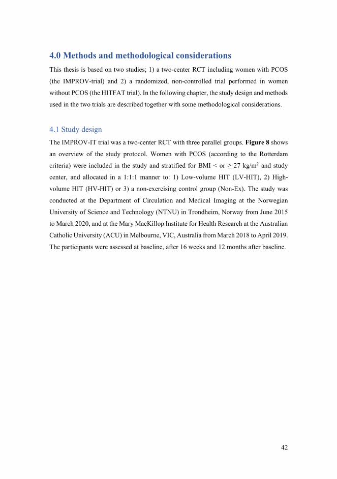
42
4.0 Methods and methodological considerations This thesis is based on two studies; 1) a two-center RCT including women with PCOS
(the IMPROV-trial) and 2) a randomized, non-controlled trial performed in women
without PCOS (the HITFAT trial). In the following chapter, the study design and methods
used in the two trials are described together with some methodological considerations.
4.1 Study design The IMPROV-IT trial was a two-center RCT with three parallel groups. Figure 8 shows
an overview of the study protocol. Women with PCOS (according to the Rotterdam
criteria) were included in the study and stratified for BMI < or ≥ 27 kg/m2 and study
center, and allocated in a 1:1:1 manner to: 1) Low-volume HIT (LV-HIT), 2) High-
volume HIT (HV-HIT) or 3) a non-exercising control group (Non-Ex). The study was
conducted at the Department of Circulation and Medical Imaging at the Norwegian
University of Science and Technology (NTNU) in Trondheim, Norway from June 2015
to March 2020, and at the Mary MacKillop Institute for Health Research at the Australian
Catholic University (ACU) in Melbourne, VIC, Australia from March 2018 to April 2019.
The participants were assessed at baseline, after 16 weeks and 12 months after baseline.
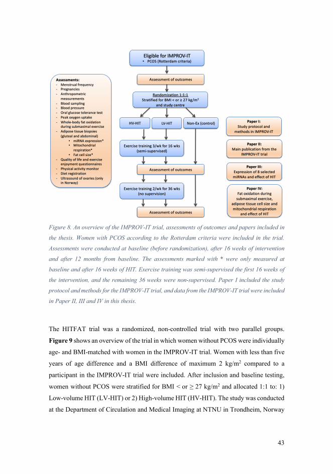
43
Figure 8. An overview of the IMPROV-IT trial, assessments of outcomes and papers included in
the thesis. Women with PCOS according to the Rotterdam criteria were included in the trial.
Assessments were conducted at baseline (before randomization), after 16 weeks of intervention
and after 12 months from baseline. The assessments marked with * were only measured at
baseline and after 16 weeks of HIT. Exercise training was semi-supervised the first 16 weeks of
the intervention, and the remaining 36 weeks were non-supervised. Paper I included the study
protocol and methods for the IMPROV-IT trial, and data from the IMPROV-IT trial were included
in Paper II, III and IV in this thesis.
The HITFAT trial was a randomized, non-controlled trial with two parallel groups.
Figure 9 shows an overview of the trial in which women without PCOS were individually
age- and BMI-matched with women in the IMPROV-IT trial. Women with less than five
years of age difference and a BMI difference of maximum 2 kg/m2 compared to a
participant in the IMPROV-IT trial were included. After inclusion and baseline testing,
women without PCOS were stratified for BMI < or ≥ 27 kg/m2 and allocated 1:1 to: 1)
Low-volume HIT (LV-HIT) or 2) High-volume HIT (HV-HIT). The study was conducted
at the Department of Circulation and Medical Imaging at NTNU in Trondheim, Norway
Eligible for IMPROV-IT• PCOS (Rotterdam criteria)
Assessments:- Menstrual frequency- Pregnancies- Anthropometric
measurements- Blood sampling- Blood pressure- Oral glucose tolerance test- Peak oxygen uptake- Whole-body fat oxidation
during submaximal exercise- Adipose tissue biopsies
(gluteal and abdominal)• miRNA expression*• Mitochondrial
respiration*• Fat cell size*
- Quality of life and exercise enjoyment questionnaires
- Physical activity monitor- Diet registration - Ultrasound of ovaries (only
in Norway)
Randomization 1:1:1Stratified for BMI < or ≥ 27 kg/m2
and study centre
HV-HIT Non-Ex (control)
Assessment of outcomes
Assessment of outcomes
LV-HIT
Exercise training 3/wk for 16 wks(semi-supervised)
Exercise training 2/wk for 36 wks(no supervision)
Assessment of outcomes
Paper I: Study protocol and
methods in IMPROV-IT
Paper II: Main publication from the
IMPROV-IT trial
Paper III: Expression of 8 selected miRNAs and effect of HIT
Paper IV: Fat oxidation during
submaximal exercise, adipose tissue cell size and mitochondrial respiration
and effect of HIT
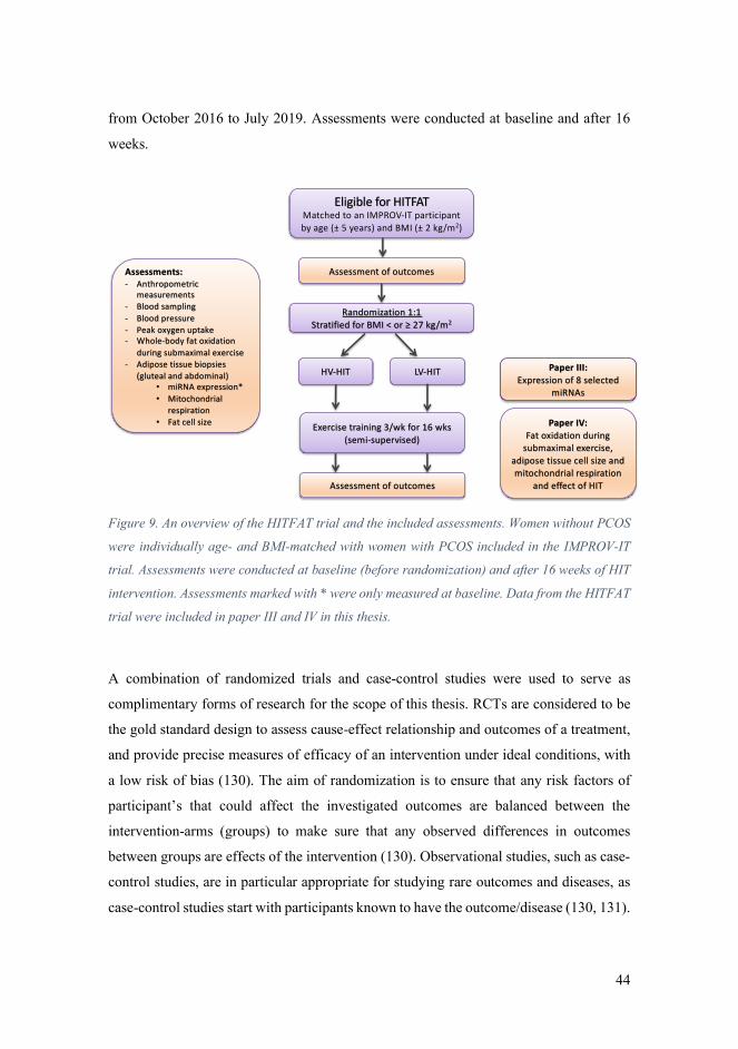
44
from October 2016 to July 2019. Assessments were conducted at baseline and after 16
weeks.
Figure 9. An overview of the HITFAT trial and the included assessments. Women without PCOS
were individually age- and BMI-matched with women with PCOS included in the IMPROV-IT
trial. Assessments were conducted at baseline (before randomization) and after 16 weeks of HIT
intervention. Assessments marked with * were only measured at baseline. Data from the HITFAT
trial were included in paper III and IV in this thesis.
A combination of randomized trials and case-control studies were used to serve as
complimentary forms of research for the scope of this thesis. RCTs are considered to be
the gold standard design to assess cause-effect relationship and outcomes of a treatment,
and provide precise measures of efficacy of an intervention under ideal conditions, with
a low risk of bias (130). The aim of randomization is to ensure that any risk factors of
participant’s that could affect the investigated outcomes are balanced between the
intervention-arms (groups) to make sure that any observed differences in outcomes
between groups are effects of the intervention (130). Observational studies, such as case-
control studies, are in particular appropriate for studying rare outcomes and diseases, as
case-control studies start with participants known to have the outcome/disease (130, 131).
Eligible for HITFATMatched to an IMPROV-IT participant
by age (± 5 years) and BMI (± 2 kg/m2)
Assessments:- Anthropometric
measurements
- Blood sampling
- Blood pressure
- Peak oxygen uptake
- Whole-body fat oxidation
during submaximal exercise
- Adipose tissue biopsies
(gluteal and abdominal)
• miRNA expression*
• Mitochondrial
respiration
• Fat cell size
Assessment of outcomes
Randomization 1:1
Stratified for BMI < or ≥ 27 kg/m2
HV-HIT
Assessment of outcomes
LV-HIT
Exercise training 3/wk for 16 wks
(semi-supervised)
Paper III: Expression of 8 selected
miRNAs
Paper IV: Fat oxidation during
submaximal exercise,
adipose tissue cell size and
mitochondrial respiration
and effect of HIT
45
Another advantage of case-control studies are that they are essential in advancing the
understanding the pathophysiology of a disease such as PCOS. To this end, women
without PCOS were included to perform a case-control comparison between women with
and without PCOS, to identify whether miRNAs, fat oxidation rates during submaximal
exercise, mitochondrial respiration and/or fat cell sizes differed in women with and
without PCOS. The overall aim of these comparisons was to assess whether these
outcomes may play a role in PCOS pathophysiology. The randomized design of the
IMPROV-IT trial and the HITFAT trial allowed us to also investigate the effect of LV-
and HV-HIT on reproductive and metabolic outcomes in women with and without PCOS.
The IMPROV-IT trial was a two-center RCT including women with PCOS in
Trondheim, Norway and Melbourne, Australia. Some of the advantages of running multi-
versus singlecenter studies are faster rates of participant recruitment and the improved
generalizability (external validity) (132). Thereby, the multicenter nature of the
IMPROV-IT study including women with PCOS from different parts of the world
provides unique scope to the investigation of PCOS. There are also disadvantages of
running multicenter rather than singlecenter studies, and one of them is that it can be
difficult to align all methods used for assessments. In the IMPROV-IT trial, body
composition was estimated using two different methods; bioelectrical impedance analysis
(InBody720, Biospace CO, Korea) was used in Norway, and Dual-energy X-ray
absorptiometry (DXA; GE Lunar iDXA Pro, Encore software version 16, General
Electric, Boston, MA, USA) in Australia. For further discussion on the use of these two
methods to estimate body composition, please see the Assessments paragraph.
4.2 Study population
In Norway, participants for the IMPROV-IT and HITFAT trials were recruited through
public announcement at the university and hospital’s homepages, at local shops and
public places, and on social media. In Australia, participants were recruited for the
IMPROV-IT trial via social media.
To be eligible for inclusion, the women had to be between 18 and 45 years old. In
the IMPROV-IT trial, women were diagnosed with PCOS according to the Rotterdam
criteria (49), with at least two of the following three features present: polycystic ovary
46
morphology (≥12 follicles (2-9 mm) per ovary and/or an ovarian volume of ≥10 mL),
hyperandrogenism (either clinical signs such as acne or hirsutism, or biomedical) and/or
oligo/amenorrhea. In the HITFAT trial, the PCOS diagnosis was ruled out in all the
women as they were normally menstruating, with no evidence of hyperandrogenism or
polycystic ovaries. Women in both trials were excluded if they were undertaking regular
endurance training with an intensity that induce heavy breathing ≥ 2 sessions/week, had
any cardiovascular diseases or endocrine disorders, were pregnant, had been
breastfeeding within the last 24 weeks, or if they were using hormonal contraceptives,
insulin sensitizers or drugs known to affect gonadotropin or ovulation (with a washout
period of 3 months prior to inclusion).
Women in all BMI-categories were included to have a representative population
of women and to increase participant enrollment. In both trials, women were randomized
for BMI < or ≥ 27 kg/m2 to ensure more homogenous intervention groups and to avoid
that any observed differences in outcomes between groups were due to baseline
differences in BMI. Similarly, women without PCOS were BMI-matched with women in
the IMPROV-IT trial (±2 kg/m2) to rule out the effect of BMI on potential differences in
the assessed outcomes.
4.3 Interventions In the IMPROV-IT trial, the two HIT protocols consisted of three weekly exercise
sessions during the first 16 weeks, and for the remaining 36 weeks of the intervention
period the participants were instructed to perform at least two weekly HIT sessions
according to the HIT protocol they were allocated to. The exercise sessions were semi-
supervised during the first 16 weeks of the intervention, where participants attended at
least one weekly supervised training session at the study centers, with the opportunity to
perform the remaining two exercise sessions under supervision or unsupervised. No
supervision was provided during the remaining 36 weeks (from 16 weeks to 12 months).
The two HIT protocols in the HITFAT trial consisted of three exercise sessions per week
during the 16 weeks intervention period, and similar to the IMPROV-IT trial, participants
attended supervised exercise at least once weekly at the study center, while the two
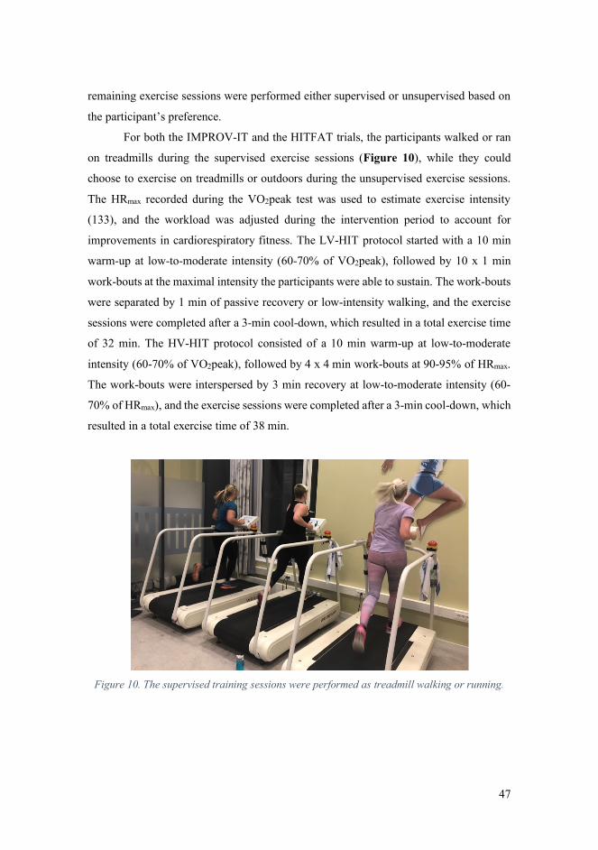
47
remaining exercise sessions were performed either supervised or unsupervised based on
the participant’s preference.
For both the IMPROV-IT and the HITFAT trials, the participants walked or ran
on treadmills during the supervised exercise sessions (Figure 10), while they could
choose to exercise on treadmills or outdoors during the unsupervised exercise sessions.
The HRmax recorded during the VO2peak test was used to estimate exercise intensity
(133), and the workload was adjusted during the intervention period to account for
improvements in cardiorespiratory fitness. The LV-HIT protocol started with a 10 min
warm-up at low-to-moderate intensity (60-70% of VO2peak), followed by 10 x 1 min
work-bouts at the maximal intensity the participants were able to sustain. The work-bouts
were separated by 1 min of passive recovery or low-intensity walking, and the exercise
sessions were completed after a 3-min cool-down, which resulted in a total exercise time
of 32 min. The HV-HIT protocol consisted of a 10 min warm-up at low-to-moderate
intensity (60-70% of VO2peak), followed by 4 x 4 min work-bouts at 90-95% of HRmax.
The work-bouts were interspersed by 3 min recovery at low-to-moderate intensity (60-
70% of HRmax), and the exercise sessions were completed after a 3-min cool-down, which
resulted in a total exercise time of 38 min.
Figure 10. The supervised training sessions were performed as treadmill walking or running.
48
All participants wore heart rate monitors (Polar M400) during all training sessions, and
registered these using an online exercise diary (www.polar.flow.com). The researchers
had access to records of the exercise sessions through this online tool and were able to
supervise adherence to the protocols.
The semi-supervised nature of the IMPROV-IT and HITFAT trials meant that
some participants performed all exercise sessions supervised, while others only attended
supervised exercise sessions once weekly. Less than ten women in the IMPROV-IT study
and less than five women in the HITFAT trial chose to perform all exercise sessions at
the study centers. A semi-supervised exercise program was chosen with the intention to
increase practicality for the participant and make it easier to implement the exercise in
their everyday life. Another reason for choosing the semi-supervised approach was to
investigate the real-world efficacy of the HIT protocols, which have previously been
studied mainly under strict supervision in a laboratory setting. Since our main research
question in IMPROV-IT required a long follow-up period, we chose programs that we
believed would promote long-term sustainability of exercise behaviors. Walking or
running in the HIT protocols were chosen because these types of exercise require
minimum equipment and are easy to perform unsupervised. Additionally, it is easier to
reach the prescribed exercise intensities by walking/running on an incline compared to
cycling, as walking/running requires activation of a greater muscle mass. The only
requirement during the exercise sessions was to follow the prescribed exercise intensities,
which the participants were able to by using the heart rate monitors they received.
The women with PCOS allocated to the control group (Non-Ex) in the IMPROV-
IT trial were asked to continue their daily physical activities and informed about the
current recommendations for physical activity for achieving health benefits; be physically
active for at least 150 min/week of moderate intensity or 75 min/week of vigorous
intensity (or an equivalent combination of both). By informing the women in the Non-Ex
group about the current recommendations for physical activity, some of the women may
have been inspired to engage in a healthier lifestyle during the study period, and thereby
reduce the chance of finding possible effects of the HIT interventions on the measured
reproductive and metabolic outcomes. The information was given for ethical reasons. We
assessed physical activity using physical activity monitors (Sensewear Armband, APC
49
Cardiovascular, UK) at all timepoints to control for physical activity. All participants
were encouraged to maintain their habitual diet throughout the study period.
4.4 Assessments Outcomes for the IMPROV-IT trial were assessed at baseline and after the 16-week
intervention at both study centers. The same assessments were executed in Norway after
12 months, while only data on menstrual frequency and questionnaires were collected in
Australia at 12 months. Outcomes for the HITFAT trial were assessed at baseline and
after the 16-week intervention. The majority of assessments were identical in the
IMPROV-IT and HITFAT trials apart from the reproductive outcomes, including
menstrual frequency diaries, ovarian morphology and pregnancies, and the quality of life
and enjoyment questionnaires that were only collected in the IMPROV-IT trial.
4.4.1 Reproductive outcomes
As noted above, the reproductive outcomes were only assessed in the IMPROV-IT trial
and these are reported in Paper II, with a description of the methodology used also
included in Paper I.
Menstrual frequency
The primary outcome in the IMPROV-IT trial was menstrual frequency, used as a proxy
of ovulation. Participants completed a menstruation diary (either on paper or using an
electronic application) to register the first day of their menstrual cycle, and the length
(number of days) during the 12-month intervention period, and sent information to the
study personnel after each menstrual cycle. The study personnel sent out reminders to fill
out the diary on a regular basis to ensure accuracy and increase reliability with the self-
reported menstrual frequency data. In addition to the self-reported menstrual frequency
data, the participants also completed questionnaires about their menstrual pattern at each
assessment timepoint (at baseline, and after 16 weeks and 12 months). We compared
groups using the number of menstrual cycles per year, and the number of observed
menstrual cycles divided by the number of expected menstrual cycle per year (with n=13
50
for the number of expected menstrual cycles per year, given a regular cycle length of 28
days).
We used menstrual frequency as a proxy of ovulation, which is a limitation as it
is possible to ovulate without a later bleeding and to have a regular cycle despite ovulatory
dysfunction (52). Due to practical reasons, we were not able to assess ovulation as that
requires daily temperature measurements, frequent urine samples to assess LH or blood
samples to assess progesterone concentrations as markers of ovulation. Menstrual
bleedings have been used by others to assess possible improvements in reproduction in
women with PCOS after an exercise intervention (29).
Ovarian morphology
Ovarian morphology was only assessed at the Norwegian study center, and all imaging
and measurements were executed by an experienced gynecologist. The ultrasound
assessments of the ovaries were performed using a multifrequency transvaginal
transducer, and included measurements of ovarian volume with and without dominant
follicles, and the number of follicles in each ovary.
The diagnostic criteria for polycystic ovaries were ≥12 follicles (2-9 mm) per
ovary and/or an ovarian volume of ≥10 mL as the study commenced in 2015, before the
criteria were revised to the presence of ≥20 follicles (2-9 mm) per ovary and/or an ovarian
volume ≥10 mL when using ultrasound transducers with a frequency bandwidth of ≥8
MHz (52) (see 1.2.2 Diagnostic features of PCOS).
Fertility and pregnancies
Participants completed a questionnaire providing background information regarding
fertility. The questionnaire included questions about number of children (natural or
assisted fertilization), whether the participant was actively seeking pregnancy, if they had
tried to become pregnant (if yes, for how long), and if they had previously experienced
miscarriages or had abortions. We registered the number of pregnancies during the 12-
month study period.
51
4.4.2 Physiological outcomes
Anthropometric measures and blood pressure
After an overnight fast (≥12 hours), body composition was estimated using InBody720
for the HITFAT trial, and either using Inbody720 or DXA in the IMPROV-IT trial. Waist
and hip circumference were measured to the nearest 0.5 cm in duplicate using measuring
tape, and waist-to-hip ratio was calculated by dividing waist circumference by hip
circumference. BMI was calculated as weight in kg divided by height in meters squared;
BMI = kg/m2.
After participants had rested for 15 min, systolic and diastolic blood pressure was
measured in triplicate on the left arm while participants were in a seated position. The
three measurements were recorded with two min break in between each measurement and
the mean of these three measurements was used in the analysis.
We had to use two different methods to estimate body composition in the
IMPROV-IT trial as we were not able to align the assessment at the two study centers;
we only had an InBody720 available in Norway and a DXA in Australia. We
acknowledge that it was not ideal to use two different methods, and it is one of the
difficulties that can occur in a multicenter study. However, both methods have been
reported reliable to estimate body composition in individuals with healthy weight and
obesity (134, 135), although, it has been reported that the Inbody 720 overestimates fat-
free mass, and underestimates body fat percentage and fat mass compared to DXA (134).
The same method was used to estimate body composition of each individual participant
at all timepoints, which should minimize the risk of it affecting our results. While the
same method for assessing body composition would have been optimal, we posit that the
multi-center nature of our study from different parts of the world provides unique scope
to the investigation of PCOS.
Cardiorespiratory fitness
Participants performed an incremental test to exhaustion on a treadmill to assess VO2peak
and HRmax. We used an individualized protocol with the goal of a total test time of 8-12
min for each participant. After 10 min warm-up at low-to-moderate intensity
(individually adjusted), the treadmill speed or inclination was increased by 0.5-1.0 km/h
52
or 1-2% every 1-2 min until volitional exhaustion. VO2peak was assessed via indirect
calorimetry (Oxycon Pro, Jaeger, Germany in Norway and TrueOne 2400, ParvoMedics,
USA in Australia), and the maximum heart rate attained during this test was used to
calculate the intensity for the HIT sessions.
Berglund and colleagues (133) found that a standard cardiorespiratory fitness test
(with a protocol similar to ours) can serve as a good estimate of HRmax compared to an
actual HRmax test (15 min warm-up at 60-70% HRmax followed by 4 x 4 min bouts at 90-
95% HRmax interspersed with 2 min at 60-70% HRmax. After the last 4-min work-bout,
treadmill speed or inclination was increased by 0.5 km/h or 1-2% every 30 seconds until
volitional exhaustion) in non-athlete individuals. Based on their findings, the HRmax
recorded during the cardiorespiratory fitness test sufficed to calculate the HIT session
intensities and thereby we could include one less assessment in the IMPROV-IT and
HITFAT trials.
Rates of whole-body fat oxidation during submaximal exercise
After an overnight fast (≥12 hours), participants performed a submaximal exercise test on
a treadmill. The protocol included 20 min warm-up at low-to-moderate intensity without
any sampling of gas, followed by 20 minutes of walking/running that elicited a ‘steady
state’ condition at approximately 60% of individual VO2peak with sampling of expired
gas. The participants recorded their dietary intake the day prior to baseline testing and
were requested to repeat that diet on the day prior to the subsequent measurement at 16
weeks and 12 months follow-up to reduce any diet-induced effect of rates of substrate
oxidation (136). Rates of whole-body fat oxidation (g/min) were calculated from five min
of steady oxygen uptake during the last 10 min of the test, and using the following
equation; 1.695 x VO2 – 1.701 x VCO2 (where VO2 is oxygen uptake and VCO2 is expired
carbon dioxide) (137).
Rates of fat oxidation were measured at ~60% of VO2peak based on previous
research. Achten and colleagues reported that the exercise intensity that elicits maximal
rates of fat oxidation is ~64% of VO2max in moderately trained men, and that the range
of intensities where fax oxidation rates are within 10% of the maximal rate, were detected
between 55 and 72% of VO2max (138). Apart from exercise intensity, rates of fat
oxidation during exercise can be affected by several factors such as gender, training status
53
and exercise training (137); maximal fat oxidation appears to be elicited at lower exercise
intensities in untrained individuals compared to trained individuals (137), maximal fat
oxidation occurs at higher exercise intensities for females compared to men (139), and
exercise training has the ability to modify fat oxidation rates. Previous studies have
investigated the effects of HIT protocols (10 x 4 min bouts at 90% of VO2peak separated
by 2 min of rest for seven sessions and three days/week for six weeks, respectively) on
rates of whole-body fat oxidation during submaximal exercise at 60% of VO2peak in
healthy recreationally active women (81), and in untrained recreationally active men and
women (82). Both studies showed that HIT improved whole-body fat oxidation during
submaximal exercise at 60% of VO2peak.
Blood biochemistry and insulin sensitivity
Resting blood samples were obtained after an overnight fast (≥12 hours), followed by a
2-hour oral glucose tolerance test (OGTT) in which the participants consumed 75 g of
glucose diluted in 250 mL water. Blood samples were collected every 30 mins during the
OGTT at 0 (fasting prior to OGTT), 30, 60, 90 and 120 min for the determination of
glucose and insulin concentrations. Plasma glucose concentrations were analyzed using
a Roche Moduclar P (Roche, Switzerland) and serum insulin concentration was measured
in duplicate using an enzyme-linked immunosorbent assay (ELISA, IBL-International,
Germany). HOMA-IR was used to estimate insulin sensitivity and calculated as; fasting
serum insulin (µIU/mL) x fasting plasma glucose (mmol/L) divided by 22.5 (140). To
evaluate glycemic control, we calculated the total area under the curve (AUC) using a
baseline of 0 mmol/L, and incremental area under the curve (iAUC) using fasting
concentration as baseline value. AUC and iAUC were calculated for glucose and insulin
concentrations using the trapezoidal method (141).
Fasting blood samples were also analyzed for lipid concentrations including total
cholesterol, HDL, LDL and triglycerides. In the IMPROV-IT trial, fasting blood samples
were also used to analyze hormone concentrations including androstenedione, 17OH-
progesterone (at baseline only), testosterone, albumin, Anti-Müllerian hormone (AMH)
and SHBG. FAI was calculated as 100 x (total testosterone/SHBG). The equipment used
for the blood analyzes are described in Paper II.
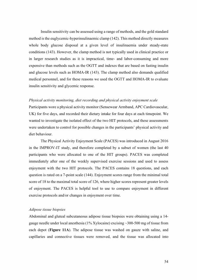
54
Insulin sensitivity can be assessed using a range of methods, and the gold standard
method is the euglycemic-hyperinsulinaemic clamp (142). This method directly measures
whole body glucose disposal at a given level of insulinaemia under steady-state
conditions (143). However, the clamp method is not typically used in clinical practice or
in larger research studies as it is impractical, time- and labor-consuming and more
expensive than methods such as the OGTT and indexes that are based on fasting insulin
and glucose levels such as HOMA-IR (143). The clamp method also demands qualified
medical personnel, and for these reasons we used the OGTT and HOMA-IR to evaluate
insulin sensitivity and glycemic response.
Physical activity monitoring, diet recording and physical activity enjoyment scale
Participants wore a physical activity monitor (Sensewear Armband, APC Cardiovascular,
UK) for five days, and recorded their dietary intake for four days at each timepoint. We
wanted to investigate the isolated effect of the two HIT protocols, and these assessments
were undertaken to control for possible changes in the participants’ physical activity and
diet behaviour.
The Physical Activity Enjoyment Scale (PACES) was introduced in August 2016
in the IMPROV-IT study, and therefore completed by a subset of women (the last 40
participants who were allocated to one of the HIT groups). PACES was completed
immediately after one of the weekly supervised exercise sessions and used to assess
enjoyment with the two HIT protocols. The PACES contains 18 questions, and each
question is rated on a 7-point scale (144). Enjoyment scores range from the minimal total
score of 18 to the maximal total score of 126, where higher scores represent greater levels
of enjoyment. The PACES is helpful tool to use to compare enjoyment in different
exercise protocols and/or changes in enjoyment over time.
Adipose tissue biopsies
Abdominal and gluteal subcutaneous adipose tissue biopsies were obtaining using a 14-
gauge needle under local anesthesia (1% Xylocaine) excising ~300-500 mg of tissue from
each depot (Figure 11A). The adipose tissue was washed on gauze with saline, and
capillaries and connective tissues were removed, and the tissue was allocated into
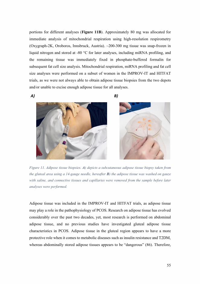
55
portions for different analyses (Figure 11B). Approximately 80 mg was allocated for
immediate analysis of mitochondrial respiration using high-resolution respirometry
(Oxygraph-2K, Oroboros, Innsbruck, Austria). ~200-300 mg tissue was snap-frozen in
liquid nitrogen and stored at -80 °C for later analyses, including miRNA profiling, and
the remaining tissue was immediately fixed in phosphate-buffered formalin for
subsequent fat cell size analysis. Mitochondrial respiration, miRNA profiling and fat cell
size analyses were performed on a subset of women in the IMPROV-IT and HITFAT
trials, as we were not always able to obtain adipose tissue biopsies from the two depots
and/or unable to excise enough adipose tissue for all analyses.
Figure 11. Adipose tissue biopsies. A) depicts a subcutaneous adipose tissue biopsy taken from
the gluteal area using a 14-gauge needle, hereafter B) the adipose tissue was washed on gauze
with saline, and connective tissues and capillaries were removed from the sample before later
analyses were performed.
Adipose tissue was included in the IMPROV-IT and HITFAT trials, as adipose tissue
may play a role in the pathophysiology of PCOS. Research on adipose tissue has evolved
considerably over the past two decades, yet, most research is performed on abdominal
adipose tissue, and no previous studies have investigated gluteal adipose tissue
characteristics in PCOS. Adipose tissue in the gluteal region appears to have a more
protective role when it comes to metabolic diseases such as insulin resistance and T2DM,
whereas abdominally stored adipose tissues appears to be “dangerous” (86). Therefore,
A) B)
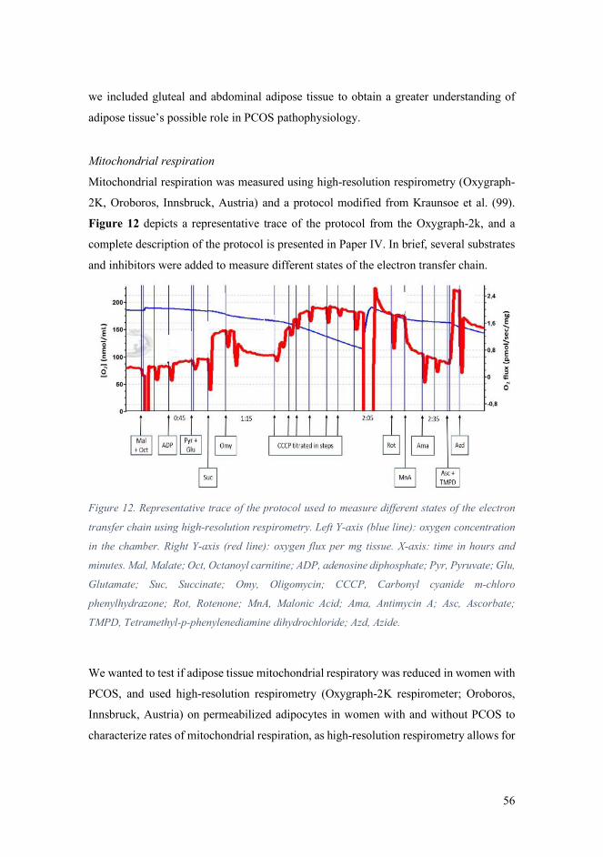
56
we included gluteal and abdominal adipose tissue to obtain a greater understanding of
adipose tissue’s possible role in PCOS pathophysiology.
Mitochondrial respiration
Mitochondrial respiration was measured using high-resolution respirometry (Oxygraph-
2K, Oroboros, Innsbruck, Austria) and a protocol modified from Kraunsoe et al. (99).
Figure 12 depicts a representative trace of the protocol from the Oxygraph-2k, and a
complete description of the protocol is presented in Paper IV. In brief, several substrates
and inhibitors were added to measure different states of the electron transfer chain.
Figure 12. Representative trace of the protocol used to measure different states of the electron
transfer chain using high-resolution respirometry. Left Y-axis (blue line): oxygen concentration
in the chamber. Right Y-axis (red line): oxygen flux per mg tissue. X-axis: time in hours and
minutes. Mal, Malate; Oct, Octanoyl carnitine; ADP, adenosine diphosphate; Pyr, Pyruvate; Glu,
Glutamate; Suc, Succinate; Omy, Oligomycin; CCCP, Carbonyl cyanide m-chloro
phenylhydrazone; Rot, Rotenone; MnA, Malonic Acid; Ama, Antimycin A; Asc, Ascorbate;
TMPD, Tetramethyl-p-phenylenediamine dihydrochloride; Azd, Azide.
We wanted to test if adipose tissue mitochondrial respiratory was reduced in women with
PCOS, and used high-resolution respirometry (Oxygraph-2K respirometer; Oroboros,
Innsbruck, Austria) on permeabilized adipocytes in women with and without PCOS to
characterize rates of mitochondrial respiration, as high-resolution respirometry allows for
57
detailed measurements of the complexes involved in the electron transport chain by
sequential addition of substrates and inhibitors (98, 99). Other studies have also used this
method to investigate the effect of an exercise protocol on mitochondrial respiration in
abdominal subcutaneous adipose tissue from male and females (100-102).
Fat cell size
Immediately after excision, adipose tissue was fixed in phosphate-buffered formalin,
embedded in paraffin and cut into sections of 4 µm, before the sections were mounted on
glass slides and dried at 37⁰C overnight in an incubator. The sections were stained with
CD68 (Mouse monoclonal anti-CD68 (Dako, M0814)) in a Dako Autostainer
(EnVision™ + Systems HRP (DAB) Mouse (Dako, K4007)) and digital images of the
sections were captured with an EVOS FL Auto 2 Imaging System (ThermoFisher
Scientific, USA) at x10 objective under guidance of an experienced user. The area of
≥200 adipocytes were analyzed using Fiji (145), and investigators were blinded during
the analysis.
miRNA
Eight selected miRNAs were measured from the circulation, and from subcutaneous
gluteal and abdominal adipose tissue of a subset of women with and without PCOS using
real-time polymerase chain reaction (RT-PCR). A complete description of the steps
performed to measure miRNA expression is presented in Paper III. In brief, the following
steps are performed in order to measure the expression of miRNAs and in the listed order;
extraction, reverse transcription and quantification. We performed miRNA quantification
on a Qiagen customized 96-well miScript miRNA PCR Array (339330, Qiagen,
Australia) with miScript SYBR using a BioRad CFX96 (BioRad, Australia) following
the manufacturer’s instructions. The PCR array also contained an inter-plate calibrator,
along with four internal control genes (housekeeping genes); RNU1A1, SNORD44, and
UniSp3 and UniSp6 RNA Spike-in controls. Adipose tissue miRNA expression was
normalized to the geometric mean of RNU1A1, SNORD44, and UniSp3 and UniSp6,
while circulating miRNA expression was normalized to the geometric mean of RNU1A1,
UniSp3 and UniSp6 (SNORD44 was not used as there was a low circulating expression).
58
After normalization, we used the 2∆∆CT method of relative quantification to calculate the
relative abundance of miRNAs in plasma and adipose tissue (146).
The eight miRNAs measured in the circulation were; hsa-miRNA-21-5p, hsa-
miRNA-27b-5p, hsa-miRNA-93-5p, hsa-miRNA-146a-5p, hsa-miRNA-155-5p, hsa-
miRNA-222-3p, hsa-miRNA-223-3p and hsa-miRNA-103a-3p. These miRNAs were
selectively targeted because previous studies had shown these miRNAs to be altered in
the circulation in women with PCOS and/or after exercise training (107, 114, 124, 147).
Furthermore, these miRNAs have been reported to play a role in hormone secretion and
metabolism, lipid and glucose metabolism, inflammation and adipogenesis (107, 114,
119, 148). As miRNAs can be transported in the circulation and taken up by tissues (105),
we chose to measure the same eight miRNAs in abdominal and gluteal subcutaneous
adipose tissue, and thereby explore any potential circulation and adipose tissue cross-talk.
The normalization strategy applied for miRNA quantification is vital for accurate
gene expression profiling and reliable results, and we normalized to the geometric mean
of multiple housekeeping genes rather than using single control normalization.
Expression of a housekeeping gene should not vary in the tissues or cells that are
investigated, or in response to an experimental intervention, although housekeeping gene
expression can vary considerably (149). Therefore, using the geometric mean of several
housekeeping genes for normalization has proven to be the best strategy that leads to more
accurate gene expression profiling (149).
4.4.3 Quality of life
In the IMPROV-IT trial, quality of life was assessed using the Polycystic Ovary
Syndrome Questionnaire (PCOSQ) developed by Cronin and colleagues (150). The
PCOSQ was developed to examine PCOS-related dysfunction and is widely used in
PCOS research today. It consists of 26 questions that are divided into five domains;
Emotions, Body Hair, Weight, Infertility problems and Menstrual problems. Each
question is rated on a 7-point scale were higher scores represent higher quality of life.
The number of questions in each domain vary between 4-8 questions, thus to ensure that
all 26 questions are weighted equally, the developers of the PCOSQ recommended that
59
results from each domain score should be divided by the number of questions in the that
domain (150).
We used the PCOSQ to investigate whether HIT had the potential to improve quality
of life in women with PCOS. Women with PCOS face challenges such as infertility,
hirsutism, acne, obesity and insulin resistance, and these features likely result in the poor
quality of life observed in PCOS (151). Effective treatments that could potentially reduce
the burden of these features and improve self-perception and quality of life, would be of
great importance for these women’s well-being.
4.5 Ethics The studies were performed according to the Helsinki declaration and approved by The
Regional Committee for Medical and Health Research Ethics in Central Norway (REK-
midt 2015/468 and 2016/545) and the ACU Human Research Ethics Committee (2017-
260H). Additionally, both trials were registered at clinicaltrials.gov (NCT02419482 and
NCT02943291). Participants were informed about the experiments and the potential risks
verbally, and their written consent was obtained prior to study entry. The two trials were
based on voluntary participation, and the participants were informed that they could
withdraw from the trial at any time.
The women with PCOS and allocated to the Non-Ex (control) group were asked
to continue their habitual daily physical activities and were not discouraged from
performing physical activity on their own. The exercise sessions were stopped if the
women felt uncomfortable or reported any pain. In case of soreness/minor injuries, the
women performed the HIT protocols on a bike ergometer until they were ready to
continue exercising on a treadmill.
The investigators were responsible for documentation of any adverse or serious
adverse event, and participants were told to contact the investigators if they experienced
any unusual symptoms during the study period. During the study period, all medical
events were recorded in the Case Report Form and any serious adverse event in a Serious
Adverse Events Report Form. All serious adverse events were to be reported to the
principal investigator within 24 hours after the site/investigators had gained knowledge
of the event.
60
4.6 Randomization and blinding After baseline testing, participants in the IMPROV-IT trial were allocated in a 1:1:1
manner to either the control group (Non-Ex) or one of the two intervention groups; LV-
HIT or HV-HIT. Participants in the HITFAT trial were allocated 1:1 to either LV-HIT or
HV-HIT after baseline testing. Group allocation for the trials were performed by a random
number generator, developed and administered at the Faculty of Medicine, Department
of Public Health and General Practice, NTNU, Trondheim, Norway. In IMPROV-IT, the
randomization sequence had block sizes of 15 and 6, where the first block size was 15
and the remaining block sizes were 6. In the HITFAT trial, the randomization sequence
had block sizes of 4. Study personnel received the group allocation results by email after
a new participant was registered. The randomization procedure used in the trials reduced
the risk of selection bias, and eliminated the influence of unknown confounding factors
by balancing risk factors that could affect the investigated outcomes between the groups,
thus support the assumption of no systematic differences between the groups at baseline
(130).
Randomized double blind placebo control studies are considered to be the gold-
standard in intervention based studies such as clinical trials (152). However, due to the
nature of our trial intervention (semi-supervised exercise training), participants and study
personnel were not blinded to group allocation. It was not possible to blind study
personnel as we did not have the capacity and the personnel who supervised the exercise
sessions also performed most of the assessments. Some precautions were taken to
minimize bias and to ensure high internal validity; all baseline assessments were
performed prior to group allocation, analyses of several assessments were done blinded
for group allocation (including ovarian morphology, blood biochemistry, diet and
physical activity monitoring, mitochondrial respiration, fat cell sizes, miRNA profiling,
and quality of life), and all statistical analyses were performed blinded for group
allocation.
4.7 Power calculation and sample size The sample size in the IMPROV-IT trial was calculated based on results reported in one
pilot study (153) and one randomized trial (39) that investigated the effect of lifestyle
61
interventions on ovulation and menstrual frequency. Accordingly, the sample size was
computed for a one-way analysis of variance test with three groups for the primary
outcome (menstrual frequency). With a statistical power of 0.80, significance level of
0.05, and a standard deviation of two menstrual cycles during a 12-month period, it was
computed that we needed 48 women to detect an increase of three menstrual cycles in the
HIT groups. We added 15% to the sample size owing to the non-normality of menstrual
frequency, and another 15% to allow for expected drop-out, which resulted in that 64
women were required to be included in the IMPROV-IT study.
A power calculation was not performed for the HITFAT trial. Originally, the
primary outcome for the HITFAT trial was to compare mitochondrial respiration in
adipose tissue of women with and without PCOS, and at the time the study commenced,
there were no former studies in women with and without PCOS on this topic. There was
one published paper on mitochondrial respiration in abdominal adipose tissue including
10 overweight but otherwise healthy men and women (100), which did not observe a
change in adipose tissue mitochondrial respiration after 6 weeks of HIT. The aim was to
include 30 women with PCOS, and 30 women without PCOS for the case-control
comparison.
We experienced difficulties in recruiting 30 women with PCOS and 30 women
without PCOS for the HITFAT trial due to multiple reasons. The speed of inclusion of
women without PCOS in the HITFAT trial was low due to strict matching criteria
between women with and without PCOS (BMI of ± 2 kg/m2 and age of ± 5 years).
Moreover, we were not always able to obtain adipose tissue biopsies from the two depots
and/or unable to excise enough adipose tissue for the analyses (especially the abdominal
adipose tissue).
4.8 Statistical analyses The principal analyses used in the papers included in this thesis are based on ‘intention
to treat’, where all primary and secondary outcomes were analyzed according to which
intervention the participants were randomized to, regardless of adherence to the exercise
protocol. Thereby, all available data from all timepoints were included in the analyses.
Due to the randomization model in the IMPROV-IT and HITFAT trials, we assumed no
62
systemic differences between groups at baseline. Similar for Paper II, III and IV; all data
were tested for normality and log-transformed when necessary to obtain normality,
baseline data are presented as mean ± SD, and comparisons within- and between-groups
are reported as estimated means with 95% confidence intervals, as number of participants
(percentage), and as frequency ratio. All statistical analyses were carried out using SPSS
version 25.0 (SPSS Inc., United States) and R version 2.13.1.
In Paper II, we used mixed Poisson regression to compare the number of
menstrual cycles during the 12-months study period (primary outcome). Participant ID
was set as random factor to account for repeated measurements, and the effect of group
allocation and timepoint were set as fixed effects with these levels; Baseline, HV-HIT at
12 months, LV-HIT at 12 months and Non-Ex at 12 months. All the women who reported
their menstrual cycles were included in the analysis, and the number of weeks they
attended the study (in case of dropouts or pregnancies) was set as the exposure variable
to account for difference in observation time. For the within-group analysis of menstrual
frequency, we compared the menstrual cycles reported in questionnaires at baseline with
the menstrual cycles reported during the 12-months study period. We used the Fisher’s
exact test to compare the number of pregnancies between groups. Linear mixed models
were used to analyze the effect of the interventions for the remaining outcome variables
with participant ID as random factor and the effect of timepoint and group allocation as
fixed effects with the levels; Baseline, HV-HIT 16 weeks, LV-HIT 16 weeks, Non-Ex 16
weeks, HV-HIT 12 months, LV-HIT 12 months and Non-Ex 12 months. We considered
p-values < 0.05 as statistically significant.
In Paper III, we compared group means for women with PCOS vs Non-PCOS
women using Student’s t-test for independent samples. We used linear mixed models with
participant ID as random factor, and with two dummy variables to uniquely identify the
two exercise groups (LV-HIT and HV-HIT) and their interactions with time as fixed
factors. The outcome variables were adjusted for baseline values as recommended by
Twisk and colleagues (154). Using this model, the coefficients for the interaction terms
(time x group) give the estimated effects of exercise training in the HV-HIT and LV-HIT
groups compared to the Non-Ex groups. We compared the miRNA expression between
gluteal and abdominal adipose tissue at baseline using paired t-tests, and the effect of HIT
on the two depots using linear mixed models as detailed but using the exercise group’s
63
interaction with AT depots as fixed factors. Due to multiple testing, we considered p-
values < 0.01 as statistically significant.
In Paper IV, we first compared all the reported outcomes between the LV-HIT
and HV-HIT groups (for PCOS and Non-PCOS women, respectively) using linear mixed
models with participant ID as random factor, and the effect of time and group allocation
as fixed factors with the following levels; PCOS baseline, Non-PCOS baseline, PCOS
Non-Ex 16 weeks, PCOS LV-HIT 16 weeks, PCOS HV-HIT 16 weeks, Non-PCOS LV-
HIT 16 weeks and Non-PCOS HV-HIT 16 weeks. As there were no between-group
differences between LV-HIT and HV-HIT groups, the groups were pooled to increase
statistical power, which resulted in three groups post intervention (at 16 weeks); PCOS
Non-Ex group, PCOS HIT group and Non-PCOS HIT group. After pooling the groups,
we used linear mixed models as detailed but with these levels; PCOS baseline, Non-PCOS
Baseline, PCOS Non-Ex 16 weeks, PCOS HIT 16 weeks and Non-PCOS HIT 16 weeks.
The outcome variables were adjusted for baseline values as recommended by Twisk and
colleagues (154). We compared group means for weekly exercise training sessions for
PCOS HIT and Non-PCOS HIT using Student’s t-test for independent samples.
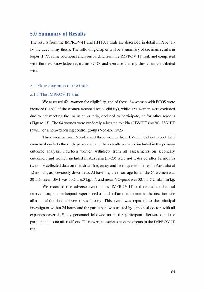
64
5.0 Summary of Results The results from the IMPROV-IT and HITFAT trials are described in detail in Paper II-
IV included in my thesis. The following chapter will be a summary of the main results in
Paper II-IV, some additional analyses on data from the IMPROV-IT trial, and completed
with the new knowledge regarding PCOS and exercise that my thesis has contributed
with.
5.1 Flow diagrams of the trials
5.1.1 The IMPROV-IT trial
We assessed 421 women for eligibility, and of these, 64 women with PCOS were
included (~15% of the women assessed for eligibility), while 357 women were excluded
due to not meeting the inclusion criteria, declined to participate, or for other reasons
(Figure 13). The 64 women were randomly allocated to either HV-HIT (n=20), LV-HIT
(n=21) or a non-exercising control group (Non-Ex; n=23).
Three women from Non-Ex and three women from LV-HIT did not report their
menstrual cycle to the study personnel, and their results were not included in the primary
outcome analysis. Fourteen women withdrew from all assessments on secondary
outcomes, and women included in Australia (n=20) were not re-tested after 12 months
(we only collected data on menstrual frequency and from questionnaires in Australia at
12 months, as previously described). At baseline, the mean age for all the 64 women was
30 ± 5, mean BMI was 30.5 ± 6.5 kg/m2, and mean VO2peak was 33.1 ± 7.2 mL/min/kg.
We recorded one adverse event in the IMPROV-IT trial related to the trial
intervention; one participant experienced a local inflammation around the insertion site
after an abdominal adipose tissue biopsy. This event was reported to the principal
investigator within 24 hours and the participant was treated by a medical doctor, with all
expenses covered. Study personnel followed up on the participant afterwards and the
participant has no after-effects. There were no serious adverse events in the IMPROV-IT
trial.
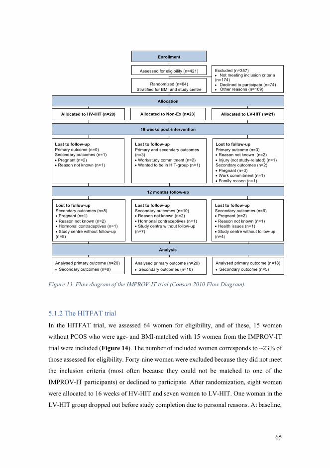
65
Figure 13. Flow diagram of the IMPROV-IT trial (Consort 2010 Flow Diagram).
5.1.2 The HITFAT trial
In the HITFAT trial, we assessed 64 women for eligibility, and of these, 15 women
without PCOS who were age- and BMI-matched with 15 women from the IMPROV-IT
trial were included (Figure 14). The number of included women corresponds to ~23% of
those assessed for eligibility. Forty-nine women were excluded because they did not meet
the inclusion criteria (most often because they could not be matched to one of the
IMPROV-IT participants) or declined to participate. After randomization, eight women
were allocated to 16 weeks of HV-HIT and seven women to LV-HIT. One woman in the
LV-HIT group dropped out before study completion due to personal reasons. At baseline,
Lost to follow-upSecondary outcomes (n=8)• Pregnant (n=1)• Reason not known (n=2)• Hormonal contraceptives (n=1)• Study centre without follow-up (n=5)
Lost to follow-up Secondary outcomes (n=10)• Reason not known (n=2)• Hormonal contraceptives (n=1)• Study centre without follow-up (n=7)
Lost to follow-up Secondary outcomes (n=6)• Pregnant (n=2)• Reason not known (n=1)• Health issues (n=1)• Study centre without follow-up (n=4)
Lost to follow-upPrimary outcome (n=0)Secondary outcomes (n=1)• Pregnant (n=2) • Reason not known (n=1)
Lost to follow-upPrimary and secondary outcomes (n=3)• Work/study commitment (n=2)• Wanted to be in HIT-group (n=1)
Lost to follow-upPrimary outcome (n=3)• Reason not known (n=2)• Injury (not study-related) (n=1)Secondary outcomes (n=2)• Pregnant (n=3)• Work commitment (n=1)• Family reason (n=1)
12 months follow-up
Analysis
Analysed primary outcome (n=20)¨ Secondary outcomes (n=8)
Analysed primary outcome (n=20)¨ Secondary outcomes (n=10)
Analysed primary outcome (n=18)¨ Secondary outcome (n=5)
Assessed for eligibility (n=421)
Enrollment
Excluded (n=357)¨ Not meeting inclusion criteria (n=174) ¨ Declined to participate (n=74)¨ Other reasons (n=109)
Randomized (n=64)Stratified for BMI and study centre
Allocation
Allocated to HV-HIT (n=20) Allocated to Non-Ex (n=23) Allocated to LV-HIT (n=21)
16 weeks post-intervention
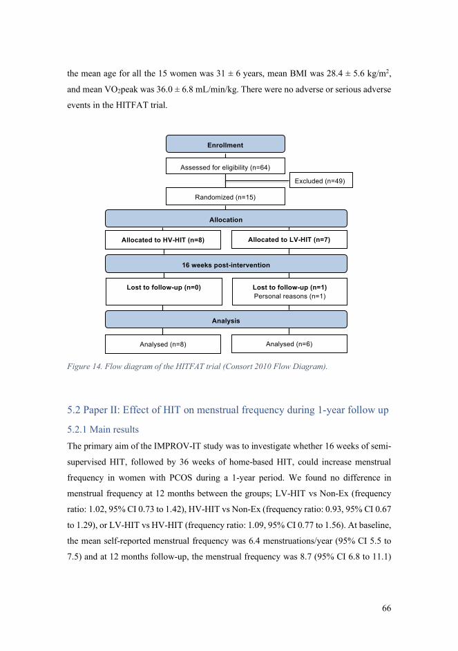
66
the mean age for all the 15 women was 31 ± 6 years, mean BMI was 28.4 ± 5.6 kg/m2,
and mean VO2peak was 36.0 ± 6.8 mL/min/kg. There were no adverse or serious adverse
events in the HITFAT trial.
Figure 14. Flow diagram of the HITFAT trial (Consort 2010 Flow Diagram).
5.2 Paper II: Effect of HIT on menstrual frequency during 1-year follow up
5.2.1 Main results
The primary aim of the IMPROV-IT study was to investigate whether 16 weeks of semi-
supervised HIT, followed by 36 weeks of home-based HIT, could increase menstrual
frequency in women with PCOS during a 1-year period. We found no difference in
menstrual frequency at 12 months between the groups; LV-HIT vs Non-Ex (frequency
ratio: 1.02, 95% CI 0.73 to 1.42), HV-HIT vs Non-Ex (frequency ratio: 0.93, 95% CI 0.67
to 1.29), or LV-HIT vs HV-HIT (frequency ratio: 1.09, 95% CI 0.77 to 1.56). At baseline,
the mean self-reported menstrual frequency was 6.4 menstruations/year (95% CI 5.5 to
7.5) and at 12 months follow-up, the menstrual frequency was 8.7 (95% CI 6.8 to 11.1)
Excluded (n=49)
Enrollment
Assessed for eligibility (n=64)
Allocation
Allocated to LV-HIT (n=7)
16 weeks post-intervention
Lost to follow-up (n=1)Personal reasons (n=1)
Randomized (n=15)
Lost to follow-up (n=0)
Allocated to HV-HIT (n=8)
Analysis
Analysed (n=6)Analysed (n=8)
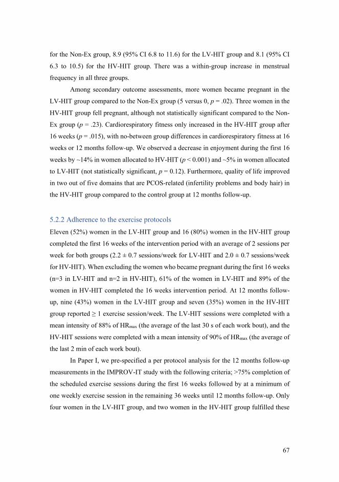
67
for the Non-Ex group, 8.9 (95% CI 6.8 to 11.6) for the LV-HIT group and 8.1 (95% CI
6.3 to 10.5) for the HV-HIT group. There was a within-group increase in menstrual
frequency in all three groups.
Among secondary outcome assessments, more women became pregnant in the
LV-HIT group compared to the Non-Ex group (5 versus 0, p = .02). Three women in the
HV-HIT group fell pregnant, although not statistically significant compared to the Non-
Ex group (p = .23). Cardiorespiratory fitness only increased in the HV-HIT group after
16 weeks (p = .015), with no-between group differences in cardiorespiratory fitness at 16
weeks or 12 months follow-up. We observed a decrease in enjoyment during the first 16
weeks by ~14% in women allocated to HV-HIT (p < 0.001) and ~5% in women allocated
to LV-HIT (not statistically significant, p = 0.12). Furthermore, quality of life improved
in two out of five domains that are PCOS-related (infertility problems and body hair) in
the HV-HIT group compared to the control group at 12 months follow-up.
5.2.2 Adherence to the exercise protocols
Eleven (52%) women in the LV-HIT group and 16 (80%) women in the HV-HIT group
completed the first 16 weeks of the intervention period with an average of 2 sessions per
week for both groups (2.2 ± 0.7 sessions/week for LV-HIT and 2.0 ± 0.7 sessions/week
for HV-HIT). When excluding the women who became pregnant during the first 16 weeks
(n=3 in LV-HIT and n=2 in HV-HIT), 61% of the women in LV-HIT and 89% of the
women in HV-HIT completed the 16 weeks intervention period. At 12 months follow-
up, nine (43%) women in the LV-HIT group and seven (35%) women in the HV-HIT
group reported ≥ 1 exercise session/week. The LV-HIT sessions were completed with a
mean intensity of 88% of HRmax (the average of the last 30 s of each work bout), and the
HV-HIT sessions were completed with a mean intensity of 90% of HRmax (the average of
the last 2 min of each work bout).
In Paper I, we pre-specified a per protocol analysis for the 12 months follow-up
measurements in the IMPROV-IT study with the following criteria; >75% completion of
the scheduled exercise sessions during the first 16 weeks followed by at a minimum of
one weekly exercise session in the remaining 36 weeks until 12 months follow-up. Only
four women in the LV-HIT group, and two women in the HV-HIT group fulfilled these
68
criteria (~15% of the 41 women allocated to the HIT groups). Due to the low adherence,
we were unable to complete a meaningful per protocol analysis.
5.3 Paper III: Circulating and adipose tissue miRNA expression and effect
of HIT We found a 1.8-fold higher basal expression of circulating miRNA-27b in women with
PCOS compared to age- and BMI-matched women without PCOS (p = .006). No
differences were observed in circulating miRNA-21, - 93, - 103a, -146a, -155, -222 or -
223 between women with and without PCOS. We do not report any differences in the
eight selected miRNAs in subcutaneous abdominal or gluteal adipose tissue of women
with and without PCOS.
Sixteen weeks of LV-HIT induced a 0.5-fold decrease in the expression of
circulating miRNA-27b in women with PCOS (p = .007), with no changes observed in
the remaining seven miRNAs. None of the eight miRNAs changed in the circulation after
16 weeks of HV-HIT, nor did we observe any changes in the eight selected miRNAs in
subcutaneous abdominal or gluteal adipose tissue of women with PCOS after 16 weeks
of either LV-HIT or HV-HIT.
5.4 Paper IV: Rates of whole-body fat oxidation during submaximal exercise
+ adipose tissue mitochondrial respiration and cell size and response to HIT At baseline, women without PCOS (n=64) had higher rates of whole-body fat oxidation
during submaximal exercise at 60% of VO2peak (p = .008 for g/min, p = .006 for
mg/FFM/min) compared to women with PCOS. We did not observe a difference in
whole-body fat oxidation rates during submaximal exercise between the women with and
without PCOS who were individually matched (n=15 for each group). At baseline,
women with PCOS had lower mitochondrial respiration through complex I + II in
subcutaneous abdominal adipose tissue compared to women without PCOS (p = .015),
while no difference was observed in mitochondrial respiration through complex I + II in
subcutaneous gluteal adipose tissue. No differences were found between women with and
without PCOS in metabolic outcomes, or subcutaneous abdominal and gluteal adipose
tissue cell size at baseline.
69
Cardiorespiratory fitness increased to a similar degree in women with (~4%, p =
0.006) and without PCOS (~6%, p = 0.003) after 16 weeks of HIT. Interestingly, rates of
whole-body fat oxidation during submaximal exercise only improved after 16 weeks of
HIT in women without, but not with PCOS (~16% improvement in g/min (p = .026) for
women without PCOS, and ~17% in mg/FFM/min (p = .023)). We observed no changes
in cell size or mitochondrial respiration in subcutaneous abdominal and gluteal adipose
tissue after HIT in either group of women.
Women with PCOS allocated to the HIT groups completed 2.2 ± 0.5 exercise
sessions/week while women without PCOS completed 2.6 ± 0.4 weekly sessions (p =
.025), which is equivalent to ~6 exercise sessions less in total over the 16 weeks.
5.5 Additional analyses from the IMPROV-IT study In these additional analyses from the IMPROV-IT study, I first compared reproductive
and metabolic outcomes between the women included in Norway and those included in
Australia, and secondly compared the same outcomes in these women based on BMI-
category (women with BMI < 27 kg/m2 and women with BMI ≥ 27 kg/ m2). These
analyses were performed as the stratification factors used in the IMPROV-IT study were
study center and BMI < or ≥ 27 kg/m2, and included to ensure equal allocation of
subgroups of participants to each intervention arm and thereby homogenous groups.
5.5.1 Norwegian versus Australian women
Table 4 shows baseline characteristics for the women with PCOS included in Norway
(n=44) and in Australia (n=20). The Norwegian and Australian women included in the
IMPROV-IT trial were fairly homogenous in metabolic and reproductive outcomes. The
Norwegian women were on average 2.6 years older (p = .04) than the Australian women,
and the Norwegian women had higher total cholesterol (p = .01), LDL-cholesterol (p =
.002), and absolute, but not relative, VO2peak (p = .02) compared to the Australian
women. The Norwegian women also had higher FFM (p = .02) compared to Australian,
however, FFM was estimated using two different methods in Norway (bioelectrical
impedance analysis) and Australia (DXA), which complicates the comparison of this
outcome.
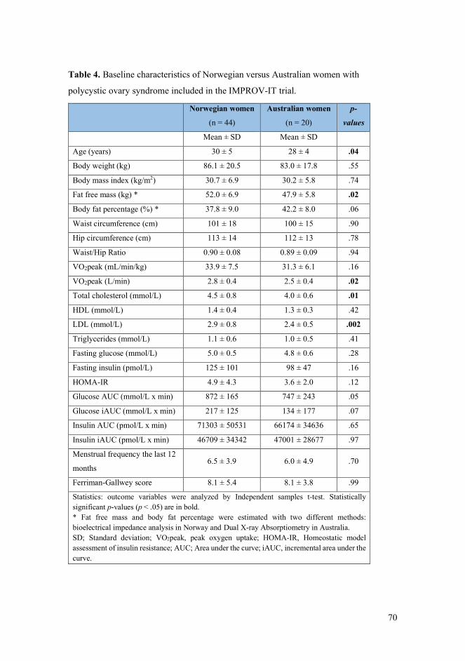
70
Table 4. Baseline characteristics of Norwegian versus Australian women with
polycystic ovary syndrome included in the IMPROV-IT trial.
Norwegian women
(n = 44)
Australian women
(n = 20)
p-
values
Mean ± SD Mean ± SD
Age (years) 30 ± 5 28 ± 4 .04
Body weight (kg) 86.1 ± 20.5 83.0 ± 17.8 .55
Body mass index (kg/m2) 30.7 ± 6.9 30.2 ± 5.8 .74
Fat free mass (kg) * 52.0 ± 6.9 47.9 ± 5.8 .02
Body fat percentage (%) * 37.8 ± 9.0 42.2 ± 8.0 .06
Waist circumference (cm) 101 ± 18 100 ± 15 .90
Hip circumference (cm) 113 ± 14 112 ± 13 .78
Waist/Hip Ratio 0.90 ± 0.08 0.89 ± 0.09 .94
VO2peak (mL/min/kg) 33.9 ± 7.5 31.3 ± 6.1 .16
VO2peak (L/min) 2.8 ± 0.4 2.5 ± 0.4 .02
Total cholesterol (mmol/L) 4.5 ± 0.8 4.0 ± 0.6 .01
HDL (mmol/L) 1.4 ± 0.4 1.3 ± 0.3 .42
LDL (mmol/L) 2.9 ± 0.8 2.4 ± 0.5 .002
Triglycerides (mmol/L) 1.1 ± 0.6 1.0 ± 0.5 .41
Fasting glucose (mmol/L) 5.0 ± 0.5 4.8 ± 0.6 .28
Fasting insulin (pmol/L) 125 ± 101 98 ± 47 .16
HOMA-IR 4.9 ± 4.3 3.6 ± 2.0 .12
Glucose AUC (mmol/L x min) 872 ± 165 747 ± 243 .05
Glucose iAUC (mmol/L x min) 217 ± 125 134 ± 177 .07
Insulin AUC (pmol/L x min) 71303 ± 50531 66174 ± 34636 .65
Insulin iAUC (pmol/L x min) 46709 ± 34342 47001 ± 28677 .97
Menstrual frequency the last 12
months 6.5 ± 3.9 6.0 ± 4.9 .70
Ferriman-Gallwey score 8.1 ± 5.4 8.1 ± 3.8 .99
Statistics: outcome variables were analyzed by Independent samples t-test. Statistically significant p-values (p < .05) are in bold. * Fat free mass and body fat percentage were estimated with two different methods: bioelectrical impedance analysis in Norway and Dual X-ray Absorptiometry in Australia. SD; Standard deviation; VO2peak, peak oxygen uptake; HOMA-IR, Homeostatic model assessment of insulin resistance; AUC; Area under the curve; iAUC, incremental area under the curve.
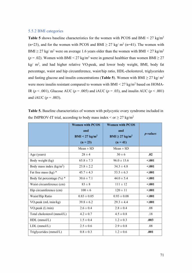
71
5.5.2 BMI categories
Table 5 shows baseline characteristics for the women with PCOS and BMI < 27 kg/m2
(n=23), and for the women with PCOS and BMI ≥ 27 kg/ m2 (n=41). The women with
BMI ≥ 27 kg/ m2 were on average 1.6 years older than the women with BMI < 27 kg/m2
(p = .02). Women with BMI < 27 kg/m2 were in general healthier than women BMI ≥ 27
kg/ m2, and had higher relative VO2peak, and lower body weight, BMI, body fat
percentage, waist and hip circumference, waist/hip ratio, HDL-cholesterol, triglycerides
and fasting glucose and insulin concentrations (Table 5). Women with BMI ≥ 27 kg/ m2
were more insulin resistant compared to women with BMI < 27 kg/m2 based on HOMA-
IR (p < .001), Glucose AUC (p = .005) and iAUC (p = .03), and insulin AUC (p < .001)
and iAUC (p = .003).
Table 5. Baseline characteristics of women with polycystic ovary syndrome included in
the IMPROV-IT trial, according to body mass index < or ≥ 27 kg/m2
Women with PCOS
and
BMI < 27 kg/m2
(n = 23)
Women with PCOS
and
BMI ≥ 27 kg/m2
(n = 41)
p-values
Mean ± SD Mean ± SD
Age (years) 28 ± 4 30 ± 6 .02
Body weight (kg) 65.8 ± 7.3 96.0 ± 15.6 <.001
Body mass index (kg/m2) 23.8 ± 2.2 34.3 ± 4.8 <.001
Fat free mass (kg) * 45.7 ± 4.3 53.5 ± 6.3 <.001
Body fat percentage (%) * 30.6 ± 7.1 44.0 ± 5.4 <.001
Waist circumference (cm) 83 ± 8 111 ± 12 <.001
Hip circumference (cm) 100 ± 6 120 ± 11 <.001
Waist/Hip Ratio 0.83 ± 0.05 0.93 ± 0.08 <.001
VO2peak (mL/min/kg) 39.8 ± 6.2 29.3 ± 4.4 <.001
VO2peak (L/min) 2.6 ± 0.4 2.8 ± 0.4 .08
Total cholesterol (mmol/L) 4.2 ± 0.7 4.5 ± 0.8 .18
HDL (mmol/L) 1.5 ± 0.4 1.2 ± 0.3 .003
LDL (mmol/L) 2.5 ± 0.6 2.9 ± 0.8 .08
Triglycerides (mmol/L) 0.8 ± 0.3 1.2 ± 0.6 .001
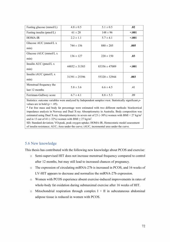
72
5.6 New knowledge This thesis has contributed with the following new knowledge about PCOS and exercise:
o Semi-supervised HIT does not increase menstrual frequency compared to control
after 12 months, but may still lead to increased chances of pregnancy.
o The expression of circulating miRNA-27b is increased in PCOS, and 16 weeks of
LV-HIT appears to decrease and normalize the miRNA-27b expression.
o Women with PCOS experience absent exercise-induced improvements in rates of
whole-body fat oxidation during submaximal exercise after 16 weeks of HIT.
o Mitochondrial respiration through complex I + II in subcutaneous abdominal
adipose tissue is reduced in women with PCOS.
Fasting glucose (mmol/L) 4.8 ± 0.5 5.1 ± 0.5 .02
Fasting insulin (pmol/L) 61 ± 28 148 ± 96 <.001
HOMA-IR 2.2 ± 1.1 5.7 ± 4.1 <.001
Glucose AUC (mmol/L x
min) 744 ± 156 880 ± 205 .005
Glucose iAUC (mmol/L x
min) 136 ± 127 220 ± 150 .03
Insulin AUC (pmol/L x
min) 44852 ± 31383 83356 ± 47009 <.001
Insulin iAUC (pmol/L x
min) 31391 ± 25396 55320 ± 32944 .003
Menstrual frequency the
last 12 months 5.8 ± 3.6 6.6 ± 4.5 .41
Ferriman-Gallwey score 6.7 ± 4.1 8.8 ± 5.3 .09
Statistics: outcome variables were analyzed by Independent samples t-test. Statistically significant p-values are in bold (p < .05). * Fat free mass and body fat percentage were estimated with two different methods: bioelectrical impedance analysis in Norway and Dual X-ray Absorptiometry in Australia. Body composition was estimated using Dual X-ray Absorptiometry in seven out of 23 (~30%) women with BMI < 27 kg/m2 and in 13 out of 41 (~32%) women with BMI ≥ 27 kg/m2. SD; Standard deviation; VO2peak, peak oxygen uptake; HOMA-IR, Homeostatic model assessment of insulin resistance; AUC; Area under the curve; iAUC, incremental area under the curve.
73
6.0 Discussion Our findings provide novel data regarding the effects of two HIT protocols on
reproductive, cardiometabolic and psychologic outcomes in women with PCOS.
Furthermore, we investigated the effect of HIT on whole-body fat oxidation during
submaximal exercise and on adipose tissue mitochondrial respiration, fat cell size, and
miRNA expression in women with and without PCOS. The following paragraphs will be
a discussion of the main findings followed by the strengths and limitations of the studies,
clinical relevance and future perspectives.
6.1 Menstrual frequency We found no between-group difference in menstrual frequency at 12 months but observed
a significant increase in menstrual frequency from baseline to 12 months follow-up in all
three groups including in the Non-Ex (control) group in the IMPROV-IT trial. Although
HV-HIT and LV-HIT did not improve menstrual frequency compared to the Non-Ex
group, pregnancy rates increased significantly in LV-HIT. It is difficult to explain the
lack of improvement in menstrual frequency after LV-HIT compared to the Non-Ex group
despite an increase in pregnancy rates in LV-HIT, and I can only speculate why this was
the case.
The major reason for the lack of difference between groups in menstrual
frequency is probably poor adherence to the HIT protocols. Only 37% of the participants
in the HIT groups completed >75% of the exercise sessions during the first 16 weeks,
while 39% reported at least one weekly exercise session at 12 months follow-up.
Additionally, our findings indicate that the Non-Ex group most likely did introduce some
lifestyle modifications during the intervention period. There was an increase in menstrual
frequency of ~2 menstruations/year from baseline to 12 months follow-up in the Non-Ex
group, along with improvements in other outcomes including reduced body weight and
BMI, and improved blood lipid profiles and FAI. Participants in the Non-Ex group were
encouraged to continue their diet and exercise habits throughout the study period.
Although we did not observe a change in the 4-day diet recording or five days of physical
activity monitoring at each timepoint, these measurements only gave a brief “snap-shot”
of the whole study period. Based on the favorable improvements in several outcomes it
74
is likely that there was a change in behavior in the Non-Ex group. Participants who sign
up for lifestyle intervention trials most often hope to be allocated to the intervention
group. The participants were possibly particularly disappointed when they were allocated
to the Non-Ex group in the IMPROV-IT trial as the chances were high for allocation to
the intervention groups (67% chance) and relatively low for allocation to the Non-Ex
group (33%). The participants may have been mentally prepared and motivated to
introduce lifestyle changes, which could lead to difficulties to continue their previously
sedentary lifestyle, and especially over the 12 months required in the IMPROV-IT trial.
One solution to this challenge, could be to offer the exercise protocol to the participants
allocated to the Non-Ex group at the end of the study period. We were, however, unable
to provide such a delayed treatment due to the length and time-demand of the IMPROV-
IT study.
Some of the women included in the IMPROV-IT trial had a regular menstrual
cycle, as we used the Rotterdam criteria for PCOS diagnosis, in which menstrual
dysfunction is not necessarily required for diagnosis. I am aware that the inclusion of
women with a regular menstrual cycle limited the chance of finding a difference in this
outcome measure; In women with an already regular menstrual frequency at baseline, a
further increase would be unlikely. However, there was no statistically significant
difference in the proportion of women with a regular menstrual cycle in the three groups
(39% in the control group, 33% in LV-HIT and 25% in HV-HIT). Neither was there any
statistical difference between groups in the number of women in whom the menstrual
pattern changed (i.e. regular cycle, oligo-amenorrhea (35-42 days cycle/ 8-9 cycles per
year), oligo-amenorrhea (42 days to 6 months cycle / 2-7 cycles per year), and amenorrhea
(> 6 months cycle / 0-1 cycles per year)). We used the Rotterdam criteria because we
wanted to increase the comparability of our results to other studies and their research
findings. Moreover, if we only were to include women with an irregular menstrual cycle,
we would not have been able to include as many women, which would have resulted in
lower statistical power for the secondary outcome measures in the trial.
Similar to our findings, Thomson and colleagues found no between-group
differences in menstrual frequency after a 20 weeks study period of either diet only, diet
and aerobic exercise, and diet and combined aerobic + resistance exercise (39). They did
not report whether there were any within-group differences in menstrual frequency from
75
baseline to 20 weeks. The diet intervention included in all three intervention groups,
complicates the interpretation of the effect of exercise on menstrual frequency, and it is
only possible to say that the exercise interventions did not have an additional effect on
menstrual frequency compared to diet alone.
Nybacka and colleagues (37) compared the effects of diet only, exercise only and
the combined effect of diet and exercise for 16 weeks on menstrual pattern and ovulation
rates (confirmed by an elevation of progesterone levels in serum during the luteal phase)
in women with PCOS. They showed that the interventions were equally effective in
improving menstrual pattern (shifting from oligo-/amenorrhea to a regular menstrual
cycle), but with no difference in ovulation rates in any group. However, in that study
women in the exercise only group also reduced their daily calorie intake by ~270 kcal,
which resulted in a 3% weight loss, thereby making it difficult to conclude that the
exercise intervention alone was the reason for the improved menstrual pattern.
Jedel and colleagues also found an increase in menstrual frequency after 16 weeks
of physical activity compared to a non-intervention, control group (29). Similar to our
study, women with PCOS were included based on the Rotterdam criteria, however, at
baseline the menstrual frequency (measured as observed menstrual cycles/expected
menstrual cycles) was substantially lower compared to in our participants: a menstrual
frequency of 0.25 menstruations/month in the Jedel et al. study versus 0.49
menstruations/year in our study. The menstrual frequency in the exercise group in their
study increased by 0.14 ± 0.33 during 16 weeks, which means that even after the 16 weeks
of exercise, the menstrual frequency of the participants in the exercise group was lower
than the menstrual frequency at baseline in our study.
6.2 Exercise adherence We have little knowledge as to whether exercise protocols implemented in clinical trials
under controlled laboratory conditions, typically comprising 8-12 weeks of supervised
exercise training, are realistic in a ‘real-world’ setting. Evidence suggests that HIT is
time-efficient (14, 17, 23) and also perceived to be more enjoyable than MICT despite
higher perceived exertion during HIT (24). Therefore, HIT may prove as a feasible
strategy to improve long-term exercise adherence and participation. However, in our
76
study we report poor long-term exercise adherence to HIT. We intended to include
exercise protocols that were time-efficient, enjoyable and easy to implement in the
women’s everyday life and assumed that the semi-supervised nature of our intervention
would increase flexibility and adherence. I speculate that the poor adherence may be
related to too monotonous exercise protocols, as we observed a decrease in enjoyment
during the first 16 weeks by ~14% in women allocated to HV-HIT (p < 0.001) and ~5%
in women allocated to LV-HIT (not statistically significant, p = 0.12). A systematic
review and meta-analysis on the effectiveness of exercise in the management of PCOS
compared to control demonstrated improvements in numerous cardiometabolic outcomes
after exercise (VO2max, fasting insulin, HOMA-IR, total cholesterol, LDL cholesterol,
triglycerides, waist circumference and body fat percentage) (155). However, subgroup
analyses revealed greater improvements when interventions were supervised and of a
shorter duration (≤ 12 weeks), and only VO2max was improved compared to control when
the exercise interventions lasted for more than 12 weeks. The limited improvements in
cardiometabolic outcomes after the HIT protocols in our study may be explained by the
partly supervised nature, the longer duration, decrease in enjoyment over time and thereby
poor adherence.
No previous studies have included and compared different HIT protocols in
women with PCOS. Nonetheless, researchers in our laboratory investigated the effects of
ten weeks of three weekly sessions of either HIT or strength training on cardiometabolic
health outcomes in women with PCOS, and observed improvements in several outcomes
in both training groups (25). Similar to ours, the exercise sessions were semi-supervised
(at least one weekly supervised exercise session) in that study, but the HIT protocols
varied during the week, with two HV-HIT sessions and one LV-HIT session. Both this
variety in the training program and the shorter intervention period may explain the higher
adherence to the HIT protocols (~90%) in that pilot RCT compared to in the IMPROV-
IT trial. In most studies investigating the effect of exercise interventions in women with
PCOS, researchers have investigated the effects of the same exercise protocol performed
three times per week, with no variation (28, 32-35, 39). It is uncommon in real life to
undertake the exact same exercise session multiple times per week with no variation.
Therefore, I suggest that future research on exercise training in women with PCOS should
focus on the combination of various exercise types/sessions to determine the best exercise
77
interventions for retaining engagement and improving health. Perhaps this should even
be the future direction for exercise interventions in general, at least if the objective is to
explore which exercise interventions work in the real world (effectiveness) rather than
merely the physiological effect (efficacy) of various exercise sessions.
6.3 Cardiorespiratory fitness We found a within-group increase in VO2peak in the HV-HIT group after 16 weeks of
HIT, while no significant changes were observed in the LV-HIT group despite the same
weekly average of exercise sessions in these groups. These findings are supported by
previous findings from Baekkerud and colleagues who compared the effects of 6 weeks
of either 4 x 4 min HIT at 85-95% of HRmax, 10 x 1 min at VO2max load, or 45 min MICT
at 70% of HRmax on VO2max in men and women with overweight/obesity (23). In that
study, only the participants in the 4 x 4 min HIT group increased their VO2max, with a
significant between-group difference versus both 10 x 1 min HIT and MICT protocols
(23). Similarly, others have shown that greater volumes of high-intensity exercise result
in greater improvements in VO2max (45, 46). A meta-analysis on the effects of different
HIT protocols on VO2max improvements demonstrated that in addition to a high volume
of high-intensity work (≥ 15 min/session), HIT with longer work bouts (≥ 2 min) and
longer training periods (4-12 weeks) resulted in the greatest improvements in VO2max in
healthy individuals and those with overweight/obesity (46). The partly supervised nature
of our study during the first 16 weeks and no supervision in the remaining 36 weeks along
with the long study duration, a decrease in enjoyment, and the poor adherence to the HIT
protocols can probably explain why we did not observe any difference in VO2peak in any
group after 12 months in the IMPROV-IT trial.
6.4 Fat oxidation and metabolic flexibility We show that women with PCOS had lower rates of whole-body fat oxidation during
submaximal exercise compared to women without PCOS at baseline, however, no
difference was observed when we compared baseline fat oxidation rates between women
with and without PCOS who were individually age- and BMI-matched (n=15 in each
group). After 16 weeks of HIT, rates of whole-body fat oxidation during submaximal
78
exercise improved in women without PCOS, whereas no improvements were observed in
women with PCOS, despite similar increases in VO2peak.
To my knowledge, no previous study has investigated the effect of a HIT
intervention on whole-body fat oxidation during submaximal exercise in women with
PCOS. The absent exercise-induced improvements in whole-body fat oxidation during
submaximal exercise in women with PCOS, despite improved VO2peak, suggest
metabolic inflexibility. Metabolic inflexibility in PCOS has been reported previously in
the fasted, insulin stimulated state (as reviewed by Rimmer and colleagues) (78). The
association between PCOS and metabolic inflexibility was indicated through unchanged
RER in the transition from fasted to insulin-stimulated state in women with PCOS
whereas there was an increase in RER in healthy women (78). Metabolic inflexibility has
also been described in adolescent girls with PCOS (80) and in lean women with PCOS
(75), suggesting an association between metabolic inflexibility and PCOS, independent
of age and BMI. Moreover, metabolic inflexibility is associated with insulin resistance
(77), and the degree of metabolic inflexibility was shown to be similar between women
with PCOS and obesity (28.8 ± 4.7 years) and middle-aged women with type 2 diabetes
and obesity (58.2 ± 9.9 years) (79).
Studies have demonstrated improved rates of whole-body fat oxidation during
submaximal exercise at 60% of VO2peak in recreationally active, healthy women after
seven sessions of HIT (10 x 4 work bouts that elicited ~90% of VO2peak separated with
2 min of rest) (81), and in recreationally active, healthy men and women after six weeks
of HIT (three days/week of 10 x 4 work bouts that elicited ~90% of VO2peak separated
with 2 min of rest) (82). Consistent with these findings, we found improved rates of
whole-body fat oxidation during submaximal exercise at 60% of VO2peak in women
without PCOS after 16 weeks of HIT. Contrary, we did not observe an exercise-induced
improvement in fat oxidation in women with PCOS. This difference may be explained by
the fact that women with PCOS exercised less, at least statistically, than the women
without PCOS (2.2 ± 0.5 versus 2.6 ± 0.4 weekly HIT sessions, equivalent to ~6 exercise
sessions less in total over 16 weeks). However, VO2peak increased to a similar degree in
women with (~ 4%) and without PCOS (~ 6%) indicating that the fewer HIT sessions
performed by women with PCOS cannot fully explain the difference in exercise-induced
improvements in fat oxidation rates after 16 weeks of HIT. A recent study by Hansen and
79
colleagues also reported absent training-induced improvements in whole-body insulin
sensitivity and glucose tolerance in women with PCOS compared to age-, BMI- and
VO2peak-matched control women without PCOS (156), despite a similar increase in
VO2peak. Their data implied that the absent training-induced improvement in whole-
body insulin action in women with PCOS was owed to a diminished ability to upregulate
glucose uptake in skeletal muscle. Collectively, the findings by Hansen and colleagues
and those from our study indicate an impaired response to exercise training in some
metabolic outcomes related to insulin resistance in women with PCOS. Further
investigation is needed to determine the underlying mechanisms behind the absent
training-induced improvements in rates of whole-body fat oxidation during submaximal
exercise in women with PCOS.
6.5 Adipose tissue in PCOS pathophysiology In the following paragraphs, I will discuss some of the main findings in Paper III and IV,
and the potential role of adipose tissue cell size, mitochondrial respiration and
microRNAs in PCOS pathophysiology, as well as the response to 16 weeks of HIT in
these outcomes.
6.5.1 Mitochondrial respiration
We found that women with PCOS had reduced mitochondrial respiration through
complex I + II in subcutaneous abdominal adipose tissue compared to women without
PCOS at baseline, with no differences in subcutaneous gluteal adipose tissue. Moreover,
mitochondrial respiration through complex I + II in both adipose tissue depots were
unaffected by 16 weeks of HIT in both groups of women. Previous studies have reported
aberrant adipose tissue in women with PCOS (75, 84, 87, 88, 157). We found that women
with PCOS had reduced mitochondrial respiration in subcutaneous abdominal adipose
tissue, supporting the notion that abnormal adipose tissue may play a central role in PCOS
pathophysiology (63, 76).
The mitochondria are the major site of ATP production via oxidative
phosphorylation, and insulin resistance has been linked to diminished mitochondrial
function in skeletal muscle (97). One prior study has compared skeletal muscle
80
mitochondrial function in women with and without PCOS using high-resolution
respirometry (98). Rabøl and colleagues observed no difference in skeletal muscle
mitochondrial respiration between women with and without PCOS in either those who
were lean (BMI < 25 kg/m2) or in those with overweight or obesity (BMI > 25 kg/m2)
(98). The findings from Rabøl et al, together with ours, suggest that mitochondrial
impairment in PCOS may be evident in (abdominal) adipose tissue rather than skeletal
muscle. Thereby, the insulin resistance observed in PCOS could be linked to dysfunctions
in the adipose tissue and not in skeletal muscle.
There are no previous publications on exercise-induced changes in subcutaneous
gluteal and abdominal adipose tissue mitochondrial respiration in women with PCOS.
However, a couple of studies have investigated exercise-induced changes in subcutaneous
abdominal mitochondrial respiration in healthy individuals, albeit with conflicting
findings. Similar to our findings, Larsen and colleagues found no change in subcutaneous
abdominal adipose tissue mitochondrial respiration in men and women with overweight
but who were otherwise healthy after 6 weeks of LV-HIT (three days/week of 5 x 1 min
maximal effort work-bouts), despite an increase in cardiorespiratory fitness (100). In
contrast, Dohlmann and colleagues observed reduced mitochondrial respiration in
subcutaneous abdominal adipose tissue taken from men and women with obesity after 18
LV-HIT sessions completed in 6 weeks (7 x 1 min work bouts with workloads
corresponding to ~100% of VO2peak) (101). Contrary to these and our findings,
Mendham and colleagues reported increased subcutaneous abdominal adipose tissue
mitochondrial respiration in black South-African women with obesity after 12 weeks of
combined aerobic and resistance exercise training (four weekly sessions lasting 40-60
min with aerobic exercise at 75-80% HRpeak and upper- and lower-body resistance
exercises at 60-70 HRpeak) (102). Similar to our findings, Mendham and colleagues
reported no change in subcutaneous gluteal adipose tissue after the exercise intervention
(102). I am unsure of the reasons for these discrepant findings, yet they may be explained
by divergent exercise modalities and intensities, as well as different study populations.
81
6.5.2 Fat cell size
We found no differences in subcutaneous gluteal and abdominal adipose tissue cell size
between women with and without PCOS, along with no changes in cell size in neither
adipose tissue depots after 16 weeks of HIT in either group of women. Contrary to our
findings, Manneras-Holm and colleagues reported enlarged abdominal adipocytes in
women with PCOS compared to individually age- and BMI-matched women without
PCOS (84). The discrepancy between our current work and the findings by Manneras-
Holm et al. may be explained by higher power (27 women in both groups) and a lower
mean BMI (< 25 kg/m2) in their study.
Absent training-induced effects on subcutaneous abdominal adipose tissue cell
volume and size have been reported previously (30, 158). Stinkens and colleagues
observed no change in subcutaneous abdominal adipocyte size after a 12-week aerobic
training program in previously inactive men with overweight or obesity (three weekly
exercise sessions which comprised two continuous, aerobic sessions for 30 min at 70%
maximal watt and one resistance training session that included upper- and lower-body
exercise drills) (158). Similarly, absent training-induced changes in subcutaneous
abdominal adipocyte volume has been reported after 16 weeks of continuous, aerobic
exercise in women with PCOS (30). The absent training-induced effect on adipose tissue
morphology in women with PCOS observed both by others (30) and by us may be
explained by less responsiveness in adipose tissue in females. As such, Deprés and
colleagues found a sex difference in training-adaptations (159); a 20-week aerobic
exercise program (40 min per session at 80% HRpeak performed 4-5 days/week) reduced
fat percentage and adipose cell weight in men, while no changes occurred in fat
percentage or adipose cell weight in women, which suggests that female adipose tissue
may be less responsive to adaptations.
6.5.3 microRNAs
Interestingly, we found higher basal expression of circulating miRNA-27b in
women with PCOS compared to women without PCOS. miRNA-27b has been implicated
in several biological processes including inflammation, fatty acid metabolism, substrate
metabolism, and adipocyte differentiation (107, 148, 160). Murri and colleagues found
82
an interaction between obesity and the group of participants in whole blood miRNA-27b
expression, in which obesity (BMI > 30 kg/m2) was associated with a reduced expression
of miRNA-27b in women without PCOS, whereas obesity was associated with an
increased expression of the same miRNA in women with PCOS (107). Collectively, the
findings by Murri et al. (2013), together with ours, suggest divergent expression of
circulating miRNA-27b in women with PCOS compared to women without PCOS. The
abnormal expression of miRNA-27b observed in women with PCOS compared to women
without PCOS suggests a potential role for this miRNA in the pathophysiology of PCOS.
miRNAs can act within the tissue of origin, or be released to the circulation and
taken up by adjacent and/or distant tissues where they exert their effects cell processes
(105). As such, we investigated the expression of the eight selected miRNAs in
subcutaneous gluteal and abdominal adipose tissue of women with and without PCOS,
however, we found no differences. Supporting these findings, previous studies have
reported the abundance of some subcutaneous abdominal adipose tissue miRNAs
including miRNA-223 to be similar in women with and without PCOS (119, 121), but to
be different between insulin resistant women (HOMA-IR ≥ 2.5) compared to insulin
sensitive women (HOMA-IR < 2.5). Despite the higher basal expression of circulating
miRNA-27b, we found no difference in the expression of this miRNA in either
subcutaneous gluteal or abdominal adipose tissue. I can only speculate about this
“disconnect” in miRNA expression between the circulation and adipose tissue.
Subcutaneous gluteal and abdominal adipose tissue may lack the specific membrane
transporters to import miRNA-27b from the circulation (161).
Previous studies have shown that exercise training can modulate the expression
of circulating miRNAs in healthy individuals and in individuals with overweight and
obesity (122-124). Indeed, we also observed a decrease in the expression of circulating
miRNA-27b after 16 weeks of LV-HIT in women with PCOS, indicating that LV-HIT
has the potential to regulate and normalize the expression of circulating miRNA-27b in
women with PCOS.
The effect of exercise training on gluteal and abdominal adipose tissue miRNAs
has only been sparsely investigated in other populations (93). Similar to our findings,
Tsiloulis and colleagues reported no effect of 6 weeks of aerobic exercise training (four
weekly exercise sessions; three moderate intensity sessions and one HIT session) on the
83
expression of 526 miRNAs in gluteal and abdominal adipose tissue in men with
overweight (93). Collectively, miRNAs in adipose tissue seem to be largely unaltered in
response to exercise training lasting up to several months.
6.6 Strengths A major strength of the IMPROV-IT study was that we compared the effects of LV-HIT
and HV-HIT to address selected aspects of metabolic and reproductive health in women
with PCOS. Further, we had strict inclusion and exclusion criteria, a long intervention
period, and included a control group. The fact that we did not include a dietary
intervention in IMPROV-IT allowed us to investigate the isolated effect of exercise
training on reproductive and cardiometabolic outcomes in women with PCOS.
Furthermore, the multicenter nature of the trial, including women with PCOS from
different parts of the world, provides unique scope to the investigation of PCOS and
improves generalizability (external validity) of the study.
The strengths of Paper III and IV are that we employed pairwise age- and BMI-
matching of women with and without PCOS. In addition, we obtained adipose tissue
samples from different adipose depots and were able to assess important metabolic
parameters including miRNAs, mitochondrial respiration and fat cell size. Collectively,
our findings advance the current knowledge about the possible role of adipose tissue in
PCOS pathophysiology.
6.7 Limitations We used menstrual frequency as a proxy of ovulation, which is a limitation as it is
possible to ovulate without a later bleeding and to have a regular cycle despite ovulatory
dysfunction (52). Due to practical reasons, we were not able to assess ovulation as that
requires daily temperature measurements, frequent urine samples to assess LH, or blood
samples to assess progesterone concentrations as markers of ovulation. Menstrual
bleedings have been used by others to assess possible improvements in reproduction in
women with PCOS after an exercise intervention (29).
The poor adherence to the exercise protocols in the intervention groups with
PCOS was the major limitation in the IMPROV-IT trial. The IMPROV-IT trial was based
84
on the intention to treat principle, in which all primary and secondary outcomes are
analyzed according to which intervention the participants are allocated to, regardless of
adherence to the protocol. Thereby, to find an effect of the intervention depends heavily
on adherence. We intended to add a per protocol analysis to investigate the effect of
following the exercise protocols, however, only four women in the LV-HIT group, and
two women in the HV-HIT group fulfilled the pre-specified criteria for the per protocol
analysis for the 12 months follow-up measurements. Thus, we could not perform a
meaningful per protocol analysis. Additionally, our findings indicate that the Non-Ex
group introduced some lifestyle modifications during the intervention period.
The limited adipose tissue quantity from the biopsies increased the risk of type II
errors (not showing an effect of the intervention even if it was present), which is the main
limitation in both Paper III and IV. In Paper III, we compared the expression of miRNAs
in subcutaneous gluteal and abdominal adipose tissue between women with and without
PCOS in eight women in each group, and secondly determined the response to LV-HIT
or HV-HIT in women with PCOS in eight women in each of the groups (total of 24
women including the Non-Ex group). However, the number of participants included in
each group (n=8) is comparable or even higher compared to the number of participants
in similar studies (93, 121).
Due to the limited adipose tissue biopsy quantity, we were unable to compare fat
cell size and mitochondrial respiration between the pairwise age- and BMI-matched
women with and without PCOS. To increase power, we pooled the LV-HIT and HV-HIT
groups into one group for women with PCOS and one group for women without PCOS
after our statistical analyses showed no differences on any outcomes between the LV-
HIT and HV-HIT groups. Thereby, we were unable to detect differences in the effects of
LV-HIT and HV-HIT in fat cell size and mitochondrial respiration, but instead able to
investigate the effect of HIT while increasing the statistical power. The number of
participants included in our exercise groups were similar to the number of participants
included in previous studies that investigated exercise-induced changes in adipose tissue
mitochondrial respiration or morphology (100, 101, 158, 159).
85
6.8 Clinical relevance and future perspectives Exercise is widely recommended for women with PCOS (52), and it is important to
determine the reproductive and metabolic effects of different types of exercise modalities
to improve the prescription of exercise as a therapeutic intervention for this population.
The studies included in this thesis have led to new insight about the effects of LV-HIT
and HV-HIT as treatment to address selected aspects of reproductive, metabolic and
psychologic health in women with PCOS, and adds to the current knowledge regarding
the health benefits of various exercise training programs in these women.
The observed improvements in menstrual frequency, pregnancy rates and mental
health in the IMPROV-IT trial are of major clinical importance for women with PCOS,
since PCOS is the primary cause of anovulatory infertility and menstrual disorders in
reproductive-aged women, and since the prevalence of impaired quality of life, anxiety
and depression is high among women with PCOS (3). Future research should focus on
different exercise protocols to determine their effects on reproductive, metabolic and
psychologic outcomes to improve the prescription for this population.
The exercise protocols used in the current studies require no equipment apart from
a heart rate monitor and can therefore easily be prescribed by clinicians and implemented
in women’s everyday life. Yet, we experienced poor adherence and a decrease in
enjoyment with the exercise protocols during the study. Strategies to enhance engagement
and adherence to a healthy lifestyle and thereby improvements in long-term health
outcomes for women with PCOS are needed as these are currently limited. Future studies
should examine whether a combination of various exercise modalities could increase
adherence and retain long-term engagement in exercise training leading to health benefits
and/or if supervised exercise sessions and a close follow-up is the best direction.
Furthermore, behavioral change techniques involving goal setting, problem solving, and
confidence training should be explored along with motivational support to prevent relapse
during lifestyle interventions and thereby improve weight management, healthy lifestyle
and wellbeing in women with PCOS.
Further studies are also required to elucidate the underlying mechanisms behind
the absent exercise-induced improvements in rates of whole-body fat oxidation during

86
submaximal exercise and the metabolic inflexibility observed in women with PCOS, as
well as to elucidate possible links between adipose tissue and PCOS pathophysiology.
87
7.0 Conclusion The overall aim of the experiments conducted for this thesis was to explore the effects of
two different HIT protocols on reproductive, cardiometabolic and quality of life outcomes
in women with PCOS along with exploring whether aberrant adipose tissue plays a role
in the pathophysiology of PCOS. Semi-supervised HIT did not increase menstrual
frequency compared to control after 12 months but HIT lead to increased pregnancy rates
and improved mental health in women with PCOS. LV-HIT and HV-HIT lead to few
improvements in cardiometabolic outcomes in women with PCOS, and the lack of
improvements were most likely due to poor adherence to the exercise protocols and
behavioral changes in the control group during the study period. We found absent
exercise-induced improvements in rates of whole-body fat oxidation during submaximal
exercise after 16 weeks of HIT in women with PCOS, which indicates metabolic
inflexibility during exercise in these women. Our findings showed that the expression of
circulating miRNA-27b was increased in PCOS, and that 16 weeks of LV-HIT decreased
and normalized the miRNA-27b expression in the circulation. Mitochondrial respiration
through complex I + II in subcutaneous abdominal adipose tissue was reduced in women
with PCOS compared with women without PCOS, which supports the notion that
abnormal adipose tissue may play a central role in PCOS pathophysiology.
88
References 1. Bozdag G, Mumusoglu S, Zengin D, Karabulut E, Yildiz BO. The prevalence and
phenotypic features of polycystic ovary syndrome: a systematic review and meta-analysis. Hum Reprod. 2016;31(12):2841-55.
2. Azziz R, Carmina E, Dewailly D, Diamanti-Kandarakis E, Escobar-Morreale HF, Futterweit W, et al. The Androgen Excess and PCOS Society criteria for the polycystic ovary syndrome: the complete task force report. Fertil Steril. 2009;91(2):456-88.
3. Teede HJ, Misso ML, Deeks AA, Moran LJ, Stuckey BG, Wong JL, et al. Assessment and management of polycystic ovary syndrome: summary of an evidence-based guideline. Med J Aust. 2011;195(6):S65-112.
4. WHO. World Health Organization - Physical activity 2018 [Available from: https://www.who.int/en/news-room/fact-sheets/detail/physical-activity.
5. Cordain L, Gotshall RW, Eaton SB, Eaton SB, 3rd. Physical activity, energy expenditure and fitness: an evolutionary perspective. Int J Sports Med. 1998;19(5):328-35.
6. WHO. World Health Organization - Global health risks: mortality and burden of disease attributable to selected major risks 2009 [Available from: https://apps.who.int/iris/bitstream/handle/10665/44203/9789241563871_eng.pdf.
7. Thyfault JP, Bergouignan A. Exercise and metabolic health: beyond skeletal muscle. Diabetologia. 2020.
8. Norton K, Norton L, Sadgrove D. Position statement on physical activity and exercise intensity terminology. J Sci Med Sport. 2010;13(5):496-502.
9. Gibala MJ, Little JP, Macdonald MJ, Hawley JA. Physiological adaptations to low-volume, high-intensity interval training in health and disease. J Physiol. 2012;590(5):1077-84.
10. MacInnis MJ, Gibala MJ. Physiological adaptations to interval training and the role of exercise intensity. J Physiol. 2017;595(9):2915-30.
11. Gibala MJ, Little JP, van Essen M, Wilkin GP, Burgomaster KA, Safdar A, et al. Short-term sprint interval versus traditional endurance training: similar initial adaptations in human skeletal muscle and exercise performance. J Physiol. 2006;575(Pt 3):901-11.
12. Cassidy S, Thoma C, Houghton D, Trenell MI. High-intensity interval training: a review of its impact on glucose control and cardiometabolic health. Diabetologia. 2017;60(1):7-23.
13. Hood MS, Little JP, Tarnopolsky MA, Myslik F, Gibala MJ. Low-volume interval training improves muscle oxidative capacity in sedentary adults. Med Sci Sports Exerc. 2011;43(10):1849-56.
14. Helgerud J, Hoydal K, Wang E, Karlsen T, Berg P, Bjerkaas M, et al. Aerobic high-intensity intervals improve VO2max more than moderate training. Med Sci Sports Exerc. 2007;39(4):665-71.
15. Weston KS, Wisloff U, Coombes JS. High-intensity interval training in patients with lifestyle-induced cardiometabolic disease: a systematic review and meta-analysis. Br J Sports Med. 2014;48(16):1227-34.
89
16. Jelleyman C, Yates T, O'Donovan G, Gray LJ, King JA, Khunti K, et al. The effects of high-intensity interval training on glucose regulation and insulin resistance: a meta-analysis. Obes Rev. 2015;16(11):942-61.
17. Tjonna AE, Lee SJ, Rognmo O, Stolen TO, Bye A, Haram PM, et al. Aerobic interval training versus continuous moderate exercise as a treatment for the metabolic syndrome: a pilot study. Circulation. 2008;118(4):346-54.
18. Karstoft K, Winding K, Knudsen SH, Nielsen JS, Thomsen C, Pedersen BK, et al. The effects of free-living interval-walking training on glycemic control, body composition, and physical fitness in type 2 diabetic patients: a randomized, controlled trial. Diabetes Care. 2013;36(2):228-36.
19. Solomon TPJ, Malin SK, Karstoft K, Knudsen SH, Haus JM, Laye MJ, et al. Association Between Cardiorespiratory Fitness and the Determinants of Glycemic Control Across the Entire Glucose Tolerance Continuum. Diabetes Care. 2015;38(5):921-9.
20. Farrell SW, Fitzgerald SJ, McAuley PA, Barlow CE. Cardiorespiratory fitness, adiposity, and all-cause mortality in women. Med Sci Sports Exerc. 2010;42(11):2006-12.
21. Little JP, Gillen JB, Percival ME, Safdar A, Tarnopolsky MA, Punthakee Z, et al. Low-volume high-intensity interval training reduces hyperglycemia and increases muscle mitochondrial capacity in patients with type 2 diabetes. J Appl Physiol (1985). 2011;111(6):1554-60.
22. Trost SG, Owen N, Bauman AE, Sallis JF, Brown W. Correlates of adults' participation in physical activity: review and update. Med Sci Sports Exerc. 2002;34(12):1996-2001.
23. Baekkerud FH, Solberg F, Leinan IM, Wisloff U, Karlsen T, Rognmo O. Comparison of Three Popular Exercise Modalities on V O2max in Overweight and Obese. Med Sci Sports Exerc. 2016;48(3):491-8.
24. Bartlett JD, Close GL, MacLaren DP, Gregson W, Drust B, Morton JP. High-intensity interval running is perceived to be more enjoyable than moderate-intensity continuous exercise: implications for exercise adherence. J Sports Sci. 2011;29(6):547-53.
25. Almenning I, Rieber-Mohn A, Lundgren KM, Shetelig Lovvik T, Garnaes KK, Moholdt T. Effects of High Intensity Interval Training and Strength Training on Metabolic, Cardiovascular and Hormonal Outcomes in Women with Polycystic Ovary Syndrome: A Pilot Study. PLoS One. 2015;10(9):e0138793.
26. Brown AJ, Setji TL, Sanders LL, Lowry KP, Otvos JD, Kraus WE, et al. Effects of exercise on lipoprotein particles in women with polycystic ovary syndrome. Med Sci Sports Exerc. 2009;41(3):497-504.
27. Bruner B, Chad K, Chizen D. Effects of exercise and nutritional counseling in women with polycystic ovary syndrome. Appl Physiol Nutr Metab. 2006;31(4):384-91.
28. Costa EC, JCF DES, Stepto NK, Costa IBB, Farias-Junior LF, Moreira S, et al. Aerobic Training Improves Quality of Life in Women with Polycystic Ovary Syndrome. Med Sci Sports Exerc. 2018;50(7):1357-66.
29. Jedel E, Labrie F, Oden A, Holm G, Nilsson L, Janson PO, et al. Impact of electro-acupuncture and physical exercise on hyperandrogenism and oligo/amenorrhea in
90
women with polycystic ovary syndrome: a randomized controlled trial. Am J Physiol Endocrinol Metab. 2011;300(1):E37-45.
30. Stener-Victorin E, Baghaei F, Holm G, Janson PO, Olivecrona G, Lonn M, et al. Effects of acupuncture and exercise on insulin sensitivity, adipose tissue characteristics, and markers of coagulation and fibrinolysis in women with polycystic ovary syndrome: secondary analyses of a randomized controlled trial. Fertil Steril. 2012;97(2):501-8.
31. Nagelberg J, Burks H, Mucowski S, Shoupe D. The effect of home exercise on ovulation induction using clomiphene citrate in overweight underserved women with polycystic ovarian syndrome. Contracept Reprod Med. 2016;1:14.
32. Orio F, Muscogiuri G, Giallauria F, Savastano S, Bottiglieri P, Tafuri D, et al. Oral contraceptives versus physical exercise on cardiovascular and metabolic risk factors in women with polycystic ovary syndrome: a randomized controlled trial. Clin Endocrinol (Oxf). 2016;85(5):764-71.
33. Ribeiro VB, Kogure GS, Lopes IP, Silva RC, Pedroso DCC, de Melo AS, et al. Effects of continuous and intermittent aerobic physical training on hormonal and metabolic profile, and body composition in women with polycystic ovary syndrome: a randomized controlled trial. Clin Endocrinol (Oxf). 2020.
34. Turan V, Mutlu EK, Solmaz U, Ekin A, Tosun O, Tosun G, et al. Benefits of short-term structured exercise in non-overweight women with polycystic ovary syndrome: a prospective randomized controlled study. J Phys Ther Sci. 2015;27(7):2293-7.
35. Vigorito C, Giallauria F, Palomba S, Cascella T, Manguso F, Lucci R, et al. Beneficial effects of a three-month structured exercise training program on cardiopulmonary functional capacity in young women with polycystic ovary syndrome. J Clin Endocrinol Metab. 2007;92(4):1379-84.
36. Benham JL, Yamamoto JM, Friedenreich CM, Rabi DM, Sigal RJ. Role of exercise training in polycystic ovary syndrome: a systematic review and meta-analysis. Clin Obes. 2018;8(4):275-84.
37. Nybacka A, Carlstrom K, Stahle A, Nyren S, Hellstrom PM, Hirschberg AL. Randomized comparison of the influence of dietary management and/or physical exercise on ovarian function and metabolic parameters in overweight women with polycystic ovary syndrome. Fertil Steril. 2011;96(6):1508-13.
38. Nybacka A, Carlstrom K, Fabri F, Hellstrom PM, Hirschberg AL. Serum antimullerian hormone in response to dietary management and/or physical exercise in overweight/obese women with polycystic ovary syndrome: secondary analysis of a randomized controlled trial. Fertil Steril. 2013;100(4):1096-102.
39. Thomson RL, Buckley JD, Noakes M, Clifton PM, Norman RJ, Brinkworth GD. The effect of a hypocaloric diet with and without exercise training on body composition, cardiometabolic risk profile, and reproductive function in overweight and obese women with polycystic ovary syndrome. J Clin Endocrinol Metab. 2008;93(9):3373-80.
40. Thomson RL, Brinkworth GD, Noakes M, Clifton PM, Norman RJ, Buckley JD. The effect of diet and exercise on markers of endothelial function in overweight and obese women with polycystic ovary syndrome. Hum Reprod. 2012;27(7):2169-76.
91
41. Thomson RL, Buckley JD, Brinkworth GD. Perceived exercise barriers are reduced and benefits are improved with lifestyle modification in overweight and obese women with polycystic ovary syndrome: a randomised controlled trial. BMC Womens Health. 2016;16:14.
42. Aktas HS, Uzun YE, Kutlu O, Pence HH, Ozcelik F, Cil EO, et al. The effects of high intensity-interval training on vaspin, adiponectin and leptin levels in women with polycystic ovary syndrome. Arch Physiol Biochem. 2019:1-6.
43. Patten RK, Boyle RA, Moholdt T, Kiel I, Hopkins WG, Harrison CL, et al. Exercise Interventions in Polycystic Ovary Syndrome: A Systematic Review and Meta-analysis. Front Physiol. 2020;11(606).
44. Greenwood EA, Noel MW, Kao CN, Shinkai K, Pasch LA, Cedars MI, et al. Vigorous exercise is associated with superior metabolic profiles in polycystic ovary syndrome independent of total exercise expenditure. Fertil Steril. 2016;105(2):486-93.
45. Duscha BD, Annex BH, Johnson JL, Huffman K, Houmard J, Kraus WE. Exercise dose response in muscle. Int J Sports Med. 2012;33(3):218-23.
46. Wen D, Utesch T, Wu J, Robertson S, Liu J, Hu G, et al. Effects of different protocols of high intensity interval training for VO2max improvements in adults: A meta-analysis of randomised controlled trials. J Sci Med Sport. 2019;22(8):941-7.
47. Stein IF, Leventhal ML. Amenorrhea associated with bilateral polycystic ovaries. Am J Obstet Gynecol. 1935;29(2):181-91.
48. Zawadski JK, Dunaif A. Diagnostic criteria for polycystic ovary syndrome: towards a rational approach. In Dunaif A, Givens JR, Haseltine FP, Merriam GR, eds. Polycystic ovary syndrome. Boston, MA: Blackwell Scientific Publications. 1992:377-84.
49. Rotterdam EA-SPcwg. Revised 2003 consensus on diagnostic criteria and long-term health risks related to polycystic ovary syndrome (PCOS). Hum Reprod. 2004;19(1):41-7.
50. Mumusoglu S, Yildiz BO. Polycystic ovary syndrome phenotypes and prevalence: Differential impact of diagnostic criteria and clinical versus unselected population. Current Opinion in Endocrine and Metabolic Research. 2020;12:66-71.
51. Ferriman D, Gallwey JD. Clinical assessment of body hair growth in women. J Clin Endocrinol Metab. 1961;21:1440-7.
52. Teede HJ, Misso ML, Costello MF, Dokras A, Laven J, Moran L, et al. Recommendations from the international evidence-based guideline for the assessment and management of polycystic ovary syndrome. Hum Reprod. 2018;33(9):1602-18.
53. Brassard M, AinMelk Y, Baillargeon JP. Basic infertility including polycystic ovary syndrome. Med Clin North Am. 2008;92(5):1163-92, xi.
54. Azziz R, Carmina E, Dewailly D, Diamanti-Kandarakis E, Escobar-Morreale HF, Futterweit W, et al. Positions statement: criteria for defining polycystic ovary syndrome as a predominantly hyperandrogenic syndrome: an Androgen Excess Society guideline. J Clin Endocrinol Metab. 2006;91(11):4237-45.
55. March WA, Moore VM, Willson KJ, Phillips DI, Norman RJ, Davies MJ. The prevalence of polycystic ovary syndrome in a community sample assessed under contrasting diagnostic criteria. Hum Reprod. 2010;25(2):544-51.
92
56. Bil E, Dilbaz B, Cirik DA, Ozelci R, Ozkaya E, Dilbaz S. Metabolic syndrome and metabolic risk profile according to polycystic ovary syndrome phenotype. J Obstet Gynaecol Res. 2016;42(7):837-43.
57. Goverde AJ, van Koert AJ, Eijkemans MJ, Knauff EA, Westerveld HE, Fauser BC, et al. Indicators for metabolic disturbances in anovulatory women with polycystic ovary syndrome diagnosed according to the Rotterdam consensus criteria. Hum Reprod. 2009;24(3):710-7.
58. Azziz R, Carmina E, Chen Z, Dunaif A, Laven JS, Legro RS, et al. Polycystic ovary syndrome. Nat Rev Dis Primers. 2016;2:16057.
59. Teede H, Deeks A, Moran L. Polycystic ovary syndrome: a complex condition with psychological, reproductive and metabolic manifestations that impacts on health across the lifespan. BMC Med. 2010;8:41.
60. O'Reilly MW, Taylor AE, Crabtree NJ, Hughes BA, Capper F, Crowley RK, et al. Hyperandrogenemia predicts metabolic phenotype in polycystic ovary syndrome: the utility of serum androstenedione. J Clin Endocrinol Metab. 2014;99(3):1027-36.
61. O'Reilly MW, Kempegowda P, Jenkinson C, Taylor AE, Quanson JL, Storbeck KH, et al. 11-Oxygenated C19 Steroids Are the Predominant Androgens in Polycystic Ovary Syndrome. J Clin Endocrinol Metab. 2017;102(3):840-8.
62. Blank SK, McCartney CR, Marshall JC. The origins and sequelae of abnormal neuroendocrine function in polycystic ovary syndrome. Hum Reprod Update. 2006;12(4):351-61.
63. Dumesic DA, Oberfield SE, Stener-Victorin E, Marshall JC, Laven JS, Legro RS. Scientific Statement on the Diagnostic Criteria, Epidemiology, Pathophysiology, and Molecular Genetics of Polycystic Ovary Syndrome. Endocr Rev. 2015;36(5):487-525.
64. Stepto NK, Cassar S, Joham AE, Hutchison SK, Harrison CL, Goldstein RF, et al. Women with polycystic ovary syndrome have intrinsic insulin resistance on euglycaemic-hyperinsulaemic clamp. Hum Reprod. 2013;28(3):777-84.
65. Nestler JE, Powers LP, Matt DW, Steingold KA, Plymate SR, Rittmaster RS, et al. A direct effect of hyperinsulinemia on serum sex hormone-binding globulin levels in obese women with the polycystic ovary syndrome. J Clin Endocrinol Metab. 1991;72(1):83-9.
66. Diamanti-Kandarakis E, Papavassiliou AG. Molecular mechanisms of insulin resistance in polycystic ovary syndrome. Trends Mol Med. 2006;12(7):324-32.
67. Boudreaux MY, Talbott EO, Kip KE, Brooks MM, Witchel SF. Risk of T2DM and impaired fasting glucose among PCOS subjects: results of an 8-year follow-up. Curr Diab Rep. 2006;6(1):77-83.
68. Orio F, Muscogiuri G, Nese C, Palomba S, Savastano S, Tafuri D, et al. Obesity, type 2 diabetes mellitus and cardiovascular disease risk: an uptodate in the management of polycystic ovary syndrome. Eur J Obstet Gyn R B. 2016;207:214-9.
69. de Groot PC, Dekkers OM, Romijn JA, Dieben SW, Helmerhorst FM. PCOS, coronary heart disease, stroke and the influence of obesity: a systematic review and meta-analysis. Hum Reprod Update. 2011;17(4):495-500.
93
70. WHO. World Health Organization - Global Health Observatory (GHO) data 2017 [cited 2020 12/10]. Available from: https://www.who.int/gho/ncd/risk_factors/overweight/en/.
71. Yildiz BO, Knochenhauer ES, Azziz R. Impact of obesity on the risk for polycystic ovary syndrome. J Clin Endocrinol Metab. 2008;93(1):162-8.
72. Kiddy DS, Sharp PS, White DM, Scanlon MF, Mason HD, Bray CS, et al. Differences in clinical and endocrine features between obese and non-obese subjects with polycystic ovary syndrome: an analysis of 263 consecutive cases. Clin Endocrinol (Oxf). 1990;32(2):213-20.
73. Cassar S, Misso ML, Hopkins WG, Shaw CS, Teede HJ, Stepto NK. Insulin resistance in polycystic ovary syndrome: a systematic review and meta-analysis of euglycaemic-hyperinsulinaemic clamp studies. Hum Reprod. 2016;31(11):2619-31.
74. Dunaif A, Segal KR, Futterweit W, Dobrjansky A. Profound peripheral insulin resistance, independent of obesity, in polycystic ovary syndrome. Diabetes. 1989;38(9):1165-74.
75. Hansen SL, Svendsen PF, Jeppesen JF, Hoeg LD, Andersen NR, Kristensen JM, et al. Molecular Mechanisms in Skeletal Muscle Underlying Insulin Resistance in Women Who Are Lean With Polycystic Ovary Syndrome. J Clin Endocrinol Metab. 2019;104(5):1841-54.
76. Villa J, Pratley RE. Adipose tissue dysfunction in polycystic ovary syndrome. Curr Diab Rep. 2011;11(3):179-84.
77. Goodpaster BH, Sparks LM. Metabolic Flexibility in Health and Disease. Cell Metab. 2017;25(5):1027-36.
78. Rimmer M, Tan BK, Teede H, Thangaratinam S, B HAW. Metabolic inflexibility in women with polycystic ovary syndrome: a systematic review. Gynecol Endocrinol. 2020;36(6):501-7.
79. Broskey NT, Tam CS, Sutton EF, Altazan AD, Burton JH, Ravussin E, et al. Metabolic inflexibility in women with PCOS is similar to women with type 2 diabetes. Nutr Metab (Lond). 2018;15:75.
80. Kim JY, Tfayli H, Michaliszyn SF, Arslanian S. Impaired Lipolysis, Diminished Fat Oxidation, and Metabolic Inflexibility in Obese Girls With Polycystic Ovary Syndrome. J Clin Endocrinol Metab. 2018;103(2):546-54.
81. Talanian JL, Galloway SD, Heigenhauser GJ, Bonen A, Spriet LL. Two weeks of high-intensity aerobic interval training increases the capacity for fat oxidation during exercise in women. J Appl Physiol (1985). 2007;102(4):1439-47.
82. Perry CG, Heigenhauser GJ, Bonen A, Spriet LL. High-intensity aerobic interval training increases fat and carbohydrate metabolic capacities in human skeletal muscle. Appl Physiol Nutr Metab. 2008;33(6):1112-23.
83. Mathew H, Castracane VD, Mantzoros C. Adipose tissue and reproductive health. Metabolism. 2017.
84. Manneras-Holm L, Leonhardt H, Kullberg J, Jennische E, Oden A, Holm G, et al. Adipose tissue has aberrant morphology and function in PCOS: enlarged adipocytes and low serum adiponectin, but not circulating sex steroids, are strongly associated with insulin resistance. J Clin Endocrinol Metab. 2011;96(2):E304-11.
85. Carmina E, Bucchieri S, Esposito A, Del Puente A, Mansueto P, Orio F, et al. Abdominal fat quantity and distribution in women with polycystic ovary syndrome
94
and extent of its relation to insulin resistance. J Clin Endocrinol Metab. 2007;92(7):2500-5.
86. Karpe F, Pinnick KE. Biology of upper-body and lower-body adipose tissue--link to whole-body phenotypes. Nat Rev Endocrinol. 2015;11(2):90-100.
87. Dunaif A, Segal KR, Shelley DR, Green G, Dobrjansky A, Licholai T. Evidence for distinctive and intrinsic defects in insulin action in polycystic ovary syndrome. Diabetes. 1992;41(10):1257-66.
88. Rosenbaum D, Haber RS, Dunaif A. Insulin resistance in polycystic ovary syndrome: decreased expression of GLUT-4 glucose transporters in adipocytes. Am J Physiol. 1993;264(2 Pt 1):E197-202.
89. James DE, Brown R, Navarro J, Pilch PF. Insulin-regulatable tissues express a unique insulin-sensitive glucose transport protein. Nature. 1988;333(6169):183-5.
90. Weyer C, Foley JE, Bogardus C, Tataranni PA, Pratley RE. Enlarged subcutaneous abdominal adipocyte size, but not obesity itself, predicts type II diabetes independent of insulin resistance. Diabetologia. 2000;43(12):1498-506.
91. Tan GD, Goossens GH, Humphreys SM, Vidal H, Karpe F. Upper and lower body adipose tissue function: a direct comparison of fat mobilization in humans. Obes Res. 2004;12(1):114-8.
92. You T, Wang X, Murphy KM, Lyles MF, Demons JL, Yang R, et al. Regional adipose tissue hormone/cytokine production before and after weight loss in abdominally obese women. Obesity (Silver Spring). 2014;22(7):1679-84.
93. Tsiloulis T, Pike J, Powell D, Rossello FJ, Canny BJ, Meex RC, et al. Impact of endurance exercise training on adipocyte microRNA expression in overweight men. FASEB J. 2017;31(1):161-71.
94. Guo R, Gu J, Zong S, Wu M, Yang M. Structure and mechanism of mitochondrial electron transport chain. Biomed J. 2018;41(1):9-20.
95. Nicholls D, Ferguson SJ. Bioenergetics4: Academic Press; 2013. 96. Medina-Gomez G. Mitochondria and endocrine function of adipose tissue. Best
Pract Res Clin Endocrinol Metab. 2012;26(6):791-804. 97. Petersen KF, Shulman GI. Etiology of insulin resistance. Am J Med. 2006;119(5
Suppl 1):S10-6. 98. Rabol R, Svendsen PF, Skovbro M, Boushel R, Schjerling P, Nilas L, et al. Skeletal
muscle mitochondrial function in polycystic ovarian syndrome. Eur J Endocrinol. 2011;165(4):631-7.
99. Kraunsoe R, Boushel R, Hansen CN, Schjerling P, Qvortrup K, Stockel M, et al. Mitochondrial respiration in subcutaneous and visceral adipose tissue from patients with morbid obesity. J Physiol. 2010;588(Pt 12):2023-32.
100. Larsen S, Danielsen JH, Sondergard SD, Sogaard D, Vigelsoe A, Dybboe R, et al. The effect of high-intensity training on mitochondrial fat oxidation in skeletal muscle and subcutaneous adipose tissue. Scand J Med Sci Sports. 2015;25(1):e59-69.
101. Dohlmann TL, Hindso M, Dela F, Helge JW, Larsen S. High-intensity interval training changes mitochondrial respiratory capacity differently in adipose tissue and skeletal muscle. Physiol Rep. 2018;6(18):e13857.
102. Mendham AE, Larsen S, George C, Adams K, Hauksson J, Olsson T, et al. Exercise training results in depot-specific adaptations to adipose tissue mitochondrial function. Sci Rep. 2020;10(1):3785.
95
103. Chen X, Liang H, Zhang J, Zen K, Zhang CY. Secreted microRNAs: a new form of intercellular communication. Trends Cell Biol. 2012;22(3):125-32.
104. Creemers EE, Tijsen AJ, Pinto YM. Circulating microRNAs: novel biomarkers and extracellular communicators in cardiovascular disease? Circ Res. 2012;110(3):483-95.
105. Russell AP, Lamon S. Exercise, Skeletal Muscle and Circulating microRNAs. Prog Mol Biol Transl Sci. 2015;135:471-96.
106. Bertoli G, Cava C, Castiglioni I. MicroRNAs: New Biomarkers for Diagnosis, Prognosis, Therapy Prediction and Therapeutic Tools for Breast Cancer. Theranostics. 2015;5(10):1122-43.
107. Murri M, Insenser M, Fernandez-Duran E, San-Millan JL, Escobar-Morreale HF. Effects of polycystic ovary syndrome (PCOS), sex hormones, and obesity on circulating miRNA-21, miRNA-27b, miRNA-103, and miRNA-155 expression. J Clin Endocrinol Metab. 2013;98(11):E1835-44.
108. Murri M, Insenser M, Fernandez-Duran E, San-Millan JL, Luque-Ramirez M, Escobar-Morreale HF. Non-targeted profiling of circulating microRNAs in women with polycystic ovary syndrome (PCOS): effects of obesity and sex hormones. Metabolism. 2018.
109. Chen Z, Ou H, Wu H, Wu P, Mo Z. Role of microRNA in the Pathogenesis of Polycystic Ovary Syndrome. DNA Cell Biol. 2019.
110. Arancio W, Calogero Amato M, Magliozzo M, Pizzolanti G, Vesco R, Giordano C. Serum miRNAs in women affected by hyperandrogenic polycystic ovary syndrome: the potential role of miR-155 as a biomarker for monitoring the estroprogestinic treatment. Gynecol Endocrinol. 2018;34(8):704-8.
111. Ding CF, Chen WQ, Zhu YT, Bo YL, Hu HM, Zheng RH. Circulating microRNAs in patients with polycystic ovary syndrome. Hum Fertil (Camb). 2015;18(1):22-9.
112. Long W, Zhao C, Ji C, Ding H, Cui Y, Guo X, et al. Characterization of serum microRNAs profile of PCOS and identification of novel non-invasive biomarkers. Cell Physiol Biochem. 2014;33(5):1304-15.
113. Sathyapalan T, David R, Gooderham NJ, Atkin SL. Increased expression of circulating miRNA-93 in women with polycystic ovary syndrome may represent a novel, non-invasive biomarker for diagnosis. Sci Rep. 2015;5:16890.
114. Sorensen AE, Wissing ML, Salo S, Englund AL, Dalgaard LT. MicroRNAs Related to Polycystic Ovary Syndrome (PCOS). Genes (Basel). 2014;5(3):684-708.
115. Sorensen AE, Udesen PB, Maciag G, Geiger J, Saliani N, Januszewski AS, et al. Hyperandrogenism and Metabolic Syndrome Are Associated With Changes in Serum-Derived microRNAs in Women With Polycystic Ovary Syndrome. Front Med (Lausanne). 2019;6:242.
116. Butler AE, Ramachandran V, Sathyapalan T, David R, Gooderham NJ, Benurwar M, et al. microRNA Expression in Women With and Without Polycystic Ovarian Syndrome Matched for Body Mass Index. Front Endocrinol (Lausanne). 2020;11:206.
117. Chen YH, Heneidi S, Lee JM, Layman LC, Stepp DW, Gamboa GM, et al. miRNA-93 inhibits GLUT4 and is overexpressed in adipose tissue of polycystic ovary syndrome patients and women with insulin resistance. Diabetes. 2013;62(7):2278-86.
96
118. Chen B, Xu P, Wang J, Zhang C. The role of MiRNA in polycystic ovary syndrome (PCOS)’. Gene. 2019;706:91-6.
119. Chuang TY, Wu HL, Chen CC, Gamboa GM, Layman LC, Diamond MP, et al. MicroRNA-223 Expression is Upregulated in Insulin Resistant Human Adipose Tissue. J Diabetes Res. 2015;2015:943659.
120. Butler AE, Ramachandran V, Hayat S, Dargham SR, Cunningham TK, Benurwar M, et al. Expression of microRNA in follicular fluid in women with and without PCOS. Sci Rep. 2019;9(1):16306.
121. Wu HL, Heneidi S, Chuang TY, Diamond MP, Layman LC, Azziz R, et al. The expression of the miR-25/93/106b family of micro-RNAs in the adipose tissue of women with polycystic ovary syndrome. J Clin Endocrinol Metab. 2014;99(12):E2754-61.
122. Parr EB, Camera DM, Burke LM, Phillips SM, Coffey VG, Hawley JA. Circulating MicroRNA Responses between 'High' and 'Low' Responders to a 16-Wk Diet and Exercise Weight Loss Intervention. PLoS One. 2016;11(4):e0152545.
123. Denham J, Gray AJ, Scott-Hamilton J, Hagstrom AD, Murphy AJ. Small non-coding RNAs are altered by short-term sprint interval training in men. Physiol Rep. 2018;6(7):e13653.
124. da Silva FC, da Rosa lop R, Andrade A, Costa VP, Filho PJBG, da Silva R. Effects of Physical Exercise on the Expression of MicroRNAs: A Systematic Review. The Journal of Strength and Conditioning Research. 2019.
125. Barber JL, Zellars KN, Barringhaus KG, Bouchard C, Spinale FG, Sarzynski MA. The Effects of Regular Exercise on Circulating Cardiovascular-related MicroRNAs. Sci Rep. 2019;9(1):7527.
126. Teede HJ, Hutchison SK, Zoungas S. The management of insulin resistance in polycystic ovary syndrome. Trends Endocrinol Metab. 2007;18(7):273-9.
127. Stepto NK, Patten RK, Tassone EC, Misso ML, Brennan L, Boyle J, et al. Exercise Recommendations for Women with Polycystic Ovary Syndrome: Is the Evidence Enough? Sports Med. 2019;49(8):1143-57.
128. Joham AE, Teede HJ, Ranasinha S, Zoungas S, Boyle J. Prevalence of infertility and use of fertility treatment in women with polycystic ovary syndrome: data from a large community-based cohort study. J Womens Health (Larchmt). 2015;24(4):299-307.
129. Palomba S, Giallauria F, Falbo A, Russo T, Oppedisano R, Tolino A, et al. Structured exercise training programme versus hypocaloric hyperproteic diet in obese polycystic ovary syndrome patients with anovulatory infertility: a 24-week pilot study. Hum Reprod. 2008;23(3):642-50.
130. Booth CM, Tannock IF. Randomised controlled trials and population-based observational research: partners in the evolution of medical evidence. Br J Cancer. 2014;110(3):551-5.
131. Checkoway H, Pearce N, Kriebel D. Selecting appropriate study designs to address specific research questions in occupational epidemiology. Occup Environ Med. 2007;64(9):633-8.
132. Youssef NCM, Reinhardt K, Sakr Y. The pros and cons of multicentre studies. Neth J Crit Care. 2008;12(3):120-22.
97
133. Berglund IJ, Soras SE, Relling BE, Lundgren KM, Kiel IA, Moholdt T. The relationship between maximum heart rate in a cardiorespiratory fitness test and in a maximum heart rate test. J Sci Med Sport. 2019;22(5):607-10.
134. McLester CN, Nickerson BS, Kliszczewicz BM, McLester JR. Reliability and Agreement of Various InBody Body Composition Analyzers as Compared to Dual-Energy X-Ray Absorptiometry in Healthy Men and Women. J Clin Densitom. 2018.
135. Nickerson BS, McLester CN, McLester JR, Kliszczewicz BM. Agreement Between 2 Segmental Bioimpedance Devices, BOD POD, and DXA in Obese Adults. J Clin Densitom. 2019.
136. Stoa EM, Nyhus LK, Borresen SC, Nygaard C, Hovet AM, Bratland-Sanda S, et al. Day to day variability in fat oxidation and the effect after only 1 day of change in diet composition. Appl Physiol Nutr Metab. 2016;41(4):397-404.
137. Jeukendrup AE, Wallis GA. Measurement of substrate oxidation during exercise by means of gas exchange measurements. Int J Sports Med. 2005;26 Suppl 1:S28-37.
138. Achten J, Gleeson M, Jeukendrup AE. Determination of the exercise intensity that elicits maximal fat oxidation. Med Sci Sports Exerc. 2002;34(1):92-7.
139. Venables MC, Achten J, Jeukendrup AE. Determinants of fat oxidation during exercise in healthy men and women: a cross-sectional study. J Appl Physiol (1985). 2005;98(1):160-7.
140. Matthews DR, Hosker JP, Rudenski AS, Naylor BA, Treacher DF, Turner RC. Homeostasis model assessment: insulin resistance and beta-cell function from fasting plasma glucose and insulin concentrations in man. Diabetologia. 1985;28(7):412-9.
141. Le Floch JP, Escuyer P, Baudin E, Baudon D, Perlemuter L. Blood glucose area under the curve. Methodological aspects. Diabetes Care. 1990;13(2):172-5.
142. DeFronzo RA, Tobin JD, Andres R. Glucose clamp technique: a method for quantifying insulin secretion and resistance. Am J Physiol. 1979;237(3):E214-23.
143. Muniyappa R, Lee S, Chen H, Quon MJ. Current approaches for assessing insulin sensitivity and resistance in vivo: advantages, limitations, and appropriate usage. Am J Physiol Endocrinol Metab. 2008;294(1):E15-26.
144. Kendzierski D, DeCarlo KJ. Physical Activity Enjoyment Scale: Two Validation Studies. Journal of Sport & Exercise Psychology. 1991;13:50-64.
145. Schindelin J, Arganda-Carreras I, Frise E, Kaynig V, Longair M, Pietzsch T, et al. Fiji: an open-source platform for biological-image analysis. Nat Methods. 2012;9(7):676-82.
146. Livak KJ, Schmittgen TD. Analysis of relative gene expression data using real-time quantitative PCR and the 2(-Delta Delta C(T)) Method. Methods. 2001;25(4):402-8.
147. Improta Caria AC, Nonaka CKV, Pereira CS, Soares MBP, Macambira SG, Souza BSF. Exercise Training-Induced Changes in MicroRNAs: Beneficial Regulatory Effects in Hypertension, Type 2 Diabetes, and Obesity. Int J Mol Sci. 2018;19(11).
148. Chen W, Yin K, Zhao G, FY Y, Tang C. The magic and mystery of microRNA-27 in atherosclerosis. Atherosclerosis. 2012;222:314-23.
149. Vandesompele J, De Preter K, Pattyn F, Poppe B, Van Roy N, De Paepe A, et al. Accurate normalization of real-time quantitative RT-PCR data by geometric
98
averaging of multiple internal control genes. Genome Biol. 2002;3(7):RESEARCH0034.
150. Cronin L, Guyatt G, Griffith L, Wong E, Azziz R, Futterweit W, et al. Development of a health-related quality-of-life questionnaire (PCOSQ) for women with polycystic ovary syndrome (PCOS). J Clin Endocrinol Metab. 1998;83(6):1976-87.
151. Deeks AA, Gibson-Helm ME, Teede HJ. Anxiety and depression in polycystic ovary syndrome: a comprehensive investigation. Fertil Steril. 2010;93(7):2421-3.
152. Misra S. Randomized double blind placebo control studies, the "Gold Standard" in intervention based studies. Indian J Sex Transm Dis AIDS. 2012;33(2):131-4.
153. Hoeger KM, Kochman L, Wixom N, Craig K, Miller RK, Guzick DS. A randomized, 48-week, placebo-controlled trial of intensive lifestyle modification and/or metformin therapy in overweight women with polycystic ovary syndrome: a pilot study. Fertil Steril. 2004;82(2):421-9.
154. Twisk J, Bosman L, Hoekstra T, Rijnhart J, Welten M, Heymans M. Different ways to estimate treatment effects in randomised controlled trials. Contemp Clin Trials Commun. 2018;10:80-5.
155. Kite C, Lahart IM, Afzal I, Broom DR, Randeva H, Kyrou I, et al. Exercise, or exercise and diet for the management of polycystic ovary syndrome: a systematic review and meta-analysis. Syst Rev. 2019;8(1):51.
156. Hansen SL, Bojsen-Moller KN, Lundsgaard AM, Hendrich FL, Nilas L, Sjoberg KA, et al. Mechanisms Underlying Absent Training-Induced Improvement in Insulin Action in Lean, Hyperandrogenic Women With Polycystic Ovary Syndrome. Diabetes. 2020;69(11):2267-80.
157. Manneras-Holm L, Benrick A, Stener-Victorin E. Gene expression in subcutaneous adipose tissue differs in women with polycystic ovary syndrome and controls matched pair-wise for age, body weight, and body mass index. Adipocyte. 2014;3(3):190-6.
158. Stinkens R, Brouwers B, Jocken JW, Blaak EE, Teunissen-Beekman KF, Hesselink MKC, et al. Exercise training-induced effects on the abdominal subcutaneous adipose tissue phenotype in obese humans. J Appl Physiol (1985). 2018.
159. Despres JP, Bouchard C, Savard R, Tremblay A, Marcotte M, Theriault G. The effect of a 20-week endurance training program on adipose-tissue morphology and lipolysis in men and women. Metabolism. 1984;33(3):235-9.
160. Fernandez-Valverde SL, Taft RJ, Mattick JS. MicroRNAs in beta-cell biology, insulin resistance, diabetes and its complications. Diabetes. 2011;60(7):1825-31.
161. Icli B, Feinberg MW. MicroRNAs in dysfunctional adipose tissue: cardiovascular implications. Cardiovasc Res. 2017;113(9):1024-34.

Paper I

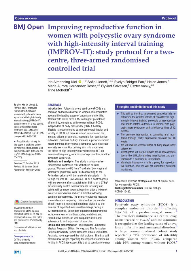
1Kiel!IA, et!al. BMJ Open 2020;10:e034733. doi:10.1136/bmjopen-2019-034733
Open access
Improving reproductive function in
women with polycystic ovary syndrome
with high- intensity interval training
(IMPROV- IT): study protocol for a two-
centre, three- armed randomised
controlled trial
Ida Almenning Kiel ,1,2 So!e Lionett,1,2,3 Evelyn Bridget Parr,3 Helen Jones,4 Maria Aurora Hernandez Røset,2,5 Øyvind Salvesen,6 Eszter Vanky,2,5 Trine Moholdt1,2
To cite: Kiel!IA, Lionett!S, Parr!EB, et!al. Improving reproductive function in women with polycystic ovary syndrome with high- intensity interval training (IMPROV- IT): study protocol for a two- centre, three- armed randomised controlled trial. BMJ Open 2020;10:e034733. doi:10.1136/bmjopen-2019-034733
► Prepublication history for this paper is available online. To view these "les, please visit the journal online (http:// dx. doi. org/ 10. 1136/ bmjopen- 2019- 034733).
Received 03 October 2019Revised 15 January 2020Accepted 04 February 2020
For numbered af"liations see end of article.
Correspondence toDr Trine Moholdt; trine. moholdt@ ntnu. no
Protocol
© Author(s) (or their employer(s)) 2020. Re- use permitted under CC BY- NC. No commercial re- use. See rights and permissions. Published by BMJ.
ABSTRACTIntroduction Polycystic ovary syndrome (PCOS) is a common endocrine disorder in women of reproductive age and the leading cause of anovulatory infertility. Women with PCOS have a 15- fold higher prevalence of infertility, compared with women without PCOS, independent of body mass index (BMI). A healthy lifestyle is recommended to improve overall health and fertility in PCOS but there is limited evidence on the isolated effects of exercise, especially for reproductive outcomes. Previous "ndings indicate superior metabolic health bene"ts after vigorous compared with moderate- intensity exercise. Our primary aim is to determine the effect of high- intensity interval training (HIT) on menstrual frequency, as a proxy of reproductive function, in women with PCOS.Methods and analysis The study is a two- centre, randomised, controlled trial with three parallel groups. Women (n=64) from Trondheim (Norway) and Melbourne (Australia) with PCOS according to the Rotterdam criteria will be randomly allocated (1:1:1) to high- volume HIT, low- volume HIT or a control group with no exercise after stratifying for BMI < or # 27 kg/m2 and study centre. Measurements for study end points will be undertaken at baseline, after a 16 week exercise intervention and at 12 months following baseline assessments. The primary outcome measure is menstruation frequency, measured as the number of self- reported menstrual bleedings divided by the number of expected menstrual bleedings during a 12- month period. Secondary outcome measurements include markers of cardiovascular, metabolic and reproductive health, as well as quality of life and adherence to and enjoyment of exercise.Ethics and dissemination The Regional Committee Medical Research Ethics, Norway, and The Australian Catholic University Human Research Ethics Committee, Australia, have approved the trial protocol. This trial will provide new insight regarding the impact of exercise on fertility in PCOS. We expect this trial to contribute to new
therapeutic exercise strategies as part of clinical care for women with PCOS.Trial registration number Clinical trial gov NCT02419482.
INTRODUCTIONPolycystic ovary syndrome (PCOS) is a complex endocrine disorder1 2 affecting 8%–13% of reproductive- aged women.3 The ovulatory disturbance is a central diag-nostic feature of PCOS,4 and the syndrome is recognised as the leading cause of anovu-latory infertility and menstrual disorders.5 A large community- based cohort study reported a 72% prevalence of infertility among women with PCOS, compared with 16% among women without PCOS.6
Strengths and limitations of this study
► This will be the "rst randomised controlled trial to determine the isolated effects of two different high- intensity interval training protocols on reproductive and health- related outcomes in women with poly-cystic ovary syndrome, with a follow- up time of 12 months.
► The exercise intervention is controlled and mon-itored through partly supervised sessions for 16 weeks.
► We will include women within all body mass index categories.
► Investigators will not be blinded for all assessments due to the dif"culty blinding investigators and par-ticipants to a behavioural intervention.
► Menstrual frequency is only a proxy for reproduc-tive function, and we will not undertake ovulation monitoring.
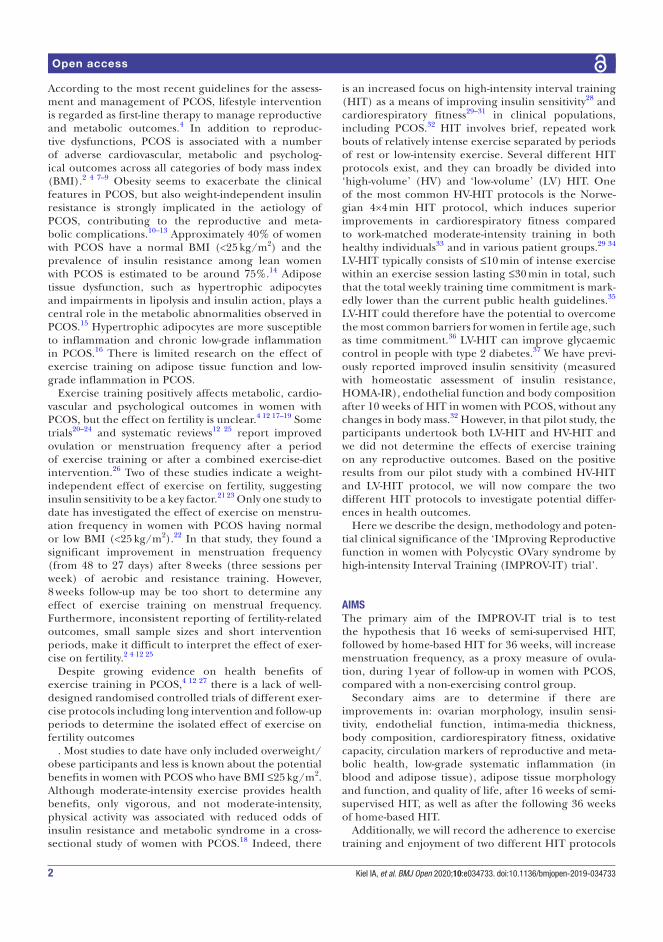
2 Kiel!IA, et!al. BMJ Open 2020;10:e034733. doi:10.1136/bmjopen-2019-034733
Open access
According to the most recent guidelines for the assess-ment and management of PCOS, lifestyle intervention is regarded as first- line therapy to manage reproductive and metabolic outcomes.4 In addition to reproduc-tive dysfunctions, PCOS is associated with a number of adverse cardiovascular, metabolic and psycholog-ical outcomes across all categories of body mass index (BMI).2 4 7–9 Obesity seems to exacerbate the clinical features in PCOS, but also weight- independent insulin resistance is strongly implicated in the aetiology of PCOS, contributing to the reproductive and meta-bolic complications.10–13 Approximately 40% of women with PCOS have a normal BMI (<25 kg/m2) and the prevalence of insulin resistance among lean women with PCOS is estimated to be around 75%.14 Adipose tissue dysfunction, such as hypertrophic adipocytes and impairments in lipolysis and insulin action, plays a central role in the metabolic abnormalities observed in PCOS.15 Hypertrophic adipocytes are more susceptible to inflammation and chronic low- grade inflammation in PCOS.16 There is limited research on the effect of exercise training on adipose tissue function and low- grade inflammation in PCOS.
Exercise training positively affects metabolic, cardio-vascular and psychological outcomes in women with PCOS, but the effect on fertility is unclear.4 12 17–19 Some trials20–24 and systematic reviews12 25 report improved ovulation or menstruation frequency after a period of exercise training or after a combined exercise- diet intervention.26 Two of these studies indicate a weight- independent effect of exercise on fertility, suggesting insulin sensitivity to be a key factor.21 23 Only one study to date has investigated the effect of exercise on menstru-ation frequency in women with PCOS having normal or low BMI (<25 kg/m2).22 In that study, they found a significant improvement in menstruation frequency (from 48 to 27 days) after 8 weeks (three sessions per week) of aerobic and resistance training. However, 8 weeks follow- up may be too short to determine any effect of exercise training on menstrual frequency. Furthermore, inconsistent reporting of fertility- related outcomes, small sample sizes and short intervention periods, make it difficult to interpret the effect of exer-cise on fertility.2 4 12 25
Despite growing evidence on health benefits of exercise training in PCOS,4 12 27 there is a lack of well- designed randomised controlled trials of different exer-cise protocols including long intervention and follow- up periods to determine the isolated effect of exercise on fertility outcomes
. Most studies to date have only included overweight/obese participants and less is known about the potential benefits in women with PCOS who have BMI !25 kg/m2. Although moderate- intensity exercise provides health benefits, only vigorous, and not moderate- intensity, physical activity was associated with reduced odds of insulin resistance and metabolic syndrome in a cross- sectional study of women with PCOS.18 Indeed, there
is an increased focus on high- intensity interval training (HIT) as a means of improving insulin sensitivity28 and cardiorespiratory fitness29–31 in clinical populations, including PCOS.32 HIT involves brief, repeated work bouts of relatively intense exercise separated by periods of rest or low- intensity exercise. Several different HIT protocols exist, and they can broadly be divided into ‘high- volume’ (HV) and ‘low- volume’ (LV) HIT. One of the most common HV- HIT protocols is the Norwe-gian 4!4 min HIT protocol, which induces superior improvements in cardiorespiratory fitness compared to work- matched moderate- intensity training in both healthy individuals33 and in various patient groups.29 34 LV- HIT typically consists of !10 min of intense exercise within an exercise session lasting !30 min in total, such that the total weekly training time commitment is mark-edly lower than the current public health guidelines.35 LV- HIT could therefore have the potential to overcome the most common barriers for women in fertile age, such as time commitment.36 LV- HIT can improve glycaemic control in people with type 2 diabetes.37 We have previ-ously reported improved insulin sensitivity (measured with homeostatic assessment of insulin resistance, HOMA- IR), endothelial function and body composition after 10 weeks of HIT in women with PCOS, without any changes in body mass.32 However, in that pilot study, the participants undertook both LV- HIT and HV- HIT and we did not determine the effects of exercise training on any reproductive outcomes. Based on the positive results from our pilot study with a combined HV- HIT and LV- HIT protocol, we will now compare the two different HIT protocols to investigate potential differ-ences in health outcomes.
Here we describe the design, methodology and poten-tial clinical significance of the ‘IMproving Reproductive function in women with Polycystic OVary syndrome by high- intensity Interval Training (IMPROV- IT) trial’.
AIMSThe primary aim of the IMPROV- IT trial is to test the hypothesis that 16 weeks of semi- supervised HIT, followed by home- based HIT for 36 weeks, will increase menstruation frequency, as a proxy measure of ovula-tion, during 1 year of follow- up in women with PCOS, compared with a non- exercising control group.
Secondary aims are to determine if there are improvements in: ovarian morphology, insulin sensi-tivity, endothelial function, intima- media thickness, body composition, cardiorespiratory fitness, oxidative capacity, circulation markers of reproductive and meta-bolic health, low- grade systematic inflammation (in blood and adipose tissue), adipose tissue morphology and function, and quality of life, after 16 weeks of semi- supervised HIT, as well as after the following 36 weeks of home- based HIT.
Additionally, we will record the adherence to exercise training and enjoyment of two different HIT protocols
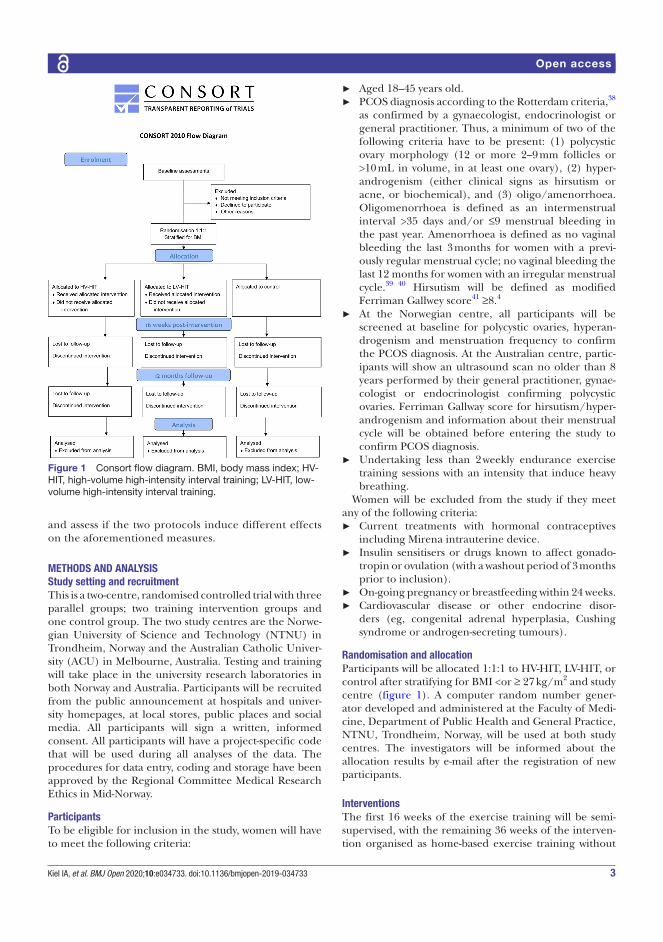
3Kiel!IA, et!al. BMJ Open 2020;10:e034733. doi:10.1136/bmjopen-2019-034733
Open access
Figure 1 Consort "ow diagram. BMI, body mass index; HV- HIT, high- volume high- intensity interval training; LV- HIT, low- volume high- intensity interval training.
and assess if the two protocols induce different effects on the aforementioned measures.
METHODS AND ANALYSISStudy setting and recruitmentThis is a two- centre, randomised controlled trial with three parallel groups; two training intervention groups and one control group. The two study centres are the Norwe-gian University of Science and Technology (NTNU) in Trondheim, Norway and the Australian Catholic Univer-sity (ACU) in Melbourne, Australia. Testing and training will take place in the university research laboratories in both Norway and Australia. Participants will be recruited from the public announcement at hospitals and univer-sity homepages, at local stores, public places and social media. All participants will sign a written, informed consent. All participants will have a project- specific code that will be used during all analyses of the data. The procedures for data entry, coding and storage have been approved by the Regional Committee Medical Research Ethics in Mid- Norway.
ParticipantsTo be eligible for inclusion in the study, women will have to meet the following criteria:
► Aged 18–45 years old. ► PCOS diagnosis according to the Rotterdam criteria,38
as confirmed by a gynaecologist, endocrinologist or general practitioner. Thus, a minimum of two of the following criteria have to be present: (1) polycystic ovary morphology (12 or more 2–9 mm follicles or >10 mL in volume, in at least one ovary), (2) hyper-androgenism (either clinical signs as hirsutism or acne, or biochemical), and (3) oligo/amenorrhoea. Oligomenorrhoea is defined as an intermenstrual interval >35 days and/or !9 menstrual bleeding in the past year. Amenorrhoea is defined as no vaginal bleeding the last 3 months for women with a previ-ously regular menstrual cycle; no vaginal bleeding the last 12 months for women with an irregular menstrual cycle.39 40 Hirsutism will be defined as modified Ferriman Gallwey score41 "8.4
► At the Norwegian centre, all participants will be screened at baseline for polycystic ovaries, hyperan-drogenism and menstruation frequency to confirm the PCOS diagnosis. At the Australian centre, partic-ipants will show an ultrasound scan no older than 8 years performed by their general practitioner, gynae-cologist or endocrinologist confirming polycystic ovaries. Ferriman Gallway score for hirsutism/hyper-androgenism and information about their menstrual cycle will be obtained before entering the study to confirm PCOS diagnosis.
► Undertaking less than 2 weekly endurance exercise training sessions with an intensity that induce heavy breathing.
Women will be excluded from the study if they meet any of the following criteria:
► Current treatments with hormonal contraceptives including Mirena intrauterine device.
► Insulin sensitisers or drugs known to affect gonado-tropin or ovulation (with a washout period of 3 months prior to inclusion).
► On- going pregnancy or breastfeeding within 24 weeks. ► Cardiovascular disease or other endocrine disor-
ders (eg, congenital adrenal hyperplasia, Cushing syndrome or androgen- secreting tumours).
Randomisation and allocationParticipants will be allocated 1:1:1 to HV- HIT, LV- HIT, or control after stratifying for BMI <or " 27 kg/m2 and study centre (figure 1). A computer random number gener-ator developed and administered at the Faculty of Medi-cine, Department of Public Health and General Practice, NTNU, Trondheim, Norway, will be used at both study centres. The investigators will be informed about the allocation results by e- mail after the registration of new participants.
InterventionsThe first 16 weeks of the exercise training will be semi- supervised, with the remaining 36 weeks of the interven-tion organised as home- based exercise training without
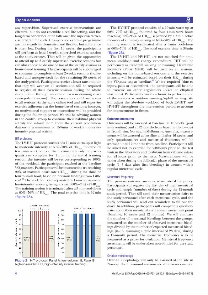
4 Kiel!IA, et!al. BMJ Open 2020;10:e034733. doi:10.1136/bmjopen-2019-034733
Open access
Figure 2 HIT protocol. Panel A: low- volume hit; Panel B: high- volume hit. HIT, high- intensity interval training.
any supervision. Supervised exercise interventions are effective, but do not resemble a real- life setting, and the long- term adherence often falls once the supervised exer-cise programme ends. Unsupervised exercise programme are more easily implemented and flexible, but adherence is often low. During the first 16 weeks, the participants will perform at least 1 weekly supervised exercise session at the study centres. They will be given the opportunity to attend up to 3 weekly supervised exercise sessions but can also choose to do one or two of the weekly sessions as home- based training. The participants will be encouraged to continue to complete at least 2 weekly sessions (home- based and unsupervised) for the remaining 36 weeks of the study period. Participants receive a heart rate monitor that they will wear on all sessions and will be required to register all their exercise sessions during the whole study period through an online exercise- training diary ( www. polar. flow. com). The researchers will have access to all sessions via the same online tool and will supervise exercise adherence at the home- based sessions; however, no motivational support or instructions will be provided during the follow- up period. We will be advising women in the control group to continue their habitual physical activity and inform them about the current recommen-dations of a minimum of 150 min of weekly moderate- intensity physical activity.
HIT protocolsThe LV- HIT protocol consists of a 10 min warm- up at light to moderate intensity at 60%–70% of HRmax followed by ten 1 min work bouts at the maximal intensity the partic-ipants can complete for 1 min. In the initial training session, the intensity will be set corresponding to 100% of the workload the participant reached at the baseline VO2max test. Participants will be instructed to try reaching 90% of maximal heart rate (HRmax) during the third or fourth work bout, based on previous findings from Little et al.37 The work bouts are separated by 1 min of passive or low- intensity recovery, trying to reach 60%–70% of HRmax. The training session is terminated after a 3 min cool- down at 60%–70% of HRmax. The total exercise time is 32 min (figure 2A).
The HV- HIT protocol consists of a 10 min warm- up at 60%–70% of HRmax, followed by four 4 min work bouts reaching 90%–95% of HRmax, separated by a 3 min active recovery of running/walking at 60%–70% of HRmax. The training session is terminated after a 3 min cool- down at 60%–70% of HRmax. The total exercise time is 38 min (figure 2B).
The LV- HIT and HV- HIT are not matched for time, mean workload and energy expenditure. HIT will be performed as treadmill walking or running. Heart rate monitors (Polar M400) will be used on all sessions, including on the home- based sessions, and the exercise intensity will be estimated based on their HRmax during the VO2max test at baseline.42 Where required (due to injury, pain or discomfort), the participants will be able to exercise on other ergometers (bikes or elliptical machines). Participants can also choose to perform some of the sessions as outdoor running/uphill walking. We will adjust the absolute workload of both LV- HIT and HV- HIT throughout the intervention period to account for improvements in fitness.
Outcome measuresOutcomes will be assessed at baseline, at 16 weeks (post intervention) and at 12 months from baseline (follow- up) in Trondheim, Norway. In Melbourne, Australia, measure-ments will be assessed at baseline and after 16 weeks, and only questionnaires and menstrual frequency will be assessed until 12 months from baseline. Participants will be asked not to exercise for >48 hours prior to the test visits in the laboratory and to abstain from caffeine intake for 24 hours prior to the tests. Measurements will be undertaken during the follicular phase of the menstrual cycle (1–7 days after first bleeding) in women with a regular menstrual cycle.
Menstrual frequencyThe primary outcome measure is menstrual frequency. Participants will register the first day of their menstrual cycle and length (number of days) during the 12- month study period. They will send their menstruation dairy to the study personnel after each menstrual cycle, and the study personnel will send out reminders to fill out the diary. In addition, participants will complete a question-naire about their menstrual cycle at each assessment point (baseline, 16 weeks and 12 months). We will compare the number of menstrual bleedings between the groups, measured as the number of observed menstrual bleed-ings divided by the number of expected menstrual bleed-ings (n=13, assuming a cycle interval of 28 days) during a 12- month period. The menstrual frequency is to be measured as a proxy for ovulation. Menstrual frequency assessments will be undertaken non- blinded for the study personnel.
Ovarian morphologyOvarian morphology will only be assessed at the site in Norway. The ultrasound assessments of the ovaries include
5Kiel!IA, et!al. BMJ Open 2020;10:e034733. doi:10.1136/bmjopen-2019-034733
Open access
ovarian volume in mL (with and without a dominating follicle), dominating follicle volume in mL, number of follicles in each ovary, diameter and distribution of folli-cles. We will use a multifrequency transvaginal transducer on all measurements. An experienced gynaecologist (MAHR) will perform all imaging and measurements. Ovarian morphology assessments will be undertaken blinded for group allocation.
Fertility and pregnanciesBackground information regarding fertility will be regis-tered by questionnaires. These include questions about number of children, natural or assisted fertilisation, if the participants are actively seeking pregnancy or have tried to become pregnant, for how long they have tried to become pregnant, and if they have experienced miscar-riages or had abortions. We will report the number of pregnancies during the intervention period, including during the 12 months of follow- up. Questionnaires will be undertaken blinded, whereas the number of pregnancies will be undertaken non- blinded for the study personnel.
Blood biochemistry and insulin sensitivityBlood samples will be obtained after an overnight fast (no food or fluid intake except water 12 hours prior to assessments) at each time point. Analysis of fasting venous blood samples will include measurements of 17- OH progesterone (baseline only), prolactin (baseline only), haemoglobin, glycated haemoglobin, blood lipids (total cholesterol, high- density lipoprotein cholesterol, low- density lipoprotein cholesterol and triglycerides), blood glucose, white blood cell count (neutrophils, basophils, eosinophils, lymphocytes and monocytes) and leucocytes. Additional blood (including buffy coat) will be stored for later analyses. These later analyses will likely include, but are not limited to, androstenedione, anti- Müllerian hormone, sex hormone- binding globulin, testosterone, insulin, leptin, adiponectin and micro- RNAs. We will also obtain two Tempus RNA tubes from each participant for later assessments of gene expression.
After fasting blood samples, glycaemic control will be measured using a 2- hour oral glucose tolerance test (OGTT). The participants will consume 75 g of glucose diluted in 250 mL water. Blood will be sampled for insulin and glucose measurements at 0 (prior to the OGGT), 30, 60, 90 and 120 min from an indwelling catheter. Glycaemic control will be calculated as total area under the curve (AUC) and incremental area under the curve (iAUC; using fasting concentrations as baseline values) using the trapezoid method, for glucose and insulin concentra-tions, and peak concentrations during the OGTT. Insulin sensitivity will be estimated using the HOMA- IR; fasting serum insulin in µU/mL ! fasting plasma glucose in mmol/L/22.5.43
Biological materials collected at NTNU will be stored in the Regional Research Biobank at St Olav’s Hospital (Biobank1, https:// biobank1. no/ nb/). Biological mate-rials from collected at ACU will be stored locally until
data collection is completed in Australia, and later sent to Norway. Sample collection and handling are performed in accordance with hospital/laboratory standard proce-dures. Blood sampling and OGTT will be undertaken blinded in Norway and unblinded in Australia.
Cardiorespiratory "tness and substrate utilisationPeak oxygen uptake will be measured on a treadmill, using indirect calorimetry (Oxycon Pro, Jaeger, Germany in Norway/TrueOneRMR, Parvo Medics, USA in Australia). Participants will walk or run until voluntary exhaustion using an individualised protocol. After a 10 min walking warm- up, the test will start by walking at moderate intensity for 3 min. The speed or inclination will then be increased every 1–2 min, by 0.5–1.0 km/h or 1%–2%. The cardiore-spiratory fitness test will be considered successful with a plateau in VO2 with a further increase in workload, and a respiratory exchange ratio "1.05. Peak oxygen uptake will be calculated as the highest consecutive 30 s measured, both absolute (mL/min) and relative (mL/min/kg). The maximal heart rate obtained during the exercise test at baseline will serve as the basis for calculating the intensity during the HIT sessions.
After an overnight fast (no food or fluid intake except water 12 hours prior to assessments), participants will complete a submaximal test on a treadmill. The protocol includes a 20 min warm- up, followed by 20 min steady- state expired gas sampling at 60% of peak oxygen uptake. We will ask participants to record their diet the day before the baseline test of oxidative capacity and to repeat this diet before the subsequent measures (at 16 weeks and 12 months). Fat oxidation rates (g/min) will be calculated from 5 min of stable oxygen uptake at the final stage of the test (during the last 10 min), using the following equation: 1.695 ! VO2 – 1.701 ! VCO2,
44 where VO2 is oxygen uptake and VCO2 is the volume of expired carbon dioxide. Assessments of these outcomes will be under-taken non- blinded for the study personnel.
Adipose tissue composition, morphology and functionAbdominal and gluteal subcutaneous adipose tissue biop-sies (~300–500 mg) will be collected under local anaes-thesia (1% xylocaine) using a 14- G sterile needle. For a subsample of participants, ~80 mg of each biopsy will be allocated for immediate analyses of mitochondrial respi-ration using high- resolution respirometry (Oxygraph- 2K, Oroboros, Austria). Approximately 200–300 mg will be snap- frozen in liquid nitrogen and stored at "80°C for later analyses, such as micro RNAs, gene expression and protein profiling. The remaining tissue will be immedi-ately fixed in phosphate- buffered formalin, processed and embedded in paraffin and sectioned at 4 µm for morphology and inflammation analyses. The sections will be incubated with CD45 and CD68 antibodies to detect leucocytes and macrophages. These sections will be captured with an EVOS FL Auto 2 Imaging System (ThermoFisher Scientific, USA) and analysed with the
6 Kiel!IA, et!al. BMJ Open 2020;10:e034733. doi:10.1136/bmjopen-2019-034733
Open access
open- source software ImageJ (Fiji).45 Assessments of these outcomes will be undertaken blinded.
Physical activity and dietWe will monitor physical activity by questionnaires and 5- day activity monitoring (Sensewear Armband, APC Cardiovascular, UK) and record diet through a 4- day diet recall, at each measurement point. Assessments of phys-ical activity and diet will be undertaken blinded.
Quality of lifeQuality of life will be assessed using the Polycystic Ovary Syndrome Questionnaire.46 Assessment of this outcome will be undertaken blinded.
Anthropometrics and body compositionAnthropometric measurements will be conducted when participants are fasted (no food or fluid intake except water 12 hours prior to assessments). Height will be measured standing, without shoes using a standard stadi-ometer. Total body mass and body composition will be measured wearing light clothing with no metal items and without shoes or socks using bioelectrical impedance analysis (InBody720 bioimpedance scale, Biospace CO, Korea) in Norway and dual- energy X- ray absorptiom-etry (DXA; GE Lunar iDXA Pro, encore software version 16, General Electric, Boston, Massachusetts, USA) in Australia. For the DXA scan, participants are required to lay supine on the scanning bed for the duration of the scan, which is approximately 15 min with one or two scans depending on the body shape of the participant. Waist and hip circumference will be measured to the nearest 0.5 cm horizontally at the level of the umbilicus, while standing and at normal expiration, using a metric tape. Assessments of these outcomes will be undertaken non- blinded for the study personnel.
Blood pressure and resting heart rateBlood pressure and resting heart rate will be measured in the seated position after 15 min rest with an auto-matic blood pressure device three times on the left arm (diastolic and systolic, in mm Hg). The mean of the three measurements will be used to calculate blood pressure and resting heart rate. Assessments of these outcomes will be undertaken blinded in Norway and unblinded in Australia.
Endothelial function and intima-media thicknessEndothelial function and intima- media thickness (IMT) will be assessed at the Norwegian site only. Following a 20 min supine rest period, the diameter of the brachial artery will be imaged (12 MHz Doppler probe, GE Vingmed Ultrasound AS, Horten, Norway). A cuff will be placed around the forearm (immediately distal to the olecranon) to produce the stimulus of forearm isch-aemia. When an optimal image is obtained, the probe will be held stable and the ultrasound parameters will be set to optimise the longitudinal, B- mode image of the lumen–arterial wall interface. The ultrasound will
also be used to attain simultaneous continuous Doppler velocity using the lowest possible insonation angle (60°). A recording of resting diameter and velocity will be taken for 1 min, then the forearm cuff will be inflated (>200 mm Hg) for 5 min. Both diameter and velocity recordings will resume 30 s before cuff deflation and continue for 3 min after deflation.47 This flow- mediated dilation (FMD) test is a measure of endothelial function. We will also measure carotid artery IMT in three angles: transversal, longitudinal and anterolateral using ultrasound (12 MHz Doppler probe, GE Vingmed Ultrasound AS, Horten, Norway). Measurements of the diameter will be imaged 1 cm below the bifurcation on the right side. Images will be obtained where the near wall is clearly visualised, with a double- line pattern and at the minimal diameter during the cardiac cycle as previously described elsewhere.48 The same person (IAK) will perform all the FMD and IMT measurements. Assessments of these outcomes will be undertaken non- blinded for the study personnel.
EnjoymentA subgroup of participants (the last 40 to be included in the trial) allocated to one of the training groups will complete a Physical Activity Enjoyment Scale (PACES)49 as well as The Borg’s scale50 to assess enjoyment and perceived exertion at one supervised weekly training session. Assessments of these outcomes will be under-taken non- blinded for the study personnel.
Sample size and statistical analysisWe computed the sample size for a one- way analysis of variance test with three groups. In women with PCOS, a menstrual frequency of on average 4.5 menstrual bleeding during 1 year is expected.51 With a statistical power of 80%, a significance level of 0.05 and a SD of two menstrual bleeding during a 12- month period, we calculated that 48 women will be required to detect an increase in menstrual bleeding to 7.5 in the intervention groups.26 Because of the non- normality of menstrual frequency, it may be necessary to use a non- parametric test. Non- parametric tests require more participants and we, therefore, added 15% to the required sample size. Additionally, to allow for expected dropouts of 10%–15%, we aim to include 64 women in the study.
We will perform all the statistical analyses blinded for group allocation. The primary analysis will be a compar-ison between groups for the number of menstrual bleeding during 12 months. We will adjust for the self- reported menstrual frequency at baseline. We will include all women who have reported menstrual frequency in the primary analysis, independent of adherence to the inter-ventions, that is, intention- to- treat analysis. The number of pregnancies during the 16 weeks of intervention and during the 12- month follow- up will be compared between groups. Secondary outcome measures will be compared between groups after 16 weeks and after 12 months and adjusted for baseline values. We will do additional ‘per protocol’ analyses where we include women in the HIT
7Kiel!IA, et!al. BMJ Open 2020;10:e034733. doi:10.1136/bmjopen-2019-034733
Open access
groups that have completed >75% of the scheduled exer-cise sessions during the intervention period (for compar-isons after 16 weeks) and a mean minimum of one HIT session/week during the 12- month follow- up (for compar-isons at the end of follow- up).
Results will be reported as means with 95% CIs and/or SD. We will report the effect of HIT as mean changes from baseline to 16 weeks of intervention and after 12 months of follow- up. We will use mixed models to test differences between groups. P values <0.05 will be considered signif-icant for both primary and secondary outcomes. We will also perform subgroup analyses to investigate differences between BMI categories and compare adherence rates and enjoyment of the two HIT protocols.
BlindingWe are unable to blind group allocation to participants or study personnel due to the nature of the intervention (supervised exercise training). All baseline assessments will be undertaken prior to randomisation and some assessments will be undertaken blinded for group alloca-tion (as outlined for each outcome measure).
MonitoringThe investigators are responsible for the documentation of any adverse event or serious adverse event. Participants will be told to contact the investigator if they have any unusual symptoms. We will record all medical events during the study in the Case Report Form. In addition, we record serious adverse events in a Serious Adverse Events Report Form. All serious adverse events will be reported to the sponsor within 24 hours after the site has gained knowledge of the event.
Patient and public involvementNo patients were involved in the development of the research question or design of the study. All partici-pants will be invited to a 1- hour educational session about healthy diet and physical activity. We will also give general information about PCOS in these meetings and encourage the participants to ask questions and give us feedback about relevant topics or issues related to their disorder and to the trial implementation. Individual test results will be disseminated to each participant after testing. We will also send out a summary of the study results to all participants at the completion of the study.
ETHICS AND DISSEMINATIONThe study is approved by the Regional Committee Medical Research Ethics in Mid- Norway and The ACU Human Research Ethics Committee and has its origin in the Declaration of Helsinki and is consistent with ICH/Good Clinical Practice and applicable regulatory require-ments. All protocol modification must be approved by the Regional Committee Medical Research Ethics in Mid- Norway and by The ACU Human Research Ethics Committee.
We will publish the results from the study as peer- reviewed articles in international journals. Our results will also be communicated through the Norwegian society of infertility, through social media channels as well as at national and international conferences.
DISCUSSIONInfertility and risk of lifestyle- related diseases (obesity, metabolic, cardiovascular and psychological diseases) are the main health challenges associated with PCOS, both for the individuals affected by the syndrome, the healthcare systems and society.2 4 To date, no treatment fully reverses or cures the symptoms of PCOS40 and lifestyle interven-tions are recommended as first- line therapy according to recent guidelines.4 52 Nevertheless, there is limited research available to give women with PCOS sufficient recommendations on physical activity and exercise. Some studies have demonstrated positive effects in reproductive outcomes after exercise, with concomitant improvements in insulin sensitivity and/or visceral fat.20–24 However, more knowledge is needed about the underlying mecha-nisms for improvement in fertility after exercise training, as discussed in several systematic reviews on lifestyle inter-ventions and fertility.12 25 53 54 HIT has shown to be time efficient, safe and well tolerated, with positive effects on metabolic and cardiovascular risk factors in several studies.29 55 56 Larger studies investigating the adherence to HIT are lacking among women in general, and among women with PCOS in particular. This is of great interest, as we need to find exercise and lifestyle programmes that are feasible over time and that can be easily implemented in everyday life. Exercise programmes that are efficient in a research setting are not necessarily effective in a real- life setting. Thus, there is a great need for studies with a longer intervention and follow- up period.
Our trial has some limitations. The use of menstrual frequency as a proxy for ovulation/reproductive function is one of these. Due to practical reasons, we are not able to undertake ovulation monitoring. The Inbody 720 and DXA are both reliable methods to measure fat- free mass (FFM), body fat percentage (BF%) and fat mass (FM) but it has been shown that the InBody 720 overestimates FFM and underestimates BF% and FM compared with DXA .57 For each individual participant, the same measurement method will be used (InBody 720 in Norway and DXA in Australia) and our purpose is to evaluate changes in body composition (ie, delta changes). Assessing enjoyment of exercise only at the supervised sessions could imply a bias. On the supervised sessions, the participants will receive motivational support from the researchers and potentially also social support from other participants that can affect their ratings of enjoyment. The enjoy-ment ratings are, however, implemented to compare the perceived enjoyment of the two HIT protocols. The semi- supervised nature of the trial could imply that some participants choose to undertake all sessions supervised, whereas others choose to exercise at home for most
8 Kiel!IA, et!al. BMJ Open 2020;10:e034733. doi:10.1136/bmjopen-2019-034733
Open access
sessions. We chose to implement a semi- supervised exer-cise programme to account for the individual preferences of the participants and to be able to include women living further away from the study centres. Such protocols can both control the exercise intervention and motivate participants, and at the same time give more flexibility based on participants’ preferences.
The IMPROV- IT trial will determine the effect of HIT on reproductive, cardiovascular, metabolic and psychological health in women with PCOS. The trial will contribute evidence of more specific exercise protocols in the treatment and management of PCOS for clinicians and answer some of the current limitations addressed by both the International evidence- based guidelines for the assessment and management of PCOS4 and several systematic reviews.2 12 25 58 This trial is highly relevant and important as PCOS is a major burden for the women with PCOS, the health services and society. As for now, we do not have an optimal treatment.
Trial statusWe have recruited 64 women with PCOS to the IMPROV- IT study, with the last included participant in March 2019. The last follow- up will be in March 2020.
Author af!liations1Department of Circulation and Medical Imaging, Faculty of Medicine and Health Sciences, Norwegian University of Science and Technology, Trondheim, Norway2Department of Obstetrics and Gynaecology, St Olavs Hospital, Trondheim University Hospital, Trondheim, Norway3Exercise and Nutrition Research Programme, Mary Mackillop Institute for Health Research, Australian Catholic University, Melbourne, Victoria, Australia4Research Institute for Sport and Exercise Science (RISES), Liverpool John Moores University, Liverpool, UK5Department of Clinical and Molecular Medicine, Faculty of Medicine and Health Sciences, Norwegian University of Science and Technology, Trondheim, Norway6Unit of Applied Clinical Research, Department of Public Health and Nursing, Faculty of Medicine and Health Sciences, Norwegian University of Science and Technology, Trondheim, Norway
Twitter Trine Moholdt @trinemoholdt
Acknowledgements The Unit for Applied Clinical Research, NTNU, provided the internet- based randomisation. The following acknowledgements apply to the Trondheim site of the trial: The equipment and lab facilities for the training sessions and exercise testing will be provided by NeXT Move, Norwegian University of Science and Technology. Blood sampling and fat biopsies will be undertaken at the Clinical Research Facility, St. Olavs Hospital. Blood and adipose tissue samples are stored in the Regional Biobank 1® of Central Norway, with the data solution BioByte.
Contributors IAK: drafted the manuscript. IAK, TM, EV, ØS and HJ: conceived and contributed to the design of the study. IAK, SL, TM and EP coordinated the study at the two sites and supervised the exercise training. IAK, SL, TM, MAHR and EP: performed measurements on test- days. EV: provided medical advice and support during the study. All authors provided feedback and approved the "nal manuscript.
Funding This work was supported by the Liaison Committee for education, research and innovation in Central Norway (Grant number: 2014/23166), the Norwegian University of Science and Technology and the Australian Catholic University. Next Move is funded by the Faculty of Medicine and Health Sciences, NTNU and Central Norway Regional Health Authority.
Competing interests None declared.
Patient consent for publication Not required.
Provenance and peer review Not commissioned; externally peer reviewed.
Open access This is an open access article distributed in accordance with the Creative Commons Attribution Non Commercial (CC BY- NC 4.0) license, which permits others to distribute, remix, adapt, build upon this work non- commercially, and license their derivative works on different terms, provided the original work is properly cited, appropriate credit is given, any changes made indicated, and the use is non- commercial. See:!http:// creativecommons. org/ licenses/ by- nc/ 4. 0/.
ORCID iDIda Almenning!Kiel http:// orcid. org/ 0000- 0001- 9926- 4994
REFERENCES 1 Yildiz BO, Bozdag G, Yapici Z, et!al. Prevalence, phenotype and
cardiometabolic risk of polycystic ovary syndrome under different diagnostic criteria. Hum Reprod 2012;27:3067–73.
2 Lim SS, Hutchison SK, Van Ryswyk E, et!al. Lifestyle changes in women with polycystic ovary syndrome. Cochrane Database Syst Rev 2019;3:Cd007506.
3 Bozdag G, Mumusoglu S, Zengin D, et!al. The prevalence and phenotypic features of polycystic ovary syndrome: a systematic review and meta- analysis. Hum Reprod 2016;31:2841–55.
4 Teede H, Misso M, Costello M, et!al. International evidence- based guideline for the assessment and management of polycystic ovary syndrome 2018. Melbourne, Australia: Monash University, 2018.
5 Goodarzi MO, Dumesic DA, Chazenbalk G, et!al. Polycystic ovary syndrome: etiology, pathogenesis and diagnosis. Nat Rev Endocrinol 2011;7:219–31.
6 Joham AE, Teede HJ, Ranasinha S, et!al. Prevalence of infertility and use of fertility treatment in women with polycystic ovary syndrome: data from a large community- based cohort study. J Womens Health 2015;24:299–307.
7 Giallauria F, Orio F, Palomba S, et!al. Cardiovascular risk in women with polycystic ovary syndrome. J Cardiovasc Med 2008;9:987–92.
8 Teede H, Deeks A, Moran L. Polycystic ovary syndrome: a complex condition with psychological, reproductive and metabolic manifestations that impacts on health across the lifespan. BMC Med 2010;8:41.
9 Moran LJ, Pasquali R, Teede HJ, et!al. Treatment of obesity in polycystic ovary syndrome: a position statement of the androgen excess and polycystic ovary syndrome Society. Fertil Steril 2009;92:1966–82.
10 Diamanti- Kandarakis E, Papavassiliou AG. Molecular mechanisms of insulin resistance in polycystic ovary syndrome. Trends Mol Med 2006;12:324–32.
11 Corbould A. Insulin resistance in skeletal muscle and adipose tissue in polycystic ovary syndrome: are the molecular mechanisms distinct from type 2 diabetes? Panminerva Med 2008;50:279–94.
12 Harrison CL, Lombard CB, Moran LJ, et!al. Exercise therapy in polycystic ovary syndrome: a systematic review. Hum Reprod Update 2011;17:171–83.
13 Cassar S, Misso ML, Hopkins WG, et!al. Insulin resistance in polycystic ovary syndrome: a systematic review and meta- analysis of euglycaemic- hyperinsulinaemic clamp studies. Hum Reprod 2016;31:2619–31.
14 Stepto NK, Cassar S, Joham AE, et!al. Women with polycystic ovary syndrome have intrinsic insulin resistance on euglycaemic- hyperinsulaemic clamp. Hum Reprod 2013;28:777–84.
15 Villa J, Pratley RE. Adipose tissue dysfunction in polycystic ovary syndrome. Curr Diab Rep 2011;11:179–84.
16 Spritzer PM, Lecke SB, Satler F, et!al. Adipose tissue dysfunction, adipokines, and low- grade chronic in"ammation in polycystic ovary syndrome. Reproduction 2015;149:R219–27.
17 Thomson RL, Buckley JD, Lim SS, et!al. Lifestyle management improves quality of life and depression in overweight and obese women with polycystic ovary syndrome. Fertil Steril 2010;94:1812–6.
18 Greenwood EA, Noel MW, Kao C- N, et!al. Vigorous exercise is associated with superior metabolic pro!les in polycystic ovary syndrome independent of total exercise expenditure. Fertil Steril 2016;105:486–93.
19 Lamb JD, Johnstone EB, Rousseau J- A, et!al. Physical activity in women with polycystic ovary syndrome: prevalence, predictors, and positive health associations. Am J Obstet Gynecol 2011;204:352.e1–352.e6.
20 Vigorito C, Giallauria F, Palomba S, et!al. Bene!cial effects of a three- month structured exercise training program on cardiopulmonary functional capacity in young women with polycystic ovary syndrome. J Clin Endocrinol Metab 2007;92:1379–84.
21 Palomba S, Giallauria F, Falbo A, et!al. Structured exercise training programme versus hypocaloric hyperproteic diet in obese polycystic
9Kiel!IA, et!al. BMJ Open 2020;10:e034733. doi:10.1136/bmjopen-2019-034733
Open access
ovary syndrome patients with anovulatory infertility: a 24- week pilot study. Hum Reprod 2008;23:642–50.
22 Turan V, Mutlu EK, Solmaz U, et!al. Bene!ts of short- term structured exercise in non- overweight women with polycystic ovary syndrome: a prospective randomized controlled study. J Phys Ther Sci 2015;27:2293–7.
23 Nybacka Åsa, Carlström K, Ståhle A, et!al. Randomized comparison of the in"uence of dietary management and/or physical exercise on ovarian function and metabolic parameters in overweight women with polycystic ovary syndrome. Fertil Steril 2011;96:1508–13.
24 Redman LM, Elkind- Hirsch K, Ravussin E. Aerobic exercise in women with polycystic ovary syndrome improves ovarian morphology independent of changes in body composition. Fertil Steril 2011;95:2696–9.
25 Hakimi O, Cameron L- C. Effect of exercise on ovulation: a systematic review. Sports Med 2017;47:1555–67.
26 Thomson RL, Buckley JD, Noakes M, et!al. The effect of a hypocaloric diet with and without exercise training on body composition, cardiometabolic risk pro!le, and reproductive function in overweight and obese women with polycystic ovary syndrome. J Clin Endocrinol Metab 2008;93:3373–80.
27 Hutchison SK, Stepto NK, Harrison CL, et!al. Effects of exercise on insulin resistance and body composition in overweight and obese women with and without polycystic ovary syndrome. J Clin Endocrinol Metab 2011;96:E48–56.
28 Dubé JJ, Allison KF, Rousson V, et!al. Exercise dose and insulin sensitivity: relevance for diabetes prevention. Med Sci Sports Exerc 2012;44:793–9.
29 Tjønna AE, Lee SJ, Rognmo Øivind, et!al. Aerobic interval training versus continuous moderate exercise as a treatment for the metabolic syndrome: a pilot study. Circulation 2008;118:346–54.
30 Cassidy S, Thoma C, Houghton D, et!al. High- Intensity interval training: a review of its impact on glucose control and cardiometabolic health. Diabetologia 2017;60:7–23.
31 Weston KS, Wisløff U, Coombes JS. High- Intensity interval training in patients with lifestyle- induced cardiometabolic disease: a systematic review and meta- analysis. Br J Sports Med 2014;48:1227–34.
32 Almenning I, Rieber- Mohn A, Lundgren KM, et!al. Effects of high intensity interval training and strength training on metabolic, cardiovascular and hormonal outcomes in women with polycystic ovary syndrome: a pilot study. PLoS One 2015;10:e0138793.
33 Helgerud J, Høydal K, Wang E, et!al. Aerobic high- intensity intervals improve VO2max more than moderate training. Med Sci Sports Exerc 2007;39:665–71.
34 Moholdt T, Aamot IL, Granøien I, et!al. Aerobic interval training increases peak oxygen uptake more than usual care exercise training in myocardial infarction patients: a randomized controlled study. Clin Rehabil 2012;26:33–44.
35 Gillen JB, Gibala MJ. Is high- intensity interval training a time- ef!cient exercise strategy to improve health and !tness? Appl Physiol Nutr Metab 2014;39:409–12.
36 Conte F, Banting L, Teede HJ, et!al. Mental health and physical activity in women with polycystic ovary syndrome: a brief review. Sports Med 2015;45:497–504.
37 Little JP, Gillen JB, Percival ME, et!al. Low- Volume high- intensity interval training reduces hyperglycemia and increases muscle mitochondrial capacity in patients with type 2 diabetes. J Appl Physiol 2011;111:1554–60.
38 Rotterdam ESHRE/ASRM- Sponsored PCOS consensus workshop group. Revised 2003 consensus on diagnostic criteria and long- term health risks related to polycystic ovary syndrome (PCOS). Hum Reprod 2004;19:41–7.
39 Roberts- Wilson TK, Spencer JB, Fantz CR. Using an algorithmic approach to secondary amenorrhea: avoiding diagnostic error. Clin Chim Acta 2013;423:56–61.
40 Practice Committee of American Society for Reproductive Medicine. Current evaluation of amenorrhea. Fertil Steril 2008;90:S219–25.
41 Ferriman D, Gallwey JD. Clinical assessment of body hair growth in women. J Clin Endocrinol Metab 1961;21:1440–7.
42 Berglund IJ, Sørås SE, Relling BE, et!al. The relationship between maximum heart rate in a cardiorespiratory !tness test and in a maximum heart rate test. J Sci Med Sport 2019;22:607–10.
43 Matthews DR, Hosker JP, Rudenski AS, et!al. Homeostasis model assessment: insulin resistance and beta- cell function from fasting plasma glucose and insulin concentrations in man. Diabetologia 1985;28:412–9.
44 Jeukendrup AE, Wallis GA. Measurement of substrate oxidation during exercise by means of gas exchange measurements. Int J Sports Med 2005;26 Suppl 1:S28–37.
45 Schindelin J, Arganda- Carreras I, Frise E, et!al. Fiji: an open- source platform for biological- image analysis. Nat Methods 2012;9:676–82.
46 Cronin L, Guyatt G, Grif!th L, et!al. Development of a health- related quality- of- life questionnaire (PCOSQ) for women with polycystic ovary syndrome (PCOS). J Clin Endocrinol Metab 1998;83:1976–87.
47 Thijssen DHJ, Black MA, Pyke KE, et!al. Assessment of "ow- mediated dilation in humans: a methodological and physiological guideline. Am J Physiol Heart Circ Physiol 2011;300:H2–12.
48 Touboul P- J, Hennerici MG, Meairs S, et!al. Mannheim carotid intima- media thickness and plaque consensus (2004-2006-2011). An update on behalf of the Advisory Board of the 3rd, 4th and 5th watching the risk symposia, at the 13th, 15th and 20th European stroke conferences, Mannheim, Germany, 2004, Brussels, Belgium, 2006, and Hamburg, Germany, 2011. Cerebrovasc Dis 2012;34:290–6.
49 Kendzierski D, DeCarlo KJ, Kendzierski D. Physical activity enjoyment scale: two validation studies. J Sport Exerc Psychol 1991;13:50–64.
50 Chen MJ, Fan X, Moe ST. Criterion- related validity of the Borg ratings of perceived exertion scale in healthy individuals: a meta- analysis. J Sports Sci 2002;20:873–99.
51 Hoeger KM, Kochman L, Wixom N, et!al. A randomized, 48- week, placebo- controlled trial of intensive lifestyle modi!cation and/or metformin therapy in overweight women with polycystic ovary syndrome: a pilot study. Fertil Steril 2004;82:421–9.
52 Teede HJ, Misso ML, Costello MF, et!al. Recommendations from the International evidence- based guideline for the assessment and management of polycystic ovary syndrome. Hum Reprod 2018;33:1602–18.
53 Haqq L, McFarlane J, Dieberg G, et!al. Effect of lifestyle intervention on the reproductive endocrine pro!le in women with polycystic ovarian syndrome: a systematic review and meta- analysis. Endocr Connect 2014;3:36–46.
54 Thomson RL, Buckley JD, Brinkworth GD. Exercise for the treatment and management of overweight women with polycystic ovary syndrome: a review of the literature. Obes Rev 2011;12:e202–10.
55 Gillen JB, Little JP, Punthakee Z, et!al. Acute high- intensity interval exercise reduces the postprandial glucose response and prevalence of hyperglycaemia in patients with type 2 diabetes. Diabetes Obes Metab 2012;14:575–7.
56 Earnest CP, Lupo M, Thibodaux J, et!al. Interval training in men at risk for insulin resistance. Int J Sports Med 2013;34:355–63.
57 McLester CN, Nickerson BS, Kliszczewicz BM, et!al. Reliability and agreement of various InBody body composition Analyzers as compared to dual- energy X- ray absorptiometry in healthy men and women. J Clin Densitom 2018. doi:10.1016/j.jocd.2018.10.008. [Epub ahead of print: 03 Nov 2018].
58 Moran LJ, Hutchison SK, Norman RJ, et!al. Lifestyle changes in women with polycystic ovary syndrome. Cochrane Database Syst Rev 2011;41:CD007506.


Paper II

This paper is awaiting publication and is not included in NTNU Open


Paper III

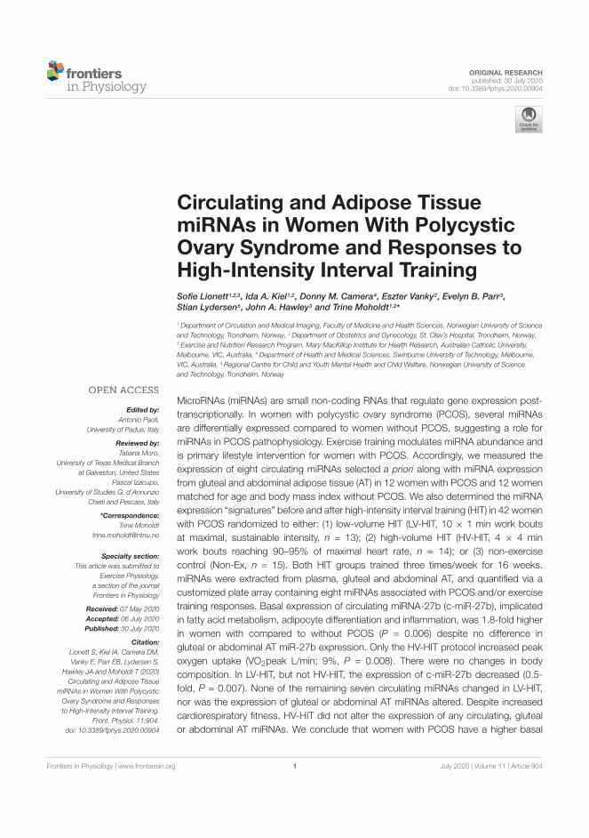
fphys-11-00904 July 28, 2020 Time: 17:59 # 1
ORIGINAL RESEARCHpublished: 30 July 2020
doi: 10.3389/fphys.2020.00904
Edited by:Antonio Paoli,
University of Padua, Italy
Reviewed by:Tatiana Moro,
University of Texas Medical Branchat Galveston, United States
Pascal Izzicupo,University of Studies G. d’Annunzio
Chieti and Pescara, Italy
*Correspondence:Trine Moholdt
Specialty section:This article was submitted to
Exercise Physiology,a section of the journalFrontiers in Physiology
Received: 07 May 2020Accepted: 06 July 2020Published: 30 July 2020
Citation:Lionett S, Kiel IA, Camera DM,Vanky E, Parr EB, Lydersen S,
Hawley JA and Moholdt T (2020)Circulating and Adipose Tissue
miRNAs in Women With PolycysticOvary Syndrome and Responsesto High-Intensity Interval Training.
Front. Physiol. 11:904.doi: 10.3389/fphys.2020.00904
Circulating and Adipose TissuemiRNAs in Women With PolycysticOvary Syndrome and Responses toHigh-Intensity Interval TrainingSofie Lionett1,2,3, Ida A. Kiel1,2, Donny M. Camera4, Eszter Vanky2, Evelyn B. Parr3,
Stian Lydersen5, John A. Hawley3 and Trine Moholdt1,2*
1 Department of Circulation and Medical Imaging, Faculty of Medicine and Health Sciences, Norwegian University of Scienceand Technology, Trondheim, Norway, 2 Department of Obstetrics and Gynecology, St. Olav’s Hospital, Trondheim, Norway,3 Exercise and Nutrition Research Program, Mary MacKillop Institute for Health Research, Australian Catholic University,Melbourne, VIC, Australia, 4 Department of Health and Medical Sciences, Swinburne University of Technology, Melbourne,VIC, Australia, 5 Regional Centre for Child and Youth Mental Health and Child Welfare, Norwegian University of Scienceand Technology, Trondheim, Norway
MicroRNAs (miRNAs) are small non-coding RNAs that regulate gene expression post-transcriptionally. In women with polycystic ovary syndrome (PCOS), several miRNAsare differentially expressed compared to women without PCOS, suggesting a role formiRNAs in PCOS pathophysiology. Exercise training modulates miRNA abundance andis primary lifestyle intervention for women with PCOS. Accordingly, we measured theexpression of eight circulating miRNAs selected a priori along with miRNA expressionfrom gluteal and abdominal adipose tissue (AT) in 12 women with PCOS and 12 womenmatched for age and body mass index without PCOS. We also determined the miRNAexpression “signatures” before and after high-intensity interval training (HIT) in 42 womenwith PCOS randomized to either: (1) low-volume HIT (LV-HIT, 10 ⇥ 1 min work boutsat maximal, sustainable intensity, n = 13); (2) high-volume HIT (HV-HIT, 4 ⇥ 4 minwork bouts reaching 90–95% of maximal heart rate, n = 14); or (3) non-exercisecontrol (Non-Ex, n = 15). Both HIT groups trained three times/week for 16 weeks.miRNAs were extracted from plasma, gluteal and abdominal AT, and quantified via acustomized plate array containing eight miRNAs associated with PCOS and/or exercisetraining responses. Basal expression of circulating miRNA-27b (c-miR-27b), implicatedin fatty acid metabolism, adipocyte differentiation and inflammation, was 1.8-fold higherin women with compared to without PCOS (P = 0.006) despite no difference ingluteal or abdominal AT miR-27b expression. Only the HV-HIT protocol increased peakoxygen uptake (VO2peak L/min; 9%, P = 0.008). There were no changes in bodycomposition. In LV-HIT, but not HV-HIT, the expression of c-miR-27b decreased (0.5-fold, P = 0.007). None of the remaining seven circulating miRNAs changed in LV-HIT,nor was the expression of gluteal or abdominal AT miRNAs altered. Despite increasedcardiorespiratory fitness, HV-HIT did not alter the expression of any circulating, glutealor abdominal AT miRNAs. We conclude that women with PCOS have a higher basal
Frontiers in Physiology | www.frontiersin.org 1 July 2020 | Volume 11 | Article 904
fphys-11-00904 July 28, 2020 Time: 17:59 # 2
Lionett et al. miRNAs in Women With PCOS
expression of c-miR-27b compared to women without PCOS and that 16 weeks ofLV-HIT reduces the expression of this miRNA in women with PCOS. Intense exercisetraining had little effect on the abundance of the selected miRNAs within subcutaneousAT depots in women with PCOS.
Keywords: exercise, miRNA-27b, insulin resistance, epigenetic modifications, cardiorespiratory fitness, female
INTRODUCTION
Polycystic ovary syndrome (PCOS) is the most commonendocrine disorder in reproductive-aged women, a�ecting up to13% of females globally (Bozdag et al., 2016). Beyond causinginfertility (Teede et al., 2011), PCOS is associated with whole-body insulin resistance and metabolic disorders such as obesityand type 2 diabetes mellitus (Anderson et al., 2014; Cassaret al., 2016; Orio et al., 2016). Despite the high prevalence andadverse health implications of PCOS, the etiology and optimaltreatments for women with PCOS are unclear. Insulin resistanceis an underlying feature of PCOS (Teede et al., 2011). Indeed,insulin sensitivity is 27% lower in women with PCOS comparedto healthy controls (Cassar et al., 2016), with up to 85% of womenwith PCOS being insulin resistant (Stepto et al., 2013). Lifestyleinterventions, including exercise training, are recommended asfirst-line therapy for womenwith PCOS (Teede et al., 2011, 2018).In this regard, a growing body of evidence demonstrate that high-intensity interval training (HIT) confers greater health benefitscompared to moderate intensity exercise in clinical cohorts(Weston et al., 2014; Cassidy et al., 2017). Such superior e�ectsof vigorous intensity exercise are also evident among womenwith PCOS (Patten et al., 2020). Thus, a greater knowledge ofthe molecular mechanisms underlying the e�cacy of exerciseinterventions such as HIT on metabolic health may help tounderstand the pathophysiology of PCOS.
MicroRNAs (miRNAs) are small non-coding RNAs that act aspost-transcriptional regulators by binding with specific mRNAtranscripts and preventing protein and gene translation or bydegrading the target mRNA (Chen X. et al., 2012). miRNAs canfunction within the tissue of origin or adjacent tissues (ChenX. et al., 2012), thus playing crucial regulatory roles in manybiological processes. Several studies in women with PCOS havereported altered expression of miRNAs in the circulation (Murriet al., 2013, 2018; Long et al., 2014; Sorensen et al., 2014, 2019;Ding et al., 2015; Sathyapalan et al., 2015; Arancio et al., 2018;Chen et al., 2019), adipose tissue (AT) (Chen et al., 2013, 2019;Sorensen et al., 2014; Chuang et al., 2015), and follicular fluidand granulosa cells (Sorensen et al., 2014; Butler et al., 2019;Chen et al., 2019), suggesting a potential role for miRNAs inthe pathophysiology of this condition. Specifically, abnormalexpression of miRNAs with putative roles in regulating glucosemetabolism in adipocytes in insulin resistant women with PCOShave been observed (Chen et al., 2013; Wu et al., 2014; Chuanget al., 2015). Furthermore, studies have reported significantlyabnormal expression of miRNAs in subcutaneous abdominal ATin insulin resistant women, regardless of PCOS status (Wu et al.,2014; Chuang et al., 2015).
Women with PCOS have larger adipocytes and reducedadipocyte insulin sensitivity compared to women matched forbody mass index (BMI) without PCOS (Dunaif et al., 1992;Manneras-Holm et al., 2011). As such, miRNAs could play arole in these AT abnormalities in women with PCOS. Abdominalobesity is associated with increased risk of insulin resistance andcardiovascular disease (Karpe and Pinnick, 2015) and womenwith PCOS have greater abdominal fat (Fauser et al., 2012).AT from di�erent depots express unique molecular, cellular andmetabolic properties and respond in distinct ways to exercise andnutritional challenges (Tan et al., 2004; You et al., 2014; Tsilouliset al., 2017). Therefore, in order to understand these di�erentialexpression profiles, interrogation of miRNAs from di�erent ATdepots in women with PCOS is necessary. However, to the best ofour knowledge, such profiles are currently lacking. Accordingly,the primary aim of the present investigation was to compare theexpression of selected miRNAs in the circulation, gluteal andabdominal AT inwomenwith PCOS to an age- and BMI-matchedcontrol group of women without PCOS. Secondary aims were toinvestigate the e�ects of two chronic protocols of HIT on the“expression signature” of these miRNAs in women with PCOS,and to assess di�erences in the selected miRNA expressionsin gluteal and abdominal AT and whether the two AT depotsresponded in distinct ways to HIT. Our main hypotheses werethat there would be di�erential expression patterns in circulatingand AT miRNA’s in women with, compared to those withoutPCOS, and that the expression signature would be modulated bychronic exercise training in women with PCOS.
MATERIALS AND METHODS
Participants and Study DesignForty-two women with PCOS and 12 women without PCOS(Non-PCOS) were recruited for this study, which was partof the Improving Reproductive Function in Women withPolycystic Ovary Syndrome with High-Intensity IntervalTraining (IMPROV-IT) trial (Kiel et al., 2020) (ClinicalTrials.govIdentifier: NCT02419482) and of the Adipose Tissue Functionand Response to Exercise Training in WomenWith andWithoutPolycystic Ovary Syndrome trial (HIT-FAT; ClinicalTrials.govIdentifier NCT02943291). The IMPROV-IT trial is a two-centerrandomized controlled trial undertaken at The NorwegianUniversity of Science and Technology (NTNU) in Trondheim,Norway and at The Australian Catholic University (ACU) inMelbourne, Australia. The study protocol for the IMPROV-ITtrial has been published previously (Kiel et al., 2020). Non-PCOSparticipants were recruited only in Norway. We selected 12
Frontiers in Physiology | www.frontiersin.org 2 July 2020 | Volume 11 | Article 904
fphys-11-00904 July 28, 2020 Time: 17:59 # 3
Lionett et al. miRNAs in Women With PCOS
women without PCOS (Non-PCOS) as a control group, whowere individually matched to 12 women with PCOS. Womenwith and without PCOS were individually matched by age andBMI with an age di�erence of 5 years and BMI di�erence within2 kg/m2. PCOS was defined according to the Rotterdam criteria,where a minimum of two of the following three conditionsmust be present: (1) polycystic ovary morphology (12 or more2–9 mm follicles or >10 mL in volume in at least one ovary),(2) hyperandrogenism (either clinical signs such as acne orhirsutism, or biomedical) and/or oligo/amenorrhea (Rotterdam,2004). Hirsutism was defined with a Ferriman Gallwey score �8(Ferriman and Gallwey, 1961). The cut-o� values for biochemicalhyperandrogenism were defined as a testosterone concentrationof >3.0 nmol/L, calculated free testosterone concentrationof >32 pmol/L, sex hormone binding globulin (SHBG)concentration of <30 nmol/L, or free androgen index (FAI as100⇥ testosterone concentration (nmol/L)/SHBG concentration(nmol/L) >5% (Al Kindi et al., 2012). Oligo/amenorrheawas defined as an intermenstrual interval >35 days and 9menstruations in the past year. Amenorrhea was defined asabsent menstruations in the past 90 days.
To be eligible for inclusion into the study, women had to beaged between 18 and 45 years. Exclusion criteria were; if womenwere undertaking regular endurance training �2 sessions/week,cardiovascular diseases or other endocrine disorders, pregnancyor breastfeeding within the last 24 weeks, physical ailments orinjuries that would hinder exercise performance, or undergoingconcurrent treatments with hormonal contraceptives, insulinsensitizers or drugs known to a�ect gonadotropin or ovulation(with a wash-out period of 3 months prior to inclusion). Womenwere not excluded based on dietary intake and were encouragedto continue with their habitual diet during the study period.For women without PCOS, inclusion and exclusion criteria werethe same as for women with PCOS, but they were normallymenstruating women with no evidence of hyperandrogenism orpolycystic ovaries.
Ethical ApprovalThe study was performed according to the Helsinki declarationand approved by The Regional Committee for Medical andHealth Research Ethics in Central Norway (REK-midt 2015/468and 2016/545) and the ACU Human Research Ethics Committee(2017-260H). Participants were informed about the experimentsand potential risks verbally and their written consent wasobtained prior to study entry.
Pre- and Post-intervention TestingFigure 1 displays an overview of the experimental protocol.Participants with a regular menstrual cycle were tested during theearly follicular phase (day 1–7 of the menstrual cycle) whereaswomen with oligo/amenorrhea were tested independent of thetime of their cycle. Participants performed an incremental testto exhaustion on a treadmill to measure peak oxygen uptake(VO2peak) and estimate maximal heart rate (HRmax) usingan individualized protocol. Following a 10 min warm-up and3 min at moderate-intensity, the treadmill speed or incline wasincreased every 1–2 min by 0.5-1.0 km/h or 1–2% until volitional
fatigue. After an overnight fast (�12 h) and refraining fromexercise for �48 h, participants returned to the laboratory andbody fat percentage (BF%) was estimated. In Australia, the BF%was estimated using dual-energy X-ray absorptiometry (DXA; GELunar iDXA Pro, Encore software version 16, General Electric,Boston, MA, United States). In Norway, the BF% was estimatedusing bioelectrical impedance analysis (InBody720, Biospace CO,South Korea). Waist and hip circumference were also measuredto the nearest 0.5 cm in duplicate. A resting blood samplewas collected in a 4 mL EDTA and a 5 mL serum tube fromthe antecubital vein. The EDTA tube was immediately spun at2,200 rpm at 20�C for 10 min while the serum tube rested 30 minbefore it was spun with the same protocol. Plasma was collectedand stored at �80�C for subsequent analysis. On the same day,abdominal and gluteal subcutaneous AT biopsies (300–500 mg)were collected using a 14-gauge needle under local anesthesiawith 1% xylocaine and washed on gauze with saline. Blood andconnective tissue were removed before approximately 100 mgwas snap-frozen in liquid nitrogen and stored at �80�C forsubsequent miRNA analyses. Non-PCOS women were tested forall the measures described at baseline, whereas all women withPCOS were tested at baseline and after the 16 weeks exerciseintervention (Figure 1). To avoid any residual e�ects of the lastexercise session on miRNA expression, resting plasma and ATwere sampled �48 h following the last exercise session.
Training ProtocolsWomen with PCOS (n = 42) were randomized to 16 weeksof HIT or a non-exercising control group. Participants werestratified for a BMI < or �27 kg/m2 and randomly assigned ina 1:1:1 manner to one of three groups: (1) LV-HIT (n = 13);(2) HV-HIT (n = 14); (3) Non-exercise (Non-Ex, n = 15). Thetraining program has been described previously (Kiel et al., 2020).Briefly, exercise training was performed on a treadmill or asoutdoor running/walking three times/week. Women assigned tothe LV-HIT group completed 10 ⇥ 1 min work bouts at themaximal intensity they could sustain, separated by 1 min ofpassive recovery or low-intensity walking. Women in the HV-HIT group completed 4 ⇥ 4 min work bouts reaching 90–95%HRmax during the first two minutes of each bout, separated by3 min of active recovery at ⇠70% of HRmax. All training sessionsincluded a 10 min warm-up and a 3-min cool-down period. Atleast one weekly session was supervised at the laboratory, and theparticipants wore HR monitors (Polar M400) during all sessionsto ensure compliance with both exercise protocols.Women in theNon-Ex group were advised to continue their habitual physicalactivity and informed about the current recommendations forphysical activity in adults.
Circulating miRNA Extraction andReverse TranscriptionAfter centrifuging thawed plasma samples for 10 min at 4�C inorder to remove cellular debris, circulating miRNAs (c-miRs)were extracted using the miRNeasy Serum/Plasma AdvancedKit (217204; Qiagen, Australia), which allows for extraction andpurification of small (<200 nt) cell-free RNA. A total of 200 µL
Frontiers in Physiology | www.frontiersin.org 3 July 2020 | Volume 11 | Article 904
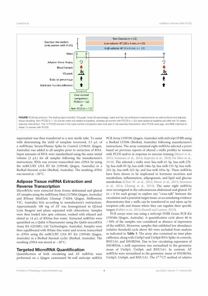
fphys-11-00904 July 28, 2020 Time: 17:59 # 4
Lionett et al. miRNAs in Women With PCOS
FIGURE 1 | Study protocol. The testing days included: VO2peak, body fat percentage, waist and hip circumference measurements as well as blood and adiposetissue sampling. Non-PCOS (n = 12) women were only tested at baseline, whereas all women with PCOS (n = 42) were tested at baseline and after the 16 weeksexercise intervention. The 12 PCOS women in the case-control comparison also took part in the exercise intervention. Non-PCOS were age- and BMI-matched tothese 12 women with PCOS.
supernatant was then transferred to a new sterile tube. To assistwith determining the yield of template recovered, 3.5 µL ofa miRNeasy Serum/Plasma Spike-In Control (219610; Qiagen,Australia) was added to all samples prior to extraction of RNA.Input amounts of RNA were standardized using the same initialvolume (2 µL) for all samples following the manufacturer’sinstructions. RNA was reverse transcribed into cDNA by usingthe miRCURY LNA RT kit (339340; Qiagen, Australia) in aBioRad thermal cycler (BioRad, Australia). The resulting cDNAwas stored at �20�C.
Adipose Tissue miRNA Extraction andReverse TranscriptionMicroRNAs were extracted from frozen abdominal and glutealAT samples using themiRNeasyMini (217004; Qiagen, Australia)and RNeasy MinElute Cleanup (74204; Qiagen, Melbourne,VIC, Australia) Kits according to manufacturer’s instructions.Approximately 100 mg of AT was homogenized in QIAzolLysis Reagent and phase separated with chloroform. Sampleswere then loaded into spin columns, washed with ethanol andeluted in 14 µL of RNAse-free water. Extracted miRNAs werequantified on a Qubit 4 Fluorometer using the Qubit microRNAAssay Kit (Q32881; Life Technologies, Australia). Samples werethen equilibrated with RNase-free water and reverse transcribedto cDNA using the miRCURY LNA RT Kit (339340; Qiagen,Australia) in a BioRad thermal cycler (BioRad, Australia). Theresulting cDNA was stored at �20�C.
Targeted MicroRNA QuantificationQuantification of both circulating and AT miRNAs wereperformed on a Qiagen customized 96-well miScript miRNA
PCRArray (339330; Qiagen, Australia) withmiScript SYBR usinga BioRad CFX96 (BioRad, Australia) following manufacturer’sinstructions. The array contained eight miRNAs selected a prioribased on previous reports of altered c-miRs profiles in womenwith PCOS and/or in response to exercise training (Murri et al.,2013; Sorensen et al., 2014; Improta et al., 2018; Da Silva et al.,2019). The selected c-miRs were hsa-miR-21-5p, hsa-miR-27b-5p, hsa-miR-93-5p, hsa-miR-146a-5p, hsa-miR-155-5p, hsa-miR-222-3p, hsa-miR-223-3p, and hsa-miR-103a-3p. These miRNAshave been shown to be implicated in hormone secretion andmetabolism, inflammation, adipogenesis, and lipid and glucosemetabolism (Chen W. et al., 2012; Murri et al., 2013; Sorensenet al., 2014; Chuang et al., 2015). The same eight miRNAswere investigated in the subcutaneous abdominal and gluteal AT(n = 8 for each group) to explore any “cross-talk” between thecirculation and a potential target tissue, as accumulating evidencedemonstrates that c-miRs can be transferred to and taken up byrecipient cells and tissues where they can regulate their specifictargets (Fabbri et al., 2012; Russell and Lamon, 2015).
PCR arrays were run using a miScript SYBR Green PCR Kit(339346; Qiagen, Australia). A quantification cycle above 40 in>50% of the samples was considered as absence of expressionof the miRNA. Moreover, samples that exhibited low abundance(relative threshold cycle above 40) were excluded from analysisas indicated in Table 3. The array also contained an inter-platecalibrator, along with UniSp3 andUniSp6 RNA Spike-in controls,RNU1A1 and SNORD44. Due to low circulating expression ofSNORD44, c-miR expression was normalized to the geometricmean of UniSp3, UniSp6, and RNU1A1. In contrast, ATmiRNAs were normalized to the geometric mean of SNORD44,UniSp3, UniSp6, and RNU1A1. The 211CT method of relative
Frontiers in Physiology | www.frontiersin.org 4 July 2020 | Volume 11 | Article 904
fphys-11-00904 July 28, 2020 Time: 17:59 # 5
Lionett et al. miRNAs in Women With PCOS
quantification was used to calculate the relative abundance ofmiRNAs in plasma and AT (Livak and Schmittgen, 2001).
Blood Biochemistry and InsulinSensitivityPlasma glucose and serum insulin concentrations andHomeostatic Model Assessment for Insulin Resistance (HOMA-IR) were determined for the case-control study (PCOS andNon-PCOS). Plasma glucose concentration was measuredusing a Roche Moduclar P (Roche, Switzerland), while seruminsulin concentration was measured in duplicates using anenzyme-linked immunosorbent assay (ELISA; IBL-International,Germany). HOMA-IR was used as a method for quantifyinginsulin resistance; fasting serum (µU/mL) ⇥ fasting plasmaglucose (mmol/L) divided by 22.5 (Matthews et al., 1985).
StatisticsDescriptive statistics are presented as means ± SD. Groupmeans for PCOS vs Non-PCOS were compared by Student’stwo-tailed t-test for independent samples. We used linearmixed models with participant as random factor, and withtwo dummy variables to uniquely identify the two exercisegroups (LV-HIT and HV-HIT) and their interactions, withtime as fixed factors. We adjusted for the baseline valuesfor the outcome variables as recommended by Twisk et al.(2018). In this model, the coe�cients for the interactionterms give the estimated exercise training e�ects in LV-HITand HV-HIT compared to the Non-Ex group. Paired t-testswere used to test for di�erences in miRNA expression inAT depots (gluteal versus abdominal) at baseline. To test ifmiRNA expression in the two AT depots would respond indistinct ways to the two HIT protocols, changes in miRNAexpression were calculated by using post-intervention minuspre-intervention values (corresponding to the 1miR expressionin Table 6). We used linear mixed models as detailed butwith the two exercise groups’ interaction with AT depots asfixed factors. Due to previously observed abnormal expressionof miRNAs in subcutaneous AT in insulin resistant womenregardless of PCOS status, we also analyzed the gluteal andabdominal subcutaneous ATmiRNA expression based on insulinresistance. Insulin resistance was estimated as HOMA-IR, withsimilar cut-o� points as previously reported (Chuang et al.,2015); HOMA-IR value <2.5 was considered normal whereasHOMA-IR � 2.5 indicated insulin resistance. Group meansfor insulin resistant and non-insulin resistant women werecompared by Student’s two-tailed t-test for independent samples.Normality of residuals was evaluated using visual inspectionof Q-Q plots and logarithmic transformation (log10) of thedependent variable was performed when necessary to obtainnormality. Due to multiple hypotheses, we considered P-values<0.01 as statistically significant. Pearson correlation betweenmiRNAs di�erent between groups (PCOS and Non-PCOS)and baseline VO2peak, BMI, fat percentage or waist/hip ratiowas calculated using GraphPad Prism version 8.1.2 (GraphPadSoftware, United States). All other analyses were carried out usingSPSS version 25.0 (SPSS Inc., United States).
RESULTS
Characteristics: PCOS vs Non-PCOSWomenCharacteristics of the participants included in the case-controlcomparison are presented in Table 1. Apart from greaterwaist/hip ratio in women with PCOS (0.90 ± 0.06 versus0.83 ± 0.05, P = 0.005), there were no di�erences between groups(Table 1). There was no di�erence in HOMA-IR between thegroups, although nine of the women with PCOS versus fivewomen without PCOS had HOMA-IR � 2.5 (Table 1).
Physiological Responses toHigh-Intensity Interval TrainingThere were no changes in body weight, BMI, body fat percentage,waist, and hip circumference or waist/hip ratio for either LV-HIT or HV-HIT post intervention (Table 2). Although notsignificant, it is worth noting that the mean fat percentagedecreased by 1.4% in the Non-Ex group. This is not asystemic decrease in fat percentage in the Non-Ex group buta result of two participants in this group decreasing by 6%in fat percentage. Following 16 weeks of HIT, absolute, butnot relative, VO2peak increased by 9% (P = 0.008) in HV-HIT (Table 2).
miRNA Expression Profile in PCOS vsNon-PCOSBasal expression of circulating miRNA-27b (c-miR-27b) washigher in women with PCOS compared to Non-PCOS (1.8-fold, P = 0.006; Figure 2B), with no significant di�erences inthe expression of c-miR-21, �93, �103a, �146a, �155, �222,and �223 (Figures 2A–H). No di�erences in basal gluteal and
TABLE 1 | Baseline characteristics of PCOS and Non-PCOS women.
Non-PCOS PCOS P-value
n 12 12
Age (years) 30 ± 7 30 ± 7 0.98
Body weight (kg) 83.0 ± 18.5 85.2 ± 19.7 0.78
BMI (kg/m2) 29.3 ± 5.8 29.8 ± 6.5 0.85
Body fat percentage (%) 36.4 ± 9.2 36.8 ± 9.0 0.91
Waist circumference (cm) 94 ± 14 102 ± 14 0.16
Hip circumference (cm) 113 ± 13 113 ± 11 0.99
Waist/Hip ratio 0.83 ± 0.05 0.90 ± 0.06 0.005
VO2peak (L/min) 2.83 ± 0.38 2.73 ± 0.38 0.54
VO2peak (mL/min/kg) 34.9 ± 7.0 33.2 ± 6.1 0.53
Glucose (mmol/L) 4.9 ± 0.4 5.0 ± 0.6 0.52
Insulin (µU/mL) 14.2 ± 10.6 15.7 ± 6.9 0.71
HOMA-IR 3.1 ± 2.3 3.6 ± 1.8 0.58
Values are mean ± SD. PCOS, polycystic ovary syndrome; BMI, body massindex; VO2peak, peak oxygen uptake; HOMA-IR, homeostatic model assessmentof insulin resistance. In the Non-PCOS group, not enough blood was collectedfor analyzing insulin concentration, so n = 11 for insulin concentration andHOMA-IR in Non-PCOS. The meaning of a bold value is that the P-value isstatistically significant.
Frontiers in Physiology | www.frontiersin.org 5 July 2020 | Volume 11 | Article 904
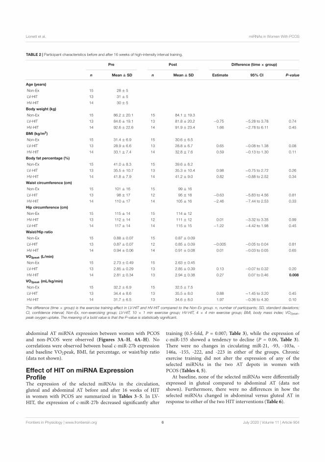
fphys-11-00904 July 28, 2020 Time: 17:59 # 6
Lionett et al. miRNAs in Women With PCOS
TABLE 2 | Participant characteristics before and after 16 weeks of high-intensity interval training.
Pre Post Difference (time ⇥ group)
n Mean ± SD n Mean ± SD Estimate 95% CI P-value
Age (years)
Non-Ex 15 28 ± 5
LV-HIT 13 31 ± 5
HV-HIT 14 30 ± 5
Body weight (kg)
Non-Ex 15 86.2 ± 20.1 15 84.1 ± 19.3
LV-HIT 13 84.6 ± 19.1 13 81.8 ± 20.2 �0.75 �5.28 to 3.78 0.74
HV-HIT 14 92.6 ± 22.6 14 91.9 ± 23.4 1.66 �2.78 to 6.11 0.45
BMI (kg/m2)
Non-Ex 15 31.4 ± 6.9 15 30.6 ± 6.5
LV-HIT 13 28.9 ± 6.6 13 28.8 ± 6.7 0.65 �0.08 to 1.38 0.08
HV-HIT 14 33.1 ± 7.4 14 32.8 ± 7.6 0.59 �0.13 to 1.30 0.11
Body fat percentage (%)
Non-Ex 15 41.0 ± 8.3 15 39.6 ± 8.2
LV-HIT 13 35.5 ± 10.7 13 35.3 ± 10.4 0.98 �0.75 to 2.72 0.26
HV-HIT 14 41.8 ± 7.9 14 41.2 ± 9.0 0.82 �0.88 to 2.52 0.34
Waist circumference (cm)
Non-Ex 15 101 ± 16 15 99 ± 16
LV-HIT 13 98 ± 17 12 95 ± 18 �0.63 �5.83 to 4.56 0.81
HV-HIT 14 110 ± 17 14 105 ± 16 �2.46 �7.44 to 2.53 0.33
Hip circumference (cm)
Non-Ex 15 115 ± 14 15 114 ± 12
HV-HIT 13 112 ± 14 12 111 ± 12 0.01 �3.32 to 3.35 0.99
LV-HIT 14 117 ± 14 14 115 ± 15 �1.22 �4.42 to 1.98 0.45
Waist/Hip ratio
Non-Ex 15 0.88 ± 0.07 15 0.87 ± 0.09
LV-HIT 13 0.87 ± 0.07 12 0.85 ± 0.09 �0.005 �0.05 to 0.04 0.81
HV-HIT 14 0.94 ± 0.06 14 0.91 ± 0.08 0.01 �0.03 to 0.05 0.65
VO2peak (L/min)
Non-Ex 15 2.73 ± 0.49 15 2.63 ± 0.45
LV-HIT 13 2.85 ± 0.29 13 2.85 ± 0.39 0.13 �0.07 to 0.32 0.20
HV-HIT 14 2.81 ± 0.34 13 2.94 ± 0.38 0.27 0.07 to 0.46 0.008
VO2peak (mL/kg/min)
Non-Ex 15 32.2 ± 6.9 15 32.5 ± 7.5
LV-HIT 13 34.4 ± 8.6 13 35.5 ± 8.0 0.88 �1.45 to 3.20 0.45
HV-HIT 14 31.7 ± 6.5 13 34.6 ± 8.0 1.97 �0.36 to 4.30 0.10
The difference (time ⇥ group) is the exercise training effect in LV-HIT and HV-HIT compared to the Non-Ex group. n, number of participants; SD, standard deviations;CI, confidence interval; Non-Ex, non-exercising group; LV-HIT, 10 ⇥ 1 min exercise group; HV-HIT, 4 ⇥ 4 min exercise group; BMI, body mass index; VO2peak,peak oxygen uptake. The meaning of a bold value is that the P-value is statistically significant.
abdominal AT miRNA expression between women with PCOSand non-PCOS were observed (Figures 3A–H, 4A–H). Nocorrelations were observed between basal c-miR-27b expressionand baseline VO2peak, BMI, fat percentage, or waist/hip ratio(data not shown).
Effect of HIT on miRNA ExpressionProfileThe expression of the selected miRNAs in the circulation,gluteal and abdominal AT before and after 16 weeks of HITin women with PCOS are summarized in Tables 3–5. In LV-HIT, the expression of c-miR-27b decreased significantly after
training (0.5-fold, P = 0.007; Table 3), while the expression ofc-miR-155 showed a tendency to decline (P = 0.06, Table 3).There were no changes in circulating miR-21, -93, -103a, -146a, -155, -222, and -223 in either of the groups. Chronicexercise training did not alter the expression of any of theselected miRNAs in the two AT depots in women withPCOS (Tables 4, 5).
At baseline, none of the selected miRNAs were di�erentiallyexpressed in gluteal compared to abdominal AT (data notshown). Furthermore, there were no di�erences in how theselected miRNAs changed in abdominal versus gluteal AT inresponse to either of the two HIT interventions (Table 6).
Frontiers in Physiology | www.frontiersin.org 6 July 2020 | Volume 11 | Article 904
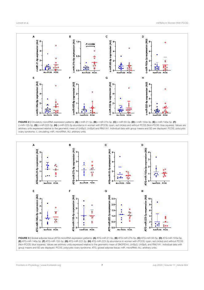
fphys-11-00904 July 28, 2020 Time: 17:59 # 7
Lionett et al. miRNAs in Women With PCOS
FIGURE 2 | Circulatory microRNA expression patterns. (A) c-miR-21-5p, (B) c-miR-27b-5p, (C) c-miR-93-5p, (D) c-miR-103a-3p, (E) c-miR-146a-5p, (F)c-miR-155-5p, (G) c-miR-222-3p, (H) c-miR-223-3p abundance in women with (PCOS; open, red circles) and without PCOS (Non-PCOS; blue squares). Values arearbitrary units expressed relative to the geometric mean of UniSp3, UniSp6 and RNU1A1. Individual data with group means and SD are displayed. PCOS, polycysticovary syndrome; c, circulating; miR, microRNA; AU, arbitrary units.
FIGURE 3 | Gluteal adipose tissue (ATG) microRNA expression patterns. (A) ATG-miR-21-5p, (B) ATG-miR-27b-5p, (C) ATG-miR-93-5p, (D) ATG-miR-103a-3p,(E) ATG-miR-146a-5p, (F) ATG-miR-155-5p, (G) ATG-miR-222-3p, (H) ATG-miR-223-3p abundance in women with (PCOS; open, red circles) and without PCOS(Non-PCOS; blue squares). Values are arbitrary units expressed relative to the geometric mean of SNORD44, UniSp3, UniSp6, and RNU1A1. Individual data withgroup means and SD are displayed. PCOS, polycystic ovary syndrome; ATG, gluteal adipose tissue; miR, microRNA; AU, arbitrary units.
Frontiers in Physiology | www.frontiersin.org 7 July 2020 | Volume 11 | Article 904
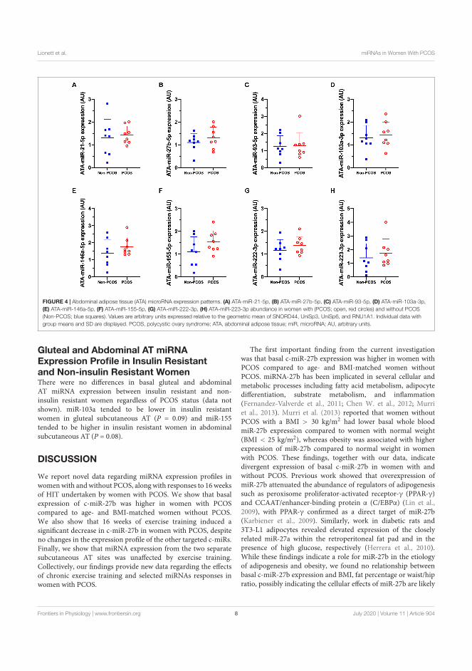
fphys-11-00904 July 28, 2020 Time: 17:59 # 8
Lionett et al. miRNAs in Women With PCOS
FIGURE 4 | Abdominal adipose tissue (ATA) microRNA expression patterns. (A) ATA-miR-21-5p, (B) ATA-miR-27b-5p, (C) ATA-miR-93-5p, (D) ATA-miR-103a-3p,(E) ATA-miR-146a-5p, (F) ATA-miR-155-5p, (G) ATA-miR-222-3p, (H) ATA-miR-223-3p abundance in women with (PCOS; open, red circles) and without PCOS(Non-PCOS; blue squares). Values are arbitrary units expressed relative to the geometric mean of SNORD44, UniSp3, UniSp6, and RNU1A1. Individual data withgroup means and SD are displayed. PCOS, polycystic ovary syndrome; ATA, abdominal adipose tissue; miR, microRNA; AU, arbitrary units.
Gluteal and Abdominal AT miRNAExpression Profile in Insulin Resistantand Non-insulin Resistant WomenThere were no di�erences in basal gluteal and abdominalAT miRNA expression between insulin resistant and non-insulin resistant women regardless of PCOS status (data notshown). miR-103a tended to be lower in insulin resistantwomen in gluteal subcutaneous AT (P = 0.09) and miR-155tended to be higher in insulin resistant women in abdominalsubcutaneous AT (P = 0.08).
DISCUSSION
We report novel data regarding miRNA expression profiles inwomenwith andwithout PCOS, along with responses to 16 weeksof HIT undertaken by women with PCOS. We show that basalexpression of c-miR-27b was higher in women with PCOScompared to age- and BMI-matched women without PCOS.We also show that 16 weeks of exercise training induced asignificant decrease in c-miR-27b in women with PCOS, despiteno changes in the expression profile of the other targeted c-miRs.Finally, we show that miRNA expression from the two separatesubcutaneous AT sites was una�ected by exercise training.Collectively, our findings provide new data regarding the e�ectsof chronic exercise training and selected miRNAs responses inwomen with PCOS.
The first important finding from the current investigationwas that basal c-miR-27b expression was higher in women withPCOS compared to age- and BMI-matched women withoutPCOS. miRNA-27b has been implicated in several cellular andmetabolic processes including fatty acid metabolism, adipocytedi�erentiation, substrate metabolism, and inflammation(Fernandez-Valverde et al., 2011; Chen W. et al., 2012; Murriet al., 2013). Murri et al. (2013) reported that women withoutPCOS with a BMI > 30 kg/m2 had lower basal whole bloodmiR-27b expression compared to women with normal weight(BMI < 25 kg/m2), whereas obesity was associated with higherexpression of miR-27b compared to normal weight in womenwith PCOS. These findings, together with our data, indicatedivergent expression of basal c-miR-27b in women with andwithout PCOS. Previous work showed that overexpression ofmiR-27b attenuated the abundance of regulators of adipogenesissuch as peroxisome proliferator-activated receptor-g (PPAR-g)and CCAAT/enhancer-binding protein a (C/EBPa) (Lin et al.,2009), with PPAR-g confirmed as a direct target of miR-27b(Karbiener et al., 2009). Similarly, work in diabetic rats and3T3-L1 adipocytes revealed elevated expression of the closelyrelated miR-27a within the retroperitoneal fat pad and in thepresence of high glucose, respectively (Herrera et al., 2010).While these findings indicate a role for miR-27b in the etiologyof adipogenesis and obesity, we found no relationship betweenbasal c-miR-27b expression and BMI, fat percentage or waist/hipratio, possibly indicating the cellular e�ects of miR-27b are likely
Frontiers in Physiology | www.frontiersin.org 8 July 2020 | Volume 11 | Article 904
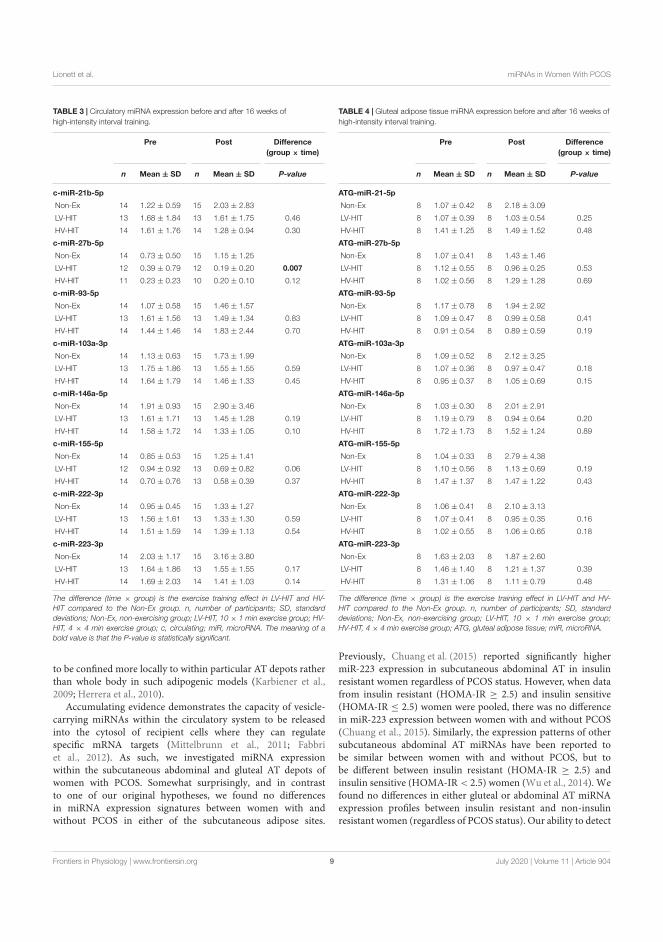
fphys-11-00904 July 28, 2020 Time: 17:59 # 9
Lionett et al. miRNAs in Women With PCOS
TABLE 3 | Circulatory miRNA expression before and after 16 weeks ofhigh-intensity interval training.
Pre Post Difference(group ⇥ time)
n Mean ± SD n Mean ± SD P-value
c-miR-21b-5p
Non-Ex 14 1.22 ± 0.59 15 2.03 ± 2.83
LV-HIT 13 1.68 ± 1.84 13 1.61 ± 1.75 0.46
HV-HIT 14 1.61 ± 1.76 14 1.28 ± 0.94 0.30
c-miR-27b-5p
Non-Ex 14 0.73 ± 0.50 15 1.15 ± 1.25
LV-HIT 12 0.39 ± 0.79 12 0.19 ± 0.20 0.007
HV-HIT 11 0.23 ± 0.23 10 0.20 ± 0.10 0.12
c-miR-93-5p
Non-Ex 14 1.07 ± 0.58 15 1.46 ± 1.57
LV-HIT 13 1.61 ± 1.56 13 1.49 ± 1.34 0.83
HV-HIT 14 1.44 ± 1.46 14 1.83 ± 2.44 0.70
c-miR-103a-3p
Non-Ex 14 1.13 ± 0.63 15 1.73 ± 1.99
LV-HIT 13 1.75 ± 1.86 13 1.55 ± 1.55 0.59
HV-HIT 14 1.64 ± 1.79 14 1.46 ± 1.33 0.45
c-miR-146a-5p
Non-Ex 14 1.91 ± 0.93 15 2.90 ± 3.46
LV-HIT 13 1.61 ± 1.71 13 1.45 ± 1.28 0.19
HV-HIT 14 1.58 ± 1.72 14 1.33 ± 1.05 0.10
c-miR-155-5p
Non-Ex 14 0.85 ± 0.53 15 1.25 ± 1.41
LV-HIT 12 0.94 ± 0.92 13 0.69 ± 0.82 0.06
HV-HIT 14 0.70 ± 0.76 13 0.58 ± 0.39 0.37
c-miR-222-3p
Non-Ex 14 0.95 ± 0.45 15 1.33 ± 1.27
LV-HIT 13 1.56 ± 1.61 13 1.33 ± 1.30 0.59
HV-HIT 14 1.51 ± 1.59 14 1.39 ± 1.13 0.54
c-miR-223-3p
Non-Ex 14 2.03 ± 1.17 15 3.16 ± 3.80
LV-HIT 13 1.64 ± 1.86 13 1.55 ± 1.55 0.17
HV-HIT 14 1.69 ± 2.03 14 1.41 ± 1.03 0.14
The difference (time ⇥ group) is the exercise training effect in LV-HIT and HV-HIT compared to the Non-Ex group. n, number of participants; SD, standarddeviations; Non-Ex, non-exercising group; LV-HIT, 10 ⇥ 1 min exercise group; HV-HIT, 4 ⇥ 4 min exercise group; c, circulating; miR, microRNA. The meaning of abold value is that the P-value is statistically significant.
to be confined more locally to within particular AT depots ratherthan whole body in such adipogenic models (Karbiener et al.,2009; Herrera et al., 2010).
Accumulating evidence demonstrates the capacity of vesicle-carrying miRNAs within the circulatory system to be releasedinto the cytosol of recipient cells where they can regulatespecific mRNA targets (Mittelbrunn et al., 2011; Fabbriet al., 2012). As such, we investigated miRNA expressionwithin the subcutaneous abdominal and gluteal AT depots ofwomen with PCOS. Somewhat surprisingly, and in contrastto one of our original hypotheses, we found no di�erencesin miRNA expression signatures between women with andwithout PCOS in either of the subcutaneous adipose sites.
TABLE 4 | Gluteal adipose tissue miRNA expression before and after 16 weeks ofhigh-intensity interval training.
Pre Post Difference(group ⇥ time)
n Mean ± SD n Mean ± SD P-value
ATG-miR-21-5p
Non-Ex 8 1.07 ± 0.42 8 2.18 ± 3.09
LV-HIT 8 1.07 ± 0.39 8 1.03 ± 0.54 0.25
HV-HIT 8 1.41 ± 1.25 8 1.49 ± 1.52 0.48
ATG-miR-27b-5p
Non-Ex 8 1.07 ± 0.41 8 1.43 ± 1.46
LV-HIT 8 1.12 ± 0.55 8 0.96 ± 0.25 0.53
HV-HIT 8 1.02 ± 0.56 8 1.29 ± 1.28 0.69
ATG-miR-93-5p
Non-Ex 8 1.17 ± 0.78 8 1.94 ± 2.92
LV-HIT 8 1.09 ± 0.47 8 0.99 ± 0.58 0.41
HV-HIT 8 0.91 ± 0.54 8 0.89 ± 0.59 0.19
ATG-miR-103a-3p
Non-Ex 8 1.09 ± 0.52 8 2.12 ± 3.25
LV-HIT 8 1.07 ± 0.36 8 0.97 ± 0.47 0.18
HV-HIT 8 0.95 ± 0.37 8 1.05 ± 0.69 0.15
ATG-miR-146a-5p
Non-Ex 8 1.03 ± 0.30 8 2.01 ± 2.91
LV-HIT 8 1.19 ± 0.79 8 0.94 ± 0.64 0.20
HV-HIT 8 1.72 ± 1.73 8 1.52 ± 1.24 0.89
ATG-miR-155-5p
Non-Ex 8 1.04 ± 0.33 8 2.79 ± 4.38
LV-HIT 8 1.10 ± 0.56 8 1.13 ± 0.69 0.19
HV-HIT 8 1.47 ± 1.37 8 1.47 ± 1.22 0.43
ATG-miR-222-3p
Non-Ex 8 1.06 ± 0.41 8 2.10 ± 3.13
LV-HIT 8 1.07 ± 0.41 8 0.95 ± 0.35 0.16
HV-HIT 8 1.02 ± 0.55 8 1.06 ± 0.65 0.18
ATG-miR-223-3p
Non-Ex 8 1.63 ± 2.03 8 1.87 ± 2.60
LV-HIT 8 1.46 ± 1.40 8 1.21 ± 1.37 0.39
HV-HIT 8 1.31 ± 1.06 8 1.11 ± 0.79 0.48
The difference (time ⇥ group) is the exercise training effect in LV-HIT and HV-HIT compared to the Non-Ex group. n, number of participants; SD, standarddeviations; Non-Ex, non-exercising group; LV-HIT, 10 ⇥ 1 min exercise group;HV-HIT, 4 ⇥ 4 min exercise group; ATG, gluteal adipose tissue; miR, microRNA.
Previously, Chuang et al. (2015) reported significantly highermiR-223 expression in subcutaneous abdominal AT in insulinresistant women regardless of PCOS status. However, when datafrom insulin resistant (HOMA-IR � 2.5) and insulin sensitive(HOMA-IR 2.5) women were pooled, there was no di�erencein miR-223 expression between women with and without PCOS(Chuang et al., 2015). Similarly, the expression patterns of othersubcutaneous abdominal AT miRNAs have been reported tobe similar between women with and without PCOS, but tobe di�erent between insulin resistant (HOMA-IR � 2.5) andinsulin sensitive (HOMA-IR < 2.5) women (Wu et al., 2014). Wefound no di�erences in either gluteal or abdominal AT miRNAexpression profiles between insulin resistant and non-insulinresistant women (regardless of PCOS status). Our ability to detect
Frontiers in Physiology | www.frontiersin.org 9 July 2020 | Volume 11 | Article 904
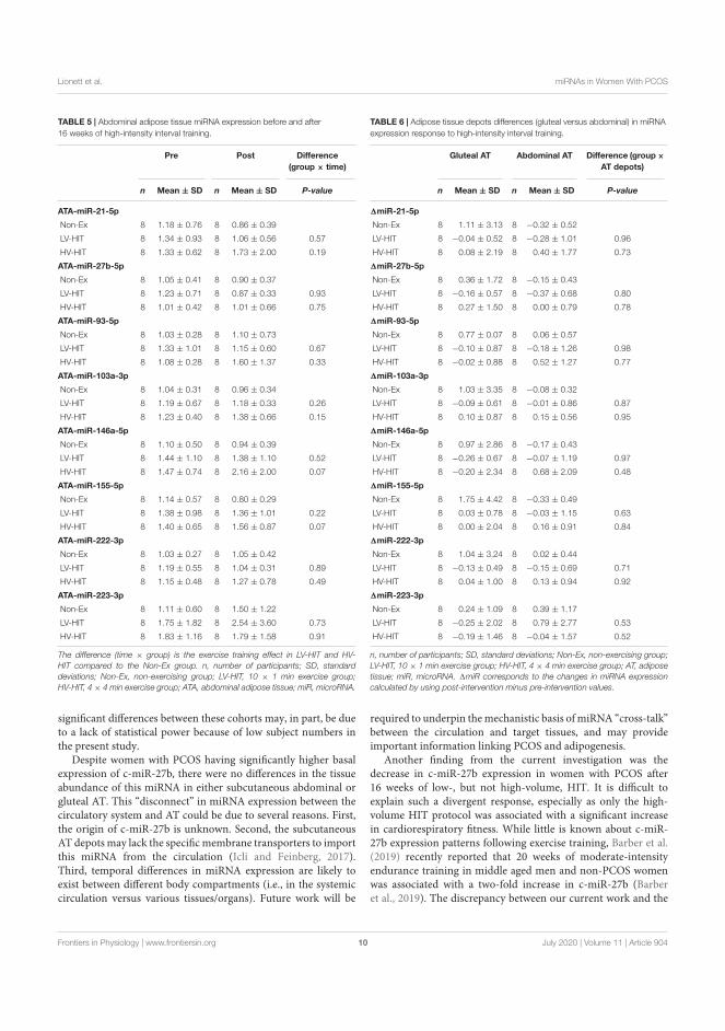
fphys-11-00904 July 28, 2020 Time: 17:59 # 10
Lionett et al. miRNAs in Women With PCOS
TABLE 5 | Abdominal adipose tissue miRNA expression before and after16 weeks of high-intensity interval training.
Pre Post Difference(group ⇥ time)
n Mean ± SD n Mean ± SD P-value
ATA-miR-21-5p
Non-Ex 8 1.18 ± 0.76 8 0.86 ± 0.39
LV-HIT 8 1.34 ± 0.93 8 1.06 ± 0.56 0.57
HV-HIT 8 1.33 ± 0.62 8 1.73 ± 2.00 0.19
ATA-miR-27b-5p
Non-Ex 8 1.05 ± 0.41 8 0.90 ± 0.37
LV-HIT 8 1.23 ± 0.71 8 0.87 ± 0.33 0.93
HV-HIT 8 1.01 ± 0.42 8 1.01 ± 0.66 0.75
ATA-miR-93-5p
Non-Ex 8 1.03 ± 0.28 8 1.10 ± 0.73
LV-HIT 8 1.33 ± 1.01 8 1.15 ± 0.60 0.67
HV-HIT 8 1.08 ± 0.28 8 1.60 ± 1.37 0.33
ATA-miR-103a-3p
Non-Ex 8 1.04 ± 0.31 8 0.96 ± 0.34
LV-HIT 8 1.19 ± 0.67 8 1.18 ± 0.33 0.26
HV-HIT 8 1.23 ± 0.40 8 1.38 ± 0.66 0.15
ATA-miR-146a-5p
Non-Ex 8 1.10 ± 0.50 8 0.94 ± 0.39
LV-HIT 8 1.44 ± 1.10 8 1.38 ± 1.10 0.52
HV-HIT 8 1.47 ± 0.74 8 2.16 ± 2.00 0.07
ATA-miR-155-5p
Non-Ex 8 1.14 ± 0.57 8 0.80 ± 0.29
LV-HIT 8 1.38 ± 0.98 8 1.36 ± 1.01 0.22
HV-HIT 8 1.40 ± 0.65 8 1.56 ± 0.87 0.07
ATA-miR-222-3p
Non-Ex 8 1.03 ± 0.27 8 1.05 ± 0.42
LV-HIT 8 1.19 ± 0.55 8 1.04 ± 0.31 0.89
HV-HIT 8 1.15 ± 0.48 8 1.27 ± 0.78 0.49
ATA-miR-223-3p
Non-Ex 8 1.11 ± 0.60 8 1.50 ± 1.22
LV-HIT 8 1.75 ± 1.82 8 2.54 ± 3.60 0.73
HV-HIT 8 1.83 ± 1.16 8 1.79 ± 1.58 0.91
The difference (time ⇥ group) is the exercise training effect in LV-HIT and HV-HIT compared to the Non-Ex group. n, number of participants; SD, standarddeviations; Non-Ex, non-exercising group; LV-HIT, 10 ⇥ 1 min exercise group;HV-HIT, 4 ⇥ 4 min exercise group; ATA, abdominal adipose tissue; miR, microRNA.
significant di�erences between these cohorts may, in part, be dueto a lack of statistical power because of low subject numbers inthe present study.
Despite women with PCOS having significantly higher basalexpression of c-miR-27b, there were no di�erences in the tissueabundance of this miRNA in either subcutaneous abdominal orgluteal AT. This “disconnect” in miRNA expression between thecirculatory system and AT could be due to several reasons. First,the origin of c-miR-27b is unknown. Second, the subcutaneousAT depots may lack the specificmembrane transporters to importthis miRNA from the circulation (Icli and Feinberg, 2017).Third, temporal di�erences in miRNA expression are likely toexist between di�erent body compartments (i.e., in the systemiccirculation versus various tissues/organs). Future work will be
TABLE 6 | Adipose tissue depots differences (gluteal versus abdominal) in miRNAexpression response to high-intensity interval training.
Gluteal AT Abdominal AT Difference (group ⇥AT depots)
n Mean ± SD n Mean ± SD P-value
1miR-21-5p
Non-Ex 8 1.11 ± 3.13 8 �0.32 ± 0.52
LV-HIT 8 �0.04 ± 0.52 8 �0.28 ± 1.01 0.96
HV-HIT 8 0.08 ± 2.19 8 0.40 ± 1.77 0.73
1miR-27b-5p
Non-Ex 8 0.36 ± 1.72 8 �0.15 ± 0.43
LV-HIT 8 �0.16 ± 0.57 8 �0.37 ± 0.68 0.80
HV-HIT 8 0.27 ± 1.50 8 0.00 ± 0.79 0.78
1miR-93-5p
Non-Ex 8 0.77 ± 0.07 8 0.06 ± 0.57
LV-HIT 8 �0.10 ± 0.87 8 �0.18 ± 1.26 0.98
HV-HIT 8 �0.02 ± 0.88 8 0.52 ± 1.27 0.77
1miR-103a-3p
Non-Ex 8 1.03 ± 3.35 8 �0.08 ± 0.32
LV-HIT 8 �0.09 ± 0.61 8 �0.01 ± 0.86 0.87
HV-HIT 8 0.10 ± 0.87 8 0.15 ± 0.56 0.95
1miR-146a-5p
Non-Ex 8 0.97 ± 2.86 8 �0.17 ± 0.43
LV-HIT 8 �0.26 ± 0.67 8 �0.07 ± 1.19 0.97
HV-HIT 8 �0.20 ± 2.34 8 0.68 ± 2.09 0.48
1miR-155-5p
Non-Ex 8 1.75 ± 4.42 8 �0.33 ± 0.49
LV-HIT 8 0.03 ± 0.78 8 �0.03 ± 1.15 0.63
HV-HIT 8 0.00 ± 2.04 8 0.16 ± 0.91 0.84
1miR-222-3p
Non-Ex 8 1.04 ± 3.24 8 0.02 ± 0.44
LV-HIT 8 �0.13 ± 0.49 8 �0.15 ± 0.69 0.71
HV-HIT 8 0.04 ± 1.00 8 0.13 ± 0.94 0.92
1miR-223-3p
Non-Ex 8 0.24 ± 1.09 8 0.39 ± 1.17
LV-HIT 8 �0.25 ± 2.02 8 0.79 ± 2.77 0.53
HV-HIT 8 �0.19 ± 1.46 8 �0.04 ± 1.57 0.52
n, number of participants; SD, standard deviations; Non-Ex, non-exercising group;LV-HIT, 10 ⇥ 1 min exercise group; HV-HIT, 4 ⇥ 4 min exercise group; AT, adiposetissue; miR, microRNA. 1miR corresponds to the changes in miRNA expressioncalculated by using post-intervention minus pre-intervention values.
required to underpin themechanistic basis ofmiRNA “cross-talk”between the circulation and target tissues, and may provideimportant information linking PCOS and adipogenesis.
Another finding from the current investigation was thedecrease in c-miR-27b expression in women with PCOS after16 weeks of low-, but not high-volume, HIT. It is di�cult toexplain such a divergent response, especially as only the high-volume HIT protocol was associated with a significant increasein cardiorespiratory fitness. While little is known about c-miR-27b expression patterns following exercise training, Barber et al.(2019) recently reported that 20 weeks of moderate-intensityendurance training in middle aged men and non-PCOS womenwas associated with a two-fold increase in c-miR-27b (Barberet al., 2019). The discrepancy between our current work and the
Frontiers in Physiology | www.frontiersin.org 10 July 2020 | Volume 11 | Article 904
fphys-11-00904 July 28, 2020 Time: 17:59 # 11
Lionett et al. miRNAs in Women With PCOS
findings by Barber et al. (2019) may be explained by di�erencesin exercise modality. HIT versus moderate-intensity continuousendurance training can di�erentially influence cardiometabolicrisk factors including blood pressure, low- and high-densitylipoproteins, body weight and insulin sensitivity in patients withcardiometabolic diseases (Weston et al., 2014). Additionally,diverse sampling timepoint (24 h post last training session in thestudy by Barber et al. (2019) compared to�48 h in our study)and study participants (middle aged men and women withoutPCOS compared to women with PCOS in our study) may, inpart, help explain these divergent findings. Parr et al. (2016),Denham et al. (2018), Barber et al. (2019), and Da Silva et al.(2019) have previously reported that chronic exercise trainingprograms can significantly modulate c-miRNA expression inhealthy individuals and in those with overweight/obesity. Thephysiological significance of such responses are unclear anddi�cult to explain due to a variety of di�erent miRNAquantification techniques reported, di�erences in sampling timepoints, a range of exercise protocols, and divergent clinicalcohorts (Gomes et al., 2015). Time-course studies are neededto span the acute and chronic sampling points for a morecomprehensive coverage of miRNA expression profiles.
We found no changes in miRNA expression in eithersubcutaneous abdominal or gluteal AT after high- or low-volumeHIT protocols in women with PCOS. This observation is inagreement with recent data from Tsiloulis et al. (2017) whoreported no e�ect of 6 weeks of endurance exercise training (foursupervised sessions/week; three moderate intensity sessions;and one HIT session) on the expression of 526 miRNAs inabdominal and gluteal AT in overweight males. Collectively,these findings indicate miRNAs are stably expressed in bothabdominal and gluteal AT and are largely unaltered in responseto exercise training protocols lasting up to several months.Furthermore, at baseline, we found no di�erence in expressionof the eight selected miRNAs in basal gluteal versus abdominalAT. Our findings are largely supported by the results of Tsilouliset al. (2017), who reported only four out of the 526 identifiedmiRNAs to be di�erentially expressed between gluteal andabdominal adipocytes in overweight males. In addition, we foundno di�erentially expressed miRNAs between the AT depotsfollowing exercise training. Other studies have reported that ATfrom di�erent depots express unique molecular, cellular, andmetabolic properties and respond in distinct ways to exercise andnutritional challenges (Tan et al., 2004; You et al., 2014). However,this does not seem to be the case for selected miRNA expression.
A major strength of the present study is the concurrentinvestigation of miRNA expression patterns in the circulationand abdominal and gluteal AT. Some miRNAs exert specificityin tissue expression within the body and can either be releasedor taken up by these tissues from the bloodstream (Russell andLamon, 2015). Thus, investigation of targeted miRNAs fromboth the circulation and a “target” tissue allowed us to compareexpression patterns between these tissues. We also acknowledgestudy limitations: we used a targeted approach to selectivelyinvestigate a small number of miRNAs and therefore mayhave missed changes in expression patterns of other miRNAs.Finally, larger subject numbers would have conferred greater
statistical power to detect small, but potential di�erences inmiRNA expression.
In conclusion, we report that the basal expression ofcirculating miR-27b was higher in women with PCOS comparedto women without PCOS. We were unable to detect anydi�erences in the eight targeted miRNAs in subcutaneousabdominal and gluteal AT. While 16 weeks of low- but nothigh-volume HIT altered the expression of c-miR-27b in womenwith PCOS, the chronic exercise training protocols employed inthis investigation did not induce alterations in the expressionof the other targeted miRNAs within the circulation or themiRNAs in subcutaneous abdominal and gluteal AT in womenwith PCOS. Further studies are required to ascertain miR-27b’s role and association with obesity, inflammation andadipogenesis in PCOS.
DATA AVAILABILITY STATEMENT
The raw data supporting the conclusions of this article will bemade available by the authors, without undue reservation.
ETHICS STATEMENT
The studies involving human participants were reviewedand approved by The Regional Committee for Medicaland Health Research Ethics in Central Norway (REK-midt 2015/468 and 2016/545) and the Australian CatholicUniversity Human Research Ethics Committee (2017-260H).The patients/participants provided their written informedconsent to participate in this study.
AUTHOR CONTRIBUTIONS
SL and DC drafted the manuscript, performed the analyses, andanalyzed the data. SL and SLy performed statistical analyses. SL,IK, DC, EV, JH, and TM were responsible for study conceptionand design. SL, IK, EP, and TM coordinated the study at the twosites, performed measurements on test-days, and supervised theexercise training. All authors provided feedback and approved thefinal manuscript.
FUNDING
This study was supported by the Liaison Committee foreducation, research and innovation in Central Norway (GrantNumbers: 2014/23166 and 2017/29014) and by a Novo NordiskFoundation Challenge Grant (NNF14OC0011493).
ACKNOWLEDGMENTS
We wish to thank the Unit for Applied Clinical Research atNTNU for providing the internet-based randomization; NeXTMove at NTNU for providing the equipment and lab facilitiesfor training sessions and exercise testing in Norway; Dr. Andrew
Frontiers in Physiology | www.frontiersin.org 11 July 2020 | Volume 11 | Article 904
fphys-11-00904 July 28, 2020 Time: 17:59 # 12
Lionett et al. miRNAs in Women With PCOS
Garnham and the Clinical Research Facility at St. Olav’s Hospitalfor performing blood and AT sampling in Australia and Norway,respectively. Blood and AT samples are stored in the RegionalBiobank 1 R� of Central Norway, with the data solution BioByte.We also acknowledge our dear friend, colleague and collaborator,
the late Professor Nigel K. Stepto. We are indebted to Nigel’sintellectual input and enthusiasm toward research in womenwithPCOS and will endeavor to continue the work he started. Lastbut not least, we want to thank all the participants for theire�ort and time.
REFERENCESAl Kindi, M. K., Al Essry, F. S., Al Essry, F. S., and Mula-Abed, W. A. (2012).
Validity of serum testosterone, free androgen index, and calculated freetestosterone in women with suspected hyperandrogenism. Oman Med. J. 27,471–474. doi: 10.5001/omj.2012.112
Anderson, S. A., Barry, J. A., and Hardiman, P. J. (2014). Risk of coronaryheart disease and risk of stroke in women with polycystic ovary syndrome: asystematic review andmeta-analysis. Int. J. Cardiol. 176, 486–487. doi: 10.1016/j.ijcard.2014.06.079
Arancio, W., Calogero Amato, M., Magliozzo, M., Pizzolanti, G., Vesco, R., andGiordano, C. (2018). Serum miRNAs in women a�ected by hyperandrogenicpolycystic ovary syndrome: the potential role of miR-155 as a biomarker formonitoring the estroprogestinic treatment. Gynecol. Endocrinol. 34, 704–708.doi: 10.1080/09513590.2018.1428299
Barber, J. L., Zellars, K. N., Barringhaus, K. G., Bouchard, C., Spinale, F. G.,and Sarzynski, M. A. (2019). The e�ects of regular exercise on circulatingcardiovascular-related microRNAs. Sci. Rep. 9:7527.
Bozdag, G., Mumusoglu, S., Zengin, D., Karabulut, E., and Yildiz, B. O. (2016). Theprevalence and phenotypic features of polycystic ovary syndrome: a systematicreview and meta-analysis. Hum. Reprod. 31, 2841–2855. doi: 10.1093/humrep/dew218
Butler, A. E., Ramachandran, V., Hayat, S., Dargham, S. R., Cunningham, T. K.,Benurwar, M., et al. (2019). Expression of microRNA in follicular fluid inwomen with and without PCOS. Sci. Rep. 9:16306.
Cassar, S., Misso, M. L., Hopkins, W. G., Shaw, C. S., Teede, H. J., and Stepto, N. K.(2016). Insulin resistance in polycystic ovary syndrome: a systematic review andmeta-analysis of euglycaemic-hyperinsulinaemic clamp studies. Hum. Reprod.31, 2619–2631. doi: 10.1093/humrep/dew243
Cassidy, S., Thoma, C., Houghton, D., and Trenell, M. I. (2017). High-intensity interval training: a review of its impact on glucose control andcardiometabolic health. Diabetologia 60, 7–23. doi: 10.1007/s00125-016-4106-1
Chen, B., Xu, P., Wang, J., and Zhang, C. (2019). The role of MiRNA in polycysticovary syndrome (PCOS)’. Gene 706, 91–96. doi: 10.1016/j.gene.2019.04.082
Chen, W., Yin, K., Zhao, G., Fy, Y., and Tang, C. (2012). The magic and mystery ofmicroRNA-27 in Atherosclerosis. Atherosclerosis 222, 314–323. doi: 10.1016/j.atherosclerosis.2012.01.020
Chen, Y. H., Heneidi, S., Lee, J. M., Layman, L. C., Stepp, D. W., Gamboa, G. M.,et al. (2013). miRNA-93 inhibits GLUT4 and is overexpressed in adipose tissueof polycystic ovary syndrome patients and women with insulin resistance.Diabetes 62, 2278–2286. doi: 10.2337/db12-0963
Chen, X., Liang, H., Zhang, J., Zen, K., and Zhang, C. Y. (2012). SecretedmicroRNAs: a new form of intercellular communication. Trends Cell Biol. 22,125–132. doi: 10.1016/j.tcb.2011.12.001
Chuang, T. Y., Wu, H. L., Chen, C. C., Gamboa, G. M., Layman, L. C., Diamond,M. P., et al. (2015). MicroRNA-223 expression is upregulated in insulin resistanthuman adipose tissue. J. Diabetes Res. 2015:943659.
Da Silva, F. C., Da Rosa Lop, R., Andrade, A., Costa, V. P., Filho, P. J. B. G., and DaSilva, R. (2019). E�ects of physical exercise on the expression of microRNAs:a systematic review. J. Strength Cond. Res. 34, 270–280. doi: 10.1519/jsc.0000000000003103
Denham, J., Gray, A. J., Scott-Hamilton, J., Hagstrom, A. D., and Murphy, A. J.(2018). Small non-coding RNAs are altered by short-term sprint intervaltraining in men. Physiol. Rep. 6:e13653. doi: 10.14814/phy2.13653
Ding, C. F., Chen, W. Q., Zhu, Y. T., Bo, Y. L., Hu, H. M., and Zheng, R. H.(2015). Circulating microRNAs in patients with polycystic ovary syndrome.Hum. Fertil. 18, 22–29. doi: 10.3109/14647273.2014.956811
Dunaif, A., Segal, K. R., Shelley, D. R., Green, G., Dobrjansky, A., and Licholai,T. (1992). Evidence for distinctive and intrinsic defects in insulin action in
polycystic ovary syndrome. Diabetes 41, 1257–1266. doi: 10.2337/diabetes.41.10.1257
Fabbri, M., Paone, A., Calore, F., Galli, R., Gaudio, E., Santhanam, R., et al. (2012).MicroRNAs bind to Toll-like receptors to induce prometastatic inflammatoryresponse. Proc. Natl. Acad. Sci. U.S.A. 109, E2110–E2116.
Fauser, B. C., Tarlatzis, B. C., Rebar, R. W., Legro, R. S., Balen, A. H., Lobo, R., et al.(2012). Consensus on women’s health aspects of polycystic ovary syndrome(PCOS): the Amsterdam ESHRE/ASRM-sponsored 3rd PCOS consensusworkshop group. Fertil. Steril. 97, 28-38.e25.
Fernandez-Valverde, S. L., Taft, R. J., and Mattick, J. S. (2011). MicroRNAs inbeta-cell biology, insulin resistance, diabetes and its complications. Diabetes 60,1825–1831. doi: 10.2337/db11-0171
Ferriman, D., and Gallwey, J. D. (1961). Clinical assessment of body hair growth inwomen. J. Clin. Endocrinol. Metab. 21, 1440–1447. doi: 10.1210/jcem-21-11-1440
Gomes, C. P., Kim, T. K., Wang, K., and He, Y. (2015). The implications on clinicaldiagnostics of using microRNA-based biomarkers in exercise. Expert Rev. Mol.Diagn. 15, 761–772. doi: 10.1586/14737159.2015.1039517
Herrera, B. M., Lockstone, H. E., Taylor, J. M., Ria, M., Barrett, A., Collins, S.,et al. (2010). Global microRNA expression profiles in insulin target tissuesin a spontaneous rat model of type 2 diabetes. Diabetologia 53, 1099–1109.doi: 10.1007/s00125-010-1667-2
Icli, B., and Feinberg, M. W. (2017). MicroRNAs in dysfunctional adipose tissue:cardiovascular implications. Cardiovasc. Res. 113, 1024–1034. doi: 10.1093/cvr/cvx098
Improta, A. C., Nonaka, C. K. V., Pereira, C. S., Soares, M. B. P., Macambira, S. G.,and Souza, B. S. F. (2018). Exercise training-induced changes in microRNAs:beneficial regulatory e�ects in hypertension, Type 2 diabetes, and obesity. Int. J.Mol. Sci. 19:3608. doi: 10.3390/ijms19113608
Karbiener, M., Fischer, C., Nowitsch, S., Opriessnig, P., Papak, C., Ailhaud, G.,et al. (2009). microRNA miR-27b impairs human adipocyte di�erentiation andtargets PPARg. Biochem. Biophys. Res. Commun. 390, 247–251. doi: 10.1016/j.bbrc.2009.09.098
Karpe, F., and Pinnick, K. E. (2015). Biology of upper-body and lower-body adiposetissue–link to whole-body phenotypes. Nat. Rev. Endocrinol. 11, 90–100. doi:10.1038/nrendo.2014.185
Kiel, I. A., Lionett, S., Parr, E. B., Jones, H., Roset, M. A. H., Salvesen, O.,et al. (2020). Improving reproductive function in women with polycysticovary syndrome with high-intensity interval training (IMPROV-IT): studyprotocol for a two-centre, three-armed randomised controlled trial. BMJ Open10:e034733. doi: 10.1136/bmjopen-2019-034733
Lin, Q., Gao, Z., Alarcon, R. M., Ye, J., and Yun, Z. (2009). A role of miR-27 in theregulation of adipogenesis. FEBS J. 276, 2348–2358.
Livak, K. J., and Schmittgen, T. D. (2001). Analysis of relative gene expressiondata using real-time quantitative PCR and the 2(-Delta Delta C(T)) Method.Methods 25, 402–408. doi: 10.1006/meth.2001.1262
Long, W., Zhao, C., Ji, C., Ding, H., Cui, Y., Guo, X., et al. (2014). Characterizationof serum microRNAs profile of PCOS and identification of novel non-invasivebiomarkers. Cell. Physiol. Biochem. 33, 1304–1315. doi: 10.1159/000358698
Manneras-Holm, L., Leonhardt, H., Kullberg, J., Jennische, E., Oden, A., Holm, G.,et al. (2011). Adipose tissue has aberrant morphology and function in PCOS:enlarged adipocytes and low serum adiponectin, but not circulating sex steroids,are strongly associated with insulin resistance. J. Clin. Endocrinol. Metab. 96,E304–E311.
Matthews, D. R., Hosker, J. P., Rudenski, A. S., Naylor, B. A., Treacher, D. F., andTurner, R. C. (1985). Homeostasis model assessment: insulin resistance andbeta-cell function from fasting plasma glucose and insulin concentrations inman. Diabetologia 28, 412–419. doi: 10.1007/bf00280883
Mittelbrunn, M., Gutierrez-Vazquez, C., Villarroya-Beltri, C., Gonzalez, S.,Sanchez-Cabo, F., Gonzalez, M. A., et al. (2011). Unidirectional transfer of
Frontiers in Physiology | www.frontiersin.org 12 July 2020 | Volume 11 | Article 904
fphys-11-00904 July 28, 2020 Time: 17:59 # 13
Lionett et al. miRNAs in Women With PCOS
microRNA-loaded exosomes from T cells to antigen-presenting cells. Nat.Commun. 2:282.
Murri, M., Insenser, M., Fernandez-Duran, E., San-Millan, J. L., and Escobar-Morreale, H. F. (2013). E�ects of polycystic ovary syndrome (PCOS), sexhormones, and obesity on circulating miRNA-21, miRNA-27b, miRNA-103,and miRNA-155 expression. J. Clin. Endocrinol. Metab. 98, E1835–E1844.
Murri, M., Insenser, M., Fernandez-Duran, E., San-Millan, J. L., Luque-Ramirez,M., and Escobar-Morreale, H. F. (2018). Non-targeted profiling of circulatingmicroRNAs in women with polycystic ovary syndrome (PCOS): e�ects ofobesity and sex hormones.Metabolism 86, 49–60. doi: 10.1016/j.metabol.2018.01.011
Orio, F., Muscogiuri, G., Nese, C., Palomba, S., Savastano, S., Tafuri, D., et al.(2016). Obesity, type 2 diabetes mellitus and cardiovascular disease risk: anuptodate in the management of polycystic ovary syndrome. Eur. J. Obstet.Gynecol. Reprod. Biol. 207, 214–219. doi: 10.1016/j.ejogrb.2016.08.026
Parr, E. B., Camera, D. M., Burke, L. M., Phillips, S. M., Co�ey, V. G., andHawley, J. A. (2016). CirculatingmicroRNA responses between ‘High’ and ‘Low’responders to a 16-Wk diet and exercise weight loss intervention. PLoS One11:e0152545. doi: 10.1371/journal.pone.0152545
Patten, R. K., Boyle, R. A., Moholdt, T., Kiel, I., Hopkins, W. G., Harrison, C. L.,et al. (2020). Exercise interventions in polycystic ovary syndrome: a systematicreview andmeta-analysis. Front. Physiol. 11:606. doi: 10.3389/fphys.2020.00606
Rotterdam, E. A.-S. (2004). Revised 2003 consensus on diagnostic criteria and long-term health risks related to polycystic ovary syndrome (PCOS). Hum. Reprod.19, 41–47. doi: 10.1093/humrep/deh098
Russell, A. P., and Lamon, S. (2015). Exercise, skeletal muscle and circulatingmicroRNAs. Prog. Mol. Biol. Transl. Sci. 135, 471–496. doi: 10.1016/bs.pmbts.2015.07.018
Sathyapalan, T., David, R., Gooderham, N. J., and Atkin, S. L. (2015). Increasedexpression of circulating miRNA-93 in women with polycystic ovary syndromemay represent a novel, non-invasive biomarker for diagnosis. Sci. Rep. 5:16890.
Sorensen, A. E., Udesen, P. B., Maciag, G., Geiger, J., Saliani, N., Januszewski,A. S., et al. (2019). Hyperandrogenism and metabolic syndrome are associatedwith changes in serum-derived microRNAs in women with polycystic ovarysyndrome. Front. Med. 6:242. doi: 10.3389/fmed.2019.00242
Sorensen, A. E., Wissing, M. L., Salo, S., Englund, A. L., and Dalgaard, L. T. (2014).MicroRNAs related to polycystic ovary syndrome (PCOS). Genes 5, 684–708.doi: 10.3390/genes5030684
Stepto, N. K., Cassar, S., Joham, A. E., Hutchison, S. K., Harrison, C. L., Goldstein,R. F., et al. (2013). Women with polycystic ovary syndrome have intrinsicinsulin resistance on euglycaemic-hyperinsulaemic clamp. Hum. Reprod. 28,777–784.
Tan, G. D., Goossens, G. H., Humphreys, S. M., Vidal, H., and Karpe, F.(2004). Upper and lower body adipose tissue function: a direct comparisonof fat mobilization in humans. Obes. Res. 12, 114–118. doi: 10.1038/oby.2004.15
Teede, H. J., Misso, M. L., Costello, M. F., Dokras, A., Laven, J., Moran, L., et al.(2018). Recommendations from the international evidence-based guideline forthe assessment and management of polycystic ovary syndrome. Hum. Reprod.33, 1602–1618.
Teede, H. J., Misso, M. L., Deeks, A. A., Moran, L. J., Stuckey, B. G., Wong,J. L., et al. (2011). Assessment and management of polycystic ovary syndrome:summary of an evidence-based guideline.Med. J. Aust. 195, S65–S112.
Tsiloulis, T., Pike, J., Powell, D., Rossello, F. J., Canny, B. J., Meex, R. C.,et al. (2017). Impact of endurance exercise training on adipocyte microRNAexpression in overweight men. FASEB J. 31, 161–171. doi: 10.1096/fj.201600678r
Twisk, J., Bosman, L., Hoekstra, T., Rijnhart, J., Welten, M., and Heymans, M.(2018). Di�erent ways to estimate treatment e�ects in randomised controlledtrials. Contemp. Clin. Trials Commun. 10, 80–85. doi: 10.1016/j.conctc.2018.03.008
Weston, K. S., Wislo�, U., and Coombes, J. S. (2014). High-intensity intervaltraining in patients with lifestyle-induced cardiometabolic disease: a systematicreview and meta-analysis. Br. J. Sports Med. 48, 1227–1234. doi: 10.1136/bjsports-2013-092576
Wu, H. L., Heneidi, S., Chuang, T. Y., Diamond, M. P., Layman, L. C., Azziz, R.,et al. (2014). The expression of themiR-25/93/106b family ofmicro-RNAs in theadipose tissue of women with polycystic ovary syndrome. J. Clin. Endocrinol.Metab. 99, E2754–E2761.
You, T.,Wang, X., Murphy, K.M., Lyles, M. F., Demons, J. L., Yang, R., et al. (2014).Regional adipose tissue hormone/cytokine production before and after weightloss in abdominally obese women. Obesity 22, 1679–1684.
Conflict of Interest: The authors declare that the research was conducted in theabsence of any commercial or financial relationships that could be construed as apotential conflict of interest.
Copyright © 2020 Lionett, Kiel, Camera, Vanky, Parr, Lydersen, Hawley andMoholdt. This is an open-access article distributed under the terms of the CreativeCommons Attribution License (CC BY). The use, distribution or reproduction inother forums is permitted, provided the original author(s) and the copyright owner(s)are credited and that the original publication in this journal is cited, in accordancewith accepted academic practice. No use, distribution or reproduction is permittedwhich does not comply with these terms.
Frontiers in Physiology | www.frontiersin.org 13 July 2020 | Volume 11 | Article 904


Paper IV

1
Absent exercise-induced improvements in fat oxidation in
women with polycystic ovary syndrome after high-intensity
interval training
Sofie Lionett1,2, Ida A. Kiel1,2, Ragnhild Røsbjørgen1, Stian Lydersen3, Steen Larsen4,5
and Trine Moholdt1,2*
1Department of Circulation and Medical Imaging, Norwegian University of Science and
Technology, Trondheim, Norway
2Department of Obstetrics and Gynecology, St. Olav’s Hospital, Trondheim, Norway
3Regional Centre for Child and Youth Mental Health and Child Welfare, Department of
Mental Health, Norwegian University of Science and Technology, Trondheim, Norway
4Department of Biomedical Sciences, University of Copenhagen, Copenhagen, Denmark
5Clinical Research Centre, Medical University of Bialystok, Bialystok, Poland
* Correspondence:
Dr. Trine Moholdt
Keywords: Endocrinology, metabolic flexibility, exercise, cardiorespiratory fitness, insulin
resistance, mitochondrial respiration, adipose tissue, PCOS

2
Word count: 5357 words
Number of figures: 3
Number of tables: 2
3
Abstract
Background: Polycystic ovary syndrome (PCOS) and metabolic inflexibility are linked to
insulin resistance, and women with PCOS appear to be metabolic inflexible in the rested,
insulin-stimulated state. Exercise training is a primary lifestyle intervention in PCOS.
Exercise training improves whole-body fat oxidation during submaximal exercise in healthy
women, yet little is known about the effect on this outcome in women with PCOS.
Methods: We measured whole-body fat oxidation during submaximal exercise before and
after 16 weeks of high-intensity interval training (HIT) in women with PCOS randomly
allocated to either: low- or high-volume HIT (n=41; low-volume HIT, 10 x 1 min work bouts
at maximal, sustainable intensity and high-volume HIT, 4 x 4 min work bouts at 90-95% of
maximal heart rate) or non-exercise control (n=23), and in women without PCOS (Non-
PCOS) allocated to low- or high volume HIT (n=15). HIT was undertaken three times
weekly. In a subset of women with and without PCOS, we measured mitochondrial
respiration in abdominal and gluteal subcutaneous adipose tissue using high-resolution
respirometry, as well as fat cell sizes in these tissues.
Results: At baseline, women with PCOS had lower whole-body fat oxidation and
mitochondrial respiration rates in abdominal adipose tissue compared to Non-PCOS. Peak
oxygen uptake (mL/min/kg) increased in women with PCOS (~4%, p = 0.006) and Non-
PCOS (~6%, p = 0.003) after 16 weeks of HIT. Whole-body fat oxidation only improved in
Non-PCOS after HIT. No changes were observed in mitochondrial respiration and cell size
in abdominal and gluteal adipose tissue after HIT in either group of women.

4
Conclusion: We observed exercise-induced improvements in whole-body fat oxidation
during submaximal exercise in Non-PCOS, but not in women with PCOS, after 16 weeks of
HIT, suggesting metabolic inflexibility in women with PCOS.
Clinical Trial Registration: www.clinicaltrials.gov, identifier NCT02419482 and
NCT02943291.
Abstract word count: 295 words.
5
1.0 Introduction
Polycystic ovary syndrome (PCOS) is the most common endocrine disorder in reproductive-
age women, affecting up to 13% of women globally (Bozdag et al., 2016). PCOS is associated
with increased risk of infertility, insulin resistance, type 2 diabetes and cardiovascular
diseases (Moran et al., 2010, de Groot et al., 2011, Teede et al., 2010). Despite the high
prevalence and adverse health complications of PCOS, the underlying mechanisms and
optimal treatment are still unclear. Insulin resistance and the compensatory hyperinsulinemia
is proposed to play a central role in the pathophysiology, contributing to the metabolic and
reproductive features of PCOS (Diamanti-Kandarakis and Papavassiliou, 2006).
Metabolic flexibility is the ability to alter substrate use in response to a physiological
stimulus, including the transition from fasting to fed states/insulin stimulation or exercise
(Goodpaster and Sparks, 2017). Metabolic inflexibility, characterized by distorted nutrient
sensing, blunted substrate switching, and impaired energy homeostasis, is linked to insulin
resistance (Goodpaster and Sparks, 2017). Women with PCOS appear to have higher
metabolic inflexibility in the rested, insulin-stimulated state compared to unaffected women
(Rimmer et al., 2020). Previous studies have suggested that the insulin resistance observed
in PCOS may be linked to aberrant adipose tissue morphology and function including
enlarged adipocytes and decreased insulin-stimulated rates of glucose utilization in
adipocytes (Dunaif et al., 1992, Manneras-Holm et al., 2011).
Lifestyle modification including regular physical activity is regarded as first-line
therapy in women with PCOS (Teede et al., 2018). Exercise training induces a multitude of
6
positive, health-related outcomes in women with PCOS (Kite et al., 2019), and superior
effects of vigorous intensity exercise compared to moderate intensity training, have been
observed among women with PCOS (Greenwood et al., 2016, Patten et al., 2020).
The aims of this study were first to compare whole-body fat oxidation during
submaximal exercise, cell size and mitochondrial respiration of adipose tissue in women with
and without PCOS, and second to assess the response to 16 weeks of HIT.
2.0 Materials and Methods
2.1 Study design
The present study is a secondary analysis of a two-center, randomized controlled trial;
IMproving Reproductive function in women with Polycystic OVary syndrome with high-
intensity Interval Training (IMPROV-IT (Kiel et al., 2020); ClinicalTrials.gov identifier:
NCT02419482) conducted at the Norwegian University of Science and Technology (NTNU)
in Trondheim, Norway and the Australian Catholic University (ACU) in Melbourne, VIC,
Australia, and a randomized, uncontrolled trial undertaken at NTNU (The Adipose Tissue
Function and Response to Exercise Training in Women With and Without Polycystic Ovary
Syndrome trial (HIT-FAT); ClinicalTrials.gov identifier: NCT02943291). The detailed study
design for the IMPROV-IT trial has been published elsewhere (Kiel et al., 2020). In brief,
after stratification for BMI < or ≥ 27 kg/m2 and study center, women with PCOS were
randomized in a 1:1:1 manner to 16 weeks of semi-supervised HIT or a no-exercise control
7
group: 1) Low-volume HIT (LV-HIT), 2) High-volume HIT (HV-HIT), or 3) Non-exercise
(Non-Ex).
In the HIT-FAT trial, women without PCOS were selected as a control group, and
individually matched by age (± 5 years) and BMI (± 2 kg/m2) to women with PCOS in the
IMPROV-IT trial. Women without PCOS were randomly allocated (1:1), after stratification
for BMI < or ≥ 27 kg/m2, to 16 weeks of semi-supervised LV-HIT or HV- HIT. For the
purpose of the analyses in this report and because no differences were observed between the
LV-HIT and HV-HIT groups, we pooled the LV-HIT and HV-HIT groups into one group for
women with PCOS (PCOS HIT) and one group for women without PCOS (Non-PCOS HIT).
2.2 Ethical approval
The studies were performed according to the Helsinki declaration and approved by The
Regional Committee for Medical and Health Research Ethics in Central Norway (REK-midt
2015/468 and 2016/545), and the ACU Human Research Ethics Committee (2017-260H).
Participants were informed about the experiments and potential risks verbally and in writing
before their written consent was obtained.
2.3 Participants
Sixty-four previously inactive women with PCOS and 15 previously inactive women without
PCOS were included in this study. PCOS was defined according to the Rotterdam criteria
(Rotterdam, 2004), with at least two of the following three features present: polycystic ovary
8
morphology (12 or more 2-9 mm follicles or >10 ml in volume in at least one ovary),
hyperandrogenism (either clinical signs such as acne or hirsutism, or biomedical) and/or
oligo/amenorrhea. Hirsutism was defined as a modified Ferriman-Gallwey score of ≥8
(Ferriman and Gallwey, 1961). The PCOS diagnosis was ruled out in all the women without
PCOS as they were normally menstruating, with no evidence of hyperandrogenism or
polycystic ovaries.
To be eligible for inclusion, the women had to be between 18 and 45 years old and
were excluded if they were undertaking regular endurance training ≥ 2 sessions/week, had
any cardiovascular diseases or endocrine disorders, were pregnant, had been breastfeeding
within the last 24 weeks, or if they were using hormonal contraceptives, insulin sensitizers
or drugs known to affect gonadotropin or ovulation (with a washout period of 3 months prior
to inclusion).
2.4 Interventions
The exercise training was semi-supervised during the 16 weeks intervention period, and
participants attended at least one weekly supervised training session, with the opportunity to
perform the two remaining weekly sessions either supervised at the study centers or
unsupervised (total of three exercise sessions per week).
The exercise training protocols have been described previously (Kiel et al., 2020).
Briefly, participants walked or ran on treadmills during the supervised exercise sessions,
while they could choose to perform the unsupervised exercise sessions walking or running
on treadmills or outdoors. The LV-HIT protocol consisted of 10 × 1 min intervals at the
9
maximal intensity the participants could sustain, interspersed by 1 min of passive recovery
or low-intensity walking. The HV-HIT protocol comprised 4 × 4 min intervals at an intensity
corresponding to 90-95% maximal heart rate (HRmax) interspersed by 3 min active recovery
at ~70% of HRmax. All training sessions included 10 min warm-up and 3 min cool-down.
Participants wore HR monitors (Polar M400) during all exercise sessions, and registered their
exercise sessions via an online exercise-training diary (Polar Flow) which the researchers
had access to. Thereby, the researchers could supervise adherence to the protocols.
Women with PCOS assigned to the Non-Ex group were advised to continue their
habitual physical activity and informed about the current recommendations of at least 150
min weekly of moderate intensity physical activity. All participants were instructed to
maintain their habitual diet throughout the intervention period, and dietary intake was
controlled using a 4-day diet recall at baseline and in the last week of the intervention.
Physical activity level was monitored using activity monitors (Sensewear Armband, APC
Cardiovascular, UK) for five days at baseline and during the last week of the intervention
period.
2.5 Outcomes
Figure 1 displays an overview of the study protocol. Outcomes were assessed at baseline and
after 16 weeks of intervention. All assessments were performed in the early follicular phase
(day 1-7 after first bleeding) of the participants’ menstrual cycle in women with a regular
menstrual cycle while women with oligo/amenorrhea were tested independent of their cycle
day.
10
Participants visited the laboratory on three separate occasions at baseline and after 16
weeks. On the first visit, participants performed an incremental test to exhaustion on a
treadmill. We used an individualized protocol to measure peak oxygen uptake (VO2peak)
and maximal heart rate (HRmax); after 10 min warm-up and 3 min at moderate intensity, the
treadmill speed or inclination was increased every 1-2 min by 0.5-1.0 km/h or 1-2% until
volitional exhaustion. The recorded HRmax was used to calculate the intensity for the HIT
sessions (Berglund et al., 2019).
On the second visit, participants returned to the laboratory after an overnight fast (≥12
hours) and refraining from exercise >48 hours prior. In Norway, body composition was
estimated using bioelectrical impedance analysis (InBody 720, Biospace CO, Korea) while
Dual-energy X-ray absorptiometry (DXA; GE Lunar iDXA Pro, Encore software version 16,
General Electric, Boston, MA, USA) was used in Australia. Body composition was estimated
using the same method in each individual at each timepoint. Waist and hip circumference
were measured in duplicate to the nearest 0.5 cm. A resting blood sample was collected in a
5 mL serum tube and rested for 30 min in room temperature before it was spun at 2,200 rpm
at 20°C for 10 min. Serum was collected and stored at -80 °C for further analysis. On the
same day, abdominal and gluteal subcutaneous adipose tissue biopsies were obtaining using
a 14-gauge needle under local anesthesia (1 % Xylocaine) excising ~300-500 mg of tissue
from each depot. The adipose tissue was washed on gauze with saline, and capillaries and
connective tissues were removed. Approximately 80 mg was allocated for immediate
analysis of mitochondrial respiration using high-resolution respirometry (Oxygraph-2K,
Oroboros, Innsbruck, Austria). Some of the remaining tissue (~200-300 mg) was snap-frozen
11
in liquid nitrogen and stored at -80 °C for later analysis, whereas some was immediately fixed
in phosphate-buffered formalin for subsequent fat cell size analysis. We were not always able
to obtain adipose tissue biopsies from both depots and/or unable to excise enough adipose
tissue for all analyses, and this is why the number of samples is lower in mitochondrial
respiration and morphology measurements.
On the third visit and following an overnight fast, participants performed a
submaximal test on a treadmill. The protocol included 20 min warm-up without gas sampling,
followed by 20 minutes of steady-state workload at 60% of VO2peak with sampling of the
expired gas. Participants recorded their dietary intake the day prior to baseline testing and
repeated this diet before the subsequent measures at 16 weeks. Fat oxidation rates (g/min)
was calculated from 5 min of steady oxygen uptake during the last 10 min of the test; 1.695
x VO2 – 1.701 x VCO2, where VO2 is oxygen uptake and VCO2 is expired carbon dioxide
(Jeukendrup and Wallis, 2005).

12
Figure 1: Study protocol. The testing days included: abdominal and gluteal subcutaneous adipose tissue
biopsies, fasting blood sampling, VO2peak, submaximal exercise test to measure fat oxidation rates, body
composition measures, and waist and hip circumference. The adipose tissue was used to measure mitochondrial
respiration and cell size. The testing days were repeated after a 16 weeks intervention period of either
performing high-intensity interval training or no exercise. PCOS, Polycystic ovary syndrome; Non-Ex, no
exercise; HIT, high-intensity interval training; LV-HIT, low-volume high-intensity interval training; HV-HIT,
high-volume high-intensity interval training; VO2peak, peak oxygen uptake. Created with BioRender.com.
13
2.6 Blood analyses
Plasma glucose concentrations were determined using a Roche Moduclar P (Roche,
Switzerland) and serum insulin concentrations were measured in duplicate using an enzyme-
linked immunosorbent assay (ELISA; IBL-International, Germany). We estimated insulin
resistance using the Homeostatic Model Assessment for Insulin Resistance (HOMA-IR);
fasting serum insulin (µIU/mL) x fasting plasma glucose (mmol/L) divided by 22.5
(Matthews et al., 1985). HOMA-IR < 2.5 was considered normal while HOMA-IR ≥ 2.5
indicated insulin resistance. These cut-off points for insulin resistance have been used
previously in the PCOS literature (Chuang et al., 2015). HbA1c was analyzed on Tosoh
Automated Glycohemoglobin Analyzer HLC-723G8 version 5.24 in Norway and using
Cobas b 101 system (Roche Diagnostics) in Australia. We measured testosterone and sex-
hormone-binding globulin (SHBG) concentrations in women with PCOS. Total testosterone
concentrations were analyzed with Agilent 1290 with 6410 Triple Quad LC/MS-MS detector
(Agilent, Santa Clara, United States) SHBG concentrations were analyzed with Advia
Centaur XPT (Siemens, Erlangen, Germany).
2.7 Mitochondrial respiration
Mitochondrial respiration in abdominal and gluteal subcutaneous adipose tissue was
measured in a subset of participants. We measured mitochondrial respiration in the
abdominal adipose tissue from 10 Non-PCOS women and 16 women with PCOS, and in
gluteal adipose tissue from 14 Non-PCOS women and 18 women with PCOS.
14
Mitochondrial respiration measurements were carried out in duplicate in ~40 mg
abdominal and gluteal subcutaneous adipose tissue using high-resolution respirometry
(Oxygraph-2K respirometer; Oroboros, Innsbruck, Austria). Each of the two chambers of
this instrument contained 2 mL Buffer Z (1 mM EGTA; 5mM MgCl26H2O; 105 mM K-Mes;
10 mM KH2PO4; 5 mg/mL BSA; pH 7.1 at 37°C). All measurements were performed at 37°C
and oxygen concentrations > 100 nmol/mL.
We used a protocol modified from Kraunsoe et al. (Kraunsoe et al., 2010). Amplex
Ultra Red (10 µM), Peroxidase from horseradish (HRP; 1 U/mL) and Superoxide dismutase
(SOD; 5 U/mL) were titrated into the chambers and stabilized before ~40 mg adipose tissue
was added. Digitonin (2 µM) was added to the chambers to permeabilize the cells and
baseline respiration was measured before substrates and inhibitors were added. Malate (2
mM) and Octanoyl carnitine (1.5 mM) were added together for measurement of a stable
respiration with electron input through complex I and from ß-oxidation. Thereafter,
Adenosine diphosphate (ADP; 5 mM) was added to stimulate phosphorylation and obtain
maximum electron flow through electron transporting flavoprotein. Pyruvate (5 mM) and
Glutamate (10 mM) were then added to measure state 3 respiration specific to complex I,
followed by addition of Succinate (10 mM) for measurements of the maximal coupled state
3 respiration. Oligomycin (2.5 mM) was added as an Adenosine 5’-triphosphate (ATP)-
synthase inhibitor, and thereafter Carbonyl cyanide m-chlorophenylhydrazone (CCCP) was
titrated (starting with 2 µM and followed by steps of 1 µM) to obtain maximal uncoupled
respiration. Chambers were then opened slightly for 2-3 min to increase the oxygen
concentration in the chamber and avoid oxygen concentrations < 100 nmol/mL for the rest
15
of the protocol. Rotenone (0.5 µM) was added to inhibit the flow of electrons through
complex I, followed by Malonic acid (MnA; 5 mM) to inhibit complex II, and later
Antimycin A (2.5 µM) to inhibit complex III. Finally, Ascorbate (2 mM) and Tetramethyl-
p-phenylenediamine dihydrochloride (TMPD; 0.5 mM) were added together to donate
electrons directly to cytochrome c oxidase (COX), and shortly after Sodium azide (≥100 mM)
was added as a COX inhibitor. Hydrogen peroxide (H2O2; 0.1 µM) was added before the
following steps: adipose tissue, digitonin, ADP, Oligomycin, Rotenone and Ascorbate to
measure H2O2 flux. All substrate and inhibitor concentrations are final concentrations.
2.8 Adipose tissue cell size
Adipose tissue was fixated in phosphate-buffered formalin immediately after sampling,
before it was embedded in paraffin and cut into 4 µm sections. These sections were mounted
on glass slides and dried at 37⁰C overnight in an incubator. The sections were stained with
CD68 (Mouse monoclonal anti-CD68 (Dako, M0814)) in a Dako Autostainer (EnVision™
+ Systems HRP (DAB) Mouse (Dako, K4007)). Digital images were captured with an EVOS
FL Auto 2 Imaging System (ThermoFisher Scientific, USA) at x10 objective. Images from
the EVOS were analyzed with Fiji (Schindelin et al., 2012). The investigators were blinded
for group (PCOS/Non-PCOS, HIT/Non-Ex) and time-point (baseline/16 weeks). The area of
≥200 adipocytes was measured per sample. Fat cell sizes in abdominal adipose tissue was
measured from 7 Non-PCOS women and 22 women with PCOS, and in gluteal adipose tissue
from 13 Non-PCOS women and 29 women with PCOS.
16
2.9 Statistical analysis
We calculated the sample size in the IMPROV-IT trial based on the primary outcome;
menstrual frequency (Kiel et al., 2020). A one-way analysis of variance test with three groups
with a 5% level of significance, a standard deviation of 2 menstrual cycles/year and statistical
power of 0.80 gave a target study population of 48 women to detect an increase of three
menstrual cycles during a 12-month period. We added 15% to the sample size owing to the
non-normality of menstrual frequency, and another 15% to allow for expected dropout, and
aimed to include 64 women with PCOS. We did not perform an a priori sample size
calculation for the outcomes reported in the present paper, but the number of participants in
the groups included in our study corresponds to previous studies with similar outcomes
(Larsen et al., 2015, Dohlmann et al., 2018, Despres et al., 1984, Talanian et al., 2007, Perry
et al., 2008). The number of Non-PCOS women from whom we measured abdominal fat cell
size (n=7) was lower compared with previous studies (Despres et al., 1984, Manneras-Holm
et al., 2011).
To determine between-group differences in the LV-HIT and HV-HIT groups (for
PCOS and Non-PCOS women, respectively) in all reported outcomes, we used linear mixed
models with participants as random factor and the effect of time and group allocation as fixed
effects with these levels: PCOS baseline, Non-PCOS baseline, PCOS Non-Ex post
intervention, PCOS LV-HIT post intervention, PCOS HV-HIT post intervention, Non-PCOS
LV-HIT post intervention and Non-PCOS HV-HIT post intervention. Since there were no
between-group differences between the two HIT groups, we pooled these groups to increase
17
statistical power, leaving us with 3 groups post intervention; 1) a PCOS Non-Ex group
(n=23), 2) a PCOS HIT group (n=41) and 3) a Non-PCOS HIT group (n=15).
We used linear mixed models with participants as random factor and the effect of
time and group allocation as fixed effects with these levels: PCOS baseline, Non-PCOS
Baseline, PCOS Non-Ex post intervention, PCOS HIT post intervention and Non-PCOS HIT
post intervention. We adjusted for baseline values as recommended by (Twisk et al., 2018).
Descriptive statistics at baseline are reported as mean ± SD, and comparisons within and
between groups are reported as estimated means with 95% confidence intervals. Normality
of residuals was evaluated using Q-Q plots. For two of the dependent variables, the residuals
were not normally distributed (insulin concentration and HOMA-IR) and logarithmically
transformed to obtain normality. As the results were substantially the same after log-
transformation, we report the results for the non-transformed variables to improve
interpretation. Group means for weekly exercise training sessions for PCOS HIT and Non-
PCOS HIT were compared using Student’s t-test for independent samples, and presented as
mean ± SD.
We considered two-sided P-values <.05 as statistically significant. All analyses were
carried out using SPSS version 25.0 (SPSS Inc., United States).
3.0 Results
The distribution of the four PCOS phenotypes (Azziz et al., 2009) were as follows for the
Non-Ex and PCOS HIT groups, respectively: 39% and 24% with phenotype A
(oligo/amenorrhea + hyperandrogenism + polycystic ovaries), 0% and 15% with phenotype
18
B (oligo/amenorrhea + hyperandrogenism), 39% and 25% with phenotype C
(hyperandrogenism + polycystic ovaries), and 22% and 37% with phenotype D
(oligo/amenorrhea + polycystic ovaries).
3.1 Comparisons between women with and without PCOS at baseline
Table 1 shows baseline characteristics of the Non-PCOS women and women with PCOS.
Women with PCOS had greater waist/hip ratio and lower fat oxidation rates compared with
Non-PCOS women (Figures 2A & B). When we compared fat oxidation rates between the
15 women with PCOS who were individually matched with the 15 Non-PCOS women, there
were no difference in fat oxidation rates; 7.81 ± 2.14 versus 8.52 ± 1.26 mg/FFM/min (p =
.28) for women with PCOS and Non-PCOS women, respectively. There was no statistically
significant difference in HOMA-IR between the groups, although 43 women with PCOS
(68%) versus five Non-PCOS women (42%) had HOMA-IR ≥ 2.5. Abdominal adipose tissue
oxygen flux was higher in Non-PCOS women compared with women with PCOS (Figure
2C). No differences were observed in gluteal adipose tissue oxygen flux (Figure 2D). Nor
were there any differences in metabolic outcomes, or abdominal and gluteal adipose tissue
cell size between these groups (Figures 2E & F).
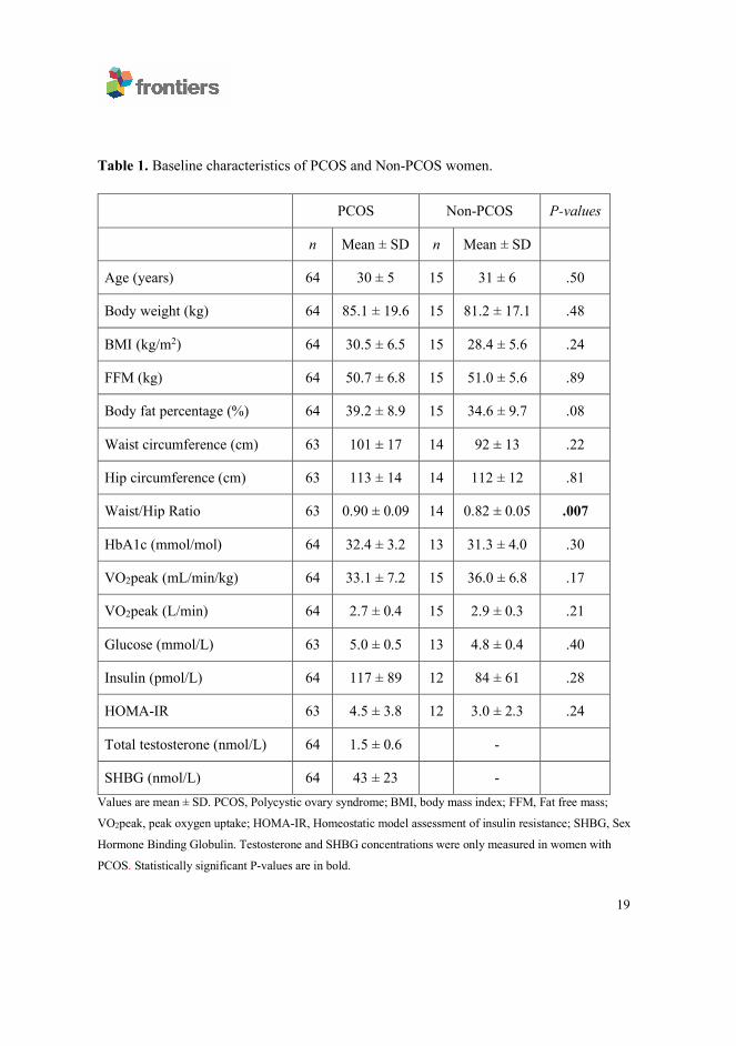
19
Table 1. Baseline characteristics of PCOS and Non-PCOS women.
Values are mean ± SD. PCOS, Polycystic ovary syndrome; BMI, body mass index; FFM, Fat free mass;
VO2peak, peak oxygen uptake; HOMA-IR, Homeostatic model assessment of insulin resistance; SHBG, Sex
Hormone Binding Globulin. Testosterone and SHBG concentrations were only measured in women with
PCOS. Statistically significant P-values are in bold.
PCOS Non-PCOS P-values
n Mean ± SD n Mean ± SD
Age (years) 64 30 ± 5 15 31 ± 6 .50
Body weight (kg) 64 85.1 ± 19.6 15 81.2 ± 17.1 .48
BMI (kg/m2) 64 30.5 ± 6.5 15 28.4 ± 5.6 .24
FFM (kg) 64 50.7 ± 6.8 15 51.0 ± 5.6 .89
Body fat percentage (%) 64 39.2 ± 8.9 15 34.6 ± 9.7 .08
Waist circumference (cm) 63 101 ± 17 14 92 ± 13 .22
Hip circumference (cm) 63 113 ± 14 14 112 ± 12 .81
Waist/Hip Ratio 63 0.90 ± 0.09 14 0.82 ± 0.05 .007
HbA1c (mmol/mol) 64 32.4 ± 3.2 13 31.3 ± 4.0 .30
VO2peak (mL/min/kg) 64 33.1 ± 7.2 15 36.0 ± 6.8 .17
VO2peak (L/min) 64 2.7 ± 0.4 15 2.9 ± 0.3 .21
Glucose (mmol/L) 63 5.0 ± 0.5 13 4.8 ± 0.4 .40
Insulin (pmol/L) 64 117 ± 89 12 84 ± 61 .28
HOMA-IR 63 4.5 ± 3.8 12 3.0 ± 2.3 .24
Total testosterone (nmol/L) 64 1.5 ± 0.6 -
SHBG (nmol/L) 64 43 ± 23 -
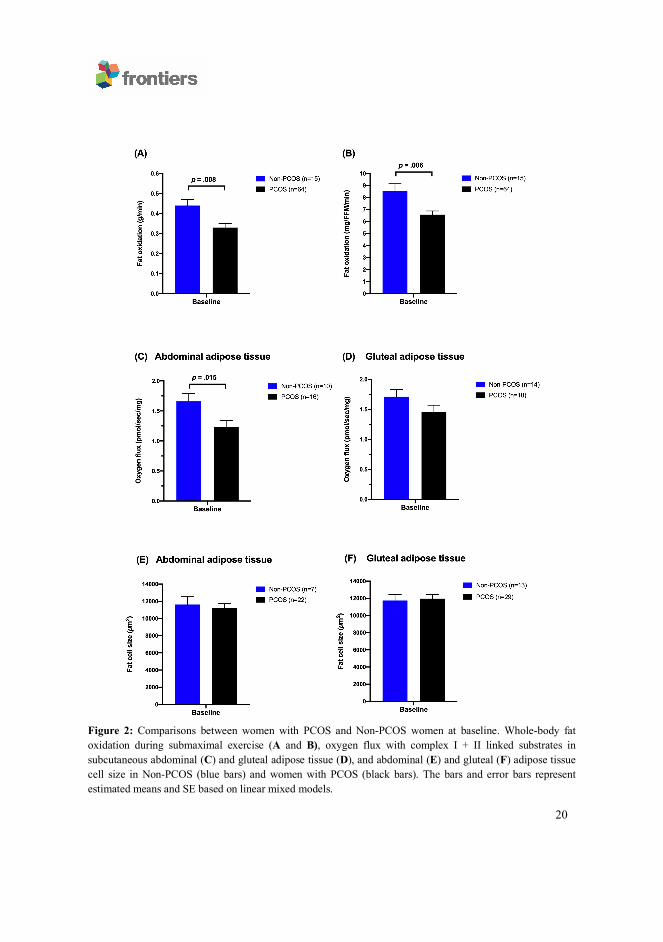
20
Figure 2: Comparisons between women with PCOS and Non-PCOS women at baseline. Whole-body fat oxidation during submaximal exercise (A and B), oxygen flux with complex I + II linked substrates in subcutaneous abdominal (C) and gluteal adipose tissue (D), and abdominal (E) and gluteal (F) adipose tissue cell size in Non-PCOS (blue bars) and women with PCOS (black bars). The bars and error bars represent estimated means and SE based on linear mixed models.
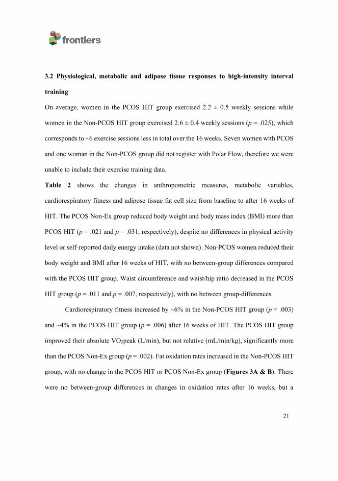
21
3.2 Physiological, metabolic and adipose tissue responses to high-intensity interval
training
On average, women in the PCOS HIT group exercised 2.2 ± 0.5 weekly sessions while
women in the Non-PCOS HIT group exercised 2.6 ± 0.4 weekly sessions (p = .025), which
corresponds to ~6 exercise sessions less in total over the 16 weeks. Seven women with PCOS
and one woman in the Non-PCOS group did not register with Polar Flow, therefore we were
unable to include their exercise training data.
Table 2 shows the changes in anthropometric measures, metabolic variables,
cardiorespiratory fitness and adipose tissue fat cell size from baseline to after 16 weeks of
HIT. The PCOS Non-Ex group reduced body weight and body mass index (BMI) more than
PCOS HIT (p = .021 and p = .031, respectively), despite no differences in physical activity
level or self-reported daily energy intake (data not shown). Non-PCOS women reduced their
body weight and BMI after 16 weeks of HIT, with no between-group differences compared
with the PCOS HIT group. Waist circumference and waist/hip ratio decreased in the PCOS
HIT group (p = .011 and p = .007, respectively), with no between group-differences.
Cardiorespiratory fitness increased by ~6% in the Non-PCOS HIT group (p = .003)
and ~4% in the PCOS HIT group (p = .006) after 16 weeks of HIT. The PCOS HIT group
improved their absolute VO2peak (L/min), but not relative (mL/min/kg), significantly more
than the PCOS Non-Ex group (p = .002). Fat oxidation rates increased in the Non-PCOS HIT
group, with no change in the PCOS HIT or PCOS Non-Ex group (Figures 3A & B). There
were no between-group differences in changes in oxidation rates after 16 weeks, but a

22
tendency of higher increase in fat oxidation rates (mg/kg/min) were observed in the Non-
PCOS HIT group compared with the PCOS HIT group (p = .075).
We observed no between-group differences in metabolic outcomes, abdominal and gluteal
adipose tissue oxygen flux (Figure 3C &D), or cell size (Figure 3E & F) after 16 weeks of
HIT.
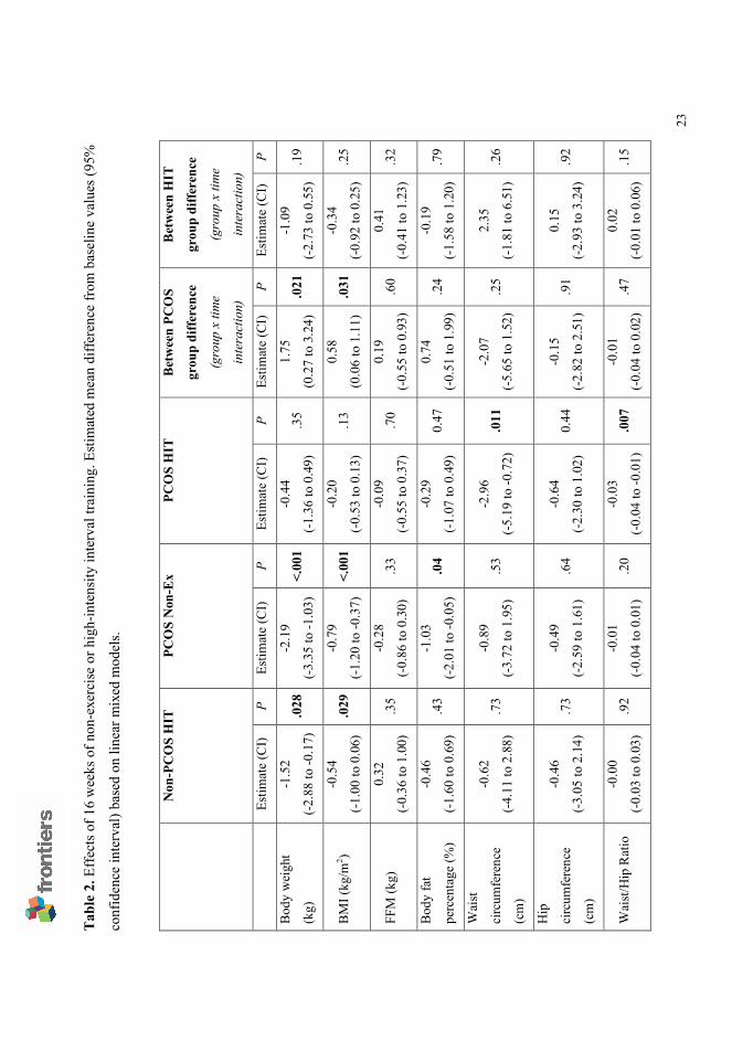
23
Tabl
e 2.
Eff
ects
of 1
6 w
eeks
of n
on-e
xerc
ise
or h
igh-
inte
nsity
inte
rval
trai
ning
. Est
imat
ed m
ean
diffe
renc
e fr
om b
asel
ine
valu
es (9
5%
conf
iden
ce in
terv
al) b
ased
on
linea
r mix
ed m
odel
s.
Non
-PC
OS
HIT
PC
OS
Non
-Ex
PCO
S H
IT
Bet
wee
n PC
OS
grou
p di
ffere
nce
(gro
up x
tim
e
inte
ract
ion)
Bet
wee
n H
IT
grou
p di
ffere
nce
(gro
up x
tim
e
inte
ract
ion)
Es
timat
e (C
I) P
Estim
ate
(CI)
P Es
timat
e (C
I) P
Estim
ate
(CI)
P Es
timat
e (C
I) P
Bod
y w
eigh
t
(kg)
-1.5
2
(-2.8
8 to
-0.1
7)
.028
-2
.19
(-3.3
5 to
-1.0
3)
<.00
1 -0
.44
(-1.3
6 to
0.4
9)
.35
1.75
(0.2
7 to
3.2
4)
.021
-1
.09
(-2.7
3 to
0.5
5)
.19
BM
I (kg
/m2 )
-0.5
4
(-1.0
0 to
0.0
6)
.029
-0
.79
(-1.2
0 to
-0.3
7)
<.00
1 -0
.20
(-0.5
3 to
0.1
3)
.13
0.58
(0.0
6 to
1.1
1)
.031
-0
.34
(-0.9
2 to
0.2
5)
.25
FFM
(kg)
0.
32
(-0.3
6 to
1.0
0)
.35
-0.2
8
(-0.8
6 to
0.3
0)
.33
-0.0
9
(-0.5
5 to
0.3
7)
.70
0.19
(-0.5
5 to
0.9
3)
.60
0.41
(-0.4
1 to
1.2
3)
.32
Bod
y fa
t
perc
enta
ge (%
)
-0.4
6
(-1.6
0 to
0.6
9)
.43
-1.0
3
(-2.0
1 to
-0.0
5)
.04
-0.2
9
(-1.0
7 to
0.4
9)
0.47
0.
74
(-0.5
1 to
1.9
9)
.24
-0.1
9
(-1.5
8 to
1.2
0)
.79
Wai
st
circ
umfe
renc
e
(cm
)
-0.6
2
(-4.1
1 to
2.8
8)
.73
-0.8
9
(-3.7
2 to
1.9
5)
.53
-2.9
6
(-5.1
9 to
-0.7
2)
.011
-2
.07
(-5.6
5 to
1.5
2)
.25
2.35
(-1.8
1 to
6.5
1)
.26
Hip
circ
umfe
renc
e
(cm
)
-0.4
6
(-3.0
5 to
2.1
4)
.73
-0.4
9
(-2.5
9 to
1.6
1)
.64
-0.6
4
(-2.3
0 to
1.0
2)
0.44
-0
.15
(-2.8
2 to
2.5
1)
.91
0.15
(-2.9
3 to
3.2
4)
.92
Wai
st/H
ip R
atio
-0
.00
(-0.0
3 to
0.0
3)
.92
-0.0
1
(-0.0
4 to
0.0
1)
.20
-0.0
3
(-0.0
4 to
-0.0
1)
.007
-0
.01
(-0.0
4 to
0.0
2)
.47
0.02
(-0.0
1 to
0.0
6)
.15
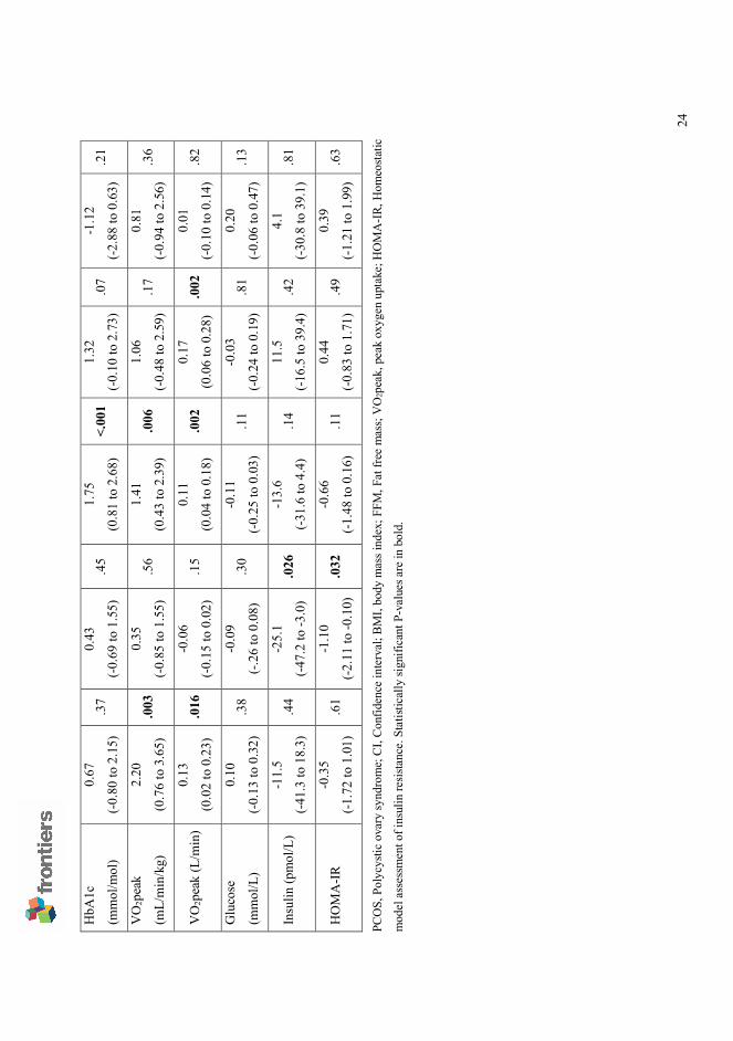
24
PCO
S, P
olyc
ystic
ova
ry sy
ndro
me;
CI,
Con
fiden
ce in
terv
al; B
MI,
body
mas
s ind
ex; F
FM, F
at fr
ee m
ass;
VO
2pea
k, p
eak
oxyg
en u
ptak
e; H
OM
A-I
R, H
omeo
stat
ic
mod
el a
sses
smen
t of i
nsul
in re
sista
nce.
Sta
tistic
ally
sign
ifica
nt P
-val
ues a
re in
bol
d.
HbA
1c
(mm
ol/m
ol)
0.67
(-0.8
0 to
2.1
5)
.37
0.43
(-0.6
9 to
1.5
5)
.45
1.75
(0.8
1 to
2.6
8)
<.00
1 1.
32
(-0.1
0 to
2.7
3)
.07
-1.1
2
(-2.8
8 to
0.6
3)
.21
VO
2pea
k
(mL/
min
/kg)
2.20
(0.7
6 to
3.6
5)
.003
0.
35
(-0.8
5 to
1.5
5)
.56
1.41
(0.4
3 to
2.3
9)
.006
1.
06
(-0.4
8 to
2.5
9)
.17
0.81
(-0.9
4 to
2.5
6)
.36
VO
2pea
k (L
/min
) 0.
13
(0.0
2 to
0.2
3)
.016
-0
.06
(-0.1
5 to
0.0
2)
.15
0.11
(0.0
4 to
0.1
8)
.002
0.
17
(0.0
6 to
0.2
8)
.002
0.
01
(-0.1
0 to
0.1
4)
.82
Glu
cose
(mm
ol/L
)
0.10
(-0.1
3 to
0.3
2)
.38
-0.0
9
(-.26
to 0
.08)
.3
0 -0
.11
(-0.2
5 to
0.0
3)
.11
-0.0
3
(-0.2
4 to
0.1
9)
.81
0.20
(-0.0
6 to
0.4
7)
.13
Insu
lin (p
mol
/L)
-11.
5
(-41.
3 to
18.
3)
.44
-25.
1
(-47.
2 to
-3.0
) .0
26
-13.
6
(-31.
6 to
4.4
) .1
4 11
.5
(-16.
5 to
39.
4)
.42
4.1
(-30.
8 to
39.
1)
.81
HO
MA
-IR
-0.3
5
(-1.7
2 to
1.0
1)
.61
-1.1
0
(-2.1
1 to
-0.1
0)
.032
-0
.66
(-1.4
8 to
0.1
6)
.11
0.44
(-0.8
3 to
1.7
1)
.49
0.39
(-1.2
1 to
1.9
9)
.63
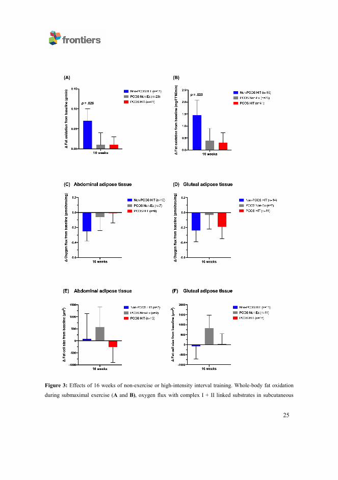
25
Figure 3: Effects of 16 weeks of non-exercise or high-intensity interval training. Whole-body fat oxidation
during submaximal exercise (A and B), oxygen flux with complex I + II linked substrates in subcutaneous
26
abdominal (C) and gluteal adipose tissue (D), and abdominal (E) and gluteal (F) adipose tissue cell size in Non-
PCOS HIT (blue bars), PCOS Non-Ex (grey bars), and PCOS HIT (red bars) after the 16 weeks intervention.
The bars and error bars represent estimated means and SE based on linear mixed models. P values are for
within-group comparisons after 16 weeks of high-intensity interval training.
4.0 Discussion
Our findings provide new data on whole-body fat oxidation during submaximal exercise, and
gluteal and abdominal adipose tissue mitochondrial respiration and cell size in women with
PCOS and Non-PCOS women, along with the effects of 16 weeks of HIT on these outcomes.
We report that 16 weeks of HIT improved fat oxidation rates during submaximal exercise in
Non-PCOS women, but not in women with PCOS, despite similar improvements in
VO2peak. Furthermore, women with PCOS had lower mitochondrial respiration with
substrates for complex I + II in subcutaneous abdominal adipose tissue compared to Non-
PCOS women. Mitochondrial respiration in both adipose tissue sites were unaffected by 16
weeks of HIT in women with and without PCOS. Finally, subcutaneous gluteal or abdominal
adipose tissue cell size did not differ between women with PCOS and Non-PCOS women
and 16 weeks of HIT did not alter adipocyte sizes in any group of women.
We report novel findings on absent exercise-induced improvements in fat oxidation
during submaximal exercise in women with PCOS, despite improved VO2peak, suggesting
metabolic inflexibility in women with PCOS. Conversely, fat oxidation rates improved in
Non-PCOS women after 16 weeks of HIT. A recent systematic review reported metabolic
inflexibility in the rested, insulin-stimulated state in women with PCOS compared with
healthy women (Rimmer et al., 2020). Metabolic inflexibility has been linked to insulin
27
resistance and type 2 diabetes (Goodpaster and Sparks, 2017), and Broskey and colleagues
found that women with PCOS and obesity (28.8 ± 4.7 years) were as metabolically inflexible
as middle-aged women with type 2 diabetes and obesity (58.2 ± 9.9 years) (Broskey et al.,
2018). Metabolic inflexibility has also been reported in lean women with PCOS (Hansen et
al., 2019) and adolescent girls with PCOS (Kim et al., 2018), suggesting that metabolic
inflexibility is associated with PCOS independent of BMI and age.
Similar to our findings in Non-PCOS women, previous studies reported
improvements in whole-body fat oxidation during submaximal exercise at 60% of VO2peak
in healthy recreationally active women after seven sessions of HIT (10 x 4 min bouts at 90%
of VO2peak separated by 2 min of rest) (Talanian et al., 2007), and in untrained recreationally
active men and women after six weeks of HIT (using the same HIT protocol as Talanian et
al. (2007) but for three days/week for 6 weeks) (Perry et al., 2008). In our study, the Non-
PCOS women exercised significantly more on a weekly basis compared to the women with
PCOS (2.6 ± 0.4 versus 2.2 ± 0.5 weekly HIT sessions, corresponding to ~6 HIT sessions
less in total over the 16 weeks), which may explain the difference observed in training-
induced improvements in fat oxidation rates. However, we report similar improvements in
VO2peak in women with PCOS and Non-PCOS women after 16 weeks of HIT, which
suggests that the 6 fewer HIT sessions performed by women with PCOS cannot fully explain
the difference in training-induced improvements in fat oxidation rates. Absent training-
induced improvements in women with PCOS have been reported previously; Hansen and
colleagues observed improved insulin sensitivity measured by the gold-standard
hyperinsulinaemic-euglycemic clamp in healthy women without PCOS, but not in women
28
with PCOS after 14 weeks of exercise training (three weekly exercise sessions; two aerobic
HIT sessions and one strength training session) (Hansen et al., 2020). Similarly, Hansen and
colleagues reported improved incremental area under the oral glucose tolerance test curve for
plasma glucose and insulin after exercise training in healthy women, but not in women with
PCOS. Their data suggested that the lack of improvements in insulin action after exercise
training were due to impaired ability to upregulate glucose uptake in skeletal muscle.
Skeletal muscle and adipose tissue are crucial tissues in energy metabolism and play
a major role in metabolic flexibility in humans. However, little research has been undertaken
on metabolic flexibility of white adipose tissue. Metabolic flexibility is driven by cellular
processes that may be linked to the mitochondria (Goodpaster and Sparks, 2017). We are the
first to explore subcutaneous abdominal and gluteal adipose tissue mitochondrial respiration
in women with and without PCOS and the responses to HIT. In our study, we observed higher
mitochondrial respiration through complex I + II in subcutaneous abdominal adipose tissue
from Non-PCOS women compared to women with PCOS at baseline, no such differences
were seen in gluteal adipose tissue. Low-grade inflammation may be one mechanism causing
the lower mitochondrial respiration in women with PCOS compared to Non-PCOS women
at baseline. There is a proposed link between inflammation and mitochondrial dysfunction in
adipocytes, in which the proinflammatory response of macrophages may promote
mitochondrial dysfunction in adipocytes (Woo et al., 2019). No changes were observed in
mitochondrial respiration in either of the adipose tissue depots in either group of women after
16 weeks of HIT, which suggests that mitochondrial respiration through complex I + II in
adipose tissue cannot explain the improved fat oxidation rates and metabolic flexibility
29
observed in Non-PCOS women. Our findings are supported by data from Larsen and
colleagues, who showed increased VO2peak but no change in mitochondrial respiration in
subcutaneous abdominal adipose tissue in overweight but otherwise healthy men and women
after 6 weeks of LV-HIT (three days/week with 5 x 60 s maximal effort work-bouts) (Larsen
et al., 2015). However, there are some indications in the literature for exercise-induced
changes in adipose tissue mitochondrial respiration. Dohlmann and colleagues (2018) found
decreased mitochondrial respiration in subcutaneous abdominal adipose tissue in healthy
men and women after 6 weeks of LV-HIT (18 HIT sessions with 7 x 1 min exercise bouts at
~100% of VO2peak) (Dohlmann et al., 2018), while Mendham and colleagues (2020)
reported increased mitochondrial respiration in subcutaneous abdominal adipose tissue in
obese, black South-African women after 12 weeks of combined aerobic and resistance
exercise training (four days/week of 40-60 min with aerobic exercise at 75-80% HRpeak and
resistance training that included upper- and lower-body exercises at 60-70% HRpeak)
(Mendham et al., 2020). We are not sure of the reasons for these divergent findings, but they
may be explained by different exercise modalities and intensities.
We did not observe changes in mitochondrial respiration in adipose tissue after 16
weeks of HIT. We speculate that changes may have occurred in skeletal muscle
mitochondrial function as Larsen and colleagues have previously reported increased
mitochondrial respiration in skeletal muscle, but not in abdominal adipose tissue, after 6
weeks of HIT in men and women who were overweight but otherwise healthy (Larsen et al.,
2015). Possible gains, or lack of gains, in skeletal muscle mitochondrial function could
explain the improved whole-body fat oxidation during submaximal exercise in Non-PCOS
30
women, and lack of improvement in women with PCOS after 16 weeks of HIT. However,
we did not obtain any muscle biopsies in our study.
We observed no differences in subcutaneous abdominal or gluteal fat cell size
between Non-PCOS women and women with PCOS, nor do we report changes in cell size
after 16 weeks of HIT in either group of women. A case-control study on women with PCOS
and age- and BMI-matched women without PCOS reported enlarged abdominal adipocyte
volumes in women with PCOS, and an association between hypertrophic adipocytes and
insulin resistance (Manneras-Holm et al., 2011). The divergent findings in our study and the
study by Manneras-Holm may be explained by higher power in their study (27 women in
both groups).
Similar to our findings, previous studies have reported no changes in subcutaneous
abdominal adipose tissue volume and size after 12 weeks of exercise training in men with
overweight or obesity (three days/week; aerobic exercise twice/week cycling for 30 min at
70% maximal watt and resistance training once weekly) (Stinkens et al., 2018), or after 16
weeks of exercise in women with PCOS (three days/week for 30 min at an intensity of faster
than normal walking pace) (Stener-Victorin et al., 2012). A longer and more intense training
program may be required to change adipose tissue structure. Després and colleagues observed
a sex difference in the sensitivity to exercise training, with less responsiveness in female
adipose tissue. Twenty weeks of endurance training for 40 min at 80% HRmax 4-5 times
weekly did not influence fat percentage or adipose cell weight in women, whereas males
showed reductions in both fat percentage and adipose cell weight (Despres et al., 1984).
31
Our data indicate that women with PCOS allocated the non-exercising control group
most likely introduced some lifestyle changes during the study period as we observed
reductions in body mass, BMI and body fat percentage and improvements in fasting insulin
concentration and HOMA-IR in this group. Although we detected no changes in self-reported
daily energy intake or physical activity level, these were only recorded for a brief period of
time (4-5 days at each timepoint). At study inclusion, women with PCOS assigned to the
control group were informed about the current recommendations of at least 150 min weekly
of moderate intensity physical activity, which could have affected their physical activity
behavior. Furthermore, individuals who volunteer for exercise studies usually hope to be
allocated to the exercise intervention, and women allocated to the control group have most
likely been motivated to introduce lifestyle changes.
Subgroup analyses from a systematic review and meta-analysis on the effectiveness
of exercise compared to control in women with PCOS showed greater improvements in
cardiometabolic outcomes in supervised versus unsupervised exercise interventions (Kite et
al., 2019), and a supervised rather than semi-supervised exercise protocol in our study could
possibly have resulted in greater gains for women with (and without) PCOS. Furthermore,
including a more varied exercise protocol with a combination of LV-HIT and HV-HIT
sessions could have induced larger improvements, as shown previously (Almenning et al.,
2015).
Major strengths of our study are the pair-wise age- and BMI-matching of women with
PCOS and Non-PCOS women, the rigorous inclusion and exclusion criteria, and that we
explored two adipose tissue depots in women with and without PCOS. We also acknowledge
32
study limitations. We used different methodologies to estimate body composition at the two
study centers, limiting the baseline comparison for these outcomes between women with and
without PCOS. The Inbody 720 scale that we used for participants in Norway underestimates
body fat and overestimates FFM compared with DXA (McLester et al., 2018). Due to limited
adipose tissue biopsy volumes, we were unable to compare mitochondrial respiration and fat
cell size between the women with PCOS and Non-PCOS women who were individually age-
and BMI-matched. Furthermore, due to limited adipose tissue volumes and statistical power,
we were unable to run meaningful statistical analyses for LV-HIT and HV-HIT groups,
which made us unable to detect differences in the effects of LV-HIT and HV-HIT on
mitochondrial respiration and fat cell size. Instead, data for LV-HIT and HV-HIT were
pooled to increase the statistical power and to investigate the effect of HIT.
In conclusion, we observed exercise-induced improvements in whole-body fat
oxidation during submaximal exercise in Non-PCOS women but not in women with PCOS
after 16 weeks of HIT. These findings suggest metabolic inflexibility in women with PCOS.
Mitochondrial respiration was lower in subcutaneous abdominal, but not gluteal, adipose
tissue in women with PCOS compared to Non-PCOS women, and 16 weeks of HIT did not
alter adipose tissue mitochondrial respiration in either group of women. Further studies are
required to ascertain the underlying mechanisms behind the absent exercise-induced
improvements in fat oxidation and metabolic inflexibility observed in women with PCOS.
Conflict of Interest
The authors declare no conflict of interest.
33
Author contributions
SLi drafted the manuscript. SLi, RR and SLa analyzed the data. SLi and SLy performed
statistical analyses. SLi, IK and TM were responsible for study conception and design,
coordinated the studies at the two sites, performed measurements on testing days, and
supervised the exercise training. All authors provided feedback and approved the final
manuscript.
Funding
This study was supported by the Liaison Committee for education, research and innovation
in Central Norway (Grant numbers: 2014/23166 and 2017/29014).
Acknowledgements
We wish to thank the Unit for Applied Clinical Research at NTNU for providing the internet-
based randomization for both trials; Next Move at NTNU for providing the equipment and
lab facilities for training sessions and exercise testing in Norway; Professor John A. Hawley
for his valued support, and for providing the equipment and lab facilities for training sessions
and testing in Australia; Dr. Andrew Garnham and the Clinical Research Facility at St. Olav’s
Hospital for performing blood and adipose tissue sampling in Australia and Norway,
respectively; Dr. Evelyn Parr for invaluable support in the laboratory in Australia; The
Cellular & Molecular Imaging Core Facility at NTNU for use of the EVOS FL Auto 2
Imaging System including technical advice. Finally, we wish to thank all the participants for
their time and effort.
34
References
ALMENNING, I., RIEBER-MOHN, A., LUNDGREN, K. M., SHETELIG LOVVIK, T.,
GARNAES, K. K. & MOHOLDT, T. 2015. Effects of High Intensity Interval
Training and Strength Training on Metabolic, Cardiovascular and Hormonal
Outcomes in Women with Polycystic Ovary Syndrome: A Pilot Study. PLoS One,
10, e0138793.
AZZIZ, R., CARMINA, E., DEWAILLY, D., DIAMANTI-KANDARAKIS, E.,
ESCOBAR-MORREALE, H. F., FUTTERWEIT, W., JANSSEN, O. E., LEGRO,
R. S., NORMAN, R. J., TAYLOR, A. E., WITCHEL, S. F., TASK FORCE ON
THE PHENOTYPE OF THE POLYCYSTIC OVARY SYNDROME OF THE
ANDROGEN, E. & SOCIETY, P. 2009. The Androgen Excess and PCOS Society
criteria for the polycystic ovary syndrome: the complete task force report. Fertil
Steril, 91, 456-88.
BERGLUND, I. J., SORAS, S. E., RELLING, B. E., LUNDGREN, K. M., KIEL, I. A. &
MOHOLDT, T. 2019. The relationship between maximum heart rate in a
cardiorespiratory fitness test and in a maximum heart rate test. J Sci Med Sport, 22,
607-610.
BOZDAG, G., MUMUSOGLU, S., ZENGIN, D., KARABULUT, E. & YILDIZ, B. O.
2016. The prevalence and phenotypic features of polycystic ovary syndrome: a
systematic review and meta-analysis. Hum Reprod, 31, 2841-2855.
35
BROSKEY, N. T., TAM, C. S., SUTTON, E. F., ALTAZAN, A. D., BURTON, J. H.,
RAVUSSIN, E. & REDMAN, L. M. 2018. Metabolic inflexibility in women with
PCOS is similar to women with type 2 diabetes. Nutr Metab (Lond), 15, 75.
CHUANG, T. Y., WU, H. L., CHEN, C. C., GAMBOA, G. M., LAYMAN, L. C.,
DIAMOND, M. P., AZZIZ, R. & CHEN, Y. H. 2015. MicroRNA-223 Expression is
Upregulated in Insulin Resistant Human Adipose Tissue. J Diabetes Res, 2015,
943659.
DE GROOT, P. C., DEKKERS, O. M., ROMIJN, J. A., DIEBEN, S. W. &
HELMERHORST, F. M. 2011. PCOS, coronary heart disease, stroke and the
influence of obesity: a systematic review and meta-analysis. Hum Reprod Update,
17, 495-500.
DESPRES, J. P., BOUCHARD, C., SAVARD, R., TREMBLAY, A., MARCOTTE, M. &
THERIAULT, G. 1984. The effect of a 20-week endurance training program on
adipose-tissue morphology and lipolysis in men and women. Metabolism, 33, 235-
9.
DIAMANTI-KANDARAKIS, E. & PAPAVASSILIOU, A. G. 2006. Molecular
mechanisms of insulin resistance in polycystic ovary syndrome. Trends Mol Med,
12, 324-32.
DOHLMANN, T. L., HINDSO, M., DELA, F., HELGE, J. W. & LARSEN, S. 2018. High-
intensity interval training changes mitochondrial respiratory capacity differently in
adipose tissue and skeletal muscle. Physiol Rep, 6, e13857.
36
DUNAIF, A., SEGAL, K. R., SHELLEY, D. R., GREEN, G., DOBRJANSKY, A. &
LICHOLAI, T. 1992. Evidence for distinctive and intrinsic defects in insulin action
in polycystic ovary syndrome. Diabetes, 41, 1257-66.
FERRIMAN, D. & GALLWEY, J. D. 1961. Clinical assessment of body hair growth in
women. J Clin Endocrinol Metab, 21, 1440-7.
GOODPASTER, B. H. & SPARKS, L. M. 2017. Metabolic Flexibility in Health and
Disease. Cell Metab, 25, 1027-1036.
GREENWOOD, E. A., NOEL, M. W., KAO, C. N., SHINKAI, K., PASCH, L. A.,
CEDARS, M. I. & HUDDLESTON, H. G. 2016. Vigorous exercise is associated
with superior metabolic profiles in polycystic ovary syndrome independent of total
exercise expenditure. Fertil Steril, 105, 486-93.
HANSEN, S. L., BOJSEN-MOLLER, K. N., LUNDSGAARD, A. M., HENDRICH, F. L.,
NILAS, L., SJOBERG, K. A., HINGST, J. R., SERUP, A. K., OLGUIN, C. H.,
CARL, C. S., WERNBLAD, L. F., HENNEBERG, M., LUSTRUP, K. M.,
HANSEN, C., JENSEN, T. E., MADSBAD, S., WOJTASZEWSKI, J. F. P.,
RICHTER, E. A. & KIENS, B. 2020. Mechanisms Underlying Absent Training-
Induced Improvement in Insulin Action in Lean, Hyperandrogenic Women With
Polycystic Ovary Syndrome. Diabetes, 69, 2267-2280.
HANSEN, S. L., SVENDSEN, P. F., JEPPESEN, J. F., HOEG, L. D., ANDERSEN, N. R.,
KRISTENSEN, J. M., NILAS, L., LUNDSGAARD, A. M., WOJTASZEWSKI, J.
F. P., MADSBAD, S. & KIENS, B. 2019. Molecular Mechanisms in Skeletal
37
Muscle Underlying Insulin Resistance in Women Who Are Lean With Polycystic
Ovary Syndrome. J Clin Endocrinol Metab, 104, 1841-1854.
JEUKENDRUP, A. E. & WALLIS, G. A. 2005. Measurement of substrate oxidation during
exercise by means of gas exchange measurements. Int J Sports Med, 26 Suppl 1,
S28-37.
KIEL, I. A., LIONETT, S., PARR, E. B., JONES, H., ROSET, M. A. H., SALVESEN, O.,
VANKY, E. & MOHOLDT, T. 2020. Improving reproductive function in women
with polycystic ovary syndrome with high-intensity interval training (IMPROV-IT):
study protocol for a two-centre, three-armed randomised controlled trial. BMJ
Open, 10, e034733.
KIM, J. Y., TFAYLI, H., MICHALISZYN, S. F. & ARSLANIAN, S. 2018. Impaired
Lipolysis, Diminished Fat Oxidation, and Metabolic Inflexibility in Obese Girls
With Polycystic Ovary Syndrome. J Clin Endocrinol Metab, 103, 546-554.
KITE, C., LAHART, I. M., AFZAL, I., BROOM, D. R., RANDEVA, H., KYROU, I. &
BROWN, J. E. 2019. Exercise, or exercise and diet for the management of
polycystic ovary syndrome: a systematic review and meta-analysis. Syst Rev, 8, 51.
KRAUNSOE, R., BOUSHEL, R., HANSEN, C. N., SCHJERLING, P., QVORTRUP, K.,
STOCKEL, M., MIKINES, K. J. & DELA, F. 2010. Mitochondrial respiration in
subcutaneous and visceral adipose tissue from patients with morbid obesity. J
Physiol, 588, 2023-32.
LARSEN, S., DANIELSEN, J. H., SONDERGARD, S. D., SOGAARD, D., VIGELSOE,
A., DYBBOE, R., SKAABY, S., DELA, F. & HELGE, J. W. 2015. The effect of
38
high-intensity training on mitochondrial fat oxidation in skeletal muscle and
subcutaneous adipose tissue. Scand J Med Sci Sports, 25, e59-69.
MANNERAS-HOLM, L., LEONHARDT, H., KULLBERG, J., JENNISCHE, E., ODEN,
A., HOLM, G., HELLSTROM, M., LONN, L., OLIVECRONA, G., STENER-
VICTORIN, E. & LONN, M. 2011. Adipose tissue has aberrant morphology and
function in PCOS: enlarged adipocytes and low serum adiponectin, but not
circulating sex steroids, are strongly associated with insulin resistance. J Clin
Endocrinol Metab, 96, E304-11.
MATTHEWS, D. R., HOSKER, J. P., RUDENSKI, A. S., NAYLOR, B. A., TREACHER,
D. F. & TURNER, R. C. 1985. Homeostasis model assessment: insulin resistance
and beta-cell function from fasting plasma glucose and insulin concentrations in
man. Diabetologia, 28, 412-9.
MCLESTER, C. N., NICKERSON, B. S., KLISZCZEWICZ, B. M. & MCLESTER, J. R.
2018. Reliability and Agreement of Various InBody Body Composition Analyzers
as Compared to Dual-Energy X-Ray Absorptiometry in Healthy Men and Women. J
Clin Densitom.
MENDHAM, A. E., LARSEN, S., GEORGE, C., ADAMS, K., HAUKSSON, J.,
OLSSON, T., FORTUIN-DE SMIDT, M. C., NONO NANKAM, P. A., HAKIM,
O., GOFF, L. M., PHEIFFER, C. & GOEDECKE, J. H. 2020. Exercise training
results in depot-specific adaptations to adipose tissue mitochondrial function. Sci
Rep, 10, 3785.
39
MORAN, L. J., MISSO, M. L., WILD, R. A. & NORMAN, R. J. 2010. Impaired glucose
tolerance, type 2 diabetes and metabolic syndrome in polycystic ovary syndrome: a
systematic review and meta-analysis. Hum Reprod Update, 16, 347-63.
PATTEN, R. K., BOYLE, R. A., MOHOLDT, T., KIEL, I., HOPKINS, W. G.,
HARRISON, C. L. & STEPTO, N. K. 2020. Exercise Interventions in Polycystic
Ovary Syndrome: A Systematic Review and Meta-analysis. Front Physiol, 11.
PERRY, C. G., HEIGENHAUSER, G. J., BONEN, A. & SPRIET, L. L. 2008. High-
intensity aerobic interval training increases fat and carbohydrate metabolic
capacities in human skeletal muscle. Appl Physiol Nutr Metab, 33, 1112-23.
RIMMER, M., TAN, B. K., TEEDE, H., THANGARATINAM, S. & B, H. A. W. 2020.
Metabolic inflexibility in women with polycystic ovary syndrome: a systematic
review. Gynecol Endocrinol, 36, 501-507.
ROTTERDAM, E. A.-S. P. C. W. G. 2004. Revised 2003 consensus on diagnostic criteria
and long-term health risks related to polycystic ovary syndrome (PCOS). Hum
Reprod, 19, 41-7.
SCHINDELIN, J., ARGANDA-CARRERAS, I., FRISE, E., KAYNIG, V., LONGAIR, M.,
PIETZSCH, T., PREIBISCH, S., RUEDEN, C., SAALFELD, S., SCHMID, B.,
TINEVEZ, J. Y., WHITE, D. J., HARTENSTEIN, V., ELICEIRI, K.,
TOMANCAK, P. & CARDONA, A. 2012. Fiji: an open-source platform for
biological-image analysis. Nat Methods, 9, 676-82.
STENER-VICTORIN, E., BAGHAEI, F., HOLM, G., JANSON, P. O., OLIVECRONA,
G., LONN, M. & MANNERAS-HOLM, L. 2012. Effects of acupuncture and
40
exercise on insulin sensitivity, adipose tissue characteristics, and markers of
coagulation and fibrinolysis in women with polycystic ovary syndrome: secondary
analyses of a randomized controlled trial. Fertil Steril, 97, 501-8.
STINKENS, R., BROUWERS, B., JOCKEN, J. W., BLAAK, E. E., TEUNISSEN-
BEEKMAN, K. F., HESSELINK, M. K. C., VAN BAAK, M. A., SCHRAUWEN,
P. & GOOSSENS, G. H. 2018. Exercise training-induced effects on the abdominal
subcutaneous adipose tissue phenotype in obese humans. J Appl Physiol (1985).
TALANIAN, J. L., GALLOWAY, S. D., HEIGENHAUSER, G. J., BONEN, A. &
SPRIET, L. L. 2007. Two weeks of high-intensity aerobic interval training increases
the capacity for fat oxidation during exercise in women. J Appl Physiol (1985), 102,
1439-47.
TEEDE, H., DEEKS, A. & MORAN, L. 2010. Polycystic ovary syndrome: a complex
condition with psychological, reproductive and metabolic manifestations that
impacts on health across the lifespan. BMC Med, 8, 41.
TEEDE, H. J., MISSO, M. L., COSTELLO, M. F., DOKRAS, A., LAVEN, J., MORAN,
L., PILTONEN, T. & NORMAN, R. J. 2018. Recommendations from the
international evidence-based guideline for the assessment and management of
polycystic ovary syndrome. Hum Reprod, 33, 1602-1618.
TWISK, J., BOSMAN, L., HOEKSTRA, T., RIJNHART, J., WELTEN, M. &
HEYMANS, M. 2018. Different ways to estimate treatment effects in randomised
controlled trials. Contemp Clin Trials Commun, 10, 80-85.

41
WOO, C. Y., JANG, J. E., LEE, S. E., KOH, E. H. & LEE, K. U. 2019. Mitochondrial
Dysfunction in Adipocytes as a Primary Cause of Adipose Tissue Inflammation.
Diabetes Metab J, 43, 247-256.
ISBN 978-82-326-6263-0 (printed ver.)ISBN 978-82-326-5600-4 (electronic ver.)
ISSN 1503-8181 (printed ver.)ISSN 2703-8084 (online ver.)
Doctoral theses at NTNU, 2021:207
Sofie Buurgaard Lionett
The effects of high-intensityinterval training on adiposetissue pathophysiology,reproductive and metabolichealth in women with polycysticovary syndrome
Doc
tora
l the
sis
Doctoral theses at N
TNU
, 2021:207Sofie Buurgaard Lionett
NTN
UN
orw
egia
n U
nive
rsity
of S
cien
ce a
nd T
echn
olog
yTh
esis
for t
he D
egre
e of
Philo
soph
iae
Doc
tor
Facu
lty o
f Med
icin
e an
d H
ealth
Sci
ence
s
Dep
artm
ent o
f Circ
ulat
ion
and
Med
ical
Imag
ing