Shell@Core Coaxial NiO@Ni Nanowire Arrays as High Performance Enzymeless Glucose Sensor
6
Journal of The Electrochemical Society, 160 (11) B207-B212 (2013) B207 0013-4651/2013/160(11)/B207/6/$31.00 © The Electrochemical Society Shell@Core Coaxial NiO@Ni Nanowire Arrays as High Performance Enzymeless Glucose Sensor Mamun Jamal, Maksudul Hasan, Michael Schmidt, Nikolay Petkov, Alan Mathewson, and Kafil M. Razeeb ∗, z Tyndall National Institute, University College Cork, Dyke Parade, Maltings, Cork, Ireland Vertically aligned nanowire array based sensor platform that can monitor electro active species without the requirement of enzyme have received much attention lately. In particular, vertically aligned Ni nanowire arrays (NiNAE) have been found to have excellent catalytic activity toward carbohydrates. However, nickel oxidizes or corrodes in air, and this presents a question about its usefulness as a reliable and stable sensing materials. To address this problem, the authors have developed a vertically aligned co-axial nickel oxide-nickel nanowire array electrode (NiO@NiNAE) platform which can detect glucose without the presence of enzyme. The fabrication procedure is simple and catalytic activity toward glucose has been found to be excellent, this is attributed to the presence of stable NiO around the Ni nanowire. Details of morphological and electrochemical characterization of the sensor platform have been studied to evaluate the sensor performance. The sensitivity of NiNAE and NiO@NiNAE has been found to be 80.0 and 170.4 mC mM −1 cm −2 , respectively. Under optimal detection conditions, the as prepared sensors exhibited linear behavior for glucose detection in the concentration up to 7 mM for both NiNAE and NiO@NiNAE with a limit of detection of 33 and 14 μM, respectively. © 2013 The Electrochemical Society. [DOI: 10.1149/2.004311jes] All rights reserved. Manuscript submitted June 12, 2013; revised manuscript received August 14, 2013. Published September 10, 2013. This was Paper 1471 presented at the Toronto, ON, Canada, Meeting of the Society, May 12–16, 2013. Fabrication of reliable and cost-effective catalysts for the precise monitoring of electro-active species is of significant interest in the development of sensing devices for point of care, environmental con- trol or industrial systems. Nanostructure based material provides a very large electrochemically active surface area, thereby leading to high detection sensitivity. Until now, noble metal nanomaterials, such as Pt, Au, Ag and their alloys, 1,2 have been extensively investigated as anodic materials for designing non-enzymatic sensor surface. Al- though these noble metal-based nanomaterials exhibit high catalytic activity toward various analytes, they are easily fouled by electro- chemically active interferents. Glucose detection is of great interest in diagnosis and management of diabetes mellitus, as well as in the monitoring and control of food processes. In spite of one of the most commercially successful sensor, an enormous work still is in progress to make glucose sensor faster, more sensitive, simple and inexpen- sive. In addition to noble metallic nanomaterial’s, various other metal and metal oxides, such as CuO, RuO 2 , and MnO 2 , have been used as anodic materials for direct oxidation of glucose. 3,4 Among them, Ni-based nanomaterials have received special attention owing to their low cost and high catalytic activity toward glucose oxidation. You et al. reported glucose biosensors based on Ni nanoparticles (NPs) deco- rated carbon nanofibers. 5 He et al. reported the glucose sensors based on Ni NPs modified TiO 2 tube arrays 6 and recently we have reported enzyme free glutamate sensor based on Ni nanowire array (NiNAE) and Pt NPs modified NiNAE. 7 In contrast to metallic Ni, NiO is rela- tively stable. The distinctive merits possessed by NiO, such as its low price, environmentally friendly nature and fast redox kinetics, make it suitable for various catalytical and capacitance applications. However, metallic Ni shows excellent conducting behavior compared to NiO, which is also crucial for catalytic activity. But until now there has been no study on the electrocatalytic activity of NiO@NiNAE with the aim of boosting catalytic activity by combining merits of both Ni and NiO. Therefore, in this work, we demonstrate the fabrication of core shell coaxial nanowire arrays of NiO and Ni, where conductive Ni is the ‘core’ and thin NiO is the ‘shell’ layer. The smartly designed vertically aligned core-shell co-axial NiO@NiNAE have been applied as a high performance electrode material for glucose detection. We also investigate the effect of NiO thickness around the Ni nanowires, the stability of NiO@NiNAE and the selectivity of this electrode toward glucose in the presence of electrochemical interference. ∗ Electrochemical Society Active Member. z E-mail: kafi[email protected] Experimental Synthesis of Ni nanowire array electrode.— Nickel nanowires were prepared inside an anodic alumina oxide (AAO) template having an average pore diameter of 250 nm. 8 One side of the template was coated (evaporated) with a 400 nm layer of Ni film (using Temescal FC-2000) which served as the working electrode. The Ohmic contact to the substrate was made using Radionics silver conductive paint, which has a volume resistivity of 0.001 cm when fully hardened. Low stress nickel sulfamate bath was prepared using nickel sulfamate (0.37 mol dm −3 ), boric acid (0.64 mol dm −3 ), nickel bromide (0.18 mol dm −3 ) and wetting agent ANKOR (R) F (10 mL l −1 ). The pH of the solution was adjusted to 3.8 by adding 1 mol dm −3 sulfonic acid at room temperature. De-ionized water with resistivity ∼18 M was used to prepare the solution. The electrolyte temperature was maintained at 50 ± 1 ◦ C, and the solution was stirred slowly at an rpm of 100. A two electrode cell was used with Ni pellets in Ti- basket as an anode and the Ni/porous AAO template as a working electrode (cathode). A 50 mA cm −2 (as the ratio of the total template area) constant current was applied to the working electrode and the deposition rate was found to be 0.6 μm min −1 . After deposition, the free standing nanowires were released by dissolving the template in dichloromethane, thoroughly washed with de-ionized water, and dried in air. Synthesis of shell@core NiO@Ni nanowire array electrode.— The nickel nanowire arrays were annealed in plasma using a March Plas- mod GCM 200 system to fabricate shell@core NiO@NiNAE. 8 The as prepared film of NiNAEs were attached onto a Si substrate by ad- hesive tape (Kapton tapes, DuPontTM, USA) at the edge, so that back side was not exposed to the plasma and thereby only the surface of the nanowires was converted to oxide. The Ni nanowire arrays were annealed for different times at power input 50 W at a constant oxygen flow in order to achieve different thicknesses of the oxide layer on the nanowires. Characterization.— The morphology of the modified electrodes and elemental analysis were performed using JEOL JSM 6100 SEM connected with EDX. The crystal structures of the nanowires were investigated using XRD (Philips X’pert PW3710-MPD diffractome- ter using filtered Cu-Kα (λ = 1.54 Å) radiation, operating at 40 kV and 35 mA. The nanostructures were examined by high resolution transmission electron microscopy (HR-TEM, JEOL 2100 HRTEM instrument operating at an acceleration voltage of 200 kV) coupled with energy dispersive X-ray spectroscopy (EDS Oxford Instruments ecsdl.org/site/terms_use address. Redistribution subject to ECS license or copyright; see 143.239.65.82 Downloaded on 2013-09-11 to IP
Transcript of Shell@Core Coaxial NiO@Ni Nanowire Arrays as High Performance Enzymeless Glucose Sensor

Journal of The Electrochemical Society, 160 (11) B207-B212 (2013) B2070013-4651/2013/160(11)/B207/6/$31.00 © The Electrochemical Society
Shell@Core Coaxial NiO@Ni Nanowire Arrays as HighPerformance Enzymeless Glucose SensorMamun Jamal, Maksudul Hasan, Michael Schmidt, Nikolay Petkov, Alan Mathewson,and Kafil M. Razeeb∗,z
Tyndall National Institute, University College Cork, Dyke Parade, Maltings, Cork, Ireland
Vertically aligned nanowire array based sensor platform that can monitor electro active species without the requirement of enzymehave received much attention lately. In particular, vertically aligned Ni nanowire arrays (NiNAE) have been found to have excellentcatalytic activity toward carbohydrates. However, nickel oxidizes or corrodes in air, and this presents a question about its usefulnessas a reliable and stable sensing materials. To address this problem, the authors have developed a vertically aligned co-axial nickeloxide-nickel nanowire array electrode (NiO@NiNAE) platform which can detect glucose without the presence of enzyme. Thefabrication procedure is simple and catalytic activity toward glucose has been found to be excellent, this is attributed to the presenceof stable NiO around the Ni nanowire. Details of morphological and electrochemical characterization of the sensor platform havebeen studied to evaluate the sensor performance. The sensitivity of NiNAE and NiO@NiNAE has been found to be 80.0 and 170.4mC mM−1 cm−2, respectively. Under optimal detection conditions, the as prepared sensors exhibited linear behavior for glucosedetection in the concentration up to 7 mM for both NiNAE and NiO@NiNAE with a limit of detection of 33 and 14 μM, respectively.© 2013 The Electrochemical Society. [DOI: 10.1149/2.004311jes] All rights reserved.
Manuscript submitted June 12, 2013; revised manuscript received August 14, 2013. Published September 10, 2013. This was Paper1471 presented at the Toronto, ON, Canada, Meeting of the Society, May 12–16, 2013.
Fabrication of reliable and cost-effective catalysts for the precisemonitoring of electro-active species is of significant interest in thedevelopment of sensing devices for point of care, environmental con-trol or industrial systems. Nanostructure based material provides avery large electrochemically active surface area, thereby leading tohigh detection sensitivity. Until now, noble metal nanomaterials, suchas Pt, Au, Ag and their alloys,1,2 have been extensively investigatedas anodic materials for designing non-enzymatic sensor surface. Al-though these noble metal-based nanomaterials exhibit high catalyticactivity toward various analytes, they are easily fouled by electro-chemically active interferents. Glucose detection is of great interestin diagnosis and management of diabetes mellitus, as well as in themonitoring and control of food processes. In spite of one of the mostcommercially successful sensor, an enormous work still is in progressto make glucose sensor faster, more sensitive, simple and inexpen-sive. In addition to noble metallic nanomaterial’s, various other metaland metal oxides, such as CuO, RuO2, and MnO2, have been usedas anodic materials for direct oxidation of glucose.3,4 Among them,Ni-based nanomaterials have received special attention owing to theirlow cost and high catalytic activity toward glucose oxidation. You etal. reported glucose biosensors based on Ni nanoparticles (NPs) deco-rated carbon nanofibers.5 He et al. reported the glucose sensors basedon Ni NPs modified TiO2 tube arrays6 and recently we have reportedenzyme free glutamate sensor based on Ni nanowire array (NiNAE)and Pt NPs modified NiNAE.7 In contrast to metallic Ni, NiO is rela-tively stable. The distinctive merits possessed by NiO, such as its lowprice, environmentally friendly nature and fast redox kinetics, make itsuitable for various catalytical and capacitance applications. However,metallic Ni shows excellent conducting behavior compared to NiO,which is also crucial for catalytic activity. But until now there hasbeen no study on the electrocatalytic activity of NiO@NiNAE withthe aim of boosting catalytic activity by combining merits of both Niand NiO.
Therefore, in this work, we demonstrate the fabrication of coreshell coaxial nanowire arrays of NiO and Ni, where conductive Niis the ‘core’ and thin NiO is the ‘shell’ layer. The smartly designedvertically aligned core-shell co-axial NiO@NiNAE have been appliedas a high performance electrode material for glucose detection. Wealso investigate the effect of NiO thickness around the Ni nanowires,the stability of NiO@NiNAE and the selectivity of this electrodetoward glucose in the presence of electrochemical interference.
∗Electrochemical Society Active Member.zE-mail: [email protected]
Experimental
Synthesis of Ni nanowire array electrode.— Nickel nanowireswere prepared inside an anodic alumina oxide (AAO) template havingan average pore diameter of 250 nm.8 One side of the template wascoated (evaporated) with a 400 nm layer of Ni film (using TemescalFC-2000) which served as the working electrode. The Ohmic contactto the substrate was made using Radionics silver conductive paint,which has a volume resistivity of 0.001 � cm when fully hardened.Low stress nickel sulfamate bath was prepared using nickel sulfamate(0.37 mol dm−3), boric acid (0.64 mol dm−3), nickel bromide (0.18mol dm−3) and wetting agent ANKOR (R) F (10 mL l−1). The pHof the solution was adjusted to 3.8 by adding 1 mol dm−3 sulfonicacid at room temperature. De-ionized water with resistivity ∼18 M�was used to prepare the solution. The electrolyte temperature wasmaintained at 50 ± 1◦C, and the solution was stirred slowly at anrpm of 100. A two electrode cell was used with Ni pellets in Ti-basket as an anode and the Ni/porous AAO template as a workingelectrode (cathode). A 50 mA cm−2 (as the ratio of the total templatearea) constant current was applied to the working electrode and thedeposition rate was found to be 0.6 μm min−1. After deposition, thefree standing nanowires were released by dissolving the template indichloromethane, thoroughly washed with de-ionized water, and driedin air.
Synthesis of shell@core NiO@Ni nanowire array electrode.— Thenickel nanowire arrays were annealed in plasma using a March Plas-mod GCM 200 system to fabricate shell@core [email protected] Theas prepared film of NiNAEs were attached onto a Si substrate by ad-hesive tape (Kapton tapes, DuPontTM, USA) at the edge, so that backside was not exposed to the plasma and thereby only the surface ofthe nanowires was converted to oxide. The Ni nanowire arrays wereannealed for different times at power input 50 W at a constant oxygenflow in order to achieve different thicknesses of the oxide layer on thenanowires.
Characterization.— The morphology of the modified electrodesand elemental analysis were performed using JEOL JSM 6100 SEMconnected with EDX. The crystal structures of the nanowires wereinvestigated using XRD (Philips X’pert PW3710-MPD diffractome-ter using filtered Cu-Kα (λ = 1.54 Å) radiation, operating at 40 kVand 35 mA. The nanostructures were examined by high resolutiontransmission electron microscopy (HR-TEM, JEOL 2100 HRTEMinstrument operating at an acceleration voltage of 200 kV) coupledwith energy dispersive X-ray spectroscopy (EDS Oxford Instruments
ecsdl.org/site/terms_use address. Redistribution subject to ECS license or copyright; see 143.239.65.82Downloaded on 2013-09-11 to IP

B208 Journal of The Electrochemical Society, 160 (11) B207-B212 (2013)
Figure 1. (a) SEM images at 45 perspective view of the NiNAE; (b) NiO@NiNAE obtained from metallic NiNAE after oxygen plasma annealing at 50 W for600 s; (c) High magnification TEM image of a NiO@Ni nanowire. (d) STEM-EDS elemental mapping shows the presence of a thin oxide layer around the Nicircumference annealed at 50 W for 600 s: dark field (DF) images of coaxial NiO@Ni nanowire, (e) nickel at the inner core, (f) oxygen at the outer shell.
INCA energy system). TEM cross section samples were prepared us-ing focused ion beam (FIB, FEI Dual Beam FIB Helios Nanolab 600i).The cross sections were analyzed by HR-TEM at 200 kV and brightfield (BF) images were taken with a medium sized objective apertureto increase the contrast. EDS line scans were taken in scanning TEM(STEM) mode with medium spot size (40–60 nm sphere diameter ofanalyzed volume). STEM analysis was performed at 30 kV and darkfield images were taken in high angle annular dark field (HAADF)mode. In all cases, the elements were identified by the INCA software.Cyclic voltammetric (CV) and amperometric measurements were per-formed with a CHI 660 (CH Instruments, Austin, TX) and Versastat3F (AMETEK PAR, Oak Ridge, TN) potentiostat/galvanostat. BothNiO@NiNAE and NiNAE were employed as a working electrode witha Ag/AgCl and a Pt sheet as the reference and the counter electrode,respectively.
Results and Discussion
Elemental characterization.— Figure 1a and 1b show SEM imagesof the NiNAE and NiO@NiNAE with an average diameter of 250 nmof the nanowire. The images were taken after removing the AAOtemplate. The average height of the nanowire is found to be around10 μm. The surface morphology of the as deposited and plasmaannealed NAEs are different, where the surface roughness of theannealed samples are increased due to oxide formation (Figure 1c).A thin oxide layer is formed around the circumference of NiNAEs.High magnification HR-TEM (STEM-EDS elemental mapping ondark field) image (Figure 1d) clearly shows the presence of a thinoxide layer at the outer surface of Ni. Elemental mapping by STEM-EDS reveals the presence of Ni in the inner core (Figure 1e) and Oin the outer shell (Figure 1f) of the annealed Ni NW. The constitutionof NiNAE and NiO@NiNAE were further characterized using XRD.
The XRD patterns of the electrodeposited Ni NWs and after theywere annealed at 50 W for 300 s in oxygen plasma is shown inFigure 2a. The XRD pattern of NiO@NiNAE show three well resolvedcharacteristic peaks of NiO that can be readily indexed at 37.3, 43.3and 63◦ to (111), (200), and (220) respectively. The peaks designatedby asterisks are associated with the metallic Ni phases and show nodiffraction peaks for NiO in the XRD patterns of the as depositedsample. The characteristic peak intensities of NiO are lower than thatof Ni, since Ni peaks have also been counted from the seed layerand thin film (3–4 μm) deposited onto the back side of seed layerduring the electrodeposition of NiNAE. Furthermore, the selectedarea diffraction image of a single nanowire (Figure 2b) shows crystalorientation for both Ni and NiO phases.
Electrochemical characterization of shell@core NiO@NiNAEs.—Prior to the implementation of the as prepared NiNAE and
NiO@NiNAE as a non-enzymatic sensor, their electrochemical be-haviors were investigated using cyclic voltammograms (CVs). Figure3a and 3b presents the CVs obtained for the NiNAE and NiO@NiNAErespectively in 1M NaOH solution at various scan rates. The CV curveof the as deposited NiNAE show an anodic peak at 0.46 V and acathodic peak at 0.34 V at a scan rate of 20 mV s−1. The anodicand cathodic peaks are attributed to the formation and reduction ofa few monolayers of NiOOH by the surface corrosion of metallicNi,9,10 Similar behavior was obtained for shell@core NiO@NiNAE(5 min annealing), where both anodic and cathodic peaks were ob-tained at 0.50 and 0.38 V respectively. In contrast to the electrochem-ical behavior of metallic Ni nanowires in alkaline solution, plasmaannealed NiO@NiNAE show a redox couple resulting from the pseu-docapacitive behavior of faradaic oxidation and reduction reactionsof NiO↔NiOOH. The thin oxide shell (NiO) also prevents the metal-lic core Ni from coming into contact with the alkaline electrolyte,
ecsdl.org/site/terms_use address. Redistribution subject to ECS license or copyright; see 143.239.65.82Downloaded on 2013-09-11 to IP

Journal of The Electrochemical Society, 160 (11) B207-B212 (2013) B209
Figure 2. XRD of NiNAE and NiO@NiNAE; (b) Electron diffraction patterntaken from the NiO@Ni nanowire showing the presence of both Ni metal andoxide d-spacing.
and therefore, the measured capacitance is completely attributed tothe surface faradaic redox reaction of NiO. Therefore, the reactionswhich occur on the surface can be described as follows:
For metallic Ni:
Ni + 2O H− → Ni(O H )2 + 2e− [1]
Ni(O H )2 + O H− ↔ Ni O O H + H2 O + e− [2]
For NiO:
Ni O + O H− → Ni O O H + e− [3]
The capacitive current of NiNAE in alkaline solution is higher thanthat of NiO, this is due to the fact that Ni is more corrosive in alka-line solution which contributes significantly to the current response.Conversely, NiO is a semiconductor in nature that prevents the cor-rosion of the metallic core Ni and the current response is totally dueto the surface faradaic oxidation and reduction reaction of NiO shell.In addition, the reduced current response of annealed Ni nanowirescan also be partially attributed to the slight depletion in interspacesbetween wires (see Figure 3c) through agglomeration of nanowires.In any case, NiNAE shows quasi reversible behavior in NaOH, wherepeak potential separation increase with the scan rate. On the otherhand, NiO@NiNAE show completely reversible characteristics withinsignificant peak separation with the scan rate, attributed to electrodediffusion controlled behavior toward the electrolyte. This phenomenonis significant for the application of NiO@NiNAE as an efficient cata-lyst in the basic media. The effect of thickness of the NiO shell (i.e. theeffect of annealing) on electrochemical activities has been discussedin our previous work.8 At higher annealing time, more Ni turned intoNiO and thinning the conductive core of Ni, thereby electrical resis-tance of the individual NW is increased. Therefore, at higher annealingtime (>5 min), thick and poorly conductive NiO in the outer shell of
Figure 3. Cyclic voltammograms of (a) NiNAE and (b) NiO@NiNAE, atdifferent scan rates, (c) plot of current density vs. scan rate; inner to outer: 20,40, 60, 80, 100, 120, 140 and 160 mVs−1; electrolyte: 1M NaOH.
the nanowires becomes practically in electrical isolation from the con-ductive inner core Ni. On the other hand, shell@core coaxial NiO@Ninanowire arrays made with less than 3 minutes annealing time is notenough to show high catalytic response in glucose oxidation due tothe absence of sufficient NiO@Ni interface.
Electro-catalytical behavior and analytical performance of theNiO@Ni nanowire sensor.— In order to address the analytical ap-plicability of these nanowire electrodes, we investigated the electro-catalytic activity toward glucose. Figure 4a and 4b present the CVresponses of NiNAEs and NiO@NiNAE (5 minutes annealing) in 1M NaOH without and with 30 mM glucose. In the absence of glucose,two redox peaks of Ni2+/Ni3+ were observed. Upon the addition ofglucose, an increase of the anodic peak current can be observed forboth NiNAE and NiO@NiNAE (Figure 4), indicating that both elec-trodes exhibit excellent electro-catalytic activity toward the oxidationof glucose without using any enzyme. It also can be seen that withthe addition of glucose, the anodic peak shifts to the higher potential
ecsdl.org/site/terms_use address. Redistribution subject to ECS license or copyright; see 143.239.65.82Downloaded on 2013-09-11 to IP
B210 Journal of The Electrochemical Society, 160 (11) B207-B212 (2013)
Figure 4. Electrocatalytic response of (a) NiNAE, (b) NiO@NiNAE (5 minannealing) in the presence of 0 and 30 mM glucose. Scan rate: 20 mVs−1;electrolyte: 1M NaOH.
in both electrodes, which may be attributed to the diffusion limitationof glucose at the electrode surface. Similar behavior was obtained inother publications for Ni electrodes.11,12 As illustrated in the litera-ture, the catalytic effect of the Ni2+/Ni3+ redox couple for oxidation ofglucose to gluconolactone was according to the following reactions:
Ni(O H )2 + O H ↔ Ni O(O H ) + H2 O + e − [4]
Ni O(O H ) + glucose → Ni(O H )2 + gluconolactone [5]
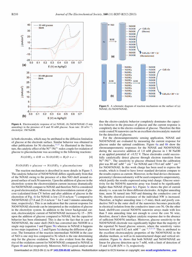
The reaction mechanism is described in more details in Figure 5.The catalytic behavior of NiO@NiNAE differs significantly from thatof the NiNAE owing to the presence of a thin NiO shell around ex-posed surface of each Ni nanowire. Upon the addition of glucose in theelectrolyte system the electrooxidation current increase dramaticallyfor NiO@NiNAE compare to NiNAE and therefore NiO is consideredas good electrocatalyst. Moreover, the electrooxidation current of glu-cose (calculated from CV before and after addition of glucose in theelectrolyte of Fig. 4) for NiNAE is low (23.8 mAcm−2) compared toNiO@NiNAE (27.8 and 25.4 mAcm−2 for 5 and 3 minutes annealingtime, respectively). This is an indication that the current response forNiO@NiNAE electrode can be interpreted as the oxidation of glucosein the electrolyte system. In addition to the lower background cur-rent, electrocatalytic current of NiO@NiNAE increases by 15 – 20%upon the addition of glucose compared to NiNAE, but the capacitivebehavior has been eliminated. This is due to the fact that in case ofNiO@NiNAE, the outer shell (NiO) has converted into NiOOH in asingle step (equation 3 and Figure 5b) whereas in NiNAE it happensin two steps (equations 1, 2 and Figure 5a) during the diffusion of glu-cose. The formation of the reaction intermediate NiOOH in the caseof NiO is one step less compared to Ni which is the rate determiningstep for the glucose oxidation. This is also evident from the sharprise of the oxidation current for NiO@NiNAE compared to NiNAE inFigure 4b and 4(a) respectively. Moreover, NiO is a good catalyst and
Figure 5. A schematic diagram of reaction mechanism on the surface of (a)NiNAE; (b) NiO@NiNAE.
thus the electro-catalytic behavior completely dominates the capaci-tive behavior in the presence of glucose and the current response iscompletely due to the electro-oxidation of glucose. Therefore the thinoxide coated Ni nanowire can be an excellent electrocatalytic materialfor the detection of glucose.
For the chronoamperometric sensing application, NiNAE andNiO@NiNAE are evaluated by measuring the current response forglucose under the optimal conditions. Figure 6a and 6b show thechronoamperometric responses for the NiNAE and NiO@NiNAEduring the successive addition of 1.0 mM glucose in 1 M NaOHat an applied potential of +0.52 V. These electrodes could success-fully catalytically detect glucose through electron transition fromNi2+/Ni3+. The sensitivity to glucose obtained from the calibrationplot was 80 mC mM−1 cm−2 for NiNAE and 170.4 mC mM−1 cm−2
for NiO@NiNAE. In this work charge has been used to express theresults, which is found to have lower standard deviation compare tothe results express as current. Moreover, in the final device chronoam-perometry/ chronocoulometry will be used compared to amperometry,which justify the results expressed using total charge. Glucose sensi-tivity for the NiO@Ni nanowire array was found to be significantlyhigher than NiNAE (Figure 6c). Figure 3c shows the plot of currentdensity vs. scan rate for three different electrodes. At higher annealingtime, more Ni turned into NiO and thinning the conductive core ofNi, thereby electrical resistance of the individual NW is increased.Therefore, at higher annealing time (>5 min), thick and poorly con-ductive NiO in the outer shell of the nanowires becomes practicallyin electrical isolation from the conductive inner core Ni. On the otherhand, shell@core coaxial NiO@Ni nanowire arrays made with lessthan 5 min annealing time not enough to cover the core Ni wire,therefore, doesn’t show highest catalytic response due to the absenceof sufficient NiO@Ni interface. Moreover, glucose sensitivity to theNiO@NiNAE electrode was significantly higher than the majority ofthe reported Ni based glucose sensors, where the sensitivity variesbetween 0.04 and 0.42 mA mM−1 cm−2.5,13–15 This is attributed tothe excellent electrocatalytic properties of the NiO@NiNAE in thealkaline medium, as well as its nanostructured shape and vertical ori-entation. Figure 6c shows that both NiNAE and NiO@NiNAE arelinear for glucose detection up to 7 mM, with a limit of detection of33 and 14 μM (S/N = 3), respectively.
ecsdl.org/site/terms_use address. Redistribution subject to ECS license or copyright; see 143.239.65.82Downloaded on 2013-09-11 to IP

Journal of The Electrochemical Society, 160 (11) B207-B212 (2013) B211
Figure 6. Chronoamperometric responses of (a) NiNAE, (b) NiO@NiNAE,5 min; upon successive addition of 1 mM glucose. (c) Corresponding concen-tration plot; Conditions: electrolyte 1M NaOH and applied potential Eapp =0.52 V.
The stability of the NiNAE and NiO@NiNAE were determinedover a period of 4 weeks, with an analysis carried out every 2 daysand 5 assays each day with a 1.0 mM glucose addition. In betweenthe testing, the electrodes were stored in air at ambient and activityremained 96% of its initial activity after 4 weeks for [email protected] was not the case for NiNAE, which needed extra cleaning toobtain the stable current response.
Interferences studies.— One of the major challenges in non-enzymatic glucose detection could be electrochemically active in-terferents, which generate electrochemical signals at the same po-tential as glucose. In our previous work, we have used the groundsubtraction method to eliminate signals generated due to ascor-bic acid and uric acid, which are common electrochemical inter-ferents in physiological fluid.16 Therefore, we have examined theamperometric responses of the NiNAE and NiO@NiNAE at anapplied potential of 0.52 V in 1M NaOH solution with contin-
Figure 7. Amperometric response of NiNAE and NiO@NiNAE upon suc-cessive additions of 1 mM glucose, 100 μM uric acid, 100 μM ascorbic acid,140 μM acetaminophen, 1 μM dopamine at −0.52 V vs. Ag/AgCl; backgroundelectrolyte 1M NaOH.
uous additions of 1 mM glucose, 100 μM uric acid, 100 μMascorbic acid, 140 μM acetaminophen and 1 μM dopamine. Fromthe current response in Figure 7, a significant signal was obtained forglucose compared to the uric acid, ascorbic acid, dopamine and ac-etaminophen. Compared to glucose, the interfering species (uric andascorbic acids) yielded current response ranging from 5–7%.
Conclusions
In this article, we have reported a new, cost effective and sensitivevertically aligned sensor platform to detect glucose based on coaxialNiO@NiNAE. Excellent catalytic activity has been found for glucosedetection, with a sensitivity of 170 mC mM−1 cm−2 and stability re-tained up to 96% after 4 weeks of regular use. The NiO/Ni nanowirewith 300 s annealing time shows better catalytic performance forglucose detection in 1M NaOH solution compared to NiNAE orNiO@NiNAE with various other annealing times. Both NiO@NiNAEand NiNAE show selectivity toward glucose in the presence of physio-logical level of uric acid, ascorbic acid, dopamine and acetaminophen.As a result, the vertically aligned ordered NiO@NiNAE has significantpromise for fabricating a highly selective, cost effective, enzyme-lessand sensitive sensor platform.
The future work will involve in testing this enzymeless glucosesensor in physiological fluids (e.g. blood samples), packaging thesensor and comparing it with commercially available glucose sensors.The nanowire array electrode film is found to be robust in terms ofhandling and dicing the film into desired shape. In this regard, thevertically grown shell@core NiO/NiNAE can be packaged on plasticsubstrates using existing technology used by the industry.
Acknowledgments
This work was financially supported by The European Union FP7scheme (e-BRAINS-FP7- ICT-257488).
References
1. A. Liu, H. Wu, X. Qiu, and W. Tang, Journal of Nanoscience and Nanotechnology,11, 11064 (2011).
2. F. Xiao, F. Zhao, D. Mei, Z. Mo, and B. Zeng, Biosensors and Bioelectronics, 24,3481 (2009).
3. J. Chen, W.-D. Zhang, and J.-S. Ye, Electrochemistry Communications, 10, 1268(2008).
4. A. Umar, M. M. Rahman, A. Al-Hajry, and Y. B. Hahn, Electrochemistry Communi-cations, 11, 278 (2009).
5. Y. Liu, H. Teng, H. Hou, and T. You, Biosensors and Bioelectronics, 24, 3329 (2009).6. S. Yu, X. Peng, G. Cao, M. Zhou, L. Qiao, J. Yao, and H. He, Electrochimica Acta,
76, 512 (2012).7. M. Jamal, M. Hasan, A. Mathewson, and K. M. Razeeb, Biosensors and Bioelectron-
ics, 40, 213 (2013).
ecsdl.org/site/terms_use address. Redistribution subject to ECS license or copyright; see 143.239.65.82Downloaded on 2013-09-11 to IP

B212 Journal of The Electrochemical Society, 160 (11) B207-B212 (2013)
8. M. Hasan, M. Jamal, and K. M. Razeeb, Electrochimica Acta, 60, 193(2012).
9. P. A. Nelson, J. M. Elliott, G. S. Attard, and J. R. Owen, Chem. Mater., 14, 524(2002).
10. M. Amjad, D. Pletcher, and C. Smith, J. Electrochem. Soc., 124, 203 (1977).11. L.-M. Lu, L. Zhang, F.-L. Qu, H.-X. Lu, X.-B. Zhang, Z.-S. Wu, S.-Y. Huan,
Q.-A. Wang, G.-L. Shen, and R.-Q. Yu, Biosensors and Bioelectronics, 25, 218(2009).
12. H. Nie, Z. Yao, X. Zhou, Z. Yang, and S. Huang, Biosensors and Bioelectronics, 30,28 (2011).
13. J. Wang, W. Bao, and L. Zhang, Analytical Methods, 4, 4009 (2012).14. T. You, O. Niwa, Z. Chen, K. Hayashi, M. Tomita, and S. Hirono, Analytical Chem-
istry, 75, 5191 (2003).15. A. Safavi, N. Maleki, and E. Farjami, Biosensors and Bioelectronics, 24, 1655 (2009).16. M. Jamal, O. Worsfold, T. McCormac, and E. Dempsey, Biosens. Bioelectron., 24,
2926 (2009).
ecsdl.org/site/terms_use address. Redistribution subject to ECS license or copyright; see 143.239.65.82Downloaded on 2013-09-11 to IP