Marine microbial response to heavy metals - CSIR-NIO
26
1 Author Version of : Bulletin of Environmental Contamination and Toxicology, vol.105(2); 2020; 182-197 Marine microbial response to heavy metals: Mechanism, Implications and Future prospect Abhay B. Fulke 1# , Atul Kotian 1 and Manisha D. Giripunje 2 1 Microbiology Division, CSIR-National Institute of Oceanography (CSIR-NIO), Regional Centre, Lokhandwala Road, Four Bungalows, Andheri (West), Mumbai-400053, Maharashtra, India. 2 Sevadal MahilaMahavidyalaya, Nagpur 440009, India #Corresponding Author Email: [email protected]; [email protected] Phone: +91-022 26359605, Extn No 1414 Abstract Growing levels of pollution in marine environment has been a matter of serious concern in recent years. Increased levels of heavy metals due to improper waste disposal has led to serious repercussions. This has increased occurrences of heavy metals in marine fauna. Marine microbes are large influencers of nutrient cycling and productivity in oceans. Marine bacteria show altered metabolism as a strategy against metal induced stress. Understanding these strategies used to avoid toxic effects of heavy metals can help in devising novel biotechnological applications for ocean clean-up. Using biological tools for remediation has advantages as it does not involve harmful chemicals and it shows greater flexibility to environmental fluctuations. This review provides a comprehensive insight on marine microbial response to heavy metals and sheds light on existing knowledge about and paves for new avenues in research for bioremediation strategies. Keywords: Heavy metals, Marine microbes, Bioremediation, Ocean clean-up
-
Upload
khangminh22 -
Category
Documents
-
view
2 -
download
0
Transcript of Marine microbial response to heavy metals - CSIR-NIO

1
Author Version of : Bulletin of Environmental Contamination and Toxicology, vol.105(2); 2020; 182-197
Marine microbial response to heavy metals: Mechanism, Implications and Future prospect
Abhay B. Fulke1#, Atul Kotian1 and Manisha D. Giripunje2 1Microbiology Division, CSIR-National Institute of Oceanography (CSIR-NIO), Regional Centre,
Lokhandwala Road, Four Bungalows, Andheri (West), Mumbai-400053, Maharashtra, India. 2Sevadal MahilaMahavidyalaya, Nagpur 440009, India
#Corresponding Author Email: [email protected]; [email protected] Phone: +91-022 26359605, Extn No 1414
Abstract
Growing levels of pollution in marine environment has been a matter of serious concern in recent years. Increased levels of heavy metals due to improper waste disposal has led to serious repercussions. This has increased occurrences of heavy metals in marine fauna. Marine microbes are large influencers of nutrient cycling and productivity in oceans. Marine bacteria show altered metabolism as a strategy against metal induced stress. Understanding these strategies used to avoid toxic effects of heavy metals can help in devising novel biotechnological applications for ocean clean-up. Using biological tools for remediation has advantages as it does not involve harmful chemicals and it shows greater flexibility to environmental fluctuations. This review provides a comprehensive insight on marine microbial response to heavy metals and sheds light on existing knowledge about and paves for new avenues in research for bioremediation strategies.
Keywords: Heavy metals, Marine microbes, Bioremediation, Ocean clean-up

2
1. Introduction:
Heavy metals are a class of metals defined by characteristically having density above 5 g/cm3
and are known to be common pollutants in both terrestrial and aquatic environments (Hawkes 1997,
Mohseniet al. 2018). Heavy metals in marine sediments have both natural and anthropogenic origin;
their distribution and accumulation are largely influenced by sediment texture, mineralogical
composition, reduction/oxidation state, adsorption and desorption processes and physical transport
(Buccolieriet al. 2006). These metals tend to be environmentally persistent and pose a severe threat
to the biota exposed to them (Rousk and Rousk 2018). Heavy metals ions also compete with
essential metal ions for binding with biomolecules (Wang et al. 1997) which explains their disruptive
physiological effects.
Biological systems, through evolution have acquired ability to utilise some metals for essential
biological functions like oxygen transportation, photosynthesis and transmitting nerve impulses
(Altenburger 2011). Among these metals Na+, K+, Ca2+, Mg2+ are termed group I, as they are
considered to be essential for stabilizing organic structures. There are additional metals such as
Vanadium (V), Chromium (Cr), Manganese (Mn), Iron (Fe), Cobalt (Co), Nickel
(Ni),Copper(Cu),Zinc(Zn), Molybdenum (Mo),Boron(B),Silicon(Si), Selenium
(Se),Fluorine(F),Iodine(I), Arsenic (As),Boron(Br) and Tin(Sn), termed group II, which are found in
some living organisms, but not in all. Most of these are required as enzyme cofactors, for structural
integrity, or to provide screening of the electrostatic interactions in the water phase (Petukh and
Alexov 2014; Jaishankaret al. 2014). Moreover, they are required in trace quantities and when they
cross their threshold values, they become toxic (Rainbow and Furness 1990, Zwolak 2015).
Speciation in metals can also reduce or enhance toxicity. Cr(III) (trivalent Chromium) is known to be
essential trace element for some bacteria, whereas Cr(VI)(hexavalent Chromium) is highly toxic,
similarly organic forms of Mercury are more toxic than inorganic and elemental Mercury (Keil 2011,
Park and Zheng 2012). Metal-protein complexes of metals like Hg are inherently toxic and trace
elements like Zn also become toxic at high concentration, which is why, cellular uptake of metals his
a heavily regulated process (Nies 1999).
Local anthropogenic activities and riverine inputs are largely responsible for the entry of
contaminants in coastal and estuarine waters (Eggleton and Thomas 2004; Roldán‐Wong et al.
2018). Mishaps such as Bento Rodrigues Dam (Brazil) burst of 2015 can introduce high amounts of
heavy metals, some even radioactive in a short span. Presence of such metals can lead to cytotoxicity
and DNA damage in local flora (Segura et al. 2016).Metals like Lead can form aerosols and can get

3
transported far away from their sources (Horta-Pugaet al. 2014). Upon their entry in the
environment, various physicochemical parameters like salinity, pH and oxidation potential influence
the sorption of metals which result in enhanced bioavailability due to increased solubility (EPA
2007).Organisms on metal exposure can exhibit varied response with different life stages in the same
organism meaning that certain larval stages show higher susceptibility of metal induced damage over
others (Liao et al. 2019). The bacterial response to metal exposure can be variable ranging from
production of siderophores to uptake iron (Carvalho and Fernandes 2010, Granger and Price 1999) to
expression of metallothioneins, a class of metal chelating proteins (Valls and De Lorenzo 2002) or
single stranded DNA breaks (Mitra and Bernstein 1978).With increasing occurrences of fishes, crabs
and molluscs being contaminated with heavy metals (Pempkowiaket al. 1999; Ahmad et al. 2010;
Zhao et al. 2010; Maulvaultet al. 2012; Naccariet al. 2015; Giripunjeet al. 2016, Delgado‐Alvarez et
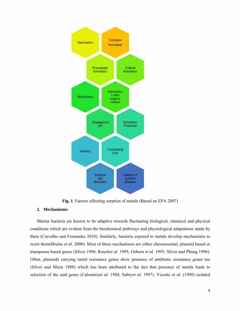
al. 2019), finding solutions to remove these metals from aquatic systems is essential. Several factors
are responsible for metal sorption in water as given in figure 1. These factors need to be considered
to determine the exact fate of metals in the environment which changes course of bioremediation
strategies. Most metals in their native form and some metal compounds do not tend to solubilize
easily. When their soluble forms are utilized for studies, there is a bias in estimating their
bioavailability and toxicity which is a major concern in designing toxicological studies (EPA 2007).
The detoxification mechanism by different genus of bacteria against the same metal may not
be the same. Hg has been observed to be removed by volatilization in Bacillus cereus (Dash et al.
2016) or by surface adsorption as shown by Pseudomonas spp. (Deng and Wang 2012).This means
that bioremediation strategies should take into account the detoxification process in order to
determine course of action. The current study illustrates role played by marine microbes and the
different responses exhibited by them in presence of heavy metals. With growing interest in
understanding use of biological systems like algae, bacteria and fungi for bioremediation
(Giripunjeet al. 2015; Ayangbenro and Babalola 2017; Rajendranet al. 2003), the response rendered
by marine bacteria can be useful in dealing specifically with marine pollution, where terrestrial
microbes cannot survive owing to hypersaline conditions.
4
Fig. 1. Factors affecting sorption of metals (Based on EPA 2007)
2. Mechanisms:
Marine bacteria are known to be adaptive towards fluctuating biological, chemical and physical
conditions which are evident from the biochemical pathways and physiological adaptations made by
them (Carvalho and Fernandes 2010). Similarly, bacteria exposed to metals develop mechanisms to
resist them(Bruins et al. 2000). Most of these mechanisms are either chromosomal, plasmid based or
transposon based genes (Silver 1996; Rouchet al. 1995; Osborn et al. 1995; Silver and Phung 1996).
Often, plasmids carrying metal resistance genes show presence of antibiotic resistance genes too
(Silver and Misra 1988) which has been attributed to the fact that presence of metals leads to
selection of the said genes (Calomiriset al. 1984; Sabryet al. 1997). Vicente et al. (1990) isolated
Complex
formationSpeciation
Precipitate formation
Colloid formation
Interactions with
organic matter
Biofixation
Changes in pH
Oxidation Potential
Competing ionsSalinity
Surface site
densities
Nature of sorbent phases

5
strains Pseudomonas aeruginosa from sewage contaminated regions of coastal Spain which could
resist simultaneous exposure of two antibiotics and a heavy metal with one isolate from highly
polluted sea water being resistant to Hg, As and Cr. This could prove that multi-resistance for metals
and antibiotics is possible in a single bacterium. Rouchet al. (1995) describe the mode for metal
resistance where the choice of detoxification pathway depends upon presence of detoxification
genes, site in the cell that requires protection and availability of nutrition. Metallothioneins (MT),
known to exist in eukaryotes, act as metal sequestering proteins. The first report of prokaryotic MT
was observed in marine cyanobacterium Synechococcussp. It was seen that the MTs rendered
tolerance to Synechococcus sp. against cadmium (Olafsonet al. 1979).
Metals may also be cell bound or precipitated thereby reducing their toxic effects (Jayshankaret
al. 2007). There can also be an active or passive uptake of metals, enabling metal bioaccumulation
(Marques 2016). Certain bacteria possess specific operons targeting detoxification of metals. An
extensively studied operon system targeting Mercury, meroperon, produces an NAD(P)H dependant
protein, MerA, which is responsible for reducing toxic mercuric Hg(II) ion to a volatile form of
elemental mercury Hg(0) which can vaporise out of the solution (Allen 2013). Here, changes in
oxidation state has reduced the toxicity of metal. Using similar processes, bacteria detoxify toxic
heavy metal ions to their less toxic oxidation states (Ayangbenro and Babalola 2017).
Number of factors are responsible for the choice of means of protection and the degree of
protection offered by the system in question. That includes
1. The number and type of metal uptake system
2. The role played by the metal in normal metabolism
3. The availability of resistance genes for metals which could either be chromosomal, carried on
plasmid or transposons (Rouchet al. 1995)
Cell membranes only allow diffusion of H2O, CO2, N2, NH3, O2, B(OH)3 and Si(OH)3 and
they require entrapping molecules to ensure loss due to them diffusing out of the cell
(Williams1981). Usually whenever uptake of metals is involved, a transporter protein is imperative
owing to the fact that biological membranes are highly impermeable to hydrophilic ions like metal
cations (Anderson 1978; Rouchet al. 1995). Figure 2 summarises some of the common responses
rendered by microbes in presence of heavy metals.

TI
d
a
o
Table 1 descInhibition Co
Table 1.
Metal
Cd Co Cr Cu Hg Mn Ni Pb Zn
Sever
deep-sea hyd
activities we
occurrence o
Fig. 2. S
cribes aboutoncentration
LD50 for ma
LD50 for mBow
B
ral studies h
drothermal v
ere chosen fo
of volcanic a
ummarisatio
t studies donn (MIC) used
ammals and
mammals (Awen1979) in BW(bodywei
1.3 50 90 −
1.5 18
110-220 70 −
ave been con
vents. Areas
or these stud
ctivity under
on of pathwa
ne to determd routinely in
MIC values
Adapted frommg/kg
ight)
nducted on m
with report
dies. Hydrot
r the seabed
ays for meta
mine mediann toxicology
s of Serretia
m MIC
microbes iso
ted heavy m
thermal vent
d (Loaëcet al
l removal / d
n lethal dosey to determin
sp. WPR3 f
for SerretiaJafarzadeet
NG400-450600 [C
NGNG
750-800450-750
700-
olated from m
metal contam
ts show pres
l. 1997).
detoxificatio
e(LD50) valune potency o
for common
sp. WPR3 (t al. 2012) in
Ga [CdSO4] 0 [CoSO4.7H
Cr(NO2)3·9HGa [CuSO4] Ga [HgCl2] 0 [MnSO4·H-500 [NiSO4
0 [Pb(NO3)2]-750 [ZnSO4
a No grow
marine wate
mination due
sence of hea
on
ue and Minif a toxin.
heavy metal
(Adapted fron mg L-1
H2O] H2O]
H2O]
4] ]
4]
wth was obs
ers, sediment
to anthropo
avy metals d
6
imum
ls
om
erved
ts and
ogenic
due to

7
2.1 Non-Specific response against metals
2.1.1 Metabolism independent biosorption
Biosorption is an energy independent process involving ionic or physiochemical adsorption by
dead or living cells. It is to be noted that metabolic pathways in living cells may influence
biosorption (White et al. 1995; Hassan et al. 2010). Functional groups like carboxyl, amine,
hydroxyl, phosphate and sulphydryl are known to interact with metal ions in their vicinity (Hassan et
al. 2010; Gadd 2009; Hoyle and Beveridge 1984). With bacterial cell wall being largely negatively
charged, it may localize positively charged metal ions on the cell surface (Beveridge 1981; Hoyle
and Beveridge 1984). This ultimately is instrumental in attracting metal ions in the vicinity of the
bacterial cell to get attracted to this highly anionic surface and adsorb.
2.1.2 Extracellular Polymeric Substances
Extracellular Polymeric substances (EPS) also known as exopolysaccharides are diverse organic
polymeric compounds with tremendous industrial applications, particularly in Biotechnology (Suresh
Kumar et al. 2007; Freitaset al. 2011). They are composed of osidic monomers with organic or
inorganic substituents like phosphate, sulphate, or lactate, succinate and pyruvate groups (Delbarre-
Ladratet al. 2014). EPS possess high affinity to dissolved compounds in sea water which includes
metals (Decho 1990; Bhaskar and Bhosle 2006). As a result, bacteria producing EPS are expected to
trap metal ions on their cell surface. Bacillus cereus BW 201B (Dash et al. 2016),
Alteromonasmacleodiisubspfijiensis (Loaëcet al. 1997), Enterobacter cloacae (Iyeret al. 2004)
isolated from diverse marine environments showed metal biosorption through EPS binding. As EPS
has highest partition coefficient among known biosorbents, organisms capable of producing it have
advantage of its greater binding ability to metals (Quigley et al. 2002; Bhaskar and Bhosle 2006).
Thus EPS offer a permeability barrier against heavy metals through which entry of toxic metals is
altogether avoided thus protecting sensitive cellular components (Bruins et al. 2000).
2.1.3 Biosurfactants
Microorganisms are capable of producing surface active substances also referred to as surfactants
which help in either reducing surface tension or allow tight binding to surfaces (Ron and Rosenberg
2011). They are amphiteric, which means that they are composed of a hydrophilic end with moieties
like an acid, peptide, cations or anions, or saccharide units and a hydrophobic end moiety like
hydrocarbons or fatty acids. Low molecular weight compounds called biosurfactants are usually
glycolipids or lipopeptides, whereas high-molecular-weight polymers called bioemulsans comprise
of polysaccharides, lipopolysaccharide proteins or lipoproteins units (Banat et al. 2010).

8
Biosurfactants have application in bioremediation owing to their ability to form metal complexes.
Metals tend to have a greater affinity for ionic surfactants over matrix, as a result, they form
complexes and get desorbed from the matrix. Cationic biosurfactants can bind to metals via ion
exchange or electrostatic interactions (Aşçıet al. 2008; Pacwa-Płociniczaket al. 2011). A
biosurfactant producing Bacillus sp., MTCC 5514 isolate from marine sample from Tamil Nadu,
India showed tolerance of 2000 mg L-1 of Hexavalent Chromium, a known carcinogen (Gnanamaniet
al. 2010). Since these substances are produced outside the cell, biosurfactants have been produced
using fermentation techniques which exhibit promising remediation activity (Juwarkaret al. 2007;
Gnanamaniet al. 2010).
2.1.4 Sulphide based precipitation
Sulphate reducing bacteria (SRB) is a group of chemolithotropic bacteria which can utilise
diverse group of compounds as electron donors and are able to integrate anaerobic electron transport
with ATP synthesis. With about 200 species spread across 60 genera, SRB have displayed their
significance in bioremediation due to their ability to metabolise hydrocarbons and detoxify ions like
Cr(VI) and U(VI) (Barton and Fauque 2009).
They utilise sulphur instead of oxygen as the terminal electron acceptor using the following
pathway described in Fu and Wang (2011)
3SO42- + 2CH3CH(OH)COOH → 3H2S + 6HCO3
-
Here, CH3CH(OH)COOH stands for simple organic compounds. This generated hydrogen sulphide
can react with aqueous metal ions, thereby forming their corresponding sulphides and precipitating
them.
M2+(aq) + H2S(g) → MS(s)↓ + 2H+
(aq)
Metal sulphides have extremely low solubility (Bhattacharyya et al. 1979) and thus the
precipitate remains stable which ensures that they become immobile and biologically unfavorable
(Harithsa et al. 2002). Such a non-specific response is beneficial not just for the bacterium itself, but
for other organisms in the vicinity as well.
2.1.5 Metal binding with proteins
Metals are known to bind to biomolecules such as chelators, glutathione derived peptides called
phytochelatin and other metal binding peptides. Binding to such molecules facilitates absorption and
transport of metal ions in bacterial cells. Heavy metals are also known to disrupt enzyme functions
by binding to thiol (-SH) and other protein groups or by replacing co-factors which renders them

9
inactive (Ayangbenro and Babalola 2017). Metallothioneins (MTs) are a class of cysteine rich
proteins capable of metal binding found across all domains of life. The abundance of Cys residues
gives access to several metal binding thiol groups (Gutiérrez et al. 2019).MTs are capable of
sequestering both essential as well as toxic transition metals by internally forming
polynuclearmetalthiolate clusters (Calvoet al. 2017). MTs are ubiquitous, and thus are found across
several taxonomic groups. However, research on prokaryotic MTs is largely limited (Gutiérrez et al.
2019). Presence of MTs has been reported in Pseudomonas and marine cyanobacterium
Synechococcus(Olafsonet al. 1979; Hingham et al. 1984). Both these isolates were found to be
growing in presence of Cd2+ (Blindauer 2011). In eukaryotes, MTs are reported to be metallated
sequentially and show presence of hemimetallated MTs (Krężel and Maret 2007) which indicates
presence of more than one metal binding location. It has been seen that MTs are capable of binding
to divalent cations of Zn2+, Cd2+, Cu2+, and Hg2+ (Calvoet al. 2017; Gutiérrez et al. 2019).
2.2 Specific response against metals
2.2.1 Chromosomal and Plasmid based genes for transport / biotransformation / efflux
Intercellular compounds, metal-binding proteins, intracellular precipitation, methylation, and
other mechanisms allow bacterial cells to imbibe metals and associate themselves with cellular
components (Hassan et al. 2010). This can involve chromosomal genes responsible for transport of
metals ions of Iron, Nickel, Manganese, Zinc and Cobalt (Silver and Walderhaug 1992) or plasmid
encoded genes in case of metals like Arsenic, Copper, Mercury and Cadmium (Silver and
Walderhaug 1992; Kaur and Rosen 1992; Nies 1999). Silver and Walderhaug (1992) observed a
common trait in chromosomally mediated ion transport that were presence of multiple transport
system for each ion; a constitutive expression of low affinity and high capacity transport system,
another being a facultative expression of high affinity and low capacity transport system in response
to nutrient deprivation. The genes expressed chromosomally are also involved in essential metal
tolerance which tend to be more complex than plasmid based systems (Silver and Walderhaug 1992;
Bruins et al. 2000). The plasmid encoded determinants allow for the bacterium to resist and efflux
toxic ions (Bruins et al. 2000). Zolgharneinet al. (2007) observed that heavy metal resistant bacteria
isolated from industrially contaminated sea water and sediments from Persian Gulf showed greater
occurrence of plasmids than natural bacteria which suggests possibility of greater abundance of
plasmid encoded tolerance genes over chromosome encoded tolerance genes for metals. Once the
metal enters the cells it is lead to the subsequent biotransformation or efflux outside the cell.

10
a) Mercury
Mercury (Hg) has largely been recognized as being toxic in all of its chemical forms (Broussard
et al. 2002). It enters aquatic ecosystem through both natural phenomena like degassing, volcanic
eruptions, leaching of metal from ores like cinnabar and rocks (Clarkson 1997; De et al. 2003;
Gworeket al. 2016) as well as anthropogenic reasons like industrial and municipal effluents, coal and
metal mining and dentistry (Clarkson 1997; De et al. 2003; Dash 2014). With no apparent biological
role (EPA 2007) and being non-biodegradable, its presence in the environment is of serious concern
(De et al. 2008). The LD50 value of Hg for mammals is 1 mg/kg BW which makes it toxic to life
forms even at low concentrations (Bowen1979). Hg behaves differently in its elemental form, as
inorganic salts and as organometallic compounds with varying degree of toxicity (Langford and
Ferner 1999). Bacteria acquire genes for resistance through plasmids and/or transposons (Silver and
Walderhaug 1992; Osborn et al. 1995). The most common method of detoxification of Hg involves
mer operon which transcribes for mercury reductase (MerA). This results in a NAD(P)H dependent
reduction of Hg(II) to Hg(0), a relatively non-toxic form of Hg to bacteria, which volatilizes in the
atmosphere (Dash and Das 2012). Themer operon operates at relatively low concentrations of
mercury salts, thus implying role of Hg(II) as a sensor leading to the switching on of the operon
(Misra 1992). When Hg(II) enters the cell envelope, it may be bound in the periplasmic space to a
Hg(II) binding protein MerP which facilitates its entry inside the bacterial cell with the help of Hg
transporter system. MerP is a periplasmic protein which has cysteine residues which have high
specificity for Hg2+. Transmembrane protein MerT is responsible for transport of the Hg2+ ions to
mercury reductase (MerA) (DeSilvaet al. 2002; Zhang et al. 2020) Cytoplasmic MerA reduces
Hg(II) to elemental mercury [Hg(0)] which can then be volatilized thus detoxifying the metal
(Rouchet al. 1995, Bruins et al. 2000).While most marine bacteria exhibit such MerA mediated
removal, recent studies show that bacteria where MerA function is not strong as in the case of
Pseudomonas pseudoalcaligenes S1 isolated from a mangrove bay in China, MerP and MerT end up
accumulating Hg retaining it in the periplasm instead of transporting inside the cell. This could be an
alternative strategy towards detoxification of Hg (Zhang et al. 2020). Hg is also methylated by
marine microbes resulting in accumulation of methylmercury (Gionfriddoet al. 2016).
Studies have focused on understanding pathway through which Hg is tolerated or removed from
the system. Acinetobacter and Bacillus spp. isolated from mangrove sediments along Qatari coast
showed significant tolerance against Hg and was studied to remove mercury from powdered
florescentlamps. It was observed that a Bacillus marisflaviisolate showed up to 96.7% removal
efficiency of Hg (Abu-Dieyehet al. 2019). Mercury tolerant Psudomonas-10 isolate found as a part

o
(
s
d
a
i
a
c
P
H
I
s
A
k
k
N
a
a
I
q
A
of gut micro
(Fong et al.
studied for t
discovered a
able to tolera
solated from
and showed
coast was s
Pseudomona
HgCl2 throug
Iyeret al.(20
show any bio
b) Arsen
Arsenic (As)
known to co
known to oc
Natural weat
activities int
acute effects
Its LD50 valu
quantities of
As(III) AsO2
obiota in Da
2019).Wate
tolerance of
a Pseudomon
ate Hg throu
m water sam
mer-operon
seen to be
as aeruginos
gh volatiliza
005) showed
osorption for
Fig 2. D
nic
) is a toxic
ontaminate g
cur almost u
thering of ro
troduces As
include can
ue for mamm
f As are eno
2-with As(V
aphnia rende
er and sedim
f Hg2+ along
nas sp. whic
ugh surface
mple off coas
based Hg vo
producing
a CH07 isol
ation (Jaysha
d EPS based
r Hg. Figure
etoxification
metalloid, w
groundwater
ubiquitously
ocks, volcan
in the envi
ncer of the bl
mals is 5 mg
ugh to prod
) resembling
ered toleran
ment isolates
g with other
ch was able
biosorption
stal Odisha,
olatilisation
methylmer
lated from C
ankaret al. 2
d biosorption
e 2 shows det
n mechanism
which has b
(Lee et al. 2
in all biotic
noes, riverin
ironment (N
ladder, skin
g/kg BW (B
duce its toxic
g phosphate
nce against m
s from Xixia
r metals like
to tolerate a
(Deng and
India was ab
(Dash et al.
rcury and v
Chennai coas
2007). An iso
n for Cd, C
toxification
m of Hg(II) (
been historic
2005; Nagve
c and abiotic
ne input alon
Nagvenkar an
and liver (M
Bowen 1979)
c effects. Ch
(PO43-) thu
mercury by
ang Estuary,
e Nickel, C
a maximum
Wang 2012
ble to tolera
2017). Con
volatilize H
st showed tol
olate of Ente
Cu and Co, h
of Hg(II) thr
(Adaptedfrom
cally known
enkar and R
c component
ng with indu
nd Ramaiah
Mandal and S
) which imp
hemically it
us interfering
detoxifying
, Shenzhen
admium and
of 120 mg
2). Bacillus c
ate 50 mg L-
sortia isolate
Hg (Gionfrid
lerance to 75
erobacter cl
however rem
rough mer o
m Nies 1999
as a homic
Ramaiah 200
ts (Mandal a
ustries, efflu
h 2009; Lee
Suzuki 2002;
plies that rel
is found as
g with phosp
g Hg(II) to H
Bay, China
d Chromium
L-1 Hg2+ and
cereus BW 1 of Hg as H
ed from Ant
ddoet al. 2
5 mg L-1 of H
loacae studie
markably di
operon.
9)
cidal agent a
9).Naturally
and Suzuki 2
uents and m
et al. 2005
; Lee et al. 2
latively mini
As(V) AsO
phate metab
11
Hg(0)
were
m and
d was
201B
HgCl2
arctic
2016).
Hg as
ed by
id not
and is
y, it is
2002).
mining
5). Its
2005).
iscule
O43- or
olism

(
p
p
e
1
a
t
(
a
a
o
a
M
t
l
t
a
i
(
d
(Nies 1999).
protein or P
particularly d
effluxed out
1999). Some
al. 1994; Nie
to five genes
(Roucheet a
arsenite efflu
a negative r
operon. Cata
arsenate redu
Marinomona
tolerate a ma
ocalize it o
thuringiensis
against As an
solated from
(Nagvenkar
detoxificatio
Phosphate a
Pst system.
difficult. Thi
in an energy
e bacteria sh
es 1999). Th
s namely ars
l. 1995; Bru
ux permease
repressor of
alysis of tra
uctase ( Wu
ascommunis
aximum of
on cell mem
s PW-05 iso
nd could wit
m consortia
and Ramai
n of As(V).
Fig 3. D
and arsenate
It is this i
is is overcom
y dependent
how presenc
he efflux inv
sR, arsA, ar
uins et al. 2
ArsB. ATP
f the operon
ansformation
and Rosen 1
IAM 12914
5 mg L-1 A
mbrane via
lated from O
thstand 232.
a from wat
iah 2009)
Detoxification
e enter the ce
interference
me by utilizi
process (W
e of ABC fa
volves use of
rsD, arsB, a
2000). As(V
driven ArsA
n and senses
n of As(V) t
1991; Ji and
4 studied by
As as Na2HA
cell surface
Odisha coast
4 mg L-1 As
ter and sed
was seen t
n mechanism
ell either thro
which mak
ing ArsC to
Willsky and M
family of gen
f arsenate op
arsC which
V) is effluxe
A helps by en
s inorganic
to As(III) is
d Silver 1992
y Takeuchi
AsO4·7H2O.
e adsorption
t, India by D
s as Na2HAs
diment sam
to uptake a
m of As(V) (
ough PIT (p
kes remova
convert arse
Malamy 1980
nes for arsen
peron (ars o
code for an
ed from the
nhancing eff
arsenic, thu
s carried ou
2; Dey and R
et al. (200
The isolate
n. Mercury
Dash et al. (2
sO4. An Ente
mples in Ma
and biotrans
(Adaptedfrom
hosphate ino
al of arsena
enate to arsen
0; Nies and S
nate uptake
operon) whic
ATPase eff
e cell as triv
flux mechan
us repressin
ut by ArsC w
Rosen 1995;
07) showed
was seen to
resistant ba
2014) was se
erobacteriace
andovi estu
sform As.
m Nies 1999
organic trans
ate from the
nite which c
Silver 1995;
(Papadopou
ch codes for
flux pump sy
valent arsen
ism. ArsR a
ng activity o
which acts
Yan et al. 2
the bacteriu
o remove A
acterium Ba
een to be tol
eae member
uary, Goa,
Figure 3 s
9)
12
sport)
e cell
can be
; Nies
ulouet
three
ystem
nic by
acts as
of the
as an
2018).
um to
s and
acillus
lerant
FW3
India
shows

A
e
m
e
1
C
z
C
o
(
t
2
a
h
i
P
(
b
C
s
c) Cadm
Although be
even at low
mammals is
exposure to m
1978) and h
Cadmium is
zinc metabo
Cadmium en
of essential d
(Laddaga an
there is no e
2000). Efflux
also respons
homolog wh
n oxidation
Pacific Ocea
(Loaëcet al.
bioaccumula
d) Chro
Chromium (
stainless stee
mium:
ing found in
concentrati
s 1.3mg/kg
miniscule qu
has been use
known to b
olism and r
nters cells th
divalent ion
d Silver 198
enzymatic d
x of Cd occu
ible for tran
hich helps in
states. Alter
an was seen
1997). Vib
ate Cd (Abd-
Fig. 4 D
omium
Cr) is an in
el (Barnhart
n trace amou
ons (Trevor
BW(body
uantities. It i
ed as an ind
be denaturing
results in m
hrough dival
s. It is seen
85; Silver an
etoxification
urs through
nsport of zin
efflux of Cd
romonasmac
n to resist 1
brio harvey
-Elnabyet al.
Detoxificatio
ndustrially im
1997). In In
unts on earth
rset al. 1986
weight)(Bow
is known to
dustrial bacte
g protein an
membrane d
lent ion tran
that Mn2+ t
nd Walderha
n involved w
a set of gene
nc and cobal
d2+. Thus, th
cleodii subsp
000 mg L-1
yi 5S-2 isol
. 2011). Fig.
on mechanis
mportant me
ndia, it has be
h surface, C
6; El-Helow
wen 1979)
induce DNA
ericide (Bab
nd binding w
damage, loss
nsporters, wh
transport sys
aug 1992). T
which is obs
es belonging
lt. Protein ca
he metal is re
p. fijiensis is1 of Cd and
lated from A
4 shows det
sm of Cd(II)
etal. Its mai
een used in i
Cadmium (Cd
wet al. 2000)
thus confir
A damage in
bich and Sto
with thiols. I
s of protec
hich are usu
stems are als
The unique f
served in Hg
g to czc and
adA, coded
emoved from
solated from
d produced E
Alexandria
toxification
) (Adapted fr
in industrial
industrial co
d) is reporte
). The LD50
rming its to
n E. coli (Mi
otzky 1978).
t interferes w
ctive functio
ally respons
so responsib
feature of Cd
g(II) or As(
cad systems
by cad syst
m the system
m hydrotherm
EPS to resis
harbor, Egy
of Cd.
rom Nies 19
use is in th
ooling towers
ed for being
0 value of C
oxic effects
itra and Bern
. Physiologi
with calcium
on ((Neis 1
sible for tran
ble for Cd u
d tolerance i
(V) (Bruins
s. The gene
em is an AT
m without cha
mal vents off
st entry of
ypt was see
99)
he manufact
s as a virtue
13
toxic
Cd for
with
nstein
cally,
m and
1999).
nsport
uptake
is that
et al.
czc is
TPase
anges
ff Fiji,
metal
en to
turing
of its

c
C
2
V
R
1
r
C
r
w
i
b
c
C
g
2
r
r
w
f
o
5
corrosion pre
Cr is most a
2007). Cr(V
Vijayrajet al
Ramírez-Día
1999). Chrom
response aga
Cervantes et
responsible f
with SO42- (C
nduces DNA
bringing dow
can directly
Chromosoma
genes are ma
2001). Chro
reductase iso
reductases. T
which is also
for its activit
out of the ce
5 shows deto
Fig.
eventing act
abundantly f
VI) is one o
l. 2019). Hex
azet al. 200
mate ion (Cr
ainst the io
t al. 2001).
for uptake o
Cervantes an
A damage (B
wn cellular l
y get affect
al genes are
ainly involve
mate reduct
olated from
They have be
o capable of
ty (Cervante
ll through ch
oxification o
5 Detoxific
tion and bioc
found as dic
of the most
xavalent Cr i
7) and has
rO42-) is chem
on as it is n
In bacterial
f Cr(VI) wh
nd Campos-G
Bridgewater
levels of glu
ted by oxid
responsible
ed in efflux
tase reduces
Pseudomona
een describe
f reducing c
es and Camp
hromosomal
f Cr(VI).
ation mecha
cidal propert
chromate [C
common co
is known to
no apparen
mically anio
not affected
l cells the s
here it acts a
García 2007
et al. 1994;
utathione and
dative stress
e for the enz
of Cr ions (C
s highly tox
as putida (P
ed with NAD
chromate as
pos-García 2
lly expressed
anism of Cr(V
ties (Bhidee
r(III)] and c
onstituent o
be a potent m
nt biological
onic which is
d by anionic
sulphate tran
as a competit
7). On enteri
; Ackerleyet
d other free
s induced b
zymatic deto
Cervantes an
xic Cr(VI) t
Park et al. 20
DH:flavinox
a secondary
2007). The r
d transporter
VI) (Adapte
et al. 1996).
chromate [C
f tannery e
mutagen and
l significanc
s unlike othe
c surface o
nsport syste
tive inhibito
ng the cell,
t al. 2006). C
thiols (Acke
by Cr, its
oxification of
nd Campos-
to less toxic
000) is one
idoreductase
y function an
reduced Cr,
r proteins (R
d from Ram
Among its c
Cr(VI)] (Pech
ffluents (Sr
d carcinogen
ce for micro
er metals. Th
f the cell (
em has been
or owing to c
it causes ox
Cr(VI) is als
erleyet al. 20
detoxificatio
f Cr, wherea
García 2007
c Cr(III). C
of the well-
e activity as
nd show NA
now as Cr(
Ramírez-Díaz
mírez-Díazet
chemical sp
hova and Pa
rinathet al. 2
n (Iyeret al. 2
oorganisms
his affects th
(Beveridge
n identified
chemical an
xidative stres
so responsib
006). Since
on is neces
as, plasmid b
7; Cervantes
ChrR, a chro
-studied chro
primary fun
ADH depend
(III) gets eff
zet al. 2007)
al. 2007)
14
ecies,
avlata
2002;
2004;
(Nies
he cell
1981;
to be
alogy
ss and
le for
DNA
ssary.
based
et al.
omate
omate
nction
dency
fluxed
). Fig.

15
2.2.2 Siderophores
Iron (Fe) is an essential micronutrient for bacterial cells. In aerobic conditions Fe tends to form
insoluble hydroxides and oxyhydroxides (Rajkumaret al. 2010). In order to sequester Fe(III) from
their surroundings, bacteria are known to produce siderophores. Siderophores act as solubilizing
agents which make Fe biologically available. On binding with siderophores, Fe3+ gets reduced to
Fe2+ and utilized by the cell (Birch andBachofen 1990; Rajkumaret al. 2010). There are three classes
of siderophores viz. hydroxamates, catecholates, and carboxylates which are classified on the basis
of ligand involved in metal chelation with Fe(III) (Sahaet al. 2015). Siderophores binding abilities
are not just restricting to Fe, but they are also able to bind to non-essential metals like Ga(III),
Cr(III), Al(III) and also essential metals likeMg, Mn, Ca (Birch and Bachofen 1990). This implies
potential of siderophore producing microbes in bioremediation of heavy metals. Siderophore
producing marine bacteria have been utilized for remedial of Cr contaminated tannery sewage
(Vijayarajet al. 2019).
3. Implication of heavy metal pollution on marine biota
Rachel Carson in her 1962 book ‘Silent Spring’ raised concern about biomagnification and
bioaccumulation. The book was instrumental in creating awareness about xenobiotics entering food
chains through accumulation by plants and lower organisms. Marine environment exhibits similar
pattern of biomagnification. A study on marine copepod Acartiatonsa by Lee and Fisher (2017)
suggests that the organism is capable of accumulating Hg and methyl mercury. A model proposed by
Schartupet al. (2017) showed uptake of methylmercury and trophic level transfer by plankton.Marine
mussels Mya arenaria and Astarte borealis collected from NorvegianSea and Baltic Sea showed
accumulated levels of heavy metals (Pempkowiaket al. 1999). Edible crab Cancer paguruscaught
from Scottish shore also showed presence of heavy metals in edible parts of the animal (Maulvaultet
al. 2012).Epinephelusareolatus, Lutjanusrusselli, and Sparussarba from cultured pond sites in Hong
Kong showed high levels of Zn, Pb and Cu (Wong et al. 2001). This illustrates that globally
organisms across different trophic levels are accumulating heavy metals from their environment.
Even controlled sites such as culture ponds are showing risk of contaminated water and
bioaccumulation of metals in fishes (Wong et al. 2001). This poses a serious health risk to humans as
most of the organisms mentioned above are routinely consumed by humans and thus become prone
to effects of bioaccumulation and biomagnification of heavy metals.

16
4. Conclusion and future prospect
Marine environment is under a constant threat to exposure of pollutants which is an impending
risk to all kinds of biota. Presence of metals which can alter or interfere homeostasis and metabolism
can be extremely detrimental for the survival of all life forms. By understanding role played by
marine organisms in metal detoxification, better strategies for wastewater treatment can be
developed and in-situ infrastructure for ‘ocean clean-up’ can be planned. Due to very limited studies
on marine microbes for their bioremediation potential, there is room for research in understanding
specific physiology played by them which is not exhibited by terrestrial microbes. Future prospects
of indigenous microbes showing high levels of metal tolerance are
Designing remediation technology
Developing strategies for metal recovery, especially for precious metals
Possible usage in ecological disasters for environmental management
Scaling up technologies for upgrading existing waste treatment facilities
Designing such remediation technology will be most promising considering increasing pollution
of heavy metals in estuarine and coastal environment. Thus, bioremediation technology using such
microbes against heavy metal contaminated wastes will be helpful in build a sustainable and eco-
friendly marine ecosystem.
Acknowledgements
Authors are grateful to Director, CSIR-National Institute of Oceanography (CSIR-NIO), Goa, India
and Scientist-in-Charge, CSIR-NIO, Regional Centre, Mumbai for their encouragement and support.
This is the CSIR-NIO contribution number____

17
References
1. Abd-Elnaby, H., Abou-Elela, G. M., and El-Sersy, N. A. (2011). Cadmium resisting bacteria
in Alexandria Eastern Harbor (Egypt) and optimization of cadmium bioaccumulation by Vibrio harveyi. African Journal of biotechnology, 10(17), 3412-3423.
2. Abu-Dieyeh, M. H., Alduroobi, H. M., & Al-Ghouti, M. A. (2019). Potential of mercury-tolerant bacteria for bio-uptake of mercury leached from discarded fluorescent lamps. Journal of environmental management,237, 217-227.
3. Ackerley, D. F., Barak, Y., Lynch, S. V., Curtin, J., andMatin, A. (2006). Effect of chromate stress on Escherichia coli K-12. Journal of Bacteriology, 188(9), 3371-3381.
4. Ahmad, M. K., Islam, S., Rahman, M. S., Haque, M. R., and Islam, M. M. (2010). Heavy metals in water, sediment and some fishes of Buriganga River, Bangladesh. International Journal of Environmental Research, 4(2), 321-332.
5. Aislabie, J., andLoutit, M. W. (1984). The effect of effluent high in chromium on marine sediment aerobic heterotrophic bacteria. Marine environmental research, 13(1), 69-79.
6. Allen, R. C., Tu, Y. K., Nevarez, M. J., Bobbs, A. S., Friesen, J. W., Lorsch, J. R., ... andHamlett, N. V. (2013). The mercury resistance (mer) operon in a marine gliding flavobacterium, Tenacibaculumdiscolor 9A5. FEMS microbiology ecology, 83(1), 135-148.
7. Altenburger, R. (2010). 1: Understanding Combined Effects for Metal Co-Exposure in Ecotoxicology. In Metal Ions in Toxicology: Effects, Interactions, Interdependencies (pp. 1-26). Royal Society of Chemistry Great Britain.
8. Andersen, O. S. (1978). Permeability properties of unmodified lipid bilayer membranes. In Concepts and Models (pp. 369-446). Springer, Berlin, Heidelberg.
9. Aşçı, Y., Nurbaş, M., andAçıkel, Y. S. (2008). A comparative study for the sorption of Cd (II) by soils with different clay contents and mineralogy and the recovery of Cd (II) using rhamnolipidbiosurfactant. Journal of Hazardous Materials, 154(1-3), 663-673.
10. Ayangbenro, A., andBabalola, O. (2017). A new strategy for heavy metal polluted environments: a review of microbial biosorbents. International journal of environmental research and public health, 14(1), 94.
11. Babich, H., andStotzky, G. (1978). Effects of cadmium on the biota: influence of environmental factors. In Advances in applied microbiology (Vol. 23, pp. 55-117). Academic Press.
12. Banat, I. M., Franzetti, A., Gandolfi, I., Bestetti, G., Martinotti, M. G., Fracchia, L., Smyth T.J. andMarchant, R. (2010). Microbial biosurfactants production, applications and future potential. Applied microbiology and biotechnology, 87(2), 427-444.
13. Barnhart, J. (1997). Occurrences, uses, and properties of chromium. Regulatory toxicology and pharmacology, 26(1), S3-S7.

18
14. Barton, L. L., andFauque, G. D. (2009). Biochemistry, physiology and biotechnology of sulfate‐reducing bacteria. Advances in applied microbiology, 68, 41-98.
15. Beveridge, T. J. (1981). Ultrastructure, chemistry, and function of the bacterial wall. In International review of cytology (Vol. 72, pp. 229-317). Academic Press.
16. Bhaskar, P. V., andBhosle, N. B. (2006). Bacterial extracellular polymeric substance (EPS): a carrier of heavy metals in the marine food-chain. Environment International, 32(2), 191-198.
17. Bhattacharyya, D., JumawanJr, A. B., and Grieves, R. B. (1979). Separation of toxic heavy metals by sulfide precipitation. Separation Science and Technology, 14(5), 441-452.
18. Bhide, J. V., Dhakephalkar, P. K., andPaknikar, K. M. (1996). Microbiological process for the removal of Cr (VI) from chromate-bearing cooling tower effluent. Biotechnology Letters, 18(6), 667-672.
19. Birch, L., andBachofen, R. (1990). Complexing agents from microorganisms. Experientia, 46(8), 827-834.
20. Bowen, H. J. M. (1979). Environmental chemistry of the elements. Academic Press.
21. Bridgewater, L. C., Manning, F. C., Woo, E. S., andPatierno, S. R. (1994). DNA polymerase arrest by adducted trivalent chromium. Molecular carcinogenesis, 9(3), 122-133.
22. Broussard, L. A., Hammett-Stabler, C. A., Winecker, R. E., andRopero-Miller, J. D. (2002). The toxicology of mercury. Laboratory medicine, 33(8), 614-625.
23. Bruins, M. R., Kapil, S., andOehme, F. W. (2000). Microbial resistance to metals in the environment. Ecotoxicology and environmental safety, 45(3), 198-207.
24. Buccolieri, A., Buccolieri, G., Cardellicchio, N., Dell'Atti, A., Di Leo, A., and Maci, A. (2006). Heavy metals in marine sediments of Taranto Gulf (Ionian Sea, southern Italy). Marine chemistry, 99(1-4), 227-235.
25. Calomiris, J. J., Armstrong, J. L., andSeidler, R. J. (1984). Association of metal tolerance with multiple antibiotic resistance of bacteria isolated from drinking water. Appl. Environ. Microbiol., 47(6), 1238-1242.
26. Calvo, J., Jung, H., andMeloni, G. (2017). Copper metallothioneins. IUBMB life, 69(4), 236-245.
27. Carson, R. (1962). Silent Spring. Boston: Houghton Mifflin Company
28. Cervantes, C., and Campos-García, J. (2007). Reduction and efflux of chromate by bacteria. In Molecular microbiology of heavy metals (pp. 407-419). Springer, Berlin, Heidelberg.
29. Cervantes, C., Campos-García, J., Devars, S., Gutiérrez-Corona, F., Loza-Tavera, H., Torres-Guzmán, J. C., and Moreno-Sánchez, R. (2001). Interactions of chromium with microorganisms and plants. FEMS microbiology reviews, 25(3), 335-347.
30. Clarkson, T. W. (1997). The toxicology of mercury. Critical reviews in clinical laboratory sciences, 34(4), 369-403.

19
31. Dash, H. R., and Das, S. (2012). Bioremediation of mercury and the importance of bacterial mer genes. International biodeteriorationand biodegradation, 75, 207-213.
32. Dash, H. R., Basu, S., and Das, S. (2017). Evidence of mercury trapping in biofilm-EPS and mer operon-based volatilization of inorganic mercury in a marine bacterium Bacillus cereus BW-201B. Archives of microbiology, 199(3), 445-455.
33. Dash, H. R., Mangwani, N., and Das, S. (2014). Characterization and potential application in mercury bioremediation of highly mercury-resistant marine bacterium Bacillus thuringiensis PW-05. Environmental Science and Pollution Research, 21(4), 2642-2653.
34. De Carvalho, C. C., andFernandes, P. (2010). Production of metabolites as bacterial responses to the marine environment. Marine drugs, 8(3), 705-727.
35. De, J., Ramaiah, N., andVardanyan, L. (2008). Detoxification of toxic heavy metals by marine bacteria highly resistant to mercury. Marine Biotechnology, 10(4), 471-477.
36. De, J., Ramaiah, N., Mesquita, A., andVerlekar, X. N. (2003). Tolerance to various toxicants by marine bacteria highly resistant to mercury. Marine Biotechnology, 5(2), 185-193.
37. Decho, A. W. (1990). Microbial exopolymer secretions in ocean environments: their role (s) in food webs and marine processes. Oceanogr Mar Biol, 28(737153), 9-16.
38. Delbarre-Ladrat, C., Sinquin, C., Lebellenger, L., Zykwinska, A., andColliec-Jouault, S. (2014). Exopolysaccharides produced by marine bacteria and their applications as glycosaminoglycan-like molecules. Frontiers in chemistry, 2, 85.
39. Delgado-Alvarez, C., Ruelas-Inzunza, J., Escobar-Sánchez, O., Covantes-Rosales, R., Pineda-Pérez, I. B., Osuna-Martínez, C. C., Aguilar‐Júarez, M., Osuna‐López, J., Voltolina, D., and Frías-Espericueta, M. G. (2019). Metal Concentrations in Age-Groups of the Clam, Megapitaria squalida, from a Coastal Lagoon in Mexico: A Human Health Risk Assessment. Bulletin of environmental contamination and toxicology, 1-6.
40. Deng, X., and Wang, P. (2012). Isolation of marine bacteria highly resistant to mercury and their bioaccumulation process. Bioresource technology, 121, 342-347.
41. Eggleton, J., and Thomas, K. V. (2004). A review of factors affecting the release and bioavailability of contaminants during sediment disturbance events. Environment international, 30(7), 973-980.
42. El-Helow, E. R., Sabry, S. A., andAmer, R. M. (2000). Cadmium biosorption by a cadmium resistant strain of Bacillus thuringiensis: regulation and optimization of cell surface affinity for metal cations. Biometals, 13(4), 273-280.
43. Fong, J. C., De Guzman, B. E., Lamborg, C. H., & Sison-Mangus, M. P. (2019). The mercury-tolerant microbiota of the zooplankton Daphnia aids in host survival and maintains fecundity under mercury stress. Environmental science & technology.
44. Freitas, F., Alves, V. D., and Reis, M. A. (2011). Advances in bacterial exopolysaccharides: from production to biotechnological applications. Trends in biotechnology, 29(8), 388-398.

20
45. Fu, F., and Wang, Q. (2011). Removal of heavy metal ions from wastewaters: a review. Journal of environmental management, 92(3), 407-418.
46. Gadd, G. M. (2009). Biosorption: critical review of scientific rationale, environmental importance and significance for pollution treatment. Journal of Chemical Technology and Biotechnology: International Research in Process, Environmental and Clean Technology, 84(1), 13-28.
47. Gionfriddo, C. M., Tate, M. T., Wick, R. R., Schultz, M. B., Zemla, A., Thelen, M. P., ...and Moreau, J. W. (2016). Microbial mercury methylation in Antarctic sea ice. Nature microbiology, 1(10), 16127.
48. Giripunje, M. D., Fulke, A. B., andMeshram, P. U. (2015). Remediation techniques for heavy‐metals contamination in lakes: A mini‐review. CLEAN–Soil, Air, Water, 43(9), 1350-1354.
49. Giripunje, M. D., Fulke, A. B., andMeshram, P. U. (2016). Assessment of heavy metals and estimation of human health risk in Tilapia fish from Naik Lake of Nagpur, India.
50. Gnanamani, A., Kavitha, V., Radhakrishnan, N., Rajakumar, G. S., Sekaran, G., andMandal, A. B. (2010). Microbial products (biosurfactant and extracellular chromate reductase) of marine microorganism are the potential agents reduce the oxidative stress induced by toxic heavy metals. Colloids and Surfaces B: Biointerfaces, 79(2), 334-339.
51. Granger, J., and Price, N. M. (1999). The importance of siderophores in iron nutrition of heterotrophic marine bacteria. Limnology and Oceanography, 44(3), 541-555.
52. Gutiérrez, J. C., de Francisco, P., Amaro, F., Díaz, S., and Martín-González, A. (2019). Structural and Functional Diversity of Microbial Metallothionein Genes. In Microbial Diversity in the Genomic Era (pp. 387-407). Academic Press.
53. Gworek, B., Bemowska-Kałabun, O., Kijeńska, M., andWrzosek-Jakubowska, J. (2016). Mercury in marine and oceanic waters—a review. Water, Air, and Soil Pollution, 227(10), 371.
54. Harithsa, S., Kerkar, S., and Bharathi, P. L. (2002). Mercury and lead tolerance in hypersaline sulfate-reducing bacteria. Marine pollution bulletin, 44(8), 726-732.
55. Hassan, S. H., Awad, Y. M., Kabir, M. H., Oh, S. E., andJoo, J. H. (2010). Bacterial biosorption of heavy metals. Biotechnology: cracking new pastures, 79-110.
56. Hawkes, S. J. (1997). What is a" heavy metal”? Journal of Chemical Education, 74(11), 1374.
57. Higham, D. P., Sadler, P. J., andScawen, M. D. (1984). Cadmium-resistant Pseudomonas putida synthesizes novel cadmium proteins. Science, 225(4666), 1043-1046.
58. Horta-Puga, G., and Carriquiry, J. D. (2014). The last two centuries of lead pollution in the southern Gulf of Mexico recorded in the annual bands of the scleractinian coral Orbicella faveolata. Bulletin of environmental contamination and toxicology, 92(5), 567-573.

21
59. Hoyle, B. D., andBeveridge, T. J. (1984). Metal binding by the peptidoglycan sacculus of Escherichia coli K-12. Canadian journal of microbiology, 30(2), 204-211.
60. Iyer, A., Mody, K., andJha, B. (2004). Accumulation of hexavalent chromium by an exopolysaccharide producing marine Enterobactercloaceae. Marine pollution bulletin, 49(11-12), 974-977.
61. Iyer, A., Mody, K., andJha, B. (2005). Biosorption of heavy metals by a marine bacterium. Marine Pollution Bulletin, 50(3), 340-343.
62. Jafarzade, M., Mohamad, S., Usup, G., and Ahmad, A. (2012). Heavy-metal tolerance and antibiotic susceptibility of red pigmented bacteria isolated from marine environment. Natural Resources, 3(04), 171.
63. Jaishankar, M., Tseten, T., Anbalagan, N., Mathew, B. B., andBeeregowda, K. N. (2014). Toxicity, mechanism and health effects of some heavy metals. Interdisciplinary toxicology, 7(2), 60-72.
64. Jaysankar, D., Ramaiah, N., Bhosle, N. B., Garg, A., Vardanyan, L., Nagle, V. L., andFukami, K. (2007). Potential of mercury-resistant marine bacteria for detoxification of chemicals of environmental concern. Microbes and Environments, 22(4), 336-345.
65. Ji, G., and Silver, S. (1992). Reduction of arsenate to arsenite by the ArsC protein of the arsenic resistance operon of Staphylococcus aureus plasmid pI258. Proceedings of the National Academy of Sciences, 89(20), 9474-9478.
66. Juwarkar, A. A., Nair, A., Dubey, K. V., Singh, S. K., andDevotta, S. (2007). Biosurfactant technology for remediation of cadmium and lead contaminated soils. Chemosphere, 68(10), 1996-2002.
67. Kaur, P., and Rosen, B. P. (1992). Plasmid-encoded resistance to arsenic and antimony. Plasmid, 27(1), 29-40.
68. Keil, D. E., Berger-Ritchie, J., andMcMillin, G. A. (2011). Testing for toxic elements: a focus on arsenic, cadmium, lead, and mercury. Laboratory Medicine, 42(12), 735-742.
69. Khambhaty, Y., Mody, K., Basha, S., andJha, B. (2009). Biosorption of Cr (VI) onto marine Aspergillusniger: experimental studies and pseudo-second order kinetics. World Journal of Microbiology and Biotechnology, 25(8), 1413.
70. Krężel, A., andMaret, W. (2007). Different redox states of metallothionein/thionein in biological tissue. Biochemical Journal, 402(3), 551-558.
71. Laddaga, R. A., and Silver, S. (1985). Cadmium uptake in Escherichia coli K-12. Journal of Bacteriology, 162(3), 1100-1105.
72. Langford, N. J., andFerner, R. E. (1999). Toxicity of mercury. Journal of human hypertension, 13(10), 651.
73. Lee, C. S., and Fisher, N. S. (2017). Bioaccumulation of methylmercury in a marine copepod. Environmental toxicology and chemistry, 36(5), 1287-1293.

22
74. Lee, J. U., Lee, S. W., Kim, K. W., and Yoon, C. H. (2005). The effects of different carbon sources on microbial mediation of arsenic in arsenic-contaminated sediment. Environmental geochemistry and health, 27(2), 159-168.
75. Liao, W., Feng, C., Liu, N., Liu, D., Yan, Z., Bai, Y., Xie, H., Shi, H. and Wu, D. (2019). Influence of Hardness and Dissolved Organic Carbon on the Acute Toxicity of Copper to Zebrafish (Danio rerio) at Different Life Stages. Bulletin of Environmental Contamination and Toxicology, 1-7.
76. Lippert, K., andGalinski, E. A. (1992). Enzyme stabilization be ectoine-type compatible solutes: protection against heating, freezing and drying. Applied microbiology and biotechnology, 37(1), 61-65.
77. Loaëc, M., Olier, R., andGuezennec, J. (1997). Uptake of lead, cadmium and zinc by a novel bacterial exopolysaccharide. Water Research, 31(5), 1171-1179.
78. Mandal, B. K., and Suzuki, K. T. (2002). Arsenic round the world: a review. Talanta, 58(1), 201-235.
79. Marques, C. R. (2016). Bio-rescue of marine environments: On the track of microbially-based metal/metalloid remediation. Science of The Total Environment, 565, 165-180.
80. Maulvault, A. L., Anacleto, P., Lourenço, H. M., Carvalho, M. L., Nunes, M. L., and Marques, A. (2012). Nutritional quality and safety of cooked edible crab (Cancer pagurus). Food chemistry, 133(2), 277-283.
81. Misra, T. K. (1992). Bacterial resistances to inorganic mercury salts and organomercurials. Plasmid, 27(1), 4-16.
82. Mitra, R. S., and Bernstein, I. A. (1978). Single-strand breakage in DNA of Escherichia coli exposed to Cd2+. Journal of bacteriology, 133(1), 75-80.
83. Mohapatra, R. K., Parhi, P. K., Pandey, S., Bindhani, B. K., Thatoi, H., and Panda, C. R. (2019). Active and passive biosorption of Pb (II) using live and dead biomass of marine bacterium Bacillus xiamenensis PbRPSD202: Kinetics and isotherm studies. Journal of environmental management, 247, 121-134.
84. Mohseni, M., Abbaszadeh, J., Maghool, S. S., andChaichi, M. J. (2018). Heavy metals detection using biosensor cells of a novel marine luminescent bacterium Vibrio sp. MM1 isolated from the Caspian Sea. Ecotoxicology and environmental safety, 148, 555-560.
85. Mukkata, K., Kantachote, D., Wittayaweerasak, B., Techkarnjanaruk, S., Mallavarapu, M., and Naidu, R. (2015). Distribution of mercury in shrimp ponds and volatilization of Hg by isolated resistant purple nonsulfur bacteria. Water, Air, and Soil Pollution, 226(5), 148.
86. Naccari, C., Cicero, N., Ferrantelli, V., Giangrosso, G., Vella, A., Macaluso, A., Naccari, F.,and Dugo, G. (2015). Toxic metals in pelagic, benthic and demersal fish species from Mediterranean FAO zone 37. Bulletin of environmental contamination and toxicology, 95(5), 567-573.
87. Nagvenkar, G. S., andRamaiah, N. (2010). Arsenite tolerance and biotransformation potential in estuarine bacteria. Ecotoxicology, 19(4), 604-613.

23
88. Nies, D. H. (1999). Microbial heavy-metal resistance. Applied microbiology and biotechnology, 51(6), 730-750.
89. Nies, D. H., and Silver, S. (1995). Ion efflux systems involved in bacterial metal resistances. Journal of industrial microbiology, 14(2), 186-199.
90. Olafson, R. W., Abel, K., andSim, R. G. (1979). Prokaryotic metallothionein: preliminary characterization of a blue-green alga heavy metal-binding protein. Biochemical and biophysical research communications, 89(1), 36-43.
91. Osborn, A. M., Bruce, K. D., Strike, P., and Ritchie, D. A. (1995). Sequence conservation between regulatory mercury resistance genes in bacteria from mercury polluted and pristine environments. Systematic and applied microbiology, 18(1), 1-6.
92. Pacwa-Płociniczak, M., Płaza, G. A., Piotrowska-Seget, Z., andCameotra, S. S. (2011). Environmental applications of biosurfactants: recent advances. International journal of molecular sciences, 12(1), 633-654.
93. Papadopoulou, B., Roy, G., Dey, S., Rosen, B. P., and Ouellette, M. (1994). Contribution of the Leishmania P-glycoprotein-related gene ltpgpA to oxyanion resistance. Journal of Biological Chemistry, 269(16), 11980-11986.
94. Park, J. D., andZheng, W. (2012). Human exposure and health effects of inorganic and elemental mercury. Journal of preventive medicine and public health, 45(6), 344.
95. Pechova, A., andPavlata, L. (2007). Chromium as an essential nutrient: a review. VETERINARNI MEDICINA-PRAHA-, 52(1), 1.
96. Pempkowiak, J., Sikora, A., andBiernacka, E. (1999). Speciation of heavy metals in marine sediments vs their bioaccumulation by mussels. Chemosphere, 39(2), 313-321.
97. Petukh, M., andAlexov, E. (2014). Ion binding to biological macromolecules. Asian journal of physics: an international quarterly research journal, 23(5), 735.
98. Quigley, M. S., Santschi, P. H., Hung, C. C., Guo, L., andHoneyman, B. D. (2002). Importance of acid polysaccharides for 234Th complexation to marine organic matter. Limnology and Oceanography, 47(2), 367-377.
99. Rainbow, P. S., and Furness, R. W. (1990). Heavy metals in the marine environment. CRC PRESS, BOCA RATON, FL(USA)., 1-3.
100. Rajendran, P., Muthukrishnan, J., andGunasekaran, P. (2003). Microbes in heavy metal remediation. Indian Journal of Experimental Biology, 41,935-944.
101. Rajkumar, M., Ae, N., Prasad, M. N. V., andFreitas, H. (2010). Potential of siderophore-producing bacteria for improving heavy metal phytoextraction. Trends in biotechnology, 28(3), 142-149.
102. Ramírez-Díaz, M. I., Díaz-Pérez, C., Vargas, E., Riveros-Rosas, H., Campos-García, J., and Cervantes, C. (2008). Mechanisms of bacterial resistance to chromium compounds. Biometals, 21(3), 321-332.

24
103. Roldán-Wong, N. T., Kidd, K. A., Ceballos-Vázquez, B. P., and Arellano-Martínez, M. (2018). Is There a Risk to Humans from Consuming Octopus Species from Sites with High Environmental Levels of Metals?. Bulletin of environmental contamination and toxicology, 101(6), 796-802.
104. Ron, E. Z., and Rosenberg, E. (2001). Natural roles of biosurfactants: Minireview. Environmental microbiology, 3(4), 229-236.
105. Rouch, D. A., Lee, B. T., andMorby, A. P. (1995). Understanding cellular responses to toxic agents: a model for mechanism-choice in bacterial metal resistance. Journal of industrial microbiology, 14(2), 132-141.
106. Rousk, J., andRousk, K. (2018). Responses of microbial tolerance to heavy metals along a century-old metal ore pollution gradient in a subarctic birch forest. Environmental pollution, 240, 297-305.
107. Sabry, S. A., Ghozlan, H. A., andAbou‐Zeid, D. M. (1997). Metal tolerance and antibiotic resistance patterns of a bacterial population isolated from sea water. Journal of applied microbiology, 82(2), 245-252.
108. Saha, M., Sarkar, S., Sarkar, B., Sharma, B. K., Bhattacharjee, S., andTribedi, P. (2016). Microbial siderophores and their potential applications: a review. Environmental Science and Pollution Research, 23(5), 3984-3999.
109. Saito, M. A., Moffett, J. W., Chisholm, S. W., and Waterbury, J. B. (2002). Cobalt limitation and uptake in Prochlorococcus. Limnology and Oceanography, 47(6), 1629-1636.
110. Sakpirom, J., Kantachote, D., Siripattanakul-Ratpukdi, S., McEvoy, J., and Khan, E. (2019). Simultaneous bioprecipitation of cadmium to cadmium sulfide nanoparticles and nitrogen fixation by Rhodopseudomonaspalustris TN110. Chemosphere, 223, 455-464.
111. Saranya, K., Sundaramanickam, A., Shekhar, S., andSwaminathan, S. (2019). Biosorption of mercury by Bacillus thuringiensis (CASKS3) isolated from mangrove sediments of southeast coast India.
112. Schartup, A. T., Qureshi, A., Dassuncao, C., Thackray, C. P., Harding, G., and Sunderland, E. M. (2017). A model for methylmercury uptake and trophic transfer by marine plankton. Environmental science and technology, 52(2), 654-662.
113. Segura, F. R., Nunes, E. A., Paniz, F. P., Paulelli, A. C. C., Rodrigues, G. B., Braga, G. Ú. L., ... and Batista, B. L. (2016). Potential risks of the residue from Samarco's mine dam burst (Bento Rodrigues, Brazil). Environmental Pollution, 218, 813-825.
114. Shah, S., andDamare, S. R. (2018). Differential protein expression in a marine-derived Staphylococcus sp. NIOSBK35 in response to arsenic (III). 3 Biotech, 8(6), 287.
115. Silver, S. (1992). Plasmid-determined metal resistance mechanisms: range and overview. Plasmid, 27(1), 1-3.
116. Silver, S. (1996). Bacterial resistances to toxic metal ions-a review. Gene, 179(1), 9-19.

25
117. Silver, S., andMisra, T. K. (1988). Plasmid-mediated heavy metal resistances. Annual Reviews in Microbiology, 42(1), 717-743.
118. Silver, S., andPhung, L. T. (1996). Bacterial heavy metal resistance: new surprises. Annual review of microbiology, 50(1), 753-789.
119. Silver, S., and Walderhaug, M. (1992). Gene regulation of plasmid-and chromosome-determined inorganic ion transport in bacteria. Microbiology and Molecular Biology Reviews, 56(1), 195-228.
120. Suresh Kumar, A., Mody, K., andJha, B. (2007). Bacterial exopolysaccharides–a perception. Journal of basic microbiology, 47(2), 103-117.
121. Takeuchi, M., Kawahata, H., Gupta, L. P., Kita, N., Morishita, Y., Ono, Y., and Komai, T. (2007). Arsenic resistance and removal by marine and non-marine bacteria. Journal of biotechnology, 127(3), 434-442.
122. Trevors, J. T., Stratton, G. W., andGadd, G. M. (1986). Cadmium transport, resistance, and toxicity in bacteria, algae, and fungi. Canadian Journal of Microbiology, 32(6), 447-464.
123. US Environmental Protection Agency. (2007). Framework for metals risk assessment. US Environmental Protection Agency, Office of the Science Advisor: Washington, DC. EPA 120/R-07/001.
124. Valls, M., and De Lorenzo, V. (2002). Exploiting the genetic and biochemical capacities of bacteria for the remediation of heavy metal pollution. FEMS microbiology Reviews, 26(4), 327-338.
125. Vicente, A. D., Aviles, M., Codina, J. C., Borrego, J. J., and Romero, P. (1990). Resistance to antibiotics and heavy metals of Pseudomonas aeruginosa isolated from natural waters. Journal of Applied Bacteriology, 68(6), 625-632.
126. Vijayaraj, A. S., Mohandass, C., and Joshi, D. (2019). Microremediation of tannery wastewater by siderophore producing marine bacteria. Environmental technology, 1-14.
127. Wang, C. L., Michels, P. C., Dawson, S. C., Kitisakkul, S., Baross, J. A., Keasling, J. D., and Clark, D. S. (1997). Cadmium removal by a new strain of Pseudomonas aeruginosa in aerobic culture. Appl. Environ. Microbiol., 63(10), 4075-4078.
128. White, C., Wilkinson, S. C., andGadd, G. M. (1995). The role of microorganisms in biosorption of toxic metals and radionuclides. International Biodeteriorationand Biodegradation, 35(1-3), 17-40.
129. Williams, R. J. P. (1981). Physico-chemical aspects of inorganic element transfer through membranes. Philosophical Transactions of the Royal Society of London. B, Biological Sciences, 294(1071), 57-74.
130. Willsky, G. R., andMalamy, M. H. (1980). Effect of arsenate on inorganic phosphate transport in Escherichia coli. Journal of bacteriology, 144(1), 366-374.

26
131. Wong, C. K., Wong, P. P. K., and Chu, L. M. (2001). Heavy metal concentrations in marine fishes collected from fish culture sites in Hong Kong. Archives of Environmental Contamination and Toxicology, 40(1), 60-69.
132. Zhao, S., Feng, C., Quan, W., Chen, X., Niu, J., andShen, Z. (2012). Role of living environments in the accumulation characteristics of heavy metals in fishes and crabs in the Yangtze River Estuary, China. Marine pollution bulletin, 64(6), 1163-1171.
133. Zolgharnein, H., Azmi, M., Lila, M., ZamriSaad, M., Rahim Mutalib, A., Mohamed, R., andAbd, C. (2007). Detection of plasmids in heavy metal resistance bacteria isolated from the Persian Gulf and enclosed industrial areas. Iranian Journal of Biotechnology, 5(4), 232-239.
134. Zwolak, I. (2015). Increased cytotoxicity of vanadium to CHO-K1 cells in the presence of inorganic selenium. Bulletin of environmental contamination and toxicology, 95(5), 593-598.