Sequence requirements for myosin gene expression and regulation in Caenorhabdi-tis elegans
20
Copyright 0 1993 by the Genetics Society of America Sequence Requirements for Myosin Gene Expression and Regulation in Caenorhabditis elegans Peter G. Okkema, Susan White Harrison,’ Verena Plunger, Arash Aryana‘ and Andrew Fire Carnegie Institution of Washington, Department of Embryology, Baltimore, Maryland 21210 Manuscript received April30, 1993 Accepted for publication June 17, 1993 ABSTRACT Four Caenorhabditis elegans genes encode muscle-type specific myosin heavy chain isoforms: myo-1 and myo-2 are expressed in the pharyngeal muscles; unc-54 and myo-3 are expressed in body wall muscles. We have used transformation-rescue and lacZ fusion assays to determine sequence require- ments for regulated myosin gene expression during development. Multiple tissue-specific activation elements are present for all four genes. For each of the four genes, sequences upstream of the coding region are tissue-specific promoters, as shown by their ability to drive expression of a reporter gene (lacz) in the appropriate muscle type. Each gene contains at least one additional tissue-specific regulatory element, as defined by the ability to enhance expression of a heterologous promoter in the appropriate muscle type. In rescue experiments with unc-54, two further requirements apparently independent of tissue specificity were found: sequences within the 3’ non-coding region are essential for activity while an intron near the 5’ end augments expression levels. The general intron stimulation is apparently independentof intron sequence, indicatinga mechanistic effect of splicing. To further characterize the myosin gene promoters and to examine the types of enhancer sequences in the genome, we have initiated a screen of C. elegans genomic DNA for fragments capable of enhancing the myo-2 promoter. The properties of enhancers recovered from this screen suggest that the promoter is limited to muscle cells in its ability to respond to enhancers. M USCLE differentiation is characterized by co- ordinated production of a large group of pro- teins that subsequently assemble to form contractile filaments and their associated structures. The nema- tode Caenorhabditiselegans provides advantages for dissecting muscle-specific gene regulation. C. elegans genes encoding many muscle structural components have been characterized in both genetic and molecular detail. Techniques for genetic transformation of C. elegans allow rapid assays for gene expression in a whole organism (FIRE 1986; MELLO et al. 1991); in particular, the availability of recessive mutations and cloned DNA for a given gene allows rescue of mutant phenotypes to be used as an assay to determine re- quirements for gene expression (WAY and CHALFIE 1988; FIRE and WATERSTON 1989). The myosin heavy chain genes have been the focus of extensive characterization by several laboratories, and were thusnaturalcandidatesfor this analysis. Four genes encode muscle myosin heavy chain pro- teins in C. elegans (DIBB et al. 1989). Two of these genes, myo-1 and myo-2 are expressed only in the pharyngeal muscles, which are used by the animal to eat; the other two genes, unc-54 and myo-3, are ex- ’ Present address: Tufts University Sackler School of Biomedical Sciences, * Present address: Johns Hopkins University School of Public Health, Boston Massachusetts. Baltimore, Maryland. Genetics 135 385-404 (October, 1993) pressed in all of the muscle cells outside of the pharynx (ARDIZZI and EPSTEIN 1987; MILLER, STOCKDALE and KARN 1986). We use the term “bodymuscle” to refer as a group to the non-pharyngeal muscles. The most numerous of these are the 95 body-wall muscle cells used for locomotion. The body musculature also in- cludes several “minor” muscle groups: four intestine- linked muscles that function in defecation, herma- phrodite-specific vulval and uterine muscles used in egg laying, and a set of male-specific muscles used for mating (WHITE 1988). The somatic sheath covering the hermaphrodite gonad is a myoepithelial cell which has also been shown to express myo-3 and unc-54 (ARDIZZI and EPSTEIN 1987). The four myosin genes are located in different chromosomal regions (EPSTEIN, WATERSTON and BRENNER 1974; ALBERTSON 1985). These genes have similar structures (Figure 1) with 7-12 exons sepa- rated by relatively short introns (38-1 069 bp); the entire coding regions are contained within 6.5-7.5 kb of genomic DNA (KARN, BRENNER and BARNETT 1983; DIBB et al. 1989). As has been found with other metazoans, virtually all C. elegans genescontain in- trons (BLUMENTHAL and THOMAS 1988; KLASS, AM- MONS and WARD 1988; FIELDS 1990). Of the four myosin genes, the unc-54 gene is the most abundantly expressed and has been the focus of the bulk of previous analysis (WATERSTON 1988). Mu-
-
Upload
independent -
Category
Documents
-
view
0 -
download
0
Transcript of Sequence requirements for myosin gene expression and regulation in Caenorhabdi-tis elegans
Copyright 0 1993 by the Genetics Society of America
Sequence Requirements for Myosin Gene Expression and Regulation in Caenorhabditis elegans
Peter G. Okkema, Susan White Harrison,’ Verena Plunger, Arash Aryana‘ and Andrew Fire Carnegie Institution of Washington, Department of Embryology, Baltimore, Maryland 21210
Manuscript received April 30, 1993 Accepted for publication June 17, 1993
ABSTRACT Four Caenorhabditis elegans genes encode muscle-type specific myosin heavy chain isoforms: myo-1
and myo-2 are expressed in the pharyngeal muscles; unc-54 and myo-3 are expressed in body wall muscles. We have used transformation-rescue and lacZ fusion assays to determine sequence require- ments for regulated myosin gene expression during development. Multiple tissue-specific activation elements are present for all four genes. For each of the four genes, sequences upstream of the coding region are tissue-specific promoters, as shown by their ability to drive expression of a reporter gene ( lacz) in the appropriate muscle type. Each gene contains at least one additional tissue-specific regulatory element, as defined by the ability to enhance expression of a heterologous promoter in the appropriate muscle type. In rescue experiments with unc-54, two further requirements apparently independent of tissue specificity were found: sequences within the 3’ non-coding region are essential for activity while an intron near the 5’ end augments expression levels. The general intron stimulation is apparently independent of intron sequence, indicating a mechanistic effect of splicing. To further characterize the myosin gene promoters and to examine the types of enhancer sequences in the genome, we have initiated a screen of C. elegans genomic DNA for fragments capable of enhancing the myo-2 promoter. The properties of enhancers recovered from this screen suggest that the promoter is limited to muscle cells in its ability to respond to enhancers.
M USCLE differentiation is characterized by co- ordinated production of a large group of pro-
teins that subsequently assemble to form contractile filaments and their associated structures. The nema- tode Caenorhabditis elegans provides advantages for dissecting muscle-specific gene regulation. C. elegans genes encoding many muscle structural components have been characterized in both genetic and molecular detail. Techniques for genetic transformation of C. elegans allow rapid assays for gene expression in a whole organism (FIRE 1986; MELLO et al. 1991); in particular, the availability of recessive mutations and cloned DNA for a given gene allows rescue of mutant phenotypes to be used as an assay to determine re- quirements for gene expression (WAY and CHALFIE 1988; FIRE and WATERSTON 1989).
The myosin heavy chain genes have been the focus of extensive characterization by several laboratories, and were thus natural candidates for this analysis. Four genes encode muscle myosin heavy chain pro- teins in C. elegans (DIBB et al. 1989). Two of these genes, myo-1 and myo-2 are expressed only in the pharyngeal muscles, which are used by the animal to eat; the other two genes, unc-54 and myo-3, are ex-
’ Present address: Tufts University Sackler School of Biomedical Sciences,
* Present address: Johns Hopkins University School of Public Health, Boston Massachusetts.
Baltimore, Maryland.
Genetics 1 3 5 385-404 (October, 1993)
pressed in all of the muscle cells outside of the pharynx (ARDIZZI and EPSTEIN 1987; MILLER, STOCKDALE and KARN 1986). We use the term “body muscle” to refer as a group to the non-pharyngeal muscles. The most numerous of these are the 95 body-wall muscle cells used for locomotion. The body musculature also in- cludes several “minor” muscle groups: four intestine- linked muscles that function in defecation, herma- phrodite-specific vulval and uterine muscles used in egg laying, and a set of male-specific muscles used for mating (WHITE 1988). The somatic sheath covering the hermaphrodite gonad is a myoepithelial cell which has also been shown to express myo-3 and unc-54 (ARDIZZI and EPSTEIN 1987).
The four myosin genes are located in different chromosomal regions (EPSTEIN, WATERSTON and BRENNER 1974; ALBERTSON 1985). These genes have similar structures (Figure 1) with 7-12 exons sepa- rated by relatively short introns (38-1 069 bp); the entire coding regions are contained within 6.5-7.5 kb of genomic DNA (KARN, BRENNER and BARNETT 1983; DIBB et al. 1989). As has been found with other metazoans, virtually all C. elegans genes contain in- trons (BLUMENTHAL and THOMAS 1988; KLASS, AM- MONS and WARD 1988; FIELDS 1990).
Of the four myosin genes, the unc-54 gene is the most abundantly expressed and has been the focus of the bulk of previous analysis (WATERSTON 1988). Mu-

386 P. G. Okkema et al.
(kb) " " " " " ' 1 2 3 4 5 6 7 8 9 1 0 1 1 1 , 2
unc-54 .. -nu my0-7 Y "u
myo-2 A
m y 0 3 sp =m-Y
FIGURE 1 .--Structure of the four C. elegans myosin heavy chain genes. Intron/exon structure of the myosin genes. Top line indi- cates the nucleotide numbering system used throughout (KARN, BRENNER and BARNETT 1983; DIBB et ul. 1989). Filled boxes represent protein coding regions; open boxes represent untrans- lated regions present in the mRNAs. m y - 3 is trans-spliced to SL1 (see text). Diagrams are oriented 5' (left) to 3' (right).
tations eliminating unc-54 function result in animals that are paralyzed but which are still viable (BRENNER 1974; EPSTEIN, WATERSTON and BRENNER 1974). This has allowed isolation of a large number of spon- taneous and induced mutations at the locus (BRENNER 1974; EPSTEIN, WATERSTON and BRENNER 1974; MACLEOD, KARN and BRENNER 198 1 ; ANDERSON and BRENNER 1984; EIDE and ANDERSON 1985a,b,c; BES- JOVIC and ANDERSON 1990). All of the point mutations so far sequenced result from either nonsense or mis- sense changes directly affecting the coding region (DIBB et al. 1985; BEJSOVIC and ANDERSON 1990). In addition, a variety of deletion and insertion mutations affecting the coding region have also been described (EIDE and ANDERSON 1985a,c). One result of the mutational analysis was those mutations causing pre- mature termination of translation lead to instability of the message (MACLEOD et al. 1979; DIBB et al. 1985). Thus, steady state myosin mRNA levels in different mutants cannot be assumed to reflect tran- scription rates. Only three mutations with no evident effect on the coding region have been described. Two, r104 and r109, are large duplications of sequences spanning the 5' end of the gene (EIDE and ANDERSON 1985b). The third, r293, is a 256-bp deletion elimi- nating most of the 3'-untranslated region (PULAK and ANDERSON 1988). Unfortunately, the analysis of spon- taneous and induced mutations has not readily yielded an understanding of signals responsible for tissue spe- cific regulation.
Transcription of unc-54 begins 85 bp upstream of the ATG used for initiation of translation (DIBB et al. 1989). Although many C . elegans genes possess stand- ard eukaryotic transcription signals (TATAA and CAAT elements) upstream of their start sites (KLASS, KINSLEY and LOPEZ 1984; SPIETH et al. 1985; KAY et al. 1986), these signals are not evident in the region just upstream of unc-54 (KARN, BRENNER and BAR-
In situ hybridization studies have shown that unc-54 mRNA accumulation occurs just in body muscle cells (KRAUSE 1986; G. SEYDOUX and A. FIRE, unpub-
NETT 1983).
lished). This is consistent with (but does not prove) transcriptional regulation as the primary control mechanism mediating tissue specificity. Quantitative measurements of transcription, mRNA, and protein levels at different developmental stages have provided evidence for both transcriptional and post-transcrip- tional modulation of body wall myosin expression levels (HONDA and EPSTEIN 1990).
In this study we have used DNA transformation techniques to analyze the signals responsible for expression of myosin in C. elegans. Two types of elements have been discovered in this analysis. The first set are general signals which allow high levels of expression of the myosin message without any obvious contribution to tissue specificity. The second set are promoter and enhancer elements which appear to act at the transcriptional level to generate the observed tissue specificity of the genes.
MATERIALS AND METHODS
Genetic stocks and procedures: Genetic stocks used in this study were as follows:
CB19O[unc-54(e190)1], PD207 1 [smg-I(e1228) unc-54(e1092)I], PD2073[unc-54(r259)1;smg-6(r896)111], TR1325[smg-I(r861) unc-54(~-293)1], TR1326[s~~~g-2(~863) unc-54(~-293)1], TR1328[~~~~-54(~-293)1; S T T Z ~ - ~ ( ~ ~ ~ ~ ) I I I ] .
The latter three TR strains and unc-54(7259) were ob- tained from R. PULAK and P. ANDERSON (EIDE and ANDER- SON 1985a; PULAK and ANDERSON 1988; HODCKIN et al. 1989). All other alleles were originally from the MRC (Cambridge) collection. unc-54(e190) is a 401-bp out-of- frame deletion in the unc-54 coding region. unc-54(e1092) is an ochre termination codon at position 5343 (DIBB et al. 1985). unc-54(~259) (EIDE and ANDERSON 1985a) is a large (>17 kb) deletion removing several kilobases of 5'-flanking DNA and most of the coding region, and unc-54(~293) is a 256-bp deletion 3' to the coding region (PULAK and ANDER- SON 1988). Each of these mutations eliminates synthesis of unc-54 product in an otherwise wild-type genetic back- ground (EPSTEIN, WATERSTON and BRENNER 1974; MC- CLEOD, WATERSTON and BRENNER 1977; FIRE'^^^ WATER- STON 1989; this work).
Construction of plasmids (FIRE, HARRISON and DIXON 1990), plasmid bookkeeping (MARCK 1988), microinjection of oocytes (FIRE 1986), microinjection of the syncytial gonad (MELLO et al. 1991), immunofluorescence (MILLER, STOCK- DALE and KARN 1986; FIRE and WATERSTON 1989) and histochemical staining for P-galactosidase (FIRE 1993) were as described.
All experiments testing rescue of unc-54 mutations were done by injection of oocytes (FIRE and WATERSTON 1989). The parent plasmid construct for the unc-54 deletions used in rescue assays (pUNK54) was constructed by ITHIRO MA- RUYAMA and is described in FIRE and WATERSON (1989). The lac2 vectors utilized have been described previously (FIRE, HARRISON and DIXON 1990). LacZ fusion experiments were performed by both oocyte injection and syncytial gonad injections. Semiquantitative assessments are always based on injections performed using the same injection protocol. In the course of this work, lacZ promoter fusions
Myosin Gene Regulation 387
to each gene as well as a variety of enhancer test constructs have been injected using both types of injection protocol. A higher number of transgenic animals (approximately 5-fold) was obtained from syncytial injections of comparable num- bers of animals, but staining patterns were identical for the two injection protocols.
Although most of the data described were obtained by directly staining the progeny of injected animals, similar results have been obtained using integrative transformation. lacZ fusions to unc-54, myo-3 and myo-2 were integrated using the marker sup-7. The resulting lines gave appropriate lacZ expression patterns, with one exception: while five independent transgenic lines derived from unc-54::lacZ stained just in body-wall tissue, a sixth line shows additional staining in pharyngeal muscle (data not shown). The behav- ior of this aberrant line could result from an integration site near to a chromosomal enhancer segment active in pharyn- geal muscle.
Some of the lacZ promoter fusions (the unc-54 fusions pPD19.64 and pPD18.42, and the myo-2 fusions pPD18.56 and pPD20.97; see Figure 4) have also been introduced at high copy number into transformed lines as described (FIRE et al. 1991 ; MELLO et al. 1991). These constructs behave as expected in their staining patterns, although the staining in these high copy number lines is often much more mosaic than the inheritance of the injected DNA (similar observa- tions have been made by M. MACMORRIS, J. SPIETH, and T. BLUMENTHAL, personal communication). In the case of the unc-54::lacZ fusion construct pPD18.42, a dominant effect of high copy numbers of the DNA was observed; animals carrying arrays of pPD18.42 show an uncoordinated phe- notype characteristic of muscle defective animals (EPSTEIN, WATERSTON and BRENNER 1974).
Analysis of myosin mRNA termini: The 5' ends of the myosin mRNAs were mapped by S1 nuclease protection (CALZONE, BRITTEN and DAVIDSON 1987). Briefly, 5' "P- labeled, single-stranded DNA probes spanning the myosin gene transcription start sites were hybridized (42" over- night) with 25 pg total C. elegans RNA (prepared as de- scribed in ROSENQUIST and KIMBLE 1988) in 12 pl 52% formamide, 0.4 M NaCI, 40 mM PIPES (pH 6.5), 1 mM EDTA. This mixture was diluted on ice with 300 pl 0.5 M NaCl, 30 mM potassium acetate (pH 4.5), 2 mM ZnS04, 5% glycerol, 25 pg/ml each double-stranded and single- stranded herring sperm DNA. Five hundred to 2000 units S1 nuclease (Boehringer Mannheim) were added and the reactions incubated 90 min at 15-25". Digestion was stopped by the addition of 80 pI 4 M ammonium acetate, 20 mM EDTA, 40 pg/ml tRNA. The reactions were extracted with phenol:chloroform, ethanol precipitated, electropho- resed on a 6% polyacrylamide sequencing gel, and visualized by autoradiography. Identically labeled dideoxy sequencing ladders were used as size standards. Probes spanning bp 2 199-3948 of myo-1, 1256-1 765 of myo-2, and 1504-2999 of myo-3 were generated as described (CALZONE, BRITTEN and DAVIDSON 1987).
The 5' ends of myo-2 and myo-3 were also mapped by polymerase chain reaction (PCR) amplification of primer extension products (FROHMAN, DUSH and MARTIN 1988; YAOITA, SHI and BROWN 1990). First strand cDNA was synthesized using gene specific oligo I (see below). Following 3' oligo(dA) addition, second strand cDNA was synthesized using oligo P13. The products were amplified by PCR using oligo PO and gene-specific oligo I1 (see below), cloned using restriction sites within the oligonucleotides, and sequenced.
Myosin heavy chain gene poly(A) addition sites were mapped by a modification of the procedure used by FROH- MAN, DUSH and MARTIN (1 988) c. elegans mixed stage total
RNA was annealed to oligonucleotide P13 (YAOITA, SHI and BROWN 1990). Following extension with reverse tran- scriptase, the resulting cDNA was selectively amplified by PCR with a gene-specific sense oligonucleotide and an oligo covering the unique linker portion of P 13 (PO). Products of these reactions were cloned using restriction sites in the oligonucleotides. A fraction of the resulting clones were spurious and unrelated to myosin genes, while the remain- der were clones of myosin cDNAs, each containing a poly(A) tail. Poly(A) sites were determined from the junction se- quences.
The unc-54 3' end was confirmed by RNase protection assays (MELTON et al. 1984) performed under conditions described by FIRE et al. (1991). A uniformly 32P-labeled antisense RNA probe covering bases 8953 to 8 130 of the gene was synthesized using T 3 RNA polymerase from a bluescript subclone of the corresponding DNA fragment. This probe protected a single prominent band of 340 f 5 nucleotides (nt) in a mixed-stage RNA population. This band is close to the size expected (337 nt) for poly(A) addition at the site determined by PCR analysis.
Oligonucleotides: Gene specific oligos for mapping 5' termini were:
myo-2 I(GCGAAGGTACTTCCATCCTGGGTCG), myo-2 II(cggatcCGTTTTCGTAATCCATTTCTGTGT), myo-3 I(GGATTTCCAGACATTTCTAGATGG), myo-3 II(cggatccGGATCTAGTGGTCGTGGG). Gene-specific oligos for mapping poly(A) sites were: unc-54 (cgggatccATCAGATCGCCATCTCGCG), myo-1 (cgggatccAAATTCCGACAGATCCAAC), myo-2 (cggatccaTCTTAGCAAGTACAGAACC), myo-3 (cggatccTGTCAAAGATGCGTAACAAG). Myosin gene sequences are in uppercase. Common oligos (YAOITA, SHI and BROWN 1990):
PO (GTCGACATCGATCTCGAG). Genomic library construction: T o screen random ge-
nomic fragments for enhancer activity, C. elegans genomic DNA (prepared as described; SULSTON and HODGKIN 1988) was digested either with PstI or with a combination of SphI and SalI. Fragments in the ranges 2.5-3.0 kb and 4.5-5.0 kb were excised from an agarose gel and cloned into the appropriate sites upstream of a myo-2::lacZ fusion (the parent construct was pPD18.56; Figure 4E). Eighteen fragments were analyzed; of these at least 17 were different as judged by Hind111 digestion patterns.
P13 (GTCGACATCGATCTCGAGTI~),
RESULTS
Structure of the myosin heavy chain transcripts: The unc-54 transcription start has been previously mapped to 79 bp upstream of the ATG translational initiation site (DIBB et al. 1989). We mapped the 5' end of the myo-1, myo-2 and myo-3 mRNAs by S1 nuclease protection and sequencing PCR amplified primer extension products (data not shown). myo-1 and myo-2 start 136 and 83 bp, respectively, upstream of their ATG initiation codons (Figure 2a).
Consistent with previous observations (DIBB et al., 1989), no consensus T A T A is found upstream of either unc-54, myo-1 or myo-2. T h e p e n t a m e r T T A T C surrounding the myo-1 and myo-2 5' ends is similar to transcriptional start sites for a variety of abundantly

388 P. G. Okkema et al.
a.
myo-2 CCCAATCCACCCACCCAGGGAAAAAGAAGGGCTCGCCGAAAAATCAAAGT~TCTCCAG~~
my09 TTCTTTGCTTGTCAACCAGCTTCTTCTTCCACTTTTACCGTCTAA
unc-54 10 8100 gltgccccog goctcsogog clccgstllcg gccgstglco tcagalcgcc atclsgsgcc cgtgcctstg a c l t c t o a G T C C R R T T R C T C TTCRRCRTCC
20 30 40 5 0 60 70 80 90 IO0
8200 CTRCRTGCTC T T T C T C C C T G TGCTCCCRCC CCCTRTTTTT G T T R T T R T C R RRRRRRCTTC T T C T T R R T T T C T T T G T T T T T T R G C T T C T T l T R R G T C R C C T
8300 CTRRCRRTGR flRTTGTGTRG RTTCRRRRRT flGRRTTRRTT C G T m R RGTCGRRRRR Af lTTGTGCTC CCTCCCCCCA TTRRTRRTRR T T C T R T C C C A
0400 RRRTCTRCRC RRTGTTCTGT GTRCRCTTCT T R T G T T T T T T T T A C T T C ~ T T T T T T T G A R A C R T C RTRGRRRRRR CCGCACRCRR RRTACCTTRT a
11300 gogaalgccg oogogcglgc sgoggttgcc gogoactcat lggttcgcat gcgcgglcoo gllgllcglt cggstaccm caoglmflTT TGCCCCClGfi
11400 B Z e 9 9 l T T C C RTGGTCCCCR CRCRCRARRC TTCRCTTCCC CRTTGACCTT CRRRRCCTCT C G T C G R T T T T C T T C C T C C T G TCCRTCRTCT TTTTCRGCRG
I1500 TGCTGCRCRC RCRRRTRRTT TCTCRTRGTT TTGTRTTTTC C C C C R C R T R T C T T G T T T R f l T T T T TCTCRGTTTC C T T T T T C C T R AT-
11600 T T T C R G T C T C R R T C A T A T T C T C T C R C T C T C TGTTTGARCG RTTCGCGCGC CGRGTCTCCG TTGRTCTTTA R R T T C T G T T G R T T T C T C C C G RCGCTCCRCR
11700 CGTCTGRART TGRTRTRATC TGRRRGTCTC GTTGRCRTCR TRRTTCTTTC GRTTCTRGCG R T C G R C C C T C C R C R T T R T C R RTCRGTRflRC TGCCTTCCRA
11800 RRGCCCCRGG CTCTAGRRRR T C A A A T C R R T TCRRRRRRCG Cf lRRCGGTGT G T C T G T C R T C R T C R T C R T C R f lCTCTTCTTT T T C A R ~ T T G ~
I I900 i&?CTCTC TTRTGTRRAT RCRRTCTCTT T C T T T C T T T C TTCTTTTCTR TTTRCTCTGC RCRRRTGRTG RTRRGRCRCC RGGRTCTGGC RGGRTACTTC
myo-1
r-s S C
”D >E
myo-2 11500 aacacgcgsc oacgccgogc aamgtagR1 CARCTGCTCT R A T T C C R T C T C G T T T C T C T C T T T T C T T T C R RTCTCTTTRC CCGTTTCTCT TCCCCTRCGT
8600 CCCCCRRRCC TRCCRCTGCR CRGTAflCRCR TRCRCRTTCR ACGTGTCRCBlUBBBRGCCC RTCRRCRCCT T C T C T G T T G T T T T T T T T C T T CRRRTGCRTT
0700 TTTTTCRCTT TGTTTRRTRR TTTTTACGCR TCGGRTTTGT RGGRTRGTGG ARRTTTGTCC TTCRATGCTT CRRTTGATTC GRCRCTTCTC RRTRTTTTTG
8800 RTC-C C R T T G R T T T T CTRTTTRGRR GRRGRTCRCT GTRRRTRRCR CCRRRRRCRT CRCAGCRRRA RRRCTCCCRC TCRRTTRTRT T f l C T G T T T T T
8900 flTTGRRRCTR -11 G R R R R T C T T d C R T R C R C R A T T A R R R R RRRCGRRRRR RGRRRRRGTC CCGTTGRTTC TCARRCTTRR RTTTTTRRAC
10300 c l t l m R C T G RRTGCCTARR TTTCATRTCC RRARGTATTR TATTCTCTGT A C A A R R R T R T GRTCRRTTGT TCf lAGRCRTC RTTRTTTTTG RCCCGCTTAC
10400 RTTTCCTGRT TCCTTCCCRR R T T T C T C A T T T R T ~ R G T G T G &T T T T R T T T C d ? ~ T T C R G R R TRRTTGCTRG RCRTGTTRRT
10 20 30 40 50 60 70 80 90 100
m y 0 - 3
FIGURE 2.-5’ and 3’ ends of myosin heavy chain gene mRNAs. Genomic DNA sequence indicating the positions of 5’ and 3’ termini, determined by sequencing of PCR products and/or direct analysis of mRNA. (a) 5’ Termini. The major transcription start sites of unc-54, myo-l and myo-2 are indicated by arrows. A minor start site for unc-54 (arrowhead) has also been mapped (DIBB et al. 1989; A. FIRE unpublished). Ambiguities of plus or minus 1-2 bp for myo-l and myo-2 are indicated by a line above the sequence. For myo-l, a cluster of bands at the indicated position were present in S1 nuclease protection assays; these bands could result either from clustered transcription start sites or incomplete protection from nuclease digestion. For myo-2 adjacent T residues in the genomic sequence prevented precise determination of the transcription start using our protocol for PCR amplification of primer extension products (see MATERIALS AND METHODS). The myo-3 trans-splice acceptor is boxed. Polypurine/polypyrimidine tracts upstream of the unc-54, myo-1 and myo-2 start sites are underlined. (b) 3’ Termini. For unc-54 a single poly(A) addition site (labeled A in the figure) was identified by RNase protection assays and three independent clones amplified by PCR show the same 3’ end. Because the genomic sequence encodes three A residues at the point of poly(A) addition, there is a nonresolvable ambiguity of four adjacent base pairs, any of which might be the site of poly(A) addition. For the three other genes, several independent 3’ poly(A)-anchored PCR products were sequenced. For myo-1, a single clone with a junction at point B, two at point C, three at D and one at E were found in seven sequenced products. For myo-2, only a single clone was recovered, with a poly(A) junction at F. For myo-3, two poly(A) junctions were identified in three sequenced termini, G (2 clones) and H (1 clone). Some independent confirmation of unc-54 and myo-3 poly(A) addition sites comes from sequence studies of random cDNA clones (MCCOMBIE et al. 1992). One unc-54 cDNA with a poly(A) junction at A and two myo-3 cDNAs with poly(A) junctions at G were obtained in those studies. Canonical polyadenlyation signals (AATAAA) (WICKENS 1990) are underlined. Boxed hexanucleotide sequences 18-19 bp upstream of the identified poly(A) addition sites show a 4-6-bp match to the canonical signal. Myosin coding sequences are indicated by lowercase.
expressed C. elegans genes (e.g., KRAUSE and HIRSH 1987; S. SPRUNGER and P. ANDERSON, personal com- munication). Although no striking conservation of the sequences upstream of the transcription start sites was evident, polypurine and/or polypyrimidine stretches are found preceding each of these myosin genes. In contrast to the other myosin genes, we found that myo-3 is trans-spliced: the SLl spliced leader (KRAUSE and HIRSH 1987) is added at an acceptor site 44 bp upstream of the myo-3 ATG. The 5’ end of the myo-3 primary transcript was not mapped.
The 3’ ends of the myosin mRNAs were mapped
by PCR amplification of oligo(dT) primed cDNA (FROHMAN, DUSH and MARTIN 1988). We found a single polyadenylation site each for unc-54 and myo-2, two sites for myo-3, and four sites for myo-1 (Figure 2b). Nuclease protection experiments confirmed in- dependently that the site identified by PCR was in- deed the predominant poly(A) site in unc-54 (data not shown); nonetheless, there could be additional poly(A) sites for both unc-54 and the other myosin heavy chain genes.
The myo-3 3’ ends are located downstream of the first AATAAA polyadenylation site. Somewhat sur-
Myosin Gene Regulation 389
prisingly, myo-1, myo-2 and unc-54 contain 1-2 addi- tional AATAAA sequences within their predicted 3‘- UTRs (abbreviation: UTR = UnTranslated Region). Degenerate versions of the conserved AATAAA hex- amer are found upstream of each of the 3’ ends observed. This suggests, as has been observed in other systems (WICKENS 1990), that sequences distinct from the AATAAA consensus contribute significantly to the choice of 3’ end.
Transformation assays: The transformation sys- tem in C. elegans allows gene expression to be assayed in transiently transformed animals or in transgenic lines (FIRE et al. 199 1 ; MELLO et al. 199 1). DNA micro- injected directly into developing oocytes or into the syncitial germline of adult hermaphrodites is incor- porated and expressed in many F1 progeny, providing a fast and reliable assay for gene activity. These tran- siently transformed animals are generally mosaic, with only a subset of cells detectably expressing the injected DNA. In a population of transiently transformed an- imals, individuals vary in extent of mosaicism: some exhibit expression in just a few scattered cells of the positive tissue(s) while others show expression in all cells of a positive tissue. It is therefore necessary to assay expression in many transformed animals to de- termine which cell types can express a given construct. At relatively low frequency, lines can be isolated con- taining the transforming DNA in semistable extra- chromosomal arrays or integrated into a chromosome. These lines can be used to generate large numbers of transformed animals suitable for antibody or histo- chemical staining.
We have used transformation assays with intact myosin genes and myosin gene::lacZ fusions to identify sequences responsible for expression. Neither tran- sient transformants nor transformed lines yield precise quantitative measures of gene expression. We report results from injected constructs as negative or positive. In most cases we also give a qualitative measure of observed activity [either rescue of a mutant phenotype or staining for &galactosidase (abbreviation = &gal)]. These qualitative measures are based on both fre- quency and intensity of staining (for lac2 fusions) and degree of phenotypic rescue (for myosin coding con- structs). When reproducible differences in transgene activity were observed, these are reported using a rough qualitative scale (+, ++, +++). It should be noted that we have not based any major conclusions solely on such differences. In several cases, negative results with individual constructions are critical. These negative results have been confirmed by assay- ing independently constructed plasmids.
Transformation rescue of unc-54 mutants: Null mutants in unc-54 have markedly defective muscle function: the animals move slowly as larvae and are paralyzed as adults (BRENNER 1974; EPSTEIN, WATER-
STON and BRENNER 1974). The adults fail to lay eggs due to paralysis of the muscles in the vulva (TRENT, TSUNG and HORVITZ 1983; ARDIZZI and EPSTEIN 1987). We have previously demonstrated that mi- croinjection of the unc-54 gene can yield phenotypic rescue of both paralysis and egg laying phenotypes, and that unc-54 myosin is expressed in the appropriate muscles (FIRE and WATERSTON 1989). We began our analysis of sequence requirements for expression by testing clones with engineered 5‘ and 3‘ deletions for ability to rescue unc-54(e190) mutants.
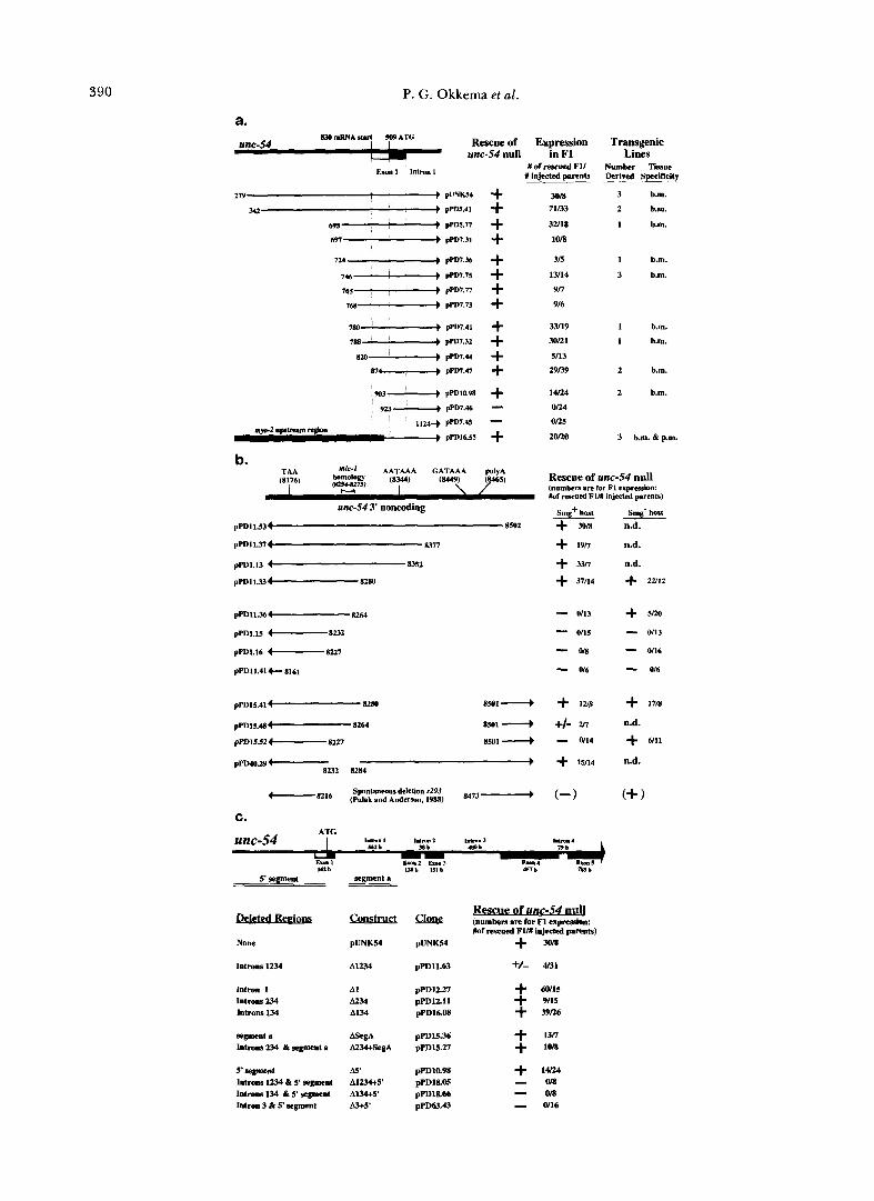
5”Flanking sequences are not necessary for proper unc-54 expression: We constructed a series of deletions removing unc-54 5’-flanking DNA (Figure 3a). Surprisingly, deletions replacing almost all DNA upstream of the ATG translation start could still res- cue unc-54(e190) mutants. In particular, replacement of all sequences to 6 bp upstream of the translation start site resulted in a construction active in the mu- tant rescue assay (pPD10.98). As expected, deletions which extended beyond the ATG completely elimi- nated gene function.
In several active deletions, the normal unc-54 tran- scription start site had been replaced. Mapping of 5’ ends for the resulting mRNAs revealed multiple start sites within the newly juxtaposed plasmid sequences. Cryptic starts were also observed in a deletion which retained the original transcriptional start site but re- tain only 42 bp of upstream sequence (data not shown).
We could assess the expression patterns of the re- introduced unc-54 deletions both by analysis of res- cued phenotypes and antibody staining. Each of the active 5’ deletion constructs rescued both motility and egg laying defects seen in the unc-54 mutant, reflect- ing expression both in body-wall striated muscles and in the vulval muscles. T o directly assess tissue specific- ity, lines were stained with an unc-54 specific mono- clonal antibody, using an antibody to myo-3 myosin as a control (MILLER, STOCKDALE and KARN 1983). The host strain unc-54(e190) shows no staining of the unc- 54 product, but exhibits abundant body muscle stain- ing with the myo-3 antibody (FIRE and WATERSTON 19891, In each of the rescued lines, unc-54 staining was restored in essentially the same pattern as wild- type: myo-3 and unc-54 staining are co-localized to body-wall and vulval muscle, with no staining seen in pharynx or non-muscle tissue (Figure 3a).
These observations demonstrAte that 5’ sequences can be deleted from unc-54 with no loss in tissue specificity. We did, however, observe a subtle quanti- tative effect in some weakly rescued lines transformed with the most proximal deletions (pPD7.32, pPD7.47 and pPD10.98; Figure 3a). In these lines the unc-54 specific monoclonal antibody detected abundant expression in egg-laying muscles with somewhat lower
390 P. G. Okkema et al.
a.
unc-54 null in F1 Rescue of Expression Transgenic
Lines Exml lnlmnl
#of rescued FII Number "issue #injected parents Derived Spedflcity
une-54 3' noncoding
pPD11.534 8502
pPDlI.374 8377
pPD1.13 4 8352
pPDll.33-8280
pPDll.36 8264
pPD1.15 -8232
pPD1.16 -8227
pPD11.414- 8161
pPD15.41c"--- 8280 as01 4
30m 3 b.m. 71/33 2 b.m.
32/18 1 b.m. l o a
315 1 b.m.
13/14 3 b.m. 9 n
9/6
33/19 1 b.m. 30n1 1 b.m.
5/13
29139 2 b.m.
14R4 2 b.m.
o n 4
on5 2ono 3 b.m. & p.m.
(numbers arc for F1 expression: Rescue of unc-54 null Yof -urd FIM injected prenb)
Smg' host Smg- host + Ml8 n.d.
+ 19!7 n.d.
+ 3v1 n.d.
"
+ 37/14 + ZU12 - 0113 + 5/20
0/15 - 0113
- WS - 0116 - 0/6 - 0/6
-
5' segment - segment a
None pUNK54 pUNK54 + m Introll. 1234 A1234 pPD11.63 +/- 4/33
Intron 1 Introns 234 lntmns 134
AI pPD1227 + w15
A134 pPDl6.08 + 39/26
A234 pPDl2.11 + 9/15
r g o c n t a pPD15.36 + 13rf Introns 234 & s p e n t a A234+SegA pPD15.27
5'aegnent A s < p P n l O . 9 8 + 14/24
Introns 134 & 5' sgmnt A134+5' Introns 1234 & 5' segment A1234+5' pPDlS.05
pPDlS.66 -Om -018
Intron 3 & 5' segment &5' pPD63.43 - 0116
+ UJKI
Myosin Gene Regulation 391
expression in body-wall muscles. Phenotypically, these lines exhibited wild-type egg-laying with only partial motility.
T o test whether the unc-54 expression obtained in the absence of 5”flanking DNA resulted fortuitously from neighboring plasmid sequences, we replaced these flanking plasmid sequences with sequences up- stream of the pharyngeal myosin gene myo-2. This construct also rescued unc-54(el90), suggesting that expression in the 5’ deletions was not simply a result of the juxtaposed vector sequence. Lines rescued with the myo-2::unc-54 chimera expressed unc-54 myosin in both non-pharyngeal and pharyngeal muscles (data not shown). The unc-54 myosin ectopically produced in the pharynx was present in radially oriented fila- ments, suggesting it could assemble in pharyngeal muscle cells. All of the lines transformed with this construct exhibited slow growth, which could conceiv- ably result from the pharyngeal unc-54 expression. These results indicate that ectopic expression of unc- 54 could be detectable using these antibodies, at least in pharyngeal muscle, and that body-wall and pharyn- geal muscle regulatory sequences can independently function on the same gene.
Sequences within the unc-54 3’-UTR are necessary
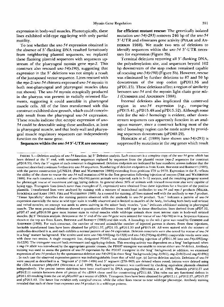
for efficient mutant rescue: The genetically isolated mutation unc-54(~293) removes 246 bp of the unc-54 3’-UTR and eliminates gene activity (PULAK and AN- DERSON 1988). We made two sets of deletions to identify sequences within the unc-54 3’-UTR neces- sary for expression (Figure 3b).
Terminal deletions removing all 3”flanking DNA, the polyadenylation site, and sequences beyond 102 bp downstream of the stop codon remained capable of rescuing unc-54(e190) (Figure 3b). However, rescue was eliminated by further deletions to 87 and 50 bp downstream of the stop codon (pPD11.36 and pPDl .15). These deletions affect a region of similarity between unc-54 and the myosin light chain gene mlc- 1 (CUMMINS and ANDERSON 1988).
Internal deletions also implicated this conserved region in unc-54 expression (e.g., comparing pPD15.41,pPD15.48andpPDl5.52).Althoughsome role for the mlc-1 homology is evident, other down- stream sequences can apparently function in an anal- ogous manner, since a construct lacking most of the mlc-1 homology region can be made active by provid- ing sequences downstream (pPD40.29).
HODCKIN et al. (1989) have shown unc-54(~293) is suppressed by mutations in the smg genes which result
~~ ~
FIGURE 3.-Deletion analysis of unc-54 function. (a) 5’ Deletion analysis. Each construct is a complete copy of the unc-54 gene which has been deleted at the 5’ end, with nematode sequences replaced by sequences from the plasmid vector ( m y - 2 sequences for construct pPDl6.55). Only the 5’ region of each construct is diagrammed. Deletion endpoints are indicated by base numbers; arrows indicate that the construct described continues further into unc-54 sequences. Numbering of deletion endpoints is from KARN, BRENNER and BARNETT (1983), with the parent construction [pUNK54; (FIRE and WATERSTON 1989)] extending from positions 279 to 9410. Expression in the F1 reflects the ability of the clone to rescue the unc-54 null mutation e190 in the first generation following injection of oocytes (FIRE and WATERSTON 1989). For each construct, a number of unc-54(eZ90) parent animals were injected, each in 5-12 oocytes, and the resulting progeny scored for phenotypic rescue of movement and egg laying defects. In general about 30% of animals with rescued movement also were capable of laying eggs. Transgenic lines (much rarer than examples of F, expression) were obtained from these injections for a fraction of the positive plasmids. Transformed lines were analyzed by staining with a mixture of monoclonal antibodies to unc-54 and m y - 3 products (MILLER, STOCKDALE and KARN 1983; FIRE and WATERSTON 1989). As a control for staining, all lines stained well with the antibody to myo-3 product. “Tissue Specificity” describes the staining pattern observed with the antibody to unc-54 product. “b.m.” indicates a pattern of unc-54 expression essentially the same as wild type: stain is readily observed and is limited to muscles of the body, including both body-wall striated and vulval muscles; no attempt was made to assess staining in the minor body muscles. “p.m.” indicates additional staining in pharyngeal muscle. The most proximal deletions showed a quantitative difference from wild type in tissue distribution: lines derived from pPD7.32, pPD7.47 and pPD10.98 gave most intense stain in vulval muscles while wild-type animals show most intense stain in body-wall striated muscles. (b) 3’ Deletion analysis. Deletions at the 3’ end of the unc-54 gene were assayed for rescue of unc-54(eZ90) as in a. Sequence features shown at the top are from KARN, BRENNER and BARNETT (1983) and this work. A homology to the mlc-Z gene was noted by CUMMINS and ANDERSON (1988). For each construct, the number of rescued animals/the number of injected parents is shown. From these injections, heritable transformed lines have been obtained for pPDll.37, pPDl.13, pPDll.33 and pPD15.48. All were stained with the mixture of antibodies described in a, and each exhibits a normal pattern of unc-54 expression. Deletion constructs were also tested for rescue of unc-54 in a Smg- mutant background. The strains used were unc-54(eZ092)smg-Z(eZZ28) and unc-54(~259)smg-6(r896); comparable results were seen with these two strains. A single transgenic line, designated PD107, was obtained following injection of pPDll.36 into unc-54(eZ092)smg- Z(eZ228). The transgene rescued both movement and egg-laying defects. This rescuing activity was dependent on a Smg- background: when a smg-Z+ allele was introduced by the appropriate genetic crosses, the PD107 transgene was unable to rescue either unc-54 defect. Antibody staining was used to assess the pattern of unc-54 expression by the PD107 transgene in the Smg- background; in addition, we examined expression of unc-54 product by the spontaneous deletion unc-54(r293) in three Smg- backgrounds [smg-Z(r861), smg-2(r863) and smg-6(~896)] . In each case the observed expression pattern was indistinguishable from that of wild type. (c) Intron deletion analysis. Deletions of unc-54 were assayed as described in a. “Segment a” (1 109-1496) and 5‘ segment (279-903) are deleted where noted; introns were deleted using the cDNA construct pMyo561 (MITCHELL et al. 1989). Each negative construct was confirmed by checking equivalent plasmids obtained independently. The precise intron deletions have been confirmed by DNA sequencing (MITCHELL et al. 1989). Plasmids pPD12.27 and pPD12.11 contain between them all pieces of the cDNA clone used for constructing pPDll.63. This rules out any functional defects in pPDll.63 resulting from the use of pMyo561 as a source of cDNA. Transgenic lines have been obtained for pPDl2.11, pPD12.27, pPD15.27 and pPDll.63. The latter line exhibits only marginal rescue, while the other lines rescue to near wild-type phenotype. Antibody staining revealed that each of these lines expresses unc-54 product in a wild-type pattern.

392 P. G . Okkema et al.
in stabilization of aberrant mRNAs. We found like- wise that several of the inactive deletions within the unc-54 3’-UTR could rescue unc-54(e190) in a Smg- genetic background (Figure 3b). In these suppressed animals, unc-54 myosin was present in the normal distribution: only in body muscle cells. This indicates that the essential sequence element within the unc-54 3’-UTR, including the mlc-1 homology, is not neces- sary for proper tissue-specific expression.
Two types of intron requirement: a non-sequence- specific stimulation by an intron and a specific activator element in unc-54 intron 3: T w o types of intron requirements have been described for optimal expression of eukaryotic genes. Nonspecific require- ments for at least one intron within the transcribed sequences have been proposed (e.g., CHOI et al. 199 1). Specific enhancer elements can also be located within introns (e.g., SCHULTZ et al. 1991). Given that we had observed proper regulation of unc-54 in the absence of 5”flanking sequence, we examined introns near the 5’ end of the gene for a role in gene expression. We have observed both a nonspecific requirement for splicing and a specific requirement for intron 3 for optimal expression of unc-54 constructs.
The unc-54 gene has eight introns (KARN, BRENNER and BARNETT 1983). Six of these are relatively short (38-79 bp) while two [intron 1 (562 bp) and intron 3 (480 bp)] are considered long for C. elegans introns (BLUMENTHAL and THOMAS 1988). We first deleted various introns in the presence of 5”flanking DNA, using a synthetic cDNA clone (MITCHELL et al. 1989) kindly provided by J. KARN (Figure 3c). The precise deletion of the first four introns essentially eliminates mutant rescue (A1234). However, any one of the first four introns can be deleted without eliminating gene function (Figure 3c). In particular, constructs contain- ing either intron 1 alone (A234) or intron 2 alone (A134) are functional. Furthermore, a construct re- moving 388 bp of internal sequences from intron 1 as well as introns 2, 3 and 4 (A234 + segment a) is sufficient for expression. These data suggested a re- quirement for splicing rather than specific sequences within intron 1.
Although A134, A234 and A234 + segment a are active, they exhibit a subtle but reproducible reduc- tion in their ability to rescue. This reduction is ob- served as a decreased frequency and quality of rescue, particularly the ability to rescue the egg-laying defect (data not shown). These observations suggested the possibility of a special contribution by the third or fourth intron to full activity.
In the absence of 5”flanking DNA, a construct deleting introns 1, 3 and 4 (A134 and 5’ segment) is unable to rescue unc-54(e190). Because a similar con- struct containing 5’ sequences is functional, introns 1, 3 or 4 must contain regulatory information redun-
dant with that in the 5”flanking DNA. The critical element is apparently in intron 3: a deletion removing only intron 3 and 5”flanking sequence is inactive. These results suggested the working model that two different sequence elements in unc-54, one upstream of the transcription start site and the other in intron 3, act as independent positive activators allowing gene expression in body-wall muscles.
lac2 fusion assays: Assays which require the func- tion of the myosin gene greatly constrain the analysis of regulatory sequences internal to the gene. In ad- dition, these assays can only be done in a mutant background. For these reasons, we used myosin::lacZ fusion constructions (Figure 4a) to further define se- quences responsible for tissue-specific regulation. We used vectors incorporating a nuclear localization pep- tide at the N terminus of @-gal; this leads to predom- inant staining in the nuclei of expressing cells, facili- tating cell identification (FIRE, HARRISON and DIXON 1990). A 3‘-non-coding sequence is placed down- stream of lacZ. Initial myo::lacZ fusions (not shown) containing the SV40-early 3’ region expressed very inefficiently in C. elegans. This inefficiency and the results with 3’ deletions described above in Figure 3b led us to make and utilize vectors which incorporate the unc-54 3’ region (FIRE, HARRISON and DIXON 1990). Because splicing also appeared necessary for efficient unc-54 expression, we included a synthetic intron sequence based on consensus splice sites (EM- MONS 1988) in many of our constructs. This intron has no homology to the myosin genes outside of the C. elegans splice consensus. These vectors (carrying the unc-54 3‘ end and synthetic intron) can express p- gal in nearly all C. elegans tissues (e.g., FIRE, HARRISON and DIXON 1990) and have no apparent bias toward expression in muscle.
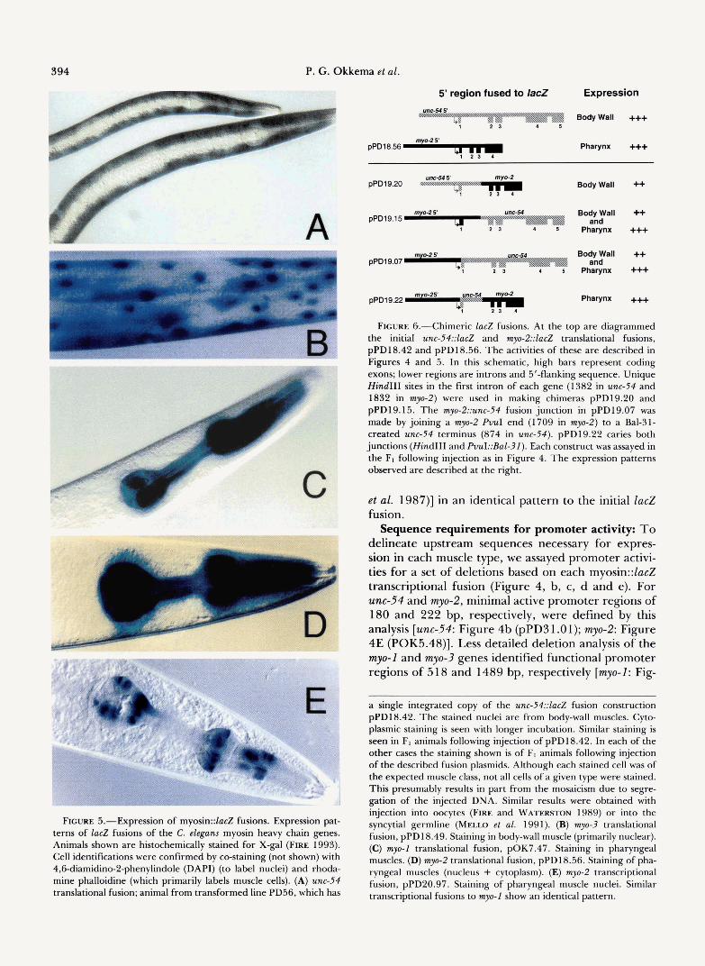
Myosin gene::lacZ fusions are properly expressed: We constructed both translational and transcriptional fusions for each of the four myosin genes (Figure 4). The unc-54::lacZ and myo-3::lacZ fusions were specifi- cally expressed in body muscle (Figure 5, A and B). This expression mimicked expression of the parent genes both in the abundant striated body-wall muscles used for locomotion, and in several sets of specialized “minor” body muscles: intestine-associated, vulva-as- sociated, and uterine sheath muscle (not shown). The myo-1::lacZ and myo-2::lacZ fusions likewise mimicked expression of their endogenous counterparts, express- ing only in pharyngeal muscle (Figure 5, C, D and E).
In several subsequent experiments, we have used modified lacZ vectors as follows to confirm that the observed expression patterns reflect promoter activity and not specific features of the vectors (not shown). Elimination of the nuclear localization signal results in constructions expressed cytoplasmically in the same
Mvosin Gene Regulation 593
a.
b.
c.
dm
e.
E. Coli U - No &gal expression YCS I
I I * Which cells express
promd.c %gal?
I I 1 I I muscle @gal activity
€ a ,.
i - 22
+++ +++ +++
++ ++ + + + + +
muscle 8-gal Body wall
activity
+++ ++ +
Pharyngeal muscle 8-gal
activity
+++ +++ ++ +
muscle D-gal Pharyngeal
activity
+++ +++ +++ ++ ++ +
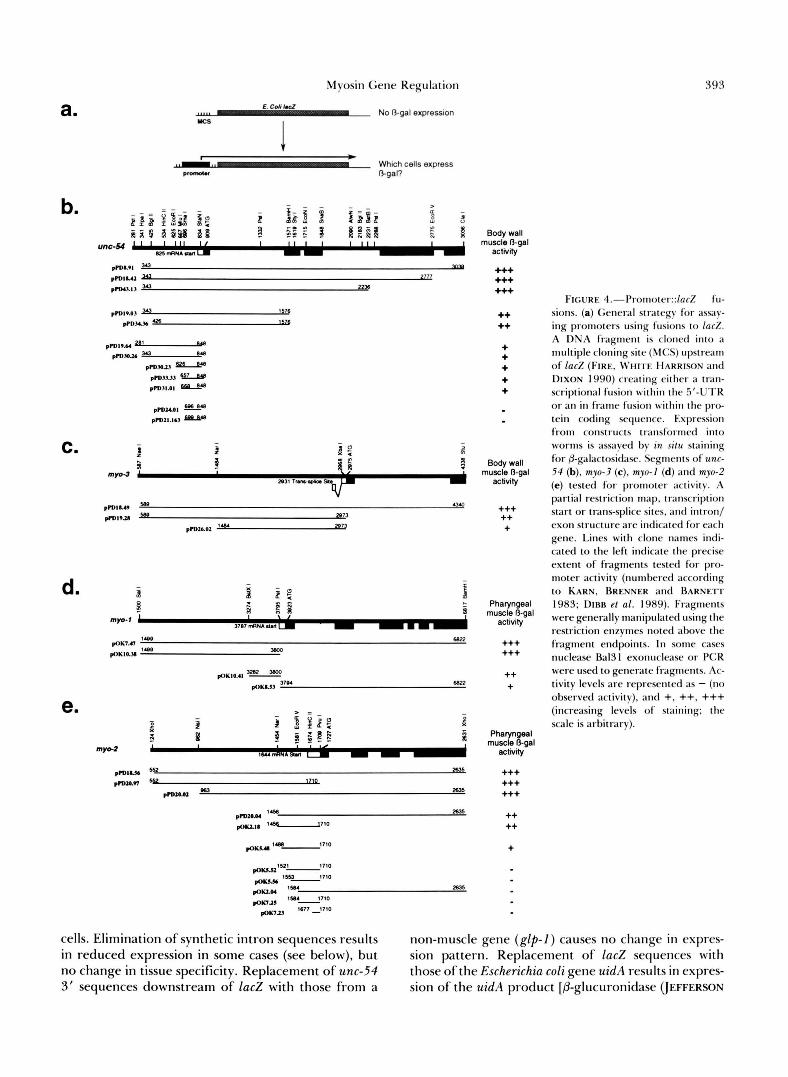
FIGURE 4.--Promorcl::laci!~ter::~ucZ fu- sions. (a) General strategy for assay- ing promoters using fusions to l a d . A DNA fragment is cloned into a multiple cloning site (MCS) upstream
DIXON 1990) creating either a tran- scriptional fusion within the 5'-UTR or an in fr;lmr fusion within the pro- tein coding sequence. Expression from constructs transformed into worms is ass;tyed by in situ staining for &galactositIase. Segments of unc- 54 (b), myo-3 (c). myo-1 (d) and myo-2 (e) tested for promoter activity. A partial restriction map, transcription start or trans-splice sites, and intron/ exon structure are indic;tted for each gene. Lines wi th clone names indi- cated to the left indicate the precise extent of fragments tested for pro- moter activity (numbered according to KARN, BRENNER and BARNETT 1983; DIRR et al. 1989). Fragments were generally manipulated using the restriction enzymes noted above the fragment endpoints. In some cases nuclease Ba131 exonuclease or PCR were used to generate fragments. Ac- tivity levels are represented as - (no observed activity). and +, ++. +++ (increasing levels of staining: the scale is arbitrary).
of (FIRE, W H I I E HARRISON alld
cells. Elimination of synthetic intron sequences results non-muscle gene ( g l p - 1 ) causes no change in expres- in reduced expression in some cases (see below), but sion pattern. Replacement of lac2 sequences with no change in tissue specificity. Replacement of unc-54 those of the Escherichia coli gene uidA results in expres- 3' sequences downstream of lac2 with those from a sion of the uidA product [P-glucuronidase (JEFFERSON
394 P. G . Okkema et al.
R
L A
5' region fused to lac2 Expression
Body Wall +++ 1 2 5
pPD18.56 Pharynx +++
FIGURE 5.-Expression of myosin::lacZ fusions. Expression pat- terns of lacZ fusions of the C. elegans myosin heavy chain genes. Animals shown are histochemically stained for X-gal (FIRE 1993). Cell identifications were confirmed by co-staining (not shown) with 4,6diamidino-2-phenylindole (DAPI) (to label nuclei) and rhoda- mine phalloidine (which primarily labels muscle cells). (A) unc-54 translational fusion; animal from transformed line PD56, which has
Body Wall ++
pPD19.15 Body Wall ++ Pharynx +++ and
pPD19.07 Body Wall ++ Pharynx +++ and
pPD19.22 Pharynx +++
FIGURE &-Chimeric lacZ fusions. At the top are diagrammed the initial unc-54::lacZ and my-2::ZacZ translational fusions, pPD18.42 and pPD18.56. The activities of these are described in Figures 4 and 5 . In this schematic, high bars represent coding exons; lower regions are introns and 5'-flanking sequence. Unique Hind111 sites in the first intron of each gene (1382 in unc-54 and 1832 in myo-2) were used in making chimeras pPD19.20 and pPD19.15. The myo-2::unc-54 fusion junction in pPD19.07 was made by joining a myo-2 PVuI end (1709 in myo-2) to a Bal-31- created unc-54 terminus (874 in unc-54). pPD19.22 caries both junctions (Hind111 and PvuI::Bal-31). Each construct was assayed in the Ft following injection as in Figure 4. The expression patterns observed are described at the right.
et al. 1987)] in an identical pattern to the initial lac2 fusion.
Sequence requirements for promoter activity: To delineate upstream sequences necessary for expres- sion in each muscle type, we assayed promoter activi- ties for a set of deletions based on each myosin::ZacZ transcriptional fusion (Figure 4, b, c, d and e). For unc-54 and myo-2, minimal active promoter regions of 180 and 222 bp, respectively, were defined by this analysis [unc-54: Figure 4b (pPD3 1 .Ol); myo-2: Figure 4E (POK5.48)]. Less detailed deletion analysis of the myo-1 and myo-3 genes identified functional promoter regions of 518 and 1489 bp, respectively [myo-I: Fig-
a single integrated copy of the unc-54::lacZ fusion construction pPD18.42. The stained nuclei are from body-wall muscles. Cyto- plasmic staining is seen with longer incubation. Similar staining is seen in FI animals following injection of pPD18.42. In each of the other cases the staining shown is of F1 animals following injection of the described fusion plasmids. Although each stained cell was of the expected muscle class, not all cells of a given type were stained. This presumably results in part from the mosaicism due to segre- gation of the injected DNA. Similar results were obtained with injection into oocytes (FIRE and WATERSTON 1989) or into the syncytial germline (Mmo et al. 1991). (B) myo-3 translational fusion, pPD18.49. Staining in body-wall muscle (primarily nuclear). (C) myo-1 translational fusion, pOK7.47. Staining in pharyngeal muscles. (D) myo-2 translational fusion, pPD18.56. Staining of pha- ryngeal muscles (nucleus + cytoplasm). (E) myo-2 transcriptional fusion, pPD20.97. Staining of pharyngeal muscle nuclei. Similar transcriptional fusions to myo-1 show an identical pattern.
Myosin Gene Regulation 395
ure 4D (pOK10.41); myo-3: Figure 4C (pPD26.02)]. In each case, the minimal active promoter fusions are less efficient than fusions including more extensive 5’- flanking DNA. The differences in activity suggest that sequence elements upstream of each minimal pro- moter augment transcription. This assay does not, however, address whether these activating sequences play a role in tissue specificity.
Characterization of regulatory sequence require- ments within the genes is less straightforward using simple deletions. Two constructions with different extents of internal sequence will produce mRNA and protein products with different structures, and hence with potentially different activity and stability. Each of the four translational fusions was somewhat more active than similar transcriptional fusions (Figure 4 and data not shown). These differences could thus reflect either a higher transcriptional activity, more efficient translation or enzyme function, or increased stability conferred by the bona fide N-terminal and 5’-untranslated sequences.
Chimeric lac2 fusions identify determinants of tissue specificity: Several chimeric lacZ fusions con- taining different parts from unc-54 and myo-2 were constructed (Figure 6). By assaying for the tissue localization of expression ( i e . , body-wall versus pha- ryngeal) from these constructs, we hoped to identify the sequences responsible for the observed difference in tissue specificity. In particular we wanted to test whether sequences inside the genes played a role in tissue specificity.
The most informative chimeric fusions were pPD19.07 and pPD19.15. Results with these con- structs delineate a sequence element within the unc- 54 transcribed region capable of activating the myo-2 promoter in body muscle. These experiments taken with results above demonstrating the activity of unc- 54::lacZ transcriptional fusions containing only up- stream unc-54 sequence [e.g., Figure 4b (1 9.64)], show that two different elements, one upstream and the other downstream of the unc-54 initiation site, are independently capable of directing gene expression to body muscles. For myo-2, no indication for an internal control element was found, although it should be noted that sequences downstream of the XhoI site in exon 4 of myo-2 have not been examined.
Body-muscle enhancer sequences: The chimera experiments above suggested a sequence within the unc-54 gene might act as a tissue specific enhancer to induce body muscle expression from the myo-2 pro- moter region. T o test this, and to further characterize such elements, we cloned fragments of unc-54 up- stream of the myo-2::lacZ translational fusion pPD18.56 (Figure 7a). Normally this construct is ex- pressed very specifically in the pharynx; hence any
element that can induce the myo-2 promoter to express outside the pharynx could be detected.
We initially found a 962-bp restriction fragment within the unc-54 gene that induced body-wall expres- sion when placed in either orientation upstream of myo-2::lacZ [Figure 7b (pPD19.84, pPD19.93); Figure 8, A and B)]. Deletion analysis of this fragment re- vealed that enhancer activity is contained within the third intron of the unc-54 gene (Figure 7b). Sequences covering the second intron of unc-54 exhibit no en- hancer activity. The ability of the element within unc- 54 intron 3 to induce body wall-muscle expression (Figures 6 and 7b) in different positions and orienta- tions proximal to the myo-2 promoter indicated this element was indeed a tissue-specific transcriptional enhancer.
Other regions from unc-54 and segments from myo- 3 were tested similarly for enhancer function. A frag- ment spanning the unc-54 promoter exhibits weak body-wall muscle enhancer activity in both orienta- tions [Figure 7b (pPD19.72, pPD19.76)]. In addition, three enhancer sequences around the myo-3 gene were identified. Two of the myo-3 enhancer segments lie upstream of the gene [Figure 7c (MC165, MC197)], while the third is within the first intron [Figure 7c (MC186)l.
Pharyngeal muscle enhancer sequences: In exper- iments analogous to those identifying body muscle- enhancer activities, we looked for pharyngeal-specific enhancer activities in myo-2 by inserting segments upstream of a myo-3::lacZ fusion construct. We iden- tified a single enhancer approximately 0.4 kb up- stream of the myo-2 transcription start site (Figure 7d). T w o overlapping 0.3-kb fragments within the region are sufficient for enhancer activity [Figure 7d (pOK4.50, pOK3.16)].
myo-1 contains at least two enhancers: a weak distal enhancer is located at least 0.5 kb upstream of the transcription start and a strong proximal enhancer is located nearer the start site [Figure 7e (pOK8.66, pOK9.05)]. Terminal deletions of the promoter prox- imal enhancer identify a 375-bp fragment sufficient for enhancer activity (Figure 7e (pOK9.05)]. A third tissue-specific element is apparently located down- stream of the transcription start in myo-1, since a fusion starting at 8 nucleotides downstream of the start is active in pharyngeal muscle [Figure 4d (pOK8.53)]. We were not able to identify an enhancer correspond- ing to this activity, suggesting that activity of the corresponding activator element(s) may be dependent on positioning relative to the promoter.
Tissue specificity of myosin enhancers: The pat- terns of expression from specific enhancer-promoter combinations such as those just described presumably reflect the underlying specificity of the promoter as well as the enhancer. We have therefore tested en-
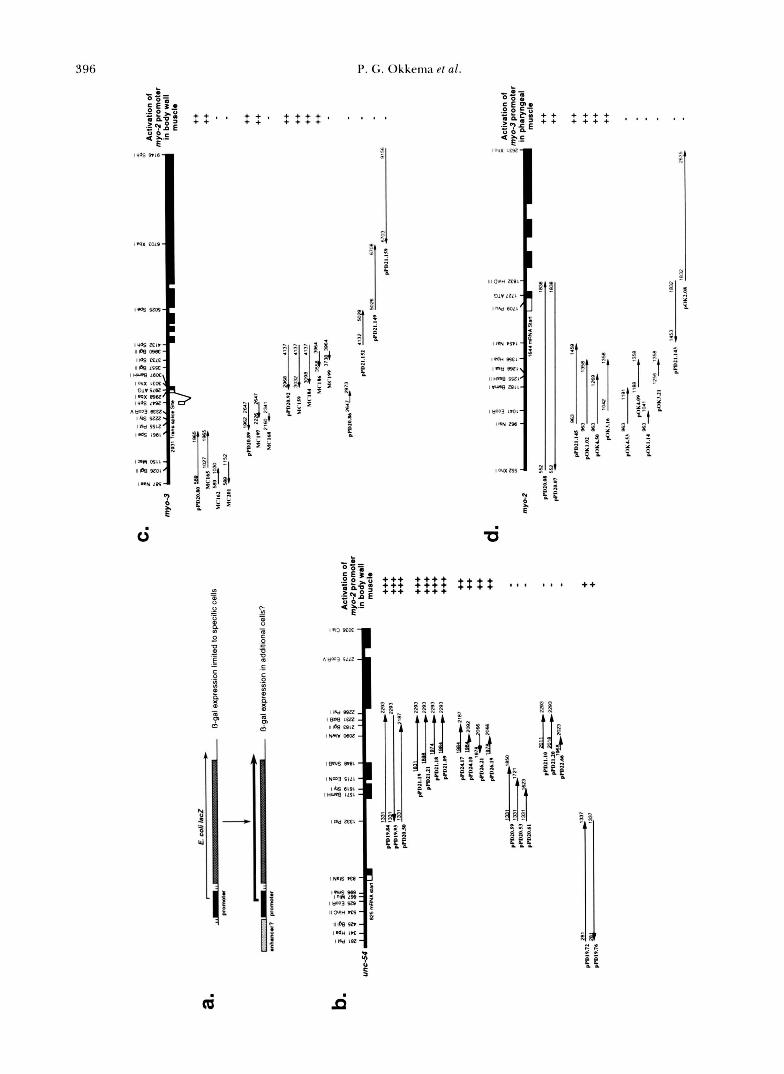
396 P. G . Okkema et al. b.
- m O B = .
- d l 0 0 0
'= - e: :E:' " .p$P : : , I + + I + + + + . I I I I
gyg 2 + + + + + + ,;;,e4:: :::: ' " I "
: EL
.- S f pi?
;On 4 A C
E E .- I uo5 PIN - y: l wx ,192 A
I -x m ~ o - 1
IIIO"" ml-
9,v ' Z U - 31 3 3 : 1"- en',' I
8 ::
ImN n T L - < I,&"
o m s n c - I, Ise 'Ssf -
3 3' IWWoicI -
3 z -pi -* ; 2 I w 3 w o k -
a' 3, ; z t-n 8 9 ~ 1 , a' t g
,I une 5521'
I w mil- ?
3 - %-I ,PI 3 P P p x - 4 t t
ION m - -. 2 .f:
IWIXLTr"
0 0 ? s
?' I
* I $ $
ci 6
- " 55:; +++ + + + + + + + +
. a;=
u) - +++ + + + + + + + + * I 0 I I I ++ - 2;: 2 +++ + + + + m 0 r = -
0
._ 4 E.. - E 0 :: - m 1.13 sa - 0 c 0 s - ._ - - ._ E
V
c E C In ._
u)
- ~ n - 3 suz - ._
._
? :: ? - B ,i.dssa-b 11, 111% (I 11 - 0
lose I u r 0, ISe FBlZ
:I:, 'li [,) % a
)I1 ia!! I: f t j : 2" i7 ilh 2 "" ;- e?! J
IN-3 SI11
lHlue $151 - 1 6 9 l B l # a $ $ $ a f a
Iwml- - - 5 - I I? *,.
4 3 1 t :?? E: 0 0 1 $ $ $
IMS"
I,%%= ; 1-3" II W'U m - Il*sn-
IWn m - 2 t
I W 1 9 - f P ". 5 C epI
r r C C
cd d

Myosin Gene Regulation
- ;= O O b .
ggs3 $ $ $ $ $ + ' I I I .
E € = - px'oE 4 'a.5
, H Y . 8 LlSS
111 a w am
,.IO WlE
III a w s m -
"+Ai$\ c
I w S81C 5 1 V i Z E -
1 " s w s c la-s m I xu# ? L a - g ; a I, pe .Ell - , a n
N - i
I . d H s e l l - 5 3 t
I l 5 W l -. ' $ ' 1
1 4 $ 4 t.t:, 5
SI, $ 5 3 * z i p t i 5
- 6 & S E = i i
ai
1 . I
I
1% 3 %
Ti f .. .. %
2 " 7 2 a- .h - o & 0 2 .f.s ; $ 2 $ e < 2 . 9 % ; o - . , p ' ,g 8 s 0 .E?.; &g$ $".;l;s.. m 0 ' - 0) 0 c * I 0 W h N r 3 . q $ E 3 2 0 3 2 m" g * + :" g % s 3 r - % s 3 E c - 5 t *.zIz %,83* 0.
e ? . + -"11g A": $;? s W & J $ 3 8' $;;**rag ,x N p) c % -$ G.5 :E ; 2; g & & 0
cc.I m g ii't c
2 :E Ec.I.(Z'i ,oy C L , o S " M ~
m 8 " o n 0hg.g y c ' L ' Z & E .r 'J E 2 "p.gP-m" 8 2 m e, 0 +.;2j
Q,, w x . s " + L > Z ? 0.5 5s4 .$ " : z e
I . - e, 3 0 3 m " 3 8 2 E s % g $ s-0
- c , & ; ? 2
e, ;-.p .r s$ g m Eik 8 a z , + 2
m . 5 2 z E$&: t 0 c . 5 e, y d ;s,xs e, s i g $ 2 u x $ 4 - 3 : = b , h G CI z . ? % s - : L S + g % o 0 - U " & 5 a e , . % C 5 5 a-0.Gg % $ g $,x, I .- $a g e 3
5 3 % 25.z m
= 4 u 2 k % s L C % 8 w : $ . z < , p J ys 2 3 z &S&+.-& Z M
. C - Q
u A E Z %.s 2 3
.- - E y g : ^ = d s E e, L Q ss?r$
- u M m E o v v , c Q G r - L L C U 3 <
v e o I
a E g 2 ~ ? 2 0
e, i 3 q K i i M, y
" $ 5 2 L$43 5 &$"! e- z E e a $ g z E $5 a8 m 5 c F m 3 = 3 2
2 0 % xu 2 0s ~ ) n " m O u C . . r c m ~ ~
&,-a '3 n f O " Z & Z
.- $ E b Z . 2 5 g . 2 &.'J 0 v f u 29, .= C k c c i c f v1- e," e, c
xi2 E E % w e , c . E !?e, w w e, L "
-,c2 . 8 E i ; ; 2 % - c err- $ 0 c 2. 2 2 s y : f c m 1 - i e, *5az.pB, .g v + $ $ g € L $ &.+ 3 .: : ,$ .g-& t 97 9 2 8 7 . S u . 5
x c ~ ? 2 i ' + !2 d 2 . 2 2 em e,
8 2 9 - 2 2 :< r- c * n y . r C , I . % 2 t: c -0 ' 5 3 1 . g e z o s g ; - ; o o m
$ 5 g s g , x2 .2 .=
5 g 3 E € 5 M 3 a m :
2 OJ E Q ! U) L'- n L v , - w
o E - C & xu) E.: "0
2 5 E % 3 U-Osy
c - 5 R?e*g$ B g $ +. s = > ;%Q MI-
~ Z ~ A Q = - > .S 8 E a m L * t- m
h& n 8 3 '5
397
398 P. G. Okkema et al.
B
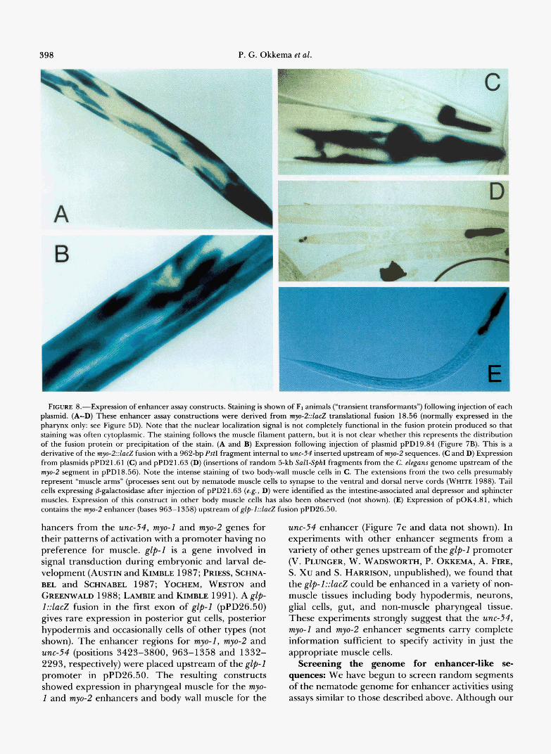
L E FIGURE 8.-Expression of enhancer assay constructs. Staining is shown of FI animals ("transient transformants") following injection of each
plasmid. (A-D) These enhancer assay constructions were derived from my-2::lac.Z translational fusion 18.56 (normally expressed in the pharynx only: see Figure 5D). Note that the nuclear localization signal is not completely functional in the fusion protein produced so that staining was often cytoplasmic. The staining follows the muscle filament pattern, but it is not clear whether this represents the distribution of the fusion protein or precipitation of the stain. (A and B) Expression following injection of plasmid pPD19.84 (Figure 7B). This is a derivative of the myo-2::lucZ fusion with a 962-bp PstI fragment internal to unc-54 inserted upstream of myo-2 sequences. (C and D) Expression from plasmids pPD2 1.6 1 (C) and pPD2 1.63 (D) (insertions of random 5-kb SaEISphI fragments from the C. eleguns genome upstream of the myo-2 segment in pPD18.56). Note the intense staining of two body-wall muscle cells in C. The extensions frod the two cells presumably represent "muscle arms" (processes sent out by nematode muscle cells to synapse to the ventral and dorsal nerve cords (WHITE 1988). Tail cells expressing @-galactosidase after injection of pPD21.63 (e.g., D) were identified as the intestine-associated anal depressor and sphincter muscles. Expression of this construct in other body muscle cells has also been observed (not shown). (E) Expression of pOK4.81, which contains the myo-2 enhancer (bases 963-1358) upstream of glp-1::lac.Z fusion pPD26.50.
hancers from the unc-54, myo-1 and myo-2 genes for their patterns of activation with a promoter having no preference for muscle. glp-1 is a gene involved in signal transduction during embryonic and larval de- velopment (AUSTIN and KIMBLE 1987; PRIES, SCHNA- BEL and SCHNABEL 1987; YOCHEM, WESTON and GREENWALD 1988; LAMBIE and KIMBLE 199 1). A glp- 1::ZacZ fusion in the first exon of glp-1 (pPD26.50) gives rare expression in posterior gut cells, posterior hypodermis and occasionally cells of other types (not shown). The enhancer regions for myo-1, myo-2 and unc-54 (positions 3423-3800, 963-1358 and 1332- 2293, respectively) were placed upstream of the glp-1 promoter in pPD26.50. The resulting constructs showed expression in pharyngeal muscle for the myo- 1 and myo-2 enhancers and body wall muscle for the
unc-54 enhancer (Figure 7e and data not shown). In experiments with other enhancer segments from a variety of other genes upstream of the glp-1 promoter (V. PLUNGER, W. WADSWORTH, P. OKKEMA, A. FIRE, S. Xu and S. HARRISON, unpublished), we found that the glp-1::lacZ could be enhanced in a variety of non- muscle tissues including body hypodermis, neurons, glial cells, gut, and non-muscle pharyngeal tissue. These experiments strongly suggest that the unc-54, myo-1 and myo-2 enhancer segments carry complete information sufficient to specify activity in just the appropriate muscle cells.
Screening the genome for enhancer-like se- quences: We have begun to screen random segments of the nematode genome for enhancer activities using assays similar to those described above. Although our

Myosin Gene Regulation
Promoter Semnea unc-Mpro unc-54 enh+pro myo-2 myo-3 myo-3
(279-848) (1832-2293 & 279-848) (125-1710) (589-2973) (1486-2973)
Average number of stained F1 cells per injected adult (numbers in parentheses are total adults injected)
399
Jntron S e m e d
B. Synthetic Intron II 19 (16)
C. Precise Deletion 0 (11)
D. Synthetic Intron n* 0 (24)
E. PpuMI deletion 0 (10)
16 (15)
A. SynthetiCIntronI AGGACCCAAAGgtatgtttcgaatgatactaacataacatagaacattttcagGAGGACCC
B. SyntheticInaonn AGGACCCAAAGgtaagtttcgaatcatactaacataacatagaacattttcagGAGGACCC C. Precisekletion AGGACCCAAAG------------------------------------------GAGGA~CC D. Synthetic Intron n* AGGACCCAAAGATAU;TTTCGAATC~TACTIVLCATAACATAG~CATTTTC~GAGGACCC E. PpuMIdeletion A G G A - - - - - - - - - - - - - - - - - - - - - - - - - - - - - - - - - - - - - - - - - - - - - - - - - - - - - - ~ ~ ~
FIGURE 9.-Stimulatory activities of synthetic introns. (Top, A-E) Five different myosin::lacZ transcriptional fusions have been tested with a variety of intron-like and non-intronic sequences in their 5’-nontranslated regions. In each case the number of stained cells were determined in the F, following injection of oocyte nuclei in 8-24 parental animals. The number of injected animals is shown in parenthesis, with the average number of stained muscle cells per injected parent represented for each combination tested. Because of some variability in injections, differences of less than twofold are not considered significant. As an alternate measure of expression, we also observed the intensity of staining: removal of introns from myo-2 and unc-54 fusions resulted in much fainter staining in those cells that were stained, while no difference in intensity was observed between the corresponding myo-3 fusions. (Bottom, A-E) Sequences of equivalent regions in the 5’- UTR. In the constructs described, intronic sequences or their residues were present between the start of transcription and the start of translation. In each case, the transcriptional start of the myosin gene was retained in the constructs, while translation would be expected to start at the ATG in the nuclear localization cassette attached just upstream of lacZ (see Figure 4). Synthetic Intron I is the original intron sequence of FIRE and WATERSTON (1989). For comparison of intron-like sequences with and without functional splice junctions, it was necessary to start with an alternate synthetic intron (Synthetic intron 11) which lacks ATG sequences, as these could potentially interfere with translation of the lacZ open reading frame located downstream (KOZAK 1991). Uppercase: sequences expected to be retained in the processed mRNA. Lowercase: sequences expected to be spliced out. Bases in outline represent changes from synthetic intron I. The promoter segments used from unc-54, m y - 2 and myo-3 correspond to segments described in Figure 4. The corresponding intron I containing constructs for unc- 54 and myo-2 were pPD19.64 and pPD20.97, respectively; the long and short myo-3 promoter constructs with intron I were pPD19.28 and pPD26.02, respectively (Figure 4). The unc-54 enhancer+promoter constructs contain the indicated intron 3 sequence in a ‘+’ orientation placed upstream of the unc-54 promoter in construct pPD19.64. All of these constructs were identical outside of the synthetic intron and promoter sequences.
initial screens were designed to survey only a small fraction of the genome, these experiments could yield several types of information relevant to the enhancer and promoter activities described above. First, we should obtain a very rough estimate for the frequency in the genome of enhancer activities as defined by these assays. Second, the different patterns of expres- sion observed with a given promoter should illuminate the underlying specificity of the promoter used, in terms of its ability to respond to enhancement in different tissues. In their normal context, the myosin gene promoter elements need only respond to signals in the muscle types where the corresponding genes are expressed. The data in the preceding sections suggests strongly that these promoter elements can respond to signals for expression in the other muscle types, if such signals are added in cis.
T o assay response to enhancement in a broad set of tissues, we have carried out a limited screen of ge- nomic DNA for sequences capable of enhancing the
myo-2 promoter. The myo-2 promoter was chosen since expression in any tissue outside the pharynx would be readily detected in enhancer assays. Random frag- ments (2-5 kb) of C. elegans DNA were inserted upstream of the active myo-2::lacZ translational fusion construct used in the assays described above. The resulting constructs were injected individually, and stained animals from the F1 generation were exam- ined for non-pharyngeal @-gal expression (Figure 8, C and D). Six of eighteen fragments tested functioned as enhancers in this assay (approximately 62 kb of genomic DNA was examined). All of the observed expression from these constructs occurs in muscle cells (Figure 8). One explanation for this would be that the myo-2 promoter segment used is selective in its re- sponse to enhancer function, responding predomi- nantly in muscle tissue. Data consistent with this hy- pothesis come from assays of two of the enhancers identified in this screen for enhancement of the glp-1 promoter segment. One of these fragments enhances
400 P. G. Okkema et al.
the glp-1 promoter in non-muscle cells (in the pharyn- geal-intestinal valve and a variety of other cells), while a second weakly enhances glp-I in muscle (data not shown). The first fragment thus appears to be capable of enhancement in both muscle and non-muscle tis- sues, however the myo-2 promoter segment responds only in muscles.
Intron requirements revisited: T o address the na- ture of requirements for intron sequences, we com- pared transcriptional fusions of the myosin genes to lac2 with and without the synthetic intron present. T o control for potential effects of 5' leader sequences on gene expression which are independent of a func- tional intron, we have generated three sets of con- structs to compare intron effects. In the first set, the synthetic intron is deleted by excision of a PpuMI restriction fragment, which creates a 12-bp deletion in the resulting mRNA. In the second, the intron is precisely deleted, so the primary transcript would be identical to the spliced message from the intron-con- taining construct. The third set of constructs contains a synthetic intron which has been mutated at the splice junctions, so that the transcripts are otherwise identi- cal.
For myo-2 and unc-54, similar results were obtained for the three sets of constructs: the lack of an intron resulted in decreased expression as assayed both by the number of stained cells and the intensity of the staining (Figure 9). For myo-3, no significant differ- ence in expression was observed between intron-con- taining and intron-lacking constructs. myo-3 is the only one of the four genes to be trans-spliced at the 5' end. These experiments initially suggested that high level intron-independent expression might correlate with trans-splicing. However, preliminary results with two other genes suggest that the actual situation could be more complex: for hlh-1,which is trans-spliced to SL1 (KRAUSE et al. 1990), we observed strong stimulation of a transcriptional fusion following addition of syn- thetic intron I; for myo-1, which is not trans-spliced, we observed no difference between a transcriptional fusion containing synthetic intron I and an equivalent intron-lacking construct (data not shown).
DISCUSSION
We have characterized sequences required for my- osin gene expression in transgenic C. elegans. These experiments have identified regulatory elements con- trolling tissue-specific expression of the myosin genes as well as general features of one of these genes which are necessary for activity. In discussing the results we will describe first the individual elements and then describe their interactions in the context of a complete functional gene.
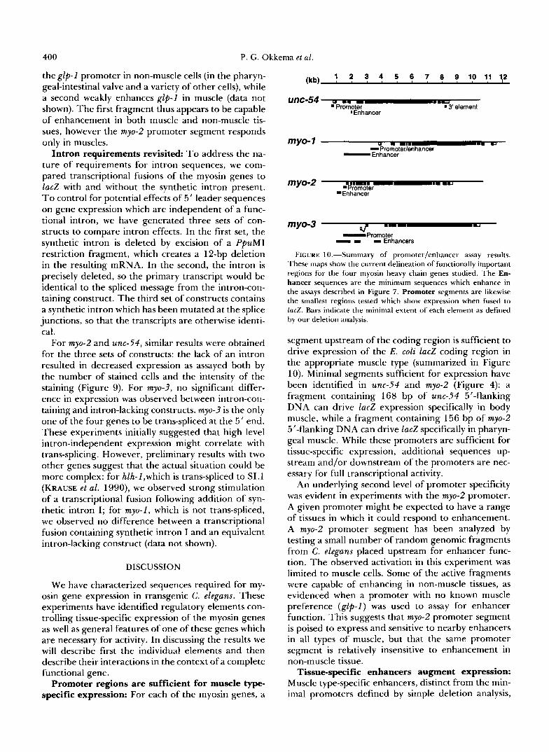
Promoter regions are sufficient for muscle type- specific expression: For each of the myosin genes, a
(kb) 1 2 3 4 5 6 7 8 9 1 0 1 1 1 2
onc-54 a .. --.- . Promoter 'Enhancer
'3' element
myo- 1 U U
-Promoter/enhancer -Enhancer
myo-2 2 'Enhancer
my0-3 -Promoter - I I Enhancers
FIGURE 10.-Summary of promoter/enhancer assay results. These maps show the current delineation of functionally important regions for the four myosin heavy chain genes studied. The En- hancer sequences are the minimum sequences which enhance in the assays described in Figure 7. Promoter segments are likewise the smallest regions tested which show expression when fused to lacZ. Bars indicate the minimal extent of each element as defined by our deletion analysis.
segment upstream of the coding region is sufficient to drive expression of the E . coli lac2 coding region in the appropriate muscle type (summarized in Figure 10). Minimal segments sufficient for expression have been identified in unc-54 and myo-2 (Figure 4): a fragment containing 168 bp of unc-54 5'-flanking DNA can drive lac2 expression specifically in body muscle, while a fragment containing 156 bp of myo-2 5"flanking DNA can drive lac2 specifically in pharyn- geal muscle. While these promoters are sufficient for tissue-specific expression, additional sequences up- stream and/or downstream of the promoters are nec- essary for full transcriptional activity.
An underlying second level of promoter specificity was evident in experiments with the myo-2 promoter. A given promoter might be expected to have a range of tissues in which it could respond to enhancement. A myo-2 promoter segment has been analyzed by testing a small number of random genomic fragments from C . elegans placed upstream for enhancer func- tion. The observed activation in this experiment was limited to muscle cells. Some of the active fragments were capable of enhancing in non-muscle tissues, as evidenced when a promoter with no known muscle preference (glp-I) was used to assay for enhancer function. This suggests that myo-2 promoter segment is poised to express and sensitive to nearby enhancers in all types of muscle, but that the same promoter segment is relatively insensitive to enhancement in non-muscle tissue.
Tissue-specific enhancers augment expression: Muscle type-specific enhancers, distinct from the min- imal promoters defined by simple deletion analysis,
Myosin Gene Regulation 40 1
were identified for each of the myosin genes (sum- marized in Figure 10). The enhancers from the body wall myosin gene unc-54 and the pharyngeal muscle myosin gene myo-2 have been examined in greatest detail. The major unc-54 enhancer is located within the third intron and activates transcription in body- wall muscle and other non-pharyngeal muscles. The myo-2 enhancer is located upstream of the promoter and activates transcription specifically in pharyngeal muscle. The unc-54 and myo-2 enhancers activate tran- scription in the appropriate muscle type when assayed upstream of both muscle and non-muscle promoters, indicating that tissue specificity is an inherent prop- erty of the enhancer and not dependent on a partic- ular promoter.
Two types of intron contribution: intron resident enhancers and mechanistic effects of splicing: Two types of requirements for intervening sequences were found in this analysis. The first are requirements for specific sequences inside of introns. These sequences (in the first intron of myo-3 and the third intron of unc-54) act as enhancers. The presence of relatively large introns near the 5’ ends of these genes provides a venue to place activating enhancers downstream as well as upstream of the promoter; this added flexibility could facilitate the evolution of highly regulated genes.
A second type of intron effect apparently reflects a mechanistic need in some circumstances for an mRNA splicing event. A stimulatory effect of splicing is par- ticularly evident in comparing the activity of unc- 54::lacZ fusions which have been constructed with and without synthetic intron sequences (we used synthetic introns that have no homology outside of the consen- sus splice sites to any of the myosin gene introns). A similar stimulation was seen with myo-2::lacZ fusions, but analogous experiments with the myo-1 and myo-3 promoters failed to detect any increased activity in the presence of the synthetic intron. Previous studies with mammalian tissue culture cells have similarly suggested that splicing-dependent stimulation is a property of the promoter (e.g., NEUBERCER and WIL- LIAMS 1988).
Although the synthetic introns dramatically stimu- lated expression of some gene fusions, no changes in tissue specificity were seen. There is thus no indication of a tissue specificity related to the mechanistic re- quirement for splicing.
unc-54 3’ sequences are required for expression but not tissue specificity: A requirement for se- quences at the 3’ end of unc-54 was evident from both whole gene assays (Figure 3b) and lac2 fusions (not shown). A short region of homology to the gene mlc- 1 plays some role, although other downstream se- quences can apparently substitute.
The 3 ’ -untranslated sequences required for unc-54
expression do not appear to contribute to tissue spec- ificity. Mutations in the smg genes, which result in stabilization of aberrant mRNAs, allow unc-54 expres- sion in the absence of otherwise essential 3’ sequences. The tissue specificity of the resulting expression is normal, indicating that these 3‘ sequences are not necessary for tissue specificity. In addition, this region from unc-54 can be replaced in l a d fusions by the 3‘- untranslated leader sequence from the gene glp-1, which has no known muscle association. The resulting fusions retain expression in just body wall muscle cells (not shown). The unc-54 3’ end can also function in a very wide variety of somatic tissues (e.g., FIRE, HAR- RISON and DIXON 1990), suggesting this region does not contain sequences which specify body wall muscle expression.
Tissue specificity of myosin gene expression re- sults from multiple positively acting control ele- ments: Our data argue strongly that the myosin heavy chain gene promoters are controlled fundamentally by activators rather than repressors of expression. In particular, none of the promoter deletions that we tested exhibit ectopic expression of lac2 or myosin. This could conceivably reflect the biological con- straints under which the gene has evolved. Muscle myosin heavy chain gene products must be expressed both at high levels and in very restricted patterns. Any expression in non-muscle cells would likely lead to interference with normal actin based processes. C. elegans has apparently evolved a control system in which both promoters and enhancers have intrinsic muscle specificity. This should serve to decrease the possibility of spurious expression in non-muscle cells.
The ability of enhancer and promoter sequences to independently direct proper expression was particu- larly evident in our assays of mutant rescue by unc-54 deletions. Constructs with either element alone were capable of rescuing the unc-54 mutant phenotype. The results with promoter deletions were particularly surprising, given the view that sequences upstream of and around the initiation site direct placement of 5’ ends by RNA polymerase 11. Transcripts initiated just upstream of the coding region were observed even in constructs with all the natural 5’-flanking sequence replaced by plasmid sequence. One possibility is that a hypothetical “initiator” element within the coding region helps direct polymerase to start within a meas- ured region just upstream. Alternatively, transcrip- tion from the 5‘ deletion constructs might initiate at many dispersed sites around the gene, while a post- transcriptional mechanism degrades those RNAs that cannot be properly processed or translated. In either case, the system has apparently evolved to allow both upstream and internal sequences to activate expres- sion.
Why does each myosin gene have more than one
402 P. G. Okkema et al.
tissue-specific activator element? Although some tis- sue-specific activation was seen with individual ele- ments, the frequency of observed expression increases when several activator elements are present in a single construct (Figure 4). Multiple control elements for each locus may be necessary to increase the probability of gene expression in a given muscle cell to 1 OO%, and to ensure that expression begins synchronously in all cells ( e .g . , WEINTRAUB 1988). In this context it should be noted that we examined only sequences within 0.8-3 kb of the myosin promoters. Thus it is quite possible that there could be additional body wall muscle specific enhancers upstream of each of these genes, which function to increase frequency of expres- sion but are not uniquely required for activity.
In other studies, regulatory sequences controlling C. elegans gene expression in several other tissues have been analyzed, using methods similar to those de- scribed here (AAMODT, CHUNG and MCGHEE 1991; WAY et al. 1991; MACMORRIS et al. 1992). The vitel- logenin genes, which are regulated by activators, be- have with broad similarity to the myosin genes (MACMORRIS et al. 1992). In contrast, the ges-1 and mec-3 genes are apparently controlled by both positive and negatively acting cis-acting elements (AAMODT, CHUNG and MCCHEE 1991; WAY et al. 1991). We expect that the full spectrum of conceivable regula- tory mechanisms will be used in C. elegans, as has been seen with other complex organisms (e.g., PTASHNE 1986; LAWRENCE 1992).
One common feature of the analyses to date of cis- acting sequences controlling expression in C . elegans (AAMODT, CHUNG and MCGHEE 199 1; WAY et al. 1991 ; MACMORRIS et al. 1992; KRAUSE et al. 1990) is that control sequences have been found relatively close to and within the genes. Initial large scale se- quencing studies of the C . elegans genome (SULSTON et al. 1992) indicate a closer packing of genes in the C. elegans genome than that seen in mammalian and insect genomes; this may mandate in general a more compact organization of control sequences. The dis- tances over which enhancers exert their effects could indeed be one of the forces driving evolution of genome size.
We are grateful to S. XU, D. MILLER, R. PULAK, P. ANDERSON, J. HODGKIN, R. WATERSTON, D. HARRISON, H. EPSTEIN, M. KRAUSE, J . KARN, D. DIXON, T. BLUMENTHAL, D. BROWN, Y. SHI, M. SEPANSKI, L. CONNAH, J. AHNN, W. KELLY and G . SEYDOUX for their help and suggestions. This work was supported by the National Institutes of Health (postdoctoral fellowship HD07532-03 to P.G.O. and grant R01-GM37706 to A.F.), the Carnegie Institution of Washington, and the Rita Allen Foundation.
LITERATURE CITED
AAMODT, E. J., M. A. CHUNC and J. A. MCGHEE, 1991 Spatial control of a gut-specific esterase during Caenorhabditis elegans development. Science 252: 579-582.
ALBERTSON, D. G. , 1985 Mapping muscle protein genes by in situ hybridization using biotin-labeled probes. EMBO J. 4 2493- 2498.
ANDERSON, P., and S. BRENNER, 1984 A selection for myosin heavy-chain mutants in the nematode C. elegans. Proc. Natl. Acad. Sci. USA 81: 4470-4474.
ARDIZZI, J. P., and H. F. EPSTEIN, 1987 Immunochemical local- ization of myosin heavy chain isoforms and paramyosin in developmentally and structurally diverse muscle cell types of C. elegans. J. Cell Biol. 105: 2763-2770.
AUSTIN, J., and J. KIMBLE, 1987 glp-1 is required in the germ line for regulation of the decision between mitosis and meiosis in C. elegans. Cell 51: 589-599.
BEJSOVEC, A., and P. ANDERSON, 1990 Functions of the myosin ATP and actin binding sites are required for C. elegans thick filament assembly. Cell 60: 133-140.
BLUMENTHAL, T., and J. THOMAS, 1988 Cis and trans mRNA splicing in C. elegans. Trends Genet. 4: 305-308.
BRENNER, S., 1974 The genetics of Caenorhabditis elegans. Ge- netics 77: 7 1-94.
CALZONE F. J., R. J. BRITTEN and E. H. DAVIDSON, 1987 Mapping of gene transcripts by nuclease protection assays and cDNA primer extension. Methods Enzymol. 152: 61 1-632.
CHOI, T., HUANG, M., C. GORMAN and R. JAENISCH, 1991 A generic intron increases gene expression in transgenic mice. Mol. Cell. Biol. 11: 3070-3074
CUMMINS, C., and P. ANDERSON, 1988 Regulatory myosin light- chain genes of Caenorhabditis elegans. Mol. Cell. Biol. 8: 5339- 5349.
DIBB, N. J., BROWN, D. M., KARN, J., MOERMAN, D. G. , S. L. BOLTEN and R. H. WATERSTON, 1985 Sequence analysis of mutations that affect the synthesis, assembly and enzymatic activity of unc-54 myosin heavy chain of C. elegans. J. Mol. Biol. 183: 543-551.
DIBB, N. J., MARUYAMA, I. N., M. KRAUSE and J. KARN, 1989 Sequence analysis of the complete Caenorhabditis elegans myosin heavy chain gene family. J. Mol. Biol. 205: 603-613.
EIDE, D., and P. ANDERSON, 1985a The gene structures of spon- taneous mutations affecting a C. elegans myosin heavy chain gene. Genetics 109: 67-79.
EIDE, D. J., and P. ANDERSON, 198513 Novel insertion mutation in C. elegans. Mol. Cell. Biol. 5: 1-6.
EIDE, D., and P. ANDERSON, 1985c Transposition of Tcl in the nematode C. elegans. Proc. Natl. Acad. Sci. USA 82: 1756- 1760.
EMMONS, S. W., 1988 The genome, pp. 47-79 in The Nematode Caenorhabditis elegans, edited by W. B. WOOD. Cold Spring Harbor Laboratory, Cold Spring Harbor, N.Y.
EPSTEIN, H. F., R. H. WATERSTON and S. BRENNER, 1974 A mutant affecting the heavy chain of myosin in C. elegans. J. Mol. Biol. 9 0 291-300.
FIELDS, C., 1990 Information content of Caenorhabditis elegans splice site sequences varies with intron length. Nucleic Acids Res. 18: 1509- 15 12.
FIRE, A,, 1986 lntegrative transformation of Caenorhabditis ele- guns. EMBO J. 5: 2673-2680.
FIRE, A., 1993 Histochemical techniques for locating Escherichia coli 0-galatosidase activity in transgenic organisms. Gene Anal. Tech. Appl. 9: 151-158.
FIRE, A., and R. H. WATERSTON, 1989 Proper expression of myosin genes in transgenic nematodes. EMBO J. 8: 3419- 3428.
FIRE, A,, S. WHITE HARRISON and D. DIXON, 1990 A modular set of lacZ fusion vectors for studying gene expression in Caenor- habditis elegans. Gene 93: 189-198.
FIRE, A,, ALBERTSON, D., S. WHITE HARRISON and D. MOERMAN, 1991 Production of antisense RNA leads to effective and
Myosin Gene Regulation 403
specific inhibition of gene expression in C. elegans muscle. Development 113: 503-514.
FROHMAN M. A,, M. K. DUSH and G. R. MARTIN, 1988 Rapid amplification of full-length cDNAs from rare transcripts: am- plification using a single gene-specific oligonucleotide. Proc. Natl. Acad. Sci. USA 85: 8998-9002.
HODGKIN, J., A. PAPP, R. PULAK, V. AMBROS and P. ANDERSON, 1989 A new kind of informational suppression in the nema- tode Caenorhabditis elegans. Genetics 123: 30 1-3 13.
HONDA, S., and H. F. EPSTEIN, 1990 Modulation of muscle gene expression in Caenorhubditis elegans: differential levels of tran- scripts, mRNAs and polypeptides for thick filament proteins during nematode development. Proc. Natl. Acad. Sci. USA 87: 876-880.
JEFFERSON, R. A., M. KLASS, N. WOLF and D. HIRSH, 1987 Expression of chimeric genes in Caenorhabditis elegans. J. Mol. Biol. 193: 41-46.
KARN, J., S. BRENNER and L. BARNETT, 1983 Protein structural domains in the C. elegans unc-54 myosin heavy chain gene are not separated by introns. Proc. Natl. Acad. Sci. USA 8 0 4253- 4257.
KAY, R. J., R. J. BOISSY, R. H. RUSSNAK and E. P. M. CANDIDO, 1986 Efficient transcription of C. elegans heat shock gene pair in mouse fibroblasts dependent on multiple promoter elements which can function bidirectionally. Mol. Cell. Biol. 6: 3134- 3143.
KLASS, M., D. AMMONS and S. WARD, 1988 Conservation in the 5’ flanking sequences of transcribed members of the Caenor- hubditis elegans major sperm protein gene family. J. Mol. Biol.
KLASS, M. R., S. KINSLEY and L. C. LOPEZ, 1984 Isolation and characterization of a sperm-specific gene family in the nema- tode C. elegans. Mol. Cell. Biol. 4: 529-537.
KOZAK, M., 1991 Structural features in eukaryotic mRNAs that modulate the initiation of translation. J. Biol. Chem. 266
KRAUSE, M. 1986 Actin gene expression in the nematode Caenor- habditis elegans. Ph.D. Thesis, University of Colorado, Boulder.
KRAUSE, M., and D. HIRSH, 1987 A trans-spliced leader sequence on actin mRNA in C. elegans. Cell 49: 753-761.
KRAUSE, M., FIRE, A,, WHITE HARRISON, S, J. PRIESS and H. WEINTRAUB, 1990 CeMyoD accumulation defines the body wall muscle cell fate during C. elegans embryogenesis. Cell 63: 907-9 19.
LAMBIE, E., and J. KIMBLE, 1991 Two homologous regulatory genes, lin-12 and g lp - I , have overlapping functions. Develop- ment 112: 231-240
LAWRENCE, P., 1992 The Making of a Fly: The Genetics of Animal Design. Blackwell Scientific Publications, Boston.
MACLEOD, A. R., J. KARN and S. BRENNER, 1981 Molecular analysis of the unc-54 myosin heavy-chain gene of C. elegans. Nature 291: 386-390.
MACLEOD, A. R., R. H. WATERSTON and S. BRENNER, 1977 An internal deletion mutant of a myosin heavy chain in C. elegans. Proc. Natl. Acad. Sci. USA 7 4 5336-5340.
MACLEOD A. R., WATERSTON R. H., R. M. FISHPOOL and S. BRENNER, 1977 Identification of the structural gene for a myosin heavy-chain in C. elegans. J. of Mol. Biol. 114: 133- 140.
MACLEOD, A. R., KARN, J., R. H. WATERSTON and S. BRENNER, 1979 The unc-54 myosin heavy chain gene of C. elegans: a model system for the study of genetic suppression in higher eukaryotes, pp. 301-3 12 in Nonsense Mutations and tRNA Sup- pressors (Proceedings of the EMBO Laboratory Course and Aarhus University 50 Year Anniversary Symposium, July 1978), edited by C. E. CELIS and J. D. SMITH. ACADEMIC PRESS, LONDON.
MACMORRIS, M., BROVERMAN, S., GREENSPOON, S., LEA, K., MADEJ,
199: 15-22.
19867-19870
C., T. BLUMENTHAL and J. SPIETH, 1992 Regulation of vitel- logenin gene expression in transgenic Caenorhabditis elegans- short sequences required for activation of the uit-2 promoter Mol. Cell. Biol. 12: 1652-1662.
MARCK, C., 1988 ‘DNA Strider’: a ‘C’ program for the fast analysis of DNA and protein sequences on the Apple Macintosh family of computers. Nucleic Acids Res. 1 6 1829-1836.
MCCOMBIE, W. R., M. D. ADAMS, J. M. KELLY, M. G. FITZGERALD, T. R. UTTERBACK, M. KHAN, M. DUBNICK, A. R. KERLAVAGE, J. C. VENTER and C. FIELDS, 1992 Caenorhabditis elegans expressed sequence tags identify gene families and potential disease homologs. Nature Genet. 1: 124-1 3 1.
MELLO C. C., J. M. KRAMER, D. STINCHCOMB and V. AMBROS, 199 1 Efficient gene transfer in C.elegans: extrachromosomal maintenance and integration of transforming sequences.
MELTON, D. A,, P. A. KRIEG, M. R. REBAGLIATI, T . MANIATIS, K. ZINN and M. R. GREEN, 1984 Efficient in vitro synthesis of biologically active RNA and RNA hybridization probes from plasmids containing a bacteriophage SP6 promoter. Nucleic Acids Res. 12: 7035-7056.
MILLER, D. M., F. E. STOCKDALE and J. KARN, 1986 Immunological identification of the genes encoding the four myosin heavy chain isoforms of C. elegans. Proc. Natl. Acad. Sci. USA 83: 2305-2309.
MILLER D. M., I . ORTIZ, G. C. BERLINER and H. F. EPSTEIN, 1983 Differential localization of two myosins within nema- tode thick filaments. Cell 34: 477-490.
MITCHELL, E. J., J. KARN, D. M. BROWN, A. NEWMAN, R. JAKFS and J. KENDRICK-JONES, 1989 Regulatory and essential light- chain-binding sites in myosin heavy chain subfragment-1 mapped by site-directed mutagenesis. J. Mol. Biol. 208: 199- 205.
NEUBERGER, M., and G. T. WILLIAMS, 1988 The intron require- ment for immunoglobin gene expression is dependent on the promoter. Nucleic Acids Res. 1 6 3969-3977.
PRIESS, J. R., H. SCHNABEL and R. SCHNABEL, 1987 The glp-1 locus and cellular interactions in early C. elegans embryos. Cell 51: 601-611.
PTASHNE, M., 1986 A Genetic Switch: Gene Control and Phage Lambda. Cell Press, Cambridge, Mass.
PULAK R. A., and P. ANDERSON, 1988 Structures of spontaneous deletions in Caenorhabditis elegans. Mol. Cell. Biol. 8: 3748- 3754.
ROSENQUIST, T. A,, and J. KIMBLE, 1988 Molecular cloning and transcript analysis of fem-3: a sex-determination gene in Cae- norhabditis elegans. Genes Dev. 2: 606-61 6.
SCHULTZ, J. R., T . TANSEY, L. GREMKE and R. V. STORTI, 199 1 A muscle-specific intron enhancer required for rescue of indirect flight muscle and jump muscle function regulates Drosophila tropomyosin I gene expression. Mol. Cell. Biol. 11: 1901-191 1
SPIETH, J., K. DENISON, S. KIRTLAND, J. CANE and T. BLUMENTHAL, 1985 The C. elegans vitellogenin genes: short sequence re- peats in the promoter regions and homology to the vertebrate genes. Nucleic Acids Res. 13: 5283-5295.
SULSTON, J., and J. HODGKIN, 1988 Methods, pp. 587-606 in The nematode Caenorhabditis elegans, W. B. Wood, ed. Cold Spring Harbor Laboratory, Cold Spring Harbor, N.Y.
SULSTON, J., 2. Du, K. THOMAS, R. WILSON, L. HILLIER, R. STADEN, N. HALLORAN, P. GREEN, J. THIERRY-MIEG, L. QIU, S. DEAR, A. COULSON, M. CRAXTON, R. DURBIN, M. BERKS, M. METZ- ENSTEIN, T . HAWKINS, R. AINSCOUGH and R. WATERSTON, 1992 The C. elegans genome sequencing project: a beginning. Nature 356 37-41
TRENT, C., N. TSUNG and H. R. HORVITZ, 1983 Egg-laying defective mutants of the nematode C. elegans. Genetics 104:
WATERSTON R. H., 1988 Muscle, pp. 281-335 in The Nematode
EMBO J. 1 0 3959-3970.
619-647.

404 P. G . Okkema et al.
Caenorhabditis elegans, edited by W. B. WOOD. Cold Spring Harbor Laboratory, Cold Spring Harbor, N.Y.
WAY, J. C., and M. CHALFIE, 1988 mec-3, a homeobox-containing gene that specifies differentiation in the touch receptor neurons in C . elegans. Cell 54: 5-16.
WAY, J. C., WANC, L., J.-Q. RUN and A. WANG, 1991 The mec-3 gene contains cis-acting elements mediating positive and nega- tive regulation in cells produced by asymmetric cell division in Caenorhubditis elegans. Genes Dev. 5: 2199-221 1.
WEINTRAUB, H., 1988 Formation of stable transcription com- plexes as assayed by analysis of individual templates. Proc. Natl. Acad. Sci. USA 85: 5819-5823.
WHITE J., 1988 The anatomy, pp. 81-122 in The Nematode Cae-
norhabditis elegans, edited by W. B. WOOD. Cold Spring Harbor Laboratory, Cold Spring Harbor, N .Y.
WICKENS, M., 1990 How the messenger got its tail: addition of poly(A) in the nucleus. Trends Biochem. Sci. 15: 277-281.
YAOITA, Y., Y . SHI and D. D. BROWN, 1990 Xenopus Laevis a and f l thyroid hormone receptors Proc. Natl. Acad. Sci. USA 87:
YOCHEM, J., K. WESTON and I. GREENWALD, 1988 The Caenor- habditis elegans lin-12 gene encodes a transmembrane protein with overall similarity to Drosophila Notch. Nature 335: 547- 550.
7090-7094.
Communicating editor: R. K. HERMAN