Remote control of neuronal activity with a light-gated glutamate receptor
11
Neuron Neurotechnique Remote Control of Neuronal Activity with a Light-Gated Glutamate Receptor Stephanie Szobota, 1,2 Pau Gorostiza, 1,7 Filippo Del Bene, 3 Claire Wyart, 1 Doris L. Fortin, 1 Kathleen D. Kolstad, 1 Orapim Tulyathan, 1,2 Matthew Volgraf, 4 Rika Numano, 1,8 Holly L. Aaron, 5 Ethan K. Scott, 3 Richard H. Kramer, 1,6 John Flannery, 1 Herwig Baier, 3 Dirk Trauner, 4,6 and Ehud Y. Isacoff 1,6, * 1 Department of Molecular and Cell Biology 2 Biophysics Graduate Program University of California, Berkeley, Berkeley, CA 94720, USA 3 Department of Physiology, Program in Neuroscience, University of California, San Francisco, San Francisco, CA 94158-2722, USA 4 Department of Chemistry 5 Molecular Imaging Center, Cancer Research Laboratory University of California, Berkeley, Berkeley, CA 94720, USA 6 Material Science Division and Physical Bioscience Division, Lawrence Berkeley National Laboratory, Berkeley, CA 94720, USA 7 Present address: Centre de Recerca en Bioenginyeria de Catalunya, Parc Cientı ´fic de Barcelona, Josep Samitier 1-5, Barcelona 08028 Spain. 8 Present address: Japan Science and Technology Agency, Saitama, 351-0198, Japan. *Correspondence: [email protected] DOI 10.1016/j.neuron.2007.05.010 SUMMARY The ability to stimulate select neurons in iso- lated tissue and in living animals is important for investigating their role in circuits and behav- ior. We show that the engineered light-gated ionotropic glutamate receptor (LiGluR), when introduced into neurons, enables remote con- trol of their activity. Trains of action potentials are optimally evoked and extinguished by 380 nm and 500 nm light, respectively, while in- termediate wavelengths provide graded con- trol over the amplitude of depolarization. Light pulses of 1–5 ms in duration at 380 nm trigger precisely timed action potentials and EPSP-like responses or can evoke sustained depolariza- tions that persist for minutes in the dark until extinguished by a short pulse of 500 nm light. When introduced into sensory neurons in zebra- fish larvae, activation of LiGluR reversibly blocks the escape response to touch. Our stud- ies show that LiGluR provides robust control over neuronal activity, enabling the dissection and manipulation of neural circuitry in vivo. INTRODUCTION The link between the activity of neurons and higher-order brain function is classically based on three types of stud- ies: the anatomical determination of wiring, the experi- mental disruption of function by lesions or transient local block of activity, and the correlation of neuronal firing with specific sensory, motor, or behavioral events. While these approaches have yielded important information about brain function, they are correlative and would be powerfully complemented by a method that can impose specific spatiotemporal patterns of activity on targeted neurons. Driving behavior by manipulating neurons in the intact nervous system is a difficult challenge. Every brain region has complex circuits, each containing many neuron types. While electrical or magnetic techniques can be used to stimulate single cells or local groups of cells, they cannot as of yet target specific neuronal cell types. Caged chemical transmitters can be released rapidly (in microseconds) by a flash of light to activate a small group of cells (Callaway and Katz, 1993; Gillespie et al., 2005), but since most transmitters act on many neuronal cell types, this approach cannot achieve complete selectivity. Such selectivity can be obtained, however, by introducing foreign receptor/channels under the control of cell-type- specific promoters, where the receptors bind nonnative ligands (Zemelman et al., 2003). Uncaging nonnative ligands permits rapid and selective remote control over activity and has been shown to trigger dramatic behaviors in Drosophila (Lima and Miesenbock, 2005). An alternative approach has been to express in neurons foreign chan- nels that are naturally light sensitive (Zemelman et al., 2002; Nagel et al., 2003; Melyan et al., 2005). Among these, the cation channel from unicellular green algae, channelrhodopsin-2, has been shown to function with fast kinetics in neurons (Boyden et al., 2005; Li et al., 2005; Ishizuka et al., 2006; Zhang et al., 2006, 2007; Zhang and Oertner, 2007; Petreanu et al., 2007; Arenkiel et al., 2007; Wang et al., 2007) and has been utilized in vivo in chick spinal cord, C. elegans, mouse retinae and brain, and Drosophila (Li et al., 2005; Nagel et al., 2005; Bi et al., 2006; Arenkiel et al., 2007; Schroll et al., 2006; Neuron 54, 535–545, May 24, 2007 ª2007 Elsevier Inc. 535
Transcript of Remote control of neuronal activity with a light-gated glutamate receptor

Neuron
Neurotechnique
Remote Control of Neuronal Activitywith a Light-Gated Glutamate ReceptorStephanie Szobota,1,2 Pau Gorostiza,1,7 Filippo Del Bene,3 Claire Wyart,1 Doris L. Fortin,1 Kathleen D. Kolstad,1
Orapim Tulyathan,1,2 Matthew Volgraf,4 Rika Numano,1,8 Holly L. Aaron,5 Ethan K. Scott,3 Richard H. Kramer,1,6
John Flannery,1 Herwig Baier,3 Dirk Trauner,4,6 and Ehud Y. Isacoff1,6,*1Department of Molecular and Cell Biology2Biophysics Graduate Program
University of California, Berkeley, Berkeley, CA 94720, USA3Department of Physiology, Program in Neuroscience, University of California, San Francisco, San Francisco, CA 94158-2722, USA4Department of Chemistry5Molecular Imaging Center, Cancer Research Laboratory
University of California, Berkeley, Berkeley, CA 94720, USA6Material Science Division and Physical Bioscience Division, Lawrence Berkeley National Laboratory, Berkeley, CA 94720, USA7Present address: Centre de Recerca en Bioenginyeria de Catalunya, Parc Cientıfic de Barcelona, Josep Samitier 1-5,Barcelona 08028 Spain.8Present address: Japan Science and Technology Agency, Saitama, 351-0198, Japan.
*Correspondence: [email protected] 10.1016/j.neuron.2007.05.010
SUMMARY
The ability to stimulate select neurons in iso-lated tissue and in living animals is importantfor investigating their role in circuits and behav-ior. We show that the engineered light-gatedionotropic glutamate receptor (LiGluR), whenintroduced into neurons, enables remote con-trol of their activity. Trains of action potentialsare optimally evoked and extinguished by380 nm and 500 nm light, respectively, while in-termediate wavelengths provide graded con-trol over the amplitude of depolarization. Lightpulses of 1–5 ms in duration at �380 nm triggerprecisely timed action potentials and EPSP-likeresponses or can evoke sustained depolariza-tions that persist for minutes in the dark untilextinguished by a short pulse of �500 nm light.When introduced into sensory neurons in zebra-fish larvae, activation of LiGluR reversiblyblocks the escape response to touch. Our stud-ies show that LiGluR provides robust controlover neuronal activity, enabling the dissectionand manipulation of neural circuitry in vivo.
INTRODUCTION
The link between the activity of neurons and higher-order
brain function is classically based on three types of stud-
ies: the anatomical determination of wiring, the experi-
mental disruption of function by lesions or transient local
block of activity, and the correlation of neuronal firing
with specific sensory, motor, or behavioral events. While
these approaches have yielded important information
about brain function, they are correlative and would be
powerfully complemented by a method that can impose
specific spatiotemporal patterns of activity on targeted
neurons.
Driving behavior by manipulating neurons in the intact
nervous system is a difficult challenge. Every brain region
has complex circuits, each containing many neuron
types. While electrical or magnetic techniques can be
used to stimulate single cells or local groups of cells,
they cannot as of yet target specific neuronal cell types.
Caged chemical transmitters can be released rapidly (in
microseconds) by a flash of light to activate a small group
of cells (Callaway and Katz, 1993; Gillespie et al., 2005),
but since most transmitters act on many neuronal cell
types, this approach cannot achieve complete selectivity.
Such selectivity can be obtained, however, by introducing
foreign receptor/channels under the control of cell-type-
specific promoters, where the receptors bind nonnative
ligands (Zemelman et al., 2003). Uncaging nonnative
ligands permits rapid and selective remote control over
activity and has been shown to trigger dramatic behaviors
in Drosophila (Lima and Miesenbock, 2005). An alternative
approach has been to express in neurons foreign chan-
nels that are naturally light sensitive (Zemelman et al.,
2002; Nagel et al., 2003; Melyan et al., 2005). Among
these, the cation channel from unicellular green algae,
channelrhodopsin-2, has been shown to function with
fast kinetics in neurons (Boyden et al., 2005; Li et al.,
2005; Ishizuka et al., 2006; Zhang et al., 2006, 2007;
Zhang and Oertner, 2007; Petreanu et al., 2007; Arenkiel
et al., 2007; Wang et al., 2007) and has been utilized
in vivo in chick spinal cord, C. elegans, mouse retinae and
brain, and Drosophila (Li et al., 2005; Nagel et al., 2005;
Bi et al., 2006; Arenkiel et al., 2007; Schroll et al., 2006;
Neuron 54, 535–545, May 24, 2007 ª2007 Elsevier Inc. 535
Neuron
Gating of Neural Activity and Behavior with Light
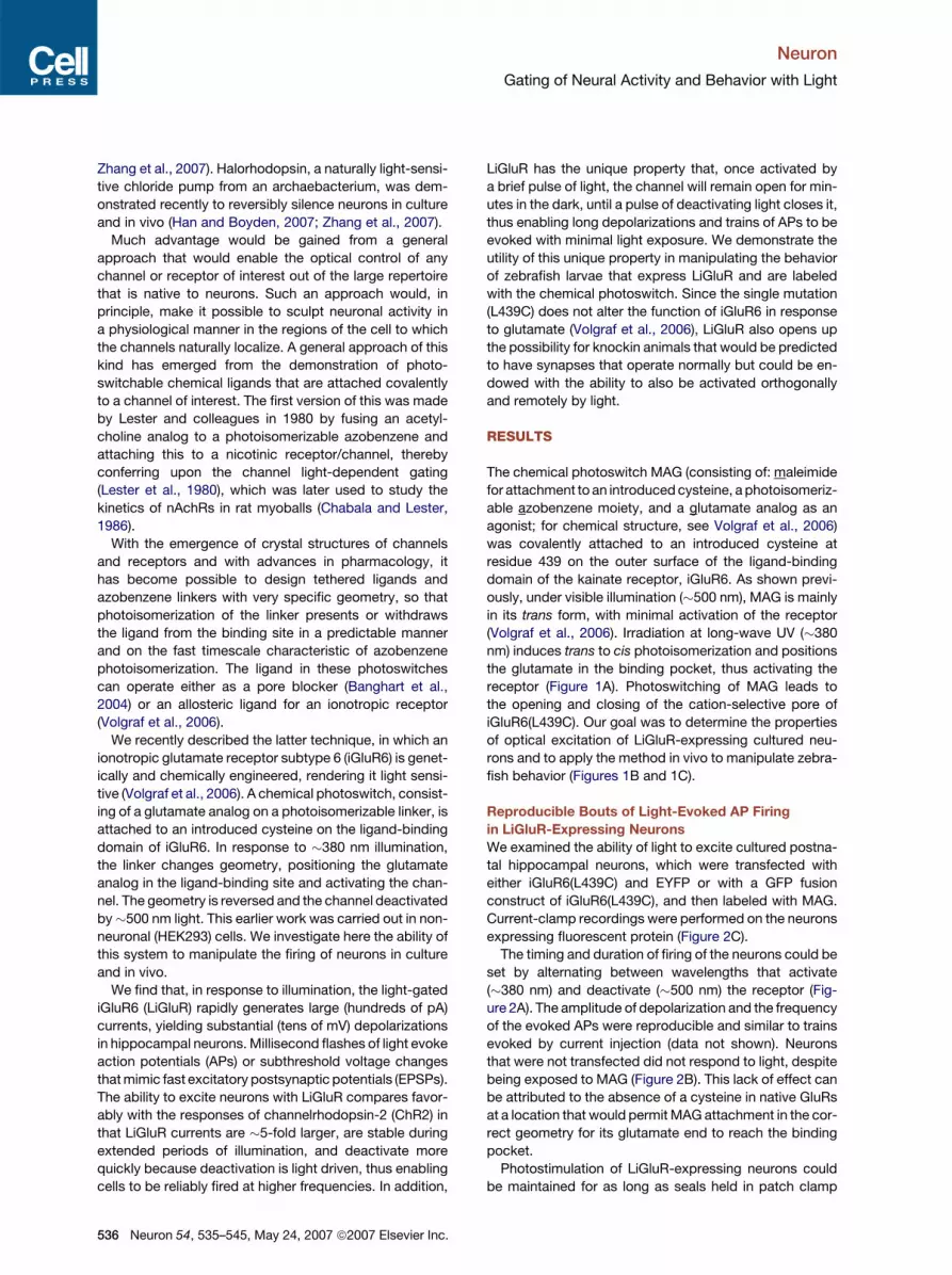
Zhang et al., 2007). Halorhodopsin, a naturally light-sensi-
tive chloride pump from an archaebacterium, was dem-
onstrated recently to reversibly silence neurons in culture
and in vivo (Han and Boyden, 2007; Zhang et al., 2007).
Much advantage would be gained from a general
approach that would enable the optical control of any
channel or receptor of interest out of the large repertoire
that is native to neurons. Such an approach would, in
principle, make it possible to sculpt neuronal activity in
a physiological manner in the regions of the cell to which
the channels naturally localize. A general approach of this
kind has emerged from the demonstration of photo-
switchable chemical ligands that are attached covalently
to a channel of interest. The first version of this was made
by Lester and colleagues in 1980 by fusing an acetyl-
choline analog to a photoisomerizable azobenzene and
attaching this to a nicotinic receptor/channel, thereby
conferring upon the channel light-dependent gating
(Lester et al., 1980), which was later used to study the
kinetics of nAchRs in rat myoballs (Chabala and Lester,
1986).
With the emergence of crystal structures of channels
and receptors and with advances in pharmacology, it
has become possible to design tethered ligands and
azobenzene linkers with very specific geometry, so that
photoisomerization of the linker presents or withdraws
the ligand from the binding site in a predictable manner
and on the fast timescale characteristic of azobenzene
photoisomerization. The ligand in these photoswitches
can operate either as a pore blocker (Banghart et al.,
2004) or an allosteric ligand for an ionotropic receptor
(Volgraf et al., 2006).
We recently described the latter technique, in which an
ionotropic glutamate receptor subtype 6 (iGluR6) is genet-
ically and chemically engineered, rendering it light sensi-
tive (Volgraf et al., 2006). A chemical photoswitch, consist-
ing of a glutamate analog on a photoisomerizable linker, is
attached to an introduced cysteine on the ligand-binding
domain of iGluR6. In response to �380 nm illumination,
the linker changes geometry, positioning the glutamate
analog in the ligand-binding site and activating the chan-
nel. The geometry is reversed and the channel deactivated
by�500 nm light. This earlier work was carried out in non-
neuronal (HEK293) cells. We investigate here the ability of
this system to manipulate the firing of neurons in culture
and in vivo.
We find that, in response to illumination, the light-gated
iGluR6 (LiGluR) rapidly generates large (hundreds of pA)
currents, yielding substantial (tens of mV) depolarizations
in hippocampal neurons. Millisecond flashes of light evoke
action potentials (APs) or subthreshold voltage changes
that mimic fast excitatory postsynaptic potentials (EPSPs).
The ability to excite neurons with LiGluR compares favor-
ably with the responses of channelrhodopsin-2 (ChR2) in
that LiGluR currents are �5-fold larger, are stable during
extended periods of illumination, and deactivate more
quickly because deactivation is light driven, thus enabling
cells to be reliably fired at higher frequencies. In addition,
536 Neuron 54, 535–545, May 24, 2007 ª2007 Elsevier Inc.
LiGluR has the unique property that, once activated by
a brief pulse of light, the channel will remain open for min-
utes in the dark, until a pulse of deactivating light closes it,
thus enabling long depolarizations and trains of APs to be
evoked with minimal light exposure. We demonstrate the
utility of this unique property in manipulating the behavior
of zebrafish larvae that express LiGluR and are labeled
with the chemical photoswitch. Since the single mutation
(L439C) does not alter the function of iGluR6 in response
to glutamate (Volgraf et al., 2006), LiGluR also opens up
the possibility for knockin animals that would be predicted
to have synapses that operate normally but could be en-
dowed with the ability to also be activated orthogonally
and remotely by light.
RESULTS
The chemical photoswitch MAG (consisting of: maleimide
for attachment to an introduced cysteine, a photoisomeriz-
able azobenzene moiety, and a glutamate analog as an
agonist; for chemical structure, see Volgraf et al., 2006)
was covalently attached to an introduced cysteine at
residue 439 on the outer surface of the ligand-binding
domain of the kainate receptor, iGluR6. As shown previ-
ously, under visible illumination (�500 nm), MAG is mainly
in its trans form, with minimal activation of the receptor
(Volgraf et al., 2006). Irradiation at long-wave UV (�380
nm) induces trans to cis photoisomerization and positions
the glutamate in the binding pocket, thus activating the
receptor (Figure 1A). Photoswitching of MAG leads to
the opening and closing of the cation-selective pore of
iGluR6(L439C). Our goal was to determine the properties
of optical excitation of LiGluR-expressing cultured neu-
rons and to apply the method in vivo to manipulate zebra-
fish behavior (Figures 1B and 1C).
Reproducible Bouts of Light-Evoked AP Firing
in LiGluR-Expressing Neurons
We examined the ability of light to excite cultured postna-
tal hippocampal neurons, which were transfected with
either iGluR6(L439C) and EYFP or with a GFP fusion
construct of iGluR6(L439C), and then labeled with MAG.
Current-clamp recordings were performed on the neurons
expressing fluorescent protein (Figure 2C).
The timing and duration of firing of the neurons could be
set by alternating between wavelengths that activate
(�380 nm) and deactivate (�500 nm) the receptor (Fig-
ure 2A). The amplitude of depolarization and the frequency
of the evoked APs were reproducible and similar to trains
evoked by current injection (data not shown). Neurons
that were not transfected did not respond to light, despite
being exposed to MAG (Figure 2B). This lack of effect can
be attributed to the absence of a cysteine in native GluRs
at a location that would permit MAG attachment in the cor-
rect geometry for its glutamate end to reach the binding
pocket.
Photostimulation of LiGluR-expressing neurons could
be maintained for as long as seals held in patch clamp

Neuron
Gating of Neural Activity and Behavior with Light
Figure 1. Study of Neuronal Circuits with
Optical Switches
(A) The light-gated glutamate receptor is based
on the reversible photoisomerization of the
tethered agonist maleimide-azobenzene-glu-
tamate (MAG) between its trans configuration
under 500 nm light and its cis configuration
under 380 nm light. MAG is covalently attached
by the maleimide moiety to a cysteine intro-
duced into the ligand-binding domain (LBD) of
the receptor. Photoswitching is driven by the
reversible binding of the glutamate moiety of
MAG, which is presented to the ligand-binding
site in the cis configuration and withdrawn in
trans. MAG binding under �380 nm light acti-
vates the receptor and opens its cation-selec-
tive channel, resulting in membrane depolar-
ization. TMD, transmembrane domain; C, C
terminus; N, N terminus.
(B) In combination with calcium- or voltage-
sensitive dyes to measure neuronal activity,
the light-gated glutamate receptor can be
used as a remote, noninvasive neuronal switch for the electrode-free, all-optical analysis of neural circuits.
(C) When expressed in vivo, the light-gated glutamate receptor provides a tool for behavioral studies and circuit analysis. In the zebrafish larva,
a touch-evoked escape response is assayed following activation of LiGluR (violet) or deactivation (blue).
(up to 45 min), without any indication of toxicity due to
illumination or MAG exposure. Cultured hippocampal
neurons were often patch-clamped 2 or more hours after
MAG conjugation, indicating that MAG is not toxic over
a short period of time. We also examined neuronal sur-
vival following 12 hr of continuous exposure to several
concentrations of MAG. This is much longer than the stan-
dard 15 min labeling time that we employed for our
recordings. Staining for dead cells using a Live-Dead via-
bility/cytotoxicity assay (Molecular Probes, kit L-7013),
we found there to be no difference in cell death between
neurons exposed to MAG and controls that were cultured
in parallel (Figure 2D). This is consistent with our earlier
observation that a model of MAG, which contains the
(2S, 4R)-4-substituted glutamate and a linker resembling
half of the azobenzene tether, has an apparent affinity of
180 mM (Volgraf et al., 2006). Thus, the typical labeling
concentration of 10 mM MAG will activate iGluR6 only min-
imally. Activation of other iGluRs will be minimal because
similarly substituted glutamate analogs have been shown
to be selective kainate receptor agonists (Pedregal et al.,
2000).
Figure 2. Photostimulation Yields Repro-
ducible Trains of AP Firing
(A) A neuron transfected with iGluR6(L439C)
and labeled with MAG is illuminated at 380
nm for 500 ms, yielding reproducible depolar-
izations that trigger trains of APs, which fire at
a frequency that is characteristic of the cell.
Illumination at 500 nm turns the response off
and permits repolarization. (Illumination was
with a monochromator at low intensity.)
(B) Untransfected neuron has no response to
light, despite exposure to MAG, but does fire
repetitively in response to current injection.
(C) Hippocampal neurons transfected with
iGluR6(L439C)-GFP are easily identified by
fluorescence.
(D) MAG has no deleterious effect on neurons.
Untransfected neurons incubated for 12 hr in
MAG (as opposed to standard 15 min labeling)
show no increase in cell death compared to
control.
Neuron 54, 535–545, May 24, 2007 ª2007 Elsevier Inc. 537

Neuron
Gating of Neural Activity and Behavior with Light
Figure 3. Designed Temporal Firing
Patterns
Millisecond-timescale pulses of laser illumina-
tion are sufficient to significantly depolarize
neurons and to trigger APs. Scale bars, 40
mV and 100 ms. In all panels, arrows indicate
timing but not duration of light pulses.
(A) A train of 1 ms pulses of 374 nm light
(arrows) reliably triggers the same temporal
pattern of AP firing in a neuron.
(B) Reproducible firing is triggered in two differ-
ent neurons by the same pattern of 374 nm light
pulses (arrows). Light pulses are 1 ms in the
upper trace and 3 ms in the lower trace.
(C) In the same cell, a train of 3 ms pulses of
374 nm light produces APs (top trace) or,
when the illumination intensity is attenuated with neutral density filters, subthreshold EPSP-like responses (bottom trace).
(D) LiGluR can be activated with a brief pulse at 374 nm and deactivated with a brief pulse at 488 nm to fire the neuron, while the interval between APs
is kept dark to minimize irradiation. Light pulses are 2 ms in duration.
Photoswitching in Milliseconds Generates APs
and Mock EPSPs
EPSPs mediated by native iGluRs are triggered by very
brief (millisecond long) and synchronous glutamate
binding events at groups of receptors in postsynaptic
membranes. Ideally, an engineered system for triggering
neuron activity would operate on the same timescale. In-
deed, brief (1–5 ms) pulses of light at 5.5–6 mW/mm2
(from an 8 mW 374 nm diode laser, attenuated with neutral
density filters) evoked currents that triggered reproducible
patterns of APs (Figure 3). Light-evoked patterns of firing
were repeatable within a neuron (Figure 3A) and in differ-
ent neurons (Figure 3B). Furthermore, the amplitude of
the responses could be easily reduced by attenuating illu-
mination intensity using neutral density filters, in order to
induce EPSP-like depolarizations (Figure 3C, lower trace).
Rather than continuously illuminating the cells while
switching back and forth between two wavelengths, we
also evoked patterned AP firing using only brief pairs of
light pulses (374 nm to activate, followed by 488 nm to
deactivate) while otherwise keeping the cell in the dark
(Figure 3D).
Because rapid stimulation of neurons is often employed
in studies of synaptic plasticity, we were interested in de-
termining the response of neurons to light pulses delivered
at high frequencies. We found that APs followed optical
stimulation reliably up to 50 Hz (Figures 4A and 5A), higher
than the frequency reported for ChR2 under similar culture
conditions (Boyden et al., 2005; Li et al., 2005). We attri-
bute this performance to the fact that LiGluR evokes larger
currents of consistent amplitude and undergoes faster,
light-driven deactivation (see the Supplemental Data and
Figure S1 available with this article online). While perfect
correlation between light stimulus and action potential fir-
ing is less likely at frequencies above 50 Hz, we found that
subthreshold depolarizations were reliably evoked up to
the highest frequency tested of 100 Hz (Figure 4B), demon-
strating that the kinetics of light-gating are quite rapid. We
found that other measures of fidelity, such as excess APs,
latency between stimulus and AP, and jitter (Figure 5B–D),
538 Neuron 54, 535–545, May 24, 2007 ª2007 Elsevier Inc.
were on par with, or better than, reported values for gluta-
mate photouncaging (Shoham et al., 2005; Yoshimura
et al., 2005) and ChR2 (Boyden et al., 2005).
Light Evokes Depolarization and AP Firing
in a Wavelength-Dependent Manner
To characterize the amplitude of depolarization evoked
by illumination at different wavelengths, we examined
iGluR6(L439C) in HEK293 cells labeled with MAG under
whole-cell current clamp. Light-induced channel opening
evoked large steady-state depolarizations. By taking ad-
vantage of the fact that the photostationary state of MAG
(i.e., the relative proportion of azobenzene in cis and trans
Figure 4. Hippocampal Neurons Follow Pulsed Photostimu-
lation of LiGluR Even at High Frequencies
(A) Trains of 5 ms laser pulses at 374 nm reproducibly trigger APs up to
50 Hz. At 75 Hz, 12 APs are evoked by 20 stimuli, and at 86 Hz, 11 APs
are evoked.
(B) Subthreshold depolarizations are reproducible at very high fre-
quencies (in this case, 100 Hz) because both channel activation and
deactivation are light driven.

Neuron
Gating of Neural Activity and Behavior with Light
Figure 5. Fidelity of Neuron Firing
Neurons are stimulated 20 times by 5 ms
pulses of 374 nm light. Values in all panels are
mean ± SEM. In some cases, error bars are
smaller than the symbols.
(A) Stimuli delivered up to 30 Hz reliably evoke
APs. At higher frequencies, fewer APs are
evoked, with substantial drop-off at >60 Hz.
(B) No excess APs occurred during stimulation
at the frequencies tested. (Excess APs are de-
fined as more than one AP per stimulus, or those
that occur more than 30 ms after the stimulus.)
(C) Latency between stimulus onset and AP
peak increases with higher-frequency stimula-
tion and may account for the fewer APs
produced at high frequencies in (A).
(D) The latencies within a train of 20 stimuli vary
more at higher frequencies. (Jitter is the stan-
dard deviation of the latencies.)
configurations) can be precisely varied by illumination
wavelength, it was possible to produce steady-state de-
polarizations whose amplitudes depended on wavelength
(Figure 6A). Similar graded depolarizations could be
evoked in cultured postnatal hippocampal neurons that
were transfected with iGluR6(L439C) and exposed to
MAG. As seen in the HEK cells, the amplitude of depolar-
ization depended on wavelength, with a maximum at
�380 nm. The largest depolarizations evoked trains of
APs (Figure 6B). We used the wavelength dependence
to adjust the size of EPSP-like waveforms that were trig-
gered by brief pulses of light, so that, for example, pulses
of light at 380 nm generated superthreshold depolariza-
tions and evoked APs, while EPSP-like responses were
induced in the same cell by pulses of light of the same
duration but at the off-peak wavelength of 430 nm (Fig-
ure 6C). Thus, the amplitude of brief excitatory events
evoked by pulses of light can be controlled either by mod-
ifying the intensity of illumination at 380 nm (Figure 3C)
or by adjusting wavelength.
Protracted Excitation in the Dark
We find that our azobenzene photoswitch is robust, yield-
ing reproducible responses for tens of minutes in hippo-
campal neurons under continuous illumination, alternating
between 380 nm and 500 nm at intensities of 5 mW/mm2
or more. These recordings typically ended only upon loss
of the seal, and recordings were equally stable with and
without illumination. However, behavioral experiments
may require activity to be manipulated over a much longer
timescale, where photodestruction of MAG or phototoxic-
ity to cells could become a concern. To reduce this prob-
lem, we explored the property of thermal bistability of
MAG in an attempt to generate sustained trains of firing
in the dark.
Depending on how azobenzene is derivatized, its
higher-energy cis configuration is stable for seconds to
minutes in the dark (Pozhidaeva et al., 2004). For MAG,
the half-life is 17.65 ± 0.03 min (Gorostiza et al., 2007).
Thus, depolarization induced by a brief pulse of 374 nm
light is followed by sustained excitation in an ensuing pe-
riod during which there is no illumination (Figure 7). This
sustained excitation in the dark can then be rapidly
Figure 6. Wavelength-Dependent Depolarization
(A) In current-clamped HEK cells expressing LiGluR, light induces
depolarization in which amplitude depends on illumination wave-
length. In this way, the membrane potential can be accurately and
stably controlled across a wide range.
(B) Illumination at a range of wavelengths depolarizes neurons. Some
depolarizations are large enough to reach threshold and trigger APs.
(C) Light-pulse stimulation with 380 nm light evokes APs while 430 nm
light induces subthreshold, EPSP-like responses in the same cell.
Neuron 54, 535–545, May 24, 2007 ª2007 Elsevier Inc. 539
Neuron
Gating of Neural Activity and Behavior with Light
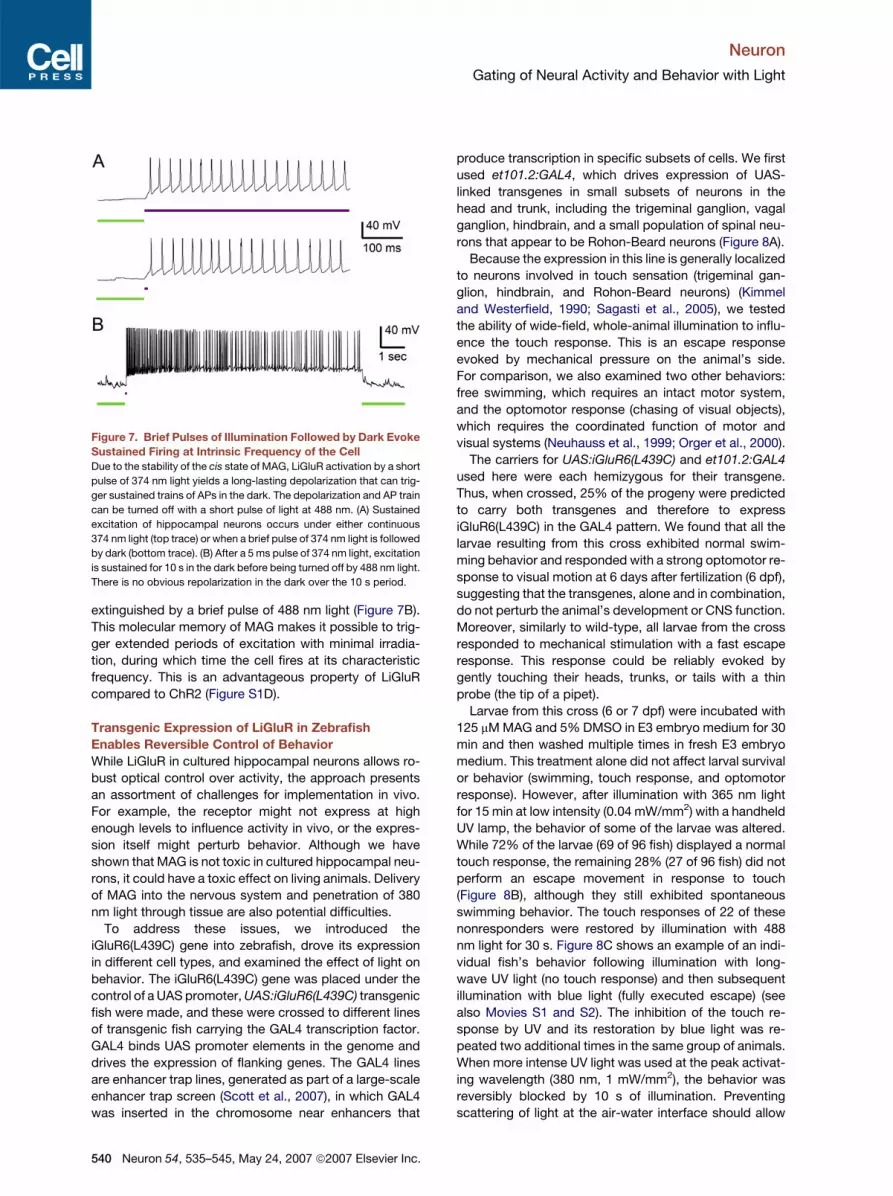
extinguished by a brief pulse of 488 nm light (Figure 7B).
This molecular memory of MAG makes it possible to trig-
ger extended periods of excitation with minimal irradia-
tion, during which time the cell fires at its characteristic
frequency. This is an advantageous property of LiGluR
compared to ChR2 (Figure S1D).
Transgenic Expression of LiGluR in Zebrafish
Enables Reversible Control of Behavior
While LiGluR in cultured hippocampal neurons allows ro-
bust optical control over activity, the approach presents
an assortment of challenges for implementation in vivo.
For example, the receptor might not express at high
enough levels to influence activity in vivo, or the expres-
sion itself might perturb behavior. Although we have
shown that MAG is not toxic in cultured hippocampal neu-
rons, it could have a toxic effect on living animals. Delivery
of MAG into the nervous system and penetration of 380
nm light through tissue are also potential difficulties.
To address these issues, we introduced the
iGluR6(L439C) gene into zebrafish, drove its expression
in different cell types, and examined the effect of light on
behavior. The iGluR6(L439C) gene was placed under the
control of a UAS promoter, UAS:iGluR6(L439C) transgenic
fish were made, and these were crossed to different lines
of transgenic fish carrying the GAL4 transcription factor.
GAL4 binds UAS promoter elements in the genome and
drives the expression of flanking genes. The GAL4 lines
are enhancer trap lines, generated as part of a large-scale
enhancer trap screen (Scott et al., 2007), in which GAL4
was inserted in the chromosome near enhancers that
Figure 7. Brief Pulses of Illumination Followed by Dark Evoke
Sustained Firing at Intrinsic Frequency of the Cell
Due to the stability of the cis state of MAG, LiGluR activation by a short
pulse of 374 nm light yields a long-lasting depolarization that can trig-
ger sustained trains of APs in the dark. The depolarization and AP train
can be turned off with a short pulse of light at 488 nm. (A) Sustained
excitation of hippocampal neurons occurs under either continuous
374 nm light (top trace) or when a brief pulse of 374 nm light is followed
by dark (bottom trace). (B) After a 5 ms pulse of 374 nm light, excitation
is sustained for 10 s in the dark before being turned off by 488 nm light.
There is no obvious repolarization in the dark over the 10 s period.
540 Neuron 54, 535–545, May 24, 2007 ª2007 Elsevier Inc.
produce transcription in specific subsets of cells. We first
used et101.2:GAL4, which drives expression of UAS-
linked transgenes in small subsets of neurons in the
head and trunk, including the trigeminal ganglion, vagal
ganglion, hindbrain, and a small population of spinal neu-
rons that appear to be Rohon-Beard neurons (Figure 8A).
Because the expression in this line is generally localized
to neurons involved in touch sensation (trigeminal gan-
glion, hindbrain, and Rohon-Beard neurons) (Kimmel
and Westerfield, 1990; Sagasti et al., 2005), we tested
the ability of wide-field, whole-animal illumination to influ-
ence the touch response. This is an escape response
evoked by mechanical pressure on the animal’s side.
For comparison, we also examined two other behaviors:
free swimming, which requires an intact motor system,
and the optomotor response (chasing of visual objects),
which requires the coordinated function of motor and
visual systems (Neuhauss et al., 1999; Orger et al., 2000).
The carriers for UAS:iGluR6(L439C) and et101.2:GAL4
used here were each hemizygous for their transgene.
Thus, when crossed, 25% of the progeny were predicted
to carry both transgenes and therefore to express
iGluR6(L439C) in the GAL4 pattern. We found that all the
larvae resulting from this cross exhibited normal swim-
ming behavior and responded with a strong optomotor re-
sponse to visual motion at 6 days after fertilization (6 dpf),
suggesting that the transgenes, alone and in combination,
do not perturb the animal’s development or CNS function.
Moreover, similarly to wild-type, all larvae from the cross
responded to mechanical stimulation with a fast escape
response. This response could be reliably evoked by
gently touching their heads, trunks, or tails with a thin
probe (the tip of a pipet).
Larvae from this cross (6 or 7 dpf) were incubated with
125 mM MAG and 5% DMSO in E3 embryo medium for 30
min and then washed multiple times in fresh E3 embryo
medium. This treatment alone did not affect larval survival
or behavior (swimming, touch response, and optomotor
response). However, after illumination with 365 nm light
for 15 min at low intensity (0.04 mW/mm2) with a handheld
UV lamp, the behavior of some of the larvae was altered.
While 72% of the larvae (69 of 96 fish) displayed a normal
touch response, the remaining 28% (27 of 96 fish) did not
perform an escape movement in response to touch
(Figure 8B), although they still exhibited spontaneous
swimming behavior. The touch responses of 22 of these
nonresponders were restored by illumination with 488
nm light for 30 s. Figure 8C shows an example of an indi-
vidual fish’s behavior following illumination with long-
wave UV light (no touch response) and then subsequent
illumination with blue light (fully executed escape) (see
also Movies S1 and S2). The inhibition of the touch re-
sponse by UV and its restoration by blue light was re-
peated two additional times in the same group of animals.
When more intense UV light was used at the peak activat-
ing wavelength (380 nm, 1 mW/mm2), the behavior was
reversibly blocked by 10 s of illumination. Preventing
scattering of light at the air-water interface should allow

Neuron
Gating of Neural Activity and Behavior with Light
Figure 8. Activation of LiGluR in the et101.2 Expression Pattern of Zebrafish Larvae Reversibly Blocks the Touch Response
(A) Lateral views of the head (left panel) and trunk (right panel) showing the pattern of expression of Kaede in larvae (7 dpf) from a cross of UAS:Kaede
and et101.2:GAL4. In the head, expression is in the trigeminal ganglion (yellow triangle), vagal ganglion (white triangle), and hindbrain (red arrow).
Expression is also scattered in the mouth (blue triangle) and heart (asterisk). In the trunk, expression is in a small set of dorsal neurons in the spinal
cord, which appear to be Rohon-Beard neurons.
(B) Statistics of light response in 96 larvae show that�25% lose the touch response following 365 nm light. The majority of larvae (25 out of 27) that lost
the touch response (TR�) following 365 nm light were subsequently found to express both iGluR6(L439C) and GAL4 (magenta). Larvae that did not
express both the GAL4 driver and iGluR6(L439C) (teal) were overwhelmingly insensitive to 365 nm light (TR+). Some larvae (16 out of 69) were positive
for both the GAL4 driver and iGluR6(L439C) but retained the touch response following 365 nm light, suggesting some variability in expression, MAG
labeling, or illumination.
(C) Representative zebrafish larva expressing both UAS:iGluR6(L439C) and et101.2:GAL4. The response to touch is lost following illumination at
365 nm (top) and regained after illumination at 488 nm (bottom).
even greater speeds of switching behavior at this light
intensity.
Since the behavioral assays were carried out on all an-
imals resulting from the cross, this functioned as a blind
analysis. Genotyping was done following the behavioral
experiment to determine the correspondence between
expression of iGluR6(L439C) and light-altered behavior.
The genotyping revealed that the majority of the light-
sensitive larval fish (25 of 27) were in fact doubly trans-
genic for et101.2:GAL4 and UAS:iGluR6(L439C) (Fig-
ure 8B). Thus, 92.5% of the light-sensitive fish expressed
iGluR6(L439C) in the et101.2 pattern.
Reversible disruption of touch response was observed
for touch to both the head and the trunk (Figure 8C and
Movie S3), consistent with expression in trigeminal
ganglion neurons, hindbrain, and Rohon-Beard neurons
(Kimmel and Westerfield, 1990; Liu and Fetcho, 1999;
Sagasti et al., 2005).
To confirm that the light-evoked perturbation mediated
by iGluR6(L439C) is specific to the neural circuit in which it
is expressed, we also crossed UAS:iGluR6(L439C) with
three other GAL4 lines that drive expression in different
sets of cells. The line et101.1:GAL4 expresses broadly
throughout the nervous system, as well as in the heart
(Figure S2A), and illumination with 365 nm light resulted
in loss of the touch response and also total paralysis, as
seen by loss of swimming behavior. This occurred in
26% of the larvae (9 out of 34), consistent with the ex-
pectation that �25% will carry UAS:iGluR6(L439C) and
et101.1:GAL4. Another line, et101.4:GAL4, which drives
expression exclusively in the heart (Figure S2B), had no
perturbation of swimming and touch responses after illu-
mination at 365 nm (0 out of 32 fish). Finally, we examined
the cross of UAS:iGluR6(L439C) with the Ath5:GAL4;
UAS:Kaede promoter fusion, which expresses only in
retinal ganglion cells (Masai et al., 2005; see Figure S2C).
Among the progeny expressing the fluorescent marker
Kaede [�50% of which also are predicted to carry the
UAS:iGluR6(L439C) transgene], 365 nm light had no effect
on the touch response (0 out of 15 fish).
Neuron 54, 535–545, May 24, 2007 ª2007 Elsevier Inc. 541
Neuron
Gating of Neural Activity and Behavior with Light
These experiments show that it is possible to use light
to reliably, reproducibly, and reversibly manipulate the
activity of LiGluR-expressing neurons in vivo and thereby
to modify zebrafish behavior. The nature of the behavioral
manipulation depends on the identity of the LiGluR-
expressing neurons, making it possible to dissect the
role of neurons and neural circuits in behavior. The degree
of specificity of such analysis will grow as new enhancer
trap GAL4 lines and promoter-GAL4 fusion lines are
developed.
DISCUSSION
Our engineered receptor, LiGluR, consists of an ionotropic
glutamate receptor (iGluR6) with an introduced cysteine
residue (L439C) for covalent attachment of a chemical
photoswitch (MAG). In response to light, LiGluR generates
large, stable currents in hippocampal neurons, providing
robust optical control over neuron depolarization and AP
firing. Individual APs are driven by 1–5 ms pulses of light,
which is on the physiological timescale of synaptic activa-
tion of glutamate receptors. Thus, millisecond-long
flashes of light can drive designed temporal trains of single
APs, while longer periods of illumination cause the cell to
fire at its characteristic frequency.
Because the MAG photoswitch is covalently attached to
the receptor, excitation is confined to the illumination vol-
ume, providing spatial resolution in which individual cells
(and potentially specific regions within a given cell) can
be selectively stimulated. Light-activation and cell viability
are maintained during experiments lasting several hours,
and a 12 hr incubation of neurons with MAG shows no
sign of toxicity to cells. Importantly, exposure to MAG
does not affect hippocampal neurons that are not ex-
pressing LiGluR. There are several reasons for this speci-
ficity. First, native receptors lack a point of attachment for
MAG that is close enough (1–2 nm) to the binding pocket.
Second, the agonist moiety of MAG is similar to kainate-
selective agonists, and so free MAG should be less likely
to activate AMPA receptors and NMDA receptors. Even
kainate receptors will not be significantly activated by free
MAG at the concentration we used for labeling (10 mM),
which is 18-fold lower than the EC50 of a similar tether
model on iGluR6 (Volgraf et al., 2006). The only neurons
that become responsive to light are ones that express
iGluR6(L439C) and are exposed to MAG. While, in our
experiments, cultured neurons were transfected ran-
domly, it should be possible to drive the expression of
iGluR6(L439C) in select neurons by the use of cell-type-
specific promoters. Since LiGluR retains the ability to be
activated by free glutamate, it could be used in knockin
animals to gain orthogonal control over the native gluta-
mate receptors.
LiGluR has several favorable properties that lend them-
selves to tight control over remote excitation. LiGluR gen-
erates steady currents, resulting in stable depolarizations
that can fire neurons at constant rates. By illuminating
with different wavelengths of light, it is possible to evoke
542 Neuron 54, 535–545, May 24, 2007 ª2007 Elsevier Inc.
either small or large depolarizations. Because LiGluR is
both activated and deactivated by light, the timing of ex-
citation can be precisely defined. This fast bidirectional
switching makes it possible to fire neurons reliably at
high frequencies. In addition, because azobenzene is bi-
stable and will reside for minutes in the higher-energy
cis state, which activates the channel, a brief pulse of light
can trigger a large depolarization that lasts for an ex-
tended period of time in the dark until deactivation is trig-
gered by illumination at the longer wavelength. This
means that long trains of firing can be evoked with LiGluR
by brief pulses of light, minimizing illumination time and
possible photodamage to cells. Because the half-life of
spontaneous (thermal) isomerization of cis (activated)
back to trans (deactivated) is �18 min, very long bouts
of firing are possible. This could be particularly useful
for many types of behavioral experiments and in fact
was essential to our touch-response experiments in ze-
brafish larvae, because their behavior is easily perturbed
by both UV and visible light (Ren et al., 2002; Risner
et al., 2006). In this way, we were able to activate LiGluR
with 365 nm light and then apply the touch stimulus
several minutes later, while observing behavior, under
low-intensity ambient light conditions.
Our transgenic zebrafish larvae do not show any sign of
problems with MAG delivery, toxicity, or interference with
native circuitry. The fish larvae behave normally both
before and after incubation with MAG. Expression of
iGluR6(L439C) in sensory neurons is robust, and the ex-
pression itself does not appear to alter behavior. While
the duration of UV illumination and concentrations of
DMSO and MAG were not tested on zebrafish larvae at
lower values, the exposure levels that were used did not
appear to cause any complications.
Optical activation of LiGluR in specific neurons in zebra-
fish larvae is capable of preventing an escape reflex when
the fish are touched with a pipet tip along the head or
trunk. While trigeminal ganglion neurons in the head and
Rohon-Beard neurons in the trunk have been implicated
in touch-evoked escape (Sagasti et al., 2005; Cox and
Fetcho, 1996), we test their function, using genetically
targeted, reversible manipulation of neuronal activity.
The larvae become unresponsive to touch, possibly be-
cause illumination of the whole animal simultaneously ac-
tivates the sensory portion of the escape circuitry on both
sides of the animal, which might interfere with making a
directed turn away from the local stimulus. Alternatively,
the larvae may become unresponsive to touch because
the local stimulus cannot be detected over the elevated
activity in the sensory cells or because of habituation after
prolonged activation (>15 min). Further studies are needed
to deduce the exact mechanism.
In conclusion, we have shown that a simple approach of
chemically re-engineering a receptor can endow it with the
ability to be controlled by light in a selective manner, even
in the complex environment of a neuron and in the sensory
circuitry of live zebrafish larvae. We have used the
approach to optically control AP firing with a light-gated

Neuron
Gating of Neural Activity and Behavior with Light
excitatory ionotropic receptor, but in principle it should be
possible to extend the approach to inhibitory ionotropic
receptors and to other classes of neuronal membrane
proteins, such as adhesion proteins, growth factor recep-
tors, and enzymes, as long as a tethered ligand is able to
modulate function. Thus, neural activity can be controlled
with light in a way that uses native proteins and closely
mimics native activity.
EXPERIMENTAL PROCEDURES
Photoswitch Synthesis and Generation of L439C Mutant
of iGluR6
Synthesis of MAG and introduction of cysteine L439C in iGluR6 were
carried out as described (Volgraf et al., 2006). Amino acid residues
are numbered from the starting methionine.
Cell Culture and Transfection
Dissociated postnatal rat hippocampal neurons (P0–P5) were plated
on poly-L-lysine-coated glass coverslips at a density of 1 3 105 cells
per 12 mm coverslip. Cells were cultured in MEM supplemented with
5% fetal bovine serum, B27 (Invitrogen), GlutaMAX (Invitrogen), and
serum extender (BD Biosciences). Ara-C (4 mM) was added after
4DIV. Cells were transfected by the calcium phosphate method using
0.8 mg of DNA per coverslip. Cells were either transfected with a
fusion construct of GFP and iGluR6(L439C) or cotransfected with
iGluR6(L439C) and EYFP at a 3:1 ratio.
HEK293 cells were plated at approximately 6 3 106 cells per cover-
slip on 25 mm poly-L-lysine-coated glass coverslips and maintained
in DMEM with 5% fetal bovine serum, 0.2 mg/mL streptomycin, and
200 U/mL penicillin. Cells were cotransfected with 4 mg iGluR6(L439C)
and 200 ng EYFP using lipofectamine 2000 (Invitrogen).
Conjugation of MAG Compounds In Vitro
To conjugate MAG to iGluR6(L439C) in hippocampal neurons for
patch-clamping experiments, the compound was diluted to 10 mM in
a solution containing (in mM) 150 NMDG-HCl, 3 KCl, 0.5 CaCl2,
5 MgCl2, 10 HEPES, and 5 glucose, pH 7.4. For HEK cells, MAG was
diluted to 10 mM in a solution of (in mM) 135 NaCl, 5.4 KCl, 0.9 MgCl2,
1.8 CaCl2, 10 HEPES, and 0.3 mg/mL Concanavalin A type VI (Sigma),
pH 7.6. The cells were incubated with MAG in the dark for 10–15 min
and then rinsed in extracellular recording solution (described below).
Whole-Cell Patch-Clamping
Patch-clamp recordings used an Axopatch 200A amplifier in the
whole-cell mode. Recordings were carried out 1–8 days after transfec-
tion in hippocampal neurons and 36–48 hr after transfection in HEK
cells. Cells were current-clamped or voltage-clamped at about �65
mV. Pipettes had resistances of 2–5 MU and were filled with a solution
containing, for neurons (in mM), 135 K-gluconate, 10 NaCl, 10 HEPES,
2 MgCl2, 2 MgATP, 1 EGTA, pH 7.4; and for HEK cells (in mM), 145
CsCl, 5 EGTA, 0.5 CaCl2, 1.0 MgCl2, and 10 HEPES, pH 7.2. The extra-
cellular recording solution for hippocampal neurons was (in mM)
138 NaCl, 1.5 KCl, 1.2 MgCl2, 2.5 CaCl2, 10 glucose, and 5 HEPES,
pH 7.4; and for HEK cells was (in mM) 135 NaCl, 5.4 KCl, 0.9 MgCl2,
1.8 CaCl2, and 10 HEPES, pH 7.6.
Illumination was applied using a TILL Photonics Polychrome mono-
chromator through the side port of the IX70 inverted microscope of the
physiology rig (Olympus) and using either a 403 or 603 objective. Fast
photoswitching experiments were carried out with a custom shuttered
laser illumination setup mounted on a large breadboard. A 488 nm,
20 mW argon-ion laser (Laser Innovations) and a 374 nm, 8 mW Cube
laser (Coherent) were combined with a dichroic mirror z405RDC
(Chroma) and coupled into a P600-2-UV/vis optical fiber (Ocean
Optics), using a 103, 0.25 NA, 16.5 mm WD objective (Newport). The
fiber was connected into the IX70 microscope (Olympus) through the
Laser B port using a custom-made adaptor. Fast shutters (Uniblitz
UHS1T2-100 driven by VMM-T1 controllers, Vincent Associates)
were placed at the output of each laser to control the illumination
pulses via software trigger. Light intensity measured at the 403 or
603 objective was 5.5–6 mW/mm2.
Electrophysiological data were recorded with pClamp software,
which was also used to automatically control the monochromator
and laser shutters by means of digital signals and sequencing keys.
Generation of Zebrafish Transgenic Lines
To make the UAS:iGluR6(L439C) transgenic construct, the
iGluR6(L439C) open reading frame was amplified by PCR and inserted
downstream of the E1b promoter in a 14xUAS:eGFP construct (Koster
and Fraser, 2001) and upstream of a SV40 polyadenylation sequence.
This expression cassette was inserted between the Tol2 recognition
sequences in the pT2KXIGDin vector (Kotani et al., 2006). Wild-type
TL embryos were injected at the one-cell stage with a solution of
25 ng/ml UAS:iGluR6(L439C) DNA, 50 ng/ml transposase mRNA (pre-
pared using the Ambion mMESSAGE mMACHINE T7 kit), and 0.04%
Phenol Red.
F1 embryos were pooled and screened by PCR for the transgene:
forward primer (50) ggcttgaggatgggaaatatgg and reverse primer (30 )
gggttgcaagggtgtgggttatacc. F0 founder animals giving rise to UAS:
iGluR6(L439C)-positive offspring were then mated to wild-type TL
fish to create stable lines.
To generate lines et101.2:GAL4, et101.1:GAL4, and et101.4:GAL4,
the heat-shock promoter in hsp(1.5kb):GAL4 (Scott et al., 2007) was
replaced with the insertion of a linker containing an I-SceI meganu-
clease site, a PI-PspI meganuclease site, the basal E1b promoter
(Argenton et al., 1996), and a KpnI restriction site, respectively, from
50 to 30. This e1b:GAL4 enhancer trapping construct was used to gen-
erate expression lines as described (Scott et al., 2007). Successful
enhancer traps were identified by crossing to UAS:Kaede and visual
screening under a dissecting microscope and then outcrossed to
generate stable lines.
Labeling and Illumination of Zebrafish Larvae
MAG was first diluted to 2.5 mM in 40 ml of DMSO and preactivated by
UV light (365 nm) for 1 min. The medium E3 was then added to reach
the final concentration of 125 mM MAG, 5% DMSO. Twenty larvae
were bathed in 800 ml of the labeling solution for 30 min at 28.5�C.
The larvae were then removed from the labeling solution to a well con-
taining fresh E3 and were washed three times. After a 30 min recovery
period, the larvae were examined to confirm that they were all respon-
sive to touch. The larvae were illuminated under a UV lamp (365 nm,
0.04 mW/mm2 for 15 min). All the larvae were tested for touch re-
sponse within 15–20 min. Single larvae were successively transferred
to the center of a 35 mm diameter Petri dish, and their response to
touch was assayed under a Zeiss Lumar fluorescent stereomicro-
scope equipped with a 0.83 magnification objective. If a larva failed
to show a response to repetitive touches with a pipet tip, it was illumi-
nated under blue light (488 nm, 1.6 mW/mm2 for 30 s) through the
objective. The recovery of the touch response was tested within a minute
after blue illumination.
Supplemental Data
The Supplemental Data for this article can be found online at http://
www.neuron.org/cgi/content/full/54/4/535/DC1/.
ACKNOWLEDGMENTS
We are grateful to H. Fiumelli, F. Tombola, H. Lee, and S. Pautot for
help with neuronal cultures and patch-clamping; to A. Johnson
Neuron 54, 535–545, May 24, 2007 ª2007 Elsevier Inc. 543

Neuron
Gating of Neural Activity and Behavior with Light
(Coherent, Inc.) for the loan of a 374 nm diode laser; to R. Staples
(Olympus, Inc.) for help with the laser setup; and to David Raible (Uni-
versity of Washington) for expert advice on the labeling pattern in our
GAL4 zebrafish line. We also thank Georg Nagel for kindly providing
ChR2. This work was supported by predoctoral fellowships from the
National Science Foundation (to S.S.), the ACS Medicinal Chemistry
Division (to M.V.), and UC Berkeley (to O.T.); by postdoctoral fellow-
ships from the Human Frontier Science Program (to F.D.B. and
P.G.), the Nanotechnology Program of the Generalitat de Catalunya
(to P.G.), an OIF Marie Curie Fellowship (to C.W), the NIH (to E.K.S.),
and the Japan Society for the Promotion of Science (to R.N.); and by
grants from the Lawrence Berkeley National Laboratory, the Human
Frontier Science Program, and the National Institutes of Health.
Work in H.B.’s lab was supported by a Sandler Opportunity Award
and a Byers Award (to H.B.).
Received: October 18, 2006
Revised: March 10, 2007
Accepted: May 4, 2007
Published: May 23, 2007
REFERENCES
Arenkiel, B., Peca, J., Davison, I., Feliciano, C., Deisseroth, K., Augus-
tine, G., Ehlers, M., and Feng, G. (2007). In vivo light-induced activation
of neural circuitry in transgenic mice expressing channelrhodopsin-2.
Neuron 54, 205–218.
Argenton, F., Arava, Y., Aronheim, A., and Walker, M.D. (1996). An ac-
tivation domain of the helix-loop-helix transcription factor E2A shows
cell type preference in vivo in microinjected zebra fish embryos. Mol.
Cell. Biol. 16, 1714–1721.
Banghart, M., Borges, K., Isacoff, E., Trauner, D., and Kramer, R.H.
(2004). Light-activated ion channels for remote control of neuronal
firing. Nat. Neurosci. 7, 1381–1386.
Bi, A., Cui, J., Ma, Y.P., Olshevskava, E., Pu, M., Dizhoor, A.M., and
Pan, Z.H. (2006). Ectopic expression of a microbial-type rhodopsin
restores visual responses in mice with photoreceptor degeneration.
Neuron 50, 23–33.
Boyden, E.S., Zhang, F., Bamberg, E., Nagel, G., and Deisseroth, K.
(2005). Millisecond-timescale, genetically targeted optical control of
neural activity. Nat. Neurosci. 8, 1263–1268.
Callaway, E.M., and Katz, L.C. (1993). Photostimulation using caged
glutamate reveals functional circuitry in living brain slices. Proc. Natl.
Acad. Sci. USA 90, 7661–7665.
Chabala, L.D., and Lester, H.A. (1986). Activation of acetylcholine re-
ceptor channels by covalently bound agonists in cultured rat myoballs.
J. Physiol. 379, 83–108.
Cox, K.J., and Fetcho, J.R. (1996). Labeling blastomeres with a calcium
indicator: a noninvasive method of visualizing neuronal activity in
zebrafish. J. Neurosci. Methods 68, 185–191.
Gillespie, D.C., Kim, G., and Kandler, K. (2005). Caged neurotransmit-
ters for probing neuronal circuits, neuronal integration and synaptic
activity. In Dynamic Studies in Biology, M. Goeldner and R. Givens,
eds. (Weinheim: Wiley VCH), pp. 232–251.
Gorostiza, P., Volgraf, M., Numano, R., Szobota, S., Trauner, D.,
and Isacoff, E.Y. (2007). Engineering a channel protein for remote
control: mechanisms of photoswitch conjugation and light activation
of an ionotropic glutamate receptor. Proc. Natl. Acad. Sci. USA,
in press.
Han, X., and Boyden, E.S. (2007). Multiple-color optical activation,
silencing, and desynchronization of neural activity, with single-spike
temporal resolution. PLoS ONE 2, e299. 10.1371/journal.pone.
0000299.
544 Neuron 54, 535–545, May 24, 2007 ª2007 Elsevier Inc.
Ishizuka, T., Kakuda, M., Araki, R., and Yawo, H. (2006). Kinetic eval-
uation of photosensitivity in genetically engineered neurons express-
ing green algae light-gated channels. Neurosci. Res. 54, 85–94.
Kimmel, C.B., and Westerfield, M. (1990). Primary neurons of the
zebrafish. In Signals and Sense, W. Gowan, ed. (New York: Wiley
Liss), pp. 561–588.
Koster, R.W., and Fraser, S.E. (2001). Tracing transgene expression in
living zebrafish embryos. Dev. Biol. 233, 329–346.
Kotani, T., Nagayoshi, S., Urasaki, A., and Kawakami, K. (2006). Trans-
poson-mediated gene trapping in zebrafish. Methods 39, 199–206.
Lester, H.A., Krouse, M.E., Nass, M.M., Wassermann, N.H., and Er-
langer, B.F. (1980). A covalently bound photoisomerizable agonist:
comparison with reversibly bound agonists at Electrophorus electro-
plaques. J. Gen. Physiol. 75, 207–232.
Li, X., Gutierrez, D.V., Hanson, M.G., Han, J., Mark, M.D., Chiel, H., He-
gemann, P., Landmesser, L.T., and Herlitze, S. (2005). Fast noninva-
sive activation and inhibition of neural and network activity by verte-
brate rhodopsin and green algae channelrhodopsin. Proc. Natl.
Acad. Sci. USA 102, 17816–17821.
Lima, S.Q., and Miesenbock, G. (2005). Remote control of behavior
through genetically targeted photostimulation of neurons. Cell 121,
141–152.
Liu, K.S., and Fetcho, J.R. (1999). Laser ablations reveal functional
relationships of segmental hindbrain neurons in zebrafish. Neuron 23,
325–335.
Masai, I., Yamaguchi, M., Tonou-Fujimori, N., Komori, A., and Oka-
mato, H. (2005). The hedgehog-PKA pathway regulates two distinct
steps of the differentiation of retinal ganglion cells: the cell-cycle exit
of retinoblasts and their neuronal maturation. Development 132,
1539–1553.
Melyan, Z., Tarttelin, E.E., Bellingham, J., Lucas, R.J., and Hankins,
M.W. (2005). Addition of human melanopsin renders mammalian cells
photoresponsive. Nature 433, 741–745.
Nagel, G., Szellas, T., Huhn, W., Kateriva, S., Adeishvili, N., Berthold,
P., Ollig, D., Hegemann, P., and Bamberg, E. (2003). Channelrhodop-
sin-2, a directly light-gated cation-selective membrane channel. Proc.
Natl. Acad. Sci. USA 100, 13940–13945.
Nagel, G., Brauner, M., Liewald, J.F., Adeishvili, N., Bamberg, E., and
Gottschalk, A. (2005). Light activation of channelrhodopsin-2 in excit-
able cells of Caenorhabditis elegans triggers rapid behavioral re-
sponses. Curr. Biol. 15, 2279–2284.
Neuhauss, S.C., Biehlmaier, O., Seeliger, M.W., Das, T., Kohler, K.,
Harris, W.A., and Baier, H. (1999). Genetic disorders of vision revealed
by a behavioral screen of 400 essential loci in zebrafish. J. Neurosci.
19, 8603–8615.
Orger, M.B., Smear, M.C., Anstis, S.M., and Baier, H. (2000). Percep-
tion of Fourier and non-Fourier motion by larval zebrafish. Nat. Neuro-
sci. 3, 1128–1133.
Pedregal, C., Collado, I., Escribano, A., Ezquerra, J., Dominguez, C.,
Mateo, A.I., Rubio, A., Baker, S.R., Goldsworthy, J., Kamboj, R.K.,
et al. (2000). 4-alkyl- and 4-cinnamylglutamic acid analogues are
potent GluR5 kainate receptor agonists. J. Med. Chem. 43, 1958–
1968.
Petreanu, L., Huber, D., Sobczyk, A., and Svoboda, K. (2007). Chan-
nelrhodopsin-2-assisted circuit mapping of long-range callosal projec-
tions. Nat. Neurosci. 10, 663–668.
Pozhidaeva, N., Cormier, M.E., Chaudhari, A., and Woolley, G.A.
(2004). Reversible photocontrol of peptide helix content: adjusting
thermal stability of the cis state. Bioconjug. Chem. 15, 1297–1303.
Ren, J.Q., McCarthy, W.R., Zhang, H., Adolph, A.R., and Li, L. (2002).
Behavioral visual responses of wild-type and hypopigmented zebra-
fish. Vision Res. 42, 293–299.

Neuron
Gating of Neural Activity and Behavior with Light
Risner, M.L., Lemerise, E., Vukmanic, E.V., and Moore, A. (2006). Be-
havioral spectral sensitivity of the zebrafish (Danio rerio). Vision Res.
46, 2625–2635.
Sagasti, A., Guido, M.R., Raible, D.W., and Schier, A.F. (2005). Repul-
sive interactions shape the morphologies and functional arrangement
of zebrafish peripheral sensory arbors. Curr. Biol. 15, 804–814.
Schroll,C.,Riemensperger,T., Bucher,D.,Ehmer, J., Voller, T.,Erbguth,
K., Gerber, B., Hendel, T., Nagel, G., and Buchner, E. (2006). Light-in-
duced activation of distinct modulatory neurons triggers appetitive or
aversive learning in Drosophila larvae. Curr. Biol. 16, 1741–1747.
Scott, E.K., Mason, L., Arrenberg, A.B., Ziv, L., Gosse, N.J., Xiao, T.,
Chi, N.C., Asakawa, A., Kawakami, K., and Baier, H. (2007). Targeting
neural circuitry in zebrafish using GAL4 enhancer trapping. Nat.
Methods 4, 323–326.
Shoham, S., O’Connor, D.H., Sarkisov, D.V., and Wang, S.S. (2005).
Rapid neurotransmitter uncaging in spatially defined patterns. Nat.
Methods 2, 837–843.
Volgraf, M., Gorostiza, P., Numano, R., Kramer, R.H., Isacoff, E.Y., and
Trauner, D. (2006). Allosteric control of an ionotropic glutamate recep-
tor with an optical switch. Nat. Chem. Biol. 2, 47–52.
Wang, H., Peca, J., Matsuzaki, K., Noguchi, J., Qiu, L., Wang, D.,
Zhang, F., Boyden, E., Deisseroth, K., Kasai, H., et al. (2007). High-
speed mapping of synaptic connectivity using photostimulation in
channelrhodopsin-2 transgenic mice. Proc. Natl. Acad. Sci. USA
104, 8143–8148.
Yoshimura, Y., Dantzker, J.L., and Callaway, E.M. (2005). Excitatory cor-
tical neurons form fine-scale functional networks. Nature 433, 868–873.
Zemelman, B.V., Lee, G.A., Ng, M., and Miesenbock, G. (2002).
Selective photostimulation of genetically chARGed neurons. Neuron
33, 15–22.
Zemelman, B.V., Nesnas, N., Lee, G.A., and Miesenbock, G. (2003).
Photochemical gating of heterologous ion channels: remote control
over genetically designated populations of neurons. Proc. Natl.
Acad. Sci. USA 100, 1352–1357.
Zhang, Y.P., and Oertner, T.G. (2007). Optical induction of synaptic
plasticity using a light-sensitive channel. Nat. Methods 4, 139–141.
Zhang, F., Wang, L.P., Boyden, E.S., and Deisseroth, K. (2006). Chan-
nelrhodopsin-2 and optical control of excitable cells. Nat. Methods 3,
785–792.
Zhang, F., Wang, L.P., Brauner, M., Liewald, J.F., Kay, K., Watzke, N.,
Wood, P.G., Bamberg, E., Nagel, G., Gottschalk, A., and Deisseroth, K.
(2007). Multimodal fast optical interrogation of neural circuitry. Nature
446, 633–639.
Neuron 54, 535–545, May 24, 2007 ª2007 Elsevier Inc. 545