Sex steroid effects on extrahypothalamic CNS. I. Estrogen augments neuronal responsiveness to...
12
40 Brain Research, 422 (1987) 40- 51 Elsevier BRE 12899 Sex steroid effects on extrahypothalamic CNS. I. Estrogen augments neuronal responsiveness to iontophoretically applied glutamate in the cerebellum Sheryl S. Smith, Barry D. Waterhouse and Donald J. Woodward* Department of Physiology and Biophysics, Hahnemann University, Philadelphia, PA 19102-1192 (U.S.A.) (Accepted 24 February 1987) Key words: Estrogen; Cerebellar Purkinje cell; Glutamate; Evoked excitation; Neuromodulation The purpose of this study was to test whether 17fl-estradiol (E2) could alter neuronal activity or responsiveness to iontophoretically applied amino acid neurotransmitters in an area not reported to contain classical E 2 receptors. Such a region is the cerebellum, which was selected as a model system for these studies because it has been well characterized electrophysiologically. Extracellular activity of cerebellar Purkinje neurons was recorded from urethane-anesthetized, adult, ovariectomized rats using multibarrel glass micropipets. Spontaneous firing rate and responses of single units to microiontophoretic pulses (10 s pulses every 40 s) of GABA (10-50 nA) or glu- tamate (GLUT, 3-40 nA) were examined before, during and after iontophoretic (0.25 mM 17fl-estradiol hemisuccinate) or jugular i.v. (100, 300 or 1000 ng/kg 17fl-estradiol) administration of E 2. Both modes of E 2 administration resulted in a significant increase in Purkinje cell excitatory responses to GLUT, independent of the direction of change in spontaneous firing rate. This effect was seen as early as one minute after iontophoretic application of E 2 and 10-40 min following i.v. E 2. In all cases, recovery to the control level of response was not observed by 2 h following E 2 administration. 17a-E2 (300 ng/kg) resulted in a less pronounced, transient increase in GLUT response, while a lower dose (I00 ng/kg) did not have any effect. Prior administration of the anti-estrogen tamoxifen did not prevent any of the observed E 2 effects. In addition, estrogen-priming did not alter E2-induced potentiation of GLUT responses. In contrast to the effect of E 2 on GLUT responsiveness, GABA-mediated inhibition of Purkinje cells was either increased, antagonized or unchanged following E 2 application. In summary, this study suggests the hypothesis that circulating levels of E 2 may alter neuronal sensitivity to specific neurotransmitter substances within the cerebellar circuitry. INTRODUCTION Although the classic function of estrogen is to act at the level of the hypothalamo-pituitary-gonadal axis and uterus to promote full reproductive func- tion, recent reports by numerous investigators indi- cate effects of estrogen on a variety of parameters in extrahypothalamic regions of the CNS. Both endoge- nous and exogenously administered estrogen have also been shown to exert global activational effects on sensorimotor function in the rat 2 and human ~4, en- hance seizure activity 28 and alter affect 8. In addition, estrogen has been demonstrated to stimulate dopa- mine release and neuronal excitability in the stria- tum 3,5, and alter receptor and synthetic/degradative enzyme systems for amino acid neurotransmitters in extrahypothalamic areas 26. Local effects of 17fl-es- tradiol (E2) on membrane permeability have also been demonstrated recently 39, suggesting the possi- bility that this steroid may act directly at the mem- brane level. A pertinent question to be asked at this stage is whether estrogen can alter neuronal physiology in an intact, local circuit of the extrahypothalamic CNS. The Purkinje neuron, the major output cell of the cere- bellum, was chosen as a test site because it is well * Present address: Dept. of Cell Biology and Anatomy, Univ. of Texas Health Science Center, Dallas, TX 75235, U.S.A. Correspondence: S.S. Smith, Department of Physiology and Biophysics, Hahnemann University, Broad and Vine, Philadelphia, PA 19102-1192, U.S.A. 0006-8993/87/$03.50 (~) 1987 Elsevier Science Publishers B.V. (Biomedical Division)
Transcript of Sex steroid effects on extrahypothalamic CNS. I. Estrogen augments neuronal responsiveness to...

40 Brain Research, 422 (1987) 40- 51 Elsevier
BRE 12899
Sex steroid effects on extrahypothalamic CNS. I. Estrogen augments neuronal responsiveness to
iontophoretically applied glutamate in the cerebellum
Sheryl S. Smith, Barry D. Waterhouse and Donald J. Woodward* Department of Physiology and Biophysics, Hahnemann University, Philadelphia, PA 19102-1192 (U.S.A.)
(Accepted 24 February 1987)
Key words: Estrogen; Cerebellar Purkinje cell; Glutamate; Evoked excitation; Neuromodulation
The purpose of this study was to test whether 17fl-estradiol (E2) could alter neuronal activity or responsiveness to iontophoretically applied amino acid neurotransmitters in an area not reported to contain classical E 2 receptors. Such a region is the cerebellum, which was selected as a model system for these studies because it has been well characterized electrophysiologically. Extracellular activity of cerebellar Purkinje neurons was recorded from urethane-anesthetized, adult, ovariectomized rats using multibarrel glass micropipets. Spontaneous firing rate and responses of single units to microiontophoretic pulses (10 s pulses every 40 s) of GABA (10-50 nA) or glu- tamate (GLUT, 3-40 nA) were examined before, during and after iontophoretic (0.25 mM 17fl-estradiol hemisuccinate) or jugular i.v. (100, 300 or 1000 ng/kg 17fl-estradiol) administration of E 2. Both modes of E 2 administration resulted in a significant increase in Purkinje cell excitatory responses to GLUT, independent of the direction of change in spontaneous firing rate. This effect was seen as early as one minute after iontophoretic application of E 2 and 10-40 min following i.v. E 2. In all cases, recovery to the control level of response was not observed by 2 h following E 2 administration. 17a-E 2 (300 ng/kg) resulted in a less pronounced, transient increase in GLUT response, while a lower dose (I00 ng/kg) did not have any effect. Prior administration of the anti-estrogen tamoxifen did not prevent any of the observed E 2 effects. In addition, estrogen-priming did not alter E2-induced potentiation of GLUT responses. In contrast to the effect of E 2 on GLUT responsiveness, GABA-mediated inhibition of Purkinje cells was either increased, antagonized or unchanged following E 2 application. In summary, this study suggests the hypothesis that circulating levels of E 2 may alter neuronal sensitivity to specific neurotransmitter substances within the cerebellar circuitry.
INTRODUCTION
Al though the classic func t ion of es t rogen is to act
at the level of the hypo tha lamo-p i tu i t a ry -gonada l
axis and u terus to p r o m o t e full r eproduc t ive func-
t ion, recent reports by n u m e r o u s invest igators indi-
cate effects of es t rogen on a var ie ty of pa ramete r s in
ex t rahypotha lamic regions of the CNS. Bo th endoge-
nous and exogenous ly admin i s te red es t rogen have
also been shown to exert global ac t ivat ional effects
on sensor imotor func t ion in the rat 2 and h u m a n ~4, en-
hance seizure activity 28 and al ter affect 8. In addi t ion ,
es t rogen has been d e m o n s t r a t e d to s t imula te dopa-
mine release and n e u r o n a l exci tabi l i ty in the stria-
tum 3,5, and al ter r ecep tor and synthet ic /degradat ive
enzyme systems for am ino acid neu ro t r ansmi t t e r s in
ex t rahypotha lamic areas 26. Local effects of 17fl-es-
t radiol (E2) on m e m b r a n e pe rmeab i l i ty have also
been demons t r a t ed recent ly 39, suggest ing the possi-
bility that this s teroid m ay act directly at the m e m -
b rane level.
A pe r t inen t ques t ion to be asked at this stage is
whe ther es t rogen can al ter n e u r o n a l physiology in an
intact , local circuit of the extrahypothalamic CNS. The
Purk in je n e u r o n , the m a j o r ou tpu t cell of the cere-
be l lum, was chosen as a test site because it is well
* Present address: Dept. of Cell Biology and Anatomy, Univ. of Texas Health Science Center, Dallas, TX 75235, U.S.A. Correspondence: S.S. Smith, Department of Physiology and Biophysics, Hahnemann University, Broad and Vine, Philadelphia, PA 19102-1192, U.S.A.
0006-8993/87/$03.50 (~) 1987 Elsevier Science Publishers B.V. (Biomedical Division)

characterized, both anatomically and electrophysio- logically ~°, and contains only low levels of classic cy- tosol/nuclear E 2 receptors 31. Thus, it is a good model system with which to test potential non-receptor-me- diated extrahypothalamic effects of sex steroids on neurotransmitter function. Specifically, the concep- tual idea tested here was whether estrogen could modulate cerebellar Purkinje cell responsiveness to putative neurotransmitters.
In this study, both local and systemically applied E 2 were shown to augment the response of cerebellar Purkinje cells to microiontophoretically applied glu- tamate, the major excitatory putative neurotransmit- ter in the cerebellum, independent of the change in spontaneous firing rate. This effect is opposite to that we have reported for progesterone 36. A preliminary report of these findings has appeared 35.
MATERIALS AND METHODS
Animals Experiments were carried out on adult (200-300
g) Sprague-Dawley female rats, ovariectomized 3-6 weeks prior to experimentation. Animals were housed in group cages (5-6 rats/cage) with free ac- cess to food and tap water and maintained under con- trolled conditions of light (lights on from 05.00-19.00 h) and temperature (23-25 °C).
Surgery On the day of the experiment, animals were anes-
thetized with halothane (0.75-2.5% in air), allowed to breathe spontaneously and the external jugular vein cannulated with Silastic tubing (0.05 cm i.d., 0.09 cm o.d.) according to the method of Harms and Ojeda 17. Animals were allowed to recover for one hour and injected with 25% urethane (1.2 g/kg in sa- line). Then, the animals were placed in a stereotaxic apparatus and a craniotomy performed to expose the caudal aspect of the anterior lobe and much of the posterior lobe of the cerebellum. Body temperature was monitored by means of a rectal probe and main- tained at 36-37 °C with a heating pad.
Electrophysiology Single unit recordings of Purkinje cells were ob-
tained from the vermis and adjacent paravermal area, lobules IV-VIII. Five-barrel glass micropipets
41
with 5-8/~m tips were used for recording extracellu- lar action potentials and for applying drugs at the recording site by microiontophoresis. The central barrel, filled with 3 M NaCI, was used for recording. Side barrels were filled by diffusion with solutions of ),-aminobutyric acid (GABA; 1.0 M, pH 4.0, Sigma), sodium L-glutamate (GLUT; 1.0 M, pH 8.0, Sigma) and, in some cases, 17fl-estradiol hemisuccinate (E2S; 0.25 mM, pH 7.5, Steraloids), 17a-estradiol hemisuccinate (A-E2S; 0.25 mM, pH 7.5, courtesy of Dr. R.L. Moss, Dallas, TX) or succinate (S; 0.25 mM, pH 7.5, Sigma). Drug solutions were ejected as cations or anions and retained by application of 15 nA currents of opposite polarity using a computer- controlled solid state iontophoresis unid 5. Steroids were prepared and ejected as described by Kelly et al. 22. Automatic current balancing was maintained through a fourth peripheral barrel containing 3 M NaC1. Positive and negative currents passed through this barrel were used to check for possible current ar- tifacts.
Action potentials of Purkinje cells, identified by their characteristic discharge pattern of single and complex spikes 1°, were monitored on an oscilloscope and converted to uniform voltage pulses by a window discriminator. The pulses were integrated over 1-s in- tervals and displayed on a computer terminal (Micro- eclipse S-20, Data General). The Unit program writ- ten by Dr. John K. Chapin (Hahnemann University, Philadelphia) processes incoming spikes into strip chart and/or histogram format, and stores data for eventual histogram construction and data analysis. Peristimulus drug histograms summing unit re- sponses during regularly repeated pulses of transmit- ter substances were used to compute the average agonist response to the neurotransmitter. To quanti- tate the agonist response, the change in discharge rate during amino acid application was compared with the rate between amino acid pulses and the dif- ference expressed as percentage inhibition or excita- tion, accordingly. To avoid artifacts due to variation in neuronal activity, all histograms were computed when spontaneous activity was steady. Once the con- trol response to an amino acid was determined, either 17fl-estradiol or 17a-estradiol was infused in- travenously at doses of either 100, 300 or 1000 ng/kg or iontophoresed locally. In addition, the vehicle so- lution (0.01% propylene glycol-saline) was injected
42
i.v. in a few representative control animals (n = 12). Cells were generally held in isolation for at least 1.5-2 h in order to generate control and drug effect histograms over time.
Potential problems with pipet drift during the course of the experiment were ruled out by 2 means in the present study: (i) the use of many animals (cu- mulative n = 50, for all doses) to test the hypothesis that E 2 increases GLUT responsiveness, as consis- tent steroid effects would not be expected if these phenomena were artifactual, i.e. resulting from changes in the proximity of the pipet to the neuron; and (ii) ensuring that the spike height on the oscillo- scope trace remained unchanged throughout the du- ration of the experimental epoch, ruling out the pos- sibility of local anesthetic effects and indicating no significant alteration in the position of the recording barrel relative to the neuron due to changes in the lo- cal microvasculature (shown in Fig. 1 inset). Indeed, others have shown that E 2 does not result in any change in cardiovascular parameters (including blood pressure and hematocrit) or respiratory/meta- bolic parameters (such as pCO2, Oz and pH) for at least 2 h after i.v. infusion of estradiol benzoate (100 ng), which has a longer half-life than unconjugated E 2 (ref. 34).
The problem of volume loading was eliminated by injecting steroids in small volumes of saline (0.2 ml) and with the use of proper controls injected only with vehicle.
Data analysis Differential changes in putative transmitter-in-
duced and spontaneous activity resulting from hor- monal administration were assessed by comparing discharge in identical portions of control and hormo- ne-drug interaction histograms with procedures sim- ilar to those employed by Freedman et a1.13. Epochs of activity during the amino acid response and of spontaneous activity were selected in the control his- togram, and the discharge rates in each were calcu- lated by dividing the number of counts (within the in- terval) by the time (total ms x 100) multiplied by the number of sweeps. The period of transmitter re- sponse was selected to begin at the particular time bin where counts deviated significantly from baseline and terminate in the bin where counts reapproached the baseline. Identical epochs of spontaneous and
transmitter-induced activity were then compared be- tween control and hormone-drug interaction histo- grams computed for a cell. To facilitate comparisons between histograms, equal numbers of agonist drug applications were routinely used for each. The paired-sample t-test or one-way analysis of variance and Student-Newman-Keuls tests were used to statis- tically assess significant differences between percen- tage changes in spontaneous activity and transmitter response induced by sex steroids.
The following operational definitions were em- ployed to assess the influence of E 2 on the actions of amino acid neurotransmitters. Enhancement of the neurotransmitter response relative to background discharge was declared when the magnitude of the amino acid response (GLUT excitation or GABA in- hibition) was increased by at least 15% more by E 2 than was any change in spontaneous discharge in the same direction as the neurotransmitter response. Steroid-amino acid interactions were termed sup- pressive when the GABA-evoked inhibition or
GLUT-evoked excitation was decreased in magni- tude by 15% or more than corresponding alterations in spontaneous discharge.
Hormone administration After 20 min of recording control data, estrogen
was administered over a 3 min period at doses of either 100, 300 or 1000 ng/kg, by intravenous injec- tions of solutions of 17fl-estradiol (E2) or its inactive isomer 17a-estradiol (Steraloids, ME). The steroids were first dissolved in a small amount of propylene glycol-saline (2:3, v/v) and then diluted to the appro- priate concentration with saline as described by Chiodo and Caggiula 5, such that the final solution contained 0.01% propylene glycol in saline (pH 7.4). In addition, the hemisuccinate derivatives of both es- trogen stereoisomers were dissolved in 0.01% propy- lene glycol-saline (0.25 mM, pH 7.5) and locally ad- ministered by iontophoretic administration. Estro- gen-primed animals were pretreated with E 2 (2 pg in 0.01% propylene glycol-saline) 24 h prior to the recording session. E2-priming results in chronic E 2 levels more closely approximating those seen en- dogenously, as reproductive effects of E2 only occur 24-36 h after the initial increase in E230.
The anti-estrogen, tamoxifen, blocks the action of estrogen by binding to specific estrogen receptor

sites and preventing the action of estrogen 2°. In this
study, ovariectomized rats were injected with 5 mg
tamoxifen (in 0.2 ml saline) 2 h before and concomi- tant with estrogen injection. This injection schedule has been shown to be effective in blocking estrogen actions 16. The neuronal effects of tamoxifen alone, administered using the same injection schedule, were also determined in terms of G L U T response. Possi- ble estrogenic effects of this drug are minimized by reducing exposure to light 16, a precaution which was
taken in the present study.
RESULTS
E 2 effects on G L U T - i n d u c e d excitation o f cerebeUar
Purkinje cells
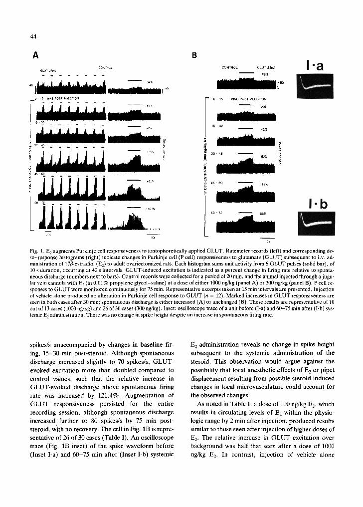
Systemic injection of 17fl-estradiol (E2) produced a marked enhancement of the excitatory response of single cerebellar Purkinje cells to iontophoretically applied glutamate (GLUT). This effect was dose-de- pendent, tested at 100,300 and 1000 ng/kg, as higher doses of the steroid produced a greater effect on GLUT response relative to spontaneous discharge, than did lower doses (see Table I). Furthermore,
43
only the highest dose tested produced a significant in-
crease in background firing rate. In most cases, E2 ef- fects on G L U T responsiveness were seen by 15-30 min post-steroid, with no recovery apparent by 2 h af- ter steroid administration. In Fig. 1A, pulsatile appli- cation of GLUT (27 nA) increased neuronal dis- charge by 34%. Within 15 min after intravenous in- jection of 17fl-estradiol (1000 ng/kg), spontaneous discharge was increased by 48%, and GLUT-induced excitation was increased by nearly twice that amount such that it represented a 50% increase in discharge relative to spontaneous firing. By 30-45 min post- steroid, GLUT-evoked excitation relative to back-
ground was increased further by 367% over control levels to a 159% excitation over background dis- charge. The Purkinje cell response to G L U T re- mained elevated although spontaneous discharge de- creased over the last 30 min of recording, yielding significant increases in evoked discharge relative to background (maximum = 1563% by 60-75 min). This case is representative of 10 out of 13 cells tested (Table I). For the second cell depicted in Fig. 1B, a lower dose of 17fl-E 2 (300 ng/kg) produced a 49.4% increase in GLUT-evoked excitation from 77 to 115
TABLE I
Average change in glutamate response and spontaneous discharge in estrogen-treated groups
Group: 17:E 2 E2-primed + 17a-E e
17a-E 2 Tamoxifen + 17fl-E 2
Dose E2: 100 300 I00 300 100 300 300 (ng/kg, i.v.)
n: (12) (30) (13) (12) (6) (12) (12)
Background discharge % increase 12.1 15.3 58.0* 34.6 7.2 10.1 11.2 S.E. 12.3 11.1 14.6 19.6 3.8 8.2 27.6 Latency to significant
response (min) 15 15 15 15 15 15 30-45
Glutamate response % increase 21.4 66.7* 150.3" 91.5" 6.8 30.0 55.2* S.E. 9.2 20.6 49.1 38.5 5.2 22.3 19.3 Latency to significant
response (min) 15 15 15 15 15 15 30-45
Relative change in evoked response over background % increase S.E. Latency to significant
response (min)
67.8* 95.7* 141.4" 68.6* -5.3 58.3* 59.1" 23.1 27.1 49.6 12.3 10.2 32.8 27.3
15 15 30 15 15 15 30-45
*P < 0.05 compared with pre-steroid values.
44
A CONTROL
GLUT 27nA
40
B CONTROL GLUT 25nA
28% I.a
0 15 MINS POST INJECTION
15 - 30
7" 30 - a5 . . . . .
§
J1Jl]ljj 30~
-- 0 - 1 5
15 - 30
o 30 - 45
45 - 60
l 60 - 75
MINS POST-INJECTION
20%
42%
55%
10s
I.b
Fig. 1. E 2 augments Purkinje cell responsiveness to iontophoretically applied GLUT. Ratemeter records (left) and corresponding do- se-response histograms (right) indicate changes in Purkinje cell (P cell) responsiveness to glutamate (GLUT) subsequent to i.v. ad- ministration of 17fl-estradiol (E2) to adult ovariectomized rats. Each histogram sums unit activity from 8 GLUT pulses (solid bar), of 10 s duration, occurring at 40 s intervals. GLUT-induced excitation is indicated as a percent change in firing rate relative to sponta- neous discharge (numbers next to bars). Control records were collected for a period of 20 min, and the animal injected through a jugu- lar vein cannula with E 2 (in 0.01% propylene glycol-saline) at a dose of either 1000 ng/kg (panel A) or 300 ng/kg (panel B). P cell re- sponses to GLUT were monitored continuously for 75 min. Representative excerpts taken at 15 min intervals are presented. Injection of vehicle alone produced no alteration in Purkinje cell response to GLUT (n = 12). Marked increases in GLUT responsiveness are seen in both cases after 30 min; spontaneous discharge is either increased (A) or unchanged (B). These results are representative of 10 out of 13 cases (1000 ng/kg) and 26 of 30 cases (300 ng/kg). Inset: oscilloscope trace of a unit before (I-a) and 60-75 min after (l-b) sys- temic E 2 administration. There was no change in spike height despite an increase in spontaneous firing rate.
spikes/s unaccompanied by changes in baseline fir-
ing, 15-30 min post-s teroid. Al though spontaneous
discharge increased slightly to 70 spikes/s, G L U T -
evoked excitation more than doubled compared to
control values, such that the relative increase in
GLUT-evoked discharge above spontaneous firing
rate was increased by 121.4%. Augmenta t ion of
G L U T responsiveness persisted for the entire
recording session, al though spontaneous discharge
increased further to 80 spikes/s by 75 min post-
steroid, with no recovery. The cell in Fig. 1B is repre-
sentative of 26 of 30 cases (Table I). A n oscilloscope
trace (Fig. 1B inset) of the spike waveform before
(Inset I-a) and 60-75 min after (Inset I-b) systemic
E 2 administrat ion reveals no change in spike height
subsequent to the systemic adminis t ra t ion of the
steroid. This observat ion would argue against the
possibility that local anesthet ic effects of E 2 or pipet
displacement resulting from possible s teroid- induced
changes in local microvasculature could account for
the observed changes.
As noted in Table I, a dose of 100 ng/kg E2, which
results in circulating levels of E2 within the physio-
logic range by 2 min after inject ion, p roduced results
similar to those seen after inject ion of higher doses of
E 2. The relative increase in G L U T excitation over
background was half that seen after a dose of 1000
ng/kg E 2. In contrast , injection of vehicle alone

GLUT 50nA 54% CONTROL
25
F MINS POST-INJECTION
0 - ~ 88,%
10 - on ~ 71%
8
<~ ~ 80,%
o
RECOVERY
30 - 40 ~ 34,%
10s 17AII3ha-E2 30 rain
41p
40S
Fig. 2. 17a-E 2, at an intermediate dose, can potentiate GLUT responsiveness of P cells. Strip chart records and peri-event his- tograms depict one effect of i.v. 17a-E 2 (300 ng/kg in 0.01% propylene glycol-saline) on GLUT response of a P cell. Solid bar indicates period of iontophoretic GLUT ejection (10 s every 40 s). After a stable control response was obtained (upper record), 17a-E 2 was administered systemically through an in- dwelling jugular cannula and GLUT response monitored con- tinuously for a period of 60 min. Representative post-steroid records indicate GLUT response calculated for 10-min inter- vals. A significant increase in GLUT response was seen by 10 min post-steroid. However, unlike results obtained with the fl- isomer, recovery was apparent by 30-40 min post-steroid. This result is representative of 5 out of 12 cells tested.
(0.01% propylene glycol-saline) did not produce
any alteration in Purkinje cell responses to G L U T
(n = 12).
17a-Estradiol effects on G L U T responsiveness The aim of this experiment was to determine the
specificity of the observed E2 effect on P cell function
by comparing effects of the less active 17a-isomer
with those of the 17fl-isomer on neuronal responsive- ness to GLUT. 17a-Estradiol was injected i.v. at doses of 100 or 300 ng/kg (Table I). Only the higher dose was able to increase GLUT-evoked excitation to a significant degree, but this increase was of a transient nature as noted in Fig. 2. In this case, a 50% increase in G L U T responsiveness was observed by 10 min after injection, with no change in spontaneous
45
discharge. Recovery of G L U T responsiveness to
control levels was observed after 30 min post-steroid.
This result was found in 40% of the cells tested. An additional 20% of the cells demonstrated a persistent
decrease in G L U T responsiveness (X = 50%) by
15-20 min after injection of 17a-estradiol. The re- maining 40% of cells failed to exhibit any change in
G L U T responsiveness after injection of 17a-E 2. At a
lower dose of 17a-E 2 (100 ng/kg), no alteration in
G L U T responsiveness was observed by 2 h post-in-
jection.
Direct application of E2-hemisuccinate and Purkinje cell responses to G L U T
Direct iontophoretic administration of estrogen,
which is possible with the use of 17fl-estradiol hemi- succinate (EzS, 20 nA), yielded results similar to
those seen after i.v. 17fl-E2: responses of P cells to G L U T were increased by 100-200%, 7 -10 min after
the onset of E2S application (see Fig. 3). In this case,
G L U T responsiveness was increased by 173%, in the
face of a 50% reduction in background discharge.
Similar effects were observed in 17 of 20 cells tested.
No recovery was achieved by 35 min after termination of hormone applicaton. Iontophoret ic application of
succinate alone (20 nA) also increased P cell respon- siveness to G L U T by 100% (see Fig. 4), but onset
and termination of this effect were concomitant with onset and termination of iontophoretic application of
the succinate (7 of 12 cells). In 5 out of 12 cells, succi- nate had no effect. In addition, local application of
17a-E2 hemisuccinate (17ALPHA-E2S) also in-
creased G L U T responsiveness by 50% immediately after onset of iontophoretic application in 4 of 10 cells
tested (see Fig. 5). Thus, both systemic and local ap-
plication of E 2 result in potentiation of G L U T excita- tion in the cerebellar Purkinje cell. Administration of
succinate produced similar results as E2S, but with parameters which were markedly different; i.e. the effects were transient and o f lesser magnitude (see
Fig. 6).
Effects of estrogen priming on neuronal responsive- ness to G L U T
This study was conducted to determine whether prior administration of E2 altered the previously ob- served effects of the sex steroid on neuronal respon- siveness. E2 administration to an E2-primed rat re-

46
CONTROL
GLUT 60nA
71%
30 ] [ 30
ESTROGEN ON: 0 3 MIN E2S 20nA
34~
c~ ESTR©GEN ON: Z 1~ MIN
194%
575%
30s 10s
Fig. 3. Iontophoretic application of 17fl-estradiol hemisucci- nate increases P cell responsiveness to GLUT. In order to test whether local application of estrogen (17fl-estradiol hemisucci- nate, E2S ) to cerebellar P cells could also effect a change in sen- sitivity to GLUT. E2S was continuously ejected iontophoreti- cally (20 nA current) for a period of 20 min concomitant with pulsed iontophoretic application of GLUT (60 nA, 10 s dura- tion at 40 s intervals). GLUT responses during the control period, at 0-3 and 7-10 min after onset of E2S application, and at 32-35 rain after the termination of iontophoretic application of the steroid are presented. GLUT-evoked excitation was markedly increased (by at least 100%) after 5 min of continu- ous E2S application (17/20 cells), and this increase was sus- tained throughout the experimental period. No recovery was achieved by 35 min after drug termination.
suited in a marked increase in G L U T responsiveness
in a manner similar to that seen after E 2 administra-
tion to an ovariectomized rat. G L U T responses were
increased by an average of 91.5% unaccompanied by
significant alterations in background firing in 10 of 12
cells tested (see Table I). This E2-induced augmenta-
tion in G L U T responsiveness was persistent, with no
recovery by 2 h post-steroid.
Tamoxifen and E2-induced effects on GLUT respon- siveness
The effect of an estrogen receptor blocker was
tested in this study to explore the possibility that re-
CONTROL GLUT 42nA
80%
3 0
S 2OnA SUCCINATE O N : 0 - 4 MIN
. . . . . 165%
30
SUCCINATE ON: 7 - 11 MIN
-- 114% o
RECOVERY 0 - 4 MIN
46%
E
30s 10s
Fig. 4. Succinate can potentiate P cell responsiveness to GLUT. Local iontophoretical application of succinate (20 nA current) resulted in an immediate increase (0-4 min) in GLUT- evoked excitation, in contrast to iontophoresed E2S which re- quired 5 min of continuous application for a similar effect to oc- cur. (In 7/12 cells succinate demonstrated this effect; in 5/12 cells succinate had no effect.) P cell responsiveness to GLUT returned to levels below the baseline immediately upon termi- nation of drug application (recovery period 0-4 min), again in contrast to E2S which did not exhibit a recovery.
ceptor-mediated actions might be involved in the ob-
served E 2 effects on G L U T responsiveness. The ad-
ministration of an antiestrogen, tamoxifen, delayed
by 30-45 min but did not prevent the E2-induced in-
crease in G L U T responsiveness normally occurring
30 min after E2 administration (Fig. 7). Tamoxifen (5
mg in saline) was administered in 2 doses 2 h prior to
and concomitant with an i.v. injection of E2 (300
ng/kg). In this case, a gradual decrease in G L U T re-
sponsiveness is observed, which by 30-45 min after
injection of E2 was reduced by 38%, concomitant
with a 30% reduction in background discharge. By 60
min post-steroid, however, a 138% increase in
GLUT-induced excitation was seen, accompanied by
a 50% increase in background discharge. This cell is
representative of 10 out of 12 cases; in 2 of 12, the po-
tentiating effect of E 2 on G L U T was not observed.
These results show that tamoxifen does not prevent

47
!o
CONTROL GLUT 25 nA 47X
17 ALPHA-E2S, 0 - 2 MIN
283%
RECOVERY l ~ 87Z
17 ALPHA-E2S ON OFF GLUT 25 nk v_ - _v . . . . . ~oo~ . . . . . . . .
SE: Fig. 5. 17a-Estradiol hemisuccinate increases P cell respon- siveness to GLUT in a similar manner to succinate. Drug re- sponse histograms and ratemeter records (bottom panel) illus- trate one case of GLUT potentiation by iontophoretic applica- tion of 17a-E2S (17a-E2S, 0.25 mM, pH 7.4). Histograms indi- cate the response of a P cell to pulsatile GLUT application (25 nA, 10 s pulses at 40 s intervals) during pre-steroid, continuous steroid application (0-2 min, 20 nA) and recovery periods. Di- rect application of 17a-E2S resulted in an immediate increase in GLUT responsiveness from 47% to a 283% excitation. A partial recovery was observed within 1 min after termination of iontophoresis. In 4 of 10 cells examined 17a-E2S resulted in this effect, in 1 cell it increased GLUT responsiveness with a longer latency, in 3 the steroid decreased GLUT responsiveness and in 2 there was no effect.
the effect of E2, a l though it a l tered the la tency to re-
sponse. A d m i n i s t r a t i o n of t amoxi fen , a lone , us ing
the same in jec t ion schedule , did n o t p roduce any al-
t e ra t ion in s p o n t a n e o u s f ir ing rate or G L U T re-
sponses of Pu rk in j e cells (da ta n o t shown) .
Effect of systemic estrogen administration on Purkinje cell responses to GABA application
In contras t to its effect o n G L U T respons iveness
E2 p roduced var iable effects on G A B A responsive-
ness (Table II) . Ou t of 14 cells tes ted, approx ima te ly
21 .4% exhib i ted decreases in G A B A responses rel-
io~( % CHANGE IN SPONTANEOUS DISCHARGE Jr 4oo~ j ° . o ~f l
too i ~ ~ ' ~ SUCCINATE, 20 n A I ~
0 , ' ~ - ~ . . . . . . . . . ~._.. ............. ~ . . . . . . . . . . . _ . ~ ( _ _ . . . _ . _ _ t
CHANGE ~N GLUT EVOKED EXCITATION 1000
_Z 60C
.z I/T\ 20C 1
% CHANGE IN SIGNAL TO-NOISE
~, . , .L I ~ L ~ " / 7 " - - - , . . . . . . . . . . . . . . . . . . . . . . . . . . . . J
ON OFF
0 5 io ~5 20 5 j~ is 30
Fig. 6. [ontophoret ic appl icat ion of ]Tf l-estradiol hemisucci- nate results in increases in spontaneous, evoked and signal-to- noise activity that are of greater magnitude and slower time course than those elicited by succinate alone. This figure illus- trates differences in percent change in spontaneous discharge (upper panel), percent change in GLUT-evoked excitation (middle panel) and percent change in the signal-to-noise ratio (evoked activity vs background) for P cells after local applica- tion of E2S or succinate alone. Bars indicate S.E.M., arrows in- dicate drug on (ON) and drug off (OFF). E2S application re- suited in a large percent change in spontaneous discharge (up- per panel) by 5 min after the onset of drug application (signifi- cantly greater than succinate at 5 min and 20 min). In addition, E2S application resulted in a proportionately greater increase in percent change of GLUT-evoked excitation (middle panel), again significantly larger than succinate at 5 and 20 min after drug onset. The most significant result, however, is found in the lower panel (percent change in signal-to-noise). In this case, succinate resulted in a percent change in signal-to-noise ratio 50 times greater than that produced by E2S (P < 0.001) imme- diately (0-1 min) after drug onset. Within 5 min after drug ap- plication the percent change in signal-to-noise produced by E2S application increased by more than 100% such that it was now significantly greater than that produced by succinate through- out the test period. In addition, although the succinate group achieved complete recovery by 5 min after drug termination, cells treated with E2S had not achieved control levels of signal- to-noise by 35 min after drug termination. E2S, n = 20 cells; succinate, n = 12 cells. *P < 0.05.
ative to b a c k g r o u n d (X = 58 .4%) by 2 0 - 3 0 min af-
ter systemic admin i s t r a t ion of 17fl-estradiol. 21 .4%
of the cells d e m o n s t r a t e d no change in Pu rk in j e cell
responsiveness to G A B A after in jec t ion of E 2, while
48
CONTROL GLUT 21hA
3s] i 35
l 0 - 15
15 - 30
E
30 - 45 O
45 60
g
MINS POST-ICJECTION 38%
33%
26*/*
tn
g
100%
30s 10s
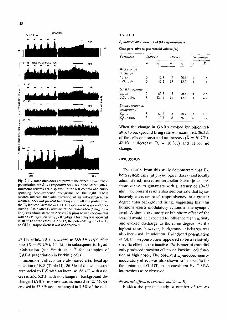
Fig. 7. I.v. tamoxifen does not prevent the effect of E2-induced potentiation of GLUT responsiveness. As in the other figures, ratemeter records are displayed in the left column and corre- sponding dose-response histograms on the right. These records indicate that administration of an anti-estrogen, ta- moxifen, does not prevent but delays until 60 min post-steroid the E2-induced increase in GLUT responsiveness normally oc- curring 30 min after E 2 administration. Tamoxifen (5 mg, in sa- line) was administered in 2 doses 2 h prior to and concomitant with an i.v. injection of E 2 (300 ng/kg). This delay was apparent in 10 of 12 of the cases; in 2 of 12, the potentiating effect of E 2 on GLUT responsiveness was not observed.
57.1% exhibi ted an increase in G A B A responsive-
ness (X = 69.2%), 10-15 min subsequent to E2 ad-
ministration (see Smith et al. 36 for examples of
G A B A potent ia t ion in Purkinje cells).
Inconsistent effects were also noted after local ap-
plication of E2S (Table II). 26.3% of the cells tested
responded to E2S with an increase, 68.4% with a de-
crease and 5.3% with no change in background dis-
charge. G A B A response was increased in 42.1%, de-
creased in 52.6% and unchanged in 5.3% of the cells.
TABLE II
E2-induced alteration in GABA responsiveness
Change relative to pre-steroid values (%)
Parameter Increase Decrease No change
n ,~ n X n A"
Background discharge E 2, i.v. 5 12.3 5 20.5 4 1.8 E2S, ionto. 5 41.2 13 12.2 1 1.1
GABA response E2, i.v. 5 63.5 5 10.6 4 2.5 E2S, ionto. 8 120.1 10 42.4 1 4.2
Evoked response: background E2, i.v. 8 69.2 3 58.4 3 1.5 E2S, ionto. 5 30.7 8 26.3 6 3.2
When the change in G A B A - e v o k e d inhibition rel-
ative to background firing rate was examined, 26.3%
of the cells demons t ra ted an increase (X = 30.7%),
42.1% a decrease (X = 26.3%) and 31.6% no
change.
DISCUSSION
The results from this study demonst ra te that E2,
both systemically (at physiological doses) and locally
administered, increases cerebel lar Purkinje cell re-
sponsiveness to glutamate with a latency of 10-35
min. The present results also demonst ra te that E2 se-
lectively alters neuronal responsiveness to a greater
degree than background firing, suggesting that this
hormone exerts modula tory actions at the synaptic
level. A simple excitatory or inhibitory effect of the
steroid would be expected to influence mean activity
and evoked discharge to the same degree. At the
highest dose, however, background discharge was
also increased. In addit ion, Ee-induced potent ia t ion
of G L U T responsiveness appeared to be a relatively
specific effect as the inactive 17a-isomer of estradiol
only produced transient effects on Purkinje cell func-
tion at high doses. The observed E2-induced neuro-
modula tory effect was also shown to be specific for
the amino acid G L U T , as no consistent E 2 - G A B A
interactions were observed.
Neuronal effects o f systemic and local E 2 Besides the present study, a number of reports
have noted widespread activational effects of system- ically administered E 2 on neuronal excitability, a phenomenon which has been demonstrated primarily in the hypothalamus 21 and, more recently, stria- tum 42. In the hypothalamic slice preparation, pre-
treatment with E 2 has been shown to potentiate exci- tatory neuronal responses to afferent stimulation and selected neurotransmitter agents 23. Local effects of E 2 on extrahypothalamic 12'27 neuronal excitability have also been noted. Although these studies dearly suggest a role for circulating and local E 2 in mediat- ing increases in neuronal excitability, hormonal in- teractions with specific excitatory neurotransmitters have not been thoroughly established in the extrahy- pothalamic CNS.
In the present study, the potentiation of GLUT re- sponse observed after administration of 17fl-estra- diol hemisuccinate, iontophoresed onto the Purkinje cell, suggests that E 2 can alter neuronal responsive- ness locally. Although, in the present study, succi- nate alone resulted in increases in glutamate respon- siveness, these were of shorter latency and duration, as well as of lesser magnitude, than those obtained with E2S which suggests that E2S is exerting effects on the neuronal membrane independent of any succi- nate effect. This possibility is strengthened by recent preliminary results from our laboratory which dem- onstrate similar augmentation of GLUT responsive- ness after local application of 17fl-E 2 by pressure ejection 37. In addition, only 50% of cells treated lo-
cally with 17a-E2S demonstrated an increase in
GLUT response, an effect which was transient and resembled the effect of succinate on this parameter rather than that of the 17fl-isomer. This finding sug- gests that like the effect of i.v. E 2, the local effect of the steroid also appears to be relatively specific for the 17fl-isomer.
Possible mechanisms of action Classic actions of sex steroids are mediated
through specific receptor binding sites and genomic mechanisms 3°. Although the rat cerebellum is not known to contain significant levels of cytosol/nuclear E2 receptors 31, specific binding sites for E2 have been localized in mouse cerebellum H. However, the pres- ent results indicate that the observed E2-effects on neuronal responsiveness are of a different nature than classic E 2 effects on reproductive function, im-
49
plying that a different mechanism of action for these effects may exist. First, doses of the 17a-isomer which have no effect on reproductive function pro- duce modest, transient increases in GLUT respon- siveness. The cytosol/nuclear E2 receptor has only a very low affinity for the 17or-isomer, and administra- tion of this isomer should not produce any effect if classic steroid receptor mechanisms were involved. Secondly, the anti-estrogen tamoxifen, which acts by binding to specific cytosol/nuclear E 2 receptors 2° and has been shown to prevent classic E 2 effects (i.e. the LH surge and lordosis) did not prevent E2 effects on neuronal responsiveness. Finally, E 2 priming did not alter the magnitude or the direction of its effect on GLUT responses. In terms of neuroendocrine func- tion, E2-priming typically results in increased E2 sen- sitivity 4, which may be due to the effect of positive cooperativity of E 2 binding to traditional cytosol/nu- clear receptors. Thus, the observed E 2 effects do not appear to be mediated through classic steroid recep- tors.
In terms of specific mechanisms of action, it is our hypothesis that E 2 may be acting directly on the membrane to alter neurotransmitter function. E2-in- duced alterations in local membrane permeability have been reported in liposomes 39. In addition, a spe- cific membrane receptor binding site for E 2 has been identified 41, and rapid, non-genomic effects of E 2 have been demonstrated on uterine microviUi 4° and hypothalamic neurons 22. However, none of the exist- ing reports directly addresses the possibility that E 2
could alter GLUT-mediated changes in ionic conduc- tance. A second possibility is that the steroid is alter- ing the degradation rate of GLUT, as previous stud- ies have shown that E 2 can alter amino acid enzyme systems 26. Therefore, a specific mechanism for the observed potentiation of GLUT responsiveness is not yet resolved.
Although not as likely, E2-receptor interactions with afferent systems should not be overlooked. Sev- eral reports have shown that E 2 is localized autora- diographically to the locus coeruleus TM, the source nucleus for an extensive noradrenergic projection to the cerebellum. This could be a relevant interaction because NE potentiates GLUT responses of Pur- kinje neurons 33, and E 2 can alter noradrenergic turn- over 6. However, E 2 neuromodulatory effects are of greater magnitude than those of NE, suggesting that if
50
noradrenergic input is found to be a factor, then a
combination of interactions may be involved.
Technical aspects of systemic steroid administration
Systemic injection, as utilized in the present study,
is the most physiologic means of administration of sex steroids, as endogenously circulating E 2 would come
into contact with cerebellar tissue in a similar manner
through its dense vascular network. The doses used
in this study, injected manually over a 2 -3 min inter- val, were in the physiologic range when integrated
over the experimental time period, as evidence indi-
cates that exogenously administered steroids clear
very rapidly from the blood and are reduced to barely
detectable levels 5 min after i.v. injection due to up- take by target organs, fat stores and CNS areas 24.
Average blood levels of E2, at a dose of 300 ng/kg, calculated over the entire 15-35 min latency period
would be in the physiologic range of a proestrous ani-
mal (40-50 pg/ml). The lowest dose shown here to have a significant effect on G L U T response (100
ng/kg) would result in immediate blood values dou-
ble those seen endogenously, physiologic levels after 2 rain, and the average level integrated over a 5 min
period would be of proestrous magnitude. There-
fore, the doses tested produce physiologic blood lev-
els of E2 within the time flame necessary for the ob- served neuronal effect of the steroid.
The time course for the observed E 2 effect on neu- ronal responsiveness was shorter than observed for most classic reproductive effects of the steroid, but this effect occurred after both the peak and eventual decline in blood levels of the steroid. Although most of the classic actions of the steroid occur with laten-
cies ranging from hours to days, much more rapid ef- fects have been demonstrated in other systems. Ge-
nomic actions of sex steroids can be seen as early as 2 min after administration 32. Non-genomic actions of
E 2 have been reported msecs to secs after systemic in- jection 21'4°, and the negative feedback effect of sys-
temic E2 on L H R H release can be observed within 15 min 43. Certainly, E e would come into contact with ce-
rebellar Purkinje cells very rapidly after systemic in-
jection, as one complete pass through the circulatory system of the rat is accomplished in 6 s 7, the lipophilic
sex steroid would cross the b lood-bra in barrier 1 and
be taken up by cerebellar tissue within minutes as has been shown both in vivo 9 and in vitro 19 using radiola-
belled E 2. Therefore, the observed latency of 10-35 min is reasonable for a steroid effect.
Summary In summary, systemic, as well as locally applied,
E 2 appears to exert long-lasting modulatory effects on
neuronal function in an extrahypothalamic area of
the CNS important for sensorimotor coordination and learning 25'29. The observed E2-induced increase
in Purkinje cell responsiveness to glutamate suggests that endogenously fluctuating levels of this hormone
may tend to increase the overall excitatory tone of
neuronal circuits in the cerebellum. This effect would be consistent with the reported activating effects of the steroid on seizure activity 28 and sensorimotor function 2 (i.e. balance beam accuracy38). Although a
direct causal relationship between these behaviors
and alterations in G L U T response cannot at this time be concluded, the widespread nature of these steroid
effects on sensorimotor behavior are consistent with a generalized action of the steroid in extrahypotha-
lamic circuits.
ACKNOWLEDGEMENTS
Supported by Grants NS25809 to S.S.S., NS18081
to B.D.W. and AA3901 and DA02338 to D.J .W. , as well as a grant from the Biological Humanics Foun- dation. The 17a-estradiol hemisuccinate used in
these experiments was generously provided by Dr.
Robert L. Moss (Dallas).
REFERENCES
1 Alfsen, A., Biophysical aspects of the mechanism of action of steroid hormones, Prog. Biophys. Mol. Biol., 42 (1983) 79-93.
2 Beatty, W.W., Gonadal hormones and sex differences in non-reproductive behaviors in rodents: organizational and activational influences, Horm. Behav., 12 (1979) 112-163.
3 Becker, J.B. and Ramirez, V.D., Sex differences in the am- phetamine stimulated release of catecholamines from rat
striatal tissue in vitro, Brain Research, 204 (1981) 361-372. 4 Camp, P. and Barraclough, C.A., Correlation of luteiniz-
ing hormone surges with estrogen nuclear and progestin cy- tosol receptors in the hypothalamus and pituitary gland. I. and II,, Neuroendocrinology, 40 (1985) 45-62.
5 Chiodo, L.A. and Caggiula, A.R., Alterations in basal fir- ing rate and autoreceptor sensitivity of dopamine neurons in the substantia nigra following acute and extended expo- sure to estrogen, Eur. J. Pharm., 67 (1980) 165-166.
6 Crowley, W..R, Effects of ovarian hormones on norepi-
nephrine and dopamine turnover in individual hypothalam- ic and extrahypothalamic nuclei, Neuroendocrinology, 34 (1982) 381-386.
7 D'Amour, F.E., Blood, F.R. and Belden, Jr., D.A., Man- ual for Laboratory Work in Mammalian Physiology, 3rd edn., Univ. Chicago Press, Chicago, 1965.
8 Dennerstein, L., Spencer-Gardner, C. and Burrows, G.D., Mood and the menstrual cycle, J. Psychiat. Res., 18 (1984) 1-12.
9 Eaton, G.G., Goy, R.W. and Resko, J.A., Brain uptake and metabolism of estradiol benzoate and estrous behavior in ovariectomized guinea pigs, Horm. Behav., 6 (1975) 81-97.
10 Eccles, J.C., Ito, M. and Szentagothai, J., The Cerebellum as a Neuronal Machine, Springer, New York, 1967, 335 pp.
11 Fox, T.O., Estradiol and testosterone binding in normal and mutant mouse cerebellum: biochemical and cellular specificity, Brain Research, 128 (1977) 263-273.
12 Foy, M.R. and Teyler, T.J., 17a-Estradiol and 17fl-estra- diol in hippocampus, Brain Res. Bull., 10 (1983) 735-739.
13 Freedman, R., Hoffer, B.J., Woodward, D.J. and Puro, D., Interaction of norepinephrine with cerebellar activity evoked by mossy and climbing fibers, Exp. Neurol., 55 (1977) 269-288.
14 Friedman, R.C. (Ed.), Behavior and the Menstrual Cycle, Marcel Dekker, New York, 1982, 467 pp.
15 Geller, H.M. and Woodward, D.J., An improved constant current source for microiontophoretic drug application studies, Electroencephalogr. Clin. Neurophysiol., 33 (1972) 430-432.
16 Hancke, J.L.and Dohler, K.D., Sexual differentiation of female brain function is prevented by postnatal treatment of rats with the estrogen antagonist tamoxifen, Neuroendo- crinol. Lett., 6 (1984) 201-206.
17 Harms, P.G. and Ojeda, S.R., A rapid and simple proce- dure for chronic cannulation of the rat jugular vein, J. Appl. Physiol., 36 (1974) 391-392.
18 Heritage, A.S., Stumpf, W.E., Madhabananda, S. and Grant, L.D., Brainstem catecholamine neurons are target sites for sex steroid hormones, Science, 207 (1980) 1377-1379.
19 Holler, M., Dengler, K. and Breuer, H., Disposition of [4- C~4]oestradiol-17fl in the isolated perfused brain of the rat, J. Steroid Biochem., 20 (1984)785-787.
20 Katzenellenbogen, B.S., Miller, M.A., Eckert, R.L. and Sudo, K., Antiestrogen pharmacology and mechanism of action, J. Steroid Biochem., 19 (1983) 59-68.
21 Kelly, M.J., Electrical effects of steroids in neurons. In K.W. McKerns and V. Pantic (Eds.), Hormonally Active Brain Peptides: Structure and Function, Plenum, New York, 1982, pp. 253-277.
22 Kelly, M.J., Moss, R.L. and Dudley, C.A., The effects of microelectrophoretically applied estrogen, cortisol and acetylcholine on medial preoptic septal unit activity throughout the estrous cycle of the female rat, Exp. Brain Res., 30 (1977) 53-64.
23 Kow, L.M. and Pfaff, D.W., Estrogen effects on neuronal responsiveness to electrical and neurotransmitter stimula- tion: an in vitro study on the ventromedial nucleus of the hypothalamus, Brain Research, 347 (1985) 1-10.
24 Larner, J.M. and Hochberg, R.B., The clearance and me- tabolism of estradiol and estradiol-17-esters in the rat, Endo, 117 (1985) 1209-1214.
25 Leaton, R.N. and Supple, W.F., Cerebellar vermis: essen-
51
tial for long-term habituation of the acoustic startle re- sponse, Science, 232 (1986) 513-515.
26 Maggi, A. and Perez, J., Role of female gonadal hormones in the CNS: clinical and experimental aspects, Life Sci., 37 (1985) 893-906.
27 Marcus, E.M., Watson, C.W. and Goldman, P.L., Effects of steroids on cerebral electrical activity, Arch. Neurol., 15 (1966) 521-531.
28 Mattson, R.H. and Cramer, J.A., Epilepsy, sex hormones, and antiepileptic drugs, Epilepsia, 26 (1985 ) $40- $51.
29 McCormick, D.A. and Thompson, R.F., Cerebellum: es- sential involvement in the classically conditioned eyelid re- sponse, Science, 223 (1984) 296-299.
30 McEwen, B.S. and Parsons, B., Gonadal steroid action on the brain: neurochemistry and neuropharmacology, Annu. Rev. Pharmacol. Toxicol., 22 (1982) 555-598.
31 McEwen, B.S. and Pfaff, D.W., Factors influencing sex hormone uptake by rat brain regions. I. Effects of neonatal treatment, hypophysectomy and competing steroid on es- tradiol uptake, Brain Research, 21 (1970) 1-16.
32 Means, A.R. and Hamilton, T.H., Early estrogen action: concomitant stimulation within 2 minutes of nuclear RNA synthesis and uptake of RNA precursor by the uterus, Proc. Natl. Acad. Sci. U.S.A, 56 (1966) 1596-1598.
33 Moises, H.C., Woodward, D.J., Hoffer, B.J. and Freed- man, R., Interactions of norepinephrine with Purkinje cell responses to putative amino acid neurotransmitters applied by microiontophoresis, Exp. Neurol., 64 (1979) 493-515.
34 Namba, H. and Sokoloff, L., Acute administration of high doses of estrogen increases glucose utilization throughout brain, Brain Research, 291 (1984) 391-394.
35 Smith, S.S., Waterhouse, B.D. and Woodward, D.J., Can estrogen alter neuronal responsiveness to putative amino acid neurotransmitters? Soc. Neurosci. Abstr., 10 (1984) 1129.
36 Smith, S.S., Waterhouse, B.D., Chapin, J.K. and Wood- ward, D.J., Progesterone alters GABA and glutamate re- sponsiveness: a possible mechanism for its anxiolytic ac- tion, Brain Research, 400 (1987) 353-359.
37 Smith, S.S., Waterhouse, B.D. and Woodward, D.J., Lo- cally applied progesterone metabolites alter neuronal re- sponsiveness in the cerebellum, Brain Res. Bull., in press.
38 Snyder, P.J., Westgate, S.A., Miller, M.M., Jenuwine, M.J. and Becker, J.B., The influence of intrastriatal estro- gen on balance beam performance in ovariectomized fe- male rats, Mtg. Neurosci. Soc., 12 (1986) 841.
39 Sze, P.Y., Armstrong, E.C., Deanin, G.G. and Gordon, M.W., Steroid hormones stabilize liposomal membrane from alteration by tubulin, Mtg. Neurosci. Soc., (1985) 1135.
40 Szego, C.M., Mechanisms of hormone action: parallels in receptor-mediated signal propagation for steroid and pep- tide effectors, Life Sci., 35 (1984) 2383-2396.
41 Towle, A.C. and Sze, P.Y., Steroid binding to synaptic plasma membrane: differential binding of glucocorticoids, and gonadal steroids, J. Steroid Biochem., 18 (1983) 135-143.
42 Van Hartsveldt, C. and Joyce, J.N., Effects of estrogen on the basal ganglia, Neurosci. Biobehav. Rev., 10 (1986) 1-14.
43 Yamagi, T., Dierschke, D.J., Bhattacharya, A.N. and Knobil, E., The negative feedback control by estradiol and progesterone of LH secretion in the ovariectomized Rhesus monkey, Endocrinology, 90 (1972) 771-777.