Advancing towards Ubiquitous EEG, Correlation of In-Ear EEG ...

r Human Brain Mapping 33:1325–1333 (2012) r
Relationship of Resting EEG with Anatomical MRIMeasures in Individuals at High and Low Risk for
Depression
Gerard E. Bruder*, Ravi Bansal, Craig E. Tenke, Jun Liu, Xuejun Hao,Virginia Warner, Bradley S. Peterson, and Myrna M. Weissman
Department of Psychiatry, Columbia College of Physicians and Surgeons and New York StatePsychiatric Institute, New York, New York
r r
Abstract: Studies have found abnormalities of resting EEG measures of hemispheric activity in depres-sive disorders. Similar EEG findings and a prominent thinning of the cortical mantle have been reportedfor persons at risk for depression. The correspondence between EEG alpha power and magnetic reso-nance imaging (MRI) measures of cortical thickness was examined in a multigenerational study of indi-viduals at risk for depression. Seventy-five participants underwent resting EEG and approximately 5years later underwent MRI scanning. High-risk participants (n ¼ 37) were biological descendants of pro-bands having major depression and low-risk participants (n ¼ 38) were descendants of individuals with-out a history of depression. EEG alpha power was interpolated across the surface of a template brain andcoregistered with measures of cortical thickness. Voxel-wise correlations of cortical thickness and alphapower were computed while covarying for age and gender. The high-risk group, when compared to thelow-risk group, showed greater alpha asymmetry in an eyes-closed condition, with relatively less activityover right parietal cortex. Alpha power correlated inversely with cortical thickness, particularly over theright posterior region, indicating that EEG evidence of reduced cortical activity was associated withincreased cortical thinning. This is the first report of widespread correlation of EEG alpha activity withMRI measures of cortical thickness. Although both EEG and MRI measures are associated with risk fordepression, we did not detect evidence that cortical thickness mediated the alpha asymmetry findings.Thus, alpha asymmetry, alone or in combination with MRI, may be a marker of vulnerability for a familialform of depression. Hum Brain Mapp 33:1325–1333, 2012. VC 2011 Wiley Periodicals, Inc.
Keywords: EEG; MRI; cortical thickness; hemispheric asymmetry; high risk; major depressive disorder
r r
INTRODUCTION
Alpha rhythms in the electroencephalogram (EEG) pro-vide a noninvasive measure of regional cortical activity.Alpha is largest over posterior brain regions in an eyes-closed resting state and is considered an ‘‘idling rhythm’’that is blocked or diminished by mental activity. Initialneuroimaging studies reported an inverse correlationbetween EEG alpha power and regional cerebral bloodflow in occipital cortex [Sadato et al., 1998]. Studies core-gistering EEG and functional MRI have generally con-firmed this relationship, with greater alpha powerassociated with less activity in occipital and parietal lobes[Goncalves et al., 2006].
Contract grant sponsor: National Institute of Mental Health;Contract grant numbers: MH36197, MH36295, MH068318, andK02-74677; Contract grant sponsors: National Alliance forResearch in Schizophrenia and Affective Disorders (NARSAD).
*Correspondence to: Gerard E. Bruder, Division of Cognitive Neu-roscience, Unit 50, New York State Psychiatric Institute, 1051 Riv-erside Drive, New York, NY 10032. E-mail: [email protected]
Received for publication 8 April 2010; Revised 10 December 2010;Accepted 19 January 2011
DOI: 10.1002/hbm.21284Published online 15 April 2011 in Wiley Online Library(wileyonlinelibrary.com).
VC 2011 Wiley Periodicals, Inc.

Early studies of resting EEG reported overall greater alphapower in depressed patients compared with healthy controlswith eyes closed [Pollack and Schneider, 1990; Shagass et al.,1988]. Given the inverse relationship between alpha and cort-ical activity, greater alpha is assumed to reflect less corticalactivity. More recent studies have found abnormal hemi-spheric asymmetries of alpha in depressed individuals. Analpha asymmetry indicating less left than right cortical activ-ity over frontal sites has been found in depressed adults[Davidson et al., 1987; Gotlib et al., 1998; Henriques andDavidson, 1991], depressed persons having a comorbid anxi-ety disorder [Bruder et al., 1997], and women who had child-hood onset of their depressive illness [Miller et al., 2002].Several studies have also reported the opposite alpha asym-metry at posterior sites, i.e., relatively less cortical activityover right parietal sites, in depressed adults [Bruder et al.,1997; Davidson et al., 1987; Reid et al., 1998], depressed ado-lescents [Kentgen et al., 2000], and remitted depressed adults[Henriques and Davidson, 1990]. Studies have providedadditional evidence of right parietotemporal dysfunction indepressed patients on neurocognitive tests of lateralized cog-nitive processing [Bruder, 2003] and on psychophysiologicmeasures during processing emotional pictures [Deldinet al., 2000; Kayser et al., 2000; Moratti et al., 2008].
Our multigenerational longitudinal study [Weissman et al.,2005] previously reported an alpha asymmetry indicating lessright than left parietal activity in family members who were athighest risk for developing a depressive disorder [Bruderet al., 2005, 2007]. Surface EEG measures, however, are limitedin their localizing capabilities, leaving unanswered the ques-tion of the neural basis for this finding. New anatomical MRIfindings from this high-risk study [Peterson et al., 2009] pro-vide independent support for an association between familialrisk for depression and asymmetries in parietal cortices. Corti-cal thickness in high and low risk groups were comparedacross the cerebral surface. Large expanses of cortical thinningacross the lateral aspects of the parietal, posterior-temporal,and frontal cortices of the right hemisphere were found in thehigh risk group. These convergent findings in the same sam-ple suggest the hypothesis that EEG evidence of relatively lesscortical activity over right parietal sites in individuals at highrisk for depression may derive from cortical thinning. Theavailability of both resting EEG and MRI measures in a sub-sample of subjects in this high-risk study provided a uniqueopportunity to examine the relation of their EEG alpha andcortical thickness, not only at parietal but also frontal siteswhere abnormal alpha asymmetry has been found in depres-sion. An additional reason for relating EEG and anatomicalMRI measures is that neuropathological processes have beenassociated with abnormalities of posterior alpha and otherEEG rhythms [Babiloni et al., 2008].
METHOD
Participants and Assessments
Probands in Generation 1 (G1) with major depressionwere originally selected from an outpatient clinic for phar-
macologic treatment of depression and had moderate tosevere depression with impairment in functioning. Nonde-pressed controls were recruited from the same communityand had no lifetime history of psychiatric illness based onseveral direct interviews. Full details of clinical assess-ments at baseline (Wave 1), 2 years later (Wave 2), and 10years later (Wave 3) have been previously described[Warner et al., 1999; Weissman et al., 1997]. A fourth waveof assessments [Weissman et al., 2005] and electrophyio-logic tests ended in 2002. A fifth wave of assessments,which included a partial sample of the second Generation(G2) and third Generation (G3) who agreed to MRI scans,began in 2002 and ended in 2007 [Peterson et al., 2009].Assessment procedures were kept similar across waves,with few exceptions, to avoid introducing bias frommethod variation [Weissman et al., 2005]. G1 participants(both probands and nondepressed controls), their spouses,offspring and grandchildren were interviewed independ-ently by mental health professionals who demonstratedhigh inter-rater reliability on the interview procedures andwho were blind to the clinical status of participants in theprevious generations. The diagnostic interviews across allwaves were conducted using semistructured instru-ments—the Schedule for Affective Disorders and Schizo-phrenia Lifetime Version (SAD-L) for adults [Mannuzzaet al., 1986] and for children between the ages 6 and 17,the child version [K-SADS-E; Orvaschel et al., 1982] modi-fied for DSM-IV, and at Wave 4, the K-SADS-PL [Kaufmanet al., 2001] was used. Lifetime diagnoses using DSM-IVcriteria at the definite level were cumulative across allwaves for grandparents and their offspring. Grandchildren(G3) were assessed at waves 4 and 5, with a few also atwave 3. Multiple sources of information were obtained,including independent assessments of offspring by directinterview, parent report, and direct assessment of both bi-ological parents as often as possible. Diagnoses were basedon the best estimate procedure [Leckman et al., 1982],which involved an independent review of all assessmentmaterials by two experienced clinicians, a child psychia-trist, and a psychologist, who were not involved in theinterviewing and were blind to the diagnostic status of theprevious generations and prior assessments.
Resting EEG was obtained during the fourth wave. Off-spring or grandchildren who were biological descendentsof the G1 probands had to be older than 7 years, live inthe geographic area of the study, and have no hearingimpairment, history of seizures, head trauma, or psycho-sis. Of the 182 G2 offspring who were eligible, EEG wasrecorded in 111. Of the 137 G3 members, 74 participatedin the EEG tests. Six of the offspring and seven grandchil-dren did not have sufficient EEG data, after blink andmovement artifact rejection, to be included in this study.The main reason eligible subjects did not participate isthat they declined to be tested. Those who did not partici-pate and those who received the electrophysiologic testsdid not differ significantly in age, gender, or depressionstatus of the probands. The sample targeted for MRI
r Bruder et al. r
r 1326 r

scanning in Wave 5 consisted of 196 G2 offspring whowere interviewed in Wave 4 and 157 G3 grandchildrenwho were interviewed in either in Wave 3 or 4. Of these,150 were scanned and 143 anatomical images were avail-able because 7 were compromised by motion or otherscanning artifact. Entry into the MRI scanning alsorequired a negative pregnancy test for females who werepostmenarchal and premenopausal and the absence of fer-romagnetic implants. Waves 4 and 5 assessments wereapproved by the Human Investigation Committee at YaleUniversity School of Medicine (where the EEG and MRIscans were performed) and the Institutional Review Boardat New York State Psychiatric Institute. This research wasperformed in compliance with the Code of Ethics of theWorld Medical Association (Declaration of Helsinki), andinformed consent was obtained from participants (orparents).
The data for 75 participants (46 G2 and 29 G3) who hadboth sufficient, artifact-free EEG recordings in Wave 4 andan MRI scan in Wave 5 are presented in this report. Off-spring and grandchildren designated as being at ‘‘highrisk’’ for depression were biological descendants of G1probands having a major depressive disorder and thosedesignated at ‘‘low risk’’ were biological descendants ofthe unaffected controls in G1. The high-risk groupincluded 37 individuals (20 females) and the low-riskgroup included 38 individuals (20 females). Althoughhigh- and low-risk groups did not differ significantly ingender, there was a group difference in age (Table I). Thehigh risk group was somewhat older than the low riskgroup at the time of the MRI scans and the same was trueduring EEG tests, when participants were on the average
3.7 years younger (SD ¼ 1.2). All were Caucasian and allbut one subject in each group were right-handed based onself-report. Only 9 of the 75 participants received psycho-active medications during the 3 months before the EEG. Inthe high-risk group, three received an antidepressant, twoan antidepressant plus a minor tranquilizer, and one aminor tranquilizer. In the low-risk group, one received anantidepressant, one an antidepressant plus a minor tran-quilizer, and one an antidepressant plus a major tranquil-izer.1 Table I also gives the percentage of offspring in thehigh- and low-risk groups who had lifetime diagnoses ofdepressive disorders, anxiety disorders, and other disor-ders. The high-risk group had significantly higher rates ofmajor depression, anxiety disorders, phobias, and disrup-tive disorders compared with the low-risk group.
EEG Procedures
Resting EEG was measured while subjects sat quietlyduring four 2-min periods (eyes open, closed, closed, openor eyes closed, open, open, closed). Subjects wereinstructed to remain still and to blink or move their eyesor body as little as possible during the recordings. In theeyes-open condition, subjects fixated on a cross centeredon a computer monitor. Scalp EEG was measured from 12electrodes over medial and lateral frontal, central, and pa-rietal regions at homologous sites over each hemisphere(F3,F4; F7,F8; C3,C4; T7,T8; P3,P4; P7,P8) using an elec-trode cap (Electro Cap International, Eaton, OH) with aleft-ear reference. The EEG was subsequently re-referenceddigitally to a linked-ears reference. Tin electrodes werealso placed on the right ear, as well as at supra-and infra-orbital sites surrounding the right eye to monitor eyeblinks and vertical eye movements and at right and left
TABLE I. Rate of lifetime diagnoses, medications, and
demographics in high- and low-risk groups
High risk(n ¼ 37)
Low risk(n ¼ 38) P
DiagnosesAny anxiety disorder 23 (62.2%) 8 (21.1%) <0.001a
Any phobia 19 (51.4%) 9 (23.7%) 0.013a
Major depressive disorder 21 (56.8%) 6 (15.8%) <0.001a
Dysthymia 6 (16.2%) 3 (7.9%) 0.268a
Alcohol abuse 7 (18.9%) 4 (10.5%) 0.304a
Drug abuse 6 (16.2%) 4 (10.5%) 0.469a
Any disruptive disorder 11 (29.7%) 4 (10.5%) 0.038a
Medicationsb
Antidepressants 5 (13.5%) 3 (7.9%) 0.431a
Major tranquilizer 0 1 (2.6%) 0.321a
Minor tranquilizer 3 (8.1%) 1 (2.6%) 0.291a
Gender (female) 20 20 0.902a
Right handed 36 37 0.985a
Age 33.9 � 11.7 27.4 � 13.5 0.028c
Means � SD are shown for age.av2 test.bWithin 3 months before EEG.ct test.
1In a prior study, we found no evidence of any change in alphapower or asymmetry in depressed patients following treatment withthe antidepressant fluoxetine [Bruder et al., 2008]. Although samplessizes in the current study are not large enough to examine medica-tion effects, secondary analyses were conducted to determinewhether medication could have impacted on the results. First, theEEG alpha power at parietal sites (P3, P4; P7, P8) in eyes closed andeyes open conditions was compared for individuals on versus offpsychotropic medications, after collapsing across the high and lowrisk groups. There was no trend for a Medication Group � Hemi-sphere or Medication Group � Hemisphere � Condition interaction(P values � 0.53), which suggests that medication had no significanteffect on alpha asymmetry at parietal sites. Second, a comparison ofcortical thickness between individuals on versus off psychotropicmedications showed no significant differences over the broadregions where negative correlations were observed between EEGalpha and cortical thickness. Finally, we found that the critical RiskGroup � Hemisphere � Condition interaction for EEG alpha power(see Results) remained significant after excluding the nine individu-als who had received psychotropic medications, F¼ 4.66, df¼ (1,57),P< 0.05. Thus, the significant difference in condition-related parietalalpha asymmetry between high- and low-risk groups was stillobserved in individuals whowere not onmedications.
r EEG and Anatomical MRI Relations r
r 1327 r

outer canthi to monitor horizontal eye movements. Allelectrode impedances were <5 kX. EEGs were recordedusing a Bioamplifier system (James Long Company, Car-oga Lake, NY) at a gain of 10 K (5 K for eye channels),with a band pass of 0.01-30 Hz. A PC-based EEG acquisi-tion system (NeuroScan, Sterling, VA) acquired and digi-tized the data continuously at 200 samples/sec duringeach recording period.
Details concerning the analysis of the EEG are given else-where [Bruder et al., 2007]. Briefly, artifact-free EEG epochs(1.28 sec every 0.64 sec; Hanning tapered) were submittedto a power spectrum analysis using a Fast Fourier Trans-form. Analyses focused on the alpha band where prior stud-ies have found differences in hemispheric asymmetry indepressed subjects [Bruder et al., 1997; Davidson et al., 1987;Gotlib et al., 1998; Henriques and Davidson, 1990, 1991;Kentgen et al., 2000; Miller et al., 2002; Reid et al., 1998]. Wehave found differences between high- and low-risk groupsin alpha but not lower or higher frequency bands [Bruderet al., 2005, 2007]. At each electrode, alpha power was aver-aged for artifact-free epochs spanning each recording periodfor each condition, and subsequently integrated over 7.0 to12.5 Hz band. Logarithms of alpha power were then com-puted to normalize the distribution of the data.
MRI Procedures
Head positioning was standardized using canthomeatallandmarks. Imaging was conducted on a Siemens Sonata1.5T scanner (Siemens AG) equipped with high-speed gra-dients (maximum amplification, 40 mT/m; slew rate, 200mT/m/sec) and running a three-dimensional MP-RAGEsequence (repetition time, 24 msec; echo time, 2.96 msec; 45�
flip angle; 256 � 192 matrix; field of view 30 cm; phase fieldof view ¼ 100%; two excitations, slice thickness 1.2 mm; and124 contiguous slices encoded for sagittal slice reconstruc-tion with voxel dimensions 1.17 � 1.17 � 1.2 mm).
The brain was isolated from nonbrain tissue by firstusing Brain Extraction Tool [Smith, 2002] and then man-ually editing each slice in sagittal, coronal, and axialviews. The cortical mantel was defined by applying aglobal threshold whose value was halfway between themean gray matter and mean white matter values, and thenmanually editing this rough classification of cortical grayin all three views [Peterson et al., 2009].
We mapped cortical thickness across cerebral surfacesusing procedures previously described [Peterson et al.,2009; Bansal et al., 2005]. Briefly, isolated brains were core-gistered using a similarity transformation to an appropri-ately selected template brain [Plessen et al., 2006]. Eachhemisphere of the coregistered brain was normalized tothe corresponding hemisphere of the template brain byfirst a rigid transformation and then a high-dimensional,nonrigid warping based on fluid dynamics, thereby identi-fying points on the surface of each hemisphere with thoseon the template surface. From the coregistered brain ofeach participant, we first subtracted its cortical mantel and
then applied a three-dimensional morphological operatorto distance-transform the brain without the cortex [Hara-lick and Shapiro, 1992; Rosenfeld and Pfaltz, 1968]. Thisoperation calculated cortical thickness as the smallest dis-tance of each point on the external cortical surface fromthe outermost surface of the white matter in the coregis-tered brain. Because thicknesses were measured from thecoregistered brains that were similarly transformed intothe template space, thickness measures inherently accountfor the generalized scaling effects within the cerebrum.Cortical thickness measures between groups were statisti-cally analyzed and the P values were color encoded anddisplayed on the high-resolution image of a single individ-ual [Mazziotta et al., 2001]. Finally, on this image wesuperimposed the manually delineated cortical gyri forbetter localization of findings.
Normalizing and Coregistering EEG and MRI
Data
We normalized EEG data to MRI data using the follow-ing procedures. First, electrode sites were interactively reg-istered onto the template brain guided by standard 10-20placements on the cortical surface [Homan et al., 1987].Second, each brain was normalized (i.e., coregistered usinga similarity transformation and nonlinearly warped) to thetemplate brain, thereby placing the EEG data for each sub-ject into the coordinate space of the template brain. Third,the EEG data from the 12 electrode sites for each subjectwere interpolated across all voxels on the template surfaceusing a conformal mapping and a method for sphericalinterpolation. Using the conformal mapping, the surface ofthe entire brain and the 12 electrode sites were mappedonto a unit sphere. On the unit sphere, the EEG power atthe 12 sites was interpolated using the method for spheri-cal splines [parameters: n ¼ 41, m ¼ 2; Perrin et al., 1989]to compute EEG power at each point on the sphere. Theinterpolated EEG power was then mapped onto the sur-face of the entire brain by inverting the conformal map-ping. We then correlated at each point on the surface ofthe two hemispheres the interpolated EEG power with thecortical thickness, while controlling for the confoundingvariables. Correlating EEG power with the cortical thick-ness on the brain surface does not imply that we havelocalized the generators of the alpha power in the corticalmantle. Instead, we interpolated the montage of EEGpower measures onto the surface of the brain, rather thanon the unit sphere, in order to map the EEG onto thesame continuous surface for which we have cortical thick-ness measures. Probability maps are presented represent-ing the significance of correlations between EEG andcortical thickness at corresponding points on that surface.
Statistical Analyses
Analyses were performed to confirm our published EEGfindings for the subsample of offspring and grandchildren
r Bruder et al. r
r 1328 r
who had MRI data. Log alpha power measures for high-and low-risk groups at each electrode site were submittedto an analysis of covariance using Risk group (high vs.low), Generation (2nd vs. 3rd) and Gender as between-subjects factors, and four within-subjects factors: Hemi-sphere (left vs. right), Region (frontal, central, parietal),Medial-Lateral (F3,4; C3,4;P3,4 vs. F7,8; C7,8; P7,8) andCondition (Eyes open vs. closed). Age was included as acovariate. When a significant interaction involving Riskgroup, Hemisphere, Region, and Condition was found,subsequent analyses followed up this interaction to deter-mine the significance of group differences in hemisphericasymmetry of alpha for each condition at frontal, central,and parietal regions. F ratios were evaluated using degreesof freedom computed with the Greenhouse-Geisser Epsi-lon correction [Jennings and Wood, 1976] where appropri-ate to counteract heterogeneity of variance–covariancematrices with repeated measures.
The coregistration procedures described above wereused to map the EEG alpha power measures across thesurface of the template brain for each risk group. Student’st-statistics determined the significance of group differencesin alpha power at every point on the surface of the tem-plate brain while covarying for age and gender, and tocompute probability maps. Voxel-wise correlations of cort-ical thickness and alpha power across the cortical surfacewere computed while covarying for age, gender, and riskgroup. Note that including scalp thickness, as measuredfrom the MRI of each participant at the most superiormidline site (Cz), as a covariate had essentially no effect
and was therefore not included in the final model. Mapsdisplaying these correlations were corrected for multiplestatistical comparisons across the cerebral surface usingthe false discovery rate (FDR) procedure [Benjamini andHochberg, 1995], which controls for the expected propor-tion of false discoveries among rejected null hypotheses.The significance of differences in correlations across hemi-spheres was examined at posterior sites where group dif-ferences in alpha asymmetry were present (P3,P4), as wellas over frontal (F3,F4) and central (C3,C4) sites, using t-tests. We also assessed whether cortical thickness medi-ated the association of familial risk with EEG alpha powerfor eyes closed minus eyes open using standard proce-dures for mediator analyses [MacKinnon et al., 2007; Peter-son et al., 2009].
RESULTS
We detected a significant hemispheric asymmetry ofEEG alpha at parietal sites (F ¼ 7.14, df ¼ (1,66), P < 0.01),but not at frontal or central electrode sites (Hemisphere �Region interaction: F ¼ 5.45, df ¼ 1,66, P < 0.01). Alphaasymmetry (i.e., right–left hemisphere alpha power) differ-ences between the high- and low-risk groups were greatestover parietal sites in the eyes closed condition (Group �Hemisphere � Region � Condition interaction: F ¼ 4.47,df ¼ 1,66, P < 0.025). Alpha asymmetry differed signifi-cantly between the high- and low-risk groups at parietalsites in the eyes closed but not eyes open condition, whichwas supported by a three-way interaction of Group �
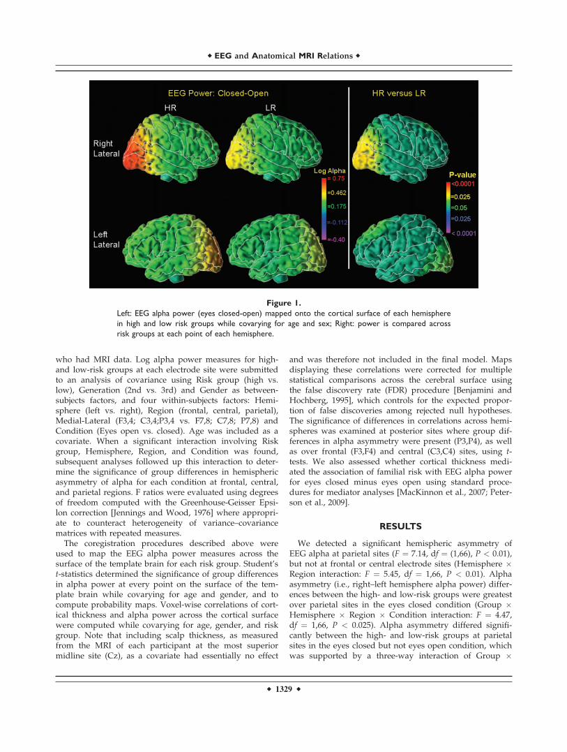
Figure 1.
Left: EEG alpha power (eyes closed-open) mapped onto the cortical surface of each hemisphere
in high and low risk groups while covarying for age and sex; Right: power is compared across
risk groups at each point of each hemisphere.
r EEG and Anatomical MRI Relations r
r 1329 r

Hemisphere � Condition interaction (F ¼ 4.87, df ¼ 1,66,P < 0.05). The high-risk group showed greater alphaasymmetry, indicating relatively less cortical activity overright parietal sites, when compared to low-risk group inthe eyes closed condition (F ¼ 4.32, df ¼ 1,66, P < 0.05).When examining individual parietal sites, there was a sig-nificant condition-related difference in alpha asymmetrybetween high and low risk groups over medial parietalsites (P3,P4; F ¼ 5.31, df ¼ 1,66, P < 0.05), but this differ-ence only approached significance over lateral parietalsites (P7,P8; F ¼ 3.50, df ¼ 1,66, P ¼ 0.07). The differencein asymmetry between high- and low-risk groups was notlikely due to an age difference for three reasons: (1) therewas no interaction involving age and risk status in theabove analysis; (2) alpha asymmetry at parietal sites didnot significantly correlate with age (r ¼ �0.19, ns), andage was included as a covariate in the analysis; and (3)maps showing EEG alpha and group differences includedage as a covariate.
The left panel of Figure 1 shows the alpha powermapped onto a surface rendering of the template brain,representing surfaces of the right and left cerebral hemi-spheres that underlie EEG measures. The high-risk groupshowed relatively less cortical activity (greater alpha) overthe right occipitoparietal region with eyes closed thanopen, whereas this was less evident in the low-risk group.
The significance of this group difference was confirmed byprobability mapping (right panel of Fig. 1).
As indicated in Table I, the rate of lifetime major depres-sion was higher in the high-risk than low-risk group (v2 ¼13.89, df ¼ 1, P < 0.001), as was the rate of lifetime anxietydisorders (v2 ¼ 13.34, df ¼ 1, P < 0.001). An analysis of co-variance was therefore performed to determine whetherthe above difference in parietal alpha asymmetry betweenhigh- and low-risk groups remains after controlling for thepresence of these disorders. This analysis used the factorsin the above analyses with an age covariate, but includingas an additional factor (Disorder)—whether the high- andlow-risk participants had a major depressive or anxietydisorder. Thirty-seven subjects had either or both disor-ders (27 HR, 10 LR) and 38 had neither disorder (10 HR,28 LR). This analysis yielded the same condition-relateddifference in parietal alpha asymmetry between the high-and low-risk groups as in the above analyses (Risk �Hemisphere � Condition interaction, F ¼ 4.35, df ¼ 1,66, P< 0.05). The asymmetry difference between high- and low-risk groups did not depend on whether the participantshad a major depressive or anxiety disorder, i.e., there wasno higher order interaction involving Risk � Hemisphere� Condition � Disorder, F ¼ 0.92, df ¼ 1,66, P ¼ 0.34.There was also no significant main effect or other interac-tion involving the factor of Disorder.
Figure 2.
Probability maps for correlations between EEG alpha power and MRI cortical thickness for eyes
open and closed conditions mapped onto each hemisphere for both groups combined while
covarying for age, sex, and risk group. Maps are corrected for multiple comparisons by control-
ling the false discovery rate.
r Bruder et al. r
r 1330 r

Figure 2 shows probability maps for voxel-wise correla-tions of alpha power with MRI measures of cortical thick-ness for both groups combined, while covarying for age,gender, risk, and group. The maps demonstrate spatiallyextensive inverse correlations across the MRI and EEGmeasures, indicating that progressively thinner corticeswere accompanied by progressively less cortical activity(greater alpha power). The same pattern of correlationswas present for each risk group separately, with no signifi-cant interaction of risk group with EEG alpha power de-tectable in any regions. At medial parietal sites (P3,P4),where condition-related differences in alpha asymmetrybetween high and low risk groups were present, there wasa significant difference in the correlation of MRI and alphapower over the right and left hemisphere in both eyesclosed (t ¼ 3.06, P < 0.005) and eyes open conditions (t ¼3.31, P ¼ 0.001). An inverse correlation between MRI andalpha was present at the right parietal site (P4), but not atthe left parietal site (P3). There was, however, no signifi-cant hemispheric difference in the correlations at frontal(F3,F4) or central sites (C3,C4). Also, mediator analysesshowed no evidence that cortical thickness mediated therelationship between risk status and alpha power (eyesclosed-open) at posterior sites (all P > 0.05).
DISCUSSION
Offspring and grandchildren of probands having majordepression, who are at particularly high risk for depres-sive disorders [Weissman et al., 2005], showed greateralpha asymmetry, with relatively less cortical activity overright parietal sites, compared to those at low risk. Theirparietal asymmetry resembled that seen in adolescents andadults having a depressive disorder [Bruder et al., 1997;Davidson et al., 1987; Kentgen et al., 2000; Reid et al.,1998]. There was, however, no evidence in high risk off-spring or grandchildren of the frontal alpha asymmetryseen in depressed individuals [Davidson et al., 1987; Got-lib et al., 1998; Henriques and Davidson, 1991]. The differ-ence in parietal asymmetry between high- and low-riskgroups did not depend on having a lifetime diagnosis ofmajor depression or anxiety disorder, and prior findingsalso suggest that this alpha asymmetry is not state-de-pendent, but may represent a trait marker of vulnerabilityfor depression. Relatively less right parietal activity wasfound in members of both second and third generationswho did not have a depressive disorder but were at highrisk [Bruder et al., 2005, 2007]. It was also present in remit-ted depressed adults who were euthymic during EEG test-ing [Henriques and Davidson, 1990]. Moreover, depressedpatients who responded to an SSRI antidepressant showedthis parietal asymmetry both before and after successfultreatment, further suggesting that the finding is stable andstate-independent [Bruder et al., 2008]. Studies indicatethat about 60% of the variance of resting alpha asymmetryis attributable to a temporally stable latent trait [Hage-
mann et al., 2005]. Also, the findings for high risk subjectscontrast with the decreased alpha seen for neurodegenera-tive disorders [Babiloni et al., 2008], indicating that theyare not likely due to neuropathology, but rather to a traitassociated with a familial form of depression.
Anatomical MRI findings [Peterson et al., 2009] for off-spring and grandchildren at risk for depression led us toexamine the relation of EEG alpha to cortical thickness ina subsample having both MRI and EEG measures.Although studies have reported that resting alpha powercorrelates inversely with cerebral metabolism or bloodflow [Goncalves et al., 2006; Sadato et al., 1998], ours is thefirst report of spatially extensive inverse correlationsbetween alpha power and local measures of cortical thick-ness. Less cortical activity (greater alpha power) was asso-ciated with progressively greater cortical thinningparticularly over the right posterior cortex, where signifi-cant differences in alpha were present between high andlow risk groups. Correlations between alpha power andcortical thickness, as well as hemispheric differences inthese correlations, were more evident over posterior thanfrontal sites. Although this might suggest that corticalthickness is more specific to understanding differences inposterior than frontal alpha, the correlations reflected ageneral relationship between alpha power and corticalthickness that was present across both high- and low-riskindividuals. The spatially extensive inverse correlations ofalpha power with cortical thickness raise the possibilitythat cortical thinning may in part account for the globalenhancement of alpha reported in patients having adepressive disorder [Pollock and Schneider, 1990; Shagasset al., 1988] or in a putative subtype of patients whorespond favorably to SSRI antidepressants [Bruder et al.,2008]. We detected no statistical evidence, however, tosupport the hypothesis that cortical thinning in right hemi-sphere regions mediated the relation between familial riskfor depression and alpha asymmetry. Alpha asymmetryindicating relatively lower cortical activity over right pos-terior sites and cortical thinning may therefore be separaterisk factors for depression. Although the spatial resolutionof the EEG montage was suboptimal for a precise localiza-tion, the interpolated EEG alpha topographies (see Fig. 1)do show the expected maximum over posterior (visual)areas, which is consistent with its condition dependency,i.e., greater alpha in eyes closed condition. The condition-related differences between high- and low-risk groups inalpha power map widely over the posterior region of thetemplate brain. Further study using high-resolution EEGmethods (high-density montage and current source densitymeasures) will more precisely detail the relationshipbetween alpha and anatomical MRI.
A neuropsychological model hypothesizes that reducedright parietotemporal activity in depression generates lowemotional arousal [Heller et al., 1995]. The right parietalcortex is known to subserve perception of emotion, andcortical activity over this region during emotional percep-tion is reduced in depressed patients [Deldin et al., 2000;
r EEG and Anatomical MRI Relations r
r 1331 r

Kayser et al., 2000], suggesting that reduced activity of theright parietal cortex in depression has important conse-quences for emotional processing. A magnetoencephalog-raphy study also found reduced cortical activity over theright temporoparietal region in depressed persons relativeto controls during the viewing of emotionally arousingpictures, indicating that depressed individuals have diffi-culty activating cortices that subserve the arousal dimen-sion of emotion [Moratti et al., 2008].
Although electrophysiological studies in high-risk sam-ples are few, preliminary evidence suggests that childrenat risk for depression may show abnormal emotional reac-tivity and hemispheric asymmetries. Most notably, lowpositive emotionality in children, identified as conferringelevated risk for developing depression, was associatedwith an EEG alpha asymmetry indicating the presence ofless right than left cortical activity, particularly over parie-tal sites [Shankman et al., 2005]. Given evidence for theimportance of the right parietal cortices in processing emo-tional stimuli, high-risk children who have relatively lowcortical activity of the right parietotemporal region may beless able to perceive, process, and respond to emotional in-formation, placing them at increased risk for depression.MRI and neuropsychological findings support a modelpostulating that familial risk for depression produces corti-cal thinning of the right hemisphere, which in turn dis-rupts attention and arousal processes, and thesedisruptions increase the risk for developing a majordepressive disorder [Peterson et al., 2009]. The extent towhich EEG measures, alone or in combination with MRIs,might serve as a biological marker for predicting lateronset of depressive disorders is currently under study.
REFERENCES
Babiloni C, Frisoni GB, Pievani M, Toscano L, Percio CD, GeroldiC, Eusebi F, Miniussi C, Rossini PM (2008): White-matter vas-cular lesions correlate with alpha EEG sources in mild cogni-tive impairment. Neuropsychologia 46:1707–1720.
Bansal R, Staib LH, Whiteman R, Wang YM, Peterson BS (2005):ROC-based assessments of 3D cortical surface-matching algo-rithms. Neuroimage 24:150–162.
Benjamini Y, Hochberg Y (1995): Controlling the false discoveryrate: A practical and powerful approach to multiple testing. JR Stat Soc Series B Stat Methodol 57:289–300.
Bruder GE (2003):Frontal and parietotemporal asymmetries indepressive disorders: Behavioral, electrophysiologic, and neu-roimaging findings. In: Hugdahl K, Davidson RJ, editors. TheAsymmetrical Brain. Cambridge, MA: MIT Press. pp719–742.
Bruder GE, Fong R, Tenke CE, Leite P, Towey JP, Stewart JW,McGrath PJ, Quitkin FM (1997): Regional brain asymmetries inmajor depression with or without an anxiety disorder: A quan-titative electroencephalographic study. Biol Psychiatry 41:939–948.
Bruder GE, Sedoruk JP, Stewart JW, McGrath PJ, Quitkin FM,Tenke CE (2008): Electroencephalographic alpha measures pre-dict therapeutic response to a selective serotonin reuptake in-
hibitor antidepressant: Pre- and post-treatment findings. BiolPsychiatry 63:1171–1177.
Bruder GE, Tenke CE, Warner V, Nomura Y, Grillon C, Hille J,Leite P, Weissman MM (2005): Electroencephalographic meas-ures of regional hemispheric activity in offspring at risk fordepressive disorders. Biol Psychiatry 57:328–335.
Bruder GE, Tenke CE, Warner V, Weissman MM (2007): Grand-children at high and low risk for depression differ in EEGmeasures of regional brain asymmetry. Biol Psychiatry62:1317–1323.
Davidson RJ, Chapman JP, Chapman LJ (1987): Task-dependentEEG asymmetry discriminates between depressed and non-depressed subjects. Psychophysiology 24:585.
Deldin PJ, Keller J, Gergen JA (2000): Right-posterior face process-ing anomaly in depression. J Abnorm Psychol 109:116–121.
Goncalves SI, de Munck JC, Pouwels PJW, Schoonhoven R, KuijerJPA, Maurits NM, Hoogduin JM, Van Someren EJW, HeethaarRM, Lopes da Silva FH (2006): Correlating the alpha rhythm toBOLD using simultaneous EEG/fMRI: Inter-subject variability.Neuroimage 30:203–213.
Gotlib IH, Ranganath C, Rosenfeld JP (1998): Frontal EEG alphaasymmetry, depression, and cognitive functioning. Cog Emot12:449–478.
Hagemann D, Hewig J, Seifert J, Naumann E, Bartussek D (2005):The latent state-trait structure of resting EEG asymmetry: Rep-lication and extension. Psychophysiology 42:740–752.
Haralick R, Shapiro L (1992): Computer and Robot Vision. Boston,MA: Addison-Wesley Longman Publishing Co., Inc. pp157–201.
Heller W, Etienne MA, Miller GA (1995): Patterns of perceptual
asymmetry in depression and anxiety: Implications for neuro-
psychological models of emotion and psychopathology. J
Abnorm Psychol 104:327–333.
Henriques JB, Davidson RJ (1991): Left frontal hypoactivation in
depression. J Abnorm Psychol 100:535–545.Henriques JB, Davidson RJ (1990): Regional brain electrical asym-
metries discriminate between previously depressed andhealthy control subjects. J Abnorm Psychol 99:22–31.
Homan RW, Herman J, Purdy P (1987): Cerebral location of inter-
national 10-20 system electrode placement. Electroencephalogr
Clin Neurophysiol 66:376–382.
Jennings JR, Wood CC (1976): The epsilon-adjustment procedure
for repeated-measures analyses of variance. Psychophysiology
13:277–278.
Kaufman J, Martin A, King RA, Charney D (2000): Are child-,ado-lescent-, and adult-onset depression one and the same disor-der? Biol Psychiatry 49:980–1001.
Kayser J, Bruder GE, Tenke CE, Stewart JE, Quitkin FM (2000):Event-related potentials (ERPs) to hemifield presentations ofemotional stimuli: Differences between depressed patients andhealthy adults in P3 amplitude and asymmetry. Int J Psycho-physiol 36:211–236.
Kentgen LM, Tenke CE, Pine DS, Fong R, Klein RG, Bruder GE(2000): Electroencephalographic asymmetries in adolescentswith major depression: Influence of comorbidity with anxietydisorders. J Abnorm Psychol 109:797–802.
Leckman JF, Sholomskas D, Thompson D, Belanger A, WeissmanMM (1982): Best estimate of lifetime diagnosis: A methodologi-cal study. Arch Gen Psychiatry 39:879–883.
MacKinnon DP, Fairchild AJ, Fritz MS (2007): Mediation analysis.Annu Rev Psychol 58:593–614.
Mannuzza S, Fyer AJ, Klein DF, Endicott J (1986): Schedule forAffectvie Disorders and Schizophrenia-lifetime version
r Bruder et al. r
r 1332 r
modified for the study of anxiety disorders (SADS-LA):Rational and conceptual development. J Psychiatric Res20:317–325.
Mazziotta J, Toga A, Evans A, Fox P, Lancaster J, Zilles K, WoodsR, Paus T, Simpson G, Pike B, Holmes C, Collins L, ThompsonP, MacDonald D, Iacoboni M, Schormann T, Amunts K, Palo-mero-Gallagher N, Geyer S, Parsons L, Narr K, Kabani N,LeGoualher G, Boomsma D, Cannon T, Kawashima R,Mazoyer B (2001): A probabilistic atlas and reference systemfor the human brain: International Consortium for Brain Map-ping (ICBM). Philos Trans R Soc London 356:1293–1322.
Miller A, Fox NA, Cohn JF, Forbes EE, Sherrill JT, Kovacs M(2002): Regional patterns of brain activity in adults with a his-tory of childhood-onset depression: Gender differences andclinical variability. Am J Psychiatry 159:934–940.
Moratti S, Rubio G, Campo P, Keil A, Ortiz T (2008): Hypofunc-tion of right temporoparietal cortex during emotional arousalin depression. Arch Gen Psychiatry 65:532–541.
Orvaschel H, Puig-Antich J, Chambers W, Tabrizi MA, Johnson R(1982): Retrospective assessment of prepubertal major depres-sion with the Kiddie-SADS-E. J Am Acad Child Adolesc Psy-chiatry 21:392–397.
Perrin F, Pernier J, Bertrand O, Echallier JF (1989): Sphericalsplines for scalp potential and current density mapping. Elec-troencephalogr Clin Neurophysiol 72:84–187.
Peterson BS, Warner V, Bansal R, Zhu H, Hao X, Liu J, Dur-kin K, Adams PB, Wickramaratne P, Weissman MM(2009): Cortical thinning in persons at increased familialrisk for major depression. Proc Natl Acad Sci USA106:6273–6278.
Plessen KJ, Bansal R, Zhu H, Whiteman R, Amat J, QuackenbushGA, Martin L, Durkin K, Blair C, Royal J, Hugdahl K, PetersonBS (2006): Hippocampus and amygdale morphology in atten-
tion-deficit/hyperactivity disorder. Arch Gen Psychiatry63:795–807.
Pollock VE, Schneider LS (1990): Topographic quantitative EEG inelderly subjects with major depression. Psychophysiology27:438–444.
Reid SA, Duke LM, Allen JJ (1998): Resting frontal electroencepha-lographic asymmetry in depression: Inconsistencies suggestthe need to identify mediating factors. Psychophysiology35:389–404.
Rosenfeld A, Pfaltz JL (1968): Distance functions in digital pic-tures. Pattern Recognition 1:33–61.
Sadato N, Nakamura S, Oohashi T, Nishina E, Fuwamoto Y, WakiA, Yonekura Y (1998): Neural networks for generation and sup-pression of alpha rhythm: A PET study. Neuroreport 9:893–897.
Shagass C, Roemer RA, Josiassen RC (1988): Some quantitativeEEG findings in unmedicated and medicated major depres-sives. Neuropsychobiology 19:169–175.
Shankman SA, Tenke CE, Bruder GE, Durbin CE, Hayden EP,Klein DN (2005): Low positive emotionality in young children:Association with EEG asymmetry. Dev Psychopathol 17:85–98.
Smith SM (2002): Fast robust automated brain extraction. HumBrain Mapp 17:143–155.
Warner V, Weissman MM, Mufson L, Wickramaratne PJ (1999):Grandparents, parents, and grandchildren at high risk fordepression: A three-generation study. J Am Acad Child Ado-lesc Psychiatry 38:289–296.
Weissman MM, Warner V, Wickramaratne P, Moreau D, OlfsonM (1997): Offspring of depressed parents. 10 years later. ArchGen Psychiatry 54:932–940.
Weissman MM, Wickramaratne P, Nomura Y, Warner V, VerdeliH, Pilowsky DJ, Grillon C, Bruder GE (2005): Families at highand low risk for depression: A 3-generation study. Arch GenPsychiatry 62:29–36.
r EEG and Anatomical MRI Relations r
r 1333 r
Copyright © 2022 FDOKUMEN