The rabbit : an illustrated anatomical guide - Scholarly ...
127
University of the Pacific University of the Pacific Scholarly Commons Scholarly Commons University of the Pacific Theses and Dissertations Graduate School 1957 The rabbit : an illustrated anatomical guide The rabbit : an illustrated anatomical guide Edwin Chin Jr. University of the Pacific Follow this and additional works at: https://scholarlycommons.pacific.edu/uop_etds Recommended Citation Recommended Citation Chin, Edwin Jr.. (1957). The rabbit : an illustrated anatomical guide. University of the Pacific, Thesis. https://scholarlycommons.pacific.edu/uop_etds/1366 This Thesis is brought to you for free and open access by the Graduate School at Scholarly Commons. It has been accepted for inclusion in University of the Pacific Theses and Dissertations by an authorized administrator of Scholarly Commons. For more information, please contact mgibney@pacific.edu.
-
Upload
khangminh22 -
Category
Documents
-
view
9 -
download
0
Transcript of The rabbit : an illustrated anatomical guide - Scholarly ...

University of the Pacific University of the Pacific
Scholarly Commons Scholarly Commons
University of the Pacific Theses and Dissertations Graduate School
1957
The rabbit : an illustrated anatomical guide The rabbit : an illustrated anatomical guide
Edwin Chin Jr. University of the Pacific
Follow this and additional works at: https://scholarlycommons.pacific.edu/uop_etds
Recommended Citation Recommended Citation Chin, Edwin Jr.. (1957). The rabbit : an illustrated anatomical guide. University of the Pacific, Thesis. https://scholarlycommons.pacific.edu/uop_etds/1366
This Thesis is brought to you for free and open access by the Graduate School at Scholarly Commons. It has been accepted for inclusion in University of the Pacific Theses and Dissertations by an authorized administrator of Scholarly Commons. For more information, please contact [email protected].

t I I'
·I I\ '''
THE RABBIT
AN ILLUS'I'RA'l1EO ANATOMICAL GUIDE
A Thesis
.Pr•esE!mted to
the Faculty of the Department of Zoology
College of the Pacific
In Partial F'ulfillment
of the Requirements for the Degree
',Master of Arts
by
Edwin Chin, Jr. .. , June 1957
---- -----

CHAPTE:R PAGE
1 I. IN'11RODUCTI ON • • . •. . . . • • • • • • • • • • • !J
Preparation of Materials • • • • • • • • • • • • J
Killixlg the specimen • • • • • • • • • • • • • 3
Embali'\ling the specimen • • • • • • • • • • • • 3
Injection of, the circulatory system • • • • • 6
Skeletal preparation • • • • • • • • • • • • • 7
!I, EX'I1ERNAI.~ FEA~'URES • • • , • • • • • • • • • • , • 8
III. • • • • • • • • • • • • • • • • •
IV. rJIUSCULAR SYS'I'EM • • • • • • • • • • • • • • • • •
Superficial f1uscles • • • • • • • • • • • • • • ' . Subcutaneous Muscles • • • • • • • • • • • . . ~
Muscles of the Neck and Back • • • • • • • • • •
f1us<~les of the 'rhroat and Chest • • • • • 0 • •
Muscles of the Arm and Shoulder • • • • • • • •
Muscles of the I~'o:t'~arm • • • • • • • • • • • • •
£1uscles of the 'rh1gh and Leg • • • .. • • • • • .. r1uscles of the Lo't';er Leg • • • • • • • • • 0 • •
v. !N'l'ERNAL ORGANS • • • • • • • • • • • • • • • • •
VI. RESPIRATOHY SYS'l'Er1 • • • • • • • • • • • . .. . • •
VII. HEART AND MAJOR VESSELS . . . . . . . . •. . . . . • • • • • • • • • • • • • • • • • • It
The Major Vessels • • • • • • • • • • • • • • •
10
45
4.?
45
.?1
52
57 60
66
68
70
73
?3
75

CHAPTER
VIII. AR.TERIAIJ CIRCULATION • • • • • • • • • • • " . .
IX.
x. XI.
Anterior Arteries
'l'hora.cio Arteries
. . . . . .. . . . . . . . . • • • • • • • • • • • • • •
Abdominal Arteries • • • • • • •
Mtv.jor ~ymphatic Ves~-;els • • • •
\f""ENOUS CIRCULATION • • • .. • • • •
An.terlor Veins • • • • • • • • •
Posterior Veins • • • • • • • •
• • •
• • • • • 0
• • • • • • •
. . " • • • •
• • 0 • • • •
• • • • • • •
• • • • • • •
• • • 8 • • •
• • • • * 16 •
XII. UROGENITAL SYSTEM . . . . . . . . .. . . . . . .
XIII~
XIV.
U:rinary Organs • • • • • • • • • • • • • • • • Male Reproductive Organs • • • • • • • • • • •
Female Reproduot:l.ve Organs
CENTHAL NE:HVOUS SYSTEN • • •
• • • • • • • • • •
• • • • • • • • • •
Meninges • • • • • • • • • •
Late:t•al Aspect of the Brain
• •
• •
. . • • • • •
• • • • • • •
Medlan Sag it tal Section of the Br·ain. • • • • •
Po:t1sal Aspect of the B:r·a.5.n • • • • • • • • • •
Ventral Aspe<:~t of the Drain • • • • • • ,. • •
Spinal COl"d. ... . . . . .. . . .. . • • • . •. . . BIBI..IOOHAPHY • • • • • • • • • • • • • • • • • •
iii
PAGE
80
80
82
82
8.5
87
87
88
91
93
100
100
101
103
105
105
105
107
J.lO
112
lll~
116

LIST OF FIGURES
FIGURE
1. Lateral View of a Mounted Skeleton • • • • • • • • 2. Lateral View of' the Skull • .. • • • • • • • • • •
3. Medial View of a Sagitt-9.1 Section of the Skull il • 4 .. Dorsal View of the Skull • • • • • • • • • • • • •
5· Ventral View of the Skull • • • • • • • • • • • •
6. Lateral View of the Mandible • • • • • • • • • • •
7. r"iedtal View of t;he Mandible! • • • • • • .. • • • • a. Anterior View of the Sternal Apparatus • • • • • • 9. .Medial View of the Scapula • • • • • e • • • • • •
10. Lateral View of the Scapula • • • • • • • • • • •
11.
12.
1:}.
14.
1.5.
16.
17.
18.
19.
;w.
Anterior and. Posterl.or Views of the Humerus
Antero ... lateral View of the Hadius and Ulna •
• • •
• • • Dors£tl View of the Bones of the Hand • • • • • .. . Inferior View of the Atlas (1st Cervical) • • • • Lateral Viow of the Axis (2nd Cervical) • • • • •
Superior View of a Typical Cervical Vertebra • • •
Lateral V1e"~<1' of' a Typical 1l'horacio Vertebra • • •
Later•al View of a Typical Lumbar Vertebra • • • •
Superior View of _a Typical Lumbar Vertebra • • • •
Ventre.l V:i,ew of the Sacrum • • • • • • • • • • • • 21. Dorsal View of the Sa.orum . . . . . . . . . . •. '
Lateral Sux•face of the Innom:l.nate • • • • •
Medial Surface of the Innominate • • • • • • . . '
PAQS
11
13
14
15
16
19
20
22
23
24
26
27
29
Jl
31
33
33
34
34
36
)6
38
39

FIGURE
24.
26.
27.
28.
Anterior and. Pos teri o:t' View of the Femur • • • • •
• • • • • • • • Dor•sal View of the Bones o1' t.he li'oot • • • • • • • Latere.l Vie~'! of the Sup(~rficial f•iuseles • •• • • •
Muscles of the Neck and Baok • • • • • • • • • • • 29. Muscles of the Throat and Chest • • • • • • • • • 30. Late:r8.1 V18w of tbe tvtusc,les of the Arm a.:r1d
Shoulder • • • • • • • • • • • • • • • • • • • • :n. t1edial View of the Muscles of the iu.,.m and
Shoulder •• • • • • • e· • • • • I • .t • . .. . . 32. Cross Sect ton Through the r116.1Jl.le of the Humerus
v
PAGE
41
43
44
46
47
50
33. Dorsal View of the Tend.o~(ls of the Forearm • • • • 59
34. Lateral Vie'tl>.r of· the Supsrficial P1uscles of the
Thigh and Lag • • • • • • • • • • • • • • • • • 62
35. Lateral Vtr~t'i of the Deep ~1uscles of the Thigh
and Leg • • • • • • • • • • • • • • • • • • •. • 63
Me.dlal ViE~W of the Muscles of the Thigh • • • • •
Cross Section Through the Middle of the Femur .. . J8. Cross Section Through the l\liddle of the
39.
40.
41.
42.
43 ..
Ti.bic-Jil1 bula • • • • • • • • • • •
Anterior Vie-;.J of the Internal Organs
Anterior View of the Thoracic .Organs
Anterlor View of the He :art • • • • •
Dorsal Vi~w of the Heart • If • • • •
Vessels of the Neck and Thorax • • •
.. . • • • • •
• • • • • • • • • • 0 • • •
• • • • • • •
• • • • • • •
• • • • • • •
64
65
67
69
72
77
78
79
i - --- --

FIGURE
vi
PAGE
Branches of the Aorta • • • • • • • • • . . . ' . ... Tribut:a.r1es of the Venae Cavae •••••••••.•
86
90
46. Semi-Diagramatie View of the Hepatio-l?ortal
System • • • • • • • • • • • • • • • •. • • • • • 92
47. Anterior View of the Liver • • • • • • • • • • • • 98
48. Sem1~D1agramat1c Vielti of the lleoceoal Junction • • 99
49.
50.
51.
The Male Urogenital. System •
~he :tt,ema.H~ urogeiti tal System
Lateral View of the Brain • •
• •
• •
• •
• • • • • • • • • 102
• • • • • • • • • 104
• • • • • • • • • 106
SZ. Medial View of a Sagittal Section of the Brain •• 109
Dorsal View of the Brain . . . . . . . . . ... . . • 111
Ventra.l View of the Brain • • • • • • • • • .• • • · .• 115

I. INTRODUCTION
While asslst;ing in a la bora. tory cou:r•se in mammalian
anatomy at the College of the Pacific in the fall of 19.55,
1 t became necessary to utilize domestic rabbits, I.t!pus
caniculus:~ as a replacement for cats as dissecting material.
As a result of this, it was noted that anatomically the
rabbit was in many ways more desirable as a dissect1..ng
mammal than the cat. Some notable advantages over the oat
were (1) a more typical hyoid apparatus; (2} possession of
twelve pairs of r1.bs; (3) persistent right azygos vein;
(4) three main branches from the aortic arch; and external
and internal iliac arteries arising from the common iliac
artery. rrhese adve,nced mammalis,n oharaoteristj_cs, as
observed in the preU.minary dissections, resembled so much
the human anatomical condition that an investigation of the
possible use of the rabbit for dissection in an introductory
anatomy or mammalian anatomy course was initiated. An added
advantage in the use of the rabbit for dls :>ection would be
the lack of personal attachment for 1st bora tory ar11rnals such
as the rabbitt whereas certain societies and groups of
individuals often react negatively to the dissection of
oats in the laboratory. The low cost and. earf:!ot' embalming
and injecting is an added inducement for the use of :t•abbi ts
as dissecting material.

2
This study on the anatomy of the rabbit was based on
d.isseoted mt.tterial from the biological laboratories of the
College of the Pacific. Beo~use of existing errors in many
of the present. references to rabbit anatomy, the gutde was
prepared to meet the expressed need for a concise, illus
trated outline of basic anatomy of the rabi;Jit which might be
used by students in an introductory course in mammalian or
human anatomy. Since it is assumed that the guide would be
supplemented by a dlsseotion manual, syllabus, or textbook,
textural material of such an outllne is he:r•e presented in
outline form.
I wish to express my deep gratitude to Dr. Alden E•
Noble, chairman of the Department of Zoology, College of the
Pacific, whose 1nspir8.tion and guldanoe has made this study
possible. I would also like to thank Dr. Donald L. Lehmann
of the College of the Pacific and Miss Ecli th Grider, refer
ence librarian of the College of the Pacific, for their
generous donation of time and advice ln helping to make
this study possible.

PREPARATION OF MATEIHALS
. Most of the larger biological supply houses supply
embalmed. rabbits; singly • doubly$ or triply injected. The
cost of these preserved rabbits is about the same as for
the cat,. .':'£!he following procedures were used tn'the prepara'!"'
tion of' mc1.terials for this study; and they are recommended
for those who 'fl~ish to use the more readily obtainable live
rabbits.
KILLING THE SPECIMEN
The animal should be starved a day or two prior to
embalming. This allows for evacuation of the digestive
tract. Also, the dissection T1vill progress taore easj.ly. with
a minimum of visceral and subcutaneous fat deposits. A
large crock or garbage pail lATi th a tightly fi tti:ng cover is
ideal as a killing chamber. A cotton wad soaked with ether
or chloroform is dropped into the cha.mber and allowed to
remain for at least 20 minutes. The animal should be dead
and ready for emba.lrni:ng.
\IJhile. smaller mammals can be preserved. by simply open ...
ing tne body cavity and immersing them in a solution of
formalin or alcohol• larger animals should be embalmed
intravenously to insure adequate brain and visceral

prese~vation. The embalming solution should be prepared ahead
of' time and stored in a large 8-10 liter bottle w:ith a two
hole rubber stopper.· ~f.lhe following solution, which is a
modification of Keller's embalming fluid for the preparation
of human cadavers in ana.tomi.ca.l study, was found to'be
satisfactory for the rabbit.
' Commercial Formalin • • • • • • • 5 Parts Melted Carbolic f1,c1d Crystals • • 5 Parts Glycerin • • • • • • • . • • • • 5 Parts Distilled Water • • • • • • . .. • 8.5 Parts
The formalin acts as preservative and also gives firm~
hess to the muscles. rrhe glycerin prevents desiccation and
hardening of the organs. The carbolic ac1d helps to I"etain
color of the tissue and to improve the oc,ior of the specimen.
The animal should be placed on an embalming boa:rd and
tied into the des1r~.1d position prior to hardening. The
sites of injection for the embalming fluid are the common
carotid and femoral arteries. The common carotid artery is
located by a small incision in the neck region lateral to
the trachea and anterior to the anterior border of the
sternomastoid muscle. This artery lies in close :relation
ship with the vagus nerve and internal jugular vein. The
femoral artery is exposed by an incision on the medial sur
face of the thigh in the region of the femoral triangle.
Palpation of this area will reveal the course of the femoral
artery. Expose the arteries from the surroundlr.ig connective

5
'~;;issue and slip ,a length of thread around them with a loose
knot at th~ end. Cover the incisions with a. cotton wad
d.ampened. w:tth distilled water to prevent the area from dry ...
ing out. 'I'he gravity bottle should be placed about three
feet .above ·the specimen containing the embalming fluid. The
opening from the bottle should be equ',.pped with a rubber tube
ancl a glass T. 11he terminal ends of the rubber tubing from
the .. · glass T should be equipped with a glass c.ammla. approxi
mately the same diameter or slightly larger than the diameter
of the arteries to be cannulated. The arteries are incised..
half-way through with a V-sl1aped cut pointing towards the
heart. •rhe glass cannula is carefully inserted into the
artery and the cannula tied securely into place with Number
8 nylon thread.. Tying the loose ends of the thread a few
times around the leg or neck region wlll prevent accidental
removal of the cannula during the embalming process. TP.e
flow of the fluid into the cannula can be controlled by two
screw-type clamps on the rubber tubing. The embalming pro ...
cess shou.ld be· c<;>m1)leted in about two hours. An average
sized rabbit requires about 1~· to 2 liters of fluid. The
embalming process is complete when the animal appears com ...
pletely bloated. rhe limbs and nead should be immovable
with the eyes bulging. If fluid is visible flowing from the
nasal or oral passages, a cotton plug.should be forced into
the aperture to prevent loss of fluid.

6
A v1etting sol uti em should be prepared and l{ept on hand
for use as the d:lssect1on progresses.
is recommended,
The following formula
Melted Carbolic Acid Crystals... • • )0 grams. Glycerin •••• ~ • • • • • • • • • 250 oo. Distilled \vater • • • • • • • • • • • 1000 co.
INJECT! ON OF TIE C IBCDLATORY SYSTEM
The identification and dissection of blood vessels can
be made easier with the aid of colored injection mass •. The
following starch m<J.Ss formula is an adequate preparation ·
Whlch :l.s 1r1expensi ve. L_atex injection rna. teri~:tls, however,
are preferable because they impart elasticity to the vessels.
Corn Starch • • • • • • • • • • • . 1 lb. Formalin • e • • • • • •••••• 100 co. Glycerin ••••••••••••• 100 co. Distilled water •••••••••• 500 co. Colored Dye
Ca:rmine • • • • • • • • • • • • • Red Berlin Blue • • • • • • • • • • • Blu$ Lead Chromate ••• , •• o ••• Yellow
'!'he injection of the circulatory system should not b~
started 1mmed1a tely after embalming. Tt<Jo days should be
allowed for sat~ura tion by the embalming fluid in the bod.y.
The injection should be ca:rried out with a large-sized
injection syringe. 'rhe arterial injection (red) is r.na.de
through the carotid and femoral arteries. Injection
should be directed toward the heart with considerable
pressure. Venous injection (blue) is made through the
1
f-----
L - ----
,___ ----- -

external jugular veln :tn the neok and the femoral vein in
the thigh. The h~patio-portal system (yellow) is injected
i;hrot1gh a:n ·1ntenst1na.1 branch of the hepatlc-portal vein,
An incision into the abdominal cavity will locate the
intestinal veins within the abdominal mesentery.
SKELETAL PREPARNriON
Freshly killed specimen are better suited for the
prepar•t:l.tion of skeletal structures tht:m are embalmed ones.
After removing as much of the soft flesh as possible; the
ske.letal elements can be boiled in the follo~.;ing sol uti on,
Hard Soap .• , • • • • • • • • • • • • 25 grams Potassium.Nitrate • • • • ... • • • 12 gx•ams Strong Ammonia • • • • • • • • • • 150 co • Distil.led Water • • ~ • • • • • • • 2000 co •
'1
On$ part of the above stock soap solution should be diluted
with J-4 parts water. Boiling should proceed, with freque:rl't
examination. until the remaining flesh falls off or becomes
soft enough ·to scrape off easily. For a disarticulated.
skull, a young specimen is preferable because of the incom
plete ossification of the sutures. Continual boiling in the
solution will loosen the sutures suffloiently so that the
bones may be easily separated.

ll • EX'l'ERNAL li'EATtmES
(No Figures)
GENERAL BODY SHAPE
SKIN
Head., neck• thCJra.x, abdomen, recurved tail.
anterior limbs • pos1;erior limbs.
Hair - Making up the furry coat.
Vibrissae ~ Enlarged tactile hair in the head region.
l'<lamrnary Nipples ... 8-10 in number on the ventral surface
of the animal in two rows.
APPENDAGES
Anterior Extremity -
Arm, forearm, hand (vd th 5 digits), claws.
?osterior Extremity "
Thigh, lower leg, :toot (with!~ digits), claws.
SE.NSOHY ORGANS
External Nares - Paired openings anteriorly located
and. close together on the nose.
Eyes - Prominent and antero ... laterally located. Movable
upper and lower eyelids and third nicitating
membrane at medial corner of. the eye.
Ears • Large external ears (pinna) at the base of which
is the external auditory meatus which leads to
the tympanic membrane.

OTHER APERTURES
Mouth - Anterior, bounded by fleshy lips. Upper li'p
divided and undivided. lower lip.
Anus - External opening of digestive tract.
Urogenital openings -
9
Male - Urogenital opening at the tip of the penis.
Female ~ Urogenital qpen1ng into the vulva.
EX'J.11U1NAL GENITAI~:tA
Male ...
Penis ~- Hi th .urogenital opening at the tip of the
glans penis.
Scrotum -Ventral and anterior to the anus.
Prepuce - Skin fold surrounding the glans penis.
Female ""'
Vulva -External urogenital sinus.
Clitoris • Female homologue of the male penis.
SI~ELETAL sr.rRUCTURES
Head Heglon ...
ltngle of mandible t zygoma..tio arch, e:xter·nal occipital
protuberance, hyoid bone.
Trunk Region -
Sternum with manubrium and xiphoid process.
Iliac crest.
Appendages ...
Anterior extremity - Olecranon process.
Posterior extremity .... Patella ana. oaloaneus•
! )

III~ SKELETAl. SYS'XIEM
ENTIRE SKELETON - (Fig. 1) The labels o~ figure one are
repeated on the appropriate portions of
figures of the disarticulated skeleton.
SKULL .~ (Figs. 2, :3 1 4, 5)
Occipital .Bone - Surrounding fo:r•amen magnume opening
through t-thioh the spinal cord passes.
Basioccipital ... Basi-ventral portion of this bone.
Exooolpi tal ""' Paired lateral regions bearing the
occipital condyles • elevations for
the articulation t<Ti th the first
cervical vertebra (atlas).
Supraoccipital ,.,. Extends clo:rsallf from foramen
magnum. Note external occipital
protuberance (pos ter1 or promier10e") •
Interparietal Bone .... Un.paired. Median tr>iangular-shaped
bone anterior to supraoccipital.
Parietal ... Large paired bone~s forming posterior portion
of the cranial vault.
Frontal - Paired larger tht2l.n parietals and anterior to
them. Each extending ventrally into the orbit.
Na.se,l - Patred. 1 broad and. flat·t~emed bones. Anterior to
the fronta.ls.
Vomer - Single bone forming a portion of the nasal cavity
and contributing to the nasal septum formation.

-\
CdLLda.l Sacral lu.mba~
i !
!
lsch\u.W\ T ibioti bll\;~ ,y,r=
Calcanel.l~-~-~~~~~~
-._.....__,_,
Thc\l'aC.ic. Cervical
.ManaA..bri~tm.
Me+act"'mion Process.
Ulnd ·' He'\-aca~?a L ~
Phalanqes Ca~Pal~ 3~~~
~~' F1s.l . SKEL~TON

12
Sphenoid. Bo:n.e .. Ventrally located.
BasiSphenoid - Anterior and mediad to basioccipital.
Containing sella turcica. on dorsal
surface.
Presphenoi~ - Slender and anterior to basisphenoid.
Orbitosphenoid - Ventral portion of orbit, poster5,or
to optic foramen.
Alisphenoid ... Extends dorsally from basisphenoid.
Temporal Bone - Lateral large flattened bones.
Squamosal ""' Rectangular plate-l:pce paired bones
forming the lateral wall of the cranium.
Be€:\:dng the posterior root of the
sygomatic aroh.
Petromastoid ... Oblox:tg • p-aired bones with ven"i..ro
later-al swellings (tympanic bulla) •.
Also containing the external andit;ory
meatus and mastoid process.
Auditory Ossioles ....
lVIalleus - Hammer.
Incus - Anvtl.
Stapes ..,. Stirrup.
Mala.r Bone - Forming the mid portion of the zygomatic arch.
Major Foramina of the Skull -
Optic Foramen • Oval-shaped within the orbit.
Transmits the optic nerve {II).
I
I ,.
I l_ __ _

~ "Klerna l 't\,: \: ·· ·. :. r o.,
AudiforY Me.atu.s
) ~a ramen .Maqnu.m
O~iPifa\. Condvle.
SuPra.or-b-.1-al Pv-ocesc.:t
tronfal lae~imal
'Sfvloid ~oeess
t"S.r Mola~s
Pa\ahne.
Nasat Proeess 0~
H··emaxi llar'i
frenuz~i llarv
Infra orbital fora MeV\
' Mcalal" Alvealal'" Proces~
Fts. 2
SKULL Latev-a.L V1ew

E.'iternal Nasal-APe.rtllre
OPiit Fotan\en Pari~tat ResPhenaid I Orbi{o_
fthrnoid 1 SPhenoid Triqemina\. ~?J Petro\lS TemPoral Et h.O\o_ T urbi r\al
Maxi lfarv _Turbinal I
Nasal
lneisive Forame.n
F1e.3
SKULL Saqittdl ~ect\on
Para flocc\llar toss a. Exoc~i {)tat
E.xi:e.rna l Oc.c.iPital Prot11berance
Fo\"ameru - Md <t t\IA.m.
TYmPaV\\c.l -Btllla J:rt+erna.l ALlditnrY
Meatus 5as\occi Pi tat

E.'JCternal--,_ No res
Maxi l\arv
':·+---- Prema"illa~Y
\\46---- Nds<ll 'Proce~s of Pt-ema'lC.i llclt'Y
~~~~J.~--lY~oma{i c. 'Process
~~~~+----Su.btllfaneous f..oce$S ~n-T--- ~roV\-\-al
;..._~'irl---A~te"'ot- SIAf'raorbital frocess
lYGo~atiC Arc.h. -+---......,..-- Po:>fet-1ot- 9uPtdGt-bital
Process ~~.,__ __ lYe:,ornatic. H·oces.~
·H-t-~~~~~--~H---3a 9i-\-t a L <3 uttA \"e
.~rJ'Irfl'~~~~~..::_--- Coronal ~Llhu·e. -~~--- SG.u.amou..s. TernPordl
..,.._ __ t'tterndl AwhtorY Medtu.s \\\~---
1Vl1erParie1a L
~:de~nal OcciPi1al f'ro~"'betance.
s~\Aamosal i'roeess cf SGuomou.3 TemPoraL
Parie+al
FIG. 4 SKUlL
Dorsdl Vtew

~---- Inc.iso\1'
Pre vtta xi\ lar'/---j.i..J.
Exlerna[ Audi+ottY Melttuc;
~o~amevt Maqn l.tVY\
~---- PalcJine Pv-oeess o-f' PremaxillarY
.,.......,.__ ___ Incisive. -Foramen
lvrfracrbital -tDramen
~~~~- Pa\a{iV\e ?roce~s of Wlaxi llarY
1'''WIIi'=~~~-~~- Po~+er\o\" fa\ahne -f-oraMeVL
.j,j......_ZYC:.oMa+i c. A ~!!.h..
·~----1---.lll---- SuPra orb i +a L Process
~,..-4-----J.-11.-- P a\ati n e.
Ante.~,·or SPhenoidal f~rameY\.
-w--r..~~WI-~---I)ac;,\ ~ Phe noi d ~~~-4---- Bd~·, occ·, Pi tal
___ ..,___ TYWtPClhiC. Bu..\\a.
....u•i\i~- M as.1oi d Proc.e ~£.> ''li-•··.--
£:)(terl'\a l OcciPital Pro11A heraVlce..
ExterV\al Carof·a! fovahten
StYIDid Proc.e~~
Fte. 5 SKULL
Vevrtva L \/ie.w

Infraorbital Fo~amen - Within maxillary bone at
zygomatio prooess.
Orbital Fissure ... Vertical sllt-llke aperture
within the o:rbi t.
Foramen Rotund.um - l?osterlor portion of orbital
fissure. Permits passage of
nerves III. IV, VI, and the
1st and 2nd divisions of V.
ll'oramen Ova.le ... Pos tertor to foramen rotundum.
17
F~or the passage of the mandibular
bran.oh of trigeminal nerve (V).
Internal Carotid Foramen - For internal carotid
artery.
Posterior Palatl.ne F'oramcn - Anterior opening
piercing hard palate.
Anterior Sphenoidal Ii'oramen ... For passage of
internal maxJ.llary
~:ll,rtery.
Stylomastoid. Foramen - },<'~or passage of facial nerve
(VII}.
Jugular Foramen - At the median posterior en<J. of
bulla. For the internal jugular
vein-
f1AND!BLE - (Ftgs. 6; 7) Consisting of t~ro rami. Bears the
lot1er teeth.
F1ental Symphysis - Anterior union of right and lef·t rami.

18
Condyle .... Articular portiotl which articula:tes wl th the
mandibular fossa of tbe temporal bone.
Coronoid Process - Anterior projection of the ascending
ramus.
Angle of the Mandible - PoBterior margin of ramus. Ven
tral to the sup:ra.-angular notch.
f1andibular Foramen - On mecUa.l surface of r'E.~.mus immed.i ...
ately posterior to last molar tooth.
Transmits nerves and vessels to the
mandible.
Mental Poramina - One 'or more on lateral surface of each
ramus at anterior end.
Alveolar .Process - Bearing the two premolar and three
mole.r teeth-
Diastema - Space between lst premolar and the two large .
incisor teeth.
HYOID BONE - Stout wedge ... shaped bone.
Body of Hyoid. - Central broad portion from Which the
. greater e.nd lesser cornu project.
Gree1.ter Cornu - Large element extending dorsally from
the body of the hyoid .•
Lesser Cornu - Small element partly cartilag:i.nous.
Connected a.ntero-dorsally to the body of
the hyoit.'l.
STE:RNUI-1 ... {Fig. 8) Linear arra.ngement of six segments
{sternebrae) •

\
Ascet\di"q RanuA.~
jlAPta-anqtA..ldr ~ · N ofe. h:. ""'
Anqle.-·-_,_ of
Mat1 dihle.
1nset-ti on. of' Masseie.. Musele
F1G.6
Condyle:. J' fhe t1\ar\d i ble.
·" . ··.\. \':-"\., .
'. ' '\\. . ._,
Body af Mar\dible
MANDIBLE Late~al VLew
I"c.i !Otr
MeW~+al -Foramen

1nc.i.~olt"
Me"fal SYMPhvs\~
F1G.7
MANDIBL[ Media.l Vie.UJ
ConJv\e ot ManJi b \e
MaVldi bt.dcn• No.\.ch.
Bod'/ ci Ma~\b\e..
I ,-
''"'· .... ''"'''"_,.,.-,....~.,...,_
Ase.e\'\tlil-tq Ramu~
--- Su.Pra_ Avtqu.la.t?
Notc..h_
Mand.\ b\l.\a..-fora\1\'\en

21
f1anubrium ... 1st sternebra. f/!ost a.nterio~ of' segmen.ts.
Body of Sternum - The middle four sternebrae.
Xiphoid Process - The elo:ngat~d sixth sternebra.
Xiphoid Cartile.ge - Broad flat cartilage attached to the
sixth sternebra or xiphoid process.
Costal Cartilages - Latere.lly placed to ster•num. F'or
the attachrnent of the r:Lbs,.
CLAVICLE - Imperfectly developed.. Slender ro.d-like partially .
of cartilage. Medially attached by sternoclavicular
lige,ment e.nd laterally by clej_dodumeral ligament.
SCAPULA - (Pigs. 9, 10) Dorsally located. ~t?riangular ... shaped
bone. Artl.culat1ng ,,ri th the humerus and forming a
portion of the pectoral girdle.
Spine of Scapula ... Prominent ridge on la:tera.l surface.
Acromion Process ~ Antero•ventral porjectlon of spine.
Metacromion Process - Second projection pes tero..,ventre.lly
from the spine near the acromion
process.
Glenoid Fossa - Cavity for articulation with the humerus.
Coracoid Process - Anterj.or projection medial to glenoid
fossa.
Supraspinous :B'ossa - La:t;eral surface of scapula. Antero
dorsal to spine,
Infraspinous Fossa - Lateral surface of scapula. Postero
ventral to spina.
Subscapular Fossa. - Ce.v i ty on medial surface of scapula.

He a.~-..-,; +--__;,__1 u.be. rc l e
Cost a l----1!:;.~ Ca.rblaqe.
1I
m 1-,
m:
111---- '/.. i Phoid Proc.ess
...,___Xi Pho\d
FIG. 8 Co~iila~e
SITRNAL APPARATUS

5u.bscaPl.lldt- foss a Coracoid ~~ S11Perior
Ned{ of ScaPu.\a. Bo..-der Su~erior Anqle
-"-- '~=}\-=.:-Su.Pra_G l.enoiJ ~
TLLber-a I.e
~lenoid tos~a. ------~<
Intra_Glenoi d Tu.berele.
Acromion· Pro~ess
""''. "'-- ~--- --- """'' .... ~-=- :::::: u "\. ·-- -~---,. ~~. ,~~--: . -Vr'--+ ,. . .· . . -"""= .. ' ~n ~~'f:.I--M 1 ~ . ·-~·· .... ,,.,. . 6> eTd~r ''' · ~··, · 'd)! P. emtmn.
1
. > ---" ,~0:_;-~~ <::==Y
..-oc.es.s A ,~c~X)··>:~~~~· .. ,f Xi\\ d ~, ,. ..:;;; ·; '-'<' -:_. • ·, ·::-- ' Bani~~ "· • ... ·~' . ·-'-
fls.9 ·scAPULA Interior Anq le
Media.L View

.5~~r;0,. S11Pr a sri no us fo~o;a . g ILfr a q le noid Col'ac.oid P ... oe es s qle ~ J. S\APeri or Rot-ode.~ Tu.berc.le
r.=--~~J ---· --·~4!11.
,,,;;;g:;~:r= =v~!;~~~·--f~ . {1!~~:-::J/~-~,
-- -----\!\;. -
!.: \I . t . ·, ~.\\' \!:. i'
~~-;;_<~;:-~·.> - _., __
Infe--ior. AV\<ile.. Infra 5PiholLS +osc;a
SPine o\l S CdPll\d.
~--Glenoid .fossa.
' Infra- Glenoid Tllberl!.Le
/n/~ Ac.roWJion Process ·
.u., Metac.romion. Process
flG.lO SCAPULA Lateral View

25
Borders of· the See, pula ...
Vertebral (Suprascapular) - Curved. ctorsal margin.
Coracoid (Supar~.(;r) - Anterl or margtn.
Ax.illary {Glenoid) - Posterior mf:trgin.
Angles of the Scapula -
Superior (Medh1.l) - dorsally located.
Inferior (Posterior) - Posteropventrally located.
Glenoid (Lateral) - Anteriorly located.
HUr1ERUS - (Fig. 11) The single bone. of ths upper arm. Articu
lates Nith the scapula of' the pectoral girdle.
Head - l'ostero-medJ.ally dirE}cted. Articulates with the
glenoid fossa of the scapula.
Greater r.ruberosity .- Antero·~lateral prominence near• the
head of the numerus.
Lesser Tuberosity - f•1ed:tal promtnence near the head.
Intertubercular Groove ... Deep furrow separating the
grea.ter e.nd lesser tuberosities.
Diaphysis - Central, elongated shaft of humerus.
Epi~hysis - The extremities (proximal and distal) of
the humerus.
Trochlea ~ Medial ~~rt of distal articulating surface.
Capitulum - Lateral part of cUstal arth1ule.ting surfaoe
for the articulation of the radius.
Medial Ep1_condyle - Small projection proximal to trochlea.
La.teral Epicondyle ... Lateral projection prox.ima.l to
capitulum.

Pol)le.rior View
~__,;..;.jf.--De ltc\cl
~---~haft-~ of
\-lumet-l.lS
NuJrient-_....,~ ..... Foramen
late~aL E Picondyla r
R:d<\Q
F,e.ll Trochlea
HUMERUS
Rid9e
An~erior View

,.,.,.,.~~- £Pi?h'lsedl l ihe
~-- (!)Leera~oV\ 'Proce~~
Shaft-----+ of
Ulna
StYloid ~ocess----~ of' ULK.a.
'~--Coronoid Proe.ec:,~ af Rad' \A.~
-~t---- Sha-f+ of Rad\u..~
',l:.ll---~ \vl oi d ?"" o eess of 'Rdd. ~ u. Ci
Fte.l2 RAD\US AND ULNA
AVl.tero_late.ral View

28
Radial Fossa - Anterior surface. For the articulation
of the radius.•
Olecranon Fossa - Posterior surface. For the articula•
tion of the olecranon process of the
ulna.
ULNA -, (lilig. 12) Larger of the two forearm bones. Vertically
flattened..
Olecranon Process -Dorsally projecting elbow process.
Semilunar Notch .... Distal to olecranon process. Articu ...
lates with the trochlea of the humerus.
fiadial Notch ... For the apticulatio:n of the radius.
Styloid Process - Projection at distal end of ulna.
RADIUS - (Irig. 12) Shorter and. smaller of the two forearm bones.
Coronoid Process ... Proxlmal projection near hee.d.
Head of Badius ..;, Proximal extremity for articulation with·
ulna. and capitulum of humerus.
Styloid Process - Projection at distal erld of radius.
CARPALS - (F,ig. 13) Nine '1wr1s t bonestt arranged in ttio rows.
:Proximf:tl Row ... Navicular, lunate, triquetrum; and pisiform.
Distal Row - Greater multan.gular, lessrJ.lr multangular;
oentral, capitate, and hamate.
METACARPALS .... Pive elements eaoh with base, body and head.
PHALANGES ... Three elements on each of the d.igi ts except the
first which has only two. Terminating in claws.
VERTEBRAL COLUMN ... (Fig. 1) Dors&.l segments divided into: Cer•
vioal, Thoracic, Lumbar, Sacral, and Caudal
elements.
\

~-S,Ylcid Proc.e«i~ Of LUnO.. <Jh \o i a Proce.c.c..--~
o~ r?ad\ u.c:,
~~ ---Ar1ic~t.\a~ ~u.rface ofliLnd{e -..w&---Triau.etnun r Navicular--~.~ + P.·.s.\Torm
Gred-\:er----11/to ... fl. ~r---Hama e · MLll i:a Vl.9 Lllar ~~:JL...Crw-~
Lessek" . Hu.Li:anqu.la.t
. Cen+ral CaPi+ate
-&----BodY at Fifth. Metatars.a l
''..:.1---~~++---~f..l+.+-- Proximal Third Phalan<J e
'""\---Middle Fi{th Pha\anqe
~-D&s+al fhalan~e ,,:·~-+--I law of F,r\k Diqi+
fiG.l3
HAND BONES Dorsa.l View

JO Cervical Vertebrae ""' 7 in numbeJ?. Supports head.
Atlas ... (F'ig. 14) The first c.ervieal vertebra articu
lating with the occipital condyles of the
ooeipital bone. No centrum present.
Foramen Transversarium ... One pair on eaoh
cervical vertebra •
. Lateral to centrum,
Atlanta! Por.amen - One on each side. Dorsal
to anterior articular sur ...
face. For passage of
vert$bral artery and vein.
Axis - (Fig. 15) The second cervical vertebra.
Odontoid Prooe.ss - Anterior projection whtch
represents the centrum of
the atlas.
Other Cervloal Vertebra ... (Fig. 16)
Centrum- Heavy bod.yof the vertebrae.
Neural Arch - Arch dorsal to centrum.
Neu~r>al Canal ... (vertebral foramen) opening
formed by arch for passage of
spinal cord caudad.
Neural Spine - (spinous process) Single dorsal
median projection.
Transverse Process ~ Usually paired lateral
projections for attachment
of muscles.

Ne.u.raL ':>Pine Anterior .Articular ~u.rtace.
AtlaV\~d l ~otaMen
.~-4--- Transver~e Ptoces~
Fle.l4. ATLAS
lVlferiar VieW
··~....,f!IIT' '~H'--- Pode~ior TtAberc.\e Ot A 'ii9
Neural--~;;... ·At'ch
Neu.ral Canal---
Odon+aid Proc.e.ss
;-.;;~~~-Pos~er\or A~1ic.u.\ar 5Urfac.e.
~~..----:
Anter\or . Artic.u.la\" SL.trface. ForaW\e\\ Tr!lnsve\"sarilAM
FIG.l5 AXIS
ld-\-eral Vi e.w

32
Articular Surfaces "" Ante:rior and po~terior
pairs.,
Thoracic Vertebrae ... (Fig. 17) Typically twelve in num
ber. Characterized by late~al
attachment of the rlbs.
Costal Facets - Articular surface for ribs, One
pair o:n en.o.s of transverse process.
Other pa,ir on Centrum, ~ith part of
each facet on centra and part on a:n
adjoining one, !tach 11 half fe,cet'•
is called a demifaoet,
Mam1llary Process - Small lateral projections on
the most caudal of the thoracic
vertebrae •.
Lumba;r Vertebrae .... (Figs. 18, 19) Seven 1n number and are
typically more massive and prominent.
Absence of costal and tubercular
facets for ribs.
'!transverse Process - Long and. extending cranio
laterally.
Centrum - Heavy in size relative to that of' other
vertebrae.
Mamillary Process - Small lateral projections near
anterior articular facets. For
attaohrnent ot the longitudinal
muscles of the back.

Neu.rat Arch
An4erlor -~~~ rubet-c.le
--~· ..,.--Po~terior .Adic.u.\dr ~tArface
lVe.llra l C anal-----~,fi-- ~--Anie.r\ar Al-1icu..lat
Po'5+eriol"-~ r u.be rc. \.e.
'3~tfac.e.
~~tP--- Fordmen Tt-am.verc;ar'nA.~
r~~r---Tt-ar\S\lerc;,e Process ./
CE?nlru.m Fte.l6 CERVICAL
N&urdl--~· SPine
Su.Pe.rior VieUJ
r~ansver~e Process
FIG.l7 THORACIC later-at Vie.w

MaMilldrY Proc.QSS _ __,,._
Pos\et-ior Atficu tar Su~face----1
Neural Ca~al ---~---~
Accessot'Y Process Transverse Procec:.~ -~r:;.... ~-Cen{ru.m
F1s.l8 . LUMBAR
Late.ral View
Posterior Articu.lar StArtare:-~
N~u.ra.l Arch.---lllfl AccessorY Pracess----..f'.'-JIMII'
§Af/).1)~._:~
Fla.l9 LUMBAR
Su.Pe.v-iar View

Accessory Process - Posteriorly directed. One
below each of the posterior
articular facets.
Sacral Vertebrae ~ (Figs. 20, 21) Four in number, With
partial or complete fusion into a
single element, the sacrum, This
provides a firm structure for the
articulation of the pelvic girdle, .
Int~rvertebra.l li'oramen - lHVifled into an.ter.tor
sacral and posterior
sacral foramina through
which the d.orsal and ven
tral nerve roots pass.
Auricular Process - For the artioulatlon of the
innominate bones. Formed by
the lateral elements of' the
first two sacral vertebrae.
Neural Spines ... Highly reduced on the dorsal surfaoe.
Cal~d.al Vertebrae - (Fig. 1) Usually 16 in number. All
are small and lack1ng prominent pro
cesses for musole attachment.
RIBS ... (J?1gs. 1 1 8) Normally 12 pairs in number. Cux>ved,.flat
tened. bones which artlculate with the tho!"aoio vertebrae
dorsally and the first seven with the sternebrae ven
trally by means \crt the cos ta.l cartilages.

S_'2.
SuPerior At-+itl!.lafinq 5Lt.r.face
Anteriot- ~acral foramen
SuPerior Att,~c.u.\a{ir\c.t 'Su.rf' d ce
.- · ;' Au.ril!u.la Sw-face
s_-s 1\1 l r~'"" ~ l~e.\A.Ydu~ s: 5rine
•M f'CY.:. teri at-Sac.ral
Fora met\
5_4
FIG. 20 Ventral ViQW SACRUM
•H1: Media~ Sac"al ForaM\1\a
FIG. 2l Dorsal View
-- ---

)7
Capitulum (Head) ..... Vii th which the rib articulates w1 th
the costal demifacets of two con
tiguous thoracic vertebrae.
Tubercle - vH th t'lhich the rib articulates with the
transverse process of a thoracic vertebrae.
Neck of the Rib - Constricted are&l betVJeen head and
tubercle.
Angle of the Bib - $harpest bend in the shaft of the rib.
Body of the Rib .... Shaft or flattened surface.
·True Ribs ... f'lrst seven ribs l....rhose cos to-sternal ends
articulate with the sternebrae directly.
False Hibs - The remalni:n:g five ribs (8-12) :not artiou""
lating with the sternum. Becoming pro
gressively a typical caudad.
Floating Ribs - Those of the false ribs whose
oosto-sternal attachments are
incomplete •
.LNNOtUNA'J:I:: BONE - {Ji'.igs. 22; 2)) F'ormed by the fusion of
three major bones on each side of. the body.
1'he right and left elements making up the
pelvic girdle. The innomin~te is formed bY.
the f'uiSion of' the ilium. ischiumJ and pubis.
Ilium ... Antero-dorsal projection.. Articulates· '!.'11th the
suriouJ.ar process of the sacrum.

1\il.lm--~~~
Nllh\ert~-___.~11114.--1 ForaV\\evt
IlioPe~tineal----luiii' Line..
IlioPecJi neal t~i nence----,.
Pu.bi s -~_,__ Pubic--F Tl.lberde ---'-'"''"'
Obhn·atar -----l~t"--f"cramen
Inferio\" -· ----~~t"iiv Rarnu.s- ot Isc.hit.U'\'t ·
ihfi----Inferior Po~terio~ S?ine
•1\'dM~r~----A~r~eti O\" Inferior SPine ~~IIH+I--- Bod'# ot I\iLl'M
···!llua---- Grea-\e~ Scidtic. N otc.h.
.,._____A ce.t a bu.\ u.m
--Ac.etabu.\dt- Notch
·;\~---lesser g~ia+ic. N~tc.h..
~-la\eral Ischial Fte. 22. ·.
INNOMINATE Tu..berosifV
-.:...,_~~~---lsc.h i \1.\'\'\
La+e.ra.L Su.~face

Anterio"--'-;~\\\~. SIA.Perior 'SPine ·
Anterior --.fill lhferior SPine
Acetabu.lar -~~ Rim
I l i oPect i neat EM i ne n c.e _ ____,.,,4:
Pu.bis-Pubi c-~~r::-tt T!Ab€rcle
P&.lbic.--~~' SYmPhYsis
~~~~~-MQdial Su.rface af 1\ i \0'\\
(Iliac Fossa)
:-l----l?o~ teri o~ lnte.rinr ~Pine
~--Au.~ic.ll.\al" Slltf'ace
~--ls~hial '3Pine
\--- Les~e~ Sciatic. Note. h.
"~~,....._-lc:,c.hi al T u.bero~itY ~-I~ch\u.m Fte. 23
INNOMINATE Med\a.l Surtdc.e

40
Isohium·,... Ventro ... postertor ,portion. Note the ischial
tuberosity and ischial spine. 'l'his bone forms
pax·t of the 1sch1o-pub1o symphysis by which the
right and left 1nnomimtes are joined ventrally9
Pubis - Ventro-anterior portion. Right and left halv~s
meet in the midline to form the pubic symphysis,.
Acetabulum - A socket lnto ~-.rhich the head of the femur
a:t>tioulates.
Acetabular Bone - Sma~.11. Forming a portion of ·the rim
of the acetabulum.
Obturator F1oramen - Large oval opening between the
ischium and pubis.
FEr-1UR - (Fig. 24) The single bone of the thigh region. ·
A~tioulates with the innominate ·bone.
Head. - Ball-shaped articulating directly vJ"ith the
'acetabulum of the innominate.
Neck - Constriction dis·ta.l to tb.e head of the femtw.
Greater· 'i1roohe.nter, ... Heavy beak-like projeot1on anterior
to the head. Forms the la t~ral and
proxlmal end of the bone.
Lesser Trochanter - Postero-medial projection distal to
·the head.
Lateral Trooh<:1.nter - On the lateral surft1.ce of trv3 femur
opposite the lesser trocha:l).ter.
Troohanterio Fossa ~ A deep depression medial to the
greater trochanter.

~ .. ....,_ __ Grea~et- t rochan-\ Head of Femu.r
u.~rient Foramen
Avr\erior View
Trochanteric. Fossa
lateral £PiCondvle
Lateral CondYle
In~ercondvlar ~~~~~Notc.h.
Poc:,terior View

42
Lateral Condyle .... Lateral distal articulatil\;~ surface.
fJJedial Condyle - Medial dis tal articulating surface ..
!ntercondyloid Fossa - Depression between the two condyles.
Patellar Groove - On anterior surfe.ce. Distal end of femur.
PATELLA ... (Fig. l) A sesamoid ... type bone 11 formed by an ossifi.,.
cation ~·11 thin the quadrlceps tendon,
TIBIO-I•'!BULA - {Fig. 25) Separate tibia and fi'bla bones at
the proximal ha.lves arJd fused throughout the
dist~ll half' of the bone.
Lateral Condyle .... ProxiiiJEl,l lateral artioulatine; surface.
Medial Condyle - Proxlm~:1.l medial articulating surface.
Crest of Tibia ... An:teT'ior ridge on til;:>ia bone.
Medial Malleolus - Medial projection at distal end.
LateN1,l Malleolus ... Later~11 projection at distal end. The
distal end of the ttbio ... fibula bone
artic.ula tes with the calcaneus and
$stragalus tarsal bones.
TAF.t:;AL BONf~S - (Fig. 26) ''Ankle bones 11 &!"ranged in two rows.
Proximal Row - Astr(;l.galu~:; and caJ,caneus.
Intermediate Bone - Navicul(.".t.r.
Distal Row - 2nd ancl Jrd cunej.form and the cuboid bone;
The 1st cu:n.eiform is partially fused with
the proximal portion of the 2nd metatarsal.
METATARSALS -. (Fig. 26) Four in number, the lst being a.bsent,
each with .F.l bf.tse, body t and head..
PHALANGBS - (Fig. 26) 3 elements on each of the digits.
f-~~-

~~~~~~~--I l'ltercondvl a r tV'~\ i nen ce ~g~~~~e-~h\ol!i\ ~r----La\era \ Cond'lle
tPiPh"fse.al. Lihe..
Mecl&a.L --+· Malleolu.g
---I-Mirl~- ~re~t at ·hbia
:~-Fibl.l\a.
EPiPh'1seal line
·1\1111~-- Ld\era\ Malleolus FtG. 25
Posterior A ieu.\al\nq SIAtfdc.e. TIBIO-FIBULA
AV\{e\"i OV' VIew

Troehlea. ----t~ of Tal\1<3
TaltLc;--
Nd\JiCLLidr--...... ~
~ econd Cu.r\ei ~orm----1ee~~, ThirJ CLAneiform
._,h!IV\ . ..111~
Third Mi£i<lle Pha\anqe
claw o~ ~eCO\'ld D~qit
i...JL.a--Ti bi~-~i bu.\ or Artic.v..lar Sv..~~ace
U----~aleaneus
~~--Cubo',d
''ftii•V.:J--1 V\sertion of Peroneu..cs Brevis
...~~.-.._ Bo~y ~f ~i~th. Meta tar-sal
FooT'6BONts Dorsal Vie.w
' !

I: i i
I I' ]I'
SUPEfiFICIAL MUSCLES - (Fig. 27)
SUBCUTANEOUS MUSCLES
Cutaneous Maximus ... A broad. thin muscle co:nnect1ng the
slrin to t~ body wall. From ·che
proximal humerus to the skin. Moves
the skin.
Platysma ~ A thin muscle arising from the manubrium to
insert at the base of the ear. Covers most
of the head and neck. Moves the facial skin.
NUSCJ ... ES OF' THE I\LECK AND BACK - (F'ig. 28)
8plenius - Nid.-d.orsal fascia to the crest of the supra
occipital bone.. Aids in turn1r:~g and raising
head.
Ocoipi toscapularis ... From the crt::~st of' the supraoccipital
bone to the inferior angle or the
scapula. Draws scapula do:rso-cra:nia.d •.
Anterior Trapezius ... (Superlor) Crest of occipital bone
to the spine and rneta.oromion process
of scapula. Draws scapula dorso
ora:n1a~.
Post:erto:r Trapezius (Inferior) Splnes of thoracic ver-
tebrae and lumbodorsal fascia to
the spine of the scapula. Draws
the soapula dorso-eaud.ad.

lahs~imu.~ Tensor Dors.i
Fa~c.id latae . BicePs Femoris
~mimernbr.anosi s lluY\bodarsa\ Fasc.id.
olec.ra non Pr~c.e.s
b~e.nso~ l:)iQiWru.m Co E"l<te.t\Sor C:ar\>i l aT~raks
S I.Af'e ri or T ra Pez.i ll':»
Parotid Glat\d Tem't>ora\is
FlG. 27 SUPERFICIAL MUSClL-~
lateral View

leva~o~ SeaPuh.e---\/eYttralis
AY\\er\ or T\--al'eli \L ':l
lric.ef>s &rac.hi. Me.+acmmi
Prcc..e.~s Pos\erior lra~e2iu.~--
Rhombo·, I:U•I"--
latis.-s\rn~.ts Dorsi--
E-x\e.rnn\. O'k,tiG~u.e-
Lu.rnbodorsat-Fasc·,o..
S11Pertic:u1l Muscles
FIG. 28
stoic! \c\a\J\cu.\aris
Tere~ Minoa
~~AcrClMiOV\ Proces~
H---·IP~~)' Ma-r~vl'd sri na"t\A.t;,
SPteni u.s ·, PitascaP~.t\ari g
~h~n\boideu.<.; CaPiTiS
Infra SPi na11A.~ ~-Serra1lA. ~
YY\aqnl.\s.
Lor\qic:,~i rY\IA.'3 Do\'"~i
~~....-['AterV\al Ob\iaiAe.
DeeP M u.scles,
NECK AND BACK MUSCLE.S
I
~--
'

48
Latissimus Dorsi - Lurnbo-dorsal fascia to the proximal
medial surface of the humerus, DraliS
the humerus dorso-oa.udad~
Levator Scapulae Ventralis - Basioccipital bone to the
metaoromion process of the
scapula. Draws the shoulder
and arm dorso-oraniad~
Rhomboideus - Spines of' cervical and thoracic vertebrae
to the vertebral border of the scapula~
Draws the scapula oranio-mediad.
Serratus Ventralls - {Magnus) Sternal ends of the 2nd ... 8th
ribs to the infer lor c"'tngle of the
scapula. Draws the scapula ventrally.
gxternal Oblique - Lumbodorsal fascia. and posterior ribs
t.;o the linea alba and pubis. Con
stricts abdomen and viscera.
Longissimus Dorsi ... Epaxial muscle tn the area of the
vertebral column. Follows the
vertebral column throughout its
entire length, Alds in moving the
vertebral colunm.
LUMBODORSAL FASCIA - (Fig. 28) A t-Jide sheet of tough, white
fascia covering the lumbar region. For
origin and insertion of several impor
tant muscles of the baok reglort~ 'lriidi·'\\(;

49
MUSCLES OF THE THROAT Atl'D CHEST ... (Fig. 29)
Sternomastoid - From the manubrium to the ~nastoid process.
Ro-tates and drerws the head ventrad.
Cleldomastoid ... Nastoid process to th0 clavicle. Draws
the clavicle cra.niad.
Basioclaviculen:•is - Basiocoipi tal bone to the clavicle.
Draws clavicle and shoulder craniad.
Masseter - ZygomG,tic arch to the lat;eri£tl surface ~and.
ramus of tho mandible. Elevates the rnand1ble.
Digastric ... t1asto1d and jugular process to the mandible
ne&\r the mental symphys,is. Depresses mandible.
!4ylohyoid - t"'ylohyoid llne of mandl ble to the median raphe.
naise:;; the floor of the mouth.
Sternohyoid. - r1stnubrium and lt~t costal cartilage to the
greater cornu of the hyoid bone. Draws
the hyoid appare.tus caudad.
~>ternothyroid - Sternum and 1st costal cartilage to the
greater co:rnu of the hyoid bono. DrtaNS
the hyoid apparatus oaude.d.
Sternothyroid - Sternum and lst costal cartilage to the
lateral surface of t;hyrold cartilage of
the larynx. nraws larynx caudad.
Cricothyroid - li'rom the cricoid cart j.lage to the thyro :ld.
cartilage of the larynx. S\J.l): :orts larynx,

50
Pectoralis MaJor - A large sheet of muscle covering most
of the chest. region. F'rom the manu
brium and sternebrae II and III·to the
proximal medial surface of the humerus.
Draws the humerus mediad.
Pectoralis Primus ... Manubrium to the mediGll surface of
the humerus. Dravn;; humerus mediad. ·
Pectoralis Minor - Manubrium to the clavicle and spine
of the scapula. Draws arm and. shoulder
towards the chest.
Scalenus - Lateral rib surface to t.he cervical vertebrae.
f>'Ioves the r1 bs craniad as in respiration.
•r:ra:nsve:rse Costarum - From the second sternebrae to the
1st rib. P:r•aws the ster:nur..1 anterior.
External Intercostal - Pibers between the true ribs. Draws
the ribs forvJa.rd as in l"'espiration.
Internal Intercostal - Fibers at rlght angle to the
external intercostal. Draws the
ribs backwards. Aids in :respiration.
JExternal Oblique - Last ribs and lumbo<lorsal fascia to the
lin~a alba an.d pubis. Constricts abdomen
Internal Oblique - Beneath external o'bllque. Lumbodorsal
fascia to linea alba.. Fibers run a:c
an arl.gle to the external oblique •
Constricts abdomen.

S\-vl oqlos~u..'l-----t,-....,. HYoid Bone __
1h'frcJ4YGid liq .. -lh'frcid Car1ila"'~-~~
IY\t. Col\ '3Plel')iU.1-~--~ :....,~Oeti t>ito ~---...-=:-____,~~caPtA.Ia ri Cora~obrach;alis-~ Sl.lb5CclPu..l
-.........~.....,.,.
Sc.a
TraV\'lver~u.s Rec..tu..~
~-Mvl~hvoiJ ~-D,'qa~h-ic.u.s
r--- Ma.~~e.ter
~-SternohYoid sterh~masro\d
~~s::::-. ~i'et-nothYtaia ~~~~-~-na~I.·, c.\au\cu.ldri~ nt==-~--:::::.._~~t: le idamd ~ toid
~--c\a "ad e.ltoid Pedov-alis Minor
Pectora \is Pri MIA.C:.
&ce.P5 Brac.hi i Br4~hia.\is
llm e t'\A. c;
Pedora\\s Ma.}or
Cu..+aheou.c::. MdXiYYJU5
Latissi~\A..s Dor~·,
9erratu.s Maqn\AS
E x1e~na\ Ob\\aWL
;.l:!'l"'r.~~fi---Linea. Alba
Abdomini'3 --- ~~ ..,liirlk~~:.&,;,JY,
De.e.P Muscle5 ~u.Pertiti a.\. Mv.sc.\ es
F,e. '2.9 TI-\ROAT ~NO CHEST MUSCLES

.52
Transversus Abd.ominis - Beneath lnternal obl:lque. Lumbo·~
fascla and pos te:rior ribs to . ,
linea alba. Constricts abdomen.
Rectus Abdominis .., Ventral surface of sternum and costal
ear·tilages to the pubic symphysis.
Runs lateral to llnea alba. Constricts
abdomen and retracts ribs.
MUSCI .. ES OF Tlm Af:H1 AND SHOULD!~R - {Figs. JO 1 31)
Clavodel told - (Clavobrachialis) Clavicle to the n1~Lddla
of the humerus. Extena.s the arm.
Acromiodeltoid - Acromion and metacromion process to the
deltoid ridge of the humerus. Flexes
li'.nd abd.ucts the arm.
Spinodal toid ... Spine· of the scapula. to the deltoid ridge.
Hotates and flexes arm.
Supraspinatus - Supraspinous fossa of' the scapula to the
greater tmberosity of the humerus.
Rotates, abducts and extends the arm.
Infraspinatus ... Infraspinous fossa to the greater
tuberosity. Rotates arm laterad..
Teres MfljOr - Axillary border of the sciipula to --the
greater tuberoslty of the humerus. Flexes
and rotates the arm mediad.
Teres Minor - Axillary border of ·bhe scapula and sub
scapularis muscle to the insertion of the
teres major. Ji'lexes and rotates arm mediad,

Subseapular1s - Medial surface of the scapula to the
lesser tuberosity of the humerus.
Ad.ducts ana. rotates arm mediad.
Coracobrachialis - Co1 .. aoold process of scapula to the
prox:um=Jl surface of the humerus.
Exten.d.s and rota.tes arm med.iad.
Triceps Brc·t¢h11 - Consist-s of three heads.
53
Lateral Head. "'"' (Caput L$tterale) From the proximal
shaft of the humerus to the olecranon
process of the ulna. Extends forearm.
Medial Head - (Caput Media.le) From the middle of the
shaft or the humerus to the olecranon
process of the ulna.. Extends the
forearm.
Lo:t1g Head, - {Caput Longum) :V"rom the axillary border
of the scapula to the olecranon proces-s •
.Extends the forearm and, flexes the arm.
11:p1 trocl'llear ... Ventrml border of the latissimus dorsi
near its :t:nsertton lnto the humerus to
the olecranon p:r.ooess of the ulna,
Extends the forearm and rotates the ul:na.
Brachial is ... From the m~.ddle of the humerus to the radius.
Flexes the forearm.
Biceps Braoh:U ..... From the- r...;upraglenold ttuberosi ty of the
scapula to the proximedial surface of'
the rad.ius. Flexes for•earm.

Pectoralis Mino Anterior TraPe'Z.iU..'=>
leva1or Scaru.la.e Ve~t Bdsicla\li c ari s
Clavicle c.ldvcdel toi d Acrcmiodet toid
Brac.hlalis-Bicepg Brachii --~:
Extensor Cd.rPi Radialis lt>n9us
~ I ,
Pos\erioy- 1rdPeZilAS
Rhomoohje l..l<5
~~In-fras~ina.t\ls
~-5Pinode.L-tDid Serratus Md~r'llA~
.;,~~rJ/fl/J:.m--,~r--Teres MaJor
~"fjf-\.a1i ssi m~A.s Dar£.'!
~--~'1-Tric.ePs Rrac.hi i (Lonq Head) .
w1.w~- C~.t-taneallc; Maxim LA.~
.~-~~-Tt-iCePs Brachi\ (Latex·u.\ Head)
Hlitf!:!r-t--f:xtensov- Diqitcru.m C.nmYntn.nis
JPHf"r+Hf-- txt e.n saY' Di q i -bru.m Late.ra lis
t:)(tensD-r Cnrvi \1\Y\aris F\.exar CarPi IA.\nai"\S
J+9-l-lf!ltT.f-- Utn Cl.
rH3.3o ARM AND SHOULDER MUSCLES
LaterciL View

Rhornbo:deus
. 1e.res Ma:rcr
Tric.ep~ ---Btac.hi i
( Lcnq Head)
Pronato'r Te.rc:'!..;..___,~
Pa.lYYiaris lonq\LS -
i=lexor Cdt-P; Ulna\"\S --
Gr-eatev- T lA. be rc c;;r\'/ -.-'"+--Lesse\" T IA.DeY'as\-\v
r:--+-::-r--L'oracobrach\a\\ s
TendoY\ ot &ic.e?s Brac.h\i
~lfrll--&ellY nt BicePs Brac'nii
TtiQePS Brae.hi\ (Me.dial Head)
t'd·. CarPi Radio\i~ lanqtA.s Ext. CarP\ Radial! s Brevis
Fltx~r Car~i 'Padia\;s
~-~:~r-- t?adi\lS
F1e. 31 ARM AND SHOULDER MUSCLES
Medi a.L Vie.w

·~~-- Clduodeltcid
l~-CePhalic Vein
Brac.hi al,·~ -_,.,..~~~~ E>ra~hial A. -_,.,_~1 Brac.hial V.
Media"- N. Ulna~ N.
~~~~iri*J-- shaft af' HtA.meru.s
'~-S\lPeY"f\e.\ a.L l?.ad\aL N.
l<adio.l t\\. ~~~-Latera.L Head o.P
Tri ee.Ps. Brac.h i i ·r-N--E:~+~nal
Per\ m" s \ \A.m
~~~"~--Lon~ ~ead of T r•' ce.Ps S r a c.\, \i
FIG. 32. CROSS SECTION THROUGH
MIDDLE OF HUMERUS
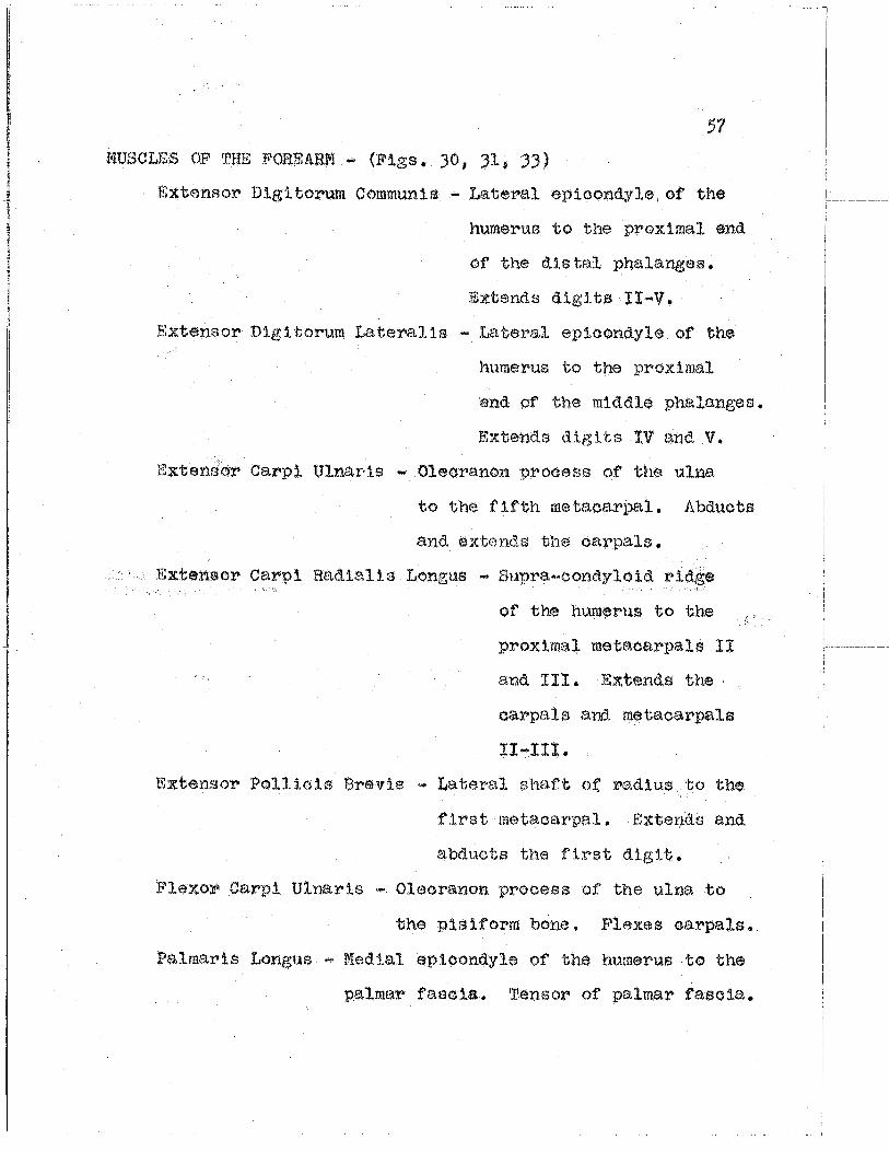
I"'USCLES OF rr.HE FOREARM ... (Figs • JO, Jl; 3))
F.;xtensor Digitorum Communis ... Lateral eplcondyle,of' the
huraerUI3 ·to the proximal end
of the dls ta.l phalanges.
Extends digits !I ... v. Extensor D:1.g1.torum Lc'lteralis - Lateral epicondyle of the
humerus to the proximal
end of the middle phalanges.
Exte:nd.s digits :tV an.d V.
Extensor Carpi Ulnar·is ... Olecranon process of the ulna
to the fifth met.aoarpa1. Abducts
and extends the carpals,
Extensor carpi RacUali s Longus ""' Sup:r.a. ... oondyloid ridge
of the hum~n·us to the
proximal metacarpals Il
and III. Extends the
carpals ana_ metacarpals
!I-I!t.
Extensor Pollicis Brevis - Lateral sh~"l.ft of: radius to the
first metacarpal. Extel)ds and
abducts the first digit.
Flexor Car.pi Ulnaris .... Olecranon process of the ulna to
the pisiform bone. Flexes oarpals.
Palmaris Longus ... Medial epicondyle of the humerus to the
palmar fasoia. Tensor· of palmar fascia.

Flexor Dig;1tO:I."Um Sublimis ... Medial· epicondyle of the
humerus to the proxir~.l end
of the middle phalanges.
Flexes the carpals and
phalanges II-V.
Flexor Dig~.torum Profundus .... f•!edial epicondyle of the
humerus to the proximal end.
of the dlstal phale.nges.
Flexes the carpals and
phalanges I-V.
Flexor Carpi Hadlalis - r1ed.ial epioon(lyle of the humerus
to the proximal third of the
metace.rpals. Flexes the carpals
and pulls the third metacarpal
tow.ar>ds the thumb.
Pro.nato:r Teres - t•1edJ.al epicondyle of the humerus to the
middle third of the medial surface of
the r>adius. Pronator of the hand,
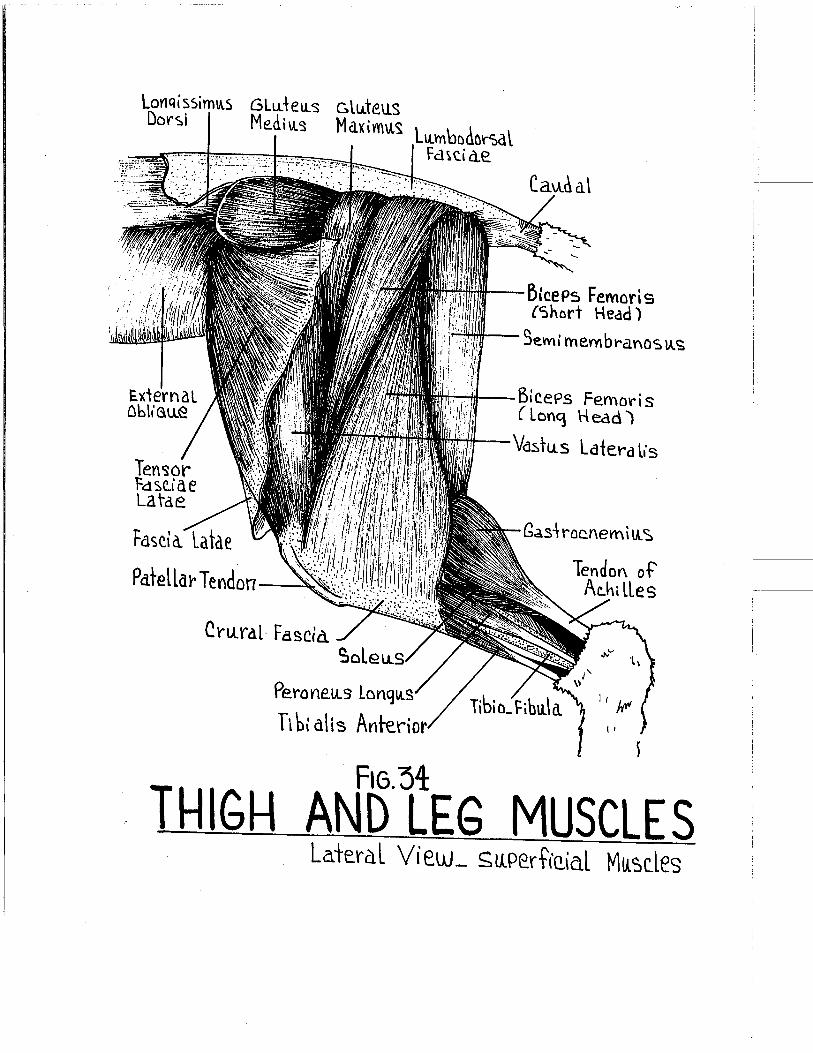
MUSCLES OF ~PHf~ THIGH 1\ND LgG •• '(Figs. )4_. 3,5~ 36)
Biceps Femoris .... Arises by tttJO head.o.
Long Head ..... (Caput IJongum) Ischial tuberosity· of the
tnnom1nate to the crural fascia..
Extends thf~ thigh and flexes the leg.
Also may abduct the llmb.;

E ~len ":lor ----tl!ll'l!r Irtdi eic;, Po\licis
Ex+en~or bi9i-\-on.Lm Later-a lis ~~\-e.nSO""-~ - C.arf>i
11 Lna ri s ~texov-
CarP\ Ulnari 5
Extenso"'-~ Ind i c..i s. Pol U c..i s
LaPitat e. ------#;~'Tf.Ca.rPat Bone
MQtacarPaL_ Pha \(lnq_ e a.l T o.'t ~\.:\:
Praxima.t--.,.1
?ha\a~qe.
Micld.l&. --b
Phala\'\Cle
Dista·-~ P~ale:mqe
C.la.UJ
1M--- ExtenS(}'f . Di'littwu.m ComMLAV\IS
~~~~--- h\en~a'f CarP\ Rddia\is lon'1JA.S
~..;.,....iJ+..- bJen<:.or ?()1\ic.is Lonq\As
~~---~Pif'hY~enl li~e ~--~tY\oid ?~ocess
of" R a d. I u. ~ .J.....;~Id--- borso.\ C a rPa\
Liqame.V\t
·+---- ~irs t Me\aearPd L Bene.
fl6. 33
FOREARNMS TENDO D()rsa\. View

60
Short· Head - (Caput ·Brevis) r..Jast sacral and first
th.ree caudal vertebroc;l sp:tn.es to the
crural fa. soia.. Exten(ls the th lgh and
flexes the leg.
Tensor Fe.scla Latae - Ventral border of the 1-lium to the
fascia lata. Makes tense the
fascia lata.
Gluteus Haximus ... Sph':tes of tne sacrum to the lateral
trochanter of the femur. Abducts the::
tt.:i.gh.
GluteuH f~(;H11us ... Crest of the ilium and 1st sacr$!.1 ver
tebrae to the grr;;ater trocha.x1.ter of' the
femt.n" • Abclucts the thlgh.
Sartorius - Inguinal ligame~nt to the medial cond.yle of
the t:t bia.. Aclducts ancl rotates the thigh.
Gro.0t:U.s - Ve.ntral Isch.1o-pub1.c symphysis to the cru:r>al
fascia. Flexes the leg B.nd adducts the .thigh.
Tenuissimus ... Slender. From the transverse processes of
th<~ second caudal vertebra to the crural
fascia. Tenses the Jateral crural fascia.
SemimembrHnosus - Ischial tuberosity to the crural
fascHt. Extends the thigh and abd.uots
the leg.
Semi tendinoeus - IschJ.al tv .. berosity to the medial condyle
of th~? femur. Extends thigh ~lnd flexes
the leg.

61
Adductor f"!a.gnus ... Lateral ,ventral pubis and ischium to
the medlal epicondyle of' the femur and
proximal .end of tibia. E:<ta:nds the
leg and aids 1n propulsion.
Adductor Longus ... Pubic symphys:ls and ischium ot the
di?.;t~al half of the shaft of the femur •.
Adducts the leg,and atds in propulsion •
.Rectus Femoris - Inferior spine of the ilium to the shaf·t
of the femur. Abducts the thigh and
extends the leg.
Vaetus Lateral is - Greatt~r troohant·:)r of. the femur to the
insertion of the reotus femorh.~ muscle.
Abd.uets th:l.gh and exten.cls the leg.
Vastus Intermedius - Grea,ter trochanter of the femur to
the patellar tendon and ortu:".al. fascia.
Abducts the thigh and flel!:ea crural
fascia.
Vastus Medialis - l?roxims.l shaft of the femur to the
patellar t.endo:n and. crural fasoia,.
Abducts thigh. and .. flexes crural :fascia..
MUSCI .. gS OF THE LOl4EB. LEG - (Figs. 34, 35, 36)
Gastrocnemius ... Lateral epj.condyle of the femur to the
tendon of Achilles. Extends the foot
and aida in propulsion.
Soleus ... VJ,teral epicondyle of the f'emur to the tend.on
of Achilles. Extends foot and aids in propulsion.
loYJqissimiA.s GLu.tetLs GltJ..te.us Dor~i Me.~i\l~ Maximu.~ LlunbDclOrsat
Fd~ti a.e
Cru.ral· Fascia. C:,ole
Perone.Lls lonq
Ti b\ a! is Anteri
Cau.d a\
~icePs Femari s {Short Head)
.~i'/1--- gemi rnembt-a.nos lA.~
~-+!~---Bl CePs Fe.mor is ( Lon9 Head)
~-vdstu.s Lafet--a t.·s
Ga~d roe.nem i lA. ~
FIG.~4
THIGH AND LEG MUSCLES Lateral View_ su.Pe.rtiQ\a.l Ml!':lc.les
I 1-i--

Ten~or Fasc.lae. Latae
C. rLu·e u:s
Patellar L:Cf aMe.\1\t
Perol'\eU..~ lo"1 Tibial\ s Anter\ o~
F\6.35
Ischial ~__...*""..--T I! berc si 1\1 ~H---ObtiAratt~v- t'l<te.rnLt~
\e.Y\v..\ ss i YY\ IA.S
~IJH.ilf--La.terc.l Troc.hahter
111/;M-- Se.Vl\t m e.mbraV\CS.\A.S :~'IIHI--9em~ Ten eli 1'\0SLLS
~"'TTH--Addu..dav- M a~mts 'tifHfii--Va':l tu.s !vr\erm ed \\A. s.
Gastroc.r\emht.s
TeVldoV\ o~ Achille~
THIGH AND LEG MUSCLES Lateral Vie.w _ DeeP Mu. ~c les
i

Pec...t\ neu..'3 -----=-~~ Ad~\.ldor lonqLL~ -----::~
~ra.c.i~is (_ C.u.t ")
Semi membran
~ertute¥1rli nos U.'-'--
Ga!lirocnemi So\eu.s
Tibialis Po~terio\"
Tend.a~ Ac.hil s
~I 1/
l 1 \
3drtO"I\t~ ( Cu..t)
Te.rtcsa r FdsC.ide la.tde.
Fdscia Lata
~~- Rec.tu. s
Jn';)e rti nq Te.ndon of Sartoriu.s
Fle.'l<Ot' biqi +n\"U • .VY\ lonqlls
Tibiall:, Antet-iar
Femoris
\<I
I FIG. 36 11-\IGH AND LEG MUSCLES
Medial View

Vas tu..s ---+~~-~ Media.Li '3
.Sartori LL s --li'H~' Femo~al A.,-~oj{.b Femo Ya L Y.--+.w:~J
ObtlLra1nr ~t--t.H~~ Femoral N.
Femu.r ~haf+ ~ed-/'if-Jhort Head ot'
Pe.c.hhetL '3
Ad~llC?.tov Lon q. Seia"t\C!.. Merve. ~emi ten dinos ttS,---;.w~~
BicePs Femori~ CJoll,!t~~F"!""'Ft:t'lfl<f---Ten u.i ssi M\AS
~-~~'-+-- lon9 Head o~ Add Ill! tor Ma~n\A.~ -~~
GraCilis-~~"
Bic.ees Femo¥-i s -~'+1---~n-ta\l SaPhen~~ V.
:~'H--~em.\ membrana su.~ -:::.~-Skin.
Fte. 37 ,
CROSS SECTION 1HROUGH Ml DDLE OF FEMUR

66
Tibialis Anterior - Latex·al cond.yle and tuheroE~i ty of
the tibia to th€! second m.etatarsal.
Flexes the foot mt1d.iad.
F'lexor D:tgl torum Longus - Proximo.l end of the tibio-fibula
to the proximal end of the distal
phalanges. Flexes distal phal-
ages and extends. foot.
'l11bi.alis Posterior ... fJfe(Ual. surface of the tibia to the
prox:tmal and mldd.J.e phalanges of
the second dlgi t. Ii:xtends the foot
and. the seo(md diglt.
Extensor Dlgitorum Longus ... Lateral ep3.oond.yle of the
femur to the proximal end.of
the d.1stal phalang~s 11-V.
Flexes foot and. extends the
toea ..
Plantaris - Lateral epicondyle of the femur to the tendon
of Achilles. .ffixtends the foot and aids in
propulsion.
Peroneus Longus - Lateral condyle of 'the tibia to the
prox.:tmE.il end of the second metat.atsa.l.
Everts and. extends the foot"-

Anteri6V' Crtu'al l=a~C.i<l
PeronetLS lon1 Lt ~ --t.:~-~.-""" Ant libi al A~ter'/ Pe\""oneus Brevis -+~--~
F i b u..l a. ---M--f:!:ii:.~&~ Perone.~.t~ Brevis H~or Di'}ifuru""'---W~~'T!
Lon~u.3
S cl e. U. ~ -----44--1+:+-!~
Lateral He.ad cf~~ Gsdstrocne.m\IA~
~~-Tibia\i'3 A"r\-er\nr
·~~~~- t )(\ensnr biqi tor\Am Lonq \AS
~~~~1t---Tibi a... ~~Wo+--E'ltt. \\a\\tAc\~ lon~\t~
.~~~~-\ibi alios 'Po~1e.~ict-
~~JH--- Sa Phe.h~\.l s A.V. N.
~~-+~---Plantar\~
~~--Medial He.ad aF G a ~1-~cc. ne."" i "'-~
SKiV\.
. FIG.3B
CROSS SECTION THQOUG\-1 MIDDLE OF TIBIO-FIBULI\

V. INTfl:RNAL OI1GANS
Figure 39

:~;I:·~=.!~~--~-1hvm\A.s Gland 1 ~t. Rt 6 -r----dM)jl~~(4~/i.~W~~~::~~
f?,·qkt Ah·
Perieard'tiA.nt-~~~¥M~
Are.Y. Crt Hedr'"7t___,~H;tti:~~~~
Fa\titorm --t:--=t~~~~ Li9ame'r'\t
Rt. Metlian. --Hr--~~ lobe
Rt Lateral-~ LobQ.
Du.oden u.m----~~Panereag ----n~
Greater Omentu.m --~ftt'-"1,.,...,.~~~--......:....
Sn1a ll----4:-+...-Inte.stine
C.o\on-~~~~~~
Vermitorm APPend i ')( ----.,~~ PlA.b\c..
SYmP\lYsis
~~----"r---....J!Hi:/-lhfe rio~ Lobe
~~~~J!!tW~-Hl~~-DiaPhYa~ M
~~~-rrte.dia "'LDbe.
i--':>"~r~--Lateral lobe ~,..,.-( au.da1e
~off#- BodY o~ 'c.:::itcma \J\....
-=...;;~. ·~~~~'--SPleen
-~-Pari{>tat Peritoneu..m
~~-+--Caec..um
' ~-,· .·: I 1 ,' ,' I
I • I I ' I F16 39
~:J~I~1~W'~ AY\terior View
. ----------

VI~ RESPIHATOBY SYSTEM
PHARYNX ... Upper end of the digestive and respiratory tube.
Oropharynx - Posterior portion of oral oavity.
Nasopharynx - Region a·bove the soft palate. Internal
nasal aperture.
Laryngopharynx ... Ventral posterior portion. Area leading
to the larynx (voice box).
Dpenings into the pharynx are:
Oral Opening - From the mQuth and oral cavity.
Nasal Opening ~ ~rom the nasal cavity dorsal to
the soft palate.
Glottis Opening - From the larynx and trachea.
Guarded by the cartilaginous
ep:lglottis.
Esophageal Opening "" From the esophagus and
digestive tract proper.
Eustachian Tube Opening - Paired. On the lateral
walls. From the middl$
ear cavity.
LARYNX .... nvoice boxn• formed by several cartilaginous segments.
Thyroid Cartilage· - Large. Forrnlng most of the ventral
wall of the laryn~.
Criooid Cartilage - Smaller than the thyroid. oart1lage.
Forms a complete ring extending around
larynx posterlor to thyroid cartilage.

71
Arytenoid Cartilages ~Small; obliquely situated oarti•
lages on the dorsal surface of
the larynx, ln the space anterior
to the ariooid and dorsal to the
thyroid cartilage_
Vocal Cords - Rudimentary vertical folds on the
internal surface of the larynx.
THYHOID GLAND - Bilobed. endoeri.ne gland located on the ventral
junction of' the larynx and trachea. Connected
ventrally b;t a narrow isthmus.
TRACHEA ... {Fig. 40) nwindpipert. Held open by a series of
incomplete cartilaginous rings. tH thin the tho:raoio
cavity, the distal portion of the trachea divid~a
into right and left bron.chi whlch penetrate the right
and left lung lobes.
LUNGS ~ (Figs, 39 1 40) Paired expansible structures, t~e sur•
faces of' which are coated. with a visceral peritoneal
membrane., The lungs lie 111ithin the right and left
pleural aavit;ies.
Superior Lobe "" Paired, Imperfectly developed on the lett.
tUd.dle Lobe - P&il'ed. Intermediate lobes on both sides •
Inferior Lobes ... Paired.. On the right sld.e, the inferior
lo'be is further subdi v1ded into the
medial and lateral lobules.
MEDIASTINUM ""' Potential space between the medta.l sur:f'aoes of'
the two pleural cavities. Contains primarily
the perieardial cavity and heart.

Internal-~.---, MammarY A R;qht -~~~-
Pre C.ava.
~u.PteMe___,~~~,;....J.,
t"'terco~ta\ Vein.
~r\_--+~.....;.....:.;j~ l24 r d~ LLV\'\
Ri q \·r\:-~~~~~.L..I--il~ Ah·i\lm
~~t.l-~-leH Common Carotid ~~t--t:f----Ttachea. .
__ ..,. LeH ~ubd-avian.A g~~h~r:----EsoYhaql.\~
· c.a\ Av.ic:. ~~-Aortic. Arc.h ~~~ PLtlmonar'l
Atter'f
;:~~~~r-lett Atriu.m
~;......f-.,.::iHil-- C.oron ar'i Vein
~~~,_Lett Vev\hitle
~~~ ~t.....med.;al Lob \Ale
~~------ f!astric.. A'rter:-/ "-.Jii-- HePatir. Aiter'Y s--- '3P\en\t A't\erY
~oel\at A"\s L\"'n''""''·Y\al 1\orta.
FIG.40 THOR/\ClC ORG/\NS
Anter'tor View
. 1 i

VII:; HEART AND MAJOR VESSELS
THYr.ms GLAND .... (Fig. 39) Antero-vent:ral portion of the thoraeio
oa v 1 ty • Irregular s ha.ped glStnd. Larger in
young specimen, Enctoortne in function.
HEART • (Figs. 39; 40, 41* 42• 43) Lies within the mediastinal
cavity. Four chambered tapering posteriorly to the
apex.
l?et>1card1um ... Loose membranous sao surrounding the hea:rt.
Filled with parioa:rdial fluid. Note its
attachment-;s to the great vessels aitcl t-o
the diaphragm.
External Features ~
AtrU.t ,.. Flight and left thin walled. structures. Note
the lateral extensions; auricles.
V<!mtrieles - Right and left thicker walled structures.
Posterior to the atria. The left ver.l. ...
t:ricle being more massive and thicker
walled than the ~ight. Right and left
sides dJ.vided by a superficially
indistinct interventricular groove •
. Coronary Arteries - Small arteries supplying the
mus9ula ture of the he art.
Arises from the base of the
aorta.
Coronary Veins ... Drains the musculc1ture of the h~a:rt.
Empties into the coronary sinus.

Thebesian Veins - Draining the same area, but
empties directly into the heart
chambers,
Internal Features ...
Interatrial Septum • Dividing the anterior atrial
chambers into right and left
atria •.
Interventricular Septum·- Dividing the heavier
walle<.\ ventricles into
a right ventr1.cle and a
much heavier walled left
ventricle. ·
Atrioventricular valves - 2 sets of double ousped
valves closing the atrio
ventricular openings,
Chordae Tendineae ... Slender cord-like stru.c ...
tures attaching to the
cusps of the a.tri.o ...
ventrtcular valves.
Papillary muscles ... '!'hick muscular projections
from the ventricular walls
for the att<twhment of the
ohord.ae tendineae.
Tra'beeulae Oa:rnea~ .... Musculs.r ridges within
the walls of the ventricles.

.Semilunar Valves .... 2 sets of three-cusped. valves
guarding the ventricular open•
ings into the pulmonary trunk
and asoe:nd.i:ng aorta.
Fossa Ovalis ... Denoting the position of the olosu:re
of the embryonic foramen ovale, an
open.tng between the atria.
MAJOH vgssELS .... (:£1, igs. 40, 41 jl 42, 4))
Aortic Arol:l ... F9rmed by a looping of the aorta as it
leaves the left ventricle.
Braohiooephalio Artery - (Innominate artery) First
major branch'from the
arch. Gives rise to the
right subclavian and right
common carotid arteries.
The left oo~mon carotid
occasionally arises from
this vessel.
Left Common Carotid Artery ... Second branch arising
very near the innominate.
Left Subclavian Artery ... Third bt>anoh from the arch
of the. aorta. 'l1he aorta
loops posteriorly and
becomes the descending
aorta.

Ligamentum Artertosum •·(Arterial ligament) Remi:nent of
the foetal connection between
the dorsal aorta and the pulmonary
trunkt ·the ductus arteriosus.
Superior Venae Cavae ... Paired. Right and left vessels
formed by a juncture of the ~xter
nal jugular, internal jugular, and
subclavian veins. They empty into
the right atrium anteriorly,
Drains the head, neck and upper
extremities.
Inferior Vena Cava - Unpaired. Lying to the x•ight of the
dorsal aorta to pass into the right
atrium. Prain.s the abdominal oavi ty
and lower extremities.
Pulmonary Trunk - (Pulmonary Aorta) Slngle vessel running
from the right ventricle antt:~riorly to
branch into the· right and left pulmonary
arteries.
Pulmonary Artery ... Paired. F'ormed by the bifuroatio.n
of the pulmonary trunk at the
region of the ligamentum arterio
sum. 'ro the right and left lungs.
Pulmonary Veins ... Tt'lfO veins from each lung entering the
left atrium ea.udo ... doraally.

Ricaht r~---Le~+ CommoV\ Carotid ArterY Su.be.l a ui a n-o~~~•
Arie'r'/ ~~h. ~--lett ~u.belauidV\ A.
Acv\ic. Arch ----;l!iib-~ ..r~-Ldl Pre C d u a.
~,£__~:...__-li q. Arter I Mu.m ~-~Pu.1 monar'(
Riqht---..!WWIJ Pre CO\Ja
Kl AvJi
AZ'/(JOS ve.tn
RiGht---'Se.mi\.\A\'\ar V Focs~a.-~ Ova.Lis
Cv;~ra~~ lerminalis
Riqht A hi n_Ve.ntri c.u.(ar
Vc1lve.
Trabecw de C.a~neae
!V\ler\JeV\~rl C.\A.l a r Grc~ve
Ve.ins
left Atriuvn 14-.1---lef t
'Sen-lilu.nar Valve 'l!~~~Lt:ft AtriLL
\leV\ \I" i C.lk lav ua Lu e. ~-' hordae
Tend ltlede.
faPi 1\arv MlA~ele
~-Moderc:r\-cr BaV\d ·
Co.,.cnavv Ar 1erv &ranch.
MvDCardiu.l'h. of Left Vevthle.Le.
........__~ -.:;.___A feY. rJ \'\tar't
FIG.4l l-ttART
AnieriDr View

Left Sllbc.Lo\J ion
AY'te.V''/
Let't
Left ComMon c.a.rotid .· A.-~~~
Pree.a.v d -~ ... <~ 3\lPre.me ---.:~ w.1~~ lntercostc\l A. Li~amentu.m--~~ Arte.riosu.m
Uesce\'\dinq Aorta.. lett ----{,w,,, A\lr\c.Le
Left · Pu.lmondt"Y A.
LeH Mdrqi Ml-·-·"""' A'rte'<''/
Pu.lmo narY Ve.\n
Poste..ricr Desc.endi Artery
Riqht tommon Ca~ot\6 Arter'/
-+--Ri'\h+ S,tA.'ocla\1\an A.
~-Innomir\a.te A.
~-:s.~-Ri~ht ?rec.aua
ll\OV\0 V' '( \<
~-Ri~ht A'rh.tV\'\
Fl6.42 HEART Dorsal V\ew
' j __

In~ernal Carofid ----..a Tr-a..n'S\Jers.e }u.~u.\a~ VeiV\.
?o)t Fdtiitl V--~.,_) &ll---An1erior Faeiat V
,.........,J.:I-4.-H--,~iJ.-..-- eat-t~ticl Bwb ~~==:I:=:~~~M/tttJI-Interna \. ,., !w~u.\av- Vein
Qo~rnon Carot:d --~~li1AIVev1e brai:-:::====:::7-\\"T1'\ Artet''l
Su.bc.ld\Jiah. Du.c.t----fiY:~~
~u.ereMe--~..-lntercostal V. Riqht Pre Cava:
CororaaV"V---1/-J~--r-~·Y.::: ArterY
Riqht---~---~-~At\"itAm
Az.Y~o<'3 ---+-VeiYl
Ivtterio r ----~tr-""'~ Vena.. Cdva.
Tho rac.i c. Du.e.t-~~1-Mll
m---A~eendil\q Ceruicat A. .......-::~--Trat\';)lJer).e
StdPu.ld r V. ·~~~ ':lu.bc\a\Jian.
A.;,.V
Ver-\ebrat Vein
VESSE~S.4bF THE NECK AND 1HORAX
-, '

VIII. AR'l~EElAI, CIRCULATION
ANTERIOR ARTERIES • (Figs. 43; 44)
Bra.chiooephalio .. From the first part of the aortic aroh.
Gives off' the right subclavi.an and right
common carotid arteries.
Subclavian - Arising independently from the aortic arch
on the left side. From the Innominate on
the right.
Internal Mammary - To the ventral chest wall.
Terminates as the superior
epigastric artery.
Vertebral • Enters the foramen transversarium of the
cervical vertebrae to supply the brain.
Supreme Intercostals - To the anterior intercostal
spaces of the ribs.
Transverse Artery of Neck - r.ro serratus ventralis
muscle.
Ascending Cervical - Ascending the side of the neek.
Axillary ~ Passing ln front of the first rib becomes
the axill~ry artery.
Ventral s;rhorac1c .... To the median pectoral muscles.
Long Thoracic ~ Supplies the latissimus dors1
muscle and. the deep pee toral
muscles•
Subscapular ... To tere~ muscle and. other shoulder
muscles.

81
Brachial - Continuation of the axillary into the
arm :region. Terminates as the radial
and ulnar arteries.
Common Carotid • Passes along ·~he lateral margins of the
trachea in close association with the
int$rnal jugular vein and vagus nerve.
Superior Thyroid - To the anterior portion of the
thyroid gland.
Muscular - Supplying the muscles of the neck.
Occipital ~ Passes deeply to the dorsal neck muscles.
Internal Carotid "" Slender passes deep to the ·
tympanic bulla and enters the
cranial cavity to supply the
brain.
External Carotid - Continuation of' common carotid.
anteriorly to the head.
Lingual .,.. Supplying the region of the tongue.
External ~1axillary - Along the masseter to · .
supply the lips and jaws.
Auricular • To the external ears or pinna.
Temporal - To the temporal region.
Internal Maxillary - D1s$ppears internal to
the masseter.
THORACIC ARTERIES ~ (Fig. 44)
Descending Aorta .... (Tho:r.acie Aorta) Dorsally looated in
the midline of the thoeac1o cavity.

82
Visceral Branches - Perioard.lal, bronchial, esophag
eal~ and mediastinal arteries.
Parietal Branches -·Intercostal arteries arising
from the aorta. segmentally to
the :tnteroos tal spaces fi
ABDOlUNAL ARTERIES ... (Fig. 44) Continuation of the dorsal
aorta into the abdominal cavity passing
dorsally to the diaphragm anterior to
the vertebral column.
Visceral Branches • Major branches from the aorta sup·
plying the abdomlnal viscera. ,,
Coeliac Axis - First major branch ·off of the aorta
after e:nterlng the abdominal oavtty.
Hepatic .... To the liver, pancreas, stomach, and
duodenum.
Left Gastric ... To the. lesser curvature of the
stomach.. Small branches to th.e
esophagus.
Splenic ... By large branches to the spleen,
pancreas, and greater curvature. of
the stomach.
Supex•ior Mes~nter:lc ... Second r.nztjor branch off of the
abdominal dorsal. aorta.
Middle Colic ... To the transverse and descending
colon.
,---

83
· ·. Pos.terior Pa.ncrea.tiao ... ouode:nal - To th~ poste ...
rio!' pancrea.s
and. the 1st
.segment of
the duodenum.
lleoeolio - To the ileum• caeoum, and . .ascending
¢clon.
Intestina.l - Numerous small branches to the.
small intestine.
Inferior Mesenteric ... Near the posterior part of
the abdominal cavity.
Left Colic - 'Po ·the ascending colon.
Superior Hemorrho1.da.l - 'J.lo the descendLng
colon and rectum.
Adrenal - From the aorta or renal art;er ies to the
right and left suprarenal glands.
Genital ~ To the ovaries in the female (ovarian) •
or to the testes in the male (spermatic).
Renal ... To the· l<idneys. Entering near the hllus of
the kidneys. The r:l.ght :renal commonly arises
oraniad to the left on 'the aorta.
Parietal Br~~nches .... ThOf:ze bre.nehas from the abc1orn1nal
aorta to the dorsal body wall.
l?hrenio .... To the diaphragm. May arise from the
coeliac axis near its origin.
I !

84
I.Atrtlbar ... Segmental branches from the aorta to the
muscles of the anteriol"' ·lumbar region.
Median Sacral ... (Caudal) Posterior continua.ti on of
the dorsal aort~ into the tail
region~ Arises from the dorsal
postv::n:;ior surface of the aorta.
Terminal Bratlches ... At the posterior boundaries of the
abdominal cavity the aot•ta bifurcates
to send branches irrto the lmver
extremities, passing under the
inguinal ligE.unent.
common Iliac - Two arteries branching from the
termlne,l portion of the abdominal
aorta to the .extremities.
Iliolumbar ... F'rom the common iliac to the
abdominal wall and. cutaneouf:l
maximus muscle ..
External Iliac - Continuation of the common iliac
into the femoral region passin.g
under the· inguinal ligament.
I<'emoral ... To the p<')Steri.or limbs.
Superficial Epigastric - Along the body wall
to anastomose l'>~i th a
br<anch of the long
thoracic.

8,$
In.ferior Er-ligastric - Passes oraniad from
the external iliac
to anastomose vii th
branches of the' ·
internal mammary
artery.
Internal Iliac - (Hypogastric) Arises from the
common 11U'l.O lying against the
dorsal wall. To pelvis and
thigh.
Umbilical - To tht~ l..l!'inf,\.ry blad.der.
Middle Hemorrhoidal ... To the rectum.
MAJOB: LYMPHNriC VESSELS .... (Fig. 4))
.Thoracic Duct ... Lies just dorsal to the dorsal aorta.
Dr·ains all the lymph from the abdorn1r1al
region• abdominal viscera, and posterior
extremi tt~as. The thoracic duct empties
into the left subclavif.m vein at its
junction with the left external jugular.
Right Lymphatic Duct - collects lymph from the right
a.nteri or quarter of · the body • ·
8mpties into the right extt?rnal
jugular vein.

Common. C.aroiid --+f----f'iir'
Ri1ht Vertebral
S!Ab'SCaP\A\a~ loM l'horAC.i c... l<'i-.hf C.oronarY--.t+-____,~;tv
lniernt\\ Mamma~Y
L!tf Co ro nl!rY --+'6-1<.-"f-Lf.f--:h.\
L ~t.\- -~~,...-V--J-INrVeV\tric.\e.
Brachia\
~v.PreMe ln-\ercosh
01kc.h.l <":l 1\\"teri o~v..~ ~--B~o"~\al
IY1ter.co":~ta\s
-1----Thoratic. Dorsa\ Ao~+a.
?h"ef\iC.-+1--:.o<.!;.c.-....,~..,..;.:.;~ \): aPhvaqM
Coeliac ""'"4QlillJJul:Duq~~~~~ A Y. i C:. ----+Pt\---------1-~ .M~;;;;;!!,_- LeH G as-h-\c Abdt>rh\na\ -...~=~t--- He~atic.
Aorta. J+----~~\en\c ~IA.I?~r\ D 'f·-'T7ti--n~-~\--,:-....,.A-......:::Jo, ... " Me~evfte'riC. ::-.c:::---~~~~-lntesHna\
S\.1..\)~fieial !nfe.~l or EPi'la~hic
Camm6~ t\iac.-~,....--~-+
'-""r::---1\eo_caeeo. c c,\; c
~~--;!~..._Ad reno l\\.mba r v~---leH Rena\
.~::::::::~~--in\erio~ Mesenie.ric ~~--Qend·al

XX. VENOUS CIRCULATION
AN1l'EB!OR VEINS - (Figs .. 40, 43 1 4,5)
Superior Venae Cavae • {Pre Cavae) Paired. Enters right
atrium. Drains the anterior lirnbs
and head and neek region,
Azygos - Empties into the right precava only,
Unpaired. Formed in the mld-dorsal line
by the intercostal veins. Drains the
parietal region of the thoracic oavi·ty.
Internal fflammary - From the thoracic and abdominal
wall. Lateral to the mid ... ventra.l
line.. t.l'ermlnates as the superior
epigastric vein.
Supreme Intercostal - Empties into the precava near
the heart. ~ehe right bxoanoh
emptying crcmiad to, .or with,
the azygos.vein.
Vertebral ... Empties into the precava anteriorly f'rom
tl:le fora.men transversarium of the oervioal
ve!'tebrae. Drains the cranial cavity.
Subclavian- Unites with the.exter:nal jugular to
enter the precava. Drains the shoulder
and upper extremities. Formed by the
braohlal and axillary veins.
External Jugular - From the anterior head region.

88
Internal Jugular - Drains the brain and cranHtl
os.vi ty ~ Buns along side of
the trachea in close rela
tionship to the common
carotid ctt'tery and vagus
nerve.
Transverse Jugular - Transverse vein connect
ing the right and left
ex.terna 1 jugular veins.
Anterior lll<!:.lcial ... From the jat.'ls ancl face.
Posterior Fao.lal - Frorn. the external ear, pinna~
POSTEB.IOH VfnNs .... (F'ig. 14·5)
Inferior Vena Cava - (Post Cava) Unpaired. Enters the
right atrium post~~rlorly after
penetrating the diaphragm into the
thoracic cavity. Lies to the right
of the dorsal aor·ta.
Phrenic .... Drains the diaphragm~ Enters the post
cava as it pasBes lnto the thorax.
Hepatic .... ~::Hngle vein fro.m the 11 ver to the post cava.
Adrenolumbar - Paired. Drains the suprarenal glands.
Renal - Paired. The right renal entering the post
oa'J»a more oraniad to tb.e left.
Genital - Ovarian or spermatic from the gonads.
Lumbar .... From the muscles anterior to the ilio ....
lumbar veins~

89
External Iliaa - Paired.. Two large tributaries f.rom
the lower extremities t>~hich empty into
the post cava.
Vesical - From the urinary bladder.
l'nferior Epigastr:tc -,From the ventral body wall.
Anteriorly it terminates as
an anastomoses with the
internal mammary vein.
1~'emoral ~· Matn vein of the thigh. Drains the exter•
nal genitalia and muscles of' the thigh.
Deep Femoral •w D:ra1.ns the deeper muscles of the
thigh.
Intern~.l Ilie.c ... (Hypoga.tStrio) From the pelvic region.
Median Sa.c:ral - {Caude.l) Drains the sacrum and tail.
Middle Hemorrhoidal ... Fx:om the region of the anus. ' I
~--!

lYlterna\ 1~9u.lar __ ........,.__. A-£i\la~v
Trib\t\ta~•es, ~crminq
1-\e(7ati c.. V.
Renal--~
G.eni+al--....-
t'lCttrl\a\ I \.1 a c.. -----~!:;~""" Smatl
'SaPhenoiA:
118----h\ernal 1\A'liA.\av ~-A~centiin~ CervicaL
~e(Jha\,·c.
Brach,'al \w--SI.lhsraPu.la\"
~uPreme
lnte~co s tal t-----lon'\ 1horaci c.
r---Interna\ Mammat'Y
+-----Postex-icv- Vena Cdua
c+-----Abdominal Po~Jcava.
Jr.:---Adrenal ~~r---Adrenc_Lu.Yhbar
~----lu.mbdr
;:::::::::~s---1\io lu.m bar ~~---ComMon Iliac
'v'S~---I~fe.rtnr tP'•1as\ric .... ~-~~-Ttlte..rnal t\\at
re.vnoral Fle.45
VENAE CAVA£ TRIBUTARIES

X. HEPA'.i~lC-PORTAL C!RCULP.TION
HEPATIC-PORTAL VEIN .... (It'ig. 46) Empties i11to the liver.
Gastroduodenal ~ Formed by the right gastroepiploic and
the anterior pancreat1ooduod.ena.l veins.
Dt·a.:t:ns the panereas, duodenum, and
portions of the pyloric stomach.
Gastrosplenic - Formed by the following veins.
Splenic ... From the spleen ;:m.d left gastroepiploie
veins of the stomach and omentum.
Coron?try - From the lesser curvature of the stomach.
Posterior Pancrec).ticoduoclenal - Collects from the
pancreas and duodenum,
Inferior l•1esenterio .... From the transverse; descending
colon and rectum.
Superior Mesenteric ... 11'rom all other intestinal regions.
Intestinal -Collects all areas of small intestine.
Ileooeooool:to - Il'rom the ileum, appendix, caecum,
and ascending colon.
Appendicular - From the vermiform appendix.
Anterior Ileocecal - ll'rom the sacculus rotundus.
Posterior Ileocecal - From the distal caecum,
1leum, and ascending colon.

~~-lef~ li lie\' I obe
D~And.enu.m
An~erio\" In~e~Jina\ f,o. '!6
Gac:.h-o. ~PiPir.i c
Ant DIA.aAena\
HEPAT\C-PORTAL SYSTEM 5emi _ Dia.(\ramo. tiC..

SALIVARY GLANDS - The oral cUgestive glan.ds. Paired.
Parotid Gland ... L~rge diffusely s 1 tuated fsland ventral
to the pinna. Drained by the parotid
du()t ( s tens on • s) • passing over the lateral
surface of the wasseter to the oral oav1 ty.
Submaxillary Gland ... Ventral to the parotid gland and.
somewhat smaller in slze. Drained
by the submaxillary duct (Wharton's).
Sublingual Gland - .1\pproxim~:t-tely the same size as the
submaxillary gland. Lies aranial to
it. The sublingual duct joins the.
submaxill<SI.ry duct to enter the oral
cavity as a single aperture.
Tl~gTH .... 'rotal number of 28 in the adult. Conipos0~d of three
typesJ 1no1sors~ premolars, and molars. Dental
formula ... i Q. • .l..J.. 1 0 2 )
TONGUE - A large mutH.rular organ a ttaohed. by muscles to the
hyC>id. apparatus and bears numerous taste buds. The
tongue covers t~he floor of the oral cavity. Attt.?.ohed
anterj_orly to the floor of the oral oewi ty by a
med.1an fold, the frenulum.
PHAHYNX - Posterior oral cavity leading to the esophagus,
Note the paired. palatine tonslls in this region.,

ESOPHAGUS - Muscular tube connec.t.:tng the poster·ior pharynx
with the stomach. Ll,.es d.orsal and to the left
of the trachea ..
STOHACH - (Pig. 39) Muscular sac trJhich lies ~Jithin the abdom
inal eavi ty at an obliquely transv~~rse angle.. Slt ...
uated towards the left of the midline. Anterior
part p$.rtia.lly covered by left lobf~s of the liver.
Greater Curvature - Lon.g posterior curved surface.
Lesser Curve.ture .... Shorter anter•ior curved surface.
Cardis.o Stomach - Proximal portion of the stomach into I
whi.ch the .esophagus empttes. Note the
cardiac sphincter valve in this region.
Functus of the Stomach - Large bulge a.t the antero-
lateral region of the stomach
near the o~~.rd.iac stomach.
Body of the Stomach - Central portion of the stomach
between the pyloric and fundic areas.
Pylbrio Stomach - Distal most portion of the stomach which
($ltnptif3S into the first portion of the
duodenum. Note the pyloric sphincter
valv~.
GREATER OM11:Nr:eut1 ... A thln. sheet of peritoneum extending from
the greater curvature of the stomach to
cover the coils of' the intestine. Usua.tlly
containing deposits of fat.

9.5
LESSSE ONE~'TUI'1 ~ Port ion. of ventral rtlf.H"iente:r.y between the
stomach~ cluodenum, and. 11ve:r•. Composed of
a gastrohepatic and. B. du.od.enohepatic omentum.
SMALL INTESTINE ... (Fig. 39)
Duodenum - First portion of the smP.Jll intestine following
the pyloric valve. Enterlng the wall of the
duodenum is the comrnon btle duct from the 11 ver,
and more posteriorly the pancreatic duct.
Jejunum - About one-half of. the :remaini-ng length of the
small intesttne beyond ·the duodenum.
Ileum - '.rhe remaining length of the small lntestine to
the ileocecal junction. Not sharply differenti
ated. from the jejunum.
COMMON BIJ.,~B DUCT ... Small duct formea. by the union of tlw b.apa.
tio ducts from the J..iver and the oysti.Q
duct f'rom the gall blaCl.(le.:r. E~ntera the
first portion of the duodenum on ·the dorsal
surface.
ILEOCECAL JUNCTION ... (Fig. L~8)
Ileum - Terminates as the U.eooecal valve. Note the dila•
t:io:n in this region, the sacculus rot.undum,
Caecum - Thin walled, very large a:nd dilated diverticulum
containing a spiral valve. 'l'he oaecutn terminates.
in an elongated slender vermiform appendix,
LARGE: !.NTEsrri.NE - (P:lgs. 39, 48) Oftem referred to as the oolon.
A co:ntlnuation from the region of the sacculus
rotund urn.

96
Ascending Colon - The first part of the large intestine.
Haustra ... Small sacouh~t1ons on the surface of the
first part of the colon.
Taenia ... Longitudinal bands of muscle separating the
haustra in the 1st part of ·the colon.
Transverse Colon .,. Short segment running from the
ascending colon to the left.
Descending Colon ... Continuation of the colon posteriorly
term1n~ttt1:ng in the mid.~inE) as the t-ectum.
Rectum ... Terminal portion of the large intestine.
Anus - External posterior opening of the digestive tract.
DIGESTIVE GLANDS ... (Figs. )9; .!4-7) All are wj.thln the a1,dominal
cavity.
Liver ... Anterior most position within the abdominal cavity.
Anterior Lobes ... Paired. Right and left r.ddes sepa.
t'ated by the falciform ligament.
Posterior Lobes - Paired. Caudad to the anterior lobes.
caudate Lobe .- Unpaired. Independent lobe cc.1.udad to
the base of ·the pos ·tertor left lobe.
Gall Bladder• ... Spherical structure imbedded in the pos
terior surface of the right a,rrt;erior liver
lobe. Thin walled.
Pancreas .,. Diffw;;e mass of glandul~n" tissue throughout the
mesentery in the region of the duodenum. The
panor~atio duct is small and enters the duod.e ....
hum at a distance posterior to the entrance
of the oommon_bile duct.

97
SPLEEN ... (Figs. 39; 46) Browntsh eloru$a,ted gland lying pos
terior and dorsal to the greater curvature of the
stomach. An organ belonging to the circulatory system.
LYMPHATIC TISSUES ... (Fig. 48)
Pancreas A sell i ... f"lesentertc lymph nod.es along the course
of the superior mesenteric artery.
Payer's Patches - Lymphatic nodules a,long the dorsal
walls of the intestine.
Sacculus Rotundus.- )4ymph.oid patch at the terminal and
of the ileum near the ileoaeoal valve.
Tonsilla Ileoceca11s Major - A large intestinal lymphoid
patch within. , the wall of the
caecum. Dorsooaudad to the
ileocecal valve.
~~onsilla Ileoceoalis Minor - Intestinal lymphoid patch
within the wall of the caecu~
Dorsocraniad to the ileocecal
valve.
Vermiform Appendix - Terminal end. of the caecum. An
elongated diverticulum which is
lymphoid in function.
Dik\PHRAGM - (Fig. 39) Forming the division betv;een the thoracic
and abdominal cavities. A, muscular partition with
a central tendon. Perforated by the inferior vena
cava and the esophagus. aespiratory in function.
I
1-

DiaPhrdqM -
t?iqk\AV\\er\cr l o'oe
~-Dorsal Aor-ta..
l Tendon ~- Faltitorm littament
Riq\\t ?os,e.'fi or
LC)be G,all B~adder
\wr-~&--.v--"ePa:\i~-?artal V.
l.ert Posterior
Lt~be
CYstle. D~~1 -·~· In{er\ or
\lena C.av£1 Cau.date Lobe.

Cae~\A.m
Colon
Ve.rMi~rM APP~ndix
Fle.48 ILEOCAECAL JUNCTION
I -

.XII. UROQENI~AL SYSTEM
URINARY ORGANS - (Figs. 49, 50)
Kidneys - Paired. Located dorsally, the right kidney being
more anterior than the left. Imbedded in fat.
Capsule - Fibrous coat immed.iately surrounding the
external surrac~ of the kidney •
. B.ena.l Hilus ... t4edian concavity. Note the ureter,
renal arteryJ and renal vein entering
or leavj.ng this area of the kidney.
Cortex ""' Narrow peripheral substance of the kidney
as seen in a longitudinal section.
fqedulla ..., Medial central portion of the kidney •
including the renal papilla. Characterized
by.the radial strlations.
Renal Pelvis • Enlarged funnel-like prox1~1l end of
the ureter within the.kidney mass
near the hilus region.
Bena.l :Papilla ... Projection of the medullary substance
into the renal pelvis.
Ureter - The major duct from the kidney passing
posteriorly to the base of the bladder.
Urinary Bladder ~ Ventro-posterior portion of the
abdominal cavity. t-1uscula.r sac.
Urethra - l\la~roow tube from bladder to external opening.
SUPRARENAL GLAND (Adrenal) • Paired anterior to the kidneys.
An endocrine gland.

101
f1ALE REPRODUCTIVE ORGANS "!" (Fig. 49)
Testes ... The male reproductive gland. Within the scrotal
sacs, on the outside of the body wall, ventral
and anterior to the anus.
Epid.idyml.s .... lt"'irst portion of the male reproductive duct.
Vas
Caput Ep:l.dJ.dymis - The enlarged anterior region.
The vas efferens empties into this
duct from the testis.
Cauda Epididymis ... The posterior constricted a:rea
Deferens -
along the medial side to the pos-
terior region of the testis.
The major genital d.uct of the male. Passes
from the poste:rtor end of the testls into
the abdominal cavity through the inguinal
canal terminating in the urethra.
Prostate Gland ... At the pc,int of juncture of the vas
d.efe:rens with the urethra» a white
massive gland.
Seminal Vesicle - Located on the dorsal surface of the
base of the bladder. Vas deferens
en·ters ventrally,
Bulbourethral Gland - Dorsal wall of urethra behind the
prostate gland.
Penis ... Anterior and ventral to the scrotal saQa. Pos; ...
terlorly directed. Attached to the ischium by
the cr•us penis.
-~

.,._ ___ Jnfe~\ot-Ventl Cnva. ---+,r--+---- Dar!.~t Aorc\d..
SllPr~ ren~\ aland
,llp........,_~~Renal Vein.
...111911--leH \<idne'/
-4---\\----Sem,nal Ves\c.L.e.
r:~-----\+---H·o~+ate. Gland
--11'---t+- ColoV\. ~-~--~_..-~f+--£.rdC\.L\atov-v
DlLd-
~+--Bu.lbo.Urdht-al Gland
~,:.:4-~~---fiii'-~~Perineal Gl4r\d
·~-ll--Lett Te"S.tes
Ft6.49
UROG~N\T~LS male.. Sv<; teYY\

103
FEMALE EJ£PRODUCTIVE ORGAN$ ... (Fig. 50)
Ovaries -. Small paired bodies along the dorsal body wall;
posterior to the kidne;ys. The reproductive
gland of the female. Supported along the dorsal
wall by the mesovarium; a peritoneal fold,
Ostium Tubae Abdominale - Broe.d funnel-like expansion of
the proximal uterine tube.
Uterine Tube ... 'rhe first portion of the oviduct distal
to the ostium tubae.
Body of the Uterus - A typical duplex-type uterus con
neotirlg independently to tb,e ve.gina
from the right and left sides. A
highly muscular structure. The
distal eeoond portion of the oviduct.
Vagina - '!'ubular posterior continuation of the genital
traot. It receives anteriorly the right and
left apertures of the ueteri. At lts exit into
the vulva,, it unites with the urethra to form
the urogenital sinus.
eulbourethral Gland - Located along the dorsal wall.

Uri narv~~....l,-.4-BI.ddd~~
Ute~ine. ---If-~~ Orific..e. u~eth~a-~-
SUPra_rena l Gland
-----~:...~-.'6f&-.~-Re"al A\"1-er'/
..........._--:....._,_,.~.,.....-..;v.-- Ova Y' ian A.~te.rv
Vaqina----h---11~~~-

Xl!I. CEN'J:-RAL NERVOUS SYS1l'EM
MENINGES (!1eninx) - Th.e membranes surrourtding the brain and
spinal cord. Contains the cerebrospinal
fluid.
Dura Mater "' ~rough outer sheath.
Pia Mater - More delloate, vascular sheath in direct
contact with the brain and cord surface.
Arachnoid - Loose somewh.e:l:. web-like membrane which
connects the pia and dura mater. N()t readily
recognized in a gross dissection.
t.A11EHAL ASPECT - (Fig. 51)
Cerebral Hemisphere - Paired. Smooth cortex due to
absence of promh1em.t gyri and
sulci. Note the sylvian (lateral)
fissure on the lateral surface.
Olfactory Bulb - Anterior projections from the region
under the oerebr•al hemisphere. Paired.
Olfe,otory TrEtC'ts - co:nneoti:ng band from olfactory bulb
to the pyriform lobe of the cerebx-um.
Optic Nerve (!l) ... The second. cranial nerve passes under
the cerebrum to the optic chiasma..
Cerebellum ... Posterior to the CE!l"'ebral hemisphere. Note
the flocculus and. vermis.
Medulla Oblongata - From which the major cranial nerves
emerge. Ventral to the cerebellum.

CeY~bra.l ~em\sPh&e Pine.al Gland 3Ylvlatt Fiss\lre., I 1 Cerebellu.m
Ot ta~tot-'1 T\-ad-
Ol fadorY 811\b 1 ~
OPtic. Nerve. 1I OP'fia.
C."iaCj.m.a
Ft6. 51
BRA\N Lateral Vie.W
Oc.u.\om~tor I . . . l I 11ITr
1 . lm Tr,qemu\a. JWL
emPora -sr lcbe. Troc.h\ear ~oC:Hl L
fotm. Lobe.. N W
Floce!A \lAS I
FO\u-th Ventricle.
· 5Pinat Cord
lhPoqlossal :xi[
A~cessorY "I[
GlossaPha"rnqeal K

filEDIAN SAGlT'rAL SECTION .... (Fig. 52)
Cerebral Hemispheres - Viewed on the medial service.
Not sectioned dor>sal to the
corpus callosum.
107
Corpus Callosum -Longitudinal broad white band passing
transversely from one hemisphere to
the other. l"orms the most dorsal
structure in the midline.·
Fornix - White narrow bt3.nd extending ventrally and
anteriorly from the hippocampus.
Foramen of' r1onro .... Small opening posterior to the column
of the fornix. Leads from the third
ventricle to the lateral right and left
ventricles or the cerebral hemisphere.
Septum Pellucidum - A thin septum between the body of the
fornix and the anterior portion or the corpus callosum.
Anter'ior Commissur'e - Ante:r•ior to the ventral end of the
fornix. A small white bund.le of
fibers.
Lamina Terminalis - Thin membrane extend.lng from the
anterior comm1sstn:•e ventrally to the
region of the optic chlasma. Marks
the anterior most boundaries of the
th1t>d ventricle.

Spinal Cord - Posteriox> (caudal) continuation of the
medulla oblongata,
108
Thalamus -. Thickened. lateral walls of the third ventricle.
Massa I:ntermedia. - Hounded. intermediate mass connecting
the right and left thb1lami.
Pineal Gland (Body) ~ Conical structure projecting from
the dorsal posterior portion of
the third ventricle.
Corpora Q.uad.r.igem1:t"lll - Four eleva, tions (quadrigeminal
bodies). in the dorsal region just
posterior to the chorioid plexus
of the third ventricle.
Superior Coll i.culus Anterlor pa1r.
Inferior Collieulus - Smaller. Poste~ior pa-ir.
Aqueduct of Sylvlus - Slender- channel ventral to the
corpora quadrigemina. connecting
the cavi t ios of the third and
fourth.ventricles.
Ante:r>lo:r> Medullary Velum - Thin memb:retne anterior to the
c~rebellum form.irlg the an-ce:rior
roof of the fourth ven.triole.
Posterior Medullary Velum - Thin membrane posterior to the
cerebellum and forms the pos~
terior roof of the fourth
ventricle and chorioid plexus.
A:r>bor Vitae ... White bra.nching structures seen o:n cross
section of the.eerebellum.
-~l

Olfad·arv Bt.t\ b
FIG. 52
BR~\N
J.liPPo~amPtA.S f>o~terior eommisure.
For nit
Of'tic. I . I Chiasma 1 t-d.
Vent. OP"fi~ LaMilna.. ][ Te.rmi nalis
I Pineal '3~alK Anteriot- Me~ll.\ larv o j Vetu.m
MaBmillarv od.v
Ma.3Sa. lntermedia of ThalamiA.s
;thai a mt.ts
rineal " b " ~..e~e e.u\Am
P~ter\or Medtlllarv Veli.U\\
Fou..\"th Venh·ic.le Jll'
u.adri qemi na l l4\Ynina.
Cen{rdl Cdndl
Saqi tta.l Sec.tion lntlll'ldi b"'-llA.M
---·---,---- - ---~--- -··-------

DORSAL ASPECT • (Fig. 53)
Cerebral Hemispheres ... Paired. B.ight and left hemi"':'
spheres separated by a median
longitudinal fissure (.sagittal
fissure).
Olfactory Bulbs ... Anterior swellings ben$S .. th 'the anterior
portions of the oerebral hemisphel"'es.
Continues posteriorly as the olfactory
tracts to_the pyriform lobe,.
Cerebellum - Pos·terior to the cerebral hemispheres.
Vermis - Median lobe.
Lateral Hemispheres - Paired lateral lobes.
Flocculus - From each hemtsphere a narrow stalked
lateral lobe.
Corpora. Quadrigemina .... Seen between the cerebrum and the
c~rebellum.
Medulla Oblongata - Part1all~ overlapped by the vermis.
Note the thin wall of the triangular ...
shaped fourth ventricle.
Spinal Cord - Cylindrical extension from the posterior
portion of the medulla caudad.. Enters the
:neural ¢anal within the vertebral column
to continue posteriorly.
VENTRAL ASPE~CT - (Fig. 54)
Olfactory Bulbs ... Anterior swellings contlnuing posteri·
orly ~a the olfactory tracts to the pyri
form lobe of the aerebrr:J.l hemispheres.

Sal! iH-al--~----'i!lll i!iS.li.U..Y'Q
(Meclidn. Lon~rh1d \ nal
fissllre)
lett Ce~ebral
He.mis ?here
StA.Pe.rior C otl i c.u.ti -~~iBJ!~ lnte ri.or ----,.:f'i~o§ECc.\Utu.li
~t .. ~~--~7!'Jt-rit'\eal Gtand ~~~~ .,...;pL---riri ~orrn Lobe
~~ Ri+--FtOCCIAllAS
~~~~~~---- Cerebe. \\IA.m
SlossoPhar-vnq c1L--~~ .... l~~~~Fi!Art"""---Vermi s n: Fou.rth. VeVJt\"i Vd~tl'3 Y
..... '~----MeaiA.\la. Oblon9ala.
-f----Hvraqlo~~al 1[
FlG.53
BRf\\N Dorsal View

112
Pyriform Lobes ~ Large vent~al•posterior portion of the
cer~bral hemispheres.
Rhinal Fissure - Separates the py:rifor:m lobe from the
rest of the oe.rebrum.
Sylvian Fissure (Lateral· Fissure) ... Extends·. dorsally
from the rhinal fis•
sure alor¥r the lat~
eral surface of the
cerebrum,,
Circle of v1illis ... Bing of blood vessels supplying the
ventral surface of the brain.
Optio Chiasma - Crossing of the right and left optio
nerves to continue posteriorly as the
opt:to tracts.
Anterior Perforated Substance - Hegion between optic
chiasma and olfactory
tracts.
Tuber Cinereum ... Rounded elevation posterior to the optic
chiasma. For the attachment of the
infundibulum of the. pituitary gland.
Pituitary Gland (Hypophysis) - In an intact specimen,
attached to the infundi
bulum. . Endocrine in
function.
Mamillary Bodies ... Rounded elevations posterior to the ·
tuber cinereum.

113
Cerebral Peduncles .... Bundles passing obliquely forward
from the region of the pons,
Pons .... On the ventral surfaqe abroad. mass of fibers·
forming a commissural band extending t!'ansversely
across the brain and. dorsally towards the cerebellum.
Oculomotor (III) ... Artses from the medial ventral region
of the cerebral pecluncles to pass
anteriorly.
'rrochlear {IV) ... Arises from the region of the anterior
medullary velum to pass tilong the lat
eral surface of the cerebral peduncles.
Trigeminal (V) ~ Large. Located posterior to the lateral
po!~tions of the pons. Arises by tvw
roots, a larger sensory. and a smaller
motor root.
Abducens {VI) - Small. Passes forward from the ventral
surface of the medulla posterior to the
pons.
Facia.l (VII) - Laterally placed. Arising f'rom the medulla
in close relation with the auditory {VIII).
Auditory (VIII) :.. Aris i:ng from the medulla pos t;erior to
the faa 1al and t:rigemino.l nerves.
Glossopharyngeal (IX) ~ Arises by several linear roots
along the lateral margins of the
medulla oblongata.

114
Vagus (X) - Arises from the late:re,l margins of the medulla
in direct line with the glossopharyngeal and
posterior to it.
Spinal Accessory (Xl) ... Arises by numerous roots along the
lateral margin of the medulla.
Hypoglossal (XII) ... Most; posterior of the twelve cranial
nerves. Arises by several roots from
the ventral surface of the medulla.
Medulla Oblongata - Anterior swelling of the spinal cord.
Ventral Long1 tudinal Fissure - f;1edian ventral groove
exte:ndi ng from the medulla
to the spinal cox·d,
S}'?INAL CORD .. A thick eylj.ndrical cord tra.versi:ng the verte
bra,l (neural) canal throughout its length. The
cord terminates in the saor•a.l region as the
filum terminale.
Spinal Nerves - Formed by the union of two rootsoi
Dors~:.tl Root - P:r•om the d.orso-la:teral region of the
spimil cord.
Ventral E.oot - From the ventro-lateral region of
the splna.l cord.
Dorsal Root Ganglion - ~nla:rged. S\'l'elltng in dorsal root.
Dorsal Fissure • Narrow median groove.
· Ventr•Etl Fissure .... ltJide ventral median g:t•oove.
Central Canal .,.. Small oa.vlty through the center of the
cord.

oL fac. tor-t ---.A'--J:.._,_l Trac.t-
S'ilvian ---1--iF
F\ssure. -p,·r:form--+-~-4---
Lo'tle... R\ q ht _..,.___
Cere.bral Hevni9Phere.
Cerebral ---l~~-__.;.;~ Pecluhc.le.
Por1s --1
~~,...-\~-OP\\c ][
Qptjc. ~---+T+-- Chias.ma
t.::;i:~rt~--r,~cr-HvPoPhvseal ~~alK
~~---if---+--Mami l\Cll·'l Bodv
:-~.)3~~--f------:h.,__Ocu..la~tott
·TII :-.::JH~ ...... ~--Troc.hlear , ... ,~-~. .:nr
Cerebe.lllA.m Ci~·:~If.~;~~(Ji§~~~rTtiqe.mi na l
""Sf: ·~""''.lltrt~~ _. . ...,....,,,___Ab~ueen'5 "SIT:
AtAditorv un~z._-Fa~i·d.l W
1ZIII ~---mo~~oPhatYilqeaL JY Va¥s .:.:.H-~t----SPin~Acce-:.c;orY
SPina.\ Cord -----+--lk-~............_r&.+--HYPC~G\loc;~a.l li.
FIG. 54
BRA\N Ventral View

BIBLIOGRAPHY
i I

XIV, BIBI.IOGBAPHY
The following references are a selection of, and intended. only as a oo:n<lense<l guide to, publications that are likely to be useful to students interested in descriptive and. experimental morphology of rabbits.
Adame • Leverett A. and Ham~~l Eddy. ,9oml?§:rat1 ve A_nf!:_tOL\lY,;, .An Introduction to the Vertebrates. New York! John Wil$y a.nd Sons, Ir1e7'; '19lj:9,. ·
Atwood, William H. Comparat:tve Anaton!,l. Second edition. St, Louis: r.rhe c.. V. f'!losby Company, 1955.
Baldwin, Fran<Hs M. "Variations in the Carotid Arteries of' the Rabbit. 11 Ana.t. if2£!~ 15:49 .... 51, 1919 •
.._..._........,"'!". 11 Notes on the BrE~-ncheG of the Aorte, (arcus a.ort~e) and the Subclavian Artery of the I1abbit.•t Anat, 1\~.9· 19:173-183, 1920.
Bensley, B. A •. Pt::actica.l i},p.atorJ11L .2f. .t.~ £1Jll2.bit. Seventh edition. Revised by Craigie, E. H. Philadelphia: The B1al<~r3ton Compa,ny1 19!~6.
Bern:, Howard and Boris Kriohesky. ttAnatomio and Histologic Studies of the Sex Aceessories of the Male Rabbit • 11
Un~v. Q!. C.g].ifot~-~& 1?,u!?J.· .t.u '~· 47:175 ... 196t 1943,
Brodal, Alf. nThe Cer~bellum of' the Habbit. 11 Jour. Go11m,. Heu:rol .. 72;6)-70, 1940~
Brown. Wade H., LouiBe Pearce, and Chester Van Allen, "Organ Weights of Normal Rabbits." JOla£• !!1.zR· Med. ltJ: 733-741; 1926.
l3uoy • Paul Clancy. "The Nici tatlng Membr,;.xl~ ...;.nd the Superior Cervical Ganglion, in the Babbit." Jour. g9m;a. Ji~urol. 43:221-230. 1922.
Crabb• Edward Duane. ~r1no1Ql<a§. Qt Functional Anatomx. of .!mft Rabb*t. St. Louis: JohnS .. Swift Comp!Si.YlYt Inc., 19;1,
____ and Mc-l.rgaret A. Kelsall. 11 0rg~~,ntzation of the Mucosa and Lymphatic Structures in the Rabbit Appendix." Jour. Morr>.h· 67: 3.51 ... 367, 1940.
----' 11 An Anomalous Everted Piece of Ileum in an Adult
Cottontail Rabbit.u University of Colorado Studies Se:r. D-1;221~22); 1941.
r

117
C ra 1g1. e. , E • Horne. . A flfbora tor u ~~kie ,1:.2. the Ana. };om~ of tll,! Jlapb}i(. Philade'!p 1a: '1't1e B < ston Company, l 5T:
Drapeau. Elizabeth E. nAn A:natonU.ce.l Study of the Furless Cond1 tion 1n Habbl ts 1 ,. Jour. Mornh• 54 (2) :.365-388, 1933.
Dunn; Thelma B. and Albert M~ Kessel. nlnjeotion and Clearing I1ethod for the Rabbit •s Ear," Jour. Nat!Q.n .• canpeu;: l.n!1l· 4;3.59 ... 360, 1944,
Eales- Nellie B. nNote on the Abnormality in. the Blood vascular System of the R~.bbit, 11 Jov..£.• A;ga.Jt.• 65:1.52-153. 19)0.
Edgeworth, It,, H. ~~Jt·Crani$1J. . .t:tuscles 9!. Y.trtebJ:1lt~s. New York: The l\lacmillan Company 1 1935.
Enuttens, C • W. u The Gt•otvth of Heproduc t i ve and Endocrine Organs of the Female Habblt,n·,roty;. Endocrinol, 1;409-416• 1939.
:t~'orster, Andre. "Le.:Masseter et.le Pterygoiden Externe Chez . Ce:t:"tains Hongeurs (cobaye, r•at, lapin). E·lmde d 1 Anatomia Comparee et d 'aptation, 11 Arch. Anat. flist.o .. l.• ~ F;mhr¥Pl.• 9:193-226, 1929. . '
Ga1sstone, s. 11 A Comparative Study in Vivo and Vitro of Hat ~nd Rabbit Molars," !T..99.• Jigz. §..QQ.. ()t.ongopJ Se:r. ft, 126: 315-330, 1938. .
Girgis~ A. 11 'l'he Development of the Heart in the Rabbit,'* Pro.9.• Zoql.. Soc. <L&n..4on) 3:755·782, 19}0.
--~-· "A Further Contribl.ltion on the Development of the Heart in the Ha.b'i-;1t, 11 Proc •. &2£1• Soc. (London) 2:421·450, 1932.
Goodrioh 1 Edwin s. .§,:tudles .. Qll tq~ Structure .1m~ DevelQ,:gme~ .2t Vipr~ebratef[. London: The I1acm1llan Com:par~y, 19}0.
Qrauer, Theophil P. . 11 Be generation in the Pancreas of Habbi t, '* /J.lme;r. ;.,ou~. Anat. }8:233•2.53, 1926.
Hegner, Hobert w. Colleg_~ Zoo~. l1'ourth edition. New York: The Nacmillan Company, 1939.
Huxley • Thomas H. ! Manual .Qf.. 1h.El Ana tomx. .Qf. yer.,t!l,"Q£a teft Ar.J1.~. New York: D. Appleton and Company, 1872.
Hyman, tibbie H. ! !@,P,orfit,Qr.JL rt~n~1!i'!.l .f.9J.: Compa:t:.~tiv~ ~patOJ.TIX.• Chicago: University of Chicag,o Press, 1922.
--~-· pompa;cat~ve yerteJ;;,rate Anato!W£. 'rhird edition. Chicago: University of Chicago Press, 194?.

Johnson; Robert P~ "Dissection of the cranial Nerves Principally of the Rabbit1• (unpublished. paper, 19.32).
Kelsall, Mar•garat A., 11 0rganization and Developmental Modifications. in the i Mucosa of the Ha.bbit. Append.ix 11
(Unpublished Master's thesis, The University of Colo:t•ado• Boulder, 1940) •. ·
__ ....,..._. 11 Structure of thr:J SacGUles in the Procolon of the Pj.ka.tt ~our,. Mor•Ph• 70~115-119, .19i.t2.
i18
Krause,' fi.ud.olph. rUki"'os,koRi.sche A;qatomJe ~. \4irbe;J.t1gn;:§ 1n !j}inzeldl}\rstelly.m;en I.· ,S§Ul~?tiera. Berlin; l4alter de Gruyter and Company, 1921.
Kuhlendeck• Harti& and B.uth N. Niller. "Pretec·tal B.egion of the Rabbit Brain, n • .J:.o.u..r.:.. Comp. N§uro;t.. 76 :323··-:362 t l9l~2.
LewiB~ /1.,. A., ancl c. H. 'J.lurne:r. '11.l'he S1ze of the Rabbit Mammary Glano. with Successive Lactations•" i_ou:,r:. ,t?ail":Y . .§£1. 25 :57,.,.59, l9ll·2.
Little, r1. E., and R. T. Kempton. fi ~!ftbor&.t§(ry M.~q,nual for Com£Eirative Anatoffi'U'. New York: 'J.1he r~taomillan Company, 1 . -a·· •. - ~ ::..&.,
92 •
McNutt, c. tv., and P. B. Salti:l.n. 11 1\ Genetic Study of the Pattern of the Vena cava Inferior in the H.abbit," Qene.piot?. 27 1; 156, 1942.
Melanby 1 May, and E~sther· K111:l.ck. ucalcific.;:ltion in Babbita,u J9ur. !:!JY.J~iol. 6l:xx11i ... xxiv, 1926.
M1zu, filitorL . "Beitrage Z;.;tr Histologischen U.ntersuchung der Ovarlen des Kaninchens 1 11 Jug:enlqvn_i, f:.£1-sshi 32: • 1927.
Nelson~ E. W. The E.~bbiffi £f. I~g.t.t,h j1mer~.0ia• North American Fauna No. 29. Washington: Government Pril'it1ng ·office, 1909 .•
Noel;· Rudolph J. "The Blood Vessels of the Jejunum and. Ileum: A Comparative Study of f-1an and Certain Labo:ra. ... tory Animals, n ~· Jou:r. fi..n.aJt.. 72: 2 9L~-326 • 19:JL~.
Palmer~ T. S. The ~Ji Ji~P..Q.i;t..§.. 9!.. ~- tTn~.t.§.£1 States. Bull. No. 8 1 n.ev. Ed .. • u. s. Dept. Agricq Div. Biol .. Survey. \vash·ington: Government l)rint:l.ng Office; 1897.
Par-ker, Thomas J.t and hlilliam A. Haswell. Ji Te;Ktboo.ls £f. ~ool..9g;z. Vol. II. Sixth edition. London:, The Maemillan Company, 19L1-9.

119
Pratt, Henry s. A Course in Vertebrate Zoolog~. Gum ana Company, ~.5:- ~... · · · Neltt York:
Quiring, Dtaniel P. c J?~unot:i.o,:n&lJ:_ i~Jltatom:£ .Qf. the VertebrfA~· New York: The McGraw Hill Book Company; l:nc., 1950.
Rowett, H. G~ Q. Dj.sseotion ~u;\dE!l§., ,U, The Rabbit .• 5 vols. New York: Rhi~ehart and Company. Inc., 1957.
Sawin, P. B., and C" W. !1ciiJ:utt~ usowe .Internal t1orphologioa1 Variations and Their Relations ·to Adult Size in the Rabbit, 11 Genetics 26:166·~167, 1941.
Sandstrom, Carl J ., and. Abraham Satzman. · n A Cmnparati ve Study of the Clavicular f.,i.gaments of the Hat; 11abb1t, Cat and Dog, 11 AIW.};. Bee. 29:23-32, 19l~I.J .•
Siddig1, r'1. A. H. j and. A. N. Millick. ttOn the Anatomy of Intercostal Spaces of' Man and Certain Other Mammals, 11
,Jolf..r:..• fm~. 69:350-355, 19)5.
'.Phomps~on 1 Bunioe L. uS tudy of Colla ter~:t.l and. Anomalous Circulation 111 the Rabh1t 11 (unpublished Na.ster•s thesis, The University of Colorado, Boulder, 1932).
Turtox Service Leaflets, 19.5.5. H.Q!i !Q !<lake Skelei_<onf!, No. 9. Chicago: General Biologlcr:tl Supply. House • 195.5.
---:--.... • ~. ~rubalm1~ and Jn.J(~ot,Aop. £f. ~ Cat and Labo:ra ... ,ts>,:t:'Y Q!tre .9! E!U~ll!!.~ ~o1mens, No. 21. Chicago: · General Biological Supply How~e, 1955 ..
__ . ........,. ·-' • In;tect:tng . e tebrate. ~kei~t(ln, No. 55. Chicago: Genera! Blologioa Supply House, 9.55.
Walk~r, Warren F •• Jr, Vertebr~te ;Q1sseqt1on. Philadelphia: w. B. Saunder's Company, 199!-. .
Walter, Herbert E., and Leone.rd P., Sayle~1.. Bio~CI!7QL .s>f .Pt~S?. yer·tebrate,§.• IJ.'hird ed.ltion. New York: The Macm.illan C ompa.ny; 19L~ 9.
ltJeichert, Charl~'3s K. Elements of Chor(late ~1m~tom:£. Ne\'11 Yorlt: MoGraw Hill Book Company, Inc., 1953• ·
\ihitehouse, H.. H •• and A. J. Grove. l.h!f. D1ssect£ou of 1.-fl!. llf'-b,12J,Jl. London: r:-Che University rrutorj~~l Press 1 193:3•


![Anatomical Illustration in Arabic Manuscripts. In: Arab Painting: Text and Image in Illustrated Arabic Manuscripts, ed. Anna Contadini [Handbuch der Orientalistik, I, 90]. Leiden:](https://static.fdokumen.com/doc/165x107/6329727a4c4a7d6a8a049003/anatomical-illustration-in-arabic-manuscripts-in-arab-painting-text-and-image.jpg)