Illustrated Glossary of the Bivalvia
211
Part N, Revised, Volume 1, Chapter 31: Illustrated Glossary of the Bivalvia Joseph G. Carter, Peter J. Harries, Nikolaus Malchus, André F. Sartori, Laurie C. Anderson, Rüdiger Bieler, Arthur E. Bogan, Eugene V. Coan, John C. W. Cope, Simon M. Cragg, José R. García-March, Jørgen Hylleberg, Patricia Kelley, Karl Kleemann, Jiří Kříž, Christopher McRoberts, Paula M. Mikkelsen, John Pojeta, Jr., Ilya Tëmkin, Thomas Yancey, and Alexandra Zieritz 2012 Lawrence, Kansas, USA ISSN 2153-4012 (online) paleo.ku.edu/treatiseonline TREATISE ONLINE Number 48
-
Upload
independent -
Category
Documents
-
view
0 -
download
0
Transcript of Illustrated Glossary of the Bivalvia

Part N, Revised, Volume 1, Chapter 31:
Illustrated Glossary of the Bivalvia
Joseph G. Carter, Peter J. Harries, Nikolaus Malchus, André F. Sartori, Laurie C. Anderson, Rüdiger Bieler,
Arthur E. Bogan, Eugene V. Coan, John C. W. Cope, Simon M. Cragg, José R. García-March, Jørgen Hylleberg, Patricia Kelley, Karl Kleemann, Jiří Kříž, Christopher McRoberts,
Paula M. Mikkelsen, John Pojeta, Jr., Ilya Tëmkin, Thomas Yancey, and Alexandra Zieritz
2012
Lawrence, Kansas, USAISSN 2153-4012 (online)
paleo.ku.edu/treatiseonline
TREATISEONLINE
Number 48

Carter, Joseph G., Peter J. Harries, Nikolaus Malchus, André F. Sartori, Laurie C. Anderson, Rüdi-ger Bieler, Arthur E. Bogan, Eugene V. Coan, John C. W. Cope, Simon M. Cragg, José R. García-March, Jørgen Hylleberg, Patricia Kelley, Karl Kleemann, Jiří Kříž, Christopher McRoberts, Paula M. Mikkelsen, John Pojeta, Jr., Ilya Tëmkin, Thomas Yancey, and Alexandra Zieritz. 2012. Part N, Revised, Volume 1, Chapter 31: Illustrated Glossary of the Bivalvia. Treatise Online 48:1–209, 327 fig.
© 2012, The University of Kansas, Paleontological Institute, ISSN (online) 2153-4012
PART N, REVISED, VOLUME 1, CHAPTER 31: ILLUSTRATED GLOSSARY OF THE BIVALVIA
Joseph G. Carter,1 peter J. harries,2 Nikolaus MalChus,3 aNdré F. sartori,4 laurie C. aNdersoN,5 rüdiGer Bieler,6 arthur e. BoGaN,7 euGeNe V. CoaN,8
JohN C. W. Cope,9 siMoN M. CraGG,10 José r. GarCía-MarCh,11 JørGeN hylleBerG,12 patriCia kelley,13 karl kleeMaNN,14 Jiří kříž,15 Christopher MCroBerts,16 paula M. MikkelseN,17 JohN poJeta, Jr.,18
ilya tëMkiN,19 thoMas yaNCey,20 and alexaNdra ZieritZ21
[1University of North Carolina, Chapel Hill, USA, [email protected]; 2University of South Florida, Tampa, USA, [email protected], [email protected]; 3Institut Català de Paleontologia (ICP), Catalunya, Spain, [email protected], [email protected]; 4Field Museum of Natural History, Chicago, USA, [email protected]; 5South Dakota School of Mines and Technology, Rapid City, [email protected]; 6Field Museum of Natural History, Chicago, USA, [email protected]; 7North Carolina State Museum of Natural Sciences, Raleigh, USA, [email protected]; 8Palo Alto, California, USA, [email protected]; 9National Museum of Wales, Cardiff, UK, [email protected]; 10Institute of Marine Sciences, Portsmouth UK, [email protected]; 11Universidad Católica de Valencia, Spain, [email protected]; 12Institute of Biology, Denmark, [email protected]; 13University of North Carolina Wilmington, USA, [email protected]; 14University of Vienna, Austria, [email protected]; 15Czech Geological Survey, Czech Republic, [email protected], [email protected]; 16State University of New York at Cortland, USA, [email protected]; 17Paleontological Research Institution and Cornell University, Ithaca, USA, [email protected]; 18Smithsonian Institution, Washington, D.C., USA, [email protected]; 19Smithsonian Institution, Washington, D.C., USA, [email protected]; 20Texas A & M University, College Station,
USA, [email protected]; 21Department of Zoology, University of Cambridge, UK, [email protected]]
PREFACE
This glossary defines terms relating to bivalve morphology, anatomy, physiology, ecology, reproduction, taxonomy, evolu-tion, phylogenetics, mineral and organic composition, shell microstructure, and fossil preservation.
Among the changes presented herein to hinge dentition terminology, the term taxodont is divided into pretaxodont, palaeotaxodont, heterotaxodont (new term), and neotaxodont (new term). Also, the term pseudoheterodont is introduced for heterodont-like hinges in nonmembers of the infraclass Heteroconchia.
The ligament terminology is based largely on Waller (1990) and Carter (1990a, p. 138), but is herein amplified by additions from MalChus (1990, 2004b), Waterhouse (2001, 2008), and hautMaNN (2004), and by the introduction of certain new terms. The definition of pseudoligament is herein restricted from Carter (2001) to the rela-tively flexible, middle portion of the shell
in laterally compressed, univalved mollusks. Also, the term parivincular is restricted to ligaments with a fibrous sublayer attaching only to the crest of the associated ligamental ridge (nymph). Externally similar liga-ments with the fibrous sublayer attaching well lateral of the associated ligamental ridge (pseudonymph) are herein called quasiparivincular. poJeta’s (1978) term preduplivincular is herein restricted to submarginal, opisthodetic ligaments with only two or three adult, postlarval, only slightly inclined ligamental groove-ridge couplets, with this condition believed to be plesiomorphic, rather than derived from a former duplivincular condition. poJeta (1978) originally used this term for all ligaments with many horizontal or nearly horizontal ridges, grooves, or growth lines. External, opisthodetic ligaments with only horizontal growth lines, and those with more than three, only slightly inclined, groove-ridge couplets are herein called monovincular and opisthodetic duplivin-cular, respectively. Monovincular ligaments

2 Treatise Online, number 48
are divided into monovincular-A, mono-vincular-D1, monovincular-D2, monovin-cular-P, and monovincular-U, according to their inferred origin from alivincular (-A), duplivincular (-D1, -D2), predu-plivincular (-P), or univalved (-U) ances-tors. The suffixes -D1 and -D2 indicate arcoid and non-arcoid ancestry, respectively. The term duplivincular/monovincular-D1 is proposed for arcoid ligaments with both duplivincular and monovincular-D1 elements. Multi-alivincular is used for limopsid ligament Type B of oliVer (1981), and alivincular-upfolded is introduced for anomiid internal ligaments. The terms external, shallow submarginal, deep submar-ginal, shallow internal, and deep internal, as applied to ligament position, are standard-ized (see ligament position and Fig. 160).
The shell microstructure terminology is based largely on Carter and Clark (1985), Carter and others (1990), and MalChus (1990), but we introduce herein the terms pseudoperiostracum, acute columnar nondenticular composite prisms, obtuse columnar nondenticular composite prisms, radial lamellar nondenticular composite prisms, fibrous simple prisms, homogeneous simple prisms, semi-foliated simple prisms, and planar spherulitic simple prisms. We also enlarge the definition of semi-foliated microstructure to include both aragonitic and calcitic varieties. The term large tablet imbricated nacre of Carter (2001) is herein restricted to exclude semi-foliated aragonite. The term compound composite prismatic of Carter and Clark (1985) and Carter and others (1990) is replaced with compound nondenticular composite prismatic; and the term crossed composite prismatic is replaced with crossed nondenticular composite pris-matic. Periostracal mineralization is divided into extraperiostracal, intraperiostracal, and infraperiostracal varieties, and is distin-guished from pseudoperiostracal mineral-ization.
purChoN (1956b, 1957, 1958a, 1958b, 1959, 1960b, 1960c, 1963a, 1985, 1987, 1990) classified bivalve stomachs into types
I, II, III, IV, IVa, IVb, and V. purChoN’s (1987) type IV is herein divided into type IV sensu stricto and type IVc; these are equiva-lent to diNaMaNi’s (1967) Section II and Section III, Group B stomach types, respec-tively.
Shell Types 1 through 4 and tooth genera-tions 1 and 2 are herein introduced by MalChus and sartori.
Terms relating to taxonomic procedure are defined in accordance with the ICZN (1999) Code. The names of bivalve orders are herein standardized with the suffix -ida instead of -oida, following Bieler, Carter, and CoaN (2010), and Carter and others (2011), among other workers. The suffix -oid is herein retained for informal reference to orders (e.g., pectinoids) to avoid confu-sion with informal references to families (e.g., pectinids).
GLOSSARY FORMAT
Terms in bold type are recommended for use. Terms combining italic with nonbold type are not recommended for use. All cited genera, species, and specimens are Recent unless indicated otherwise.
Repository abbreviations: AMNH: Amer-ican Museum of Natural History; ANSP: Academy of Natural Sciences of Philadel-phia; FMNH: Field Museum of Natural History, Chicago; NHMUK: Natural History Museum, United Kingdom; NMW: National Museum of Wales; RMMO: Swedish Museum of National History; UF: University of Florida, Gainesville; UNC: University of North Carolina, Chapel Hill; USNM: United States National Museum; YPM: Yale University Peabody Museum; ZMUC: Zoological Museum, University of Copenhagen.
ACKNOwLEDGMENTSWe thank E. M. Harper for editorial
comments on a preliminary version of this glossary. Figures from the glossary of MikkelseN and Bieler (2007) are repro-duced with permission of the authors and
3Illustrated Glossary of the Bivalvia
Princeton University Press. Additional figure credits are indicated in the glossary.
This work was supported by NSF Award EAR 0003431 to J. G. Carter, and by NSF Awards DEB 9979119, 0732854/0732903, 0918982 to R. Bieler and P. M. Mikkelsen.
abapical. Away from the apex (beak) of the shell. Opposite of adapical.
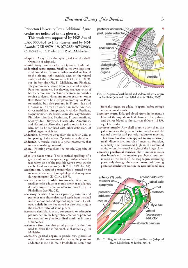
abaxial. Away from a shell axis. Opposite of adaxial.abdominal sense organs. Small paired swellings situ-
ated lateral to the anus, either medial or lateral to the left and right ctenidial axes, on the ventral surface of the adductor muscle (thiele, 1889), e.g., in Pteriidae (Fig. 1), Malleidae, and Pinnidae. They receive innervation from the visceral ganglion. Function unknown, but showing characteristics of both chemo- and mechanoreceptors, so possibly serving to detect vibrations and/or excurrent water flow. Believed to be a symplesiomorphy for Pteri-omorphia, but also present in Trigoniidae and Unionidae. Known to occur in some Arcidae, Glycymerididae, Limopsidae, Mytilidae, Pteriidae, Isognomonidae, Malleidae, Ostreidae, Gryphaeidae, Pinnidae, Limidae, Pectinidae, Propeamussiidae, Spondylidae, Dimyidae, Plicatulidae, Anomiidae, and Placunidae. Also called a pallial organ in Ostre-idae, not to be confused with other definitions of pallial organ, which see.
abduction. Movement away from the median axis, as in opening of the valves. Opposite of adduction.
abductor. A muscle, such as a pedal protractor, that moves something outward.
aboral. Pointing away from the mouth. Opposite of adoral.
absolute tautonymy. The identical spelling of a genus and one of its species, e.g., Villosa villosa. In taxonomy, one of the possible ways a type species can be fixed for a genus (see ICZN, 1999, Art. 68).
acceleration. A type of peramorphosis caused by an increase in the rate of morphological development during ontogeny (E. Cope, 1887).
accessory anterior adductor muscle. A separate, small anterior adductor muscle anterior to a larger, dorsally migrated anterior adductor muscle, e.g., in Pholadidae (see Fig. 24).
accessory cavities. Cavities separating anterior and posterior myophore plates and teeth from the shell wall in caprotinid and caprinid hippuritoids. Devel-oped chiefly in the free valve but also occurring in the attached valve of some genera.
accessory denticle. A small, compressed or triangular prominence on the hinge plate anterior or posterior to a cardinal or pseudocardinal tooth, as in some Unionoidea.
accessory foot. An elongated process on the foot used to clean the infrabranchial chamber, e.g., in Malleidae.
accessory genital organ. A pendulous, glandular organ on the posteroventral surface of the posterior adductor muscle in male Pholadidae; secretions
from this organ are added to sperm before storage in the seminal vesicle.
accessory hearts. Enlarged blood vessels in the mantle lobes of the suprabranchial chamber that pulsate and deliver blood to the auricles (harry, 1985), e.g., Ostreoidea.
accessory muscle. Any shell muscle other than the pallial muscles, the pedal retractor muscles, and the normal anterior and posterior adductor muscles. This term has also been applied to any relatively small, discrete shell muscle of uncertain function, especially one positioned high in the umbonal cavity or on the ventral margin of the hinge plate.
accessory pedobyssal muscles. Short, minor muscles that branch off the anterior pedobyssal retractor muscle at the level of the esophagus, extending posteriorly through the visceral mass and forming posterior attachment scars in the near-umbonal area
FiG. 1. Diagram of anal funnel and abdominal sense organ in Pteriidae (adapted from Mikkelsen & Bieler, 2007).
FiG. 2. Diagram of anatomy of Teredinidae (adapted from Mikkelsen & Bieler, 2007).
4 Treatise Online, number 48
(tëMkiN, 2006a, p. 300), e.g., some Pterioidea. Called posterior levators by herdMaN (1904).
accessory plate. A calcareous or chitinous structure that protects the soft body or serves as an attach-ment for muscles, and that is added to the normal shell margins later in growth, commonly at sexual maturity, or is secreted as a separate structure distal to the normal shell margins. Examples include the callum, hypoplax, mesoplax, metaplax, protoplax, and siphonoplax in Pholadoidea, and siphonal plates in some Corbulidae.
accessory shell plate. Same as accessory plate, which see.
accessory ventral adductor muscle. An adductor muscle separate from the normal anterior and posterior adductor muscles, joining the valves ventrally, as in Teredinidae (Fig. 2), or posteroven-trally, as in some Gastrochaenoidea and Tellinoidea. Also present in some Pholadomyidae, Vacunellidae (e.g., Vacunella Waterhouse, 1965), Periploma-tidae, and Laternulidae, where this may be repre-sented by an orbital muscle (see ruNNeGar, 1966, fig. 1, ava). See also cruciform and orbital muscles.
acentric sculpture. Sculpture that is not commarginal. Compare with oblique and scissulate.
aciculate. Slender and tapering to a sharp point.acinus (pl., acini). A small compartment of the
gonad in which gametes are produced; also called an alveolus (pl., alveoli) or a follicle, e.g., many Unionoidea.
acline. Shell features that are perpendicular, or almost so, to the hinge. Same as orthocline.
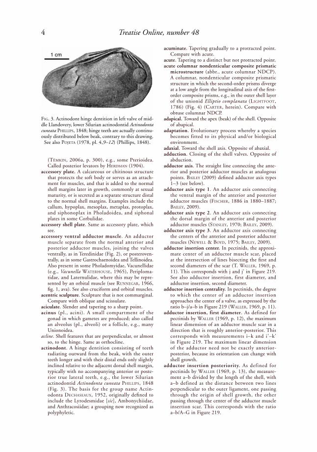
actinodont. A hinge dentition consisting of teeth radiating outward from the beak, with the outer teeth longer and with their distal ends only slightly inclined relative to the adjacent dorsal shell margin, typically with no accompanying anterior or poste-rior true lateral teeth, e.g., the lower Silurian actinodontid Actinodonta cuneata phillips, 1848 (Fig. 3). The basis for the group name Actin-odonta deChaseaux, 1952, originally defined to include the Lyrodesmidae [sic], Ambonychiidae, and Anthracosiidae; a grouping now recognized as polyphyletic.
acuminate. Tapering gradually to a protracted point. Compare with acute.
acute. Tapering to a distinct but not protracted point. acute columnar nondenticular composite prismatic
microstructure (abbr., acute columnar NDCP). A columnar, nondenticular composite prismatic structure in which the second-order prisms diverge at a low angle from the longitudinal axis of the first-order composite prisms, e.g., in the outer shell layer of the unionid Elliptio complanata (liGhtFoot, 1786) (Fig. 4) (Carter, herein). Compare with obtuse columnar NDCP.
adapical. Toward the apex (beak) of the shell. Opposite of abapical.
adaptation. Evolutionary process whereby a species becomes fitted to its physical and/or biological environment.
adaxial. Toward the shell axis. Opposite of abaxial.adduction. Closing of the shell valves. Opposite of
abduction.adductor axis. The straight line connecting the ante-
rior and posterior adductor muscles at analogous points. Bailey (2009) defined adductor axis types 1–3 (see below).
adductor axis type 1. An adductor axis connecting the ventral margin of the anterior and posterior adductor muscles (FisCher, 1886 in 1880–1887; Bailey, 2009).
adductor axis type 2. An adductor axis connecting the dorsal margin of the anterior and posterior adductor muscles (staNley, 1970; Bailey, 2009).
adductor axis type 3. An adductor axis connecting the centers of the anterior and posterior adductor muscles (NeWell & Boyd, 1975; Bailey, 2009).
adductor insertion center. In pectinids, the approxi-mate center of an adductor muscle scar, placed at the intersection of lines bisecting the first and second diameters of the scar (T. Waller, 1969, p. 11). This corresponds with j and j' in Figure 219. See also adductor insertion, first diameter, and adductor insertion, second diameter.
adductor insertion centrality. In pectinids, the degree to which the center of an adductor insertion approaches the center of a valve, as expressed by the ratio b–j/a–b in Figure 219 (Waller, 1969, p. 11).
adductor insertion, first diameter. As defined for pectinids by Waller (1969, p. 12), the maximum linear dimension of an adductor muscle scar in a direction that is roughly anterior-posterior. This corresponds with measurements i–k and i'–k' in Figure 219. The maximum linear dimension of the adductor need not be exactly anterior-posterior, because its orientation can change with shell growth.
adductor insertion posteriority. As defined for pectinids by Waller (1969, p. 13), the measure-ment a–b divided by the length of the shell, with a–b defined as the distance between two lines perpendicular to the outer ligament, one passing through the origin of shell growth, the other passing through the center of the adductor muscle insertion scar. This corresponds with the ratio a–b/A–G in Figure 219.
FiG. 3. Actinodont hinge dentition in left valve of mid-dle Llandovery, lower Silurian actinodontid Actinodonta cuneata phillips, 1848; hinge teeth are actually continu-ously distributed below beak, contrary to this drawing.
See also poJeta (1978, pl. 4,9–12) (Phillips, 1848).
1 cm

5Illustrated Glossary of the Bivalvia
adductor insertion, second diameter. As defined for pectinids by Waller (1969, p. 13), the maximum dimension of that portion of the adductor muscle insertion underlying the striate portion of the muscle, measured in a roughly dorsoventral direc-tion. The orientation of the second diameter need not be exactly perpendicular to the first diameter of adductor insertion (which see), because this angle varies somewhat with growth. This corresponds with measurements p–q and p'–q' in Figure 219.
adductor insertion ventrality. As defined for pectinids by Waller (1969, p. 13), the distance b–j divided by the height of the shell, where b–j is the distance between two parallel lines, one coinciding with the outer ligament, the other passing through the center of the adductor muscle scar. This corre-sponds with ratios b–j/A–M and b'–j'/A'–M' in Figure 219.
adductor muscle. A muscle connecting the two shell valves and drawing them together in opposition to the ligament; generally divided into a faster acting quick muscle and a more resilient, slower acting catch muscle, e.g., in the venerid Merce-naria mercenaria (liNNaeus, 1758) (see Fig. 130). Usually marked on the shell interior as a scar or impression. Anterior and posterior adductors are usually present, but some bivalves retain only one adductor (e.g., the posterior adductor in Ostreidae), and some have a third adductor produced by cross-fusion of pallial muscles posteroventrally, ventrally, or dorsoanteriorly (see accessory anterior adductor muscle and accessory ventral adductor muscle).
adductor muscle scar. An impression or other mark in the inner shell layer corresponding with the attach-ment site of an adductor muscle.
adnate. Joined to a different element, e.g., an anterior adductor muscle joined to a pallial retractor muscle. See also conjoined.
adopt. In taxonomy, to use an unavailable name as the valid name of a taxon in a way that establishes it as a new name with its own authorship and date (ICZN, 1999).
adoral. Pointing toward the mouth. Opposite of aboral.
adoral mantle sense organ (=adoral sense organ; cephalic sense organ). In many Protobranchia, a sense organ present on either side of the mouth close to the proximal palp ridge and immediately ventral to the cerebral ganglia, innervated from the latter. Present in the nuculids Nucula nucleus liNNaeus (1758) (according to hirasaka, 1927, with pigment cells with a cornea, although this was refuted by sChaeFer, 2000) and Deminucula atacel-lana (sCheNCk, 1939), as a thickened epithelium innervated from the cerebral ganglia, without a cornea and pigment cells (rhiNd & alleN, 1992). Also present in Solemyidae, Nuculanidae, and Sareptidae. An olfactory organ according to steM-pell (1899) and Vlès (1905). sChaeFer (2000) proposed a chemoreceptor function and plesiomor-phic status for this feature in Protobranchia. Not the same as marginal sense organ or anterior mantle sense organ, which see.
adoral sense organ. Same as adoral mantle sense organ, which see.
adult. Individuals developed to the point of being able to reproduce; sexually mature.
advanced isocyprinoid grade hinge. See isocyprinoid grade hinge.
advanced veneroid grade hinge. A heterodont hinge grade characteristic of some Veneridae, with early and late evolutionary phases (late phase shown in Fig. 5). Compare with early veneroid, veneroid, arcticoid, isocyprinoid, lucinoid, and venielloid grade hinges. r. N. GardNer (2005, p. 336) described this hinge grade as follows: “In the veneroid hinge, lateral teeth AI and AII gradually become detached from cardinal teeth 1 and 2a, respectively. This ultimately permits laterals AI and AII to develop further cardinal teeth. The advanced veneroid hinge is marked by the development of a moderately prosocline second cardinal tooth 1 at the posterior extremity of lateral A1, and/or by the formation of a moderately proso-cline second cardinal tooth 2a at the posterior end of lateral AII. These newly developed cardinals are defined here as teeth 1
2 and 2a
2. Tooth 1
2 usually
terminates in a recess situated between the more posteriorly positioned slightly prosocline tooth 3a, and the dorsally positioned lateral AIII. Cardinal tooth 2a
2 projects posterodorsally, and then descends
to transform into a lamella that fuses with the dorsal extremity of the united teeth 2a and 2b. The devel-opment, position, and orientation of 1
2 and 2a
2 are
perfectly illustrated by species of Dosinia sCopoli, 1777 (Upper Eocene–Recent), and Notocallista iredale, 1924. . . . In the advanced veneroid hinge, posterior lateral teeth are usually absent or obso-lete. However, some genera, including Dosiniopsis CoNrad, 1864 (cited in keeN & Casey, 1969) (Paleocene–Eocene), and Notocallista . . . continue to develop functional posterior lateral teeth. The general hinge formula showing the final position of cardinal teeth for the advanced veneroid hinge is . . .: AI (AIII ) 1
2 3a 1 3b (PI ) / AII (AIV ) 2a
2 2a
2b 4b (PII).”
FiG. 4. Acute columnar nondenticular composite prismatic structure, a slightly reclined variety from outer shell layer of Unionoidea (adapted from Carter
& others, 1990, p. 617).

6 Treatise Online, number 48
adventitious. Of, or belonging to, a structure formed in an unusual place, such as a calcareous tube secreted around the shell and body of the animal (e.g., the gastrochaenoidean Eufistulana eaMes, 1951, and the teredinid Kuphus Guettard, 1770); thick conchiolin sublayers within an inner shell layer in some Unionidae, Cyrenidae, and Corbu-lidae; articulating structures on parts of the shell other than the hinge (e.g., in Pandoridae; also called laminar buttresses); and articulating struc-tures present on or near the hinge but believed to be developed independently of true hinge teeth
(e.g., isodont crura in plicatulids, shell marginal teeth in mytilids).
adventive. Not native; referring to an organism trans-ported to, and becoming established in, a new geographic area by human or natural means.
aequipectinoid form. Having the shell shape of Aequipe-cten FisCher, 1886 in 1880–1887, i.e, with a shallow byssal notch late in ontogeny, a relatively flaring, equilateral disc, and auricles nearly equal in length (Waller, 2006). This associates with early byssal attachment and later freedom and mobility. See also chlamydoid, pectinoid, and amusioid forms.
FiG. 5. Late phase of advanced veneroid grade hinge. 1–2, Diagram of left and right valves (adapted from R. N. Gardner, 2005, fig. 10.5b, 11.5b, courtesy of R. N. Gardner); 3, Oligocene venerid Notocallista watti MarWiCk,
1938 (adapted from R. N. Gardner, 2005, fig. 13, courtesy of R. N. Gardner).
cardinal 3a
lateral AIII
lateral AI
cardinal 1 2 cardinal 1 cardinal 3b
cardinal 4blaminar II
laminar IV
lateral AIV
cardinal 2b cardinal 2acardinal 2a2
lateral AII
PII
left valve
2a2
AII
1
2
3
left valve
right valve
7Illustrated Glossary of the Bivalvia
ala (pl., alae). A prominent, anterior or posterior, dorsal projection of the shell; a term commonly used in reference to unionoid glochidial and adult shells, e. g., in the unionid Hyriopsis cumingii (lea, 1852) (Fig. 63). Compare with wing.
alate. Having a winglike or auriculate extension of the shell, usually dorsally and anterior and/or posterior of the beak, but rarely in other positions; e.g., some pterioids, pectinoids, unionoids, and all alatoconchids. The term bialate is used for two such extensions.
alimentary canal. The tube that extends from the mouth to the anus, in bivalves typically consisting of the mouth, esophagus, stomach, style sac, midgut, hindgut, rectum, and anus, e.g., in the venerid Mercenaria mercenaria (liNNaeus, 1758) (see Fig. 130).
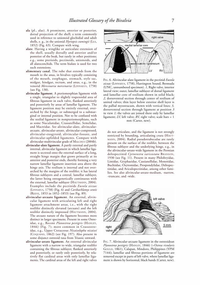
alivincular ligament. A pteriomorphian ligament with a single, triangular to slightly trapezoidal area of fibrous ligament in each valve, flanked anteriorly and posteriorly by areas of lamellar ligament. The ligament position may be entirely external, over-arched by the hinge, or submerged to a submar-ginal or internal position. Not to be confused with the resilial ligament in nonpteriomorphians, such as some Nuculanidae, Crassatellidae, Semelidae, and Mactridae. See alivincular-alate, alivincular-arcuate, alivincular-areate, alivincular-compressed, alivincular-exogyroid, alivincular-fossate, and alivincular-upfolded ligaments. Compare with alivincular-multivincular grade of MalChus (2004b).
alivincular-alate ligament. A partly external and partly internal, alivincular ligament in which lamellar liga-ment is secreted onto the external surface of a long, straight hinge margin that grows primarily at its anterior and posterior ends, thereby forming a very narrow lamellar ligament insertion area above the hinge axis. The resilium is internal and not over-arched by the margins of the resilifer; it has lateral fibrous sublayers and a central, lamellar sublayer, the latter being ontogenetically continuous with the external, lamellar sublayer (hautMaNN, 2004). Examples include the pectinids Euvola ziczac (liNNaeus, 1758) (Fig. 6) and Caribachlamys sentis (reeVe, 1853 in 1852–1853) (see Fig. 89).
alivincular-arcuate ligament. An external, alivin-cular ligament with articulating left and right ligament attachment areas, i.e., with the right resilifer distinctly elevated (arcuate) and the left resilifer distinctly impressed (hautMaNN, 2004). The arcuate nature of the ligament becomes more distinct in larger specimens. Present in some Ostre-idae, e.g., Recent Planostrea pestigris (haNley, 1846) (Fig. 7); more common in Crassostre-idae, e.g., Upper Cretaceous Nicaisolopha nicaisei (CoquaNd, 1862) (see Fig. 197). Also present in some disjunct ostreoid taxa from Triassic onward.
alivincular-areate ligament. An external alivincular ligament with a narrow to wide, triangular resilifer containing the fibrous sublayer, flanked anteriorly and posteriorly, or rarely only posteriorly, by rela-tively flat cardinal areas with only lamellar liga-ments. The cardinal areas of the left and right valves
do not articulate, and the ligament is not strongly restricted by bounding, articulating crura (haut-MaNN, 2004). Radial pseudotrabeculae are rarely present on the surface of the resilifer, between the fibrous sublayer and the underlying hinge, e.g., in the alivincular-areate-wide ligament in the Permian deltopectinid Cyrtorostra varicostata BraNsoN, 1930 (see Fig. 11). Present in many Philobryidae, Limidae, Gryphaeidae, Cassianellidae, Monotidae, Buchiidae, Oxytomidae, Prospondylidae, Deltopec-tinidae, and Aviculopectinidae, among other fami-lies. See also alivincular-areate-medium, -narrow, -truncate, and -wide.
FiG. 6. Alivincular-alate ligament in the pectinid Euvola ziczac (liNNaeus, 1758), Harrington Sound, Bermuda (UNC, unnumbered specimen). 1, Right valve, interior lateral view; outer, lamellar sublayer of dorsal ligament and lamellar core of resilium shown in solid black; 2, dorsoventral section through center of resilium of united valves; thin layer below exterior shell layer is the pallial myostracum, shown with vertical lines; 3, dorsoventral section through ligament at position 3 in view 1; the valves are joined there only by lamellar ligament; LV, left valve; RV, right valve; scale bars = 1
mm (Carter, new).
FiG. 7. Alivincular-arcuate ligament in the ostreoidean Planostrea pestigris (haNley, 1846) (=Ostrea rivularis Gould, 1861), Calapan, Mindoro, Philippines (YPM 7144); lamellar and fibrous portions of ligament are removed except in parts of left valve, where lamellar liga-ment is shown by horizontal, black bands (Carter, new).

8 Treatise Online, number 48
alivincular-areate-medium ligament. An alivincular-areate ligament with resilifer that is medium-width in comparison with the flanking bourrelets, e.g., in the limid Ctenoides scabra (BorN, 1778) (Fig. 8), the malleid Malleus malleus (liNNaeus, 1758) (see Fig. 248), and the Eocene crassostreid Turkostrea VialoV, 1936 (see Fig. 318). In limopsids, this ligament is also called a limopsid type C ligament (oliVer, 1981), e.g., in Limopsis chuni thiele & JaeCkel, 1931 (see Fig. 164).
alivincular-areate-narrow ligament. An alivincular-areate ligament with a resilifer that is narrow rela-tive to the width of the flanking bourrelets, e.g.,
in the philobryid Cratis antillensis (dall, 1881) (Fig. 9).
alivincular-areate-truncate ligament. An alivincular-areate ligament in which only the resilium and the posterior bourrelet are present, with the resilifer consequently occupying the entire anterior end of the ligament insertion area, as in some members of the Upper Triassic eurydesmatid (?) Krumbeckiella iChikaWa, 1958 (Waterhouse, 2008, fig. 64A, F), some members of the Upper Cretaceous inoc-eramid Tenuipteria argentea (CoNrad, 1858) (which also has some multivincular individuals) (Fig. 10), and the Mesozoic buchiid Buchia keyserlingi lahuseN, 1888 (see Fig. 42). Called truncavincular by Waterhouse (2008). Compare with alivincular-exogyroid.
alivincular-areate-wide ligament. An alivincular-areate ligament with a resilifer that is very wide in comparison with the flanking bourrelets, as in the pteriid Pinctada imbricata rödiNG, 1798, and the Permian deltopectinid Cyrtorostra varicostata BraNsoN, 1930 (Fig. 11). This was called lativin-cular by Waterhouse (2001, fig. 9b).
alivincular-compressed ligament. An external, alivin-cular ligament with a moderately wide, triangular resilifer containing the fibrous sublayer, flanked by single, narrow, triangular pseudoresilifers anteriorly and posteriorly, with no space between the resilifer and the pseudoresilifers. All three components radiate from a point below the beak. Anterior and posterior to the pseudoresilifers, the cardinal area is covered by periostracum. This corresponds with oliVer’s (1981) limopsid type A ligament, as in Limopsis sulcata Verrill & Bush, 1898 (see Fig. 162; see limopsid type A ligament), and with his limopsid type D ligament, where the flanking
FiG. 8. Alivincular-areate-medium ligament in the limid Ctenoides scabra (BorN, 1778), Biscayne Bay, Florida (YPM 9702). 1, Hinge and ligament of right valve, interior lateral view; 2, dorsoventral section through united valves at position 2 in view 1; 3, dorsoventral section through united valves at position 3 in view 1. Lower scale bar applies to views 2 and 3 (Carter, new).
FiG. 9. Hinge and beak of philobryid Cratis antillensis (dall, 1881), showing its alivincular-areate-narrow ligament, prodissoconch-1, and nepioconch; figure width 2–3 mm (adapted from Mikkelsen & Bieler, 2007).
alivincular-areateligament
nepioconch
prodissoconch-1
resilium
bourrelet

9Illustrated Glossary of the Bivalvia
cardinal areas have a thicker periostracal cover (especially so at the ends of the cardinal area), as in Limopsis lilliei (E. A. sMith, 1915) (see Fig. 165; and see limopsid type D ligament).
alivincular-exogyroid ligament. An external, aliv-incular ligament in which the resilifer and the anterior bourrelet are run together, the posterior bourrelet is reduced to a narrow ridge, and the length axis of the ligament area is nearly parallel with the hinge axis (MalChus, 1990), e.g., the gryphaeid Exogyra say, 1820 (Fig. 12). In cases where the anterior bourrelet is indistinguishable from the resilifer, the ligament is better classified as alivincular-areate-truncate, which see.
alivincular-fossate ligament. A partly external and partly internal alivincular ligament with the lamellar and fibrous sublayers crowded together by anterior and posterior crura, and with the resilium at least partially overarched by the cardinal area, as in the spondylid Spondylus gussonii o. G. Costa, 1829 (Fig. 13); also found in Dimyidae and Plicat-uloidea. In the more derived condition, the resilium is entirely overarched by the dorsal cardinal area, and the crura and the growing ends of the resilium are positioned entirely below the hinge axis (haut-MaNN, 2004). hautMaNN (2004, fig. 1D) indicated that the hinges are united along their dorsal margin by secondary (i.e., periostracal) ligament, rather than by lamellar ligament, but Waller (1978) maintained that in Dimyidae, Plicatulidae, and Spondylidae, they are united dorsally by lamellar ligament. Note that lamellar ligament is present as a medial core in the resilium in Spondylidae (as in Pectinidae), but not in Dimyidae and Plicatulidae.
alivincular-multivincular grade. MalChus (2004b, p. 1562) defined this ligament grade for aliv-incular and multivincular bivalves with clearly opisthogyrate larval shells, plus their orthogyrate derivatives. The larval fibrous sublayer originates anterior of the larval straight hinge or antero-centrally below it, and it grows either anterior-ward or anteroventrally. Early postlarval growth of this fibrous sublayer is ventral or anteroventral. However, this larval fibrous sublayer is typically abandoned early in ontogeny. The adult ligament
may have no repetition or extensive repetition of its sublayers, with the sublayers added in a shifting center-proliferation manner (which see) either seri-ally (predominantly) or irregularly alternating (in Pulvinitidae). Three subgrades were distinguished: multivincular subgrade, alivincular subgrade with one sublayer repetition, and alivincular subgrade without sublayer repetition (see entries for each).
alivincular subgrade with one sublayer repetition. An alivincular-multivincular grade ligament (which see), as defined by MalChus (2004b), with two and only two fibrous sublayers in the adult ligament, as in the Malleidae and most Pteriidae (MalChus, 2004b, p. 1562).
alivincular subgrade without sublayer repetition. An alivincular-multivincular grade ligament (which see), as defined by MalChus (2004b), with a single adult fibrous sublayer, as in the pteriid Pinctada and most Ostreoidea (MalChus, 2004b).
alivincular-upfolded ligament. A left-right asym-metrical, internal, alivincular ligament as developed in Mesozoic and Cenozoic Anomioidea, e.g., Monia macrochisma (deshayes, 1839) (Fig. 14). The name derives from MalChus’s (2004b, p. 1561) “overarched, upfolded,” alivincular ligament. As described by yoNGe (1980) for various living Anomioidea, the ligament insertion area is vertically instead of laterally disposed, the resilium is posi-tioned entirely ventral to the dorsal shell margins, and it is rotated so that its insertion area faces
FiG. 10. Alivincular-areate-truncate ligament in the Maastrichtian, Upper Cretaceous inoceramid Tenuip-teria argentea (CoNrad, 1858), Owl Creek Formation, Owl Creek, Mississippi (UNC 9566), camera lucida drawing of lateral interior view of left valve. This species also has some multivincular individuals (Carter, new).
FiG. 11. Alivincular-areate-wide ligament with radial pseudotrabeculae on surface of the resilifer, beneath fibrous sublayer of ligament; right valve of Permian deltopectinid Cyrtorostra varicostata BraNsoN, 1930, Willis Branch Formation, Glass Mountains, western Texas (USNM 388883) (adapted from Newell & Boyd, 1995, fig. 2a; courtesy
of the American Museum of Natural History).

10 Treatise Online, number 48
dorsoventrally instead of laterally. Narrow anterior and posterior lamellar sublayers flank the moder-ately wide fibrous sublayer in Pododesmus philippi, 1837, and Anomia (Anomia) liNNaeus, 1758, but these flanking fibrous sublayers are united in an arch over the fibrous sublayer in the more derived Anomia (Patro) Gray, 1850. The right resilifer is elevated by a straight to convex ligamental chon-drophore (also called a crurum). The fibrous and lamellar sublayers extend continuously between the valves. In Placuna liGhtFoot, 1786, thickened periostracum unites the valves dorsally, whereas the internal ligament provides opening thrust.
allometric growth . Growth at unequal rates in different parts of an organism.
allometry. (1) the relative growth of a structure in relation to another structure or to the remainder of the organism; (2) the measurement or study of such growth.
allopatric. Nonoverlapping geographic ranges of two populations. Compare with sympatric.
allotype. A specimen, in a series of type specimens, which is the opposite sex of the holotype.
allozyme data. Results from protein electropho-resis, separated first by electric charge and then by molecular weight. Such data are potentially useful for analyzing phylogenetic relationships.
amorphous calcium carbonate (abbr., ACC). A noncrystalline form of calcium carbonate. Some prodissoconchs contain ACC as a precursor to crystalline (typically aragonitic) calcium carbonate (Weiss & others, 2002).
amphidetic. Entending anterior and posterior to the beaks, usually in reference to the ligament or hinge dentition, e.g., the monovincular-A ligament in the pectinoid Strebloboydia montpelierensis (Girty, 1910) (Fig. 15). Compare with prosodetic and opisthodetic.
amphi-pleurothetic. Living with the left or right side of the body down, with both orientations occurring in the same species, e.g., Etheriidae.
amusioid form. Having a shell shape like the pectinid Amusium pleuronectes (liNNaeus, 1758), i.e, with equilateral, equally convex valves, large, permanent shell gapes, and a smooth or nearly smooth exterior (Waller, 1991, p. 10) (Fig. 16). This shell shape generally associates with active swimming life habits. Compare with aequipectinoid, chlamydoid, and pectinoid forms.
amyarian. Lacking adductor muscles in the adult stage, e.g., Penicillidae.
amylase. A starch-digesting enzyme found in the crystalline style of most mollusks, including many bivalves.
anaboly. The introduction of a new feature at the end of the embryonic stage. Compare with cenogenesis.
anachomata (sing., anachoma). Small tubercles or ridgelets on the periphery of the inner surface of a valve close to the commissure, as in lower Eocene crassostreid Turkostrea duvali (GardNer, 1927) (Fig. 17). Compare with chomata and catachomata.
anal aperture or siphon. Same as excurrent aperture or siphon.
anal canal. A long, narrow, dorsal channel in Teredin-idae, continuous with the suprabranchial chamber and the excurrent siphon, which terminates in a sphincter muscle in some species. The rectum empties into the anal canal.
anal flap. See anal funnel.anal funnel. An ear-shaped membranous structure
protruding from the tip of the rectum, which encloses the anal opening at its base, e.g., many Arcidae, Pteriidae, Isognomonidae, Malleidae, Pinnidae, and some Ostreidae. Also called an anal process (herdMaN, 1904), anal membrane (raNsoN, 1961), anal flap (hayes, 1972), anal flag, and anal ear (pelseNeer, 1911). In Pterioidea, this structure varies in shape from triangular to lanceolate to rounded (tëMkiN, 2006a), and it functions to direct fecal pellets outside the mantle cavity (hayes, 1972), e.g., in the pteriid Pinctada imbricata rödiNG, 1798 (Fig. 1, Fig. 18), in the Pinnidae (see Fig. 211), and in the Malleidae (see Fig. 238). Compare with anal papilla.
anal orifice. The posterodorsal, excurrent aperture in nonsiphonate bivalves, positioned dorsal to the incurrent aperture, e.g., in the crassatellid Eucrassatella iredale, 1924. Homologous with the excurrent siphonal aperture in siphonate bivalves.
anal papilla. A small protuberance on the tip of the rectum, just dorsal to the anal opening, e.g., in some Unionidae. Compare with anal funnel.
anal tentacles. Enlarged pallial tentacles on the posterodorsal mantle margin, e.g., some Limidae.
analogous. Structures with similar function but different evolutionary origins, e.g., the hinge teeth in venerids and the crura in plicatulids.
anastomosis (adj., anastomosing). The union of parts or branches, as in blood vessels, or elements of exterior shell ornament.
angular. Same as angulate.angulate. Having two edges that join to form a more
or less sharp corner; not rounded. Same as angular.
FiG. 12. Alivincular-exogyroid ligament in Cretaceous exogyrid Exogyra say, 1820 (adapted from Malchus,
1990, fig. 25d).
resilifer
thin bourrelet ridge

11Illustrated Glossary of the Bivalvia
anisomyarian. (1) Having anterior and posterior adductor muscles of unequal size; same as hetero-myarian (CoaN, sCott, & BerNard, 2000, p. 732); (2) having the anterior adductor muscle smaller than the posterior adductor muscle, or absent; includes heteromyarian and monomyarian condi-tions; the basis for Anisomyarier [sic] NeuMayr, 1884, which originally included heteromyarian and monomyarian taxa. Definition 1 is in prevailing usage and is preferred over the term heteromyarian.
annular. Having the form of a ring, as in the circular or conical diaphragm at the tip of many excurrent siphons.
annulus (pl., annuli). (1) A ring; (2) a commar-ginal line on or within the shell or periostracum, commonly referred to as a growth line, often presumed to be annual in occurrence.
anodont. Same as edentulous, which is preferred.antagonistic. Parts or processes with counteracting
effects, such as the ligament and adductor muscles.
antecarinal sulcus or groove. In trigonioids, a narrow, radial depression at the posterior edge of the flank of the shell, just anterior to a marginal carina, extending from the umbo to the posteroventral shell margin (leaNZa, 1993, p. 16).
anteriad. Positioned toward the anterior; of or relating to the anterior, e.g., the anteriad beaks in Crassatel-lidae.
anteriodorsal. Same as anterodorsal and dorsoanterior, which are preferred.
anterior. (1) In soft anatomy, the direction toward the mouth and parallel with a line passing through the mouth and anus; (2) in shell morphology, the direction toward the mouth and parallel with the anteroposterior shell axis (which see), the latter potentially being defined in several ways.
anterior adductor. The adductor muscle located near the anterior end of the shell, typically close to the mouth.
anterior cleft. See umbonal fissure.
FiG. 13. Alivincular-fossate ligament and isodont hinge dentition in spondylid Spondylus gussonii o. G. Costa, 1829, Gulf of Mexico (YPM 7233); camera lucida drawing (Carter, new).
5 mm
fibrous sublayer
lamellar sublayer
right valve
lamellarsublayer
5 mm
fibrous sublayer
left
right

12 Treatise Online, number 48
anterior cruciform apparatus. See pedal aperture muscles.
anterior dorsal half-diameter of disc. As defined for pectinids by Waller (1969, p. 9), the distance between two parallel lines, one coinciding with the outer ligament, the other passing through the most anterior point on the shell disc. This corresponds with measurements G–P and G'–P' in Figure 219.
anterior flaring ratio of disc. As defined for pectinids by Waller (1969, p. 9), the anterior half-diameter of the disc divided by the anterior dorsal half-diameter of the disc. This corresponds with the ratios D–G/G–P and D'–G'/G'–P' in Figure 219.
anterior half-diameter of disc. As defined for pectinids by Waller (1969, p. 9), the distance between two lines that are perpendicular to the outer ligament, one passing through the origin of growth, the other passing through the most anterior point on the shell disc. This corresponds with measurements D–G and D'–G' in Figure 219.
anterior mantle sense organ. See marginal sense organ.anterior outer ligament. In reference to pectinid
shells, the portion of the lamellar, outer ligament that is anterior to the origin of growth (Waller, 1969, p. 9).
anterior pedal gland. A small, white to cream colored gland on the anterior of the foot near the base of the pedal probing organ in members of the
superfamily Gastrochaenoidea, e.g., Lamychaena cordiformis (NeVill & NeVill, 1869) (Fig. 19). Believed to secrete a fluid that dissolves a minute tubular opening in calcium carbonate substrata.
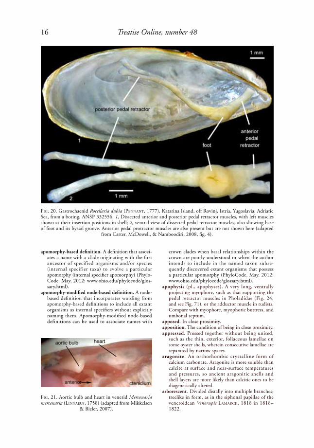
anterior pedal retractor muscle. A pedal retractor muscle, or group of such muscles, attached near the dorsoposterior and/or posterior margin of the anterior adductor muscle scar, e.g., in the gastrochaenid Rocellaria dubia (peNNaNt, 1777) (Fig. 20) and in the Unionoidea (see Fig. 307). Absent in most monomyarian taxa, such as the Pectinidae. Some small, dorsally attached pedal muscles previously identified as anterior pedal retractor muscles in Pteria sCopoli, 1777, and Lima BruGuière, 1797, are now regarded as elevator muscles.
anterior-posterior axis. Same as anteroposterior axis, which see.
anterior slope. The outer shell surface located anterior and ventral to the beaks.
anterioventral. See anteroventral. antero-. The combining form of the term anterior,
as in anteroventral. The spelling anterio- is not preferred.
anterodorsal. Both anterior and dorsal; same as dorso-anterior.
anteroposterior axis (abbr., APA). An alternative spelling of anterior-posterior axis, which can be
FiG. 14. Alivincular-upfolded ligament in the anomiid Monia macroschisma (deshayes, 1839). Dorsal areas of left valve (above) and right valve (below) facing each other as in life, with the ligament cut between the two valves; two lamellar sublayers and central, fibrous sublayer of ligament all originally extended continuously between two valves
(adapted from Yonge, 1977, fig. 4a–b, courtesy of the Royal Society of London).
fibrous sublayer
left
lamellarsublayer
right
2 cm
supradorsal fusion in left valvechondrophore
lamellarsublayer
cut shell
byssal notch

13Illustrated Glossary of the Bivalvia
defined in four ways: (1) the hinge axis or cardinal axis, i.e., the line about which the shells are hinged (Cox, Nuttall, & trueMaN, 1969, p. 105); a useful definition when the soft anatomy is poorly understood; (2) the oro-anal axis, i.e., the line connecting the mouth and anus; (3) the adductor axis, i.e., the straight line connecting the anterior and posterior adductor muscles at analogous points (Bailey, 2009); (4) the functional anteroposterior axis, i.e., the straight line defined by the intersec-tion of the central, longitudinal axis of a burrow or boring with the deepest and shallowest points on the shell.
anteroventral. Both anterior and ventral; same as ventroanterior, which is less commonly used.
anthropogenic. Of, relating to, or involving the impact of humans on nature.
antiligamentat. The troughlike surface of the inner nacreous shell layer below a relatively thick, pris-matic ligamentat (JohNstoN & ColloM, 1998, p. 350), e.g., in Inoceramidae.
antimarginal. Oriented more or less perpendicular to successive shell margins, whether or not this is truly radial (Waller, 1986) (Fig. 62). Compare with radial.
anvil-type fibrous prismatic microstructure (abbr., anvil-type FP). See fibrous prismatic microstruc-ture, anvil-type.
aorta. A large, tubular vessel carrying blood from the heart to the other organs.
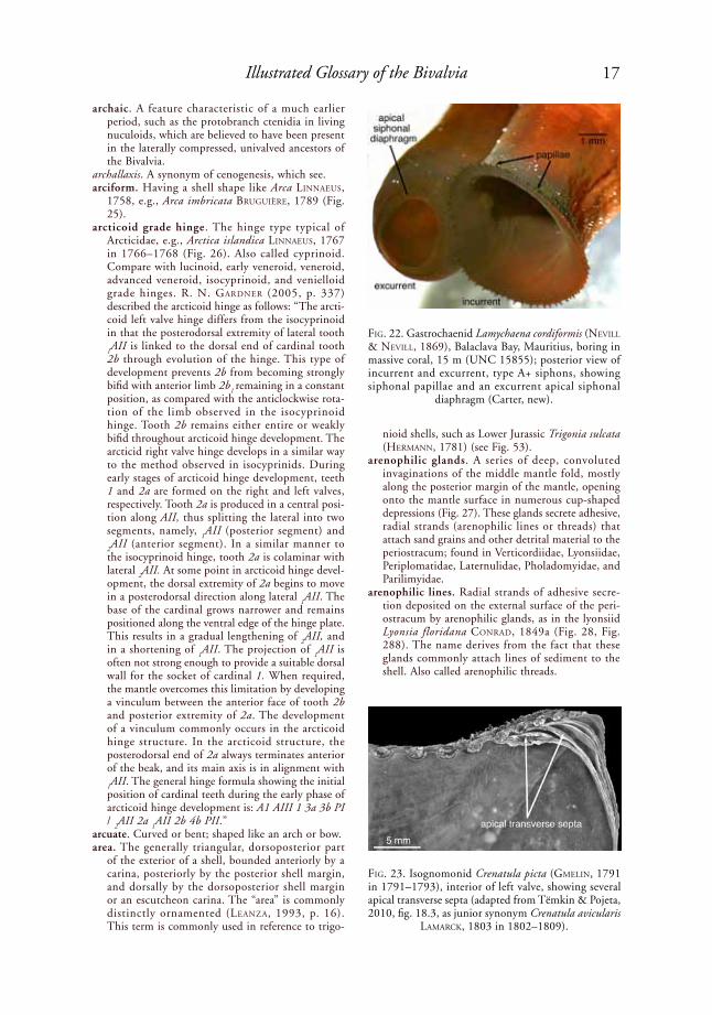
aortic bulb. A muscular, spongy, pendulous structure on the ventral side of the posterior aorta and hindgut just posterior to the heart, functioning to prevent rupture of the heart when the siphons and foot are suddenly retracted, forcing haemo-lymph backward into the posterior aorta. Found in Gastrochaenoidea, Veneridae, Psammobiidae, Phar-
idae, Mactridae, and some Pandoridae, Cardiidae, Tellinidae, Semelidae, Pharidae and Solecurtidae; e.g., the venerid Mercenaria mercenaria (liNNaeus, 1758) (Fig. 21).
apertural valve. Same as apical siphonal diaphragm, which see.
aperture. An opening or hole. In Bivalvia, this gener-ally refers to a localized opening between otherwise fused left and right mantle margins, as in a pedal aperture, siphonal aperture, or fourth mantle aperture.
apex (pl., apices). The tip or summit of an object, such as the tip of a siphon or the beak of a shell.
apical filament. One of several enlarged ctenidial filaments occupying the crest of a plica; present in
FiG. 15. Hinge and amphidetic ligament of Permian streblochondriid Strebloboydia montpelierensis (Girty, 1910). This species was cited by Waterhouse (2001, p. 114) as an example of a lativincular ligament, but it is herein regarded as monovincular-A (adapted from Newell & Boyd, 1995, fig. 37.2; courtesy of the Ameri-
can Museum of Natural History).
FiG. 16. Amusium pleuronectes (liNNaeus, 1758); 1, exterior and 2, interior of right valve. Shell height is about 30 mm (adapted from H. Adams & A. Adams, 1858).
1 2

14 Treatise Online, number 48
growth track of an anachoma
anachoma eroded open
relict anachomata anachomata
anachomata
insertion of adductor muscle
remnant of aragonite pad
some but not all plicate ctenidia. Compare with principal filament and ordinary filament.
apical plane. The vertical plane intersecting the beak of the shell and oriented perpendicular to the anteroposterior shell axis (Bailey, 2009, p. 494).
apical siphonal diaphragm. A thin to thick, annular to conical extension at the posterior end of the excurrent or incurrent siphon, serving to change the diameter of its aperture. Present in most siphonate bivalves. Also called an apical valvular
membrane. Examples from the family Gastrochae-nidae include Lamychaena cordiformis (NeVill & NeVill, 1869) (Fig. 22), Spengleria sp. (see Fig. 56), and Gastrochaena cuneiformis speNGler, 1783 (see Fig. 291).
apical transverse septa. Thin, concave, shelly parti-tions extending from the ventral margin of the hinge plate to the anterior shell margin, thereby chambering the umbonal cavity. Successively secreted septa seal off older parts of the umbonal cavity, and may protect the anterior adductor muscle from exterior abrasion. The septa might also be produced by splitting of the apex of the shell due to breakage of the anterior end of the ligament (tëMkiN, 2006a, p. 305). Present in many Pinnidae and in the isognomonid Crenatula picta (GMeliN, 1791 in 1791–1793) (Fig. 23).
apical valvular membrane . See apical siphonal diaphragm.
apomorphy. An evolutionary novelty. Apomorphies are derived character states, as opposed to plesiomor-phies (ancestral character states).
apomorphy-based clade. A clade originating from the ancestor in which a particular derived character state (apomorphy) originated; a clade whose name is defined using an apomorphy-based definition (PhyloCode, May, 2012: www.ohio.edu/phylocode/glossary.html).
FiG. 17. Anachomata and relict anachomata in lower Eocene crassostreid Turkostrea duvali (GardNer, 1927), Caldwell Knob Oyster Bed, Wilcox Group, Moss Branch, Bastrop County, Texas; 1, right valve viewed from an-terior end; 2, interior of right valve; maximum shell dimension is 92 mm (adapted from Stenzel, 1971, fig. J31).
1
2
FiG. 18. Anal funnel of pteriid Pinctada imbricata röd-iNG, 1798 (adapted from Mikkelsen & Bieler, 2007).
anal funnel

15Illustrated Glossary of the Bivalvia
FiG. 19. Gastrochaenid Lamychaena cordiformis (NeVill & NeVill, 1869), Lizard Island, northern Great Barrier Reef, boring in massive coral (UNC, unnumbered specimen). 1, Animal in opened boring, showing anterior pedal gland and byssally attached foot; 2, same as upper left, only showing fused inner mantle fold mantle distended and middle mantle fold reflected over anterior shell margins; 3, foot with byssus (detached from boring wall) in byssal groove, extended pedal probing organ, and anteroventral inner mantle fold; 4, ventral view of animal with ventral mantle removed, showing foot, byssus, pedal probing organ (ppo), labial palps (palps), and ctenidia (Carter, new).
2
3 4
1
16 Treatise Online, number 48
apomorphy-based definition. A definition that associ-ates a name with a clade originating with the first ancestor of specified organisms and/or species (internal specifier taxa) to evolve a particular apomorphy (internal specifier apomorphy) (Phylo-Code, May, 2012: www.ohio.edu/phylocode/glos-sary.html).
apomorphy-modified node-based definition. A node-based definition that incorporates wording from apomorphy-based definitions to include all extant organisms as internal specifiers without explicitly naming them. Apomorphy-modified node-based definitions can be used to associate names with
crown clades when basal relationships within the crown are poorly understood or when the author intends to include in the named taxon subse-quently discovered extant organisms that possess a particular apomorphy (PhyloCode, May, 2012: www.ohio.edu/phylocode/glossary.html).
apophysis (pl., apophyses). A very long, ventrally projecting myophore, such as that supporting the pedal retractor muscles in Pholadidae (Fig. 24; and see Fig. 71), or the adductor muscle in rudists. Compare with myophore, myophoric buttress, and umbonal septum.
apposed. In close proximity.apposition. The condition of being in close proximity.appressed. Pressed together without being united,
such as the thin, exterior, foliaceous lamellae on some oyster shells, wherein consecutive lamellae are separated by narrow spaces.
aragonite. An orthorhombic crystalline form of calcium carbonate. Aragonite is more soluble than calcite at surface and near-surface temperatures and pressures, so ancient aragonitic shells and shell layers are more likely than calcitic ones to be diagenetically altered.
arborescent. Divided distally into multiple branches; treelike in form, as in the siphonal papillae of the veneroidean Venerupis laMarCk, 1818 in 1818–1822.
FiG. 20. Gastrochaenid Rocellaria dubia (peNNaNt, 1777), Katarina Island, off Rovinj, Istria, Yugoslavia, Adriatic Sea, from a boring, ANSP 332556. 1, Dissected anterior and posterior pedal retractor muscles, with left muscles shown at their insertion positions in shell; 2, ventral view of dissected pedal retractor muscles, also showing base of foot and its byssal groove. Anterior pedal protractor muscles are also present but are not shown here (adapted
from Carter, McDowell, & Namboodiri, 2008, fig. 4).
FiG. 21. Aortic bulb and heart in venerid Mercenaria mercenaria (liNNaeus, 1758) (adapted from Mikkelsen
& Bieler, 2007).
17Illustrated Glossary of the Bivalvia
archaic. A feature characteristic of a much earlier period, such as the protobranch ctenidia in living nuculoids, which are believed to have been present in the laterally compressed, univalved ancestors of the Bivalvia.
archallaxis. A synonym of cenogenesis, which see.arciform. Having a shell shape like Arca liNNaeus,
1758, e.g., Arca imbricata BruGuière, 1789 (Fig. 25).
arcticoid grade hinge. The hinge type typical of Arcticidae, e.g., Arctica islandica liNNaeus, 1767 in 1766–1768 (Fig. 26). Also called cyprinoid. Compare with lucinoid, early veneroid, veneroid, advanced veneroid, isocyprinoid, and venielloid grade hinges. r. N. GardNer (2005, p. 337) described the arcticoid hinge as follows: “The arcti-coid left valve hinge differs from the isocyprinoid in that the posterodorsal extremity of lateral tooth
1AII is linked to the dorsal end of cardinal tooth
2b through evolution of the hinge. This type of development prevents 2b from becoming strongly bifid with anterior limb 2b
1 remaining in a constant
position, as compared with the anticlockwise rota-tion of the limb observed in the isocyprinoid hinge. Tooth 2b remains either entire or weakly bifid throughout arcticoid hinge development. The arcticid right valve hinge develops in a similar way to the method observed in isocyprinids. During early stages of arcticoid hinge development, teeth 1 and 2a are formed on the right and left valves, respectively. Tooth 2a is produced in a central posi-tion along AII, thus splitting the lateral into two segments, namely,
1AII (posterior segment) and
2AII (anterior segment). In a similar manner to
the isocyprinoid hinge, tooth 2a is colaminar with lateral
2AII. At some point in arcticoid hinge devel-
opment, the dorsal extremity of 2a begins to move in a posterodorsal direction along lateral
1AII. The
base of the cardinal grows narrower and remains positioned along the ventral edge of the hinge plate. This results in a gradual lengthening of
2AII, and
in a shortening of 1AII. The projection of
1AII is
often not strong enough to provide a suitable dorsal wall for the socket of cardinal 1. When required, the mantle overcomes this limitation by developing a vinculum between the anterior face of tooth 2b and posterior extremity of 2a. The development of a vinculum commonly occurs in the arcticoid hinge structure. In the arcticoid structure, the posterodorsal end of 2a always terminates anterior of the beak, and its main axis is in alignment with
1AII. The general hinge formula showing the initial
position of cardinal teeth during the early phase of arcticoid hinge development is: A1 AIII 1 3a 3b PI /
2AII 2a
1AII 2b 4b PII.”
arcuate. Curved or bent; shaped like an arch or bow.area. The generally triangular, dorsoposterior part
of the exterior of a shell, bounded anteriorly by a carina, posteriorly by the posterior shell margin, and dorsally by the dorsoposterior shell margin or an escutcheon carina. The “area” is commonly distinctly ornamented (leaNZa, 1993, p. 16). This term is commonly used in reference to trigo-
nioid shells, such as Lower Jurassic Trigonia sulcata (herMaNN, 1781) (see Fig. 53).
arenophilic glands. A series of deep, convoluted invaginations of the middle mantle fold, mostly along the posterior margin of the mantle, opening onto the mantle surface in numerous cup-shaped depressions (Fig. 27). These glands secrete adhesive, radial strands (arenophilic lines or threads) that attach sand grains and other detrital material to the periostracum; found in Verticordiidae, Lyonsiidae, Periplomatidae, Laternulidae, Pholadomyidae, and Parilimyidae.
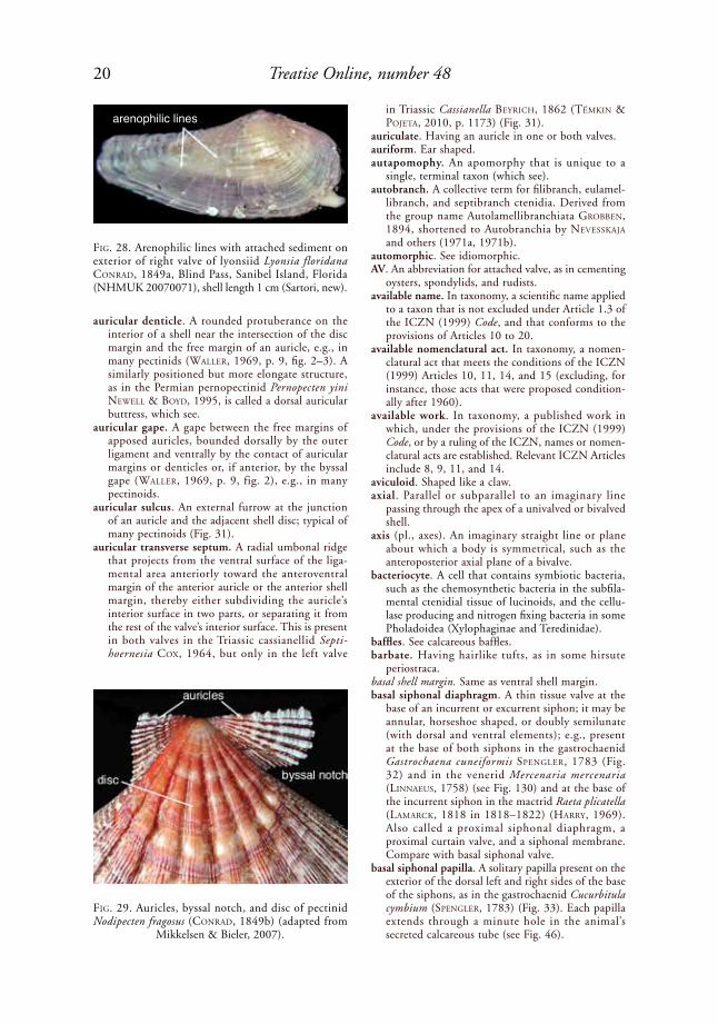
arenophilic lines. Radial strands of adhesive secre-tion deposited on the external surface of the peri-ostracum by arenophilic glands, as in the lyonsiid Lyonsia floridana CoNrad, 1849a (Fig. 28, Fig. 288). The name derives from the fact that these glands commonly attach lines of sediment to the shell. Also called arenophilic threads.
FiG. 22. Gastrochaenid Lamychaena cordiformis (NeVill & NeVill, 1869), Balaclava Bay, Mauritius, boring in massive coral, 15 m (UNC 15855); posterior view of incurrent and excurrent, type A+ siphons, showing siphonal papillae and an excurrent apical siphonal
diaphragm (Carter, new).
FiG. 23. Isognomonid Crenatula picta (GMeliN, 1791 in 1791–1793), interior of left valve, showing several apical transverse septa (adapted from Tëmkin & Pojeta, 2010, fig. 18.3, as junior synonym Crenatula avicularis
laMarCk, 1803 in 1802–1809).

18 Treatise Online, number 48
arenophilic papilla. A siphonal papilla immediately distal to an arenophilic mantle gland, e.g., in the laternulid Laternula elliptica (kiNG & Broderip, 1832) (see Fig. 288).
arenophilic threads. Same as arenophilic lines.
articulated. Having the two shell valves joined together along their hinges.
ascending lamella (pl., lamellae). The ctenidial lamella positioned farther from the ctenidial axis than the descending lamella and therefore not attached directly to the ctenidial axis; it extends dorsally from the ventral margin of the demibranch in a typical W-shaped ctenidium. The ascending lamella of the outer and inner demibranchs is generally attached to the mantle and to the visceral mass, respectively. Posterior to the visceral mass, the ascending lamellae of the left and right inner demi-branchs commonly come into contact, forming an interctenidial connection, which see. In bivalves with an extension of the mantle cavity posterior to the shell, the ascending lamellae of the outer and inner demibranchs may be attached to the mantle and to a siphonal ctenidial septum, respec-tively, e.g., in the gastrochaenid Lamychaena hians (GMeliN, 1791 in 1791–1793) (see Fig. 289).
ascending pallial sinus. A pallial sinus in which the anterior apex is directed anterodorsally, toward the umbonal cavity.
FiG. 24. Diagram of morphological features of Pholadidae (adapted from Turner, 1969, p. 704, fig. E163).
FiG. 25. Arcid Arca imbricata BruGuière, 1789, show-ing its subtrapezoidal shape, amphidetic, duplivincular/monovincular-D-1 ligament, neotaxodont dentition, and byssal sinus; shell length several cm (adapted from
Mikkelsen & Bieler, 2007).
protoplax
umbonal reflection
proraanterior slope
callum
anterior margin of shellumbonal-ventral sulcus
disc
mesoplax
metaplax
posterior slope
hypoplax
siphonoplax
post
erio
r end
posterior adductor muscle
siphonoplax
post
erio
r end
pallial line pallial sinus
ventral adductor muscleventral condyle
umbonal-ventral ridge
anterior margin of shell
pallial line
callum
prora
accessory anterioradductor muscle
dorsal extensionof callum
anterior adductor muscleumbonal reflection
umbochondrophore
apophysis
19Illustrated Glossary of the Bivalvia
left valve
4b
2b
cardinals2a
lateral 1AII gradually shortening
laminar IV
lateral AIV
lateral 2AII
1AII
2a2AII
PII
asiphonate. Lacking siphons.attenuate. Tapering gradually.auct. (or auctt.). Short for auctorum, i.e., a name as
commonly used or defined by some authors, but not as used or defined by the original author.
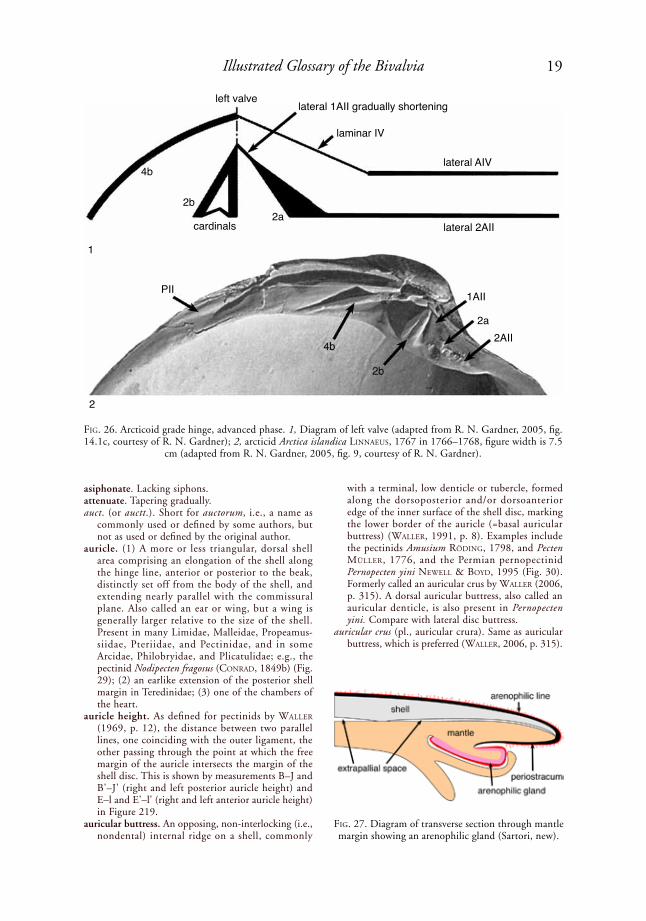
auricle. (1) A more or less triangular, dorsal shell area comprising an elongation of the shell along the hinge line, anterior or posterior to the beak, distinctly set off from the body of the shell, and extending nearly parallel with the commissural plane. Also called an ear or wing, but a wing is generally larger relative to the size of the shell. Present in many Limidae, Malleidae, Propeamus-siidae, Pteriidae, and Pectinidae, and in some Arcidae, Philobryidae, and Plicatulidae; e.g., the pectinid Nodipecten fragosus (CoNrad, 1849b) (Fig. 29); (2) an earlike extension of the posterior shell margin in Teredinidae; (3) one of the chambers of the heart.
auricle height. As defined for pectinids by Waller (1969, p. 12), the distance between two parallel lines, one coinciding with the outer ligament, the other passing through the point at which the free margin of the auricle intersects the margin of the shell disc. This is shown by measurements B–J and B'–J' (right and left posterior auricle height) and E–l and E'–l' (right and left anterior auricle height) in Figure 219.
auricular buttress. An opposing, non-interlocking (i.e., nondental) internal ridge on a shell, commonly
with a terminal, low denticle or tubercle, formed along the dorsoposterior and/or dorsoanterior edge of the inner surface of the shell disc, marking the lower border of the auricle (=basal auricular buttress) (Waller, 1991, p. 8). Examples include the pectinids Amusium rödiNG, 1798, and Pecten Müller, 1776, and the Permian pernopectinid Pernopecten yini NeWell & Boyd, 1995 (Fig. 30). Formerly called an auricular crus by Waller (2006, p. 315). A dorsal auricular buttress, also called an auricular denticle, is also present in Pernopecten yini. Compare with lateral disc buttress.
auricular crus (pl., auricular crura). Same as auricular buttress, which is preferred (Waller, 2006, p. 315).
FiG. 26. Arcticoid grade hinge, advanced phase. 1, Diagram of left valve (adapted from R. N. Gardner, 2005, fig. 14.1c, courtesy of R. N. Gardner); 2, arcticid Arctica islandica liNNaeus, 1767 in 1766–1768, figure width is 7.5
cm (adapted from R. N. Gardner, 2005, fig. 9, courtesy of R. N. Gardner).
FiG. 27. Diagram of transverse section through mantle margin showing an arenophilic gland (Sartori, new).
1
2
20 Treatise Online, number 48
arenophilic lines
auricular denticle. A rounded protuberance on the interior of a shell near the intersection of the disc margin and the free margin of an auricle, e.g., in many pectinids (Waller, 1969, p. 9, fig. 2–3). A similarly positioned but more elongate structure, as in the Permian pernopectinid Pernopecten yini NeWell & Boyd, 1995, is called a dorsal auricular buttress, which see.
auricular gape. A gape between the free margins of apposed auricles, bounded dorsally by the outer ligament and ventrally by the contact of auricular margins or denticles or, if anterior, by the byssal gape (Waller, 1969, p. 9, fig. 2), e.g., in many pectinoids.
auricular sulcus. An external furrow at the junction of an auricle and the adjacent shell disc; typical of many pectinoids (Fig. 31).
auricular transverse septum. A radial umbonal ridge that projects from the ventral surface of the liga-mental area anteriorly toward the anteroventral margin of the anterior auricle or the anterior shell margin, thereby either subdividing the auricle’s interior surface in two parts, or separating it from the rest of the valve’s interior surface. This is present in both valves in the Triassic cassianellid Septi-hoernesia Cox, 1964, but only in the left valve
in Triassic Cassianella BeyriCh, 1862 (tëMkiN & poJeta, 2010, p. 1173) (Fig. 31).
auriculate. Having an auricle in one or both valves.auriform. Ear shaped.autapomophy. An apomorphy that is unique to a
single, terminal taxon (which see).autobranch. A collective term for filibranch, eulamel-
libranch, and septibranch ctenidia. Derived from the group name Autolamellibranchiata GroBBeN, 1894, shortened to Autobranchia by NeVesskaJa and others (1971a, 1971b).
automorphic. See idiomorphic.AV. An abbreviation for attached valve, as in cementing
oysters, spondylids, and rudists.available name. In taxonomy, a scientific name applied
to a taxon that is not excluded under Article 1.3 of the ICZN (1999) Code, and that conforms to the provisions of Articles 10 to 20.
available nomenclatural act. In taxonomy, a nomen-clatural act that meets the conditions of the ICZN (1999) Articles 10, 11, 14, and 15 (excluding, for instance, those acts that were proposed condition-ally after 1960).
available work. In taxonomy, a published work in which, under the provisions of the ICZN (1999) Code, or by a ruling of the ICZN, names or nomen-clatural acts are established. Relevant ICZN Articles include 8, 9, 11, and 14.
aviculoid. Shaped like a claw.axial. Parallel or subparallel to an imaginary line
passing through the apex of a univalved or bivalved shell.
axis (pl., axes). An imaginary straight line or plane about which a body is symmetrical, such as the anteroposterior axial plane of a bivalve.
bacteriocyte. A cell that contains symbiotic bacteria, such as the chemosynthetic bacteria in the subfila-mental ctenidial tissue of lucinoids, and the cellu-lase producing and nitrogen fixing bacteria in some Pholadoidea (Xylophaginae and Teredinidae).
baffles. See calcareous baffles. barbate. Having hairlike tufts, as in some hirsute
periostraca. basal shell margin. Same as ventral shell margin.basal siphonal diaphragm. A thin tissue valve at the
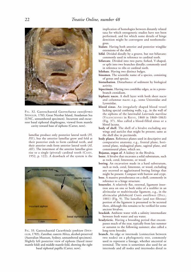
base of an incurrent or excurrent siphon; it may be annular, horseshoe shaped, or doubly semilunate (with dorsal and ventral elements); e.g., present at the base of both siphons in the gastrochaenid Gastrochaena cuneiformis speNGler, 1783 (Fig. 32) and in the venerid Mercenaria mercenaria (liNNaeus, 1758) (see Fig. 130) and at the base of the incurrent siphon in the mactrid Raeta plicatella (laMarCk, 1818 in 1818–1822) (harry, 1969). Also called a proximal siphonal diaphragm, a proximal curtain valve, and a siphonal membrane. Compare with basal siphonal valve.
basal siphonal papilla. A solitary papilla present on the exterior of the dorsal left and right sides of the base of the siphons, as in the gastrochaenid Cucurbitula cymbium (speNGler, 1783) (Fig. 33). Each papilla extends through a minute hole in the animal’s secreted calcareous tube (see Fig. 46).
FiG. 28. Arenophilic lines with attached sediment on exterior of right valve of lyonsiid Lyonsia floridana CoNrad, 1849a, Blind Pass, Sanibel Island, Florida (NHMUK 20070071), shell length 1 cm (Sartori, new).
FiG. 29. Auricles, byssal notch, and disc of pectinid Nodipecten fragosus (CoNrad, 1849b) (adapted from
Mikkelsen & Bieler, 2007).

21Illustrated Glossary of the Bivalvia
basal siphonal valve. A flaplike protrusion of the mantle at the base of the incurrent siphon, serving to regulate water flow, e.g., in the lithophaginid Lithophaga (Leiosolenus) hancocki T. soot-ryeN, 1955 (Fig. 34) and in some Mactridae. Also called a curtain valve, proximal siphonal valve, and siphonal membrane (for the latter, see yoNGe, 1955, fig. 10). Compare with basal siphonal diaphragm.
bead. A small, rounded protuberance, commonly on an external rib.
beak. The more or less pointed, conical, early devel-opmental portion of each valve of a shell, the tip or apex of which represents the origin of growth; e.g., the venerid Periglypta listeri (Gray, 1838) (Fig. 35). Compare with umbo.
beekite. A diagenetic mineral structure consisting of concentric siliceous layers in rosette form, e.g., in some fossil oysters.
bent foliated microstructure. A term proposed by MalChus (1990, p. 64) for what is herein called transitional irregular simple prismatic/irregular complex crossed foliated mictrostructure (abbr., ISP/ICCF), which see.
benthic. Living on or in sediment. Also spelled benthonic, although benthic is preferred.
Bernard system of notation. A system of homology of individual hinge teeth developed by BerNard (1895, 1896a, 1896b, 1897), based on the early ontogeny of Cenozoic heterodont bivalves (Fig. 36). The adult hinge teeth are regarded as dissev-ered parts of originally continuous primary lamellae appearing in the juvenile shell as simple anterior and posterior ridges, those in the right valve inter-locking with those in the left, with the anterior lamellae separated from the posterior ones by the juvenile ligament. During ontogeny, the posterior
FiG. 30. Internal morphology of Permian pernopectinid Pernopecten yini NeWell & Boyd, 1995. 1, Left valve, scale bar = 1 mm; 2, right valve, scale bar = 5 mm. bab, basal auricular buttress; dab, dorsal auricular buttress; ldb, lateral disc buttress; olg, outer ligament groove; olr, outer ligament ridge (adapted from Newell & Boyd, 1995, fig. 53.2b,
53.3; courtesy of the American Museum of Natural History).
FiG. 31. 1, Auricular transverse septum on the internal surface of left valve of Triassic cassianellid Cassianella BeyriCh, 1862; 2, corresponding auricular sulcus on shell’s exterior (adapted from Münster in Goldfuss, 1836 in
1833–1841, pl. 116,10).
scroll
bab
ldb
dab olgbab
ldb
olr dabdab
bab
ldb1 2
auricular sulcusauricular transverse septum
1
2
22 Treatise Online, number 48
lamellae produce only posterior lateral teeth (PI, PII ), but the anterior lamellae grow and fold at their posterior ends to form cardinal teeth, and their anterior ends form anterior lateral teeth (AI, AII ). The innermost of the anterior lamellae gives rise to a single (pivotal) cardinal tooth (Casey, 1952, p. 122). A drawback of the system is the
implication of homologies between distantly related taxa for which ontogenetic studies have not been performed, and for which some details of hinge dentition might be convergent and nonhomolo-gous.
bialate. Having both anterior and posterior winglike extensions of the shell.
bifid. Divided distally by a groove, but not bifurcate; commonly used in reference to cardinal teeth.
bifurcate. Divided into two parts; forked, V-shaped, or split into two branches distally; commonly used in reference to ribs or cardinal teeth.
bilobate. Having two distinct bulges.binomen. The scientific name of a species, consisting
of genus and species.bioturbation. Disturbance of sediment by biological
activity.bipectinate. Having two comblike edges, as in a proto-
branch ctenidium. biphasic nacre. A shell layer with both sheet nacre
and columnar nacre; e.g., some Unionidae and Lyonsiidae.
blood sinus. An irregularly shaped blood vessel lacking special confining walls, e.g., in the wall of the siphons of the laternulid Laternula marilina (ValeNCieNNes in reeVe, 1860 in 1860–1863) (Fig. 37). Also called a blood-filled sinus or a blood lacuna.
body of shell. The shell of a bivalve excluding any wings and auricles that might be present; same as the shell disc in pectinoids.
body planes. Reference planes used in descriptive and comparative anatomy, e.g., frontal plane, hori-zontal plane, midsagittal plane, sagittal plane, and commissural plane, which see.
Bojanus, organ of. A kidney in the Bivalvia. borer. A bivalve that excavates a solid substratum, such
as rock, coral, limestone, or wood. boring. An excavation made in a hard substratum,
such as rock, coral, limestone, or wood, excluding any secreted or agglutinated boring linings that might be present. Compare with burrow and crypt.
boss. A massive protuberance on a shell, commonly in reference to a hinge structure.
bourrelet. A relatively flat, external, ligament inser-tion area on one or both sides of a resilifer in an alivincular or multivincular ligament, e.g., in the alivincular philobryid Cratis antillensis (dall, 1881) (Fig. 9). The lamellar (and not fibrous) portion of the ligament is presumed to be secreted there, although this remains to be verified for some ancient bivalves.
brackish. Ambient water with a salinity intermediate between fresh water and sea water.
bradytictic. Having a brooding period that encom-passes much of the year, typically from late summer or autumn to the following summer; also called a long-term brooder.
branch. An edge or internode (connection between two nodes) on a phylogenetic tree, commonly used to represent a lineage, whether ancestral or terminal. The term is sometimes also used for an internode and all nodes and internodes distal to
FiG. 32. Gastrochaenid Gastrochaena cuneiformis speNGler, 1783, Great Nicobar Island, Anadaman Sea (UNC, unnumbered specimen). Incurrent and excur-rent basal siphonal diaphragms, viewed from mantle
cavity toward base of siphons (Carter, new).
FiG. 33. Gastrochaenid Cucurbitula cymbium (speN-Gler, 1783), Zanzibar, eastern Africa, alcohol-preserved (Australian Museum, Sydney, unnumbered specimen). Slightly left posterior view of siphons (fused inner mantle fold) and middle mantle fold, showing the right
basal siphonal papilla (Carter, new).

23Illustrated Glossary of the Bivalvia
(descended from) it (PhyloCode, May, 2012: www.ohio.edu/phylocode/glossary.html).
branch-based clade. A clade originating from a partic-ular branch (internode) on a phylogenetic tree; a clade encompassing a particular branch on a phylo-genetic tree and all nodes and branches descended from that branch; a clade whose name is defined using a branch-based definition (PhyloCode, May, 2012: www.ohio.edu/phylocode/glossary.html). Compare with node-based clade.
branch-based definition. A definition that associates a name with a clade originating with a branch (on a phylogenetic tree) representing the ancestral lineage of specified organisms and/or species (internal specifiers) after its divergence from the ancestral lineage of other specified organisms and/or species (external specifiers) (PhyloCode, May, 2012: www.ohio.edu/phylocode/glossary.html). Compare with node-based definition.
branch-modified node-based definition. A node-based definition that incorporates wording from branch-based definitions to include all extant organisms as internal specifiers without explicitly naming them. Branch-modified node-based definitions can be used to associate names with crown clades when basal relationships within the crown are poorly understood or when the author intends to include in the named taxon subsequently discovered extant organisms that share a more recent common ancestor with the currently known members of the named taxon than with other currently known taxa (PhyloCode, May, 2012: www.ohio.edu/phylocode/glossary.html).
branchia (pl., branchiae). Same as a ctenidium, which see.
branchial membrane. Same as ctenidial septum, which see.
branchial orifice. The posteroventral, incurrent aper-ture in nonsiphonate bivalves, positioned ventral to the excurrent aperture, and homologous with the incurrent siphonal aperture in siphonate bivalves, e.g., in the crassatellid Eucrassatella iredale, 1924.
branchial passage. A conduit confined ventrally by the ctenidia and carrying the excurrent water stream.
branchial septum. Same as ctenidial septum, which see. branchial sieve. See under septibranch.branchial siphon. Same as incurrent siphon.branching crossed foliated microstructure (abbr.,
BCF). A calcitic crossed foliated microstructure in which the first-order crossed folia are highly branching as seen in horizontal sections and on the depositional surface, e.g., in the middle shell layer in the Jurassic gryphaeid Gryphaea (Bilobissa) bilobata J. de C. soWerBy, 1840, in soWerBy & soWerBy, 1812–1846 (Fig. 38). Compare with irregular crossed foliated.
branching crossed lamellar microstructure (abbr., BCL). An aragonitic crossed lamellar microstruc-ture in which the first-order lamellae are highly branching as seen in horizontal sections and on the depositional surface (see Fig. 82).
branchitellum (pl., branchitella). The point on the posteroventral shell margin, nearest to the animal’s
palliobranchial fusion, commonly corresponding with a conspicuously projecting posteroventral tip on the left shell valve, e.g., in many sickle-shaped oysters, such as Eocene Cubitostrea wemmelensis (GliBert, 1936) (Fig. 39); also present in some nearly circular oysters, such as the lophinid Alec-tryonella plicatula (GMeliN, 1791 in 1791–1793). The aboral end of the ctenidium points toward the branchitellum.
breviaxis (adj., breviaxial). With the shell’s length determined by the maximum anteroposterior shell axis, the shorter of the two segments of this axis defined by the line passing through the beak, perpendicular to the anteroposterior axis (Bailey, 2009).
brevidetic. The position of a ligament or other feature on the dorsal region of the shorter end of an inequi-lateral shell (Bailey, 2009). See also longidetic. The
FiG. 34. Type A siphon and basal siphonal valve in lithophaginid Lithophaga (Leiosolenus) hancocki T. soot-ryeN, 1955, Panama Bay, just north of Panama City, boring in massive coral, 3 m depth. Ventral view, showing incurrent channel bordered dorsally by tissue-grade fusion of inner mantle fold, which forms a siphonal pallial septum, a basal siphonal valve, and the ventral margin of the excurrent, tissue-grade siphon; width of figure approximately 10 mm (Carter, new).
FiG. 35. Venerid Periglypta listeri (Gray, 1838), dorsal view of united valves showing lunule, escutcheon, beak, and umbo (adapted from Mikkelsen & Bieler, 2007).
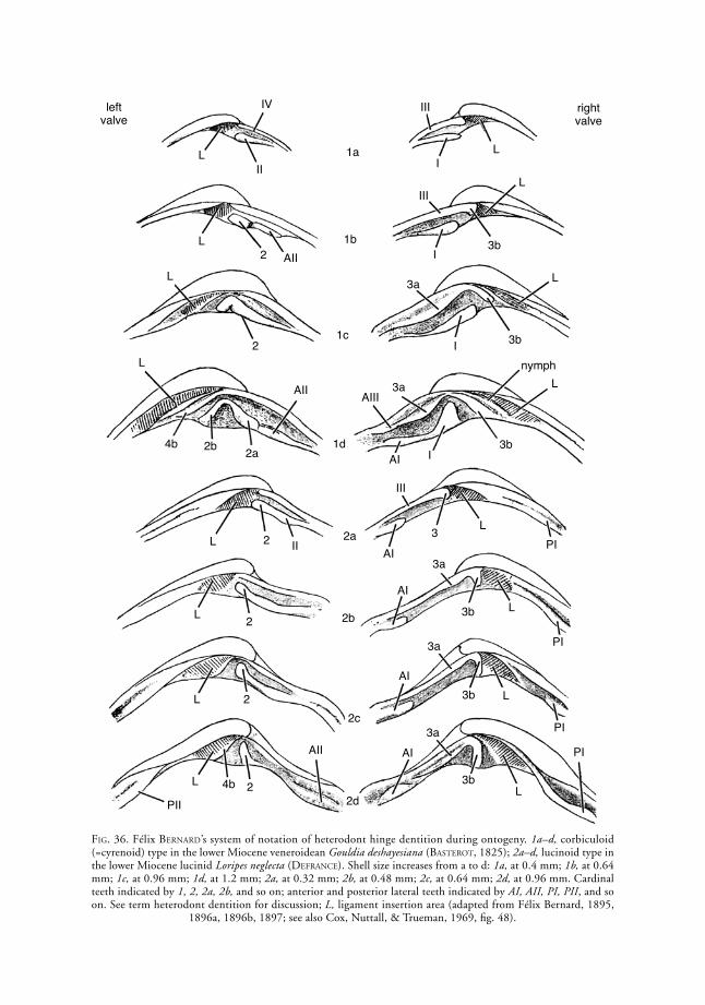
24 Treatise Online, number 48
FiG. 36. Félix BerNard’s system of notation of heterodont hinge dentition during ontogeny. 1a–d, corbiculoid (=cyrenoid) type in the lower Miocene veneroidean Gouldia deshayesiana (Basterot, 1825); 2a–d, lucinoid type in the lower Miocene lucinid Loripes neglecta (deFraNCe). Shell size increases from a to d: 1a, at 0.4 mm; 1b, at 0.64 mm; 1c, at 0.96 mm; 1d, at 1.2 mm; 2a, at 0.32 mm; 2b, at 0.48 mm; 2c, at 0.64 mm; 2d, at 0.96 mm. Cardinal teeth indicated by 1, 2, 2a, 2b, and so on; anterior and posterior lateral teeth indicated by AI, AII, PI, PII, and so on. See term heterodont dentition for discussion; L, ligament insertion area (adapted from Félix Bernard, 1895,
1896a, 1896b, 1897; see also Cox, Nuttall, & Trueman, 1969, fig. 48).
left valve
right valve
1a
1b
1c
1d
2a
2b
2c
2d
L
IV
II
LAII2
L
2L
4b 2b 2a
AII
L 2 II
L 2
L 2
L 2
AII
PII
4b
III
I
3b
AI
3a
L
PI
III
III
I
I
I
3b
3b
3b
3b
3b
AI
AI
AI
3a
3a
3a
L
L
L
L
L
L
PI
PI
PI
AIII
nymph
3
AI3a
L
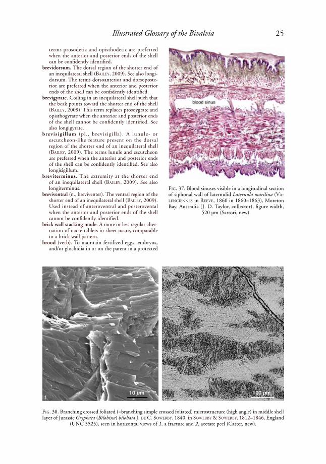
25Illustrated Glossary of the Bivalvia
terms prosodetic and opisthodetic are preferred when the anterior and posterior ends of the shell can be confidently identified.
brevidorsum. The dorsal region of the shorter end of an inequilateral shell (Bailey, 2009). See also longi-dorsum. The terms dorsoanterior and dorsoposte-rior are preferred when the anterior and posterior ends of the shell can be confidently identified.
brevigyrate. Coiling in an inequilateral shell such that the beak points toward the shorter end of the shell (Bailey, 2009). This term replaces prosoygrate and opisthogyrate when the anterior and posterior ends of the shell cannot be confidently identified. See also longigyrate.
brevis igi l lum (pl . , brevis ig i l la) . A lunule- or escutcheon-like feature present on the dorsal region of the shorter end of an inequilateral shell (Bailey, 2009). The terms lunule and escutcheon are preferred when the anterior and posterior ends of the shell can be confidently identified. See also longisigillum.
breviterminus. The extremity at the shorter end of an inequilateral shell (Bailey, 2009). See also longiterminus.
breviventral (n., breviventer). The ventral region of the shorter end of an inequilateral shell (Bailey, 2009). Used instead of anteroventral and posterovental when the anterior and posterior ends of the shell cannot be confidently identified.
brick wall stacking mode. A more or less regular alter-nation of nacre tablets in sheet nacre, comparable to a brick wall pattern.
brood (verb). To maintain fertilized eggs, embryos, and/or glochidia in or on the parent in a protected
FiG. 37. Blood sinuses visible in a longitudinal section of siphonal wall of laternulid Laternula marilina (Va-leNCieNNes in reeVe, 1860 in 1860–1863), Moreton Bay, Australia (J. D. Taylor, collector), figure width,
520 µm (Sartori, new).
FiG. 38. Branching crossed foliated (=branching simple crossed foliated) microstructure (high angle) in middle shell layer of Jurassic Gryphaea (Bilobissa) bilobata J. de C. soWerBy, 1840, in soWerBy & soWerBy, 1812–1846, England
(UNC 5525), seen in horizontal views of 1, a fracture and 2, acetate peel (Carter, new).
21

26 Treatise Online, number 48
environment, as in many Unionoidea, Hyrioidea, and Teredinidae, and in some Nuculidae, Vener-idae, and Carditidae.
brooding period. The interval during which fertilized eggs, embryos, and/or glochidia are brooded; also called the gravid period.
buccal funnel. An expanded, trumpet-shaped mouth, as in some Cuspidariidae, Poromyidae (Fig. 40), and Verticordiidae (see Fig. 85).
burrow. A term generally used for a dwelling space in soft sediment, but sometimes also used for a dwelling space in a bored rock, coral, shell, or wood substratum, exclusive of any secreted or agglutinated burrow or boring linings that might be present. Compare with boring and crypt.
burrower. An animal that penetrates loose sediment. buttress. A distinct shell thickening that supports
the hinge, ligament, or a muscle attachment, or
that otherwise reinforces the shell, and that does not articulate with a socket in the opposite valve. The slatlike internal buttresses on the disc of the Propeamussiidae are also called internal ribs. Waller (2006, p. 315) advocated replacing the term nonarticulating crus/crura with the term buttress. See also auricular buttress, lateral disc buttress, and myophoric buttress. Compare with myophore, apophysis, umbonal septum, clavicle, and myofringing ridge.
byssal collar. A relatively large, rounded, anatomically anterior but functionally ventral extension of the shell in each valve, e.g., in the Permian alatocon-chid Saikraconcha tunisiensis yaNCey & Boyd, 1983 (Fig. 41).
byssal ear. A right anterior auricle that is developed as a tonguelike process not in alignment with the hinge, but rather extending toward the left valve and resting in a left cardinal area socket (=Gelenkgrube) in front of the beak, e.g., the Jurassic and Cretaceous buchiid Buchia rouillier, 1845 (Fig. 42).
byssal fasciole. The filled-in track left by a migrating byssal notch, as seen on the shell’s exterior; it is bounded by the margin of the shell disc and by the most ventral costa on the auricle, e.g., in the pectinid Similipecten nanus (Verrill & Bush in Verrill, 1897) (see Fig. 237).
byssal foramen. An aperture in a valve through which a byssus or calcified byssus extends; e.g., in the right valve of Anomiidae. Compare with byssal gape.
byssal gape. A permanent opening between the shell margins, serving for the passage of a byssus or byssate foot, e.g., in the arcid Arca imbricata BruGuière, 1789 (Fig. 43). This feature is accom-panied by a byssal notch and/or a byssal sinus in the shell margin.
byssal gland. A gland situated around the byssal pit and along the underside of the foot, which secretes byssal threads.
byssal groove. A ventral groove on the sole of the foot, extending anteriorly from a byssal pit. In some species, the byssal groove has a small depres-sion on one end, e.g., the byssal disc-pit in Mytilus liNNaeus, 1758. In other species, the byssal groove merely meets the anteroventral margin of the sole of the foot, as in Lamychaena cordiformis (NeVill & NeVill, 1869) (Fig. 19).
byssal hair. A hirsute structure attached to the exterior of the shell, secreted by the pedal byssal gland. Present in many Mytilidae, where they are some-times (inaccurately) referred to as periostracal hairs.
byssal notch. A rounded or trigonal embayment in the margin of a shell valve, generally the right valve, bounded dorsally by the free margin of an anterior auricle and ventrally by part of the auricle and/or the margin of the disc of the shell. The notch serves for passage of the byssus and/or foot; e.g., in the isognomonid Isognomon bicolor (C. B. adaMs, 1845) (Fig. 44), and the pectinids Caribachlamys sentis (reeVe, 1853 in 1852–1853) (see Fig. 89) and Nodipecten fragosus (CoNrad, 1849b) (Fig. 29). Compare with byssal gape and byssal sinus.
FiG. 39. Eocene ostreoidean Cubitostrea wemmelensis (GliBert, 1936), showing branchitellum in left valve and size discrepancy of valves after deterioration of conchiolin-rich prismatic fringe on right valve (adapted
from Glibert, 1936 and Stenzel, 1971, fig. J17).
FiG. 40. Diagram of anatomy of Poromyidae (adapted from Mikkelsen & Bieler, 2007).
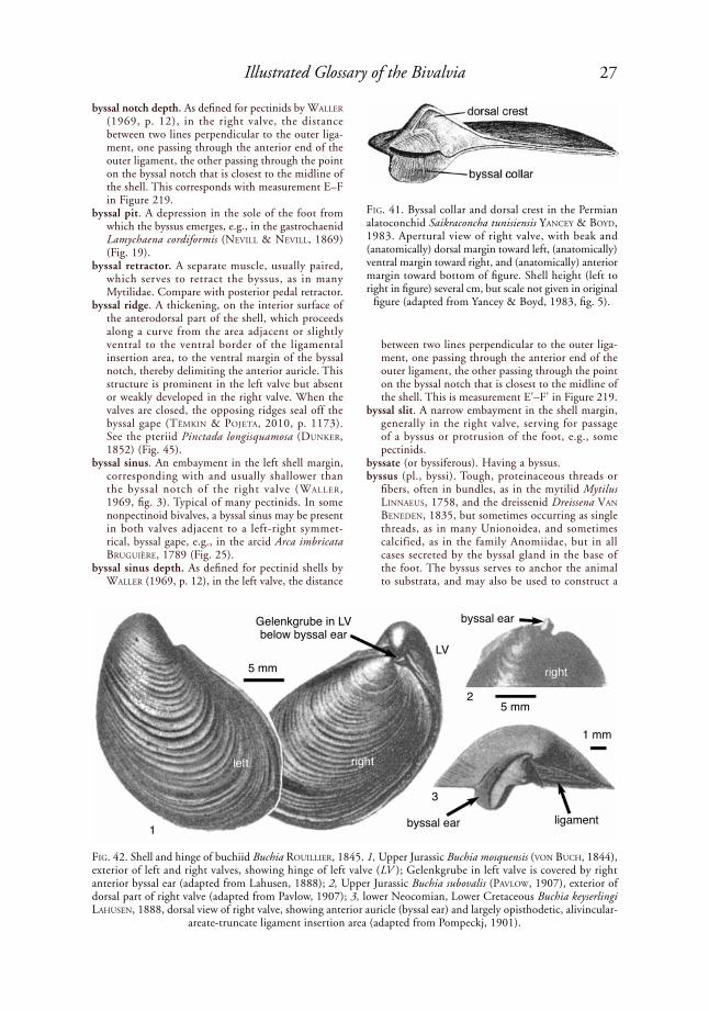
27Illustrated Glossary of the Bivalvia
byssal notch depth. As defined for pectinids by Waller (1969, p. 12), in the right valve, the distance between two lines perpendicular to the outer liga-ment, one passing through the anterior end of the outer ligament, the other passing through the point on the byssal notch that is closest to the midline of the shell. This corresponds with measurement E–F in Figure 219.
byssal pit. A depression in the sole of the foot from which the byssus emerges, e.g., in the gastrochaenid Lamychaena cordiformis (NeVill & NeVill, 1869) (Fig. 19).
byssal retractor. A separate muscle, usually paired, which serves to retract the byssus, as in many Mytilidae. Compare with posterior pedal retractor.
byssal ridge. A thickening, on the interior surface of the anterodorsal part of the shell, which proceeds along a curve from the area adjacent or slightly ventral to the ventral border of the ligamental insertion area, to the ventral margin of the byssal notch, thereby delimiting the anterior auricle. This structure is prominent in the left valve but absent or weakly developed in the right valve. When the valves are closed, the opposing ridges seal off the byssal gape (tëMkiN & poJeta, 2010, p. 1173). See the pteriid Pinctada longisquamosa (duNker, 1852) (Fig. 45).
byssal sinus. An embayment in the left shell margin, corresponding with and usually shallower than the byssal notch of the right valve (Waller, 1969, fig. 3). Typical of many pectinids. In some nonpectinoid bivalves, a byssal sinus may be present in both valves adjacent to a left-right symmet-rical, byssal gape, e.g., in the arcid Arca imbricata BruGuière, 1789 (Fig. 25).
byssal sinus depth. As defined for pectinid shells by Waller (1969, p. 12), in the left valve, the distance
between two lines perpendicular to the outer liga-ment, one passing through the anterior end of the outer ligament, the other passing through the point on the byssal notch that is closest to the midline of the shell. This is measurement E'–F' in Figure 219.
byssal slit. A narrow embayment in the shell margin, generally in the right valve, serving for passage of a byssus or protrusion of the foot, e.g., some pectinids.
byssate (or byssiferous). Having a byssus.byssus (pl., byssi). Tough, proteinaceous threads or
fibers, often in bundles, as in the mytilid Mytilus liNNaeus, 1758, and the dreissenid Dreissena VaN BeNedeN, 1835, but sometimes occurring as single threads, as in many Unionoidea, and sometimes calcified, as in the family Anomiidae, but in all cases secreted by the byssal gland in the base of the foot. The byssus serves to anchor the animal to substrata, and may also be used to construct a
FiG. 41. Byssal collar and dorsal crest in the Permian alatoconchid Saikraconcha tunisiensis yaNCey & Boyd, 1983. Apertural view of right valve, with beak and (anatomically) dorsal margin toward left, (anatomically) ventral margin toward right, and (anatomically) anterior margin toward bottom of figure. Shell height (left to right in figure) several cm, but scale not given in original
figure (adapted from Yancey & Boyd, 1983, fig. 5).
FiG. 42. Shell and hinge of buchiid Buchia rouillier, 1845. 1, Upper Jurassic Buchia mosquensis (VoN BuCh, 1844), exterior of left and right valves, showing hinge of left valve (LV ); Gelenkgrube in left valve is covered by right anterior byssal ear (adapted from Lahusen, 1888); 2, Upper Jurassic Buchia subovalis (paVloW, 1907), exterior of dorsal part of right valve (adapted from Pavlow, 1907); 3, lower Neocomian, Lower Cretaceous Buchia keyserlingi lahuseN, 1888, dorsal view of right valve, showing anterior auricle (byssal ear) and largely opisthodetic, alivincular-
areate-truncate ligament insertion area (adapted from Pompeckj, 1901).
Gelenkgrube in LVbelow byssal ear
5 mm
byssal ear ligament
3
1 mm
5 mm
LV
byssal ear
1
2

28 Treatise Online, number 48
byssal nest or a brood pouch. Not homologous with the larval threads of unionoid glochidial larvae.
byssus gland. Same as byssal gland, which is preferred.byssus retractor. Same as byssal retractor, which is
preferred.caecum (pl., caeca). (1) A pouch, canal, or duct that
is open only at one end, e.g., the large caecum in the stomach of Teredinidae (Fig. 2), or the smaller caeca in type V stomachs of many veneroids (see Fig. 305); (2) a shell pore (tubule) that contains a papillose outgrowth of the mantle epithelium; also called a pallial caecum or a mantle caecum (reiNdl & hasZpruNar, 1996); e.g., many arcoids, Astar-tidae, Carditidae, Condylocardiidae, Cyrenidae, and Sphaeriidae, among other groups. Mantle caeca have evolved independently in other phyla and in other molluscan classes, such as Brachiopoda, Poly-placophora, and fissurellid Gastropoda (see reiNdl & hasZpruNar, 1996).
calcareous baffles. Spikelike or oval projections from the siphonal or basal siphonal boring or calcareous tube lining, which locally restrict the diameter of the boring or tube; e.g., in the basal siphonal
portion of the calcareous igloo of Cucurbitula cymbium (speNGler, 1783) (Fig. 46).
calcareous ossicle. Same as lithodesma, which see.calcareous tube. A secreted calcium carbonate struc-
ture that largely surrounds a bivalve, except at the siphonal opening or openings. Calcareous tubes may be unattached, as in some Clavagelloidea and the gastrochaenid Eufistulana eaMes, 1951, or attached on one side to a substratum, as in some species of the gastrochaenid Cucurbitula Gould, 1861 (Fig. 46). Some tubes are adventitious, being secreted by endolithic bivalves only when they have outgrown their substratum, or when they are exposed by erosion or breakage of their substratum (e.g., the gastrochaenid Rocellaria BlaiNVille, 1828 in 1816–1830, boring into thin shells). Calcareous tubes generally have a posterior siphonal portion and an anterior shell chamber portion. See also crypt and igloo.
calcite. A rhombohedral crystalline form of calcium carbonate. This is less soluble than aragonite at surface and near-surface temperatures and pres-sures, so calcitic shells are more likely to resist dissolution than aragonitic ones in the fossil record.
calcite blade forms. Calcite blade shapes defined by their interfacial gamma angle (Fig. 47), viewed perpendicular to the broad surface of the blade (i.e., not necessarily perpendicular to the surface of deposition, as the blade may dip into the plane of deposition) (Carter & Clark, 1985). These include gamma 120, 129, 135, and 150 forms. These forms do not necessarily correlate with actual calcite crystallographic orientations (ruNNeGar, 1984, p. 284; CheCa, esteBaN-delGado, & rodríGues-NaVarro, 2007). All of these forms are known to occur among various representatives of the Ostreoidea, so they have limited significance for higher level phylogenetic relationships in the Bivalvia. Gamma 129 blades are also known to occur in the Cambrian tergomyan Pseudomyona queenslandica (ruNNeGar & Jell, 1976), and gamma 135 blades also occur in the propeamussiid Propeamussium dalli (e. a. sMith, 1885) (Carter & Clark, 1985). According to CheCa, esteBaN-delGado, and rodríGues-NaVarro (2007), the blades in bivalve foliated structure have only one possible crystallographic arrangement, i.e., with the flat, main surface of the blade coinciding with the {I0Ī8} rhombohedral crystal face. This differs from all other known biomineral systems and inor-ganically precipitated calcite crystals, apparently due to stabilization this crystal face by preferential absorption of specific organic molecules (addadi & WeiNer, 1985).
callum. An adventitious secretion on the anterior end of the shell of many Pholadoidea, comprising a more or less smooth wall that closes or partially closes the pedal gape in the mature stage (Fig. 24). Generally, the left and right halves of the callum do not quite meet, and they are joined by a periostracal fold with only a minute pore remaining between them. See also the pholadid Martesia striata (liNNaeus, 1758) (Fig. 48–49).
FiG. 43. Byssal gape in arcid Arca imbricata BruGuière, 1789, shell length 4.8 cm (adapted from Mikkelsen &
Bieler, 2007).
FiG. 44. Isognomonid Isognomon bicolor (C. B. adaMs, 1845), showing byssal notch; figure is approximately 30 mm wide (adapted from Mikkelsen & Bieler, 2007).
byssal notch
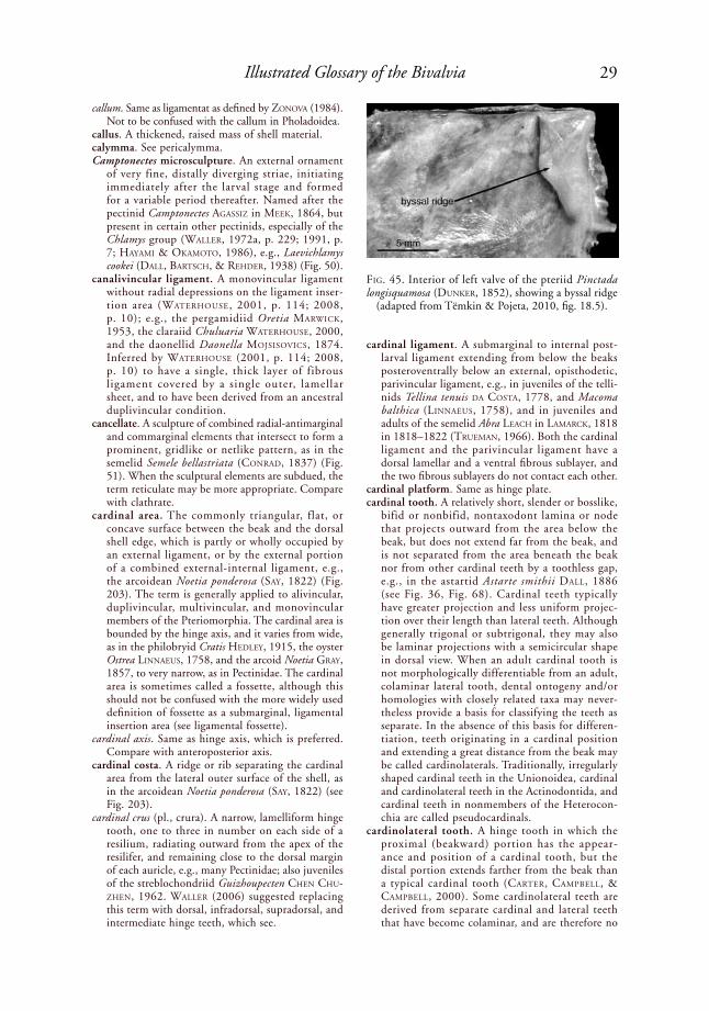
29Illustrated Glossary of the Bivalvia
callum. Same as ligamentat as defined by ZoNoVa (1984). Not to be confused with the callum in Pholadoidea.
callus. A thickened, raised mass of shell material.calymma. See pericalymma.Camptonectes microsculpture. An external ornament
of very fine, distally diverging striae, initiating immediately after the larval stage and formed for a variable period thereafter. Named after the pectinid Camptonectes aGassiZ in Meek, 1864, but present in certain other pectinids, especially of the Chlamys group (Waller, 1972a, p. 229; 1991, p. 7; hayaMi & okaMoto, 1986), e.g., Laevichlamys cookei (dall, BartsCh, & rehder, 1938) (Fig. 50).
canalivincular ligament. A monovincular ligament without radial depressions on the ligament inser-tion area (Waterhouse, 2001, p. 114; 2008, p. 10); e.g., the pergamidiid Oretia MarWiCk, 1953, the claraiid Chuluaria Waterhouse, 2000, and the daonellid Daonella MoJsisoViCs, 1874. Inferred by Waterhouse (2001, p. 114; 2008, p. 10) to have a single, thick layer of fibrous ligament covered by a single outer, lamellar sheet, and to have been derived from an ancestral duplivincular condition.
cancellate. A sculpture of combined radial-antimarginal and commarginal elements that intersect to form a prominent, gridlike or netlike pattern, as in the semelid Semele bellastriata (CoNrad, 1837) (Fig. 51). When the sculptural elements are subdued, the term reticulate may be more appropriate. Compare with clathrate.
cardinal area. The commonly triangular, flat, or concave surface between the beak and the dorsal shell edge, which is partly or wholly occupied by an external ligament, or by the external portion of a combined external-internal ligament, e.g., the arcoidean Noetia ponderosa (say, 1822) (Fig. 203). The term is generally applied to alivincular, duplivincular, multivincular, and monovincular members of the Pteriomorphia. The cardinal area is bounded by the hinge axis, and it varies from wide, as in the philobryid Cratis hedley, 1915, the oyster Ostrea liNNaeus, 1758, and the arcoid Noetia Gray, 1857, to very narrow, as in Pectinidae. The cardinal area is sometimes called a fossette, although this should not be confused with the more widely used definition of fossette as a submarginal, ligamental insertion area (see ligamental fossette).
cardinal axis. Same as hinge axis, which is preferred. Compare with anteroposterior axis.
cardinal costa. A ridge or rib separating the cardinal area from the lateral outer surface of the shell, as in the arcoidean Noetia ponderosa (say, 1822) (see Fig. 203).
cardinal crus (pl., crura). A narrow, lamelliform hinge tooth, one to three in number on each side of a resilium, radiating outward from the apex of the resilifer, and remaining close to the dorsal margin of each auricle, e.g., many Pectinidae; also juveniles of the streblochondriid Guizhoupecten CheN Chu-ZheN, 1962. Waller (2006) suggested replacing this term with dorsal, infradorsal, supradorsal, and intermediate hinge teeth, which see.
cardinal ligament. A submarginal to internal post-larval ligament extending from below the beaks posteroventrally below an external, opisthodetic, parivincular ligament, e.g., in juveniles of the telli-nids Tellina tenuis da Costa, 1778, and Macoma balthica (liNNaeus, 1758), and in juveniles and adults of the semelid Abra leaCh in laMarCk, 1818 in 1818–1822 (trueMaN, 1966). Both the cardinal ligament and the parivincular ligament have a dorsal lamellar and a ventral fibrous sublayer, and the two fibrous sublayers do not contact each other.
cardinal platform. Same as hinge plate. cardinal tooth. A relatively short, slender or bosslike,
bifid or nonbifid, nontaxodont lamina or node that projects outward from the area below the beak, but does not extend far from the beak, and is not separated from the area beneath the beak nor from other cardinal teeth by a toothless gap, e.g., in the astartid Astarte smithii dall, 1886 (see Fig. 36, Fig. 68). Cardinal teeth typically have greater projection and less uniform projec-tion over their length than lateral teeth. Although generally trigonal or subtrigonal, they may also be laminar projections with a semicircular shape in dorsal view. When an adult cardinal tooth is not morphologically differentiable from an adult, colaminar lateral tooth, dental ontogeny and/or homologies with closely related taxa may never-theless provide a basis for classifying the teeth as separate. In the absence of this basis for differen-tiation, teeth originating in a cardinal position and extending a great distance from the beak may be called cardinolaterals. Traditionally, irregularly shaped cardinal teeth in the Unionoidea, cardinal and cardinolateral teeth in the Actinodontida, and cardinal teeth in nonmembers of the Heterocon-chia are called pseudocardinals.
cardinolateral tooth. A hinge tooth in which the proximal (beakward) portion has the appear-ance and position of a cardinal tooth, but the distal portion extends farther from the beak than a typical cardinal tooth (Carter, CaMpBell, & CaMpBell, 2000). Some cardinolateral teeth are derived from separate cardinal and lateral teeth that have become colaminar, and are therefore no
FiG. 45. Interior of left valve of the pteriid Pinctada longisquamosa (duNker, 1852), showing a byssal ridge
(adapted from Tëmkin & Pojeta, 2010, fig. 18.5).
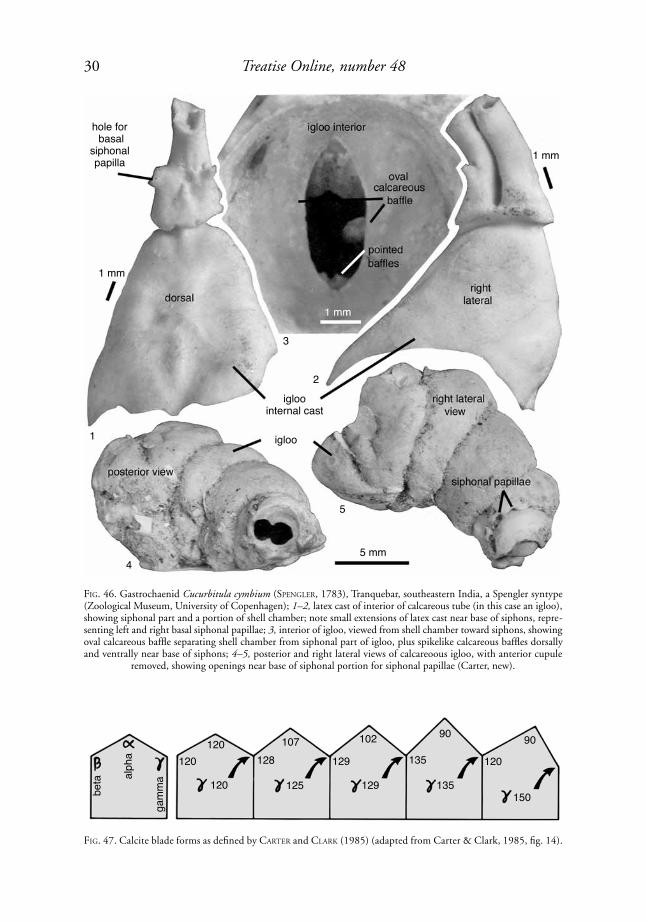
30 Treatise Online, number 48
FiG. 46. Gastrochaenid Cucurbitula cymbium (speNGler, 1783), Tranquebar, southeastern India, a Spengler syntype (Zoological Museum, University of Copenhagen); 1–2, latex cast of interior of calcareous tube (in this case an igloo), showing siphonal part and a portion of shell chamber; note small extensions of latex cast near base of siphons, repre-senting left and right basal siphonal papillae; 3, interior of igloo, viewed from shell chamber toward siphons, showing oval calcareous baffle separating shell chamber from siphonal part of igloo, plus spikelike calcareous baffles dorsally and ventrally near base of siphons; 4–5, posterior and right lateral views of calcareoous igloo, with anterior cupule
removed, showing openings near base of siphonal portion for siphonal papillae (Carter, new).
FiG. 47. Calcite blade forms as defined by Carter and Clark (1985) (adapted from Carter & Clark, 1985, fig. 14).
hole for basal
siphonal papilla
1 mm
1 mm
igloointernal cast
igloo
5 mm
1
2
3
4
5
beta al
pha
gam
ma
120120
120
107128
125
102
129
129
135
90
135
120
90
150

31Illustrated Glossary of the Bivalvia
longer differentiable in the adult shell. In such instances, dental ontogeny and/or homologies with closely related taxa may nevertheless allow one to correlate the proximal and distal portions of the tooth with antecedent cardinal and lateral teeth. In the order Actinodontida, cardinolateral teeth, as well as cardinal teeth, are traditionally called pseudocardinals.
cardiolariid hinge. A hinge with posterior gradidentate teeth overlapping larger, subumbonal, radiating hinge teeth (Cope, 1997, p. 736), e.g., the Middle Ordovician afghanodesmatid Cardiolaria beirensis (sharpe, 1853) (Fig. 52). Compare with heterotax-odont and gradidentate.
carina (pl., carinae). (1) A prominent radial, antimar-ginal, or oblique elevation that delimits major areas of the shell’s exterior, such as an umbonal carina or a marginal carina between the area and flank of a trigonioid, or an escutcheon carina between the area and escutcheon of the Lower Jurassic trigonioid Trigonia sulcata (herMaNN, 1781) (Fig. 53). The terms keel and ridge have been used in the same sense as carina, but they are more general terms; (2) an internal rib carina, which see.
carinate. Having a carina.cartilage. An obsolete term for an internal ligament.caruncle. A fleshy protuberance on the posteroventral
mantle margins of female unionids, e.g., in Toxo-lasma raFiNesque, 1831, apparently serving to attract glochidial host fish.
catch muscle. The unstriated portion of an adductor muscle, specialized for holding the shell closed for a long period of time, which uses ATP at a slower rate than the striated, quick portion of the adductor muscle; e.g., in the pectinid Chlamys islandica (o. F. Müller, 1776) (see Fig. 57 for muscle scars). It is typically white, opaque, and opalescent, with a tough consistency, compared with the yellowish or light brown, semitranslucent appearance and softer consistency of the quick muscle.
catachomata (sing., catachoma). Small pits or grooves on the periphery of the inner surface of a valve close to the commissure, which receive the anachomata projecting from the opposite valve. See also anacho-mata and chomata.
categorical rank. A formal term, such as phylum, class, order, family, genus, and species, denoting position in a hierarchy of nested taxa. The species category
FiG. 48. Pholadid Martesia striata (liNNaeus, 1758), showing callum, hypoplax, and siphonoplax; shell length 20 mm (adapted from Mikkelsen & Bieler,
2007).
FiG. 49. Accessory structures on pholadid Martesia striata (liNNaeus, 1758), shell length 20 mm; see also Figure 175 (adapted from Mikkelsen & Bieler, 2007).
FiG. 50. Camptonectes-type microsculpture on proximal portion of right disc of pectinid Laevichlamys cookei (dall, BartsCh, & rehder, 1938); scale bar = 100 µm (adaped
from Waller, 1972a, fig. 11, as Chlamys cookei).
FiG. 51. Cancellate ornament on tellinoidean Semele bellastriata (CoNrad, 1837), shell length 8.4 mm
(adapted from Mikkelsen & Bieler, 2007).

32 Treatise Online, number 48
is treated as a rank in evolutionary and paracladistic systematics but as a level of biological organization in phylogenetic (i.e., cladistic) systematics (PhyloCode, May, 2012: www.ohio.edu/phylocode/glossary.html).
cellulase. An enzyme that facilitates the digestion of plant material by catalyzing the hydrolysis of cellulose.
cellulo-prismatic structure. Vertically oriented, polyg-onal chambers (prismatic cells) within the calcitic walls of many rudists, e.g., within pseudopillars in the Upper Cretaceous radiolitid Lapeirousella orientalis MiloVaNoVić, 1938 (Fig. 54).
central slope. The exterior shell surface between the anterior and posterior slopes.
centrality of adductor insertion. See adductor inser-tion centrality.
cephalic eyes. An inappopriate term for postlarval ctenidial ocelli, which see.
cephalic hood. An extension of the mantle and perio-stracum over the strongly dorsally shifted anterior adductor muscle in many Pholadoidea. In mature specimens, the cephalic hood is covered by one or more accessory shell plates.
cephalic sense organ. Same as adoral mantle sense organ, which see.
cerebral eyes. Same as postlarval ctenidial ocelli.cerebro-pleural ganglia. Left and right anterior
ganglia, not far posterior to the anterior adductor muscle (if present), close to the esophagus, below the commissure. They innervate the labial palps, the anterior adductor muscle, and the anterior mantle. These are divided into distinct cerebral and pleural ganglia only in some Protobranchia, e.g., in Nucula but not in Yoldia nor in most Autobranchia.
cerebro-visceral. Referring to nerve fibers connecting the cerebral and visceral ganglia.
cerebro-visceral connective. The band of nerve tissue connecting the cerebral and visceral ganglia. Some-times erroneously called cerebro-visceral commis-sure, but commissures only connect ganglia of the same type, which is not the case here. See kraus, doeller, and sMith (1988).
chalky deposit. A calcitic, usually lensatic sublayer within an otherwise calcitic foliated shell layer, consisting of chalky structure (which see), e.g., in the ostreid Crassostrea gigas (thuNBerG,1793) (Fig. 55). Also called mocret (MalChus, 1990).
chalky microstructure. A minutely porous, calcitic shell microstructure consisting of mutually parallel and irregularly aggregated blades, fibrous prisms, and/or irregular spherulitic prisms. It occurs in chalky-white patches and sublayers within calcitic foliated shell layers in many Ostreoidea, e.g., the ostreid Crassostrea gigas (thuNBerG, 1793) (Fig. 55). The pores are smaller than those in vesicular structure (which see). Called mocret by MalChus (1990).
chambers. Originally fluid-filled, thin, lenticular cavities between solid shell layers, present in the calcitic foliated shell layers of many oysters. Most chambers are in the left valve under the visceral mass, between the umbo and the insertion of the adductor muscle (steNZel, 1971, fig. J24–J25). Also called structural chambers (MalChus, 1990). MalChus ranked the density of structural chambers in oysters shells from 0 (absent or very isolated chambers), to 1 (few and mostly small chambers, but present in almost all shells), 2 (more than in rank 1 but comprising less than half the volume of the shell), 3 (comprising half the volume of the shell), and 4 (comprising over half the volume of the shell).
channel. (1) A pathway for feeding-ventilating currents within a siphon; (2) a minute pore, either open or filled with organic material, extending from the base of a mineralized periostracal structure to the undersurface of the periostracum, where its opening may or may not be surrounded by a convex button (GloVer & taylor, 2010).
character. Any attribute of organisms used for recog-nizing, differentiating, or classifying taxa.
FiG. 52. Left valve of afghanodesmatid Cardiolaria beirensis (sharpe, 1853), Llandeilo, Middle Ordovician, illustrating a cardiolariid-type hinge (adapted from
Bradshaw, 1970, fig. 2).
FiG. 53. Toarcian, Lower Jurassic trigoniid Trigonia sulcata (herMaNN, 1781), showing descriptive terminol-ogy of its external features (adapted from Bayle, 1878).
1 mm

33Illustrated Glossary of the Bivalvia
check. A stoppage, commonly but not exclusively seasonal, in shell growth. Also called a growth check.
chemoreceptor. A sense organ that is sensitive to chemical stimuli.
chevron groove. A reverse V-shaped furrow on the insertion area of a duplivincular ligament, e.g., in many arcoids and some early pterioids and pectinoids.
chevron-shaped ribs. A rib sculpture pattern with the form of an inverted V. Compare with V-shaped ribs.
chimney. (1) In Pholadoidea, a divided tube of agglu-tinated particles produced by boring activities, fitting over the posterior end of the shell, and in some species extending nearly to the umbos; not to be confused with a siphon; e.g., Parapholas CoNrad, 1849a, and Xylophaga turtoN, 1822; (2) in Gastrochaenoidea, a posterior extension of a calcareous, endolithic boring lining, extending above the surface of the bored substratum, either as a figure-8 shape, partially divided tube (e.g., Gastrochaena), or as separate, cylindrical tubes for each siphon (e.g., Spengleria sp., Fig. 56).
chitin. A hard, water-insoluble, amorphous polysac-charide, composed of acetyl-glucosamine, which is a component of the exoskeleton of some inver-tebrates (e.g., Arthropoda), the organic portion of the ligament of some bivalves, and the support rods in some bivalve ctenidia.
chitinous. Consisting of or having the nature of chitin.chlamydoid form. Having the shell form of Chlamys
rödiNG, 1798, i.e, with a deep byssal notch, a strong and persistent ctenolium, an oblique form, and auricular asymmetry (staNley, 1970, p. 31; Waller, 1972a, p. 238; 1991, p. 10). This shell form associates with persistent byssal attachment, e.g., Chlamys islandica (o. F. Müller, 1776) (Fig. 57).
chomata (sing., choma). A collective term for anacho-mata and catachomata, which see. These occur in many Ostreoidea (including the Upper Cretaceous eligmid Bouleigmus Basse, 1933) and in some Spondylidae, Dimyidae, Chondrodontidae, and Plicatulidae. harry (1985) and MalChus (1990) defined the following types of chomata (which see): vermiculate, neopycnodonteine, lophine, ostreine, pustulose, lath-type, and relict chomata. See also protochomatal bands, the possible evolutionary precursors of chomata.
chomata-influenced crossed foliated microstructure (abbr., CICF). A calcitic foliated microstructure in which chomata have influenced the orientation of the folia, giving the structure a rippled appear-ance in vertical sections, e.g., in the Cretaceous gryphaeid Amphidonte pyrenaicum (leyMerie, 1851) (Fig. 58).
chondrophore. A shelflike or spoon-shaped, deeply submarginal or internal projection of the hinge, which supports a deeply submarginal or internal, nonparivincular ligament; also called a resiliophore. Compare with resilial buttress, which is a less prominent, internal ligament support. In Myidae and Pholadidae, the chondrophore in the left
valve passes under a resilial buttress in the right valve, e.g., in the myid Mya arenaria (liNNaeus, 1758) (see Fig. 70); for Pholadidae, see Figure 24. Some early Clavagellidae have a chondrophore in both valves, the left one being larger, the right one being smaller, more wedge shaped, and more deeply submarginal, e.g., in middle Eocene Clava-gella insulana Belokrys, 1991 (see Fig. 212). The Mactridae and Laternulidae have symmetrical left and right chondrophores, e.g., in Laternula elliptica (kiNG & Broderip, 1832) (Fig. 59) and Laternula anatina rödiNG, 1798 (see Fig. 64).
cicatrix. A pitted and wrinkled area on each valve of an early ontogenetic shell positioned dorsocentrally close to the straight hinge, believed to mark the center of stress exerted on the shell when the adduc-tors pull the valves together.
cilial interfilamentar junction. See interfilamentar junction.
cilial junction. A junction effected by cilia. Cilial junctions commonly join left and right mantle
FiG. 54. Cellulo-prismatic structure comprising a pseu-dopillar in Campanian, Upper Cretaceous radiolitid Lapeirousella orientalis MiloVaNoVić, 1938; transverse section through pseudopillar in attached valve (adapted from Dechaseaux & Coogan, 1969, fig. E273,6, origi-
nally from Milovanović, 1938).
FiG. 55. Chalky microstructure (also called mocret) (lower left) in a vertical fracture through ostreoidean Crassostrea gigas (thuNBerG, 1793), eastern Atlantic (market specimen; N. Malchus no. s04661, stb 0241). Also visible is foliated shell layer (upper right) and a tubule or an endolithic boring (far right); shell exterior
is toward upper right (Malchus, new).
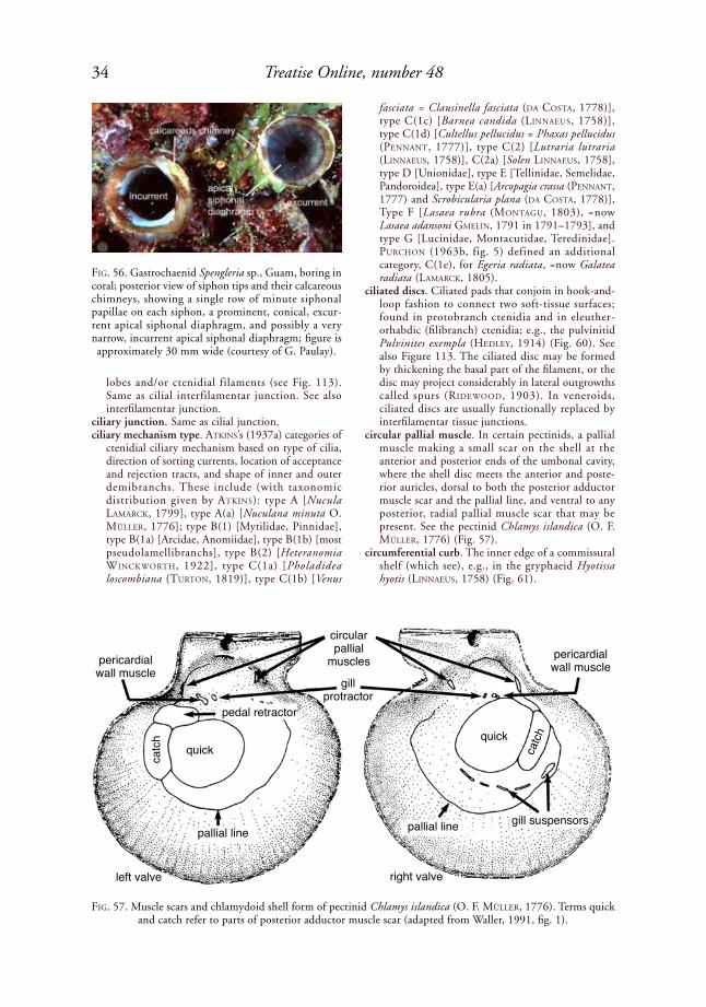
34 Treatise Online, number 48
lobes and/or ctenidial filaments (see Fig. 113). Same as cilial interfilamentar junction. See also interfilamentar junction.
ciliary junction. Same as cilial junction. ciliary mechanism type. atkiNs’s (1937a) categories of
ctenidial ciliary mechanism based on type of cilia, direction of sorting currents, location of acceptance and rejection tracts, and shape of inner and outer demibranchs. These include (with taxonomic distribution given by atkiNs): type A [Nucula laMarCk, 1799], type A(a) [Nuculana minuta O. Müller, 1776]; type B(1) [Mytilidae, Pinnidae], type B(1a) [Arcidae, Anomiidae], type B(1b) [most pseudolamellibranchs], type B(2) [Heteranomia WiNCkWorth, 1922], type C(1a) [Pholadidea loscombiana (turtoN, 1819)], type C(1b) [Venus
fasciata = Clausinella fasciata (da Costa, 1778)], type C(1c) [Barnea candida (liNNaeus, 1758)], type C(1d) [Cultellus pellucidus = Phaxas pellucidus (peNNaNt, 1777)], type C(2) [Lutraria lutraria (liNNaeus, 1758)], C(2a) [Solen liNNaeus, 1758], type D [Unionidae], type E [Tellinidae, Semelidae, Pandoroidea], type E(a) [Arcopagia crassa (peNNaNt, 1777) and Scrobicularia plana (da Costa, 1778)], Type F [Lasaea rubra (MoNtaGu, 1803), =now Lasaea adansoni GMeliN, 1791 in 1791–1793], and type G [Lucinidae, Montacutidae, Teredinidae]. purChoN (1963b, fig. 5) defined an additional category, C(1e), for Egeria radiata, =now Galatea radiata (laMarCk, 1805).
ciliated discs. Ciliated pads that conjoin in hook-and-loop fashion to connect two soft-tissue surfaces; found in protobranch ctenidia and in eleuther-orhabdic (filibranch) ctenidia; e.g., the pulvinitid Pulvinites exempla (hedley, 1914) (Fig. 60). See also Figure 113. The ciliated disc may be formed by thickening the basal part of the filament, or the disc may project considerably in lateral outgrowths called spurs (rideWood, 1903). In veneroids, ciliated discs are usually functionally replaced by interfilamentar tissue junctions.
circular pallial muscle. In certain pectinids, a pallial muscle making a small scar on the shell at the anterior and posterior ends of the umbonal cavity, where the shell disc meets the anterior and poste-rior auricles, dorsal to both the posterior adductor muscle scar and the pallial line, and ventral to any posterior, radial pallial muscle scar that may be present. See the pectinid Chlamys islandica (o. F. Müller, 1776) (Fig. 57).
circumferential curb. The inner edge of a commissural shelf (which see), e.g., in the gryphaeid Hyotissa hyotis (liNNaeus, 1758) (Fig. 61).
FiG. 56. Gastrochaenid Spengleria sp., Guam, boring in coral; posterior view of siphon tips and their calcareous chimneys, showing a single row of minute siphonal papillae on each siphon, a prominent, conical, excur-rent apical siphonal diaphragm, and possibly a very narrow, incurrent apical siphonal diaphragm; figure is approximately 30 mm wide (courtesy of G. Paulay).
FiG. 57. Muscle scars and chlamydoid shell form of pectinid Chlamys islandica (o. F. Müller, 1776). Terms quick and catch refer to parts of posterior adductor muscle scar (adapted from Waller, 1991, fig. 1).
pericardialwall muscle
catc
h
quick
pedal retractor
pallial line
left valve right valve
pallial linegill suspensors
catchquick
circularpallial
muscles
gillprotractor
pericardialwall muscle

35Illustrated Glossary of the Bivalvia
clade. An ancestor (an organism, population, or species) and all of its descendants (PhyloCode, May, 2012: www.ohio.edu/phylocode/glossary.html).
cladistics. The theoretical framework for inferring genealogical relationships on the principle that shared, derived, heritable traits reflect the sequence of genealogical divergence, commonly referred to as recency of common ancestry.
cladistic systematics (phylogenetics). Classification of organisms on the basis of recency of common ancestry. Membership in a clade is inferred on the basis of shared possession of derived, inherit-able characters, and clades are defined by various combinations of apomorphies, nodes, branches, internal specifiers, external specifiers, and quali-fier statements. Cladistic systematics differs from both evolutionary and paracladistic systematics in excluding paraphyletic groups and in restricting application of the term ancestor to individual species. Called both phylogenetics and phyloge-netic systematics by heNNiG (1950, 1966), the latter term currently has a much broader meaning for many biologists. Compare with paracladistic systematics and evolutionary systematics.
clam. Any of various bivalved mollusks, but commonly referring to commercial species of venerids or myids, and not to scallops or oysters.
claspers. See clasper spines.clasper spines. Outgrowths of the shell that wrap
around and secure the shell to a generally narrow substratum, such as mangrove roots or gorgonian corals; e.g., in the ostreoidean Dendostrea frons (liNNaeus, 1758) (Fig. 62). Also called claspers and clasping shelly processes.
clasping shelly processes. See clasper spines.clathrate. Broadly cancellate. claustrum (pl., claustra). A pseudohinge tooth in
anodontine unionoids, which haas (1969, p. 4) described as fol lows (herein translated): “. . . lamellae that resemble lateral teeth; these
FiG. 58. Vertical thin section through shell of Creta-ceous gryphaeid Amphidonte pyrenaicum (leyMerie, 1851), showing chomata-influenced crossed foliated (CICF) microstructure (adapted from Malchus, 1990,
pl. 26,6 ).
FiG. 59. Left valve of laternulid Laternula elliptica (kiNG & Broderip, 1832), from Icaro Cove, Antarctica (USNM 1027262), showing a left-right symmetrical internal ligament supported by a chondrophore. This view is oblique to the commissural plane, about 30˚ toward ventral direction. A clavicle (out of focus) supports chondrophore. There is also a reduced, submarginal, elongate, opisthodetic, simple ligament with a lamellar and possibly (?) also
a fibrous sublayer (Carter, new).

36 Treatise Online, number 48
lamellae are, however, not situated on the hinge, but directly on the shell, close to the dorsal margin, and are, consequently, analogous (homoplasic) but not homologous to the lateral teeth of the unionine hinge type.” The nonhomology of these teeth with regular hinge teeth has yet to be proven. The term claustrum was used inappropriately by saVaZZi and yao (1992) for a prominent dorsal extension of the shell margin above the hinge line, e.g., in the unionid Hyriopsis cumingii (lea, 1852). For the latter structure, the term dorsal ala is more appro-priate (Fig. 63).
clavate. Having a straight, elongate club shape, with a wide end tapering gradually to a narrower end, e.g., the calcareous tube of the gastrochaenoidean Eufistulana eaMes, 1951.
clavicle. A thin, platelike, strongly projecting, shelly support for a chondrophore; thinner and more strongly projecting than a buttress, e.g., in the later-nulid Laternula anatina rödiNG, 1798 (Fig. 64).
cloacal passage. A passage in the excurrent mantle chamber into which excrement and gonadal prod-ucts are discharged.
clutch. The eggs laid by a bivalve during a single discharge.
coarsely prismatic microstructure. A prismatic micro-structure in which the first-order prisms are gener-ally over 100 µm in width.
cockle. A vernacular term for a member of the super-family Cardioidea. Sometimes also used for other ribbed shells, such as the arcid Anadara Gray, 1847.
Code. In the context of bivalve systematics, the Inter-national Code of Zoological Nomenclature (Inter-national Commission on Zoological Nomenclature, 1999). Commonly italicized as Code.
coelom. In mollusks, a body cavity that is homologous with the primordial split in the mesoderm of proto-stomes, and is lined with mesoderm. In bivalves, this is represented by little more than the peri-cardium, the renal organs, and the gonadal ducts.
coelomoduct. Same as renal organs.colaminar. The condition in which a cardinal tooth
is aligned with a lateral tooth, with the two teeth thereby becoming more or less indistinguishable in the adult shell, e.g., cardinal tooth 2b1 is colaminar with lateral tooth 1AII in the early isocyprinoid grade hinge (see Fig. 153) (r. N. GardNer, 2005, fig. 10).
collecting vessel. A protruding vessel in each of two mantle lobes in both valves, connecting the prox-imal ends of several neobranch units either: (1) in the incurrent mantle chamber, parallel to the mantle margins, beginning near the isthmian mantle, some distance inward from its distal margin, extending ventrally and abruptly terminating at the antero-ventral curvature of the shell margin, where it disappears in the musculature of the mantle margin (=submarginal collecting vessel); or (2) in the excur-rent mantle chamber, parallel with and adjacent to the posterior, ventral, and anteroventral edges of the posterior adductor muscle (=perimyal collecting vessel) (harry, 1985, p. 124).
columnar nacreous microstructure. A variety of nacreous shell microstructure in which the tablets are stacked in vertical columns, and the individual tablets are more or less equidimensional in the plane of the shell layer, i.e., not elongate. In the Bivalvia, this form of nacre is largely restricted to faster-growing parts of the shell, such as on slightly reflected shell margins (Fig. 65). Compare with lenticular nacre, sheet nacre, and row stack nacre.
columnar nondenticular composite prismatic micro-structure (abbr., columnar NDCP). A nonden-ticular composite prismatic shell microstructure in which the first-order composite prisms are relatively wide, vertical to slightly reclined, gener-ally polygonal cylinders. The second-order prisms may diverge from the central prisms axis at a low angle, as in the outer shell layer of the unionid Cyprogenia aberti (CoNrad, 1850) (acute columnar
FiG. 60. Two ciliated discs projecting from spurs on neighboring filaments in ctenidium of pulvinitid Pul-vinites exempla (hedley, 1914); discs show nontissue-grade union. SEM of abfrontal surface of a ctenidial lamella (adapted from Tëmkin, 2006b, fig. 16D,
courtesy of John Wiley & Sons).
FiG. 61. Circumferential curb and commissural shelf in gryphaeid Hyotissa hyotis (liNNaeus, 1758); figure is approximately 18 cm wide (adapted from Mikkelsen
& Bieler, 2007).
10 µm

37Illustrated Glossary of the Bivalvia
NDCP, Fig. 4), or at a higher angle, sometimes nearly horizontally, as in the outer shell layer of the hiatellid Cyrtodaria siliqua (speNGler, 1793) (obtuse columnar NDCP, see Fig. 200). Compare with planar spherulitic simple prismatic and with noncolumnar NDCP.
columnar prismatic microstructure. A prismatic shell microstructure consisting of vertical to slightly reclined, more or less polygonal, cylindrical, and relatively wide (i.e., nonfibrous) first-order prisms. This category includes regular simple prismatic, regular spherulitic prismatic, and columnar, nondenticular composite prismatic microstructures (which see).
commarginal. (1) For shell ornament: parallel with the shell margin at the time of secretion; e.g., commar-ginal ridges in the astartid Astarte crenata subequi-latera G. B. soWerBy ii, 1854 (see Fig. 74); (2) for shell sections: a vertical section that is parallel with a former shell margin at the position of the section. “Comarginal” is an incorrect spelling. Compare with concentric, a term commonly used to describe both commarginal and concentric sensu stricto features.
commarginal lamellae. Thin, sheetlike projections, more or less parallel with the shell margins, rising at a high angle from the external surface of the shell. The lamellae may or may not have distal scales, as in the lucinid Lucina pensylvanica (liNNaeus, 1758) (Fig. 66).
commissural angle. The acute angle between a shell’s current or former commissural plane and a line that is perpendicular to the shell margin at a designated point on the shell exterior, and also lies in the plane tangent to shell’s exterior at this designated point (kříž, 1979). In the cardiolid in Figure 67, commissural angles C
1 and C
2 are defined by lines
T1 and T
2, which lie in former commissural plane
CP1 and present commissural plane CP
2, respec-
tively (kříž, 1979). commissural denticles. A row of moderately small,
low projections on or near the nonhinge, inner shell margins, commonly with alternating posi-
tions in the two valves, e..g., in the astartid Astarte smithii dall, 1886 (Fig. 68). Also called marginal denticles. This excludes hinge teeth, chomata, and microdenticulations. When opposing commis-sural denticles interlock, they are also called shell marginal teeth, which see. Commissural denticles are larger than microdenticulations, and they are distinct from marginal crenulations.
commissural plane. The surface, not necessarily planar, defined by the line of contact of the closed shell valves (Waller, 1969, p. 13). Compare with sagittal plane and midsagittal plane.
commissural shelf. A nearly flat, internal surface along the nonhinge periphery of the shell, posi-tioned distal to a circumferential curb, e.g., in the gryphaeid Hyotissa hyotis (liNNaeus, 1758) (Fig. 61). In the right valve, the distal part of the commissural shelf commonly consists of a flex-ible prismatic outer shell layer. A commissural
FiG. 62. Clasper spines and antimarginal plicae on ostreid Dendostrea frons (liNNaeus, 1758), shell length several cm (adapted from Mikkelsen & Bieler, 2007).
FiG. 63. Dorsal ala on left valve of unionid Hy-riopsis cumingii (lea, 1852), shell length 8.5 cm
(Zieritz, new).
clasper spines
dorsal ala

38 Treatise Online, number 48
shelf is also present in the attached valve of some Hippuritida.
commissure. (1) The more proximal part of the line or area of contact of the two shell valves; the proximal limit of the commissural shelf where the latter is present; (2) a band of neural tissue linking the left and right sides of the nervous system.
compact foliated layer. A calcitic foliated shell layer that lacks chalky lenses and lensic structural cham-bers; e.g., the foliated shell layers in most Pect-inoidea and in some Ostreoidea.
comparative convexity. The convexity of one valve compared to the convexity of the opposite valve.
compensation sac. Same as septal valvula, which see.competent larva. A larva that is ready to undergo
metamorphosis.
complete pallial line. A pallial line without a sinus. Also called an entire pallial line.
complex crossed foliated microstructure (abbr., CCF). The calcitic analog of aragonitic complex crossed lamellar shell microstructure. See irregular CCF and cone CCF.
complex crossed lamellar microstructure (abbr., CCL). An aragonitic, nonprismatic shell micro-structure consisting of elongate, basic structural units with three or more predominant dip direc-tions. Varieties include fine CCL, irregular CCL, and cone CCL. If calcitic, the structure is called complex crossed foliated.
complex prismatic microstructure. (1) A prismatic shell microstructure in which each prism has a central, longitudinal crystallographic optic axis, and, lateral to this, optic axes diverging toward the sides of the prism (BøGGild, 1930, p. 248). This is not a microstructural term per se, because prisms with these optical properties may be simple or composite. Some authors (e.g., MaCCliNtoCk, 1967) have used this term to indicate composite prismatic microstructures; (2) a portion of an otherwise fibrous prismatic shell layer in which the prism orientations are highly irregular, as locally in the fibrous prismatic outer and inner shell layer of Mytilus californianus CoNrad, 1837.
composite fibrous prismatic microstructure. A predomi-nantly antimarginal, reclined prismatic shell micro-structure that combines fibrous prisms with very narrow irregular spherulitic prisms and very narrow nondenticular composite prisms (NDCP), e.g., the aragonitic, reclined prismatic outer shell layer in Clinocardium nuttallii (CoNrad, 1837) (sChNeider & Carter, 2001, p. 608, fig. 8.1, mislabelled “compound fibrous prismatic”). This term is easily confused with composite prismatic, and should therefore be replaced with the description: “fibrous prismatic to very narrow irregular spherulitic pris-matic to very narrow NDCP.”
composite mold. An internal mold with traces of the external ornament superimposed by compaction of sediment during or after dissolution of the shell.
composite prismatic microstructure (abbr., CP). A prismatic shell microstructure in which each first-order prism consists of stacked three-dimensional fans of smaller, second-order prisms. This includes denticular and nondenticular composite prisms, which see.
compound composite prismatic microstructure. A term used by Carter (1980b), Carter and Clark (1985), and Carter and others (1990, p. 634) for compound nondenticular composite prismatic (=compound NDCP) microstructure, which is preferred.
compound nondenticular composite prismatic micro-structure (abbr., compound NDCP). A prismatic shell microstructure in which first-order prisms similar to the first-order prisms in normal NDCP structure are arranged into larger (zeroth-order), three-dimensional, stacked, fanlike aggregations as a consequence of deposition on a coarsely denticu-lated or crenulated, reflected shell margin (Carter,
FiG. 64. Laternulid Laternula anatina rödiNG, 1798, showing the clavicle and chondrophore (adapted from
Fitch, 1953).
FiG. 65. Columnar nacre, diagram showing depositional surface (H ), plus radial (R), and transverse (T ) sections
(adapted from Carter & others, 1990, p. 618).
10 µm

39Illustrated Glossary of the Bivalvia
1980b; Carter & Clark, 1985; Carter & others, 1990, p. 650), e.g., the outer shell layer of some Veneroidea (Fig. 69). Also locally developed in the outer shell layer of the lucinid Codakia (Codakia) orbicularis (liNNaeus, 1758). Compare with crossed NDCP and radial lamellar NDCP.
compressed. Laterally flattened; commonly referring to shell shape.
compressed crossed lamellar microstructure (abbr., compressed CL). A variety of crossed lamellar shell microstructure in which the first-order lamellae are elongate in the antimarginal direction, but their second-order lamellae have commarginal or slightly oblique dip directions (Carter & others, 1990, p. 634) (see Fig. 82). Not to be confused with radially oriented crossed lamellar microstructure.
concave. Curved or rounded inward.concavoconvexodont tooth. A taxodont hinge tooth
that is dorsally concavodont and ventrally convex-odont (BaBiN, 1966, p. 39) (see Fig. 207).
concavodont tooth. A taxodont hinge tooth in which the concave side is toward the beak (BaBiN, 1966, p. 39) (see Fig. 207).
concentric. (1) Having a common center, as in circles or parts of circles symmetrically arranged, one within the other, i.e., concentric sensu stricto; (2) curving about a common center, but not neces-sarily following a perfect circle; a common use of the term concentric for what is more appropriately called commarginal.
conchin. Same as conchiolin, which is preferred.conchiolin. The organic, predominantly proteinous
component of a molluscan shell that serves as the
framework and/or stereochemical template for shell mineralization. Originally defined as the residue of the shell remaining after treatment in HCl, and which is insoluble in water, alcohol, and ether (FreMy, 1855, p. 96).
conchological. Of or relating to shells.conchology. The study of shells.concordant valves. Shell valves with margins fitting
together congruently.condylar boss. An articulating condyle in the form of
a rounded knob, e.g., in the pholadid Cyrtopleura costata (liNNaeus, 1758) (Fig. 71).
FiG. 66. Commarginal lamellae with scales in lucinid Lucina pensylvanica (liNNaeus, 1758); SEM of surface view (adapted from Taylor & others, 2004, fig. 2, cour-
tesy of the American Malacological Society).
FiG. 67. Schematic representation of a cardiolid shell showing commissural angles C1 and C
2, formed by lines T
1
and T2, which are perpendicular to shell margin at designated points on shell’s exterior, and lines CP
1 and CP
2,
which are former and current commissural planes, respectively (adapted from Kříž, 1979, fig. 6).
T1
T2
C1
C2
CP1
CP2T1
T2

40 Treatise Online, number 48
condylar ridge. An articulating condyle in the form of a raised, linear elevation, as in the myid Mya arenaria (liNNeaus, 1758) (Fig. 70).
condyles. Knoblike or ridgelike processes on the dorsal shell margin (Myidae, Corbulidae), or on the dorsal and ventral shell margins (some Pholadidae, Teredinidae), which provide opposing pivot points for rocking the valves about a more or less vertical axis during burrowing or boring. Dorsal condyles in Myidae tend to be ridgelike (Fig. 70), whereas those in Pholadidae, Parilimyidae, and Pholadoidea tend to be rounded, e.g., the condylar boss on the dorsal shell margin of the pholadid Cyrtopleura costata
(liNNaeus, 1758) (Fig. 71), and on the ventral shell margin of certain other pholadids (Fig. 24). See also condylar boss and condylar ridge.
cone complex crossed foliated microstructure (abbr., cone CCF). The calcitic analog of aragonitic cone complex crossed lamellar shell microstructure; e.g., the inner part of the inner shell layer of the Lower Jurassic gryphaeid Gryphaea (Gryphaea) arcuata (laMarCk, 1801) (Fig. 72).
cone complex crossed lamellar microstructure (abbr., cone CCL). An aragonitic, CCL shell microstruc-ture in which the first-order lamellae have a cone-in-cone or spiral-conical second-order structure, with the cone axes more or less perpendicular to the depositional surface, and with strongly inter-digitating cone boundaries, e.g., in the mactrid Lutraria sp. (Fig. 73). The second-order lamellae typically consist of elongate, flattened, third-order lamellae radiating from the cone axis toward the depositional surface. Similar to composite pris-matic structure, but the third-order lamellae are lathic rather than fibrous, they diverge at a more uniform angle from the cone’s central axis, the lateral boundaries of the cones are more strongly interdigitating, and the structure commonly inter-grades in the same shell layer with irregular CCL (Carter & teVesZ, 1978a; Carter, 1980b; Carter & Clark, 1985; Carter & others, 1990, p. 613, 634). The calcitic form of this structure is called cone complex crossed foliated, and usually has larger first-order cones and lower dip angles of the second-order units.
confluent pallial sinus. A pallial sinus with its ventral margin coincident with the posteroventral pallial line. This can reflect a ventral shift in the lower margin of the pallial sinus, as in the gastrochaenid Rocellaria dubia (peNNaNt, 1777) (see Fig. 192) or dorsoventral thickening of the entire ventral pallial band, as in some species of the gastrochaenid Gastrochaena speNGler, 1783. Compare with free pallial sinus.
confluent pedal retractor muscle scar . A pedal retractor muscle scar with one of its sides merged with the adjacent adductor muscle scar.
congeneric. Belonging to the same genus.conglutinate. A mass of glochidia bound together in a
gelatinous or mucous mass, which may mimic food items of glochidial host fish.
conjoined. Joined to another organ, e.g., the anterior adductor muscle scar conjoined with a pallial muscle. Compare with adnate.
consecutive hermaphroditism. The common form of hermaphroditism in bivalves, in which the indi-vidual changes from male to female (protandry) or sometimes from female to male (protogyny).
conserved name. A name, otherwise unavailable or invalid, that the ICZN, by exercise of its plenary power under Article 81 (ICZN, 1999), has enabled to be used as a valid name by removal of the obstacles to such use. The process of conserving a name is normally initiated by a taxonomist in a petition to the ICZN. The petition is published in the Bulletin of Zoological Nomenclature, and
FiG. 68. Interior of astartid Astarte smithii dall, 1886, showing cardinal teeth, lateral teeth, commissural den-ticles, and heterodont dentition, shell length 5.7 mm
(adapted from Mikkelsen & Bieler, 2007).
FiG. 69. Compound nondenticular composite prismatic (compound NDCP) microstructure. Diagram showing horizontal (H ), radial (R), and transverse (T ) sections
(adapted from Carter & others, 1990, p. 617).
200 µm

41Illustrated Glossary of the Bivalvia
responses and commentaries generated by the peti-tion are published in that journal. The responses and commentaries are then considered when the ICZN makes a ruling on the petition.
conserved work. A taxonomic work that the ICZN has ruled to be available. See also rejected work.
conspecific. Belonging to the same species.continuous pallial line. A pallial line or band with no
interruptions.contour. (1) The outline, shape or form of an object;
(2) in the context of early ontogenetic shells (e.g., prodissoconch, metaconch), the outline of the shell along its commissure (MalChus & sartori, herein). See also profile.
contour graph. The graphic representation of a shell contour built on a set of standard rules for better comparability (MalChus, 2006a; MalChus & sartori, herein). Same as standardized outline, which is preferred.
conus (or shell conus). A conical elevation of the cicatrix zone, sometimes extending beyond the
cicatrix zone, in a hat-shaped early ontogenetic shell (MalChus & sartori, herein).
convex. Outwardly curved or rounded, generally in reference to a shell valve or a hinge tooth. See also right-convex and left-convex.
FiG. 70. Myid Mya arenaria (liNNaeus, 1758), Massachusetts (UNC, unnumbered specimen), showing hinge and ligament of left and right valves. 1, Left valve, in nearly dorsal, slightly lateral interior view, showing projecting chondrophore, condylar ridge, remnants of lamellar sublayer of resilium, and concave insertion area of fibrous sublayer of resilium; 2, right valve, interior, nearly ventral view, showing resilial buttress, condylar ridge, remnants of fibrous sublayer of resilium, and area of insertion of lamellar sublayer of resilium; scale bar applies to both views
(Carter, new).
FiG. 71. Descriptive terminology for interior of phola-did Cyrtopleura costata (liNNaeus, 1758), shell length several cm (adapted from Mikkelsen & Bieler, 2007).
condylar ridge
lamellar sublayer
fibrous sublayer
chondrophore
1 mm
1
2
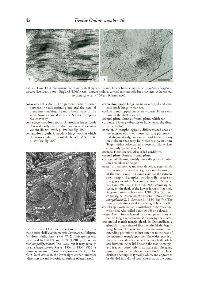
42 Treatise Online, number 48
convexity (of a shell). The perpendicular distance between the midsagittal plane and the parallel plane just touching the most lateral edge of the valve. Same as lateral inflation. See also compara-tive convexity.
convexoconcavodont tooth. A taxodont hinge tooth that is dorsally convexodont and ventrally conca-vodont (BaBiN, 1966, p. 39) (see Fig. 207).
convexodont tooth. A taxodont hinge tooth in which the convex side is toward the beak (BaBiN, 1966, p. 39) (see Fig. 207).
corbiculoid grade hinge. Same as veneroid and cyre-noid grade hinge, which see.
cord. A round-topped, moderately coarse, linear eleva-tion on the shell’s exterior.
coronal plane. Same as frontal plane, which see. coronate. Having tubercles or lamellae at the distal
parts of ribs.corselet. A morphologically differentiated area on
the exterior of a shell, posterior to a posteroven-tral diagonal ridge or carina, and lateral to any escutcheon that may be present; e.g., in some Trigonioidea. Also called a posterior slope. Less commonly spelled corcelet.
cordate. Heart shaped. Also called cordiform.coronal plane. Same as frontal plane.corrugated. Having roughly mutually parallel, rather
small wrinkles or ridges. costa (pl., costae). A moderately wide, exterior rib
that is not expressed as a groove on the interior of the shell, except, in some cases, at the extreme shell margin. Examples include radial costae on the glycymeridid Tucetona pectinata (GMeliN, 1791 in 1791–1793) (see Fig. 265); commarginal costae on the flank of the Lower Jurassic trigonioid Trigonia sulcata (herMaNN, 1781) (Fig. 53), and commarginal costae on the astartid Astarte crenata subequilatera G. B. soWerBy II, 1854 (Fig. 74). The term is sometimes used interchangeably with rib.
costella (pl., costellae, adj., costellate). A narrow costa, which see. Also called a narrow rib or a thread.
cotype. A term formerly used for a syntype or paratype, but no longer recommended for use by the ICZN.
crassatellid mantle margin gland. In Crassatellidae, a glandular organ shaped like a scythe blade, begin-ning below the anterior adductor muscle and extending posteriorly to just anterior to the base of the incurrent mantle aperture. The widest part is at the anterior end, where it occupies nearly the whole area between the pallial line and the mantle margin, and it tapers posteriorly to an acute tip. The gland projects into the mantle cavity as a thick pad, lacks distinct openings, is typically white, and appears to be divided into dorsal and ventral parts, the dorsal
FiG. 72. Cone CCF microstructure in inner shell layer of Liassic, Lower Jurassic gryphaeid Gryphaea (Gryphaea) arcuata (laMarCk, 1801), England (UNC 5526); acetate peels. 1, vertical section; scale bar = 0.5 mm; 2, horizontal
section; scale bar = 100 µm (Carter, new).
FiG. 73. Cone CCL microstructure just below pris-matic outer shell layer in mactrid Lutraria sp., Calapan, Mindoro, Philippines (YPM 9742). This species was identified by Carter and lutZ (1990, p. 7) as Lu-trarium philippinarum deshayes, but it may actually be L. philippinarum reeVe, 1854 in 1854–1855, a junior synonym of Lutraria rhynchaena JoNas, 1844; short, black arrow, in the lower right corner, indicates direction toward depositional surface (Carter, new).
1 2

43Illustrated Glossary of the Bivalvia
part being more translucent and larger, and the ventral part being more opaque. When ruptured, the gland exudes colorless spheres about 0.1 mm in diameter, each containing minute oily droplets. The gland has thin walls surrounding a large cavity containing the granules, which stain strongly with eosin (harry, 1966; alleN, 1968; taylor, GloVer, & WilliaMs, 2005). Similar glands occur in a few other heteroconch bivalves, where they are called pallial glands, which see.
crenate. Same as crenulate, which is preferred.crenulate (adj., also spelled crenulated). Having a
series of alternating elevations and indentations, generally in reference to a shell margin or the surface of a hinge tooth, e.g., crenulate hinge teeth in lyrodesmatids and trigoniids, and the shell marginal crenulations in the Eocene carditid Venericardia parva I. lea, 1833 (see Fig. 314). Where shell marginal crenulations are narrow or very narrow, the terms denticulate and serrate may be more appropriate, respectively.
crenulations. See marginal crenulations.crisscross crossed lamellar microstructure (abbr.,
XCL). A crossed lamellar shell microstructure in which the first-order lamellae have crisscrossed shapes as seen on the depositional surface and in horizontal sections (see Fig. 82). The dip directions of the third-order lamellae are generally oblique rather than directly opposed. This structure is locally developed in the middle shell layer of many thin, elongate, heterodont shells such as Ensis sChuMaCher, 1817.
cross bar. A small ridge situated transversely across a rib.
crossed acicular microstructure (abbr., CA). An aragonitic crossed shell microstructure comprised of narrow, elongate crystallites with two and only two predominant dip directions. The crystal-lites are typically commarginally or only slightly
obliquely aligned, as in CL structure, and they are not aggregated into distinct first- and/or second-order crossed lamellae (Carter & others, 1990, p. 612). Commonly present in the inner part of a combined CL + CA outer shell layer, wherein the first-order crossed lamellae become narrower toward the interior. This microstructure is difficult to differentiate from fine complex crossed lamellar and homogeneous microstructure at the optical level, and usually requires SEM for identification. Crossed acicular structure comprises part of the middle shell layer of the malletiid Malletia obstusa G. o. sars, 1872 (Fig. 75) and the arcticid Arctica islandica (liNNaeus, 1767 in 1766–1768) (Fig. 76).
crossed bladed microstructure. A calcitic laminar shell microstructure consisting of nearly horizontal
FiG. 74. Commarginal costae on astartid Astarte crenata subequilatera G. B. soWerBy ii, 1854, shell length 22
mm (adapted from Mikkelsen & Bieler, 2007).
FiG. 75. Crossed acicular microstructure in middle shell layer of malletiid Pseudomalletia obtusa G. sars, 1872, U.S. Fish Commission Station 2221, Atlantic Ocean; SEM of 1, radial and 2, commarginal vertical fractures (adapted
from Carter, 2001, fig. 1.1–1.2).
10 µm1 2
5 µm

44 Treatise Online, number 48
sheets of locally mutually parallel laths or blades, with lath or blade orientations differing more or less randomly in adjacent sheets and in different parts of the same sheet (arMstroNG, 1969, for strophomenid brachiopods; Carter & Clark, 1985; Carter & others, 1990, p. 619, 635, for bivalves). Locally present in the outer shell layer of some Limidae (Fig. 77).
crossed composite prismatic microstructure. A term used by Carter and Clark (1985) and Carter and others (1990, p. 636) for crossed nondenticular composite prismatic shell microstructure (which is preferred).
crossed foliated microstructure (abbr., CF). A calcitic shell microstructure consisting of mutually parallel, rod-, blade-, or lathlike basic structural units orga-nized into first-order lamellae, with each first-order lamella oriented with its height axis more or less perpendicular to the depositional surface, and with its length axis more or less commarginally aligned.
The basic structural units may be aggregated into second-order lamellae, in which case the basic structural units are called third-order lamellae, and the structure is called simple crossed foliated, e.g., in the Jurassic oxytomid Oxytoma inequivalve (J. soWerBy, 1819, in soWerBy & soWerBy, 1812–1846) (Fig. 78). When intermediate (second-order) lamellae are absent, the structure is called rod-type crossed foliated. CF structures are categorized as low angle and high angle CF (see below). In all CF structures, the basic structural units show only two predominant dip directions, and these dip direc-tions may be uniformly alternating and opposite, or alternating and oblique, in adjacent first-order lamellae (Carter, 1980b; Carter & Clark, 1985; Carter & others, 1990, p. 621, 626). Not to be confused with herringbone cross foliated of MalChus (1990) (herein called herringbone regu-larly foliated), wherein the dip direction alternates in successive shell sublayers, rather than over the same depositional surface. Varieties of CF structure based on the shape of the first-order lamellae in horizontal sections (or on the depositional surface) include linear CF and branching CF. Linear CF and branching CF generally differ from linear CL and branching CL in having larger first-order lamellae and lower dip angles of their basic structural units (BøGGild, 1930, p. 251, 261).
crossed foliated microstructure, high angle (abbr., high angle CF). A calcitic crossed foliated shell microstructure in which the basic structural units dip more than 15˚ from the depositional surface. Common in the middle shell layer of Gryphaeidae (Fig. 38) and Oxytomidae (Fig. 78).
crossed foliated microstructure, low angle (abbr., low angle CF). A calcitic crossed foliated shell micro-structure in which the basic structural units dip less than 15˚ (more commonly only a few degrees) from the depositional surface, e.g., in the middle shell layer of many Pectinidae and Propeamussiidae, e.g., Propeamussium dalli (e. a. sMith, 1885) (Fig. 79).
crossed lamellar branching index. In reference to simple crossed lamellar shell microstructure, the ratio of the number of first-order crossed lamellar branches encountered along a given standard tran-sect (which see), divided by the number of contig-uous first-order lamellae encountered along that transect (Carter, 1976).
crossed lamellar length. In reference to simple crossed lamellar shell microstructure, the maximum dimen-sion of a single branch of a first-order crossed lamella measured perpendicular to a standard transect (which see). The line of measurement must stay within the first-order lamella, so some strongly curved, long first-order lamellae can have short measured lengths. Crossed lamellar lengths are generally given as the arithmetic average of several adjacent first-order lamellae (Carter, 1976). In Figure 81, measurements a–b and c–d represent crossed lamellar lengths.
crossed lamellar length extent. In reference to simple crossed lamellar shell microstructure, the maximum dimension of an entire first-order crossed lamella,
FiG. 76. Crossed acicular microstructure in the middle shell layer of arcticid Arctica islandica (liNNaeus, 1767 in 1766–1768); polished and then acid-etched radial
section; scale bar = 5 µm (Carter, new).
FiG. 77. Crossed bladed microstructure, diagram show-ing horizontal (H ) and vertical (V ) sections (adapted
from Carter & others, 1990, p. 619).
1 µm

45Illustrated Glossary of the Bivalvia
including all of its contiguous branches, measured parallel to a line perpendicular to a standard tran-sect (which see) (Carter, 1976). In Figure 81e–f is a measure of crossed lamellar length extent.
crossed lamellar microstructure (abbr., CL). An aragonitic shell microstructure consisting of mutu-ally parallel, rod-, blade-, or lathlike basic struc-tural units (third-order lamellae) aggregated into first-order lamellae, with each first-order lamella oriented with its height axis more or less perpen-dicular to the depositional surface, and with its length axis generally commarginally (rarely antimarginally) oriented. The third-order lamellae show only two predominant dip directions, which may be opposite or oblique in adjacent first-order lamellae (BøGGild, 1930, p. 251; Carter, 1980a, 1980b; Carter & Clark, 1985; Carter & others, 1990, p. 612, 636). When the third-order lamellae are aggregated into second-order lamellae, the structure is called simple crossed lamellar, which see. When second-order lamellae are absent, the structure is called rod-type CL; rod-type CL is seldom seen in bivalves. The example herein (Fig. 80) of simple crossed lamellar structure is the middle shell layer of the chamid Chama iostoma CoNrad, 1837. The calcitic analog of aragonitic CL is called crossed foliated, which see. Shape varieties of CL structure are based on the appear-ance of the first-order lamellae in horizontal sections or on the depositional surface. These include linear, branching, irregular, compressed, triangular, and crisscross CL. CL microstructure is not to be confused with crossed acicular structure, in which third-order lamellae but not first and second-order lamellae are present. The unmodi-fied term crossed lamellar is herein considered to
exclude complex crossed lamellar and rod-type crossed lamellar microstructure.
crossed lamellar signature (abbr., CL signature). In reference to simple crossed lamellar shell micro-structure, the shape of a first-order crossed lamella as seen on the depositional surface or in a hori-zontal section. Crossed lamellar shapes can often be made distinct by viewing the shell’s depositional surface in high intensity, low-angle, reflected light. Varieties include linear, branching, crisscross, irreg-ular, compressed, triangular, and diffuse (Fig. 82). Only linear and branching CL signatures are known
FiG. 78. Crossed foliated microstructure in Oxfordian, Upper Jurassic oxytomid Oxytoma inequivalve (J. soWerBy, 1819, in soWerBy & soWerBy, 1812–1846), Bucks, England (UNC 4527), acetate peels; 1, horizontal section, with shell margin toward top; scale bar = 50 µm; 2, transverse (commarginal) section, with shell interior toward
top; scale bar = 50 µm (Carter, new).
FiG. 79. Low-angle crossed foliated microstructure in the propeamussiid Propeamussium dalli (e. a. sMith, 1885), west of Martinique, Windward Islands (YPM 8387); SEM of horizontal fracture through middle layer of left valve (adapted from Carter & Lutz, 1990, pl. 7,B).
1 2
2 µm
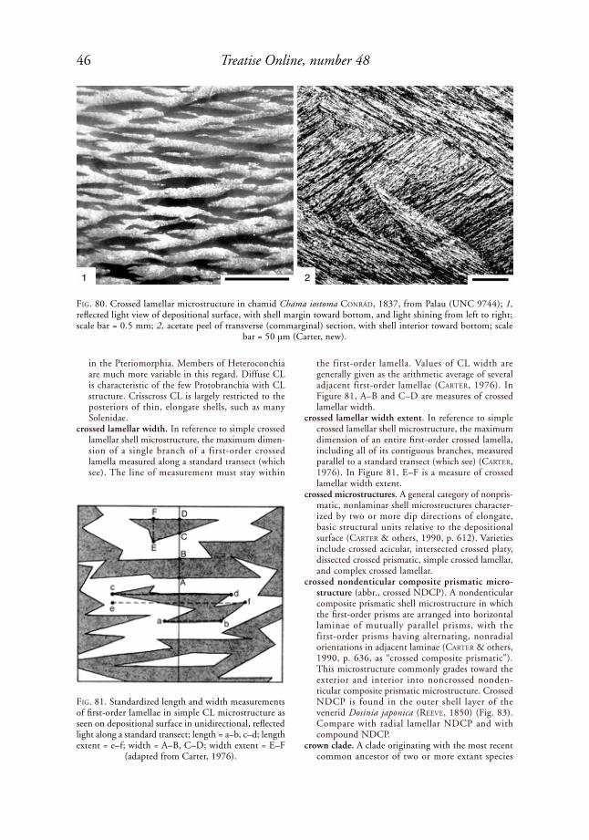
46 Treatise Online, number 48
in the Pteriomorphia. Members of Heteroconchia are much more variable in this regard. Diffuse CL is characteristic of the few Protobranchia with CL structure. Crisscross CL is largely restricted to the posteriors of thin, elongate shells, such as many Solenidae.
crossed lamellar width. In reference to simple crossed lamellar shell microstructure, the maximum dimen-sion of a single branch of a first-order crossed lamella measured along a standard transect (which see). The line of measurement must stay within
the first-order lamella. Values of CL width are generally given as the arithmetic average of several adjacent first-order lamellae (Carter, 1976). In Figure 81, A–B and C–D are measures of crossed lamellar width.
crossed lamellar width extent. In reference to simple crossed lamellar shell microstructure, the maximum dimension of an entire first-order crossed lamella, including all of its contiguous branches, measured parallel to a standard transect (which see) (Carter, 1976). In Figure 81, E–F is a measure of crossed lamellar width extent.
crossed microstructures. A general category of nonpris-matic, nonlaminar shell microstructures character-ized by two or more dip directions of elongate, basic structural units relative to the depositional surface (Carter & others, 1990, p. 612). Varieties include crossed acicular, intersected crossed platy, dissected crossed prismatic, simple crossed lamellar, and complex crossed lamellar.
crossed nondenticular composite prismatic micro-structure (abbr., crossed NDCP). A nondenticular composite prismatic shell microstructure in which the first-order prisms are arranged into horizontal laminae of mutually parallel prisms, with the first-order prisms having alternating, nonradial orientations in adjacent laminae (Carter & others, 1990, p. 636, as “crossed composite prismatic”). This microstructure commonly grades toward the exterior and interior into noncrossed nonden-ticular composite prismatic microstructure. Crossed NDCP is found in the outer shell layer of the venerid Dosinia japonica (reeVe, 1850) (Fig. 83). Compare with radial lamellar NDCP and with compound NDCP.
crown clade. A clade originating with the most recent common ancestor of two or more extant species
FiG. 80. Crossed lamellar microstructure in chamid Chama iostoma CoNrad, 1837, from Palau (UNC 9744); 1, reflected light view of depositional surface, with shell margin toward bottom, and light shining from left to right; scale bar = 0.5 mm; 2, acetate peel of transverse (commarginal) section, with shell interior toward bottom; scale
bar = 50 µm (Carter, new).
FiG. 81. Standardized length and width measurements of first-order lamellae in simple CL microstructure as seen on depositional surface in unidirectional, reflected light along a standard transect; length = a–b, c–d; length extent = e–f; width = A–B, C–D; width extent = E–F
(adapted from Carter, 1976).
1 2

47Illustrated Glossary of the Bivalvia
FiG. 82. Crossed lamellar (CL) signatures, showing appearance of first-order, aragonitic simple crossed lamellae on depositional surface in low-angle, reflected light, with latter shining from left to right. Dip direction refers to second- and third-order lamellae. Dip directions are not necessarily directly opposed in adjacent first-order lamellae, but there
are always only two predominant dip directions (adapted from Carter & others, 1990, p. 621).
linear CL branching CL
or
lamella dip directions
shell margin
compressed CL
crisscross CL irregular CL
triangular CL diffuse CL
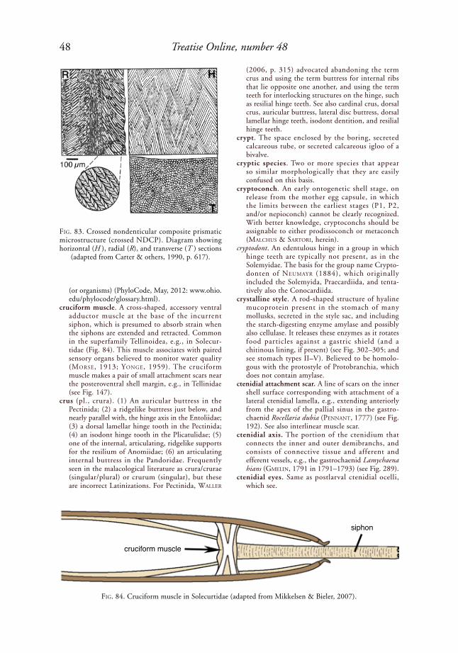
48 Treatise Online, number 48
(or organisms) (PhyloCode, May, 2012: www.ohio.edu/phylocode/glossary.html).
cruciform muscle. A cross-shaped, accessory ventral adductor muscle at the base of the incurrent siphon, which is presumed to absorb strain when the siphons are extended and retracted. Common in the superfamily Tellinoidea, e.g., in Solecur-tidae (Fig. 84). This muscle associates with paired sensory organs believed to monitor water quality (Morse, 1913; yoNGe, 1959). The cruciform muscle makes a pair of small attachment scars near the posteroventral shell margin, e.g., in Tellinidae (see Fig. 147).
crus (pl., crura). (1) An auricular buttress in the Pectinida; (2) a ridgelike buttress just below, and nearly parallel with, the hinge axis in the Entoliidae; (3) a dorsal lamellar hinge tooth in the Pectinida; (4) an isodont hinge tooth in the Plicatulidae; (5) one of the internal, articulating, ridgelike supports for the resilium of Anomiidae; (6) an articulating internal buttress in the Pandoridae. Frequently seen in the malacological literature as crura/crurae (singular/plural) or crurum (singular), but these are incorrect Latinizations. For Pectinida, Waller
(2006, p. 315) advocated abandoning the term crus and using the term buttress for internal ribs that lie opposite one another, and using the term teeth for interlocking structures on the hinge, such as resilial hinge teeth. See also cardinal crus, dorsal crus, auricular buttress, lateral disc buttress, dorsal lamellar hinge teeth, isodont dentition, and resilial hinge teeth.
crypt. The space enclosed by the boring, secreted calcareous tube, or secreted calcareous igloo of a bivalve.
cryptic species. Two or more species that appear so similar morphologically that they are easily confused on this basis.
cryptoconch. An early ontogenetic shell stage, on release from the mother egg capsule, in which the limits between the earliest stages (P1, P2, and/or nepioconch) cannot be clearly recognized. With better knowledge, cryptoconchs should be assignable to either prodissoconch or metaconch (MalChus & sartori, herein).
cryptodont. An edentulous hinge in a group in which hinge teeth are typically not present, as in the Solemyidae. The basis for the group name Crypto-donten of NeuMayr (1884), which originally included the Solemyida, Praecardiida, and tenta-tively also the Conocardiida.
crystalline style. A rod-shaped structure of hyaline mucoprotein present in the stomach of many mollusks, secreted in the style sac, and including the starch-digesting enzyme amylase and possibly also cellulase. It releases these enzymes as it rotates food particles against a gastric shield (and a chitinous lining, if present) (see Fig. 302–305; and see stomach types II–V). Believed to be homolo-gous with the protostyle of Protobranchia, which does not contain amylase.
ctenidial attachment scar. A line of scars on the inner shell surface corresponding with attachment of a lateral ctenidial lamella, e.g., extending anteriorly from the apex of the pallial sinus in the gastro-chaenid Rocellaria dubia (peNNaNt, 1777) (see Fig. 192). See also interlinear muscle scar.
ctenidial axis. The portion of the ctenidium that connects the inner and outer demibranchs, and consists of connective tissue and afferent and efferent vessels, e.g., the gastrochaenid Lamychaena hians (GMeliN, 1791 in 1791–1793) (see Fig. 289).
ctenidial eyes. Same as postlarval ctenidial ocelli, which see.
FiG. 83. Crossed nondenticular composite prismatic microstructure (crossed NDCP). Diagram showing horizontal (H ), radial (R), and transverse (T ) sections
(adapted from Carter & others, 1990, p. 617).
FiG. 84. Cruciform muscle in Solecurtidae (adapted from Mikkelsen & Bieler, 2007).
cruciform muscle
siphon

49Illustrated Glossary of the Bivalvia
ctenidial filament. One of several thin, flat or elon-gate, mutually parallel structures in a ctenidium, attaching on either side of the ctenidial axis (see Fig. 104, Fig. 113). See also protobranch, fili-branch, and eulamellibranch.
ctenidial lamella (pl., ctenidial lamellae). One of four sheets of adjacent filaments in a typical W-shaped autobranch ctenidium. Also called a gill lamella. See ascending lamella and descending lamella.
ctenidial ocelli. Same as postlarval ctenidial ocelli, which see.
ctenidial retractor insertion separation. As defined for pectinids by Waller (1969, p. 13, as “separation of gill retractor insertions”), the linear distance between the centers of the ctenidial retractor inser-tions. This corresponds with measurements f–g and f '–g' in Figure 219.
ctenidial septum (pl., septa). (1) An interlamellar septum (which see) within a ctenidium; (2) a
transverse membrane, at least partially ctenidial in origin, separating the infrabranchial chamber from the suprabranchial chamber in a septibranch bivalve, e.g., in the cuspidariid Cardiomya cleryana (d’orBiGNy, 1846 in 1835–1847) (see Fig. 277) and in the Verticordiidae (Fig. 85). Also called a branchial diaphragm, branchial membrane, or branchial septum; (3) a siphonal ctenidial septum, which see.
ctenidiobranch. A protobranch ctenidium in the Nucu-lida, as compared with the so-called palaeobranch Solemyida. A term based on phylogenetic affinity rather than solely on morphology. The basis for the superorder name Ctenidiobranchia of salViNi-plaWeN (1980), equivalent to Protobranchia of later authors minus the Solemyida.
ctenidium (pl., ctenidia). In many bivalves, an organ of both respiration and food-gathering, usually suspended by tissue, cuticular, and/or cilial
FiG. 85. Anatomy of Verticordiidae (adapted from Mik-kelsen & Bieler, 2007).
FiG. 86. Ctenidium-palp association category I of sta-sek (1963) in unionid Anodonta californiensis I. lea, 1852; ID, inner demibranch; OD, outer demibranch
(adapted from Stasek, 1963, fig. 1c).
FiG. 87. Ctenidium-palp association category II of sta-sek (1963) in cardiid Clinocardium nuttallii (CoNrad, 1837); ID, inner demibranch; OD, outer demibranch
(adapted from Stasek, 1963, fig. 1d).
FiG. 88. Ctenidium-palp association category III of stasek (1963) in isognomonid Isognomon costellatus (CoNrad, 1837); ID, inner demibranch; OD, outer
demibranch (adapted from Stasek, 1963, fig. 2).

50 Treatise Online, number 48
junctions from the dorsal mantle cavity, e.g., the gastrochaenid Lamychaena hians (GMeliN, 1791 in 1791–1793) (see Fig. 289). The synonym gill is sometimes preferred for the sake of brevity (see entries under gill). In Autobranchia, the entire ctenidium (a holobranch) is typically W-shaped, with inner and outer demibranchs, and with each demibranch having ascending and descending lamellae with food grooves at the distal edges and/or at the junction of the two demibranchs. The ctenidium may or may not have interlamellar junctions connecting the two lamellae of each demibranch. See also eulamellibranch, filibranch,
heterorhabdic, homorhabdic, protobranch, pseu-dolamellibranch, plicate, septibranch, and synap-torhabdic.
ctenidium-palp association category I of StaSek (1963). An association between the ctenidium and labial palps in which the ventral tips of at least the first few or, usually, of many anterior filaments of the inner demibranch are inserted, unfused, into a distal oral groove (stasek, 1963, p. 91), e.g., the unionid Anodonta californiensis I. lea, 1852 (Fig. 86). Found in some Nuculoidea, Pristiglomoidea, Solemyoidea, Manzanelloidea, Malletioidea (Malletiidae, Tindar-idae), Nuculanoidea (Nuculanidae, Phaseolidae, Sareptidae, Zealedidae), Mytilida (Mytiloidea), Unionida (Unionidae), Carditida (Astartidae), Poro-myida (Poromyidae, Cuspidariidae, Verticordiidae), and possibly Trigonioidea.
ctenidium-palp association category II of StaSek (1963). Association between ctenidium and labial palps in which the ventral tips of the anteriormost filaments of the inner demibranch are inserted into and fused to a distal oral groove (stasek, 1963, p. 91), e.g., the cardiid Clinocardium nuttallii (CoNrad, 1837) (Fig. 87). Found in some Limidae, Cardiidae, Carditidae, Glossidae, Arcticidae, Chamoidea, Veneroidea (some species are Category III), Mactroidea (some species are Category III), Semelidae, Gastrochaendae, Hiatel-lidae, Pharidae, Dreissenidae, Lyonsiidae, and possibly in the pholadid Xylophaga turtoN, 1822.
ctenidium-palp association category III of StaSek (1963). An association of ctenidium and labial palps in which the ventral tips of the anteriormost filaments of the inner demibranch are not inserted into a distal oral groove (and are therefore not fused with it), although the lateral surface of the anteriormost ctenidial filament may be fused to the inner palp lamella, as in the mactrid Mactra dolabrata reeVe, 1854 in 1854–1855, and the tellinid Macoma nasuta (CoNrad, 1837) (stasek, 1963, p. 91). The example herein (Fig. 88) is the isognomonid Isognomon costellatus (CoNrad, 1837). Found in some Arcidae, Limopsidae, Philobryidae, Isognomonidae, Malleidae, Ostreidae, Gryphaeidae, Pinnidae, Limidae, Pectinidae, Propeamussidae, Spondylidae, Plicatulidae, Anomiidae, Crassatel-lidae, Sphaeriidae, Cyamiidae, Pandoridae, Periplo-matidae, Thraciidae, Verticordiidae, Lucinidae, Ungulinidae, Thyasiridae, Lasaeidae, Trapezidae, Veneridae (e.g., Transennella dall, 1883; Gemma deshayes, 1853), Cyrenidae, Tellinidae, Psammo-biidae, Semelidae, Solecurtidae, Mactridae, Myidae, Corbulidae, and Teredinidae.
ctenodont. A hinge with numerous taxodont teeth; a term generally restricted to taxodont nuculoids, ctenodontid solemyoids, and nuculanoids.
ctenolium. A comblike row of successively secreted, hook-shaped denticles along the inner edge of the ventral margin of a byssal notch, along the base of the anterior disc flank. Present in the right valve of Pectinidae, at least in the early postlarval (dissoconch) stage, e.g., in Caribachlamys sentis (reeVe, 1853 in 1852–1853) (Fig. 89). The cteno-lium strengthens byssal attachment by preventing
FiG. 89. Pectinid Caribachlamys sentis (reeVe, 1853 in 1852–1853), showing auricle, byssal notch, ctenolium, external lamellar sublayer of ligament, resilifer, and alivincular-alate ligament (adaped from Mikkelsen &
Bieler, 2007).
FiG. 90. Cuticular junction between outer demibranch and epithelium of visceral mass, as seen in a transverse, histological section through lyonsiid Lyonsia norwegica (GMeliN, 1791 in 1791–1793), Newbiqqin, Northum-berland, UK, 110 m depth (NHMUK 20070070),
figure width 166 µm (Sartori, new).

51Illustrated Glossary of the Bivalvia
twisting of the threads where they cross the edge of the shell (Waller, 1984). Also called pectinidium or pectineum. See also pseudoctenolium.
cuneate. Wedge shaped; cuneiform.cuneiform. Wedge shaped, as in the donacid Donax.cupulated. Consisting of a cupule or a series of
cupules, e.g., the posterior end of the shell as well as the secreted igloo of the gastrochaenid Cucurbitula cymbium (speNGler, 1783) (Fig. 46).
cupule. A part of a shell or a secreted calcareous tube with a smooth, evenly rounded, convex exterior surface that is cup shaped or half-cup shaped, e.g., the shell posterior and one of the calcareous rings comprising the igloo of the gastrochaenid Cucur-bitula Gould, 1861 (Fig. 46.4–46.5).
curtain valve. A basal siphonal valve, which see.cuspidate. Sharply pointed.
cuticular junction. A junction effected by the cuticle only. Cuticular junctions commonly join left and right mantle lobes and/or the ascending lamellae of the ctenidium with the mantle or visceral mass (Fig. 90).
cyclodont. A hinge with a small or absent hinge plate and teeth curving out from below the hinge margin, seeming to spring from the umbonal cavity, and bending into articulating position on the cardinal margin, e.g., in the glossid Glossus and certain cardiids.
cymbiform. Boat shaped, as in the united valves of the gastrochaenid Cucurbitula cymbium (speNGler, 1783) (Fig. 91,3).
cyprinoid grade hinge. A heterodont hinge grade that is intermediate between lucinoid and veneroid. It is characterized by two or three cardinal teeth in
FiG. 91. Syntypes of gastrochaenid Cucurbitula cymbium (speNGler, 1783), Tranquebar, southeastern India, Zoologisk Museum, University of Copenhagen; 1–3, 5, smaller of two syntypes; 1, dorsal view; 2, ventral view perpendicular to anterior hinge, scale bar = 1 mm; 3, ventral view; 5, exterior view of right valve; shell length 11.6 mm, height 4.6 mm, width of united valves 6.2 mm; 4, 6, left valve of larger of two syntypes in interior and exterior
views, respectively; shell length 12.9 mm, height 4.7 mm (Carter, new).
12
5 6
3
4
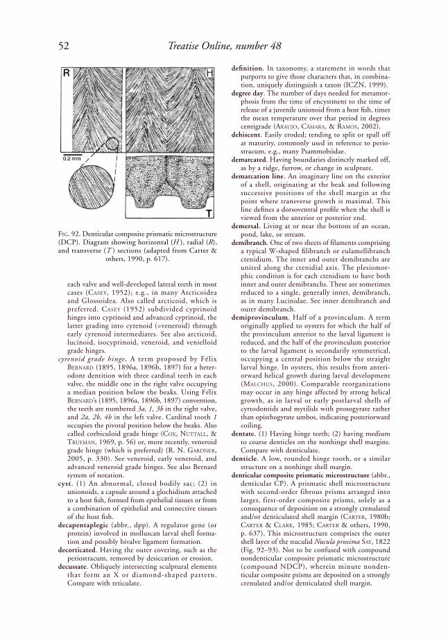
52 Treatise Online, number 48
each valve and well-developed lateral teeth in most cases (Casey, 1952); e.g., in many Arcticoidea and Glossoidea. Also called arcticoid, which is preferred. Casey (1952) subdivided cyprinoid hinges into cyprinoid and advanced cyprinoid, the latter grading into cyrenoid (=veneroid) through early cyrenoid intermediates. See also arcticoid, lucinoid, isocyprinoid, veneroid, and venielloid grade hinges.
cyrenoid grade hinge . A term proposed by Félix BerNard (1895, 1896a, 1896b, 1897) for a heter-odont dentition with three cardinal teeth in each valve, the middle one in the right valve occupying a median position below the beaks. Using Félix BerNard’s (1895, 1896a, 1896b, 1897) convention, the teeth are numbered 3a, 1, 3b in the right valve, and 2a, 2b, 4b in the left valve. Cardinal tooth 1 occupies the pivotal position below the beaks. Also called corbiculoid grade hinge (Cox, Nuttall, & trueMaN, 1969, p. 56) or, more recently, veneroid grade hinge (which is preferred) (r. N. GardNer, 2005, p. 330). See veneroid, early veneroid, and advanced veneroid grade hinges. See also Bernard system of notation.
cyst. (1) An abnormal, closed bodily sac; (2) in unionoids, a capsule around a glochidium attached to a host fish, formed from epithelial tissues or from a combination of epithelial and connective tissues of the host fish.
decapentaplegic (abbr., dpp). A regulator gene (or protein) involved in molluscan larval shell forma-tion and possibly bivalve ligament formation.
decorticated. Having the outer covering, such as the periostracum, removed by desiccation or erosion.
decussate. Obliquely intersecting sculptural elements that form an X or diamond-shaped pattern. Compare with reticulate.
definition. In taxonomy, a statement in words that purports to give those characters that, in combina-tion, uniquely distinguish a taxon (ICZN, 1999).
degree day. The number of days needed for metamor-phosis from the time of encystment to the time of release of a juvenile unionoid from a host fish, times the mean temperature over that period in degrees centigrade (arauJo, CáMara, & raMos, 2002).
dehiscent. Easily eroded; tending to split or spall off at maturity, commonly used in reference to perio-stracum, e.g., many Psammobiidae.
demarcated. Having boundaries distinctly marked off, as by a ridge, furrow, or change in sculpture.
demarcation line. An imaginary line on the exterior of a shell, originating at the beak and following successive positions of the shell margin at the point where transverse growth is maximal. This line defines a dorsoventral profile when the shell is viewed from the anterior or posterior end.
demersal. Living at or near the bottom of an ocean, pond, lake, or stream.
demibranch. One of two sheets of filaments comprising a typical W-shaped filibranch or eulamellibranch ctenidium. The inner and outer demibranchs are united along the ctenidial axis. The plesiomor-phic condition is for each ctenidium to have both inner and outer demibranchs. These are sometimes reduced to a single, generally inner, demibranch, as in many Lucinidae. See inner demibranch and outer demibranch.
demiprovinculum. Half of a provinculum. A term originally applied to oysters for which the half of the provinculum anterior to the larval ligament is reduced, and the half of the provinculum posterior to the larval ligament is secondarily symmetrical, occupying a central position below the straight larval hinge. In oysters, this results from anteri-orward helical growth during larval development (MalChus, 2000). Comparable reorganizations may occur in any hinge affected by strong helical growth, as in larval or early postlarval shells of cyrtodontids and mytilids with prosogyrate rather than opisthogyrate umbos, indicating posteriorward coiling.
dentate. (1) Having hinge teeth; (2) having medium to coarse denticles on the nonhinge shell margins. Compare with denticulate.
denticle. A low, rounded hinge tooth, or a similar structure on a nonhinge shell margin.
denticular composite prismatic microstructure (abbr., denticular CP). A prismatic shell microstructure with second-order fibrous prisms arranged into larger, first-order composite prisms, solely as a consequence of deposition on a strongly crenulated and/or denticulated shell margin (Carter, 1980b; Carter & Clark, 1985; Carter & others, 1990, p. 637). This microstructure comprises the outer shell layer of the nuculid Nucula proxima say, 1822 (Fig. 92–93). Not to be confused with compound nondenticular composite prismatic microstructure (compound NDCP), wherein minute nonden-ticular composite prisms are deposited on a strongly crenulated and/or denticulated shell margin.
FiG. 92. Denticular composite prismatic microstructure (DCP). Diagram showing horizontal (H ), radial (R), and transverse (T ) sections (adapted from Carter &
others, 1990, p. 617).

53Illustrated Glossary of the Bivalvia
denticulate (or denticulated). Having a series of moderately fine, alternating elevations and inden-tations on a shell margin (see Fig. 68, Fig. 243). When the features are moderately wide, the term crenulate is more appropriate. When the features are very fine, the terms serrate, pectinate, or micro-pectinate are more appropriate.
dentition. In Bivalvia, the hinge teeth and sockets.deposit-feeding. Nonselectively harvesting organic
particles by the siphons, foot, or palp probos-
cides, from surface or near-surface sediments. Compare with suspension-feeding and detritus-feeding.
depressed. Dorsoventrally flattened, in bivalves usually in reference to shell shape.
descending lamella (pl., lamellae). A ctenidial lamella that is attached to the ctenidial axis, e.g., in the gastrochaenid Lamychaena hians (GMeliN, 1791 in 1791–1793) (see Fig. 289). Compare with ascending lamella.
FiG. 93. Denticular composite prisms in outer shell layer of nuculid Nucula proxima say, 1822, Long Island Sound, New York (YPM 10014). 1, Transverse acetate peel showing four adjacent first-order denticular composite prisms and underlying nacreous shell layer; 2, SEM of radial fracture, showing second-order irregular fibrous prisms that
comprise each denticular composite prism (see also Carter & Lutz, 1990, pl. 19).
2
1
5 µm
50 µm
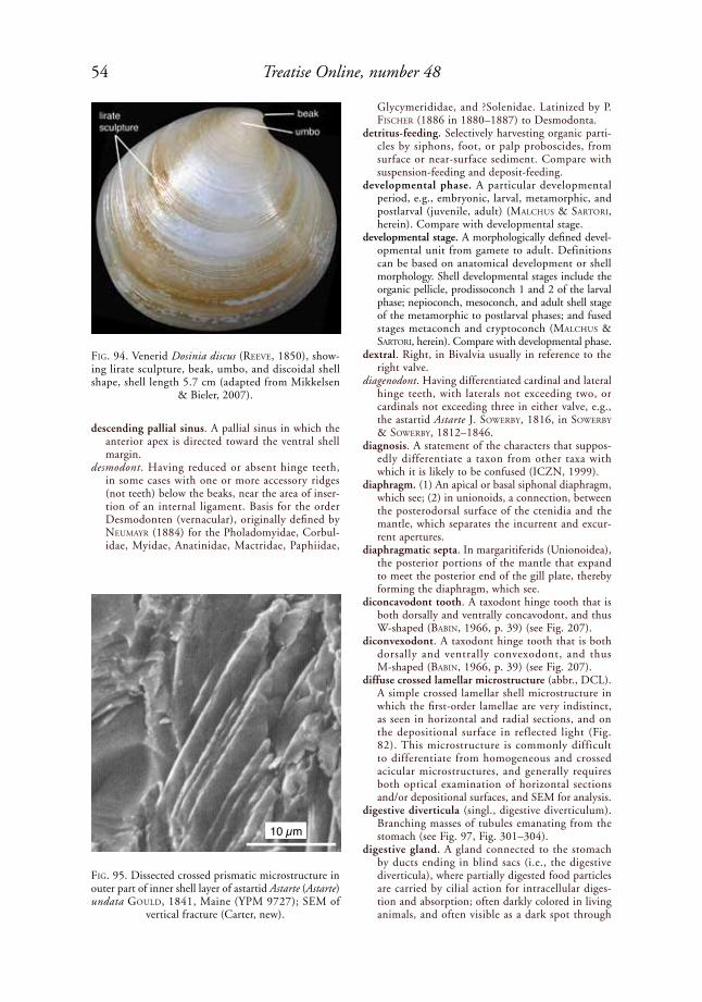
54 Treatise Online, number 48
descending pallial sinus. A pallial sinus in which the anterior apex is directed toward the ventral shell margin.
desmodont. Having reduced or absent hinge teeth, in some cases with one or more accessory ridges (not teeth) below the beaks, near the area of inser-tion of an internal ligament. Basis for the order Desmodonten (vernacular), originally defined by NeuMayr (1884) for the Pholadomyidae, Corbul-idae, Myidae, Anatinidae, Mactridae, Paphiidae,
Glycymerididae, and ?Solenidae. Latinized by p. FisCher (1886 in 1880–1887) to Desmodonta.
detritus-feeding. Selectively harvesting organic parti-cles by siphons, foot, or palp proboscides, from surface or near-surface sediment. Compare with suspension-feeding and deposit-feeding.
developmental phase. A particular developmental period, e.g., embryonic, larval, metamorphic, and postlarval (juvenile, adult) (MalChus & sartori, herein). Compare with developmental stage.
developmental stage. A morphologically defined devel-opmental unit from gamete to adult. Definitions can be based on anatomical development or shell morphology. Shell developmental stages include the organic pellicle, prodissoconch 1 and 2 of the larval phase; nepioconch, mesoconch, and adult shell stage of the metamorphic to postlarval phases; and fused stages metaconch and cryptoconch (MalChus & sartori, herein). Compare with developmental phase.
dextral. Right, in Bivalvia usually in reference to the right valve.
diagenodont. Having differentiated cardinal and lateral hinge teeth, with laterals not exceeding two, or cardinals not exceeding three in either valve, e.g., the astartid Astarte J. soWerBy, 1816, in soWerBy & soWerBy, 1812–1846.
diagnosis. A statement of the characters that suppos-edly differentiate a taxon from other taxa with which it is likely to be confused (ICZN, 1999).
diaphragm. (1) An apical or basal siphonal diaphragm, which see; (2) in unionoids, a connection, between the posterodorsal surface of the ctenidia and the mantle, which separates the incurrent and excur-rent apertures.
diaphragmatic septa. In margaritiferids (Unionoidea), the posterior portions of the mantle that expand to meet the posterior end of the gill plate, thereby forming the diaphragm, which see.
diconcavodont tooth. A taxodont hinge tooth that is both dorsally and ventrally concavodont, and thus W-shaped (BaBiN, 1966, p. 39) (see Fig. 207).
diconvexodont. A taxodont hinge tooth that is both dorsally and ventrally convexodont, and thus M-shaped (BaBiN, 1966, p. 39) (see Fig. 207).
diffuse crossed lamellar microstructure (abbr., DCL). A simple crossed lamellar shell microstructure in which the first-order lamellae are very indistinct, as seen in horizontal and radial sections, and on the depositional surface in reflected light (Fig. 82). This microstructure is commonly difficult to differentiate from homogeneous and crossed acicular microstructures, and generally requires both optical examination of horizontal sections and/or depositional surfaces, and SEM for analysis.
digestive diverticula (singl., digestive diverticulum). Branching masses of tubules emanating from the stomach (see Fig. 97, Fig. 301–304).
digestive gland. A gland connected to the stomach by ducts ending in blind sacs (i.e., the digestive diverticula), where partially digested food particles are carried by cilial action for intracellular diges-tion and absorption; often darkly colored in living animals, and often visible as a dark spot through
FiG. 94. Venerid Dosinia discus (reeVe, 1850), show-ing lirate sculpture, beak, umbo, and discoidal shell shape, shell length 5.7 cm (adapted from Mikkelsen
& Bieler, 2007).
FiG. 95. Dissected crossed prismatic microstructure in outer part of inner shell layer of astartid Astarte (Astarte) undata Gould, 1841, Maine (YPM 9727); SEM of
vertical fracture (Carter, new).
10 µm

55Illustrated Glossary of the Bivalvia
the gills or through a thin shell; in earlier literature, this was commonly referred to as the liver, midgut gland, or hepatopancreas.
digitation. A fingerlike projection.digitiform. Fingerlike.dimyarian. Having two and only two adductor
muscles, one anterior and one posterior. Compare with monomyarian and trimyarian.
dioecious. Having sexes separate throughout life; not hermaphroditic. Compare with gonochoristic.
diphyletic. Having members descended from two ancestral lines, with the group excluding their immediate common ancestor.
direct development. Development, from embryo to juvenile, which does not pass through an interme-diate larval stage and metamorphosis; sometimes (incorrectly) applied to certain bivalves (compare with indirect development) (turNer, peCheNik, & CalloWay, 1987).
disc. The central portion of a shell valve other than the wings and auricles or other than the anterior and posterior slopes; the main body of the shell, e.g., the shell disc in the pectinid Nodipecten fragosus (CoNrad, 1849b) (Fig. 29), and in the Pholadidae (Fig. 24). The spelling disc is preferred over disk in reference to anatomical features.
disc flanks. In pectinoids, the anterodorsal and posterodorsal portions of the shell disc, diverging from the beak and generally differing in curvature and ornament from the adjacent auricles and disc proper (Waller, 1969, fig. 2).
disc gapes. In pectinoids, the gapes between the anterodorsal and posterodorsal margins of the shell disc, referred to, respectively, as the anterior disc gape and the posterior disc gape (Waller, 1969, p. 12, fig. 2).
disc height (along curvature). As defined for pectinids by Waller (1969, p. 12), the distance from the origin of shell growth to the ventral shell margin, measured along the external surface of a centrally located antimarginal plica or rib.
disc height (linear). As defined for pectinids by Waller (1969, p. 12), the distance between two parallel lines, one coinciding with the outer liga-ment, the other passing through the most ventral point on the valve. This corresponds to measure-ments A–M and A'–M' in Figure 219.
disc length. As defined for pectinids by Waller (1969, p. 12), the distance between two lines perpendic-ular to the outer ligament, one passing through the most anterior point on the disc, the other passing through the most posterior point on the disc. This corresponds with measurements A–G and A'–G' in Figure 219.
discoidal. Shaped like or nearly like a disc, as in the venerid Dosinia discus (reeVe, 1850) (Fig. 94).
discontinuous pallial line. A pallial line with one or more gaps. If the gaps are numerous, the pallial line is also called disjunct.
discordant margins. Adducted shell margins that are not in exact juxtaposition, but rather overlap.
discrepant. Different on the left and right valves, e.g., discrepant ornament.
disjunct fibrous ligament ontogeny. See disjunct ligament.
disjunct ligament. A ligament with the successive development of at least two morphologically distinct fibrous sublayers. Characteristic of Pterio-morphia (see ligaments 1 and 2), but also occur-ring in some Protobranchia (e.g., Palaeoneilo; see Carter, 1990a), Tellinoidea (e.g., Tellina and Abra; see trueMaN, 1966), and possibly other bivalves.
disjunct pallial line. A discontinuous pallial line that is broken up into many separate, mostly unequal parts, e.g., in many Pteriidae and Bakevelliidae.
disk. Same as disc, the preferred spelling for compara-tive anatomy.
FiG. 96. Divaricate riblets on limid Ctenoides sanctipauli stuardo, 1982, figure is several mm wide (adapted
from Mikkelsen & Bieler, 2007).
FiG. 97. Dorsal hood and left pouch seen in dorsal view of anterior portion of digestive tract in lyonsiid Lyonsia norwegica (GMeliN, 1791 in 1791–1793), Newbiggin, Northumberland, UK, 110 m depth, figure width is 2.5
mm (NHMUK 20070070) (Sartori, new).

56 Treatise Online, number 48
dissected crossed prismatic microstructure. A pris-matic shell microstructure in which one or two oblique structural trends subdivide mutually parallel prisms into parallelogram-shaped segments (Carter & others, 1990, p. 612), e.g., in the translucent shell windows or lenses of the cardiid Corculum, where the prisms grade into an under-lying crossed lamellar microstructure. Also found in the outer part of the inner shell layer of the astartid Astarte (Astarte) undata Gould, 1841 (Fig. 95). Also called trans-prismatic.
dissoconch . The entire post larval ( i .e . , post -metamorphic) bivalve shell (JaCksoN, 1890, p. 281) (Fig. 237). Compare with prodissoconch, nepioconch, and mesoconch. The inception of the dissoconch roughly coincides with the onset of adult life habits and shell characters, except in the case of some pseudoplanktonic bivalves.
distal. Away from the center, point of origin, or attach-ment site; opposite of proximal.
distal oral groove. A narrow extension of the base of the labial palps.
distended. (1) Expanded or swollen; (2) enlarged from internal pressure.
divaricate sculpture. An oblique ornament that is oppositely angled on adjacent parts of the shell, forming an upside-down V where the sculptural elements intersect, e.g., the divaricate riblets on the limid Ctenoides sanctipauli stuardo, 1982 (Fig. 96). Also called divaricating sculpture. Compare with divergent sculpture.
divaricating sculpture. Same as divaricate sculpture, which is preferred.
divergent sculpture. Having ribs that divide distally in such a manner that their divisions are not truly radial.
dorsal. (1) For soft anatomy, the direction perpen-dicular to the line passing through the mouth and anus, parallel with the commissural plane, and toward the beak and hinge; (2) in shell morphology, the direction perpendicular to the hinge axis, parallel with the commissural plane, and toward the beak.
dorsal crest. A prominent, rounded, anatomically dorsoposterior but functionally dorsal wing on the
shell of alatoconchids and wallowaconchids (Fig. 41, and see Fig. 320).
dorsal crus (pl., crura). A nonarticulating ridge just ventral to, and nearly parallel with the hinge axis, e.g., in Entoliidae. Waller (2006) advocated replacing this term with dorsal buttress.
dorsal hood. An area resembling a hood in the stomach wall, whose aperture faces the head (distal tip) of the crystalline style (Fig. 97).
dorsal ligament. An external and/or shallow submar-ginal ligament (see ligament position).
dorsal muscles. Small, discrete muscles inserting onto the shell in the umbonal cavity or on the ventral margin of the hinge plate, e.g., in unionoids (see Fig. 307). These might represent mantle attach-ment, gill attachment, and/or pedal muscles. See also visceral suspensory muscle scars.
dorsal pallial organ. See pallial organ.dorsal shell edge. The intersection of the dorsal, exte-
rior shell surface (including the cardinal area, where present) with the commissural plane, near the hinge axis. The dorsal shell edge is the point of reference for differentiating between external and submar-ginal ligaments. Where alivincular, multivincular, and duplivincular ligaments are present, the dorsal shell edge roughly corresponds with the hinge axis. Compare with dorsal shell margin.
dorsal shell margin. The area of opposition of the left and right shell valves immediately below, anterior and posterior to the beaks, and dorsal to an imagi-nary horizontal line drawn tangent to the dorsal edge of the adductor muscle scar(s). Compare with dorsal shell edge, which is part of the dorsal shell margin.
dorsal teeth. (1) Teeth dorsally placed on the hinge; (2) a pair of lamellar teeth on each side of the hinge in the right valve of a pectinoid, beneath the linear outer ligament and above the socket for the infradorsal teeth of the left valve, or above the sockets and teeth of multiple intermediate teeth and sockets of the left valve (Waller, 1991, p. 8). See also supradorsal teeth, infradorsal teeth, and intermediate teeth.
dorsoposterior. Both dorsal and posterior; same as posterodorsal.
FiG. 98. Alternating type of lamellar sublayer insertion in a duplivincular ligament, showing descriptive coding proposed by MalChus (2004b) for lamellar sublayers (adapted from Malchus, 2004b, fig. 5B, based mostly on
Recent Arcidae).
1p 2p 4p 6p 3a 1a5a
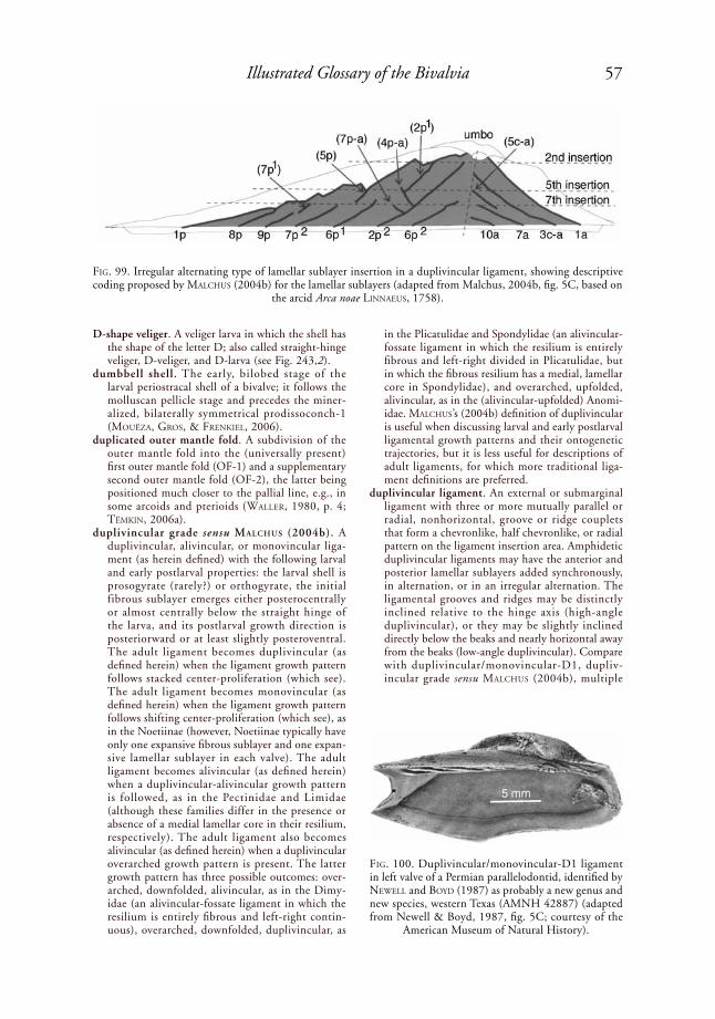
57Illustrated Glossary of the Bivalvia
D-shape veliger. A veliger larva in which the shell has the shape of the letter D; also called straight-hinge veliger, D-veliger, and D-larva (see Fig. 243,2).
dumbbell shell. The early, bilobed stage of the larval periostracal shell of a bivalve; it follows the molluscan pellicle stage and precedes the miner-alized, bilaterally symmetrical prodissoconch-1 (MouëZa, Gros, & FreNkiel, 2006).
duplicated outer mantle fold. A subdivision of the outer mantle fold into the (universally present) first outer mantle fold (OF-1) and a supplementary second outer mantle fold (OF-2), the latter being positioned much closer to the pallial line, e.g., in some arcoids and pterioids (Waller, 1980, p. 4; tëMkiN, 2006a).
duplivincular grade sensu MalchuS (2004b). A duplivincular, alivincular, or monovincular liga-ment (as herein defined) with the following larval and early postlarval properties: the larval shell is prosogyrate (rarely?) or orthogyrate, the initial fibrous sublayer emerges either posterocentrally or almost centrally below the straight hinge of the larva, and its postlarval growth direction is posteriorward or at least slightly posteroventral. The adult ligament becomes duplivincular (as defined herein) when the ligament growth pattern follows stacked center-proliferation (which see). The adult ligament becomes monovincular (as defined herein) when the ligament growth pattern follows shifting center-proliferation (which see), as in the Noetiinae (however, Noetiinae typically have only one expansive fibrous sublayer and one expan-sive lamellar sublayer in each valve). The adult ligament becomes alivincular (as defined herein) when a duplivincular-alivincular growth pattern is followed, as in the Pectinidae and Limidae (although these families differ in the presence or absence of a medial lamellar core in their resilium, respectively). The adult ligament also becomes alivincular (as defined herein) when a dupli vincular overarched growth pattern is present. The latter growth pattern has three possible outcomes: over-arched, downfolded, alivincular, as in the Dimy-idae (an alivincular-fossate ligament in which the resilium is entirely fibrous and left-right contin-uous), overarched, downfolded, duplivincular, as
in the Plicatulidae and Spondylidae (an alivincular-fossate ligament in which the resilium is entirely fibrous and left-right divided in Plicatulidae, but in which the fibrous resilium has a medial, lamellar core in Spondylidae), and overarched, upfolded, alivincular, as in the (alivincular-upfolded) Anomi-idae. MalChus’s (2004b) definition of duplivincular is useful when discussing larval and early postlarval ligamental growth patterns and their ontogenetic trajectories, but it is less useful for descriptions of adult ligaments, for which more traditional liga-ment definitions are preferred.
duplivincular ligament. An external or submarginal ligament with three or more mutually parallel or radial, nonhorizontal, groove or ridge couplets that form a chevronlike, half chevronlike, or radial pattern on the ligament insertion area. Amphidetic duplivincular ligaments may have the anterior and posterior lamellar sublayers added synchronously, in alternation, or in an irregular alternation. The ligamental grooves and ridges may be distinctly inclined relative to the hinge axis (high-angle duplivincular), or they may be slightly inclined directly below the beaks and nearly horizontal away from the beaks (low-angle duplivincular). Compare with duplivincular/monovincular-D1, dupliv-incular grade sensu MalChus (2004b), multiple
FiG. 99. Irregular alternating type of lamellar sublayer insertion in a duplivincular ligament, showing descriptive coding proposed by MalChus (2004b) for the lamellar sublayers (adapted from Malchus, 2004b, fig. 5C, based on
the arcid Arca noae liNNaeus, 1758).
FiG. 100. Duplivincular/monovincular-D1 ligament in left valve of a Permian parallelodontid, identified by NeWell and Boyd (1987) as probably a new genus and new species, western Texas (AMNH 42887) (adapted from Newell & Boyd, 1987, fig. 5C; courtesy of the
American Museum of Natural History).

58 Treatise Online, number 48
planivincular, preduplivincular, and reduced low-angle duplivincular.
duplivincular (alternating type) ligament. A dupliv-incular ligament with lamellar sublayers initiated in a regularly alternating sequence anteriorly and posteriorly (MalChus, 2004b, p. 1554) (Fig. 98).
duplivincular (irregular alternating type) ligament. An amphidetic duplivincular ligament with the anterior and posterior lamellar sublayers initiated in an irregularly alternating pattern on the anterior and posterior sides of the ligament, and in some cases, also with some posterior lamellar sublayers inclined in a direction opposite to most other posterior lamellar sublayers (MalChus, 2004b, p. 1554) (Fig. 99).
duplivincular/monovincular-D1 ligament. An arcoid ligament that combines duplivincular and mono-vincular-D1 elements. Present in many Parallelo-dontidae, where the monovincular-D1 portion may be anterior or posterior to the duplivincular portion
(Fig. 100). Also present in some Arcidae, such as Scapharca (Scapharca) inaequivalvis (BruGuière, 1789), where the monovincular-D1 portion is flanked by a single, deeply incised, anterior and posterior, duplivincular ligament groove (NeWell, 1969a, fig. C5,6b), and Arca imbricata BruGuière, 1789 (Fig. 25).
duplivincular (synchronous type) ligament. An amphidetic duplivincular ligament with the ante-rior and posterior lamellar sublayers initiated simul-taneously (MalChus, 2004b, p. 1554) (Fig. 101).
D-veliger. Same as D-shape veliger, which see.dwarfism. Evolutionary process leading to an adult
that is distinctly smaller than the adult of the ancestor.
dysodont. Lacking true hinge teeth but having small, weak, shell marginal teeth near the umbos. A term commonly applied to certain members of the Myti-lidae, e.g., Brachidontes exustus (liNNaeus, 1758). See also shell marginal tooth.
FiG. 101. Synchronous lamellar sublayer insertion in a duplivincular ligament, showing descriptive coding proposed by MalChus (2004b) for lamellar sublayers (adapted from Malchus, 2004b, fig. 5A, based on the glycymeridid
Glycymeris).
FiG. 102. Early veneroid grade hinge. 1, Diagram of left and right valves (adapted from R. N. Gardner, 2005, fig. 14.1c, courtesy of R. N. Gardner); 2, Upper Jurassic venerid Eocallista (Eocallista) caudata (GoldFuss, 1840)
(adapted from Keen & Casey, 1969, fig. E140,10c–d).
1p 2p 3p 3a 2a 1a
4b
left valve
2b 2acardinals
residue of 2b1 and lateral 1AIIlaminar IV
lateral AII
lateral AIII
lateral AI
3a
right valve
1cardinals
3b
PII
4b2b 2a
AII AIII
AI 1
3b
PI
lateral AIV3a
1
2
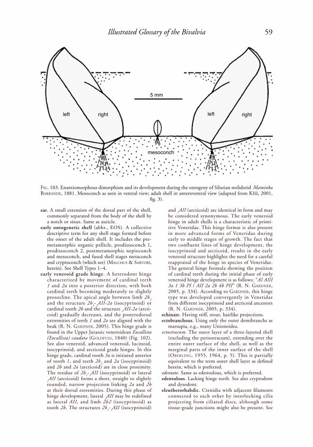
59Illustrated Glossary of the Bivalvia
left right
5 mm
left right
ear. A small extension of the dorsal part of the shell, commonly separated from the body of the shell by a notch or sinus. Same as auricle.
early ontogenetic shell (abbr., EOS). A collective descriptive term for any shell stage formed before the onset of the adult shell. It includes the pre-metamorphic organic pellicle, prodissoconch 1, prodissoconch 2, postmetamorphic nepioconch and mesoconch, and fused shell stages metaconch and cryptoconch (which see) (MalChus & sartori, herein). See Shell Types 1–4.
early veneroid grade hinge. A heterodont hinge characterized by movement of cardinal teeth 1 and 2a into a posterior direction, with both cardinal teeth becoming moderately to slightly prosocline. The apical angle between limb 2b
2
and the structure 2b1-
1AII-2a (isocyprinoid) or
cardinal tooth 2b and the structure 1AII-2a (arcti-
coid) gradually decreases, and the posterodorsal extremities of teeth 1 and 2a are aligned with the beak (r. N. GardNer, 2005). This hinge grade is found in the Upper Jurassic veneroidean Eocallista (Eocallista) caudata (GoldFuss, 1840) (Fig. 102). See also veneroid, advanced veneroid, lucinoid, isocyprinoid, and arcticoid grade hinges. In this hinge grade, cardinal tooth 3a is initiated anterior of tooth 1, and teeth 2b
2 and 2a (isocyprinoid)
and 2b and 2a (arcticoid) are in close proximity. The residue of 2b
1-
1AII (isocyprinoid) or lateral
1AII (arcticoid) forms a short, straight to slightly
rounded, narrow projection linking 2a and 2b at their dorsal extremities. During this phase of hinge development, lateral
2AII may be redefined
as lateral AII, and limb 2b2 (isocyprinoid) as tooth 2b. The structures 2b
1-
1AII (isocyprinoid)
and 1AII (arcticoid) are identical in form and may
be considered synonymous. The early veneroid hinge in adult shells is a characteristic of primi-tive Veneridae. This hinge format is also present in more advanced forms of Veneridae during early to middle stages of growth. The fact that two confluent lines of hinge development, the isocyprinoid and arcticoid, results in the early veneroid structure highlights the need for a careful reappraisal of the hinge in species of Veneridae. The general hinge formula showing the position of cardinal teeth during the initial phase of early veneroid hinge development is as follows: “AI AIII 3a 1 3b PI / AII 2a 2b 4b PII” (r. N. GardNer, 2005, p. 334). According to GardNer, this hinge type was developed convergently in Veneridae from different isocyprinoid and arcticoid ancestors (r. N. GardNer, 2005, p. 334).
echinate. Having stiff, stout, hairlike projections.ectobranchous. Using only the outer demibranchs as
marsupia, e.g., many Unionoidea.ectostracum. The outer layer of a three-layered shell
(excluding the periostracum), extending over the entire outer surface of the shell, as well as the marginal parts of the inner surface of the shell (oBerliNG, 1955, 1964, p. 5). This is partially equivalent to the term outer shell layer as defined herein, which is preferred.
edentate. Same as edentulous, which is preferred.edentulous. Lacking hinge teeth. See also cryptodont
and dysodont.eleutherorhabdic. Ctenidia with adjacent filaments
connected to each other by interlocking cilia projecting from ciliated discs, although some tissue-grade junctions might also be present. See
FiG. 103. Enantiomorphous dimorphism and its development during the ontogeny of Silurian stolidotid Maminka BarraNde, 1881. Mesoconch as seen in ventral view; adult shell in anteroventral view (adapted from Kříž, 2001,
fig. 3).
mesoconch
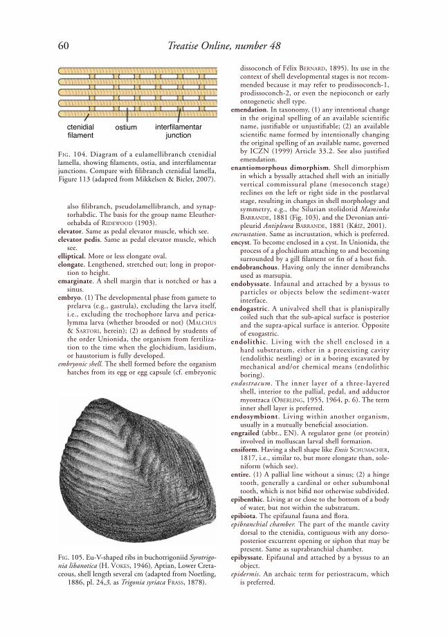
60 Treatise Online, number 48
also filibranch, pseudolamellibranch, and synap-torhabdic. The basis for the group name Eleuther-orhabda of rideWood (1903).
elevator. Same as pedal elevator muscle, which see.elevator pedis. Same as pedal elevator muscle, which
see.elliptical. More or less elongate oval.elongate. Lengthened, stretched out; long in propor-
tion to height. emarginate. A shell margin that is notched or has a
sinus.embryo. (1) The developmental phase from gamete to
prelarva (e.g., gastrula), excluding the larva itself, i.e., excluding the trochophore larva and perica-lymma larva (whether brooded or not) (MalChus & sartori, herein); (2) as defined by students of the order Unionida, the organism from fertiliza-tion to the time when the glochidium, lasidium, or haustorium is fully developed.
embryonic shell. The shell formed before the organism hatches from its egg or egg capsule (cf. embryonic
dissoconch of Félix BerNard, 1895). Its use in the context of shell developmental stages is not recom-mended because it may refer to prodissoconch-1, prodissoconch-2, or even the nepioconch or early ontogenetic shell type.
emendation. In taxonomy, (1) any intentional change in the original spelling of an available scientific name, justifiable or unjustifiable; (2) an available scientific name formed by intentionally changing the original spelling of an available name, governed by ICZN (1999) Article 33.2. See also justified emendation.
enantiomorphous dimorphism. Shell dimorphism in which a byssally attached shell with an initially vertical commissural plane (mesoconch stage) reclines on the left or right side in the postlarval stage, resulting in changes in shell morphology and symmetry, e.g., the Silurian stolidotid Maminka BarraNde, 1881 (Fig. 103), and the Devonian anti-pleurid Antipleura BarraNde, 1881 (kŕíž, 2001).
encrustation. Same as incrustation, which is preferred.encyst. To become enclosed in a cyst. In Unionida, the
process of a glochidium attaching to and becoming surrounded by a gill filament or fin of a host fish.
endobranchous. Having only the inner demibranchs used as marsupia.
endobyssate. Infaunal and attached by a byssus to particles or objects below the sediment-water interface.
endogastric. A univalved shell that is planispirally coiled such that the sub-apical surface is posterior and the supra-apical surface is anterior. Opposite of exogastric.
endolithic . Living with the shell enclosed in a hard substratum, either in a preexisting cavity (endolithic nestling) or in a boring excavated by mechanical and/or chemical means (endolithic boring).
endostracum. The inner layer of a three-layered shell, interior to the pallial, pedal, and adductor myo straca (oBerliNG, 1955, 1964, p. 6). The term inner shell layer is preferred.
endosymbiont. Living within another organism, usually in a mutually beneficial association.
engrailed (abbr., EN). A regulator gene (or protein) involved in molluscan larval shell formation.
ensiform. Having a shell shape like Ensis sChuMaCher, 1817, i.e., similar to, but more elongate than, sole-niform (which see).
entire. (1) A pallial line without a sinus; (2) a hinge tooth, generally a cardinal or other subumbonal tooth, which is not bifid nor otherwise subdivided.
epibenthic. Living at or close to the bottom of a body of water, but not within the substratum.
epibiota. The epifaunal fauna and flora.epibranchial chamber. The part of the mantle cavity
dorsal to the ctenidia, contiguous with any dorso-posterior excurrent opening or siphon that may be present. Same as suprabranchial chamber.
epibyssate. Epifaunal and attached by a byssus to an object.
epidermis. An archaic term for periostracum, which is preferred.
FiG. 104. Diagram of a eulamellibranch ctenidial lamella, showing filaments, ostia, and interfilamentar junctions. Compare with filibranch ctenidial lamella, Figure 113 (adapted from Mikkelsen & Bieler, 2007).
FiG. 105. Eu-V-shaped ribs in buchotrigoniid Syrotrigo-nia libanotica (h. Vokes, 1946), Aptian, Lower Creta-ceous, shell length several cm (adapted from Noetling,
1886, pl. 24,3, as Trigonia syriaca Frass, 1878).
ctenidial filament
ostium interfilamentar junction
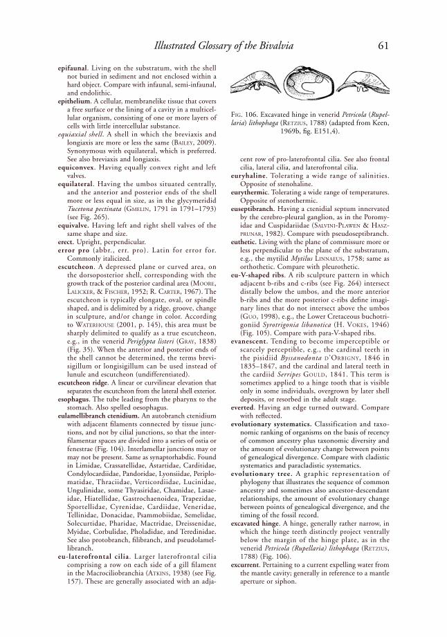
61Illustrated Glossary of the Bivalvia
epifaunal. Living on the substratum, with the shell not buried in sediment and not enclosed within a hard object. Compare with infaunal, semi-infaunal, and endolithic.
epithelium. A cellular, membranelike tissue that covers a free surface or the lining of a cavity in a multicel-lular organism, consisting of one or more layers of cells with little intercellular substance.
equiaxial shell. A shell in which the breviaxis and longiaxis are more or less the same (Bailey, 2009). Synonymous with equilateral, which is preferred. See also breviaxis and longiaxis.
equiconvex. Having equally convex right and left valves.
equilateral. Having the umbos situated centrally, and the anterior and posterior ends of the shell more or less equal in size, as in the glycymeridid Tucetona pectinata (GMeliN, 1791 in 1791–1793) (see Fig. 265).
equivalve. Having left and right shell valves of the same shape and size.
erect. Upright, perpendicular.error pro (abbr., err. pro). Latin for error for.
Commonly italicized.escutcheon. A depressed plane or curved area, on
the dorsoposterior shell, corresponding with the growth track of the posterior cardinal area (Moore, laliCker, & FisCher, 1952; r. Carter, 1967). The escutcheon is typically elongate, oval, or spindle shaped, and is delimited by a ridge, groove, change in sculpture, and/or change in color. According to Waterhouse (2001, p. 145), this area must be sharply delimited to qualify as a true escutcheon, e.g., in the venerid Periglypta listeri (Gray, 1838) (Fig. 35). When the anterior and posterior ends of the shell cannot be determined, the terms brevi-sigillum or longisigillum can be used instead of lunule and escutcheon (undifferentiated).
escutcheon ridge. A linear or curvilinear elevation that separates the escutcheon from the lateral shell exterior.
esophagus. The tube leading from the pharynx to the stomach. Also spelled oesophagus.
eulamellibranch ctenidium. An autobranch ctenidium with adjacent filaments connected by tissue junc-tions, and not by cilial junctions, so that the inter-filamentar spaces are divided into a series of ostia or fenestrae (Fig. 104). Interlamellar junctions may or may not be present. Same as synaptorhabdic. Found in Limidae, Crassatellidae, Astartidae, Carditidae, Condylocardiidae, Pandoridae, Lyonsiidae, Periplo-matidae, Thraciidae, Verticordiidae, Lucinidae, Ungulinidae, some Thyasiridae, Chamidae, Lasae-idae, Hiatellidae, Gastrochaenoidea, Trapezidae, Sportellidae, Cyrenidae, Cardiidae, Veneridae, Tellinidae, Donacidae, Psammobiidae, Semelidae, Solecurtidae, Pharidae, Mactridae, Dreissenidae, Myidae, Corbulidae, Pholadidae, and Teredinidae. See also protobranch, filibranch, and pseudolamel-libranch.
eu-laterofrontal cilia. Larger laterofrontal cilia comprising a row on each side of a gill filament in the Macrociliobranchia (atkiNs, 1938) (see Fig. 157). These are generally associated with an adja-
cent row of pro-laterofrontal cilia. See also frontal cilia, lateral cilia, and laterofrontal cilia.
euryhaline. Tolerating a wide range of salinities. Opposite of stenohaline.
eurythermic. Tolerating a wide range of temperatures. Opposite of stenothermic.
euseptibranch. Having a ctenidial septum innervated by the cerebro-pleural ganglion, as in the Poromy-idae and Cuspidariidae (salViNi-plaWeN & hasZ-pruNar, 1982). Compare with pseudoseptibranch.
euthetic. Living with the plane of commissure more or less perpendicular to the plane of the substratum, e.g., the mytilid Mytilus liNNaeus, 1758; same as orthothetic. Compare with pleurothetic.
eu-V-shaped ribs. A rib sculpture pattern in which adjacent b-ribs and c-ribs (see Fig. 264) intersect distally below the umbos, and the more anterior b-ribs and the more posterior c-ribs define imagi-nary lines that do not intersect above the umbos (Guo, 1998), e.g., the Lower Cretaceous buchotri-goniid Syrotrigonia libanotica (h. Vokes, 1946) (Fig. 105). Compare with para-V-shaped ribs.
evanescent. Tending to become imperceptible or scarcely perceptible, e.g., the cardinal teeth in the pisidiid Byssanodonta d’orBiGNy, 1846 in 1835–1847, and the cardinal and lateral teeth in the cardiid Serripes Gould, 1841. This term is sometimes applied to a hinge tooth that is visible only in some individuals, overgrown by later shell deposits, or resorbed in the adult stage.
everted. Having an edge turned outward. Compare with reflected.
evolutionary systematics. Classification and taxo-nomic ranking of organisms on the basis of recency of common ancestry plus taxonomic diversity and the amount of evolutionary change between points of genealogical divergence. Compare with cladistic systematics and paracladistic systematics.
evolutionary tree. A graphic representation of phylogeny that illustrates the sequence of common ancestry and sometimes also ancestor-descendant relationships, the amount of evolutionary change between points of genealogical divergence, and the timing of the fossil record.
excavated hinge. A hinge, generally rather narrow, in which the hinge teeth distinctly project ventrally below the margin of the hinge plate, as in the venerid Petricola (Rupellaria) lithophaga (retZius, 1788) (Fig. 106).
excurrent. Pertaining to a current expelling water from the mantle cavity; generally in reference to a mantle aperture or siphon.
FiG. 106. Excavated hinge in venerid Petricola (Rupel-laria) lithophaga (retZius, 1788) (adapted from Keen,
1969b, fig. E151,4).
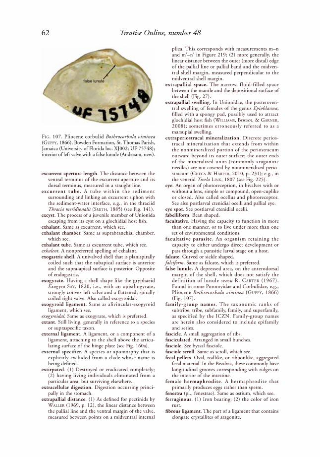
62 Treatise Online, number 48
excurrent aperture length. The distance between the ventral terminus of the excurrent aperture and its dorsal terminus, measured in a straight line.
excurrent tube . A tube wi th in the sediment surrounding and linking an excurrent siphon with the sediment-water interface, e.g., in the thraciid Thracia meridionalis (sMith, 1885) (see Fig. 141).
excyst. The process of a juvenile member of Unionida escaping from its cyst on a glochidial host fish.
exhalant. Same as excurrent, which see.exhalant chamber. Same as suprabranchial chamber,
which see. exhalant tube. Same as excurrent tube, which see. exhalent. A nonpreferred spelling of exhalant.exogastric shell. A univalved shell that is planispirally
coiled such that the subapical surface is anterior and the supra-apical surface is posterior. Opposite of endogastric.
exogyrate. Having a shell shape like the gryphaeid Exogyra say, 1820, i.e., with an opisthogyrate, strongly convex left valve and a flattened, spirally coiled right valve. Also called exogyroidal.
exogyroid ligament. Same as alivincular-exogyroid ligament, which see.
exogyroidal. Same as exogyrate, which is preferred.extant. Still living, generally in reference to a species
or supraspecific taxon. external ligament. A ligament, or a component of a
ligament, attaching to the shell above the articu-lating surface of the hinge plate (see Fig. 160a).
external specifier. A species or apomorphy that is explicitly excluded from a clade whose name is being defined.
extirpated. (1) Destroyed or eradicated completely; (2) having living individuals eliminated from a particular area, but surviving elsewhere.
extracellular digestion. Digestion occurring princi-pally in the stomach.
extrapallial distance. (1) As defined for pectinids by Waller (1969, p. 12), the linear distance between the pallial line and the ventral margin of the valve, measured between points on a midventral internal
plica. This corresponds with measurements m–n and m'–n' in Figure 219; (2) more generally, the linear distance between the outer (more distal) edge of the pallial line or pallial band and the midven-tral shell margin, measured perpendicular to the midventral shell margin.
extrapallial space. The narrow, fluid-filled space between the mantle and the depositional surface of the shell (Fig. 27).
extrapallial swelling. In Unionidae, the posteroven-tral swelling of females of the genus Epioblasma, filled with a spongy pad, possibly used to attract glochidial host fish (WilliaMs, BoGaN, & GarNer, 2008); sometimes erroneously referred to as a marsupial swelling.
extraperiostracal mineralization. Discrete perios-tracal mineralization that extends from within the nonmineralized portion of the periostracum outward beyond its outer surface; the outer ends of the mineralized units (commonly aragonitic needles) are not covered by nonmineralized perio-stracum (CheCa & harper, 2010, p. 231); e.g., in the venerid Tivela liNk, 1807 (see Fig. 225).
eye. An organ of photoreception, in bivalves with or without a lens, simple or compound, open-cuplike or closed. Also called ocellus and photoreceptor. See also postlarval ctenidial ocelli and pallial eye.
eye spot. See postlarval ctenidial ocelli.fabelliform. Bean shaped.facultative. Having the capacity to function in more
than one manner, or to live under more than one set of environmental conditions.
facultative parasite . An organism retaining the capacity to either undergo direct development or pass through a parasitic larval stage on a host.
falcate. Curved or sickle shaped.falciform. Same as falcate, which is preferred.false lunule. A depressed area, on the anterodorsal
margin of the shell, which does not satisfy the definition of lunule sensu r. Carter (1967). Found in some Poromyidae and Corbulidae, e.g., Pliocene Bothrocorbula viminea (Guppy, 1866) (Fig. 107).
family-group names . The taxonomic ranks of subtribe, tribe, subfamily, family, and superfamily, as specified by the ICZN. Family-group names are herein also considered to include epifamily and series.
fascicle. A small aggregation of ribs.fasciculated. Arranged in small bunches.fasciole. See byssal fasciole.fasciole scroll. Same as scroll, which see. fecal pellets. Oval, rodlike, or ribbonlike, aggregated
fecal material. In the Bivalvia, these commonly have longitudinal grooves corresponding with ridges on the interior of the intestine.
female hermaphrodite . A hermaphrodite that primarily produces eggs rather than sperm.
fenestra (pl., fenestrae). Same as ostium, which see.ferruginous. (1) Iron bearing; (2) the color of iron
rust.fibrous ligament. The part of a ligament that contains
elongate crystallites of aragonite.
FiG. 107. Pliocene corbulid Bothrocorbula viminea (Guppy, 1866), Bowden Formation, St. Thomas Parish, Jamaica (University of Florida loc. XJ002; UF 75748); interior of left valve with a false lunule (Anderson, new).

63Illustrated Glossary of the Bivalvia
fibrous prismatic microstructure (abbr., FP). A finely prismatic shell microstructure in which each first-order prism is not divided into elongate, second-order structural units, and the prisms have generally very high length/width ratios. Categories include regular fibrous prismatic (e.g., anvil-type, lath-type, rod-type, and simple lamellar) and irregular fibrous prismatic (Carter & others, 1990). Fibrous prisms deposited on a strongly reflected and crenulate and/or denticulate shell margin have a concat-enated, three-dimensional fanlike arrangement called denticular composite prismatic (Fig. 93). Compare with fibrous simple prismatic.
fibrous prismatic microstructure, anvil-type (abbr., anvil-type FP). A regular fibrous prismatic shell microstructure in which the prisms have a generally convex upper surface, a concave lower surface, and concave sides (Carter & others, 1990, p. 629); e.g., the outer shell layer of the mytilid Mytilus edulis liNNaeus, 1758 (Fig. 108).
fibrous prismatic microstructure, irregular (abbr., irregular FP). A fibrous prismatic shell micro-structure in which the prisms generally have very irregular shapes, in many cases showing nodular subunits along their length (Carter & Clark, 1985; Carter & others, 1990, p. 610, 645). Found locally in the outer shell layer of the venerid Cyclina sinensis (GMeliN, 1791 in 1791–1793) (Fig. 109),
and also comprising the denticular composite prisms in the nuculid Nucula proxima say, 1822 (Fig. 93).
fibrous prismatic microstructure, lath-type (abbr., lath-type FP). A regular fibrous prismatic shell microstructure in which the prisms have shapes intermediate between rectangular rods and blades (Carter & others, 1990, p. 610). The prisms are not arranged into distinct, sheetlike aggregations as in calcitic foliated and aragonitic simple lamellar fibrous prismatic microstructures. This structure is found in the outer shell layer of some Limidae (Fig. 110), where it has been misdiagnosed by some authors as a foliated microstructure.
fibrous prismatic microstructure, rod-type (abbr., rod-type FP). A regular fibrous prismatic shell micro-structure in which the prisms have rounded and/or polygonal cross sections, and are therefore not laths or concavo-convex rods (Carter & others, 1990, p. 610); e.g., in the outer shell layer of the crenellid Crenella glandula (totteN, 1834) (Fig. 111).
fibrous prismatic microstructure, simple lamellar (abbr., simple lamellar FP). A regular fibrous pris-matic shell microstructure in which the prisms are aggregated into mutually parallel sheets similar to the second-order lamellae of a simple crossed lamellar microstructure, but the second-order lamellae and their fibers are all mutually parallel
FiG. 108. Anvil-type fibrous prismatic microstructure in calcitic outer shell layer of mytilid Mytilus edulis liNNaeus, 1758, New England (UNC 13620). 1, SEM of depositional surface cleaned with sodium hypochlorite; shell margin toward top; 2, diagram of prisms viewed along their length axes (above) and across their length axes (below); ap-
proximately same scale as SEM on left (Carter, new).
1 2
1 µm

64 Treatise Online, number 48
(Carter & others, 1990, p. 610). This is found in the hinge teeth of some Arcidae and Dreissenidae (Carter & Clark, 1985; Carter & others, 1990, p. 658) (Fig. 112). MaNo (1971, p. 67) called this microstructure simple lamellar.
fibrous simple prismatic microstructure. A simple pris-matic shell microstructure in which each first-order prism has a fibrous prismatic second-order structure, e.g., in the malleid Malleus regulus (Forsskål, 1775). This corresponds only in part with the foliated prismatic microstructure illustrated by esteBaN-delGado and others (2008), which also includes
semi-foliated simple prismatic microstructure. Compare with semi-foliated simple prismatic and homogeneous simple prismatic.
fibrous sublayer of ligament. See fibrous ligament.fide. Latin for according to. Commonly italicized.filamentous. In reference to ctenidia, having well-
separated filaments, as in members of the Proto-branchia.
filibranch ctenidium. A relatively simple autobranch ctendium consisting of a series of narrow, elongate filaments, with adjacent filaments connected by cilial and not tissue-grade junctions (Fig. 113). Interlamellar junctions may be present or absent. Found in Arcidae, Glycymerididae, Limopsidae, Philobryidae, Mytilidae, some Pteriidae, some Isog-nomonidae, some Malleidae, and some Pectinida (e.g., Propeamussiidae, Plicatulidae, Anomiidae). See also eleutherorhabdic.
filiform. Threadlike.filosus structure. A sculpture of fine, antimarginal
to radial fila, costellae, and/or tiny ridgelets, commonly distally diverging, visible on the inner surface of the calcitic outer shell layer in the left valve of some pectinoids (allasiNaZ, 1972; Waller, 2006, p. 320). The term derives from the Triassic entolioidid Filopecten filosus (hauer, 1857). Filosus structure probably reflects the depositional surface of radially to slightly obliquely oriented, calcitic fibrous prisms and irregular spherulitic prisms, such as in the outer shell layer of the left valve of upper Permian Guizhoupecten CheN, 1962 (see NeWell & Boyd, 1985, fig. 3). Filosus structure also occurs in the Upper Triassic, transitional entolioidid-propeamussiid Filamussium schafhaeutli (WiNkler, 1859) (Fig. 114).
filter-feeding. Feeding by using the ctenidia to sift organic particles from water, then sorting them and transporting them to the mouth for ingestion. See also suspension-feeding and deposit-feeding.
filum (pl., fila). A very narrow commarginal ridge that is slightly coarser and more regularly shaped than a typical growth line.
fimbriate. Fringed; regularly gathered into wrinkles at the shell margin, as in the lucinid Fimbria MeGerle VoN MühlFeld, 1811.
fine complex crossed lamellar microstructure (abbr., fine CCL). A complex crossed lamellar shell micro-structure in which first-order lamellae are either absent, or consist of a few mutually parallel, narrow, elongate structural units; e.g., in parts of the inner shell layer of the veneroidean Tivela (Tivela) byronensis (Gray, 1838) (Fig. 115). This micro-structure is difficult to differentiate from crossed acicular and homogeneous structures at the optical level, and usually requires SEM for its determina-tion. However, fine CCL microstructure is often restricted to inner shell layers, whereas crossed acicular microstructure is commonly restricted to the inner part of a combined CL-crossed acicular outer (or outer + middle) shell layer.
finely prismatic microstructure. A general term for any prismatic shell microstructure in which the first-order prisms are generally less than 5 µm wide.
FiG. 109. Irregular fibrous prismatic microstructure. Diagram showing horizontal (H ), radial (R), and transverse (T ) sections (adapted from Carter & others,
1990, p. 616).
FiG. 110. Lath-type fibrous prismatic microstructure. Diagram showing horizontal (H ), radial (R), and transverse (T ) sections (adapted from Carter & others, 1990, p. 616).
1 µm
1 µm

65Illustrated Glossary of the Bivalvia
fingerprint shell structure. Light brown, translucent material periodically deposited on the inner surface of some oyster shells, e.g., the lophinid Alectryonella plicatula (GMeliN, 1791 in 1791–1793), visible in localized patches on either valve, between the hinge and the adductor muscle and between the adductor muscle and the adjacent valve margin (Fig. 116). The deposits show numerous narrow, curving threads about 0.3 mm wide, unbranching to confluent to dichotomous, in a plane conforming with the calcitic foliated shell layers (steNZel, 1971, p. 989).
first diameter of adductor insertion. See adductor insertion, first diameter.
first inner mantle fold. Same as inner mantle fold 1, which see.
first-order folia. The largest structural units in a calcitic crossed foliated or complex crossed foliated microstructure. Each first-order folia consists of narrow, elongate, basic structural subunits (third-order folia) that are arranged into sheetlike, second-order folia.
first-order lamellae (sing., lamella). The largest struc-tural units in an aragonitic crossed lamellar or complex crossed lamellar shell microstructure. Each first-order lamella consists of narrow, elongate, third-order lamellae, which are generally, but not always, arranged into sheetlike, second-order lamellae; e.g., in Anomiidae (see Fig. 271). Patterns produced by first-order crossed lamellae, as seen on the depo-sitional surface and in horizontal sections, are called crossed lamellar signatures (which see). The term first-order lamellae is sometimes also applied to calcitic crossed foliated shell microstructures, although the term first-order folia is preferred.
first outer mantle fold. The mantle fold situated between the periostracum and the distal shell margin, homologous with the outer mantle fold of most bivalves (Waller, 1980, p. 4). See also second outer mantle fold.
first reviser. The person who discovers that two or more names or spellings of a taxon are equally available, and who gives precedence, by his or her choice, to one of the names or spellings. This is governed by ICZN (1999) Article 24.2.
fissure. See umbonal fissure.flabelliform. Fan shaped.flange. (1) A projecting rim or collar; (2) in unionoid
glochidial larvae, a curved, inwardly projecting, ventral margin, e.g., in Potamilus raFiNesque, 1818. See also umbonal reflection, as in Phola-doidea.
flank. The entire lateral, median part of the exterior surface of a shell, sometimes limited posteriorly by an umbonal or marginal carina, e.g., in the Lower Jurassic trigonioid Trigonia sulcata (herMaNN, 1781) (Fig. 53). The flank is roughly equivalent to the disc of a pectinoid shell.
flat ctenidial lamella. A ctenidial lamella that is not folded in its relaxed state; it is generally also nonplicate.
fleshy fold. An enlarged posterior wall of the food-sorting (gastric) caecum, dividing the stomach into
anterior and posterior sections, extending from the mid-dorsal part of the stomach roof down along the left wall, and widening at the floor of the stomach (purChoN, 1985, 1987; tëMkiN, 2006a, p. 293). Also called a fleshy buttress (purChoN, 1957), the left wall of the caecum (GrahaM, 1949), a longitudinal ridge (NakaZiMa, 1958), and an axial fold (diNaMaNi, 1967). Found in many Pterioidea and Ostreoidea.
flexuous. Having a curve or bend.fluted. Grooved, furrowed, or ruffled; commonly
applied to shell margins that are more or less
FiG. 111. Rod-type fibrous prismatic microstructure. Diagram showing horizontal (H ), radial (R), and transverse (T ) sections (adapted from Carter & others,
1990, p. 656).
FiG. 112. Simple lamellar fibrous prismatic microstruc-ture. Diagram showing horizontal (H ), radial (R), and transverse (T ) sections (adapted from Carter & others,
1990, p. 616).
50 µm
1 µm

66 Treatise Online, number 48
smoothly undulating, rather than sharply deflected into well-defined ridges and sockets, for which the term crenulate is more appropriate. Also applied to scales with a rounded excavation on one side. Compare with serrate and denticulate.
fold. (1) A rather broad undulation in the surface of a shell, whether radial, antimarginal, or commarginal, which affects the entire thickness of the shell at its margin and in some instances also a great distance from the margin; (2) a mantle fold, which see.
foliaceous ornament. A commarginal shell sculp-ture consisting of prominent, vertical to reflected lamellae (Fig. 117).
foliated aragonite. A term formerly applied to semi-foliated aragonite, which is preferred.
foliated microstructure. A general category of shell microstructure characterized by sheetlike aggrega-tions of calcitic blades and/or laths with sharply angular, pointed terminations on the depositional surface. Foliated microstructures may be laminar (e.g., regularly foliated, irregularly foliated, crossed bladed) or crossed (e.g., crossed foliated, complex crossed foliated). Foliated microstructures should not be confused with calcitic semi-foliated micro-
structure, wherein the depositional surface shows sharply angular blades and/or laths, but vertical fractures do not reveal a distinctly blade- or lath-like substructure. Also excluded from the foliated category are lathic fibrous prismatic structures, wherein the laths do not comprise sheetlike aggre-gations, as well as aragonitic simple lamellar fibrous prismatic and aragonitic semi-foliated microstruc-tures, wherein the blades and laths lack sharply angular, pointed terminations on the depositional surface (their terminations are instead rectangular, rounded, and/or irregular). Compare with semi-foliated microstructure.
foliated prismatic microstructure. A calcitic simple pris-matic shell microstructure in which each first-order prism has a semi-foliated and/or fibrous prismatic second-order structure, e.g., semi-foliated in the Recent propeamussiid Propeamussium (esteBaN-delGado & others, 2008, fig. 2J), and fibrous prismatic in the Recent malleid Malleus regulus (Forsskål, 1775) (esteBaN-delGado & others, 2006, 2008, fig. 2B). The term semi-foliated simple prismatic is preferred for the former example; the term foliated prismatic is not appropriate for the latter example, because the prisms lack a truly foli-ated substructure. See also semi-foliated simple pris-matic, fibrous simple prismatic, homogeneous simple prismatic, and planar spherulitic simple prismatic.
foliobranchiate. Having leaflike ctenidia, as in the Protobranchia.
food groove. A ciliated tract at the ventral, angular junction of the ascending and descending lamellae of the outer demibranch, and at the ventral, angular junction of the ascending and descending lamellae of the inner demibranch in a typical W-shaped ctenidium (Fig. 118). The food grooves can be open and closed, and they extend anteriorly to the labial palps. Sometimes called a marginal food groove.
FiG. 113. Diagram of filibranch ctenidial lamella. Com-pare with eulamellibranch ctenidial lamella, Figure 104
(adapted from Mikkelsen & Bieler, 2007).
FiG. 114. Filosus structure in transitional entolioidesid-propeamussiid Filamussium schafhaeutli (WiNkler, 1859), Norian–Rhaetian, Upper Triassic, Kössener Schichten, Kotalm, Schweinsberg bei Miesbach, Ger-many. Composite external mold of left valve, also show-ing superimposed external radial costae and diagenetic calcite-filled imprints of internal, radial ribs; scale bar =
10 µm (adapted from Waller, 2006, fig. 7B).
FiG. 115. Fine complex crossed lamellar microstructure in the veneroidean Tivela (Tivela) byronensis (Gray, 1838), Guaymas, Gulf of California (YPM 9737). SEM of vertical, radial fracture through inner shell layer; scale
bar = 10 µm (Carter, new).
ctenidial filament ciliated disc cilial interfilamentar
junction

67Illustrated Glossary of the Bivalvia
foot. The muscular organ on the anteroventral visceral mass, used through contraction and expansion for locomotion, burrowing, mechanical boring, and/or anchoring the animal, and rarely also used for local-ized chemical boring through the action of a pedal probing organ, e.g., the gastrochaenid Lamychaena cordiformis (NeVill & NeVill, 1869) (Fig. 19). The foot may be absent (Ostreidae), angularly bent (Cardiidae), arcuate, expanded (Macoma leaCh, 1819; Neotrigonia CossMaNN, 1912; Callo-cardia a. adaMs, 1864; Galeomma turtoN, 1825), byssiferous, clavate (Lucina BruGuière, 1797), compressed, conical, degenerate, deeply grooved, disc shaped, digitiform (Mytilus liNNaeus, 1758), flat (Nucula laMarCk, 1799; Yoldia Möller, 1842), furrowed, geniculate (Cardiidae), grooved (many bivalves), hoodlike, keeled, phalliform (Poromya ForBes, 1844), slender, or subcylindrical (many Pholadoidea and Gastrochaenoidea).
foramen. An opening or hole.fossa. A pit, cavity, or depression, such as occurs at
the end of the fossate ridge in the Permian bakev-elliid Cassiavellia galtarae tëMkiN & poJeta, 2010 (Fig. 119).
fossate ridge. A radial thickening, on the inner surface of a shell, which extends along a slight curve from the umbonal cavity (not adjacent to the hinge plate), and terminates distally at a fossa proximal to the anteroventral shell margin. It is typically most strongly developed at its ventral extremity, and gradually diminishes in height and thickness dorsally. Found in the left valve only of the middle Permian bakevelliid Cassiavellia galtarae tëMkiN & poJeta, 2010 (Fig. 119).
fossette. A small, short or elongate hollow or depres-sion, such as a small dental socket or a shallow, elongate groove for a submarginal, simple ligament.
fourth mantle fold. Same as inner mantle fold 3, which see.
fourth pallial aperture. A posteroventral opening in largely fused ventral mantle margins, positioned between the pedal and incurrent apertures, typi-cally near the base of the incurrent siphon, e.g., in the lyonsiid Lyonsia floridana CoNrad, 1849a (Fig. 120). The other three pallial apertures are excurrent, incurrent, and pedal. Also present in some Pholadomyidae, Thraciidae, Clavagellidae, Periplomatidae, Myochamidae, Cleidothaeridae, Hiatellidae, Solenidae, Pharidae, and Mactridae. The function of the fourth pallial aperture is unknown in most taxa, but in Mactridae it has been observed to rapidly discharge water and pseu-dofeces from the mantle cavity when the adductor muscles are suddenly contracted (atkiNs, 1937b; yoNGe, 1948).
free pallial sinus. A pallial sinus in which the ventral margin is not confluent with the posteroventral pallial line, e.g., the middle Eocene clavagellid Clavagella insulana Belokrys, 1991 (see Fig. 212). Compare with confluent pallial sinus.
FiG. 116. Fingerprint shell structure on depositional surface of right valve of ostreid Alectryonella plicatula (GMeliN, 1791 in 1791–1793), Nosí-Be, Madagascar
(adapted from Stenzel, 1971, fig. J29,1b). FiG. 117. Foliaceous ornament on venerid Chione MeGerle VoN MühlFeld, 1811 (adapted from Oliver,
1992, fig. 12, courtesy of P. G. Oliver).
FiG. 118. Food groove on ventral margin of inner demibranch of thraciid Thracia meridionalis (sMith, 1885), Admiralty Bay, King George Island, Antarctica, figure width approximately 2.1 mm (Flávio D. Passos,
collector) (Sartori, new).

68 Treatise Online, number 48
frill. A thin, projecting flange, generally in reference to shell ornament. Ornamental frills may coalesce to produce a secondary shell surface, as in some Cryptopecten dall, BartsCh, & rehder, 1938 (hayaMi, 1984).
fringe. (1) A very thin periostracal extension of the shell margin, as in many Philobryidae; (2) a thin extension of the shell margin consisting of both periostracum and the prismatic outer shell layer, as in the right valve of many Ostreoidea.
frontal cilia. (1) Cilia on the frontal epithelium of a gill filament (peCk, 1877, p. 63), which serve primarily to convey particles to food grooves provided with terminal frontal cilia (see Fig. 157); (2) functionally similar cilia in a slightly different position on a gill filament (atkiNs, 1938, p. 346). In transverse sections of the gill filament, two kinds of tracts of frontal cilia can generally be distin-guished: a median tract of coarse cilia and lateral tracts of fine cilia.
frontal epithelium. The epithelium on the part of a ctenidial filament directly facing the water current generated by the ctenidium (peCk, 1877, p. 63) (see Fig. 157). Compare with lateral epithelium and laterofrontal epithelium.
frontal plane. (1) The plane of symmetry bisecting a body perpendicular to the mouth-anus axis, and dividing it into anterior (mouth end) and posterior (anus end) halves of equal length; (2) for shells, the plane of symmetry bisecting the shell perpendicular to the hinge axis, and dividing the shell into anterior and posterior halves of equal length; for histological sections, same as horizontal plane, which see.
funnel plates. Transverse laminae in the wall of the fixed valve of a radiolitid, inclined downward, funnel-wise, toward the axis, combining with radial laminae to produce a cellular structure.
fused mantle margins. Tissue-grade union of the left and right inner, or inner and middle mantle folds, other than at the mantle isthmus. In bivalves with extensive mantle fusion, the pedal aperture, the two siphonal apertures, and in some instances a fourth pallial aperture remain open, e.g., the gastrochaenid Lamychaena cordiformis (NeVill & NeVill, 1869) (Fig. 19).
fusiform. Spindle shaped, tapering at both ends. fusion layer. Thickened periostracum that functionally
extends a ligament anteriorly and/or posteriorly (yoNGe, 1978). yoNGe (1982a) abandoned this term in favor of the term thickened periostracum.
FV. Abbreviation of free valve, i.e., the noncementing valve in a cementing species. The cementing valve is called the attached valve (AV).
G1, G2. Abbreviations for generation 1 teeth and generation 2 teeth, which see.
gape. In reference to shells, an opening remaining between the shell margins when the shell valves are fully adducted.
gaping. A shell that, when fully adducted, has an opening between the valves, typically a pedal gape, byssal gape, or siphonal gape, e.g., the byssal gape in Arca imbricata BruGuière, 1789 (Fig. 43).
gashes. See radial gashes.gastric shield. A hardened, noncalcareous, cuticular
area on the stomach wall, against which the crystal-line style rotates to facilitate digestion.
Gelenkgrube. Literally a hinge pit, either (1) a short, narrow, sinuslike concavity in the left cardinal area of Buchia rouillier, 1845, extending from the beak to the hinge margin, which receives a projecting anterior byssal ear from the right valve (Fig. 42); (2) a horizontal groove at the top of the anterior cardinal area in the left valve, which receives the upper margin of the projecting ante-rior byssal ear from the right valve. See the Lower Jurassic buchiid Pseudaucella MarWiCk, 1926 (Fig. 121).
generation 1 teeth (abbr., G1). The first of two main developmental grades of hinge teeth, with subseries G1a (larval), and G1b and G1c (postlarval). G1a represents the typical provinculum of an auto-branch larva, consisting of a variable number of
FiG. 119. Guadalupian, middle Permian bakevelliid Cassiavellia galtarae tëMkiN & poJeta, 2010, left valve showing a fossate ridge terminating in a fossa (adapted
from Tëmkin & Pojeta, 2010, fig. 9.1).
FiG. 120. Lateroventral view of the lyonsiid Lyonsia flori-dana CoNrad, 1849a, Lake Surprise (SW quadrant), Key Largo, Upper Florida Keys, Florida,1 m depth (R. Bieler & P. M. Mikkelsen, collectors; FMNH 28890), showing incurrent aperture, excurrent aperture, pedal aperture, and fourth pallial aperture, figure width ap-
proximately 7.5 mm (Sartori, new).
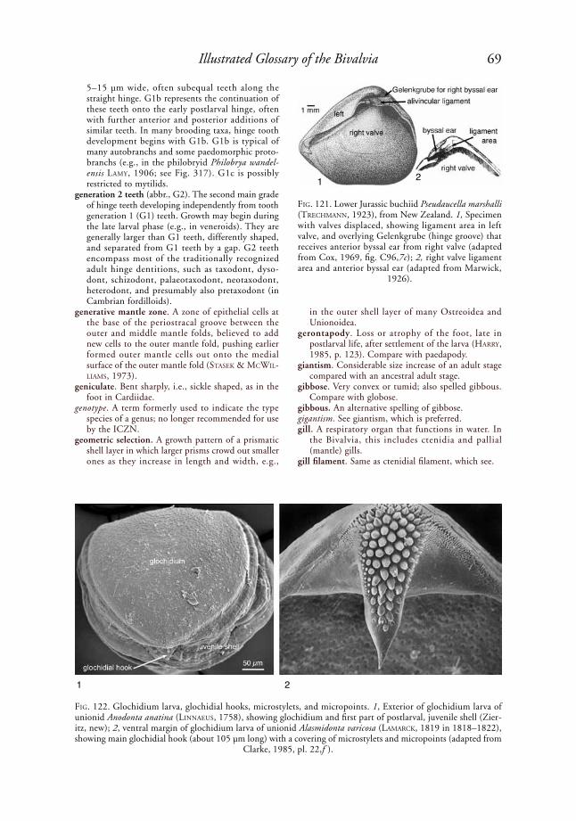
69Illustrated Glossary of the Bivalvia
5–15 µm wide, often subequal teeth along the straight hinge. G1b represents the continuation of these teeth onto the early postlarval hinge, often with further anterior and posterior additions of similar teeth. In many brooding taxa, hinge tooth development begins with G1b. G1b is typical of many autobranchs and some paedomorphic proto-branchs (e.g., in the philobryid Philobrya wandel-ensis laMy, 1906; see Fig. 317). G1c is possibly restricted to mytilids.
generation 2 teeth (abbr., G2). The second main grade of hinge teeth developing independently from tooth generation 1 (G1) teeth. Growth may begin during the late larval phase (e.g., in veneroids). They are generally larger than G1 teeth, differently shaped, and separated from G1 teeth by a gap. G2 teeth encompass most of the traditionally recognized adult hinge dentitions, such as taxodont, dyso-dont, schizodont, palaeotaxodont, neotaxodont, heterodont, and presumably also pretaxodont (in Cambrian fordilloids).
generative mantle zone. A zone of epithelial cells at the base of the periostracal groove between the outer and middle mantle folds, believed to add new cells to the outer mantle fold, pushing earlier formed outer mantle cells out onto the medial surface of the outer mantle fold (stasek & MCWil-liaMs, 1973).
geniculate. Bent sharply, i.e., sickle shaped, as in the foot in Cardiidae.
genotype. A term formerly used to indicate the type species of a genus; no longer recommended for use by the ICZN.
geometric selection. A growth pattern of a prismatic shell layer in which larger prisms crowd out smaller ones as they increase in length and width, e.g.,
in the outer shell layer of many Ostreoidea and Unionoidea.
gerontapody. Loss or atrophy of the foot, late in postlarval life, after settlement of the larva (harry, 1985, p. 123). Compare with paedapody.
giantism. Considerable size increase of an adult stage compared with an ancestral adult stage.
gibbose. Very convex or tumid; also spelled gibbous. Compare with globose.
gibbous. An alternative spelling of gibbose.gigantism. See giantism, which is preferred.gill. A respiratory organ that functions in water. In
the Bivalvia, this includes ctenidia and pallial (mantle) gills.
gill filament. Same as ctenidial filament, which see.
FiG. 121. Lower Jurassic buchiid Pseudaucella marshalli (treChMaNN, 1923), from New Zealand. 1, Specimen with valves displaced, showing ligament area in left valve, and overlying Gelenkgrube (hinge groove) that receives anterior byssal ear from right valve (adapted from Cox, 1969, fig. C96,7c); 2, right valve ligament area and anterior byssal ear (adapted from Marwick,
1926).
1 2
FiG. 122. Glochidium larva, glochidial hooks, microstylets, and micropoints. 1, Exterior of glochidium larva of unionid Anodonta anatina (liNNaeus, 1758), showing glochidium and first part of postlarval, juvenile shell (Zier-itz, new); 2, ventral margin of glochidium larva of unionid Alasmidonta varicosa (laMarCk, 1819 in 1818–1822), showing main glochidial hook (about 105 µm long) with a covering of microstylets and micropoints (adapted from
Clarke, 1985, pl. 22,f ).
1 2
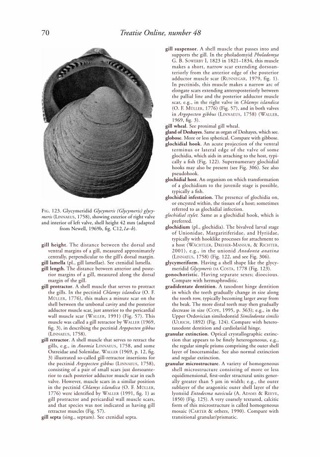
70 Treatise Online, number 48
gill height. The distance between the dorsal and ventral margins of a gill, measured approximately centrally, perpendicular to the gill’s dorsal margin.
gill lamella (pl., gill lamellae). See ctenidial lamella. gill length. The distance between anterior and poste-
rior margins of a gill, measured along the dorsal margin of the gill.
gill protractor. A shell muscle that serves to protract the gills. In the pectinid Chlamys islandica (o. F. Müller, 1776), this makes a minute scar on the shell between the umbonal cavity and the posterior adductor muscle scar, just anterior to the pericardial wall muscle scar (Waller, 1991) (Fig. 57). This muscle was called a gill retractor by Waller (1969, fig. 3), in describing the pectinid Argopecten gibbus (liNNaeus, 1758).
gill retractor. A shell muscle that serves to retract the gills, e.g., in Anomia liNNaeus, 1758, and some Ostreidae and Solenidae. Waller (1969, p. 12, fig. 3) illustrated so-called gill-retractor insertions for the pectinid Argopecten gibbus (liNNaeus, 1758), consisting of a pair of small scars just dorsoante-rior to each posterior adductor muscle scar in each valve. However, muscle scars in a similar position in the pectinid Chlamys islandica (o. F. Müller, 1776) were identified by Waller (1991, fig. 1) as gill protractor and pericardial wall muscle scars, and that species was not indicated as having gill retractor muscles (Fig. 57).
gill septa (sing., septum). See ctenidial septa.
gill suspensor. A shell muscle that passes into and supports the gill. In the pholadomyid Pholadomya G. B. soWerBy i, 1823 in 1821–1834, this muscle makes a short, narrow scar extending dorsoan-teriorly from the anterior edge of the posterior adductor muscle scar (ruNNeGar, 1979, fig. 1). In pectinids, this muscle makes a narrow arc of elongate scars extending anteroposteriorly between the pallial line and the posterior adductor muscle scar, e.g., in the right valve in Chlamys islandica (o. F. Müller, 1776) (Fig. 57), and in both valves in Argopecten gibbus (liNNaeus, 1758) (Waller, 1969, fig. 3).
gill wheal. See proximal gill wheal.gland of Deshayes. Same as organ of Deshayes, which see.globose. More or less spherical. Compare with gibbose.glochidial hook. An acute projection of the ventral
terminus or lateral edge of the valve of some glochidia, which aids in attaching to the host, typi-cally a fish (Fig. 122). Supernumerary glochidial hooks may also be present (see Fig. 306). See also pseudohook.
glochidial host. An organism on which transformation of a glochidium to the juvenile stage is possible, typically a fish.
glochidial infestation. The presence of glochidia on, or encysted within, the tissues of a host; sometimes referred to as glochidial infection.
glochidial stylet. Same as a glochidial hook, which is preferred.
glochidium (pl., glochidia). The bivalved larval stage of Unionidae, Margaritiferidae, and Hyriidae, typically with hooklike processes for attachment to a host (WäChtler, dreher-MaNsur, & riChter, 2001), e.g., in the unionid Anodonta anatina (liNNaeus, 1758) (Fig. 122, and see Fig. 306).
glycymeriform. Having a shell shape like the glycy-meridid Glycymeris da Costa, 1778 (Fig. 123).
gonochoristic. Having separate sexes; dioecious. Compare with hermaphroditic.
gradidentate dentition. A taxodont hinge dentition in which the teeth gradually change in size along the tooth row, typically becoming larger away from the beak. The more distal teeth may then gradually decrease in size (Cope, 1995, p. 363); e.g., in the Upper Ordovician similodontid Similodonta similis (ulriCh, 1892) (Fig. 124). Compare with hetero-taxodont dentition and cardiolariid hinge.
granular extinction. Optical crystallographic extinc-tion that appears to be finely heterogeneous, e.g., the regular simple prisms comprising the outer shell layer of Inoceramidae. See also normal extinction and regular extinction.
granular microstructure. A variety of homogeneous shell microstructure consisting of more or less equidimensional, first-order structural units gener-ally greater than 5 µm in width; e.g., the outer sublayer of the aragonitic outer shell layer of the lyonsiid Entodesma navicula (A. adaMs & reeVe, 1850) (Fig. 125). A very coarsely textured, calcitic form of this microstructure is called homogeneous mosaic (Carter & others, 1990). Compare with transitional granular/prismatic.
FiG. 123. Glycymeridid Glycymeris (Glycymeris) glycy-meris (liNNaeus, 1758), showing exterior of right valve and interior of left valve, shell height 42 mm (adapted
from Newell, 1969b, fig. C12,1a–b).

71Illustrated Glossary of the Bivalvia
granular prismatic structure. An inner sublayer of slightly irregular simple prismatic microstruc-ture deposited below and intergrading with an outer sublayer of granular microstructure, e.g., in the aragonitic outer shell layer of the lyon-siid Entodesma navicula (A. adaMs & reeVe, 1850) (harper, CheCa, & rodriGueZ-NaVarro, 2009). This application of granular prismatic is misleading, because the term granular refers to a different sublayer than the prismatic microstruc-ture. Compare with granular/prismatic shell layer and transitional granular/prismatic microstructure.
granular/prismatic shell layer. A shell layer consisting of granular microstructure passing inwardly into prismatic microstructure, e.g., the outer shell layer of the lyonsiid Entodesma navicula (A. adaMs & reeVe, 1850). Called granular pris-matic structure by harper, CheCa, and rodri-GueZ-NaVarro (2009). Compare with transitional granular/prismatic microstructure.
granulated. Same as granulose.granulose. Grainy, or covered by granules, as on the
exterior of the thraciid Thracia morrisoni petit, 1964 (Fig. 126).
gregarious. Living together or growing in clusters, as in the case of Mytilus edulis liNNaeus, 1758.
groove. (1) A narrow, elongate element of incised sculpture; wider than a striation; (2) a narrow, elongate, recessed area on a ligament insertion area, as in a duplivincular or parivincular ligament, generally coinciding with insertion of the lamellar sublayer of the ligament.
growth check. A commarginal line on the exterior of a shell, reflecting periodic slowing or cessation of growth.
growth line. A commarginal, generally fine line on the exterior of a shell, which marks the position of a former shell margin. Compare with internal growth line and growth check.
growth ruga. An irregular wrinkle on the exterior of a shell, generally corresponding with a more pronounced hiatus in growth than a growth line.
growth stria (pl., growth striae). Same as a growth line, which see.
growth thread. (1) A threadlike, commarginal eleva-tion on the exterior of a shell, corresponding with a fine growth line; (2) a radial or antimarginal, continuous or discontinuous, threadlike eleva-tion on the exterior of a shell, sometimes forming nodules where it intersects a growth line.
gryphaeate. Same as gryphaeiform, which see. gryphaeiform. Having a shell shape like Gryphaea
laMarCk, 1801, i.e., with the left valve orthogyrate, strongly convex, and dorsally incurved, and the right valve flat to concave (Fig. 127).
gryphaeoid ligament. An alivincular ligament with a relatively high, straight or nearly straight, somewhat trapezoidal insertion area (MalChus, 1990, p. 77), as in the Lower Jurassic gryphaeid Gryphaea (Gryphaea) arcuata laMarCk, 1801, and the Upper Cretaceous gryphaeid Gryphaea (Africogryphaea) costellata (douVillé, 1916) (Fig. 128). Compare with gyrostreoid ligament.
gutter. A commarginal, dorsally facing groove for an external, simple ligament (Carter, 1990a, p. 138).
gyrate. Having a spirally curved umbo.gyrostreoid ligament. An alivincular ligament that
expands in width early in ontogeny to nearly the maximum adult anterior-posterior ligament width, giving the ligament insertion area a somewhat
FiG. 124. Gradidentate taxodont hinge teeth in right valve of similodontid Similodonta similis (ulriCh, 1892), upper Upper Ordovician, Maquoketa Shale, Bristol, Minnesota (adapted from Pojeta, 1978, pl. 2,8,
courtesy of the Royal Society of London).
FiG. 125. Aragonitic granular-type homogeneous mi-crostructure in outer part of outer shell layer of lyonsiid Entodesma navicula (A. adaMs & reeVe, 1850), Hok-kaido, Japan; SEM of a fracture (adapted from Harper, Checa, & Rodriguez-Navarro, 2009, fig. 6, courtesy of
John Wiley & Sons).
10 µm

72 Treatise Online, number 48
trapezoidal shape (MalChus, 1990), e.g., the Cretaceous–?lower Tertiary crassostreid Gyrostrea MirkaMaloV, 1963 (Fig. 129). Compare with gryphaeoid ligament.
gyrous. Same as gyrate, which is preferred.haemocoel. A body cavity other than a true coelom,
formed by enlargement of the venous blood spaces. Also spelled hemocoel. Compare with lacuna.
hatchet shaped. Resembling an axe head, i.e., with a broad, laterally compressed cutting end and a blunt hammer end, e.g., shells of the family Sareptidae, or the foot of some bivalves.
haustorium (pl., haustoria). (1) The parasitic larval stage of the Iridinidae, consisting of an elongate body with a very long larval thread (Fryer, 1959,
1961); (2) the second stage of development of the lasidium in the Iridinidae (WäChtler, dreher-MaNsur, & riChter, 2001).
heart. The muscular pumping organ surrounded by a large pericardium and lying at the dorsal midline, between the visceral mass and the posterior adductor muscle, e.g., in the venerid Mercenaria mercenaria (liNNaeus, 1758) (Fig. 130) and the gastrochaenid Spengleria sp. (see Fig. 176). In bivalves, the heart pumps hemolymph through the anterior and poste-rior aortae into the hemocoelic sinuses.
height of shell. The distance between two mutu-ally parallel planes oriented perpendicular to the commissural plane and parallel with the hinge axis (=cardinal axis), which just touch the most dorsal and most ventral parts of the shell. For inequi-valve shells, another plane of symmetry, or plane of near-symmetry, parallel with the commissural plane, may be used for reference instead of the commissural plane.
helical microstructure . A shell microstructure consisting of nested, spirally coiled, aragonitic rods or rod segments, with the helix axes all oriented perpendicular to the depositional surface. This is rarely seen in bivalves, e.g., in the inner shell layer of the pisidiid Pisidium dubium (say, 1817) (Fig. 131) but is common in pteropod gastropods.
hemibranch. Of or pertaining to a ctenidium in which ctenidial filaments are present only on one side.
hemocoel. Same as haemocoel, which see.hepatopancreas. A digestive gland, which see. hermaphrodite. An individual capable of producing
eggs and sperm. See also female hermaphrodite, male hermaphrodite, sequential hermaphrodite, and simultaneous hermaphrodite.
herringbone cross foliated. A term used by MalChus (1990) for herringbone regularly foliated micro-structure, which see.
FiG. 126. Granulose sculpture from periostracal min-eralization in thraciid Thracia morrisoni petit, 1964, figure width several mm (adapted from Mikkelsen &
Bieler, 2007).
FiG. 127. Liassic, Lower Jurassic gryphaeid Gryphaea (Gryphaea) arcuata laMarCk, 1801, England, illustrating gryphaeiform shell shape, maximum figure dimension 46 mm (adapted from Stenzel, 1971, fig. J74,3e).
initialattachment
area
xenomorphic sculpture
right valve radial posterior sulcus
left valve

73Illustrated Glossary of the Bivalvia
herringbone regularly foliated microstructure (abbr., herringbone RF). A calcitic, regularly foliated micro-structure consisting of two or more sublayers with alternating lath or blade dip directions, thereby forming a herringbone-like pattern in radial, vertical sections, e.g., in the Liassic, Lower Jurassic pectinoid Pseudopecten aequivalvis J. soWerBy, 1816, in soWerBy & soWerBy, 1812–1846 (Fig. 132). This differs from crossed foliated structure in that the dip direction of the calcitic laths or blades alternates in successive sublayers rather than over small areas of the same depositional surface. This structure was called zigzag lamellar structure by BøGGild (1930, p. 266–267), and herringbone cross foliated structure by MalChus (1990, p. 65, pl. 26,8).
heterochrony. Evolutionary change in the relative time of appearance and/or rate of development of characters.
heteroclite. Having a folded or twisted commissural plane, as in the oyster Flemingostrea VredeNBurG, 1916.
heterodont dentition. A hinge dentition characterized by relatively few teeth, which are differentiated into cardinals and laterals, typically 2–3 cardinals, 1–2 anterior laterals, and 1–2 posterior laterals, although the laterals may be absent anteriorly or anteriorly and posteriorly. Typical of many Hetero-conchia, e.g., the astartid Astarte smithii dall, 1886 (Fig. 68). Similar hinges in non-Heteroconchia are called pseudoheterodont. This is the basis for the group name Heterodonten [sic] NeuMayr, 1884 (=Heterodonta of later authors), originally containing what are now classified as Unionidae, Cardiniidae, Astartidae, Crassatellidae, Mega-lodontidae, Hippuritoidea, Chamidae, Tridac-ninae, Erycinidae, Cardiidae, Lucinidae, Cyrenidae, Arcticidae, Veneridae, Tellinidae, Mactridae, and Donacidae. A tooth numbering system for heter-odonts, proposed by Félix BerNard (1895, 1896a, 1896b, 1897) (Fig. 36), labels hinge teeth according
to their position and appearance during ontogeny, as follows: right anterior cardinal tooth = 3a; right middle cardinal tooth = 1; right posterior cardinal tooth = 3b; left anterior cardinal tooth = 2a; left middle cardinal tooth = 2b; left posterior cardinal tooth = 4b; anterior lateral teeth = left AII fitting between right AI and AIII; posterior lateral teeth = left PII fitting between right PI and PIII. Unfortunately, the ontogenetic origin of teeth remains unknown for most bivalves, so determining homologous tooth numbers can be problematic for distantly related taxa. The teeth develop from lamellae on the hinge of postlarval shells. There are usually two anterior lamellae in the right valve and one anterior lamella in the left valve fitting between them. Another lamella may be developed as a marginal rim on the left valve. The anterior lamellae give rise to cardinals and anterior laterals. The posterior lamellae may give rise to posterior laterals. The postlarval lamellae on the right valve carry odd Roman numerals (I, III, V ), and those on the left valve carry even Roman numerals (II, IV, VI ). Cardinals developing from the postlarval
FiG. 128. 1, Alivincular gryphaeoid ligament in left valve of Upper Cretaceous gryphaeid Gryphaea (Africogryphaea) costellata (douVillé, 1916) (adapted from Douvillé, 1916), figure height 27 mm; 2, in right valve of Lower Jurassic
gryphaeid Gryphaea (Gryphaea) arcuata laMarCk, 1801 (adapted from Stenzel, 1971, fig. J74,3d).
FiG. 129. Diagram of alivincular gyrostreoid ligament in Gyrostrea MirkaMaloV, 1963 (adapted from Malchus,
1990, fig. 25b).
1 cm
bourreletresilifer
left valve right
valve
1
2
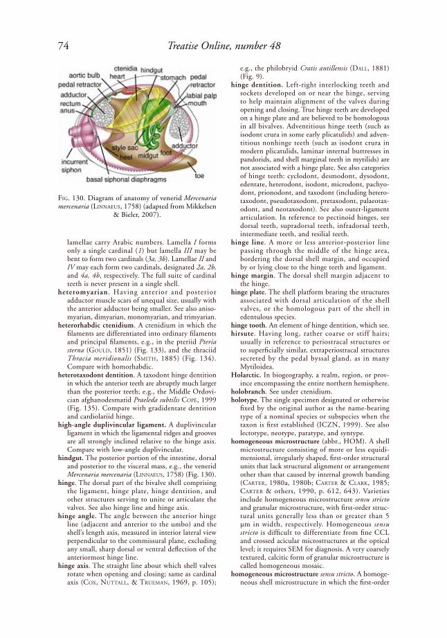
74 Treatise Online, number 48
lamellae carry Arabic numbers. Lamella I forms only a single cardinal (1) but lamella III may be bent to form two cardinals (3a, 3b). Lamellae II and IV may each form two cardinals, designated 2a, 2b, and 4a, 4b, respectively. The full suite of cardinal teeth is never present in a single shell.
heteromyarian . Having anterior and posterior adductor muscle scars of unequal size, usually with the anterior adductor being smaller. See also aniso-myarian, dimyarian, monomyarian, and trimyarian.
heterorhabdic ctenidium. A ctenidium in which the filaments are differentiated into ordinary filaments and principal filaments, e.g., in the pteriid Pteria sterna (Gould, 1851) (Fig. 133), and the thraciid Thracia meridionalis (sMith, 1885) (Fig. 134). Compare with homorhabdic.
heterotaxodont dentition. A taxodont hinge dentition in which the anterior teeth are abruptly much larger than the posterior teeth; e.g., the Middle Ordovi-cian afghanodesmatid Praeleda subtilis Cope, 1999 (Fig. 135). Compare with gradidentate dentition and cardiolariid hinge.
high-angle duplivincular ligament. A duplivincular ligament in which the ligamental ridges and grooves are all strongly inclined relative to the hinge axis. Compare with low-angle duplivincular.
hindgut. The posterior portion of the intestine, dorsal and posterior to the visceral mass, e.g., the venerid Mercenaria mercenaria (liNNaeus, 1758) (Fig. 130).
hinge. The dorsal part of the bivalve shell comprising the ligament, hinge plate, hinge dentition, and other structures serving to unite or articulate the valves. See also hinge line and hinge axis.
hinge angle. The angle between the anterior hinge line (adjacent and anterior to the umbo) and the shell’s length axis, measured in interior lateral view perpendicular to the commissural plane, excluding any small, sharp dorsal or ventral deflection of the anteriormost hinge line.
hinge axis. The straight line about which shell valves rotate when opening and closing; same as cardinal axis (Cox, Nuttall, & trueMaN, 1969, p. 105);
e.g., the philobryid Cratis antillensis (dall, 1881) (Fig. 9).
hinge dentition. Left-right interlocking teeth and sockets developed on or near the hinge, serving to help maintain alignment of the valves during opening and closing. True hinge teeth are developed on a hinge plate and are believed to be homologous in all bivalves. Adventitious hinge teeth (such as isodont crura in some early plicatulids) and adven-titious nonhinge teeth (such as isodont crura in modern plicatulids, laminar internal buttresses in pandorids, and shell marginal teeth in mytilids) are not associated with a hinge plate. See also categories of hinge teeth: cyclodont, desmodont, dysodont, edentate, heterodont, isodont, microdont, pachyo-dont, prionodont, and taxodont (including hetero-taxodont, pseudotaxodont, pretaxodont, palaeotax-odont, and neotaxodont). See also outer-ligament articulation. In reference to pectinoid hinges, see dorsal teeth, supradorsal teeth, infradorsal teeth, intermediate teeth, and resilial teeth.
hinge line. A more or less anterior-posterior line passing through the middle of the hinge area, bordering the dorsal shell margin, and occupied by or lying close to the hinge teeth and ligament.
hinge margin. The dorsal shell margin adjacent to the hinge.
hinge plate. The shell platform bearing the structures associated with dorsal articulation of the shell valves, or the homologous part of the shell in edentulous species.
hinge tooth. An element of hinge dentition, which see.hirsute. Having long, rather coarse or stiff hairs;
usually in reference to periostracal structures or to superficially similar, extraperiostracal structures secreted by the pedal byssal gland, as in many Mytiloidea.
Holarctic. In biogeography, a realm, region, or prov-ince encompassing the entire northern hemisphere.
holobranch. See under ctenidium.holotype. The single specimen designated or otherwise
fixed by the original author as the name-bearing type of a nominal species or subspecies when the taxon is first established (ICZN, 1999). See also lectotype, neotype, paratype, and syntype.
homogeneous microstructure (abbr., HOM). A shell microstructure consisting of more or less equidi-mensional, irregularly shaped, first-order structural units that lack structural alignment or arrangement other than that caused by internal growth banding (Carter, 1980a, 1980b; Carter & Clark, 1985; Carter & others, 1990, p. 612, 643). Varieties include homogeneous microstructure sensu stricto and granular microstructure, with first-order struc-tural units generally less than or greater than 5 µm in width, respectively. Homogeneous sensu stricto is difficult to differentiate from fine CCL and crossed acicular microstructures at the optical level; it requires SEM for diagnosis. A very coarsely textured, calcitic form of granular microstructure is called homogeneous mosaic.
homogeneous microstructure sensu stricto. A homoge-neous shell microstructure in which the first-order
FiG. 130. Diagram of anatomy of venerid Mercenaria mercenaria (liNNaeus, 1758) (adapted from Mikkelsen
& Bieler, 2007).

75Illustrated Glossary of the Bivalvia
structural units are generally less than 5 µm in width, e.g., locally developed in the middle shell layer of the hiatellid Cyrtodaria siliqua (speNGler, 1793) (Fig. 137). Compare with transitional irreg-ular simple prismatic/homogeneous.
homogeneous mosaic microstructure. A very coarsely textured, calcitic form of granular homogeneous shell microstructure consisting of more or less equidimensional first-order structural units gener-ally greater than 5 µm in width. This is found in the outer shell layer of the left valve of the Upper Triassic gryphaeid Gryphaea (Gryphaea) nevadensis MCroBerts, 1992 (Fig. 136).
homogeneous simple prismatic microstructure. A simple prismatic shell microstructure in which each first-order prism has a homogeneous second-order structure; e.g., the calcitic regular simple prismatic outer shell layer in many Pteriidae, Pinnidae, Inoceramidae, and some Anomiidae (Fig. 138). Compare with fibrous simple prismatic, semi-
foliated simple prismatic, and transitional irregular simple prismatic/homogeneous microstructure.
homologous. Structures with the same evolutionary origin, which now fulfill the same or different functions.
homomyarian. Having anterior and posterior adductor muscle scars of equal or nearly equal size; same as isomyarian. Size, in this case, is generally judged by the area of each muscle scar, rather than by its maximum dimension.
homonym. (1) In the family-group of names (which see), two or more available names having the same spelling, or differing only in suffix, which denote different taxa; (2) in the genus-group of names, two or more available genera or subgenera having the same spelling but denoting different taxa; (3) in the species-group of names, two or more avail-able species or subspecies names having the same spelling, or having spellings deemed under Article 58 of the ICZN (1999) Code to be the same,
FiG. 131. Sphaeriid Pisidium dubium (say, 1817), Burton’s Landing, Au Sable River, Section 10, Au Sable Town-ship, T26N, R3W, Crawford County, Michigan (UNC 15044), showing helical structure in inner shell layer. 1–2, Horizontal and vertical acetate peels, respectively, through helical structure; scale bar = 50 µm, applies to both figures; 3, SEM of vertical fracture through helical structure, showing nested helices; scale bar = 10 µm (Carter, new).
1 2
3

76 Treatise Online, number 48
which denote different taxa, and which are either originally (primary homonymy) or subsequently (secondary homonymy) placed within the same genus or subgenus (ICZN, 1999). The later estab-lished homonym is rejected as a junior homonym, except under circumstances outlined by the ICZN Code. Some spelling differences involving vowels or dipthongs, which result in the same sound, are still considered to be homonyms, but in most cases, a single letter difference makes the names nonhomonyms. See also primary homonym and secondary homonym.
homorhabdic ctenidium. A ctenidium in which all the filaments are alike, i.e., they are all ordinary fila-ments (which see), e.g., the pterioidean Isognomon alatus (GMeliN, 1791 in 1791–1793) (Fig. 139). Compare with heterorhabdic ctenidium.
hood. See dorsal hood.horizontal plane. (1) In soft anatomy, a plane perpen-
dicular to the midsagittal plane and including the mouth-anus axis, separating the body into
dorsal (with mantle isthmus) and ventral (without mantle isthmus) portions; (2) for shells, a plane of symmetry perpendicular to the midsagittal plane and including the hinge axis, separating the shell into dorsal and ventral portions; (3) for shell micro-structure, parallel to the plane of a shell layer, as in a horizontal section.
hyaline. Transparent or nearly transparent. hyaline organs. Mantle structures equipped with
lenses that transmit light to symbiotic zooxanthellae within the tissue of the host, e.g., in Tridacninae.
hyote. Tubular or nearly tubular, such as spines with U-shaped cross sections, open on the underside and on their tips; e.g., the gryphaeid Hyotissa hyotis (liNNaeus, 1758).
hypermorphosis. A type of peramorphosis caused by delay of onset of maturation and resultant extension of the juvenile growth period (de Beer, 1930, 1958). The early juvenile development will commence at the same time and proceed at the same rate as in the ancestor, so the adult will attain
FiG. 132. Herringbone regularly foliated microstruc-ture, seen in radial, vertical section of Liassic, Lower Ju-rassic “Pecten aequivalvis” (=Pseudopecten aequivalvis J. soWerBy, 1816, in soWerBy & soWerBy, 1812–1846), figure is a few mm wide (adapted from Bøggild, 1930,
pl. 1,6 ).
FiG. 133. Heterorhabdic ctenidium in pteriid Pteria sterna (a. Gould, 1851), as seen in transverse histological section through a single demibranch (7 µm thick, stained with Alcian Blue/Periodic Acid/Schiff ’s trichrome stain) (adapted from Tëmkin, 2006a, fig. 13C, courtesy of
John Wiley & Sons).
FiG. 134. Transverse section through several filaments in heterorhabdic inner demibranch of thraciid Thracia meridionalis (sMith, 1885), Admiralty Bay, King George Island, Antarctica (O. Domaneschi, collector), showing principal and ordinary filaments, figure width 326 µm
(Sartori, new).
FiG. 135. Heterotaxodont dentition in Llanvirn, upper Middle Ordovician afghanodesmatid Praeleda subtilis Cope, 1999, Wales (paratype NMW 95.44G); latex cast of internal mold of left valve (adapted from Cope,
1999, pl. 1,3).

77Illustrated Glossary of the Bivalvia
a larger maximum size (MCNaMara, 1986). The opposite of progenesis.
hypertrophied. Enlarged, commonly meaning abnor-mally enlarged.
hypobranchial glands. Dorsoposterior glands positioned on the inner wall of the mantle in the suprabran-chial chamber, primitively serving to consolidate, in a mucous stream for expulsion via the incurrent aperture, waste material or particles not sieved out of the water current by the ctenidium. Present in many but not all Protobranchia, e.g., in Nuculida, in some but not all Solemyida, and in some but not all Nuculanida, as mucous cells flanked by inversely conical and regenerative cells (Fig. 240). Only rarely present in Autobranchia, e.g., in some Philobryoidea, Anomioidea, Lucinoidea, and Cyrenoidea. This gland secretes a brood pouch attached to the shell in Nuculoma CossMaNN, 1907 (Nuculidae); it provides
nutrition for developing larvae incubated in the suprabranchial chamber in Corbicula MeGerle VoN MühlFeld, 1811 (Cyrenidae), and possibly also in Fimbria MeGerle VoN MühlFeld, 1811 (Lucinidae) (dreW, 1901; MortoN, 1977).
hypolimnion. The cold-water layer below the thermo-cline in a thermally stratified body of water.
hypoplax. A generally long, narrow, accessory shell plate covering the space between valves, and joined to the ventral margins of the valves by periostracum, e.g., in the pholadid Martesia striata (liNNaeus, 1758) (Fig. 24, Fig. 48).
hypostracum. A term used by thiele (1893) for the innermost shell layer in chitons and in some bivalves (pteriids and arcids), including, but not restricted to, any associated myostracum, but also used by thiele (1893) for only the adductor myo stracum in unionids. Subsequent workers (JaMesoN, 1912;
FiG. 136. Homogeneous mosaic microstructure in calcitic outer shell layer of left valve of Norian, Upper Triassic gryphaeid Gryphaea (Gryphaea) nevadensis MCroBerts, 1992, lower member of Luning Formation, Pilot Mountain, west-central Nevada (UNC 15004), as seen in 1, radial acetate peel and 2, by SEM of a polished then acid-etched
surface; scale bars = 10 µm (Carter, new).
1
2

78 Treatise Online, number 48
Gutsell, 1930; NeWell, 1938, 1942; luCas, 1952; steNZel, 1971, p. 979) restricted this term to myostracal deposits, thereby making it synonymous with myostracum, which is preferred.
ICZN. The International Commission on Zoological Nomenclature.
idiomorphic. Having a normal form or shape, i.e., not deformed by crowding or attachment to an object. Also called automorphic.
igloo. A cupulated, calcareous tube, such as that secreted by the gastrochaenid Cucurbitula cymbium (speNGler, 1783) (Fig. 46). Compare with crypt.
imbricate (or imbricated). Closely packed and over-lapping, like roof tiles, e.g., the regular simple prismatic outer shell layer developed into imbricate scales on the exterior of the pteriid Pinctada imbri-cata rödiNG, 1798 (Fig. 140).
incertae sedis. Latin for uncertain seat. In taxonomy, referr ing to uncertain taxonomic posit ion. Commonly italicized.
incised. Having a deeply notched edge.inclined. Dipping toward the beak, i.e., in terms of
shell ornament and shell microstructure, having
the innermost point closer to the beak than the outermost point. Opposite of reclined.
incrustation. A coating of sediment particles, secreted calcium carbonate, and/or precipitated metal oxides (commonly iron or manganese) on the exterior of the shell, exterior to any periostracum that may still be intact; e.g., in many Lithophaginae, where the incrustation may be smooth or sculptured. Also spelled encrustation, which is not preferred.
incubatory. Retaining eggs, larvae, and sometimes juveniles in the mantle cavity or in a brood pouch.
incurrent aperture length. The distance from the ventral terminus of the incurrent aperture to its dorsal terminus, measured in a straight line.
incurrent aperture or siphon. A generally posterior, rarely anterior, mantle opening that controls water intake into the mantle cavity. Also called a bran-chial, pallial, or inhalant aperture or siphon.
incurrent tube . A tube wi th in the sed iment surrounding and linking the incurrent siphon with the sediment-water interface, e.g., the thraciid Thracia meridionalis (sMith, 1885) (Fig. 141)
incurved. Curved inward, i.e., toward the base or apex.indication. In taxonomy, a reference to previously
published information or a published act. In the absence of a definition or a description, this allows a name, proposed before 1931 and otherwise satis-fying the relevant provisions of Articles 10 and 11 of the ICZN Code, to be regarded as available (ICZN, 1999).
indirect development. A biphasic mode of develop-ment that includes a larval phase and metamor-phosis; probably present in all bivalves. See also direct development, larviparous, oviparous, ovovi-viparous, and viviparous.
inductura. Calcareous shell material deposited exterior to the periostracum by an outward reflection of the outer mantle fold, e.g., inductural spines on the exterior of the cardiid Ctenocardia imbricata (G. B. soWerBy ii, 1840, in G. B. soWerBy I, 1832–1841) (Fig. 142), and smooth, flat inductura on the poste-rior of the Upper Cretaceous cardiid Protocardia (Pachycardium) stantoni (Wade, 1926) (sChNeider & Carter, 2001, fig. 5.2). Inductural deposits are common in some gastropod groups, such as cypraeids, but are rarely found in the Bivalvia.
inequiaxial. A shell in which the breviaxis and longiaxis have significantly different lengths (Bailey, 2009). Synonymous with inequilateral, which is preferred.
inequiconvex. Having one shell valve less convex than the other.
inequilateral. Having the umbos appreciably closer to one end of the shell than the other, as in the mytilid Modiolus squamosus Beauperthuy, 1967 (see Fig. 179).
inequivalve. Having left and right valves of dissimilar size, shape, or sculpture. This generally refers to having one valve larger or more convex than the other.
infaunal. Living with the shell buried or enclosed in sediment. Compare with semi-infaunal, epifaunal, and endolithic.
inflated. Strongly convex, swollen, or expanded. Compare with gibbose.
FiG. 137. Homogeneous microstructure sensu stricto in a radial fracture through middle shell layer of hiatellid Cyrtodaria siliqua (speNGler, 1793) (adapted from
Carter & Lutz, 1990, pl. 15B).
FiG. 138. Homogeneous simple prismatic micro-structure in calcitic outer shell layer of right valve of Recent anomiid Anomia simplex d’orBiGNy, 1853 in 1841–1853, New Haven, Connecticut (YPM 9715).
SEM of radial, vertical fracture (Carter, new).
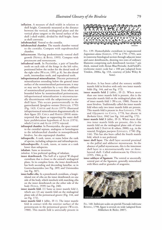
79Illustrated Glossary of the Bivalvia
inflation. A measure of shell width in relation to shell height. Commonly measured as the distance between the vertical, midsagittal plane and the vertical plane tangent to the lateral surface of the shell (=shell width), divided by shell height. Same as shell convexity.
infrabranchial. Ventral to the ctenidia.infrabranchial chamber. The mantle chamber ventral
to the ctenidia. Compare with suprabranchial chamber.
infracrescent. Having predominantly ventral shell growth (NeWell & Boyd, 1995). Compare with procrescent and retrocrescent.
infradorsal teeth. In Pectinidae, a pair of lamellar teeth on each side of the hinge in the left valve, positioned below the socket for the dorsal teeth of the right valve (Waller, 1991, p. 8). See also dorsal teeth, intermediate teeth, and supradorsal teeth.
infraperiostracal mineralization. Discrete periostracal mineralization extending below the general inner surface of the nonmineralized periostracum; it may or may not be underlain by a very thin sublayer of nonmineralized periostracum. Even where not bounded below by nonmineralized periostracum, infraperiostracal mineralization is microstructur-ally discontinuous with the subperiostracal outer shell layer. This occurs posteroventrally in the gastrochaenid Spengleria rostrata (speNGler, 1793) (Fig. 143). Carter and aller (1975) illustrated infraperiostracal mineralization diagrammatically in their figure 2D. CheCa and harper (2010) misin-terpreted that figure as supporting the outer shell layer prefabrication hypothesis of aller (1974), which Carter and aller (1975) rejected.
infraseptal chamber. In Poromyoidea, the space ventral to the ctenidial septum, analogous or homologous to the infrabranchial chamber in nonseptibranch bivalves. See also supraseptal chamber.
infraspecific. A rank, taxon, or name below the rank of species, including subspecies and infrasubspecies.
infrasubspecific. A rank, taxon, or name at a rank lower than subspecies.
inhalant. Same as incurrent.inhalent. A less preferred spelling of inhalant.inner demibranch. The half of a typical W-shaped
ctenidium that is closer to the animal’s midsagittal plane. In its complete form, the inner demibranch has both ascending and decending lamellae, as in Gastrochaenoidea (see Fig. 289) and Unionoidea (see Fig. 307).
inner leaflet cilia. In a protobranch ctenidium, a longit-idunal row of cilia on the inner demibranch on one side of the body, which interlock with matching cilia on the inner demibranch on the other side of the body (yoNGe, 1939) (see Fig. 240).
inner mantle fold. (1) Same as inner mantle fold 2, which see; (2) any mantle fold on the midsagittal plane side of the periostracal groove (Fig. 144, and see Fig. 172).
inner mantle fold 1 (abbr., IF-1). The inner mantle fold in contact with the exterior surface of the periostracum in the periostracal groove (Waller, 1980). This mantle fold is universally present in
bivalves. It has been called the sensory middle mantle fold in bivalves with only two inner mantle folds (Fig. 144, and see Fig. 172).
inner mantle fold 2 (abbr., IF-2). When more than one inner mantle fold is present, this is the muscular mantle fold on the midsagittal plane side of inner mantle fold 1 (Waller, 1980). Present in most bivalves. Traditionally called the inner mantle fold when only two inner mantle folds are present. This mantle fold is present in Arca liNNaeus, 1758, and Glycymeris da Costa, 1778, but absent in Barbatia Gray, 1842 (see Fig. 144 and Fig. 172).
inner mantle fold 3 (abbr., IF-3). When more than two inner mantle folds are present, this is the mantle fold on the midsagittal plane side of inner mantle fold 2 (sartori & others, 2008); e.g., in the venerid Periglypta puerpera (liNNaeus, 1758) (Fig. 144). This has also been called the fourth mantle fold, which is not preferred.
inner shell layer. The shell layer secreted proximal to the pallial and adductor myostracum. In the absence of pallial myostracum, this is the innermost shell layer in a microstructurally two- or three-layered shell. Called endostracum by oBerliNG (1955, 1964, p. 6).
inner sublayer of ligament. The ventral or ancestrally ventral part of the ligament, generally mineralized with fibers and/or granules of aragonite.
FiG. 139. Homorhabdic ctenidium in isognomonid Isognomon alatus (GMeliN, 1791 in 1791–1793), seen in transverse histological section through adjacent inner and outer demibranchs, showing two rows of ordinary filaments comprising each demibranch (section 7 µm thick, stained with Alcian Blue/Periodic Acid/Schiff ’s trichrome stain); scale bar = 100 µm (adapted from Tëmkin, 2006a, fig. 13A, courtesy of John Wiley &
Sons).
FiG. 140. Imbricate scales on pteriid Pinctada imbricata rödiNG, 1798, figure is several cm wide (adapted from
Mikkelsen & Bieler, 2007).

80 Treatise Online, number 48
insertion scar. Same as muscle scar.integripalliate. Having a pallial line without a sinus.
Compare with sinupalliate.interband. A longitudinal band on the surface of the
attached valve of a radiolitid rudist, separating the two bands designated as siphonal bands.
intercalary cells. Cells of a different type inserted between other cells, e.g., ctenidial filament cells with microvillar borders in Lucinidae. Also called intercalated cells.
intercalated ribs . Ribs inserted ontogenetically between earlier initiated ribs.
interctenidial connection. An area of union of the dorsal edges of the ascending lamellae of the left and right inner demibranchs (tëMkiN, 2006a, p.
287). Interctenidial connections may be effected by ciliary, cuticular, or tissue junctions.
interdemibranchial buttress. A membrane connecting the dorsal interlamellar septa of a demibranch within the ctenidium over a fusion area of descending lamellae, e.g., in the isognomonid Isog-nomon liGhtFoot, 1786. Believed to have evolved by fusion of adjacent dorsal interlamellar septa (tëMkiN, 2006a, p. 289).
interdental projection. A small protuberance on an interdentum.
interdentum. An edentulous area between hinge teeth or groups of hinge teeth. In unionoids, this is the area between the posterior end of a pseudocardinal tooth and the anterior end of the posterior lateral teeth.
FiG. 141. Thraciid Thracia meridionalis (sMith, 1885), shown in life position in sediment, with incurrent and excurrent siphons in incurrent and excurrent tubes, figure width 35 mm (adapted from Sartori & Domaneschi,
2005, fig. 6).
incurrent tube
excurrent tube
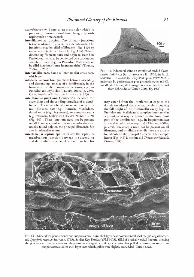
81Illustrated Glossary of the Bivalvia
interdissoconch. Same as nepioconch (which is preferred). Formerly used interchangeably with nepioconch or mesoconch.
interfilamentar junction. One of many junctions between adjacent filaments in a demibranch. The junctions may be cilial (filibranch; Fig. 113) or tissue grade (eulamellibranch; Fig. 104). Where descending filaments turn and begin to ascend in Pterioidea, they may be connected by a continuous stretch of tissue (e.g., in Pteriidae, Malleidae), or by cilial junctions (some Isognomonidae) (tëMkiN, 2006a, p. 286).
interlamellar bars. Same as interlamellar cross bars, which see.
interlamellar cross bars. Junctions between ascending and descending lamellae of a demibranch, in the form of multiple, narrow connections, e.g., in Pinnidae and Mytilidae (tëMkiN, 2006a, p. 289). Called interlamellar bars by rideWood (1903).
interlamellar junctions. Connections between the ascending and descending lamellae of a demi-branch. These may be absent or represented by multiple cross bars (e.g., Pinnidae, Mytilidae), dorsal septa (e.g., Isognomon), or complete septa (e.g., Pteriidae, Malleidae) (tëMkiN, 2006a, p. 289) (Fig. 145). These junctions need not be present on all filaments, and in plicate ctenidia they are usually found only on the principal filaments. See also interlamellar septum.
interlamellar septum (pl., interlamellar septa). A membranous junction between the ascending and descending lamellae of a demibranch. This
may extend from the interlamellar edge to the dorsalmost edge of the lamellae, thereby occupying the full height of the interlamellar cavity (e.g., in Pteriidae and Malleidae; a complete interlamellar septum), or it may be limited to the dorsalmost part of the demibranch (e.g., in Isognomonidae; a dorsal interlamellar septum) (tëMkiN, 2006a, p. 289). These septa need not be present on all filaments, and in plicate ctenidia they are usually found only on the principal filaments. The example herein (Fig. 146) is the thraciid Thracia meridionalis (sMith, 1885).
FiG. 142. Inductural spine on exterior of cardiid Cteno-cardia imbricata (G. B. soWerBy ii, 1840, in G. B. soWerBy I, 1832–1841), Panay, Philippines (YPM 9720), underlain by periostracum plus prismatic outer and CL middle shell layers; shell margin is toward left (adapted
from Schneider & Carter, 2001, fig. 10.1).
FiG. 143. Mineralized periostracum and subperiostracal outer shell layer near posteroventral shell margin of gastrochae-nid Spengleria rostrata (speNGler, 1793), Soldier Key, Florida (YPM 9473). SEM of a radial, vertical fracture, showing the periostracum and its extra- to infraperiostracal aragonitic spikes; desiccation has pulled periostracum away from
subperiostracal outer shell layer, into which spikes were slightly embedded (Carter, new).
100 µm

82 Treatise Online, number 48
interlinear muscle scar. A muscle scar connecting the anterior end of the pallial sinus with the anterior adductor muscle scar, e.g., in many Tellinidae (Fig. 147) and Gastrochaenidae (see Fig. 192). In at least the Gastrochaenidae, this is a ctenidial attachment scar. Not to be confused with an orbital muscle scar.
interlocking shell margins. (1) Shell margins that, upon closure, engage with one another so that shearing of the valves along the hinge is minimized; (2) for larval shells, shell margins that engage in a pill box or tongue-in-groove fashion (Waller, 1981); normally applied to cases in which the interlocking occurs over a large distance beyond the hinge.
intermediate teeth. A series of hinge teeth on each side of the hinge, representing subdivisions of the infra-dorsal tooth and socket, e.g., in the pectinid Pecten Müller, 1776 (Waller, 1991, p. 8). Compare with supradorsal teeth, infradorsal teeth, and dorsal teeth.
internal carina. See internal rib carina.internal characters. Features visible on the inner
surface of a shell, such as the hinge, hinge teeth, internal ligaments, internal ridges and buttresses, and muscle scars.
internal ligament. A ligament, or a component of a ligament, attaching below the articulating surface of the hinge plate (see Fig. 160e and 160f ). The liga-ment may insert on the underside of a hinge plate
FiG. 146. Transverse section through inner demibranch of thraciid Thracia meridionalis (sMith, 1885), Admi-ralty Bay, King George Island, Antarctica (O. Doman-eschi, collector), showing several interlamellar septa,
figure width 3 mm (Sartori, new).
FiG. 147. Muscle scars in tellinid Arcopagia fausta (pulteNey, 1799); add., adductors, shell length 8.4 cm
(adapted from Mikkelsen & Bieler, 2007).
FiG. 144. Transverse section through mantle margin of venerid Periglypta puerpera (liNNaeus, 1758), Kung-kraben Bay, Thailand (A. F. Sartori, C. Printrakoon, P. Mikkelsen & R. Bieler, collectors), showing outer mantle fold and inner mantle folds 1, 2, and 3, figure width 5.4
mm (Sartori, new).
FiG. 145. Three types of ctenidial interlamellar junc-tions between ascending and descending ctenidial lamellae. 1, Complete septum as in most Pterioidea; 2, dorsal septum as in isognomonid Isognomon; 3, mul-tiple cross bars, as in Pinnidae and Mytilidae (adapted from Tëmkin, 2006a, fig. 12, courtesy of John Wiley
& Sons).
1 2 3
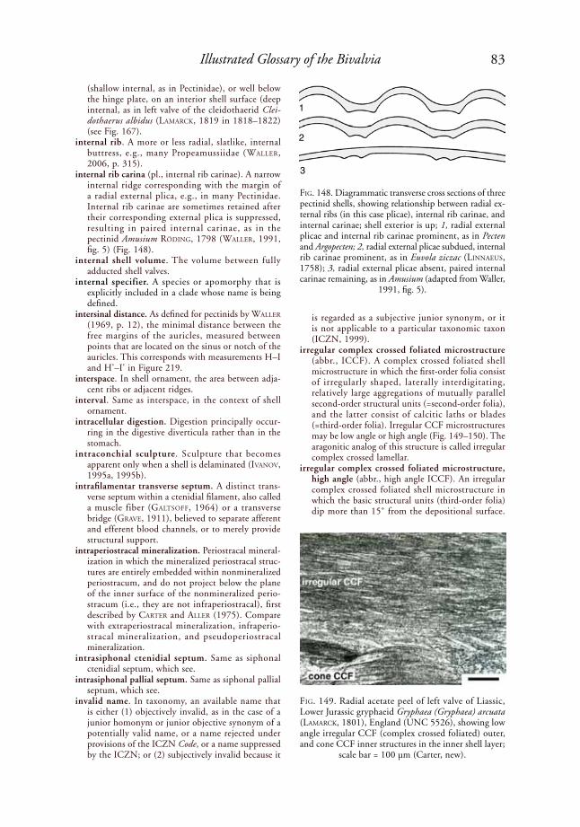
83Illustrated Glossary of the Bivalvia
(shallow internal, as in Pectinidae), or well below the hinge plate, on an interior shell surface (deep internal, as in left valve of the cleidothaerid Clei-dothaerus albidus (laMarCk, 1819 in 1818–1822) (see Fig. 167).
internal rib. A more or less radial, slatlike, internal buttress, e.g., many Propeamussiidae (Waller, 2006, p. 315).
internal rib carina (pl., internal rib carinae). A narrow internal ridge corresponding with the margin of a radial external plica, e.g., in many Pectinidae. Internal rib carinae are sometimes retained after their corresponding external plica is suppressed, resulting in paired internal carinae, as in the pectinid Amusium rödiNG, 1798 (Waller, 1991, fig. 5) (Fig. 148).
internal shell volume. The volume between fully adducted shell valves.
internal specifier. A species or apomorphy that is explicitly included in a clade whose name is being defined.
intersinal distance. As defined for pectinids by Waller (1969, p. 12), the minimal distance between the free margins of the auricles, measured between points that are located on the sinus or notch of the auricles. This corresponds with measurements H–I and H'–I' in Figure 219.
interspace. In shell ornament, the area between adja-cent ribs or adjacent ridges.
interval. Same as interspace, in the context of shell ornament.
intracellular digestion. Digestion principally occur-ring in the digestive diverticula rather than in the stomach.
intraconchial sculpture. Sculpture that becomes apparent only when a shell is delaminated (iVaNoV, 1995a, 1995b).
intrafilamentar transverse septum. A distinct trans-verse septum within a ctenidial filament, also called a muscle fiber (GaltsoFF, 1964) or a transverse bridge (GraVe, 1911), believed to separate afferent and efferent blood channels, or to merely provide structural support.
intraperiostracal mineralization. Periostracal mineral-ization in which the mineralized periostracal struc-tures are entirely embedded within nonmineralized periostracum, and do not project below the plane of the inner surface of the nonmineralized perio-stracum (i.e., they are not infraperiostracal), first described by Carter and aller (1975). Compare with extraperiostracal mineralization, infraperio-stracal mineralization, and pseudoperiostracal mineralization.
intrasiphonal ctenidial septum. Same as siphonal ctenidial septum, which see.
intrasiphonal pallial septum. Same as siphonal pallial septum, which see.
invalid name. In taxonomy, an available name that is either (1) objectively invalid, as in the case of a junior homonym or junior objective synonym of a potentially valid name, or a name rejected under provisions of the ICZN Code, or a name suppressed by the ICZN; or (2) subjectively invalid because it
is regarded as a subjective junior synonym, or it is not applicable to a particular taxonomic taxon (ICZN, 1999).
irregular complex crossed foliated microstructure (abbr., ICCF). A complex crossed foliated shell microstructure in which the first-order folia consist of irregularly shaped, laterally interdigitating, relatively large aggregations of mutually parallel second-order structural units (=second-order folia), and the latter consist of calcitic laths or blades (=third-order folia). Irregular CCF microstructures may be low angle or high angle (Fig. 149–150). The aragonitic analog of this structure is called irregular complex crossed lamellar.
irregular complex crossed foliated microstructure, high angle (abbr., high angle ICCF). An irregular complex crossed foliated shell microstructure in which the basic structural units (third-order folia) dip more than 15˚ from the depositional surface.
FiG. 148. Diagrammatic transverse cross sections of three pectinid shells, showing relationship between radial ex-ternal ribs (in this case plicae), internal rib carinae, and internal carinae; shell exterior is up; 1, radial external plicae and internal rib carinae prominent, as in Pecten and Argopecten; 2, radial external plicae subdued, internal rib carinae prominent, as in Euvola ziczac (liNNaeus, 1758); 3, radial external plicae absent, paired internal carinae remaining, as in Amusium (adapted from Waller,
1991, fig. 5).
FiG. 149. Radial acetate peel of left valve of Liassic, Lower Jurassic gryphaeid Gryphaea (Gryphaea) arcuata (laMarCk, 1801), England (UNC 5526), showing low angle irregular CCF (complex crossed foliated) outer, and cone CCF inner structures in the inner shell layer;
scale bar = 100 µm (Carter, new).
1
2
3

84 Treatise Online, number 48
Common in Gryphaeidae and Oxytomidae, e.g., in the inner shell layer of Lower Jurassic Oxytoma (Oxytoma) inequivalve (J. soWerBy, 1819, in soWerBy & soWerBy, 1812–1846) (Fig. 150).
irregular complex crossed foliated microstructure, low angle (abbr., low angle ICCF). An irregular complex crossed foliated shell microstructure in which the basic structural units (third-order folia) dip less than 15˚ (usually only a few degrees) from the depositional surface; e.g., parts of the inner shell layer of the Lower Jurassic gryphaeid Gryphaea (Gryphaea) arcuata (laMarCk, 1801) (Fig.
149), and in many other Ostreoidea, plus many Pectinidae and Propeamussiidae. Commonly called irregularly foliated.
irregular complex crossed lamellar microstructure (abbr., ICCL). A complex crossed lamellar shell microstructure in which the first-order lamellae consist of irregularly shaped, laterally interdigi-tating, relatively large aggregations of mutually parallel second-order structural units, the latter being represented by fibers, rods, or second-order lamellae (Carter & others, 1990, p. 613); e.g., the inner shell layer of the poromyid Poromya (Poromya) sp. (Fig. 151). The calcitic analog of this structure is called irregular complex crossed foliated.
irregular crossed foliated microstructure (abbr., ICF). A simple crossed foliated shell microstructure in which the first-order crossed folia are very irregu-larly shaped, as seen on the depositional surface and in horizontal sections; e.g., the middle shell layer in the Cretaceous gryphaeid Rhynchostreon mermeti (CoquaNd, 1862) (MalChus, 1990, pl. 26,11). Compare with branching crossed foliated and herringbone foliated.
irregular crossed lamellar microstructure (abbr., ICL). A crossed lamellar shell microstructure in which the first-order crossed lamellae are irregularly shaped as seen on the depositional surface and in horizontal sections. Not to be confused with irregular complex crossed lamellar. Locally present in the middle shell layer of cyamiids, gastrochaenids, and tellinids (Fig. 82).
irregular fibrous prismatic microstructure. See fibrous prismatic microstructure.
irregular simple prismatic microstructure (abbr., ISP). A simple prismatic shell microstructure in which
FiG. 150. High angle irregular complex crossed foliated microstructure in inner shell layer of Liassic, Lower Jurassic oxytomid Oxytoma (Oxytoma) inequivalve (J. soWerBy, 1819, in soWerBy & soWerBy, 1812–1846), seen in acetate peels. 1, horizontal section; 2, radial section, with depositional surface toward bottom; scale bars = 50 µm (adapted
from Carter & Lutz, 1990, pl. 8B).
FiG. 151. Nacreous (N ) middle and irregular complex crossed lamellar (CCL) inner shell layers in Eastern Atlantic, deep sea poromyid Poromya (Poromya) sp., seen on a polished and then acid-etched surface of a nearly horizontal, slightly oblique section, scale bar = 10 µm (adapted from Carter & Lutz, 1990, pl. 10,A).
1 2
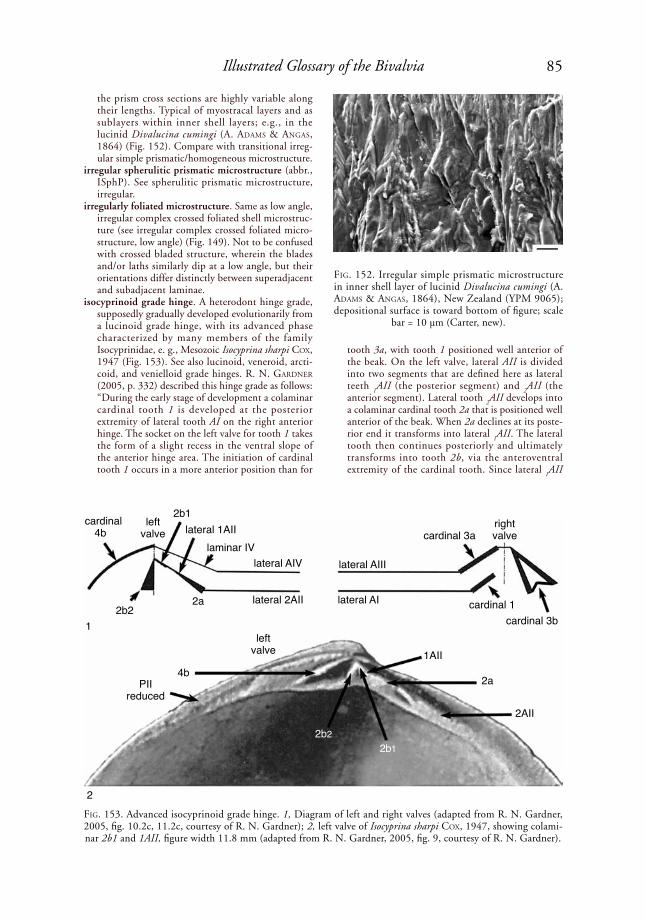
85Illustrated Glossary of the Bivalvia
the prism cross sections are highly variable along their lengths. Typical of myostracal layers and as sublayers within inner shell layers; e.g., in the lucinid Divalucina cumingi (A. adaMs & aNGas, 1864) (Fig. 152). Compare with transitional irreg-ular simple prismatic/homogeneous microstructure.
irregular spherulitic prismatic microstructure (abbr., ISphP). See spherulitic prismatic microstructure, irregular.
irregularly foliated microstructure. Same as low angle, irregular complex crossed foliated shell microstruc-ture (see irregular complex crossed foliated micro-structure, low angle) (Fig. 149). Not to be confused with crossed bladed structure, wherein the blades and/or laths similarly dip at a low angle, but their orientations differ distinctly between superadjacent and subadjacent laminae.
isocyprinoid grade hinge. A heterodont hinge grade, supposedly gradually developed evolutionarily from a lucinoid grade hinge, with its advanced phase characterized by many members of the family Isocyprinidae, e. g., Mesozoic Isocyprina sharpi Cox, 1947 (Fig. 153). See also lucinoid, veneroid, arcti-coid, and venielloid grade hinges. r. N. GardNer (2005, p. 332) described this hinge grade as follows: “During the early stage of development a colaminar cardinal tooth 1 is developed at the posterior extremity of lateral tooth AI on the right anterior hinge. The socket on the left valve for tooth 1 takes the form of a slight recess in the ventral slope of the anterior hinge area. The initiation of cardinal tooth 1 occurs in a more anterior position than for
tooth 3a, with tooth 1 positioned well anterior of the beak. On the left valve, lateral AII is divided into two segments that are defined here as lateral teeth
1AII (the posterior segment) and
2AII (the
anterior segment). Lateral tooth 2AII develops into
a colaminar cardinal tooth 2a that is positioned well anterior of the beak. When 2a declines at its poste-rior end it transforms into lateral
1AII. The lateral
tooth then continues posteriorly and ultimately transforms into tooth 2b, via the anteroventral extremity of the cardinal tooth. Since lateral
1AII
FiG. 152. Irregular simple prismatic microstructure in inner shell layer of lucinid Divalucina cumingi (A. adaMs & aNGas, 1864), New Zealand (YPM 9065); depositional surface is toward bottom of figure; scale
bar = 10 µm (Carter, new).
FiG. 153. Advanced isocyprinoid grade hinge. 1, Diagram of left and right valves (adapted from R. N. Gardner, 2005, fig. 10.2c, 11.2c, courtesy of R. N. Gardner); 2, left valve of Isocyprina sharpi Cox, 1947, showing colami-nar 2b1 and 1AII, figure width 11.8 mm (adapted from R. N. Gardner, 2005, fig. 9, courtesy of R. N. Gardner).
cardinal
4b
left valve
2b22a
2b1lateral 1AII
laminar IVlateral AIV
lateral 2AII
cardinal 3aright valve
lateral AIII
lateral AI cardinal 1cardinal 3b
PIIreduced
4b
1AII
2a
2AII
2b22b1
left valve
1
2
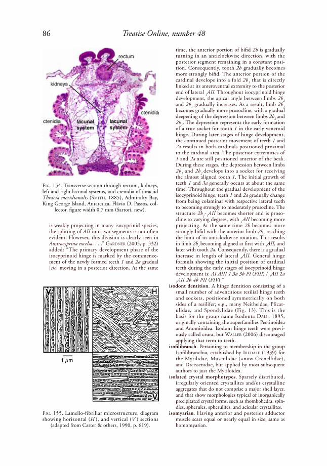
86 Treatise Online, number 48
is weakly projecting in many isocyprinid species, the splitting of AII into two segments is not often evident. However, this division is clearly seen in Austrocyprina excelsa. . . .” GardNer (2005, p. 332) added: “The primary development phase of the isocyprinoid hinge is marked by the commence-ment of the newly formed teeth 1 and 2a gradual [sic] moving in a posterior direction. At the same
time, the anterior portion of bifid 2b is gradually turning in an anticlockwise direction, with the posterior segment remaining in a constant posi-tion. Consequently, tooth 2b gradually becomes more strongly bifid. The anterior portion of the cardinal develops into a fold 2b
1 that is directly
linked at its anteroventral extremity to the posterior end of lateral
1AII. Throughout isocyprinoid hinge
development, the apical angle between limbs 2b2
and 2b1 gradually increases. As a result, limb 2b
1
becomes gradually more prosocline, with a gradual deepening of the depression between limbs 2b
2 and
2b1. The depression represents the early formation
of a true socket for tooth 1 in the early veneroid hinge. During later stages of hinge development, the continued posterior movement of teeth 1 and 2a results in both cardinals positioned proximal to the cardinal area. The posterior extremities of 1 and 2a are still positioned anterior of the beak. During these stages, the depression between limbs 2b
1 and 2b
2 develops into a socket for receiving
the almost aligned tooth 1. The initial growth of teeth 1 and 3a generally occurs at about the same time. Throughout the gradual development of the isocyprinoid hinge, teeth 1 and 2a gradually change from being colaminar with respective lateral teeth to becoming strongly to moderately prosocline. The structure 2b
1-
1AII becomes shorter and is proso-
cline to varying degrees, with 1AII becoming more
projecting. At the same time 2b becomes more strongly bifid with the anterior limb 2b
1 reaching
the limit of its anticlockwise rotation. This results in limb 2b
1 becoming aligned at first with
1AII, and
later with tooth 2a. Consequently, there is a gradual increase in length of lateral
2AII. General hinge
formula showing the initial position of cardinal teeth during the early stages of isocyprinoid hinge development is: AI AIII 1 3a 3b PI (PIII) /
2AII 2a
1AII 2b 4b PII (PIV).”
isodont dentition. A hinge dentition consisting of a small number of adventitious resilial hinge teeth and sockets, positioned symmetrically on both sides of a resilifer; e.g., many Neitheidae, Plicat-ulidae, and Spondylidae (Fig. 13). This is the basis for the group name Isodonta dall, 1895, originally containing the superfamilies Pectinoidea and Anomioidea. Isodont hinge teeth were previ-ously called crura, but Waller (2006) discouraged applying that term to teeth.
isofilibranch. Pertaining to membership in the group Isofilibranchia, established by iredale (1939) for the Mytilidae, Musculidae (=now Crenellidae), and Dreissenidae, but applied by most subsequent authors to just the Mytiloidea.
isolated crystal morphotypes. Sparsely distributed, irregularly oriented crystallites and/or crystalline aggregates that do not comprise a major shell layer, and that show morphologies typical of inorganically precipitated crystal forms, such as rhombohedra, spin-dles, spherules, spherulites, and acicular crystallites.
isomyarian. Having anterior and posterior adductor muscle scars equal or nearly equal in size; same as homomyarian.
FiG. 154. Transverse section through rectum, kidneys, left and right lacunal systems, and ctenidia of thraciid Thracia meridionalis (sMith, 1885), Admiralty Bay, King George Island, Antarctica, Flávio D. Passos, col-
lector, figure width 0.7 mm (Sartori, new).
FiG. 155. Lamello-fibrillar microstructure, diagram showing horizontal (H ), and vertical (V ) sections
(adapted from Carter & others, 1990, p. 619).
1 µm
87Illustrated Glossary of the Bivalvia
isthmian mantle. The confluence of the mantle lobes dorsally, just below the hinge.
iteroparous. Having repeated reproductive cycles, strictly speaking only one per year, although some taxa may breed more than once per year. Most marine bivalves and nearly all freshwater bivalves are iteroparous. Compare with semelparous.
junior homonym. The later proposed of two homonyms; in the case of simultaneous proposal, the one not given precedence under Article 24 of the ICZN (1999) Code.
junior synonym. The later proposed of two synonyms; in the case of simultaneous proposal, the one not given precedence under Article 24 of the ICZN (1999) Code; see also Article 23.9.
justified emendation. In taxonomy, a valid correction of an incorrect original spelling in accordance with ICZN (1999) Article 33.2; the corrected name maintains the author and date of the original name.
juvenile. The growth stage prior to reproductive competence.
Keber’s gland. See pericardial glands.keel. A distinct, linear or curvilinear elevation similar
in shape to a ship’s keel or a chicken's breastbone. Compare with the more specific term carina.
knob. A rounded protuberance; in shell sculpture, usually a large protuberance, as opposed to smaller tubercles or pustules.
labial. Of or pertaining to the lips.labial palp. One of the paired organs near the antero-
ventral end of the ctenidia, which serve to separate food from rejected particles and transfer the former to the oral groove, e.g., in Nuculidae (see Fig. 213), Veneridae (Fig. 130), and Gastrochaenoidea (Fig. 19). The palps form posteriorly extended prolonga-tions of the upper (anterior) and lower (posterior) lips on either side of the mouth, and they usually have ridged and ciliated opposing surfaces. They are typically extended into a distal oral groove in close proximity to the anterior filaments of the inner demibranch. stasek (1963) defined three categories for the relationship between the distal oral groove of the palps and the ventral tips of the anterior ctenidial filaments of the inner demibranchs. See ctenidium-palp association categories I, II, and III.
labial palp distal bifurcation. The unfused portion of the dorsal margin of a pair of labial palps.
labial palp height. The distance from the dorsal to the ventral margin of a labial palp, measured at the dorsal end of the palp connection to the visceral mass.
labial palp length. The distance from the distal terminus of a labial palp to the midpoint of its basal connection, measured in a straight line.
labial palp muscle scar. See muscle scar a, e.g., in Pachydomidae.
lachrymose. Teardrop shaped, i.e., broadly rounded at one end, tapering at the other end.
lacuna (pl., lacunae). A space, within a tissue, which functions in the place of vessels for the circula-tion of body fluids, e.g., the lateral lacunal system accompanying the kidneys in Thraciidae and Verti-cordiidae (Fig. 154).
lamella (pl., lamellae, adj., lamellar). (1) In refer-ence to shell microstructure, a thin plate, such as that comprising first-, second-, and or third-order lamellae in aragonitic crossed lamellar microstruc-tures; analogs in calcitic crossed foliated micro-structures are called folia; (2) in reference to shell ornament, a thin, raised ridge; (3) one of up to four sheets of filaments comprising a filibranch or eulamellibranch ctenidium (Fig. 104, Fig. 113, Fig. 289).
lamellar dentition. A hinge dentition consisting of elongate teeth and sockets, typically at both ends of the hinge, commonly accompanied by nonlamellar hinge teeth (NeWell & Boyd, 1995, p. 13).
lamellar ligament. The lamellar sublayer of the liga-ment.
lamellar sublayer of ligament. The nonmineralized portion of a ligament, excluding the periostracum; commonly referred to as lamellar ligament.
lamellate. Same as lamellar, which is preferred.
FiG. 156. Laminar buttresses in left valve of pandorid Pandora (Heteroclidus) punctata (CoNrad, 1837); diagram of shell interior (adapted from Keen, 1969c,
fig. F22,7b).
FiG. 157. Transverse fracture through a ctenidial fila-ment in thraciid Thracia meridionalis (sMith, 1885), Admiralty Bay, King George Island, Antarctica (Flávio D. Passos, collector), showing ctenidial cilia and epithe-lia, figure width approximately 60 µm (Sartori, new).

88 Treatise Online, number 48
lamelliform. Having the shape of a thin, elongate plate or sheet.
lamello-fibrillar microstructure. An aragonitic, laminar, plywoodlike shell microstructure consisting of sheets of more or less mutually parallel, hori-zontal fibers, with fiber orientations differing in successive laminae (erBeN, 1972, p. 28, for the cephalopod Spirula; Carter & others, 1990, p. 619, 646) (Fig. 155). This is the Spirula “nacre” of MutVei (1964, p. 269), so named because of its laminar arrangement and nacreous luster, although it lacks nacre tablets. kouChiNsky (1999, p. 177) called an approximation of this structure “stepwise texture” in the lower Cambrian stenothecid tergo-myans Anabarella plana VostokoVa, 1962, and Watsonella GraBau, 1900 (see Fig. 300).
lamina. A thin plate.laminar buttresses. Internal reinforcing ridges radi-
ating from the beak, in some cases articulating with the opposite valve, thereby comprising an adventitious hinge dentition (not homologous with true hinge teeth); e.g., the pandorid Pandora (Heteroclidus) punctata (CoNrad, 1837) (Fig. 156).
laminar microstructures. A general category of shell microstructures consisting of mutually parallel, horizontal or slightly imbricated sheets (laminae), with each sheet comprised of granules, rods, laths, blades, or tablets. If the laminae are inclined, there is only one predominant dip direction in the shell layer. This includes nacreous, semi-nacreous, lamello-fibrillar, crossed-bladed, Sepia-type laminar, matted, regularly foliated and semi-foliated micro-structures (Carter & others, 1990, p. 611).
laminate. Thinly layered.lanceolate. Shaped like the tip of a lance, i.e., much
longer than wide, with one end pointed.lanceolate periostracum. Periostracum in the form
of long, needlelike bristles standing more or less erect (oliVer, 1981, p. 80). Compare with spicate, thatched, stubbly, and pilose periostracum.
lapsus calami. Latin for slip of the pen; an uninten-tional error made by an author in composing a text, or a typographical or printer’s error. Commonly italicized.
large tablet, imbricated nacre. A nacreous shell micro-structure with overlapping laminae on the deposi-
tional surface, and with tablet diameters substantially larger than typical nacre, i.e., 30–40 µm as opposed to less than 20 µm wide. This is a restriction of the term “large tablet imbricated nacre” of Carter (2001, p. 233), which also included semi-foliated aragonite. Locally present in the inner shell layer of Pojetaia runnegari Jell, 1980 (see VeNdrasCo, CheCa, & kouChiNsky, 2011, pl. 2,5 ).
larva. The developmental stage between gastrula and metamorphosis.
larval development. The progression of bivalve growth from the trochophore stage to the prodissoconch-1, prodissoconch-2, nepioconch, and dissoconch stages.
larval hinge teeth. Hinge teeth formed during the prodissoconch-2 phase of larval development.
larval shell. The prodissoconch, i.e., the generally mineralized (aragonitic) part of a larva, functioning as the shell up to metamorphosis. See also prodisso-conch.
larval thread. An elongate, proteinous strand secreted between the valves of some unionoid glochidial larvae; believed to be nonhomologous with byssal threads.
larviparous. A mode of indirect development in which eggs are hatched internally, and free-living larvae are then released (liNColN, Boxshall, & Clark, 1988). Compare with direct development, ovipa-rous, ovoviviparous, and viviparous.
lasidium (pl., lasidia). The larval stage of the unionoid families Mulleriidae and Iridinidae, characterized by an elongate body, a very long larval thread, and paired ventral chitinous hooks (WäChtler, dreher-MaNsur, & riChter, 2001).
lateral. (1) On, from, or directed toward the side; (2) in the context of hinge dentition, located on either side of the beak, as in a lateral tooth.
lateral bulbs. Elaborations of the lips in Lyonsiidae.lateral cilia. Cilia on the lateral epithelium of a
ctenidial filament (peCk, 1877, p. 63) (Fig. 157). They serve primarily to generate water currents. See also frontal cilia and eu-laterofrontal cilia.
lateral compression. The degree of reduction in width of a shell relative to its length and height.
lateral disc buttress. A buttress present on the lateral interior surface of the disc of a pectinoid shell
FiG. 158. Diagram of lativincular ligament (adapted from Waterhouse, 2001, fig. 9b).
bourreletresiliferbourrelet
89Illustrated Glossary of the Bivalvia
(Waller, 2006, p. 315), e.g., in the Permian perno-pectinid Pernopecten yini NeWell & Boyd, 1995 (Fig. 30). Compare with auricular buttress.
lateral epithelium. The epithelium on the sides of a ctenidial filament, continuing into the water passages and covering the ctenidial interlamellar surfaces (peCk, 1877, p. 63) (Fig. 157). Compare with frontal epithelium and laterofrontal epithelium.
lateral hinge system. Hinge structures present in some prodissoconchs, anterior and posterior to the provinculum.
lateral tooth. A short to elongate, straight to slightly curved, socketed, nontaxodont shell lamella, posi-tioned on the anterior or posterior hinge area, usually parallel or subparallel with the adjacent shell margin, with its proximal end not extending all the way to the beak, e.g., in the astartid Astarte smithii dall, 1886 (Fig. 36, Fig. 68). Lateral teeth in the order Actinodontida and in nonmembers of the infraclass Heteroconchia are traditionally called pseudolaterals.
laterofrontal cilia. (1) The cilia on the laterofrontal epithelium of a ctenidial filament (peCk, 1877, p. 63), serving primarily to strain particles from the current generated by the ctenidia; (2) functionally similar cilia in a slightly different position on the ctenidial filament (atkiNs, 1938, p. 346). Compare with eu-laterofrontal cilia.
laterofrontal epithelium. The epithelium on a ctenidial filament on each side of the frontal epithelium, between the frontal epithelium and the lateral epithelium (peCk, 1877, p. 63) (Fig. 157). Compare with frontal epithelium and lateral epithelium.
lath-type chomata. Chomata in the form of elongate, straight, well-defined ridges, spaced about as far apart as their widths. They are mostly antimargin-ally aligned, except in some Texigryphaea steNZel, 1959, and typically either thin (approximately 1 mm) and long, or wide (1–3 mm) and rather stout. Lath-type chomata are the most widely distrib-uted type in the Ostreoidea, and they commonly transition into pustulose chomata (harry, 1985; MalChus, 1990).
lathic simple prismatic microstructure. A simple pris-matic microstructure in which the prisms are wider and longer (in the horizontal plane) than they are tall (in the vertical direction), and they are distinctly radially elongate (Carter & others, 1990, p. 647). Same as radially elongate simple prismatic, which is preferred.
lath-type fibrous prismatic microstructure. See fibrous prismatic microstructure, lath-type.
lativincular ligament. An alivincular ligament with a very broad, often shallow resilifer flanked by narrower, anterior and posterior bourrelets (Water-house, 2001, fig. 9b) (Fig. 158). Typical of some Streblochondriidae, e.g., upper Carboniferous Orbiculopecten GoNZáleZ, 1978. This corresponds with the term alivincular-areate-wide ligament, which see.
lecithotrophic. A larval type in which the major or exclusive energy source during early development is
vitelline (yolk). Generally characterized by a short-term, free-swimming stage (veliger) that does not derive nutrition from plankton. Conventionally, bivalve eggs with a diameter above 100 µm are considered to be yolk rich. Prodissoconch-1 size is roughly correlated with egg size (oCkelMaNN, 1965; MaCkie, 1984).
lecithotrophic-planktotrophic. Larval type in which the energy source during early development is both vitelline (yolk) and plankton. Found in most Ostreinae. The oyster Tiostrea may be exceptional in having eggs very rich in yolk (over 200 µm in width) yet having prerelease, late-stage veliger larvae already ingesting plankton (oCkelMaNN, 1965; MaCkie, 1984).
lectotype. A specimen from a suite of syntypes that is designated as the single name-bearing type spec-imen of a species or subspecies, subsequent to the original description of the species or subspecies. The remaining syntypes are, by intention or by default, called paralectotypes. Governed by ICZN (1999) Article 74 with modification in Declaration 44 of 2003 [Bulletin of Zoological Nomenclature 60(4):263]. Compare with holotype, paratype, syntype, and neotype.
ledge. A blunt, external, commarginal shell feature corresponding with a relatively short-lived, distinct disruption of shell margin convexity, affecting the entire thickness of the shell; e.g., in the pectinid Nodipecten dall, 1898 (Waller, 1991, p. 6).
left-convex. Having both valves convex, but with the left valve being more convex than the right (Waller, 1969, p. 12).
left pouch. An embayment, on the stomach’s left wall, which may be blind or connected to the digestive gland. The left pouch typically houses a flare from the gastric shield (Fig. 97; and see Fig. 303–305).
left valve. The shell valve on the observer’s left when united valves are held with the anterior end
FiG. 159. Aragonitic, transitional irregular simple (ISP)prismatic/homogeneous outer shell layer and underly-ing lenticular nacreous middle shell layer in mytilid Mytella guyanensis (laMarCk, 1819 in 1818–1822), Panama (YPM 731). SEM of a radial, vertical fracture
in posterior of shell; exterior is up (Carter, new).

90 Treatise Online, number 48
pointing away from the observer, the commissural plane vertical, and the hinge uppermost.
length of anterior outer ligament. In pectinoids, the distance along the outer ligament from the origin of growth to its anterior end (Waller, 1969, p. 12).
length of posterior outer ligament. In pectinoids, the distance along the outer ligament from the origin of growth to its posterior end (Waller, 1969, p. 12).
length of resilial insertion. The linear distance between the most anterior and the most posterior points on a resilifer, measured parallel to its base and near the base of the resilifer (Waller, 1969, p. 12). This is commonly also referred to as the width or breadth of the resilifer.
length of shell. (1) The distance between two mutually parallel planes oriented perpendicular to the hinge axis (=cardinal axis) and just touching the anterior and posterior ends of the shell. This definition is independent of the position of the adductor muscle scars, which may be unknown, so it is applicable to both monomyarian and dimyarian bivalves; (2) the maximum anteroposterior axis defined by a line passing through analogous points (center, dorsal, or ventral) on the anterior and posterior adductor muscles (staNley, 1970; Bailey, 2009). This defi-nition is not applicable to monomyarian bivalves, nor to those for which one adductor muscle scar is unknown.
lensic foliated. A foliated shell layer with chalky lenses and/or lensic, structural chambers; e.g., many Ostreoidea.
lentic. Relating to a still body of water, such as a lake or reservoir.
lenticular. Shaped like a biconvex lens.lenticular nacreous microstructure. A variety of
nacreous shell microstructure characterized by tablets with irregular thicknesses and often convex and concave surfaces (taylor, keNNedy, & hall, 1969, p. 28). This is found in the middle shell layer of some Nuculidae, Trigoniidae, Unionidae, Pandoridae, and Mytilidae (Fig. 159). Not to be confused with semi-nacreous microstructure.
leucocyte (or leukocyte). A white or colorless blood cell, commonly a nucleated amoeboid cell.
levator muscles. Same as pedal elevator muscles, which see.
ligament. The dorsal, medial part of a bivalve shell that is structurally specialized to enhance dorsal flexing, and that has distinct insertion areas in the two valves. The ligament includes any superficial periostracum and all lamellar and fibrous sublayers, but it excludes any far anterior and far posterior, distinct periostracal thickenings that may func-tionally extend the ligament. In the case of some deeply submerged or internal ligaments, thickened periostracum along the dorsal shell margin may functionally replace the lamellar sublayer of the ligament. An internal ligament is generally called a resilium. See also ligament 1 and ligament 2. Compare with pseudoligament.
ligament 1 (abbr., L1). The first-formed larval liga-ment and its postlarval derivatives, produced
FiG. 160. Terminology of ligament position, illustrated by idealized transverse sections (left) and a composite interior view of hinge (right). Fibrous and lamellar ligament sublayers are not differentiated. Simple ligaments are portrayed in three transverse sections on left (with a, b, and c); composite transverse section to right of these (with d, e, and f ) shows a left hinge and part of a right hinge in a myid left-right asymmetrical ligament (d, e), plus an unrelated, cleidothaerid deep internal ligament (with f ). Duplivincular, alivincular, multivincular, and other ligaments with a resilium are portrayed in composite interior view of a hinge on right. Only resilifers (not also the bourrelets) are shown for alivincular ligaments. Left: a, external simple ligament; b, shallow submarginal simple ligament; c, shallow to deep submarginal simple ligament; d, deep submarginal ligament, as in left valve of Myidae; e, shallow internal ligament, as in right valve of Myidae; f, deep internal ligament, as in left valve of cleidothaerid Cleidothaerus albidus laMarCk, 1819 in 1818–1822. Right: a, external duplivincular ligament; b, shallow submarginal resilifer; c, shallow to deep submarginal resilifer; d, shallow submarginal to internal resilifer; e, internal resilifer, as in right
valve of anomiid Monia macroschisma (deshayes, 1839) (see Fig. 14) (Carter, new).
a b c
f
e
d
a
c b
d e

91Illustrated Glossary of the Bivalvia
independently of ligament 2 (MalChus, 2004b; MalChus & WaréN, 2005).
ligament 2 (abbr., L2). The postlarval ligament and its derivatives, produced independently of ligament 1 (MalChus, 2004b; MalChus & WaréN, 2005). See also disjunct ligament.
ligament fulcrum. A long, narrow platform extending posteriorly from the beak along or near the dorsal shell margin, serving for attachment of the fibrous sublayer of a simple, parivincular, or quasiparivin-cular ligament.
ligament grades. See pteriomorphian ligament grades.
ligament pit. A typically triangular or cup-shaped depression that serves for the insertion of part or all of a ligament, regardless of its fibrous, granular, and/or lamellar composition. See also resilifer and trabeculae.
ligament position. The insertion area of the liga-ment relative to the hinge plate or other articu-lating surfaces. This may be external, shallow submarginal, deep submarginal, shallow internal, or deep internal (Carter, 1990a, p. 138) (Fig. 160). External and shallow submarginal ligaments are also called dorsal ligaments.
FiG. 161. Ligamental ridges, grooves, and terraces in duplivincular ligament of glycymeridid Glycymeris pectinata (GMeliN, 1791 in 1791–1793), Tobago, West Indies (YPM 6039). Acetate peel of an acid-etched, transverse, vertical
section through ligament (right) and ligamental insertion area (left) (Carter, new).
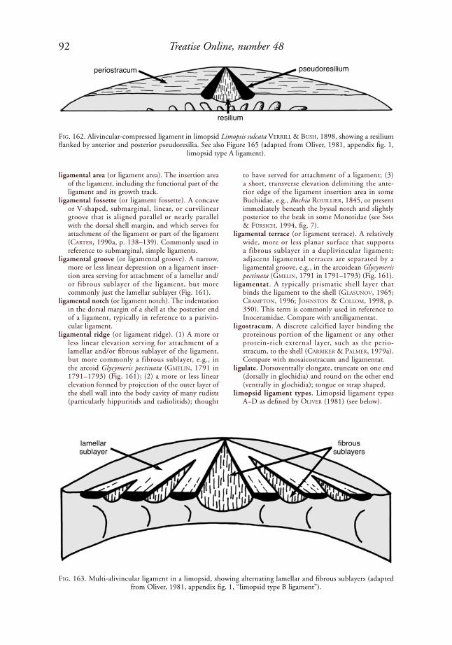
92 Treatise Online, number 48
ligamental area (or ligament area). The insertion area of the ligament, including the functional part of the ligament and its growth track.
ligamental fossette (or ligament fossette). A concave or V-shaped, submarginal, linear, or curvilinear groove that is aligned parallel or nearly parallel with the dorsal shell margin, and which serves for attachment of the ligament or part of the ligament (Carter, 1990a, p. 138–139). Commonly used in reference to submarginal, simple ligaments.
ligamental groove (or ligamental groove). A narrow, more or less linear depression on a ligament inser-tion area serving for attachment of a lamellar and/or fibrous sublayer of the ligament, but more commonly just the lamellar sublayer (Fig. 161).
ligamental notch (or ligament notch). The indentation in the dorsal margin of a shell at the posterior end of a ligament, typically in reference to a parivin-cular ligament.
ligamental ridge (or ligament ridge). (1) A more or less linear elevation serving for attachment of a lamellar and/or fibrous sublayer of the ligament, but more commonly a fibrous sublayer, e.g., in the arcoid Glycymeris pectinata (GMeliN, 1791 in 1791–1793) (Fig. 161); (2) a more or less linear elevation formed by projection of the outer layer of the shell wall into the body cavity of many rudists (particularly hippuritids and radiolitids); thought
to have served for attachment of a ligament; (3) a short, transverse elevation delimiting the ante-rior edge of the ligament insertion area in some Buchiidae, e.g., Buchia rouillier, 1845, or present immediately beneath the byssal notch and slightly posterior to the beak in some Monotidae (see sha & FürsiCh, 1994, fig. 7).
ligamental terrace (or ligament terrace). A relatively wide, more or less planar surface that supports a fibrous sublayer in a duplivincular ligament; adjacent ligamental terraces are separated by a ligamental groove, e.g., in the arcoidean Glycymeris pectinata (GMeliN, 1791 in 1791–1793) (Fig. 161).
ligamentat. A typically prismatic shell layer that binds the ligament to the shell (GlasuNoV, 1965; CraMptoN, 1996; JohNstoN & ColloM, 1998, p. 350). This term is commonly used in reference to Inoceramidae. Compare with antiligamentat.
ligostracum. A discrete calcified layer binding the proteinous portion of the ligament or any other protein-rich external layer, such as the perio-stracum, to the shell (Carriker & palMer, 1979a). Compare with mosaicostracum and ligamentat.
ligulate. Dorsoventrally elongate, truncate on one end (dorsally in glochidia) and round on the other end (ventrally in glochidia); tongue or strap shaped.
limopsid ligament types. Limopsid ligament types A–D as defined by oliVer (1981) (see below).
FiG. 162. Alivincular-compressed ligament in limopsid Limopsis sulcata Verrill & Bush, 1898, showing a resilium flanked by anterior and posterior pseudoresilia. See also Figure 165 (adapted from Oliver, 1981, appendix fig. 1,
limopsid type A ligament).
FiG. 163. Multi-alivincular ligament in a limopsid, showing alternating lamellar and fibrous sublayers (adapted from Oliver, 1981, appendix fig. 1, “limopsid type B ligament”).
periostracum
resilium
pseudoresilium
lamellarsublayer
fibroussublayers

93Illustrated Glossary of the Bivalvia
limopsid type A ligament. An external, amphidetic, alivincular ligament with a central, moderately wide, triangular resilifer containing the fibrous sublayer, flanked anteriorly and posteriorly by narrower, triangular pseudoresilifers containing only lamellar ligament; all three areas radiate from a point below the beak; e.g., in the limopsid Limopsis sulcata Verrill & Bush (1898) (Fig. 162). The remainder of the cardinal area is covered by a thin layer of periostracum (oliVer, 1981, appendix fig. 1). This ligament type is herein called alivincular-compressed.
limopsid type B ligament. An external, amphidetic, multi-alivincular ligament (which see), with several resilifers and flanking lamellar ligament areas radi-ating from a point below the beak. The central resilifer is wider than those flanking it, and only the central resilifer reaches the beak. The resilifers contain fibrous ligament; the remainder of the liga-mental area is covered by lamellar ligament. Found in some larger members of Limopsis morphological class I of oliVer (1981, appendix fig. 1) (Fig. 163).
limopsid type C ligament. An external, amphidetic, alivincular-areate-medium ligament, as in Limopsis
chuni thiele & JaeCkel, 1931 (oliVer, 1981, appendix fig. 1) (Fig. 164).
limopsid type D ligament. An alivincular ligament similar to limopsid type A of oliVer (1981), except that the anterior and posterior cardinal areas are covered by thick rather than thin periostracum. This is herein called alivincular-compressed; e.g., Limopsis lilliei E. A. sMith, 1915 (oliVer, 1981, appendix fig. 1) (Fig. 165).
line. (1) A very narrow, elongate, radial, antimarginal, commarginal, or slightly oblique element of shell ornament that does not appear to be visibly raised to the naked eye. A line differs from a thread in lacking noticeable relief; (2) a growth line, which may be external or internal, and which is commonly incised on the exterior of the shell.
linear crossed lamellar microstructure (abbr., LCL). A simple crossed lamellar shell microstructure in which the first-order lamellae are very elongate and minimally branching, as observed on the deposi-tional surface and in horizontal sections (Fig. 82).
lineavincular ligament. An opisthodetic, low-angle duplivincular ligament with only posteriorly dipping grooves and ridges (Waterhouse, 2001,
FiG. 164. Alivincular-areate-medium ligament in limopsid Limopsis chuni thiele & JaeCkel, 1931 (adapted from Oliver, 1981, appendix fig. 1, limopsid type C ligament).
FiG. 165. Alivincular-compressed ligament in limopsid Limopsis lilliei E. A. sMith, 1915. See also Figure 162 (adapted from Oliver, 1981, appendix fig. 1, limopsid type D ligament, with thickened periostracum at ends of cardinal area).
lamellar sublayerfibrous sublayer
thickenedperiostracum
resilium
pseudoresilium
94 Treatise Online, number 48
p. 114); e.g., the upper Carboniferous myalinid Selenimyalina meliniformis (Meek & WortheN, 1866) (NeWell & Boyd, 1987, fig. 7F; Carter, 1990a, fig. 29) (Fig. 166). Some low-angle dupliv-incular ligaments have long, posteriorly dipping ligamental grooves and ridges, plus some very short, anteriorly dipping ligamental grooves and ridges, all of which may be opisthodetic, as in Septimyalina perattenuata (Meek & haydeN, 1858) (NeWell & Boyd, 1987, fig. 7E). Such examples are herein classified as amphidetic, low-angle duplivincular and not lineavincular. Waterhouse (2008, p. 24) included ambonychiid ligaments here, but they are herein called monovincular-P, because their puta-tive ligamental ridges are actually just growth lines.
lips. Raised outgrowths of the buccal dermis (integu-ment surrounding the mouth), continuous with the labial palps. These are usually simple flaps, dorsal and ventral to the mouth, but they are hypertro-phied into arborescent or ruffled structures in the Pectinidae, Limidae, and Spondylidae. In some Limidae, the dorsal and ventral lips are fused, leaving only a series of ostia as mouth openings.
Hypertrophied lips are believed to filter potential food particles.
lira (pl., lirae, adj., lirate). A minute, commarginal or nearly commaraginal elevation, usually between two furrows; less prominent than a ridge but more prominent than a growth line, e.g., on the venerid Dosinia discus (reeVe, 1850) (Fig. 94). Same as a filum. Not to be confused with lyrate.
lithodesma. An unpaired, medial, calcareous structure reinforcing the inner, fibrous or mostly fibrous part of an internal or deeply submarginal ligament. Also called a calcareous ossicle or osciculum. Present in many Periplomatidae, Thraciidae, Lyonsiidae, Pandoridae, Myochamidae, Cleidothaeridae, Verti-cordiidae, Cuspidariidae, and Clavagellidae; rarely present in Lasaeidae and Leptonidae. The example herein (Fig. 167) is from the cleidothaerid Clei-dothaerus albidus laMarCk, 1819 in 1818–1822.
lithodesmodont. An elongate hinge with a lithodesma (CoaN, sCott, & BerNard, 2000, p. 738).
littoral. Intertidal, i.e., the coastal zone between the highest high-water mark and the lowest low-water mark.
longest shell diameter (abbr., LD). The largest diam-eter across a shell, measured along the major shell axis, which see (MalChus, herein).
longiaxis (adj., longiaxial). With the shell’s length determined by the maximum anteroposterior axis (which see), the longer of the two segments of this axis delimited by the line passing through the beak, perpendicular to the anteroposterior axis (Bailey, 2009).
longidetic. The position of a ligament or other feature on the dorsal region of the longer end of an inequi-lateral shell (Bailey, 2009). The terms prosodetic and opisthodetic are preferred when the anterior and posterior ends of the shell can be identified.
longidorsum. The dorsal region of the longer end of an inequilateral shell (Bailey, 2009). Compare with brevidorsum. The terms dorsoanterior and dorsoposterior are preferred when the anterior and posterior ends of the shell can be identified.
longigyrate. Having the beaks in an inequilateral shell pointing toward the longer end of the shell. A term
FiG. 166. Right valve of upper Carboniferous myalinid Selenimyalina meliniformis (Meek & WortheN, 1866), Kansas (AMNH 42889), showing an opisthodetic, low-angle duplivincular ligament with only posteriorly inclined ridges and grooves. Also called lineavincular. Arrows mark intersections of two ligamental ridges with ventral margin of ligament insertion area, demonstrating that they are slightly inclined and not merely growth lines (adapted from
Newell & Boyd, 1987, fig. 7F; courtesy of the American Museum of Natural History).
FiG. 167. Nearly ventral, slightly oblique internal view of hinge and deep internal ligament of cleidothaerid Cleidothaerus albidus (laMarCk, 1819 in 1818–1822), Campbell Bay, Auckland, New Zealand (USNM 679728); right (attached) valve; scale is in mm (Carter,
new).

95Illustrated Glossary of the Bivalvia
proposed by Bailey (2009) to replace prosoygrate and opisthogyrate when the anterior and posterior ends of the shell cannot be identified. Compare with brevigyrate.
longisigillum (pl., longisigilla). A lunule-like or escutcheon-like (undifferentiated) feature on the exterior dorsal margin of the longer end of an inequilateral shell (Bailey, 2009). The terms lunule and escutcheon are preferred when the anterior and posterior ends of the shell can be identified.
longiterminus. The extremity at the long end of an inequilateral shell (Bailey, 2009).
longitudinal. In a direction parallel to the hinge axis.longitudinal axis. Same as anteroposterior axis.
longitudinal plane. See body planes.longiventral (n., longiventer). Of or pertaining to the
ventral region on the long end of an inequilateral shell. A term proposed by Bailey (2009) to replace anteroventral and posterovental when the anterior and posterior ends of the shell cannot be identified.
long-term brooder. A bivalve that broods glochidia for much of the year, typically from late summer or autumn to the following summer; also called bradytictic.
lophine chomata. Chomata in the form of minute pustules in adjacent lines that are antimarginally oriented, with one to several pustules per line. Lophine chomata may form a broad, continuous band completely around the shell margin, or they
FiG. 168. Low-angle, amphidetic, duplivincular ligament in a silicified left valve of Guadalupian, medial Permian pterinopectinid Pterinopectinella spinifera NeWell & Boyd, 1995, Road Canyon Formation, Glass Mountains, Texas
(adapted from Newell & Boyd, 1995, fig. 19.1; courtesy of American Museum of Natural History).
FiG. 169. Lucinoid grade hinge. 1, Diagram of left and right hinges (adapted from R. N. Gardner, 2005, fig. 10.1, 11.1; courtesy of R. N. Gardner); 2, left and right hinges of lucinid Jagolucina concava (deFraNCe, 1825 in deshayes,
1824–1837) (adapted from Chavan, 1969, p. 505, fig. E11.1a,b).
cardinal 4bleft
valve laminar IV
lateral AIV
lateral AII
cardinal 2b
cardinal 3aright valve
lateral AIII
lateral AI
cardinal 3b
PIV
PII
4b 2b
AIV
AII
AIII
3a 3b PIII
2
1

96 Treatise Online, number 48
may occur only at a few places along the shell margin. They may occur on both valves or on just the right valve; in either case, there are no corre-sponding pits in the opposite valve (harry, 1985, p. 133). Found in the ostreid tribe Lophini.
lotic. Of or relating to flowing waters, such as rivers, streams and creeks.
low-angle duplivincular ligament. A duplivincular ligament in which the ligamental ridges and grooves are only slightly inclined relative to the hinge axis. Compare with reduced low-angle duplivincular, wherein the ligamental groove–ridge couplets are reduced to 1 or 2 in the adult stage. A low-angle duplivincular ligament may be amphidetic, as in the Permian pterinopectinid Pterinopectinella spinifera NeWell & Boyd, 1995 (Fig. 168), or opisthodetic, as in many Myalinidae (Fig. 166). The ligament insertion area may be external or submarginal. Among the opisthodetic examples, the ligamental ridges and grooves may be all inclined toward the posterior (Fig. 166) or they may have long, posteriorly dipping ridges and grooves and short, anteriorly dipping ridges and grooves, as in the upper Carboniferous myalinid Septimyalina perattenuata (Meek & haydeN, 1858) (see Fig. 201–202). Compare with multiple planivincular ligament.
lucinoid grade hinge. A heterodont hinge with two cardinal teeth in each valve, the anterior tooth
in the left valve occupying the median position below the beaks (Félix BerNard, 1895, 1896a, 1896b, 1897); e.g., the Eocene lucinid Jagolucina concava (deFraNCe, 1825, in deshayes, 1824–1837) (Fig. 169). This hinge grade is not present in the Astartidae, Crassatellidae, Carditoidea, or Cardioidea, contrary to earlier accounts, according to r. N. GardNer and CaMpBell (2002) and r. N. GardNer (2005). The cardinal teeth in the left valve are numbered 3a, 3b, those in right valve are numbered 2b, 4b. Cardinal 2b occupies the pivotal position below the beaks. r. N. GardNer (2005, p. 332) restricted application of lucinoid hinge to the Lucinoidea and described it as follows: “char-acterised [sic] by lateral tooth AI being positioned along the ventral edge of the right valve anterior hinge area, and by the absence of cardinal teeth 1 and 2a. Lateral tooth AII is positioned along the left valve anterior hinge area proximal to the ventral edge, and lateral PI is placed along the ventral edge of the right valve posterior hinge area. When lateral AII reaches the cardinal area, it forms cardinal tooth 2b, by way of the anteroventral extremity of the cardinal. Laterals AIII, AIV, and PIII are positioned proximal to the ventral slope of the anterodorsal and posterodorsal margins. In some genera, AIII and AIV take the form of a lamella. Cardinal tooth 2b is orthocline or slightly opisthocline, and occasionally also develops a bifid form. General hinge formula in Lucinidae is: AI AIII 3a 3b PI (PIII ) / AII AIV 2b 4b PII (PIV ).” Compare with corbiculoid, arcticoid, isocyprinoid, early veneroid, veneroid, advanced veneroid, and venielloid grade hinges.
lumen (pl., lumina). The duct, cavity, or canal of a tubular organ.
lunate. Crescent shaped, as in a quarter-phase moon.lunular sector. The growth track, on the dorsoan-
terior shell surface, of the anterior margin of the anterodorsal hinge, the lateral boundary of which is not marked by an incised line, a change in ornament, and/or or a change in color (r. Carter, 1967). Compare with lunule and false lunule.
lunule. (1) A depressed plane or curved area on the shell’s exterior, adjacent to the hinge line in front of the beak, equivalent to the anterior part of the cardinal area (Moore, laliCker, & FisCher, 1952). The lunular area is typically subcircular, heart, or crescent shaped, e.g., in the venerid Periglypta listeri (Gray, 1838) (Fig. 35); (2) the visible track of the anterior margin of the anterodorsal hinge, the surface of which is covered by periostracum and either a thinner than normal outer shell layer (e.g., in Crassatellidae and some Veneridae), the middle shell layer (the more typical condition), or the inner shell layer (r. Carter, 1967). According to this definition, the lateral margin of the lunule is marked by an incised line, a change in sculpture, and/or a change in color. In the venerid Meretrix laMarCk, 1799, for example, the lunule sensu r. Carter (1967) is marked only by a change in color. See also false lunule and lunular sector.
FiG. 170. Malleid Malleus malleus (liNNaeus, 1758); 1, external and 2, internal views of right valve, hinge length 21 cm (adapted from Cox in Hertlein & Cox,
1969, fig. C54,2a–b).
FiG. 171. Mantle attachment scars interior to pallial line in Middle Devonian ambonychiid Gosseletia triquetra (CoNrad, 1838) (adapted from Bailey, 1983, fig. 14B; courtesy of J. B. Bailey and the American Museum of
Natural History).
2
1

97Illustrated Glossary of the Bivalvia
If the anterior and posterior ends of the shell cannot be determined, the terms brevisigillum and longisigillum may be used instead of lunule and escutcheon (undifferentiated).
lyrate. Pinnately lobed, with the lobes increasing in size along the length axis.
maculate (n., maculation). Spotted, generally with spots of irregular size, shape, and distribution (CoaN, sCott, & BerNard, 2000, p. 738).
main conchiolin layer (abbr., MCL). The thickest conchiolin layer in a corbulid shell. It may func-tion to protect the shell from chemical borers and/or dissolution in reducing sediments, or to seal the valve margins upon closure (leWy & saMtleBeN, 1979). The MCL is sometimes accompanied by a second conchiolin layer (SCL).
major shell axis. The axis passing through the two most distant points on a shell margin. It is used to measure the largest diameter across a valve.
malacology. The study of mollusks.
male hermaphrodite. A hermaphrodite that produces primarily sperm instead of primarily eggs.
malleiform. Shaped like the malleid Malleus malleus (liNNaeus, 1758) (Fig. 170).
mammillate . Having gently rounded papil lae. Commonly misspelled mamillate.
mantle. A membranous organ consisting of left and right lobes, united dorsomedially by an isthmus, lining the interior of the shell valves and the liga-ment, surrounding the mantle cavity, secreting and repairing the periostracum, shell, and liga-ment, comprising the siphons where present, and provided with 2 to 4 muscular and/or sensory folds (Fig. 144, Fig. 172); e.g., the venerid Mercenaria mercenaria (liNNaeus, 1758).
mantle attachment scars. Sites of attachment of the mantle to the shell other than at the pallial line, typically represented by pits just proximal to the pallial line, or dispersed over much of the inner shell layer, as in the Ordovician falcatodontid
FiG. 172. Marginal mantle fold numbering system of Waller (1980). Radial, vertical sections through shell and mantle margin (adapted from Waller, 1980, fig. 3).
Arca, Glycymeris
inner fold 1eye
outer fold 1
periostracum
outer fold 2
inner shell layer
outer and middle shell layer
Barbatia, Philobrya
inner fold 2
inner fold 1
eye
outer fold 1
outer fold 2
periostracum
outer and middle shell layer
inner shell layer

98 Treatise Online, number 48
Falcatodonta (see Cope, 1996, pl. 7,3) and the Middle Devonian ambonychiid Gosseletia triquetra (CoNrad, 1838) (Fig. 171). Also called pallial punctae. The underlying shell microstructure is typically irregular simple prismatic, as in a typical myostracal deposit.
mantle bridge. A connection between the left and right posterodorsal mantle tissues, which separates the excurrent aperture from a supra-anal aperture. Present in some but not all unionoids (see Fig. 307).
mantle caecum. See caecum.mantle cavity. The chamber between the mantle
and the visceral mass, containing the ctenidia. It includes any ctenidium-bearing extensions of this chamber posterior to the shell valves in siphonate species, e.g., in teredinids and in the gastrochaenid Lamychaena FreNeix in FreNeix & roMaN, 1979. It is typically divided into infrabranchial and supra-branchial chambers. Also called the pallial cavity (see Fig. 277).
mantle crest. The dorsal union of the mantle lobes beneath the ligament. It is lobed in crassatellids
but undivided in hemidonacids (see Boss, 1982, p. 1139).
mantle currents. Cilia-driven water currents within the mantle cavity (CoaN, sCott, & BerNard, 2000, p. 738).
mantle flap. A modification of the mantle in certain unionids (e.g., Lampsilis raFiNesque, 1820), just ventral to the incurrent aperture, comprising a variable expansion of tissue that mimics the prey of potential glochidial hosts. Believed to function as lures for glochidial hosts. See also mantle folds (definition 2).
mantle folds. (1) Soft tissues developed into lobe-like, papillate, tentaculate, and/or sensory extensions at or near the shell margins. Some or all of the marginal mantle folds may be partially or entirely fused medially along the sagittal plane. Waller (1980) classified marginal mantle folds as outer mantle fold 1 and 2 (OF-1, OF-2) and inner mantle fold 1 and 2 (IF-1, IF-2) (see entries for each type; Fig. 172). OF-1 is the outer mantle fold as defined by most authors; IF-1 is the middle mantle fold as defined by most authors; and IF-2 is the inner mantle fold as defined by most authors. These are also called marginal mantle folds, and in the older literature, mantle lobes; (2) in certain unionoids, a modification of the mantle margin, principally in long-term brooders such as Medi-onidus siMpsoN, 1900, and Ligumia sWaiNsoN, 1840, which is generally not as deep nor as well developed as a mantle flap (which see); believed to lure potential glochidial hosts.
mantle gills. Same as pallial gills, which see.mantle isthmus. The line of primary continuity of the
left and right mantle lobes, where the ligament is secreted. See also isthmian mantle.
mantle lobe. (1) Each of two lateral (left and right) subdivisions of the mantle, united medially by an isthmus; see also mantle; (2) same as mantle fold, which is the preferred term for the muscular, sensory, and secretory protuberances of the mantle margins.
FiG. 173. Matted microstructure in inner shell layer of upper Carboniferous edmondiid Edmondia gibbosa (M’Coy, 1844), Kendrick Shale Member, Breathitt Formation, Ligon, Kentucky (UNC 13751b). 1, SEM of polished and
acid-etched vertical section and 2, horizontal fracture; scale bars = 1 µm (Carter, new).
FiG. 174. Mesoplax of pholadid Cyrtopleura costata (liNNaeus, 1758), figure width approximately 30 mm
(adapted from Mikkelsen & Bieler, 2007).
21
99Illustrated Glossary of the Bivalvia
mantle margin gland. See crassatellid mantle margin gland and pallial glands.
mantle retractor muscles. Generally radially oriented mantle muscles that attach to the shell within or proximal to the pallial line, and that serve to retract or contract the mantle margin. This includes siphonal retractor and orbital muscles, which see.
marginal carina. A carina that extends in a more or less radial or antimarginal direction from the umbonal area toward the posteroventral shell margin, and that is developed primarily near the posteroventral shell margin. Commonly used in reference to trigonioid shells. Compare with umbonal carina.
marginal crenulations. Small deflections on or near the shell margin, weaker than denticles but stronger than microdenticulations. These may be nodular, serrated, or fluted (oliVer, 1981, p. 79). Nodular crenulations are marked by nodules and pits or ridges and troughs. Serrated crenulations are marked by fine serrations on an otherwise smooth shell margin. Fluted crenulations are marked by weak undulations or corrugations that coincide with radial external ribs.
marginal denticles. Same as commissural denticles, which see.
marginal food groove. See food groove.marginal glochidial host. A host species on which
glochidia are capable of transforming to the juvenile stage, but for which the success rate is much lower relative to species identified as primary hosts. Some-times referred to as a secondary glochidial host.
marginal grooves. Commarginal recesses near the posterodorsal and anterodoral shell margins in the left valve of some subequivalve oysters (=Rand-furchen of MalChus, 1990, p. 79); e.g., the ostreid Ostrea liNNaeus, 1758.
marginal ridge. An internal, low elevation bordering the shell disc, e.g., present in some pectinoids.
marginal sense organ. A ciliated flap of middle mantle fold tissue on either side of the foot, as in the Pris-tiglomidae, Malletiidae, Neilonellidae, Sareptidae, Nuculanidae, Phaseolidae, and Tindariidae. Also called an anterior mantle sense organ. Absent in Gastropoda, Monoplacophora, Nuculidae, Sole-myidae, Manzanellidae, Pteriomorphia, Palaeo-heterodonta, Heterodonta, Myoidea, Pholadoidea, Pholadomyidae, Parilimyidae, Poromyidae, Verti-cordiidae, and Cuspidariidae.
marginal tooth. (1) A shell-marginal tooth, which see; (2) a lateral tooth sensu Bailey (2009), but not as this term is herein defined.
marsupial gill. A ctenidium that has been modified to serve as an incubatory chamber for brooded larvae.
marsupial swelling. A posterior inflated area on the shell of long-term brooding female unionids. This term is often incorrectly applied to the postero-ventral shell expansion in Epioblasma raFiNesque, 1831, which accommodates mantle modifications other than those related to marsupia.
marsupium (pl., marsupia). A specialized incubatory chamber or brood pouch that is an integral part of the bivalve. This may be represented by a modifi-cation of the ctenidium, as in many unionoids, or
by an invagination of the shell margin, as in the carditid Milneria dall, 1881.
matted microstructure. A laminar shell microstructure consisting of mutually parallel sheets of predomi-nantly horizontal, elongated crystallites otherwise oriented more or less randomly within each sheet (Carter & others, 1990, p. 612). Differs from fine CCL microstructure in that the crystallites are horizontal rather than dipping relative to the depositional surface. Differs from laminar lamello-fibrillar microstructure in that the crystallites are more randomly oriented within each lamina. Present in some permophorids, early cardiids, and edmondiids; e.g., the inner shell layer of the upper Carboniferous edmondiid Edmondia gibbosa (M’Coy, 1844) (Fig. 173). In some cases, matted microstructure represents an evolutionary transi-tion between ancestral nacre and fine CCL. This is suggested by the fact that in Edmondia gibbosa, the matted microstructure in the outer part of the middle shell layer includes some irregularly shaped plates that may be disordered nacre tablets.
medial. Of or toward the middle; opposite of lateral.median. (1) In the middle; lying or running in the
axial plane; (2) the middle point.median carina, groove, or sulcus. For trigonioids, a
narrow, antimarginal ridge, fold, or depression that divides the area (which see) of the shell into parts closer to and farther away from the commissural plane, and that extends from the beak or umbo to the middle of the dorsoposterior shell margin. When the area is divided into two asymmetrical parts, the terms submedian carina, groove, or sulcus are used instead (leaNZa, 1993, p. 17).
median plane. Same as midsagittal plane, which see.mesectostracum. The outer layer of a two-layered shell,
excluding the periostracum, situated exterior to the pallial myostracum (oBerliNG, 1955, 1964, p. 9). The terms outer shell layer or outer shell layer plus middle shell layer are simpler and more readily understood. See outer shell layer and middle shell layer.
mesendostracum. The inner layer of a two-layered shell, excluding the periostracum, situated interior to the pallial myostracum (oBerliNG, 1955). This is synonymous with inner shell layer as defined herein.
FiG. 175. Pholadid Martesia striata (liNNaeus, 1758), showing metaplax, protoplax, and siphonoplax, shell length 20 mm. See also Figures 48–49 (adapted from
Mikkelsen & Bieler, 2007).

100 Treatise Online, number 48
mesoconch. An intermediate growth stage of a disso-conch, which is separated from the dissoconch’s earliest growth stage (=nepioconch) and its later growth stage (the remainder of the dissoconch) by a pronounced ornamental, mineralogical, and/or microstructural discontinuity. Compare with prodissoconch, nepioconch, and dissoconch.
mesoplax. An accessory shell plate, usually wider than long, that straddles the valves at the umbos and thereby protects the posterior portion of a dorsally shifted, anterior adductor muscle. This is found in many Pholadidae and Teredinidae (Fig. 24, Fig.
174). The mesoplax usually originates ventral to the anterior adductor muscle, and it may consist of one or two pieces. It is positioned posterior to the protoplax.
mesosoma. Same as metasoma, which is preferred.mesostracum. In a three-layered shell (excluding the
periostracum), the middle shell layer, i.e., the one positioned immediately exterior to the pallial and adductor myostraca (oBerliNG, 1955, 1964, p. 6). The term middle shell layer is preferred herein.
metaconch. Early ontogenetic shell stage on release f rom the mother or egg capsule , in which
FiG. 176. Gastrochaenid Spengleria sp., Elizabeth Reef, western outer slope, Tasman Sea, 20˚ 57.2’ S x 159˚ 01.2’ E, 12–17 m, formalin-preserved, with shell, part of ventral and inner mantle folds, and gills removed. 1, Dorsal view, showing anterior adductor (ant. add.), posterior adductor ( post. add.), anterior pedal retractor (ant. pedal retractor), posterior pedal retractor ( post. ped. retractor), pedal protractor muscle ( pedal protrac.), and heart; 2, nearly ventral, slightly right lateral view showing visceral mass, its posteroventral extension into a metasoma, foot, inner mantle fold (mostly cut away), pallial muscles, and the pallial gland (sectioned longitudinally) (Carter, new).
2
1

101Illustrated Glossary of the Bivalvia
prodissoconch and (postlarval) nepioconch are fused but discernible (MalChus & sartori, herein).
metamorphic line. A sharp line of demarcation between the prodissoconch and the succeeding postlarval shell (dissoconch), corresponding to the time when a veliger settles and undergoes a series of rapid anatomical changes, such as loss of the velum, increase in size of the foot, adductor(s), and gills, and elaboration of the mantle folds (Carriker & palMer, 1979b, p. 106).
metamorphic shell lip. A narrow shell rim at the prodissoconch-nepioconch boundary. It may converge with the shell produced during the settling phase (MalChus & sartori, herein). Also called a nepionic flange.
metamorphosis. The generally rapid period of trans-formation of a larva into the juvenile stage. In the case of a veliger larva, the velar lobes disappear and the adult organs and shell microstructures develop during metamorphosis.
metaplax. A narrow, anteroposteriorly elongate acces-sory shell plate covering the gap between the dorso-posterior shell margins behind the beaks, joined to the valves by a periostracal fold. Common in Pholadidae and Teredinidae (Fig. 24, Fig. 49, Fig. 175).
metasoma. A posteroventral, saclike extension of the visceral mass, commonly containing the stomach’s style sac plus part of the gonad, e.g., in the gastro-chaenid Spengleria sp. (Fig. 176). Also called a mesosoma or pyloric process.
microciliobranch ctenidium. A ctenidium in which the laterofrontal tracts of the filaments consist of a row of micro-laterofrontal cilia. The basis for the group name Microciliobranchia of atkiNs (1938) originally including only the order Pseudolamelli-branchia, with superfamilies Arcoidea, Anomioidea, Pterioidea, Pectinoidea, and Ostreoidea.
microdenticulations. Microscopic denticulations on or near the shell margins, commonly representing the growing ends of individual nondenticular composite prisms.
microdont dentition. A hinge dentition consisting of extremely small pseudotaxodont teeth; e.g., in the upper Permian prilukiellid Microdontella problem-atica leBedeV, 1944 (Fig. 177).
micro-laterofrontal cilia. Tenuous, small, laterofrontal cilia comprising a row on each side of each ctenidial filament, e.g., in the group Microciliobranchia (atkiNs, 1938).
microlure. A very small mantle projection located just ventral to the incurrent aperture, serving as a lure for hosts, which is displayed against the mantle pad within the extrapallial swelling; e.g., some species of the unionid Epioblasma raFiNesque, 1831.
micro-ornament. Same as microsculpture, which see.micropectinate. Having very fine, closely parallel,
toothlike projections, as on the shell margins of the nuculids Ennucula obliqua (laMarCk, 1819 in 1818–1822) and Middle Devonian Nuculoidea deceptriformis Bailey, 1983. Compare with pectinate.
micropoint. A small (<0.2 µm), non-elongate projec-tion on the ventral margin of some unionoid
glochidia and/or on the flanks of their glochidial hook (Fig. 122, and see Fig. 306). Compare with microstylet.
microridges. Rows of microscopic, interlocking, taxodont-like or vermiculate, short ridges and gutters close to the hinge axis of many Pectinida. The ridge crests are typically smooth and the gutters tend to have irregular surfaces (CheCa, esteBaN-delGado, & rodríGues-NaVarro, 2007). They are possibly derived from postlarval tooth generation 1, which see (MalChus, 2006b).
microsculpture. Extremely fine elements of external shell ornament, such as shagreen microsculpture (which see). Characteristic of some prodissoconchs and nepioconchs, but also present on some adult shells.
microstructure. The arrangement of the basic struc-tural units (granules, tablets, fibers, laths, rods, blades) in a shell. Compare with ultrastructure.
microstructure category. An association of shell microstructures defined by their tendency to inter-grade within the same general shell layer, e.g., aragonitic prismatic, calcitic prismatic, foliated, nacreous, and porcelaneous categories.
microstructure group. An association of shell micro-structures defined by similarity of structural
FiG. 177. Microdont hinge dentition in upper Permian prilukiellid Microdontella problematica leBedeV, 1944; scale bar = 1.0 mm (adapted from Lebedev, 1944; see
also Weir, 1969, p. 409, fig. D11,3b).
FiG. 178. Calcitic mineralogical aberrations in veneroi-dean Irus (Irus) crenata (laMarCk, 1818 in 1818–1822), New South Wales, Australia (YPM 9748); calcite ap-pears as dark patches on a lighter, aragonitic background
(Carter, 1980a, fig. 2).

102 Treatise Online, number 48
arrangement, e.g., crossed, homogeneous, laminar, prismatic, and spherulitic (Carter, 1980a; Carter & others, 1990).
microstylet. A small (0.2–17 µm), elongate projection on the ventral margin of some unionoid glochidia and/or on their glochidial hook (Clarke, 1981, p. 13; hoGGarth, 1999) (Fig. 122). Isolated micro-stylets not on the main glochidial hook are called supernumerary glochidial hooks (see Fig. 306).
microtubules. Same as tubules, which see.middle mantle fold. In most bivalves, the middle of
three mantle folds, generally serving for sensory reception, and carrying, especially posteriorly in siphonate species, papillae, tentacles, and/or eyes. It might contribute to secretion of the periostracum in some bivalves. Waller (1980, p. 4) proposed a numbering system in which this is called inner mantle fold 1 (Fig. 172).
middle shell layer. The microstructurally differentiated shell layer between the outer shell layer (which see) and the pallial and adductor myostraca. See outer shell layer for identification of the middle shell layer
when only one microstructurally distinct shell layer is present between the periostracum and the pallial and adductor myostraca, or when a prismatic outer shell layer is limited to the early postlarval juvenile shell, or when a juvenile prismatic outer shell layer becomes submerged ontogenetically within the middle shell layer. oBerliNG’s (1955, 1964, p. 6) term mesostracum is only partially equivalent to middle shell layer as defined herein.
midgut. The generally coiled portion of the intestines between the stomach and the hindgut, lying within the visceral mass, e.g., in the venerid Mercenaria mercenaria (liNNaeus, 1758) (Fig. 130). Compare with hindgut.
midsagittal plane. The plane dividing a body into left and right halves. In left-right symmetrical bivalves, this includes the hinge axis. Compare with sagittal planes, which include the midsagittal plane and planes parallel to it.
mineralogical aberrations . Discrete patches of calcite secreted within an otherwise aragonitic shell layer, e.g., in the veneroidean Irus (Irus) crenata (laMarCk, 1818 in 1818–1822) (Fig. 178). The calcite commonly appears as darkly pigmented triangles on a lighter aragonitic back-ground. The abundance of calcitic aberrations is sometimes related to ambient temperature (Carter, 1980a, fig. 2; Carter, Barrera, & teVesZ, 1998).
mocret. A term proposed by MalChus (1990) for chalky microstructure in oyster shells (Fig. 55).
modioliform. Having a shell shape like Modiolus laMarCk, 1799, i.e., a strongly inequilateral shell with not quite terminal beaks, an expanded poste-rior, and an anteroventral region with a slight lateral bulge (Fig. 179).
moniliform. Having a necklacelike row of beads.monoecious. Having male and female reproductive
organs in the same individual; same as hermaph-roditic.
FiG. 179. Modioliform shell shape as illustrated by mytilid Modiolus squamosus Beauperthuy, 1967, shell several cm long (adapted from Mikkelsen & Bieler,
2007).
FiG. 180. Monovincular-A ligament in right valve of Permian deltopectinid Deltopecten sp., South Kennedy, Queensland, Australia (AMNH 42890) (adapted from Newell & Boyd, 1987, fig. 8A; courtesy of the American
Museum of Natural History).
5 mm

103Illustrated Glossary of the Bivalvia
monomyarian. Having one and only one adductor muscle, typically the posterior one; e.g., the gryphaeid Hyotissa hyotis (liNNaeus, 1758) (Fig. 61). This muscle may produce a single scar or multiple scars on the interior of the shell. Compare with dimyarian and trimyarian.
monophyletic. A set consisting of an ancestor and all of its descendants; usually used for groups whose members share a more recent common ancestor with one another than with any nonmembers, though monophyletic groups of organisms within sexually reproducing species or populations may not have this property (PhyloCode, May, 2012: www.ohio.edu/phylocode/glossary.html).
monotypic. Represented by only one member taxon, such as a family with only one genus or a genus with only one species.
monotypy (abbr., M). In taxonomy, the situation arising: (1) when an author establishes a genus or subgenus for what he or she considers to be a single species or subspecies, and denotes that species or subspecies by an available name (the nominal species or subspecies so named is the type species
or subspecies by monotypy); or (2) when an author bases a nominal species or subspecies on a single specimen, but does not explicitly designate it as the holotype. Governed by ICZN (1999) Articles 68.3 and 73.1.2, respectively. See also subsequent monotypy.
monovincular ligament. An anteroposteriorly elon-gate, external or submarginal ligament with an insertion area marked by growth lines and some-times also by vertical or radial but not mutu-ally parallel, steeply inclined ridges and grooves. Resilifers and bourrelets are either lacking, or they are not apparent from the surface of the liga-ment insertion area. Monovincular-A, -D1, -D2, -P, and -U ligaments are defined on the basis of inferred derivation from an immediately ancestral alivincular, duplivincular arcoid, duplivincular non-arcoid, preduplivincular, or univalved condition, respectively (Fig. 180–184, Fig. 203).
monovincular-A ligament (abbr., mono-A). An external monovincular ligament believed to have been derived from a former alivincular condition. The ligament insertion area lacks both vertical
FiG. 181. Monovincular-D1 ligament in noetiid Noetia ponderosa (say, 1822). 1, Camera lucida drawing of cardinal area of juvenile left valve, with ligament partially cut away to reveal pseudotrabeculae; 2, transverse sections through
united valves, showing lamellar (solid black) and fibrous (dashed ) sublayers. See also Figure 203 (Carter, new).
FiG. 182. Replivincular variety of monovincular-D2 ligament, with several pseudoresilifers (adapted from Water-house, 2001, fig. 9g, for Claraia and Halobia).
periostracum
pseudotrabeculaelamellarsublayer
left rightleftright
fibrous sublayer
5 mm
pseudoresilifers
2
1

104 Treatise Online, number 48
FiG. 183. Monovincular-P ligament in Middle Devonian ambonychiid Gosseletia triquetra (CoNrad, 1838), Solsville Member, Marcellus Formation, north of Morrisville, New York (YPM 10062). 1, Lateral interior view of cardinal area and hinge; 2, acetate peel of transverse section through ligament of united valves, showing three horizontal, fibrous sublayers; fibrous sublayers have apparently been pushed slightly inward (ventrally) by postdepositional sediment compaction. Arrow points to calcitic homogeneous mosaic ligostracum separating ligament from nacreous middle and inner shell layers. External growth lines on ligamental insertion area (not the same as ligamental ridges and grooves in a duplivincular ligament) are outlined by a drawn black line (adapted from Carter, 1990a, fig. 27).
2
1

105Illustrated Glossary of the Bivalvia
and inclined ligamental ridges and grooves; e.g., the deltopectinid Deltopecten sp. (Fig. 180), the eurydesmatid Eurydesma Morris , 1845, the pergamidiid subfamily Oretiinae, the buchiid Buchia rouillier, 1845, and the Manticulidae. The ligament in the Permian streblochondriid Streblo-boydia montpelierensis (Girty, 1910) was described by Waterhouse (2001, p. 114) as lativincular (=alivincular-areate-wide), but it lacks discernible bourrelets, and it is therefore herein regarded as monovincular-A (Fig. 15). Some individuals of Deltopecten limaeformis Morris have this type of ligament, whereas other individuals are alivincular (Waterhouse, 2001, p. 129). Similar ligamental variation is observed among species of Eurydesma (see Waterhouse, 2008). This suggests that the monovincular-A ligament arose by expansion of an alivincular resilifer to the exclusion of the bour-relets, or by truncation of the anterior bourrelet and the resilifer, leaving only the posterior bour-relet. The latter scenario probably characterizes Eurydesma.
monovincular-D1 ligament (abbr., mono-D1). An external monovincular ligament believed to have been derived from a former duplivincular arcoid condition, e.g., in Noetiidae (cf. monovincular-D2). The ligament insertion area may have low, vertical ridges and swales, as in the noetiid Striarca CoNrad, 1862, or distinct, vertical pseudotra-beculae, as in Noetia Gray, 1857 (Fig. 181, and see Fig. 203). The ligament insertion area is covered by a single sheet of fibrous ligament in each valve. The fibrous ligament is discontinuous between the valves. Lamellar ligament is secreted in a narrow zone along the anterior and posterior margins of the fibrous ligament (most apparent in juvenile speci-mens with a very narrow, alivincular-areate inser-tion area) and on the upper surface of the fibrous ligament. The lamellar ligament comprises a single, continuous sheet, but it is distinctly thickened along vertical lines. These thickenings of lamellar ligament are independent of the pseudotrabeculae in Noetia. In Striarca, unlike Noetia, slight vertical thickenings of the fibrous ligament correspond with
low, vertical swales on the ligament insertion area. The lamellar ligament ridges are positioned higher above the hinge axis than the rest of the lamellar ligament, so they contribute more significantly to the opening force of the ligament.
monovincular-D2 ligament (abbr., mono-D2). An external monovincular ligament believed to have been derived from a former duplivincular, non-arcoid condition (cf. -D1, above). The ligament insertion area lacks vertical ridges and grooves, and it may be smooth except for growth lines, or it may have radially disposed, narrowly triangular depressions (pseudoresilifers) (=replivincular sensu Waterhouse, 2001), which see. This ligament type occurs without pseudoresilifers in the pergamidiid Oretia MarWiCk, 1953, in the claraiid Chuluaria Waterhouse, 2000, and in the daonellid Daonella MoJsisoViCs, 1874. It occurs with pseudoresilifers in the daonellid Halobia BroNN, 1830, and in the claraiid Claraia BittNer, 1901 (Fig. 182).
monovincular-P ligament (abbr., mono-P). An external or submarginal, monovincular ligament
FiG. 184. Insertion area of monovincular-U ligament in lower Cambrian fordillid Pojetaia runnegari Jell, 1980, left lateral view of internal mold, figure is 0.63 mm wide (adapted from Runnegar & Bentley, 1983, fig. 6A).
FiG. 185. Mosaicostracum in mytilid Mytilus gallo-provincialis laMarCk, 1819 in 1818–1822, Tasmania (UNC 15418) (adapted from Carter & Seed, 1998,
pl. 2,2).
ligament insertion area

106 Treatise Online, number 48
believed to have been derived from a former predu-plivincular condition, and having a ligament inser-tion area marked only by (horizontal) growth lines, without inclined, vertical, or radiating ridges and grooves. Sections through the Middle Devo-nian ambonychiid Gosseletia triquetra (CoNrad, 1838) reveal that its monovincular-P ligament has several horizontal fibrous sublayers, each inserting over several adjacent growth lines (Fig. 183). Also found in some Matheriidae, Atomodesmatidae, Praecardiina, and earlier members of the Cyrt-odontidae (i.e., its subfamily Cyrtodontinae, not Ptychodesmatinae). This ligament type was called preduplivincular by poJeta (1978), but herein that name excludes monovincular-P ligaments.
monovincular-U ligament (abbr., mono-U). A submarginal, monovincular ligament believed to have been derived from an ancestral pseudo-ligament in univalved ancestors. In the lower Cambrian fordillid Pojetaia runnegari Jell, 1980 (ruNNeGar, 1983, fig. 10F) (Fig. 184), the liga-ment insertion area occupies the entire thickness of the dorsoposterior shell margin, and it is nearly flat, steeply inclined, and growth lined, without vertical, radiating, or inclined ridges and grooves. The ligament is believed to consist of the perio-stracum, a thin columnar prismatic outer sublayer, and a thicker, laminar inner sublayer that may have represented extensions of the laterally adjacent outer and inner shell layers, only with a higher proportion of organic matrix (VeNdrasCo, CheCa, & kouChiNsky, 2011), or minutely alternating lamellar and fibrous sublayers (Carter, herein).
mosaicostracum. A thin calcareous crust deposited between the periostracum and the underlying outer shell layer. haMiltoN (1969) defined five varieties of mosaicostracum on the basis of surface texture rather than internal microstructure: crys-talline, pustular, linear, mosaic, and planar. The example herein (Fig. 185), from the mytilid Mytilus galloprovincialis laMarCk, 1819 in 1818–1822, is transitional linear-pustular.
mother-of-pearl. Same as nacre.mottled. Having colored spots or blotches. mucilaginous. Moist, soft, and viscous.mucous (adj.). Of, pertaining to, or resembling mucus;
secreting or containing mucus; slimy.mucus (n.). A viscous, slippery secretion produced by
mucous membranes.multi-alivincular ligament. An external, amphidetic
ligament consisting of a moderately wide, central resilifer flanked by narrower resilifers, separated by flat areas with lamellar ligament. The resilifers radiate from below the beak. This is oliVer’s (1981, appendix fig. 1) limopsid type B ligament
FiG. 186. Multiple planivincular ligament in left valve of lower Kimmeridgian, Upper Jurassic mytilid Regulifer beirensis (sharpe, 1850) (adapted from Fürsich & Werner, 1987, fig. 15).
FiG. 187. Multivincular ligament in isognomonid Isog-nomon alatus (GMeliN, 1791 in 1791–1793), milepost 56.5, just north of U.S. Highway 1, west of Key Largo, Florida (UNC 7373). 1, Interior view of left hinge; 2, dorsoventral section through valves at position 2 in view 1, showing lamellar ligament (solid black); 3, dorsoventral section through valves at position 3 in view 1, showing fibrous ligament. Lower scale bar applies to
both views 2 and 3 (Carter, new).
2 cm
1 mm
1
2 3
1 mm
2 3left valve
aragonite calcite

107Illustrated Glossary of the Bivalvia
(Fig. 163). Multi-alivincular ligaments are found in some larger members of Limopsis morphological class I of oliVer (1981, appendix fig. 1). Not to be confused with multivincular.
multicostate. Having ranks of new costae added during growth (NeWell & Boyd, 1995, p. 13).
multiple planivincular ligament. A planivincular liga-ment in which the original planivincular ligament has been replicated several times in vertical succes-sion, apparently in response to hyperthickening of the hinge, e.g., in the Upper Jurassic mytilid Regu-lifer beirensis (sharpe, 1850) (FürsiCh & WerNer, 1987, fig. 15) (Fig. 186). Compare with superfi-cially similar opisthodetic preduplivincular and opisthodetic, low-angle duplivincular ligaments.
multivincular l igament . An external l igament consisting of serially repeated, not distinctly radi-ating elements of the alivincular type, as in the isognomonid Isognomon alatus (GMeliN, 1791 in 1791–1793) (Fig. 187). CraMptoN (1988, fig. 3) classified the resilifer shapes as rectangular, square, elongate-ovate, ovate, scooped, multilobate, multiserial, and boxlike, and the intervening ridges as angular, flat, or rounded. In some cases, the resilifers do not extend to the base of the ligament insertion area, or they breach it, or project ventrally beyond it as crenulations (Fig. 188). The juvenile ligament may show a tendency for the resilifers to radiate, but the resilifers are not distinctly radially disposed in the later formed ligament. Compare with multi-alivincular.
multivincular subgrade. An alivincular-multivincular grade ligament (which see) with at least three fibrous sublayers in the adult stage, e.g., Bakevel-liidae, Isognomonidae, and Pulvinitidae (MalChus, 2004b, p. 1562).
muricate. Having numerous short, hard outgrowths, e.g., short shell spines.
muscle impression. Same as muscle scar.muscle scar. The impression of a shell muscle on the
shell’s depositional surface. Muscle scars are typi-cally associated with the deposition of aragonitic, irregular simple prisms.
muscle scar a. A short, linear muscle scar, or a row of separate, minute muscle scars, positioned slightly
posteroventral to the anterior adductor muscle scar, extending from the pallial line inward to varying degrees, in a dorsoposterior direction (ruNNeGar, 1966, fig. 1; 1974, fig. 8D); e.g., the Permian pachydomids Myonia daNa, 1847 (see Myonia valida daNa, 1847; Fig. 189); Pyramus, daNa, 1847; and Megadesmus J. de C. soWerBy in MitChell, 1838 (ruNNeGar, 1966; ruNNeGar, 1974, fig. 8D). These might correspond with labial palp or pedal aperture muscles.
muscle scar b. A narrow, linear muscle scar posi-tioned below and slightly inclined or parallel to the lower edge of the posterior adductor muscle scar, extending inward for a short distance in an anterior direction from the pallial line; e.g., Pyramus daNa, 1847; Myonia daNa, 1847; Megadesmus J. de C. soWerBy in MitChell, 1838; and Vacunella? cf. waterhousei (duN, 1932) (ruNNeGar, 1966, fig. 1) (Fig. 189). This is believed to correspond with a siphonal pallial septum muscle (ruNNeGar, 1974, p. 905, fig. 8D).
muscle track. A depression on the inner surface of a shell, caused by a thinning of shell layers over an underlying (buried) myostracum. The muscle track shows the direction of movement of the muscle insertion area during growth.
FiG. 188. Terminology of multivincular ligamental ridges and resilifers. All terms refer to resilifers except those identified as ridges (adapted from Crampton, 1988, fig. 3).
FiG. 189. Muscle scars a and b sensu ruNNeGar (1966) in Permian pachydomid Myonia valida daNa, 1847
(adapted from Runnegar, 1974, fig. 8D).
angular ridge
flat ridge
breach
crenulation
rounded ridge
square
elongate-ovate
ovate
scooped
multilobate
boxlike
multiserial
pedal retractor
muscle a(palps?)
mantle attachment
pits
muscle b (siphonal septum)
pedal retractor
pedal levator
pedal protractor
108 Treatise Online, number 48
mushroom body. In unionoids, cells comprising the mantle of encysted glochidia, enlarged and projected into the mantle cavity, involved in the digestion of the larval adductor muscle and tissues of the host, the latter being trapped between the glochidial valves.
myofringing crest. A straight, extended, thickened partition on the interior surface of a shell valve, extending from the middle or posteroventral margin of the hinge plate, to the posterior margin
of the posterior adductor muscle scar, effectively dividing the shell cavity into a larger anterior and smaller posterior part. The structure is widest and most elevated in its dorsal extremity and gradually diminishes in width and height ventrally (tëMkiN & poJeta, 2010, p. 1173). This occurs in Lower Jurassic plicatostylids, e.g., in both valves in Cochle-arites loppianus (tausCh, 1890) (Fig. 190), and in the pedestal valve (identified as either left or right valve) in Lithiotis GüMBel, 1871.
myofringing ridges. Short, arcuate, elongate, thick-ened areas on the inner surface of the shell in both valves, present anterior and posterior to the anterior adductor muscle scar. The dorsal extremity of the anterior ridge is continuous with the anterior margin of the hinge plate. The poste-rior ridge extends from the ventral surface of the hinge plate. This is found in the Pterineidae in the Paleozoic genera Cornellites h. s. WilliaMs, 1908, and Leptodesma hall, 1883 in 1883–1884 (see tëMkiN & poJeta, 2010, p. 1172) (Fig. 191). The term buttress is not recommended for these struc-tures, because it implies a functional significance that might not be warranted (tëMkiN & poJeta, 2010, p. 1173).
myophore. A narrow, flangelike or spoonlike support for a muscle, usually a pedal retractor muscle, as
FiG. 190. Interior of Triassic plicatostylid Cochlearites loppianus (tausCh, 1890), showing a myofringing crest adjacent to posterior adductor muscle scar (adapted
from Reis, 1903, p. 3, fig. 1b).
FiG. 191. Interior of left valve of Middle Devonian pterineid Cornellites fasciculata (GoldFuss, 1836 in 1833–1841), showing myofringing ridges positioned anterior and posterior to anterior adductor muscle scar (adapted from
Tëmkin & Pojeta, 2010, fig. 18.1).

109Illustrated Glossary of the Bivalvia
FiG. 192. Shell and latex boring cast of gastrochaenid Rocellaria dubia (peNNaNt, 1777), St. John’s Point, Donegal Bay, Ireland, boring in calcareous siltstone (NMW 2.1987.074.4), P. G. Oliver, collector; 1, interior of left valve showing pallial sinus, pallial line, interlinear (ctenidial attachment) scars, anterior adductor muscle scar (ant. add.), posterior adductor muscle scar ( p. add.), posterior pedal retractor muscle scar ( p. ped. ret.), and anterior pedal retractor muscle scar (ant. pedal ret.); 2, exterior of left valve; anterior is toward top; 3, latex cast of boring, showing shell chamber and siphonal part of boring, plus casts of probing tubules made by pedal probing organ; 4, ventral view of united valves viewed perpendicular to hinge, showing anterior pedal protractor muscle scars (app.) and left
myophore (m) (Carter, new).
ant. pedal ret.
1 mm
ant.add.
5 m
m
1 mm
left valve boring castprobing tubules
1
2 34

110 Treatise Online, number 48
in the gastrochaenid Rocellaria dubia (peNNaNt, 1777) (Fig. 192); also in the dreissenid Mytilopsis leucophaeata (CoNrad, 1831 in 1831–1833) (see Fig. 278). Compare with myophoric buttress, apophysis, and umbonal septum.
myophorian-grade hinge. A hinge dentition grade characterized by the Triassic trigonioid Myophoria BroNN, 1834 in 1834–1838, and also present in the Triassic trigonioid Neoschizodus GieBel, 1855 (Fig. 193). Paraphrasing NeWell and Boyd (1975, p. 75–76), this hinge is characterized by short, generally unstriated hinge teeth, although incipient, incomplete dental striations may be present; a biramous pivotal tooth in the left valve, with its anterior portion comprising the main mass of the tooth, and its posterior portion elongate and functioning as a second, inner lateral [actually pseu-dolateral] tooth; the two limbs of this tooth and the walls of the corresponding socket of the right valve diverge from the beak downward at an angle as small as 50°; a narrow posterior tooth in the right valve is conspicuously elongate; and in some cases, a total hiatus in the hinge plate is present below the median socket of the right valve.
myophoric buttress. A distinct thickening of the shell at a site of muscle attachment; less projecting than a myophore, an apophysis, and an umbonal septum.
Also called a myophorous buttress, e.g., by NeWell and Boyd (1975, p. 76).
myophorous buttress. Same as myophoric buttress, which is preferred.
myostracal pillars. Mutually isolated, narrow columns of irregular simple prisms within an otherwise nonpris-matic (usually porcelaneous) shell layer; believed to represent sites of shell-mantle attachment. The pillars are commonly expressed on the depositional surface as minute elevations or slight depressions.
myostracum. Shell material secreted at sites of shell-muscle attachment. These are always aragonitic and almost always irregular simple prismatic.
mytiliform . Having a shell shape l ike Mytilus liNNaeus, 1758, e.g., also the mytilid Ischadium recurvum (raFiNesque, 1820) (Fig. 194).
nacre (adj., nacreous). An aragonitic shell microstruc-ture consisting of mutually parallel laminae, each comprised of polygonal, rounded, and/or elongate tablets. The laminae are essentially parallel with the general depositional surface (Fig. 195). See also biphasic nacre, columnar nacre, imbricate nacre, lenticular nacre, row stack nacre, and sheet nacre.
nacre tongue. A wedgelike thickening of the nacreous inner shell layer, visible in radial sections passing through the posterior adductor myostracum, present in some adult Pinnidae, e.g., Pinna nobilis liNNaeus, 1758 (Fig. 196). The shape of the nacre tongue varies with ontogeny and is related to shell growth rate. In adults, as shell growth is resumed after a winter break, a new nacre tongue is developed slightly interior and posterior to the previous tongue, separated from it by a gap occu-pied by prisms from the calcitic outer shell layer. In juveniles up to 5 to 6 years of age, only slight seasonal thickenings of nacre are developed in a comparable position, and these do not form distinct nacre tongues.
nacroprismatic. Having both nacre and prismatic microstructure in the shell, and not also porcela-neous or foliated microstructure.
naiad. A formerly recognized bivalve group, approxi-mately equivalent to the order Unionida. Also spelled naiade.
name-bearing type. The genus, type species, holo-type, lectotype, series of syntypes (which together
FiG. 193. Myophorian-grade hinge in Middle Triassic Neoschizodus laevigatus (GoldFuss, 1837 in 1833–1841), from Germany (adapted from Newell & Boyd, 1975, fig. 12C–12D; courtesy of the American Museum of Natural History).
FiG. 194. Mytiliform shell shape as illustrated by mytilid Ischadium recurvum (raFiNesque, 1820), shell length 29.2 mm (adapted from Mikkelsen & Bieler, 2007).

111Illustrated Glossary of the Bivalvia
constitute the name-bearing type) or neotype that provides the objective standard of reference for application of a name in taxonomy (ICZN, 1999).
natural group. A monophyletic clade or a set of species that would represent a monophyletic clade were it not for the inclusion of the ancestor (seminal species) of one or more descendent clades.
needle. In reference to periostracal mineralization, a projecting structure with a length/height ratio greater than 35. Compare with spike and pin.
neobranch. A collective term for all of the neobranch units in an individual bivalve. Present in all extant oysters, according to harry (1985, p. 124).
neobranch unit. A tubular ridge, perpendicular to the mantle margin and sometimes branching at one or at both ends, representing a protrusion of a vascular area (or so-called blood vessel, although not defined by special cells lining its interior) of the parenchymatous tissue between the two epithelial layers bounding the surfaces of a mantle lobe. Present in both the incurrent and excurrent mantle chambers, submarginally along the medial surface of both mantle lobes, beginning slightly below the isthmian mantle junction and increasing in size and becoming more closely spaced toward the ventral margin of the shell (harry, 1985, p. 124). These occur in all extant Ostreoidea. Neobranch units commonly leave an impression on the inner shell layer, as in Campanian, Upper Cretaceous Nicaiso-lopha nicaisei (CoquaNd, 1862) (Fig. 197).
neopycnodonteine chomata. Modifications of vermic-ulate chomata characterized by very pronounced ridges and troughs, with the chomata greatly enlarged as they cross the crests of ridges on the right valve. Pits (chomatal troughs) in the left valve receive the individual enlargements (harry, 1985, p. 128). This kind of chomata is restricted to the shell margins near the ligament insertion area, e.g., in the gryphaeid Neopycnodonte steNZel, 1971.
neotaxodont dentition. A hinge dentition, present in some Pteriomorphia, consisting of five or more, more or less regularly shaped, short taxodont teeth that are not distinctly ventrally bifid; the teeth may be orthomorphodont, concavodont, convex-odont, or a combination thereof; e.g., in the arcid Arca imbricata BruGuière, 1789 (Fig. 25) and the
glycymeridid Glycymeris (Glycymeris) glycymeris (liNNaeus, 1758) (Fig. 123). This term has a partially phylogenetic basis because its application is limited to the Pteriomorphia. Similar hinge dentitions in the Protobranchia are called palaeo-taxodont, and are believed to be convergent. Unlike pretaxodont teeth, neotaxodont teeth increase in number with shell growth beyond about 1 mm shell length. This is the basis for the group name Neotax-odonta, originally proposed by koroBkoV (1954) as a substitute for Prionodonta MaCNeil, 1937.
neotype. The single specimen selected as the name-bearing type of a nominal species or subspecies when: (1) there is a need to define the nominal taxon objectively, and a name-bearing type spec-imen is believed to be lost or destroyed; or (2) stability and universality are threatened because an existing name-bearing type specimen is either taxonomically inadequate or is not in accord with the prevailing usage of a name, and the ICZN has ruled to use its plenary power to set aside that type
FiG. 195. Nacre (sheet nacre), diagram showing hori-zontal (H ), radial (R), and transverse (T ) sections
(adapted from Carter & others, 1990, p. 618).
FiG. 196. Nacre tongues in shell of pinnid Pinna nobilis liNNaeus, 1758, Mediterranean (R. Garcia-March, collec-tor). Anterior-posterior vertical thin section passing through posterior adductor myostracum, with shell exterior
up and shell posterior toward right (García-March, new).
5 µm

112 Treatise Online, number 48
and to designate a neotype. Governed by ICZN (1999) Article 75.
nephridiopore. A discharging opening of the kidney into the suprabranchial chamber.
nephridium (pl., nephridia). One of the tubules of the excretory organ (kidney) of a bivalve.
nephrolith. A granule or spherical concretion, calcium rich, produced by the kidney.
nephroduct. Same as renal duct, which see.nephrostome. The coelomic opening of a nephridium.
In bivalves, a pair of nephrostomes connects the kidneys to the pericardial cavity.
nephroproct. An opening from the kidney.nepioconch. The earliest formed, postmetamor-
phic part of a dissoconch, differentiated from the remainder of the dissoconch (including its mesoconch, when present) by a change in orna-mentation, shell microstructure, and/or mineralogy; e.g., in the philobryid Cratis antillensis (dall, 1881) (Fig. 9). The nepioconch sometimes begins with a metamorphic shell lip (which see), and it is commonly but not invariably separated from the subsequent dissoconch by a discontinuity in texture, shape, and/or hinge features (JaCksoN, 1890, p. 290, 293). In some bivalves, e.g., Pectin-idae, the right and left valves of the nepioconch differ in microstructure, ornamentation, and/or mineralogy. Compare with prodissoconch, meso-conch, and dissoconch.
nepioconch stage. The developmental stage of a glochidial larva during which it becomes encysted on the fins or gills of a fish host.
nepionic. The earliest, postlarval stage of shell growth.nepionic flange. Same as metamorphic shell, which
see.neritic. Living in coastal waters.node. A point or vertex on a phylogenetic tree,
commonly used to represent the split of one lineage to form two or more lineages (internal node) or
the lineage at the present time (terminal node) (PhyloCode, May, 2012: www.ohio.edu/phylocode/glossary.html).
node-based clade. A clade originating from a particular node on a phylogenetic tree; a clade encompassing a particular node on a phylogenetic tree and all branches (internodes) and nodes descended from that node; a clade whose name is defined using a node-based definition (PhyloCode, May, 2012: www.ohio.edu/phylocode/glossary.html). Compare with branch-based clade.
node-based definition. A definition that associ-ates a name with a clade originating at a node (on a phylogenetic tree) representing the most recent common ancestor of specified descen-dent organisms and/or species (internal speci-fiers) (PhyloCode, May, 2012: www.ohio.edu/phylocode/glossary.html). Compare with branch-based definition.
nodose. Having small tubercles, knobs, or protuber-ances.
nodule. A small, generally irregular, rounded mass or lump on the surface of a shell.
nomen conservandum (abbrev., nom. cons.; pl., nomina conservanda). A name in taxonomy that is retained by authority of the ICZN, although the name contravenes one or more of the provisions in its Code. The current Code (ICZN, 1999) uses the term conserved name rather than its Latin form. Commonly italicized.
nomen correctum (abbr., nom. correct.; pl., nomina correcta). A taxonomic name with an intentionally altered spelling of the sort required or allowable by the ICZN (1999) Code, but not dependent on a change in taxonomic rank. Commonly italicized.
nomen dubium (abbr., nom. dub.; pl., nomina dubia). A taxonomic name that cannot be adequately defined based on its original definition; commonly a name published without an adequate description and/or with missing type material. Commonly italicized.
nomen novum (abbr., nom. nov..; pl., nomina nova). A replacement name for an available taxonomic name that is invalid because it is a junior homonym. Commonly italicized.
nomen nudum (abbr., nom. nud.; pl., nomina nuda). A taxonomic name that, if published prior to 1931, fails to conform with Article 12 of the ICZN (1999) Code regarding availability; or, if published after 1930, fails to conform with Article 13 of the ICZN (1999) Code, which requires a description or figure. Commonly italicized. Because a nomen nudum is not an available name, the same name may later be made available for the same or a different concept; in such cases it takes the author-ship and date from the later act of establishment, and not from any earlier publication as a nomen nudum.
nomen nullum (abbr., nom. null.; pl., nomima nulla). An unintentional misspelling of an estab-lished scientific name in taxonomy. The current Code (ICZN, 1999) does not employ this term. Commonly italicized.
FiG. 197. Alivincular-arcuate ligament and traces of neobranch units in left valve of upper Campanian, Upper Cretaceous ostreoidean Nicaisolopha nicaisei (CoquaNd, 1862); posterior adductor muscle scar is also visible, maximum dimension is 83 mm (adapted
from Malchus, 1990, pl. 20,5 ).
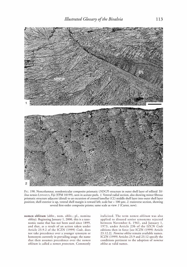
113Illustrated Glossary of the Bivalvia
nomen oblitum (abbr., nom. oblit.; pl., nomina oblita). Beginning January 1, 2000, this is a taxo-nomic name that has not been used since 1899, and that, as a result of an action taken under Article 23.9.2 of the ICZN (1999) Code, does not take precedence over a younger synonym or homonym currently in prevailing usage; the name that then assumes precedence over the nomen oblitum is called a nomen protectum. Commonly
italicized. The term nomen oblitum was also applied to disused senior synonyms rejected between November 6, 1961, and January 1, 1973, under Article 23b of the IZCN Code editions then in force [see ICZN (1999) Article 23.12.2]. Nomina oblita remain available names. ICZN (1999) Articles 23.9 and 23.12 specify the conditions pertinent to the adoption of nomina oblita as valid names.
FiG. 198. Noncolumnar, nondenticular composite prismatic (NDCP) structure in outer shell layer of tellinid Tel-lina nemies liNNaeus, Fiji (YPM 10199), seen in acetate peels. 1, Ventral radial section, also showing minor fibrous prismatic structure adjacent (distal) to an excursion of crossed lamellar (CL) middle shell layer into outer shell layer position; shell exterior is up, ventral shell margin is toward left; scale bar = 100 µm; 2, transverse section, showing
several first-order composite prisms; same scale as view 1 (Carter, new).
1
2
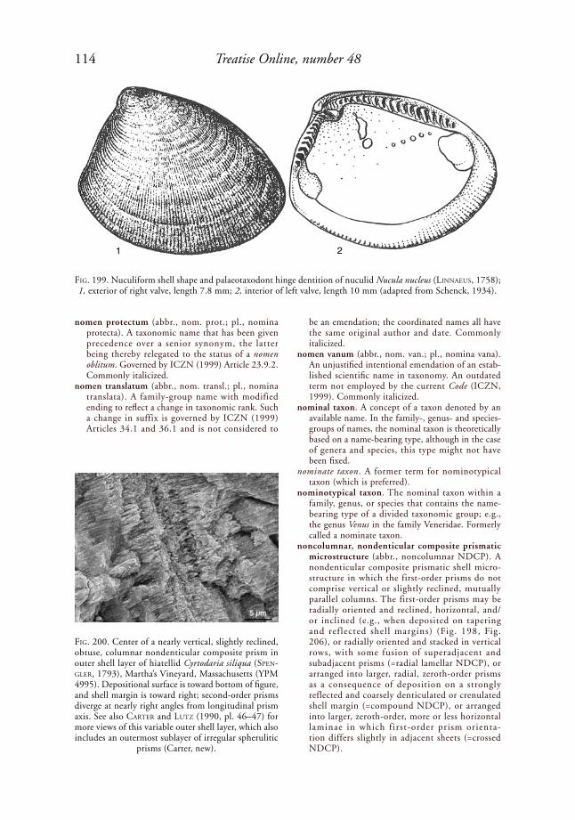
114 Treatise Online, number 48
nomen protectum (abbr., nom. prot.; pl., nomina protecta). A taxonomic name that has been given precedence over a senior synonym, the latter being thereby relegated to the status of a nomen oblitum. Governed by ICZN (1999) Article 23.9.2. Commonly italicized.
nomen translatum (abbr., nom. transl.; pl., nomina translata). A family-group name with modified ending to reflect a change in taxonomic rank. Such a change in suffix is governed by ICZN (1999) Articles 34.1 and 36.1 and is not considered to
be an emendation; the coordinated names all have the same original author and date. Commonly italicized.
nomen vanum (abbr., nom. van.; pl., nomina vana). An unjustified intentional emendation of an estab-lished scientific name in taxonomy. An outdated term not employed by the current Code (ICZN, 1999). Commonly italicized.
nominal taxon. A concept of a taxon denoted by an available name. In the family-, genus- and species-groups of names, the nominal taxon is theoretically based on a name-bearing type, although in the case of genera and species, this type might not have been fixed.
nominate taxon. A former term for nominotypical taxon (which is preferred).
nominotypical taxon. The nominal taxon within a family, genus, or species that contains the name-bearing type of a divided taxonomic group; e.g., the genus Venus in the family Veneridae. Formerly called a nominate taxon.
noncolumnar, nondenticular composite prismatic microstructure (abbr., noncolumnar NDCP). A nondenticular composite prismatic shell micro-structure in which the first-order prisms do not comprise vertical or slightly reclined, mutually parallel columns. The first-order prisms may be radially oriented and reclined, horizontal, and/or inclined (e.g., when deposited on tapering and reflected shell margins) (Fig. 198, Fig. 206), or radially oriented and stacked in vertical rows, with some fusion of superadjacent and subadjacent prisms (=radial lamellar NDCP), or arranged into larger, radial, zeroth-order prisms as a consequence of deposition on a strongly reflected and coarsely denticulated or crenulated shell margin (=compound NDCP), or arranged into larger, zeroth-order, more or less horizontal laminae in which first-order prism orienta-tion differs slightly in adjacent sheets (=crossed NDCP).
FiG. 199. Nuculiform shell shape and palaeotaxodont hinge dentition of nuculid Nucula nucleus (liNNaeus, 1758); 1, exterior of right valve, length 7.8 mm; 2, interior of left valve, length 10 mm (adapted from Schenck, 1934).
FiG. 200. Center of a nearly vertical, slightly reclined, obtuse, columnar nondenticular composite prism in outer shell layer of hiatellid Cyrtodaria siliqua (speN-Gler, 1793), Martha’s Vineyard, Massachusetts (YPM 4995). Depositional surface is toward bottom of figure, and shell margin is toward right; second-order prisms diverge at nearly right angles from longitudinal prism axis. See also Carter and lutZ (1990, pl. 46–47) for more views of this variable outer shell layer, which also includes an outermost sublayer of irregular spherulitic
prisms (Carter, new).
1 2
115Illustrated Glossary of the Bivalvia
nondenticular composite prismatic microstructure (abbr., NDCP). A composite prismatic shell micro-structure in which second-order prisms are arranged into three-dimensional, stacked, fanlike first-order prisms independently of coarse denticulation or crenulation of the depositional surface, although at high magnification, the depositional surface may show some microrelief (Carter, 1980b; Carter & Clark, 1985; Carter & others, 1990, p. 650). Categories include columnar NDCP and nonco-lumnar NDCP. Compare with denticular composite prismatic.
non-incubatory. Not brooding the larvae.nonplicate ctenidium. A ctenidium that does not
appear minutely folded in its relaxed, uncon-tracted state; e.g., members of Isognomonidae and Malleidae. Such ctenidia lack principal filaments (rideWood, 1903; see also tëMkiN, 2006a, p. 291, fig. 13a–b).
normal extinction. Optical crystallographic extinction that is parallel to the length axis of an element of shell microstructure. See also granular extinction and regular extinction.
nuculiform. Having a shell shape like Nucula laMarCk, 1799, i.e., subovate, anteriorly produced, and opis-thogyrate (Fig. 199).
nymph (pl., nymphae). An external or submarginal, opisthodetic, elongate, dorsally projecting, more or less blunt-edged ridge that is morphologically differentiated from the rest of the hinge, that supports the fibrous sublayer of a dorsally arched ligament, and that is flanked laterally by a groove for the lamellar sublayer of the ligament. To be a true nymph, rather than a pseudonymph, the fibrous sublayer of the ligament must not extend laterally beyond the base of the nymph. A true nymph is the defining component of a parivincular ligament. Compare with pseudonymph.
obconical. Bluntly conical.objective synonym. Each of two or more synonyms
that denote nominal taxa with the same name-bearing type, or (in the case of family-group and genus-group names) that denote nominal taxa with name-bearing types whose own names are themselves objectively synonymous (ICZN, 1999).
obligate. Restricted to a condition of life, as in the case of obligate parasites, which must use a host to survive.
oblique. Extending in a direction that is neither parallel nor perpendicular to growth lines, commonly in reference to shell ornament. Guo (1998, p. 278) defined four types of oblique ribs (a, b, c, d) for the superfamily Trigonioidoidea (see Fig. 264). See also V-shaped ribs, reversed V-shaped ribs, eu-V-shaped ribs, and para-V-shaped ribs.
obliquity. (1) The angle between the dorsal shell margin and the line bisecting the umbonal angle, which see; (2) the angle between the dorsal shell margin and the most distant point on the ventral shell margin.
oblong. Having an elongate, broadly subelliptical shape, with corners that are more rounded than rectangular or subrectangular.
obscure. Faint, indistinct, lacking prominence, commonly in reference to hinge teeth or shell ornament.
obsolete. Indistinct or absent, commonly in reference to a hinge tooth that is more strongly developed in closely related taxa.
obtrullate. Shaped like a trowel blade, i.e., with four straight sides, widest above the middle, and with a length/width ratio of 1.5 to 2.0. Compare with trullate.
obtuse. Bluntly pointed.obtuse columnar nondenticular composite prismatic
microstructure (abbr., obtuse columnar NDCP). A
FiG. 201. Right valve of upper Carboniferous myalinid Septimyalina perattenuata (Meek & haydeN, 1858), Kansas (AMNH 42888), showing opisthodetic, low-angle duplivincular ligament with both posteriorly and anteriorly inclined ridges and grooves (adapted from Newell & Boyd, 1987, fig. 7E; courtesy of the American
Museum of Natural History).
FiG. 202. Upper Carboniferous myalinid Septimyalina perattenuata (Meek & haydeN, 1858), diagram based on specimen from Magoffin Member, Breathitt Forma-tion, Milepost 55 on the Daniel Boone Parkway (UNC 13664). 1, Internal view of left hinge showing multiple ligamental groove/ridge couplets in opisthodetic, low-angle, duplivincular ligament; figure width is about 15 mm; 2, transverse section through posterior part of ligament, with lamellar sublayers restored as solid black bands; SP, calcitic regular simple prismatic outer shell layer; HOM, calcitic homogeneous outer shell layer; N,
nacre (adapted from Carter, 2004, fig. 2.1–2.2).
1
2 100 µm

116 Treatise Online, number 48
columnar, nondenticular composite prismatic shell microstructure in which the second-order prisms diverge at a high angle, sometimes nearly horizon-tally, from the longitudinal axis of the first-order prism; e.g., the outer shell layer of the hiatellid Cyrtodaria siliqua (speNGler, 1793) (Fig. 200) (Carter, herein). Compare with acute columnar NDCP and planar spherulitic simple prismatic microstructure.
ocellus (pl., ocelli). A minute, simple eye. See post-larval ctenidial ocelli.
oesophagus. An alternate spelling of esophagus.Official Indexes. Any of the four indexes maintained
and published by the ICZN, citing works or taxo-nomic names that have been rejected by their rulings. They include rejected and declared-invalid names of works, of family names, of generic names, and of specific names.
Official Lists. Any of the four lists maintained and published by the ICZN, citing works or taxonomic names that have been ruled upon and accepted as available in the Opinions of the Commission. These include: Official List of Works Approved as Available for Zoological Nomenclature, Official List of Family-Group Names in Zoology, Official List of Generic Names in Zoology, and Official List of Specific Names in Zoology. See also their List of Available Names in Zoology.
ontogeny. The sequence of development and growth of an individual.
oocyte. An ovum; a developing egg.oogenesis. The formation and maturation of female
gametes (ova).oogonium (pl., oogonia). A primordial female germ
cell that gives rise to an oocyte.Opinion. A published ruling by the ICZN involving
applications, interpretations, or suspensions of provisions of the ICZN Code in cases affecting one or more taxonomic names, nomenclatural acts, or works. An Opinion states how the ICZN Code is
to be applied or interpreted, or the course of action to be followed in a particular case.
opisthocline. (1) In reference to hinge dentition, a tooth, usually a cardinal tooth, which is inclined, from its lower end to its upper end, toward the posterior, i.e., having a length axis dipping toward the anterior end of the shell; (2) in reference to shell shape, having the axis of maximum growth inclined, from its distal, lower end to its proximal end, toward the posterior, i.e., having the axis of maximum growth dipping toward the anterior end of the shell; same as procrescent.
opisthodetic. Positioned entirely posterior to the beaks, commonly in reference to a ligament. See also amphidetic and prosodetic. When the ante-rior and posterior ends of the shell cannot be determined, the terms brevidetic and longidetic can be used instead. Note that some opisthodetic, low-angle duplivincular ligaments have ligamental grooves and ridges that may be both anteriorly and posteriorly inclined, as in the upper Carbonif-erous myalinid Septimyalina perattenuata (Meek & haydeN, 1858) (Fig. 201–202).
opisthogyrate. Having the umbo, beak, and/or the entire shell coiled such that the beak points toward the posterior; e.g., the noetiid Noetia ponderosa (say, 1822) (Fig. 203).
opisthog yrous. Same as opisthogyrate, which is preferred.
opisthopodium. (1) A posteriorly projecting sensory appendage, commonly finlike, developed on the posterior end of the visceral mass (B. MortoN, 1980), as in the pholadomyid Pholadomya G. B. soWerBy i, 1823 in 1821–1834; (2) an expanded heel of the foot, as in the arcoidean Noetia ponderosa (say, 1822).
optic tentacle. A tentacle that supports an eye, e.g., on the tip of the incurrent and excurrent siphons of the laternulid Laternula elliptica (kiNG & Broderip, 1832) (see Fig. 288).
optically heterogenous. Appearing as multiple optical crystallographic entities in thin sections under crossed polarized light, e.g., the regular simple prisms in the calcitic outer shell layer of Pterioidea and Ostreoidea.
optically homogeneous. Appearing as a single optical crystallographic entity in thin sections under crossed polarized light, e.g., the regular simple prisms in the calcitic outer shell layer of many Pinnoidea.
oral groove. Each of left and right ciliated grooves or channels extending from the anterior end of a labial palp to the mouth.
orbicular. Having a circular or nearly circular outline.orbital muscles. A band of ventral pallial muscles
inserting on the shell proximal to the normal pallial line, and extending into a (generally fused, except at the pedal aperture) inner mantle fold. These muscles may be evidenced by hyper-widening of the pallial line into a pallial band, or by a second line of pallial muscle attachment proximal to the normal pallial line, as in the gastrochaenid Dufoichaena dentifera (duFo, 1840)
FiG. 203. Dorsal view of united valves of noetiid Noetia ponderosa (say, 1822), showing monovincular D-1 liga-ment, opisthogyrate beaks, and other external features, shell length several cm (adapted from Mikkelsen &
Bieler, 2007).
117Illustrated Glossary of the Bivalvia
(Fig. 204). Not to be confused with interlinear and ctenidial attachment scars, which are more dorsally positioned. Also present in some Trape-zidae and Hiatellidae.
ordinary filament. (1) A filament that forms part of a uniform series of filaments in a homorhabdic ctenidium, i.e., without differentiated, intervening, larger (principal) filaments; (2) the smaller fila-ments in a heterorhabdic ctenidium, i.e., one with differentiated larger principal filaments (Fig.
133–134). rideWood (1903) noted that the devel-opment of ordinary filaments ontogenetically precedes the appearance of principal filaments. Compare with principal filaments.
organ of Bojanus. A kidney in the Bivalvia.organ of Deshayes. The body containing nitrogen-
fixing, cellulase-producing bacteria in the ctenidia of Teredinidae, wherein ducts in the afferent bran-chial vessels empty into the esophagus. Also called the gland of Deshayes.
FiG. 204. Gastrochaenid Dufoichaena dentifera (duFo, 1840), Lizard Island, northern Great Barrier Reef, alcohol-preserved (Australian Museum, Sydney, unnumbered specimen). 1, Anteroventral interior view of left valve, showing pallial sinus, pallial band, and orbital muscle scars; 2, similar part of interior of right valve, but with overlying mantle tissues intact (except for the free mantle margins, which have been cut away), showing pallial gland (Carter, new).

118 Treatise Online, number 48
organs of will. Pigmented glandular structures on the middle and inner mantle folds of Pinnidae; they are not eyespots, but are possibly associated with the production of pigment rays on the shell.
original designation (abbr., OD). A designation of the type species of a genus in the original publi-cation. Governed by ICZN (1999) Articles 68.1 and 73.1.1.
ornament. Another term for shell sculpture.oro-anal axis. The line connecting the mouth with the
anus, or with the center of the posterior adductor muscle (which approximately coincides with the anus) (JaCksoN, 1890).
orthocline. (1) In reference to hinge dentition, a tooth, generally a cardinal tooth, oriented with its longitu-dinal axis perpendicular or almost perpendicular to the hinge axis; (2) in reference to shell shape, a shell that does not slope appreciably anteriorly or poste-riorly, i.e., is neither prosocline nor opisthocline.
orthogyrate. Having the umbo, beak, and/or the entire shell coiling such that the beak points toward the commissural plane rather than toward the anterior or posterior, as in the glycymeridid Tucetona pecti-nata (GMeliN, 1791 in 1791–1793) (Fig. 205). Compare with prosogyrate and opisthogyrate.
orthogyrous. Same as orthogyrate, which is preferred.orthomorphic. Same as orthomorphodont, which is
preferred (sáNCheZ, 1999).orthomorphodont hinge tooth. A taxodont hinge
tooth that is more or less straight, i.e., neither convexodont nor concavodont (BaBiN, 1966, p. 39) (Fig. 207). Also called orthomorphic (sáNCheZ, 1999).
orthothetic. Same as euthetic, which see.osciculum. Same as lithodesma, which is preferred.oscules. The openings in the free valve of a hippuritid
bivalve, usually near its margin, corresponding with a pillar in the opposite, attached valve.
osculum (pl., oscula). See under septibranch.osphradia (sing., osphradium). Sensory patches of
epithelium present on the wall of the mantle cavity close to the ctenidia. Possibly chemoreceptors for testing the quality of incurrent water (Félix BerNard, 1890; yoNGe, 1947, p. 510), or otherwise related to the regulation of water flow (yoNGe,
1947). These generally occur in the suprabranchial chamber as small patches on the undersurface of the posterior adductor muscle, but in Pecten Müller, 1776, and Glycymeris da Costa, 1778, each osphra-dium forms a ridge along the axis of the posterior extremity of each ctenidium.
ossicle. Same as lithodesma, which is preferred.ossiculum. Same as lithodesma, which is preferred.ostium (pl., ostia). A small opening in a ctenidium,
formed by the intersection of filaments and inter-filamental junctions, or, in a ctenidial septum, containing ctenidial filaments. Also called a fenestra. See also eulamellibranch and septibranch.
ostracum. The shell excluding the periostracum.ostreiform. Having a shell shape like Ostrea liNNaeus,
1758.ostreine chomata. Chomata in the form of pustules,
sometimes individually antimarginally elongate, occurring in a single line parallel with the shell margin, and present only in the right valve, with pits to receive them in the left valve (harry, 1985, p. 133); e.g., members of the ostreid tribe Myra-keenini.
ostreoid ligament. An alivincular ligament with a high, triangular ligament area that in the first few milli-meters of growth is curved or altogether straight, but afterward becomes straight or only slightly curved in the opposite direction (MalChus, 1990, p. 76). These ligaments may be alivincular-arcuate or alivincular-areate (which see), e.g., alivincular-arcuate in the Recent ostreid Planostrea pestigris (haNley, 1846) (Fig. 7).
otocyst. Same as statocyst, which see. outer demibranch. The half of a typical W-shaped
ctenidium that is farther from the animal’s midsag-ittal plane, or its homolog in modified ctenidia. In its most complete form, the outer demibranch has both ascending and descending lamellae (see Fig. 289 and Fig. 307).
outer ligament. (1) The lamellar sublayer of the ligament, secreted beneath the periostracum and topologically above the fibrous sublayer of the ligament. This may occur as the outer part of a simple, parivincular, or quasiparivincular ligament, as part of each lamellar-fibrous ligament couplet in a duplivincular ligament, as the anterior and posterior parts of an alivincular ligament, or as the external part of an alivincular ligament with an external lamellar component and an internal lamellar + fibrous component (e.g., in Pectinidae); (2) a dorsal band of periostracum serving as a functional replacement for the lamellar sublayer of a ligament, when the true lamellar sublayer of the ligament is submerged below the axis of rotation of the valves; in such cases, the term periostracal outer ligament is preferred over the term outer ligament.
outer-ligament articulation. Articulation of the shell valves in certain pectinoids along a line at or near the dorsal shell margins, ventral to any scrolls that may be present. The lamellar, outer sublayer of the ligament attaches to a dorsal ridge in the right valve and to a dorsal groove in the left valve (e.g., Pernopectinidae), or vice versa (e.g., Entolioididae,
FiG. 205. Dorsal view of gylcymeridid Tucetona pectina-ta (GMeliN, 1791 in 1791–1793), showing orthogyrate beaks, shell length is 16.2 mm (adapted from Mikkelsen
& Bieler, 2007).
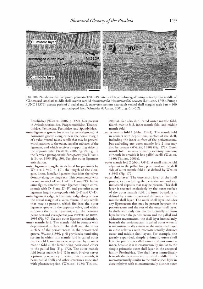
119Illustrated Glossary of the Bivalvia
Entoliidae) (Waller, 2006, p. 322). Not present in Aviculopectinoidea, Propeamussiidae, Tosapec-tinidae, Neitheidae, Pectinidae, and Spondylidae.
outer ligament groove (or outer ligamental groove). A horizontal groove along or near the dorsal margin of a valve, ventral to any scrolls that may be present, which attaches to the outer, lamellar sublayer of the ligament, and which receives a supporting ridge in the opposite valve (Waller, 2006, fig. 2); e.g., in the Permian pernopectinid Pernopecten yini NeWell & Boyd, 1995 (Fig. 30). See also outer-ligament articulation.
outer ligament length. As defined for pectinids by Waller (1969, p. 12), the length of the elon-gate, linear, lamellar ligament that joins the valves dorsally along the hinge axis. This corresponds with measurements C–F and C'–F' in Figure 219. In this same figure, anterior outer ligament length corre-sponds with D–F and D'–F', and posterior outer ligament length corresponds with C–D and C'–D'.
outer ligament ridge. A horizontal ridge along or near the dorsal margin of a valve, ventral to any scrolls that may be present, which fits into the outer ligament groove in the opposite valve, and which supports the outer ligament; e.g., the Permian pernopectinid Pernopecten yini NeWell & Boyd, 1995 (Fig. 30). See also outer-ligament articulation.
outer mantle fold. The mantle fold in contact with depositional surface of the shell and the inner surface of the periostracum in the periostracal groove. Waller (1980, p. 4) provided a numbering system in which this mantle fold is called outer mantle fold 1, sometimes accompanied by an outer mantle fold 2, the latter being positioned closer to the pallial line (Fig. 172). The outer mantle fold (outer mantle fold 1) in most bivalves serves a primarily secretory function, but in arcoids, it bears pallial ocelli and other structures associated with photoreception (Waller, 1980; tëMkiN,
2006a). See also duplicated outer mantle fold, fourth mantle fold, inner mantle fold, and middle mantle fold.
outer mantle fold 1 (abbr., OF-1). The mantle fold in contact with depositional surface of the shell, including the inner surface of the periostracum, but excluding any outer mantle fold 2 that may also be present (Waller, 1980) (Fig. 172). Outer mantle fold 1 serves a primarily secretory function, althouth in arcoids it has pallial ocelli (Waller, 1980; tëMkiN, 2006a).
outer mantle fold 2 (abbr., OF-2). A small mantle fold adjacent to the pallial line, positioned on the shell side of outer mantle fold 1, as defined by Waller (1980) (Fig. 172).
outer shell layer. The outermost layer of the shell proper, i.e., excluding the periostracum and any inductural deposits that may be present. This shell layer is secreted exclusively by the outer surface of the outer mantle fold. Its inner boundary is defined by a microstructural difference from the middle shell layer. The outer shell layer includes any ligostracum that may be present between the periostracum and the rest of the outer shell layer. In shells with only one microstructurally uniform layer between the periostracum and the pallial and adductor myostracum, the shell layer immediately beneath the periostracum is called outer when it is microstructurally similar to the outer shell layer in close relatives with microstructually distinct outer and middle shell layers. For example, the greatly expanded, simple prismatic outer shell layer in pinnids is called outer and not outer + inner, because it is microstructurally similar to the simple prismatic outer shell layer in the ancestral family Pterineidae. The shell layer immediately beneath the periostracum is called middle if it is microstructurally similar to the middle shell layer in close relatives with microstructurally distinct outer
FiG. 206. Nondenticular composite prismatic (NDCP) outer shell layer submerged ontogenetically into middle of CL (crossed lamellar) middle shell layer in cardiid Acanthocardia (Acanthocardia) aculeata (liNNaeus, 1758), Europe (UNC 15376); acetate peels of 1, radial and 2, transverse sections near adult ventral shell margin; scale bars = 100
µm (adapted from Schneider & Carter, 2001, fig. 6.1–6.2).
1 2

120 Treatise Online, number 48
and middle shell layers. For example, a CL layer, which comprises the entire shell exterior to the pallial and adductor myostracum, is called middle if it is similar to the middle shell layer in closely related species with a prismatic outer shell layer and a CL middle shell layer. In some veneroids, only isolated traces of an ancestral prismatic outer shell layer may be present. In pectinids, a simple prismatic outer shell layer is typically present only in the early postlarval, juvenile stage, and a foliated
middle shell layer comprises the entire adult shell exterior to the pallial and adductor myostracum. In the cardiid Acanthocardia Gray, 1851, the prismatic outer shell layer submerges, early in ontogeny, into the middle of the CL middle shell layer (popoV, 1977; sChNeider & Carter, 2001) (Fig. 206).
outer sublayer of ligament. The dorsal, nonmineral-ized portion of a ligament, excluding the perio-stracum, or in a portion of the ligament, such as in a single lamellar-fibrous couplet in a duplivin-cular ligament. This excludes any medial lamellar sublayer between the left and right fibrous sublayers of an internal, pectinoid ligament.
outline. The lateral profile of a shell.oval. Broadly elliptical or ellipsoidal.ovary. One of the usually paired female reproductive
organs, which produces eggs; in bivalves, this is often a prominent part of the anatomy, as in the lyonsiid Entodesma beana (d’orBiGNy, 1853 in 1841–1853) (see Fig. 311).
ovate. Shaped like the longitudinal section of an egg. override. To pass over without interruption, such as
commarginal ornament overriding radial ornament.oviparous. A mode of indirect development in which
eggs are laid and then hatched externally (liNColN, Boxshall, & Clark, 1988). See also larviparous, ovoviviparous, viviparous, and direct development.
ovoviviparous. Producing eggs that are retained within the body of the female until a larval or juvenile stage is attained, at which point the offspring are released. See also larviparous, oviparous, viviparous, direct development, and indirect development.
ovum (pl., ova). A female gamete; an egg.pachyodont. Having heavy, blunt, amorphous hinge
teeth, e.g., many Hippuritidae.padded. Slightly thickened or inflated.paedapody. Complete loss of the foot early in post-
larval life, when the larva settles and attaches, rather than afterward, e.g., Ostreoidea (harry, 1985, p. 123). Compare with gerontapody.
paedomorphosis. Heterochronic changes involving the suppression of stages or characters from an ancestral developmental sequence. The opposite of peramorphosis.
palaeobranch. A protobranch ctenidium as developed in the order Solemyida, as opposed to the ctenid-iobranch ctenidium present in other members of Protobranchia. A term based on both morphology and phylogenetic relationship.
palaeotaxodont dentit ion . A hinge dentit ion consisting of five or more regularly shaped taxodont teeth that are not ventrally bifid, and that may be orthomorphodont, concavodont, convexodont, or a combination thereof (Fig. 207). This term is herein restricted to members of the Protobranchia, e.g., Nucula nucleus (liNNaeus, 1758) (Fig. 199). Superficially similar hinges in the Pteriomorphia (e.g., cyrtodontoids and arcoids) are called neotax-odont. Palaeotaxodont (and neotaxodont) hinge teeth differ from pretaxodont hinge teeth in that they increase significantly in number with shell growth beyond 1 mm shell length. Palaeotaxodont hinges may be gradidentate or heterotaxodont
FiG. 207. Terminology of palaeotaxodont tooth shape, based on BaBiN (1966, fig. 3) (Carter, new).
diconvexodontconvexoconcavodont
convexodontorthomorphodont
concavodontconcavoconvexodont
diconcavodont
FiG. 208. Composite diagrammatic sketch of a pallet in teredinid Bankia Gray, 1842, showing all characters
(adapted from Turner, 1969, p. 705, fig. E164,3).
distal endembryonic capinner marginouter margin
blunt awnwebnonserrated awn
smooth marginfine serrationsserrated awnsperiostracal portioncalcaneous portion
comblike serrations
coarse serrations
stal
kco
nebl
ade
cone
s di
stan
tly s
pace
dco
nes
clos
ely
spac
ed

121Illustrated Glossary of the Bivalvia
(which see). This is the basis for the group name Palaeotaxodonta, originally proposed by koroBkoV (1954) for the Nuculidae, Ledidae (=Nuculanidae), and Malletiidae.
pallets. Paired left-right, paddle-shaped, calcareous-periostracal structures secreted by the posterior mantle at the base of the siphons, serving to close off the entrance of a wood boring when the siphons are retracted. Pallets may be in one piece, or they may consist of several elements, as indicated for the teredinid Bankia Gray, 1842, in Figure 208. Present in all extant Teredinidae, where their morphology is useful for distinguishing species.
pallial. Pertaining to the mantle (=pallium).pallial aperture. An opening delimited by partially or
entirely fused left and right mantle margins, e.g., a pedal aperture, incurrent siphonal aperture, excur-rent siphonal aperture, or fourth mantle aperture.
pallial band. A relatively wide pallial line (which see), e.g., ventrally in the gastrochaenid Dufoichaena dentifera (duFo, 1840) (Fig. 204).
pallial caecum. See caecum.pallial canals. Longitudinal, thin-walled canals in the
shell wall of some Caprinidae, present in either or both valves. The canals are bounded by radial, transverse, and tangential plates lying normal to the commissural plane.
pallial cavity. Same as mantle cavity, which see.pallial curtain. A typically wide, innermost mantle
fold present in many members of Pteriomorphia, which can be manipulated to vary the shape, size, and location of openings between opposing mantle margins.
pallial eyes. Organs of photoreception, innervated by visceral ganglia, on a mantle fold near the shell margin (e.g., Pectinidae, Spondylidae) or near the end of a siphon (e.g., some Cardiidae and around the incurrent siphon in the laternulid Laternula truncata (laMarCk, 1818 in 1818–1822) (Fig. 209). Also called pallial ocelli.
pallial fold. (1) A gutterlike, tapering extension of the inner mantle fold that meets the tip of a ctenidium; (2) a symmetrical structure present on opposite sides of the posterior portion of the mantle margins, separating the incurrent and excurrent mantle apertures, the former positioned below and the latter positioned above the pallial fold. Where present in Pterioidea, e.g., in the pteriid Pinctada imbricata rödiNG, 1798, the left and right pallial folds are united by interlocking tentacles of the inner mantle fold (tëMkiN, 2006a, p. 283).
pallial gills. Secondary respiratory organs formed by modification of the inner surface of the mantle, e.g., near the anterior adductor muscle in the lucinid Phacoides pectinatus (GMeliN, 1791 in 1791–1793) (Fig. 210). These structures also aid in separating respiratory surfaces from parts of the anatomy containing endosymbionts. Also called mantle gills.
pallial gland. A thickened, smooth or rugose portion of the inner mantle, densely filled with secretion, just proximal to the base of the inner mantle fold and the pallial line, commonly containing two types of
secretory cells, and covered, on the mantle cavity side, by ciliated epithelium. Secretes mucus to assist in cleansing the mantle cavity and possibly also to assist chemical boring, e.g., in the gastrochaenoi-deans Dufoichaena dentifera (duFo, 1840) (Fig. 204) and Spengleria (Fig. 176). In gastrochaenoideans, this gland follows the curve of the pallial line from the anteroventral mantle cavity to the base of the pallial sinus (Carter, 1978). These glands cannot be applied directly to the bored substratum, contrary to B. MortoN (1982b). Positionally similar glands occur in the crassatellid Crassatella laMarCk, 1799 (harry, 1966; alleN, 1968), and in some Cardi-tidae (see yoNGe, 1969), Myidae, Corbulidae, and Hiatellidae. Also called pallial mucus glands and crassatellid mantle margin glands.
pallial junction. The place where the anterior and posterior branches of the pallial line meet.
pallial line. The line of attachment of the pallial muscles to the shell. Relatively wide pallial lines are also called pallial bands. The pallial line may be continuous or discontinuous, and it includes the pallial sinus and any orbital muscle scars that may be present (Fig. 192). If an anterior adductor muscle is no longer present, the pallial line may extend only from the posterior adductor muscle scar toward the hinge. In Pinnidae, where a pallial line is absent, pallial muscle attachment is limited to a small area anteroventral to the posterior adductor muscle (see pallial retractor muscle).
pallial mucus glands. See pallial glands.pallial muscles. Radially oriented mantle muscles
extending from the pallial line or pallial band distally into one or more mantle folds. These
FiG. 209. Pallial eyes at tip of tentacles around incur-rent siphon in laternulid Laternula truncata (laMarCk, 1818 in 1818–1822), Kungkraben Bay, Thailand, A F. Sartori, collector, figure width approximately 2.9 mm
(Sartori, new).

122 Treatise Online, number 48
muscles attach the mantle to the shell and serve to retract its margins; e.g., the gastrochaenid Spen-gleria sp. (Fig. 176). In gastrochaenids, orbital pallial muscles may also be present, inserting proximal to the normal pallial line, and in some instances forming a second, more proximal line of pallial muscle attachment.
pallial nerve. A nerve running close to and more or less parallel with the mantle margin on each side of the body, connected anteriorly with the cerebropleural ganglion and posteriorly with the visceral ganglion.
pallial ocelli. Same as pallial eyes, which see.pallial organ. (1) The pallial organ of pelseNeer
(1911), a sensory organ near the siphonal retractor muscles and innervated by the posterior pallial nerve. pelseNeer speculated that this organ tests respiratory water in siphonate bivalves, like the abdominal sense organ in asiphonate bivalves. Absent in many Protobranchia and Pteriomorphia, present in Nuculanidae, Carditidae, Mactridae, Donacidae, Pholadidae, and some Tellinidae; (2) in Ostreidae, same as the abdominal sense organ, which see; (3) in Pinnidae, a conspicuous, hydro-static, tube-shaped organ extending posteriorly
from the posterodorsal surface of the visceral mass toward the posterior gape (tëMkiN, 2006a) (Fig. 211), probably serving for chemical defense and to clean the mantle cavity (yoNGe, 1953; liaNG & MortoN, 1988).
pallial protractors. Radial mantle muscles that serve to extend the mantle edge.
pallial punctae. Small, mutually isolated mantle attachment scars, which see.
pallial region. The part of the shell interior to the pallial line or pallial band.
pallial retractors. Radial mantle muscles that serve to withdraw the mantle margins. These appear on the shell interior as a pallial line, a pallial band, a pallial sinus, a line of orbital muscle scars, a single muscle scar anteroventral to the posterior adductor muscle as in Pinnidae (Fig. 211) and Malleidae (see Fig. 238), or as a short line of muscle scars between the posterior adductor muscle and the hinge (e.g., in Ostreoidea).
pallial rib. Same as pallial ridge, which is preferred.pallial ridge. An internal buttress extending in each
valve from either the visceral rim or, more dorsally, from the insertion of the pallial retractor muscle, more or less medially in the shell, toward the ventral shell margin. Called the pallial rib by Boss and Moore (1967). The pallial ridge corresponds with the line along which the ctenidia are fully extended (Boss & Moore, 1967). Present in many Malleidae, e.g., Malleus candeanus (d’orBiGNy, 1853 in 1841–1853) (see Fig. 326).
pallial septum. See siphonal pallial septum.pallial sinus. An embayment in the posterior part of
the pallial line that corresponds with attachment of the siphonal retractor muscles. It demarcates that part of the mantle cavity into which the siphons can be retracted (Fig. 192, Fig. 212).
pallial siphon. See siphon.pallial veil. An expanded inner mantle fold that serves
to close or partially close an unfused mantle margin when the valves are gaping, e.g., in Pteriidae, Isog-nomonidae, Malleidae, Ostreidae, Gryphaeidae, Pinnidae, Limidae, Pectinidae, Propeamussiidae, Spondylidae, Plicatulidae, and Anomiidae. Also called a velum. It is typically supplied with radial and circular muscles, and it may have small and large papillae and/or tentacles, e.g., in Limidae.
palliobranchial fusion. Tissue grade union between a portion of the ctenidia and the mantle. Typical of Ostreoidea, where the aboral ends of the ctenidia are fused to the mantle lobes.
palp. See labial palps.palp caecum. See palp pouch.palp lamellae. One of four usually acutely triangular,
leaflike, contiguous flaps comprising the labial palps. The lamellae of the upper (anterior) and lower (posterior) lip are called the outer and inner lamellae, respectively.
palp pouch. A non-extensible appendage representing a pair of hypertrophied palp folds on each side of the body at the posterior end of the labial palps, positioned at the base of the two palp proboscides. The palp pouch receives potential food particles
FiG. 210. Pallial (mantle) gills lying along and extend-ing ventrally beyond gutter between anterior adductor muscle and anterior mantle margin in lucinid Phacoides pectinatus (GMeliN, 1791 in 1791–1793); scale bar = 1 mm (adapted from Taylor & Glover, 2006, fig. 10C,
courtesy of John Wiley & Sons).
FiG. 211. Diagram of anatomy of Pinnidae (adapted from Mikkelsen & Bieler, 2007).

123Illustrated Glossary of the Bivalvia
from the palp proboscides prior to transport to the palps for sorting. Present in Nuculidae (Fig. 213). Also called a palp caecum.
palp proboscides (sing., palp proboscis). A pair of extensile, ciliate, tentacle-like extensions of the dorsoposterior corner of the left and right sides of the outer palp lamella. Present in many Protobran-chia, where it gathers food during deposit-feeding, e.g., in Nuculidae (Fig. 213) and Sareptidae (see Fig. 234).
papilla (pl., papillae). A small protuberance, distally tapered or rounded, typically digitate, commonly occurring on mantle margins at or near a mantle aperture or siphon; less prominent and less complex than a tentacle, e.g., on the incurrent and excurrent siphons in the gastrochaenid Lamychaena cordi-formis (NeVill & NeVill, 1869) (Fig. 22).
papillate. Having papillae.papilliform. Shaped like a papilla; a small, nipplelike
process or projection.paracladistic systematics. A modification of cladistic
systematics in which paraphyletic natural groups (which see) are allowed, with their size being mini-mized through the use of paraplesions; plesions and paraplesions are ranked; and the amount of evolutionary change is considered when assigning taxonomic rank. The methods and definitions of cladistic systematics otherwise apply. In paracla-distic systematics, as in evolutionary systematics, one may regard the common ancestor of modern birds as derived from a nontype species of Archae-opteryx, whereas in cladistic systematics, one must erect a new, monospecific genus for the purpose of containing such an ancestral species.
FiG. 212. Middle Eocene clavagellid Clavagella insulana Belokrys, 1991, Dnepropetrovsk region, Ukraine (Geo-logic Museum of Krivorozh’ye Mining Institute, holotype (BV-147/51); right, slightly less convex valve. 1, Ventral view of right valve showing chondrophore, posterior adductor muscle scar, and adjacent posterior pedal retractor muscle scar; 2, lateral interior view of same valve, showing chondrophore, pallial sinus, anterior adductor muscle scar (ant. add.), posterior adductor muscle scar ( post. add.), and posterior pedal retractor muscle scar ( post. pedal
retractor) (Carter, new).
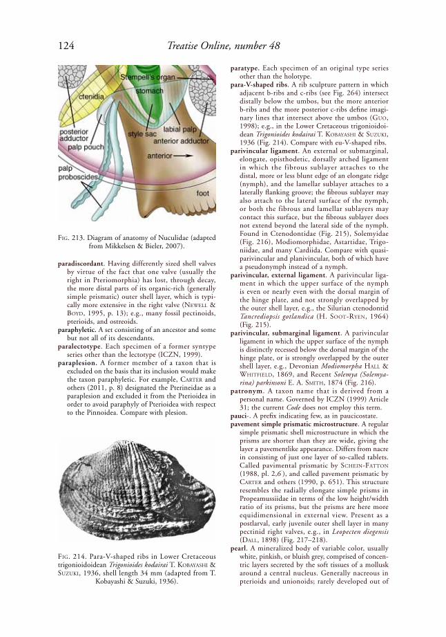
124 Treatise Online, number 48
paradiscordant. Having differently sized shell valves by virtue of the fact that one valve (usually the right in Pteriomorphia) has lost, through decay, the more distal parts of its organic-rich (generally simple prismatic) outer shell layer, which is typi-cally more extensive in the right valve (NeWell & Boyd, 1995, p. 13); e.g., many fossil pectinoids, pterioids, and ostreoids.
paraphyletic. A set consisting of an ancestor and some but not all of its descendants.
paralectotype. Each specimen of a former syntype series other than the lectotype (ICZN, 1999).
paraplesion. A former member of a taxon that is excluded on the basis that its inclusion would make the taxon paraphyletic. For example, Carter and others (2011, p. 8) designated the Pterineidae as a paraplesion and excluded it from the Pterioidea in order to avoid paraphyly of Pterioidea with respect to the Pinnoidea. Compare with plesion.
paratype. Each specimen of an original type series other than the holotype.
para-V-shaped ribs. A rib sculpture pattern in which adjacent b-ribs and c-ribs (see Fig. 264) intersect distally below the umbos, but the more anterior b-ribs and the more posterior c-ribs define imagi-nary lines that intersect above the umbos (Guo, 1998); e.g., in the Lower Cretaceous trigonioidoi-dean Trigonioides kodairai t. koBayashi & suZuki, 1936 (Fig. 214). Compare with eu-V-shaped ribs.
parivincular ligament. An external or submarginal, elongate, opisthodetic, dorsally arched ligament in which the fibrous sublayer attaches to the distal, more or less blunt edge of an elongate ridge (nymph), and the lamellar sublayer attaches to a laterally flanking groove; the fibrous sublayer may also attach to the lateral surface of the nymph, or both the fibrous and lamellar sublayers may contact this surface, but the fibrous sublayer does not extend beyond the lateral side of the nymph. Found in Ctenodontidae (Fig. 215), Solemyidae (Fig. 216), Modiomorphidae, Astartidae, Trigo-niidae, and many Cardiida. Compare with quasi-parivincular and planivincular, both of which have a pseudonymph instead of a nymph.
parivincular, external ligament. A parivincular liga-ment in which the upper surface of the nymph is even or nearly even with the dorsal margin of the hinge plate, and not strongly overlapped by the outer shell layer, e.g., the Silurian ctenodontid Tancrediopsis gotlandica (H. soot-ryeN, 1964) (Fig. 215).
parivincular, submarginal ligament. A parivincular ligament in which the upper surface of the nymph is distinctly recessed below the dorsal margin of the hinge plate, or is strongly overlapped by the outer shell layer, e.g., Devonian Modiomorpha hall & WhitField, 1869, and Recent Solemya (Solemya-rina) parkinsoni e. a. sMith, 1874 (Fig. 216).
patronym. A taxon name that is derived from a personal name. Governed by ICZN (1999) Article 31; the current Code does not employ this term.
pauci-. A prefix indicating few, as in paucicostate.pavement simple prismatic microstructure. A regular
simple prismatic shell microstructure in which the prisms are shorter than they are wide, giving the layer a pavementlike appearance. Differs from nacre in consisting of just one layer of so-called tablets. Called pavimental prismatic by sCheiN-FattoN (1988, pl. 2,6 ), and called pavement prismatic by Carter and others (1990, p. 651). This structure resembles the radially elongate simple prisms in Propeamussiidae in terms of the low height/width ratio of its prisms, but the prisms are here more equidimensional in external view. Present as a postlarval, early juvenile outer shell layer in many pectinid right valves, e.g., in Leopecten diegensis (dall, 1898) (Fig. 217–218).
pearl. A mineralized body of variable color, usually white, pinkish, or bluish grey, comprised of concen-tric layers secreted by the soft tissues of a mollusk around a central nucleus. Generally nacreous in pterioids and unionoids; rarely developed out of
FiG. 213. Diagram of anatomy of Nuculidae (adapted from Mikkelsen & Bieler, 2007).
FiG. 214. Para-V-shaped ribs in Lower Cretaceous trigonioidoidean Trigonioides kodairai t. koBayashi & suZuki, 1936, shell length 34 mm (adapted from T.
Kobayashi & Suzuki, 1936).
125Illustrated Glossary of the Bivalvia
porcelaneous structure in some Cardiida, such as Tridacninae and Veneridae.
pearl nucleus. Extraneous material around which concentric layers of calcium carbonate (often nacre) are deposited to form a pearl.
pearl sac. The mantle epithelium (usually within the gonad) secreting a cultured pearl.
pectinate. Having closely parallel, toothlike projec-tions; comblike. Compare with micropectinate.
pectineum. Same as ctenolium, which is preferred.pectinidium. Same as ctenolium, which is preferred.
pectinid measurements. Measurements of pectinid shells have been defined by Waller (1969, p. 9–13) in reference to his figure 4, reproduced in Figure 219. With dashes indicating measurements, these include adductor insertion center (j), adductor insertion centrality (b–j/a–b), adductor insertion first diameter (i–k), adductor insertion posteriority (a–b/A–G), adductor insertion second diameter (p–q), adductor insertion ventrality (b–j/A–M), anterior dorsal half-diameter of disc (G–P), ante-rior flaring ratio of disc (D–G/G–P), anterior
FiG. 215. External, opisthodetic, parivincular ligament in Silurian ctenodontid Tancrediopsis gotlandica (H. soot-ryeN, 1964); camera-lucida drawing based on a transverse section just posterior to beaks (adapted from Carter,
2001, fig. 9.3).
FiG. 216. Submarginal, opisthodetic, parivincular ligament in solemyid Solemya (Solemyarina) parkinsoni e. a. sMith, 1874, Awarua Bay, New Zealand (YPM 5364); camera lucida drawing based on a transverse section just
behind beaks (adapted from Carter, 1990a, fig. 17).
prismatic outer shell layer
lamellarsublayer
nymph1 mm
fibrous sublayer
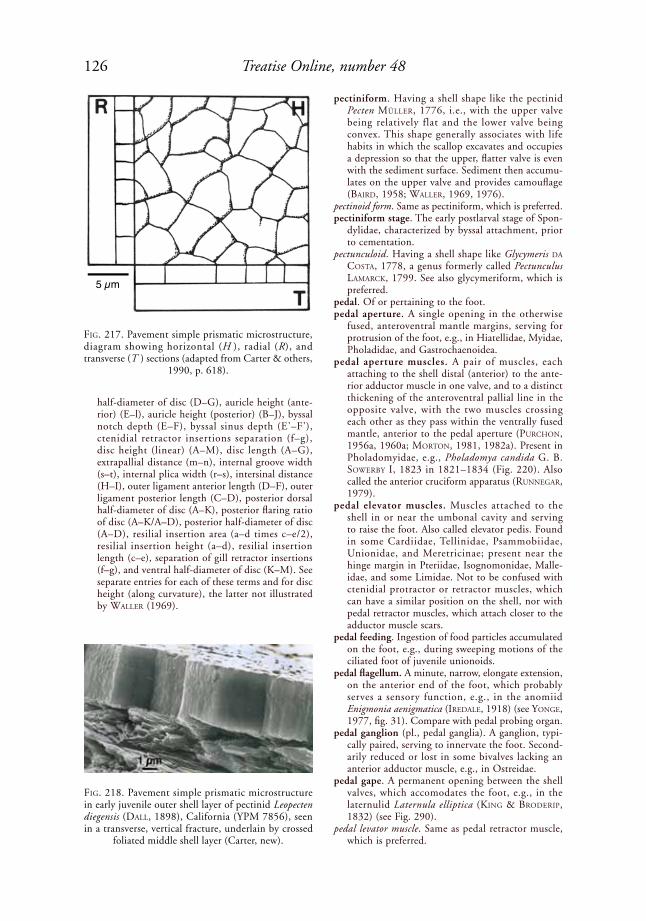
126 Treatise Online, number 48
half-diameter of disc (D–G), auricle height (ante-rior) (E–l), auricle height (posterior) (B–J), byssal notch depth (E–F), byssal sinus depth (E'–F'), ctenidial retractor insertions separation (f–g), disc height (linear) (A–M), disc length (A–G), extrapallial distance (m–n), internal groove width (s–t), internal plica width (r–s), intersinal distance (H–I), outer ligament anterior length (D–F), outer ligament posterior length (C–D), posterior dorsal half-diameter of disc (A–K), posterior flaring ratio of disc (A–K/A–D), posterior half-diameter of disc (A–D), resilial insertion area (a–d times c–e/2), resilial insertion height (a–d), resilial insertion length (c–e), separation of gill retractor insertions (f–g), and ventral half-diameter of disc (K–M). See separate entries for each of these terms and for disc height (along curvature), the latter not illustrated by Waller (1969).
pectiniform. Having a shell shape like the pectinid Pecten Müller, 1776, i.e., with the upper valve being relatively flat and the lower valve being convex. This shape generally associates with life habits in which the scallop excavates and occupies a depression so that the upper, flatter valve is even with the sediment surface. Sediment then accumu-lates on the upper valve and provides camouflage (Baird, 1958; Waller, 1969, 1976).
pectinoid form. Same as pectiniform, which is preferred. pectiniform stage. The early postlarval stage of Spon-
dylidae, characterized by byssal attachment, prior to cementation.
pectunculoid. Having a shell shape like Glycymeris da Costa, 1778, a genus formerly called Pectunculus laMarCk, 1799. See also glycymeriform, which is preferred.
pedal. Of or pertaining to the foot.pedal aperture. A single opening in the otherwise
fused, anteroventral mantle margins, serving for protrusion of the foot, e.g., in Hiatellidae, Myidae, Pholadidae, and Gastrochaenoidea.
pedal aperture muscles. A pair of muscles, each attaching to the shell distal (anterior) to the ante-rior adductor muscle in one valve, and to a distinct thickening of the anteroventral pallial line in the opposite valve, with the two muscles crossing each other as they pass within the ventrally fused mantle, anterior to the pedal aperture (purChoN, 1956a, 1960a; MortoN, 1981, 1982a). Present in Pholadomyidae, e.g., Pholadomya candida G. B. soWerBy i, 1823 in 1821–1834 (Fig. 220). Also called the anterior cruciform apparatus (ruNNeGar, 1979).
pedal elevator muscles. Muscles attached to the shell in or near the umbonal cavity and serving to raise the foot. Also called elevator pedis. Found in some Cardiidae, Tellinidae, Psammobiidae, Unionidae, and Meretricinae; present near the hinge margin in Pteriidae, Isognomonidae, Malle-idae, and some Limidae. Not to be confused with ctenidial protractor or retractor muscles, which can have a similar position on the shell, nor with pedal retractor muscles, which attach closer to the adductor muscle scars.
pedal feeding. Ingestion of food particles accumulated on the foot, e.g., during sweeping motions of the ciliated foot of juvenile unionoids.
pedal flagellum. A minute, narrow, elongate extension, on the anterior end of the foot, which probably serves a sensory function, e.g., in the anomiid Enigmonia aenigmatica (iredale, 1918) (see yoNGe, 1977, fig. 31). Compare with pedal probing organ.
pedal ganglion (pl., pedal ganglia). A ganglion, typi-cally paired, serving to innervate the foot. Second-arily reduced or lost in some bivalves lacking an anterior adductor muscle, e.g., in Ostreidae.
pedal gape. A permanent opening between the shell valves, which accomodates the foot, e.g., in the laternulid Laternula elliptica (kiNG & Broderip, 1832) (see Fig. 290).
pedal levator muscle. Same as pedal retractor muscle, which is preferred.
FiG. 217. Pavement simple prismatic microstructure, diagram showing horizontal (H ), radial (R), and transverse (T ) sections (adapted from Carter & others,
1990, p. 618).
FiG. 218. Pavement simple prismatic microstructure in early juvenile outer shell layer of pectinid Leopecten diegensis (dall, 1898), California (YPM 7856), seen in a transverse, vertical fracture, underlain by crossed
foliated middle shell layer (Carter, new).
5 µm

127Illustrated Glossary of the Bivalvia
pedal muscles. The muscles used to move the foot, passing either around the visceral mass (many burrowing bivalves) or largely through the visceral mass (many byssally attached bivalves). Divided into pedal elevators, pedal retractors, pedobyssal retractors, and pedal protractors.
pedal organ. See pedal probing organ. pedal probing organ. A spatulate to digitate exten-
sion of the anterior of the foot, which is capable of extreme elongation and chemically producing pinhole-size borings in calcareous substrata. The chemical boring agent is probably produced by the anterior pedal gland, at the base of the probing organ. Present in most gastrochaenids, e.g., Lamychaena cordiformis (NeVill & NeVill, 1869) (Fig. 19). The minute probes apparently test the thickness of the substratum in order to help the animal determine an advantageous boring direction (Carter, 1978). Compare with pedal flagellum.
pedal protractor muscle. A muscle that extends the foot. The muscle may be indistinctly or distinctly differentiated morphologically and in terms of its shell attachment scar from the anterior pedal retractor muscle. The associated muscle scar may be posterior, dorsoposterior, posteroventral, or anteroventral to the anterior adductor muscle scar; e.g., the gastrochaenids Rocellaria dubia (peNNaNt, 1777) (Fig. 192) and Spengleria (Fig. 176); and many unionoids (see Fig. 307).
pedal retractor muscle. A muscle serving to pull the foot toward the body, e.g., the gastrochae-nids Rocellaria dubia (peNNaNt, 1777) (Fig. 20) and Spengleria sp. (Fig. 176). See also pedobyssal retractor muscle, anterior pedal retractor muscle, and posterior pedal retractor muscle. Not to be
confused with pedal elevator muscles, nor with gill retractor or protractor muscles.
pedal scar. An impressed or raised area on the inner wall of a boring or on the inner surface of a secreted calcareous tube or igloo, marking the position of the foot in life; e.g., many endolithic and tube-dwelling Gastrochaenidae.
pediveliger. In Autobranchia, a larval stage in which both the velum and foot are present. The associ-ated shell form differs little from the preceding veliger stage, apart from its larger size and, in some
FiG. 219. Scheme of measurement of pectinoid shells, as illustrated by pectinid Argopecten gibbus (liNNaeus, 1758). Upper-case letters refer to points delimiting external measurements, and lower-case letters refer to points delimiting internal measurements; prime sign (') is placed after points on left valve to distinguish them from points on right valve; point L, not shown, is point on exterior valve surface that is farthest from commissure plane; point O, also not shown, is directly beneath point L on commissure plane; points a and D are the same. See pectinid measure-
ments (adapted from Waller, 1969, fig. 4).
FiG. 220. Pedal aperture muscles and pedal aperture in pholadomyid Pholadomya candida G. B. soWerBy i, 1823 in 1821–1834, St. Thomas, West Indies (ZMUC 5/8/1838), viewed from interior of mantle cavity, figure
width 17.8 mm (Sartori, new).
anterior adductorpedal aperture
muscles
pedal aperture
A' B'C' D' E' F' G'
H' I'
K'
M'
P'
J'
ABCDEFG
HI
K
M
P
J
a b
c d e
f g
i
k
m
j
b'
i'k'
m'
p'
j'
t'
q'
n'
rt
q
n
su
vr' s'
f'g'p
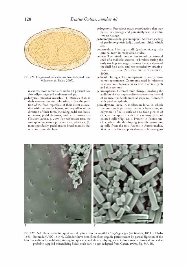
128 Treatise Online, number 48
instances, more accentuated umbo (if present). See also veliger stage and umbonate veliger.
pedobyssal retractor muscles. (1) Muscles that, in their contraction and relaxation, affect the posi-tion of the foot, regardless of their direct associa-tion with the foot or byssus, and regardless of the direction of their force, including pedal and byssal retractors, pedal elevators, and pedal protractors (tëMkiN, 2006a, p. 299). For nonbyssate taxa, the corresponding term is pedal retractor, which see; (2) more specifically, pedal and/or byssal muscles that serve to retract the foot.
pedogenesis. Precocious sexual reproduction that may persist in a lineage and potentially lead to evolu-tionary change.
pedomorphosis (adj., pedomorphic). Alternate spelling of paedomorphosis (adj., paedomorphic), which see.
pedunculate. Having a stalk (peduncle), e.g., the cardinal teeth in many Solecurtidae.
pellicle. The initial, more or less round, periostracal shell of a mollusk; secreted in bivalves during the early trochophore stage, covering the apical pole of the shell field cells, and not preceded by invagina-tion of this zone (MouëZa, Gros, & FreNkiel, 2006).
pellucid. Having a clear, transparent, or nearly trans-parent appearance. Commonly used in reference to myostracal deposits, as viewed in acetate peels and thin sections.
peramorphosis. Heterochronic changes involving the addition of new stages and/or characters to the end of an ancestral developmental sequence. Compare with paedomorphosis.
pericalymma larva. A molluscan larva in which the embryo is protected below a layer (test, or calymma) of cells with one to four girdles of cilia, at the apex of which is a sensory plate of ciliated cells (Fig. 221). Present in Protobran-chia, where the developing juvenile grows out apically from the test. Absent in Autobranchia. Whether the bivalve pericalymma is homologous
FiG. 221. Diagram of pericalymma larva (adapted from Mikkelsen & Bieler, 2007).
FiG. 222. 1–2, Fluorapatite intraperiostracal cylinders in the mytilid Lithophaga nigra (d’orBiGNy, 1853 in 1841–1853), Bermuda (UNC 13147). Cylinders have been freed from organic periostracum by partial digestion of the latter in sodium hypochlorite, rinsing in tap water, and then air drying; view 1 also shows periostracal pores that
probably supplied mineralizing fluids; scale bars = 1 µm (adapted from Carter, 1990a, fig. 54A–B).
1 2

129Illustrated Glossary of the Bivalvia
with other molluscan pericalymma larvae, and with the bivalve veliger larva, remains an open question. See also veliger, umbonate veliger, and pediveliger.
pericardial cavity. The space surrounding the heart.pericardial glands. Glands occurring as lappets on the
auricles of the heart or as pouches on the pericardial wall, either inside or outside the pericardial cavity; these glands typically extend into the mantle and function in excretion.
pericardial wall muscle. A minute muscle passing from the left and right shell valves to the pericardial wall in some pectinoids. The corresponding muscle scar is immediately dorsal to the posterior adductor muscle scar, e.g., the pectinid Chlamys islandica (Müller, 1776) (Fig. 57). This was previously called a gill-retractor muscle by Waller (1969, fig. 3), in his description of the pectinid Argopecten gibbus (liNNaeus, 1758).
pericardium. A saclike membrane, of coelomic origin, enclosing the heart and sometimes also part of the intestines, or the cavity delimited by such a membrane. The pericardium in bivalves generally encloses two lateral auricles and a median ventricle.
perimyal collecting vessel. See collecting vessel. periostracal cylinders. Minute, cylindrical, mineral-
ized structures secreted within the periostracum. These are fluorapatitic in the mytilid Lithophaga nigra (d’orBiGNy, 1853 in 1841–1853) (Fig. 222). Compare with periostracal granules, spikes, pins, needles, and plaques.
periostracal granules. Discrete, more or less equi-dimensional, mineralized structures initiated by the inner surface of the outer mantle fold and secreted simultaneously with normal, nonmineral-ized periostracum. The granules may be cemented to the exterior of the subperiostracal shell, as in the thraciid Thracia morrisoni petit, 1964 (Fig. 126),
FiG. 223. Aragonitic intraperiostracal granules and spikes in mytilid Trichomya hirsuta (laMarCk, 1819 in 1818–1822), Western Pacific (YPM 10124). 1, Acetate peel of a radial, vertical section through periostracum, showing numerous periostracal granules; underlying outer shell layer is also visible; 2, SEM of granules and a few spikes freed from organic portion of periostracum by digestion of the latter in sodium hypochlorite, followed by rinsing
in tap water and then air drying (Carter, new).
1
2
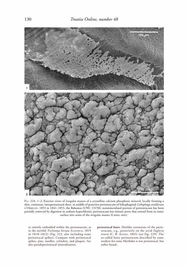
130 Treatise Online, number 48
or entirely embedded within the periostracum, as in the mytilid Trichomya hirsuta (laMarCk, 1819 in 1818–1822) (Fig. 223, also including some periostracal spikes). Compare with periostracal spikes, pins, needles, cylinders, and plaques. See also pseudoperiostracal mineralization.
periostracal hairs. Hairlike extensions of the perio-stracum, e.g., posteriorly on the arcid Fugleria tenera (C. B. adaMs, 1845) (see Fig. 229). The so-called hairy periostracum described by some workers for some Mytilidae is not periostracal, but rather byssal.
FiG. 224. 1–2, Exterior views of irregular masses of a crystalline calcium phosphatic mineral, locally forming a thin, contiuous, intraperiostracal sheet, in middle of posterior periostracum of lithophaginid Lithophaga antillarum d’orBiGNy, 1853 in 1841–1853, the Bahamas (UNC 13150); nonmineralized portion of periostracum has been partially removed by digestion in sodium hypochlorite; periostracum has minute pores that extend from its inner
surface into some of the irregular masses (Carter, new).
1
2
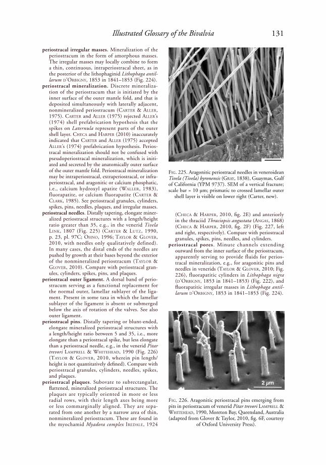
131Illustrated Glossary of the Bivalvia
periostracal irregular masses. Mineralization of the periostracum in the form of amorphous masses. The irregular masses may locally combine to form a thin, continuous, intraperiostracal sheet, as in the posterior of the lithophaginid Lithophaga antil-larum d’orBiGNy, 1853 in 1841–1853 (Fig. 224).
periostracal mineralization. Discrete mineraliza-tion of the periostracum that is initiated by the inner surface of the outer mantle fold, and that is deposited simultaneously with laterally adjacent, nonmineralized periostracum (Carter & aller, 1975). Carter and aller (1975) rejected aller’s (1974) shell prefabrication hypothesis that the spikes on Laternula represent parts of the outer shell layer. CheCa and harper (2010) inaccurately indicated that Carter and aller (1975) accepted aller’s (1974) prefabrication hypothesis. Perios-tracal mineralization should not be confused with pseudoperiostracal mineralization, which is initi-ated and secreted by the anatomically outer surface of the outer mantle fold. Periostracal mineralization may be intraperiostracal, extraperiostracal, or infra-periostracal, and aragonitic or calcium phosphatic, i.e., calcium hydroxyl apatite (Waller, 1983), fluorapatite, or calcium fluorapatite (Carter & Clark, 1985). See periostracal granules, cylinders, spikes, pins, needles, plaques, and irregular masses.
periostracal needles. Distally tapering, elongate miner-alized periostracal structures with a length/height ratio greater than 35, e.g., in the venerid Tivela liNk, 1807 (Fig. 225) (Carter & lutZ, 1990, p. 23, pl. 97C; ohNo, 1996; taylor & GloVer, 2010, with needles only qualitatively defined). In many cases, the distal ends of the needles are pushed by growth at their bases beyond the exterior of the nonmineralized periostracum (taylor & GloVer, 2010). Compare with periostracal gran-ules, cylinders, spikes, pins, and plaques.
periostracal outer ligament. A dorsal band of perio-stracum serving as a functional replacement for the normal outer, lamellar sublayer of the liga-ment. Present in some taxa in which the lamellar sublayer of the ligament is absent or submerged below the axis of rotation of the valves. See also outer ligament.
periostracal pins. Distally tapering or blunt-ended, elongate mineralized periostracal structures with a length/height ratio between 5 and 35, i.e., more elongate than a periostracal spike, but less elongate than a periostracal needle, e.g., in the venerid Pitar trevori laMprell & Whitehead, 1990 (Fig. 226) (taylor & GloVer, 2010, wherein pin length/height is not quantitatively defined). Compare with periostracal granules, cylinders, needles, spikes, and plaques.
periostracal plaques. Subovate to subrectangular, flattened, mineralized periostracal structures. The plaques are typically oriented in more or less radial rows, with their length axes being more or less commarginally aligned. They are sepa-rated from one another by a narrow area of thin, nonmineralized periostracum. These are found in the myo chamid Myadora complex iredale, 1924
(CheCa & harper, 2010, fig. 2E) and anteriorly in the thraciid Thraciopsis angustata (aNGas, 1868) (CheCa & harper, 2010, fig. 2F) (Fig. 227, left and right, respectively). Compare with periostracal granules, spikes, pins, needles, and cylinders.
periostracal pores . Minute channels extending outward from the inner surface of the perio stracum, apparently serving to provide fluids for perios-tracal mineralization, e.g., for aragonitic pins and needles in venerids (taylor & GloVer, 2010; Fig. 226), fluorapatitic cylinders in Lithophaga nigra (d’orBiGNy, 1853 in 1841–1853) (Fig. 222), and fluorapatitic irregular masses in Lithophaga antil-larum d’orBiGNy, 1853 in 1841–1853 (Fig. 224).
FiG. 225. Aragonitic periostracal needles in veneroidean Tivela (Tivela) byronensis (Gray, 1838), Guaymas, Gulf of California (YPM 9737). SEM of a vertical fracture; scale bar = 10 µm; prismatic to crossed lamellar outer
shell layer is visible on lower right (Carter, new).
FiG. 226. Aragonitic periostracal pins emerging from pits in periostracum of venerid Pitar trevori laMprell & Whitehead, 1990, Moreton Bay, Queensland, Australia (adapted from Glover & Taylor, 2010, fig. 6F, courtesy
of Oxford University Press).
2 µm

132 Treatise Online, number 48
periostracal shingles. Nearly flat, elongate, imbricated extensions of the periostracum, e.g., ventrally in the arcid Fugleria tenera (C. B. adaMs, 1845) (Fig. 229).
periostracal spikes. Distally tapering, elongate miner-alized periostracal structures with a length/height ratio less than 5, e.g., in the gastrochaenid Spen-gleria rostrata (speNGler, 1783) (Fig. 143), and in the mytilid Gregariella sp. (Fig. 228). Compare with periostracal granules, cylinders, pins, needles, and plaques.
periostracum (pl., periostraca). The outermost, largely proteinous, nonmineralized or weakly mineralized part of a molluscan shell (excluding any byssal structures and inductural deposits that may be present), the secretion of which is initiated in the
periostracal groove and completed by the anatomi-cally inner surface and extreme distal edge of the outer mantle fold, distal to the subperiostracal outer shell layer. In some species with extensively fused mantle margins, the periostracum may be secreted by a topologically outer surface of the outer mantle fold, but this surface is homologous with the inner surface of the outer mantle fold. The periostracum sometimes contains discrete extraperiostracal, intraperiostracal, and/or infra-periostracal mineralization (which see). The exterior surface of the periostracum may be flat, ridged, or grooved, or it may have projecting shingles and/or hairs, e.g., in the arcid Fugleria tenera (C. B. adaMs, 1845) (Fig. 229). Periostracal hairs should not be confused with byssal hairs (which see) that can form a periostracum-like covering exterior to the true periostracum.
periphery. The part of the shell at or closest to the shell margin.
perisiphonal. The area surrounding the siphon or surrounding the siphonal aperture or apertures.
persistent. Remaining present.phasic muscle. Same as quick muscle, which see.phenetic. Pertaining to systematic schemes based on
overall similarity rather than evolutionary relation-ships.
photoreceptor. See eye.phylogenetic. Of or pertaining to the history of
ancestry and descent (PhyloCode, May, 2012: www.ohio.edu/phylocode/glossary.html).
phylogenetics. Same as cladistic systematics, which see. phylogenetic systematics. (1) The study of evolu-
tionary relationships among organisms through explicitly documented inference of shared, derived, inheritable traits. This includes evolutionary systematics, cladistic systematics, and paracladistic systematics; (2) as originally defined by heNNiG
FiG. 227. Aragonitic periostracal plaques cemented to exterior of outer shell layer in 1, myochamid Myadora complexa iredale, 1924, and 2, anteriorly in thraciid Thraciopsis angustata (aNGas, 1868) (adapted from Checa & Harper,
2010, fig. 2E–F, reprinted with permission of the Marine Biological Laboratory, Woods Hole, Massachusetts).
FiG. 228. Aragonitic periostracal spikes from perio-stracum of mytilid Gregariella sp., South Pacific (UNC 13148). Nonmineralized part of periostracum was di-gested in sodium hypochlorite, and mineralized residue was then rinsed in tap water and air dried (Carter, new).
1 2
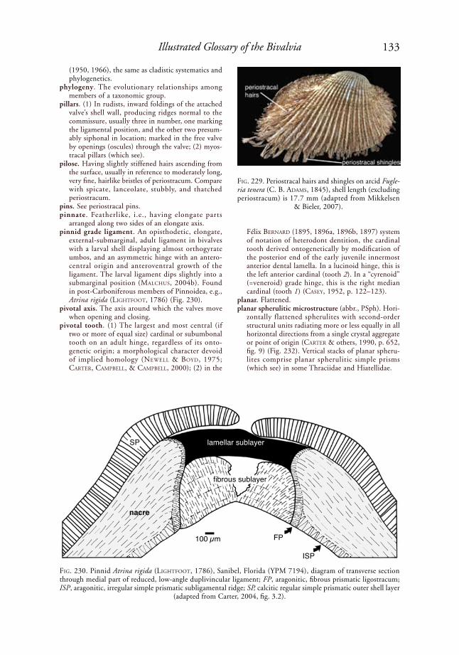
133Illustrated Glossary of the Bivalvia
(1950, 1966), the same as cladistic systematics and phylogenetics.
phylogeny. The evolutionary relationships among members of a taxonomic group.
pillars. (1) In rudists, inward foldings of the attached valve’s shell wall, producing ridges normal to the commissure, usually three in number, one marking the ligamental position, and the other two presum-ably siphonal in location; marked in the free valve by openings (oscules) through the valve; (2) myos-tracal pillars (which see).
pilose. Having slightly stiffened hairs ascending from the surface, usually in reference to moderately long, very fine, hairlike bristles of periostracum. Compare with spicate, lanceolate, stubbly, and thatched periostracum.
pins. See periostracal pins.pinnate. Featherlike, i.e., having elongate parts
arranged along two sides of an elongate axis.pinnid grade ligament. An opisthodetic, elongate,
external-submarginal, adult ligament in bivalves with a larval shell displaying almost orthogyrate umbos, and an asymmetric hinge with an antero-central origin and anteroventral growth of the ligament. The larval ligament dips slightly into a submarginal position (MalChus, 2004b). Found in post-Carboniferous members of Pinnoidea, e.g., Atrina rigida (liGhtFoot, 1786) (Fig. 230).
pivotal axis. The axis around which the valves move when opening and closing.
pivotal tooth. (1) The largest and most central (if two or more of equal size) cardinal or subumbonal tooth on an adult hinge, regardless of its onto-genetic origin; a morphological character devoid of implied homology (NeWell & Boyd, 1975; Carter, CaMpBell, & CaMpBell, 2000); (2) in the
Félix BerNard (1895, 1896a, 1896b, 1897) system of notation of heterodont dentition, the cardinal tooth derived ontogenetically by modification of the posterior end of the early juvenile innermost anterior dental lamella. In a lucinoid hinge, this is the left anterior cardinal (tooth 2). In a “cyrenoid” (=veneroid) grade hinge, this is the right median cardinal (tooth 1) (Casey, 1952, p. 122–123).
planar. Flattened.planar spherulitic microstructure (abbr., PSph). Hori-
zontally flattened spherulites with second-order structural units radiating more or less equally in all horizontal directions from a single crystal aggregate or point of origin (Carter & others, 1990, p. 652, fig. 9) (Fig. 232). Vertical stacks of planar spheru-lites comprise planar spherulitic simple prisms (which see) in some Thraciidae and Hiatellidae.
FiG. 229. Periostracal hairs and shingles on arcid Fugle-ria tenera (C. B. adaMs, 1845), shell length (excluding periostracum) is 17.7 mm (adapted from Mikkelsen
& Bieler, 2007).
FiG. 230. Pinnid Atrina rigida (liGhtFoot, 1786), Sanibel, Florida (YPM 7194), diagram of transverse section through medial part of reduced, low-angle duplivincular ligament; FP, aragonitic, fibrous prismatic ligostracum; ISP, aragonitic, irregular simple prismatic subligamental ridge; SP, calcitic regular simple prismatic outer shell layer
(adapted from Carter, 2004, fig. 3.2).
SP
nacre
100 µm FP
ISP
lamellar sublayer
fibrous sublayer

134 Treatise Online, number 48
planar spherulitic simple prismatic microstructure (abbr., PSphSP). A columnar simple prismatic microstructure in which each first-order prism consists of a stack of planar spherulites (which see), e.g., locally developed in the aragonitic outer shell layer of some Thraciidae (Carter & lutZ, 1990, p. 16, pl. 52) and Hiatellidae, e.g., Panopea generosa (Gould, 1850) (Carter & lutZ, 1990, p. 16, pl. 48, labeled irregular simple prisms) (Fig. 231). This microstructure commonly grades into obtuse columnar nondenticular composite
prismatic microstructure, which is organization-ally similar (cf. Fig. 200). Not to be confused with planar spherulitic microstructure.
plane of commissure. See commissural plane. planivincular ligament. An opisthodetic, elongate,
partially external and partially submarginal to internal ligament in bivalves with orthogyrate (or nongyrate) larval and ortho- to prosogyrate post-larval shells. The fibrous portion of the larval liga-ment is discontinuous with a single adult fibrous ligamental sublayer, and the larval fibrous ligament
FiG. 231. Planar spherulitic simple prismatic (PSphSP) microstructure in outer shell layer of hiatellid Panopea generosa (Gould, 1850), Puget Sound, Washington (YPM 9746). 1, Horizontal acetate peel showing several first-order prisms separated by voids; scale bar = 100 µm; 2, SEM of polished and then acid-etched, vertical section through outer shell layer in shell posterior, with prism in middle of picture cut along its medial longitudinal axis;
scale bar = 5 µm (Carter, new).
1
2

135Illustrated Glossary of the Bivalvia
has a posterocentral or central origination point, followed by posteriorward growth of the adult liga-ment. The adult fibrous sublayer of the ligament is buttressed laterally by a prismatic shelly ridge called a pseudonymph (Carter, 1990a; Waller, 1990; MalChus, 2004b, p. 1561). Typical of the Mytilidae, e.g., Upper Cretaceous Modiolus meeki (eVaNs & shuMard, 1857) (Fig. 233), and Recent Mytilus edulis liNNaeus, 1758 (see Fig. 281). The fibrous sublayers are rarely repeated in the adult stage (see multiple planivincular ligament).
planktonic. Passively drifting or floating in the water column. This term applies to free-swimming veliger larvae, but it may also include some pericalymma larvae (see Zardus & Morse, 1998, and refer-ences therein), although some of the latter may be demersal rather than planktonic. Less commonly spelled planktic.
planktonic-planktotrophic. Planktonic larvae that feed on plankton. The term planktotrophic has commonly been interpreted to include a plank-tonic life habit, but some xylophaginid larvae are only facultatively planktonic (kNudseN, 1961; turNer, 1976). Less commonly spelled planktic-planktotrophic.
planktotrophic. Feeding on plankton. For autobranch bivalves, this term generally refers to larvae feeding on plankton during early development from yolk-poor eggs, which require an extended period of plankton feeding to complete development. The size of such yolk-poor eggs is known to vary from about 35 µm to 90 µm. Egg size correlates roughly with the size of prodissoconch-1. Larval planktot-rophy is generally but not invariably associated with planktonic life habits.
plantigrade stage. A recently metamorphosed, post-larval benthic developmental stage with functional ctenidial filaments, on the verge of settling (BayNe, 1976). This roughly corresponds with the nepio-conch to mesoconch stage (which see), and it may be subdivided according to anatomy and/or shell sculpture (Carriker, 1961; Gros, FreNkiel, & MouëZa, 1997).
platyvincular l igament . A monovincular-A or monovincular-P ligament (Waterhouse, 2001, 2008).
plesion. An extinct, monophyletic sister group to a crown clade or to a more inclusive clade composed of a crown clade and several members of its stem. Plesions are nonranked in cladistic systematics but ranked in paracladistic systematics. Like paraple-sions, plesions lack a normally expected, immedi-ately higher Linnean category.
pleurothetic. Epifaunal resting, reclining, or cementing on one side of the body, e.g., left-valve down Ostre-oidea, right-valve down Anomiidae, and either valve down (amphi-pleurothetic) Etheriidae. Compare with euthetic and orthothetic.
plica (pl., plicae; adj., plicate). A radial or antimarginal, relatively large rib forming an interlocking fold on the shell margin (plication), affecting the entire thickness of the margin (see Fig. 62, Fig. 148).
plicate ctenidium. A ctenidium that appears to be minutely superficially folded in its relaxed, uncon-
tracted state. This commonly reflects the presence of principal ctenidial filaments, although principal ctenidial filaments are absent in the plicate ctenidia of Mya arenaria liNNaeus, 1758 (rideWood, 1903, p. 251). Plicate ctenidia appear to be developed independently of the presence or absence of inter-lamellar junctions; the homorhabdic ctenidia in Malleidae and Pulvinitidae have the same kind of interlamellar septa as the plicate ctenidia in Pteri-idae (tëMkiN, 2006a, p. 291).
plicate organ. A vascularized, longitudinal series of transverse folds between each inner demibranch and the visceral mass in Mytilidae, serving as an accessory respiratory organ.
plication. (1) A relatively large, interlocking fold on a shell margin, produced by a plica; (2) a minute, superficial fold in a gill, parallel with the ctenidial filaments, present in the ctenidium in its relaxed, uncontracted state.
plywood microstructure. A shell microstructure consisting of horizontal laminae comprised of hori-zontal, elongate structural units, typically fibers, oriented in different directions in subadjacent and superadjacent laminae (Carter & others, 1990, p. 611). The lamello-fibrillar variety of plywood microstructure occurs in some Cambrian laterally compressed tergomyans.
polytypic. A biological species consisting of two or more geographic subspecies, or two or more widely divergent ecomorphs.
porcelaneous (also spelled porcellaneous, porcela-nous). (1) Having a shiny or dull, dense, opaque, porcelain-like (non-nacreous, nonfoliaceous) luster (hatChett, 1799, p. 316); (2) a category of aragonitic shell microstructures characterized by a porcelain-like depositional surface; this includes simple crossed lamellar, complex crossed lamellar,
FiG. 232. Planar spherulitic microstructure, diagram showing horizontal (H ), radial (R), and transverse (T ) sections (adapted from Carter & others, 1990, p.
618, fig. 9).
10 µm
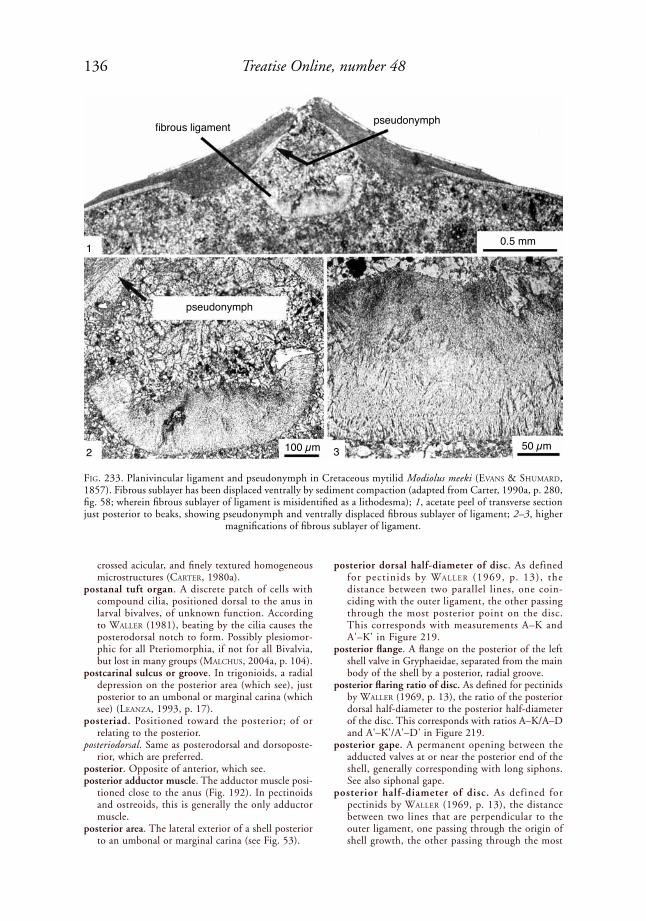
136 Treatise Online, number 48
crossed acicular, and finely textured homogeneous microstructures (Carter, 1980a).
postanal tuft organ. A discrete patch of cells with compound cilia, positioned dorsal to the anus in larval bivalves, of unknown function. According to Waller (1981), beating by the cilia causes the posterodorsal notch to form. Possibly plesiomor-phic for all Pteriomorphia, if not for all Bivalvia, but lost in many groups (MalChus, 2004a, p. 104).
postcarinal sulcus or groove. In trigonioids, a radial depression on the posterior area (which see), just posterior to an umbonal or marginal carina (which see) (leaNZa, 1993, p. 17).
posteriad. Positioned toward the posterior; of or relating to the posterior.
posteriodorsal. Same as posterodorsal and dorsoposte-rior, which are preferred.
posterior. Opposite of anterior, which see.posterior adductor muscle. The adductor muscle posi-
tioned close to the anus (Fig. 192). In pectinoids and ostreoids, this is generally the only adductor muscle.
posterior area. The lateral exterior of a shell posterior to an umbonal or marginal carina (see Fig. 53).
posterior dorsal half-diameter of disc. As defined for pectinids by Waller (1969, p. 13), the distance between two parallel lines, one coin-ciding with the outer ligament, the other passing through the most posterior point on the disc. This corresponds with measurements A–K and A'–K' in Figure 219.
posterior flange. A flange on the posterior of the left shell valve in Gryphaeidae, separated from the main body of the shell by a posterior, radial groove.
posterior flaring ratio of disc. As defined for pectinids by Waller (1969, p. 13), the ratio of the posterior dorsal half-diameter to the posterior half-diameter of the disc. This corresponds with ratios A–K/A–D and A'–K'/A'–D' in Figure 219.
posterior gape. A permanent opening between the adducted valves at or near the posterior end of the shell, generally corresponding with long siphons. See also siphonal gape.
posterior half-diameter of disc. As defined for pectinids by Waller (1969, p. 13), the distance between two lines that are perpendicular to the outer ligament, one passing through the origin of shell growth, the other passing through the most
FiG. 233. Planivincular ligament and pseudonymph in Cretaceous mytilid Modiolus meeki (eVaNs & shuMard, 1857). Fibrous sublayer has been displaced ventrally by sediment compaction (adapted from Carter, 1990a, p. 280, fig. 58; wherein fibrous sublayer of ligament is misidentified as a lithodesma); 1, acetate peel of transverse section just posterior to beaks, showing pseudonymph and ventrally displaced fibrous sublayer of ligament; 2–3, higher
magnifications of fibrous sublayer of ligament.
fibrous ligament
1
32
pseudonymph
pseudonymph
100 µm 50 µm
0.5 mm

137Illustrated Glossary of the Bivalvia
posterior point on the disc. This corresponds with measurements A–D and A'–D' in Figure 219.
posterior internal ridge. A more or less radially oriented, commonly subtle, linear or slightly curvi-linear elevation on the posterior, inner surface of the shell. Same as proximal gill wheal, which see.
posteriority of adductor insertion. See adductor inser-tion posteriority.
posterior keel. Same as umbonal or marginal carina. posterior lateral tooth. A lateral tooth positioned
posterior to the beaks.posterior partition wall (of stomach). A wall extending
into the lumen of the stomach from its posterior wall to the right of the intestinal orifice, creating a left wall of a posteriorly extending embayment of digestive diverticula on the right posterior side of the stomach (kuWataNi, 1965; tëMkiN, 2006a, p. 294), e.g., in the pteriid Pteria sCopoli, 1777.
posterior pedal retractor muscle. A pedal retractor muscle attached to the shell near the posterior adductor muscle scar, typically on the dorsal or dorsoanterior side of that scar, as in the gastro-chaenid Rocellaria dubia (peNNaNt, 1777) (Fig. 20). In Limidae, the posterior pedal retractor muscle attaches to the shell ventral or posteroventral to the posterior adductor muscle scar. The muscle is sometimes divided into three to six bundles of muscle fibers, as in Mytilus edulis liNNaeus, 1758, wherein the most anterior bundle is called the pedal retractor sensu stricto, and the more posterior bundles are called the byssal retractors, although all of them merge together near the shell to make a single, left and right muscle scar. In Anomia liNNaeus, 1758, the posterior pedal retractor muscle is modified to form a byssal muscle or muscles, developed only on the left side. In the Pectinidae, the posterior pedal retractor, if devel-oped at all, occurs only on the left side, along the dorsal margin of the posterior adductor muscle scar.
posterior ridge. Same as umbonal or marginal carina, which is preferred.
posterior sensory tentacle. A narrow, elongate tentacle on one or both sides of the body, extending poste-riorly from the posteroventral mantle margin, e.g., on the left or right side in some Malletiidae, Tindariidae, Nuculanidae, Phaseolidae, and Sarep-tidae (e.g., Yoldiinae, Fig. 234); present on both sides or altogether absent in different species of Solemya; absent in Manzanellidae. Also called a posterior siphonal tentacle and a posterior unpaired tentacle (see yoNGe, 1939).
posterior siphonal tentacle. Same as posterior sensory tentacle, which see.
posterior slope. A sector on the exterior of a bivalve shell extending posteriorly and posteroventrally from the umbo, commonly in reference to the part of the shell, called the area, between an umbonal carina and the dorsoposterior shell margin.
posterior unpaired tentacle. Same as posterior sensory tentacle, which see.
postero-. The combining form of posterior, as in posteroventral. The spelling posterio- is not preferred.
posterodorsal. Both posterior and dorsal; same as dorsoposterior.
posterodorsal notch. A small, sinoidal recess of the posterodorsal shell margin in the left valve of oysters, formed at or shortly after the beginning of the growth of prodissoconch-2, and ending at the prodissoconch-2/nepioconch boundary (Waller, 1981). This notch leaves a conspicuous antimarginal growth track, several µm wide, on the posterodorsal exterior and interior of the left prodissoconch-2 (MalChus, 2004a, p. 88). It correlates with the presence of a postanal tuft organ, which see. The corresponding margin of the right prodissoconch-2 sometimes shows a barely perceptible inflection. This feature is lacking in ostreids with very long brood protection, e.g., Ostrea chilensis philippi, 1846, and Ostrea lutaria
FiG. 234. Diagram of anatomy of Yoldiinae (Sareptidae) (adapted from Mikkelsen & Bieler, 2007, p. 45).
FiG. 235. Diagram of anatomy of Propeamussiidae (adapted from Mikkelsen & Bieler, 2007).

138 Treatise Online, number 48
huttoN, 1873 (JoZeFoWiCZ & Ó’FoiGhil, 1998; MalChus, 2000). Restriction of the notch to the left valve may reflect the asymmetry of valve shape in oysters. See also posterodorsal outlet and posterodorsal ridge.
posterodorsal outlet. A generally narrow, posterodorsal breach through the inner commarginal crest in prodissoconch-2 with interlocking shell margins, occurring symmetrically in the left and right valves (MalChus, 2004a). Species lacking interlocking shell margins may still have a tiny inflection of the shell margin in this position. This feature is typical of oysters and bakevelliids but also occurs in some pinnids, anomiids, teredinids, and xylophaginids (MalChus, 2004a; MalChus, herein). An inflec-tion appears to be present in this position in Arca noae liNNaeus, 1758 (see lutZ & others, 1982, fig. 1b). In some cases, this outlet is accompanied by a posterodorsal ridge on the shell exterior. The breach, and the inflection, may mark the passage of the excurrent water jet close to the anus, rather than the position of the postanal tuft organ (MalChus, 2004a).
posterodorsal ridge. A low, narrow, round-crested, posterodorsal elevation curving approximately antimarginally on the posterodorsal side of prodis-soconch-2. Originally described for Jurassic to Recent arcoids (kiel, 2004; MalChus, 2004a, citing kiel, personal communication, 2004), but also occurring in xylophaginid pholads (turNer, lutZ, & JaBloNski, 1985, fig. 25b–c), which also have a posterodorsal outlet. It occupies the same position as the growth track of the ostreoid posterodorsal notch, but unlike the latter, it is found on both valves and does not widen during prodissoconch-2 growth (kiel, 2004). All taxa herein known to develop the ridge belong to Shell Type 2, which see. See also posterodorsal outlet and posterodorsal notch.
postlarval ctenidial ocelli (sing., ocellus). Eyes consisting of a cup of apically ciliated, pigmented cells located on the most anterior filament of the descending lamellae of the inner demibranch, at the ctenidium-palp junction. Also called a ctenidial eye, eye spot, cephalic eye, and cerebral eye, although in the latter two cases inappropriately so. Present
FiG. 236. Wenlock, medial Silurian colpomyid Aleodonta burei lilJedahl, 1994, Mulde Beds, Djupvik, Gotland, Sweden. 1, Hinge of left valve, showing submarginal grooves for insertion of preduplivincular ligament (RMMo 25423); figure is 7.8 mm wide (adapted from Liljedahl, 1994, fig. 46E, courtesy of Wiley-Blackwell Press); 2–3, transverse, vertical sections through ligament and hinge just posterior to beaks (2) and farther posterior to beaks (3), showing later ontogenetic addition of a second fibrous sublayer (RMMo 23851); scale bars = 100 µm (Carter, new).
1
2 3

139Illustrated Glossary of the Bivalvia
in the isognomonid Isognomon costellatus (CoNrad, 1837) (Fig. 88). A lens may be absent, as in most pterioids, or present, as in Malleus laMarCk, 1799, and some Pteria sCopoli, 1777 (pelseNeer, 1899, 1911; tëMkiN, 2006a, p. 301) (see also B. MortoN, 1995). Present in some Mytilidae, Pteriidae, Malle-idae, Isognomonidae, Arcidae, Philobryidae, and Anomiidae. Found only on the first filament of the left ctenidium in the anomiid Monia macros-chisma (deshayes, 1839). According to B. MortoN (1999), these are neotenous structures that are also present in some bivalve larvae.
postrectal muscle. A small, adductor-like muscle dorsal to the hindgut, more or less dorsoposterior to the posterior adductor muscle, e.g., in Propea-mussiidae (Fig. 235).
preduplivincular ligament. A submarginal, opistho-detic ligament with only two or three, nearly hori-zontal, postlarval, ligamental groove-ridge couplets, and with a discontinuity between the larval and first postlarval fibrous ligamental sublayers. This ligament type is not derived from an ancestral duplivincular or planivincular condition. Younger individuals with only one ligamental groove-ridge couplet may appear ligamentally similar to bivalves with an adult submarginal simple ligament, but in the latter case, there is no discontinuity between the larval and postlarval fibrous sublayers. Found in Colpomyidae and Modiolopsidae, e.g., in the medial Silurian colpomyid Aleodonta burei lilJe-dahl, 1994 (Fig. 236). This is a restriction of poJeta’s (1978, p. 238) term preduplivincular, which also included some monovincular-P and low-angle, opisthodetic duplivincular ligaments, as defined herein.
prefabrication. A term proposed by aller (1974) for secretion of a part of the subperiostracal outer shell layer in advance of the solid shell margin. aller’s examples were shown by Carter and aller (1975) to represent periostracal mineralization (which see). True prefabrication of the outer shell layer is presently known only in some Pholadidae, where anterior shelly rasps in Barnea truncata (say, 1822) are sometimes prefabricated distal to the solid shell margin (Carter, herein).
preoccupied. In the context of formal zoological nomenclature, a name that has been previously used for a different taxon.
pre-oral gland. An unpaired, excretory gland posi-tioned dorsal to the mouth in Pinnidae.
preradial stage. The early growth stage of the left valve of ribbed pectinid species prior to the initiation of interlocking, radial or antimarginal ribs, generally corresponding with the ontogenetic disappearance of the columnar, regular simple prisms of the calcitic outer shell layer (Waller, 1991, p. 6).
pretaxodont dentition. A hinge dentition consisting of two to four stout, more or less orthomorpho-dont teeth that do not significantly increase in number with an increase in shell length beyond 1 mm (Carter, CaMpBell, & CaMpBell, 2000). The associated hinge plate does not extend far
posterior to the beaks because that part of the shell is occupied by the ligament. Pretaxodont dentition characterizes the lower Cambrian fordillid Pojetaia sarhroensis Geyer & streNG, 1998.
primary glochidial host. A host species on which glochidia are capable of transforming to the juvenile stage with optimal success.
primary homonym. Each of two or more iden-tical specific or subspecific names established for different nominal taxa and originally combined with the same genus name (usually by different authors). Governed by ICZN (1999) Article 57.2; see also Article 58. Compare with secondary homonym.
primary inner mantle fold. Same as first inner mantle fold, which see.
primary ligament. The ligament, including its super-ficial periostracum and underlying lamellar and fibrous sublayers, but excluding any functional anterior and/or posterior periostracal extensions (yoNGe, 1978). The periostracal extensions were formerly called fusion layers and secondary liga-ment, but they are now identified as thickened periostracum, thereby making the term primary ligament obsolete (yoNGe, 1982a).
primary ornament. An element of shell ornament appearing earlier in ontogeny, as in primary and secondary ribs, i.e., with primary ribs appearing before the secondary ribs.
primary outer mantle fold. Same as first outer mantle fold, which see.
primary shell. The early, purely organic shell of a molluscan larva secreted prior to detachment of the shell field cells from the shell (BaNdel, 1982, 1988). Same as pellicle and dumbbell shell, which are preferred. See also secondary shell.
primary shell layer. An archaic term for the subperio-stracal outer shell layer, based on the incorrect assumption that the outer shell layer is initiated earlier in ontogeny than the underlying so-called secondary shell layer.
primary synonymy. A list of all scientific names applied to a given species subsequent to its original description, but not necessarily including all of its subsequent generic assignments.
FiG. 237. Pectinid Similipecten nanus (Verrill & Bush in Verrill, 1897), showing byssal fasciole and prodis-
soconch (adapted from Mikkelsen & Bieler, 2007).

140 Treatise Online, number 48
principal filament. A ctenidial filament occupying the bottom of a depression between two successive plicae in a plicate ctenidium; the principal filament is larger than adjacent, ordinary ctenidial fila-ments (rideWood, 1903, p. 153) (Fig. 133–134). Compare with ordinary filament.
principle of priority. In taxonomy, the principle that the valid name of a taxon is the oldest available name applied to it, taking into consideration the provisions of Article 23 of the ICZN (1999) Code, provided that the name is not invalidated by any other provisions of the ICZN Code, or by any ruling of the ICZN.
prionodont. A hinge with teeth developed transverse to the hinge margin (dall, 1889). Same as taxodont, although the term prionodont has traditionally been applied only to members of the orders Cyrt-odontida and Arcida. The basis for the group name Prionodonta, originally established by MaCNeil (1937) for filibranch bivalves with a prionodont hinge, such as the Cyrtodontoidea, Arcoidea, and Limopsoidea.
prismatic cells. Vertically oriented, polygonal cham-bers built into the walls of many hippuritoid bivalves, comprising a cellulo-prismatic structure; e.g., many Radiolitidae.
prismatic microstructure. A shell microstructure consisting of mutually parallel, first-order columns that do not strongly interdigitate along their mutual boundaries. The prisms are called coarse when they are generally greater than 100 µm in width, fine when they are generally less than 5 µm in width, and medium when they fall between these values. Carter and others (1990, p. 610) defined four categories of prismatic microstruc-ture: simple, fibrous, spherulitic, and composite (which see).
prismatic stage. An ontogenetic stage in the Pectinidae characterized by the presence of columnar, regular simple prisms in the outer shell layer of the right valve. These prisms are not secreted after this stage. The left valve at this stage lacks these prisms (Waller, 1991, p. 5).
prismatonacreous. Same as nacroprismatic, which is preferred.
probing organ. See pedal probing organ.probing tubules. Minute perforations of a calcareous
substratum, usually bored coral or shell, produced chemically by a pedal probing organ, e.g., in the gastrochaenid Rocellaria dubia (peNNaNt, 1777) (Fig. 192).
proboscides (sing., proboscis). See palp proboscides.procrescent. Having predominantly anterior shell
growth (NeWell & Boyd, 1995). Compare with retrocrescent and infracrescent.
prodissoconch. A larval shell, equivalent to the proto-conch of gastropods, consisting of periostracum and underlying aragonitic, commonly homo-geneous, granular, and/or irregularly prismatic microstructures, e.g., in the pectinid Similipecten nanus (Verrill & Bush in Verrill, 1897) (Fig. 237) and in the philobryid Cratis antillensis (dall, 1881) (Fig. 9). Planktonic developing autobranch bivalves typically have two distinct growth phases, called prodissoconch-1 and prodissoconch-2, sepa-rated by a distinct change in growth lines or sculp-ture. Nonplanktonic developing bivalves tend to lack this distinct transition (Fig. 243). See also cryptoconch and metaconch.
prodissoconch-1 (abbr., P-1). The earlier formed part of the prodissoconch, secreted initially by the shell field and later eventually by a differentiated mantle of the larva prior to the prodissoconch-1-2 boundary; typically aragonitic with a periostracal ligament (Waller, 1981) (see Fig. 243). It differs from the subsequent prodissoconch-2 in lacking commarginal growth lines and provincular hinge teeth.
prodissoconch-1-2 boundary (abbr., P-1-2 boundary). The discontinuity in larval shell growth believed to correspond with the moment the shell valves reach sufficient size to completely enclose the developing embryo (Waller, 1981) (see Fig. 243). In some cases, however, the periostracal shell may already be sufficiently large to do so, e.g., in Chione cancellata (liNNaeus, 1767 in 1766–1768) (MouëZa, Gros, & FreNkiel, 2006) before calcification begins. Formerly believed to correspond with the transi-tion from secretion by the shell field to secretion by the mantle margin. The presence of marginal shell accretion bands on the prodissoconch-1 of Ostrea edulis liNNaeus, 1758 (cf. Waller, 1981) can also be explained by progressive activation of shell field cells (MalChus & sartori, herein).
prodissoconch-1/2 ratio (abbr., P-1/2 ratio). The ratio of prodissoconch-1 length divided by prodissoconch-2 length. Higher ratios generally indicate larger egg size and lecithotrophy, whereas lower ratios indicate smaller egg size and plankto-trophy (oCkelMaNN, 1965).
FiG. 238. Diagram of anatomy of Malleidae, showing promyal passage, anal funnel, and a pallial retractor muscle; post., posterior (adapted from Mikkelsen &
Bieler, 2007).

141Illustrated Glossary of the Bivalvia
prodissoconch-2 (abbr., P-2). The later-formed part of the prodissoconch, secreted by the mantle and differentiated mantle margin after the valves have completely enfolded the body and can close against each other (Waller, 1981) (Fig. 243). Differs from prodissoconch-1 in having commarginal growth lines and provincular hinge teeth; near the end of its development, the ligament may have developed a fibrous sublayer.
produced (adj.). A feature that is extended.profile. (1) A representation of a shape in outline. (2)
more specifically, an anterior-posterior cross section that cuts the shell at its maximum width normal to the sagittal plane (MalChus & sartori, herein).
progenesis. A type of paedomorphosis caused by precocious maturation and resultant reduction of the juvenile growth period. The opposite of hypermorphosis.
pro-laterofrontal cilia. Smaller laterofrontal cilia comprising a row on each side of a ctenidial fila-ment in the Macrociliobranchia (atkiNs, 1938); generally associated with an adjacent row of eu-laterofrontal cilia, which see.
promyal passage. An opening for a ventilating water current either between the posterior adductor muscle and the visceral mass (e.g., in Malleidae), or above the posterior adductor muscle, between the anterior and posterior parts of the excurrent mantle chamber, lateral to the visceral mass (in many Ostreoidea). In Ostreoidea, this passage is present only on the right side in Crassostreinae and in the Pycnodonteinae except for Hyotissa steNZel, 1971 (where it is present on both sides), and it is entirely absent in Lophinae and Ostreinae. The promyal passage actually has a supramyal, not promyal position, so tëMkiN (2006a) called it the supramyal passage in Malleidae. harry (1985, p. 125) retained the term promyal passage for oysters because of widespread usage. The promyal passage (supramyal passage) allows for efficient passage of excurrent water toward the posterior, e.g., in Malleidae (Fig. 238). See also supramyal passage.
prophylaxis conchiolin sublayers. Laterally extensive conchiolin sublayers, the secretion of which does not represent a response to an external stimulus, such as corrosion or parasitic infestation of the shell or mantle (teVesZ & Carter, 1980). Present in many unionoids, e.g., in the margaritiferid Margaritifera falcata (Gould, 1850) (Fig. 239). Compare with proto-ostracum.
prora. In Pholadoidea, the anterodorsal part of the anterior slope of the shell, which terminates ante-riorly either bluntly or acutely above the pedal gape. It may be sharply or obscurely delimited from the posteroventral part of the anterior slope and by the angulation of the commarginal ornament passing around the border of the pedal gape (kelly, 1988, p. 346) (Fig. 24). A term introduced by kelly (1988) to replace the term beak sensu turNer (1954, 1955, 1969, p. 704, fig. E163).
prosocline. (1) In reference to a hinge tooth, generally a cardinal tooth, inclined, from its lower end to its upper end, toward the anterior, i.e., having a length axis that dips toward the posterior; (2) in refer-ence to shell shape, having the axis of maximum growth inclined, from its distal, lower end to its proximal end, toward the anterior, i.e., the axis of maximum growth dips toward the posterior; same as retrocrescent.
prosocoelous. Same as prosogyrate, which is preferred.prosodetic. The position of a ligament, or other
shell feature, anterior to the beaks. Compare with amphidetic and opisthodetic. When the anterior and posterior ends of the shell cannot be identi-fied, the terms brevidetic and longidetic may be used instead.
prosogyrate. Having the umbo, beak, and/or the entire shell coiling such that the beak points toward the anterior, as a consequence of retrocrescent, heli-coidal growth, e.g., in the veneroidean Periglypta listeri (Gray, 1838) (Fig. 35). Compare with ortho-gyrate and opisthogyrate.
prosogyrous. Same as prosogyrate, which is preferred.
FiG. 239. Prophylaxis conchiolin sublayers in nacreous inner shell layer of margaritiferid Margaritifera falcata (Gould, 1850), Ozette River, Washington State (YPM 10158) (adapted from Tevesz & Carter, 1980, fig. 8).

142 Treatise Online, number 48
gonadrectum
posterioradd.
anus ctenidiumpalp proboscide
anterior add.
foot
hypobranchial gland
ciliated knobs
ctenidium
ventral mantle
ventral blood vesselciliated pores
ctenidial axisdorsal blood vessel
inner leaflet ciliaterminal cilia
prosopon. A term proposed by Gill (1949) for what is herein called exterior shell ornament or sculp-ture. poJeta (1962, p. 173) objected to the use of ornament because it carries an anthropomorphic connotation. However, the term ornament is widely
used, and its meaning is obvious to both amateur and professional malacologists.
protandry (adj., protandrous, protandric). A form of hermaphroditism in which the functional male phase precedes the functional female phase during the life cycle of the individual. See also hermaph-roditic and protogyny.
proteinaceous. Comprised primarily or entirely of protein.
protobranch ctenidium. A ctenidium consisting of flat, nonreflected filaments, often called leaflets or platelets, as in members of the Protobranchia (Fig. 240). The demibranchs are symmetrical or slightly asymmetrical and comprised of simple, broad, leaf-like filaments. Adjacent filaments are unconnected or have at most only loose, sparse cilial attach-ments on the filament faces. The filaments have chitinous supporting rods and are used primarily for respiration rather than for feeding. Divided into ctenidiobranch and palaeobranch types by salViNi-plaWeN (1980, 1988). The basis of the subclass Protobranchiés (vernacular), originally defined by pelseNeer (1889) to include the Nuculidae and Solemyidae; subsequently emended and referred to as Protobranchia.
FiG. 240. Protobranch ctenidia in 1a–1b, solemyid Solemya togata (poli, 1791 in 1791–1795), and in 2, malletiid Pseudomalletia obtusa (G. sars, 1872). 1a, Diagrammatic transverse section through middle of region occupied by ctenidia, showing left and right ctenidia, relation of organs, and ventral intucking of fused ventral mantle; dorsal is up; shell height is 6.8 mm; 1b, ctenidia and mantle cavity viewed from right side; shell length is 6.4 mm; 2, four filaments from right ctenidium from region posterior to foot, viewed from an anteroventral aspect; ctenidium width
is about 1.0 mm; add., adductor muscle (redrawn by Carter from Yonge, 1939, fig. 11, 13, 15).
FiG. 241. Protoplax of the pholadid Cyrtopleura costata (liNNaeus, 1758), length 33 mm. See also Figure 175
(adapted from Mikkelsen & Bieler, 2007).
1a
1b
2
ctenidial leaflet
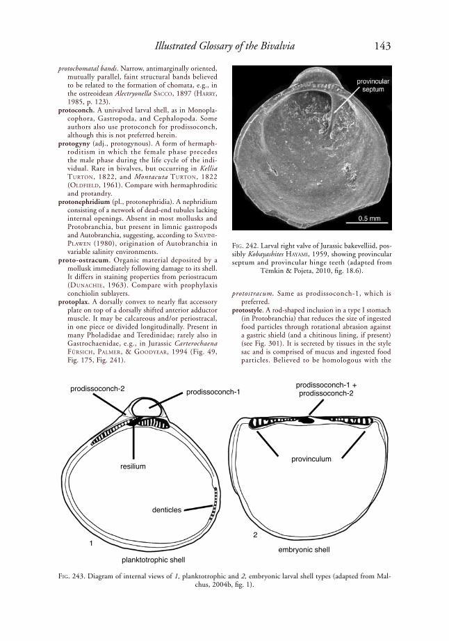
143Illustrated Glossary of the Bivalvia
protochomatal bands. Narrow, antimarginally oriented, mutually parallel, faint structural bands believed to be related to the formation of chomata, e.g., in the ostreoidean Alectryonella saCCo, 1897 (harry, 1985, p. 123).
protoconch. A univalved larval shell, as in Monopla-cophora, Gastropoda, and Cephalopoda. Some authors also use protoconch for prodissoconch, although this is not preferred herein.
protogyny (adj., protogynous). A form of hermaph-roditism in which the female phase precedes the male phase during the life cycle of the indi-vidual. Rare in bivalves, but occurring in Kellia turtoN, 1822, and Montacuta turtoN, 1822 (oldField, 1961). Compare with hermaphroditic and protandry.
protonephridium (pl., protonephridia). A nephridium consisting of a network of dead-end tubules lacking internal openings. Absent in most mollusks and Protobranchia, but present in limnic gastropods and Autobranchia, suggesting, according to salViNi-plaWeN (1980), origination of Autobranchia in variable salinity environments.
proto-ostracum. Organic material deposited by a mollusk immediately following damage to its shell. It differs in staining properties from periostracum (duNaChie, 1963). Compare with prophylaxis conchiolin sublayers.
protoplax. A dorsally convex to nearly flat accessory plate on top of a dorsally shifted anterior adductor muscle. It may be calcareous and/or periostracal, in one piece or divided longitudinally. Present in many Pholadidae and Teredinidae; rarely also in Gastrochaenidae, e.g., in Jurassic Carterochaena FürsiCh, palMer, & Goodyear, 1994 (Fig. 49, Fig. 175, Fig. 241).
protostracum. Same as prodissoconch-1, which is preferred.
protostyle. A rod-shaped inclusion in a type I stomach (in Protobranchia) that reduces the size of ingested food particles through rotational abrasion against a gastric shield (and a chitinous lining, if present) (see Fig. 301). It is secreted by tissues in the style sac and is comprised of mucus and ingested food particles. Believed to be homologous with the
FiG. 242. Larval right valve of Jurassic bakevelliid, pos-sibly Kobayashites hayaMi, 1959, showing provincular septum and provincular hinge teeth (adapted from
Tëmkin & Pojeta, 2010, fig. 18.6).
FiG. 243. Diagram of internal views of 1, planktotrophic and 2, embryonic larval shell types (adapted from Mal-chus, 2004b, fig. 1).
prodissoconch-2 prodissoconch-1
resilium
denticles
planktotrophic shellembryonic shell
provinculum
prodissoconch-1 + prodissoconch-2
12

144 Treatise Online, number 48
crystalline style of other bivalves, which similarly contains amylase.
protractor muscle. Same as pedal protractor muscle, which see.
provincular hinge teeth. The hinge teeth present on a provinculum, e.g., in the Jurassic bakevelliid Kobayashites? (Fig. 242). Provincular teeth are retained into the adult stage in some bivalves, e.g., in Euchondriidae (see NeWell, Cox, & hertleiN, 1969, p. 340).
provincular septum. A nearly straight, thickened partition on the inner surface of prodissoconch-2, extending slightly anteroventrally from the postero-ventral surface of the provinculum, thereby subdi-viding the dorsal part of the shell cavity into larger anterior and smaller posterior parts, as in the right valve of the Jurassic bakevelliid Kobayashites? (Fig. 242). This corresponds to the myofringing crest of adult Plicatostylidae, suggesting that it might be its
ontogenetic precursor (tëMkiN & poJeta, 2010, p. 1173). See also myofringing crest.
provinculum. (1) The median part of the hinge of a prodissoconch, usually bearing small hinge teeth or crenulations (Cox, Nuttall, & trueMaN, 1969, p. 107); (2) the entire larval hinge system (MalChus, 2004a, p. 90, 2004b) (Fig. 243); (3) the vertically striated area on each side of the resilifer in some Pectinoidea, more appropriately called the resilial hinge area.
proximal. Toward the center, or origin, or point of attachment; opposite of distal.
proximal curtain valve. Same as basal siphonal valve, which see.
proximal gill wheal. A low, more or less dorsoventral ridge or break in slope, on the shell’s inner surface, which marks the proximal margin of the ctenidium. This is found in many Ostreoidea, in the Silu-rian frejid Freja lilJedahl, 1984, in the Silurian umburrid Umburra p. a. JohNstoN, 1991 (Fig. 244), and in some lucinoids. The wheal is neither contiguous with nor aligned with the trace of the anterior margin of the posterior adductor muscle scar, and in some instances, it extends ventrally beyond that scar. Also called a posterior internal ridge. The spelling “weal” is not preferred.
proximal siphonal diaphragm. Same as basal siphonal diaphragm, which see.
proximal siphonal valve. Same as basal siphonal valve, which see.
proximal valve. Same as basal siphonal valve, which see.
pseudoaxial suspensory septum. A tissue connection extending from the interctenidial connection to the midventral surface of the visceral mass. Absent in Pterioidea but present in many Ostreoidea (harry, 1985; tëMkiN, 2006a, p. 287). Called the pseu-doaxial suspensory membrane by harry (1985).
pseudocardinal tooth. An irregularly shaped cardinal tooth in the Unionida; a cardinal or cardinolateral tooth in the Actinodontida; or a cardinal tooth in a nonmember of the Heteroconchia (Fig. 245).
pseudoctenodont dentition. A hinge dentition consisting of numerous, short teeth oriented more or less perpendicular to the hinge axis, in bivalves believed to have been derived from ancestors with more or less elongate, longitudinally oriented hinge teeth. This is the basis for the group name Pseu-doctenodonta, originally defined by deChaseaux (1952) for the families Arcidae and Parallelodon-tidae. See also neotaxodont dentition.
pseudoctenolium. A ctenolium-like structure formed from external sculpture on the edge of the right side of the byssal gape in some Pectinidae and in Propeamussiidae; not formed in the same way as a true ctenolium. Also present in some Monotidae, Buchiidae and Oxytomidae (sha & FürsiCh, 1994, table 5, as “ctenolium”).
pseudofeces (or pseudofaeces). Non-ingested waste material from the respiratory-feeding current, bound in mucus and carried by mantle cilia to be discharged, generally through the incurrent siphon,
FiG. 244. Proximal gill wheal in Silurian umburrid Umburra cinefacta JohNstoN, 1991, shell height 35 mm (adapted from Johnston, 1991, fig. 2B, courtesy
of Alcheringa).
FiG. 245. Pseudoheterodont hinge with pseudocardinal and pseudolateral teeth; Devonian cyrtodontid Ptych-odesma knappianum hall & WhitField, 1871, Marcel-lus Formation, Morrisville, New York (YPM 10057-7)
(adapted from Carter & Tevesz, 1978b, pl. 1,2b).
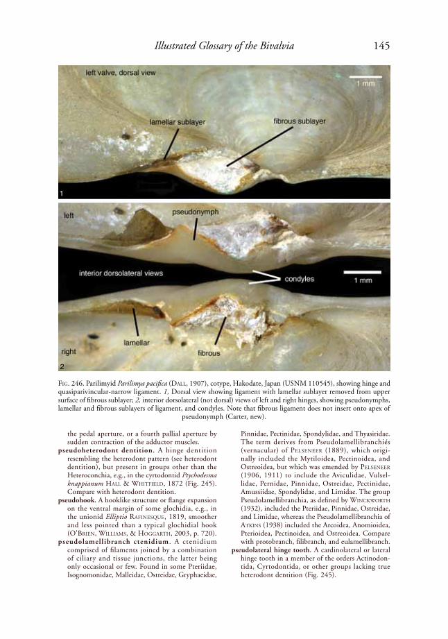
145Illustrated Glossary of the Bivalvia
the pedal aperture, or a fourth pallial aperture by sudden contraction of the adductor muscles.
pseudoheterodont dentition. A hinge dentition resembling the heterodont pattern (see heterodont dentition), but present in groups other than the Heteroconchia, e.g., in the cyrtodontid Ptychodesma knappianum hall & WhitField, 1872 (Fig. 245). Compare with heterodont dentition.
pseudohook. A hooklike structure or flange expansion on the ventral margin of some glochidia, e.g., in the unionid Elliptio raFiNesque, 1819, smoother and less pointed than a typical glochidial hook (o'BrieN, WilliaMs, & hoGGarth, 2003, p. 720).
pseudolamellibranch ctenidium . A ctenidium comprised of filaments joined by a combination of ciliary and tissue junctions, the latter being only occasional or few. Found in some Pteriidae, Isognomonidae, Malleidae, Ostreidae, Gryphaeidae,
Pinnidae, Pectinidae, Spondylidae, and Thyasiridae. The term derives from Pseudolamellibranchiés (vernacular) of pelseNeer (1889), which origi-nally included the Mytiloidea, Pectinoidea, and Ostreoidea, but which was emended by pelseNeer (1906, 1911) to include the Aviculidae, Vulsel-lidae, Pernidae, Pinnidae, Ostreidae, Pectinidae, Amussiidae, Spondylidae, and Limidae. The group Pseudolamellibranchia, as defined by WiNCkWorth (1932), included the Pteriidae, Pinnidae, Ostreidae, and Limidae, whereas the Pseudolamellibranchia of atkiNs (1938) included the Arcoidea, Anomioidea, Pterioidea, Pectinoidea, and Ostreoidea. Compare with protobranch, filibranch, and eulamellibranch.
pseudolateral hinge tooth. A cardinolateral or lateral hinge tooth in a member of the orders Actinodon-tida, Cyrtodontida, or other groups lacking true heterodont dentition (Fig. 245).
FiG. 246. Parilimyid Parilimya pacifica (dall, 1907), cotype, Hakodate, Japan (USNM 110545), showing hinge and quasiparivincular-narrow ligament. 1, Dorsal view showing ligament with lamellar sublayer removed from upper surface of fibrous sublayer; 2, interior dorsolateral (not dorsal) views of left and right hinges, showing pseudonymphs, lamellar and fibrous sublayers of ligament, and condyles. Note that fibrous ligament does not insert onto apex of
pseudonymph (Carter, new).
1
2
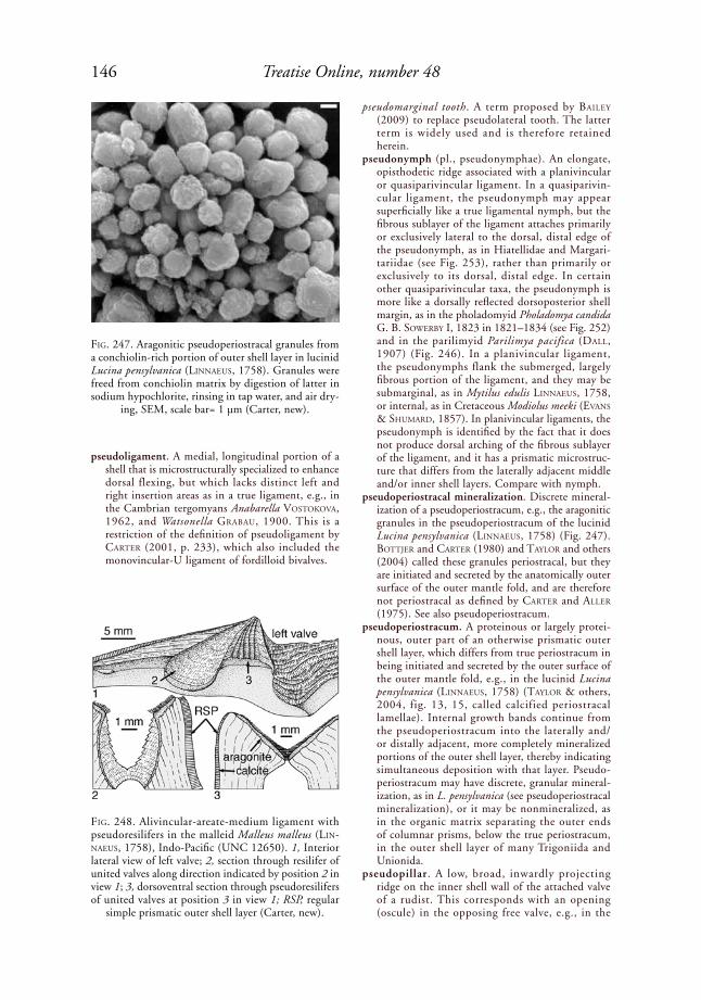
146 Treatise Online, number 48
pseudoligament. A medial, longitudinal portion of a shell that is microstructurally specialized to enhance dorsal flexing, but which lacks distinct left and right insertion areas as in a true ligament, e.g., in the Cambrian tergomyans Anabarella VostokoVa, 1962, and Watsonella GraBau, 1900. This is a restriction of the definition of pseudoligament by Carter (2001, p. 233), which also included the monovincular-U ligament of fordilloid bivalves.
pseudomarginal tooth. A term proposed by Bailey (2009) to replace pseudolateral tooth. The latter term is widely used and is therefore retained herein.
pseudonymph (pl., pseudonymphae). An elongate, opisthodetic ridge associated with a planivincular or quasiparivincular ligament. In a quasiparivin-cular ligament, the pseudonymph may appear superficially like a true ligamental nymph, but the fibrous sublayer of the ligament attaches primarily or exclusively lateral to the dorsal, distal edge of the pseudonymph, as in Hiatellidae and Margari-tariidae (see Fig. 253), rather than primarily or exclusively to its dorsal, distal edge. In certain other quasiparivincular taxa, the pseudonymph is more like a dorsally reflected dorsoposterior shell margin, as in the pholadomyid Pholadomya candida G. B. soWerBy i, 1823 in 1821–1834 (see Fig. 252) and in the parilimyid Parilimya pacifica (dall, 1907) (Fig. 246). In a planivincular ligament, the pseudonymphs flank the submerged, largely fibrous portion of the ligament, and they may be submarginal, as in Mytilus edulis liNNaeus, 1758, or internal, as in Cretaceous Modiolus meeki (eVaNs & shuMard, 1857). In planivincular ligaments, the pseudonymph is identified by the fact that it does not produce dorsal arching of the fibrous sublayer of the ligament, and it has a prismatic microstruc-ture that differs from the laterally adjacent middle and/or inner shell layers. Compare with nymph.
pseudoperiostracal mineralization. Discrete mineral-ization of a pseudoperiostracum, e.g., the aragonitic granules in the pseudoperiostracum of the lucinid Lucina pensylvanica (liNNaeus, 1758) (Fig. 247). BottJer and Carter (1980) and taylor and others (2004) called these granules periostracal, but they are initiated and secreted by the anatomically outer surface of the outer mantle fold, and are therefore not periostracal as defined by Carter and aller (1975). See also pseudoperiostracum.
pseudoperiostracum. A proteinous or largely protei-nous, outer part of an otherwise prismatic outer shell layer, which differs from true periostracum in being initiated and secreted by the outer surface of the outer mantle fold, e.g., in the lucinid Lucina pensylvanica (liNNaeus, 1758) (taylor & others, 2004, fig. 13, 15, called calcified periostracal lamellae). Internal growth bands continue from the pseudoperiostracum into the laterally and/or distally adjacent, more completely mineralized portions of the outer shell layer, thereby indicating simultaneous deposition with that layer. Pseudo-periostracum may have discrete, granular mineral-ization, as in L. pensylvanica (see pseudoperiostracal mineralization), or it may be nonmineralized, as in the organic matrix separating the outer ends of columnar prisms, below the true periostracum, in the outer shell layer of many Trigoniida and Unionida.
pseudopillar. A low, broad, inwardly projecting ridge on the inner shell wall of the attached valve of a rudist. This corresponds with an opening (oscule) in the opposing free valve, e.g., in the
FiG. 247. Aragonitic pseudoperiostracal granules from a conchiolin-rich portion of outer shell layer in lucinid Lucina pensylvanica (liNNaeus, 1758). Granules were freed from conchiolin matrix by digestion of latter in sodium hypochlorite, rinsing in tap water, and air dry-
ing, SEM, scale bar= 1 µm (Carter, new).
FiG. 248. Alivincular-areate-medium ligament with pseudoresilifers in the malleid Malleus malleus (liN-Naeus, 1758), Indo-Pacific (UNC 12650). 1, Interior lateral view of left valve; 2, section through resilifer of united valves along direction indicated by position 2 in view 1; 3, dorsoventral section through pseudoresilifers of united valves at position 3 in view 1; RSP, regular
simple prismatic outer shell layer (Carter, new).
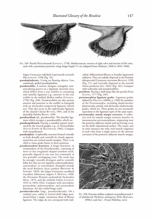
147Illustrated Glossary of the Bivalvia
Upper Cretaceous radiolitid Lapeirousella orientalis MiloVaNoVić, 1938 (Fig. 54).
pseudoplanktonic. Living on floating objects. Less commonly spelled pseudoplanktic.
pseudoresilifer. A narrow, elongate, triangular, non-articulating groove on a ligament insertion area, which differs from a true resilifer in containing only lamellar ligament; e.g., anterior to the true resilifer in the malleid Malleus malleus (liNNaeus, 1758) (Fig. 248). Pseudoresilifers are also present anterior and posterior to the resilifer in limopsids with an alivincular-compressed ligament (which see). They also occur in the replivincular ligament of the claraiid Claraia BittNer, 1901, and of the daonellid Halobia BroNN, 1830.
pseudoresilium (pl., pseudoresilia). The lamellar liga-ment which occupies a pseudoresilifer, which see.
pseudoseptibranch. Having a ctenidial septum inner-vated by the visceral ganglia, e.g., in Verticordiidae (salViNi-plaWeN & hasZpruNar, 1982). Compare with euseptibranch.
pseudosiphon. A siphonlike structure formed ventrally or both dorsally and ventrally by closely approxi-mated but not united mantle margins. There is no cilial or tissue grade fusion in these positions.
pseudotaxodont dentition. A hinge dentition, in nonmembers of the Protobranchia, consisting of more or less irregularly shaped taxodont teeth, generally aligned in one row, less commonly in two partially overlapping rows. The teeth may be strongly ventrally divergent and/or ventrally bifid, but they are not regularly orthomorphodont, concavodont, or convexodont. Examples include the arcid Barbatia (Acar) gradata (Broderip & soWerBy, 1829), the Upper Cretaceous cucullaeid Cucullaea (Idonearca) vulgaris S. MortoN, 1830, the Devonian–Permian euchondriid Euchondria Meek, 1874, and the iridinid Iridina laMarCk, 1819 in 1818–1822. Compare with microdont, pretaxodont, palaeotaxodont, and neotaxodont dentitions. See also resilial hinge teeth.
pseudotrabeculae (sing., pseudotrabecula). On a cardinal area, radial or vertical shell ridges posi-tioned below a single, continuous sheet of fibrous ligament. The ridges do not correspond with indi-
vidual, differentiated fibrous or lamellar ligamental sublayers. They are radially disposed in the Permian deltopectinid Cyrtorostra varicostata BraNsoN, 1930 (Fig. 11), and vertically disposed in the noetiid Noetia ponderosa (say, 1822) (Fig. 181). Compare with trabeculae and pseudoresilifers.
pteriiform. Having a shell shape like the pteriid Pteria sCopoli, 1777 (Fig. 249).
pteriomorphian ligament grades. Ligament grades defined by MalChus (2004b, p. 1560) for members of the Pteriomorphia, including duplivincular, planivincular, pinnid, and alivincular-multivincular grades, which see. These grades are not necessarily equivalent to the ligament types of the same name.
pteriomorphic mantle margin retractors. A collec-tive term for mantle margin retractor muscles, in monomyarian pteriomorphians, originating near the posterior adductor muscle and not leaving a scar on the shell’s depositional surface. The major, and in some instances the only, such muscle originates in each valve from a single source on the anterior curvature of the posterior adductor muscle margin,
FiG. 249. Pteriid Pteria hurundo (liNNaeus, 1758), Mediterranean; exterior of right valve and interior of left valve, each with a prominent posterior wing; hinge length 9.5 cm (adapted from Deshayes, 1848 in 1845–1848).
FiG. 250. Punctate-stellate sculpture on prodissoconch-1 of philobryid Philobrya meleagrina (Félix BerNard,
1896c); scale bar = 10 µm (Malchus, new).

148 Treatise Online, number 48
at a slight indentation between the quick and catch parts of the adductor muscle. In some instances, a few similar but smaller mantle margin retractor muscles originate along the ventral and posterior margins of the adductor muscle (harry, 1985, p. 125). See also secondary mantle margin retractor muscles.
puncta (pl., punctae). A pinpricklike depression or minute pit visible on the surface of a shell, exter-nally or internally, but generally internally. See also mantle attachment scars.
punctate-stellate sculpture. A shell sculpture present on the cicatrix of many larval shells, comprised of a radial pattern with external pits; e.g., in the philobryid Philobrya meleagrina (Félix BerNard, 1896c) (Fig. 250) (MalChus, herein).
pustule (adj., pustulose). A small, calcareous, blister-like, round or oblong prominence or projection on the surface of a shell; smaller than a tubercle, which see.
pustulose chomata. Chomata with a bumplike shape, generally less than 1 mm wide, often occurring in several rather regular rows along the commis-sure (MalChus, 1990), e.g., in the oysters Lopha rödiNG, 1798, and Gryphaeligmus leWy, 1982. Pustulose chomata sometimes occur along with lath-type chomata, e.g., in the oyster Alectryonella saCCo, 1897.
pyloric process. Same as metasoma, which is preferred.pyriform. Pear shaped, i.e., wide on one end, narrow
on the other end.quadrangular. Having four angles, most generally
somewhat square or rectangular in lateral profile, as in the hiatellid Hiatella arctica (liNNaeus, 1767 in 1766–1768) (Fig. 251).
quadrate. Having four sides, usually in reference to a shell shape that is close to being square or rectangular.
qualifier statement. An indication of the contents of a monophyletic clade based on closer common ancestry with an internal specifier than with an external specifier.
quasiparivincular ligament. An external, elongate, opisthodetic, dorsally arched ligament in which the fibrous sublayer attaches to both the dorsal
and lateral surfaces of a ligamental ridge (pseud-onymph), and also extensively lateral to this ridge (Carter, herein). See quasiparivincular-narrow and quasiparivincular-wide.
quasiparivincular-narrow ligament. A quasiparivin-cular ligament in which the pseudonymph is repre-sented by the dorsally upturned, dorsoposterior shell margin, as in the pholadomyid Pholadomya candida G. B. soWerBy i, 1823 in 1821–1834 (Fig. 252) and the parilimyid Parilimya pacifica (dall, 1907) (Fig. 246).
quasiparivincular-wide ligament. A quasiparivincular ligament in which the pseudonymph is a low ridge on or near the margin of a wide, flat, more or less horizontal ligamental insertion area, and not just an upturned, dorsoposterior shell margin, e.g., in the hiatellid Panopea generosa (Gould, 1850) (Fig. 253).
Quenstedt muscle. A small, circular muscle scar posi-tioned between the posterior adductor muscle scar and the umbonal cavity, e.g., in pseudomonotids (NeWell & Boyd, 1970), rhombopteriids (JohN-stoN, 1992), umburrids (JohNstoN, 1991), and ostreids. Not homologous with a pedal muscle, contrary to steNZel (1971, p. 965) (see harry, 1985; JohNstoN, 1991, p. 313). This is an anterior ctenidial elevator or ctenidial protractor muscle scar, according to Boss (1982) and harry (1985). The Quenstedt muscle sometimes does not attach to the shell, and consequently leaves no muscle scar, e.g., in the Pteriidae (MalChus, herein). Ctenidial elevator and ctenidial protractor muscles are prob-ably plesiomorphic for the Pteriomorphia and perhaps other bivalves.
Quenstedt muscle scar. A shell muscle scar produced by the Quenstedt muscle, which see.
quick muscle. The striated muscle portion of the adductor muscle, responsible for rapidly closing the shell, which uses ATP more rapidly than the unstriated, catch muscle, e.g., in the pectinid Chlamys islandica (o. F. Müller, 1776) (Fig. 57). This muscle is typically yellowish or light brown and semitranslucent in appearance, with a very soft consistency, as compared with the white, opaque, opalescent appearance and tougher consistency of the catch muscle. Also called phasic muscle.
rachis. The central axis or stalk of a ctenidium.radial. (1) Having parts arranged like rays around a
common center; (2) in terms of shell ornament, having parts fanning outward from the beak, but not necessarily radial in the strict sense; the parts may be truly radial or antimarginal, e.g., in the glycymeridid Tucetona pectinata (GMeliN, 1791 in 1791–1793) (see Fig. 265); (3) in reference to shell sections, perpendicular to both the plane of the shell layer and to external and internal growth lines.
radial gashes. Millimeter-thin, more or less radial, sharp-edged incisions visible on the exterior of the outer shell layer of some gryphaeids with a flat to concave, lidlike upper valve.
radial lamellar nondenticular composite prismatic microstructure (abbr., radial lamellar NDCP). A nondenticular composite prismatic shel l
FiG. 251. Quadrangular shell shape illustrated by hiatel-lid Hiatella arctica (liNNaeus, 1767 in 1766–1768), shell length 11.7 mm (adapted from Mikkelsen &
Bieler, 2007).

149Illustrated Glossary of the Bivalvia
microstructure in which radially oriented first-order NDCP prisms are stacked into more less vertical columns, commonly visible as minute, vertical microridges on the surface of a strongly reflected shell margin. There is commonly some fusion of superadjacent and subadjacent first-order NDCP prisms. This structure commonly grades above and/or below into normal, radial NDCP. Found in the outer shell layer of the venerid Tapes (Tapes) literata (liNNaeus, 1758) (Fig. 254), and in the outer shell layer of the lucinid Codakia (Codakia) orbicu-laris (liNNaeus, 1758). Compare with compound NDCP and crossed NDCP.
radial mantle glands. Same as arenophilic glands, which see.
radial pallial muscles. (1) The pallial retractor muscles that insert along the pallial line or pallial band; (2) in pectinids, a single muscle that leaves a small, linear scar on the posterior margin of the umbonal
cavity, just dorsal to a circular pallial muscle scar; it may be distinct from or confluent with the circular pallial muscle scar; e.g., present in Argopecten gibbus (liNNaeus, 1758) (Waller, 1969, fig. 3), but apparently absent in Chlamys islandica (o. F. Müller, 1776) (Waller, 1991, fig. 1).
radial section. An antimarginal shell section that is perpendicular to the plane of the shell layer. A truly radial section can only be approximated for most bivalves because of their generally nonplanar coiling and non-orthogyrate shapes.
radial umbonal ridge. A radial elevation in the umbonal cavity for which association with a particular muscle scar or fossa cannot be ascer-tained (tëMkiN & poJeta, 2010, p. 1173). See also myofringing ridge, fossate ridge, radial umbonal septum, auricular transverse septum, apical trans-verse septa, myofringing crest, byssal ridge, poste-rior internal ridge, and provincular septum.
FiG. 252. The pholadomyid Pholadomya candida G. B. soWerBy i, 1823 in 1821–1834, Tortola, West Indies (USNM 109211), showing hinge and ligament. 1, Dorsal view of quasiparivincular-narrow ligament; 2, left hinge viewed perpendicular to commissural plane, showing pseudonymph, flanking ligament, a triangular condyle below beak,
and anterior, shell marginal socket; scale bar applies to both figures (Carter, new).
2
1
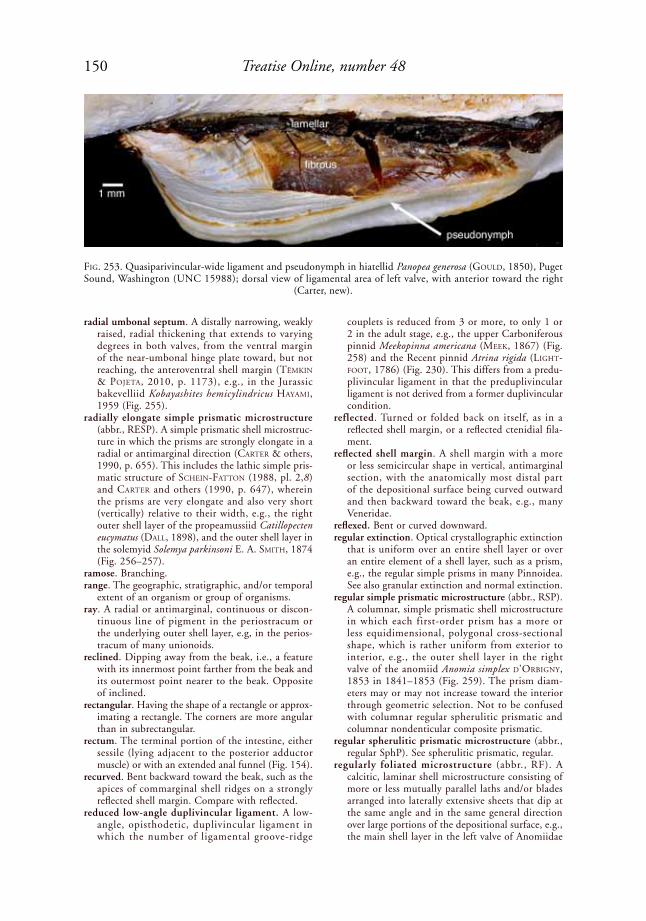
150 Treatise Online, number 48
radial umbonal septum. A distally narrowing, weakly raised, radial thickening that extends to varying degrees in both valves, from the ventral margin of the near-umbonal hinge plate toward, but not reaching, the anteroventral shell margin (tëMkiN & poJeta, 2010, p. 1173), e.g., in the Jurassic bakevelliid Kobayashites hemicylindricus hayaMi, 1959 (Fig. 255).
radially elongate simple prismatic microstructure (abbr., RESP). A simple prismatic shell microstruc-ture in which the prisms are strongly elongate in a radial or antimarginal direction (Carter & others, 1990, p. 655). This includes the lathic simple pris-matic structure of sCheiN-FattoN (1988, pl. 2,8) and Carter and others (1990, p. 647), wherein the prisms are very elongate and also very short (vertically) relative to their width, e.g., the right outer shell layer of the propeamussiid Catillopecten eucymatus (dall, 1898), and the outer shell layer in the solemyid Solemya parkinsoni e. a. sMith, 1874 (Fig. 256–257).
ramose. Branching.range. The geographic, stratigraphic, and/or temporal
extent of an organism or group of organisms.ray. A radial or antimarginal, continuous or discon-
tinuous line of pigment in the periostracum or the underlying outer shell layer, e.g, in the perios-tracum of many unionoids.
reclined. Dipping away from the beak, i.e., a feature with its innermost point farther from the beak and its outermost point nearer to the beak. Opposite of inclined.
rectangular. Having the shape of a rectangle or approx-imating a rectangle. The corners are more angular than in subrectangular.
rectum. The terminal portion of the intestine, either sessile (lying adjacent to the posterior adductor muscle) or with an extended anal funnel (Fig. 154).
recurved. Bent backward toward the beak, such as the apices of commarginal shell ridges on a strongly reflected shell margin. Compare with reflected.
reduced low-angle duplivincular ligament. A low-angle, opisthodetic, duplivincular ligament in which the number of ligamental groove-ridge
couplets is reduced from 3 or more, to only 1 or 2 in the adult stage, e.g., the upper Carboniferous pinnid Meekopinna americana (Meek, 1867) (Fig. 258) and the Recent pinnid Atrina rigida (liGht-Foot, 1786) (Fig. 230). This differs from a predu-plivincular ligament in that the preduplivincular ligament is not derived from a former duplivincular condition.
reflected. Turned or folded back on itself, as in a reflected shell margin, or a reflected ctenidial fila-ment.
reflected shell margin. A shell margin with a more or less semicircular shape in vertical, antimarginal section, with the anatomically most distal part of the depositional surface being curved outward and then backward toward the beak, e.g., many Veneridae.
reflexed. Bent or curved downward.regular extinction. Optical crystallographic extinction
that is uniform over an entire shell layer or over an entire element of a shell layer, such as a prism, e.g., the regular simple prisms in many Pinnoidea. See also granular extinction and normal extinction.
regular simple prismatic microstructure (abbr., RSP). A columnar, simple prismatic shell microstructure in which each first-order prism has a more or less equidimensional, polygonal cross-sectional shape, which is rather uniform from exterior to interior, e.g., the outer shell layer in the right valve of the anomiid Anomia simplex d’orBiGNy, 1853 in 1841–1853 (Fig. 259). The prism diam-eters may or may not increase toward the interior through geometric selection. Not to be confused with columnar regular spherulitic prismatic and columnar nondenticular composite prismatic.
regular spherulitic prismatic microstructure (abbr., regular SphP). See spherulitic prismatic, regular.
regularly foliated microstructure (abbr., RF). A calcitic, laminar shell microstructure consisting of more or less mutually parallel laths and/or blades arranged into laterally extensive sheets that dip at the same angle and in the same general direction over large portions of the depositional surface, e.g., the main shell layer in the left valve of Anomiidae
FiG. 253. Quasiparivincular-wide ligament and pseudonymph in hiatellid Panopea generosa (Gould, 1850), Puget Sound, Washington (UNC 15988); dorsal view of ligamental area of left valve, with anterior toward the right
(Carter, new).

151Illustrated Glossary of the Bivalvia
FiG. 254. Radial lamellar nondenticular composite prismatic microstructure in outer shell layer of veneroidean Tapes (Tapes) litterata liNNaeus, 1758, the Philippines (YPM 9679); acetate peels of 1, radial and 2, transverse sections; bottom figure shows outer (top right) and inner (bottom left) portions of outer shell layer, with inner portion showing less regular vertical stacking of prisms. Scale bar (50 µm) applies to both figures (adapted from
Carter, 1980b, fig. 11).
1
2
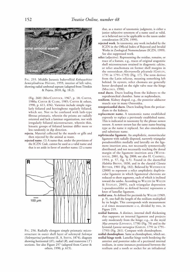
152 Treatise Online, number 48
(Fig. 260) (MaCCliNtoCk, 1967, p. 18; Carter, 1980a; Carter & Clark, 1985; Carter & others, 1990, p. 611, 656). Varieties include simple regu-larly foliated and herringbone regularly foliated, which see. Not to be confused with lath-type fibrous prismatic, wherein the prisms are radially oriented and lack a laminar organization, nor with irregularly foliated microstructure, wherein thin, lensatic groups of foliated laminae differ more or less randomly in dip direction.
rejecta. Material collected by the mantle or gills and then rejected by the animal as waste.
rejected name. (1) A name that, under the provisions of the ICZN Code, cannot be used as a valid name and that is set aside in favor of another name; (2) a name
that, as a matter of taxonomic judgment, is either a junior subjective synonym of a name used as valid, or is believed not to be applicable to the taxon under consideration (ICZN, 1999).
rejected work. In taxonomy, any work included by the ICZN in the Official Index of Rejected and Invalid Works in Zoological Nomemclature (ICZN, 1999). See also suppressed work.
relict (adjective). Representing the residue, remains, or trace of a feature, e.g., traces of original aragonitic shell microstructure retained in diagenetic calcite, or relict anachomata on former shell margins in the ostreoidean Alectryonella plicatula (GMeliN, 1791 in 1791–1793) (Fig. 17). The term derives from the Latin relictus, meaning something left behind. In oysters, relict chomata are generally better developed on the right valve near the hinge (MalChus, 1990).
renal ducts. Ducts leading from the kidneys to the suprabranchial chamber. Same as nephroducts.
reniform. Kidney shaped, e.g., the posterior adductor muscle scar in many Ostreoidea.
renopericardial ducts. Ducts leading from the pericar-dium to the kidneys.
replacement name. A taxonomic name established expressly to replace a previously established name. This is indicated in taxonomy by the phrase nomen novum. A nomen novum has the same name-bearing type as the name it replaced. See also emendation and substitute name.
replivincular ligament. An amphidetic, monovincular ligament with radially disposed, narrowly triangular pseudoresilifers medially and laterally on the liga-ment insertion area, not necessarily symmetrically distributed, and not necessarily reaching the dorsal margin of the ligament insertion area (Water-house, 2001, fig. 9g; 2008; see also H. CaMpBell, 1994, p. 57, fig. 4.5). Found in the daonellid Halobia BroNN, 1830, and in the claraiid Claraia BittNer, 1901 (Fig. 182). Believed by Waterhouse (2008) to represent a relict amphidetic duplivin-cular ligament in which ligamental chevrons are reduced to short segments, each of which is inclined toward the umbo. According to Waller (in Waller & staNley, 2005), each triangular depression (=pseudoresilifer as defined herein) represents a knot of lamellar ligament.
resilial area. As defined for pectinids by Waller (1969, p. 9), one-half the length of the resilium multiplied by its height. This corresponds with measurement a–d times measurement c–e, divided by two, in Figure 219.
resilial buttress. A distinct, internal shell thickening that supports an internal ligament and projects only moderately from the hinge; e.g., in the myid Mya arenaria (liNNaeus, 1758) (Fig. 70) and in the lyonsiid Lyonsia norwegica (GMeliN, 1791 in 1791–1793) (Fig. 261). Compare with chondrophore.
resilial chondrophore. Same as chondrophore, which see.resilial hinge teeth. Lamellar hinge teeth bordering the
anterior and posterior sides of a pectinoid internal resilium, in some instances positioned between the resilium and a tooth or socket for an infradorsal
FiG. 255. Middle Jurassic bakevelliid Kobayashites hemicylindricus hayaMi, 1959, interior of left valve, showing radial umbonal septum (adapted from Tëmkin
& Pojeta, 2010, fig. 18.2).
FiG. 256. Radially elongate simple prismatic micro-structure in outer shell layer of solemyid Solemya (Solemyarina) parkinsoni (E. A. sMith, 1874), diagram showing horizontal (H ), radial (R), and transverse (T ) sections. See also Figure 257 (adapted from Carter &
others, 1990, p. 615).
100 µm
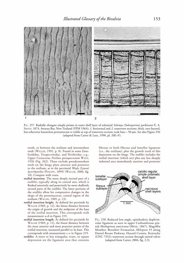
153Illustrated Glossary of the Bivalvia
tooth, or between the resilium and intermediate teeth (Waller, 1991, p. 8). Found in some Ento-lioididae, Tosapectinidae, and Neitheidae, e.g., Upper Cretaceous Neithea quinquecostata Wade, 1926 (Fig. 262). These exclude pseudotaxodont teeth on the hinge plate anterior and posterior to the resilium, as in the pectinoid Weyla (Lywea) lycorrhynchus (philippi, 1899) (Waller, 2006, fig. 10). Compare with crura.
resilial insertion. The more deeply incised part of a resilifer, typically along its central axis, which is flanked anteriorly and posteriorly by more shallowly incised parts of the resilifer. The latter portions of the resilifer allow for compressive changes in the shape of the proteinaceous, central region of the resilium (Waller, 1969, p. 13).
resilial insertion height. As defined for pectinids by Waller (1969, p. 12), the linear distance between the origin of growth and the midpoint of the base of the resilial insertion. This corresponds with measurement a–d in Figure 219.
resilial insertion length. As defined for pectinids by Waller (1969, p. 12), the linear distance between the most anterior and most posterior points of the resilial insertion, measured parallel to its base. This corresponds with measurement c–e in Figure 219.
resilifer. A more or less triangular, ovate, or square depression on the ligament area that contains
fibrous or both fibrous and lamellar ligament (i.e., the resilium), plus the growth track of this depression on the hinge. The resilifer includes the resilial insertion (which see) plus any less sharply indented area immediately anterior and posterior
FiG. 257. Radially elongate simple prisms in outer shell layer of solemyid Solemya (Solemyarina) parkinsoni e. a. sMith, 1874, Awarua Bay, New Zealand (YPM 5364); 1, horizontal and 2, transverse sections; thick, two-layered, but otherwise featureless periostracum is visible at top of transverse section; scale bars = 50 µm. See also Figure 256
(adapted from Carter & Lutz, 1990, pl. 20E–F).
FiG. 258. Reduced low-angle, opisthodetic duplivin-cular ligament as seen in upper Carboniferous pin-nid Meekopinna americana (Meek, 1867), Magoffin Member, Breathitt Formation, Milepost 55 along Daniel Boone Parkway, Hazard County, Kentucky (UNC 7352); transverse section through united valves
(adapted from Carter, 2004, fig. 2.3).
1 2
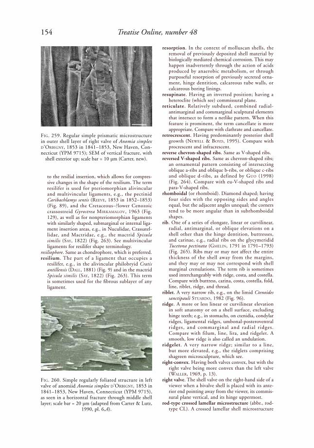
154 Treatise Online, number 48
to the resilial insertion, which allows for compres-sive changes in the shape of the resilium. The term resilifer is used for pteriomorphian alivincular and multivincular ligaments, e.g., the pectinid Caribachlamys sentis (reeVe, 1853 in 1852–1853) (Fig. 89), and the Cretaceous–?lower Cenozoic crassostreid Gyrostrea MirkaMaloV, 1963 (Fig. 129), as well as for nonpteriomorphian ligaments with similarly shaped, submarginal or internal liga-ment insertion areas, e.g., in Nuculidae, Crassatel-lidae, and Mactridae, e.g., the mactrid Spisula similis (say, 1822) (Fig. 263). See multivincular ligaments for resilifer shape terminology.
resiliophore. Same as chondrophore, which is preferred.resilium. The part of a ligament that occupies a
resilifer, e.g., in the alivincular philobryid Cratis antillensis (dall, 1881) (Fig. 9) and in the mactrid Spisula similis (say, 1822) (Fig. 263). This term is sometimes used for the fibrous sublayer of any ligament.
resorption. In the context of molluscan shells, the removal of previously deposited shell material by biologically mediated chemical corrosion. This may happen inadvertently through the action of acids produced by anaerobic metabolism, or through purposeful resorption of previously secreted orna-ment, hinge dentition, calcareous tube walls, or calcareous boring linings.
resupinate. Having an inverted position; having a heteroclite (which see) commissural plane.
reticulate. Relatively subdued, combined radial-antimarginal and commarginal sculptural elements that intersect to form a netlike pattern. When this feature is prominent, the term cancellate is more appropriate. Compare with clathrate and cancellate.
retrocrescent. Having predominantly posterior shell growth (NeWell & Boyd, 1995). Compare with procrescent and infracrescent.
reverse chevron-shaped ribs. Same as V-shaped ribs. reversed V-shaped ribs. Same as chevron-shaped ribs;
an ornamental pattern consisting of intersecting oblique a-ribs and oblique b-ribs, or oblique c-ribs and oblique d-ribs, as defined by Guo (1998) (Fig. 264). Compare with eu-V-shaped ribs and para-V-shaped ribs.
rhomboidal (or rhomboid). Diamond shaped; having four sides with the opposing sides and angles equal, but the adjacent angles unequal; the corners tend to be more angular than in subrhomboidal shapes.
rib. One of a series of elongate, linear or curvilinear, radial, antimarginal, or oblique elevations on a shell other than the hinge dentition, buttresses, and carinae, e.g., radial ribs on the glycymeridid Tucetona pectinata (GMeliN, 1791 in 1791–1793) (Fig. 265). Ribs may or may not affect the entire thickness of the shell away from the margins, and they may or may not correspond with shell marginal crenulations. The term rib is sometimes used interchangeably with ridge, costa, and costella. Compare with buttress, carina, costa, costella, fold, line, riblet, ridge, and thread.
riblet. A very narrow rib, e.g., on the limid Ctenoides sanctipauli stuardo, 1982 (Fig. 96).
ridge. A more or less linear or curvilinear elevation in soft anatomy or on a shell surface, excluding hinge teeth; e.g., in stomachs, on ctenidia, condylar ridges, ligamental ridges, umbonal-posteroventral r idges, and commarginal and radial r idges. Compare with filum, line, lira, and ridgelet. A smooth, low ridge is also called an undulation.
ridgelet. A very narrow ridge; similar to a line, but more elevated, e.g., the ridglets comprising shagreen microsculpture, which see.
right-convex. Having both valves convex, but with the right valve being more convex than the left valve (Waller, 1969, p. 13).
right valve. The shell valve on the right-hand side of a viewer when a bivalve shell is placed with its ante-rior end pointing away from the viewer, its commis-sural plane vertical, and its hinge uppermost.
rod-type crossed lamellar microstructure (abbr., rod-type CL). A crossed lamellar shell microstructure
FiG. 259. Regular simple prismatic microstructure in outer shell layer of right valve of Anomia simplex d’orBiGNy, 1853 in 1841–1853, New Haven, Con-necticut (YPM 9715); SEM of vertical fracture, with
shell exterior up; scale bar = 10 µm (Carter, new).
FiG. 260. Simple regularly foliated structure in left valve of anomiid Anomia simplex d’orBiGNy, 1853 in 1841–1853, New Haven, Connecticut (YPM 9715), as seen in a horizontal fracture through middle shell layer; scale bar = 20 µm (adapted from Carter & Lutz,
1990, pl. 6,A).

155Illustrated Glossary of the Bivalvia
in which the elongate, basic structural units are not organized into second-order lamellae (Carter & others, 1990, p. 612). This structure rarely occurs in the Bivalvia, e.g., within the pallial band of the Upper Triassic myophoriid Costatoria ornata (MüNster, 1841). Rod-type CL structure is common in chiton shells.
rod-type fibrous prismatic microstructure. See fibrous prismatic microstructure, rod-type.
rostrate. (1) Beaked or pointed; (2) having an elon-gate or drawn-out extension of the shell (rostrum), typically associated with siphons or siphonlike extensions of the mantle cavity, as in the cuspidariid Cuspidaria rostrata (speNGler, 1793) (Fig. 266).
rostrum. An elongate or drawn-out extension of the shell, e.g., on the cuspidariid Cuspidaria rostrata (speNGler, 1793) (Fig. 266).
rotund. Spherical or nearly so.rounded pallial sinus. A pallial sinus in which the
anterior apex is circular or broadly ovate.row stack nacreous microstructure . A form of
nacreous shell microstructure with elongate tablets aligned end-to-end in mutually parallel rows, with each row representing a vertical column of tablets. Known only in the Pinnoidea (Fig. 267). Compare with sheet nacreous and columnar nacreous micro-structures.
rudimentary. Undeveloped, vestigial or abortive.rugose. Having an ornament of rugae (singl., ruga),
i.e., relatively coarse, wrinkle-like elements, as in the Permian bakevelliid Cassiavellia galtarae tëMkiN & poJeta, 2010 (Fig. 268).
rugulose. Finely wrinkled.saddle-shaped shell. (1) A shell with a margin or
margins upturned along one axis and downturned
along a second axis, the second axis being more or less perpendicular to the first one; e.g., the Eocene ostreid Cubitostrea sellaeformis (CoNrad, 1832 in 1832–1835); (2) for larval shells, synonymous with the term dumbbell shell, which is preferred.
sagittal plane. (1) Any plane that is parallel with the midsagittal plane, including the latter; (2) just the midsagittal plane, in which case, all other planes parallel to it are called longitudinal planes.
satiny. Satinlike; smooth, glossy, e.g., the surface of some periostraca.
scabrous. Rough or scaly.scale. A tile-shaped or hoodlike projection, commonly
positioned on the apex of a rib. Compare with scute.
scallop. A vernacular term for a pectinoid, usually in reference to the Pectinidae.
scalloped. Having a series of regular flutings corre-sponding with the distal terminations of external ribs, as on the inner shell margin of many shells with strong external ribs.
scar. See muscle scar.schizodian-grade hinge. The hinge grade character-
ized by Carboniferous–Permian Schizodus de VerNeuil & MurChisoN, 1844 (Fig. 269). As described by NeWell and Boyd (1975, p. 77): hinge teeth short and unstriated, pivotal tooth of left valve typically not extended posteriorly, and with a more or less triangular, chisel-, or wedge-like shape, with a faint median sulcus on the lower (distal) margin, the latter representing the precursor of the cleft in this position in myopho-rian and trigonian-grade hinges. Main tooth of right valve is the anterior of the principal teeth of the two valves, and it is reinforced by an arcuate
FiG. 261. Lyonsiid Lyonsia norwegica (GMeliN, 1791 in 1791–1793), Plymouth, England (USNM 304916), showing a submarginal-internal ligament with lithodesma removed, supported by an elongate, internal resilial buttress. 1, Left hinge viewed perpendicular to commissure plane; 2, right hinge viewed ventrally and slightly obliquely (Carter, new).
1
2
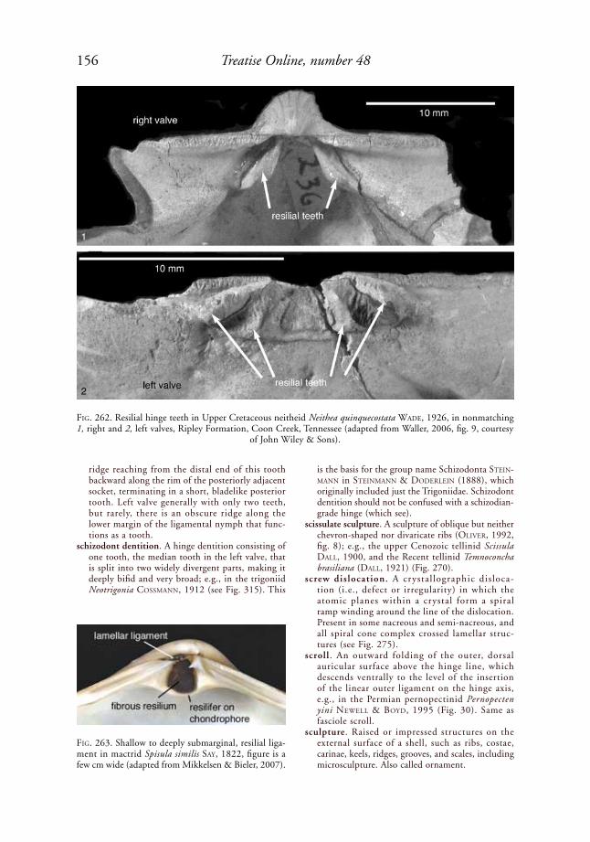
156 Treatise Online, number 48
ridge reaching from the distal end of this tooth backward along the rim of the posteriorly adjacent socket, terminating in a short, bladelike posterior tooth. Left valve generally with only two teeth, but rarely, there is an obscure ridge along the lower margin of the ligamental nymph that func-tions as a tooth.
schizodont dentition. A hinge dentition consisting of one tooth, the median tooth in the left valve, that is split into two widely divergent parts, making it deeply bifid and very broad; e.g., in the trigoniid Neotrigonia CossMaNN, 1912 (see Fig. 315). This
is the basis for the group name Schizodonta steiN-MaNN in steiNMaNN & doderleiN (1888), which originally included just the Trigoniidae. Schizodont dentition should not be confused with a schizodian-grade hinge (which see).
scissulate sculpture. A sculpture of oblique but neither chevron-shaped nor divaricate ribs (oliVer, 1992, fig. 8); e.g., the upper Cenozoic tellinid Scissula dall, 1900, and the Recent tellinid Temnoconcha brasiliana (dall, 1921) (Fig. 270).
screw dislocation. A crystal lographic disloca-tion (i.e., defect or irregularity) in which the atomic planes within a crystal form a spiral ramp winding around the line of the dislocation. Present in some nacreous and semi-nacreous, and all spiral cone complex crossed lamellar struc-tures (see Fig. 275).
scroll . An outward folding of the outer, dorsal auricular surface above the hinge line, which descends ventrally to the level of the insertion of the linear outer ligament on the hinge axis, e.g., in the Permian pernopectinid Pernopecten yini NeWell & Boyd, 1995 (Fig. 30). Same as fasciole scroll.
sculpture. Raised or impressed structures on the external surface of a shell, such as ribs, costae, carinae, keels, ridges, grooves, and scales, including microsculpture. Also called ornament.
FiG. 262. Resilial hinge teeth in Upper Cretaceous neitheid Neithea quinquecostata Wade, 1926, in nonmatching 1, right and 2, left valves, Ripley Formation, Coon Creek, Tennessee (adapted from Waller, 2006, fig. 9, courtesy
of John Wiley & Sons).
FiG. 263. Shallow to deeply submarginal, resilial liga-ment in mactrid Spisula similis say, 1822, figure is a few cm wide (adapted from Mikkelsen & Bieler, 2007).
1
2

157Illustrated Glossary of the Bivalvia
scutes. Very large scales, e.g., on the ribs of the tridacnid Tridacna squamosa laMarCk, 1819 in 1818–1822.
second diameter of adductor insertion. See adductor insertion, second diameter.
second inner mantle fold. Same as inner mantle fold 2, which see.
second outer mantle fold. Same as outer mantle fold 2, which see.
secondary. (1) An element of shell ornament appearing later in ontogeny than a primary element, gener-ally referring to radial ribs; (2) diagenetic, such as secondary calcite replacing original shell aragonite.
secondary fusion. Fusion of the left and right mantle lobes, other than in the mantle isthmus.
secondary glochidial host. Same as marginal glochidial host, which see.
secondary hinge teeth. Hinge teeth believed to have evolved independently of hinge teeth in most other bivalves, e.g., isodont hinge teeth in Spondylidae and Plicatulidae.
secondary homonym. Each of two or more iden-tical specific or subspecific names established for different nominal taxa and originally combined with different genus names, but subsequently placed in the same genus (ICZN, 1999). The later established name becomes the junior secondary homonym and is invalid except under circum-stances outlined by the ICZN (see ICZN, 1999, Articles 53.3, 57.3, 59). Compare with primary homonym.
secondary inner mantle fold. Same as second inner mantle fold, which see.
secondary ligament. Thickened periostracum that functionally extends an external or submarginal, elongate ligament anteriorly and/or posteriorly, or which unites the valves along the dorsal shell
margin in cases where the nonperiostracal ligament is entirely internal. Secondary ligament is herein called thickened periostracum.
secondary mantle margin retractors. Mantle margin retractor muscles originating at isolated spots along a line at about the level of the gill diaphragm. Each muscle extends as a single bundle or branches before inserting into the mantle margin. These muscles occasionally leave an irregular, interrupted line of scars in the position of a pallial line (harry, 1985, p. 125). Present in the ostreid Saccostrea dollFus & dautZeNBerG, 1920 in 1902–1920.
secondary outer mantle fold. Same as outer mantle fold 2, which see.
secondary shell. The calcified molluscan larval shell (cf. primary shell of BaNdel, 1988). Use is not recom-mended, as it corresponds with the prodissoconch in bivalves and protoconch in univalved mollusks. Also, secondary implies the replacement of one structure by another, which is not the case here.
secondary water tubes. Small tubes formed by acces-sory partitions in the gravid marsupial portion of
FiG. 264. Oblique rib types a through d, as defined by Guo (1998) for Trigonioidoidea. Exterior of left valve
(adapted from Guo, 1998, fig. 1).
FiG. 265. Exterior of glycymeridid Tucetona pectinata (GMeliN, 1791 in 1791–1793), showing equilateral shell shape and radial ribs (also called radial costae), shell length 16.2 mm (adapted from Mikkelsen &
Bieler, 2007).
FiG. 266. Cuspidariid Cuspidaria rostrata (speNGler, 1793), illustrating rostrum, shell length 20 mm (adapt-
ed from Mikkelsen & Bieler, 2007).
a
b cd
rostrum

158 Treatise Online, number 48
the ctenidium, present just before and persisting until just after the gravid period, e.g., in the unionid tribe Anodontini.
second-order folia (sing., folium). The more or less planar structural units that comprise the first-order folia in calcitic simple crossed-foliated (CF) and complex crossed foliated (CCF) microstruc-tures. Each second-order folium contains mutually parallel, lath- or bladelike, elongate structural units, called third-order folia, which have sharply angular terminations on the depositional surface.
second-order lamellae (sing., lamella). The more or less planar structural units that comprise the first-order lamellae in aragonitic simple crossed-lamellar (CL) and complex crossed lamellar (CCL) micro-structures, e.g., in the anomiid Anomia simplex d’orBiGNy, 1853 in 1841–1853 (Fig. 271). Also called second-order crossed lamellae, these consist
of narrow, elongate structural subunits called third-order lamellae. Second-order lamellae are not present in some CL and CCL structures, such as rod-type CL and fine CCL. Rod-type CL has only first- and third-order lamellae, whereas fine CCL has only third-order lamellae, i.e., structural subunits similar in size and shape to the third-order lamellae in normal CL structure. The term second-order lamellae has also been applied to second-order folia in calcitic simple crossed foliated and complex crossed foliated structures, but second-order folia is preferred for these structures.
sedentary. Same as sessile, which see.semelparous. Having only one reproductive period
during the life of the organism. Rare in bivalves, but occurring in some Pisidiidae (MaCkie, 1984, p. 375). Compare with iteroparous.
semi-foliated aragonite. The aragonitic form of semi-foliated microstructure (which see). This is found in the middle and inner shell layers of the lower Cambrian fordillids Pojetaia runnegari Jell, 1980, and Fordilla troyensis BarraNde, 1881. The basic structural units are blades or laths with flat to irregular terminations in the middle and inner shell layers of F. troyensis and the middle shell layer of P. runnegari. However, they have rounded terminations in the inner shell layer of P. runnegari (see ruNNeGar & BeNtley, 1983, fig. 4a) (Fig. 272), where some large-tablet, imbricated nacre is also present. Semi-foliated aragonite also occurs in some Recent tryblidiid monoplacophorans (CheCa, sáNCheZ-NaVas, & rodríGueZ-NaVarro, 2009, as “foliated aragonite”).
semi-foliated calcite. The calcitic form of semi-foliated microstructure. This is found in the transition between chalky and foliated microstructures in
FiG. 267. Row stack nacre in pinnid Atrina rigida (liGhtFoot, 1786), Beaufort, North Carolina (UNC, unnumbered specimen, from Amelie Scheltema). 1, Diagram showing horizontal (H ) and vertical (V ) sections (adapted from
Carter & others, 1990, p. 618); 2, depositional surface showing aligned nacre tablets (Carter, new).
FiG. 268. Guadalupian, middle Permian bakevelliid Cassiavellia galtarae tëMkiN & poJeta, 2010. Left valve showing auricular sulcus, wing, and rugose ornament
(adapted from Tëmkin & Pojeta, 2010, fig. 6.1).
1 2
10 µm
10 µm
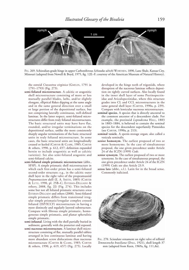
159Illustrated Glossary of the Bivalvia
the ostreid Crassostrea virginica (GMeliN, 1791 in 1791–1793) (Fig. 273).
semi-foliated microstructure. A calcitic or aragonitic shell microstructure consisting of more or less mutually parallel blades, laths, and/or slightly elongate, elliptical flakes dipping at the same angle and in the same general direction over a small or large portion of the depositional surface, but not comprising laterally continuous, well-defined laminae. In the latter respect, semi-foliated micro-structures differ from truly foliated microstructures. The basic structural units may have have flat, rounded, and/or irregular terminations on the depositional surface, unlike the more consistently sharply angular terminations of the basic structural units in truly foliated microstructure. In some cases, the basic structural units are longitudinally creased or keeled (Carter & Clark, 1985; Carter & others, 1990, p. 612, 657; definition expanded herein to include aragonitic as well as calcitic varieties). See also semi-foliated aragonite and semi-foliated calcite.
semi-foliated simple prismatic microstructure (abbr., SFSP). A simple prismatic shell microstructure in which each first-order prism has a semi-foliated second-order structure; e.g., in the calcitic outer shell layer in the right valve of the propeamussiid Propeamussium dalli (e. a. sMith, 1885) (Carter & lutZ, 1990, pl. 25B–C; esteBaN-delGado & others, 2008, fig. 2J) (Fig. 274). This includes some but not all foliated prismatic structures sensu esteBaN-delGado and others (2008). Semi-foliated simple prismatic differs from transitional irreg-ular simple prismatic/irregular complex crossed foliated (ISP/ICCF) microstructure in having a more distinctly and regularly layered substructure. Compare with fibrous simple prismatic, homo-geneous simple prismatic, and planar spherulitic simple prismatic.
semi-infaunal. Living with the shell partially buried in sediment, generally with the posterior end exposed.
semi-nacreous microstructure. A laminar shell micro-structure consisting of flat, mutually parallel tablets arranged in less continuous laminae and having more abundant screw dislocations than nacreous microstructure (Carter & Clark, 1985; Carter & others, 1990, p. 619, 657) (Fig. 275). Locally
developed in the hinge teeth of trigoniids, where disruption of the nacreous laminae reflects deposi-tion on tightly curved surfaces. Also locally found in the inner shell layer of some Pterinopectin-idae and Aviculopectinidae, where this structure grades into CL and CCL microstructures in the same general shell layer (Carter, 1990a, p. 239). Compare with lenticular nacreous microstructure.
seminal species. A species that is directly ancestral to the common ancestor of a descendent clade. For example, the pterineid Leptodesma hall, 1883 in 1883–1884, is believed to contain the seminal species for the descendent superfamily Pinnoidea (see Carter, 1990a, p. 213).
seminal vesicle. A sperm-storage organ; also called a vesicula seminalis.
senior homonym. The earliest proposed of two or more homonyms. In the case of simultaneous proposal, the one given precedence under Article 24 of the ICZN (1999) Code.
senior synonym. The earliest proposed of two or more synonyms. In the case of simultaneous proposal, the one given precedence under Article 24 of the ICZN (1999) Code; see also Article 23.9.
sensu lato (abbr., s.l.). Latin for in the broad sense. Commonly italicized.
FiG. 269. Schizodian-grade hinge in upper Carboniferous Schizodus ulrichi WortheN, 1890, Lane Shale, Kansas City, Missouri (adapted from Newell & Boyd, 1975, fig. 12E–F; courtesy of the American Museum of Natural History).
FiG. 270. Scissulate ornament on right valve of tellinid Temnoconcha brasiliana (dall, 1921), shell length 37
mm (adapted from Keen, 1969a, fig. 111,8a).
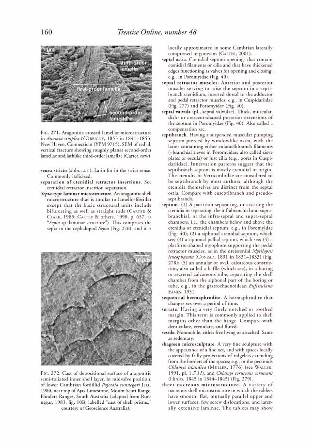
160 Treatise Online, number 48
sensu stricto (abbr., s.s.). Latin for in the strict sense. Commonly italicized.
separation of ctenidial retractor insertions. See ctenidial retractor insertion separation.
Sepia-type laminar microstructure. An aragonitic shell microstructure that is similar to lamello-fibrillar except that the basic structural units include bifurcating as well as straight rods (Carter & Clark, 1985; Carter & others, 1990, p. 657, as “Sepia sp. laminar structure”). This comprises the septa in the cephalopod Sepia (Fig. 276), and it is
locally approximated in some Cambrian laterally compressed tergomyans (Carter, 2001).
septal ostia. Ctenidial septum openings that contain ctenidial filaments or cilia and that have thickened edges functioning as valves for opening and closing; e.g., in Poromyidae (Fig. 40).
septal retractor muscles. Anterior and posterior muscles serving to raise the septum in a septi-branch ctenidium, inserted dorsal to the adductor and pedal retractor muscles, e.g., in Cuspidariidae (Fig. 277) and Poromyidae (Fig. 40).
septal valvula (pl., septal valvulae). Thick, muscular, dish- or crescent-shaped posterior extensions of the septum in Poromyidae (Fig. 40). Also called a compensation sac.
septibranch. Having a suspended muscular pumping septum pierced by windowlike ostia, with the latter containing either eulamellibranch filaments (=branchial sieves in Poromyidae; also called sieve plates or oscula) or just cilia (e.g., pores in Cuspi-dariidae). Innervation patterns suggest that the septibranch septum is mostly ctenidial in origin. The ctenidia in Verticordiidae are considered to be septibranch by most authors, although the ctenidia themselves are distinct from the septal ostia. Compare with euseptibranch and pseudo-septibranch.
septum. (1) A partition separating, or assisting the ctenidia in separating, the infrabranchial and supra-branchial, or the infra-septal and supra-septal chambers, i.e., the chambers below and above the ctenidia or ctenidial septum, e.g., in Poromyidae (Fig. 40); (2) a siphonal ctenidial septum, which see; (3) a siphonal pallial septum, which see; (4) a platform-shaped myophore supporting the pedal retractor muscles, as in the dreisseniid Mytilopsis leucophaeata (CoNrad, 1831 in 1831–1833) (Fig. 278); (5) an annular or oval, calcareous constric-tion, also called a baffle (which see), in a boring or secreted calcareous tube, separating the shell chamber from the siphonal part of the boring or tube, e.g., in the gastrochaenoidean Eufistulana eaMes, 1951.
sequential hermaphrodite. A hermaphrodite that changes sex over a period of time.
serrate. Having a very finely notched or toothed margin. This term is commonly applied to shell margins other than the hinge. Compare with denticulate, crenulate, and fluted.
sessile. Nonmobile, either free living or attached. Same as sedentary.
shagreen microsculpture. A very fine sculpture with the appearance of a fine net, and with spaces locally covered by frilly projections of ridgelets extending from the borders of the spaces; e.g., in the pectinids Chlamys islandica (Müller, 1776) (see Waller, 1991, pl. 1,7,11), and Chlamys coruscans coruscans (hiNds, 1845 in 1844–1845) (Fig. 279).
sheet nacreous microst ructure . A var ie ty o f nacreous shell microstructure in which the tablets have smooth, flat, mutually parallel upper and lower surfaces, few screw dislocations, and later-ally extensive laminae. The tablets may show
FiG. 271. Aragonitic crossed lamellar microstructure in Anomia simplex d’orBiGNy, 1853 in 1841–1853, New Haven, Connecticut (YPM 9715), SEM of radial, vertical fracture showing roughly planar second-order lamellae and lathlike third-order lamellae (Carter, new).
FiG. 272. Cast of depositional surface of aragonitic semi-foliated inner shell layer, in midvalve position, of lower Cambrian fordillid Pojetaia runnegari Jell, 1980, near top of Ajax Limestone, Mount Scott Range, Flinders Ranges, South Australia (adapted from Run-negar, 1983, fig. 10B; labelled “cast of shell prisms,”
courtesy of Geoscience Australia).

161Illustrated Glossary of the Bivalvia
irregular, stair step, and/or brick wall stacking modes (Fig. 280). Compare with columnar nacreous, lenticular nacreous, row stack nacreous, and semi-nacreous.
shell. An exoskeleton secreted by mantle epithelium and consisting of a nonmineralized to weakly mineralized periostracum and more strongly mineralized, subperiostracal shell layers, the latter generally referred to as outer, middle, myostracal and inner, or outer, myostracal and inner. In the Bivalvia, the shell also includes a dorsomedian ligament with one or more pairs of unmineralized lamellar, and partially mineralized, generally fibrous sublayers. Secretion of the shell begins during the larval phase. See calcareous tube, accessory plate, and pallets for skeletal components with a post-larval onset.
shell chamber. The portion of a boring or secreted calcareous tube or igloo that contains the shell valves, e.g., in the gastrochaenid Rocellaria dubia (peNNaNt, 1777) (Fig. 192). The shell-bearing portion of a crypt (which see).
shell conus. See conus.shell field. An area of ectodermal cells on the dorsal
side of a molluscan trochophore larva responsible for shell secretion (cf. MouëZa, Gros, & FreNkiel, 2006). Called shell gland by some authors (e.g., BaNdel, 1982, 1988). See also pellicle.
shell gland. Same as shell field, which see.shell height. See height of shell.shell layer. A bed of shell material exhibiting uniform
microstructure or uniform fine alternations of microstructures. In bivalves, the shell layers are commonly referred to as the periostracum, outer, middle, myostracal, and inner.
shell length. See length of shell.
shell marginal socket. (1) A socket receiving a shell-marginal tooth, e.g., in Crassatella laMarCk, 1799, and on the left dorsoanterior shell margin of Pholadomya candida G. B. soWerBy i, 1823 in 1821–1834, where the dorsoanterior right shell margin is received (Fig. 252); (2) one of several minute, articulating sockets near the anterior shell margin of some mytilids, not believed to be homologous with normal hinge tooth sockets; e.g., in Mytilus liNNaeus, 1758 (Fig. 281).
FiG. 273. Semi-foliated microstructure, diagram show-ing horizontal (H ), and vertical (V ) sections (adapted
from Carter & others, 1990, p. 619).
FiG. 274. Semi-foliated simple prismatic microstructure in outer sublayer of calcitic outer shell layer in right valve of propeamussiid Propeamussium dalli (e. a. sMith, 1885), west of Martinique, Windward Islands (YPM 8387); underlying sublayer of calcitic outer shell layer is predominantly fibrous prismatic to irregular spherulitic prismatic (see also Carter & lutZ, 1990, pl. 25B–C). SEM of a polished and then acid-etched, radial, vertical
section (Carter, new).
FiG. 275. Semi-nacrous microstructure, diagram show-ing horizontal (H ) and vertical (V ) sections (adapted
from Carter & others, 1990, p. 619).
1 µm
1 µm
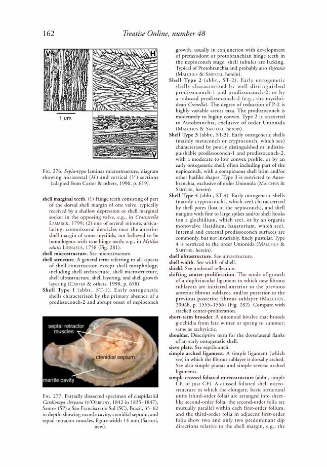
162 Treatise Online, number 48
shell marginal teeth. (1) Hinge teeth consisting of part of the dorsal shell margin of one valve, typically received by a shallow depression or shell marginal socket in the opposing valve, e.g., in Crassatella laMarCk, 1799; (2) one of several minute, articu-lating, commissural denticles near the anterior shell margin of some mytilids, not believed to be homologous with true hinge teeth; e.g., in Mytilus edulis liNNaeus, 1758 (Fig. 281).
shell microstructure. See microstructure.shell structure. A general term referring to all aspects
of shell construction except shell morphology, including shell architecture, shell microstructure, shell ultrastructure, shell layering, and shell growth layering (Carter & others, 1990, p. 658).
Shell Type 1 (abbr., ST-1). Early ontogenetic shells characterized by the primary absence of a prodissoconch-2 and abrupt onset of nepioconch
growth, usually in conjunction with development of pretaxodont or protobranchian hinge teeth in the nepioconch stage; shell tubules are lacking. Typical of Protobranchia and probably also Pojetaia (MalChus & sartori, herein).
Shell Type 2 (abbr., ST-2). Early ontogenetic she l l s character i zed by we l l d i s t ingui shed prodissoconch-1 and prodissoconch-2, or by a reduced prodissoconch-2 (e.g., the mytiloi-dean Crenella). The degree of reduction of P-2 is highly variable across taxa. The prodissoconch is moderately to highly convex. Type 2 is restricted to Autobranchia, exclusive of order Unionida (MalChus & sartori, herein).
Shell Type 3 (abbr., ST-3). Early ontogenetic shells (mainly metaconch or cryptoconch, which see) characterized by poorly distinguished or indistin-guishable prodissoconch-1 and prodissoconch-2, with a moderate to low convex profile, or by an early ontogenetic shell, often including part of the nepioconch, with a conspicuous shell brim and/or other hatlike shapes. Type 3 is restricted to Auto-branchia, exclusive of order Unionida (MalChus & sartori, herein).
Shell Type 4 (abbr., ST-4). Early ontogenetic shells (mainly cryptoconchs, which see) characterized by shell pores (lost in the nepioconch), and shell margins with fine to large spikes and/or shell hooks (on a glochidium, which see), or by an organic monovalve (lasidium, haustorium, which see). Internal and external prodissoconch surfaces are commonly, but not invariably, finely pustular. Type 4 is restriced to the order Unionida (MalChus & sartori, herein).
shell ultrastructure. See ultrastructure.shell width. See width of shell.shield. See umbonal reflection.shifting center-proliferation. The mode of growth
of a duplivincular ligament in which new fibrous sublayers are initiated anterior to the previous anterior fibrous sublayer, and/or posterior to the previous posterior fibrous sublayer (MalChus, 2004b, p. 1555–1556) (Fig. 282). Compare with stacked center-proliferation.
short-term brooder. A unionoid bivalve that broods glochidia from late winter or spring to summer; same as tachytictic.
shoulder. Descriptive term for the dorsolateral flanks of an early ontogenetic shell.
sieve plate. See septibranch.simple arched ligament. A simple ligament (which
see) in which the fibrous sublayer is dorsally arched. See also simple planar and simple reverse arched ligaments.
simple crossed foliated microstructure (abbr., simple CF, or just CF). A crossed foliated shell micro-structure in which the elongate, basic structural units (third-order folia) are arranged into sheet-like second-order folia, the second-order folia are mutually parallel within each first-order folium, and the third-order folia in adjacent first-order folia show two and only two predominant dip directions relative to the shell margin, e.g., the
FiG. 276. Sepia-type laminar microstructure, diagram showing horizontal (H ) and vertical (V ) sections
(adapted from Carter & others, 1990, p. 619).
FiG. 277. Partially dissected specimen of cuspidariid Cardiomya cleryana (d’orBiGNy, 1842 in 1835–1847), Santos (SP) a São Francisco do Sul (SC), Brazil, 35–62 m depth, showing mantle cavity, ctenidial septum, and septal retractor muscles, figure width 14 mm (Sartori,
new).
1 µm

163Illustrated Glossary of the Bivalvia
middle shell layer of the Middle Jurassic gryphaeid Gryphaea (Bilobissa) bilobata J. de C. soWerBy, 1840, in soWerBy & soWerBy, 1812–1846 (Fig. 38). Simple crossed foliated structure is commonly referred to as just crossed foliated.
simple crossed lamellar microstructure (abbr., simple CL, or just CL). An aragontic crossed lamellar shell microstructure in which the elongate, basic structural units (third-order lamellae) are arranged into sheetlike second-order lamellae, the second-order lamellae are mutually parallel within each first-order lamella, and the third-order lamellae in adjacent first-order lamellae show two and only two predominant dip directions relative to the shell margin (Wise, 1968, p. 325; Waller, 1978; Carter & Clark, 1985; Carter & others, 1990, p. 612, 658), e.g., in the trapeziid Trapezium (Neotrapezium) sublaevigatum (laMarCk, 1819 in 1818–1822) (Fig. 283). This category excludes rod-type CL structure, wherein the basic structural units do not form sheetlike second-order lamellae. It also excludes complex crossed lamellar micro-structures, which have more than two dip direc-tions. The unmodified term crossed lamellar (CL) is generally considered to indicate simple crossed lamellar microstructure.
simple foliated. A term used by MalChus (1990) for simple regularly foliated microstructure, which see.
simple lamellar fibrous prismatic microstructure. See fibrous prismatic microstructure, simple lamellar.
simple ligament. An elongate, external or submar-ginal, largely or entirely opisthodetic ligament with a single adult fibrous sublayer supported by an external gutter or by a submarginal fossette, without a differentiated nymph or pseudonymph. The fibrous sublayer of the larval ligament is assumed to be continuous with the fibrous sublayer of the adult ligament (Carter, 1990a; Waller, 1990); e.g., the submarginal simple ligament in Lucina pensylvanica (liNNaeus, 1758) (Fig. 284). The adult fibrous sublayer may be dorsally arched (=simple arched), planar or nearly planar (=simple planar), or ventrally arched (=simple reverse arched), which see. Not to be confused with preduplivincular ligaments.
simple pallial line. An entire or complete pallial line, which see.
simple papillae. Papillae that are not distally divided or branched.
simple planar ligament. A simple ligament (which see) in which the fibrous sublayer is nearly planar,
FiG. 278. Dreisseniid Mytilopsis leucophaeata (CoNrad, 1831 in 1831–1833), New Bern, North Carolina (UNC 15989), interior view of right hinge, showing opisthodetic, submarginal simple ligament, septum, myophore, and
muscle scars; 1, with posterior part of ligament removed; 2, with all of ligament removed (Carter, new).
1
2

164 Treatise Online, number 48
as opposed to dorsally or ventrally arched. Compare with simple arched and simple reverse arched ligaments.
simple prismatic microstructure. A prismatic shell microstructure in which each first-order prism has well-defined, not strongly laterally interdigi-
tating boundaries, a low to moderate length/width ratio, a length axis at a high angle to the deposi-tional surface, and second-order structural units that do not diverge in a fanlike or cone-in-cone manner toward the depositional surface. Each prism may have a homogeneous, fibrous prismatic,
FiG. 279. Shagreen microsculpture, showing uneroded areas with strongly projecting, cell-forming flanges and eroded areas forming typical shagreen pattern. Near ventral edge of right disc of Chlamys coruscans coruscans (hiNds, 1845 in 1844–1845), Tuamotu Islands. Coated with ammonium chloride; scale bar = 500 µm (adapted
from Waller, 1972a, fig. 12).
FiG. 280. Sheet nacre in inner shell layer of pteriid Pinctada imbricata rödiNG, 1798, from Bimini, the Bahamas (YPM 6889); SEM of oblique fracture; scale
bar = 10 µm (Carter, new).
FiG. 281. Planivincular ligament and shell marginal teeth and sockets in mytilid Mytilus edulis liNNaeus, 1758; 1, lateral interior view of hinge based on camera lucida, Long Island Sound (YPM 9542); and 2, medial, dorsoventral, transverse section through hinge, New England (UNC 13620); FP, fibrous prismatic outer shell layer. Note that
innermost part of fibrous ligament is not yet fully mineralized (Carter, new).
vacuolated pseudonymph
shell marginal teeth 5 mm
periostracum
lamellar
fibrous sublayerFP
nacrepseudonymph
vacuole 1 mm
1
2

165Illustrated Glossary of the Bivalvia
semi-foliated, or planar spherulitic substructure (see homogeneous simple prismatic, fibrous simple pris-matic, semi-foliated simple prismatic, and planar spherulitic simple prismatic). Simple prism shapes are classified as regular, irregular, asymmetric, radially elongate, blocky, and pavement simple prismatic. Optical properties of simple prisms, as seen in vertical thin sections in crossed-polarized light, may be optically homogeneous and clear, optically heterogenous and granular, or optically heterogeneous and irregular-wavy (see optically heterogeneous and optically homogeneous).
simple regularly foliated microstructure (abbr., simple RF). A regularly foliated shell microstructure in which the third-order laths and blades are mutu-ally parallel throughout the shell layer, as in some Ostreoidea and Anomioidea, e.g., in the placunid Placuna (Fig. 285). This structure was called simple foliated by MalChus (1990, p. 64, pl. 26,1).
simple reverse arched ligament. A simple liga-ment (which see) in which the fibrous sublayer is ventrally arched, e.g., in the Middle Devonian montanariid Montanaria sp. (Fig. 286). Compare with simple arched and simple planar ligaments.
simultaneous hermaphrodite. An hermaphrodite that produces eggs and sperm at the same time.
sinuate (or sinuous) pallial line. A pallial line with a pallial sinus.
sinupalliate. Having a pallial sinus.sinus. An indentation or embayment, e.g., a pallial
sinus or an embayment of the shell margin, as in the Toarcian, Lower Jurassic trigonioid Trigonia sulcata (herMaNN, 1781) (Fig. 53).
siphon . A mantle extension that serves for the passage of incurrent and/or excurrent water, and that involves cuticular and/or tissue-grade union of the left and right, inner mantle folds, and in some cases also the middle mantle folds. See also siphon sensu lato and siphon sensu stricto. The unmodified term siphon generally refers to a siphon sensu lato.
siphon sensu lato. The siphon sensu stricto (which see) plus any associated ctenidium-bearing, posterior extension of the mantle cavity.
siphon sensu stricto. The part of a mantle extension, serving for the passage of water, that does not contain a posterior extension of the ctenidia (see
Fig. 291). This excludes any ctenidium-bearing, posterior extension of the mantle cavity, which is, however, part of the siphon sensu lato.
siphon type. yoNGe (1957, 1982b) defined siphon types A, A+, B, B/C, C, and D based on their observed or inferred degree of mantle fusion (see below). Waller’s (1980) mantle fold terminology (inner fold 1, inner fold 2, outer fold 1, outer fold 2) is herein added to yoNGe’s definitions, which originally used the traditional terms inner, middle, and outer mantle fold. Note: yoNGe (1982b) indi-cated that his siphon type B/C should be regarded as simply type B.
siphon type A. A siphonal apparatus with at least the dorsal wall of the excurrent aperture formed by tissue-grade fusion of the inner (=inner fold 2) mantle fold, but not the middle (=inner fold 1) or middle and outer (=outer fold 1) mantle folds, and without tentacles and eyes on the ends of the siphons (yoNGe, 1957, 1982b), e.g., the mytilid Lithophaga (Leiosolenus) hancocki T. soot-ryeN, 1955 (Fig. 34). The middle mantle fold is retained in the plesiomorphic position around the posterior shell margins, and the siphon lacks a periostracal covering. In Yoldiella Verrill & Bush, 1897, cilial
FiG. 282. Shifting center-proliferation growth pattern of lamellar (L) and fibrous (F ) sublayers in a multivincular ligament. 1, Elementary L-F-L pattern in first adult ligament; 2, L-F-L pattern in mature adult ligament (adapted
from Malchus, 2004b, fig. 6B,D).
FiG. 283. Simple crossed lamellar microstructure in outer shell layer of trapeziid Trapezium (Neotrapezium) sublaevigatum (laMarCk, 1819 in 1818–1822), Bahia, Philippines (YPM 9717); SEM of a radial, vertical
fracture; scale bar = 5 µm (Carter, new).
L1 F1 L1 L1 F4 F3 F2 F1 F2 F3 F4 L11 2
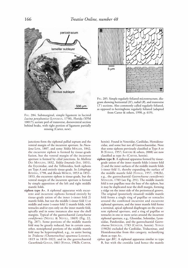
166 Treatise Online, number 48
junctions form the siphonal pallial septum and the ventral margin of the incurrent aperture. In Nucu-lana liNk, 1807, and some Yoldia Möller, 1842, the excurrent siphon is formed by tissue-grade fusion, but the ventral margin of the incurrent aperture is formed by cilial junctions. In Malletia des MouliNs, 1832, Yoldia limatula (say, 1831), the Erycinidae, and the Tellinoidea, both siphons are Type A and entirely tissue-grade. In Lithophaga rödiNG, 1798, and Botula MörCh, 1853 in 1852–1853, the excurrent siphon is tissue-grade, but the ventral margin of the incurrent aperture is formed by simple apposition of the left and right middle mantle folds.
siphon type A+. A siphonal apparatus with excur-rent and incurrent siphons formed entirely by tissue-grade union of the inner (=inner fold 2) mantle folds, but not the middle (=inner fold 1) or middle and outer (=outer fold 1) mantle folds, with tentacles and/or eyes only on the inner mantle fold, apically and in some instances also near the shell margins. Typical of the gastrochaenid Lamychaena cordiformis (NeVill & NeVill, 1869) (Fig. 22, Fig. 287). Some portions of the middle mantle fold may be greatly reduced, but in certain cases, other, nonsiphonal portions of the middle mantle fold may be hypertrophied, e.g., to assist boring in Tridacna (Chametrachea) squamosa laMarCk, 1819 in 1818–1822, and in the gastrochaenid Cucurbitula Gould, 1861 (yoNGe, 1982b; Carter,
FiG. 284. Submarginal, simple ligament in lucinid Lucina pensylvanica (liNNaeus, 1758), Florida (YPM 10017); acetate peel of transverse, dorsoventral section behind beaks, with right portion of ligament partially
missing (Carter, new).
FiG. 285. Simple regularly foliated microstructure, dia-gram showing horizontal (H ), radial (R), and transverse (T ) sections. Also commonly called regularly foliated, as opposed to herringbone regularly foliated (adapted
from Carter & others, 1990, p. 619).
herein). Found in Veneridae, Cardiidae, Hemidona-cidae, and some but not all Gastrochaenidae. Note that some siphons previously classified as Type A or B (yoNGe, 1957; sartori & others, 2008) are now classified as type A+ (Carter, herein).
siphon type B. A siphonal apparatus formed by tissue-grade union of the inner mantle folds (=inner fold 2) and the inner surfaces of the middle mantle folds (=inner fold 1), thereby expanding the surface of the middle mantle fold (yoNGe, 1957, 1982b), e.g., the gastrochaenid Gastrochaena cuneiformis speNGler, 1783 (see Fig. 291). The middle mantle fold is not papillate near the base of the siphon, but it may be duplicated near the shell margin, forming a ridge on the inner side of the periostracal groove. The original (posteriorly situated) middle mantle fold forms a single ring of papillae or tentacles around the combined incurrent and excurrent siphonal apertures, and the inner mantle fold forms a terminal, apical siphonal diaphragm on the excur-rent siphonal aperture, and a ring of papillae or tentacles in one or more series around the incurrent siphonal aperture, e.g., Glossidae, Solenidae, Lyon-siidae, Pandoridae, and the gastrochaenid Gastro-chaena speNGler, 1783 (Carter, herein). yoNGe (1982b) excluded the Cardiidae, Tridacninae, and Hemidonacidae from this category, reclassifying them as type A+.
siphon type B/C. A siphonal apparatus similar to type B, but with the ctenidia (and hence the mantle
5 µm
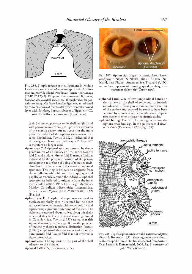
167Illustrated Glossary of the Bivalvia
cavity) extended posterior to the shell margins, and with periostracum covering this posterior extension of the mantle cavity, but not covering the more posterior surface of the siphons sensu stricto, e.g., some Pholadidae. yoNGe (1982b) indicated that this category is better regarded as type B. Type B/C is therefore no longer used.
siphon type C. A siphonal apparatus formed by tissue-grade union of all surfaces of the inner (=inner fold 2) and middle (=inner fold 1) mantle folds, as indicated by the posterior position of the perios-tracal groove at the base of a ring of tentacles encir-cling both the incurrent and excurrent siphonal apertures. This ring is believed to originate from the middle mantle fold, and the diaphragm and papillae or tentacles around the individual siphonal apertures are believed to originate from the inner mantle fold (yoNGe, 1957, fig. 9), e.g., Mactridae, Myidae, Corbulidae, Hiatelloidea, Laternulidae. See Laternula elliptica (kiNG & Broderip, 1832) (Fig. 288).
siphon type D. A siphonal apparatus encased in a calcareous shelly sheath secreted by the outer surface of the outer mantle fold (=outer fold 1), and representing a posterior extension of the shell. The siphons are attached about halfway along this shelly tube, and they lack a periostracal covering. Found in Cuspidariidae. yoNGe (1957) noted that this siphonal anatomy is like type B, but the presence of the shelly sheath requires a distinction. yoNGe (1982b) emphasized that the outer surface of the outer mantle fold (=outer fold 1) is not involved in siphon formation.
siphonal area. The siphons, or the part of the shell adjacent to the siphons.
siphonal baffles. See calcareous baffles.
FiG. 286. Simple reverse arched ligament in Middle Devonian montanariid Montanaria sp., Hecla Bay For-mation, Melville Island, Northwest Territories, Canada (TMP 87.123.3). Diagram of reconstructed ligament, based on dorsoventral acetate peel through valves far pos-terior to beak; solid black, lamellar ligament, as indicated by concentrations of framboidal pyrite; ventrally bowed layer with hatching, fibrous sublayer of ligament; CL,
crossed lamellar microstructure (Carter, new).
FiG. 287. Siphon tips of gastrochaenid Lamychaena cordiformis (NeVill & NeVill, 1869), Ko Khai Nai Island, near Phuket, Andaman Sea, Thailand (UNC, unnumbered specimen), showing apical diaphragm on
excurrent siphon tip (Carter, new).
FiG. 288. Type C siphons in laternulid Laternula elliptica (kiNG & Broderip, 1832), showing periostracal sheath with arenophilic threads (or lines) (adapted from Sartori, Dias Passos, & Domaneschi, 2006, fig. 3, courtesy of
John Wiley & Sons).
siphonal band. One of two longitudinal bands on the surface of the shell of some rudists (mainly radiolitids), differing in ornament from the rest of the surface and believed by some to have been secreted by a portion of the mantle where respira-tory currents enter or leave the mantle cavity.
siphonal boring. The part of a boring containing the siphons sensu lato, e.g., in the gastrochaenid Rocel-laria dubia (peNNaNt, 1777) (Fig. 192).
CLbourrelet
1 mm

168 Treatise Online, number 48
siphonal branchial septum. Same as siphonal ctenidial septum, which see.
siphonal burrow. The part of a burrow or secreted calcareous tube containing the siphons sensu lato.
siphonal ctenidial septum. A horizontal, widely perforated partition (more like an open scaf-folding) separat ing the suprabranchial and infrabranchial chambers at the level of the left and right ctenidial axes, positioned posterior to the visceral mass and anterior to any siphonal pallial septum that may be present, e.g., in the gastrochaenid Lamychaena hians (GMeliN, 1791 in 1791–1793) (Fig. 289). The posterior end of the ctenidial septum corresponds with the anterior end (base) of any siphon sensu stricto that may be present. A siphonal ctenidial septum extending posterior to the shell margins is located within a posterior extension of the mantle cavity, and not within the siphon sensu stricto.
siphonal diaphragm. See apical and basal siphonal diaphragms.
siphonal ganglia. A concentration of nerve cells inner-vating the siphons.
siphonal gape. A permanent gape between the fully adducted shell valves through which the siphon or siphons extend, e.g., in the laternulid Laternula elliptica (kiNG & Broderip, 1832) (Fig. 290).
siphonal membrane. A term used by kelloGG (1915) for what is herein called a basal siphonal diaphragm or a basal siphonal valve.
siphonal pallial septum. A flat, solid, horizontal partition consisting of fused left and right mantle tissue, which separates the incurrent and excurrent channels within the siphon sensu stricto, and which defines the anterior (proximal) end of the siphon sensu stricto; e.g., in the gastrochaenid Gastro-chaena cuneiformis speNGler, 1783 (Fig. 291). The posterior ends of the ctenidia may be attached by cuticular or tissue-grade fusion to the proximal end of the pallial septum. Compare with siphonal ctenidial septum.
siphonal retractors. Pallial retractor muscles serving to withdraw the siphons, typically attached along the pallial sinus. These represent modified radial mantle margin muscles.
siphonal septum. A siphonal pallial septum or a siphonal ctenidial septum, which see.
siphonal septum muscle. A muscle extending from the base of a siphonal pallial septum to the shell below the posterior adductor muscle scar, serving to assist in siphonal retraction. See muscle scar b.
siphonal sheath. A periostracal covering on the siphon sensu lato, e.g., in the myid Mya liNNaeus, 1758, and in the laternulid Laternula elliptica (kiNG & Broderip, 1832) (Fig. 288).
siphonal tentacle. Same as posterior sensory tentacle, which see.
siphonal valve. Same as basal siphonal valve, which see.
FiG. 289. Siphonal ctenidial septum and associated ctenidial anatomy in gastrochaenid Lamychaena hians (GMeliN, 1791 in 1791–1793); ID, inner demibranch; OD, outer demibranch (adapted from Carter, 1978,
fig. 5).
FiG. 290. Ventral view of laternulid Laternula elliptica (kiNG & Broderip, 1832), Hangar Cove, Antarctica, 4 m depth, showing siphonal and pedal shell gapes, figure width 7.9 mm (Sartori, new).
pedal gape
siphonal gape

169Illustrated Glossary of the Bivalvia
siphonoplax. A calcareous and/or periostracal struc-ture added to the posterior end of the shell in the adult stage, serving to protect the proximal end of the siphons, e.g., the pholadid Martesia striata (liNNaeus, 1758) (Fig. 24, Fig. 48–49, Fig. 175) and many other Pholadidae plus some Teredinidae.
skewed rib. A rib in which the slope is steeper on one side than on the other side.
slope. A posterior or anterior area on the exterior surface of a shell, below the dorsal margin, which is characterized by different ornament and/or a flatter surface than the central part (flank) of the shell, e.g., the anterior and posterior slopes in Pholadidae (Fig. 24).
slow muscle. Same as catch muscle, which see.smooth ctenidium. Same as a nonplicate ctenidium,
which see.socket. A recess or depression that receives a tooth in
the opposite valve.
socketed tooth. A hinge tooth that is received by a distinct recess or depression in the hinge of the opposite valve. Not to be confused with a shell-marginal tooth, which is received only by a slight depression on the opposite shell margin.
soleniform. Having a shell shape like Solen liNNaeus, 1758 (Fig. 292), i.e., similar to but slightly less elongate than ensiform.
solitary. Living isolated from other members of the species; not growing in clusters.
sorting area. A ciliated area on the stomach wall where ingested particles are accepted as food or rejected, largely on the basis of particle size and density.
spat. A newly settled bivalve larva. Also used as a collective term for such larvae.
spatulate. Elongate spoon shaped, i.e., oblong, with a broad, rounded end and a narrow, attenuate base. Also spelled spathulate.
species germinalis. The earliest member or members of a branch-based clade, identifiable retrospectively
FiG. 291. Gastrochaenid Gastrochaena cuneiformis speNGler, 1783, Kiribati, Tarawa, western Pacific (FMNH 282418), sagittal section through alcohol-preserved specimen showing type B siphons, siphonal pallial septum, ctenidium, posterior adductor muscle, incurrent siphon (bottom thick arrow), excurrent siphon (top thick arrow),
and incurrent and excurrent apical siphonal diaphragms (Carter, new).
FiG. 292. Soleniform shell shape as illustrated by solenid Solen vagina liNNaeus, 1758, Europe; exterior of left valve; scale bar = 1 cm (adapted from Chenu, 1862).
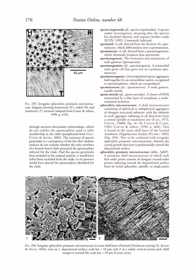
170 Treatise Online, number 48
through ancestor-descendant relationships, which do not exhibit the apomorphies used to infer membership in the clade (paraphrased from Cela-CoNde & altaBa, 2002). The existence of species germinalis is a consequence of the fact that cladistic analyses do not evaluate whether the early members of a branch-based clade possessed the apomorphies inferred for the clade. Had the species germinalis been included in the original analysis, it would have either been excluded from the clade, or its presence would have altered the apomorphies identified for the clade.
species inquirenda (pl., species inquirendae). A species under investigation, meaning that the species has doubtful identity and requires further study (ICZN, 1999). Commonly italicized.
spermatid. A cell, derived from the division of a sper-matocyte, which differentiates into a spermatozoan.
spermatocyte. A cell, derived from a spermatogonium, which ultimately produces four spermatids.
spermatogenesis. The formation and maturation of male gametes (spermatozoa).
spermatogonium (pl., spermatogonia). A primordial male germ cell that gives rise to a primary sper-matocyte.
spermatozeugmata. Unencapsulated sperm aggregates held together by an extracellular matrix, as opposed to spermatophores, which are encapsulated.
spermatozoan (pl., spermatozoa). A male gamete, usually motile.
sperm morula (pl., sperm morulae). A cluster of DNA surrounded by a thin layer of cytoplasm; a multi-nucleated inclusion.
spherulitic microstructure. A shell microstructure consisting of spherical to subspherical aggregates of elongate structural subunits, with the subunits in each aggregate radiating in all directions from a central spindle or nucleation site (FlaJs, 1972; Carter, 1980b, fig. 16–18; Carter & Clark, 1985; Carter & others, 1990, p. 660). This is found in the outer shell layer of the lucinid Anodontia (Pegophysema) bialata (pilsBry, 1895) (Fig. 296). Not to be confused with irregular spherulitic prismatic microstructure, wherein the crystal growth direction is perferentially toward the depositional surface.
spherulitic prismatic microstructure (abbr., SphP). A prismatic shell microstructure in which each first-order prism consists of elongate second-order prisms radiating toward the depositional surface from an initial spherulite, spindle, or single point
FiG. 293. Irregular spherulitic prismatic microstruc-ture, diagram showing horizontal (H ), radial (R), and transverse (T ) sections (adapted from Carter & others,
1990, p. 618).
FiG. 294. Irregular spherulitic prismatic microstructure in outer shell layer of lucinid Divalucina cumingi (A. adaMs & aNGas, 1864), seen on 1, depositional surface, scale bar = 10 µm; and 2, in a radial, vertical acetate peel, shell
margin is toward left, scale bar = 50 µm (Carter, new).
50 µm
1 2

171Illustrated Glossary of the Bivalvia
of origin (Fig. 293–295). Varieties include regular and irregular spherulitic prismatic (see below). Not to be confused with composite prismatic and spherulitic microstructures.
spherulitic prismatic microstructure, irregular (abbr., irregular SphP). A spherulitic prismatic shell micro-structure in which the first-order prisms have irreg-ularly conical shapes with strongly interdigitating mutual boundaries. Each first-order prism consists of a bundle of elongate second-order prisms radi-ating toward the depositional surface from a single spherulite, spindle, or point of origin (MutVei, 1964; Carter, 1980b; Carter & others, 1990, p. 659) (Fig. 293). Not to be confused with composite prismatic structure, where the first-order prisms are more columnal. Present in the calcitic outer shell layer of the left valve of some Strebochondriidae, Pernopectinidae, Entolioididae, and early Propea-mussiidae, along with some radial or antimarginal fibrous prisms. Present in the aragonitic outer shell layer of the lucinid Divalucina cumingi (A. adaMs & aNGas, 1864) (Fig. 294).
spherulitic prismatic microstructure, regular (abbr., regular SphP). A spherulitic prismatic structure in which the first-order prisms are columnar (Carter & Clark, 1985; Carter & others, 1990, p. 656) (Fig. 295). Similar to columnar nondenticular composite prismatic microstructure, but with a single, three-dimensional, fanlike, instead of concatenated fanlike, second-order structure. Found in the outer shell layer of some extant Monoplacophora and in the initial growth stage of some columnar nondenticular composite prisms in trigonioids and unionoids. Compare with irregular spherulitic prismatic.
spicate periostracum. A periostracum with short, bladelike bristles standing more or less erect and not forming a wide fringe (oliVer, 1981, p. 80). See also thatched, lanceolate, and pilose perio-stracum.
spike. In reference to periostracal mineralization, a projecting or entirely embedded, mineralized struc-ture with a length/height ratio <5 (see Fig. 143). See also periostracal pins and needles.
spine. A stiff, long, pointed structure.spinule (adj., spinulose). A small spine.spiral cone complex crossed foliated microstructure
(abbr., spiral cone CCF). A cone complex crossed foliated shell microstructure in which each first-order folia consists of a single, spiral second-order folia.
spiral cone complex crossed lamellar microstructure (abbr., spiral cone CCL). A cone complex crossed lamellar shell microstructure in which each first-order lamella consists of a spiral second-order lamella (Carter & others, 1990, p. 660); e.g., in the cyrenid Corbicula cf. C. fluminea (Müller, 1774 in 1773–1774) (Fig. 297).
spirogyrate. Coiled outward from the sagittal plane; a term applied to umbos.
squamose. Having small scales.stacked center-proliferation. The mode of growth
of a duplivincular ligament in which new fibrous
sublayers emerge directly below the point of emer-gence of the previous fibrous sublayer, causing the growing ends of the older fibrous sublayers to move anteriorly and/or posteriorly away from their initial point of emergence (MalChus, 2004b, p. 1555–1556) (Fig. 298). The anterior and posterior branches of the ligament may be added synchro-nously, in alternation, or in irregular alternation (see duplivincular synchronous). Compare with shifting center-proliferation.
stair step nacreous microstructure. A variety of sheet nacreous shell microstructure characterized by
FiG. 295. Regular spherulitic prismatic microstruc-ture, diagram showing horizontal (H ), radial (R), and transverse (T ) sections (adapted from Carter & others,
1990, p. 618).
FiG. 296. Spherulitic microstructure in outer shell layer of lucinid Anodontia (Pegophysema) bialata (pilsBry, 1895), tidal sand flat, Tsingtao, China (YPM 8963); SEM of a radial, vertical fracture; scale bar = 10 µm
(Carter, new).
50 µm
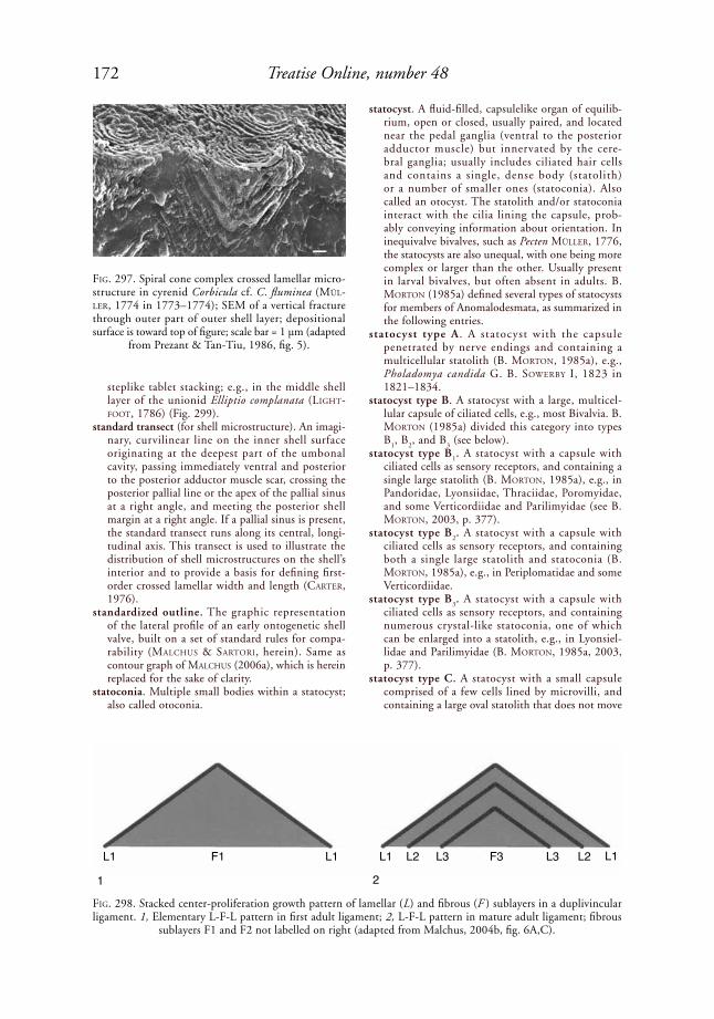
172 Treatise Online, number 48
steplike tablet stacking; e.g., in the middle shell layer of the unionid Elliptio complanata (liGht-Foot, 1786) (Fig. 299).
standard transect (for shell microstructure). An imagi-nary, curvilinear line on the inner shell surface originating at the deepest part of the umbonal cavity, passing immediately ventral and posterior to the posterior adductor muscle scar, crossing the posterior pallial line or the apex of the pallial sinus at a right angle, and meeting the posterior shell margin at a right angle. If a pallial sinus is present, the standard transect runs along its central, longi-tudinal axis. This transect is used to illustrate the distribution of shell microstructures on the shell’s interior and to provide a basis for defining first-order crossed lamellar width and length (Carter, 1976).
standardized outline. The graphic representation of the lateral profile of an early ontogenetic shell valve, built on a set of standard rules for compa-rability (MalChus & sartori, herein). Same as contour graph of MalChus (2006a), which is herein replaced for the sake of clarity.
statoconia. Multiple small bodies within a statocyst; also called otoconia.
statocyst. A fluid-filled, capsulelike organ of equilib-rium, open or closed, usually paired, and located near the pedal ganglia (ventral to the posterior adductor muscle) but innervated by the cere-bral ganglia; usually includes ciliated hair cells and contains a single, dense body (statolith) or a number of smaller ones (statoconia). Also called an otocyst. The statolith and/or statoconia interact with the cilia lining the capsule, prob-ably conveying information about orientation. In inequivalve bivalves, such as Pecten Müller, 1776, the statocysts are also unequal, with one being more complex or larger than the other. Usually present in larval bivalves, but often absent in adults. B. MortoN (1985a) defined several types of statocysts for members of Anomalodesmata, as summarized in the following entries.
statocyst type A . A statocyst with the capsule penetrated by nerve endings and containing a multicellular statolith (B. MortoN, 1985a), e.g., Pholadomya candida G. B. soWerBy i, 1823 in 1821–1834.
statocyst type B. A statocyst with a large, multicel-lular capsule of ciliated cells, e.g., most Bivalvia. B. MortoN (1985a) divided this category into types B
1, B
2, and B
3 (see below).
statocyst type B1. A statocyst with a capsule with
ciliated cells as sensory receptors, and containing a single large statolith (B. MortoN, 1985a), e.g., in Pandoridae, Lyonsiidae, Thraciidae, Poromyidae, and some Verticordiidae and Parilimyidae (see B. MortoN, 2003, p. 377).
statocyst type B2. A statocyst with a capsule with
ciliated cells as sensory receptors, and containing both a single large statolith and statoconia (B. MortoN, 1985a), e.g., in Periplomatidae and some Verticordiidae.
statocyst type B3. A statocyst with a capsule with
ciliated cells as sensory receptors, and containing numerous crystal-like statoconia, one of which can be enlarged into a statolith, e.g., in Lyonsiel-lidae and Parilimyidae (B. MortoN, 1985a, 2003, p. 377).
statocyst type C. A statocyst with a small capsule comprised of a few cells lined by microvilli, and containing a large oval statolith that does not move
FiG. 297. Spiral cone complex crossed lamellar micro-structure in cyrenid Corbicula cf. C. fluminea (Mül-ler, 1774 in 1773–1774); SEM of a vertical fracture through outer part of outer shell layer; depositional surface is toward top of figure; scale bar = 1 µm (adapted
from Prezant & Tan-Tiu, 1986, fig. 5).
FiG. 298. Stacked center-proliferation growth pattern of lamellar (L) and fibrous (F ) sublayers in a duplivincular ligament. 1, Elementary L-F-L pattern in first adult ligament; 2, L-F-L pattern in mature adult ligament; fibrous
sublayers F1 and F2 not labelled on right (adapted from Malchus, 2004b, fig. 6A,C).
1 2
L1 F1 L1 L1 L2 L3 L3 L2 L1F3
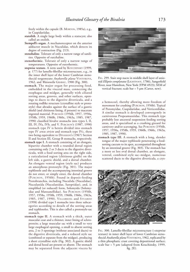
173Illustrated Glossary of the Bivalvia
freely within the capsule (B. MortoN, 1985a), e.g., in Cuspidariidae.
statolith. A single large body within a statocyst; also called an otolith.
Stempell’s organ. A mechanoreceptor on the anterior adductor muscle in Nuculidae, which detects its degree of contraction (Fig. 213).
stenohaline. Tolerant of only a narrow range of sanili-ties. Opposite of euryhaline.
stenothermic. Tolerant of only a narrow range of temperatures. Opposite of eurythermic.
stepwise texture. A term used by kouChiNsky (1999, p. 177) for lamello-fibrillar microstructure, e.g., in the inner shell layer of the lower Cambrian steno-thecid tergomyans Anabarella plana VostokoVa, 1962, and Watsonella GraBau, 1900 (Fig. 300).
stomach. The major organ for processing food, embedded in the visceral mass, connecting the esophagus and midgut, generally with ciliated sorting areas, grooves, and other surfaces, open-ings to ducts to the digestive diverticula, and a rotating rodlike structure (crystalline style or proto-style) that abrades against the surface of a gastric shield (and chitinous lining, if present) to macerate ingested material. purChoN (1956b, 1957, 1958a, 1958b, 1959, 1960b, 1960c, 1963a, 1985, 1987, 1990) classified bivalve stomachs into types I, II, III, IV, IVa, IVb, and V. purChoN’s (1987, 1990) stomach type IV is herein divided into stomach type IV sensu stricto and stomach type IVc, these two being equivalent to diNaMaNi’s (1967) Section II and Section III, Group B stomachs, respectively.
stomach type I. A stomach consisting of an elongate, bipartite chamber with a rounded dorsal region containing only 2 or 3 ducts to the digestive diver-ticula, with a food sorting area on the right side, a cuticularized region (chitinous girdle) on the left side, a gastric shield, and a dorsal chamber. An elongate ventral region (style sac) produces an amorphous protostyle (Fig. 301). The major typhlosole and the accompanying intestinal groove do not enter, or simply enter, the dorsal chamber (purChoN, 1956b). Found in deposit-feeding Protobranchia, including Nuculida (Nuculidae), Nuculanida (Nuculanidae, Sareptidae), and, in simplified (or reduced) form, Solemyida (Solemy-idae and Manzanellidae). See purChoN (1956b, 1957, 1958a, 1958b, 1959, 1960b, 1960c, 1963a, 1985, 1987, 1990). Villarroel and stuardo (1998) divided type I stomachs into three subcat-egories according to details of the sorting areas and typhlosoles. This is also called a protostyle-sac stomach.
stomach type II. A stomach with a thick, outer muscular coat and a thinner, inner lining of sclero-protein; a large muscular sac with a small to very large esophageal opening, a small to absent sorting area, 2 to 3 openings (without associated ducts) to the digestive diverticula, and a reduced style sac (combined or separate from the midgut), producing a short crystalline style (Fig. 302). A gastric shield and dorsal hood are present or absent. The stomach may be separated from the adjacent viscera by
a hemocoel, thereby allowing more freedom of movement for crushing (purChoN, 1956b). Typical of Poromyidae, Cuspidariidae, and Verticordiidae. A similar stomach is developed convergently in carnivorous Propeamussiidae. This stomach type probably lost ancestral suspension feeding sorting areas, and is specialized as a crushing gizzard for carnivory and/or scavenging. See purChoN (1956b, 1957, 1958a, 1958b, 1959, 1960b, 1960c, 1963a, 1985, 1987, 1990).
stomach type III. A stomach with a long, slender tongue of the major typhlosole penetrating a food-sorting caecum to its apex, accompanied throughout by an intestinal groove (Fig. 303). The stomach has a more or less oval dorsal chamber, an elongate, ventral, combined style sac–midgut, numerous scattered ducts to the digestive diverticula, a crys-
FiG. 299. Stair step nacre in middle shell layer of unio-nid Elliptio complanata (liGhtFoot, 1786), Sangerfield River, near Hamilton, New York (YPM 4923); SEM of
vertical fracture; scale bar = 5 µm (Carter, new).
FiG. 300. Lamello-fibrillar microstructure (=stepwise texture) in inner shell layer of lower Cambrian steno-thecid Anabarella plana VostokoVa, 1962, replicated by a thin phosphatic crust covering depositional surface; scale bar = 5 µm (adapted from Kouchinsky, 1999,
fig. 2E).

174 Treatise Online, number 48
talline style, and a gastric shield. Some examples have a ciliated groove linking the apex of the tongue of the major typhlosole with the apex of the dorsal hood (purChoN, 1956b, 1957, 1958a, 1958b, 1959, 1960b, 1960c, 1963a, 1985, 1987, 1990). Found in many pteriomorphian suspension feeders, including Arcida (Arcidae, Glycymerididae, Limopsidae, Philobryidae), Mytilida (Mytilidae), and Pteriida (Pteriidae, Isognomonidae, Malleidae,
Ostreidae, Gryphaeidae, and Pinnidae). This is equivalent to Section I of diNaMaNi (1967).
stomach type IV sensu Purchon (1957, 1958b). A stomach with a more or less oval, dorsal chamber with an elongate ventral style sac–midgut, the latter two components being separate or combined. The dorsal chamber has a major typhlosole (and accompanying intestinal groove) passing across the floor of the stomach (without extension or deviation) and terminating close to the left pouch. There are numerous clustered ducts to the diges-tive diverticula, usually in the left pouch (which serves to anchor the gastric shield), between the left pouch and the major typhlosole, and on the right side of the stomach. A crystalline style and gastric shield are present (purChoN, 1957, 1958b) (Fig. 304). Found in many suspension feeders, e.g., Pteriomorphia (Limidae, Pectinidae, Spondylidae, Plicatulidae, and Anomiidae), and Heteroconchia (Crassatellidae, Astartidae, Carditidae, Condylo-cardiidae, some Psammobiidae, Lucinidae, Thyasir-idae, some Chamidae, Lasaeidae, Hiatellidae, some Veneridae, some Donacidae, Gastrochaenoidea, Pandoridae, Lyonsiidae, Periplomatidae, and Thra-ciidae). This stomach type is herein divided into stomach types IV sensu stricto, IVa, IVb, and IVc (J. Carter, herein).
stomach type IV sensu Purchon (1987, 1990). purChoN (1987, 1990) restricted his original (1957, 1958b) definition of stomach type IV to exclude stomach types IVa and IVb. This category is herein further subdivided into type IV sensu stricto and type IVc, corresponding with diNaMaNi’s (1967) Group B, Section II, and Section III stomach types, respec-tively.
stomach type IV sensu stricto. A subset of stomach type IV of purChoN (1957, 1958b) and also a subset of stomach type IV sensu purChoN (1987, 1990), which excludes stomach types IVa and IVb of purChoN and stomach type IVc, defined herein (=diNaMaNi’s, 1967, Group B, Section III stomach type) (J. Carter, herein). Stomach type IV sensu stricto is characterized by the major typhlosole and its accompanying intestinal groove emerging from the midgut and curving evenly to the left across the stomach floor, without a typhlosole tongue, and without entering into an embayment on the right anterior side of the stomach. There are many scattered or clustered duct orifices. The major typhlosole passes toward the left pouch, and there is a conspicuous sorting area on the stomach floor between the esophageal orifice and the intestinal groove. This is equivalent to stomach Section II of diNaMaNi (1967, p. 244). Typical of Pectin-idae, Spondylidae, Limoidea, and Anomioidea.
stomach type IVa sensu Purchon (1987, 1990). A stomach similar to type IV sensu purChoN (1987, 1990) except for the lack of an extensive sorting area on the stomach floor. The major typhlo-sole and intestinal groove pass toward the left caecum in Trigonioidea, Crassatellidae, Astartidae, Carditoidea, Hiatelloidea, Pholadomyidae, and in
FiG. 301. Diagram of stomach type I of purChoN (1956b) (adapted from Mikkelsen & Bieler, 2007).
FiG. 302. Diagram of stomach type II of purChoN (1956b) (adapted from Mikkelsen & Bieler, 2007).
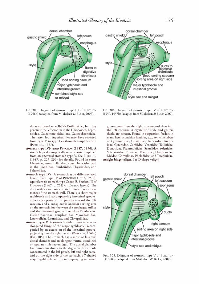
175Illustrated Glossary of the Bivalvia
the transitional type II/IVa Parilimyidae, but they penetrate the left caecum in the Unionoidea, Lepto-noidea, Galeommatoidea, and Gastrochaenoidea. The latter four superfamilies may have reverted from type V to type IVa through simplification (purChoN, 1987).
stomach type IVb sensu Purchon (1987, 1990). A stomach paedomorphically or otherwise simplified from an ancestral stomach type V. See purChoN (1987, p. 227–230) for details. Found in some Chamidae, some Tellinidae, some Donacidae, and in the Lucinidae, Fimbriidae, Thyasiridae, and Sphaeriidae.
stomach type IVc. A stomach type differentiated herein from type IV of purChoN (1987, 1990), equivalent to stomach type Group B, Section III of diNaMaNi (1967, p. 262) (J. Carter, herein). The duct orifices are concentrated into a few embay-ments of the stomach wall. There is a short major typhlosole and accompanying intestinal groove, either very posterior or passing toward the left caecum, and a conspicuous anterior sorting area on the stomach floor between the esophageal orifice and the intestinal groove. Found in Pandoridae, Cleidothaeridae, Periplomidae, Myochamidae, Laternulidae, Lyonsiidae, and Clavagellidae.
stomach type V. A stomach with a semicircular or elongated flange of the major typhlosole, accom-panied by an extension of the intestinal groove, projecting into the right caecum (purChoN, 1960b)(Fig. 305). The stomach has a more or less oval dorsal chamber and an elongate, ventral combined or separate style sac–midgut. The dorsal chamber has numerous ducts to the digestive diverticula concentrated in the left pouch, left and right caeca; and on the right side of the stomach, a 7-shaped major typhlosole and its accompanying intestinal
groove enter into the right caecum and then into the left caecum. A crystalline style and gastric shield are present. Found in suspension feeders in many heteroconchian families, e.g., some members of Cyrenoididae, Chamidae, Trapezidae, Arctic-idae, Cyrenidae, Cardiidae, Veneridae, Tellinidae, Donacidae, Psammobiidae, Semelidae, Solenidae, Solecurtidae, Pharidae, Mactridae, Dreissenidae, Myidae, Corbulidae, Pholadidae, and Teredinidae.
straight hinge veliger. See D-shape veliger.
FiG. 303. Diagram of stomach type III of purChoN (1956b) (adapted from Mikkelsen & Bieler, 2007).
FiG. 304. Diagram of stomach type IV of purChoN (1957, 1958b) (adapted from Mikkelsen & Bieler, 2007).
FiG. 305. Diagram of stomach type V of purChoN (1960b) (adapted from Mikkelsen & Bieler, 2007).

176 Treatise Online, number 48
striation. A very narrow element of elongate, incised shell or tooth sculpture; narrower than a groove, e.g., dental striations in many trigonioids.
structural cavities. See chambers.structure. See shell structure.style. See crystalline style, protostyle, style sac.style sac. A ventral or posterior extension of the
stomach in many mollusks, including most bivalves, which secretes and holds a rod-shaped inclusion (crystalline style or protostyle) that reduces the size of ingested food particles through rotation against a gastric shield and, in most mollusks, through the
digestive actions of the included enzyme amylase (Fig. 130, Fig. 301–305).
stylet. Same as a glochidial hook, which see.styliform. Having the shape of a style, i.e., elongate,
with nearly parallel sides, one end pointed, the other end blunt.
subcircular. Having a nearly circular shape, e.g., the shell of the glycymeridid Glycymeris (Glycymeris) glycymeris (liNNaeus, 1758) (Fig. 123).
subexternal ligament. An external ligament tending slightly toward a submarginal position.
subinternal ligament. An internal ligament tending slightly toward a submarginal position.
subjective synonym. In taxonomy, each of two or more names whose synonymy is a matter of the taxono-mist’s opinion. Compare with objective synonym.
submargin. One of the dorsal edges of the disc or body of the shell in Pectinoidea, adjoining the lower border of an auricle.
submarginal collecting vessel. See collecting vessel.submarginal ligament. A ligament, or a component of
a ligament, attaching to the shell on the articulating surface of the hinge plate, either just below its dorsal surface (shallow submarginal), on its ventral surface (deep submarginal), or both. See ligament position and Figure 160.
submerged ribs. Shell ribs, generated by marginal denticles, not appearing on the shell exterior, and covered internally by the inner shell layer. Away from the shell margins, the ribs are visible only in sections through the shell. Present in the nuculid Nucula laMarCk, 1799.
FiG. 306. Supernumerary hook and micropoints in glochidium larva of unionid Epioblasma triquetra (raFiNesque, 1820); scale bar = 2 µm (adapted from Hoggarth, 1999, fig. 69D, courtesy of the American
Malacological Society).
FiG. 307. Unionid anatomy, viewed with right valve removed and mantle cut away, showing supra-anal aperture (adapted from S. Trammel in J. Williams, Bogan, & Garner, 2008, fig. 9.11).
posterior pedal retractor
posterior adductor
supra-analaperture
mantle bridge
excurrentaperture
incurrent aperture
mantle marginshell outer demibranch inner demibranch
foot
labial palps
pedalprotractor
anterioradductor
dorsal musclescut mantle
shell
anterior pedal retractor

177Illustrated Glossary of the Bivalvia
subnacreous luster. A slightly pearly luster, i.e., not as brilliant as typical nacreous microstructure. The associated shell microstructures may be nacreous, matted, or foliated.
subsequent designation (abbr., SD). The designation of the type species of a genus after the original publication. Governed by ICZN (1999) Articles 69.1, 74, 75.
subsequent monotypy (abbr., SM). The situation arising when a nominal genus or subgenus was established before 1931 without an included nominal species, and only one species denoted by an available name was first subsequently referred to that genus. Governed by ICZN (1999) Article 69.3.
subspecies (pl., subspecies). The taxonomic rank immediately below species. This is the lowest taxo-nomic rank regulated by the ICZN (1999) Code.
substitute name. In taxonomy, any available name, whether new or not, used to replace an older avail-able name. See also emendation, replacement name (nomen novum), and synonym.
substratum (pl., substrata). The material on or in which an organism lives; the material at the bottom of a body of water. Substrate, and not substratum, is used in the context of chemical reactions.
subtended. Extended under.subtrapezoidal. Having a nearly trapezoidal shape,
e.g., the arcid Arca imbricata BruGuière, 1789 (Fig. 25).
subulate. Slender and tapering to a point.sulcus (adj., sulcate). A radial depression on the
exterior of a shell, commonly on the anterior side of an umbonal carina or ridge, or between anterior and posterior slopes, e.g., the anterior auricular sulcus in the Permian bakevelliid Cassia-vellia galtarae tëMkiN & poJeta, 2010 (Fig. 268), and the posteroventral sulcus in the Lower Jurassic gryphaeid Gryphaea arcuata laMarCk, 1801 (Fig. 127). See also the posteroventral sulcus in the Lower Jurassic trigoniid Trigonia sulcata (herMaNN, 1781) (Fig. 53) and the umbonal-ventral sulcus in Pholadidae (Fig. 24).
summit. The dorsalmost point of a shell when viewed along the hinge axis, or from the side with the hinge axis being horizontal.
superconglutinate. A packet of glochidia containing the entire annual reproductive output of an indi-vidual, tethered to the excurrent aperture of the producing female by a hollow mucous tube, which mimics the food of glochidial hosts. Present in the unionid Hamiota roe & hartField, 2005.
supernumerary glochidial hooks. Small (2–5 µm) triangular to lanceloate projections on the ventral margin of a glochidium larva, other than the main glochidial hook; present in many Unionoidea, e.g., in the unionid Epioblasma triquetra (raFiNesque, 1820) (Fig. 306). Supernumerary hooks are smaller than the main glochidial hook and are essentially isolated microstylets (which see).
suppressed work. A taxonomic work that the ICZN has ruled to be unpublished or unavailable. See also published work.
supra-anal aperture. A posterodorsal opening just dorsal to the excurrent aperture, e.g., in many unionoids (Fig. 307).
supra-anal aperture length. The distance between the ventral terminus of the supra-anal aperture and its dorsal terminus, measured in a straight line.
supraaxial extension. A well-defined, dorsal exten-sion of the outer lamella of the outer demibranch present in some bivalves with synaptorhabdic ctenidia, e.g., many Veneridae.
suprabranchial chamber . The narrow, elongate mantle chamber dorsal to the ctenidia, above
FiG. 308. Supramyal septum as developed in many Pterioidea (adapted from Tëmkin, 2006a, fig. 6).
FiG. 309. Silurian cardiolid Cardiola contrastans Bar-raNde, 1881, showing swollen band in each valve produced in ontogenetic stage III; different ontogenetic stages are shown by different patterns on shell (adapted
from Kříž, 2007, fig. 11).
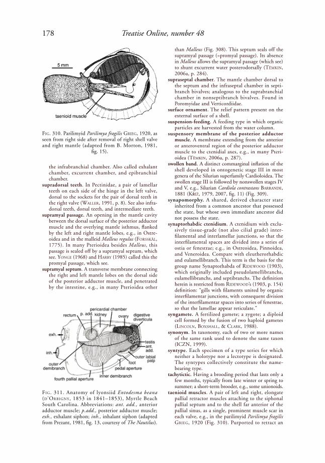
178 Treatise Online, number 48
the infrabranchial chamber. Also called exhalant chamber, excurrent chamber, and epibranchial chamber.
supradorsal teeth. In Pectinidae, a pair of lamellar teeth on each side of the hinge in the left valve, dorsal to the sockets for the pair of dorsal teeth in the right valve (Waller, 1991, p. 8). See also infra-dorsal teeth, dorsal teeth, and intermediate teeth.
supramyal passage. An opening in the mantle cavity between the dorsal surface of the posterior adductor muscle and the overlying mantle isthmus, flanked by the left and right mantle lobes, e.g., in Ostre-oidea and in the malleid Malleus regulus (Forsskål, 1775). In many Pterioidea besides Malleus, this passage is sealed off by a supramyal septum, which see. yoNGe (1968) and harry (1985) called this the promyal passage, which see.
supramyal septum. A transverse membrane connecting the right and left mantle lobes on the dorsal side of the posterior adductor muscle, and penetrated by the intestine, e.g., in many Pterioidea other
than Malleus (Fig. 308). This septum seals off the supramyal passage (=promyal passage). Its absence in Malleus allows the supramyal passage (which see) to shunt excurrent water posterodorsally (tëMkiN, 2006a, p. 284).
supraseptal chamber. The mantle chamber dorsal to the septum and the infraseptal chamber in septi-branch bivalves; analogous to the suprabranchial chamber in nonseptibranch bivalves. Found in Poromyidae and Verticordiidae.
surface ornament. The relief pattern present on the external surface of a shell.
suspension-feeding. A feeding type in which organic particles are harvested from the water column.
suspensory membrane of the posterior adductor muscle. A membrane extending from the anterior or anteroventral region of the posterior adductor muscle to the ctenidial axes, e.g., in many Pteri-oidea (tëMkiN, 2006a, p. 287).
swollen band. A distinct commarginal inflation of the shell developed in ontogenetic stage III in most genera of the Silurian superfamily Cardioloidea. The swollen stage III is followed by nonswollen stages IV and V, e.g., Silurian Cardiola contrastans BarraNde, 1881 (kříž, 1979, 2007, fig. 11) (Fig. 309).
synapomorphy. A shared, derived character state inherited from a common ancestor that possessed the state, but whose own immediate ancestor did not possess the state.
synaptorhabdic ctenidium. A ctenidium with exclu-sively tissue-grade (not also cilial grade) inter-filamental and interlamellar junctions, so that the interfilamental spaces are divided into a series of ostia or fenestrae; e.g., in Ostreoidea, Pinnoidea, and Veneroidea. Compare with eleutherorhabdic and eulamellibranch. This term is the basis for the group name Synaptorhabda of rideWood (1903), which originally included pseudolamellibranchs, eulamellibranchs, and septibranchs. The definition herein is restricted from rideWood’s (1903, p. 154) definition: “gills with filaments united by organic interfilamentar junctions, with consequent division of the interfilamentar spaces into series of fenestrae, so that the lamellae appear reticulate.”
syngamete. A fertilized gamete; a zygote; a diploid cell formed by the fusion of two haploid gametes (liNColN, Boxshall, & Clark, 1988).
synonym. In taxonomy, each of two or more names of the same rank used to denote the same taxon (ICZN, 1999).
syntype. Each specimen of a type series for which neither a holotype nor a lectotype is designated. The syntypes collectively constitute the name-bearing type.
tachytictic. Having a brooding period that lasts only a few months, typically from late winter or spring to summer; a short-term brooder, e.g., some unionoids.
taenioid muscles. A pair of left and right, elongate pallial retractor muscles attaching to the siphonal pallial septum and to the shell far anterior of the pallial sinus, as a single, prominent muscle scar in each valve, e.g., in the parilimyid Parilimya fragilis GrieG, 1920 (Fig. 310). Purported to retract an
FiG. 310. Parilimyid Parilimya fragilis GrieG, 1920, as seen from right side after removal of right shell valve and right mantle (adapted from B. Morton, 1981,
fig. 15).
FiG. 311. Anatomy of lyonsiid Entodesma beana (d’orBiGNy, 1853 in 1841–1853), Myrtle Beach South Carolina. Abbreviations: ant. add., anterior adductor muscle; p.add., posterior adductor muscle; exh., exhalant siphon; inh., inhalant siphon (adapted from Prezant, 1981, fig. 13, courtesy of The Nautilus).
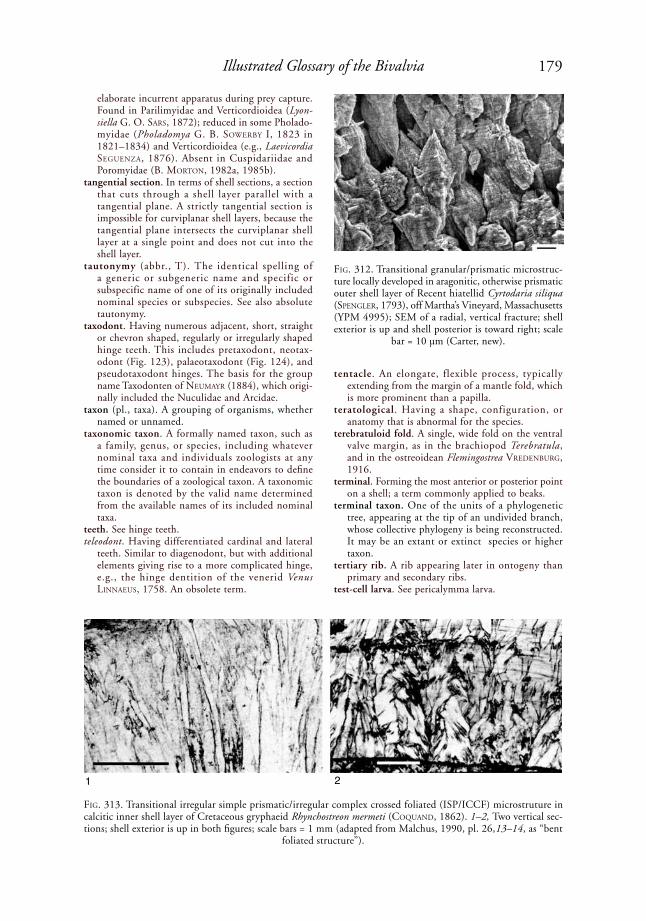
179Illustrated Glossary of the Bivalvia
elaborate incurrent apparatus during prey capture. Found in Parilimyidae and Verticordioidea (Lyon-siella G. o. sars, 1872); reduced in some Pholado-myidae (Pholadomya G. B. soWerBy i, 1823 in 1821–1834) and Verticordioidea (e.g., Laevicordia seGueNZa, 1876). Absent in Cuspidariidae and Poromyidae (B. MortoN, 1982a, 1985b).
tangential section. In terms of shell sections, a section that cuts through a shell layer parallel with a tangential plane. A strictly tangential section is impossible for curviplanar shell layers, because the tangential plane intersects the curviplanar shell layer at a single point and does not cut into the shell layer.
tautonymy (abbr., T). The identical spelling of a generic or subgeneric name and specific or subspecific name of one of its originally included nominal species or subspecies. See also absolute tautonymy.
taxodont. Having numerous adjacent, short, straight or chevron shaped, regularly or irregularly shaped hinge teeth. This includes pretaxodont, neotax-odont (Fig. 123), palaeotaxodont (Fig. 124), and pseudotaxodont hinges. The basis for the group name Taxodonten of NeuMayr (1884), which origi-nally included the Nuculidae and Arcidae.
taxon (pl., taxa). A grouping of organisms, whether named or unnamed.
taxonomic taxon. A formally named taxon, such as a family, genus, or species, including whatever nominal taxa and individuals zoologists at any time consider it to contain in endeavors to define the boundaries of a zoological taxon. A taxonomic taxon is denoted by the valid name determined from the available names of its included nominal taxa.
teeth. See hinge teeth.teleodont. Having differentiated cardinal and lateral
teeth. Similar to diagenodont, but with additional elements giving rise to a more complicated hinge, e.g., the hinge dentition of the venerid Venus liNNaeus, 1758. An obsolete term.
tentacle. An elongate, flexible process, typically extending from the margin of a mantle fold, which is more prominent than a papilla.
teratological. Having a shape, configuration, or anatomy that is abnormal for the species.
terebratuloid fold. A single, wide fold on the ventral valve margin, as in the brachiopod Terebratula, and in the ostreoidean Flemingostrea VredeNBurG, 1916.
terminal. Forming the most anterior or posterior point on a shell; a term commonly applied to beaks.
terminal taxon. One of the units of a phylogenetic tree, appearing at the tip of an undivided branch, whose collective phylogeny is being reconstructed. It may be an extant or extinct species or higher taxon.
tertiary rib. A rib appearing later in ontogeny than primary and secondary ribs.
test-cell larva. See pericalymma larva.
FiG. 312. Transitional granular/prismatic microstruc-ture locally developed in aragonitic, otherwise prismatic outer shell layer of Recent hiatellid Cyrtodaria siliqua (speNGler, 1793), off Martha’s Vineyard, Massachusetts (YPM 4995); SEM of a radial, vertical fracture; shell exterior is up and shell posterior is toward right; scale
bar = 10 µm (Carter, new).
FiG. 313. Transitional irregular simple prismatic/irregular complex crossed foliated (ISP/ICCF) microstruture in calcitic inner shell layer of Cretaceous gryphaeid Rhynchostreon mermeti (CoquaNd, 1862). 1–2, Two vertical sec-tions; shell exterior is up in both figures; scale bars = 1 mm (adapted from Malchus, 1990, pl. 26,13–14, as “bent
foliated structure”).
1 2
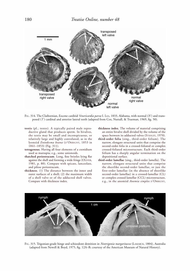
180 Treatise Online, number 48
testis (pl., testes). A typically paired male repro-ductive gland that produces sperm. In bivalves, the testis may be small and inconspicuous, or relatively large and highly convoluted, as in the lyonsiid Entodesma beana (d’orBiGNy, 1853 in 1841–1853) (Fig. 311).
tetragenous. Having all four elements of a ctenidium used as marsupia; e.g., some unionoids.
thatched periostracum. Long, fine bristles lying flat against the shell and forming a wide fringe (oliVer, 1981, p. 80). Compare with spicate, lanceolate, and pilose periostracum.
thickness. (1) The distance between the inner and outer surfaces of a shell; (2) the maximum width of a shell valve or of the adducted shell valves. Compare with thickness index.
thickness index. The volume of material comprising an entire bivalve shell divided by the volume of the space between its adducted valves (staNley, 1970).
third-order folia (sing., third-order folium). The narrow, elongate structural units that comprise the second-order folia in a crossed-foliated or complex crossed-foliated microstructure. Each third-order folium has a sharply angular termination on the depositional surface.
third-order lamellae (sing., third-order lamella). The narrow, elongate structural units that comprise the sheetlike second-order lamellae, or just the first-order lamellae (in the absence of sheetlike second-order lamellae) in a crossed-lamellar (CL) or complex crossed lamellar (CCL) microstructure, e.g., in the anomiid Anomia simplex d’orBiGNy,
FiG. 314. The Claibornian, Eocene carditid Venericardia parva I. lea, 1833, Alabama, with normal (N ) and trans-posed (T ) cardinal and anterior lateral teeth (adapted from Cox, Nuttall, & Trueman, 1969, fig. 50).
FiG. 315. Trigonian-grade hinge and schizodont dentition in Neotrigonia margaritacea (laMarCk, 1804), Australia (adapted from Newell & Boyd, 1975, fig. 12A–B; courtesy of the American Museum of Natural History).
1 mm
transposed right valve
transposed left valve
normal left valve
normal right valve

181Illustrated Glossary of the Bivalvia
1853 in 1841–1853 (Fig. 271). The term third-order lamellae has also been applied to the third-order structural units in calcitic crossed foliated and complex crossed foliated structures, but the term third-order folia is preferred for these structures.
thread. A very narrow rib; narrower than a riblet.toe. A digitate or spatulate organ on the anterior of the
foot. In some bivalves, such as the gastrochaenid Lamychaena cordiformis (NeVill & NeVill, 1869), the toe is modified into a pedal probing organ (Fig. 19).
tooth generation 1. See generation 1 teeth.tooth generation 2. See generation 2 teeth.tooth numbering system. See heterodont.topotype (adj., topotypic). An unofficial term (not
regulated by the ICZN Code, 1999) for a specimen originating from the same locality and geological horizon as a type specimen or type series of a species or subspecies.
trabeculae (sing., trabecula). Radial ridges and grooves on the underside of (i.e., beneath) a prismatic shell layer deposited over a ligament insertion area (JohNstoN & ColloM, 1998, p. 350); e.g., present in many Inoceramidae. Compare with pseudotra-beculae and pseudoresilifers.
transitional granular/prismatic microstructure. A shell microstructure consisting of first-order struc-tural units that are generally greater than 5 µm in width and that vary in shape from more or less equidmensional to slightly vertically elongate; e.g., in the outer shell layer of the hiatellid Cyrtodaria siliqua (speNGler, 1793) (Fig. 312).
transitional irregular simple prismatic/homoge-neous microstructure (abbr., transitional ISP/HOM). A shell microstructure that appears to be homogeneous at the level of optical microscopy but that consists of short, irregular simple prisms with poorly defined mutual boundaries, in some instances with minor amounts of true homogeneous structure. This occurs in the aragonitic outer shell layer of the mytilid Mytella guyanensis (laMarCk, 1819 in 1818–1822) (Fig. 159).
transitional irregular simple prismatic/irregular complex crossed foliated microstructure (abbr., transitional ISP/ICCF). A calcitic, superficially irregular simple prismatic shell microstructure in which each prism shows irregularly inclined second-order structural trends. The dip directions of the second-order structural trends are more irregular than in semi-foliated prismatic micro-strucure. Present in the inner shell layer in some Ostreoidea (MalChus, 1990, pl. 26,13–14 ) (Fig. 313). MalChus (1990, p. 64) called this structure “bent foliated.” Compare with semi-foliated simple prismatic.
transitional ligament. An opisthodetic, elongate, external or submarginal ligament with one long, posterior, lamellar sublayer, one long, posterior, fibrous sublayer below this, and a second, much shorter, lamellar sublayer below the beaks, anterior to the fibrous sublayer. The ligament insertion area otherwise shows only fine, horizontal growth lines and no discrete oblique or vertical ridges
and grooves (NeWell & Boyd, 1987). Examples include the Paleozoic cypricardiniid Cypricardinia hall, 1859 in 1859–1861, the Permian eurydes-matid Eurydesma Morris, 1845, and the upper Paleozoic–?Triassic atomodesmatid Atomodesma BeyriCh, 1865. This category is too broadly defined to be useful, as it includes some preduplivincular, transitional simple arched/parivincular, and mono-vincular ligaments.
transitional simple/parivincular ligament. A ligament that resembles a shallowly impressed, external, simple ligament, except for having a very low and indistinct ligamental ridge (incipient nymph) on the commissural side of the ligament groove; e.g., in the upper Carboniferous edmondiid Edmondia gibbosa (M’Coy, 1844) (see Fig. 327). The ligamental ridge is less distinct than in a typical parivincular ligament.
transposed hinge. A hinge in which certain hinge teeth occupy positions that are more typical of teeth in the other valve, in closely related species or in other
FiG. 316. Tubercles on exterior of left valve of Aptian, Lower Cretaceous myophorellid Yaadia (Quadratotrigonia) nodosa (J. de C. soWerBy, 1826, in soWerBy & soWerBy,
1812–1846) (adapted from Lycett, 1872–1873).
FiG. 317. Hinge of juvenile of philobryid Philobrya wandelensis laMy, 1906, Antarctica or Southern Ocean region, Natural History Museum, Stockholm (SMNH 1079), showing ligament, G1b hinge teeth, and open-
ings of several tubules (Malchus, new).
5 mm
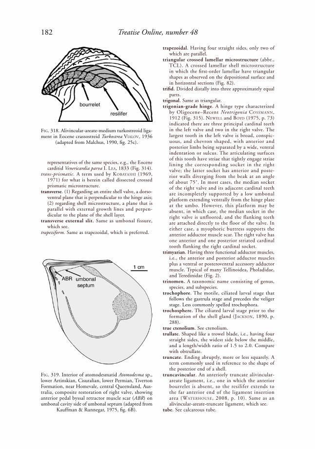
182 Treatise Online, number 48
representatives of the same species, e.g., the Eocene carditid Venericardia parva I. lea, 1833 (Fig. 314).
trans-prismatic. A term used by koBayashi (1969, 1971) for what is herein called dissected crossed prismatic microstructure.
transverse. (1) Regarding an entire shell valve, a dorso-ventral plane that is perpendicular to the hinge axis; (2) regarding shell microstructure, a plane that is parallel with external growth lines and perpen-dicular to the plane of the shell layer.
transverse external slit. Same as umbonal fissure, which see.
trapeziform. Same as trapezoidal, which is preferred.
trapezoidal. Having four straight sides, only two of which are parallel.
triangular crossed lamellar microstructure (abbr., TCL). A crossed lamellar shell microstructure in which the first-order lamellae have triangular shapes as observed on the depositional surface and in horizontal sections (Fig. 82).
trifid. Divided distally into three approximately equal parts.
trigonal. Same as triangular.trigonian-grade hinge. A hinge type characterized
by Oligocene–Recent Neotrigonia CossMaNN, 1912 (Fig. 315). NeWell and Boyd (1975, p. 73) indicated there are three principal cardinal teeth in the left valve and two in the right valve. The largest tooth in the left valve is broad, conspic-uous, and chevron shaped, with anterior and posterior limbs being separated by a wide, ventral indentation or sulcus. The articulating surfaces of this tooth have striae that tightly engage striae lining the corresponding socket in the right valve; the latter socket has anterior and poste-rior walls diverging from the beak at an angle of about 75˚. In most cases, the median socket of the right valve and its adjacent cardinal teeth are incompletely supported by a low umbonal platform extending ventrally from the hinge plate at the umbo. However, this platform may be absent, in which case, the median socket in the right valve is unfloored, and the flanking teeth are attached directly to the floor of the valve. In either case, a myophoric buttress supports the anterior adductor muscle scar. The right valve has one anterior and one posterior striated cardinal tooth flanking the right cardinal socket.
trimyarian. Having three functional adductor muscles, i.e., the anterior and posterior adductor muscles plus a ventral or posteroventral accessory adductor muscle. Typical of many Tellinoidea, Pholadidae, and Teredinidae (Fig. 2).
trinomen. A taxonomic name consisting of genus, species, and subspecies.
trochophore. The motile, ciliated larval stage that follows the gastrula stage and precedes the veliger stage. Less commonly spelled trochophora.
trochosphere. The ciliated larval stage prior to the formation of the shell gland (JaCksoN, 1890, p. 288).
true ctenolium. See ctenolium.trullate. Shaped like a trowel blade, i.e., having four
straight sides, the widest side below the middle, and a length/width ratio of 1.5 to 2.0. Compare with obtrullate.
truncate. Ending abruptly, more or less squarely. A term commonly used in reference to the shape of the posterior end of a shell.
truncavincular. An anteriorly truncate alivincular-areate ligament, i.e., one in which the anterior bourrelet is absent, so the resilifer extends to the far anterior end of the ligament insertion area (Waterhouse, 2008, p. 10). Same as an alivincular-areate-truncate ligament, which see.
tube. See calcareous tube.
FiG. 318. Alivincular-areate-medium turkostreoid liga-ment in Eocene crassostreid Turkostrea VialoV, 1936
(adapted from Malchus, 1990, fig. 25c).
FiG. 319. Interior of atomodesmatid Atomodesma sp., lower Artinskian, Cisuralian, lower Permian, Tiverton Formation, near Homevale, central Queensland, Aus-tralia, composite restoration of right valve, showing anterior pedal byssal retractor muscle scar (ABR) on umbonal cavity side of umbonal septum (adapted from
Kauffman & Runnegar, 1975, fig. 6B).
resiliferbourrelet
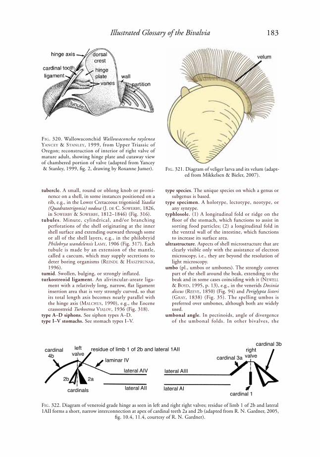
183Illustrated Glossary of the Bivalvia
tubercle. A small, round or oblong knob or promi-nence on a shell, in some instances positioned on a rib, e.g., in the Lower Cretaceous trigonioid Yaadia (Quadratotrigonia) nodosa (J. de C. soWerBy, 1826, in soWerBy & soWerBy, 1812–1846) (Fig. 316).
tubules. Minute, cylindrical, and/or branching perforations of the shell originating at the inner shell surface and extending outward through some or all of the shell layers, e.g., in the philobryid Philobrya wandelensis laMy, 1906 (Fig. 317). Each tubule is made by an extension of the mantle, called a caecum, which may supply secretions to deter boring organisms (reiNdl & hasZpruNar, 1996).
tumid. Swollen, bulging, or strongly inflated.turkostreoid ligament. An alivincular-areate liga-
ment with a relatively long, narrow, flat ligament insertion area that is very strongly curved, so that its total length axis becomes nearly parallel with the hinge axis (MalChus, 1990), e.g., the Eocene crassostreid Turkostrea VialoV, 1936 (Fig. 318).
type A–D siphons. See siphon types A–D.type I–V stomachs. See stomach types I–V.
type species. The unique species on which a genus or subgenus is based.
type specimen. A holotype, lectotype, neotype, or any syntype.
typhlosole. (1) A longitudinal fold or ridge on the floor of the stomach, which functions to assist in sorting food particles; (2) a longitudinal fold in the ventral wall of the intestine, which functions to increase its surface area.
ultrastructure. Aspects of shell microstructure that are clearly visible only with the assistance of electron microscopy, i.e., they are beyond the resolution of light microscopy.
umbo (pl., umbos or umbones). The strongly convex part of the shell around the beak, extending to the beak and in some cases coinciding with it (NeWell & Boyd, 1995, p. 13), e.g., in the venerids Dosinia discus (reeVe, 1850) (Fig. 94) and Periglypta listeri (Gray, 1838) (Fig. 35). The spelling umbos is preferred over umbones, although both are widely used.
umbonal angle. In pectinoids, angle of divergence of the umbonal folds. In other bivalves, the
FiG. 320. Wallowaconchid Wallowaconcha raylenea yaNCey & staNley, 1999, from Upper Triassic of Oregon; reconstruction of interior of right valve of mature adult, showing hinge plate and cutaway view of chambered portion of valve (adapted from Yancey & Stanley, 1999, fig. 2, drawing by Roxanne Jumer). FiG. 321. Diagram of veliger larva and its velum (adapt-
ed from Mikkelsen & Bieler, 2007).
FiG. 322. Diagram of veneroid grade hinge as seen in left and right right valves; residue of limb 1 of 2b and lateral 1AII forms a short, narrow interconnection at apex of cardinal teeth 2a and 2b (adapted from R. N. Gardner, 2005,
fig. 10.4, 11.4, courtesy of R. N. Gardner).
cardinal4b
2b 2a
cardinals
left valve
residue of limb 1 of 2b and lateral 1AII
laminar IV
lateral AIV
lateral AII
cardinal 3a
lateral AIII
lateral AIcardinal 1
right valve
cardinal 3b

184 Treatise Online, number 48
4b2b
cardinals2a
lateral 1AII dissectedlaminar IV
lateral AIV
lateral 2AII
lateral AIII
lateral AI
3a
1cardinals 3b
2b
1AII
2AII
4b
approximate angle of divergence of the dorsopos-terior and dorsoanterior parts of a lateral profile of a shell.
umbonal carina. A carina that extends in a more or less radial or antimarginal direction from the umbonal area, where best developed, toward the posteroven-tral shell margin. Commonly used in reference to trigonioid shells. When the carina is very ridgelike, the term umbonal ridge is sometimes used instead. Compare with marginal carina.
umbonal cavity. The space inside a single shell valve between the umbo and the hinge plate.
umbonal crack. Same as umbonal fissure, which is preferred.
umbonal deck. Same as an umbonal septum, which see.
umbonal depression. A small depression at the apex of the umbo, e.g., in some Arcoidea.
umbonal fissure. A more or less dorsoventral, tensional fracture formed during shell growth, present in both valves of some thin-shelled taxa just anterior to the beaks, e.g., many Periplomatidae and Later-nulidae (Fig. 59). Also called a transverse external slit, an anterior cleft, and an umbonal crack.
umbonal fold. A ridge originating at the umbo and separating the body of the shell from the auricle. Commonly applied to pectinoid shells.
2
FiG. 323. Venielloid grade hinge. 1, Diagram of left and right valves (adapted from R. N. Gardner, 2005, fig. 15.1c, 16.1c, courtesy of R. N. Gardner); 2, left valve of Upper Cretaceous veniellid Veniella conradi (S. MortoN, 1833),
view 2 is 5.5 cm wide (adapted from R. N. Gardner, 2005, fig. 9, courtesy of R. N. Gardner).
FiG. 324. Vesicular structure in right valve of gryphaeid Hyotissa hyotis (liNNaeus, 1758), Calapan, Mindoro, Philippines, YPM 7150; scale bar = 10 µm (adapted from Carter, 1990b, p. 355, fig. 2E, there misidentified
as Dendostrea folium [liNNaeus, 1758]).
1
left valve
right valve
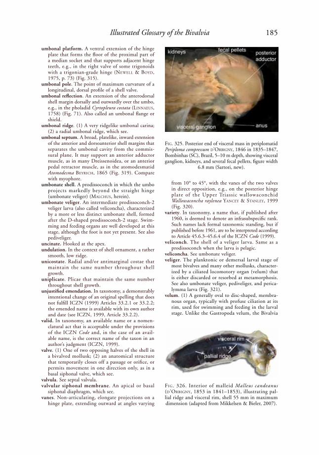
185Illustrated Glossary of the Bivalvia
umbonal platform. A ventral extension of the hinge plate that forms the floor of the proximal part of a median socket and that supports adjacent hinge teeth, e.g., in the right valve of some trigonoids with a trigonian-grade hinge (NeWell & Boyd, 1975, p. 73) (Fig. 315).
umbonal pole. The point of maximum curvature of a longitudinal, dorsal profile of a shell valve.
umbonal reflection. An extension of the anterodorsal shell margin dorsally and outwardly over the umbo, e.g., in the pholadid Cyrtopleura costata (liNNaeus, 1758) (Fig. 71). Also called an umbonal flange or shield.
umbonal ridge. (1) A very ridgelike umbonal carina; (2) a radial umbonal ridge, which see.
umbonal septum. A broad, platelike, inward extension of the anterior and dorsoanterior shell margins that separates the umbonal cavity from the commis-sural plane. It may support an anterior adductor muscle, as in many Dreissenoidea, or an anterior pedal retractor muscle, as in the atomodesmatid Atomodesma BeyriCh, 1865 (Fig. 319). Compare with myophore.
umbonate shell. A prodissoconch in which the umbo projects markedly beyond the straight hinge (umbonate veliger) (MalChus, herein).
umbonate veliger. An intermediate prodissoconch-2 veliger larva (also called veliconcha), characterized by a more or less distinct umbonate shell, formed after the D-shaped prodissoconch-2 stage. Swim-ming and feeding organs are well developed at this stage, although the foot is not yet present. See also pediveliger.
uncinate. Hooked at the apex.undulation. In the context of shell ornament, a rather
smooth, low ridge.unicostate. Radial and/or antimarginal costae that
maintain the same number throughout shell growth.
uniplicate. Plicae that maintain the same number throughout shell growth.
unjustified emendation. In taxonomy, a demonstrably intentional change of an original spelling that does not fulfill ICZN (1999) Articles 33.2.1 or 33.2.2; the emended name is available with its own author and date (see ICZN, 1999, Article 33.2.2).
valid. In taxonomy, an available name or a nomen-clatural act that is acceptable under the provisions of the ICZN Code and, in the case of an avail-able name, is the correct name of the taxon in an author’s judgment (ICZN, 1999).
valve. (1) One of two opposing halves of the shell in a bivalved mollusk; (2) an anatomical structure that temporarily closes off a passage or orifice, or permits movement in one direction only, as in a basal siphonal valve, which see.
valvula. See septal valvula.valvular siphonal membrane. An apical or basal
siphonal diaphragm, which see.vanes. Non-articulating, elongate projections on a
hinge plate, extending outward at angles varying
from 10° to 45°, with the vanes of the two valves in direct opposition, e.g., on the posterior hinge plate of the Upper Triassic wallowaconchid Wallowaconcha raylenea yaNCey & staNley, 1999 (Fig. 320).
variety. In taxonomy, a name that, if published after 1960, is deemed to denote an infrasubspecific rank. Such names lack formal taxonomic standing, but if published before 1961, are to be interpreted according to Article 45.6.3–45.6.4 of the ICZN Code (1999).
veliconch. The shell of a veliger larva. Same as a prodissoconch when the larva is pelagic.
veliconcha. See umbonate veliger.veliger. The planktonic or demersal larval stage of
most bivalves and many other mollusks, character-ized by a ciliated locomotory organ (velum) that is either discarded or resorbed at metamorphosis. See also umbonate veliger, pediveliger, and perica-lymma larva (Fig. 321).
velum. (1) A generally oval to disc-shaped, membra-nous organ, typically with profuse ciliation at its rim, used for swimming and feeding in the larval stage. Unlike the Gastropoda velum, the Bivalvia
FiG. 325. Posterior end of visceral mass in periplomatid Periploma compressum d’orBiGNy, 1846 in 1835–1847, Bombinhas (SC), Brazil, 5–10 m depth, showing visceral ganglion, kidneys, and several fecal pellets, figure width
6.8 mm (Sartori, new).
FiG. 326. Interior of malleid Malleus candeanus (d’orBiGNy, 1853 in 1841–1853), illustrating pal-lial ridge and visceral rim, shell 55 mm in maximum dimension (adapted from Mikkelsen & Bieler, 2007).
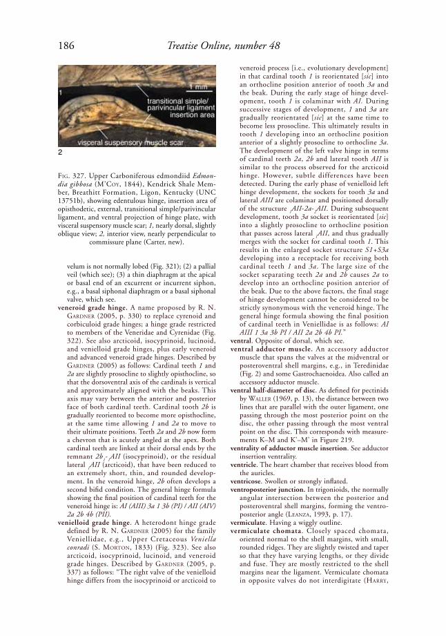
186 Treatise Online, number 48
velum is not normally lobed (Fig. 321); (2) a pallial veil (which see); (3) a thin diaphragm at the apical or basal end of an excurrent or incurrent siphon, e.g., a basal siphonal diaphragm or a basal siphonal valve, which see.
veneroid grade hinge. A name proposed by r. N. GardNer (2005, p. 330) to replace cyrenoid and corbiculoid grade hinges; a hinge grade restricted to members of the Veneridae and Cyrenidae (Fig. 322). See also arcticoid, isocyprinoid, lucinoid, and venielloid grade hinges, plus early veneroid and advanced veneroid grade hinges. Described by GardNer (2005) as follows: Cardinal teeth 1 and 2a are slightly prosocline to slightly opisthocline, so that the dorsoventral axis of the cardinals is vertical and approximately aligned with the beaks. This axis may vary between the anterior and posterior face of both cardinal teeth. Cardinal tooth 2b is gradually reoriented to become more opisthocline, at the same time allowing 1 and 2a to move to their ultimate positions. Teeth 2a and 2b now form a chevron that is acutely angled at the apex. Both cardinal teeth are linked at their dorsal ends by the remnant 2b
1-
1AII (isocyprinoid), or the residual
lateral 1AII (arcticoid), that have been reduced to
an extremely short, thin, and rounded develop-ment. In the veneroid hinge, 2b often develops a second bifid condition. The general hinge formula showing the final position of cardinal teeth for the veneroid hinge is: AI (AIII) 3a 1 3b (PI) / AII (AIV) 2a 2b 4b (PII).
venielloid grade hinge. A heterodont hinge grade defined by r. N. GardNer (2005) for the family Veniellidae, e.g., Upper Cretaceous Veniella conradi (S. MortoN, 1833) (Fig. 323). See also arcticoid, isocyprinoid, lucinoid, and veneroid grade hinges. Described by GardNer (2005, p. 337) as follows: “The right valve of the venielloid hinge differs from the isocyprinoid or arcticoid to
veneroid process [i.e., evolutionary development] in that cardinal tooth 1 is reorientated [sic] into an orthocline position anterior of tooth 3a and the beak. During the early stage of hinge devel-opment, tooth 1 is colaminar with AI. During successive stages of development, 1 and 3a are gradually reorientated [sic] at the same time to become less prosocline. This ultimately results in tooth 1 developing into an orthocline position anterior of a slightly prosocline to orthocline 3a. The development of the left valve hinge in terms of cardinal teeth 2a, 2b and lateral tooth AII is similar to the process observed for the arcticoid hinge. However, subtle differences have been detected. During the early phase of venielloid left hinge development, the sockets for tooth 3a and lateral AIII are colaminar and positioned dorsally of the structure
1AII-2a-
2AII. During subsequent
development, tooth 3a socket is reorientated [sic] into a slightly prosocline to orthocline position that passes across lateral
1AII, and thus gradually
merges with the socket for cardinal tooth 1. This results in the enlarged socket structure S1+S3a developing into a receptacle for receiving both cardinal teeth 1 and 3a. The large size of the socket separating teeth 2a and 2b causes 2a to develop into an orthocline position anterior of the beak. Due to the above factors, the final stage of hinge development cannot be considered to be strictly synonymous with the veneroid hinge. The general hinge formula showing the final position of cardinal teeth in Veniellidae is as follows: AI AIII 1 3a 3b PI / AII 2a 2b 4b PI.”
ventral. Opposite of dorsal, which see. ventral adductor muscle. An accessory adductor
muscle that spans the valves at the midventral or posteroventral shell margins, e.g., in Teredinidae (Fig. 2) and some Gastrochaenoidea. Also called an accessory adductor muscle.
ventral half-diameter of disc. As defined for pectinids by Waller (1969, p. 13), the distance between two lines that are parallel with the outer ligament, one passing through the most posterior point on the disc, the other passing through the most ventral point on the disc. This corresponds with measure-ments K–M and K'–M' in Figure 219.
ventrality of adductor muscle insertion. See adductor insertion ventrality.
ventricle. The heart chamber that receives blood from the auricles.
ventricose. Swollen or strongly inflated.ventroposterior junction. In trigonioids, the normally
angular intersection between the posterior and posteroventral shell margins, forming the ventro-posterior angle (leaNZa, 1993, p. 17).
vermiculate. Having a wiggly outline.vermiculate chomata. Closely spaced chomata,
oriented normal to the shell margins, with small, rounded ridges. They are slightly twisted and taper so that they have varying lengths, or they divide and fuse. They are mostly restricted to the shell margins near the ligament. Vermiculate chomata in opposite valves do not interdigitate (harry,
FiG. 327. Upper Carboniferous edmondiid Edmon-dia gibbosa (M’Coy, 1844), Kendrick Shale Mem-ber, Breathitt Formation, Ligon, Kentucky (UNC 13751b), showing edentulous hinge, insertion area of opisthodetic, external, transitional simple/parivincular ligament, and ventral projection of hinge plate, with visceral suspensory muscle scar; 1, nearly dorsal, slightly oblique view; 2, interior view, nearly perpendicular to
commissure plane (Carter, new).
1
2

187Illustrated Glossary of the Bivalvia
1985; MalChus, 1990), e.g., Pycnodonteinae and Exogyrinae in Ostreoidea.
verrucose. Having small, wartlike projections.vertical. In the context of shell microstructure, a
section that is perpendicular to the plane of the shell layer.
vesicula seminalis. Latin for seminal vesicle, which see.vesicular structure. A very porous, spongelike shell
layer or part of a shell layer, consisting of a network of thin calcareous partitions enclosing numerous small cavities. The cavities have rounded to irregu-larly polygonal shapes and are readily visible at low magnification (10×), unlike the smaller cavi-ties comprising chalky structure. Found in many Pycnodonteinae, e.g., Hyotissa hyotis (liNNaeus, 1758) (Fig. 324).
vestigial. A degenerate or imperfectly developed structure or organ serving little or no function, but in ancestors or in a former developmental stage serving a useful function.
vinculum (pl., vincula). (1) A shelly callosity devel-oped between two hinge teeth, thus causing the dentition to appear simpler than dental homology suggests. A vinculum may become incorporated into the basic hinge structure, as in the transition from Resatrix s.s. Casey, 1952, to Dosiniopsella Casey, 1952, wherein a vinculum unites teeth 2a and 2b at their apices (Casey, 1952, p. 166); (2) a secondary deposit of shell material on the hinge, thereby partially submerging and obscuring the earlier formed dentition, e.g., the medial part of the hinge in some gerontic Glycymeris da Costa, 1778.
viscera. Same as visceral mass, which see.visceral attachment muscles. Muscles attaching the
visceral mass to an internal shell surface proximal to the pallial line. This excludes adductor, retractor, protractor, orbital, and radial mantle muscles. These muscles make a rather large, crescentic scar behind the anterior adductor and a smaller, more elongated scar in a mediodorsal position in the iridinid Spatha i. lea, 1838. In the solemyid Solemya laMarCk, 1818 in 1818–1822, they make a narrow band that ascends obliquely from the posteroventral corner of the anterior adductor muscle scar toward the dorsal margin, where it broadens to merge with the pedal retractor muscle scars. See also visceral suspensory muscle scars.
visceral ganglion (pl., ganglia). A concentration of nerve cells innervating the heart, ctenidia, pericar-dium, the posterior part of the mantle (including the siphons), and the posterior adductor muscle, e.g., in the periplomatid Periploma compressum d’orBiGNy, 1846 in 1835–1847 (Fig. 325). The left and right visceral ganglia lie below the rectum, close to the posterior adductor muscle.
visceral lobe. A lateral extension of the visceral mass in Lucinidae, containing part of the reproductive organs.
visceral mass. The region of a bivalve’s body containing most if not all of the digestive and excretory systems and the central part of the circulatory and nervous systems, suspended dorsally between the ctenidia by pedal retractor and visceral suspensory muscles,
and terminated ventrally by the foot. A distinct, posteroventral extension of the visceral mass is called a metasoma (Fig. 176). The visceral mass does not include the ctenidia, mantle, siphons, palps, nor most of the foot.
visceral rim. A roughly commarginal, distinct eleva-tion on the inner shell surface where the prismatic outer and nacreous inner shell layers meet. The mantle and ctenidia can be withdrawn proximal to this rim, thereby protecting them when the valves are closed. Present in most Isognomonidae and Malleidae, e.g., Malleus candeanus (d’orBiGNy, 1853 in 1841–1853) (Fig. 326).
visceral suspensory muscle scar. A muscle scar or line of scars inserting on a narrow, flangelike, ventral projection of the posterior hinge plate, e.g., upper Carboniferous Edmondia gibbosa (M’Coy, 1844) (Fig. 327).
vitelline membrane. The thin casing that encloses an egg (ovum).
viviparous. Producing offspring by direct development from within the body of the parent (liNColN, Boxshall, & Clark, 1988). See also larviparous, oviparous, ovoviviparous, and indirect develop-ment.
V-shaped ribs. A group of ribs formed by the inter-section of b-ribs with c-ribs below the umbo, either as eu-V-shaped ribs (which see) or para-V-shaped ribs (which see) (Guo, 1998). See the term oblique and Figure 264 for rib types b and c, as defined by Guo (1998). Compare with reversed V-shaped ribs.
wall lamellae. A pair of left and right mantle ridges or flaps extending anteriorly from the sides of the base of an incurrent siphon, to a point below the apex of the pallial sinus. Also called the wall lamellae of the waste canal (kelloGG, 1915). They probably guide pseudofeces into the incurrent siphon, e.g., in the mactrid Raeta plicatella (laMarCk, 1818) (harry, 1969, fig. 3).
waste canal. A ciliated groove bounded by folds and produced on the inner surface of the mantle margins distal to the upper edge of the ascending lamellae of the outer demibranchs (tëMkiN, 2006a, p. 286), e.g., Pinnidae.
water tube. One of many canals in the intralamellar space of a ctenidium, extending from the dorsal to ventral margin of the ctenidium.
width of shell. The distance between two planes, both parallel with the commissural plane and the hinge axis, which just touch the most lateral part of the left valve and the most lateral part of the right valve, with the valves closed.
wing. A more or less triangular, anterior or posterior extension of the shell that is set off from the beak, umbo, or main body of the shell by an elevation, groove, or other change in slope, e.g., the poste-rior wing in the Permian bakevelliid Cassiavellia galtarae tëMkiN & poJeta, 2010 (Fig. 268) and in the Recent pteriid Pteria hirundo (liNNaeus, 1758) (Fig. 249). The terms wing and auricle are often used interchangeably, but wings are generally more prominent than auricles. Compare with ala.

188 Treatise Online, number 48
wrinkle. In terms of shell ornament, a small, irregular furrow or ridge.
xenomorphic sculpture. A shell sculpture that reflects the substratum of attachment, as in some attached and unattached valves in Anomiidae, Gryphaeidae, and Ostreidae (Fig. 127). Sometimes erroneously called an allomorphism.
zigzag lamellar structure. A term used by BøGGild (1930, p. 266–267) for herringbone regularly foli-ated microstructure, defined herein.
zooxanthellae. Unicellular algal cells that live symbi-otically within the cells of a larger organism, e.g., in many Cardioidea.
REFERENCESAdams, Arthur. 1864. On the species of Neaera found
in the seas of Japan. Annals and Magazine of Natural History (series 3) 13:206–209.
Adams, Arthur, & G. F. Angas. 1864. Descriptions of new species of shells from the Australian seas, in the collection of George French Angas. Zoological So-ciety of London, Proceedings for 1863(3):418–428, pl. 377.
Adams, Arthur, & L. A. Reeve. 1850. Mollusca. In A. Adams, ed., The Zoology of the Voyage of H. M. S. Samarang, under the Command of Captain Sir Edward Belcher, during the years 1843–1846. Reeve & Benham. London. x + p. 25–87, pl. 10–24.
Adams, Charles B. 1845. Synopsis Conchyliorum Jamaicensium, Etc.: Speciarum Novarum Conchyli-orum, in Jamaica Repertorum, Synopsis. Boston. 17 p. Extracted from Boston Society of Natural History, Proceedings.
Adams, Henry, & A. Adams. 1858. The Genera of Recent Mollusca, Arranged According to their Orga-nization. Van Voorst. London. 2 vol. Collated in two vol. by the authors on p. 661 of vol. 2, although usu-ally bound in 3 vol. Vol. 1: xl + 484 p.; vol. 2: 1–604; collation: i–xl + 605–661; vol. 3: 138 pl. [Bivalvia in vol. 2]. Dates from vol. 2, p. 661, and A. Trew (1992). Vol. 2 + associated plates: p. 541–572, pl. 129–132 [January, 1858]; p. 573–604, pl. 133–136 [May, 1858]. Collation + plates: i–xl + p. 605–661, pl. 137–138 [November, 1858].
Addadi, Lia, & S. Weiner. 1985. Interactions between acidic proteins and crystals: Stereochemical require-ments in biomineralization. Proceedings of the National Academy of Sciences, USA 82:4110–4114, 5 fig.
Allasinaz, Andrea. 1972. Revisione dei Pettinidi Trias-sici. Rivista Italiana di Paleontologia e Stratigrafia 78(2):189–428, 48 pl.
Allen, John A. 1968. The functional morphology of Crassinella mactracea (Linsley) (Bivalvia: Astarta-cea). Malacological Society of London, Proceedings 38:27–40.
Allen, John A., & H. L. Sanders. 1969. Nucinella serrei Lamy (Bivalvia; Protobranchia), a monomyarian solemyid and possible living actinodont. Malacologia 7(2–3):381–396.
Aller, Robert C., 1974. Prefabrication of shell orna-mentation in the bivalve Laternula. Lethaia 7:43–56.
Angas, George F. 1868. On a new genus and some new species of marine Mollusca from Port Jackson, New South Wales. Zoological Society of London, Proceed-ings 1867(3):908–911.
Araujo, Rafael, N. Cámara, & M. A. Ramos. 2002. Glochidium metamorphosis in the endangered freshwater mussel Margaritifera auricularia (Spengler, 1793): A histological and scanning electron micros-copy study. Journal of Morphology 254:259–265.
Armstrong, J. D. 1969. The crossed bladed fabrics of the shells of Terrakea solida (Etheridge and Dun) and Streptorhynchus pelicanensis Fletcher. Palaeontology 12:310–320.
Atkins, Daphne. 1937a. On the ciliary mechanisms and interrelationships of lamellibranchs. Part III. Types of lamellibranch gills and their food currents. Quarterly Journal of Microscopical Science 79(3):375–421.
Atkins, Daphne. 1937b. On the ciliary mechanisms and interrelationships of lamellibranchs. Part IV. Cuticular fusion, with special reference to the fourth aperture in certain lamellibranchs. Quarterly Journal of Microscopical Science 79(3):423–445.
Atkins, Daphne. 1938. On the ciliary mechanisms and interrelationships of lamellibranchs. Part VII. Latero-frontal cilia of the gill filaments and their phy-logenetic value. Quarterly Journal of Microscopical Science (new series) 80(3):345–436, pl. 29.
Babin, Claude. 1966. Mollusques Bivalves et Cépha-lopodes du Paléozoïque Armoricain. Étude Sys-tematique. Essai sur la Phylogénie des Bivalves. Esquisse Paléoécologique. Imprimerie Commerciale et Administrative. Brest. p. 1–470, 18 pl.
Bailey, Jack B. 1983. Middle Devonian Bivalvia from the Solsville Member (Marcellus Formation), central New York State. American Museum of Natural His-tory, Bulletin 174(3):193–325.
Bailey, Jack B. 2009. Shell orientation terminology among the Bivalvia (Mollusca): Problems and pro-posed solutions. Journal of Paleontology 83(3):493–495.
Baird, R. H. 1958. On the swimming behaviour of scallops (Pecten maximus L.). Malacological Society of London, Proceedings 33(2):67–71.
Bandel, Klaus. 1982. Morphology and formation of the early ontogenetic shells of conchiferan mollusks. Facies 7:1–198, 22 pl.
Bandel, Klaus. 1988. Stages in the ontogeny and a model of the evolution of bivalves (Mollusca). Paläontologische Zeitschrift 62(3–4):217–254.
Barrande, Joachim. 1881. Systême Silurien du centre de la Bohême, Classe des Mollusques, Ordre des Acéphalés, vol. 6. Bellman. Prague & Paris. i–xxiv + p. 1–342, 356 pl.
Basse, É. 1933. Paléontologie de Madagascar. XVIII. Fauna malacologique du Crétacé supérieur du sud-ouest de Madagascar. Annales de Paléontologie 22(1):11–37, pl. 1–4.
Basterot, M. B. de B. 1825. Mémoire geologique sur les environs de Bordeaux. Société d’Histoire Naturelle de Paris, Mémoires 2:1–100.
Bayle, Émile. 1878. Fossiles Principaux des Terrains. In A. Dufrénoy, L. E. de Beaumont, É. Bayle, & R. Zeiller, eds., 1841–1879, Explication de la Carte
189Illustrated Glossary of the Bivalvia
Géologique de la France rédigée sous la Direction de M. Brochant de Villiers. Service de la Carte Géologiqe de la France, Ministère des Travaux Pub-lics. Imprimerie Royale, Paris. 4 vol. in 3, + Atlas to vol. 4 (176 pl.). Vol. 2–4 printed by Imprimerie Nationale, vol. 3, pt. 2, and text of vol. 4, pt. 1, not published. Plates accompanied by leaf with descrip-tive text. Vol. 4, Atlas, pt. 1, Fossiles Principaux des Terrains, by E. Bayle, 1878; pt. 2, Végétaux Fossiles du Terrain Houiller, by R. Zeiller.
Bayne, Brian L., ed. 1976. Marine Mussels: Their Ecol-ogy and Physiology. Cambridge University Press. Cambridge. xvii + 506 p.
Beauperthuy, Iris. 1967. Los mytilidos de Venezuela (Mollusca: Bivalvia). Universidad de Oriente, In-stituto Oceanografico, Boletín 6(1):7–115, 21 pl.
de Beer, Gavin R. 1930. Embryology and Evolution. Clarendon Press. Oxford. vi + [2] + 116 p.
de Beer, Gavin R. 1958. Embryos and Ancestors, 3rd edit. Clarendon Press. Oxford. 197 p.
Belokrys, L. S. 1991. Dvustvorki-trubkozhily iz Eotsena Ukrainy. Paleontologicheskii Zhurnal 1991(4):32–41. Translated in Belokrys, L. S. 1993. Tube-dwelling bivalves from the Eocene of the Ukraine. Paleonto-logical Journal 25(4):40–53.
Bernard, Félix. 1890. Recherches sur les organes pal-léaux des gastéropodes prosobranches. Annales des Sciences Naturelles, Zoologie (series 7) 9:89–404.
Bernard, Félix. 1895. Première note sur le développe-ment et la morphologie de la coquille chez les lamel-libranches. Societé Géologique de France, Bulletin (series 3) 23(7–10):104–154.
Bernard, Félix. 1896a. Deuxième note sur le dével-oppement et la morphologie de la coquille chez les lamellibranches (Taxodontes). Société Géologique de France, Bulletin (series 3) 24(4–6):54–82.
Bernard, Félix. 1896b. Troisième note sur le dével-oppement et la morphologie de la coquille chez les lamellibranches (anisomyaires). Société Géologique de France, Bulletin (series 3) 24(26–29):412–449.
Bernard, Félix. 1896c. Diagnoses de coquilles nouvelles de lamellibranches (genres Hochstetteria et Condylo-cardia). Muséum d’Histoire Naturelle (Paris), Bul-letin 2(5):193–197.
Bernard, Félix. 1897. Quatrième et dernière note sur le développement et la morphologie de la coquille chez les lamellibranches (anisomyaires). Société Géologique de France, Bulletin (series 3) 25(35–36):559–566.
Beyrich, August H. E. 1862. Zwei aus dem Deutschen Muschelkalk noch nicht bekannte Avicula-artige Muscheln. Deutsche Geologische Gesellschaft, Zeitschrift 14:9–10.
Beyrich, H. E. von. 1865. Über eine Koblenkalk-Fauna von Timor. Abhandlungen der Königlich [Preus-sischen] Akademie der Wissenschaften zu Berlin, aus dem Jahr 1864:61–98 + pl. I–III.
Bieler, Rüdiger, J. G. Carter, & E. V. Coan. 2010. Clas-sification of bivalve families. In P. Bouchet & J.-P. Rocroi, eds., 2010, Nomenclator of bivalve families. Malacologia 52(2):1–184.
Bittner, Alexander. 1901. Ueber Pseudomonotis telleri und verwandte Arten der unteren Trias. Kaiserlich-
Königlichen Geologischen Reichsanstalt, Wien, Jahrbuch 50(4):559–592 + pl. XXII–XXIV.
Blainville, Henri-Marie D. de. 1816–1830. Vénérupe. In F. Cuvier & “plusieurs Professeurs du Jardin du Roi, et des principales Écoles de Paris,” Diction-naire des Sciences Naturelles dans lequel on Traite Méthodiquement des Différents Êtres de la Natur, Considérés soit en Eux-mêmes, d’après l’état Actuel de nos Connoissances, soit Relativement à l’Utilité qu’en Peuvent Retirer la Médecine, l’Agriculture, le Commerce et les Arts. Suivi d’une Biographie des Plus Célèbres Naturalistes, vol. 57. F. G. Levrault. Strasbourg & Paris. p. 237–246.
Bøggild, Ove B. 1930. The shell structure of the mollusks. Det Kongelige Danske Videnskabernes Selskab, Skrifter, Naturvidenskabelige og Mathema-tiske Afhandlinger (series 9) 2(2):231–326, pl. 1–15.
Born, Ignatius [Ignaz] von. 1778. Index Rerum Natu-ralium Musei Caesarei Vindobonensis. Pars I (Tes-tacea). Verzeichniss der Natürlichen Seltenheiten des K. K. Naturalien Kabinets zu Wien, Erster Theil, Schalthiere. Joannus Paulus Kraus. Vienna. xlii + 458 + [78] p., 1 pl.
Boss, Kenneth J. 1982. Mollusca. In S. P. Parker, ed., Synopsis and Classification of Living Organisms, vol. 1. McGraw-Hill. New York. p. 945–1166.
Boss, Kenneth J., & D. R. Moore. 1967. Notes on Mal-leus (Parimalleus) candeanus (d’Orbigny) (Mollusca: Bivalvia). Bulletin of Marine Science 17(1):85–94.
Bottjer, David J., & J. G. Carter. 1980. Functional and phylogenetic significance of projecting periostracal structures in the Bivalvia (Mollusca). Journal of Paleontology 54(1):200–216.
Boyd, Donald W., & N. D. Newell. 1979. Permian pelecypods from Tunisia. American Museum Novi-tates 2686:1–22.
Bradshaw, Margaret A. 1970. The dentition and musculature of some Middle Ordovician (Llan-deilo) bivalves from Finistère France. Palaeontology 13(4):623–645.
Branson, C. C. 1930. Paleontology and stratigraphy of the Phosphoria Formation. University of Missouri Studies 5(2):1–99 + 16 pl.
Broderip, William J., & G. B. Sowerby, I. 1829. Observations on new or interesting Mollusca con-tained, for the most part, in the Museum of the Zoological Society. The Zoological Journal (London) 4(15):359–376, pl. 9.
Bronn, Heinrich G. 1830. Ueber die Muschel-Versteinerungen des Süd-Deutschen Steinsalzge-birges, welche bisher unter dem Namen Pectinites salinarius zusammenbegriffen wurden. Jahrbuch für Mineralogie Geognosie; Geologie und Petrefakten-kunde 1:295–285.
Bronn, Heinrich G. 1834–1838. Lethaea Geognostica oder Abbildungen und Beschreibungen der für die Gebirgs-Formationen bezeichnendsten Versteiner-ungen. Schweizerbart. Stuttgart. Vol. 1: vi + 544 p. (1834–1837); Vol. 2: p. 545–1350 (1837–1838); Atlas: p. 1–16, pl. 1–47 (1834–1837).
Bruguière, Jean G. 1789. In J. G. Bruguière, G. P. Deshayes, & C. H. Hwass, 1789–1832, Encyclo-pédique Méthodique. Histoire Naturelle des Vers.
190 Treatise Online, number 48
3 vol.: vol. 1. Panckoucke. Paris; vol. 2 and 3, H. Agasse. Paris. vol. 1, part 1, xiii + p. 1–344, June, 1789, by Bruguière; vol. 1, first part of part 2, p. 345–585, by Bruguière, 1792a; second part of part 2, p. 586–757 (on Conus) by Hwass, with notes and translations by Deshayes, February 13, 1792. vol. 2, part 1, viii + p. 1–256, 1830, by Deshayes; part 2, p. 1–144, 1830, by Deshayes; part 2, p. 145–594, 1832, by Deshayes; volume 3, p. 595–1152, 1832, by Deshayes.
Bruguière, Jean G. 1797. Plates 190–268. In J. G. Bruguière, J. B. P. A. de M. de Lamarck, Jean B. G. M. Bory de Saint-Vincent, G. P. Deshayes, & O. F. Müller, eds., 1791–1827, Tableau Encyclopédique et Méthodique des Trois Règnes de la Nature Conten-ant l’Helminthologie, ou les Vers Infusoires, les Vers Intestins, les Vers Mollusques, &c. H. Agasse. Paris. 3 vol., viii + 180 + 16 p., + 488 [actually 493] pl.
Buch, Christian L. von. 1844. Über einige neue Ver-steinerungen aus Moskau. Neues Jahrbuch fur Mineralogie, Geologie und Palaeontologie, 1844 (Nouvelle Mémoire, Société Imperiale Naturelle de Moscou) 17: 23–25.
Campbell, Hamish J. 1994. The Triassic bivalves Da-onella and Halobia in New Zealand, New Caledonia, and Svalbard. Institute of Geological & Nuclear Sci-ences Monograph 4 (New Zealand Geological Survey Paleontological Bulletin) 66:1–166, 12 pl.
Carriker, Melbourne R. 1961. Interrelation of func-tional morphology, behavior, and autecology in early stages of the bivalve Mercenaria mercenaria. Journal of the Elisha Mitchell Scientific Society 77(2):168–241.
Carriker, Melbourne R., & R. E. Palmer. 1979a. A new mineralized layer in the hinge of the oyster. Science 206:691–693.
Carriker, Melbourne R., & R. E. Palmer. 1979b. Ultra-structural morphogenesis of prodissoconch and early dissoconch valves of the oyster Crassostrea virginica. Proceedings of the National Shellfisheries Association 69:103–128.
Carter, Joseph G. 1976. The Structural Evolution of the Bivalve Shell, with Notes on the Phylogenetic Significance of Crossed Lamellar Structures, vol. 1 of 3. Doctoral Dissertation. Yale University. New Haven, Connecticut. p. 1–255.
Carter, Joseph G. 1978. Ecology and evolution of the Gastrochaenacea, with notes on the evolution of the endolithic habitat. Peabody Museum of Natural His-tory, Yale University, Bulletin 41:1–92.
Carter, Joseph. G. 1980a. Environmental and biological controls of bivalve shell mineralogy and microstruc-ture, Chapter 2. In D. C. Rhoads & R. A. Lutz, eds., Skeletal Growth of Aquatic Organisms. Plenum. New York. p. 69–113.
Carter, Joseph G. 1980b. Guide to bivalve shell mi-crostructures. In D. C. Rhoads & R. A. Lutz, eds., Skeletal Growth of Aquatic Organisms. Plenum. New York. Appendix 2, Part B, p. 645–670, 50 fig.
Carter, Joseph G. 1990a. Evolutionary significance of shell microstructure in the Palaeotaxodonta, Pterio-morphia and Isofilibranchia (Bivalvia: Mollusca). In J. G. Carter, ed., Skeletal Biomineralization: Patterns,
Processes and Evolutionary Trends, vol. I. Van Nos-trand Reinhold. New York. p. 135–296.
Carter, Joseph. G. 1990b. Shell microstructural data for the Bivalvia. Part IV. Order Ostreoida. In J. G. Carter, ed., Skeletal Biomineralization: Patterns, Pro-cesses and Evolutionary Trends, vol. I. Van Nostrand Reinhold. New York. p. 347–362.
Carter, Joseph G. 2001. Shell and ligament microstruc-ture of selected Silurian and Recent palaeotaxodonts (Mollusca: Bivalvia). American Malacological Bul-letin 16(1–2):217–238.
Carter, Joseph G. 2004. Evolutionary implications of a duplivincular ligament in the carboniferous pin-nid Pteronites (Mollusca, Bivalvia, Pteriomorphia). Journal of Paleontology 78(1):235–240.
Carter, Joseph. G., & R. C. Aller. 1975. Calcification in the bivalve periostracum. Lethaia 8(4):315–320.
Carter, Joseph G., C. R. Altaba, L. C. Anderson, R. Araujo, A. S. Biakov, A. E. Bogan, D. C. Camp-bell, M. Campbell, Chen Jin-hua, J. C. W. Cope, G. Delvene, H. H. Dijkstra, Fang Zong-jie, R. N. Gardner, V. A. Gavrilova, I. A. Goncharova, P. J. Harries, J. H. Hartman, M. Hautmann, W. R. Hoeh, J. Hylleberg, Jiang Bao-yu, P. A. John-ston, L. Kirkendale, K. Kleemann, J. Koppka, J. Kříž, D. Machado, N. Malchus, O. Mandic, A. Márquez-Aliaga, J.-P. Masse, C. A. McRoberts, P. U. Middelfart, S. Mitchell, L. A. Nevesskaja, S. Özer, J. Pojeta, Jr., I. V. Polubotko, J. Maria Pons, S. Popov, T. Sánchez, A. F. Sartori, R. W. Scott, I. I. Sey, J. H. Signorelli, V. Silantiev, P. W. Skelton, T. Steuber, J. B. Waterhouse, G. L. Wingard, & T. Yancey. 2011. A synoptical classification of the Bivalvia (Mollusca). Paleontological Contributions 4:1–47.
Carter, Joseph G., K. Bandel, V. de Buffrénil, S. J. Carl-son, J. Castanet, M. A. Crenshaw, J. E. Dalingwater, H. Francillon-Vieillot, J. Géraudie, F. J. Meunier, H. Mutvei, A. Ricqlès, J. Y. de Sire, A. B. Smith, J. Wendt, A. Williams, & L. Zylberberg. 1990. Glos-sary of Skeletal Biomineralization. In J. G. Carter, ed., Skeletal Biomineralization, Patterns, Processes and Evolutionary Trends, vol. 1. Van Nostrand Re-inhold. New York. p. 609–671.
Carter, Joseph G., E. Barrera, & M. J. S. Tevesz. 1998. Thermal potentiation and mineralogical evolution in the Bivalvia (Mollusca). Journal of Paleontology 72(6):991–1010.
Carter, Joseph G., D. C. Campbell, & M. R. Campbell. 2000. Cladistic perspectives on early bivalve evolu-tion. In E. M. Harper, J. D. Taylor, & J. A. Crame, eds., The Evolutionary Biology of the Bivalvia. The Geological Society of London, Special Publication 177:47–79.
Carter, Joseph G., & G. R. Clark II. 1985. Classifica-tion and phylogenetic significance of molluscan shell microstructure. In T. W. Broadhead, ed., Mollusks, Notes for a Short Course, organized by D. J. Bottjer, C. S. Hickman, and P. D. Ward. University of Ten-nessee Department of Geological Sciences Studies in Geology 13:50–71.
Carter, Joseph G., & R. A. Lutz. 1990. Part 2, Bi-valvia (Mollusca). In J. G. Carter, ed., Skeletal

191Illustrated Glossary of the Bivalvia
Biomineralization: Patterns, Processes and Evolution-ary Trends, vol. II, Atlas. Van Nostrand Reinhold. New York. p. 5–28, pl. 1–121.
Carter, Joseph G., T. McDowell, & N. Namboodiri. 2008. The identity of Gastrochaena cuneiformis Spengler, 1783, and the evolution of Gastrochae-na, Rocellaria, and Lamychaena (Mollusca, Bival-via, Gastrochaenoidea). Journal of Paleontology 82(1):102–117.
Carter, Joseph G., & R. Seed. 1998. Thermal potentia-tion and mineralogical evolution in Mytilus. In P. A. Johnston & J. W. Haggart, eds., Bivalves: An Eon of Evolution. University of Calgary Press. Calgary. p. 87–117.
Carter, Joseph G., & M. J. S. Tevesz. 1978a. Shell microstructure of a Middle Devonian (Hamilton Group) bivalve fauna from central New York. Journal of Paleontology 52:859–880.
Carter, Joseph G., & M. J. S. Tevesz. 1978b. The shell structure of Ptychodesma (Cyrtodontidae; Bivalvia) and its bearing on the evolution of the Pteriomor-phia. Royal Society of London, Philosophical Trans-actions (series B, Biological Sciences) 284:367–374.
Carter, Robert M. 1967. On the nature and definition of the lunule, escutcheon and corcelet in the Bival-via. Malacological Society of London, Proceedings 37(4):243–263, pl. 35–36.
Casey, Raymond. 1952. Some genera and sub-genera, mainly new of Mesozoic heterodont lamellibranchs. Malacological Society of London, Proceedings 29:121–176, pl. 7–9.
Cela-Conde, C., & C. R. Altaba. 2002. Multiplying genera versus moving species: A new taxonomic proposal for the family Hominidae. South African Journal of Science 98:1–4.
Chavan, André. 1969. Superfamily Lucinacea. In R. C. Moore, ed., Treatise on Invertebrate Paleontology. Part N, Mollusca 6, Bivalvia, vol. 1–2. The Geo-logical Society of America & University of Kansas. Boulder, Colorado & Lawrence, Kansas. p. 491–517.
Checa, Antonio G. 2000. A new model for perios-tracum and shell formation in Unionidae (Bivalvia, Mollusca). Tissue & Cell 32(5):405–416.
Checa, Antonio G., F. J. Esteban-Delgado, & A. B. Rodrígues-Navarro. 2007. Crystallographic structure of the foliated calcite of bivalves. Journal of Structural Biology 157:393–402.
Checa, Antonio, & E. M. Harper. 2010. Spikey bi-valves: Intra-periostracal crystal growth in anom-alodesmatans. Biological Bulletin 219:231–248.
Checa, Antonio G., A. Sánchez-Navas, & A. Rodríguez-Navarro. 2009. Crystal growth in the foliated ara-gonite of monoplacophorans (Mollusca). Crystal Growth and Design 9:4574–4580.
Chen Chu-zhen. 1962. Lamellibranchiata from the upper Permian of Ziyun, Guizhou (Kueichow). Acta Palaeontologica Sinica 10(2):191–204. In Chinese with English summary.
Chenu, Jean C. 1862. Manuel de Conchyliologie et de Paléontologie Conchyliologique. Masson. Paris. Vol. 2 [Bivalves, Brachiopods], [i] + p. 1–327.
Clarke, Arthur H., Jr. 1981. The tribe Alasmidontini (Unionidae: Anodontinae). Part 1: Pegias, Alasmi-
donta, and Arcidens. Smithsonian Contributions to Zoology 326:1–101.
Clarke, Arthur H., Jr. 1985. The tribe Alasmidontini (Unionidae: Anodontinae). Part 2: Lasmigona and Simpsonaias. Smithsonian Contributions to Zoology 399:1–75.
Cleevely, R. J. 1974. The Sowerbys, the Mineral Conchology, and their fossil collection. Society for the Bibliography of Natural History, Journal 6(6):418–481.
Coan, Eugene V., P. V. Scott, & F. R. Bernard. 2000. Bivalve Seashells of Western North America. Marine Bivalve Mollusks from Arctic Alaska to Baja Cali-fornia. Studies in Biodiversity No. 2. Santa Barbara Museum of Natural History Monographs 2:viii + 764 p., 124 pl.
Conrad, Timothy A. 1831–1833. American Marine Conchology. T. A. Conrad. Philadelphia. 72 p., 17 pl. part 1:1–12, pl. 1–2 [April 1831]; part 2:13–28, pl. 3–5 [September, 1831]; part 3:29–40, pl. 6–8 [May, 1832]; part 4:41–?, pl. 9–? [1832]; part 5:?–71, pl. ?–17 [1833].
Conrad, Timothy A. 1832–1835. Fossil Shells of the Tertiary Formations of North America. “Volume 1” issued in 4 parts [no more published after vol. 1]. No. 1 includes pl. 1–6. No. 2 [December, 1832, published by Judah Dobson, Philadelphia] includes p. 21–28, pl. 7–14. No. 3, p. 29–38 [1833], Eocene Fossils of Claiborne, with Observations on this For-mation in the United States, and a Geological Map of Alabama, issued in 1833 without plates, reissued in 1835 with map, plates 15–20, and plate explana-tions. No. 4 [1833] has no plates.
Conrad, Timothy A. 1837. Descriptions of new marine shells, from Upper California, collected by Thomas Nuttall, Esq. Academy of Natural Sciences of Phila-delphia, Journal 7(2):227–268, pl. 17–20.
Conrad, Timothy A. 1838. Report on the paleontologi-cal department of the survey. New York Geological Survey, Annual Report 2:107–119.
Conrad, Timothy A. 1849a. Description of two new genera and new species of Recent shells, &c. Acad-emy of Natural Sciences of Philadelphia, Proceedings 4 [for 1848–1849](6):121.
Conrad, Timothy A. 1849b. Notes on shells, with descriptions of new genera and species. Academy of Natural Sciences of Philadelphia, Journal (series 2) 1(3):210–214.
Conrad, Timothy A. 1850. Descriptions of new fresh water and marine shells. Academy of Natural Sciences of Philadelphia, Journal (series 2) 1(4):275–280, pl. 37–39.
Conrad, Timothy A. 1858. Observations on a group of Cretaceous fossil shells found in Tippah County, Miss., with a description of 56 new species. Acad-emy of Natural Sciences of Philadelphia, Journal 2(3):323–336.
Conrad, Timothy A. 1862. Descriptions of new genera, subgenera and species of Tertiary and Recent shells. Academy of Natural Sciences of Philadelphia, Pro-ceedings 14:284–291.
Conrad, Timothy A. 1864. Notes on shells, with de-scriptions of new fossil genera and species. Academy

192 Treatise Online, number 48
of Natural Sciences of Philadelphia, Proceedings 16:211–214.
Cope, Edward D. 1887 [1886]. The Origin of the Fittest: Essays on Evolution. D. Appleton & Co. New York. 467 p.
Cope, John C. W. 1995 [1996]. The early evolution of the Bivalvia. In J. D. Taylor, ed., Origin and Evolu-tionary Radiation of the Mollusca. Oxford University Press. Oxford. p. 361–370. Date of issue December 7, 1995, from note appended by Cope to his reprints.
Cope, John C. W. 1996. Early Ordovician (Arenig) bivalves from the Llangynog inlier, South Wales. Palaeontology 39(4):979–1025, pl. 1–7.
Cope, John C. W. 1997. The early phylogeny of the Class Bivalvia. Palaeontology 40(3):713–746.
Cope, John C. W. 1999. Middle Ordovician bivalves from mid-Wales and the Welsh Borderland. Palaeon-tology 42:467–499.
Coquand, Henri. 1862. Géologie et paléontologie de la region sud de la provence de Constantine. Société d’Emulation de Provence, Mémoires 2:5–341, + errata p., + Atlas [entitled Géologie et paléontologie de la Province de Constantine] with 35 pl.
Cossmann, Alexandre É. M. 1907. Description des gastropodes et pélécypodes. Société Géologique de France, Paléontologie, Mémoire 37:6–42, pl. 1–6.
Cossmann, Alexandre É. M. 1912. Sur l’évolution des Trigonies. Annales de Paléontologie 7(2):59–81.
Cossmann, Alexandre É. M., & P. Thiery. 1907. Note sur le Callovien de Haute-Marne et spécialement sur un gisement situé dans la commune de Bricon. VIth Congrès de l’Association Franc-Comtoise (Vesoul, 1906). Société d’Agriculture, Lettres, Sciences et Arts de la Haute-Saône, Bulletin (1907). Also issued as separate: Libraire de Louis Bon, Vesoul, 79 p., 2 pl.
Costa, Oronzio G. 1829. Catalogo Sistematico e Ra-gionato de Testacei delle due Sicilie. Dalla Tipografia della Minerva. Napoli. p. 1–8, + 1–132, 2 pl.
Cox, Leslie R. 1947. The lamellibranch family Cyprini-dae in the Lower Oolites of England. Malacological Society of London, Proceedings 27:141–184, pl. 8–10.
Cox, Leslie R. 1964. Notes concerning the taxonomy and nomenclature of fossil Bivalvia (mainly Meso-zoic). Malacological Society of London, Proceedings 36(1):39–48, 1 pl.
Cox, Leslie R. 1969. Family Buchiidae. In R. C. Moore, ed., Treatise on Invertebrate Paleontology. Part N, Mollusca 6, Bivalvia, vol. 1–2. Geological Society of America & University of Kansas Press. Boulder, Colorado & Lawrence, Kansas. p. 374–377.
Cox, Leslie R., C. P. Nuttall, & E. R. Trueman. 1969. General features of Bivalvia. In R. C. Moore, ed., Treatise on Invertebrate Paleontology. Part N, Mol-lusca 6, Bivalvia, vol. 1–2. Geological Society of America & University of Kansas Press. Boulder, Colorado & Lawrence, Kansas. p. 2–129.
Crampton, James S. 1988. Comparative taxonomy of the bivalve families Isognomonidae, Inoceramidae, and Retroceramidae. Palaeontology 31:965–996.
Crampton, James S. 1996. Biometric analysis, system-atics and evolution of Albian Actinoceramus (Creta-
ceous Bivalvia, Inoceramidae). Institute of Geological and Nuclear Sciences Monograph 15:1–80.
da Costa, Emanuel M. 1778. Historia Naturalis Testa-ceorum Britanniae, or, the British Conchology. Containing the Descriptions and other Particulars of Natural History of the Shells of Great Britain and Ireland: Illustrated. . . . In English and French. = Historia Naturalis Testaceorum Britanniae, ou la Conchologie Britannique. . . . Printed for the author and sold by Messrs. Millan, B. White, Elmsley, & Robson. London. xii + 254 + x p., 17 pl.
Dall, William H. 1881. XV. Preliminary report on the Mollusca. In Reports on the results of dredging, un-der the supervision of Alexander Agassiz, in the Gulf of Mexico, and in the Caribbean Sea, 1877–1879, by the United States Coast Guard Steamer “Blake.” Harvard College [University], Museum of Compara-tive Zoology, Bulletin 9(2):33–144: p. 33–48 [July 12]; p. 49–64 [August 12]; p. 65–80 [August 25]; p. 81–96 [September 26]; p. 97–112 [October 31]; p. 113–128 [November 26]; p. 129–144 [December 5].
Dall, William H. 1883. On a collection of shells sent from Florida by Mr. Henry Hemphill. U. S. National Museum, Proceedings 6:318–342, pl. 10.
Dall, William H. 1886. XXIX. Report on the Mollusca. Part 1. Brachiopoda and Pelecypoda. In Reports on the results of dredging, under the supervision of Al-exander Agassiz, in the Gulf of Mexico (1877–1878) and in the Caribbean Sea (1879–1880), by the U.S. Coast Survey Steamer “Blake,” Lieut.-Commander C. D. Sigsbee, U.S.N., and Commander J. R. Bartlett, U.S.N., commanding. 1886 + 1889. Har-vard College, Museum of Comparative Zoology, Bulletin 12(6):171–318, pl. 1–9 with explanations.
Dall, William H. 1889. On the hinge of pelecypods and its development, with an attempt toward a better subdivision of the group. American Journal of Sci-ence and Arts (series 3) 38(228):445–462.
Dall, William H. 1895. Contributions to the Tertiary fauna of Florida, with especial reference to the Mio-cene silex-beds of Tampa and the Pliocene beds of the Caloosahatchie River. Part III, A new classification of the Pelecypoda. Wagner Free Institute of Science of Philadelphia, Transactions 3(3):483–570.
Dall, William H. 1898. Contributions to the Tertiary fauna of Florida with especial reference to the Miocene Silex beds of Tampa and the Pliocene beds of the Ca-loosahatchie River, including in many cases a complete revision of the generic groups treated and of their American Tertiary Species. Part IV. 1. Prionodesmacea: Nucula to Julia. 2. Teleodesmacea: Teredo to Ervilia. Wagner Free Institute of Science of Philadelphia, Transactions 3(4):i–viii + p. 571–947, pl. 23–35.
Dall, William H. 1900. Contributions to Tertiary fauna of Florida, with especial reference to the Miocene Silex-beds of Tampa and the Pliocene beds of the Caloosahatchie River. Part V, Teleodesmacea: Solen to Diplodonta. Wagner Free Institute of Science of Phil-adelphia, Transactions 3(5):949–1218, pl. 36–47.
Dall, William H. 1907. Descriptions of new species of shells, chiefly Buccinidae, from the dredgings of the U.S.S. “Albatross” during 1906, in the north-
193Illustrated Glossary of the Bivalvia
western Pacific, Bering, Okhotsk, and Japanese Seas. Smithsonian Miscellaneous Collections 50(2)[1727]:139–173.
Dall, William H. 1921. Summary of the marine shell-bearing mollusks of the northwest coast of America, from San Diego, California, to the Polar Sea, mostly contained in the collection of the U. S. National Museum, with illustrations of hitherto unfigured species. U. S. National Museum, Bulletin 112:iii + 217 p., 22 pl.
Dall, William H., P. Bartsch, & H. A. Rehder. 1938. A manual of the Recent and fossil marine pelecypod mollusks of the Hawaiian Islands. Bulletin of the Bernice P. Bishop Museum (Honolulu) 153:iv + 233 p., 58 pl.
Dana, James D. 1847. Descriptions of fossil shells of the collections of the exploring expedition under the command of Charles Wilkes U. S. N. obtained in Australia, from the lower layers of the coal formation in Illawarra, and from a deposit probably of nearly the same age at Harper’s Hill, valley of the Hunter. American Journal of Science (series 2) 4:151–160.
Dechaseaux, Colette. 1952. Classes des Lamellibranches (Lamellibranchiata Blainville, 1816). In J. Piveteau, ed., Traité de Paléontologie, vol. 2. Masson et Cie. Paris. p. 220–364.
Dechaseaux, Colette, & A. H. Coogan. 1969. Family Radiolitidae. In R. C. Moore, ed., Treatise on Inver-tebrate Paleontology. Part N, Mollusca 6, Bivalvia, vol. 1–2. Geological Society of America & University of Kansas Press. Boulder, Colorado & Lawrence, Kansas. p. 803–817.
Deshayes, Gérard-Paul. 1824–1837. Description des Coquilles Fossiles des Environs de Paris, Tome 1, Con-chifères. Published by the author and others. Paris. Fasc. 1 (1824), p. 1–80; fasc. 2 (1825a), p. 81–170; fasc. 3 (1829), p. 171–238; fasc. 4 (1830), p. 239–322; fasc. 5 (1832c), p. 323–392. Atlas, F.-G. Levrault, Paris (1837).
Deshayes, Gérard Paul. 1839. Nouvelles espèces de mollusques, provenant des côtes de la Californie, du Mexique, de Kamtschatka et de Nouvelle-Zélande. Revue Zoologique par la Société Cuviérienne 2(12):356–361.
Deshayes, Gérard-Paul. 1845–1848. Histoire Naturelle des Mollusques. Tome Premier, Mollusques Acéphalés, Text + Atlas. In Exploration Scientifique de l’Algérie pendant les Années 1840, 1841, 1842, Sciences Physiques, Zoologie 1, xx + 609 p., Paris, Imprimerie Nationale, 1844–1848; and Atlas, 1845–1848, 160 p. + “143” + A–E pl. (including some plates with both letter and number, e.g., IV, IVA, IVB; some of the plates were never published; publication suspended by the revolution of 1848); text p. 1–82 (1846); p. 83–152 (between 1846–1848); p. 153–609 (1848b).
Deshayes, Gérard-Paul. 1853. Catalogus Concharum Bivalvium quae in Museo Britannico Asservantur [Catalogue of the Conchifera or Bivalve shells in the Collection of the British Museum, part 1]. Veneridae, Cyprinidae and Glauconomidae. British Museum of Natural History. London. p. 1–216. J. Gray, ed.; au-thorship attributed by Gray to Deshayes: “Catalogum elaboravit dominus Deshayes . . . .”
Des Moulins, C. 1832. Description d’un nouveau genre de coquille bivalve, fluviatile, de l’Amérique Septentrionale. Société Linnéenne de Bordeaux, Actes 5(26):1–13.
Dinamani, P. 1967. Variation in the stomach structure of the Bivalvia. Malacologia 5:225–268.
Dollfus, Gustave F., & P. Dautzenberg. 1902–1920. Conchyliologie du Miocène moyen du Bassin de la Loire. Première partie: Pélécypodes. Au siège de la Société Géologique de France, Paris. Société Géologique de France, Mémoires, Paléontologie 27:1–500, 21 pl. Issued in fascicles: fasc. 1:1–106, pl. 1–5 (1902); fasc. 2:107–162, pl. 6–10 (1904); fasc. 3:163–240, pl. 11–15 (1906); fasc. 4:241–296, pl. 16–22 (1909); fasc. 5:297–378, pl. 23–33 (1913); fasc. 6:379–500, pl. 34–51 (1920).
Douvillé, [Joseph] Henri F. 1916. Les terrains secon-daires dans le massif du Moghara à l’est de l’Isthme de Suez, d’après les explorations de M. Couyat-Barthoux, Paléontologie. Première partie, Terrains Triasique et Jurassique. Academie des Sciences de l’Institute de France, Mémoire 54:1–184, 50 fig., 21 pl.
Drew, Gillman A. 1901. The life history of Nucula delphinodonta. Quarterly Journal of Microscopical Science (new series 175) 44(3):313–391.
Dufo, H. 1840. Observations sur les Mollusques marins, terrestres et fluviatiles des îles Séchelles et des Amirantes. Part II. Annales des Sciences Naturelles, Paris, Zoologie (series 2) 14:166–221.
Dun, William S. 1932. The Lower Marine forms of Myonia with notes on the proposed new genus Pachymyonia. Records of the Australian Museum 18:411–414.
Dunachie, J. F. 1963. The periostracum of Mytilus edulis. Royal Society of Edinburgh, Transactions 65(15):383–411, 13 fig., 1 pl.
Dunker, Wilhelm B. R. H. 1852. Aviculacea nova. Zeitschrift für Malakozoologie 9(5):73–80.
Eames, Frank E. 1951. A contribution to the study of the Eocene in Western Pakistan and Western India. B. The description of the Lamellibranchia from stan-dard sections in the Rakhi Nala and Zinda Pir Areas of the Western Punjab and in the Kohat Distract. Royal Society of London, Philosophical Transactions B, Biological Sciences 235(627):311–482, pl. 9–17.
Erben, H. K. 1972. On the formation and growth of na-cre. Biomineralization Research Reports 4:15–46, 6 pl.
Esteban-Delgado, Francisco J., A. G. Checa, E. M. Harper, & A. B. Rodriguez-Navarro. 2006. Ultra-structural relationship between prismatic and foliated layers in some pteriomorph bivalves. In N. Malchus & J. M. Pons, eds., Bivalvia 2006. International Congress on Bivalvia, 22–27 July, 2006, Bellatera, Catalonia, Spain. Organisms, Diversity and Evolu-tion, Electronic Supplement 16:29–30.
Esteban-Delgado, Francisco J., E. M. Harper, A. G. Checa, & A. B. Rodríguez-Navarro. 2008. Origin and expansion of foliated microstructure in pterio-morph Bivalves. Biological Bulletin 214:153–165.
Evans, John, & B. F. Shumard. 1857. On some new species of fossils from the Cretaceous Formation of

194 Treatise Online, number 48
Nebraska Territory. Academy of Science of St. Louis, Transactions 1:38–42.
Fischer, Paul-Henri [first of name]. 1880–1887. Manuel de Conchyliologie et de Paléontologie Conchyli-ologique, ou Histoire Naturelle des Mollusques Vi-vants et Fossiles suivi d’un Appendice sue les Brachio-podes par D. P. Oehlert. Avec 23 planches contenant 600 figures dessinées par S. P. Woodward. F. Savy. Paris. xxiv + 1369 p., 1138 fig., 23 pl.
Fitch, John E. 1953. Common marine bivalves of Cali-fornia. California, Department of Fish and Game, Fish Bulletin 90:102 p., 1 pl.
Flajs, G. 1972. On the ultrastructure of the hinge of the Bivalvia, 1. Biomineralization Research Reports 6:49–65, 5 pl.
Forbes, Edward, Jr. 1844. Report on the Mollusca and Radiata of the Aegean Sea, and on their distribution, considered as bearing on geology. Report of the Thirteenth Meeting of the British Association for the Advancement of Science; held at Cork in August, 1843, London 1844:130–193.
Forsskål, Peter. 1775. Descriptiones Animalium, Avi-um, Amphibiorum, Piscium, Insectorum, Vermium; quae in Itinere Orientali Observavit Petrus Forskål. Post Mortem Auctoris Edidit Carsten Niebuhr. Adjuncta est Materia Medica Kahirina Atque Tabula Maris Rubri Geographica. Mölleri. Copenhagen. 19 + xxxiv + 164 p., [1] leaf of pl., map.
Fraas, O. 1878. Geologisches aus dem Libanon. Jahreshefte des Vereins für Vaterländische Natur-kunde in Württemberg 34:257–391, 6 pl.
Fremy, E. 1855. Recherches chimiques sur les os. An-nales de Chimie et de Physique (series 3) 43:47–107.
Freneix, Suzanne, & J. Roman. 1979. Gastrochaenidae endobiotes d’Échinides Cénozoiques (Clypeaster et autres). Nouvelle classification de ces bivalves. Mu-séum National d’Histoire Naturelle, Paris, Bulletin (series 4) 1 (section C) 4:287–313, 3 pl.
Fryer, G. 1959. Development in a mutelid Lamel-libranch. Nature 183(4671):1342–1343.
Fryer, G. 1961. The developmental history of Mu-tela bourguignati (Ancey) Bourguignat (Mollusca: Bivalvia). Royal Society of London, Philosophical Transactions (series B) 244(711):259–298.
Fürsich, Franz T., T. J. Palmer, & K. L. Goodyear. 1994. Growth and disintegration of bivalve-dominated patch reefs in the Upper Jurassic of southern Eng-land. Palaeontology 37(1):131–171, 7 pl.
Fürsich, Franz T., & W. Werner. 1987. The Upper Jurassic Bivalvia of Portugal. Part I, Palaeotaxo-donta and Pteriomorphia (Arcoida and Mytiloida). Comunições dos Serviços Geológicos de Portu-gal 73(1–2):103–144, 12 pl., 2 tables.
Futuyma, Douglas J. 1998. Evolutionary Biology. Sinauer Associates, Inc. Sunderland, Massachusetts. xviii + p. 1–828.
Galtsoff, Paul S. 1964. The American oyster, Crassostrea virginica Gmelin. United States, Department of the Interior, Fish and Wildlife Service, Fishery Bulletin 64:iii + 480 p.
Gardner, Julia A. 1927. New species of mollusks from the Eocene of Texas. Washington Academy of Sci-ences, Journal 17(14):360–383.
Gardner, Ronald N. 2005. Middle-Late Jurassic bivalves of the superfamily Veneroidea from New Zealand and New Caledonia. New Zealand Journal of Geology and Geophysics 48(2):325–376.
Gardner, Ronald N., & H. J. Campbell. 2002. Middle to late Jurassic bivalves of the subfamily Astartinae from New Zealand and New Caledonia. New Zea-land Journal of Geology and Geophysics 45(1):1–51.
Geyer, Gerd, & M. Streng. 1998. Middle Cambrian pelecypods from the Anti-Atlas, Morocco. Revista Espanola de Paleontologia (Numero extra, Homenaje al Prof. Gonzalo Vidal):83–96.
Giebel, Christoph G. A. 1855. Zeitschift für die Gessammten Naturwissenschaften. Naturwissen-schaftlicher Verein für Sachsen und Thüringen in Halle 5:34–36.
Gill, Edmund D. 1949. Prosopon, a term proposed to replace the biologically erroneous term ornament. Journal of Paleontology 23:572.
Girty, George H. 1910. The fauna of the phosphate beds of Park City Formation in Idaho, Wyoming and Utah. U. S. Geological Survey Bulletin 436:1–82.
Glasunov, V. S. 1965 [1966]. New data on the hinge dentition in some Late Cretaceous sphenoceramids. Trudy Vsesojuznyi Geologorazvedochnyj Institut 115(1):170–186. Also cited as: Trudy Veseoiuznogo Nauchno-Issledovatel’skogo Geologicheskogo Insti-tuta (VSEGEI) (new series) 11:170–186.
Glibert, Maxime. 1936. Faune malacologique des Sables de Wemmel. I. Pélécypodes. Mémoires du Musée Royal d’Histoire Naturelle de Belgique 78:1–242, 7 pl.
Glover, Emily A., & J. D. Taylor. 2010. Needles and pins: Acicular crystalline periostracal calcification in venerid bivalves (Bivalvia: Veneridae). Journal of Molluscan Studies 76:157–179.
Gmelin, Johann F. 1791–1793. Tome I, Pars VI (Ver-mes), 2 + p. 3021–3910. In Caroli A. Linné, and J. F. Gmelin, 1788–1793, Systema Naturae per Regna Tria Naturae, Secundum Classes, Ordines, Genera, Species, cum Characteribus, Differentiis, Synonymis, Locis. Editio Decima Tertia [13th edit.], Aucta, Reformata. 3 vol. Georg. Emanuel Beer, Lipsiae. Tom. I, Pars 6, Vermes, 2 + p. 3021–3910, Lipsiae, “1790,” Lugduni (post 1790?; commonly cited as 1791). Tom. I, Pars 7, Index to 1–3, p. 3911–4120, Lipsiae, “1792,” Lugduni (post 1792?).
Goldfuss, Georg A. [with contribution by Georg G. von Munster]. 1833–1841. Petrefacta Germaniae tam ea, quae in Museo Universitatis Regiae Borussicae Fridericiae Wilhelmiae Rhenanae Servantur quam alia Quaecunquae in Museis Hoenighusiano Muen-steriano Aliisque Extant, Iconibus et Descriptionibus Illustrata. Abbildungen und Beschreibungen der Petrefacten Deutschlands und der Angränzanden Länder unter Mitwirkung des Herrn Grafen Georg zu Münster. Arnz & Co. Düsseldorf. 3 text vol. + atlas.
Goldfuss, Georg A. 1840. Bemerkungen über den Bau der Rudisten. Neues Jahrbuch für Miner-alogie, Geognosie, Geologie und Petrefaktenkunde 1840:59–68, 1 pl.
González, Carlos. R. 1978. Orbiculopecten gen. nov. (Aviculopectinidae, Bivalvia), from the Upper Car-

195Illustrated Glossary of the Bivalvia
boniferous of Patagonia, Argentina. Journal of Paleontology 52(5):1086–1092.
Gould, Augustus A. 1841. Report on the Invertebrata of Massachusetts, comprising the Mollusca, Crustacea, Annelida, and Radiata. Folsom, Wells and Thurston. Cambridge. xiii + 373 p., 15 pl.
Gould, Augustus A. 1850. [Shells from the United States Exploring Expedition]. Boston Society Natu-ral History, Proceedings 3:252–256 + 275–278 + 292–296 + 309–312.
Gould, Augustus A. 1851. [Descriptions of a number of California shells, collected by Maj. William Rich and Lieut. Thomas P. Green, United States Navy . . .]. Boston Society of Natural History, Pro-ceedings 4(4):87–93.
Gould, Augustus A. 1861. Descriptions of shells col-lected by the North Pacific exploring expeditions. Boston Society of Natural History, Proceedings 8:14–32 + 33–40.
Grabau, Amadeus W. 1900. Palaeontology of the Cambrian terranes of the Boston Basin. Occasional Papers of the Boston Society of Natural History 4:601–694.
Graham, Alistair. 1949. The molluscan stomach. Royal Society of Edinburgh, Transactions 61(3):737–778.
Grave, Benjamin H. 1911. Anatomy and physiology of the wing-shell, Atrina rigida. Bulletin of the U. S. Bureau of Fisheries 29(744):409–439, pl. 48–50.
Gray, John E. 1838. Catalogue of the species of the genus Cytherea, of Lamarck, with the descrip-tion of some new genera and species. The Analyst 8(24):302–309.
Gray, John E. 1842. Molluscs. In [multiauthored] Syn-opsis of the Contents of the British Museum, edit. 44. G. Woodfall & Son. London. p. 48–92.
Gray, John E. 1847. A list of the genera of Recent Mol-lusca, their synonyma and types. Zoological Society of London, Proceedings 15(178):129–219.
Gray, John E. 1850. On the species of Anomiadae. Zoo-logical Society of London, Proceedings, for 1849(17) (197):113–124, pl. 4.
Gray, John E. 1851. Part VII. Mollusca Acephala and Brachiopoda. In J. E. Gray, G. R. Gray, A. White, J. F. Stephens, F. Smith, H. Denny, H. T. Stainton, & E. Shepherd, eds., 1845–1855, List of the Specimens of the British Animals in the Collection of the Brit-ish Museum. British Museum (Natural History). Department of Zoology. Printed by order of the Trustees. London. 17 volumes by various authors. iv + 167 p.
Gray, John E. 1857. A revision of the genera of some of the families of Conchifera or bivalve shells. The Annals and Magazine of Natural History (series 2) 19(113):366–373.
Grieg, James A. 1920. Brachiopoda, Scaphopoda, Gas-tropoda and Lamellibranchiata from the “Michael Sars” North Atlantic Deep-Sea Expedition 1910. Report of the Scientific Results of the “Michael Sars” North Atlantic Deep-Sea Expedition 3(2):1–16, pl. 1.
Grobben, Carl. 1894. Zur Kenntniss der Morphologie, der Verwandtschaftsverhältnisse und des Systems der Mollusken. Kaiserliche Akademie der Wissenschaften
(Mathematisch-Naturwissenschaftlichen Classe), Sitzungsberichte 103(1):61–86.
Gros, Olivier, L. Frenkiel, & M. Mouëza. 1997. Embryonic, larval, and post-larval development in the symbiotic clam Codakia orbicularis (Bivalvia: Lucinidae). Invertebrate Biology 116(2):86–101.
Gu Zhi-wei. 1998. Evolutionary trends in non-marine Cretaceous bivalves of Northeast China. In P. A. Johnston & J. W. Haggart, eds., Bivalves: An Eon of Evolution. University of Calgary Press. Calgary. p. 267–289.
Guettard, Jean-Étienne. 1770. Mémoires sur Différ-entes Parties des Sciences et Arts. Laurent Prault. Paris. 5 volumes, published 1768–1783. See Tome Troisiéme, 544 p., 71 pl., published 1770: Deuxième Mémoire, Qui renferme la concordance des auteurs qui ont parts des tuyaux marins fossiles, auxquels on a comparé ceux qui se pêchent actuellement dans la mer, p. 18–544.
Gümbel, Carl W. [Ritter von]. 1871. Die sogenannten Nulliporen (Lithothamnium und Dactylopora) und ihre Betheiligung an der Zusammensetzung der Kalkgesteine. Erster Theil: die Nulliporen des Pflan-zenreichs (Lithothamnium). Königlich Bayerischen Akademie der Wissenschaften, Abhandlungen der Mathematisch-Physikalischen Classe 11(1):11–52, 2 pl.
Guo Fu-xiang. 1998. Origin and phylogeny of the Trigonioidoidea (non-marine Cretaceous bivalves). In P. A. Johnston & J. W. Haggart, eds., Bivalves: An Eon of Evolution. University of Calgary Press. Calgary. p. 277–289.
Guppy, Robert J. L. 1866. On the Tertiary Mollusca of Jamaica. Geological Society of London, Quarterly Journal 22:281–295, pl. 16–18.
Gutsell, J. S. 1930. Natural history of the bay scallop. U.S. Department of Commerce Bureau of Fisheries Bulletin 46(1100):569–632.
Haas, Fritz. 1969. Superfamily Unionacea. In R. Mar-tens, and W. Hennig, eds., Das Tierreich. Walter de Gruyter. Berlin. 88:1–663.
Hall, James. 1859–1861. Natural History of New York. Paleontology of New York. Geological Survey of New York. Paleontology. Vol. 3. Containing Descriptions and Figures of the Lower Helderberg Group and the Oriskany Sandstone. van Benthuysen. Albany. Part 1 [text], i–xii + 533 p. (September, 1859). Part 2 [plates], pl. 1–120 (1861).
Hall, James. 1883–1884. Natural History of New York. Paleontology of New York. Geological Survey of New York. Paleontology. Vol. 5, Part 1. Lamellibranchiata I. Containing Descriptions and Figures of the Mono-myaria of the Upper Helderberg, Hamilton, Portage and Chemung Groups. van Benthuysen & Sons. Albany. i–xviii + p. 1–268, pl. 1–92.
Hall, James, & R. P. Whitfield. 1869. Preliminary notice of the lamellibranchiate shells of the upper Helderberg, Hamilton and Chemung groups, with others from the Waverly sandstones, Part 2, 35th Annual Report. New York State Museum of Natural History. Albany. 97 p.
Hall, James, & R. P. Whitfield. 1872. Descriptions of new species of fossils, from the vicinity of Louisville,

196 Treatise Online, number 48
Kentucky, and the falls of the Ohio; New York State Museum of Natural History, Annual Report 24:181–200a.
Hamilton, G. H. 1969. The taxonomic significance and theoretical origin of surface patterns on a newly discovered bivalve shell layer, the mosaicostracum. The Veliger 11(3):185–194, pl. 26–38.
Hanley, Sylvanus C. T. 1846. A description of new species of Ostreae, in the collection of H. Cuming, Esq. Zoological Society of London, Proceedings, for 1845[13](152):105–107.
Harper, Elizabeth M., A. G. Checa, & A. B. Rodriguez-Navarro. 2009. Organization and mode of secretion of the granular prismatic microstructure of En-todesma navicula (Bivalvia: Mollusca). Acta Zoologica (Copenhagen) 90(2):132–141.
Harry, Harold W. 1966. Studies on bivalve molluscs of the genus Crassinella in the northwestern Gulf of Mexico: Anatomy, ecology and systematics. Pub-lications of the Institute of Marine Science, Texas 11:65–89.
Harry, Harold W. 1969. Anatomical notes on the mac-trid bivalve, Raeta plicatella Lamarck, 1818, with a review of the genus Raeta and related genera. The Veliger 12(1):1–23.
Harry, Harold W. 1985. Synopsis of the supraspecific classification of the living oysters (Gryphaeidae and Ostreidae). The Veliger 28(2):121–158.
Hatchett, Charles. 1799. Experiments and observations on shell and bone. Royal Society of London, Philo-sophical Transactions 1799(1):315–334.
Hauer, Franz [Ritter von]. 1857. Ein Beitrag zur Kennt-nis der Fauna der Raibler Schichten. Sitzungs berichte der Kaiserlichen Akademie der Wissenschaften [Wien], Mathematisch-Naturwissenschaftliche Classe 24:537–566.
Hautmann, Michael. 2004. Early Mesozoic evolution of alivincular bivalve ligaments and its implications for the timing of the ‘Mesozoic marine revolution.’ Lethaia 37:165–172.
Hayami, Itaru. 1959. Some pelecypods from the upper Aratozaki formation including a new genus Kobayas-hites. Palaeontological Society of Japan, Transactions and Proceedings (new series) 35:138–141, pl. 14.
Hayami, Itaru. 1984. Natural history and evolution of Cryptopecten (a Cenozoic-Recent pectinid genus). The University Museum, University of Tokyo, Bul-letin 24:ix + 1–149.
Hayami, Itaru, S., & T. Okamoto. 1986. Geometric regularity of some oblique sculptures in pectinid and other bivalves: Recognition by computer simulations. Paleobiology 12(4):433–449.
Hayes, H. L. 1972. The Recent Pteriidae (Mollusca) of the Western Atlantic and Eastern Pacific Oceans. Ph. D. dissertation. Georgetown University. Washington, D.C. 202 p.
Hedley, Charles. 1914. “Mollusca (continued from Vol. 1, p. 114)” [. . . obtained by the F. I. S. “Endeavor” chiefly off Cape Wiles, South Australia]. In Zoolog-ical Results of the Fishing Experiments Carried on by the F. I. S. ‘Endeavour,’ 1909–10 under H. C. Dannevig, Commonwealth Director of Fisheries. Volume II. Part I (published Jan. 2, 1914), p.
3–62, pl. 1–7; Part II (published Feb. 2, 1914), p. 63–74, pl. 8–12; Part III (published July 3, 1914), p. 75–163, pl. 13–34; Part IV (published Aug. 13, 1914), p. 164–188, pl. 35–38. Commonwealth of Australia, Department of Trade and Customs. Fish-eries. Hedley article: Part II, p. 63–74, pl. 8–12.
Hedley, Charles. 1915. Studies on Australian Mollusca. Part XII. Linnean Society of New South Wales, Pro-ceedings 39:695–755, pl. 77–85.
Hennig, Willi. 1950. Grundzüge einer Theorie der phy-logenetischen Systematik. Deutscher Zentralverlag. Berlin. [4] + 370 p.
Hennig, Willi. 1966. Phylogenetic Systematics. Univer-sity of Illinois Press. Urbana. 263 p.
Herdman, William A. 1904. Anatomy of the pearl oyster (Margaritifera vulgaris, Schum.). In W. A. Herd-man, ed., 1903–1904, Report to the Government of Ceylon on the Pearl Oyster Fisheries of the Gulf of Manaar . . . with Supplementary Reports upon the Marine biology of Ceylon, by Other Naturalists. Royal Society. London. vol. 1: vii + 307 p., illust. [pearl oysters: p. 1–146, post-August 1903]; vol. 2: vii + 300 p., illust. [pearl oysters, p. 1–106, post-July 1904].
Hermann, Johannis. 1781. Brief über einige Petrefac-ten. Der Naturforscher 15:115–134, pl. 4–5.
Hertlein, Leo G., & L. R. Cox. 1969. Family Mal-leidae. In R. C. Moore, ed., Treatise on Invertebrate Paleontology. Part N, Mollusca 6, Bivalvia, vol. 1–2. Geological Society of America & University of Kansas Press. Boulder, Colorado & Lawrence, Kansas. p. 326–332.
Hinds, Richard B. 1844–1845. The zoology of the voy-age of H. M. S. Sulphur, under the command of Capt. Sir Edward Belcher, . . . during the years 1836–42, Vol. 2 [Mollusca]. Smith, Elder & Co. London. 72 p., 21 pl.; p. 1–24, pl. 1–7 [July 1844]; 25–48, pl. 8–14 [October 1844]; 49–72, pl. 15–21 [January 1845].
Hirasaka, K. 1927. Notes on Nucula. Marine Bio-logical Association of the United Kingdom, Journal 14(3):629–645.
Hoggarth, M. A. 1999. Description of some of the glochidia of the Unionidae (Mollusca: Bivalvia). Malacologia 41(1):1–118.
Hutton, F. W. 1873. Catalogue of the Tertiary Mollusca and Echinodermata of New Zealand, in the collec-tion of the Colonial Museum. Colonial Museum and Geological Survey Department. Wellington. xvi + p. 1– 48.
Ichikawa, Kochiro. 1958. Zur Taxonomie und Phylog-enie der Triadischen “Pteriidae” (Lamellibranchia) mit besonderer Berücksichtigung der Gattungen Claraia, Eumorphotis, Oxytoma, und Monotis. Palae-ontographica (Abteilung A, Palaeozoologie, Stratig-raphie) 111(5–6):131–212, pl. 21–24.
ICZN (International Commission on Zoological Nomenclature). 1987. Opinion 1429. A ruling on the authorship and dates of the Sowerby’s Mineral Conchology of Great Britain. Bulletin of Zoological Nomenclature 44(1):64–67.
ICZN (International Commission on Zoological No-menclature). 1999. International Code of Zoological Nomenclature, 4th edit. Adopted by the Internation-al Union of Biological Sciences. International Trust
197Illustrated Glossary of the Bivalvia
for Zoological Nomenclature, The Natural History Museum. London. i–xxix + 1–306 p.
Iredale, Thomas. 1918. Molluscan nomenclatural prob-lems and solutions—No. 1. Malacological Society of London, Proceedings 13:28–40.
Iredale, Thomas. 1924. Results from Roy Bell’s mollus-can collections. Linnean Society of New South Wales, Proceedings 49(3):179–278, pl. 33–36.
Iredale, Thomas. 1939. Mollusca, Part 1. Great Barrier Reef Expedition, 1928–29, Scientific Report, vol. 5, no. 6. British Museum (Natural History). London. p. 209–425, pl. 1–7.
Ivanov, Alexey V. 1995a. Novye taksony podotrjada Ex-ogyrina (Ostreoida, Bivalvia) [New taxa of suborder Exogyrina (Ostreoida, Bivalvia)]. Paleontologicheskii Zhurnal 1995(3):26–36, pl. 2. In Russian.
Ivanov, Alexey V. 1995b. Novyj podrod Oxytoma (Bi-valvia, Pectinoida) iz Senomana Povolzh’ja [A new subgenus of Oxytoma (Bivalvia, Pectinoida) from the Cenomanian of Povolzh’e]. Paleontologicheskii Zhurnal (for 1996) 1:112–114. In Russian.
Jackson, Robert T. 1890. Phylogeny of the Pelecypoda, the Aviculidae and their allies. Boston Society of Natural History, Memoir 4(8):277–400, pl. 23–30.
Jameson, H. Lyster. 1912. Studies on pearl-oysters and pearls. I. The structure of the shell and pearls of the Ceylon pearl-oyster (Margaritifera vulgaris Schum-acher): With an examination of the cestode theory of pearl-production. Zoological Society of London, Proceedings 1912:260–358, pl. 33–47.
Jell, P. A. 1980. Earliest known pelecypod on earth—A new early Cambrian genus from South Australia. Alcheringa 4:233–239.
Johnston, Paul A. 1991. Systematics and ontogeny of a new bivalve, Umburra cinefacta, from the Silurian of Australia: Implications for pteriomorphian evolution. Alcheringa 15(4):293–319.
Johnston, Paul A. 1992. The Paleozoic pelecypod family Rhombopteriidae and the evolution of the Ostreina. Fifth North American Paleontological Convention, Abstracts and Program. The Paleontological Society Special Publication 6:153.
Johnston, Paul A., & C. J. Collom. 1998. The bi-valve heresies —Inoceramidae are Cryptodonta, not Pteriomorphia. In P. A. Johnston & J. W. Haggart, eds., Bivalves: an Eon of Evolution—Paleobiological Studies Honoring Norman D. Newell. University of Calgary Press. Calgary. p. 347–360.
Jonas, J. H. 1844. Vorläufige Diagnosen neuer Con-chylien, welche ausführlicher beschrieben und abgebildet nächstens erscheinen werden. Zeitschrift für Malakozoologie, Jahrgang 1844, March:33–37.
Jozefowicz, C. J., & D. O’Foighil. 1998. Phylogenetic analysis of southern hemisphere flat oysters based on partial mitochondrial 16S rDNA gene sequences. Mo-lecular Phylogenetics and Evolution 10(3):426–435.
Kauffman, Erle G., & B. Runnegar. 1975. Atomodesma (Bivalvia) and Permian species of the United States. Journal of Paleontology 49(1):23–41, 4 pl.
Keen, A. Myra. 1969a. Superfamily Tellinacea de Blainville, 1814. In R. C. Moore, ed., Treatise on Invertebrate Paleontology. Part N, Mollusca 6, Bivalvia, vol. 1–2. Geological Society of America
& University of Kansas Press. Boulder, Colorado, & Lawrence, Kansas. p. 613–639.
Keen, A. Myra. 1969b. Superfamily Veneracea Rafin-esque, 1815. In R. C. Moore, ed., Treatise on Invertebrate Paleontology. Part N, Mollusca 6, Bivalvia, vol. 1–2. Geological Society of America & University of Kansas Press. Boulder, Colorado, & Lawrence, Kansas. p. 670–690.
Keen, A. Myra. 1969c. Superfamily Pandoracea Rafin-esque, 1815. In R. C. Moore, ed., Treatise on Inver-tebrate Paleontology. Part N, Mollusca 6, Bivalvia, vol. 1–2. Geological Society of America & University of Kansas Press. Boulder, Colorado, & Lawrence, Kansas. p. 843–852.
Keen, A. Myra, & R. Casey. 1969. Superfamily Cor-biculacea Gray, 1847. In R. C. Moore, ed., Treatise on Invertebrate Paleontology. Part N, Mollusca 6, Bivalvia, vol. 1–2. Geological Society of America & University of Kansas Press. Boulder, Colorado, & Lawrence, Kansas. p. 664–670.
Keen, A. Myra, & L. R. Cox. 1969. Family Laternu-lidae Hedley, 1918. In R. C. Moore, ed., Treatise on Invertebrate Paleontology. Part N, Mollusca 6, Bivalvia, vol. 1–2. Geological Society of America & University of Kansas Press. Boulder, Colorado, & Lawrence, Kansas. p. 844–845.
Kellogg, James L. 1915. Ciliary mechanisms of lamel-libranchs. Journal of Morphology 26(4):625–701.
Kelly, Simon R. A. 1988. Cretaceous wood-boring bivalves from western Antarctica with a review of the Mesozoic Pholadidae. Palaeontology 31(2):340–372.
Kiel, Steffen. 2004. Presence of a posterodorsal ridge in Jurassic to Recent larval shells of arcoidean bivalves. Journal of Molluscan Studies 70:302–304.
King, Phillip P., & W. J. Broderip. 1832. Description of the Cirrhipeda, Conchifera and Mollusca, in a collection formed by the officers of H.M.S. Adven-ture and Beagle employed between the years 1826 and 1830 in surveying the southern coasts of South America, including the Straits of Magalhaens and the coast of Tierra del Fuego. Zoological Journal 5:332–349.
Knudsen, Jorgen. 1961. The bathyal and abyssal Xylophaga (Pholadidae, Bivalvia). Galathea Report 5:163–209.
Kobayashi, Iwao. 1969. Internal microstructure of the shell of bivalve molluscs. American Zoologist 9:663–672.
Kobayashi, Iwao. 1971. Internal shell microstructure of Recent bivalvian molluscs. Science Reports of Niigata University (series E, Geology and Mineral-ogy) 2:27–50, 10 pl.
Kobayashi, Teiichi, & K. Suzuki. 1936. Non-marine shells of the Naktong-Wakinosa Series. Japanese Jour-nal of Geology and Geography 13(3–4):234–257, pl. 27–29.
Korobkov, Ilja A. 1954. Spravochnik i Metodicheskoe Rukovodstvo po Tretichnym Molljuskam. Plastin-chatozhabernye [Handbook and Systematic Guide to the Tertiary Mollusca. Lamellibranchiata]. Gos-toptekhizdat. Leningrad. 444 p., 96 pl. In Russian.
Kouchinsky, Artem V. 1999. Shell microstructures of the Early Cambrian Anabarella and Watsonella as

198 Treatise Online, number 48
new evidence on the origin of the Rostroconchia. Lethaia 32:173–180.
Kraus, David W., J. E. Doeller, & P. R. Smith. 1988. A physiological comparison of bivalve mollusc cerebro-visceral connectives with and without neurohemo-globin. 1. Ultrastructural and electrophysiological characteristics. Biological Bulletin (Woods Hole) 174(1):54–66.
Kříž, Jiří. 1979. Silurian Cardiolidae (Bivalvia). Sborník Geologikých Věd, Ústřední Ústav Geologický, Pale-ontologie 22:1–157, 40 pl.
Kříž, Jiří. 2001. Enantiomorphous dimorphism in Silurian and Devonian bivalves; Maminka Barrande, 1881 (Lunulacardiidae, Silurian)—The oldest known example. Lethaia 34:309–322.
Kříž, Jiří. 2007. Origin, evolution and classifica-tion of the new superorder Nepiomorphia (Mol-lusca, Bivalvia, Lower Paleozoic). Palaeontology 50(6):1341–1365.
Kuwatani, Y. 1965. On the anatomy and the function of the stomach of Japanese pearl oyster, Pinctada martensii (Dunker). Japanese Society of Scientific Fisheries, Bulletin 31:174–186.
Lahusen, Joseph [Iosef ] I. 1888. Ueber die russischen Aucellen. Mémoires de la Committee Géologique, St. Petersburg 8(1):1–46.
Lamarck, Jean B. P. A. de Monet de. 1799. Prodrome d’une nouvelle classification des coquilles, com-prenant une rédaction appropriée des caractères génériques, et l’établissement d’un grand nombre de genres nouveaux. Société d’Histoire Naturelle de Paris, Mémoires 1:63–91.
Lamarck, Jean B. P. A de Monet de. 1801. Système des Animaux sans Vertèbres, ou Tableau Général des Classes, des Ordres, et des Genres de ces Ani-maux. Présentant leurs Caractères Essentiels et leur Distribution, d’après la Considération de leurs Rap-ports Naturels et de leur Organisation, et Suivant l’Arrangement établi dans les Galeries du Muséum National d’Histoire Naturelle, Parmi leurs Dépouilles Conservées, Précedée du Discours d’Ouverture du Cours de Zoologie, Donné dans le Muséum Na-tional d’Histoire Naturelle. L’an 8 de la Republique [=1801]. Deterville [or chez l’auteur]. Paris. viii + 432 p., tables.
Lamarck, Jean B. P. A. de Monet de. 1802–1809. Mémoires sur les fossiles des environs de Paris, com-prenant la détermination des espèces qui appartien-nent aux animaux marins sans vertèbres, et dont la plupart sont figurés dans la collection des vélins du Muséum. Muséum National d’Histoire Naturelle, Paris, Annales: 1802: vol. 1, p. 299–312, 383–391, 474–479; 1803: vol. 2, p. 57–64, 163–169, 217–227, 315–321, 385–391; 1804: vol. 3, p. 163–170, 266–274, 343–352, 436–441; 1804: vol. 4, p. 46–55, 105–115, 212–222, 289–298, 429–436; 1804: vol. 5, p. 28–36, 91–98, 179–188, 237–245, 349–357; 1805: vol. 6, p.117–126, 214–228, pl. I–IV, 337–345, 407–415; 1806: vol. 7, p. 53–62, 130–139, 231–244, pl. V–VII, 419–430; 1806: vol. 8, p. 77–79, 156–166, 347–355, 383–388, 461–469, pl. VIII–XIV; 1807: vol. 9, p. 236–240,
399–401, pl. XV–XX; 1808: vol. 12, p. 456–459, pl. XXI–XXIV; 1809: vol. 14, p. 374–375, pl. XXV–XXVIII.
Lamarck, Jean B. P. A. de Monet de. 1804. Un nouvelle espèce de Trigonie, et sur une nouvelle d’Huitre, découverts dans le voyage du capitaine Baudin. Muséum Nationale d’Histoire Naturelle, Annales 4:351–359 + pl. 67.
Lamarck, Jean B. P. A. de Monet de. 1805. Sur la Galathée, nouveau genre de coquillage bivalve. Muséum Nationale d’Histoire Naturelle, Annales 4:430–434, pl. 28.
Lamarck, Jean B. P. A. de Monet de. 1818–1822. Histoire Naturelle des Animaux sans Vertèbres. Verdière. Paris. vol. 5, 612 p. (1818); vol. 6, Part 1, 232 p. (1819); vol. 6, Part 2 (1822); vol. 7, 711 p. (1822).
Lamprell, Kevin L., & T. Whitehead. 1990. Eight new marine bivalves from Australia (Mollusca, Bi-valvia). Malacological Society of Australia, Journal 11:33–52.
Lamy, Édouard. 1906. Lamellibranches recuellis par l’Expedition Antarctique Française du Dr. Charcot. Museum d’Histoire Naturelle, Bulletin 12:44–52.
Lea, Isaac. 1833. Tertiary Formation of Alabama. In Isaac Lea, ed., Contributions to Geology. Carey, Lea & Blanchard. Philadelphia. p. 9–186, pl. 1–6, + Supplement, p. 187–208.
Lea, Isaac. 1838. Description of new freshwater and land shells. American Philosophical Society, Transac-tions (new series) 6(1):1–154, pl. 1–24.
Lea, Isaac. 1852. Descriptions of new species of the family Unionidae. American Philosophical Soci-ety, Transactions (new series) 10(2):253–294, pl. 12–29.
Leach, William E. 1819. A list of invertebrate animals discovered by H. M. S. Isabella, in a voyage to the Arctic regions: Corrected by W. E. Leach. In J. Ross, ed., 1819, A Voyage of Discovery, Made under the Orders of the Admiralty, in His Majesty’s ships Isabella and Alexander, for the Purpose of Exploring Baffin’s Bay, and Inquiring into the Possibility of a Northwest Passage. J. Murray. London. 2 + xxxix, [1], 252 p., cxliv p. front, pl., maps, tables. Appendix 2, p. 61–62.
Leanza, Hector A. 1993. Jurassic and Cretaceous trigo-niid bivalves from West-Central Argentina. Bulletins of American Paleontology 105(343):1–95, 17 pl.
Lebedev, Ivan V. 1944. O nekotorykh plastinchato-zhabernykh iz kolchuginskoj svity Kuzbassa [On some pelecypods from the Kolchugino series of the Kuznetsk Basin]. Izvestija Akademii Nauk SSSR, Otdelenie Biologicheskikh Nauk [Bulletin of the Academy of Sciences of the URSS, Class of Biologi-cal Sciences] 1944(2):78–88, 1 pl. In Russian with English summary.
Lewy, Zeev. 1982. Gryphaeligmus n. gen. (Bivalvia; Malleidae) from the Bathonian of the Middle East. Journal of Paleontology 56:811–815.
Lewy, Zeev, & C. Samtleben. 1979. Functional mor-phology and paleontological significance of the

199Illustrated Glossary of the Bivalvia
conchiolin layers in corbulid pelecypods. Lethaia 12(4):341–351.
Leymerie, Alexandre F. G. A. 1851. Sur un nouveau type pyrénéen. Mémoires de la Société Géologique de France (series 2) 4:177–202.
Liang Xian-yuan, & B. Morton. 1988. The pallial organ of Atrina pectinata (Bivalvia: Pinnidae): Its structure and function. Journal of Zoology, London 216(3):469–477, 1 pl.
[Lightfoot, John.] 1786. A Catalogue of the Port-land Museum, Lately the Property of the Duchess Dowager of Portland, Deceased. Skinner & Co. London. viii + 194 p.
Liljedahl, Louis. 1984. Silurian silicified bivalves from Gotland. Sveriges Geologiska Undersökning (series C) 804:1–82.
Liljedahl, Louis. 1994. Silurian nuculoid and modiomor-phid bivalves from Sweden. Fossils and Strata 33:1–89.
Lincoln, R. J., G. A. Boxshall, & P. F. Clark. 1988. A Dictionary of Ecology, Evolution, and Systematics. Cambridge University Press. Cambridge. 298 p.
Link, H. F. 1807. Beschreibung der Naturalien-Sammlung der Universität zu Rostock. Mollusken. Adlers Erben, Rostock 2/3:82–160; 4:6–23.
Linnaeus, Carolus A. 1758. Systema Naturae per Regna Tria Naturae, Secundum Classes, Ordines, Genera, Species, cum Characteribus, Differentiis, Synonymis, Locis. Editio Decima, reformata, 2 vol., paged continuously. Tomus I, Regnum Animale. [4] + p. 1–823 + [1 errata] p. Impensis Laurentii Salvii. Lipsiae, Stockholm. In Latin.
Linnaeus, Carolus A. 1766–1768. Systema Natu-rae per Regna Tria Naturae, Secundum Classes, Ordines, Genera, Species, cum Characteribus, Differentiis, Synonymis, Locis. Editio duodecima [12], reformata. Laurentii Salvii. Stockholm. 3 vol., 3 folded pl. Vol. 1 in 2 parts, part 1 (p. 1–532) published 1766, part 2 (“Vermes Testacea,” p. 533–1328) published 1767. Vol. 2 includes Mantissa plantarum, Generum editionis VI, et Specierum editionis II, published 1767. Vol. 3, Regnum Lapideum, 1768.
Lucas, Gabriel. 1952. Étude microscopique et pé-trographique de la coquille des Lamellibranches. In J. Piveteau, ed., Traité de Paléontologie, vol. 2. Masson et Cie. Paris. p. 246–260.
Lutz, Richard A., J. Goodsell, M. Castagna, S. Chap-man, C. Newell, H. Hidu, R. Mann, D. Jablonski, V. Kennedy, S. Siddall, R. Goldberg, H. Beattie, C. Falmagne, A. Chestnut, & A. Partridge. 1982. Preliminary observations on the usefulness of hinge structures for identification of bivalve larvae. Journal of Shellfish Research 2(1):65–70.
Lycett, John. 1872–1873. A monograph of the British fossil Trigoniae. Palaeontographical Society. London. 245 + 19 p., 41 + 4 pl.
MacClintock, Copeland. 1967. Shell structure of patel-loid and bellerophontoid gastropods (Mollusca). Peabody Museum of Natural History, Yale University, Bulletin 22:1–140, pl. 1–32.
Mackie, G. L. 1984. Bivalves. In A. S. Tompa, N. H. Verdonk, & J. van den Biggelaar, eds., The Mollusca,
vol. 7, Reproduction. Academic Press. Orlando. p. 351–418.
MacNeil, Francis S. 1937. The systematic position of the pelecypod genus Trinacria. Journal of the Washington Academy of Sciences 27(11):452–458.
Malchus, Nikolaus. 1990. Revision der Kreide-Austern (Bivalvia: Pteriomorphia) Ägyptens (Biostratigraphie, Systematik). Berliner Geowissenschaftliche Abhand-lungen (Reihe A) 125:1–231, 27 pl.
Malchus, Nikolaus. 2000. Evolutionary significance of fossil larval shell characters: A case study from the Ostreoidea (Bivalvia: Pteriomorphia) In E. M. Harper, J. D. Taylor, & J. A. Crame, eds., The Evolutionary Biology of the Bivalvia. The Geological Society of London, Special Publication 177:303–312.
Malchus, Nikolaus. 2004a. Early ontogeny of Jurassic bakevelliids and their bearing on bivalve evolution. Acta Palaeontologica Polonica 49(1):85–110.
Malchus, Nikolaus. 2004b. Constraints in the ligament ontogeny and evolution of pteriomorphian Bivalvia. Palaeontology 47(6):1539–1574.
Malchus, Nikolaus. 2006a. Amended description of the arcoid bivalve Philobrya brattstromi Soot-Ryen, 1957, from Chile. The Nautilus 120(1):8–14.
Malchus, Nikolaus. 2006b. The bivalve tooth puzzle: Non? homology of hinge teeth and dent-like struc-tures. In N. Malchus & J. M. Pons, eds., Abstracts and Posters of the International Congress on Bival-via, 22–27 July, 2006, Bellaterra, Catalonia, Spain. Organisms, Diversity and Evolution, electronic supplement 6:54–55.
Malchus, Nikolaus, & A. Warén. 2005. Shell and hinge morphology of juvenile Limopsis (Bivalvia: Arcoida)—Implications for limopsid evolution. Marine Biology Research 1(5):350–364.
Mano, Katsutomo. 1971. Microscopic structure of hinge teeth in Taxodonta, Lamellibranchia—II. The hinge teeth structure of the genus Arca. Venus 30:67–74, pl. 7–8.
Marwick, John. 1926. Myalinidae from the Juras-sic of New Zealand: A new genus and a new species. New Zealand Institute, Transactions 56:304–306.
Marwick, John. 1938. Notocallista and its allies. Royal Society of New Zealand, Transactions 68:60–81, pl. 10–14.
Marwick, John. 1953. Divisions and faunas of the Ho-konui System (Triassic and Jurassic). New Zealand Geological Survey Paleontological Bulletin 21:1–141, pl. 1–17.
McNamara, Kenneth J. 1986. A Guide to the Nomen-clature of Heterochrony. Journal of Paleontology 60(1):4–13.
M’Coy, Frederick. 1844. In Richard J. Griffith, A Synopsis of the Characters of the Carboniferous Limestone Fossils of Ireland. M. H. Gill, at The University Press. Dublin. 207 p. + pl. 1–29.
McRoberts, Christopher A. 1992. Systematics and pa-leobiogeography of Late Triassic Gryphaea (Bivalvia) from the North American Cordillera. Journal of Paleontology 66:28–39.

200 Treatise Online, number 48
Meek, Fielding B. 1864. Checklist of the invertebrate fossils of North America. Cretaceous and Jurassic. Smithsonian Miscellaneous Collections 7(177):ii + p. 1–40.
Meek, Fielding B. 1867. Note by F. B. Meek in his review of Prof. Geinitz in regard to Nebraska fos-sils. American Journal of Science 94 (series 2, vol. 44):282–283.
Meek, Fielding B. 1874. New genus Euchondria Meek. American Journal of Science and Arts (series 3) 7:445.
Meek, Fielding B., & F. V. Hayden. 1858. Descrip-tions of new organic remains from north-eastern Kansas, indicating the existence of Permian rocks in that territory. Transactions of the Albany Institute 4:73–88.
Meek, Fielding B., & A. H. Worthen. 1866. Descrip-tions of Paleozoic fossils from the Silurian, Devonian and Carbonferous rocks of Illinois and other western states. The Chicago Academy of Sciences, Proceed-ings 1:11–23.
Megerle von Mühlfeld, Johann K. 1811. Entwurf eines neuen Systems der Schalthiergehäuse, Erste Abthei-lung, Die Muscheln. Gesellschaft Naturforschender Freunde zu Berlin, Magazin für die Neuesten Ent-deckungen in der Gesammten Naturkunde 5:38–72, fig. 1–3, pl. 3.
Mikkelsen, Paula M., & R. Bieler. 2007. Seashells of Southern Florida. Living Marine Mollusks of the Florida Keys and Adjacent Regions. Bivalves. Princeton University Press, Princeton. viii + [title p. + map] + 1–503 p. Published in November, 2007, contrary to copyright date of 2008 on inside cover.
Milovanović, Branislav. 1938. Anatomie comparée et relations phylogénétiques des Lapeirouseiinae. Académie des Sciences Mathématiques et Naturel-les (series B, Sciences Naturelles) 4:85–152, 22 fig., pl. 1–6.
Mirkamalov, Khamza Kh. 1963. Klassifikatsija ekzogir [Classification of Exogyras]. Bjulleten’ Moskovsk-ogo Obshchestva Ispytatelej Prirody, Otdel Geo-logicheskij [Moscow Society of Naturalists, Bulletin, Geological Series] 38(5):152–153. In Russian.
Mitchell, Thomas L. 1838. Three expeditions into the interior of eastern Australia, with descriptions of the recently explored region of Australia Felix and of the present colony of New South Wales. T. & W. Boone. London. 2 vol., vol. 1, xxii + [2] + 351 p., vol. 2, ii + [4] + viii + 405 p.
Mojsisovics, Edmund von. 1874. Über die Triadischen Pelecypoden-Gattungen Daonella und Halobia. Kaiserlich-Königlichen Geologischen Reichsanstalt, Abhandlungen 7(2):1–38.
Möller, Hans P. C. 1842. Index molluscorum Groen-landiae. Naturhistorisk Tidsskrift 4(1):76–97. Also issued separately, Salomon, Hafniae, 24 p.
Montagu, George. 1803. Testacea Britannica or Natural History of British Shells, Marine, Land, and Fresh-water, Including the Most Minute: Systematically Arranged and Embellished with Figures. J. S. Hollis. London. Part 1, xxxvii + errata 1 p., title page, and p. 1–291. Part 2, p. 293–606, + 4 p. index to the
plates, + 16 pl. Parts 1 and 2 combined: xxxix + 606 + [4] p., pl. 1–16.
Moore, Raymond C., C. G. Lalicker, & A. G. Fischer. 1952. Invertebrate Fossils. McGraw-Hill. New York. xiii + 766 p.
Mörch, Otto A. L. 1852–1853. Catalogus Conchyli-orum, quae Reliquit D. Alphonso d’Aguirra & Gadea, Comes de Yoldi: Regis Danie Cubiculariorum Princeps, Ordinis Dannebrogici in Prima Classe & Ordinis Caroli Tertii Eques. Ludovici Kleini. Co-penhagen. Part 1, 170 p. (1852), Part 2, Acephala, Annulata Cirripedia, Echinodermata, [iv] + 74 p. (1853). In Latin.
Morris, John. 1845. Descriptions of fossils. In P. E. de Strzelecki, ed., Physical Description of New South Wales and Van Diemen’s Land. Longman, Brown, Green, & Longmans. London. p. 270–291.
Morse, Edward S. 1913. Observations on living Sole-nomya. Biological Bulletin of the Marine Biological Laboratory 25(4):261–281.
Morton, Brian S. 1977. The hypobranchial gland in the Bivalvia. Canadian Journal of Zoology 55(8):1225–1234.
Morton, Brian S. 1980. The anatomy of the “living fossil” Pholadomya candida Sowerby, 1823 (Bivalvia: Anomalodesmata: Pholadomyacea). Videnskabelige Meddelelser fra Dansk Naturhistorisk Forening 142:7–102.
Morton, Brian S. 1981. The Anomalodesmata. Mala-cologia 21(1–2):35–60.
Morton, Brian S. 1982a. The functional morphology of Parilimya fragilis (Bivalvia: Parilimyidae nov. fam.) with a discussion on the origin and evolution of the carnivorous septibranchs and a reclassification of the Anomalodesmata. Zoological Society of London, Transactions 36(3):153–216, pl. 1–5.
Morton, Brian S. 1982b. Pallial specializations in Gastrochaena (Cucurbitula) cymbium Spengler 1783 (Bivalvia; Gastrochaenacea). In B. S. Morton & C. K. Tseng, eds., Proceedings of the First International Marine Biological Workshop: The Marine Flora and Fauna of Hong Kong and Southern China, Hong Kong, 1980. Hong Kong University Press. Hong Kong. p. 859–873.
Morton, Brian S. 1985a. Statocyst structure in the Anomalodesmata (Bivalvia). Journal of Zoology, London (A) 206(1):23–34.
Morton, Brian S. 1985b. Chapter 9: Adaptative radia-tion in the Anomalodesmata. In E. R. Trueman & M. R. Clarke, eds., The Mollusca, vol. 10, Evolution. Academic Press. Orlando. p. 405–459.
Morton, Brian S. 1995. The biology and functional morphology of Pteria brevialata (Bivalvia: Pterioi-dea), epizoic on gorgonians in Hong Kong. Journal of Zoology, London 236(2):223–241.
Morton, Brian S. 1999. The evolution of eyes in the Bivalvia. Malacological Society of London, Biology and Evolution of the Bivalvia, 14–17th September, 1999, Paper and Poster Abstracts. Cambridge, U.K. p. 38–39.
Morton, Brian S. 2003. The functional morphology of Bentholyonsia teramachii (Bivalvia: Lyonsiellidae): Clues to the origin of predation in the deep water
201Illustrated Glossary of the Bivalvia
Anomalodesmata. Journal of Zoology, London 261:363–380.
Morton, Samuel G. 1830. Synopsis of the organic remains of the ferruginous sand formation of the United States; with geological remarks. American Journal of Science and Arts (series 1) 17:274–295.
Morton, Samuel G. 1833. Supplement to the “Synopsis of the Organic Remains of the Ferruginous Sand Formation of the United States” contained in vol. XVII and XVIII of this Journal. American Journal of Science and Arts (series 1) 23:288–294, pl. 5, 8, + 24:128–132, pl. 9–10.
Mouëza, Marcel, O. Gros, & L. Frenkiel. 2006. Embry-onic development and shell differentiation in Chione cancellata (Bivalvia, Veneridae): An ultrastructural analysis. Invertebrate Biology 125(1):21–33.
Müller, Othone F. 1773–1774. Vermium Terrestrium et Fluviatilium, seu Animalium Infusoriorum, Helmin-thicorum et Testaceorum, non Marinorum, Succincta Historia, &c. Heineck & Faber. Havniae & Lipsiae. 2 vol. Vol. 1: [xxx] + 135 p. [1773]; 72 + [viii] p. [1774]; vol. 2: xxxvi + 214 + [x] [1774].
Müller, Othone F. 1776. Zoologiae Danicae Prodro-mus, seu Animalium Daniae et Norvegiae Indigena-rum Characteres, Nomina, et Synonyma Imprimis Popularium. Impenis Auctoris. Hallageriis. Copen-hagen. xxxii + 282 p. In Latin.
Münster, Georg [Graf von]. 1841. Beschreibung und Abbildung der in den Kalkmergelschichten von St. Cassian gefunden Versteinerungen. In H. L. Wissmann & G. G. Münster, eds., Beiträge zur Petrefactenkunde, II. Buchner’sche Buchhandlung. Bayreuth. p. 68–152.
Mutvei, Harry. 1964. On the shells of Nautilus and Spi-rula with notes on the secretions in non-cephalopod molluscs. Arkiv för Zoologi 16(4):221–278, pl. 1–22.
Nakazima, Masao. 1958. On the differentiation of the stomach of Pelecypoda (I). Venus 20(2):197–207, pl. 12–14.
Neumayr, Melchior. 1884. Zur Morphologie des Bival-venschlosses. Mathematisch-Naturwissenschaftlichen Classe der Kaiserlichen Akademie der Wissenschaf-ten, Sitzungsberichte 88(1):385–419, pl. 1–2.
Nevesskaja, Liydia A., O. A. Scarlato, Y. I. Starobogatov, & A. G. Eberzin. 1971a. Novye predstavleniia o sis-teme dvustvorchatykh molliuskov [New ideas on the systematics of bivalved mollusks]. Paleontologicheskii Zhurnal 1971(2):3–20. In Russian.
Nevesskaja, Liydia A., O. A. Scarlato, Y. I. Starobogatov, & A. G. Eberzin. 1971b. New ideas on bivalve sys-tematics. Paleontological Journal 5:141–155. English translation of Nevesskaja and others, 1971a.
Nevill, Geoffrey, & H. L. Nevill. 1869. Descriptions of marine Gastropoda from Ceylon, &c. Asiatic Society of Bengal, Journal 38(2):157–164, pl. 17.
Newell, Norman D. 1938 [1937]. Late Paleozoic pelecypods: Pectinacea. State Geological Survey of Kansas, Bulletin 10:1–123, 20 pl.
Newell, Norman D. 1942. Late Paleozoic pelecypods: Mytilacea. State Geological Survey of Kansas 10(2):l–115, including 15 pl., with explanations and index.
Newell, Norman D. 1969a. Superfamily Arcacea Lamarck, 1809. In R. C. Moore, ed., Treatise on In-
vertebrate Paleontology. Part N, Mollusca 6, Bivalvia, vol. 1–2. Geological Society of America & University of Kansas Press. Boulder, Colorado & Lawrence, Kansas. p. 250–264.
Newell, Norman D. 1969b. Superfamily Limopsacea Dall, 1895, families Limopsidae Dall, 1895, and Glycymerididae Newton, 1922. In R. C. Moore, ed., Treatise on Invertebrate Paleontology. Part N, Mollusca 6, Bivalvia, vol. 1–2. Geological Society of America & University of Kansas Press. Boulder, Colorado & Lawrence, Kansas. p. 264–269.
Newell, Norman D., & D. W. Boyd. 1970. Oyster-like Permian Bivalvia. American Museum of Natural His-tory, Bulletin 143(4):217–282.
Newell, Norman D., & D. W. Boyd. 1975. Parallel evo-lution in early trigoniacean bivalves. American Mu-seum of Natural History, Bulletin 154(2):53–162.
Newell, Norman D., & D. W. Boyd. 1985. Permian scallops of the Pectinacean family Streblochondri-idae. American Museum Novitates 2831:1–13, fig. 1–13, tables 1–6.
Newell, Norman D., & D. W. Boyd. 1987. Iteration of ligament structures in pteriomorphian bivalves. American Museum Novitates 2875:1–11, pl. 1–10.
Newell, Norman D., & D. W. Boyd. 1995. Pectinoid bivalves of the Permian-Triassic crisis. American Museum of Natural History, Bulletin 227:1–95.
Newell, Norman D., L. Cox, & L. G. Hertlein. 1969. Family Aviculopectinidae Meek & Hayden, 1864. In R. C. Moore, ed., Treatise on Invertebrate Paleontology. Part N, Mollusca 6, Bivalvia, vol. 1–2. Geological Society of America and University of Kansas. Boulder, Colorado, and Lawrence, Kansas. p. 335–341.
Noetling, Fritz. 1886. Entwurf einer Gliederung der Kreideformation in Syrien und Palästina. Deutschen Geologischen Gesellschaft, Zeitschrift 38:824–875, pl. 24–28, 3 tables.
Oberling, Jean-Jacques. 1955. Shell structure of West American Pelecypoda. Journal of the Washington Academy of Sciences 45(4):128–130.
Oberling, Jean-Jacques. 1964. Observations on some structural features of the pelecypod shell. Mitteilun-gen der Naturforschenden Gesellschaft in Bern (new series) 20:1–63, pl. 1–6.
O’Brien, C. A., J. D. Williams, & M. A. Hoggarth. 2003. Morphological variation in glochidia shells of six species of Elliptio from Gulf of Mexico and Atlantic Coast drainages in the southeastern United States. Proceedings of the Biological Society of Washington 116(3):719–731.
Ockelmann, Kurt W. 1965. Developmental types in marine bivalves and their distribution along the Atlantic coast of Europe. In L. R. Cox & J. F. Peake, eds., Proceedings of the First European Malacologi-cal Congress, London, 1962. Conchological Society of Great Britain and Ireland and the Malacological Society of London. London. p. 25–35.
Ohno, Terufumi. 1996. Intra-periostracal calcified needles of the bivalve family Veneridae. In A. Al-lemand & Jean-Pierre Cuif, eds., Biomineralization 93, 7th International Symposium on Biominer-alization, Monaco, 17–20 November 1993, 4.
202 Treatise Online, number 48
Biomineralization and Global Oceanology. Institut Océanographique, Monaco, Bulletin, Spécial No. 14:305–314.
Oldfield, Ellen W. 1961. The functional morphology of Kellia suborbicularis (Montagu), Montacuta fer-ruginosa (Montagu) and M. substriata (Montagu) (Mollusca, Lamellibranchiata). Malacological Society of London, Proceedings 34(5):255–295.
Oliver, P. Graham. 1981. The functional morphology and evolution of Recent Limopsidae (Bivalvia, Ar-coidea). Malacologia 21(1–2):61–93.
Oliver, P. Graham. 1992. Bivalved Seashells of the Red Sea. Christa Hemmen and National Museum of Wales, Cardiff. Wiesbaden. p. 1–330, 46 pl.
Orbigny, Alcide d’. 1835–1847. Voyage dans l’Amérique Méridionale (le Brésil, la République de Orientale de l’Uruguay, la République Argentine, la Patagonie, République du Chili, la République de Bolivia, la République de Pérou), exécuté pendant les années 1826, 1827, 1828, 1829, 1830, 1831, 1832 et 1833. Tome 5, Partie 3, Mollusques. A. Bertrand, & V. Levrault. Paris & Strasbourg.
Orbigny, Alcide d’. 1841–1853. Mollusques. In Ramon de la Sagra, Alcide d’Orbigny, J. T. Cocteau, G. Bibron, A. Guichenot, F.-E. Guérin-Méneville, J. F. C. Montagne, & A. Richard, 1838–1857, Histoire Physique, Politique, et Naturelle de l’Ile de Cuba, 12 vol. + maps. Issued as of 1853, but published in parts. French edit., 2 vol. and atlas: vol 1, parts 1–14, p. 1–208, 1841b; parts 15–17, p. 209–264, 28 pl., 1842c; vol 2, parts 1–7, p. 1–112, 1842c; parts 8–24, p. 113–380, probably 1853a, although dated “1846” from p. 149 onward; Atlas, 28 pl., 1842; all issued with new title pages, 1853a.
Patterson, C., & D. E. Rosen. 1977. Review of the ichthyodectiform and other Mesozoic teleost fishes and the theory and practice of classifiying fossils. American Museum of Natural History, Bulletin 158:85–172.
Pavlow, Alexey P. 1907. Enchaînement de Aucelles et Aucellines du Crétacé russe. Société Impériale des Naturalistes de Moscou, Nouveaux Mémoires 17(1):1–93, 20 fig., inset, 6 folded pl.
Peck, R. Holman. 1877. The minute structure of the gills of lamellibranch Mollusca. Quarterly Journal of Microscopical Science (new series) 17(1):43–66, pl. 4–7.
Pelseneer, Paul. 1889. Sur la classification phylogenet-ique de pelecypods. Bulletin Scientifique de la France et de la Belgique 20(series 3, no. 2):(1–4):27–52.
Pelseneer, Paul. 1899. Les yeux céphaliques chez les la-mellibranches. Archives de Biologie 16:97–103, pl. 7.
Pelseneer, Paul. 1906. Part V, Mollusca. In E. R. Lank-ester, ed., A Treatise on Zoology. A. and C. Black. London. ii + p. 1–355.
Pelseneer, Paul . 1911. Les lamell ibranches de l’Expédition du Siboga. Partie anatomique. In M. Weber, ed., Siboga-Expeditie. Uitkomsten op Zo-ologisch, Botanisch, Oceanographisch en Geologisch Gebied Verzameld in Nederlandsch Oost-Indie 1899–1900, aan Boord H. M. Siboga onder Com-mand van Luitenant ter zee 1e kl. G. F. Tydeman
Uitgegeven door Dr. Max Weber. E. J. Brill. Leiden. Vol. 34 (Monograph 53a)[=Livr. 61]:1–125 + [2] p., 26 pl.
Pennant, Thomas. 1777. [Anonymous] British Zoology. Cymmorodorion Society, London, 4th edit., vol. 4, Crustacea, Mollusca, Testacea. B. White. London. Quarto and octavo editions were printed: either 136 p., 93 pl., or 154 + 2 p., 90 pl.
Petit, Richard E. 1964. A new Thracia from South Carolina (Mollusca: Pelecypoda). Biological Society of Washington, Proceedings 77:157–159.
Philippi, Rudolph A. 1837. Pododesmus, ein neues Genus der Acephalen. Archiv für Naturgeschichte 3(1):385–387, fig. 1, pl. 9.
Philippi, Rudolph A. 1846. [Monograph of ] Ostrea. Abbildungen und Beschreibungen neuer oder wenig gekannter Conchylien 2(3):81–82, pl. 1.
Philippi, Rudolph A. 1899. Los Fósiles Secundarios de Santiago Chile. F. A. Brockhaus. Leipzig. vi + p. 1–104, pl. 1–42.
Phillips, John. 1848. The Malvern Hills compared with the Palaeozoic districts of Abberley, Woolhope, May Hill, Tortworth, and Usk. Memoirs of the Geologi-cal Survey of Great Britain, and of the Museum of Practical Geology in London 2(1):1–330, pl. I–III, with Palaeontological Appendix by J. Phillips and J. W. Salter, p. 331–386, pl. IV–XXX.
Pilsbry, Henry A. 1895. Catalogue of the Marine Mol-lusks of Japan with Descriptions of New Species and Notes on Others Collected by Frederick Stearns. Stearns. Detroit. viii + 196 p., 11 pl.
Pojeta, John, Jr. 1962. The pelecypod genus Byssonychia as it occurs in the Cincinnatian at Cincinnati, Ohio. Palaeontographica Americana 4(30):169–216, pl. 22.
Pojeta, John, Jr. 1978. The origin and early taxonomic diversification of pelecypods. Royal Society of Lon-don, Philosophical Transactions (series B) Biological Sciences 284(1001):225–246, pl. 1–15.
Poli, Giuseppe S. 1791–1795. Testacea Utriusque Si-ciliae Eorumque Historia et Anatome Tabulis Aeneis Illustrata. Rusconi, Parma; Chiaje, Neapoli. Vol. 1 [1791]: [6] + x + 90 + 50 + lxxiii p., frontispiece, pl. 1–8. Vol. 2 [1795]: [4] + 264 p. + lxxvi p. + pl. 9–39. In Latin, with Italian and French explanations of plates.
Po m p e c k j , J . F. 1 9 0 1 . Ü b e r Au c e l l e n u n d Aucellen-ähnliche Formen. Neues Jahrbuch für Mineralogie, Geologie und Paläontologie, Beilage-Band 14:319–368.
Popov, Sergey V. 1977. The shell structure and system of the cardiids. Transactions of the Paleontological Insti-tute, Academy of Sciences of the USSR 153:1–124, pl. 1–13. In Russian.
Prezant, Robert S. 1981. Taxonomic re-evaluation of the bivalve family Lyonsiidae. The Nautilus 95(2):58–72.
Prezant, Robert S., & A. Tan-Tiu. 1986. Spiral crossed-lamellar shell growth in Corbicula (Mollusca: Bival-via). American Microscopical Society, Transactions 105(4):338–347.
Pulteney, Richard. 1799. Catalogues of the Birds, Shells, and Some of the More Rare Plants, of

203Illustrated Glossary of the Bivalvia
Dorsetshire. From the New and Enlarged Edition of Mr. Hutchins’s History of that Country. Nichols. London. 92 p.
Purchon, Richard D. 1956a. A note on the biology of Brechites penis (L.). Lamellibranchia. Journal of the Linnean Society, Zoology 43:43–54.
Purchon, Richard D. 1956b. The stomach in the Protobranchia and Septibranchia (Lamellibran-chia). Zoological Society of London, Proceedings 127(4):511–525.
Purchon, Richard D. 1957. The stomach in the Fili-branchia and Pseudolamellibranchia. Zoological Society of London, Proceedings 129(1):27–60.
Purchon, Richard D. 1958a. Phylogeny in the Lamel-libranchia. Centenary and Bicentenary Congress of Biology [University of Malaya, Singapore], 1958, Abstracts of Papers. p. 11–13.
Purchon, Richard D. 1958b. The stomach in the Eula-mellibranchia; stomach type IV. Zoological Society of London, Proceedings 131(4):487–525.
Purchon, Richard D. 1959. Phylogenetic classification of the Lamellibranchia, with special reference to the Protobranchia. Malacological Society of London, Proceedings 33(5):224–230.
Purchon, Richard D. 1960a. A further note on the biology of Brechites penis (L.). Lamellibranchia. Mala-cological Society of London, Proceedings 34:19–23.
Purchon, Richard D. 1960b. The stomach in the Eula-mellibranchia; stomach types IV and V. Zoological Society of London, Proceedings 135(3):431–489.
Purchon, Richard D. 1960c. Phylogeny in the Lamel-libranchia. In R. D. Purchon, ed., Proceedings of the Centenary and Bicentenary Congress of Biology, Singapore, December, 1958. University of Malaya Press. Singapore. p. 69–82.
Purchon, Richard D. 1963a. Phylogenetic classifica-tion of the Bivalvia, with special reference to the Septibranchia. Malacological Society of London, Proceedings 35(2–3):71–80.
Purchon, Richard D. 1963b. A note on the biol-ogy of Egeria radiata Lam. (Bivalvia, Donacidae). Malacological Society of London, Proceedings 35(6):251–271.
Purchon, Richard D. 1985. Studies on the internal structure and function of the stomachs of bivalve molluscs: Stomach types III, IV and V. In B. Morton & D. Dudgeon, eds., Proceedings of the Second International Workshop on the Malacofauna of Hong Kong and Southern China, Hong Kong, 1983. Hong Kong University Press. Hong Kong. p. 337–361.
Purchon, Richard D. 1987. The stomach in the Bivalvia. Royal Society of London, Philosophical Transactions (series B, Biological Sciences) 316(1177):183–276.
Purchon, Richard D. 1990. Stomach structure, clas-sification and evolution of the Bivalvia. In B. Mor-ton, ed., The Bivalvia—Proceedings of a Memorial Symposium in Honour of Sir Charles Maurice Yonge (1899–1986), Edinburgh, 1986. Hong Kong Uni-versity Press. Hong Kong. p. 73–82.
Rafinesque, Constantine S. 1818. Discoveries in natural history, made during a journey through the
western region of the United States, by Constan-tine Samuel Rafinesque, Esq. Addressed to Samuel L. Mitchill, President and the other members of the Lyceum of Natural History, in a letter dated at Louisville, Falls of Ohio, 20th July 1818. The American Monthly Magazine and Critical Review 3(5):354–356.
Rafinesque, Constantine S. 1819. Prodrome de 70 nouveaux Genres d’Animaux découverts dans l’in-térieur des États-Unis d’Amérique, durant l’année 1818. Journal de Physique, de Chimie, d’Histoire Naturelle et des Arts 88:417–429.
Rafinesque, Constantine S. 1820. Monographie des coquilles bivalves fluviatiles de la Rivière Ohio, contenant douze genres et soixante-huit espèces. Annales Générales des Sciences Physiques (Bruxelles) 5:287–322, pl. 80–82.
Rafinesque, Constantine S. 1831. Continuation of a monograph of the bivalve shells of the River Ohio, and other rivers of the western states. Containing 46 species, from Number 76 to Number 121. Including an appendix on some bivalve shells of the rivers of Hindustan, with a supplement on the fossil bivalve shells of the Western states, and the Tulosites, a new genus of fossils. Philadelphia. 8 p.
Ranson, Gilbert. 1961. Les espèces d’huîtres perlières du genre Pinctada (biologie de quelques-unes d’entre elles). Institut Royal des Sciences Naturelles de Bel-gique, Mémoires (2)67:1–95, 42 pl.
Reeve, Lovell A. [& G. B. Sowerby II]. 1850. Mono-graph of the genus Artemis. In L. A. Reeve, ed., Conchologia Iconica; or, Illustrations of the Shells of Molluscous Animals. L. A. Reeve. London. Vol. 6. Unnumbered pages are captions for the plates.
Reeve, Lovell A. [& G. B. Sowerby II]. 1852–1853. Monograph of the genus Pecten. In L. A. Reeve, ed., Conchologia Iconica; or, Illustrations of the Shells of Molluscous Animals, vol. 8. L. A. Reeve. London. [unnumbered pages are captions for the plates]. 35 pl.: pl. 1–8 [November 1852]; pl. 9–12 [December 1852]; pl. 13–16 [February 1853]; pl. 17–20 [April 1853]; pl. 21–28 [May 1853]; pl. 29–32 [June 1853]; 33–35 [August 1853].
Reeve, Lovell A. [& G. B. Sowerby II]. 1854–1855. Monograph of the genus Lutraria. In L. A. Reeve, ed., Conchologia Iconica; or, Illustrations of the Shells of Molluscous Animals, vol. 8. L. A. Reeve. London. [unnumbered pages are captions for the plates]. 5 pl.: pl. 1–4 [August 1854]; pl. 5 [May 1855].
Reeve, Lovell A. [& G. B. Sowerby II]. 1860–1863. Monograph of the genus Anatina. In L. A. Reeve, ed., Conchologia Iconica; or, Illustrations of the Shells of Molluscous Animals, vol. 14. L. A. Reeve. London. [unnumbered pages are captions for the plates]. 4 pl.: pl. 2 [December, 1860]; pl. 1, 3–4 [February, 1863].
Reindl, Sonja, & G. Haszprunar. 1996. Fine structure of caeca and mantle of arcoid and limopsoid bivalves (Mol-lusca: Pteriomorpha) [sic]. The Veliger 39(2):101–116.
Reis, Otto M. 1903. Über Lithiotiden. Kaiserlich-Königliche Geologische Reichsanstalt, Wien, Ab-handlungen 17(6):1–44, 7 pl.

204 Treatise Online, number 48
Retzius, Anders J. 1788. Dissertatio Historico-Natura-lis Sistens Nova Testaceorum Genera. Quam Venia Ampliss. Facult. Philosophicae Praeside D. M. Andr. J. Retzio [=A. J. Retzius], Ad Publicum Exa-men Defert Laurentius Münter Philipsson Scanus. Ad Diem X. Decembris 1788. Typis Berlingianis, Lundae, [4] + p. [1]–23. A thesis by Laurentius Münter Philipsson prepared under the direction of A. J. Retzius at the University of Lund. The student Philipsson is the author of this work, and is to be credited with its new names (see ICZN Opinion 495).
Rhind, P. M., & John A. Allen 1992. Studies on the deep-sea Protobranchia (Bivalvia); the family Nu-culidae. Bulletin of the British Museum of Natural History (Zoology) 58(1):61–93.
Ridewood, W. G. 1903. On the structure of the gills of the Lamellibranchia. Royal Society of London, Philosophical Transactions (series B, Biological Sci-ences) 195(211):147–284, 61 fig.
Röding, Peter F. 1798. Museum Boltenianum sive Cata-logus Cimeliorum e Tribus Regnis Naturae quae olim Collegerat Joa. Fried. Bolten . . . . Pars Secunda Con-tinens Conchylia sive Testacea Univalvia, Bivalvia, & Multivalvia. Johan Christi Trappii. Hamburgi. viii + 199 p. In German.
Roe, K. J., & P. D. Hartfield. 2005. Hamiota, a new ge-nus of freshwater mussel (Bivalvia: Unionidae) from the Gulf of Mexico drainages of the southeastern United States. The Nautilus 119(1):1–10.
Rouillier, Carl. 1845. [Summary of presentation]. In Séance du 15 Janvier 1845. Société Impériale des Naturalistes de Moscou, Bulletin 18(1):289.
Runnegar, Bruce. 1966. Systematics and biology of some desmodont bivalves from the Australian Perm-ian. Journal of the Geological Society of Australia 13(2):373–386.
Runnegar, Bruce. 1974. Evolutionary history of the bivalve subclass Anomalodesmata. Journal of Pale-ontology 48(5):904–939, 5 pl.
Runnegar, Bruce. 1979. Pholadomya candida Sowerby: The last cadaver unearthed. The Veliger 22:171–173.
Runnegar, Bruce. 1983. Molluscan phylogeny revisited. Memoirs of the Association of Australasian Palaeon-tologists 1:121–144.
Runnegar, Bruce. 1984. Crystallography of the foliated calcite shell layers of bivalve molluscs. Alcheringa 8:273–290.
Runnegar, Bruce, & C. Bentley. 1983. Anatomy, ecol-ogy and affinities of the Australian early Cambrian bivalve Pojetaia runnegari Jell. Journal of Paleontol-ogy 57(1):73–92.
Runnegar, Bruce, & P. A. Jell. 1976. Australian Middle Cambrian molluscs and their bearing on early mol-luscan evolution. Alcheringa 1:109–138.
Sacco, Federico. 1897. Parte 23. Pelecypoda (Ostreidae, Anomiidae e Dimyidae). In Luigi Bellardi & F. Sacco, eds., 1872–1904, I Molluschi dei Terreni Terziarii del Piemonte e della Liguria. 31 parts. C. Clausen. Torino. 66 p., 11 pl.
Salvini-Plawen, Luitfried von. 1980. A reconsideration of systematics in the Mollusca (phylogeny and higher classification). Malacologia 19(2):249–278, 4 tables.
Salvini-Plawen, Luitfried von. 1988. The structure and function of molluscan digestive systems. In E. R. Trueman & M. R. Clarke, eds., The Mollusca, vol. 11. In Karl M. Wilbur, ed., The Mollusca. Academic Press. San Diego & New York. p. 301–378.
Salvini-Plawen, Luitfried von, & G. Haszprunar. 1982. On the affinities of Septibranchia (Bivalvia). The Veliger 25(1):83–85.
Sánchez, Teresa M. 1999. New Late Ordovician (early Caradoc) bivalves from the Sierra de Villicum (Ar-gentine Precordillera). Journal of Paleontology 73(1):66–76.
Sars, George O. 1872. On some remarkable forms of animal life from the great deeps off the Norwegian coast. I. Partly from the posthumous manuscripts of the late Professor Dr. Michael Sars. Christiana (Brøgger & Christie). viii + 82 p., 6 pl.
Sartori, André F., F. Dias Passos, & O. Domaneschi. 2006. Arenophilic mantle glands in the Laternulidae (Bivalvia: Anomalodesmata) and their evolutionary significance. Acta Zoologica 87:265–272.
Sartori, André F., & O. Domaneschi. 2005. The functional morphology of the Antarctic bivalve Thracia meridionalis Smith, 1885 (Anomalodes-mata: Thraciidae). Journal of Molluscan Studies 71(3):199–210.
Sartori, André F., C. Printrakoon, P. M. Mikkelsen, & R. Bieler. 2008. Siphonal structure in the Veneridae (Bivalvia: Heterodonta) with an assessment of its phylogenetic application and a review of venerids of the Gulf of Thailand. The Raffles Bulletin of Zool-ogy, 2008, Supplement 18:103–125.
Savazzi, Enrico, & Yao Pei-yi. 1992. Some morpho-logical adaptations in freshwater bivalves. Lethaia 25(2):195–209.
Say, Thomas. 1817. Conchology. In W. Nicholson, ed., American Edition of the British Encyclopedia or Dic-tionary of Arts and Sciences, Comprising an Accurate and Popular View of the Present Improved State of Human Knowledge. 2 (1st edit.), 15 unumbered pages.
Say, Thomas. 1820. Art. IV. Observations on some species of Zoophytes, shells, etc. principally fossil [continued from vol. 1, p. 387]. American Jour-nal of Science and Arts 2(1):34–45. Reprinted in Bulletins of American Paleontology, 1896, 1(5):12–23.
Say, Thomas. 1822. An account of some of the marine shells of the United States. Academy of Natural Sciences of Philadelphia, Journal 2(2):221–248 + 257–276 + 302–325.
Say, Thomas. 1831. American Conchology, or descrip-tions of the shells of North America. Illustrated by colored figures from original drawings executed from nature. School Press. New Harmony. Part 2, pages not numbered; with colored plates.
Schaefer, Kurt. 2000. The adoral sense organ in pro-tobranch bivalves (Mollusca): Comparative fine structure with special reference to Nucula nucleus. Invertebrate Biology 119(2):188–214.
Schein-Fatton, Élisabeth. 1988. Un Pectinacea (Bival-via) trés primitif: Bathypecten vulcani, du site hydro-thermal de 13º N (Pacific oriental). Oceanologica Acta (special volume) 8:83–98.

205Illustrated Glossary of the Bivalvia
Schenck, Hubert G. 1934. Classification of nuculid pelecypods. Musée Royal d’Histoire Naturelle de Belgique, Bulletin 10(20):1–78, 5 pl.
Schenck, Hubert G. 1939. Revised nomenclature for some nuculid pelecypods. Journal of Paleontology 13(1):24–41, pl. 5–8.
Schneider, Jay A., & J. G. Carter. 2001. Evolution and phylogenetic significance of cardioidean shell microstructure (Mollusca, Bivalvia). Journal of Pale-ontology 75(3):607–643.
Schumacher, Chrétien F. 1817. Essai d’un Nouveau Système des Habitations des Vers Testacés avec XXII Planches. L’Imprimerie de Ma. le directeur Schultz. Copenhagen. [3] + 20 p. of text, Table systématique des monothalmes, p. 21–30 [no genera therein], Table abrégée des genres, p. 31–263, p. 264 (blank), Table de noms Français des genres et des espèces de coquilles, p. 265–271, Index nominum Latinorum generum atque specierum testaceorum, p. 272–278, Table des synonymes, p. 279–284, Explication des figures, p. 285–286, Errata, p. 287, 22 pl. With parallel French and Latin texts.
Scopoli, Joannis A. 1777. Introductio ad Historiam Naturalem Sistens Genera Lapidum, Plantarum, et Animalium Hactenus Detecta, Caracteribus Essen-tialibus Donata, in Tribus Divisa, Subinde ad Leges Naturae. Wolfgangum Gerle. Pragae. x + 506 + 36 p.
Seguenza, Giuseppe. 1876. Cenni intorno alle Verti-cordie fossili del Plioceno italiano. Rendiconti dell’ Accademia delle Scienze Fisische e Matematiche [Napoli], Anno 15:104–112.
Sereno, Paul C. 2005. The logical basis of phylogenetic taxonomy. Systematic Biology 54(4):595–619.
Sha Jin-geng, & F. T. Fürsich. 1994. Bivalve faunas of eastern Heilongjiang, northeastern China. Part II, The Late Jurassic and Early Cretaceous buchiid fauna. Beringeria 12:3–93, 9 pl., 7 tables.
Sharpe, Daniel. 1850. On the secondary district of Portugal, which lies on the north of the Tagus. Geological Society of London, Quarterly Journal 6:135–201, pl. 14–26.
Sharpe, Daniel. 1853. Description of the new species of Zoophyta and Mollusca (appendix B to Carlos Ribeiro, Carboniferous and Silurian formations of the neighbor-hood of Bussaco in Portugal). Geological Society of London, Quarterly Journal 9:146–158, pl. 7–9.
Simpson, Charles T. 1900. New and unfigured Unioni-dae. Academy of Natural Sciences of Philadelphia, Proceedings 52(1)74–86, pl. 1–5.
Smith, Edgar A. 1874. Mollusca. In John Richardson & J. E. Gray, eds., 1844–1875, The Zoology of the Voyage of H.M.S. Terror, under the Command of Captain Sir James Clark Ross, During the Years 1839 to 1843. By the Authority of the Lords Com-missioners of the Admiralty, vol. 2, Reptiles, Fishes, Crustacea, Insects, Mollusca. E. W. Janson. London. p. 1–7, pl. 1–4. Each section paginated separately and with plates numbered separately.
Smith, Edgar A. 1885. Report on the Lamellibranchiata collected by H. M. S. Challenger during the years 1873–76. In Report on the Scientific Results of the Voyage of H. M. S. Challenger during the years 1873–76, Zoology 13(35): viii +1–341 p., 25 pl.
Smith, Edgar A. 1915. Mollusca, part 1. Gastropoda Prosobranchia, Scaphopoda and Pelecypoda. In British Antarctic (“Terra Nova”) Expedition, 1910, Natural History Report, vol. 2. Zoology. British Museum. London. p. 61–112.
Soot-Ryen, Helen. 1964. Nuculoid pelecypods from the Silurian of Gotland. Arkiv för Mineralogi och Geologi 3(28):489–519, 5 pl.
Soot-Ryen, Tron. 1955. A report on the family Mytili-dae (Pelecypoda). University of Southern California, Allan Hancock Foudation, Allan Hancock Pacific Expeditions 20(1):v + 1–261, 10 pl.
Sowerby I, George B. [with illustrations by James Sowerby & James de Carl Sowerby]. 1821–1834. The Genera of Recent and Fossil Shells, for the Use of Students in Conchology and Geology. London.
Sowerby I, George B. [with contributions by G. B. Sowerby II & J. E. Gray]. 1832–1841. The Concho-logical Illustrations, or Coloured Figures of all the Hitherto Unfigured Recent Shells, by G. B. Sowerby, Jun’. Plates drawn and engraved by G. B. Sowerby II, also some text by him. Work issued in parts originally with one plate accompanied by single sheet.
Sowerby II, George B. 1854. Monograph of the genus Astarte. Thesaurus Conchyliorum; or, Monographs of Genera of Shells 2(15):778–783, pl. 167.
Sowerby, James, & J. de C. Sowerby. 1812–1846. The Mineral Conchology of Great Britain; or Coloured Figures and Description of those Remains of Testaceous Animals or Shells Which Have Been Preserved at Various Times and Depths in the Earth. 7 vols. B. Meredith, London. Published in parts. James Sowerby published numbers 1–66, 1812 to 1822; his son, James de Carle Sowerby, completed numbers 67–113, 1823–1846. Colla-tion based on Cleevely (1974) and Opinion 1429 (Bulletin of Zoological Nomenclature 44(1):64–67), which ratified Cleevely’s collation; with minor changes and corrections from E. V. Coan (personal communication, March, 2012): “Several pages and plates were misnumbered. Some of them were marked with an asterisk and are listed as printed. Duplicate numbers that were not so marked are indicated by an asterisk in square brackets, in one case, two asterisks, being necessary due to triplica-tion. The plate usually listed as Plate 184A is here listed as [184]A as it is shown on the plate and in the text as Tab. [=Plate] A. It will be noted that in Vol. 2 there are no p. 73–76. These were omitted, but plate numbers run consecutively. These missing numbers were “made up” with 85[*]–88[*]. All parts had 6 pl., except for parts 1 & 43 (3 pl.); 12, 14, 35, 44, 51, 62, 67, 96, 107, 108, 109, 110, 111 & 112 (5 pl.); 32 & 70 (7 pl.); 106 (4 pl.).”
Spengler, Lorenz. 1783. Lorentz Spenglers Beskrivelse over en nye Slægt af toskallede Muskeler, som kan kaldes Gastrochaena, i tre foranderlige Arter, hvoraf hver boer i et forskielligt Ormehus [Lorentz Spen-gler’s description of a new genus of bivalved shells, which may be called Gastrochaena, with three variable species, each of them inhabiting a different worm tube]. Nye Samling af det Kongelige Danske Viden-skabers Selskabs Skrifter 2:174–183, 1 pl. In Danish.
206 Treatise Online, number 48
Also cited as: Novis Actis Hafniensia, 2nd Theil, or Tome 2, 1783, with same pagination as above.
Spengler, Lorenz. 1793. Beskrivelse over et nyt Slægt af de toskallede Konkylier, forhen af mig kaldet Chaena, saa og over det Linneiske Slægt Mya, hvilket noeiere bestemmes, og inddeles i tvende Slægter [Descrip-tion of a new genus of the bivalved shells by me previously called Chaena as well as of the Linnean genus Mya that is identified in detail and separated into two genera]. Skrifter af Naturhistorie-Selskabet (Kiøbenhavn) 3(1):16–51, pl. 2. In Danish.
Stanley, Steven M. 1970. Relation of shell form to life habits in the Bivalvia (Mollusca). Geological Society of America Memoir 125:xiii + 1–296, 40 pl.
Stasek, Charles R. 1963. Synopsis and discussion of the association of ctenidia and labial palps in the bivalved molluscs. The Veliger 6(2):91–97.
Stasek, Charles R., & W. R. McWilliams. 1973. The comparative morphology and evolution of the mol-luscan mantle edge. The Veliger 16(1):1–19.
Steinmann, G., & L. Döderlein. 1888. Elemente der Paläontologie, vol. 1. Engelmann. Leipzig. xi + 336 p.
Stempell, Walter. 1899. Zur Anatomie von Solemya togata Poli. Zoologische Jahrbücher, Abteilung für Anatomie und Ontogenie der Tiere 13(1):89–170, pl. 8–10.
Stenzel, Henryk B. 1959. Cretaceous oysters of south-western North America. International Geological Congress, XX Session, Mexico City, 1956, El Sistema Cretácico 1:15–37, 19 fig.
Stenzel, Henryk B. 1971. Oysters. In R. C. Moore, ed., Treatise on Invertebrate Paleontology. Part N, Mollusca 6, Bivalvia, vol. 3. Geological Society of America & University of Kansas. Boulder, Colorado & Lawrence, Kansas. p. 953–1224.
Stuardo, J. 1982. A new species of Ctenoides from the central Atlantic (Bivalvia: Limidae). Sociedad de Biologia de Concepcion, Boletin 53:145–149.
Swainson, William. 1840. A Treatise on Malacology or the Natural Classification of Shells and Shell-fish. Volume 122 of “The Cabinet Cyclopaedia,” edited by Dionysius Lardner. A. Longman, Orme, Brown, Green & Longmans, & John Taylor. London. [vii]–viii + 1–419 + [1] p. errata slip, 130 fig.
Tausch, Leopold von Glöckelsthurn. 1890. Zur Kenn-tnis der Fauna der “Grauen Kalke” der Süd Al-pen. Geologische Reichsanstalt, Abhandlungen 15(2):1–42, 9 pl.
Taylor, John D., & E. A. Glover. 2006. Lucinidae (Bivalvia)—The most diverse group of chemosym-biotic molluscs. Zoological Journal of the Linnean Society of London 148(3):421–438.
Taylor, John D., & E. A. Glover. 2010. Needles and pins: Acicular crystalline periostracal calcification in venerid bivalves (Bivalvia: Veneridae). Journal of Molluscan Studies 76(2):159–179.
Taylor, John D., E. A. Glover, M. Peharda, G. Bitatti, & A. Ball. 2004. Extraordinary flexible shell sculpture: The structure and formation of calcified periostracal lamellae in Lucina pensylvanica (Bivalvia: Lucinidae). Malacologia 46(2):277–294.
Taylor, John D., E. A. Glover, & S. T. Williams. 2005. Another bloody bivalve: Anatomy and relationships
of Eucrassatella donacina from south Western Austra-lia (Mollusca: Bivalvia: Crassatellidae). In F. E. Wells, D. I. Walker, & G. A. Dendrick, eds., The Marine Flora and Fauna of Esperance, Western Australia. Western Australian Museum. Perth, Australia. p. 261–288.
Taylor, John D., W. J. Kennedy, & A. Hall. 1969. The shell structure and mineralogy of the Bivalvia. Introduction. Nuculacea-Trigonacea. Bulletin of the British Museum (Natural History), Zoology, Supple-ment 3:1–125, 29 pl.
Tëmkin, Ilya. 2006a. Morphological perspectives on the classification and evolution of Recent Pterioidea (Mollusca: Bivalvia). In R. Bieler, ed., Bivalvia—A Look at the Branches, a Special Issue of Invited Papers on Bivalve Systematics. Zoological Journal of the Linnean Society 148(3):253–312.
Tëmkin, Ilya. 2006b. Anatomy, shell morphology, and microstructure of the living fossil Pulvinites exempla (Hedley, 1914) (Mollusca: Bivalvia: Pulvinitidae). In R. Bieler, ed., Bivalvia—A Look at the Branches, a Special Issue of Invited Papers on Bivalve System-atics. Zoological Journal of the Linnean Society of London 148(3):523–552.
Tëmkin, Ilya, & J. Pojeta, Jr. 2010. Cassiavellia galtarae, new species, new genus: A new Permian bivalve and its significance for pterioidean systematics. Journal of Paleontology 84(6):1152–1176.
Tevesz, Michael J. S., & J. G. Carter. 1980. Chapter 8: Environmental relationships of shell form and struc-ture of unionacean bivalves. In D. C. Rhoads & R. A. Lutz, eds., Skeletal Growth of Aquatic Organisms. Plenum. New York. p. 295–322.
Thiele, Emil K. H. J. 1889. Die abdominalen Sin-nesorgane der Lamellibranchier. Zeitschrift für Wis-senschaftliche Zoologie 48(1):47–59, pl. 4.
Thiele, Emil K. H. J. 1893. Beiträge zur Kenntnis der Mollusken. Part II, Über die Molluskenschale. Zeitschrift für Wissenschaftliche Zoologie 55:220–251, pl. 11.
Thiele, Johannes, & S. H. Ferdinand Jaeckel. 1931. Muscheln der Deutschen Tiefsee-Expedition. Wis-senschaftliche Ergebniss der Deutschen Tiefsee-Expedition auf dem Dampfer “Valdivia” 1898–1899, vol. 21, no. 1. Fischer, Jena. p. 159–268. [1–110], pl. 6–10 [1–5].
Thunberg, Carl P. 1793. Tekning och beskrifning på en stor ostronsort ifrån Japan. Kongliga Svenska Vetenskaps-Akademiens, Handlingar 14(2):140–142, pl. 6.
Totten, Joseph G. 1834. Description of some new shells, belonging to the coast of New England. American Journal of Science 26(2):366–369, 1 pl.
Trechmann, C. T. 1923. Jurassic Rocks of New Zealand. Geological Society of London, Quarterly Journal 79(3):246–268, pl. 12–17.
Trueman, Edwin R. 1966. The cardinal ligament of the Tellinacea. Malacological Society of London, Proceedings 37(2):111–117.
Turner, Ruth D. 1954. The family Pholadidae in the Western Atlantic and the Eastern Pacific. Part 1, Pholadinae. Johnsonia 3(33):1–64, 34 pl.
Turner, Ruth D. 1955. The family Pholadidae in the Western Atlantic and the Eastern Pacific. Part 2,
207Illustrated Glossary of the Bivalvia
Martesiinae, Jouannetiinae and Xylophaginae. John-sonia 3(34):65–160, pl. 35–93.
Turner, Ruth D. 1969. Superfamily Pholadacea. In R. C. Moore, ed., Treatise on Invertebrate Paleontol-ogy. Part N, Mollusca 6, Bivalvia, vol. 1–2. Geo-logical Society of America & University of Kansas Press. Boulder, Colorado & Lawrence, Kansas. p. 702–742.
Turner, Ruth D. 1976. Some factors involved in the settlement and metamorphosis of marine bivalve larvae. In J. M. Sharpley & A. M. Kaplan, eds., Proceedings of the Third International Biodegrada-tion Symposium. Applied Science Publishers Ltd. London. p. 409–416.
Turner, Ruth D., R. A. Lutz, & D. Jablonski. 1985. Modes of molluscan larval development at deep-sea hydrothermal vents. Bulletin of the Biological Society of Washington 6:167–184.
Turner, Ruth D., J. A. Pechenik, & C. B. Calloway. 1987. The language of benthic marine invertebrate development patterns: Problems and needs. In M.-F. Thompson, R. Sarojini, & R. Nagabushanam, eds., Biology of Marine Benthic Organisms: Techniques and Methods as Applied to the Indian Ocean. Oxford & IBH Publ. Co. New Delhi. p. 227–235.
Turton, William [“assisted by his daughter”]. 1819. A Conchological Dictionary of the British Islands. John Booth. London. xxvi + [ii] + 272 p., 28 pl.
Turton, William. 1822. Conchylia Insularum Britan-nicarum. The Shells of the British Islands System-atically Arranged. M. A. Nattali, Covent-Garden, & Combe and Son. Leicester. xlvii + 277 p. + 1 p., 20 pl.
Turton, William. 1825. Description of some new Brit-ish shells; accompanied by figures from the original specimens. The Zoological Journal 2(7):361–367.
Ulrich, E. O. 1892. New lower Silurian Lamellibran-chiata, chiefly from Minnesota rocks. The Geological and Natural History Survey of Minnesota. Annual Report 19:211–248.
Van Beneden, P. J. 1835. Histoire naturelle et anatomique du Dreissena polymorpha, genre nouveau dans la famille des Mytilacées. Académie Royale des Sciences, des Lettres et des Beaux-Arts de Belgique, Bulletin 2:25–26 + 166–169.
Vendrasco, Michael J . , A. G. Checa, & A. V. Kouchinsky. 2011. Shell microstructure of the early bivalve Pojetaia and the independent origin of nacre within the Mollusca. Palaeontology 54(4):825–850, pl. 1–8.
Verneuil, Philippe E. P. de, & R. I. Murchison. 1844. Note sur les équivalents du système Per-mien en Europe, suivie d’un coup d’oeil général sur l’ensemble de ses fossiles, et d’un tableau des espèces. Société Géologique de France, Bulletin (series 2) 1:475–517.
Verrill, Addison E. [with contributions by K. J. Bush]. 1897. A study of the family Pectinidae, with a revision of the genera and subgenera. Connecti-cut Academy of Arts and Sciences, Transactions 10(1):41–96, 6 pl.
Verrill, Addison E., & K. J. Bush. 1897. Revision of Ledidae and Nuculidae of the Atlantic coast of the
United States. American Journal of Science 153 [series 4, vol. 3](13):51–63.
Verrill, Addison E., & K. J. Bush. 1898. Revision of the deep-water Mollusca of the Atlantic coast of North America, with descriptions of new genera and species. Part 1, Bivalvia. U. S. National Museum, Proceedings 20(1139):775–901, pl. 71–97.
Vialov, Oleg S. 1936. O klassifikatsii ustrits [Sur la classification des huitres]. Doklady Akademii Nauk SSSR 4(13), no. 1(105):19–22. In Russian.
Vlès, Fred. 1905. Sur un novel organe sensitif de Nu-cula nucleus. Société Zoologique de France, Bulletin 30:88–90.
Vokes, Harold E. 1946. Contribution to the Paleontol-ogy of the Lebanon Mountains, Reports of Lebanon. Part 3, The pelecypod fauna of the Olive Locality (Aptian) at Abeih. American Museum of Natural History, Bulletin 87(3):143–215.
Vostokova, Varvara A. 1962. Kembrijskie gastropody Sibirskoj platformy i Tajmyra [Cambrian gastropods of the Siberian Platform and Taimyr]. Sbornik Statej po Paleontologii i Biostratigrafii [Collected papers on Pale-ontology and Biostratigraphy] 28:51–74, 4 fig., pl. 1–5.
Vredenburg, Ernest W. 1916. Flemingostrea, an eastern group of Upper Cretaceous and Eocene Ostreidae: With descriptions of two new species. Records of the Geological Survey of India 47(3):196–203. Fide Stenzel, 1971, part 3 was received by the U. S. Geo-lological Survey Library April 23, 1917.
Wächtler, K., M. C. Dreher-Mansur, & T. Richter. 2001. Larval types and early postlarval biology in Naiada (Unionoida). In B. Bauer & K. Wächtler, eds., Ecology and Evolution of the Freshwater Mus-sels Unionoida. Ecological Studies 145. Springer Verlag. Berlin. p. 93–125.
Wade, Bruce. 1926. The fauna of the Ripley Formation on Coon Creek, Tennessee. U. S. Geological Survey, Professional Papers 137:272 p., 72 pl.
Waller, Thomas R. 1969. The evolution of the Argopec-ten gibbus stock (Mollusca: Bivalvia), with emphasis on the Tertiary and Quaternary species of eastern North America. Journal of Paleontology 43(5)(supplement 2; Paleontological Society Memoir 3):v + 80 p., 8 pl.
Waller, Thomas R. 1972a. The Pectinidae (Mollusca: Bivalvia) of Eniwetok Atoll, Marshall Islands. The Veliger 14(3):221–264.
Waller, Thomas R. 1972b. The functional significance of some shell microstructures in the Pectinacea (Mollusca: Bivalvia). In Proceedings of the Interna-tional Geological Congress, 24th Session, Montreal, Canada, Section 7, Paleontology. p. 48–56.
Waller, Thomas R. 1976. The development of the larval and early post-larval shell of the bay scallop, Argopecten irradians. Bulletin of the American Mala-cological Union, Inc., for 1976:46.
Waller, Thomas R. 1978. Morphology, morphoclines and a new classification of the Pteriomorphia (Mol-lusca: Bivalvia). Royal Society of London, Philo-sophical Transactions (series B, Biological Sciences) 284(1001):345–365.
Waller, Thomas R. 1980. Scanning electron microscopy of shell and mantle in the order Arcoida (Mollusca:
208 Treatise Online, number 48
Bivalvia). Smithsonian Contributions to Zoology 313:1–58.
Waller, Thomas R. 1981. Functional morphology and development of the veliger larvae of the European oyster, Ostrea edulis Linné. Smithsonian Contribu-tions to Zoology 328:1–70, 152 fig.
Waller, Thomas R. 1983. Dahllite in the periostracum of Lithophaga nigra (Mollusca: Bivalvia) and its taxonomic and functional implications. American Malacological Bulletin 1:101.
Waller, Thomas R. 1984. The ctenolium of scallop shells: Functional morphology and evolution of a key family-level character in the Pectinacea (Mollusca: Bivalvia). Malacologia 25(1):203–219.
Waller, Thomas R. 1986. A new genus and species of scallop (Bivalvia: Pectinidae) from off Somalia and the definition of a new tribe Decatopectinini. The Nautilus 100(2):39–46, 13 fig.
Waller, Thomas R. 1990. The evolution of ligament sys-tems in the Bivalvia. In B. Morton, ed., The Bivalvia —Proceedings of a Memorial Symposium in Honour of Sir Charles Maurice Yonge, Edinburgh, 1986. Hong Kong University Press. Hong Kong. p. 49–71.
Waller, Thomas R. 1991. Evolutionary relationships among commercial scallops (Mollusca: Bivalvia: Pec-tinidae). In S. E. Shumway, ed., Scallops—Biology, Ecology and Aquaculture. Elsevier Science. Amster-dam. p. 1–73, 8 pl.
Waller, Thomas R. 2006. Phylogeny of families in the Pectinoidea (Mollusca: Bivalvia): Importance of the fossil record. Zoological Journal of the Linnean Society 148(3):313–342.
Waller, Thomas R., & G. D. Stanley, Jr. 2005. Mid-dle Triassic Pteriomorphian Bivalvia (Mollusca) from the New Pass Range, West-Central, Nevada: Systematics, biostratigraphy, paleoecology, and paleobiogeography. Journal of Paleontology 79(1, supplement):1–64.
Waterhouse, J. Bruce. 1965. Generic diagnosis for some burrowing bivalves of the Australian Permian. Malacologia 2(3):367–380.
Waterhouse, J. Bruce. 2000. Early Triassic Pectinidina (Mollusca: Bivalvia) from Dolpo and Manang, Ne-pal Himalaya. Records of the Canterbury Museum 14:155–186.
Waterhouse, J. Bruce. 2001. Late Paleozoic Brachiopo-da and Mollusca chiefly from Wairaki Downs, New Zealand, with notes on Scyphozoa and Triassic am-monoids and new classifications of Linoproductoidea (Brachiopoda) and Pectinida (Bivalvia). Earthwise 3:1–196, pl. 1–10.
Waterhouse, J. Bruce. 2008. Aspects of the evolutionary record for fossils of the bivalve subclass Pteriomor-phia Beurlen. Earthwise 8:1–219.
Weir, John. 1969. Order Unionoida Stoliczka, 1871, superfamily Archanodontacea Weir, new superfamily, and superfamily Anthracosiacea Amalitsky, 1892. In R. C. Moore, ed., Treatise on Invertebrate Paleon-tology. Part N, Mollusca 6, Bivalvia, vol. 1–2. Geo-logical Society of America & University of Kansas. Boulder, Colorado & Lawrence, Kansas. p. 401–411.
Weiss, I. M., N. Tuross, L. Addadi, & S. Weiner, 2002. Mollusc larval shell formation: Amorphous calcium carbonate is a precursor phase for aragonite. Journal of Experimental Zoology 293:478–491.
Wiley, E. O., D. Siegel-Causey, D. R. Brooks, & V. A. Funk. 1993. The Compleat Cladist—A Primer to Phylogenetic Procedures. 2nd printing, with corrections. Museum of Natural History, Lawrence, Kansas, Special Publication 19:x + 158 p.
Williams, Henry S. 1908. On the revision of the mollusk genus Pterinea, Goldfuss. U. S. National Museum, Proceedings 34:83–90.
Williams, James D., A. E. Bogan, & J. T. Garner. 2008. Freshwater Mussels of Alabama and the Mobile Basin in Georgia, Mississippi and Tennessee. University of Alabama Press. Tuscaloosa. xv + 908 p.
Winckworth, Ronald A. 1922. Note on the British species of Anomia. Malacological Society of London, Proceedings 15(1):32–34, pl. 1.
Winckworth, Ronald A. 1932. A list of the marine Mollusca of the British Isles. Journal of Conchology 19(7):211–252.
Winkler, G. 1859. Die Schichten mit Avicula contorta in-ner- und außerhalb der Alpen. Palm. München. 51 p.
Wise, Jr., S. W. 1968. Scanning electron microscopy and the ultrastructure of the molluscan crossed la-mellar layer. Geological Society of America Program with Abstracts, Annual Meeting. Mexico City. 1968:325–326.
Worthen, Amos H. 1890. Descriptions of fossil inver-tebrates. Geological Survey of Illinois 8:69–154, pl. 9–28.
Yancey, Thomas E., & D. W. Boyd. 1983. Revision of the Alatoconchidae: A remarkable family of Permian bivalves. Palaeontology 26(3):497–520.
Yancey, Thomas E., & K. Ozaki. 1986. Redescription of the genus Shikamaia and clarification of the hinge characters of the family Alatoconchidae (Bivalvia). Journal of Paleontology 60(1):116–125, 7 fig.
Yancey, Thomas E., & G. D. Stanley, Jr. 1999. Giant alatoform bivalves in the Upper Triassic of western North America. Palaeontology 42:1–23, 16 pl.
Yonge, C. Maurice. 1939. The protobranchiate Mol-lusca: A functional interpretation of their structure and evolution. Royal Society of London, Philo-sophical Transactions (series B, Biological Sciences) 230:79–147, pl. 15.
Yonge, C. Maurice. 1947. The pallial organs in the as-pidobranch Gastropoda and their evolution through-out the Mollusca. Royal Society of London, Philo-sophical Transactions (series B, Biological Sciences) 232:443–518, 1 pl.
Yonge, C. Maurice. 1948. Cleansing mechanisms and the function of the fourth pallial aperture in Spisula subtruncata (da Costa) and Lutraria lutraria (L.). Ma-rine Biological Association of the United Kingdom, Journal 27(3):585–596.
Yonge, C. Maurice. 1953. Form and habit in Pinna carnea Gmelin. Royal Society of London, Philo-sophical Transactions (series B, Biological Sciences) 237(648):335–374.

209Illustrated Glossary of the Bivalvia
Yonge, C. Maurice. 1955. Adaptation to rock boring in Botula and Lithophaga (Lamellibranchia, Mytilidae) with a discussion on the evolution of this habit. Quarterly Journal of Microscopical Science 96(3)[(3)36]:383–410.
Yonge, C. Maurice. 1957. Mantle fusion in the lamel-libranchia. Pubblicazione dela Stazione Zoologica di Napoli 29:151–171.
Yonge, Charles M. 1959. On the structure, biology and systematic position of Pharus legumen (L.). Marine Biological Association of the United Kingdom, Journal 38:277–290.
Yonge, C. Maurice. 1968. Form and habit in species of Malleus (including the “hammer oysters”) with comparative observations on Isognomon isognomon. Biological Bulletin of the Marine Biological Labora-tory 135(2):378–405.
Yonge, C. Maurice. 1969. Functional morphology and evolution within the Carditacea (Bivalvia). Malacological Society of London, Proceedings 38(6):493–527.
Yonge, C. Maurice. 1977. Form and evolution in the Anomiacea—Pododesmus (Monia), Anomia, Patro, Enigmonia (Anomiidae); Placunanomia, Placuna, (Placunidae, fam. nov.). Royal Society of London, Philosophical Transactions (series B, Biological Sci-ences) 276(950):453–523.
Yonge, C. Maurice. 1978. Significance of the ligament in the classification of the Bivalvia. Royal Society of London, Proceedings (series B) 202(1147):231–248.
Yonge, C. Maurice. 1980. On Patro australis with comparisons of structure throughout Anomiidae (Bivalvia). Malacologia 20(1):143–151.
Yonge, C. Maurice. 1982a. Ligamental structure in Mactracea and Myacea (Mollusca: Bivalvia). Marine Biological Association of the United Kingdom, Journal 62(1):171–186.
Yonge, C. Maurice. 1982b. Mantle margins with a revision of siphonal types in the Bivalvia. Journal of Molluscan Studies 48(1):102–103.
Zardus, John D., & M. P. Morse. 1998. Embryo-genesis, morphology and ultrastructure of the pericalymma larva of Acila castrensis (Bivalvia: Protobranchia: Nuculoida). Invertebrate Biology 117(3):221–241.
Zonova, Tatiana D. 1984. Stroenie svjazachnogo appa-rata radial’no-rebristykh inotseramid Tikhookeanskoj paleobiogeograficheskoj oblasti i ego znachenie dlja stratigrafii i sistematiki [Structure of the ligamentous apparatus of radial-ribbed inoceramids of the Pacific Ocean paleobiogeographic region and its significance for stratigraphy and systematics]. In Z. N. Poyar-kova, ed., Novye Dannye po Detal’noj Biostratigrafii Fanerozoja Dal’nego Vostoka [New Data for the Detailed Biostratigraphy of the Phanerozoic of the Far East]. Akademija Nauk SSSR, Dal’nevostochnyj Nauchnyj Tsentr, Dal’nevostochnyj Geologicheskij Institut [Academy of Science of the USSR, Far East Science Centre, Far East Geological Institute]. Vladi-vostok. p. 111–117 + 146–147, pl. 18–21. In Russian.