Frontal bilateral synchronous theta waves and the resting EEG coherence in children aged 7-8 and...
17
FRONTAL BILATERAL SYNCHRONOUS THETA WAVES AND THE RESTING EEG COHERENCE IN CHILDREN AGED 7-8 AND 9-10 WITH LEARNING DIFFICULTIES R.I. Machinskaya, A.V. Kurgansky Institute of Developmental Physiology of Russian Academy of Education, Moscow e-mail: [email protected] Abstract The resting state cortical functional connectivity was studied in children of 7-8 (N = 29) and 9-10 (N=23) years with learning difficulties whose EEG showed the frontal bilateral synchronous theta waves (FTW) and in the control children of 7-8 (N = 32) and 9-10 (N=16) years who did not experience school difficulties and whose EEG did not show signs of abnormality. The functional connectivity was estimated in the θ, α1, α2, and β1 frequency bands via measuring the coherence for the resting EEG that was free from any abnormal patterns. Compared to control children, there was a reduction in the strength of the functional coupling between the frontal and anterior temporal cortices found predominantly in the left hemisphere of FTW children. The relative weakness of the coupling between the frontal cortex and the other cortical areas was more pronounced in children of 7-8 than in children of 9-10 years. These between- group differences were unaffected by the frequency band or gender factors. Key words: frontal theta waves, resting EEG coherence, children with learning difficulties INTRODUCTION Bilateral-synchronous bursts of frontal theta waves (FTW) (fig. 1) are EEG patterns typical for children with learning difficulties who had no history of neurological or psychiatric disorders. These EEG patterns were detected by visual analysis in children aged 6-7 and 7-8 with low level of academic performance (60-80 % in different samples) along with no learning difficulties and EEG abnormality in control groups [1-3]. The results of visual EEG analysis agree with spectral analysis data: there is an increase of spectral theta power in fronto-central cortices in children with learning difficulties [4, 5]. Complex electroencephalographical and neuropsychological studies [3, 6] showed that the main cause of poor academic performance of children aged 6-7 and 7-8 with FTW was deficit of programming, voluntary regulation and control. Paste fig. 1 Seeking the neurophysiological basis of detected cognitive deficits we turned to the clinical electroencephalographical data on the etiology of slow waves in frontal areas. These EEG patterns appeared to be thalamus damage manifestations, especially its medial parts [7, 8]. Damage of thalamic medial parts, in turn, is accompanied by cognitive and behavior regulation difficulties similar to the lobe syndrome symptoms [9-11]. The reason of lobe syndrome deficits manifestation in the case of damage to the mediodorsal thalamic nucleus (MD) is due to its close connectivity with the prefrontal cortex (PFC) via strong reciprocal links which form together the integrated morpho-functional fronto-thalamic system [12-
Transcript of Frontal bilateral synchronous theta waves and the resting EEG coherence in children aged 7-8 and...
FRONTAL BILATERAL SYNCHRONOUS THETA WAVES AND THE RESTING EEG COHERENCE
IN CHILDREN AGED 7-8 AND 9-10 WITH LEARNING DIFFICULTIES
R.I. Machinskaya, A.V. Kurgansky
Institute of Developmental Physiology of Russian Academy of Education, Moscow
e-mail: [email protected]
Abstract
The resting state cortical functional connectivity was studied in children of 7-8 (N = 29) and 9-10 (N=23)
years with learning difficulties whose EEG showed the frontal bilateral synchronous theta waves (FTW)
and in the control children of 7-8 (N = 32) and 9-10 (N=16) years who did not experience school
difficulties and whose EEG did not show signs of abnormality. The functional connectivity was estimated
in the θ, α1, α2, and β1 frequency bands via measuring the coherence for the resting EEG that was free
from any abnormal patterns. Compared to control children, there was a reduction in the strength of the
functional coupling between the frontal and anterior temporal cortices found predominantly in the left
hemisphere of FTW children. The relative weakness of the coupling between the frontal cortex and the
other cortical areas was more pronounced in children of 7-8 than in children of 9-10 years. These between-
group differences were unaffected by the frequency band or gender factors.
Key words: frontal theta waves, resting EEG coherence, children with learning difficulties
INTRODUCTION
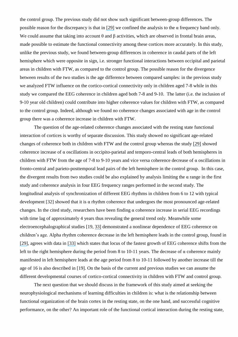
Bilateral-synchronous bursts of frontal theta waves (FTW) (fig. 1) are EEG patterns typical for
children with learning difficulties who had no history of neurological or psychiatric disorders. These EEG
patterns were detected by visual analysis in children aged 6-7 and 7-8 with low level of academic
performance (60-80 % in different samples) along with no learning difficulties and EEG abnormality in
control groups [1-3]. The results of visual EEG analysis agree with spectral analysis data: there is an
increase of spectral theta power in fronto-central cortices in children with learning difficulties [4, 5].
Complex electroencephalographical and neuropsychological studies [3, 6] showed that the main cause of
poor academic performance of children aged 6-7 and 7-8 with FTW was deficit of programming, voluntary
regulation and control.
Paste fig. 1
Seeking the neurophysiological basis of detected cognitive deficits we turned to the clinical
electroencephalographical data on the etiology of slow waves in frontal areas. These EEG patterns
appeared to be thalamus damage manifestations, especially its medial parts [7, 8]. Damage of thalamic
medial parts, in turn, is accompanied by cognitive and behavior regulation difficulties similar to the lobe
syndrome symptoms [9-11]. The reason of lobe syndrome deficits manifestation in the case of damage to
the mediodorsal thalamic nucleus (MD) is due to its close connectivity with the prefrontal cortex (PFC) via
strong reciprocal links which form together the integrated morpho-functional fronto-thalamic system [12-

14]. The immediate evidence of connection between FTW and thalamic neurons activity was found when
modeling the lobe syndrome in mice by cobalt application to their frontal cortex [15]. These findings state
that slow synchronous θ waves in the frontal cortex are accompanied by generation of θ waves in MD
thalamic neurons and, in addition to that, θ oscillations are coherent in both structures. Comparing the data
above and results of our own electroencephalographical and neuropsychological studies prepared the
ground for treating FTW as a sign of non-optimal functioning of the fronto-thalamic brain system.
In our previous study [16] we compared directed intracortical interactions* during FTW recordings
and periods free of these patterns by vector autoregressive model [17]. The analysis of the mutual influence
of EEG signals showed that the presence of common neuronal activity in the θ frequency range
synchronized due to the connectivity with deep brain structures is the most possible reason for the
emergence of synchronous slow waves in the frontal cortices. It was also shown that cortical functional
interaction during the FTW recording is determined by the increase of diffuse involvement of frontal and
central areas to the interaction both between each other and with other cortices.
These data is in a good agreement with findings of the fronto-thalamic etiology of slow
synchronous oscillations in frontal cortices recorded in the rest condition with eyes closed. The occasional
reduction in the degree of autonomy of frontal cortices as a result of fronto-thalamic system non-optimal
functioning found in the study [16] could be one of the reasons of cognitive deficits in children with FTW.
The significance of the rest state as the initial background for the cognitive activity was shown in studies
conducted by D.A. Farber et al [19, 20], who compared age-related changes of the background EEG and
cognitive processes in children. It could be assumed that one to several seconds long malfunctions in the
frontal brain areas during the FTW recording may impede switchover from the rest state to the activity
state. However, the extent to which such malfunctions influence the brain work during cognitive activity is
still the question in the frame of background EEG analysis.
At the same time, quantitative background EEG analysis allows investigating another question
which is also required to be studied to search the reasons of cognitive deficits in children with FTW. We
are speaking about the possible negative influence of non-optimal fronto-thalamic functioning on the
morpho-functional maturation of intracortical connectivity during developmental course.
Fronto-thalamic system plays an important role in selective modulation of cortical activity during
attentive processing [21, 22], preparation for the relevant information analysis [2] and keeping information
in working memory [23]. The lacking selective modulation influence of fronto-thalamic system on the
brain cortex could have an effect not only on the brain functioning during the cognitive performance but
also cortical neuronal network formation during developmental course. The latter assumption is reasonable
because both local cortical neuronal networks [24] and brain conduction pathways are being matured for a
* Hereinafter terms “intracortical interaction”, “functional interaction” or “functional connectivity” are used to describe linear
statistical relationship between spatially distant neurophysiological events [18], in this case between frequency components of
EEG signals recorded from spatially distant cortices.

long period during developmental course [25]. Moreover, there are findings testifying the dependence of
the brain morpho-functional maturation on the individual experience, particularly those types of activity
which require the involvement of selective voluntary attention [26, 27].
It was shown that maturation of brain neuronal networks during developmental course is reflected
in background EEG characteristics [19, 28]. In this case, if non-optimal fronto-thalamic functioning really
influences the formation of cortical neuronal connections, we can expect this influence to be reflected in
cortico-cortical connectivity estimated via resting-state EEG coherence during the periods free of FTW.
The previous study of resting-state α coherence was conducted in another group of primary schoolchildren
who had EEG signs of immature brain regulatory systems including immature fronto-thalamic system
manifested as FTW. These findings published in [29] point to the specificity of cortical functional
interaction via the α rhythm in children with FTW. In the current study we continued to analyze functional
brain cortex organization in children with FTW presented in our previous publication [16] and our task was
to estimate the parameters of EEG rhythms coherence in four frequency ranges: θ, α 1, α 2 and β 1. We
compared brain electrical activity (EA) recordings in children aged 7-8 and 9-10 with learning difficulties
whose EEG showed frontal θ waves and in children of the same age with no learning difficulties and EEG
abnormalities.
METHODS AND MATERIALS
We analyzed EEG recordings of 100 children aged 7-8 and 9-10, who attended regular primary
schools and had no history of neurological or psychiatric disorders. All the children and their parents gave
informed consent to participate in the study. The distribution of sample by age, sex and the presence of
FTW in children EEG is reflected in table 1. Approximately one half of the sample demonstrated low level
of academic performance (scored 2 or 3 according to the Russian five point grading system) in the main
courses and had bilateral synchronous θ waves in their EEG recordings, the other half of the sample were
of the same age but experienced no learning or behavioral difficulties and had no brain EA abnormalities.
Paste table 1
EEG was recorded in the rest condition with eyes closed from 12 symmetrical leads: F3/F4, C3/C4,
T3/T4, P3/P4, T5/T6, O1/O2 placed according to the international 10-20 system. EEG was referenced to the
numerical average of the right and left mastoid channels.
As our current task was to analyze functional interaction of areas during the periods free of FTW
and other EA abnormalities an expert first of all looked through EEG recordings to mark and cut segments
which contained artifacts. Finally, FTW and artifact-free EEG was not a continuous recording but a
sequence of segments different in size and separated by deleted chunks. In each individual EEG recording
such segments were subdivided into adjacent intervals 0.5 seconds long. Total duration of “pure” (FTW
and artifact-free) recording varied from child to child. To avoid the influence of this circumstance on

analyzed parameters of EEG coherence we used the sequence of first forty segments 0.5 seconds long (the
total duration was 20 seconds) for computing individual parameters of the coherence. The choice of the
number of segments being processed was determined by the shortest “pure” recording in the sample.
Functional cortico-cortical interactions were estimated by 12-channel vector autoregressive model
(hereinafter VAR-model). The VAR model is the simplest model capturing the linear statistical
dependencies in multichannel time series and successfully used in electrophysiology (see [17, 30] for a
review). Due to the fact that 40 segments chosen to analyze were not a continuous recording we used the
method suggested in [31] to calculate VAR coefficients. A VAR-model order of 20 was used as an optimal
order. This choice of order guaranteed the avoidance of serial correlations of autoregressive residuals and
simultaneously provided the spectral resolution of about 1 Hz which is high enough [30]. Using the VAR
coefficients computed for each pair of leads with numbers k and m we calculated the cross-spectrum
)( fSkm and power spectra )( fSkk and )( fSmm to get the coherence function:
)()()()( fSfSfSfC mmkkkmkm .
To analyze the coherence functions we chose four frequency ranges with bounds individually
selected for each subject by the following procedure. First, the fixed frequency margins 4–7 Hz for θ, 7–9
Hz for α1, 9–12 Hz for α2, and 12–15 Hz for β1 were chosen. Then, for each subject we made a common
for all lead pairings histogram of frequency locations (with 0.5 Hz step within 3-16 Hz range) of the local
maxima of coherence function. This histogram was smoothed using 11-point Bohman window. Then all
four a priori chosen frequency ranges were checked for local maxima of the smoothed histogram. If there
was one maximum within the range we applied 2 Hz wide interval with the center corresponding to the
frequency location of this maximum as an individual range. If there were two or more maxima we chose
the biggest one and set the range the way we did in the previous case. If there were no such maxima we
chose the entire a priori frequency range for the individual range. Mean coherence values in each
individual range were calculated as individual coherence function estimates were used in the further group
statistical analysis.
Statistical testing was performed using multivariate analysis of variance (MANOVA). Pairwise
comparisons were accomplished with ANOVA.
RESULTS
Using multivariate analysis of variance (MANOVA) we studied the influence of age (two-level
AGE factor: 7-8 and 9-10 years) and frontal θ waves presence (two-level FTW factor) on EEG coherence
in four frequency ranges (four-level RANGE factor: θ, α 1, α 2 and β) for interhemispheric and
intrahemispheric pairs of leads. To study connectivity specific for the left and right hemispheres we
included the HEMISPHERE factor (two-level) into MANOVA schema and the LOCATION factor to
analyze regional differences. According to the fact that there was different boy-girl ratio in the compared

groups (see table 1) the sex factor was also taken into account when performing MANOVA. To minimize
pairwise comparisons during the statistical analysis of LOCATION factor we applied MANOVA
separately for several subsets of lead pairs: F-X, C-X, P-X, Ta-X and Tp-O, where X corresponds to each
remaining lead in the same hemisphere. One and the same pair of leads was never used in different subsets.
By analyzing the influence of between-subjects factors it was shown that age, sex and the presence
of FTW in EEG had no significant main effect on the EEG coherence value in any subset of lead pairs.
Whenever the between-subject factors affected the coherence they did it via interaction with other factors.
As the main task of this study was to estimate a specific character of cortico-cortical functional
interactions in children with frontal θ waves in EEG we analyzed only the impact of FTW factor or its
interaction with other independent variables mentioned below. Table 2 lists significant effects for all
subsets of leads. Table 3 contains descriptive statistics for coherence in separate pairs of leads.
First of all it should be mentioned that the main effect of FTW was not found in this sample of
children. FTW factor influence was mediated by other conditions most commonly associated with the
location of EEG leads (HEMISPHERE and/or LOCATION factors). In all subsets of lead pairs we found
no connection between FTW influence and either sex or EEG frequency range. Due to this fact the post-
hoc analysis of EEG data was performed regardless of the subjects’ sex and EEG frequency ranges. Thus
below are shown the results of comparison of coherence value averaged over four frequency ranges.
Paste table 2 and 3
The impact of FTW factor on the interaction of frontal areas with other cortices depended on
children’s age in the studied sample. This dependence was reflected in FTW AGE interaction as a
tendency and in significant FTW LOCATION AGE interaction. Post-hoc analysis of FTW AGE
interaction showed that average (over four frequency ranges) coherence value in F-X lead pairs is higher
in the control group of children aged 7-8 (С = 0.456±0.021) than in children with frontal θ waves (C =
0.424±0.022). This difference found in junior age group turned to be significant (F(1, 59) = 4.546, p =
0.037). There were no significant differences for this factor in children aged 9-10 (F(1, 37) = 1.043, p =
0.314), with coherence value being lower in the control group than in group with FTW (С = 0.428±0.029
and С = 0.447±0.024, respectively). As regards the other “participant” of this interaction, which is the AGE
factor, we found no significant differences in both control and FTW groups. Coherence value seemed to
decrease in control group from 7-8 to 9-10 years (С = 0.456±0.02 at 7-8 years, С = 0.428±0.028 at 9-10
years, F(1, 46) = 2.778, p = 0.103) and increased in group with FTW (С = 0.425±0.023 at 7-8 years, С =
0.447±0.026 at 9-10 years, F(1,050) = 1.834, p = 0.182).
Significant FTW LOCATION AGE interaction suggests that FTW influence on coherence in the
F-X lead pairs depended not only on age but LOCATION of another lead in the pair. Therefore in each of
the five lead pairs included in this subset (omitting the HEMISPHERE factor) we compared coherence
over the FTW factor separately in groups of children aged 7-8 and 9-10. Differences with p < 0.01 were
regarded as significant after correction for multiple comparisons (five pairs of leads). Hence, the

difference for the lead pair F-P (F(1, 59) = 5.20, p = 0.02) found in children aged 7-8 considered to be only
a tendency, while, for the lead pair F-Ta, it was accepted as significant (F(1, 59) = 8.62, p = 0.005). In both
lead pairs, the level of functional cortical interaction was lower in children with FTW as compared to the
control group. Children aged 9-10 with FTW also had low coherence in F-Ta lead pair, but the difference
reached a significant level only in the left hemisphere (F(1, 37) = 5.85, p = 0.004). In other F-X lead pairs
we found no FTW factor influence both in 7-8 and 9-10 children. Finally, completing to analyze functional
connectivity of frontal areas it should be noted that the influence of FTW factor was in general (omitting
AGE and lead LOCATION) more pronounced in the left hemisphere. As compared to the control group,
EEG signals recorded from frontal areas of the left hemisphere in children with FTW turned to be less
statistically related to EEG signals from other areas of the same hemisphere (С = 0.475±0.015 in control
group, C = 0.442±0.015 in group with FTW, F(1, 92) = 4.73, p = 0.015). For lead pairs of the right
hemisphere, we found no statistically significant differences in EEG coherence between the control group
and children with FTW. Such a selective FTW influence on functional connectivity of frontal cortices
found in the left hemisphere was reflected in the significant HEMISPHEREFTW interaction (see table
2).
The dependence of FTW factor influence on children’s age was also found for interhemispheric
functional connections (table 2). Descriptive statistics of interhemispheric coherence values is presented in
table 4.
Paste table 4
In younger group of children with FTW, interhemispheric interaction of symmetrical cortices was
weaker than in the control group (in control group С = 0.497±0.018, in group with FTW С = 0.457±0.020;
F(1, 57) = 3.930, p = 0.027). In 7-8 years old children the FTW effect was differently manifested for
different pairs of symmetrical areas (FTW LOCATION, F(5, 53) = 3.36, p = 0.01)). The coherence
decrease in children with FTW as compared to the control group reached the statistical significance
(corrected for 6 comparisons) for interhemispheric connections between frontal areas (F(1, 57) = 6.767, p =
0.002). In the older age group, the picture of interhemispheric coherence relationship was opposite:
children with FTW had a stronger interhemispheric interaction as compared to the control group, however
the difference did not reach the significance (С = 0.494±0.027 vs. С = 0.472±.026; F(1, 35) = 1.618, p =
0.121). In children aged 9-10, there were also no significant regional differences in FTW influence on
interhemispheric coherence.
Since no FTW AGE interaction was found for any other subset of leads all post-hoc comparisons
presented below were performed leaving aside AGE factor. Significant
FTWHEMISPHERELOCATION interaction (see table 2) allows analyzing FTW factor influence on
the coherence in separate lead pairs for each subset of leads.
Analyzing coherence of EEG signals in pairs of leads, included central areas, we again found the
stronger FTW influence on the functional connectivity in the left hemisphere as compared to the right

hemisphere. In left hemisphere lead pairs С3-T3 and С3-T5, the coherence was higher in children of the
control group as compared to children with FTW. Taking into account multiple comparisons (eight lead
pairs of the right and left hemisphere) the significant differences were found only for C3-T3 (F(1, 98) =
7.798, p = 0.006). In the right hemisphere, such differences were observed only for С4-T4 (F(1, 98) =
8.454, p = 0.005), and coherence in the lead pair C4-T6 did not differ even for average values. The analysis
of the coherence in P-X lead pairs showed no differences except a single almost significant difference in
P3 – O1 lead pair (F (1, 98) = 5.705, p = 0.019). It should be noted that, unlike the anterior cortices, the
coherence value for the pairs of posterior locations of the left hemisphere was higher in the children with
FTW than in the control group (see table 3). There was no FTW factor influence on the right hemisphere
coherence. For either hemisphere, the FTW factor had no significant influence on the strength of anterior
temporal to inferior temporal or occipital areas functional links; nor had it any impact onto the connection
strength between inferior temporal and occipital areas.
Thus, the analysis of the resting state EEG coherence showed that the decrease in functional
connectivity of fronto-temporo-central cortices, mainly in the left hemisphere, was a common phenomenon
for children aged 7-8 and 9-10 with FTW as compared to the control group. In the parieto-occipital areas of
the left hemisphere, on the contrary, children with FTW approached a significant increase in the strength of
cortico-cortical interaction.
We found no significant age-related changes of EEG coherence in both FTW and control groups.
Yet, the coherence analysis demonstrated age influence on the degree of between-group differences: a
relative weakness of functional connectivity between frontal and other areas was in general more
pronounced in children with FTW at 7-8 years.
DISCUSSION
Results of the study made it possible to answer affirmatively the question of whether the difference
between spatio-temporal organization of artifact-free background EEG in children with FTW and the
control group exists. A qualitative comparison of current results with our previous results [29] on the
background EEG coherence in children with FTW obtained in other sample with other device and methods
for coherence calculation allows speaking of similarity in these data. Like in the previous study, current
results demonstrated the background EEG coherence decrease mainly in closely spaced lead pairs which
included anterior temporal area of the left hemisphere (T3). The similarity of results of the two independent
studies points to their robustness. This allows considering the “weakening” of connections between the left
anterior temporal area and nearby areas of the same hemisphere as a specificity of the resting state cortico-
cortical interaction in children with learning difficulties and frontal θ waves in EEG. Along with it, the
current study demonstrated the weakening of functional links between the frontal and anterior temporal
areas of the left hemisphere and frontal interhemispheric connection in children with FTW, as compared to
the control group. The previous study did not show such significant between-group differences. The
possible reason for the discrepancy is that in [29] we confined the analysis to the α frequency band only.
We could assume that taking into account θ and β activities, which are observed in frontal brain areas,
made possible to estimate the functional connectivity among these cortices more accurately. In this study,
unlike the previous study, we found between-group differences in coherence in caudal parts of the left
hemisphere which were opposite in sign, i.e. stronger functional interactions between occipital and parietal
areas in children with FTW, as compared to the control group. The possible reason for the divergence
between results of the two studies is the age difference between compared samples: in the previous study
we analyzed FTW influence on the cortico-cortical connectivity only in children aged 7-8 while in this
study we compared the EEG coherence in children aged both 7-8 and 9-10. The latter (i.e. the inclusion of
9-10 year old children) could contribute into higher coherence values for children with FTW, as compared
to the control group. Indeed, although we found no coherence changes associated with age in the control
group there was a coherence increase in children with FTW.
The question of the age-related coherence changes associated with the resting state functional
interaction of cortices is worthy of separate discussion. This study showed no significant age-related
changes of coherence both in children with FTW and the control group whereas the study [29] showed
coherence increase of α oscillations in occipito-parietal and temporo-central leads of both hemispheres in
children with FTW from the age of 7-8 to 9-10 years and vice versa coherence decrease of α oscillations in
fronto-central and parieto-posttemporal lead pairs of the left hemisphere in the control group. In this case,
the divergent results from two studies could be also explained by analysis limiting the α range in the first
study and coherence analysis in four EEG frequency ranges performed in the second study. The
longitudinal analysis of synchronization of different EEG rhythms in children from 6 to 12 with typical
development [32] showed that it is α rhythm coherence that undergoes the most pronounced age-related
changes. In the cited study, researchers have been finding α coherence increase in serial EEG recordings
with time lag of approximately 4 years thus revealing the general trend only. Meanwhile some
electroencephalographical studies [19, 33] demonstrated a nonlinear dependence of EEG coherence on
children’s age. Alpha rhythm coherence decrease in the left hemisphere leads in the control group, found in
[29], agrees with data in [33] which states that locus of the fastest growth of EEG coherence shifts from the
left to the right hemisphere during the period from 8 to 10-11 years. The decrease of α coherence mainly
manifested in left hemisphere leads at the age period from 8 to 10-11 followed by another increase till the
age of 16 is also described in [19]. On the basis of the current and previous studies we can assume the
different developmental courses of cortico-cortical connectivity in children with FTW and control group.
The next question that we should discuss in the framework of this study aimed at seeking the
neurophysiological mechanisms of learning difficulties in children is: what is the relationship between
functional organization of the brain cortex in the resting state, on the one hand, and successful cognitive
performance, on the other? An important role of the functional cortical interaction during the resting state,
viewed as a condition that determines the readiness for information processing and overt activity, was
shown in a number of electrophysiological studies that have been traced back to 90s of the previous
century [20, 28, 33, 34]. Mainly it were the studies of age-related changes in spatio-temporal organization
of the brain EA that lightened the fact that even when a person is just relaxing and does not join any
activity the different brain cortices function as a united and relatively stable system. It was also
demonstrated that resting state functional connectivity is changing in its character and strength during the
developmental course [20, 28, 32, 35, 36]. By analyzing the slow (about 0.1 Hz) oscillating metabolic
processes in neuronal networks (BOLD signals) it was found over the last few years that the functional
connectivity between different brain structures is topographically specific not only for cognitive
performance but also for the resting state. Such a functional coupling of brain structures was called a
resting state network (RSN) [37, 38] or an intrinsic connectivity network (ICN) [39]. In most cases, the
resting state network includes structures which are also being activated during different motor and
cognitive performances [37, 40] and structures which are silent (default mode networks - DMN) when
performing a cognitive task [41]. According to S. Bressler [39] resting state networks, topographically
similar to cognitive neuronal networks, give evidence to the fact that the human brain is initially organized
into relatively stable distributed systems. Resting state networks provide a sort of skeleton for more
dynamic functional connectivities during cognitive performance. EEG and fMRT data comparison [41,43]
showed that the configuration of resting state functional connectivity estimated through the analysis of
slow oscillating metabolic processes is mostly similar to that found with EEG analysis. These data prove
the existence of relationship between the spatio-temporal organization of resting state rhythmical EA and
the functional organization of actual neuronal networks, effectiveness of inner linking of which determines
the effectiveness of different cognitive performances. The above mentioned data allow treating the results
of this study as an evidence for the specificity of the morpho-functional brain organization in children with
FTW.
Regarding the current study results in the framework of the reflection of functional brain
organization in the resting state EEG we can assume that the non-optimal fronto-thalamic functioning
impede the actual interaction among frontal, temporal and central cortices of mostly the left hemisphere.
The age-related specificity of differences between control children and children with FTW point to rather
indirect association between the non-optimal fronto-thalamic functioning and the decrease of the left
hemisphere functional cortical interaction. And since it is so, it is reasonable to assume atypical morpho-
functional maturation of cortical neuronal networks in children with FTW. One of the possible reasons for
the sensitivity of the left hemisphere neuronal networks to the non-optimal fronto-thalamic functioning can
be its neurochemical specificity, e.g. the higher, than in the right hemisphere, activity of dopaminergic
neuronal connectivity [43]. Fronto-thalamic system is, in its turn, linked to the limbic-cortical dopamine
system through MD. A lacking involvement of this connectivity can negatively affect the formation of left
hemisphere neuronal networks. The other possible reason is the poor selective modulation of the cortex
exerted by non-specific thalamic nuclei. This can affect negatively the morpho-functional maturation of the
left hemisphere neuronal networks which are known to be more locally specialized than in the right
hemisphere [44].
In previous complex neurophysiological and electrophysiological studies it was shown that children
with FTW demonstrated deficit of the nominative function of speech in addition to the pronounced lack of
executive control. This deficit reflects in poor vocabulary and semantic errors during both speech (writing
by dictation, retelling) and visuo-spatial (recognition and description of visual objects) performance [6, 45].
The quantitative analysis of resting state EEG presented in this study provides an additional information
about neurophysiological mechanisms of cognitive deficits mentioned above. According to Luria’s clinical
studies [46] the nominative function of speech is associated with the left hemisphere functioning and
specifically disturbed in the case of damage to postfrontal and temporal areas of this hemisphere. It was
these areas in children with FTW that showed a relative weakness of cortico-cortical interaction.
Summarizing the results of electroencephalographical and neuropsychological studies of children with
FTW versus the syndromes of local brain injuries allows to assume that the basis of deficit of the
nominative function of speech in children with immature or dysfunctioning fronto-thalamic system can be
an inappropriate functional connectivity between frontal and temporal left hemisphere structures.
Results of this study suggest that the relative weakness of cortico-cortical interaction in fronto-
temporal region of the left hemisphere can be one of the neurophysiological factors of cognitive deficits
and associated learning difficulties in primary schoolchildren, whose EEG shows frontal bilateral-
synchronous θ waves.
CONCLUSIONS
1. Coherence analysis of artifact-free resting state EEG in θ, α 1, α 2 and β 1 frequency ranges
showed that children aged 7-8 and 9-10 with learning difficulties and frontal θ waves (FTW) have a
decreased, as compared to control children of the same age, functional connectivity of frontal and anterior
temporal cortices, the decrease being more pronounced in the left hemisphere.
2. In children with FTW, the specificity of resting state functional cortical connectivity estimated
via analyzing the coherence of brain rhythmic electrical activity did not depend on children’s sex and EEG
frequency range.
3. The non-optimal fronto-thalamic functioning, reflected in FTW presence, exerts more influence
on the frontal cortex functional connectivity in children aged 7-8 than in children of 9-10.
ACKNOWLEDGMENTS
This study was supported by the Russian Foundation for Humanities Research. Grant no. 10-06-00693а.
REFERENCES
1. Machinskaia, R.I., Lukashevich, I.P., and Fishman, M.N., Dynamics of Brain Electrical Activity
in 5-to 8-year-old Normal Children and Children with Learning Difficulties, Hum. Physiol., 1997, vol. 23,
no. 5, p. 5.
2. Machinskaya, R. I., Functional Maturation of the Brain and Formation of the Neurophysiological
Mechanisms of Selective Voluntary Attention in Young Schoolchildren, Hum. Physiol., 2006, vol. 32, no.
1, p. 20.
3. Semenova, O.A. and Machinskaya, R. I., Neiropsihologicheskii i neirofiziologicheskii analiz
vozrastnyh preobrazovanii poznavatel’nyh funktsii i riskov uchebnoi dezadaptatsii v predshkol’nom
vozraste (Neuropsychological and neurophysiological analysis of age-related changes of cognitive
functions and risks of learning and behavioral difficulties in preschool children), Nov. Issled., 2012, vol.
30, no. 1, p. 45.
4. Lubar, J.F., Bianchini, K.J., Calhoun, W.H., et al., Spectral Analysis of EEG Differences
Between Children With and Without Learning Disabilities, J. Learn. Disabil., 1985, vol. 18, no. 7, p. 403.
5. Chabot, R.J., Michele, F., Prichep, L., and John, R., The Clinical Role of Computerized EEG in
the Evaluation and Treatment of Learning and Attention Disorders in Children and Adolescents, J.
Neuropsychiatry Clin. Neurosci., 2001, vol. 13, no. 2, p. 171.
6. Machinskaya, R. I. and Semenova, O. A., Peculiarities of Formation of the Cognitive Functions
in Junior School Children with Different Maturity of Regulatory Brain Systems, J. Evol. Biochem. Physiol.,
2004, vol. 40, no. 5, p. 528.
7. Mayorchik, V.E., Izmeneniia EEG v zavisimosti ot lokalizatsii opuholi mozga (Changes in EEG
Depending on the Localization of Brain Tumor), in Klinicheskaya elektroentsefalografiya (Clinical
Electroencephalography), Rusinov, V.S., Ed., Moscow: Meditsina, 1973, p. 106.
8. Lukashevich, I.P. and Sazonova, O.B., The Effect of Lesions of Different Parts of the Optic
Thalamus on the Nature of the Bioelectrical Activity of the Human Brain, Zh. Vyssh. Nerv. Deiat. Im. I.P.
Pavlova, 1996, vol. 46, no. 5, p. 866.
9. Terao, Y., Sakurai, Y., Sakuta, M., et al., FDG-PET in an Amnestic and Hypersomnic Patient
with Bilateral Paramedian Thalamic Infarction, Rinsho Shinkeigaku, 1993, vol. 33, no. 9, p. 951.
10. Kalashnikova, L.A., Gulevskaya, T.S., and Kashina, E.M., Disorders of Higher Mental Function
Due to Single Infarctions in the Thalamus and in the Area of the Thalamofrontal Tracts, Neurosci. Behav.
Physiol., 1999, vol. 29, no. 4, p. 397.
11. Zoppelt, D., Koch, B., Schwarz, M., et al., Involvement of the Mediodorsal Thalamic Nucleus
in Mediating Recollection and Familiarity, Neuropsychologia, 2003, vol. 41, no. 9, p. 1160
12.Goldman-Rakic, P.S., Porrino, L.J., The primate mediodorsal (MD) nucleus and its projection to
the frontal lobe, J. Comp. Neurol., 1985, vol. 242, p. 535. doi: 10.1002/cne.902420406

13. Zhang, D., Snyder, A.Z., Shimony, J.S., et al., Noninvasive Functional and Structural
Connectivity Mapping of the Human Thalamocortical System, Cerebr. Cortex., 2010, vol. 20, no. 5, p.
1187.
14. Lujan, J.L., Chaturvedi, A., and McIntyre, C.C., Tracking the Mechanisms of Deep Brain
Stimulation for Neuropsychiatric Disorders, Front. Biosci., 2008, vol. 13, p. 5892.
15. Kim, J., Woo, J., Park, Y-G., et al., Thalamic T-Type Ca 2-Channels Mediate Frontal Lobe
Dysfunctions Caused by a Hypoxia-Like Damage in the Prefrontal Cortex, J. Neurosci., 2011, vol. 31, no.
11, p. 4063. doi: 10.1523/JNEUROSCI.4493-10.2011
16. Kurgansky, A. V. and Machinskaya, R. I., Bilateral Frontal Theta-Waves in EEG of 7–8-Year-
Old Children with Learning Difficulties: Qualitative and Quantitative Analysis, Hum. Physiol., 2012, vol.
38, no. 3, p. 37.
17. Kaminski, M., Determination of Transmission Patterns in Multichannel Data, Phil. Trans. R.
Soc. B., 2005, vol. 360, no. 1457, p. 947. doi: 10.1098/rstb.2005.1636
18. Fingelkurts, Al.A., Fingelkurts, An.A, and Kahkonen, S., Functional Connectivity in the Brain –
is it an Elusive Concept?, Neurosci. Biobehav. Rev., 2005, vol. 28, no. 8, p. 827
19. Alferova, V.V. and Farber, D.A., Otrazhenie vozrastnyh osobennostei funktsional’noi
organizatsii mozga v elektroentsefalogramme pokoia (the Reflection of Age-related Specificity of
Functional Brain Organization in Resting State Electroencephalogram), in Strukturno-funktsional’naia
organizatsiia razvivaushegosia mozga (Principles of Structural and Functional Brain Organization in
Ontogenesis), Adrianov, O.S., Ed., Leningrad: Nauka, 1990, p. 45
20. Kniazeva, M.G. and Farber, D.A., Spatial Structure of Intra- and Inter-hemispheric
Connections: Factor Analysis of the Coherence of the Resting EEG, Fiziol. Cheloveka, 1996, vol. 22, no. 5
p. 37
21. Machinskaia, R.I., Neurophysiological Mechanisms of Voluntary Attention: A Review, Zh.
Vyssh. Nerv. Deiat. Im. I.P. Pavlova, 2003, vol. 53, no. 2, p. 133.
22. Joseph, R., Neuroscience: Neuropsychology, Neuropsychiatry, Behavioral Neurology, Brain &
Mind: Primer, Overview & Introduction, University Press Science Publishers, 2011, 4th ed.
23. Watanabe, Y. and Funahashi, Sh., Thalamic Mediodorsal Nucleus and Working Memory,
Neurosci. Biobehav. Rev., 2012, vol. 36, no. 1, p. 134.
24. Huttenlocher, P.R., Neural Plasticity. The Effects of Environment on the Development of the
Cerebral Cortex, Cambridge, Massachusetts, London: Harvard University Press, 2002
25. Hagmann, P., Sporns, O., Madan N. , et al., White Matter Maturation Reshapes Structural
Connectivity in the Late Developing Human Brain, Proc. Natl. Acad. Sci. U S A, 2010, vol. 107, no. 44, p.
9067. doi: 10.1073/pnas.1009073107

26. Segalowitz, S.J., The Role of Neuroscience in Historical and Contemporary Theories of Human
Development, in Human behavior, learning, and the developing brain, Coch, D., Fischer, K.W., and
Dawson, G., Eds., New York: Guilford, 2007.
27. Galvan, A., Neural Plasticity of Development and Learning, Hum. Brain Mapp., 2010, vol. 31,
no. 6, p. 879. doi: 10.1002/hbm.21029
28. Thatcher, R.W., North, D.M., and Biver, C.J., Development of Cortical Connections as
Measured by EEG Coherence and Phase Delays, Hum. Brain Mapp., 2008, vol. 29, no. 12, p. 1400. doi:
10.1002/hbm.20474
29. Machinskaia, R.I., Sokolova, L.S., and Krupskaia, E.V., Formation of the Functional
Organization of the Cerebral Cortex at Rest in Young Schoolchildren Varying in the Maturity of Cerebral
Regulatory Systems: II. Analysis of EEG alpha-rhythm Coherence, Fiziol Cheloveka, 2007, vol. 33, no. 2,
p. 5.
30. Kurganskii, A.V., Study of Cortico-Cortical Functional Connectivity with Vector
Autoregressive Model of Multichannel EEG, Zh. Vyssh. Nerv. Deiat. Im. I.P. Pavlova, 2010, vol. 60, no. 6,
p. 740.
31. Cui, J., Xu, L., Bressler, S.L., et al., BSMART: A Matlab/C Toolbox for Analysis of
Multichannel Neural Time Series, Neural Netw., 2008, vol. 21, no. 8, p. 1094. doi:
10.1016/j.neunet.2008.05.007
32. Gmehlin, D., Thomas, C., Weisbrod, M., et al., Development of Brain Synchronisation within
School-age – individual Analysis of Resting (α) Coherence in a Longitudinal Data Set, Clin. Neurophysiol.,
2011, vol. 122, no. 10, p. 1973. doi: 10.1016/j.clinph.2011.03.016
33. Thatcher, R.W., Cyclic Cortical Re-Organization: Origins of Human Cognitive Development, in
Human Behavior and the Developing Brain, Dawson, G. and Fischer, K., Eds., New York: Guilford, 1994,
p. 232.
34. Marosi, E., Harmony, T., Sanchez, L., et al., Maturation of the Coherence of EEG Activity in
Normal and Learning-disabled Children, Electroencephalogr. Clin. Neurophysiol., 1992, vol. 83, no. 6, p.
350.
35. Shepoval'nikov, A.N., Tsitseroshin, M.N., and Pogosian, A.A., Role of Various Cortical Areas
and Their Connections in the Formation of Spatial Ordering in the Field of Brain Biopotentials During
Postnatal Ontogenesis, Fiziol Cheloveka, 1997, vol. 23, no. 2, p. 12.
36. Bezrukikh, M.M. and Terebova, N.N., Functional Organization of the Brain Cortical Areas at
Rest Condition in Children 5, 6 and 7 years old, Fiziol Cheloveka, 2010, vol. 36, no. 6, p. 61.
37. Damoiseaux, J.S., Rombouts, S.A.R.B., Barkhof, F., et al., Consistent Resting-State Networks
Across Healthy Subjects, Proc. Natl. Acad. Sci. U S A, 2006, vol. 103, no. 37, p. 13848. doi:
10.1073/pnas.0601417103
38. Van den Heuvel, M.P., Mandl, R.C.W., Kahn, R.S., et al., Functionally Linked Resting-State
Networks Reflect the Underlying Structural Connectivity Architecture of the Human Brain, Hum. Brain
Mapp., 2009, vol. 30, no. 10, p. 3127. doi: 10.3389/fnana.2012.00036
39. Bressler, S. L. and Menon, V., Large-scale Brain Networks in Cognition: Emerging Methods
and Principles, Trends Cognitive Sci., 2010, vol. 14, no. 6, p. 277. doi: 10.1016/j.tics.2010.04.004
40. Britz, J., Van de Ville, D., and Michel, Ch.M., BOLD Correlates of EEG Topography Reveal
Rapid Resting-State Network Dynamics, Neuroimage, 2010, vol. 52, no. 4, p. 1162. doi:
10.1016/j.neuroimage.2010.02.052
41. Greicius, M.D., Kaustubh, S., Menon, V., and Dougherty, R.F., Resting-State Functional
Connectivity Reflects Structural Connectivity in the Default Mode Network, Cereb. Cortex, 2009, vol. 19,
no. 1, p. 72. doi: 10.1093/cercor/bhn059
42. Mantini, D., Perrucci, M.G., Del Gratta, C., et al., Electrophysiological Signatures of Resting
State Networks in the Human Brain, Proc. Natl. Acad. Sci. U S A, 2007, vol. 104, no. 32, p. 13170. doi:
10.1073/pnas.0700668104
43. Toga, A.W. and Thompson, P.M., Mapping Brain Asymmetry, Nat. Rev. Neurosci., 2003, vol.
4, no. 1, p. 37.
44. Hutsler, J., Galuske, R.A.W., Hemispheric Asymmetries in Cerebral Cortical Networks, Trends
Cognitive Sci., 2003, vol. 26, no.8, p. 429. doi: 10.1016/S0166-2236(03)00198-X
45. Sugrobova, G.A., Semenova, O.A., and Machinskaia, R.I., Osobennosti reguliatornyh i
informatsionnyh komponentov poznavatel’noi deiatel’nosti u detei 7-8 let s priznakami SDVG
(Peculiarities of Regulatory and Information-Related Components of Cognitive Activity in 7-8 Year Old
Children with ADHD), J. Hum. Ecol., 2010, no. 11, p. 19.
46. Luria, A.R., Vysshie korkovye funktsii i ih narusheniia pri lokal’nyh porazheniiah mozga
(Higher Cortical Functions in Man and Their Impairment Caused by Local Brain Lesions), Moscow: MSU
Press, 1962.

O2
P4
C4
F4
T4
T6
O1
P3
C3
F3
T3
T5
FIGURES, LEGENDS AND TABLES
Figure 1. Resting state EEG of a 7-year-old child. Bilateral-synchronous frontal theta waves (FTW) are
underlined
Table 1. The distribution of sample by age, sex and the presence of frontal θ waves in EEG.
7-8 years 9 -10 years
boys girls total boys girls total
Controls 18 14 N = 32
mean age =
7.68±0.42
10 6 N = 16
mean age =
9.60±0.57
FTW 23 6 N = 29
mean age =
7.82±0.56
20 3 N = 23
mean age =
9.80±0.56
Total 41 20 N = 61
30 9 N = 39

Table 2. Significant influence of frontal θ waves presence (FTW factor) on resting state EEG coherence.
MANOVA results.
Factors Pairs of leads
F-O, F-P,
F-C, F-Ta,
F-Tp
C-O, C-P,
C-Ta, C-Tp
P-O, P-Ta,
P-Tp
Ta –O,
Ta - Tp
Tp-O
Interhemispheric
FTW AGE F(1, 92) = 3.70,
p = 0.057
F(1, 92) = 3.81,
p = 0.045
FTW HEMISPHERE F(1, 92) = 6.61,
p = 0.012
FTW LOCATION F( 4, 89) = 3.16,
p = 0.018
F(3, 90) = 3.30,
p = 0.024
F(5, 88) = 2.62,
p = 0.03
FTW LOCATION
AGE
F(4, 89) = 2.86,
p = 0.028
F(5, 88) = 2.44,
p = 0.041
FTW
HEMISPHERE
LOCATION
F(3, 90) = 5.98,
p = 0.001
F(2, 91) = 5.97,
p = 0.004
F(1, 92) = 7.73,
p = 0.007
Table 3. Mean values and standard deviations of EEG coherence* over the left (LH) and right (RH) hemispheres in 7-8 and 9-
10-year-old children of the control and FTW groups.
Age Pairs Control group Children with FTW
RH LH mean over
hemispheres
RH LH mean over
hemispheres
7-8 F-C 0.741(0.082) 0.766(0.064) 0.753(0.065) 0.711(0.082) 0.735(0.081) 0.723(0.076)
F-P 0.433(0.104) 0.474(0.074) 0.454(0.084) 0.392(0.094) 0.413(0.104) 0.402(0.093)
F-Ta 0.602(0.082) 0.612(0.077) 0.607(0.072) 0.557(0.085) 0.545(0.099) 0.551(0.077)
F-Tp 0.240(0.086) 0.283(0.075) 0.262(0.067) 0.238(0.071) 0.257(0.070) 0.247(0.057)
F-O 0.205(0.069) 0.205(0.070) 0.205(0.065) 0.196(0.061) 0.197(0.058) 0.196(0.054)
C-P 0.739(0.071) 0.769(0.054) 0.754(0.054) 0.714(0.064) 0.714(0.074) 0.714(0.061)
C-Ta 0.710(0.093) 0.720(0.086) 0.715(0.082) 0.655(0.076) 0.666(0.108) 0.661(0.078)
C-Tp 0.449(0.097) 0.510(0.074) 0.479(0.071) 0.425(0.109) 0.456(0.100) 0.440(0.086)
C-O 0.320(0.085) 0.312(0.084) 0.316(0.078) 0.319(0.085) 0.305(0.089) 0.312(0.076)
P-Ta 0.531(0.105) 0.570(0.089) 0.550(0.087) 0.503(0.089) 0.540(0.113) 0.522(0.086)
P-Tp 0.711(0.066) 0.754(0.050) 0.733(0.047) 0.704(0.095) 0.736(0.063) 0.720(0.067)
P-O 0.695(0.050) 0.648(0.070) 0.671(0.047) 0.706(0.066) 0.681(0.079) 0.693(0.066)
Ta-Tp 0.503(0.103) 0.557(0.092) 0.530(0.084) 0.490(0.090) 0.541(0.122) 0.516(0.082)
Ta-O 0.236(0.078) 0.249(0.070) 0.242(0.065) 0.246(0.075) 0.255(0.088) 0.250(0.060)
Tp-O 0.645(0.082) 0.696(0.070) 0.671(0.057) 0.668(0.076) 0.661(0.085) 0.664(0.069)
9-10 F-C 0.705(0.082) 0.766(0.080) 0.736(0.068) 0.725(0.081) 0.751(0.077) 0.738(0.073)
F-P 0.341(0.098) 0.420(0.108) 0.381(0.099) 0.412(0.104) 0.451(0.100) 0.432(0.096)
F-Ta 0.579(0.102) 0.643(0.064) 0.611(0.078) 0.577(0.131) 0.571(0.135) 0.574(0.119)
F-Tp 0.199(0.050) 0.254(0.090) 0.226(0.059) 0.267(0.085) 0.270(0.082) 0.268(0.074)
F-O 0.184(0.066) 0.186(0.079) 0.185(0.063) 0.217(0.090) 0.230(0.089) 0.223(0.087)
C-P 0.700(0.072) 0.721(0.111) 0.710(0.082) 0.716(0.110) 0.739(0.079) 0.727(0.084)
C-Ta 0.707(0.089) 0.744(0.057) 0.726(0.061) 0.664(0.088) 0.691(0.108) 0.678(0.081)
C-Tp 0.365(0.080) 0.457(0.105) 0.411(0.073) 0.449(0.135) 0.448(0.120) 0.449(0.118)
C-O 0.295(0.084) 0.258(0.094) 0.277(0.070) 0.302(0.107) 0.313(0.088) 0.307(0.090)
P-Ta 0.496(0.080) 0.547(0.110) 0.521(0.088) 0.518(0.096) 0.566(0.097) 0.542(0.083)
P-Tp 0.672(0.076) 0.719(0.106) 0.696(0.084) 0.738(0.084) 0.733(0.059) 0.735(0.066)
P-O 0.686(0.119) 0.622(0.098) 0.654(0.097) 0.686(0.082) 0.664(0.064) 0.675(0.059)
Ta-Tp 0.436(0.080) 0.536(0.107) 0.486(0.081) 0.501(0.113) 0.540(0.102) 0.520(0.079)
Ta-O 0.218(0.042) 0.213(0.098) 0.215(0.056) 0.244(0.102) 0.281(0.109) 0.263(0.097)
Tp-O 0.669(0.091) 0.691(0.063) 0.680(0.062) 0.685(0.088) 0.709(0.091) 0.697(0.077)
* table contains descriptive statistics with the RANGE factor excluded; table cells contain MEAN
VALUES (STANDART DEVIATION); coherence values with significant differences for FTW factor
pairwise comparisons are shown in bold.

Table 4*. Mean values and standard deviations of interhemispheric EEG coherence in 7-8 and 9-10-year-
old children of the control and FTW groups.
age pairs of leads control group children with FTW
7-8 F4-F3 0.764(0.056) 0.723(0.076)
C4-C3 0.601(0.082) 0.565(0.115)
P4-P3 0.592(0.107) 0.539(0.113)
T4-T3 0.192(0.083) 0.202(0.077)
T6-T5 0.224(0.059) 0.220(0.065)
O2-O1 0.600(0.092) 0.616(0.068)
9-10 F4-F3 0.736(0.101) 0.744(0.062)
C4-C3 0.570(0.083) 0.596(0.088)
P4-P3 0.534(0.084) 0.557(0.111)
T4-T3 0.195(0.087) 0.234(0.117)
T6-T5 0.180(0.060) 0.207(0.074)
O2-O1 0.592(0.100) 0.629(0.115)
* The format of this table is the same as that for the table 3.