Changes in theta and alpha oscillatory signatures of ...
24
Eur J Neurosci. 2021;00:1–24. | 1 wileyonlinelibrary.com/journal/ejn 1 | INTRODUCTION Over past decades, the predominant view of age-related changes in cognitive function was that of a continuous decline (Dempster, 1992; Salthouse, 1996; West, 1996). However, recent findings suggest that older adults are able to efficiently recruit alternative cognitive mechanisms when performing cognitive tasks (Cabeza et al., 2018; Daselaar et al., 2015; Park & Reuter-Lorenz, 2009; Reuter- Lorenz & Park, 2014). An important trajectory of research Received: 20 December 2020 | Revised: 9 April 2021 | Accepted: 23 April 2021 DOI: 10.1111/ejn.15259 RESEARCH REPORT Changes in theta and alpha oscillatory signatures of attentional control in older and middle age Eleanor Huizeling 1,2,3 | Hongfang Wang 2 | Carol Holland 1,4 | Klaus Kessler 1,2 This is an open access article under the terms of the Creative Commons Attribution-NonCommercial License, which permits use, distribution and reproduction in any medium, provided the original work is properly cited and is not used for commercial purposes. © 2021 The Authors. European Journal of Neuroscience published by Federation of European Neuroscience Societies and John Wiley & Sons Ltd Eleanor Huizeling, Carol Holland was contributed equally to this work. 1 Aston Research Centre for Healthy Ageing, Aston University, Birmingham, UK 2 Institute of Health and Neurodevelopment, Aston University, Birmingham, UK 3 Max Planck Institute for Psycholinguistics, Nijmegen, Netherlands 4 Centre for Ageing Research, Division of Health Research, Lancaster University, Lancaster, UK Correspondence Klaus Kessler, Life and Health Sciences, Psychology, Aston University, Birmingham, UK, B4 7ET. Email: [email protected] Eleanor Huizeling, Max Planck Institute for Psycholinguistics, Wundtlaan 1, Nijmegen, The Netherlands, 6525 XD. Email: [email protected] Funding information The Rees Jeffreys Road Fund; School of Life and Health Sciences, Aston University; The Wellcome Trust; Dr Hadwen Trust for Humane Research Edited by: John Foxe Abstract Background: Recent behavioural research has reported age-related changes in the costs of refocusing attention from a temporal (rapid serial visual presentation) to a spatial (visual search) task. Using magnetoencephalography, we have now compared the neural signatures of attention refocusing between three age groups (19–30, 40– 49 and 60+ years) and found differences in task-related modulation and cortical lo- calisation of alpha and theta oscillations. Efficient, faster refocusing in the youngest group compared to both middle age and older groups was reflected in parietal theta effects that were significantly reduced in the older groups. Residual parietal theta activity in older individuals was beneficial to attentional refocusing and could reflect preserved attention mechanisms. Slowed refocusing of attention, especially when a target required consolidation, in the older and middle-aged adults was accompanied by a posterior theta deficit and increased recruitment of frontal (middle-aged and older groups) and temporal (older group only) areas, demonstrating a posterior to anterior processing shift. Theta but not alpha modulation correlated with task per- formance, suggesting that older adults' stronger and more widely distributed alpha power modulation could reflect decreased neural precision or dedifferentiation but requires further investigation. Our results demonstrate that older adults present with different alpha and theta oscillatory signatures during attentional control, reflecting cognitive decline and, potentially, also different cognitive strategies in an attempt to compensate for decline. KEYWORDS ageing, attentional control, brain oscillations, magnetoencephalography, middle age
-
Upload
khangminh22 -
Category
Documents
-
view
0 -
download
0
Transcript of Changes in theta and alpha oscillatory signatures of ...
Eur J Neurosci. 2021;00:1–24. | 1wileyonlinelibrary.com/journal/ejn
1 | INTRODUCTION
Over past decades, the predominant view of age- related changes in cognitive function was that of a continuous decline (Dempster, 1992; Salthouse, 1996; West, 1996).
However, recent findings suggest that older adults are able to efficiently recruit alternative cognitive mechanisms when performing cognitive tasks (Cabeza et al., 2018; Daselaar et al., 2015; Park & Reuter- Lorenz, 2009; Reuter- Lorenz & Park, 2014). An important trajectory of research
Received: 20 December 2020 | Revised: 9 April 2021 | Accepted: 23 April 2021
DOI: 10.1111/ejn.15259
R E S E A R C H R E P O R T
Changes in theta and alpha oscillatory signatures of attentional control in older and middle age
Eleanor Huizeling1,2,3 | Hongfang Wang2 | Carol Holland1,4 | Klaus Kessler1,2
This is an open access article under the terms of the Creative Commons Attribution- NonCommercial License, which permits use, distribution and reproduction in any medium, provided the original work is properly cited and is not used for commercial purposes.© 2021 The Authors. European Journal of Neuroscience published by Federation of European Neuroscience Societies and John Wiley & Sons Ltd
Eleanor Huizeling, Carol Holland was contributed equally to this work.
1Aston Research Centre for Healthy Ageing, Aston University, Birmingham, UK2Institute of Health and Neurodevelopment, Aston University, Birmingham, UK3Max Planck Institute for Psycholinguistics, Nijmegen, Netherlands4Centre for Ageing Research, Division of Health Research, Lancaster University, Lancaster, UK
CorrespondenceKlaus Kessler, Life and Health Sciences, Psychology, Aston University, Birmingham, UK, B4 7ET.Email: [email protected]
Eleanor Huizeling, Max Planck Institute for Psycholinguistics, Wundtlaan 1, Nijmegen, The Netherlands, 6525 XD.Email: [email protected]
Funding informationThe Rees Jeffreys Road Fund; School of Life and Health Sciences, Aston University; The Wellcome Trust; Dr Hadwen Trust for Humane Research
Edited by: John Foxe
AbstractBackground: Recent behavioural research has reported age- related changes in the costs of refocusing attention from a temporal (rapid serial visual presentation) to a spatial (visual search) task. Using magnetoencephalography, we have now compared the neural signatures of attention refocusing between three age groups (19– 30, 40– 49 and 60+ years) and found differences in task- related modulation and cortical lo-calisation of alpha and theta oscillations. Efficient, faster refocusing in the youngest group compared to both middle age and older groups was reflected in parietal theta effects that were significantly reduced in the older groups. Residual parietal theta activity in older individuals was beneficial to attentional refocusing and could reflect preserved attention mechanisms. Slowed refocusing of attention, especially when a target required consolidation, in the older and middle- aged adults was accompanied by a posterior theta deficit and increased recruitment of frontal (middle- aged and older groups) and temporal (older group only) areas, demonstrating a posterior to anterior processing shift. Theta but not alpha modulation correlated with task per-formance, suggesting that older adults' stronger and more widely distributed alpha power modulation could reflect decreased neural precision or dedifferentiation but requires further investigation. Our results demonstrate that older adults present with different alpha and theta oscillatory signatures during attentional control, reflecting cognitive decline and, potentially, also different cognitive strategies in an attempt to compensate for decline.
K E Y W O R D S
ageing, attentional control, brain oscillations, magnetoencephalography, middle age
2 | HUIZELING Et aL.
has emerged that aims to find out which areas of cogni-tion remain high- functioning for longer and can be utilised to support lesser preserved processes. Here we aimed to understand how flexible refocusing of attention in time and space might be affected in middle and older age and whether certain processing elements, such as bottom- up stimulus- driven processing, are affected more strongly than others, such as top- down attentional control (or vice versa). A further aim was to investigate whether preserved func-tioning could be recruited to support more affected pro-cessing elements.
Age- related deterioration of performance has been re-ported separately for temporal as well as spatial selective at-tention (Bennett et al., 2012; Foster et al., 1995; Humphrey & Kramer, 1997; Lahar et al., 2001; Lee & Hsieh, 2009; Maciokas & Crognale, 2003; Nagamatsu et al., 2013; Plude & Doussard- Roosevelt, 1989). More specifically, beyond a general slowing with increased age, an age- related decline in spatial attention has been found when a serial visual search is required but not when the target is salient and “pops- out” of the visual display (Bennett et al., 2012; Foster et al., 1995; Humphrey & Kramer, 1997; Nagamatsu et al., 2013; Plude & Doussard- Roosevelt, 1989). Furthermore, older adults are slower at processing visual stimuli (Ball et al., 2006; Rubin et al., 2007) and display an increased magnitude of the “at-tentional blink” effect. The attentional blink effect is when, for up to 500 ms after detecting a (first) target in a rapid se-rial visual presentation (RSVP) stream, there is a reduced ability to detect a second target (Raymond et al., 1992). This effect is stronger and lasts for longer with increased age (Lahar et al., 2001; Lee & Hsieh, 2009; van Leeuwen et al., 2009; Maciokas & Crognale, 2003; Shih, 2009), which, again, cannot be explained by general slowing alone (Lee & Hsieh, 2009; Maciokas & Crognale, 2003).
Abilities in switching between temporal and spatial attention have remained underinvestigated (Callaghan et al., 2017), despite dynamic refocusing of attention poten-tially being crucial for everyday activities such as driving (Callaghan et al., 2017; Huizeling et al., 2020; Torrens- Burton et al., 2020). Our recent findings show that older age groups are less efficient at switching from a temporal to a spatial focus of attention (Callaghan et al., 2017). The aim of the current study was to investigate the neural patterns that reflect age- related changes in the ability to refocus or real-locate attention from time to space. Using a paradigm devel-oped in our recent behavioural work (Callaghan et al., 2017), we compared three age groups on their ability to switch from a standard temporal attention task, which required the iden-tification of a single target in a stream of distractors (RSVP), to allocating attention spatially and to identify a target in a visual search (VS) task.
Overlapping brain networks across occipital, frontal, pa-rietal and motor regions have been implicated in directing
attention in both time and space (Coull & Nobre, 1998; Fu et al., 2005; Gross et al., 2004; Madden et al., 2007; Nagamatsu et al., 2013). In addition to finding overlapping activation for temporal and spatial attention, Coull and Nobre (1998) found distinct subpatterns of activation for the two types of attention. The latter suggests that the human brain might have to be “retuned” when switching from a tempo-ral to a spatial focus of attention (and vice versa), a dynamic process that could be particularly affected by age- related de-cline. For our current study, we therefore expected fronto- parietal networks in conjunction with occipital areas to reveal age- related changes (see Table 1 H1a– c, H4a– c, H5a– c). To complicate matters, findings are inconsistent as to whether ageing results in reduced activity in these cortical attention networks (Cabeza, 2002; Madden & Gottlob, 1997; Madden et al., 2002; Ross et al., 1997) or more widely distributed activity across the cortex (Adamo, Westerfield, Haist, & Townsend, 2003; Lague- Beauvais et al., 2013; Madden et al., 2007; Nagamatsu et al., 2013).
One view is that ageing leads to increased activity across the cortex due to dedifferentiation of cognitive mechanisms (Cabeza, 2002). Such a view is compatible with theories of impaired neural inhibition with increased age (Shih, 2009), which could result in difficulties in reaching raised acti-vation thresholds (Adamo et al., 2003; Aydin et al., 2013). Inhibition has been strongly linked to alpha oscillations (8– 12 Hz), including task- related modulations in amplitude and phase (Capotosto et al., 2009; van Diepen et al., 2015, 2019; Foxe et al., 1998; Hanslmayr et al., 2005, 2007; Jensen & Mazaheri, 2010; Klimesch et al., 2007; Rohenkohl & Nobre, 2011; Sauseng et al., 2005; Thut et al., 2006; Worden et al., 2000; Yamagishi et al., 2003). It has been reported that older adults do not modulate alpha oscillations to the same ex-tent as younger adults (Deiber et al., 2013; Hong et al., 2015; Pagano et al., 2015; Vaden et al., 2012). This seems to be particularly the case in anticipation of a visual target (Deiber et al., 2013; Zanto et al., 2011). However, reduced modula-tion of alpha oscillations does not seem to consistently result in impaired performance. Older individuals have been found to successfully inhibit visual information despite a lack of alpha modulation (Vaden et al., 2012), possibly indicating the implementation of alternative neural mechanisms, whilst alpha might become a mere indicator for progressing dedif-ferentiation. However, the aforementioned research that pres-ents alpha oscillations as a primary candidate for attentional gating has predominantly been conducted with young adults, mostly under the age of 30 years. It is therefore unclear to what extent attentional processes in young adults generalise to attention mechanisms in older participants. The present study sets out to shed further light on whether changes in alpha oscillations could be indicative of dedifferentiation or reduced inhibition in older age groups and explain deficits in attentional focusing.
| 3HUIZELING Et aL.
In support of alternative processing strategies as a reason for more widely distributed brain activity in older age, there is evidence to suggest that older adults are able to compensate for attentional deficits with top- down control of attention, such as utilising cues more effec-tively than younger people in selective attention tasks (McLaughlin & Murtha, 2010; Neider & Kramer, 2011; Watson & Maylor, 2002). As proposed by the “Scaffolding Theory of Ageing and Cognition” (STAC; Park & Reuter- Lorenz, 2009; Reuter- Lorenz & Park, 2014), successful compensatory cognitive strategies are likely to recruit ad-ditional neural resources, which could be reflected by a wider distribution of brain activity— prominently involving brain areas related to top- down control. Accordingly, the “posterior to anterior shift in ageing hypothesis” (PASA; Davis et al., 2008) proposes that there is a compensa-tory shift in activity towards frontal regions in conjunc-tion with declines in occipital sensory processing, which has accrued supporting evidence (Buckner et al., 2000; Cabeza et al., 2004; Davis et al., 2008; Grady, 2000; Huettel et al., 2001; Madden, 2007; Madden et al., 2002; Ross et al., 1997). Crucially, increased frontal activity has been shown to correlate with decreased occipital activity (Cabeza et al., 2004; Davis et al., 2008) and improved task performance (Davis et al., 2008; Madden, 2007).
However, inconsistent with a simple formulation of the PASA hypothesis of ageing (Davis et al., 2008), theta modu-lations (3– 7 Hz) along the frontal midline have been reported to diminish with increasing age— in both resting state and task- related conditions (Cummins & Finnigan, 2007; Reichert et al., 2016; van de Vijver et al., 2014). Theta oscillations are associated with a broad array of tasks measuring exec-utive function and cognitive control (Cavanagh et al., 2009; Cavanagh & Frank, 2014; Demiralp & Başar, 1992; Green & McDonald, 2008; Min & Park, 2010; Sauseng et al., 2010). Although one recent magnetoencephalography (MEG) study found decreased theta with increased age— in left frontal eye fields (FEF), right dorsolateral prefrontal cortex (DLPFC) and right postcentral gyrus— in a visual processing task (Wiesman & Wilson, 2019), age- related reductions in frontal midline theta have most commonly been observed in memory recall tasks and during resting state and were mostly recorded with electroencephalography (EEG; Cummins & Finnigan, 2007; Reichert et al., 2016; van de Vijver et al., 2014). Alternative evidence suggests that theta power decreases from childhood throughout adulthood, which could reflect increased experi-ence and reduced cognitive effort, yet increases again later in life (Gómez et al., 2013). Consistent with these findings and a PASA hypothesis of ageing (Davis et al., 2008), Gazzaley et al., (2008) found an increase in frontal midline theta power
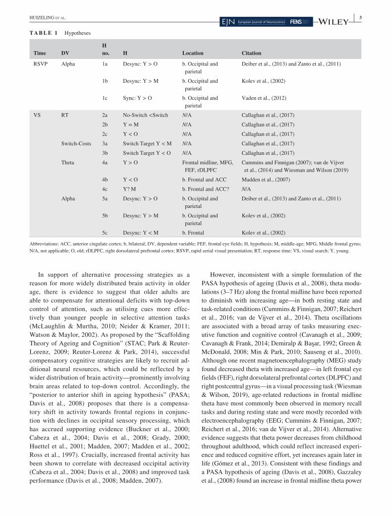
T A B L E 1 Hypotheses
Time DVH no. H Location Citation
RSVP Alpha 1a Desync: Y > O b. Occipital and parietal
Deiber et al., (2013) and Zanto et al., (2011)
1b Desync: Y > M b. Occipital and parietal
Kolev et al., (2002)
1c Sync: Y > O b. Occipital and parietal
Vaden et al., (2012)
VS RT 2a No- Switch <Switch N/A Callaghan et al., (2017)
2b Y = M N/A Callaghan et al., (2017)
2c Y < O N/A Callaghan et al., (2017)
Switch- Costs 3a Switch Target Y < M N/A Callaghan et al., (2017)
3b Switch Target Y < O N/A Callaghan et al., (2017)
Theta 4a Y > O Frontal midline, MFG, FEF, rDLPFC
Cummins and Finnigan (2007); van de Vijver et al., (2014) and Wiesman and Wilson (2019)
4b Y < O b. Frontal and ACC Madden et al., (2007)
4c Y? M b. Frontal and ACC? N/A
Alpha 5a Desync: Y > O b. Occipital and parietal
Deiber et al., (2013) and Zanto et al., (2011)
5b Desync: Y > M b. Occipital and parietal
Kolev et al., (2002)
5c Desync: Y < M b. Frontal Kolev et al., (2002)
Abbreviations: ACC, anterior cingulate cortex; b, bilateral; DV, dependent variable; FEF, frontal eye fields; H, hypothesis; M, middle- age; MFG, Middle frontal gyrus; N/A, not applicable; O, old; rDLPFC, right dorsolateral prefrontal cortex; RSVP, rapid serial visual presentation; RT, response time; VS, visual search; Y, young.
4 | HUIZELING Et aL.
in older adults when implementing a visual attention task, which could reflect an increase in the implementation of top- down attentional guidance. However, it remains unclear whether such increased activity is beneficial for performance or rather a further indication of dedifferentiation and lack of neural precision.
In light of the aforementioned inconsistencies and com-peting theoretical accounts, we set out to clarify whether impaired attentional control (refocusing from a temporal to a spatial task) in older adults (Callaghan et al., 2017) is char-acterised by an increased spread of activation or a reduced activation across cortical networks. Based on the reviewed findings, our primary focus of investigation was centred on modulations of alpha and theta frequency bands. We used MEG to increase spatial resolution over previous EEG stud-ies, whilst achieving the necessary temporal resolution for frequency- specific analysis, thus, allowing for oscillatory analysis in source space.
The aim of the current study was to investigate the oscilla-tory patterns that reflect age- related changes in the ability to switch from allocating attention in time, to allocating atten-tion spatially. Specifically, we compared age groups on their ability to switch from identifying a single target in an RSVP stream (spatially focal but temporally changing), to identify-ing a target in a VS display (spatially distributed but tempo-rally unchanging). The cost of switching from the temporal attention task to the spatial attention task was manipulated by altering the position of the target in the RSVP stream. As in Callaghan et al., (2017), the RSVP target was (a) the first item in the stream, which behaved as a No- Switch condition, because the participant was no longer required to attend to the RSVP stream once they had identified the target; (b) to-wards the end of the RSVP stream (Switch Target condition) or (c) absent from the RSVP stream (Switch No- Target con-dition), which each (both b and c) behaved as Switch condi-tions, because the participant was required to attend to the RSVP stream until (near to) the end of the stream. Based on Callaghan et al., (2017), we expected faster RTs in the No- Switch compared to the two Switch conditions and greater costs of switching in the older and middle- aged groups in comparison to the younger group, (especially in the Switch Target condition; Table 1 H3a– b).
In addition to comparing younger and older aged adults, we compared performance in a middle- aged group. Visual attention processing is understudied in middle age. However, there is some evidence to suggest that attentional con-trol is already less efficient in middle age compared to in young adults (Callaghan et al., 2017; Georgiou- Karistianis et al., 2006; Huizeling et al., 2020; Zhou, Fan, Lee, Wang, & Wang, 2011). The current study provides valuable novel insights into neural oscillatory signatures in middle age during attention switching. Middle- aged adults have been shown to display a posterior- anterior shift in ongoing (upper;
10– 15 Hz) alpha oscillations during visual processing com-pared to younger adults, although only in increased phase- locking and ongoing power and not in prestimulus alpha power, or alpha power compared to baseline, in which there were no age group differences (Kolev et al., 2002). More recently, Reuter et al., (2019) found reduced event- related potential latencies and amplitudes for middle- compared to both older- and younger- age groups during visual processing and attentional control. In the current work, it was hypothe-sised that the middle- age group could either begin to reflect a similar pattern to the older group, for example, a posterior- anterior shift in processing resources (Davis et al., 2008; Kolev et al., 2002) or show efficient processing signatures (Reuter et al., 2019), more similar to the younger group (see Table 1 H1b, H4c, H5b– c).
Based on the reviewed literature, it was expected that older adults would display age- related differences in alpha modulation during RSVP and VS processing, either through a weaker alpha power decrease (Table 1 H1a, H5a; Deiber et al., 2013; Zanto et al., 2011) that could be indicative of reduced target processing, or through a weaker alpha power increase (Table 1 H1c; Vaden et al., 2012) that could be indic-ative of reduced distractor suppression. We expected to ob-serve age differences in alpha power modulation in bilateral parietal and occipital visual attention regions (see Table 1 H1a– c, H5a– c).
We hypothesised that, in response to the VS display, there could be an increase in frontal theta activity with increased age (Table 1, H4b), either reflecting beneficial, additional top- down processing (Davis et al., 2008; Fabiani et al., 2006; Gazzaley et al., 2008; Madden, 2007) or merely reflecting dedifferentiation (Cabeza, 2002). The former would be re-flected in an improvement in performance with increased theta modulation. Conversely, in the latter, no such improve-ment in performance would be observed with increased cor-tical recruitment. Alternatively, reduced theta power could be observed over the midline, as demonstrated in previous EEG studies (Cummins & Finnigan, 2007; van de Vijver et al., 2014), or in bilateral middle frontal gyrus (MFG), FEF and right DLPFC, as demonstrated through MEG and functional magnetic resonance imaging (fMRI; Madden et al., 2007; Wiesman & Wilson, 2019).
Whilst temporal attention manifests as a sharp focus of attention to a single location, and a strong inhibition of the surrounding locations, an efficient switch to spatial atten-tion is expected to require a rapid release of these inhibition processes, combined with a sharpening of attention to the surrounding locations that were previously inhibited. We ex-pected higher level attentional control regions, such as the frontal cortex and anterior cingulate cortex (ACC; Table 1 H4a– c), as well as parietal cortex (Table 1 H1a– c, H5a– c), to be involved in coordinating such attentional control, as well as alpha synchronisation (for inhibition; Table 1 H1c),
| 5HUIZELING Et aL.
desynchronisation (for enhanced attention; Table 1 H1a– b, H5a– c) and theta increases (for increased processing; Table 1 H4a– c).
2 | METHODS
2.1 | Participants
Participants were recruited from Aston University staff and students and the community. Participants aged over 60 years were also recruited from the Aston Research Centre for Healthy Ageing (ARCHA) participation panel. Participants provided written informed consent before participating and were screened for contraindications to having an MRI or MEG scan and received standard payment according to local rules. The research was approved by Aston University Research Ethics Committee (#776) and complied with the Declaration of Helsinki.
Sixty- three participants in three age groups (19– 30, 40– 49 and 60+ years; see Table 2 for details) were included in the final analysis. An age range of 40– 49 years was selected for the middle- age group, so as to be an equal number of years apart from the youngest and oldest groups. Whilst an age range of 40– 49 years might not cover the full range of mid-dle age, it is representative of middle age and avoids debates about the exact start and end age of middle age. Participants with visual impairments, photosensitive epilepsy and a his-tory of brain injury or stroke were excluded from participa-tion. All participants in the 60+ years group (60– 82 years)
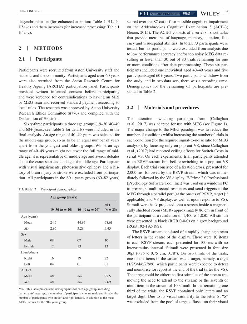
scored over the 87 cut- off for possible cognitive impairment on the Addenbrookes Cognitive Examination 3 (ACE- 3; Noone, 2015). The ACE- 3 consists of a series of short tasks that provide measures of language, memory, attention, flu-ency and visuospatial abilities. In total, 73 participants were tested, but six participants were excluded from analysis due to low performance accuracy and/or too noisy MEG data re-sulting in fewer than 30 out of 80 trials remaining for one or more conditions after data preprocessing. These six par-ticipants included one individual aged 40– 49 years and five participants aged 60+ years. Two participants withdrew from the study, and in two data sets, there was a recording error. Demographics for the remaining 63 participants are pre-sented in Table 2.
2.2 | Materials and procedures
The attention switching paradigm from (Callaghan et al., 2017) was adapted for use with MEG (see Figure 1). The major change to the MEG paradigm was to reduce the number of conditions whilst increasing the number of trials in each condition (for the required signal- to- noise ratio for MEG analysis), by focusing only on pop- out VS, since Callaghan et al., (2017) had reported ceiling effects for Switch- Costs in serial VS. On each experimental trial, participants attended to an RSVP stream first before switching to a pop- out VS display. Each trial consisted of a fixation cross, presented for 2,000 ms, followed by the RSVP stream, which was imme-diately followed by the VS display. E- Prime 2.0 Professional (Psychology Software Tool. Inc.) was used on a windows PC to present stimuli, record responses and send triggers to the MEG through a parallel port (at the onsets of RSVP, target [if applicable] and VS display, as well as upon response to VS). Stimuli were back- projected onto a screen inside a magneti-cally shielded room (MSR) approximately 86 cm in front of the participant at a resolution of 1,400 × 1,050. All stimuli were presented in black (RGB 0- 0- 0) on a grey background (RGB 192- 192- 192).
The RSVP stream consisted of a rapidly changing stream of letters in the centre of the display. There were 10 items in each RSVP stream, each presented for 100 ms with no interstimulus interval. Stimuli were presented in font size 30pt (0.75 × 0.75 cm, 0.78°). On two thirds of the trials, one of the items in the stream was a target, namely, a digit (1/2/3/4/6/7/8/9), which participants were expected to detect and memorise for report at the end of the trial (after the VS). The target could be either the first stimulus of the stream (re-moving the need to attend to the stream) or the seventh or ninth item in the stream of 10 stimuli. In the remaining one third of the trials, the RSVP contained only letters and no target digit. Due to its visual similarity to the letter S, “5” was excluded from the pool of targets. Based on their visual
T A B L E 2 Participant demographics
Age group (years)
19– 30 (n = 20) 40– 49 (n = 20)60+ (n = 23)
Age (years)
Mean 24.6 44.95 68.61
SD 2.96 3.28 5.43
Sex
Male 08 07 10
Female 12 13 13
Handedness
Right 16 19 22
Left 04 01 01
ACE- 3
Mean n/a n/a 95.5
SD n/a n/a 2.69
Note: This table presents the demographics for each age group, including participants' mean age, the number of participants who are male and female, the number of participants who are left and right handed, in addition to the mean ACE- 3 scores for the 60+ years group.

6 | HUIZELING Et aL.
similarity to certain numbers, letters I, O and S were excluded from the stream. Letters K and Z were targets defined for the VS task and were therefore also not employed as distrac-tors in the RSVP. It should be noted that the current RSVP task differs from a standard attentional blink paradigm as the RSVP stream only contained a maximum of a single target.
The VS display consisted of eight letters presented in a circle around a fixation cross in the centre of the screen, in-cluding seven distractors and one target. Participants were instructed to keep their eyes fixed on the cross at the centre of the screen, whilst they completed the VS and to respond as quickly as possible. The target letter was always either a “K” or a “Z” and distractors were always a “P.” rendering a “pop- out” VS, conforming to effects observed by Callaghan et al., (2017; see Section 1 for details). Stimuli were pre-sented in font size 20 pt (0.50 × 0.50 cm, 0.52°), and the cen-tre of each stimulus was 2.3 cm (2.40°) from the centre of the fixation cross. Participants pressed a button with their right index finger once they had identified the VS target. Note that conforming to Callaghan et al., (2017), this button press did not discriminate between K or Z but merely indicated that the participant had identified the target on that trial. Participants' RTs to press this button were recorded and allowed for a more accurate and less variable search time estimate than a dis-criminative response (for detailed discussion, see Callaghan et al., 2017). For MEG it had the added benefit that this re-sponse did not trigger different neural motor patterns (e.g., for different finger taps). Subsequently, participants pressed a button to indicate whether it was a “K” (right index finger response) or a “Z” (left index finger response) in the display. Participants were then prompted to indicate whether they had seen a target digit in the RSVP stream (yes: right index finger response; no: left index finger response). If a digit was cor-rectly detected in the RSVP stream, participants then pressed the button that corresponded with the number that they saw. Participants wore earphones through which a “ding” sound was played after a correct response and a chord sound was played after an incorrect response.
To manipulate the cost of switching, the position of the target in the RSVP stream that preceded the VS was either
the first item in the stream (No- Switch condition) or the target was either the seventh or ninth item in the stream (Switch Target condition) or absent from the stream (Switch No- Target condition). Illustrations of the RSVP stream and of the VS display are presented in Figure 1. There were 80 trials of each of the three conditions (No- Switch/Switch Target/Switch No- Target), with a total of 240 trials. To pro-vide the opportunity for breaks, trials were divided into 10 blocks. Trials were randomised within blocks. Participants completed 24 practise trials before starting the experimental trials.
MEG data were recorded with a 306- channel Elekta Neuromag system (Vectorview; Elekta) in a MSR at a sam-pling rate of 1,000 Hz. The 306 sensors were made up of 102 triplets incorporating one magnetometer and two orthogonal planar gradiometers. Data were recorded in two halves within the same session.
Head position was recorded continuously throughout data acquisition via the location of five HPI coils. Three HPI coils were positioned across the participant's forehead and one on each mastoid. The position of each HPI coil, three fidu-cial points and 300– 500 points evenly distributed across the head surface were recorded prior to the MEG recording with Polhemus Fastrak head digitisation. A T1 structural MRI was obtained for each participant and acquired using a 3T Siemens MAGNETOM Trio MRI scanner with a 32- channel head coil.
2.3 | Data analysis
2.3.1 | Response times
Participants' median VS RTs (ms) on trials where both VS and RSVP responses were correct were extracted. Participants' proportions of correct VS target identifications and RSVP target identifications were also extracted.
Differences in median VS RTs between age groups and RSVP conditions were analysed in a 3 × 3 mixed ANOVA, where RSVP condition (No- Switch/Switch Target/Switch
F I G U R E 1 Illustration of trial structure and stimulus examples. The rapid serial visual presentation (RSVP) stream illustration (left) displays a Switch Target RSVP stream (a target digit at position 7 in the RSVP). Each trial consisted of a fixation cross (2,000 ms) followed by an RSVP stream immediately followed by a pop- out visual search (VS) display (right). ISI Inter stimulus interval
| 7HUIZELING Et aL.
No- Target) was a within subjects factor and age group (19– 30, 40– 49 and 60+ years) was a between subjects factor. Multiple comparisons were corrected for with Bonferroni correction.
The data were expected to violate assumptions of equality of variance due to increases in interindividual variability with age (Hale et al., 1988; Morse, 1993), yet there is evidence to support that ANOVA is robust to violations of homogene-ity of variance (Budescu, 1982). Where Mauchly's Test of Sphericity was significant, indicating that the assumption of sphericity had been violated, Greenhouse– Geisser corrected statistics were reported.
To interpret the age group × RSVP condition interactions, “Switch- Costs” were calculated as the percentage differ-ence in RTs between Switch Target and No- Switch condi-tions (Target Switch- Costs) and between Switch No- Target and No- Switch conditions (No- Target Switch- Costs) for each individual. As interaction effects were already shown to be statistically significant in the ANOVA, Restricted Fisher's Least Significant Difference test was applied and corrections for multiple comparisons were not conducted (Snedecor & Cochran, 1967). Where Levene's test for equality in variance was significant (p < .05) when computing t tests, “Equality of variance not assumed” statistics were reported.
2.3.2 | MEG
MEG data were preprocessed in Elekta software using MaxFilter (temporal signal space separation, tSSS, 0.98 cor-relation; Taulu & Hari, 2009) to remove noise from sources inside and outside the sensor array. Seventeen participants displayed magnetic interference from dental work and so a tSSS correlation of 0.90 was applied instead. This included five participants from the 19– 30 years group, six from the 40– 49 years group and six from the 60+ years group. Movement correction was applied to one participant in the 40– 49 years group due to head movement (>7 mm).
Data were read into the Matlab® toolbox Fieldtrip version 20151004 (Oostenveld et al., 2011), with Matlab® 2015a, band- pass filtered between 0.5 and 85 Hz and epoched from 3.5 s preceding VS onset (i.e., 2.5s preceding RSVP stream onset) to 2.0 s after the onset of the VS display. Trials were visually inspected for artefacts, and any noisy trials were removed. Fieldtrip version 20161031 was used for further analysis.
Trials with incorrect responses were excluded. After ex-cluding inaccurate and noisy trials, the mean number of trials that remained for each condition was 68.93 (SD = 6.58) for the 19– 30 years group, 67.97 (SD = 7.51) for the 40– 49 years group and 60.36 (SD = 9.85) for the 60+ years group. Participants with fewer than 30 trials were excluded from the analysis (see Methods: Participants section).
2.3.3 | Sensor level analysis
For data cleaning prior to sensor level analysis, noisy sensors were interpolated with the average of neighbouring sensors. Independent components analyses (ICA) were implemented for each participant, across all conditions, and components with eye blink or heartbeat signatures were removed from the data.
Time- frequency analysis was carried out on signals from the planar gradient representation of 102 gradiometer pairs using a Hanning taper from 2– 30 Hz (for every 1 Hz), with four cycles per time window in stages of 50 ms. For each participant, trials were averaged within each condition (No- Switch/Switch Target/Switch No- Target).
To investigate the direction of task- related changes in oscillatory power in each age group, thereby improving in-terpretability of subsequent source level effects, we com-pared “active” task periods (3– 5 Hz: 550– 1,550 ms relative to RSVP onset; 10– 14 Hz: 450– 950 ms and 1,000– 1,500 ms relative to RSVP onset; see Source level analysis section for details) to a baseline period (3– 5 Hz: −1,500 to −500 ms; 10– 14 Hz: −1,000 to −500 ms). Conditions were collapsed to obtain the average across all conditions. Two- tailed de-pendent t tests were carried out to compare the active task periods with a baseline period separately for each age group. Multiple comparisons were corrected for using nonparamet-ric cluster permutations (Maris & Oostenveld, 2007), with 2,000 permutations (cluster alpha = 0.05).
2.3.4 | Source level analysis
For source localisation using spatial filters (beamformers), noisy sensors were excluded rather than interpolated and ICA was not implemented to remove eye blinks and cardio arte-facts. Due to size restrictions of the MEG data file, each data set was recorded in two halves within the same session and were therefore MaxFiltered separately prior to concatenating the data, which could lead to different components being re-moved in each half of data (see MaxFilter details in above MEG section). To reduce potential artefacts due to applying MaxFiltering to the two halves of data separately, a principle components analysis was implemented to reduce data dimen-sionality to components that accounted for 99% of the vari-ance. The participant remained in the scanner, and the door to the MSR remained shut between recording the two halves of data.
Using an in- house Matlab script and Elekta software MRI Lab, individual MRIs were aligned with the sensor array, by aligning the individual's MRI with the fiducial positions and head shape that were recorded with Polhemus Fastrak head digitisation. Individual single- shell head- models (5- mm vox-els) were created from the coregistered MRIs. Head- models
8 | HUIZELING Et aL.
were normalised to MNI space (Montreal Neurological Institute template).
To identify the cortical generators of sensor level fre-quency modulations, we extracted time- frequency tiles from the time frequency representations (TFRs) in Figure 3, se-lecting 3– 5 Hz with a time window of 550– 1,550 ms (relative to RSVP onset) and 10– 14 Hz with a time window of 450– 950 and 1,000– 1,500 ms (relative to RSVP onset) for theta and alpha frequencies, respectively. Note that this does not inflate type- 1 error rates, as the selection was not made by contrasting conditions or age groups but rather on the over-all pattern across all conditions and groups. A 3– 5 Hz theta range (selected from inspection of Figure 3) is lower than a typical theta band of 4– 7 Hz and overlaps with typical delta frequency (0– 4 Hz), however, is in line with early accounts of a theta response (3– 6 Hz) to visual stimuli (Demiralp & Başar, 1992). Similarly, 10– 14 Hz is higher than a typical alpha range of 8– 12 Hz but is consistent with a range in which effects have previously been found in visual processing stud-ies (Vaden et al., 2012). Moreover, the overlap between alpha and theta frequency ranges was minimised in order to capture distinct processing. To estimate theta frequency (centred at 4 Hz), we required a time window of 1,000 ms. Given that the average VS RT of the younger participants was 550 ms in the No- Switch condition, we avoided selecting a time win-dow that went beyond 550 ms after VS onset (1,550 ms after RSVP onset), which would be contaminated with processing of, and response to, the follow- up question, “Was the letter a K or a Z?”. To estimate alpha frequency (centred at 12 Hz), we required a time window of 500 ms. For alpha, it was there-fore possible to select two separate time windows (theta did not allow for such temporal resolution) to capture the two distinct phases of each trial (temporal and spatial tasks). The first alpha time window (450– 950 ms) was selected to cap-ture processing of the end of the RSVP stream and the prepa-ration to switch whilst avoiding any spectral leakage from the onset of the VS display. This window included the RSVP target in the Switch Target condition, which was presented at either 700 or 900 ms. The second alpha time window (1,000– 1,500 ms) was time- locked to the onset of the VS display to capture VS processing immediately after the switch.
Frequency band specific Dynamic Imaging of Coherent Sources (DICS; Gross et al., 2001) beamformers (spatial filters; 2% lambda regularisation) were calculated based on cross- spectral densities obtained from the fast Fourier transform (FFT) of signals from 204 gradiometers using a Hanning taper, spectral smoothing of ±2 Hz and 2.0 s of data padding. No baseline correction was applied, and conditions were directly compared instead. Note that although group differences were also present in the beta frequency band (15– 25 Hz), our hypotheses focused on alpha and theta bands based on the previous literature (see Introduction). Surface
plots in Figures 5, 6 and 8– 11 were plotted in BrainNet Viewer 1.63 (Xia et al., 2013).
Two- tailed dependent t tests were carried out to compare each of the Switch conditions (Switch Target/Switch No- Target) with the No- Switch condition separately for each age group. Multiple comparisons were corrected for with non-parametric cluster permutations (Maris & Oostenveld, 2007). Second level analysis was carried out by comparing Switch- Costs at the group level (Bögels et al., 2014; Wang et al., 2016). For each participant, the No- Switch condition was subtracted from each of the Switch conditions separately. These differences were entered into two two- tailed indepen-dent cluster permutation t tests (2,000 permutations) to com-pare age groups (19– 30 years vs. 40– 49 years/19– 30 years vs. 60+ years).
To explore the relationship between behavioural perfor-mance and power changes in theta and alpha frequencies in the two older groups (to better understand cognitive de-cline), differences in power (at peaks of the main significant clusters of the source analysis) between Switch Target and No- Switch conditions, in theta and alpha power, were en-tered into one- sided Spearman's correlation analyses with behavioural RT Target Switch- Costs. Power change was ex-tracted from single voxels in which the strongest effect of switching was observed (as per the t- statistics presented in Figures 5, 8 and 10a). Coordinates of peaks were visually inspected to ensure they were indeed in the centre of the most prominent clusters. As no significant age group differences were found in No- Target Switch- Costs, we focused only on correlations between power change in the Switch Target con-dition (compared to No- Switch) and Target Switch- Costs in RT. Similarly, as the youngest group showed no significant difference in RTs between Switch and No- Switch conditions, and because we were specifically interested in understanding impaired switching performance in the two older groups, we focused on correlations in only the middle- and older- aged groups. To investigate possible correlations between be-haviour and residual activity in regions shown to be involved in younger but not older groups, power change at the younger group's cluster peaks were additionally entered into the older groups' correlation analyses. Bonferroni correction was used to adjust the level of alpha to control for the number of cor-relations (the number of correlations is described below).
For the correlation of theta power change with Target Switch- Costs, one (parietal) region of interest (ROI) was chosen from the source level analysis (presented in Figure 5) from the 19– 30 years group (reflecting residual activity in the older groups), in addition to two ROIs from each of the older and middle- aged groups. This resulted in six correla-tions for theta power in total, with three for each (older) age group (i.e., 60+ years: parietal, MFG and temporal lobe; 40– 49 years: parietal, ACC and occipital lobe).

| 9HUIZELING Et aL.
For the correlation of Target Switch- Costs and alpha power change during the RSVP window, one (parietal) ROI was chosen from the source level analysis of the 19– 30 years group, and one ROI was chosen for each of the older and middle- aged groups (from the source level analysis pre-sented in Figure 8). This resulted in four correlations, with two for each (older) age group (i.e., 60+ years: parietal and superior temporal gyrus; 40– 49 years: parietal and posterior parietal).
For the correlation of Target Switch- Costs with alpha power change during the VS window, one (inferior frontal gyrus; IFG) ROI was chosen from the source level analysis from the 19– 30 years group, and one ROI was chosen for each of the older and middle- aged groups (from the source level analysis presented in Figure 10). This resulted in four correlations, with two for each (older) age group (i.e., 60+ years: IFG and cerebellum; 40– 49 years: IFG and cerebel-lum). Coordinates for the selected peaks can be found in Table S1– S3 in the supporting information.
3 | RESULTS
3.1 | Response times: switch- costs
All groups correctly identified over 96% of VS targets in all three conditions. Thus, no further analysis was carried out on VS accuracy. All groups correctly identified over 73% of RSVP targets in both RSVP conditions. RSVP accuracy was unrelated to the aims and hypotheses of the current study, and no further analysis was carried out on RSVP accuracy. The proportion of correct RSVP target identifications in the two
Target conditions is presented in Figure S1. Group means of participants' median VS RTs are presented in Figure 2.
The 3 × 3 (RSVP condition × age group) mixed ANOVA on participants' median VS RTs revealed a significant main effect of age (F(2, 60) = 11.36, p < .001, η²p = .28), a sig-nificant main effect of RSVP condition (F(2, 120) = 35.21, p < .001, η²p = .37) and a significant interaction between age and RSVP condition (F(4, 120) = 7.05, p < .001, η²p = .19).
Post hoc comparisons revealed that the main effect of age resulted from significantly slower RTs in the 60+ years group in comparison to both the 19– 30 (p < .001) and 40– 49 years (p = .029) groups. There was no significant difference be-tween the 19– 30 and 40– 49 years groups (p > .10).
The main effect of RSVP condition resulted from signifi-cantly slower RTs in both the Switch Target (p < .001) and Switch No- Target (p < .001) conditions in comparison to the No- Switch condition. There was no significant difference in RTs between the Switch Target and Switch No- Target condi-tions (p > .10).
To investigate the hypothesis that there would be signifi-cantly greater Switch- Costs in both the 40– 49 and 60+ years groups in comparison to the 19– 30 years group and to in-terpret the interaction between age and RSVP condition, in-dependent t tests were carried out comparing Switch- Costs across age groups. Please refer to Methods for a description of how Switch- Costs were calculated for each participant. Means and SDs of participants' Switch- Costs are presented in Table 3.
Switch- Costs, when the target was presented towards the end of the RSVP (Switch Target), were significantly greater in both the 40– 49 (df = 38, t = −3.45, p < .001) and 60+ (df = 41, t = −5.15, p < .001) years groups in comparison to
F I G U R E 2 Group means of participants' median visual search (VS) response times (RTs). Vertical bars represent the SE. The figure was created in R (R Core Team, 2018) using the RainCloudPlots package (Allen et al., 2021)

10 | HUIZELING Et aL.
the 19– 30 years group. There were no significant age group differences in Switch- Costs (p > .10), when no target was presented in the RSVP (Switch No- Target).
The RT results replicated findings from Callaghan et al., (2017) by demonstrating deficits in switching in both the 40– 49 years and 60+ years groups in comparison to the 19– 30 years group. Consistent with Callaghan et al., (2017), greater Switch- Costs in the older age groups were only significant when participants were required to process a target digit before switching. When there was no target in the RSVP stream, older participants seemed better able to switch from the temporal to the spatial attention task, suggesting either an increased de-mand for more processing resources or differences in strategies
used to switch when target consolidation was required. To im-prove our understanding of the neurocognitive processing used to switch between modalities of attention across the three age groups, in the following sections, we will investigate group dif-ferences in task- related oscillatory signatures.
3.2 | MEG results
Frequencies from 2– 30 Hz were explored, and TFRs are shown in Figure 3, averaged over a group of posterior sen-sors for visualisation purposes only. Although a similar pat-tern was seen across frontal sensors (see Figure S3), here we present posterior sensors due to the improved signal- to- noise ratio compared to frontal sensors. Note that although group differences may also be present in the beta frequency band (15– 25 hz), our hypotheses focused on alpha and theta bands based on previous literature (see Section 1) and we therefore omitted beta in our analysis.
3.2.1 | Theta power
The TFRs in Figure 3 and sensor level analysis in Figure 4 illustrate that there was a theta increase in response to the
T A B L E 3 Means and SDs of Switch- Costs for each age group
Age group (years)
19– 30 (n = 20) 40– 49 (n = 20) 60+ (n = 23)
Target Switch- Costs
Mean 4.02 19.67 26.65
SD 12.72 15.78 15.67
No- target Switch- Costs
Mean 12.59 17.29 17.98
SD 15.24 15.66 18.43
F I G U R E 3 Time frequency representations (TFRs) present power in relation to a baseline period of −0.6 to −0.01 s in a group of four posterior gradiometer pairs (gradiometer pair positions are illustrated as black dots on an empty topographical plot of the magnetoencephalography (MEG) helmet, top- right corner of the figure). The onset of the rapid serial visual presentation (RSVP) stream occurred at 0.0 s. Black lines placed over TFRs indicate the onset of the visual search (VS) display, and RSVP target onset occurred at either 0.7 or 0.9 s

| 11HUIZELING Et aL.
VS display, relative to baseline, in a time window of 550– 1,550 ms, in all age groups, which was significantly greater than baseline in the 19– 30 and 40– 49 years groups. Statistical results comparing theta power in Switch Target and No- Switch conditions and exploring the interaction between RSVP condition and age group are presented in Figure 5 (for details, see Methods, Section 2).
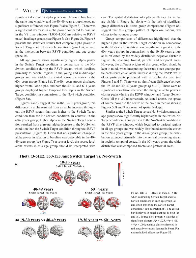
Figure 5 illustrates that all age groups displayed a signifi-cantly higher theta increase in the Switch Target condition in comparison to the No- Switch condition, which localised to superior and inferior parietal gyri, occipital gyri and the MFG in the 19– 30 years group, bilateral frontal cortex and the ACC in the 40– 49 years group and the superior frontal gyrus, temporal gyri and the cerebellum in the 60+ years group (Figure 5a). In summary, the 19– 30 years group dis-played higher theta in parietal regions, and the two older groups demonstrated extensive frontal recruitment. The correlation between increased left MFG theta power and decreased Switch- Costs in the 60+ years group (r = −0.40, p = .029) did not reach significance using a more stringent alpha level of p < .008 after Bonferroni correction to control for the number of tests performed (n = 6). The 60+ years group additionally displayed higher temporal lobe theta. The 40– 49 years group additionally presented with a posterior (occipital/cerebellar) negative cluster, which reflects lower theta in the Switch Target condition in comparison to the No- Switch condition, although this did not reach significance in a two- sided test (p = .033). Note that the spread of source power to the centre of the brain in medial slices in Figures 5, 8 and 9 is likely to be a result of spatial leakage, a known challenge in the spatial resolution of MEG source analysis, particularly towards the centre of the brain, where the signal- to- noise ratio is lower and source estimation is less precise (Hillebrand & Barnes, 2002).
Age- group comparisons of differences between Switch Target and No- Switch conditions, which are presented in Figure 5b, confirmed that the higher theta increase in the Switch Target condition was greater in the 19– 30 years group in parietal regions in comparison to the 40– 49 years group
(p = .020) and the 60+ years group (p = .075), although the latter did not reach significance. These positive clusters ad-ditionally extended to occipital cortex, resulting from lower theta in the Switch Target condition in comparison to the No- Switch condition in the older groups but not the younger group. In the 60+ years group, greater theta power increases in the parietal region were associated with decreased Target RT- Switch- Costs (r = −0.53, p = .005). Importantly, due to reduced parietal theta in the 60+ years group overall (Figure 5b), the coordinates for the parietal correlation effect were adopted from the 19– 30 years group, in order to specif-ically investigate whether residual theta power in the oldest participants would be beneficial for attention switching. No such correlations were observed for the middle- aged group (p > .10 uncorrected).
Figure 6a reveals that there was no significant differ-ence between Switch No- Target and No- Switch conditions in theta frequency in the 19– 30 years group, suggesting that the differences observed in theta between Switch Target and No- Switch conditions in this age group (see Figure 5) were a result of processing the RSVP target in the Switch Target condition.
In contrast, both the 40– 49 and 60+ years groups again display negative clusters that localise to the occipital lobes, indicating deficient theta increases in the Switch No- Target condition, a finding that cannot be due to RSVP target pro-cessing. The 60+ years group again showed higher theta in the Switch No- Target condition in comparison to the No- Switch condition that localised to frontal regions and the left temporal lobe. However, group differences did not reach sig-nificance for a two- sided test (Figure 6b).
3.2.2 | Alpha power
There was a nonsignificant increase in alpha power in rela-tion to baseline in the 450– 950 ms time window (relative to RSVP onset) in the 19– 30 years group (see Figure 7; also Figure 3). In contrast, the 60+ years group showed a
F I G U R E 4 Effects in theta (3– 5 Hz) when contrasting Switch period (550– 1,550 ms; collapsed across all three rapid serial visual presentation [RSVP] conditions) to the baseline period (−1,500 to −500 ms), for each age group. Sensor topographies present t- statistics of significant clusters (*p < .025, ***p < .001; positive clusters denoted in red)
12 | HUIZELING Et aL.
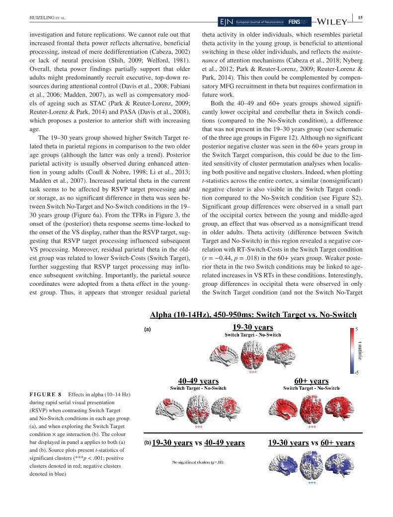
significant decrease in alpha power in relation to baseline in the same time window, and the 40– 49 years group showed no significant difference (see Figure 7; also Figure 3). There was a significant decrease in alpha power compared to baseline in the VS time window (1,000– 1,500 ms relative to RSVP onset) in all age groups (see Figure 7; also Figure 3). Figure 8 presents the statistical results that compare alpha power in Switch Target and No- Switch conditions (panel a), as well as the interaction between RSVP condition and age group (panel b).
All age groups show significantly higher alpha power in the Switch Target condition in comparison to the No- Switch condition during the RSVP stream, which localised primarily to parietal regions in the young and middle- aged groups and was widely distributed across the cortex in the 60+ years group (Figure 8a). The 60+ years groups displayed higher frontal lobe alpha, and both the 40– 49 and 60+ years groups displayed higher temporal lobe alpha in the Switch Target condition in comparison to the No- Switch condition (Figure 8a).
Figures 3 and 7 suggest that, in the 19– 30 years group, this difference in alpha resulted from an alpha increase through-out the RSVP stream that was higher in the Switch Target condition than the No- Switch condition. In contrast, in the 60+ years group, higher alpha in the Switch Target condi-tion resulted from a greater alpha decrease in the No- Switch condition than the Switch Target condition throughout RSVP presentation (Figure 3). Given that no significant change in alpha power in relation to baseline was detectable in the 40– 49 years group (see Figure 7) at sensor level, the source level alpha effects in this age group should be interpreted with
care. The spatial distribution of alpha oscillatory effects that are visible in Figure 8a, along with the lack of significant group differences in direct group comparisons (Figure 8b), suggest that this group's pattern of alpha oscillations, was closer to the younger group.
Group comparisons of differences highlighted that the higher alpha in the Switch Target condition in comparison to the No- Switch condition was significantly greater in the 60+ years groups in comparison to the 19– 30 years group, as is reflected by the widely distributed negative cluster in Figure 8b, spanning frontal, parietal and temporal areas. However, the different origins of this group effect should be kept in mind, when interpreting the result, since younger par-ticipants revealed an alpha increase during the RSVP, whilst older participants presented with an alpha decrease (see Figures 3 and 7). There was no significant difference between the 19– 30 and 40– 49 years groups (p > .10). There were no significant correlations between the change in alpha power at cluster peaks (during the RSVP window) and Target Switch- Costs (all p > .10 uncorrected). As stated above, the spread of source power to the centre of the brain in medial slices in Figures 5, 8 and 9 is a result of spatial leakage.
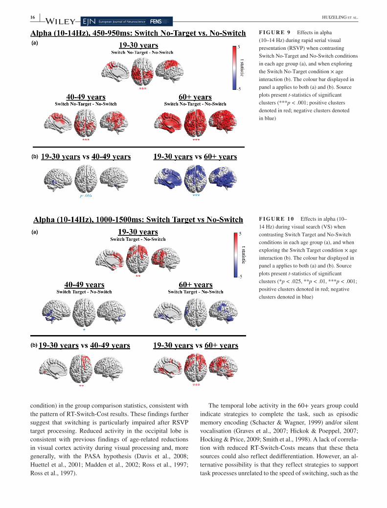
Similar to the Switch Target versus No- Switch contrast, all age groups show significantly higher alpha in the Switch No- Target condition in comparison to the No- Switch condition in the RSVP time window, which localised to parietal regions in all age groups and was widely distributed across the cortex in the 60+ years group. In the 40– 49 years group, the distri-bution extended primarily into the ventral processing stream in occipito- temporal cortex. In the 60+ years group the wider distribution also comprised frontal and prefrontal areas.
F I G U R E 5 Effects in theta (3– 5 Hz) when contrasting Switch Target and No- Switch conditions in each age group (a), and when exploring the Switch Target condition × age interaction (b). The colour bar displayed in panel a applies to both (a) and (b). Source plots present t- statistics of significant clusters (*p < .025, **p < .01, ***p < .001; positive clusters denoted in red; negative clusters denoted in blue). For unthresholded effects see Figure S2

| 13HUIZELING Et aL.
Similar to the pattern seen when comparing Switch Target and No- Switch conditions in Figure 8, lower alpha in the No- Switch condition in comparison to the Switch No- Target con-dition appears to have resulted from a greater alpha increase in the Switch No- Target condition in the 19– 30 years group and a greater alpha decrease in the No- Switch condition in the 60+ years group (see Figures 3 and 7), which is import-ant to consider when interpreting intragroup and intergroup effects.
Group comparisons revealed that the higher alpha in the Switch No- Target condition in comparison to the No- Switch condition was significantly higher in the 60+ years group in comparison to the 19– 30 years group, as is reflected by the negative clusters in Figure 9b. Group differences between the 19– 30 and 40– 49 years groups did not reach significance (p = .056). Whilst alpha effects were contained to parietal regions in the 19– 30 years group, in the 60+ years group the higher alpha effects were both stronger and more widely dis-tributed across the cortex.
In response to VS onset, the 19– 30 years group displayed a greater alpha decrease in the No- Switch compared to the Switch Target condition in frontal cortex (Figure 10a). In con-trast, the 40– 49 years and 60+ years groups show a greater alpha decrease in the Switch Target condition compared to the No- Switch condition in parietal cortex and cerebellum. Group comparisons demonstrated that such group differences were significant (Figure 10b). There were no significant cor-relations between the change in alpha power at cluster peaks (during the VS window) and Target Switch- Costs (p > .10 uncorrected).
In response to VS onset, both the 19– 30 years and 60+ years groups displayed a greater alpha decrease in left frontal and parietal cortex in the No- Switch compared to the
Switch No- Target condition (Figure 11a). The 40– 49 years group showed a greater alpha decrease in occipital cortex in the Switch No- Target condition compared to the No- Switch condition. The cluster in the 40– 49 years group also extends to the cerebellum; however, the estimation of sources close to the edge of the sensor array can be poor and should be interpreted with caution, as this could be a result of spatial leakage.
Group differences between the 19– 30 years and 40– 49 years groups did not reach significance in the group com-parisons (Figure 11b), nor did the greater alpha decrease in left frontal cortex in the No- Switch compared to the Switch No- Target condition in the 19– 30 years group compared to the 60+ years group.
4 | DISCUSSION
In our previous work, we demonstrated that older adults find refocusing attention from time to space more difficult than younger adults (Callaghan et al., 2017). In the current study, we replicated these results and found that the older (60+) as well as the middle- aged (40– 49) group had increased Switch- Costs compared to the younger (19– 30) group, as reflected by disproportionately increased RTs when required to refocus at-tention from a temporal RSVP task (when it included a target) to a spatial VS task. Age group differences cannot be attributed to spatial attention deficits, as age group differences are typi-cally absent for pop- out VS (beyond general slowing; Bennett et al., 2012; Foster et al., 1995; Humphrey & Kramer, 1997; Nagamatsu et al., 2013; Plude & Doussard- Roosevelt, 1989), which was used here. The primary aim of the current study was to investigate the age- related changes in neural mechanisms
F I G U R E 6 Effects in theta (3– 5 Hz) when contrasting Switch No- Target and No- Switch conditions in each age group (a), and when exploring the Switch No- Target condition × age interaction (b). The colour bar displayed in panel a applies to both (a) and (b). Source plots present t- statistics of significant clusters (**p < .01, **p < .001; positive clusters denoted in red; negative clusters denoted in blue)

14 | HUIZELING Et aL.
that may underlie this difficulty in refocusing attention from events changing in time to stimuli distributed spatially. We aimed to determine whether changes in attention refocusing are characterised by a reduced activation across cortical net-works or an increased spread of activation, which could reflect either increased compensation or dedifferentiation.
Also consistent with Callaghan et al., (2017), RTs of the 60+ years group were slower overall in comparison to the 19– 30 years group. On the other hand, RTs of the 40– 49 and 19– 30 years groups did not significantly differ, implying that the 40– 49 years group found the baseline No- Switch condition no more demanding than younger adults. However, the 40– 49 years group again presented significantly higher Switch- Costs than the 19– 30 years group, suggesting that they found the Switch Target condition disproportionality more demanding than the No- Switch condition, contrasting with the 19– 30 years group. The 40– 49 years group indeed seems to represent an intermediate stage of ageing, where some aspects of attentional control function at a similar level to younger adults, whereas other aspects coincide more with patterns observed in older adults, as observed in both RTs and neural oscillations.
Conforming to our hypotheses based on previous reports (Cummins & Finnigan, 2007; Deiber et al., 2013; Gazzaley et al., 2008; Vaden et al., 2012; van de Vijver et al., 2014), we indeed observed modulations of theta and alpha oscilla-tory power (Figures 3– 11). The enhanced spatial resolution of MEG compared to EEG allowed us to go beyond the previ-ous literature to investigate group differences in source space.
4.1 | Theta
The hypothesis that there would be reduced theta power with increased age (Table 1 H4a) was partially supported. There
was a theta increase after VS onset in all age groups, which is typically seen during the processing of a visual stimulus (e.g., Demiralp & Başar, 1992; Wiesman & Wilson, 2019). By comparing theta across Switch and No- Switch condi-tions, we can investigate the effect switching has on process-ing the VS display, as illustrated in Figure 12. We observed increased theta in the Switch conditions relative to the No- Switch condition in all age groups. In the youngest group, the dominant pattern of theta oscillations was an increase in parietal theta. In the two older groups, the dominant pattern was a theta increase in frontal (middle- aged and older group) and temporal (older group) regions, accompanied by weaker occipital theta in the Switch compared to No- Switch condi-tions (a schematic of these results is presented in Figure 12). However, only differences in parietal and occipital theta were significantly different from younger adults in the middle- aged group in the direct group comparisons (Switch Target condi-tion), effects that were observed as nonsignificant trends in the oldest group. Increased temporal lobe theta in the oldest group compared to the youngest group was again observed as a nonsignificant trend in the group comparisons (Switch No- Target).
Our findings do not support previous findings of a reduc-tion in frontal midline theta power, as indicated by several previous EEG reports (Cummins & Finnigan, 2007; Reichert et al., 2016; van de Vijver et al., 2014). An increase in frontal midline theta with increased age has, instead, previously been observed by Gazzaley et al., (2008). Although the observed correlation between reduced Switch- Costs and higher MFG theta power did not reach significance with a conservative Bonferroni correction, due to insufficient power, and thus cannot be regarded as reliable at the current stage, the strength of the correlation (with a medium effect size, of r = −0.40; Cohen, 1992) indicates that it is worthy to guide further
F I G U R E 7 Effects in alpha (10– 14 Hz) when contrasting an rapid serial visual presentation (RSVP) window (450– 950 ms) and the visual search (VS) onset window (1,000– 1,500 ms) to the baseline period (−1,000 to −500 ms), for each age group, collapsed across conditions. Sensor topographies present t- statistics of significant clusters (**p < .01, ***p < .001; positive clusters denoted in red; negative clusters denoted in blue)
| 15HUIZELING Et aL.
investigation and future replications. We cannot rule out that increased frontal theta power reflects alternative, beneficial processing, instead of mere dedifferentiation (Cabeza, 2002) or lack of neural precision (Shih, 2009; Welford, 1981). Overall, theta power findings partially support that older adults might predominantly recruit executive, top- down re-sources during attentional control (Davis et al., 2008; Fabiani et al., 2006; Madden, 2007), as well as compensatory mod-els of ageing such as STAC (Park & Reuter- Lorenz, 2009; Reuter- Lorenz & Park, 2014) and PASA (Davis et al., 2008), which proposes a posterior to anterior shift with increasing age.
The 19– 30 years group showed higher Switch Target re-lated theta in parietal regions in comparison to the two older age groups (although the latter was only a trend). Posterior parietal activity is usually observed during enhanced atten-tion in young adults (Coull & Nobre, 1998; Li et al., 2013; Madden et al., 2007). Increased parietal theta in the current task seems to be affected by RSVP target processing and/or storage, as no significant difference in theta was seen be-tween Switch No- Target and No- Switch conditions in the 19– 30 years group (Figure 6a). From the TFRs in Figure 3, the onset of the (posterior) theta response seems time- locked to the onset of the VS display, rather than the RSVP target, sug-gesting that RSVP target processing influenced subsequent VS processing. Moreover, residual parietal theta in the old-est group was related to lower Switch- Costs (Switch Target), further suggesting that RSVP target processing may influ-ence subsequent switching. Importantly, the parietal source coordinates were adopted from a theta effect in the young-est group. Thus, it appears that stronger residual parietal
theta activity in older individuals, which resembles parietal theta activity in the young group, is beneficial to attentional switching in these older individuals, and reflects the mainte-nance of attention mechanisms (Cabeza et al., 2018; Nyberg et al., 2012; Park & Reuter- Lorenz, 2009; Reuter- Lorenz & Park, 2014). This then could be complemented by compen-satory MFG recruitment in theta but requires confirmation in future work.
Both the 40– 49 and 60+ years groups showed signifi-cantly lower occipital and cerebellar theta in Switch condi-tions (compared to the No- Switch condition), a difference that was not present in the 19– 30 years group (see schematic of the three age groups in Figure 12). Although no significant posterior negative cluster was seen in the 60+ years group in the Switch Target comparison, this could be due to the lim-ited sensitivity of cluster permutation analyses when localis-ing both positive and negative clusters. Indeed, when plotting t- statistics across the entire cortex, a similar (nonsignificant) negative cluster is also visible in the Switch Target condi-tion compared to the No- Switch condition (see Figure S2). Significant group differences were observed in a small part of the occipital cortex between the young and middle- aged group, an effect that was observed as a nonsignificant trend in older adults. Theta activity (difference between Switch Target and No- Switch) in this region revealed a negative cor-relation with RT- Switch- Costs in the Switch Target condition (r = −0.44, p = .018) in the 60+ years group. Weaker poste-rior theta in the two Switch conditions may be linked to age- related increases in VS RTs in these conditions. Interestingly, group differences in occipital theta were observed in only the Switch Target condition (and not the Switch No- Target
F I G U R E 8 Effects in alpha (10– 14 Hz) during rapid serial visual presentation (RSVP) when contrasting Switch Target and No- Switch conditions in each age group (a), and when exploring the Switch Target condition × age interaction (b). The colour bar displayed in panel a applies to both (a) and (b). Source plots present t- statistics of significant clusters (***p < .001; positive clusters denoted in red; negative clusters denoted in blue)
16 | HUIZELING Et aL.
condition) in the group comparison statistics, consistent with the pattern of RT- Switch- Cost results. These findings further suggest that switching is particularly impaired after RSVP target processing. Reduced activity in the occipital lobe is consistent with previous findings of age- related reductions in visual cortex activity during visual processing and, more generally, with the PASA hypothesis (Davis et al., 2008; Huettel et al., 2001; Madden et al., 2002; Ross et al., 1997; Ross et al., 1997).
The temporal lobe activity in the 60+ years group could indicate strategies to complete the task, such as episodic memory encoding (Schacter & Wagner, 1999) and/or silent vocalisation (Graves et al., 2007; Hickok & Poeppel, 2007; Hocking & Price, 2009; Smith et al., 1998). A lack of correla-tion with reduced RT- Switch- Costs means that these theta sources could also reflect dedifferentiation. However, an al-ternative possibility is that they reflect strategies to support task processes unrelated to the speed of switching, such as the
F I G U R E 9 Effects in alpha (10– 14 Hz) during rapid serial visual presentation (RSVP) when contrasting Switch No- Target and No- Switch conditions in each age group (a), and when exploring the Switch No- Target condition × age interaction (b). The colour bar displayed in panel a applies to both (a) and (b). Source plots present t- statistics of significant clusters (***p < .001; positive clusters denoted in red; negative clusters denoted in blue)
F I G U R E 1 0 Effects in alpha (10– 14 Hz) during visual search (VS) when contrasting Switch Target and No- Switch conditions in each age group (a), and when exploring the Switch Target condition × age interaction (b). The colour bar displayed in panel a applies to both (a) and (b). Source plots present t- statistics of significant clusters (*p < .025, **p < .01, ***p < .001; positive clusters denoted in red; negative clusters denoted in blue)

| 17HUIZELING Et aL.
maintenance of task goals or storing targets in working mem-ory (e.g., silent vocalisations could facilitate either target re-call or the maintenance of task goals). Anecdotally, whilst collecting behavioural data outside of the scanner (Callaghan et al., 2017), the first author noticed that older participants have a tendency to speak quietly to themselves during the task (e.g., whispering the target). It could be that a similar strategy was applied silently in the MEG scanner (in which they were instructed not to speak and keep their face still and relaxed). However, such an explanation is speculative and re-quires further investigation.
4.2 | Alpha
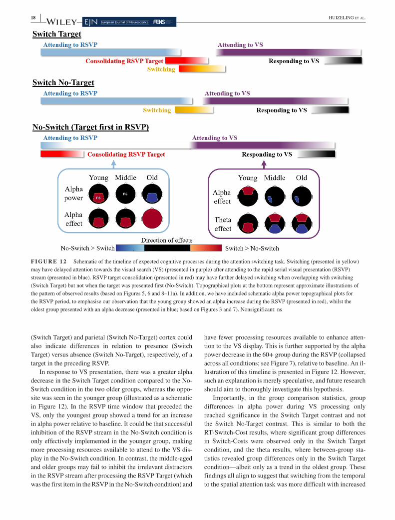
As anticipated, there were age- related changes in task related alpha modulation during both the RSVP and VS time win-dows (Table 1 H1a– c, H5a– c). During the RSVP window, in-stead of showing an alpha increase to inhibit irrelevant visual information (Vaden et al., 2012), the 60+ years age group showed an alpha decrease. This stronger and widely dis-tributed alpha desynchronization (Figures 3 and 7– 9) could reflect an enhanced attention strategy rather than an inhibi-tion strategy. A lack of alpha synchronisation is in line with the hypothesis of reduced inhibition in older adults (Adamo et al., 2003). Such group differences in temporal attention strategies may have impaired the older groups' ability to effi-ciently switch to the spatial attention task, as fewer attentional resources were available to refocus attention (illustrated in Figure 12). In other words, it is possible that slower switch-ing resulted from a deficit in temporal attention. However, the pattern of alpha power modulation during the RSVP time
window appeared to be very similar when the target was both present (Switch Target) and absent (Switch No- Target) from the RSVP stream, across all groups. In contrast, age group differences in Switch- Costs were only observed in the Switch Target condition. It therefore seems unlikely that a deficit in temporal attention alone can explain difficulties in switching. Furthermore, there is some evidence to suggest that increased Switch- Costs are also observed when switching from spatial to temporal attention (Jefferies et al., 2015), but future work should aim to provide further confirmatory evidence.
The middle- aged group presented an intermediate pattern of alpha power at sensor level (Figure 3), where no signifi-cant difference in alpha power from baseline was detectable (collapsed across all conditions; see Figure 7), which differed from a significant alpha increase in the younger adults and a significant alpha decrease in the older adults. In source space (RSVP time window), the pattern of switch- costs in the middle- aged group was again somewhere between the younger and older groups, where alpha modulation was addi-tionally observed in the temporal lobe, but this wider distri-bution did not reach significance in direct group comparisons (Figures 8 and 9).
During the VS window, all groups displayed an alpha de-synchronization in relation to baseline. In the younger group, the alpha desynchronization was greater in the No- Switch compared to both Switch conditions but predominantly local-ised to frontal cortex in the Switch Target condition and left parietal cortex in the Switch No- Target contrast. It could be that this greater alpha desynchronization in frontal and pari-etal regions reflects increased resources available to attend to the VS in the No- Switch condition compared to the Switch conditions. In addition, the distinct localisations to frontal
F I G U R E 1 1 Effects in alpha (10– 14 Hz) during visual search (VS) when contrasting Switch No- Target and No- Switch conditions in each age group (a), and when exploring the Switch No- Target condition × age interaction (b). The colour bar displayed in panel a applies to both (a) and (b). Source plots present t- statistics of significant clusters (*p < .025, **p < .01; positive clusters denoted in red; negative clusters denoted in blue)
18 | HUIZELING Et aL.
(Switch Target) and parietal (Switch No- Target) cortex could also indicate differences in relation to presence (Switch Target) versus absence (Switch No- Target), respectively, of a target in the preceding RSVP.
In response to VS presentation, there was a greater alpha decrease in the Switch Target condition compared to the No- Switch condition in the two older groups, whereas the oppo-site was seen in the younger group (illustrated as a schematic in Figure 12). In the RSVP time window that preceded the VS, only the youngest group showed a trend for an increase in alpha power relative to baseline. It could be that successful inhibition of the RSVP stream in the No- Switch condition is only effectively implemented in the younger group, making more processing resources available to attend to the VS dis-play in the No- Switch condition. In contrast, the middle- aged and older groups may fail to inhibit the irrelevant distractors in the RSVP stream after processing the RSVP Target (which was the first item in the RSVP in the No- Switch condition) and
have fewer processing resources available to enhance atten-tion to the VS display. This is further supported by the alpha power decrease in the 60+ group during the RSVP (collapsed across all conditions; see Figure 7), relative to baseline. An il-lustration of this timeline is presented in Figure 12. However, such an explanation is merely speculative, and future research should aim to thoroughly investigate this hypothesis.
Importantly, in the group comparison statistics, group differences in alpha power during VS processing only reached significance in the Switch Target contrast and not the Switch No- Target contrast. This is similar to both the RT- Switch- Cost results, where significant group differences in Switch- Costs were observed only in the Switch Target condition, and the theta results, where between- group sta-tistics revealed group differences only in the Switch Target condition— albeit only as a trend in the oldest group. These findings all align to suggest that switching from the temporal to the spatial attention task was more difficult with increased
F I G U R E 1 2 Schematic of the timeline of expected cognitive processes during the attention switching task. Switching (presented in yellow) may have delayed attention towards the visual search (VS) (presented in purple) after attending to the rapid serial visual presentation (RSVP) stream (presented in blue). RSVP target consolidation (presented in red) may have further delayed switching when overlapping with switching (Switch Target) but not when the target was presented first (No- Switch). Topographical plots at the bottom represent approximate illustrations of the pattern of observed results (based on Figures 5, 6 and 8– 11a). In addition, we have included schematic alpha power topographical plots for the RSVP period, to emphasise our observation that the young group showed an alpha increase during the RSVP (presented in red), whilst the oldest group presented with an alpha decrease (presented in blue; based on Figures 3 and 7). Nonsignificant: ns
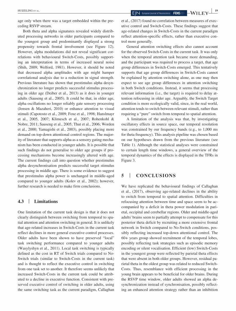
| 19HUIZELING Et aL.
age only when there was a target embedded within the pre-ceding RSVP stream.
Both theta and alpha signatures revealed widely distrib-uted processing networks in older participants compared to the youngest group and predominantly displayed a strong propensity towards frontal involvement (see Figure 12). However, alpha modulations did not reveal significant cor-relations with behavioural Switch- Costs, possibly support-ing an interpretation in terms of increased neural noise (Shih, 2009; Welford, 1981). However, it should be noted that decreased alpha amplitudes with age might hamper correlational analysis due to a reduction in signal strength. Previous literature has shown that prestimulus alpha desyn-chronization no longer predicts successful stimulus process-ing in older age (Deiber et al., 2013) as it does in younger adults (Sauseng et al., 2005). It could be that, in older age, alpha oscillations no longer reliably gate sensory processing (Jensen & Mazaheri, 2010) or enhance attention to visual stimuli (Capotosto et al., 2009; Foxe et al., 1998; Hanslmayr et al., 2005, 2007; Klimesch et al., 2007; Rohenkohl & Nobre, 2011; Sauseng et al., 2005; Thut et al., 2006; Worden et al., 2000; Yamagishi et al., 2003), possibly placing more demand on top- down attentional control regions. The major-ity of literature that supports alpha as a sensory gating mecha-nism has been conducted in younger adults. It is possible that such findings do not generalise to older age groups if pro-cessing mechanisms become increasingly altered with age. The current findings call into question whether prestimulus alpha desynchronisation predicts successful target stimulus processing in middle age. There is some evidence to suggest that prestimulus alpha power is unchanged in middle- aged compared to younger adults (Kolev et al., 2002); however, further research is needed to make firm conclusions.
4.3 | Limitations
One limitation of the current task design is that it does not clearly distinguish between switching from temporal to spa-tial attention and attention switching in general. It is unlikely that age- related increases in Switch- Costs in the current task reflect declines in more general executive control processes. Older adults have been shown to have preserved “local” task switching performance compared to younger adults (Wasylyshyn et al., 2011). Local task switching is typically defined as the cost in RT of Switch trials compared to No- Switch trials (similar to Switch- Costs in the current task) and is thought to reflect the executive control in switching from one task set to another. It therefore seems unlikely that increased Switch- Costs in the current task could be attrib-uted to a decline in executive function. Consistent with pre-served executive control of switching in older adults, using the same switching task as the current paradigm, Callaghan
et al., (2017) found no correlation between measures of exec-utive control and Switch- Costs. These findings suggest that age- related changes in Switch- Costs in the current paradigm reflect attention- specific effects, rather than executive con-trol more generally.
General attention switching effects also cannot account for the observed Switch- Costs in the current task. It was only when the temporal attention task became more demanding, and the participant was required to process a target, that age group differences in Switch- Costs emerged. This tentatively supports that age group differences in Switch- Costs cannot be explained by attention switching alone, as one may then expect to see age group differences in attention switching in both Switch conditions. Instead, it seems that processing relevant information (i.e., the target) is required to delay at-tention refocusing in older age. Arguably, the Switch Target condition is more ecologically valid, since, in the real world, attention tends to switch between relevant stimuli, rather than requiring a “pure” switch from temporal to spatial attention.
A limitation of the analysis was that, by investigating oscillatory effects in source space, our temporal resolution was constrained by our frequency bands (e.g., to 1,000 ms for theta frequency). This analysis pipeline was chosen based on our hypotheses drawn from the previous literature (see Table 1). Although the statistical analyses were constrained to certain length time windows, a general overview of the temporal dynamics of the effects is displayed in the TFRs in Figure 3.
5 | CONCLUSIONS
We have replicated the behavioural findings of Callaghan et al., (2017), observing age- related declines in the ability to switch from temporal to spatial attention. Difficulties in refocusing attention between time and space seem to be ac-companied by a deficit in theta power modulation in pari-etal, occipital and cerebellar regions. Older and middle- aged adults' brains seem to partially attempt to compensate for this posterior theta deficit by recruiting a more extensive frontal network in Switch compared to No- Switch conditions, pos-sibly reflecting increased top- down attentional control. The 60+ years group showed recruitment of the temporal lobes, possibly reflecting task strategies such as episodic memory encoding or silent vocalisation. Efficient (low) Switch- Costs in the youngest group were reflected by parietal theta effects that were absent in both older groups. However, residual pa-rietal theta in the oldest group was related to reduced Switch- Costs. Thus, resemblance with efficient processing in the young brain appears to be beneficial for older brains. During the RSVP time window, older adults showed an alpha de-synchronization instead of synchronisation, possibly reflect-ing an enhanced attention strategy rather than an inhibition

20 | HUIZELING Et aL.
strategy, as well as a stronger and more extensive task- related alpha power modulation across the cortex. In contrast to theta oscillations, alpha power modulations were not correlated with Switch- Costs, indicating that increases in the extent of power modulation could merely reflect dedifferentiation and/or reduced neural precision in older participants.
The pattern of group differences in RT- Switch- Costs was further reflected in alpha and theta results, where group dif-ferences were predominantly seen when the temporal atten-tion task involved target processing shortly before the switch, rather than when there was no target in the preceding tempo-ral attention task.
Overall, our results demonstrate that older adults may partially compensate for declines in attentional flexibility with the recruitment of theta- related neural mechanisms. These findings have important implications for future work, as they raise the question as to whether this recruit-ment can be enhanced with cognitive training programmes to improve compensation for functional decline. Improving older adults' attentional flexibility could improve their performance in everyday functions such as driving, where one is required to quickly switch between fast changing events in multiple surrounding locations (e.g., Huizeling et al., 2020).
ETHICS APPROVAL AND CONSENT TO PARTICIPATEParticipants provided written informed consent before par-ticipating and were screened for contraindications to having an MRI or MEG scan and received standard payment ac-cording to local rules. The research was approved by Aston University Research Ethics Committee (#776) and complied with the Declaration of Helsinki.
ACKNOWLEDGEMENTSThis research was supported by funding from The Rees Jeffreys Road Fund and by the School of Life and Health Sciences at Aston University. Scanning costs were supported by The Wellcome Trust Lab for MEG Studies and the Dr Hadwen Trust for Humane Research. In addition, we would like to thank colleagues at the Aston Brain Centre for assist-ing with MRI data acquisition.
CONFLICT OF INTERESTThe authors declare no conflict of interest.
AUTHOR CONTRIBUTIONSWriting- Review & Editing: E.H., H.W., C.H. and K.K.; Conceptualization and Methodology: E.H., C.H. and K.K.; Formal Analysis, Visualization, Data Collection and Curation and Project Administration: E.H.; Software: E.H. and H.W.; Writing- Original Draft Preparation: E.H.
and K.K.; Supervision and Funding Acquisition: C.H. and K.K.
PEER REVIEWThe peer review history for this article is available at https://publo ns.com/publo n/10.1111/ejn.15259.
DATA AVAILABILITY STATEMENTThe datasets collected and analysed during the current study are available from the corresponding author on reasonable request.
ORCIDEleanor Huizeling https://orcid.org/0000-0001-7338-5966
REFERENCESAdamo, M., Westerfield, M., Haist, F., & Townsend, J. (2003). Cross-
modal shifting attention in healthy aging: An fMRI study. Society for Neuroscience Abstract Viewer and Itinerary Planner, Abstract No. 933.938.
Allen, M., Poggiali, D., Whitaker, K., Marshall, T., van Langen, J., & Kievit, R. (2021). Raincloud plots: a multi- platform tool for robust data visualization. Wellcome Open Research, 4, 63. https://doi.org/10.12688/ wellc omeop enres.15191.2
Aydin, S., Strang, N. C., & Manahilov, V. (2013). Age- related deficits in attentional control of perceptual rivalry. Vision Research, 77, 32– 40. https://doi.org/10.1016/j.visres.2012.11.010
Ball, K. K., Roenker, D. L., Wadley, V. G., Edwards, J. D., Roth, D. L., McGwin, G., … Dube, T. (2006). Can high- risk older drivers be identified through performance- based measures in a department of motor vehicles setting? Journal of the American Geriatrics Society, 54(1), 77– 84. https://doi.org/10.1111/j.1532- 5415.2005. 00568.x
Bennett, I. J., Motes, M. A., Rao, N. K., & Rypma, B. (2012). White matter tract integrity predicts visual search performance in young and older adults. Neurobiology of Aging, 33(2), 433.e21– 433.e31. https://doi.org/10.1016/j.neuro biola ging.2011.02.001
Bögels, S., Barr, D. J., Garrod, S., & Kessler, K. (2014). Conversational interaction in the scanner: Mentalizing during language processing as revealed by MEG. Cerebral Cortex, 25(9), 3219– 3234. https://doi.org/10.1093/cerco r/bhu116
Buckner, R. L., Snyder, A. Z., Sanders, A. L., Raichle, M. E., & Morris, J. C. (2000). Functional brain imaging of young, nondemented, and demented older adults. Journal of Cognitive Neuroscience, 12, 24– 34. https://doi.org/10.1162/08989 29005 64046
Budescu, D. V. (1982). The power of the F test in normal populations with heterogeneous variances. Educational and Psychological Measurement, 42(2), 409– 416. https://doi.org/10.1177/00131 64482 04200202
Cabeza, R. (2002). Hemispheric asymmetry reduction in older adults: The HAROLD model. Psychology and Aging, 17(1), 85– 100. https://doi.org/10.1037//0882- 7974.17.1.85
Cabeza, R., Albert, M., Belleville, S., Craik, F. I. M., Duarte, A., Grady, C. L., … Rajah, M. N. (2018). Maintenance, reserve and compensation: The cognitive neuroscience of healthy ageing. Nature Reviews Neuroscience, 19(11), 701– 710. https://doi.org/10.1038/s4158 3- 018- 0068- 2
| 21HUIZELING Et aL.
Cabeza, R., Daselaar, S. M., Dolcos, F., Prince, S. E., Budde, M., & Nyberg, L. (2004). Task- independent and task- specific age effects on brain activity during working memory, visual attention and episodic retrieval. Cerebral Cortex, 14(4), 364– 375. https://doi.org/10.1093/cerco r/bhg133
Callaghan, E., Holland, C., & Kessler, K. (2017). Age- related changes in the ability to switch between temporal and spatial attention. Frontiers in Aging Neuroscience, 9, 28. https://doi.org/10.3389/fnagi.2017.00028
Capotosto, P., Babiloni, C., Romani, G. L., & Corbetta, M. (2009). Frontoparietal cortex controls spatial attention through modulation of anticipatory alpha rhythms. The Journal of Neuroscience, 29(18), 5863– 5872. https://doi.org/10.1523/JNEUR OSCI.0539- 09.2009
Cavanagh, J. F., Cohen, M. X., & Allen, J. J. B. (2009). Prelude to and resolution of an error: EEG phase synchrony reveals cogni-tive control dynamica during action monitoring. The Journal of Neuroscience: The Official Journal of the Society for Neuroscience, 29(1), 98– 105. https://doi.org/10.1523/JNEUR OSCI.4137- 08.2009
Cavanagh, J. F., & Frank, M. J. (2014). Frontal theta as a mechanism for cognitive control. Trends in Cognitive Sciences, 18(8), 414– 421. https://doi.org/10.1016/j.tics.2014.04.012
Cohen, J. (1992). A power primer. Psychological Bulletin, 112(1), 155. https://doi.org/10.1037/0033- 2909.112.1.155
Coull, J. T., & Nobre, A. C. (1998). Where and when to pay attention: The neural systems for directing attention to spatial locations and to time intervals as revealed by both PET and fMRI. Journal of Neuroscience, 18(18), 7426– 7435. https://doi.org/10.1523/JNEUR OSCI.18- 18- 07426.1998
Cummins, T. D. R., & Finnigan, S. (2007). Theta power is reduced in healthy cognitive aging. International Journal of Psychophysiology, 66(1), 10– 17. https://doi.org/10.1016/j.ijpsy cho.2007.05.008
Daselaar, S. M., Iyengar, V., Davis, S. W., Eklund, K., Hayes, S. M., & Cabeza, R. E. (2015). Less wiring, more firing: Low- performing older adults compensate for impaired white matter with greater neural activity. Cerebral Cortex, 25(4), 983– 990. https://doi.org/10.1093/cerco r/bht289
Davis, S. W., Dennis, N. A., Daselaar, S. M., Fleck, M. S., & Cabeza, R. (2008). Qué PASA? The posterior- anterior shift in aging. Cerebral Cortex, 18(5), 1201– 1209. https://doi.org/10.1093/cerco r/bhm155
Deiber, M.- P., Ibanez, V., Missonnier, P., Rodriguez, C., & Giannakopoulos, P. (2013). Age- associated modulations of cerebral oscillatory patterns related to attention control. NeuroImage, 82, 531– 546. https://doi.org/10.1016/j.neuro image.2013.06.037
Demiralp, T., & Başar, E. (1992). Theta rhythmicities follow-ing expected visual and auditory targets. International Journal of Psychophysiology, 13(2), 147– 160. https://doi.org/10.1016/0167- 8760(92)90054 - F
Dempster, F. N. (1992). The rise and fall of the inhibitory mech-anism: Toward a unified theory of cognitive development and aging. Developmental Review, 12(1), 45– 75. https://doi.org/10.1016/0273- 2297(92)90003 - K
Fabiani, M., Low, K. A., Wee, E., Sable, J. J., & Gratton, G. (2006). Reduced suppression or labile memory? Mechanisms of ineffi-cient filtering of irrelevant information in older adults. Journal of Cognitive Neuroscience, 18(4), 637– 650. https://doi.org/10.1162/jocn.2006.18.4.637
Foster, J. K., Behrmann, M., & Stuss, D. T. (1995). Aging and visual search: Generalized cognitive slowing or selective deficit in attention?
Aging and Cognition, 2(4), 279– 299. https://doi.org/10.1080/13825 58950 8256604
Foxe, J. J., Simpson, G. V., & Ahlfors, S. P. (1998). Parieto- occipital ∼1 0 Hz activity reflects anticipatory state of visual attention mecha-nisms. NeuroReport, 9(17), 3929– 3933.
Fu, S. M., Greenwood, P. M., & Parasuraman, R. (2005). Brain mech-anisms of involuntary visuospatial attention: An event- related po-tential study. Human Brain Mapping, 25(4), 378– 390. https://doi.org/10.1002/hbm.20108
Gazzaley, A., Clapp, W., Kelley, J., McEvoy, K., Knight, R. T., & D'Esposito, M. (2008). Age- related top- down suppression deficit in the early stages of cortical visual memory processing. Proceedings of the National Academy of Sciences, 105(35), 13122– 13126. https://doi.org/10.1073/pnas.08060 74105
Georgiou- Karistianis, N., Tang, J., Mehmedbegovic, F., Farrow, M., Bradshaw, J., & Sheppard, D. (2006). Age- related differences in cognitive function using a global local hierarchical paradigm. Brain Research, 1124, 86– 95. https://doi.org/10.1016/j.brain res.2006.09.070
Gómez, C., Pérez- Macías, J. M., Poza, J., Fernández, A., & Hornero, R. (2013). Spectral changes in spontaneous MEG activity across the lifespan. Journal of Neural Engineering, 10(6), 066006. https://doi.org/10.1088/1741- 2560/10/6/066006
Grady, C. L. (2000). Functional brain imaging and age- related changes in cognition. Biological Psychology, 54(1– 3), 259– 281. https://doi.org/10.1016/s0301 - 0511(00)00059 - 4
Graves, W. W., Grabowski, T. J., Mehta, S., & Gordon, J. K. (2007). A neural signature of phonological access: Distinguishing the effects of word frequency from familiarity and length in overt picture nam-ing. Journal of Cognitive Neuroscience, 19(4), 617– 631. https://doi.org/10.1162/jocn.2007.19.4.617
Green, J. J., & McDonald, J. J. (2008). Electrical neuroimaging reveals timing of attentional control activity in human brain. PLoS Biology, 6(4), 730– 738. https://doi.org/10.1371/journ al.pbio.0060081
Gross, J., Kujala, J., Hämäläinen, M., Timmermann, L., Schnitzler, A., & Salmelin, R. (2001). Dynamic imaging of coherent sources: Studying neural interactions in the human brain. Proceedings of the National Academy of Sciences, 98(2), 694– 699. https://doi.org/10.1073/pnas.98.2.694
Gross, J., Schmitz, F., Schnitzler, I., Kessler, K., Shapiro, K., Hommel, B., & Schnitzler, A. (2004). Modulation of long- range neural syn-chrony reflects temporal limitations of visual attention in humans. Proceedings of the National Academy of Sciences of the United States of America, 101(35), 13050– 13055. https://doi.org/10.1073/pnas.04049 44101
Hale, S., Myerson, J., Smith, G. A., & Poon, L. W. (1988). Age, variabil-ity, and speed: Between- subjects diversity. Psychology and Aging, 3(4), 407. https://doi.org/10.1037/0882- 7974.3.4.407
Hanslmayr, S., Aslan, A., Staudigl, T., Klimesch, W., Herrmann, C. S., & Bäuml, K.- H. (2007). Prestimulus oscillations predict visual perception performance between and within subjects. NeuroImage, 37(4), 1465– 1473. https://doi.org/10.1016/j.neuro image.2007.07.011
Hanslmayr, S., Klimesch, W., Sauseng, P., Gruber, W., Doppelmayr, M., Freunberger, R., & Pecherstorfer, T. (2005). Visual discrimination performance is related to decreased alpha amplitude but increased phase locking. Neuroscience Letters, 375(1), 64– 68. https://doi.org/10.1016/j.neulet.2004.10.092
22 | HUIZELING Et aL.
Hickok, G., & Poeppel, D. (2007). The cortical organization of speech processing. Nature Reviews Neuroscience, 8, 393. https://doi.org/10.1038/nrn2113
Hillebrand, A., & Barnes, G. R. (2002). A quantitative assessment of the sensitivity of whole- head MEG to activity in the adult human cor-tex. Neuroimage, 16(3, Part A), 638– 650. https://doi.org/10.1006/nimg.2002.1102
Hocking, J., & Price, C. J. (2009). Dissociating verbal and nonverbal audiovisual object processing. Brain and Language, 108(2), 89– 96. https://doi.org/10.1016/j.bandl.2008.10.005
Hong, X. F., Sun, J. F., Bengson, J. J., Mangun, G. R., & Tong, S. B. (2015). Normal aging selectively diminishes alpha lateralization in visual spatial attention. NeuroImage, 106, 353– 363. https://doi.org/10.1016/j.neuro image.2014.11.019
Huettel, S. A., Singerman, J. D., & McCarthy, G. (2001). The effects of aging upon the hemodynamic response measured by func-tional MRI. NeuroImage, 13(1), 161– 175. https://doi.org/10.1006/nimg.2000.0675
Huizeling, E., Wang, H., Holland, C., & Kessler, K. (2020). Age- related changes in attentional refocusing during simulated driving. Brain Sciences, 10(8), 530. https://doi.org/10.3390/brain sci10 080530
Humphrey, D. G., & Kramer, A. F. (1997). Age differences in visual search for feature, conjunction, and triple- conjunction targets. Psychology and Aging, 12(4), 704– 717. https://doi.org/10.1037//0882- 7974.12.4.704
Jefferies, L., Roggeveen, A., Enns, J., Bennett, P., Sekuler, A., & Di Lollo, V. (2015). On the time course of attentional focusing in older adults. Psychological Research Psychologische Forschung, 79(1), 28– 41. https://doi.org/10.1007/s0042 6- 013- 0528- 2
Jensen, O., & Mazaheri, A. (2010). Shaping functional architecture by oscillatory alpha activity: Gating by inhibition. Frontiers in Human Neuroscience, 4, 186. https://doi.org/10.3389/fnhum.2010.00186
Klimesch, W., Sauseng, P., & Hanslmayr, S. (2007). EEG alpha oscil-lations: The inhibition– timing hypothesis. Brain Research Reviews, 53(1), 63– 88. https://doi.org/10.1016/j.brain resrev.2006.06.003
Kolev, V., Yordanova, J., Basar- Eroglu, C., & Basar, E. (2002). Age effects on visual EEG responses reveal distinct frontal alpha net-works. Clinical Neurophysiology, 113(6), 901– 910. https://doi.org/10.1016/S1388 - 2457(02)00106 - 2
Lague- Beauvais, M., Brunet, J., Gagnon, L., Lesage, F., & Bherer, L. (2013). A fNIRS investigation of switching and inhibition during the modified Stroop task in younger and older adults. NeuroImage, 64, 485– 495. https://doi.org/10.1016/j.neuro image.2012.09.042
Lahar, C. J., Isaak, M. I., & McArthur, A. D. (2001). Age differences in the magnitude of the attentional blink. Aging, Neuropsychology, and Cognition, 8(2), 149– 159. https://doi.org/10.1076/anec.8.2.149.842
Lee, T.- Y., & Hsieh, S. (2009). The limits of attention for visual per-ception and action in aging. Aging Neuropsychology and Cognition, 16(3), 311– 329. https://doi.org/10.1080/13825 58090 2741351
Li, L., Gratton, C., Fabiani, M., & Knighta, R. T. (2013). Age- related frontoparietal changes during the control of bottom- up and top- down attention: An ERP study. Neurobiology of Aging, 34(2), 477– 488.
Maciokas, J. B., & Crognale, M. A. (2003). Cognitive and atten-tional changes with age: Evidence from attentional blink defi-cits. Experimental Aging Research, 29(2), 137– 153. https://doi.org/10.1080/03610 73039 0176095
Madden, D. J. (2007). Aging and visual attention. Current Directions in Psychological Science, 16(2), 70– 74. https://doi.org/10.1111/j.1467- 8721.2007.00478.x
Madden, D. J., & Gottlob, L. R. (1997). Adult age differences in strategic and dynamic components of focusing visual attention. Aging Neuropsychology and Cognition, 4(3), 185– 210. https://doi.org/10.1080/13825 58970 8256647
Madden, D. J., Spaniol, J., Whiting, W. L., Bucur, B., Provenzale, J. M., Cabeza, R., … Huettel, S. A. (2007). Adult age differences in the functional neuroanatomy of visual attention: A combined fMRI and DTI study. Neurobiology of Aging, 28(3), 459– 476. https://doi.org/10.1016/j.neuro biola ging.2006.01.005
Madden, D. J., Turkington, T. G., Provenzale, J. M., Denny, L. L., Langley, L. K., Hawk, T. C., & Coleman, R. E. (2002). Aging and attentional guidance during visual search: Functional neuroanatomy by positron emission tomography. Psychology and Aging, 17(1), 24– 43. https://doi.org/10.1037//0882- 7974.17.1.24
Maris, E., & Oostenveld, R. (2007). Nonparametric statistical testing of EEG- and MEG- data. Journal of Neuroscience Methods, 164(1), 177– 190. https://doi.org/10.1016/j.jneum eth.2007.03.024
McLaughlin, P. M., & Murtha, S. J. E. (2010). The effects of age and exogenous support on visual search performance. Experimental Aging Research, 36(3), 325– 345. https://doi.org/10.1080/03610 73x.2010.484752
Min, B.- K., & Park, H.- J. (2010). Task- related modulation of anterior theta and posterior alpha EEG reflects top- down preparation. BMC Neuroscience, 11(1), 1– 8. https://doi.org/10.1186/1471- 2202- 11- 79
Morse, C. K. (1993). Does variability increase with age? An archival study of cognitive measures. Psychology and Aging, 8(2), 156. https://doi.org/10.1037/0882- 7974.8.2.156
Nagamatsu, L. S., Munkacsy, M., Liu- Ambrose, T., & Handy, T. C. (2013). Altered visual- spatial attention to task- irrelevant information is associated with falls risk in older adults. Neuropsychologia, 51(14), 3025– 3032. https://doi.org/10.1016/j.neuro psych ologia.2013.10.002
Neider, M. B., & Kramer, A. F. (2011). Older adults capitalize on con-textual information to guide search. Experimental Aging Research, 37(5), 539– 571. https://doi.org/10.1080/03610 73x.2011.619864
Noone, P. (2015). Addenbrooke’s cognitive examination- III. Occupational Medicine, 65(5), 418– 420. https://doi.org/10.1093/occme d/kqv041
Nyberg, L., Lovden, M., Riklund, K., Lindenberger, U., & Backman, L. (2012). Memory aging and brain maintenance. Trends in Cognitive Sciences, 16(5), 292– 305. https://doi.org/10.1016/j.tics.2012.04.005
Oostenveld, R., Fries, P., Maris, E., & Schoffelen, J.- M. (2011). FieldTrip: Open source software for advanced analysis of MEG, EEG, and in-vasive electrophysiological data. Computational Intelligence and Neuroscience, 2011, 1– 9. https://doi.org/10.1155/2011/156869
Pagano, S., Fait, E., Monti, A., Brignani, D., & Mazza, V. (2015). Electrophysiological correlates of subitizing in healthy aging. PLoS One, 10(6), e0131063. https://doi.org/10.1371/journ al.pone.0131063
Park, D. C., & Reuter- Lorenz, P. (2009). The adaptive brain: Aging and neurocognitive scaffolding. Annual Review of Psychology, 60(1), 173– 196. https://doi.org/10.1146/annur ev.psych.59.103006.093656
Plude, D. J., & Doussard- Roosevelt, J. A. (1989). Aging, selective atten-tion and feature integration. Psychology and Aging, 4(1), 98– 105. https://doi.org/10.1037//0882- 7974.4.1.98
R Core Team. (2018). R: A language and environment for statistical computing. R Foundation for Statistical Computing.
Raymond, J. E., Shapiro, K. L., & Arnell, K. M. (1992). Temporary suppression of visual processing in an RSVP task: An attentional blink? Journal of Experimental Psychology: Human Perception
| 23HUIZELING Et aL.
and Performance, 18(3), 849. https://doi.org/10.1037/0096- 1523.18.3.849
Reichert, J. L., Kober, S. E., Witte, M., Neuper, C., & Wood, G. (2016). Age- related effects on verbal and visuospatial memory are mediated by theta and alpha II rhythms. International Journal of Psychophysiology, 99, 67– 78. https://doi.org/10.1016/j.ijpsy cho.2015.11.004
Reuter, E.- M., Vieluf, S., Koutsandreou, F., Hübner, L., Budde, H., Godde, B., & Voelcker- Rehage, C. (2019). A non- linear relationship between selective attention and associated ERP markers across the lifespan. Frontiers in Psychology, 10, 30. https://doi.org/10.3389/fpsyg.2019.00030
Reuter- Lorenz, P. A., & Park, D. C. (2014). How does it STAC up? Revisiting the scaffolding theory of aging and cognition. Neuropsychology Review, 24(3), 355– 370. https://doi.org/10.1007/s1106 5- 014- 9270- 9
Rohenkohl, G., & Nobre, A. C. (2011). Alpha oscillations related to anticipatory attention follow temporal expectations. Journal of Neuroscience, 31(40), 14076– 14084. https://doi.org/10.1523/jneur osci.3387- 11.2011
Ross, M. H., Yurgelun- Todd, D. A., Renshaw, P. F., Maas, L. C., Mendelson, J. H., Mello, N. K., … Levin, J. M. (1997). Age- related reduction in functional MRI response to photic stim-ulation. Neurology, 48(1), 173– 176. https://doi.org/10.1212/WNL.48.1.173
Rubin, G. S., Ng, E. S., Bandeen- Roche, K., Keyl, P. M., Freeman, E. E., & West, S. K. (2007). A prospective, population- based study of the role of visual impairment in motor vehicle crashes among older drivers: The SEE study. Investigative Ophthalmology and Visual Science, 48(4), 1483. https://doi.org/10.1167/iovs.06- 0474
Salthouse, T. A. (1996). The processing- speed theory of adult age dif-ferences in cognition. Psychological Review, 103(3), 403. https://doi.org/10.1037/0033- 295X.103.3.403
Sauseng, P., Griesmayr, B., Freunberger, R., & Klimesch, W. (2010). Control mechanisms in working memory: A possible func-tion of EEG theta oscillations. Neuroscience & Biobehavioral Reviews, 34(7), 1015– 1022. https://doi.org/10.1016/j.neubi orev.2009.12.006
Sauseng, P., Klimesch, W., Stadler, W., Schabus, M., Doppelmayr, M., Hanslmayr, S., … Birbaumer, N. (2005). A shift of visual spatial attention is selectively associated with human EEG alpha activity. European Journal of Neuroscience, 22(11), 2917– 2926. https://doi.org/10.1111/j.1460- 9568.2005.04482.x
Schacter, D. L., & Wagner, A. D. (1999). Medial temporal lobe activa-tions in fMRI and PET studies of episodic encoding and retrieval. Hippocampus, 9(1), 7– 24. https://doi.org/10.1002/(sici)1098- 1063
Shih, S.- I. (2009). Using the attention cascade model to probe cog-nitive aging. Psychology and Aging, 24(3), 550– 562. https://doi.org/10.1037/a0016724
Smith, E. E., Jonides, J., Marshuetz, C., & Koeppe, R. A. (1998). Components of verbal working memory: Evidence from neuroim-aging. Proceedings of the National Academy of Sciences, 95(3), 876– 882.
Snedecor, G. W., & Cochran, W. (1967). Statistical methods (pp. 593). Iowa State Univ.
Taulu, S., & Hari, R. (2009). Removal of magnetoencephalographic ar-tifacts with temporal signal- space separation: Demonstration with single- trial auditory- evoked responses. Human Brain Mapping, 30(5), 1524– 1534. https://doi.org/10.1002/hbm.20627
Thut, G., Nietzel, A., Brandt, S. A., & Pascual- Leone, A. (2006). alpha- Band electroencephalographic activity over occipital cortex indexes visuospatial attention bias and predicts visual target de-tection. Journal of Neuroscience, 26(37), 9494– 9502. https://doi.org/10.1523/jneur osci.0875- 06.2006
Torrens- Burton, A., Hanley, C. J., Wood, R., Basoudan, N., Norris, J. E., Richards, E., & Tales, A. (2020). Lacking pace but not preci-sion: Age- related information processing changes in response to a dynamic attentional control task. Brain Sciences, 10(6), 390. https://doi.org/10.3390/brain sci10 060390
Vaden, R. J., Hutcheson, N. L., McCollum, L. A., Kentros, J., & Visscher, K. M. (2012). Older adults, unlike younger adults, do not modulate alpha power to suppress irrelevant information. NeuroImage, 63(3), 1127– 1133. https://doi.org/10.1016/j.neuro image.2012. 07.050
van de Vijver, I., Cohen, M. X., & Ridderinkhof, K. R. (2014). Aging affects medial but not anterior frontal learning- related theta os-cillations. Neurobiology of Aging, 35(3), 692– 704. https://doi.org/10.1016/j.neuro biola ging.2013.09.006
van Diepen, R. M., Cohen, M. X., Denys, D., & Mazaheri, A. (2015). Attention and temporal expectations modulate power, not phase, of ongoing alpha oscillations. Journal of Cognitive Neuroscience, 27(8), 1573– 1586. https://doi.org/10.1162/jocn_a_00803
Van Diepen, R. M., Foxe, J. J., & Mazaheri, A. (2019). The functional role of alpha- band activity in attentional processing: The current zeitgeist and future outlook. Current Opinion in Psychology, 29, 229– 238. https://doi.org/10.1016/j.copsyc.2019.03.015
van Leeuwen, S., Müller, N. G., & Melloni, L. (2009). Age effects on attentional blink performance in meditation. Consciousness and Cognition, 18(3), 593– 599. https://doi.org/10.1016/j.concog.2009.05.001
Wang, H., Callaghan, E., Gooding- Williams, G., McAllister, C., & Kessler, K. (2016). Rhythm makes the world go round: An MEG- TMS study on the role of right TPJ theta oscillations in embodied perspective taking. Cortex, 75, 68– 81. https://doi.org/10.1016/j.cortex.2015.11.011
Wasylyshyn, C., Verhaeghen, P., & Sliwinski, M. J. (2011). Aging and task switching: A meta- analysis. Psychology and Aging, 26(1), 15– 20. https://doi.org/10.1037/a0020912
Watson, D. G., & Maylor, E. A. (2002). Aging and visual marking: Selective deficits for moving stimuli. Psychology and Aging, 17(2), 321– 339. https://doi.org/10.1037/0882- 7974.17.2.321
Welford, A. T. (1981). Signal, noise, performance, and age. Human Factors, 23(1), 97– 109. https://doi.org/10.1177/00187 20881 02300109
West, R. L. (1996). An application of prefrontal cortex function theory to cognitive aging. Psychological Bulletin, 120(2), 272– 292. https://doi.org/10.1037/0033- 2909.120.2.272
Wiesman, A. I., & Wilson, T. W. (2019). The impact of age and sex on the oscillatory dynamics of visuospatial processing. NeuroImage, 185, 513– 520. https://doi.org/10.1016/j.neuro image.2018. 10.036
Worden, M. S., Foxe, J. J., Wang, N., & Simpson, G. V. (2000). Anticipatory biasing of visuospatial attention indexed by retinoto-pically specific alpha- band electroencephalography increases over occipital cortex. Journal of Neuroscience, 20(6), art. no.- RC63.
Xia, M., Wang, J., & He, Y. (2013). BrainNet Viewer: A network visual-ization tool for human brain connectomics. PLoS One, 8(7), e68910. https://doi.org/10.1371/journ al.pone.0068910

24 | HUIZELING Et aL.
Yamagishi, N., Callan, D. E., Goda, N., Anderson, S. J., Yoshida, Y., & Kawato, M. (2003). Attentional modulation of oscillatory activ-ity in human visual cortex. NeuroImage, 20(1), 98– 113. https://doi.org/10.1016/S1053 - 8119(03)00341 - 0
Zanto, T. P., Pan, P., Liu, H., Bollinger, J., Nobre, A. C., & Gazzaley, A. (2011). Age- related changes in orienting attention in time. Journal of Neuroscience, 31(35), 12461– 12470. https://doi.org/10.1523/jneur osci.1149- 11.2011
Zhou, S.- S., Fan, J., Lee, T. M. C., Wang, C.- Q., & Wang, K. (2011). Age- related differences in attentional networks of alerting and ex-ecutive control in young, middle- aged, and older Chinese adults. Brain and Cognition, 75(2), 205– 210. https://doi.org/10.1016/j.bandc.2010.12.003
SUPPORTING INFORMATIONAdditional supporting information may be found online in the Supporting Information section.
How to cite this article: Huizeling E, Wang H, Holland C, Kessler K. Changes in theta and alpha oscillatory signatures of attentional control in older and middle age. Eur J Neurosci. 2021;00:1– 24. https://doi.org/10.1111/ejn.15259